The pentapeptide LQVVR plays a pivotal role in human cystatin C fibrillization
6
The pentapeptide LQVVR plays a pivotal role in human cystatin C fibrillization Paraskevi L. Tsiolaki, Stavros J. Hamodrakas, Vassiliki A. Iconomidou ⇑ Department of Cell Biology and Biophysics, Faculty of Biology, University of Athens, Panepistimiopolis, Athens 157 01, Greece article info Article history: Received 1 October 2014 Revised 10 November 2014 Accepted 19 November 2014 Available online 3 December 2014 Edited by Jesus Avila Keywords: Human cystatin C Hereditary Cystatin C Amyloid Angiopathy Alzheimer’s disease 3D-domain swapping ‘Aggregation-prone’ pentapeptide LQVVR Amyloid fibrils abstract Human cystatin C (HCC) is a low molecular weight member of the cystatin family (type2). HCC consists of 120 amino acids. Normally it is an inhibitor of cysteine proteases, but in pathological conditions it forms amyloid fibrils in brain arteries of young adults. An ‘aggregation-prone’ penta- peptide ( 47 LQVVR 51 ) was located within the HCC sequence using AmylPred, an ‘aggregation-prone’ peptide prediction algorithm developed in our lab. This peptide was synthesized and self-assembled into amyloid-like fibrils in vitro, as electron microscopy, X-ray fiber diffraction, Attenuated Total Reflectance Fourier-Transform Spectroscopy and Congo red staining studies reveal. Thus, the 47 LQVVR 51 peptide seems to have an important role in HCC fibrillization. Ó 2014 Published by Elsevier B.V. on behalf of the Federation of European Biochemical Societies. 1. Introduction Human cystatins are proteins that have the capacity to regulate normal body processes and usually their down-regulation causes several diseases [1]. The first cystatin sequence determined was that for a protein isolated from human urine, which was first called c-trace due to its electrophoretic mobility and it was later renamed cystatin C [2]. Cystatins form a single chain ‘superfamily’ of revers- ible inhibitors of cysteine proteases of the papain-like (C1) and legumain-related (C13) families, which consists of three major types, with proteins homologous in sequence [3]. These groups, based on molecular organization, have different distributions in an organism. It is obvious that evolutionary pressure has created different branches of cystatins by duplication events far back in time. Today, we know that cystatins are present in many kinds of eukaryotic organisms such as mammals, birds, fish, insects, plants and some protozoa [4]. The basic structural fold of monomeric representatives of the cystatin superfamily has been defined by the crystal structure of chicken cystatin [5]. Human cystatin C (HCC) is a low molecular weight protein which belongs to the cystatin family [6]. HCC, expressed in all nucleated human cells, is a secretory type 2 cystatin, and it has, predominantly, extracellular functions. It is widely used as an endogenous marker of several illnesses since its presence in all types of cells seems to be independent of age, sex and muscle mass. It is found in all tissues and body fluids [4] and it is present at particularly high concentrations in cerebrospinal fluid [2,6]. It con- sists of a polypeptide chain of 120 amino acid residues [3] and its canonical structural features include a long alpha helix (a1 helix) running across a large, five-stranded antiparallel beta-sheet (Fig. 1). Recently, a monomer-stabilized HCC with an engineered disulfide bond [(L47C)–(G69C)] has been created by Koladziejczyk et al. [5]. Three regions are implicated in its interaction with the enzyme papain family: The N-terminal segment and the two hair- pin loops L1 and L2, constitute a structural epitope, which shows high affinity and binds to the enzyme [7] (Fig. 1). HCC is co-deposited in the amyloid plaques of Alzheimer’s dis- ease and Down’s syndrome [8] and it is also involved in tissue- degenerative diseases such as osteoporosis and periodontosis [9]. In pathological processes, it forms amyloid deposits in brain arter- ies of young adults, which lead to fatal cerebral hemorrhage. In advanced age, under denaturing conditions the protein oligomeri- zation leads to amyloid deposits in the leptomeninges, cerebral cortex and brainstem [10]. These amyloid fibrils are composed of a mutated and truncated variant of cystatin C that eventually http://dx.doi.org/10.1016/j.febslet.2014.11.041 0014-5793/Ó 2014 Published by Elsevier B.V. on behalf of the Federation of European Biochemical Societies. Abbreviations: HCC, human cystatin C; HCCAA, Hereditary Cystatin C Amyloid Angiopathy; ATR FT-IR Spectroscopy, Attenuated Total Reflectance Fourier-Transform Spectroscopy ⇑ Corresponding author. Fax: +30 210 7274254. E-mail address: [email protected] (V.A. Iconomidou). FEBS Letters 589 (2015) 159–164 journal homepage: www.FEBSLetters.org
-
Upload
independent -
Category
Documents
-
view
3 -
download
0
Transcript of The pentapeptide LQVVR plays a pivotal role in human cystatin C fibrillization

FEBS Letters 589 (2015) 159–164
journal homepage: www.FEBSLetters .org
The pentapeptide LQVVR plays a pivotal role in human cystatin Cfibrillization
http://dx.doi.org/10.1016/j.febslet.2014.11.0410014-5793/� 2014 Published by Elsevier B.V. on behalf of the Federation of European Biochemical Societies.
Abbreviations: HCC, human cystatin C; HCCAA, Hereditary Cystatin C AmyloidAngiopathy; ATR FT-IR Spectroscopy, Attenuated Total ReflectanceFourier-Transform Spectroscopy⇑ Corresponding author. Fax: +30 210 7274254.
E-mail address: [email protected] (V.A. Iconomidou).
Paraskevi L. Tsiolaki, Stavros J. Hamodrakas, Vassiliki A. Iconomidou ⇑Department of Cell Biology and Biophysics, Faculty of Biology, University of Athens, Panepistimiopolis, Athens 157 01, Greece
a r t i c l e i n f o
Article history:Received 1 October 2014Revised 10 November 2014Accepted 19 November 2014Available online 3 December 2014
Edited by Jesus Avila
Keywords:Human cystatin CHereditary Cystatin C Amyloid AngiopathyAlzheimer’s disease3D-domain swapping‘Aggregation-prone’ pentapeptide LQVVRAmyloid fibrils
a b s t r a c t
Human cystatin C (HCC) is a low molecular weight member of the cystatin family (type2). HCCconsists of 120 amino acids. Normally it is an inhibitor of cysteine proteases, but in pathologicalconditions it forms amyloid fibrils in brain arteries of young adults. An ‘aggregation-prone’ penta-peptide (47LQVVR51) was located within the HCC sequence using AmylPred, an ‘aggregation-prone’peptide prediction algorithm developed in our lab. This peptide was synthesized and self-assembledinto amyloid-like fibrils in vitro, as electron microscopy, X-ray fiber diffraction, Attenuated TotalReflectance Fourier-Transform Spectroscopy and Congo red staining studies reveal. Thus, the47LQVVR51 peptide seems to have an important role in HCC fibrillization.
� 2014 Published by Elsevier B.V. on behalf of the Federation of European Biochemical Societies.
1. Introduction Human cystatin C (HCC) is a low molecular weight protein
Human cystatins are proteins that have the capacity to regulatenormal body processes and usually their down-regulation causesseveral diseases [1]. The first cystatin sequence determined wasthat for a protein isolated from human urine, which was first calledc-trace due to its electrophoretic mobility and it was later renamedcystatin C [2]. Cystatins form a single chain ‘superfamily’ of revers-ible inhibitors of cysteine proteases of the papain-like (C1) andlegumain-related (C13) families, which consists of three majortypes, with proteins homologous in sequence [3]. These groups,based on molecular organization, have different distributions inan organism. It is obvious that evolutionary pressure has createddifferent branches of cystatins by duplication events far back intime. Today, we know that cystatins are present in many kinds ofeukaryotic organisms such as mammals, birds, fish, insects, plantsand some protozoa [4]. The basic structural fold of monomericrepresentatives of the cystatin superfamily has been defined bythe crystal structure of chicken cystatin [5].
which belongs to the cystatin family [6]. HCC, expressed in allnucleated human cells, is a secretory type 2 cystatin, and it has,predominantly, extracellular functions. It is widely used as anendogenous marker of several illnesses since its presence in alltypes of cells seems to be independent of age, sex and muscle mass.It is found in all tissues and body fluids [4] and it is present atparticularly high concentrations in cerebrospinal fluid [2,6]. It con-sists of a polypeptide chain of 120 amino acid residues [3] and itscanonical structural features include a long alpha helix (a1 helix)running across a large, five-stranded antiparallel beta-sheet(Fig. 1). Recently, a monomer-stabilized HCC with an engineereddisulfide bond [(L47C)–(G69C)] has been created by Koladziejczyket al. [5]. Three regions are implicated in its interaction with theenzyme papain family: The N-terminal segment and the two hair-pin loops L1 and L2, constitute a structural epitope, which showshigh affinity and binds to the enzyme [7] (Fig. 1).
HCC is co-deposited in the amyloid plaques of Alzheimer’s dis-ease and Down’s syndrome [8] and it is also involved in tissue-degenerative diseases such as osteoporosis and periodontosis [9].In pathological processes, it forms amyloid deposits in brain arter-ies of young adults, which lead to fatal cerebral hemorrhage. Inadvanced age, under denaturing conditions the protein oligomeri-zation leads to amyloid deposits in the leptomeninges, cerebralcortex and brainstem [10]. These amyloid fibrils are composed ofa mutated and truncated variant of cystatin C that eventually
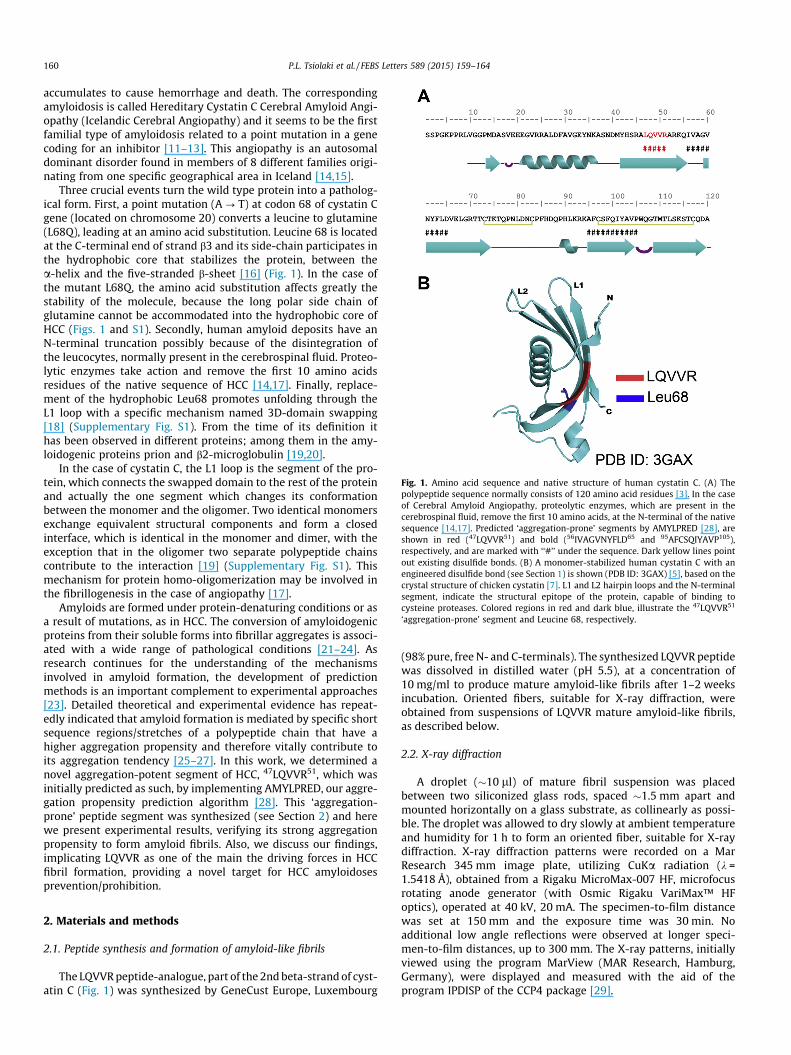
Fig. 1. Amino acid sequence and native structure of human cystatin C. (A) Thepolypeptide sequence normally consists of 120 amino acid residues [3]. In the caseof Cerebral Amyloid Angiopathy, proteolytic enzymes, which are present in thecerebrospinal fluid, remove the first 10 amino acids, at the N-terminal of the nativesequence [14,17]. Predicted ‘aggregation-prone’ segments by AMYLPRED [28], areshown in red (47LQVVR51) and bold (56IVAGVNYFLD65 and 95AFCSQIYAVP105),respectively, and are marked with ‘‘#’’ under the sequence. Dark yellow lines pointout existing disulfide bonds. (B) A monomer-stabilized human cystatin C with anengineered disulfide bond (see Section 1) is shown (PDB ID: 3GAX) [5], based on thecrystal structure of chicken cystatin [7]. L1 and L2 hairpin loops and the N-terminalsegment, indicate the structural epitope of the protein, capable of binding tocysteine proteases. Colored regions in red and dark blue, illustrate the 47LQVVR51
‘aggregation-prone’ segment and Leucine 68, respectively.
160 P.L. Tsiolaki et al. / FEBS Letters 589 (2015) 159–164
accumulates to cause hemorrhage and death. The correspondingamyloidosis is called Hereditary Cystatin C Cerebral Amyloid Angi-opathy (Icelandic Cerebral Angiopathy) and it seems to be the firstfamilial type of amyloidosis related to a point mutation in a genecoding for an inhibitor [11–13]. This angiopathy is an autosomaldominant disorder found in members of 8 different families origi-nating from one specific geographical area in Iceland [14,15].
Three crucial events turn the wild type protein into a patholog-ical form. First, a point mutation (A ? T) at codon 68 of cystatin Cgene (located on chromosome 20) converts a leucine to glutamine(L68Q), leading at an amino acid substitution. Leucine 68 is locatedat the C-terminal end of strand b3 and its side-chain participates inthe hydrophobic core that stabilizes the protein, between thea-helix and the five-stranded b-sheet [16] (Fig. 1). In the case ofthe mutant L68Q, the amino acid substitution affects greatly thestability of the molecule, because the long polar side chain ofglutamine cannot be accommodated into the hydrophobic core ofHCC (Figs. 1 and S1). Secondly, human amyloid deposits have anN-terminal truncation possibly because of the disintegration ofthe leucocytes, normally present in the cerebrospinal fluid. Proteo-lytic enzymes take action and remove the first 10 amino acidsresidues of the native sequence of HCC [14,17]. Finally, replace-ment of the hydrophobic Leu68 promotes unfolding through theL1 loop with a specific mechanism named 3D-domain swapping[18] (Supplementary Fig. S1). From the time of its definition ithas been observed in different proteins; among them in the amy-loidogenic proteins prion and b2-microglobulin [19,20].
In the case of cystatin C, the L1 loop is the segment of the pro-tein, which connects the swapped domain to the rest of the proteinand actually the one segment which changes its conformationbetween the monomer and the oligomer. Two identical monomersexchange equivalent structural components and form a closedinterface, which is identical in the monomer and dimer, with theexception that in the oligomer two separate polypeptide chainscontribute to the interaction [19] (Supplementary Fig. S1). Thismechanism for protein homo-oligomerization may be involved inthe fibrillogenesis in the case of angiopathy [17].
Amyloids are formed under protein-denaturing conditions or asa result of mutations, as in HCC. The conversion of amyloidogenicproteins from their soluble forms into fibrillar aggregates is associ-ated with a wide range of pathological conditions [21–24]. Asresearch continues for the understanding of the mechanismsinvolved in amyloid formation, the development of predictionmethods is an important complement to experimental approaches[23]. Detailed theoretical and experimental evidence has repeat-edly indicated that amyloid formation is mediated by specific shortsequence regions/stretches of a polypeptide chain that have ahigher aggregation propensity and therefore vitally contribute toits aggregation tendency [25–27]. In this work, we determined anovel aggregation-potent segment of HCC, 47LQVVR51, which wasinitially predicted as such, by implementing AMYLPRED, our aggre-gation propensity prediction algorithm [28]. This ‘aggregation-prone’ peptide segment was synthesized (see Section 2) and herewe present experimental results, verifying its strong aggregationpropensity to form amyloid fibrils. Also, we discuss our findings,implicating LQVVR as one of the main the driving forces in HCCfibril formation, providing a novel target for HCC amyloidosesprevention/prohibition.
2. Materials and methods
2.1. Peptide synthesis and formation of amyloid-like fibrils
The LQVVR peptide-analogue, part of the 2nd beta-strand of cyst-atin C (Fig. 1) was synthesized by GeneCust Europe, Luxembourg
(98% pure, free N- and C-terminals). The synthesized LQVVR peptidewas dissolved in distilled water (pH 5.5), at a concentration of10 mg/ml to produce mature amyloid-like fibrils after 1–2 weeksincubation. Oriented fibers, suitable for X-ray diffraction, wereobtained from suspensions of LQVVR mature amyloid-like fibrils,as described below.
2.2. X-ray diffraction
A droplet (�10 ll) of mature fibril suspension was placedbetween two siliconized glass rods, spaced �1.5 mm apart andmounted horizontally on a glass substrate, as collinearly as possi-ble. The droplet was allowed to dry slowly at ambient temperatureand humidity for 1 h to form an oriented fiber, suitable for X-raydiffraction. X-ray diffraction patterns were recorded on a MarResearch 345 mm image plate, utilizing CuKa radiation (k =1.5418 Å), obtained from a Rigaku MicroMax-007 HF, microfocusrotating anode generator (with Osmic Rigaku VariMax™ HFoptics), operated at 40 kV, 20 mA. The specimen-to-film distancewas set at 150 mm and the exposure time was 30 min. Noadditional low angle reflections were observed at longer speci-men-to-film distances, up to 300 mm. The X-ray patterns, initiallyviewed using the program MarView (MAR Research, Hamburg,Germany), were displayed and measured with the aid of theprogram IPDISP of the CCP4 package [29].
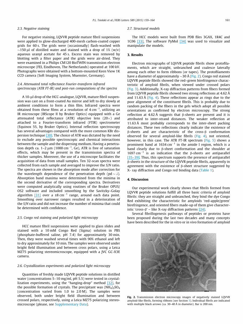
Fig. 2. Transmission electron microscopy images of negatively stained LQVVRamyloid-like fibrils, forming ribbons (see Section 3). Individual fibrils are indicatedwith multiple black arrows (ca. 30–40 Å in diameter). Bar is 200 nm.
P.L. Tsiolaki et al. / FEBS Letters 589 (2015) 159–164 161
2.3. Negative staining
For negative staining, LQVVR peptide mature fibril suspensionswere applied to glow-discharged 400-mesh carbon-coated coppergrids for 60 s. The grids were (occasionally) flash-washed with�150 ll of distilled water and stained with a drop of 1% (w/v)aqueous uranyl acetate for 45 s. Excess stain was removed byblotting with a filter paper and the grids were air-dried. Theywere examined in a Philips CM120 BioTWIN transmission electronmicroscope (FEI, Eindhoven, The Netherlands) operated at 100 kV.Photographs were obtained with a bottom-mounted Keen View 1KCCD camera (Soft Imaging System, Muenster, Germany).
2.4. Attenuated total reflectance Fourier-transform infraredspectroscopy (ATR FT-IR) and post-run computations of the spectra
A 10-ll drop of the HCC-analogue, LQVVR, mature fibril suspen-sion was cast on a front-coated Au mirror and left to dry slowly atambient conditions to form a thin film. Infrared spectra wereobtained from these films at a resolution of 4 cm�1, utilizing anIR microscope (IRScope II by Bruker Optics) equipped with a Geattenuated total reflectance (ATR) objective lens (20�) andattached to a Fourier-transform infrared (FTIR) spectrometer(Equinox 55, by Bruker Optics). Internal reflection spectroscopyhas several advantages compared with the more common KBr dis-persion technique [30]. The choice of ATR was dictated by the needto exclude any possible spectroscopic and chemical interactionsbetween the sample and the dispersing medium. Having a penetra-tion depth ca. 1–2 lm (1000 cm�1, Ge), ATR is free of saturationeffects, which may be present in the transmission spectra ofthicker samples. Moreover, the use of a microscope facilitates theacquisition of data from small samples. Ten 32-scan spectra werecollected from each sample and averaged to improve the S/N ratio.The spectra are shown in the absorption mode after correction forthe wavelength dependence of the penetration depth (pd � k).Absorption band maxima were determined from the minima inthe second derivative of the corresponding spectra. Derivativeswere computed analytically using routines of the Bruker OPUS/OS2 software and included smoothing by the Savitzky–Golayalgorithm [31] over a ±8 cm�1 range, around each data point.Smoothing over narrower ranges resulted in a deterioration ofthe S/N ratio and did not increase the number of minima that couldbe determined with confidence.
2.5. Congo red staining and polarized light microscopy
HCC mature fibril suspensions were applied to glass slides andstained with a 10 mM Congo Red (Sigma) solution in PBS(phosphate-buffered saline, pH 7.4) for approximately 30 min.Then, they were washed several times with 90% ethanol and leftto dry approximately for 10 min. The samples were observed underbright field illumination and between cross polars, using a LeicaMZ75 polarizing stereomicroscope, equipped with a JVC GC-X3Ecamera.
2.6. Crystallization experiments and polarized light microscopy
Quantities of freshly made LQVVR peptide solutions in distilledwater (concentrations 5–10 mg/ml, pH 5.5) were tested in crystal-lization experiments, using the ‘‘hanging-drop’’ method [32], forthe possible formation of crystals. The precipitant was (NH4)2SO4
(concentration varied from 1.0 to 2.0 M). The samples wereobserved, both under bright field illumination and betweencrossed polars, respectively, using a Leica MZ75 polarizing stereo-microscope (please, see Supplementary Data).
2.7. Structural models
The HCC models were built from PDB files 3GAX, 1R4C and1G96 [33]. The software PyMol [34] was used to visualize andmanipulate the models.
3. Results
Electron micrographs of LQVVR peptide fibrils show protofila-ments, which are straight, unbranched and coalesce laterallyamong each other to form ribbons (or tapes). The protofilamentshave a diameter of approximately �30 Å (Fig. 2). Congo red stainedLQVVR peptide fibrils showed the red–green birefringence charac-teristic of amyloid fibrils, when viewed under crossed polars(Fig. 3). Additionally, X-ray diffraction patterns from fibers formedfrom LQVVR peptide fibrils showed two strong reflections at 4.62 Åand 11.63 Å (Fig. 4). These reflections appear as rings due to thepoor alignment of the constituent fibrils. This is probably due torandom packing of the fibers in the gels which adopt all possibleorientations as confirmed by electron microscopy. The strongreflection at 4.62 Å suggests that b-sheets are present and it isattributed to inter-strand distances. The weaker reflection at11.63 Å most probably corresponds to the inter-sheet packingdistances. These two reflections clearly indicate the existence ofb-sheets and are characteristic of the cross-b conformationobserved for several amyloid-like fibrils (Fig. 4), not oriented,however, in this case. The ATR FT-IR spectrum (Fig. 5) shows aprominent band at 1634 cm�1 in the amide I region, which is aband clearly due to b-sheet conformation and the shoulder at1697 cm�1 is an indication that the b-sheets are antiparallel[35–39]. Thus, this spectrum supports the presence of antiparallelb-sheets in the structure of the LQVVR peptide fibrils, apparently inagreement with the existence of b-sheet structure suggested byX- ray diffraction and Congo red binding data (Table 1).
4. Discussion
Our experimental work clearly shows that fibrils formed fromLQVVR peptide solutions fulfill all three basic criteria of amyloidfibrils: they are straight and unbranched, they bind the dye CongoRed exhibiting the characteristic for amyloids ‘red-apple/green’birefringence, and oriented fibers made-up of them give character-istic ‘cross-b’ – like X-ray diffraction patterns [24].
Several fibrillogenesis pathways of peptides or proteins havebeen proposed during the last two decades and many conceptshave been described for the in vitro or in vivo formation of amyloid
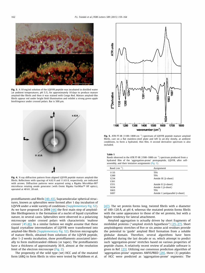
Fig. 3. A 10 mg/ml solution of the LQVVR peptide was incubated in distilled water(at ambient temperatures, pH 5.5), for approximately 10 days to produce matureamyloid-like fibrils and then it was stained with Congo Red. Mature amyloid-likefibrils appear red under bright field illumination and exhibit a strong green-applebirefringence under crossed polars. Bar is 500 lm.
Fig. 4. X-ray diffraction pattern from aligned LQVVR peptide mature amyloid-likefibrils. Reflections with spacings of 4.62 Å and 11.63 Å, respectively, are indicatedwith arrows. Diffraction patterns were acquired using a Rigaku MicroMax-007microfocus rotating anode generator (with Osmic Rigaku VariMax™ HF optics),operated at 40 kV, 20 mA.
Fig. 5. ATR FT-IR (1100–1800 cm�1) spectrum of LQVVR peptide mature amyloidfibrils, cast on a flat stainless-steel plate and left to air-dry slowly, at ambientconditions, to form a hydrated, thin film. A second derivative spectrum is alsoincluded.
Table 1Bands observed in the ATR FT-IR (1500–1800 cm�1) spectrum produced from ahydrated film of the ‘aggregation-prone’ pentapeptide, LQVVR, after self-assembly, and their tentative assignments (Fig. 5).
Band (cm�1) Assignment
1135 TFA1200 TFA1234 Amide III (b-sheet)13911551 Amide II (b-sheet)1634 Amide I (b-sheet)1663 TFA1697 Amide I (antiparallel b-sheet)
162 P.L. Tsiolaki et al. / FEBS Letters 589 (2015) 159–164
protofilaments and fibrils [40–43]. Supramolecular spherical struc-tures, known as spherulites were formed after 1 day incubation ofLQVVR under a wide variety of conditions (Supplementary Fig. S2).As we have proposed in 2004 [44] the first main step of amyloid-like fibrillogenesis is the formation of a nuclei of liquid crystallinenature, in several cases. Spherulites were observed in a polarizingmicroscope under crossed polars with characteristic ‘maltesecrosses’ [45,46]. In a similar fashion we might assume that theseliquid crystalline intermediates of LQVVR were transformed intoamyloid-like fibrils (Supplementary Fig. S2). Electron micrographsof mature fibrils, obtained from solutions of the LQVVR peptide,after 1–2 weeks incubation, show protofilaments associated later-ally to form multistranded ribbons (or tapes). The protofilamentshave a thickness of approximately 30 Å, almost at the resolutionlimit of the electron microscope (Fig. 2).
The propensity of the wild type (wt) HCC and of the mutatedform L68Q to form fibrils in vitro were tested by Wahlbom et al.
[47]. The wt protein forms long, twisted fibrils with a diameterof 100–120 Å, at pH 4, whereas the mutated protein forms fibrilswith the same appearance to those of the wt protein, but with ahigher tendency for lateral attachment.
Amyloid aggregation is actually driven by short fragments ofmisfolded proteins (‘‘amyloid stretch hypothesis’’) [25–27]. Shortamyloidogenic stretches of five or six amino acid residues providethe potential to ‘guide’ amyloid fibril formation from a solubleglobular domain. Therefore, several algorithms have beenpublished during the last decade or so, which attempt to predictsuch ‘aggregation-prone’ stretches based on various properties ofpeptide chains. A relatively recent review of available software isgiven in Ref. [23]. Utilizing our consensus prediction algorithm of‘aggregation-prone’ segments AMYLPRED [28], three (3) peptidesof HCC were predicted as ‘aggregation-prone’ segments: The
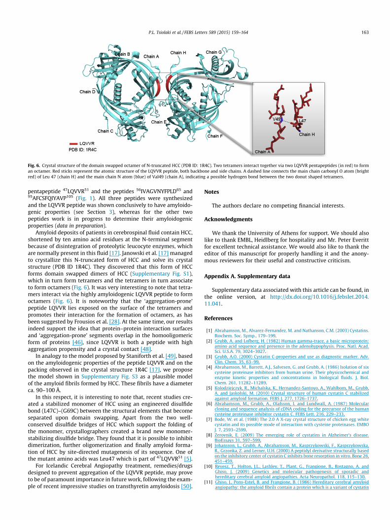
Fig. 6. Crystal structure of the domain swapped octamer of N-truncated HCC (PDB ID: 1R4C). Two tetramers interact together via two LQVVR pentapeptides (in red) to forman octamer. Red sticks represent the atomic structure of the LQVVR peptide, both backbone and side chains. A dashed line connects the main chain carbonyl O atom (brightred) of Leu 47 (chain H) and the main chain N atom (blue) of Val49 (chain A), indicating a possible hydrogen bond between the two donut shaped tetramers.
P.L. Tsiolaki et al. / FEBS Letters 589 (2015) 159–164 163
pentapeptide 47LQVVR51 and the peptides 56IVAGVNYFPLD65 and95AFCSFQIYAVP105 (Fig. 1). All three peptides were synthesizedand the LQVVR peptide was shown conclusively to have amyloido-genic properties (see Section 3), whereas for the other twopeptides work is in progress to determine their amyloidogenicproperties (data in preparation).
Amyloid deposits of patients in cerebrospinal fluid contain HCC,shortened by ten amino acid residues at the N-terminal segmentbecause of disintegration of proteolytic leucocyte enzymes, whichare normally present in this fluid [17]. Janowski et al. [17] managedto crystallize this N-truncated form of HCC and solve its crystalstructure (PDB ID 1R4C). They discovered that this form of HCCforms domain swapped dimers of HCC (Supplementary Fig. S1),which in turn form tetramers and the tetramers in turn associateto form octamers (Fig. 6). It was very interesting to note that tetra-mers interact via the highly amyloidogenic LQVVR peptide to formoctamers (Fig. 6). It is noteworthy that the ‘aggregation-prone’peptide LQVVR lies exposed on the surface of the tetramers andpromotes their interaction for the formation of octamers, as hasbeen suggested by Frousios et al. [28]. At the same time, our resultsindeed support the idea that protein–protein interaction surfacesand ‘aggregation-prone’ segments overlap in the homooligomericform of proteins [46], since LQVVR is both a peptide with highaggregation propensity and a crystal contact [48].
In analogy to the model proposed by Staniforth et al. [49], basedon the amyloidogenic properties of the peptide LQVVR and on thepacking observed in the crystal structure 1R4C [17], we proposethe model shown in Supplementary Fig. S3 as a plausible modelof the amyloid fibrils formed by HCC. These fibrils have a diameterca. 90–100 Å.
In this respect, it is interesting to note that, recent studies cre-ated a stabilized monomer of HCC using an engineered disulfidebond (L47C)–(G69C) between the structural elements that becomeseparated upon domain swapping. Apart from the two well-conserved disulfide bridges of HCC which support the folding ofthe monomer, crystallographers created a brand new monomer-stabilizing disulfide bridge. They found that it is possible to inhibitdimerization, further oligomerization and finally amyloid forma-tion of HCC by site-directed mutagenesis of its sequence. One ofthe mutant amino acids was Leu47 which is part of 47LQVVR51 [5].
For Icelandic Cerebral Angiopathy treatment, remedies/drugsdesigned to prevent aggregation of the LQVVR peptide, may proveto be of paramount importance in future work, following the exam-ple of recent impressive studies on transthyretin amyloidosis [50].
Notes
The authors declare no competing financial interests.
Acknowledgments
We thank the University of Athens for support. We should alsolike to thank EMBL, Heidlberg for hospitality and Mr. Peter Everittfor excellent technical assistance. We would also like to thank theeditor of this manuscript for properly handling it and the anony-mous reviewers for their useful and constructive criticism.
Appendix A. Supplementary data
Supplementary data associated with this article can be found, inthe online version, at http://dx.doi.org/10.1016/j.febslet.2014.11.041.
References
[1] Abrahamson, M., Alvarez-Fernandez, M. and Nathanson, C.M. (2003) Cystatins.Biochem. Soc. Symp., 179–199.
[2] Grubb, A. and Lofberg, H. (1982) Human gamma-trace, a basic microprotein:amino acid sequence and presence in the adenohypophysis. Proc. Natl. Acad.Sci. U.S.A. 79, 3024–3027.
[3] Grubb, A.O. (2000) Cystatin C-properties and use as diagnostic marker. Adv.Clin. Chem. 35, 63–99.
[4] Abrahamson, M., Barrett, A.J., Salvesen, G. and Grubb, A. (1986) Isolation of sixcysteine proteinase inhibitors from human urine. Their physicochemical andenzyme kinetic properties and concentrations in biological fluids. J. Biol.Chem. 261, 11282–11289.
[5] Kolodziejczyk, R., Michalska, K., Hernandez-Santoyo, A., Wahlbom, M., Grubb,A. and Jaskolski, M. (2010) Crystal structure of human cystatin C stabilizedagainst amyloid formation. FEBS J. 277, 1726–1737.
[6] Abrahamson, M., Grubb, A., Olafsson, I. and Lundwall, A. (1987) Molecularcloning and sequence analysis of cDNA coding for the precursor of the humancysteine proteinase inhibitor cystatin C. FEBS Lett. 216, 229–233.
[7] Bode, W. et al. (1988) The 2.0 A X-ray crystal structure of chicken egg whitecystatin and its possible mode of interaction with cysteine proteinases. EMBOJ. 7, 2593–2599.
[8] Zerovnik, E. (2009) The emerging role of cystatins in Alzheimer’s disease.BioEssays 31, 597–599.
[9] Johansson, L., Grubb, A., Abrahamson, M., Kasprzykowski, F., Kasprzykowska,R., Grzonka, Z. and Lerner, U.H. (2000) A peptidyl derivative structurally basedon the inhibitory center of cystatin C inhibits bone resorption in vitro. Bone 26,451–459.
[10] Revesz, T., Holton, J.L., Lashley, T., Plant, G., Frangione, B., Rostagno, A. andGhiso, J. (2009) Genetics and molecular pathogenesis of sporadic andhereditary cerebral amyloid angiopathies. Acta Neuropathol. 118, 115–130.
[11] Ghiso, J., Pons-Estel, B. and Frangione, B. (1986) Hereditary cerebral amyloidangiopathy: the amyloid fibrils contain a protein which is a variant of cystatin

164 P.L. Tsiolaki et al. / FEBS Letters 589 (2015) 159–164
C, an inhibitor of lysosomal cysteine proteases. Biochem. Biophys. Res.Commun. 136, 548–554.
[12] Ghiso, J., Jensson, O. and Frangione, B. (1986) Amyloid fibrils in hereditarycerebral hemorrhage with amyloidosis of Icelandic type is a variant of gamma-trace basic protein (cystatin C). Proc. Natl. Acad. Sci. U.S.A. 83, 2974–2978.
[13] Yamada, M. (2000) Cerebral amyloid angiopathy: an overview.Neuropathology 20, 8–22.
[14] Asgeirsson, B., Haebel, S., Thorsteinsson, L., Helgason, E., Gudmundsson, K.O.,Gudmundsson, G. and Roepstorff, P. (1998) Hereditary cystatin C amyloidangiopathy: monitoring the presence of the Leu-68 ? Gln cystatin C variant incerebrospinal fluids and monocyte cultures by MS. Biochem. J. 329 (Pt 3), 497–503.
[15] Olafsson, I. and Grubb, A. (2000) Hereditary cystatin C amyloid angiopathy.Amyloid 7, 70–79.
[16] Rostagno, A., Holton, J.L., Lashley, T., Revesz, T. and Ghiso, J. (2010) Cerebralamyloidosis: amyloid subunits, mutants and phenotypes. Cell. Mol. Life Sci.67, 581–600.
[17] Janowski, R., Abrahamson, M., Grubb, A. and Jaskolski, M. (2004) Domainswapping in N-truncated human cystatin C. J. Mol. Biol. 341, 151–160.
[18] Liu, Y. and Eisenberg, D. (2002) 3D domain swapping: as domains continue toswap. Protein Sci. 11, 1285–1299.
[19] Nelson, R. and Eisenberg, D. (2006) Recent atomic models of amyloid fibrilstructure. Curr. Opin. Struct. Biol. 16, 260–265.
[20] Nelson, R. and Eisenberg, D. (2006) Structural models of amyloid-like fibrils.Adv. Protein Chem. 73, 235–282.
[21] Kelly, J.W. (1996) Alternative conformations of amyloidogenic proteins governtheir behavior. Curr. Opin. Struct. Biol. 6, 11–17.
[22] Zerovnik, E. (2002) Amyloid-fibril formation. Proposed mechanisms andrelevance to conformational disease. Eur. J. Biochem. 269, 3362–3371.
[23] Hamodrakas, S.J. (2011) Protein aggregation and amyloid fibril formationprediction software from primary sequence: towards controlling theformation of bacterial inclusion bodies. FEBS J. 278, 2428–2435.
[24] Sunde, M. and Blake, C.C. (1998) From the globular to the fibrous state: proteinstructure and structural conversion in amyloid formation. Q. Rev. Biophys. 31,1–39.
[25] Lopez de la Paz, M. and Serrano, L. (2004) Sequence determinants of amyloidfibril formation. Proc. Natl. Acad. Sci. U.S.A. 101, 87–92.
[26] Esteras-Chopo, A., Serrano, L. and Lopez de la Paz, M. (2005) The amyloidstretch hypothesis: recruiting proteins toward the dark side. Proc. Natl. Acad.Sci. U.S.A. 102, 16672–16677.
[27] Teng, P.K. and Eisenberg, D. (2009) Short protein segments can drive a non-fibrillizing protein into the amyloid state. Protein Eng. Des. Sel. 22, 531–536.
[28] Frousios, K.K., Iconomidou, V.A., Karletidi, C.M. and Hamodrakas, S.J. (2009)Amyloidogenic determinants are usually not buried. BMC Struct. Biol. 9, 44.
[29] Winn, M.D. et al. (2011) Overview of the CCP4 suite and currentdevelopments. Acta Crystallogr. D Biol. Crystallogr. 67, 235–242.
[30] de Jongh, H.H., Goormaghtigh, E. and Ruysschaert, J.M. (1996) The differentmolar absorptivities of the secondary structure types in the amide I region: anattenuated total reflection infrared study on globular proteins. Anal. Biochem.242, 95–103.
[31] Savitsky, A. and Golay, M.J.E. (1964) Smoothing and differentiation of data bysimplified least-squares procedures. Anal. Chem. 36, 1627–1639.
[32] Blundell, T.L. and Johnson, L.N. (1976) Protein Crystallography, AcademicPress, London-New York-San Francisco.
[33] Berman, H.M., Westbrook, J., Feng, Z., Gilliland, G., Bhat, T.N., Weissig, H.,Shindyalov, I.N. and Bourne, P.E. (2000) The protein data bank. Nucleic AcidsRes. 28, 235–242.
[34] Delano, W.L. (2005) The PyMOL Molecular Graphics System, DeLano ScientificLLC.
[35] Surewicz, W.K., Mantsch, H.H. and Chapman, D. (1993) Determination ofprotein secondary structure by Fourier transform infrared spectroscopy: acritical assessment. Biochemistry 32, 389–394.
[36] Jackson, M. and Mantsch, H.H. (1995) The use and misuse of FTIR spectroscopyin the determination of protein structure. Crit. Rev. Biochem. Mol. Biol. 30, 95–120.
[37] Haris, P.I. and Chapman, D. (1995) The conformational analysis of peptidesusing Fourier transform IR spectroscopy. Biopolymers 37, 251–263.
[38] Krimm, S. and Bandekar, J. (1986) Vibrational spectroscopy and conformationof peptides, polypeptides, and proteins. Adv. Protein Chem. 38, 181–364.
[39] Valenti, L.E., Paci, M.B., De Pauli, C.P. and Giacomelli, C.E. (2011) Infrared studyof trifluoroacetic acid unpurified synthetic peptides in aqueous solution:trifluoroacetic acid removal and band assignment. Anal. Biochem. 410, 118–123.
[40] Rochet, J.C. and Lansbury Jr., P.T. (2000) Amyloid fibrillogenesis: themes andvariations. Curr. Opin. Struct. Biol. 10, 60–68.
[41] Chiti, F., Webster, P., Taddei, N., Clark, A., Stefani, M., Ramponi, G. and Dobson,C.M. (1999) Designing conditions for in vitro formation of amyloidprotofilaments and fibrils. Proc. Natl. Acad. Sci. U.S.A. 96, 3590–3594.
[42] Kelly, J.W. (2000) Mechanisms of amyloidogenesis. Nat. Struct. Biol. 7, 824–826.
[43] Chiti, F. and Dobson, C.M. (2009) Amyloid formation by globular proteinsunder native conditions. Nat. Chem. Biol. 5, 15–22.
[44] Hamodrakas, S.J., Hoenger, A. and Iconomidou, V.A. (2004) Amyloidfibrillogenesis of silkmoth chorion protein peptide-analogues via a liquid–crystalline intermediate phase. J. Struct. Biol. 145, 226–235.
[45] Shtukenberg, A.G., Punin, Y.O., Gunn, E. and Kahr, B. (2012) Spherulites. Chem.Rev. 112, 1805–1838.
[46] Castillo, V. and Ventura, S. (2009) Amyloidogenic regions and interactionsurfaces overlap in globular proteins related to conformational diseases. PLoSComput. Biol. 5, e1000476.
[47] Wahlbom, M., Wang, X., Lindstrom, V., Carlemalm, E., Jaskolski, M. and Grubb,A. (2007) Fibrillogenic oligomers of human cystatin C are formed bypropagated domain swapping. J. Biol. Chem. 282, 18318–18326.
[48] Janin, J. (1997) Specific versus non-specific contacts in protein crystals. Nat.Struct. Biol. 4, 973–974.
[49] Staniforth, R.A., Giannini, S., Higgins, L.D., Conroy, M.J., Hounslow, A.M., Jerala,R., Craven, C.J. and Waltho, J.P. (2001) Three-dimensional domain swapping inthe folded and molten-globule states of cystatins, an amyloid-formingstructural superfamily. EMBO J. 20, 4774–4781.
[50] Connelly, S., Choi, S., Johnson, S.M., Kelly, J.W. and Wilson, I.A. (2010)Structure-based design of kinetic stabilizers that ameliorate the transthyretinamyloidoses. Curr. Opin. Struct. Biol. 20, 54–62.