The orosomucoid 1 protein (α1 acid glycoprotein) is overexpressed in odontogenic myxoma
9
RESEARCH Open Access The orosomucoid 1 protein (α1 acid glycoprotein) is overexpressed in odontogenic myxoma Alejandro García-Muñoz 1 , Mario A Rodríguez 1* , Ronell Bologna-Molina 2,3 , Febe E Cázares-Raga 1 , Fidel C Hernández-Hernández 1 , J Eduardo Farfán-Morales 4 , Juan J Trujillo 5 , Carlos Licéaga-Escalera 5 and Guillermo Mendoza-Hernández 6 Abstract Background: Odontogenic myxoma (OM) is a benign, but locally invasive, neoplasm occurring in the jaws. However, the molecules implicated in its development are unknown. OM as well as Dental Follicle (DF), an odontogenic tissue surrounding the enamel organ, is derived from ectomesenchymal/mesencyhmal elements. To identify some protein that could participate in the development of this neoplasm, total proteins from OM were separated by two-dimensional electrophoresis and the profiles were compared with those obtained from DF, used as a control. Results: We identified eight proteins with differential expression; two of them were downregulated and six upregulated in OM. A spot consistently overexpressed in odontogenic myxoma, with a molecular weight of 44-kDa and a pI of 3.5 was identified as the orosomucoid 1 protein. Western blot experiments confirmed the overexpression of this protein in odontogenic myxoma and immunohistochemical assays showed that this protein was mainly located in the cytoplasm of stellate and spindle-shaped cells of this neoplasm. Conclusion: Orosomucoid 1, which belongs to a group of acute-phase proteins, may play a role in the modulation of the immune system and possibly it influences the development of OM. Keywords: Odontogenic myxoma, Dental follicle, Proteomic analysis, Orosomucoid 1, α1 acid glycoprotein Background Odontogenic Myxoma (OM) is a relatively rare, benign neoplasm occurring in the jaws. This neoplasm is char- acterized by the presence of stellate and spindle-shaped cells embedded in an abundant myxoid or mucoid extra- cellular matrix. OM represents 3-20% of all odontogenic tumours and, in most studies, OM is the third most fre- quent odontogenic tumor [1]. Conservative surgery by enucleation and curettage is recommended when lesions of OM are smaller than 3 cm, but a segmental resection with immediate reconstruction is preferred in patients affected by bigger tumors [2]. OM as well as Dental Follicle (DF), an odontogenic tissue surrounding the enamel organ, and the dental pa- pilla of the developing tooth germ prior to eruption [3], is derived from ectomesenchymal/mesencyhmal ele- ments. Thus, OM could be mimicked by DF and dental papilla, both containing myxoid areas [4-6]. Indeed, a pathologist who is not familiar with the histology of a tooth germ can mistake a myxoid DF for an OM [6]. Up to now there are few studies comparing molecules of OM with other odontogenic mesenchymal tissues. Some authors compared the expression of α-SMA, S-100 and vimentin between OM and other mesenchymal tis- sues [7,8], but not substantial differences were found. Another study described that the hyaluronic acid concen- tration in OM is four times higher than that of other gly- cosaminoglycans, such as chondroitin sulphate, which is inversely found in mesenchymal tissues from dental pulp, gingival and periodontal ligament, but not in DF [9]. It was also reported that 90% of OM cells expressed the metalloproteinase 2 (MMP-2), while only 10% of the cells in DF and myxoid dental pulp expressed this protein [10]. These authors also showed an increased expression of * Correspondence: [email protected] 1 Departamento de Infectómica y Patogénesis Molecular, CINVESTAV-IPN, México, D.F., México Full list of author information is available at the end of the article © 2012 Garcia-Muñoz et al.; licensee BioMed Central Ltd. This is an Open Access article distributed under the terms of the Creative Commons Attribution License (http://creativecommons.org/licenses/by/2.0), which permits unrestricted use, distribution, and reproduction in any medium, provided the original work is properly cited. García-Muñoz et al. Proteome Science 2012, 10:49 http://www.proteomesci.com/content/10/1/49
-
Upload
independent -
Category
Documents
-
view
3 -
download
0
Transcript of The orosomucoid 1 protein (α1 acid glycoprotein) is overexpressed in odontogenic myxoma

García-Muñoz et al. Proteome Science 2012, 10:49http://www.proteomesci.com/content/10/1/49
RESEARCH Open Access
The orosomucoid 1 protein (α1 acid glycoprotein)is overexpressed in odontogenic myxomaAlejandro García-Muñoz1, Mario A Rodríguez1*, Ronell Bologna-Molina2,3, Febe E Cázares-Raga1,Fidel C Hernández-Hernández1, J Eduardo Farfán-Morales4, Juan J Trujillo5, Carlos Licéaga-Escalera5
and Guillermo Mendoza-Hernández6
Abstract
Background: Odontogenic myxoma (OM) is a benign, but locally invasive, neoplasm occurring in the jaws.However, the molecules implicated in its development are unknown. OM as well as Dental Follicle (DF), anodontogenic tissue surrounding the enamel organ, is derived from ectomesenchymal/mesencyhmal elements. Toidentify some protein that could participate in the development of this neoplasm, total proteins from OM wereseparated by two-dimensional electrophoresis and the profiles were compared with those obtained from DF, usedas a control.
Results: We identified eight proteins with differential expression; two of them were downregulated and sixupregulated in OM. A spot consistently overexpressed in odontogenic myxoma, with a molecular weight of 44-kDaand a pI of 3.5 was identified as the orosomucoid 1 protein. Western blot experiments confirmed theoverexpression of this protein in odontogenic myxoma and immunohistochemical assays showed that this proteinwas mainly located in the cytoplasm of stellate and spindle-shaped cells of this neoplasm.
Conclusion: Orosomucoid 1, which belongs to a group of acute-phase proteins, may play a role in the modulationof the immune system and possibly it influences the development of OM.
Keywords: Odontogenic myxoma, Dental follicle, Proteomic analysis, Orosomucoid 1, α1 acid glycoprotein
BackgroundOdontogenic Myxoma (OM) is a relatively rare, benignneoplasm occurring in the jaws. This neoplasm is char-acterized by the presence of stellate and spindle-shapedcells embedded in an abundant myxoid or mucoid extra-cellular matrix. OM represents 3-20% of all odontogenictumours and, in most studies, OM is the third most fre-quent odontogenic tumor [1]. Conservative surgery byenucleation and curettage is recommended when lesionsof OM are smaller than 3 cm, but a segmental resectionwith immediate reconstruction is preferred in patientsaffected by bigger tumors [2].OM as well as Dental Follicle (DF), an odontogenic
tissue surrounding the enamel organ, and the dental pa-pilla of the developing tooth germ prior to eruption [3],
* Correspondence: [email protected] de Infectómica y Patogénesis Molecular, CINVESTAV-IPN,México, D.F., MéxicoFull list of author information is available at the end of the article
© 2012 Garcia-Muñoz et al.; licensee BioMed CCreative Commons Attribution License (http:/distribution, and reproduction in any medium
is derived from ectomesenchymal/mesencyhmal ele-ments. Thus, OM could be mimicked by DF and dentalpapilla, both containing myxoid areas [4-6]. Indeed, apathologist who is not familiar with the histology of atooth germ can mistake a myxoid DF for an OM [6].Up to now there are few studies comparing molecules
of OM with other odontogenic mesenchymal tissues.Some authors compared the expression of α-SMA, S-100and vimentin between OM and other mesenchymal tis-sues [7,8], but not substantial differences were found.Another study described that the hyaluronic acid concen-tration in OM is four times higher than that of other gly-cosaminoglycans, such as chondroitin sulphate, which isinversely found in mesenchymal tissues from dental pulp,gingival and periodontal ligament, but not in DF [9]. Itwas also reported that 90% of OM cells expressed themetalloproteinase 2 (MMP-2), while only 10% of the cellsin DF and myxoid dental pulp expressed this protein [10].These authors also showed an increased expression of
entral Ltd. This is an Open Access article distributed under the terms of the/creativecommons.org/licenses/by/2.0), which permits unrestricted use,, provided the original work is properly cited.
García-Muñoz et al. Proteome Science 2012, 10:49 Page 2 of 9http://www.proteomesci.com/content/10/1/49
Bcl-2 and Bcl-x in OM. However, other studies reportedless than 1% of Bcl-2 positive cells in OM [11,12]. Finally,some works have been focused on the histological changesthat occur in the hyperplastic DF and normal DF ofimpacted third molars and their histological association toOM and the possibilities of misinterpretation as OM[4,6,13,14].In recent years, the use of high-throughput genomics
and proteomics has expanded rapidly in biomedical sci-ence. These technologies have evolved and make pos-sible several discoveries in clinical cancer research,including the identification of biomarkers, molecularclassification of tumors, molecular prediction of metas-tasis, treatment response, and prognosis. Particularly,the study of the proteome, the collection of all the pro-teins expressed from specific cells in all isoforms, poly-morphisms and post-translational modifications [15],has allowed the detection of new biomarkers in diversetypes of neoplasitic tissues, for example in urinary blad-der cancer [16], ovarian carcinomas [17], oral squamouscell carcinoma [18], and lung cancer [19]. However, inthe literature we have not found any previous studyabout OM or other odontogenic tumors using thisapproach.To identify some proteins that could participate in the
biological behavior of OM, in this work we used theproteomic technology based on 2-dimensional electro-phoresis (2DE) combined with liquid chromatography-tandem mass spectrometry (LC-MS/MS) for comparingodontogenic myxoma (neoplastic) versus dental follicle(normal) tissues. A spot consistently overexpressed inodontogenic myxoma was identified as the orosomucoid1 protein, which was located in the cytoplasm of thetumor cells.
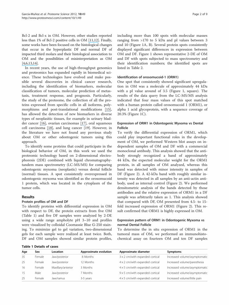
ResultsProtein profiles of OM and DFTo identify proteins with differential expression in OMwith respect to DF, the protein extracts from five OM(Table 1) and five DF samples were analyzed by 2-DEusing a wide range ampholyte pH 3–10 and profileswere visualized by colloidal Coomassie Blue G-250 stain-ing. To minimize gel to gel variation, two-dimensionalgels for each sample were realized at least twice. Both,DF and OM samples showed similar protein profiles,
Table 1 Details of cases
Age Sex Location Approximate evolution
35 Female Jaw/posterior 8 Months
25 Female Jaw/posterior 12 Months
16 Female Maxillary/anterior 3 Months
15 Male Jaw/posterior 7 Months
25 Female Jaw/ramus 3-5 years
including more than 100 spots with molecular massesranging from >170 to 5 kDa and pI values between 3and 10 (Figure 1A, B). Several protein spots consistentlydisplayed significant differences in expression betweenOM and DF. Figure 1 shows representative 2-DE of OMand DF with spots subjected to mass spectrometry andtheir identification numbers; the identified spots arelisted in Table 2.
Identification of orosomucoid-1 (ORM1)One spot that consistently showed significant upregula-tion in OM was a molecule of approximately 44 kDawith a pI value around of 3.5 (Figure 1, square). Theresults of the data query from the LC-MS/MS analysisindicated that four mass values of this spot matchedwith a human protein called orosomucoid 1 (ORM1), oralpha 1 acid glycoprotein, with a sequence coverage of20.3% (Figure 1C).
Expression of ORM1 in Odontogenic Myxoma vs DentalFollicleTo verify the differential expression of ORM1, whichcould play important functional roles in the develop-ment of OM, we performed Western blot assays on in-dependent samples of OM and DF with a commercialmonoclonal antibody. This analysis showed that the anti-body strongly recognized a band of approximately44 kDa, the expected molecular weight for the ORM1protein, in all samples of OM analyzed, whereas thisband was detected with minor intensity in samples ofDF (Figure 2). A 42-kDa band with roughly similar in-tensity was detected in all samples by an anti-actin anti-body, used as internal control (Figure 2). We performeddensitometric analysis of the bands detected by thoseantibodies and the relative expression of ORM1 in a DFsample was arbitrarily taken as 1. This analysis showedthat compared with DF, OM presented from 4.5- to 15-fold increased expression of ORM1 (Figure 2). This re-sult confirmed that ORM1 is highly expressed in OM.
Expression pattern of ORM1 in Odontogenic Myxoma vsnormal Dental FollicleTo determine the in situ expression of ORM1 in thetumoral mass of OM, we performed an immunohisto-chemical assay on fourteen OM and ten DF samples
Approximate diameter Symptoms
3 × 2 cm/with expanded cortical Increased volume/asymptomatic
4 × 2 cm/with expanded cortical Increased volume/paresthesia
4 × 5 cm/with expanded cortical Increased volume/asymptomatic
9 × 5 cm/with expanded cortical Increased volume/asymptomatic
4 × 5 cm/with expanded cortical Increased volume/little pain

Figure 1 2DE protein profiles of Odontogenic Myxoma (OM) and Dental Follicle (DF) and identification of ORM1. Proteins from DF andOM were extracted and separated in 2DE. Then, gels were stained with Colloidal Coomassie Blue G-250. (A) Protein profile of OM. (B) Proteinprofile of DF. Proteins differentially expressed between both samples are numbered and one of the spots that consistently showed significantupregulation in OM is indicated by a frame. Under each gel are shown the magnifications and differential intensity analyses for the spotindicated by the frame. (C) This spot was excised from gels and subsequently analyzed by LC-MS/MS. This analysis identified this spot as theorosomucoid 1 protein (ORM1), which amino acid sequence is shown. The peptides identified by LC-MS/MS are underlined.
Table 2 Identification of proteins with differential expression in Odontogenic Myxoma
Spot ID NCBI No. Name TheoreticalProtein IDMass KDa/pl
MascotScore
SequenceCoverage
Major functions
Up-regulated proteins
1 gi|112877 Orosomucoid-1 23497/4.93 182 21% Acute phase with inflammatory andimmunomodulating properties
2 gi|4507677 GRP94 92696/4.76 471 21% Molecular chaperone and cell signalling
3 gi|4507651 Tropomyosin alpha-4 28619/4.67 383 35% Calcium binding and acti-binding
4 gi|4507953 14-3-3 protein 27899/4.73 341 40% Cell signaling, cycle control, apoptosisand metabolism
5 gi|90108664 Apolipoprotein A-1 28061/5.27 717 57% Lipid transport, metabolism, apoptosisand autophagy
6 gi|15783061 Serum Albumin in aComplex With MyristicAcid And Tri-lodobenzoicAcid
67988/5.69 239 33% Protein of binding to cations, fatty acids,bilirubin and other
Down-regulated proteins
7 gi|494066 GlutathioneS-transferase
23438/5.44 143 22% Detoxify endogenous and environmentalsubstances
8 gi|4502517 Carbonic anhydrase 1 28909/6.59 376 55% Ubiquitous metalloenzyme; bone resorption,calcification, ion transport, acid–basetransport and metabolic processes
Accession numbers are from the MASCOT database (http://www.matrixscience.com).
García-Muñoz et al. Proteome Science 2012, 10:49 Page 3 of 9http://www.proteomesci.com/content/10/1/49

Figure 2 Western blot assays for detection of the ORM 1protein. Protein extracts from OM and DF samples were separatedby PAGE-SDS and submitted to Western blot assays using antibodiesagainst ORM1 and against actin, the latter used as an internalcontrol. Relative intensities of the bands recognized by theantibodies were documented and analyzed by densitometry. Therelative expression of ORM1 in a DF sample was arbitrary taken as 1.
Figure 3 In situ expression of ORM1 in OM and DF. Tissue sections frommonoclonal antibody against ORM1, then, with a biotinylated antimouse areaction products were visualized by the incubation with 3,3´-diaminobenzmicroscopy (20X). Insets show magnifications (40X) of the marked areas. Arshowed contaminant epithelial cells positive for ORM1.
García-Muñoz et al. Proteome Science 2012, 10:49 Page 4 of 9http://www.proteomesci.com/content/10/1/49
using the monoclonal antibody against this protein. OMdisplayed positive cytoplasmic staining in majority of thestellate and spindle-shaped cells in all the analyzed sam-ples (Figure 3A, B), while mesenchymal cells of DF didnot exhibit immunopositivity (Figure 3C, D). In addition,in both tissues (OM and DF) the endothelial cells oflarge and small blood vessels showed ORM1 positivity(Figure 3A-D). Positivity was also found in contaminantepithelial cells of DF (Figure 3D).
DiscussionThe pathogenesis and source of OM is still controversial.Some authors have proposed an odontogenic origin, par-ticularly from the dental follicle or from the periodontalligament [20,21]. Other authors have suggested that OMmay be the result of a myxoid change in a pre-existingmesenchymal lesion or that it may represent a degenera-tive form of odontogenic fibroma [22]. In contrast, someauthors have posed that the OM has a myofibroblastic ori-gin [23,24]. Nevertheless, in the last years many studieshave compared the biochemical composition, particularly
different samples of OM (A, B) and DF (C, D) were incubated with antibody, and finally with the streptavidine/peroxidase complex. Theidine-H2O2 substrate. Finally, samples were analyzed by opticrows indicate endothelial cells of blood vessels (B-D). Panel D also

García-Muñoz et al. Proteome Science 2012, 10:49 Page 5 of 9http://www.proteomesci.com/content/10/1/49
in the extracellular matrix, of OM with organs (dentalpulp, dental follicles, gingival tissue and periodontal liga-ment) of a developing tooth [11,25,26]. The resultsobtained in the present study showed that the protein pro-files of OM and DF are very similar; supporting the notionthat OM could originate from DF.To analyze proteins differentially expressed in OM
and DF we used a proteomic approach based on 2-DEand peptide mass fingerprint by LC-MS/MS. This prote-omic analysis revealed the variation of eight proteinsidentified (Table 1).Expression of carbonic anhydrase I (CA I) and gluta-
thione S-transferase (GST) was downregulated in OM.Carbonic anhydrases catalyze the hydration of carbon di-oxide and forms bicarbonate. CA I not only enhancesthe hydration reaction of CO2, but it also promotes thecombining of bicarbonate with calcium to form the solidprecipitant of calcium carbonate [27], a principal com-ponent of bones. Although OM is considered a benignneoplasm, it shows a high potential for bone resorption[28]. Thus, downregulation of CA I may affect the bal-ance between bone resorption and apposition. On theother hand, GSTs are a large family of enzymes thatcatalyze the conjugation of reduced glutathione througha sulfhydryl group to electrophilic sites on a wide varietyof substrates that could lead to the generation of reactiveoxygen species (ROS) [29]. The products of GST cataly-sis are more water soluble, promoting ROS detoxifica-tion and thereby protecting tissues from oxidativedamage. Thus, GST could be acting as a caretaker pro-tein by protecting cells against genome damage inducedby carcinogens and as a tumor-suppressor protein lead-ing to tumor growth when inactivated [30]. It is, there-fore, speculative that downexpression of GST in OMwould lead to genome damage accumulation and be fur-ther injurious to the oral tissue.A glucose-regulated protein (GRP94), albumin in a
complex with myristic acid and tri-iodobenzoic acid, thetropomyosin alpha-4, the 14-3-3 protein zeta/delta, theapolipoprotein A-I, and the orosomucoid-1 protein wereup-regulated in OM. Interestingly, overexpression oftropomyosin alpha-4 was also detected in esophagealsquamous cell carcinoma [31], although their participa-tion in tumor development remains to be investigated.GRPs refer to a set of endoplasmic reticulum (ER) cha-
perones that have multiple functions in maintaining cel-lular homeostasis [32]. The endoplasmic reticulum stresspathways and the GRPs have been linked to cancergrowth and drug resistance [33]. GRPs represent novelmarkers for cancer progression and chemo-responsive-ness, as well as targets for cancer therapy. GRP94, alsoknown as gp96, is the most abundant glycoprotein inER and its overexpression associates with cellulartransformation, tumorigenicity and decreased sensitivity
to X-rays, whereas suppression of GRP94 sensitizes cellsto etoposide treatment [32].The 14-3-3 proteins belong to a family consisting of
highly conserved acidic proteins with molecular weightsof 25–30 kDa. They participate in phosphorylation-dependent protein-protein interactions that controlprogression through the cell cycle, initiation and main-tenance of DNA damage checkpoints, activation of MAPkinases, prevention of apoptosis and coordination of in-tegrin signaling and cytoskeletal dynamics [34]. Accu-mulating evidence now supports the concept that eitheran abnormal state of 14-3-3 protein expression, or dysre-gulation of 14-3-3/client protein interactions, contri-butes to the development of a large number of humandiseases. In particular, clinical investigations in the fieldof oncology have demonstrated a correlation betweenupregulated 14-3-3 levels and poor survival of cancerpatients [35].ApoA-I is the major protein in HDL and plays an im-
portant role in reverse cholesterol transport by extract-ing cholesterol and phospholipids from peripheral cellsand transferring it to the liver for excretion. In additionto its antiatherogenic properties, apoA-I also possessesanti-inflammatory and antioxidant properties [36].Decreased levels of Apolipoprotein were found in a var-iety of cancer [37-39], but such as in OM, Apolipopro-tein A-I was increased in breast cancer and brainmetastases in lung cancer [40,41]. This controversyabout the regulation of ApoA-I in cancer cells needs tobe clarified in future studies.By proteomics,Western blot and immunohistochem-
ical assays, in the present study we showed that theORM1 protein is overexpressed in OM. Interestingly,the same strategies allowed the identification ofincreased levels of ORM1 in urine samples of patientswith urinary bladder cancer [16]. Moreover, increasedlevels of ORM1 have been reported in the sera ofpatients with different malignant diseases, includingsquamous cell carcinoma of head and neck [42-46].ORM1 belongs to a group of acute-phase proteins
found in plasma. Such proteins undergo dramaticchanges in concentration as a response of the organismto a disturbance of its homeostasis. These plasmatic pro-teins constitute a group of serum factors related to dif-ferent immunological regulator functions and they havealso been associated with tumor development andgrowth. However, it is uncertain whether the serumlevels of acute-phase proteins, such as ORM1, increaseas a response of the host to tumor growth or as a conse-quence of neoplastic cell production.Human hepatocytes are normally the site of ORM1
production, but endothelial cells and some tumor cellscan also produce it [16,45,47]. Additionally, some studieshave shown that ORM1 is synthesized by lymphocytes,

García-Muñoz et al. Proteome Science 2012, 10:49 Page 6 of 9http://www.proteomesci.com/content/10/1/49
granulocytes, macrophages and monocytes [48,49]. Inthe present study the expression of ORM1 in OM wasmainly detected in the cytoplasm of stellate and spindle-shaped cells. However, this protein was also detected inthe endothelial cells of blood vessels in both OM andDF tissue samples. It has been reported that ORM1alone enhances migration but not the proliferation ofhuman dermal microvascular endothelial cells, but inthe presence of ORM1 and the vascular endothelialgrowth factor A (VEGF-A) the endothelial cells are cap-able to induce the development of endothelial tubes,suggesting that ORM1 seems to be involved in the regu-lation of angiogenesis [50]. Irmak et al. [16] proposedthat the highest increase of ORM1 levels in advancedstages of urinary bladder cancer, which correspond to avascularized tumor, could be due in part to the produc-tion of this protein by the augmented number of endo-thelial cells of angiogenically active blood vessels. Thepro-angiogenic collaborative property of ORM1 maypossibly occur in OM, but further studies with the asso-ciation of angiogenic markers and ORM1 in OM areneeded to test this hypothesis.The presence of ORM1 in odontogenic myxoma also
suggests a possible immunomodulatory function and arole in the growth and invasion potential of thetumoral cells. ORM1 is able to inhibit polymorpho-nuclear neutrophil activation and is considered a nat-ural anti-inflammatory, anti-neutrophil, anti-complementand immunomodulatory agent [51]. Thus, the overexpres-sion of ORM1 in OM may inhibit the immune response,resulting in an increase of tumor cell proliferation. Alter-natively, the high expression of ORM1 in OM could rep-resent a defense mechanism against proliferation andinvasion of the tumor cells, similar to what occurs incolon cancer cells. In the latter neoplastic cells, the over-expression of ORM1 results in a reduced colony-formingcapacity, as well as in a decrease of invasion and adhe-sion, whereas the inhibition of the expression of ORM1by antisense oligodeoxynucleotides produces an increaseof these events [52]. However, due to the multiple rolesthat have been described for ORM1 [51], it is difficult atthis moment to assign just one specific function of thisprotein in OM.On the other hand, ORM1 has very high carbohydrate
content (45%). Glycoproteins contain carbohydrate resi-dues from less than 1% until 80% of their total molecularweight and when glycoproteins include more than 4% ofcarbohydrates they are often called mucoproteins, becausethey have a high viscosity [53]. Macroscopically OM is aninfiltrative mass of mucoid or slimy material, with a highviscosity. It is a slow growing tumor consisting of an accu-mulation of mucoid ground substance and, in someinstances this mucoid mass can be infiltrative and destruc-tive. The presence of ORM1 in OM possibly can justify
the classical mucoid appearance of this tumor. However,in the immunohistochemical assays we only observed acytoplasmic expression of this protein, whereas extracellu-lar expression was not detected.
ConclusionsOur results showed that protein profiles of OM and DFare very similar, supporting the hypothesis that OMcould originate from DF. We also identified eight pro-teins with differential expression between these samples.By Western blot and immunochemistry we confirmedthe overexpression of the ORM1 protein in OM. Thisprotein was located in the cytoplasm of stellate andspindle-shaped cells of OM as well as in the endothelialcells of large and small blood vessels. The properties andfunctions of ORM1 in this tumor are not clear, althoughthe current evidence suggests possible immunomodula-tory and/or angiogenic properties of this glycoprotein inthe biological behavior of OM.
MethodsTissue samplesTissue samples were provided from the Department ofMaxillofacial Surgery of the Juarez Hospital in MexicoCity. The protocol was approved by the institutionalcommittee of research and ethics under the registrationnumber HJM 1996/11.03.08. For proteomics and West-ern blot analysis a total of five cases of OM diagnosedduring the period between 2009 and 2011 were used inthis work (Table 1), as well as five samples of DF, the lat-ter used as control tissues. Besides to the samples previ-ously described, for immunolocalization assays of ORM1we added nine OM and five DF samples, previously fixedin 10% neutral formalin and paraffin-embedded, whichwere obtained from the service of Maxillofacial Surgeryof the Hospital Juarez de México and from Universidadde la República (UDELAR) Uruguay.
Tissue preparationDF were separated from the mineralized tooth orextracted from alveolar bone in the routine extraction ofthe third molars from 16–20 year-old people. Then,samples were cleaned using physiological solution, intro-duced to liquid nitrogen and stored at −70°C until use.OM specimens were removed during surgery, cleanedwith physiological solution, frozen in liquid nitrogen andstored at −70°C. In addition, paraffin-embedded sectionsof eleven OM and five DF samples were examined byimmunohistochemical assays.
Protein extractionProtein extraction of DF and OM was based on the select-ive extraction method described by Gorg et al. [54] andPerez et al. [55] with minor modifications. Briefly, samples

García-Muñoz et al. Proteome Science 2012, 10:49 Page 7 of 9http://www.proteomesci.com/content/10/1/49
were rinsed in physiological solution, frozen in liquid ni-trogen, mechanically pulverized and suspended (400 mgtissue/ml) in sample buffer (7 M urea, 2 M tiourea, 4%CHAPS, 2% IPG buffer, 40 mM DTT) containingcomplete™ protease inhibitor cocktail (Roche, Germany).Then, samples were disrupted by sonication. Insolublematerial was removed by centrifugation (20,000 xg for5 min at 4°C), and the supernatant was preserved. Add-itionally, proteins were precipitated with acetone-TCAand the 2D Clean-Up Kit (Amersham Biosciences, USA).The precipitate was diluted in rehydration solution (7 Murea, 2 M thiourea, 2% CHAPS, 0.5%, IPG buffer and0.1% bromophenol blue) supplemented with 2 mM DTT.Protein concentration was measured using 2D Quant Kit(Amersham Biosciences, USA) according to the manufac-turer’s recommendations.
Two dimensional electrophoresis (2-DE)Protein extracts suspended in rehydration solution(250 μl) were used to rehydrate Immobiline DrystripGels, pH 3–10 of 13 cm (GE Healthcare, USA) for 18 hat room temperature. Electrofocusing was performed inan Ettan IPGphor 3 Isoelectric Focusing System (GEHealthcare, USA) at 16–20 kVh for 5 h. Then, theimmobilized pH gradient (IPG) strips were incubated for10 min in reducing and alkylating 2-DE equilibrationbuffer (6 M urea, 75 mM Tris–HCl, pH 8.8, 29.3% gly-cerol, 2% sodium dodecyl sulfate and 0.1% bromophenolblue) plus 65 mM DTT and 135 mM iodoacetamide,successively. For SDS-polyacrylamide gel electrophoresis(SDS-PAGE), a standard vertical electrophoresis systemwas used with 10% polyacrylamyde gels (15 cm× 13 cm)in a Gibco BRL V16 gel system. Gels were stained withColloidal Coomassie Blue G-250 (Bio-Safe CoomassieStain, Bio-Rad Laboratories, USA). A digital image ofthe gels was obtained using scanning densitometry(Image Scanner, Amersham Biosciences, USA) and ana-lyzed with Image Master 2D Platinum software, version7.0 (GE Healthcare Life Sciences, Switzerland).
Identification of overexpressed proteins in OMSix spots consistently overexpressed and two underex-pressed in OM were excised, subjected to in-gel trypticdigestion and analyzed by LC-MS/MS [56]. Peptide massfingerprinting and MS/MS data were searched againstthe human genome database using the MASCOT 2.1program (http://www.matrixscience,com) allowing amonoisotopic mass tolerance of 1 Da. Methionine oxida-tion and one missed tryptic cleavage were used duringthe database search.
Western blotWestern blot assays were performed as previouslydescribed [57]. Briefly, proteins in rehydration solution
were separated by 10% SDS-PAGE and transferred tonitrocellulose membranes. After blocking for 2 h in 5%nonfat milk in Tris-buffered saline (TBS) containing0.05% Tween-20, membranes were incubated with amonoclonal antibody against the orosomucoid 1 protein(ORM1) (1:5000) (Abcam, UK) and then with an anti-mouse secondary antibody conjugated to horseradish per-oxidase (Invitrogen, USA) (1:10,000). As internal control,samples were probed with antibodies against α-actin(kindly provided by Dr. Manuel Hernández-Hernández,CINVESTAV-IPN). Antibody detection was developed bychemioluminescence (ECL, GE Healthcare Life Sciences,Switzerland). Relative intensities were documented andanalyzed by densitometry.
Histopathology and Immunohistochemical stainingOM and DF specimens were fixed in 10% buffered forma-lin and embedded in paraffin. Tissue sections of 2 μmthick were obtained and stained with hematoxylin andeosin, under standard procedures. All slides were reviewedfor histopathological classification of odontogenic tumorsaccording to the recent classification of head and necktumors of the World Health Organization [1].For immunohistochemical studies, tissue sections from
fourteen OM and ten DF samples were treated with 0.1 Msodium citrate (pH 6.2) and Tween-20 for the unravelingof the epitopes. Endogenous peroxidases were blockedwith 0.9% hydrogen peroxide, followed by incubation with1% BSA in PBS for 5 min, in order to eliminate non-specific binding. Then, samples were incubated withmonoclonal antibodies against ORM1 (1:200), and thenwith a biotinylated anti-mouse antibody, and finally withthe streptavidine/peroxidase complex (LSAB+Labeledstreptavid-Biotin, Dako Corporation, USA). The reactionproducts were visualized by incubation with 3,3´-diamino-benzidine-H2O2 as substrate (Dako Corporation, USA).Sections were counterstained with Mayer´s hematoxylinsolution and visualized by optical microscopy. As a nega-tive control, PBS was applied to substitute the primaryantibody.
Abbreviations2-DE: Two-dimensional electrophoresis; DF: Dental follicle; IPG: ImmobilizedpH gradients; LC-MS/MS: Liquid chromatography-tandem mass spectrometry;OM: Odontogenic myxoma; ORM1: Orosomucoid 1 protein; SDS-PAGE:SDS-polyacrylamide gel electrophoresis.
Competing interestsThe authors declare that they have no competing interests.
Authors’ contributionsAGM conceived and carried out experiments, analyzed data and drafted themanuscript; MAR conceived and designed the study, analyzed data anddrafted the manuscript; RBM participated in the design of the study,analyzed data and helped to draft the manuscript; FECR and FCHHparticipated in the design of the study and analyzed data; JEFM carried outthe immunohistochemical assays; GMH performed the mass spectrometry
García-Muñoz et al. Proteome Science 2012, 10:49 Page 8 of 9http://www.proteomesci.com/content/10/1/49
analysis; JJT and CLE were involved in collecting samples for this study andclinical data analysis. All authors read and approved the final manuscript.
AcknowledgementsThis work was supported by Instituto de Ciencia y Tecnología del DistritoFederal (ICyTDF, Mexico) and Consejo Nacional de Ciencia y Tecnología(CONACyT, Mexico). We thank Leticia Cortes-Martinez for her invaluabletechnical assistance.
Author details1Departamento de Infectómica y Patogénesis Molecular, CINVESTAV-IPN,México, D.F., México. 2Departamento de Investigación, Escuela deOdontología, Universidad Juárez del Estado de Durango, Durango, México.3Facultad de Odontología, Universidad de la República (UDELAR),Montevideo, Uruguay. 4Laboratorio de Patología Molecular, Instituto Nacionalde Pediatría, México, D.F., México. 5Departamento de Cirugía Maxilofacial,Hospital Juárez de México, México, D.F., México. 6Departamento deBioquímica, Facultad de Medicina, UNAM, México D.F., México.
Received: 7 February 2012 Accepted: 3 August 2012Published: 13 August 2012
References1. Buchner A, Odell E: Odontogenic myxoma/myxofibroma. In Head and neck
tumours Pathology and genetics WHO Classification of tumours. Edited byBarnes L, Eveson J, Reichart P, Sidransky D. Lyon: IARC Press; 2005:316.
2. Boffano P, Gallesio C, Barreca A, Bianchi FA, Garzino-Demo P, Roccia F:Surgical treatment of odontogenic myxoma. J Craniofac Surg 2011,22:982–987.
3. Huang GT, Gronthos S, Shi S: Mesenchymal stem cells derived fromdental tissues vs. those from other sources: their biology and role inregenerative medicine. J Dent Res 2009, 88:792–806.
4. Suarez PA, Batsakis JG, El-Naggar AK: Don't confuse dental soft tissueswith odontogenic tumors. Ann Otol Rhinol Laryngol 1996, 105:490–494.
5. Muller H, Slootweg PJ: A peculiar complication in Le Fort I osteotomy.J Craniomaxillofac Surg 1988, 16:238–239.
6. Kim J, Ellis GL: Dental follicular tissue: misinterpretation as odontogenictumors. J Oral Maxillofac Surg 1993, 51:762–767. discussion 767–768.
7. Lombardi T, Lock C, Samson J, Odell EW: S100, alpha-smooth muscle actinand cytokeratin 19 immunohistochemistry in odontogenic and softtissue myxomas. J Clin Pathol 1995, 48:759–762.
8. Lombardi T, Samson J, Bernard JP, Di Felice R, Fiore-Donno G, Muhlhauser J,Maggiano N: Comparative immunohistochemical analysis between jawmyxoma and mesenchymal cells of tooth germ. Pathol Res Pract 1992,188:141–144.
9. Mosqueda-Taylor A: New findings and controversies in odontogenictumors. Med Oral Patol Oral Cir Bucal 2008, 13:E555–E558.
10. Bast BT, Pogrel MA, Regezi JA: The expression of apoptotic proteins andmatrix metalloproteinases in odontogenic myxomas. J Oral MaxillofacSurg 2003, 61:1463–1466.
11. Martinez-Mata G, Mosqueda-Taylor A, Carlos-Bregni R, de Almeida OP,Contreras-Vidaurre E, Vargas PA, Cano-Valdez AM, Dominguez-Malagon H:Odontogenic myxoma: clinico-pathological, immunohistochemical andultrastructural findings of a multicentric series. Oral Oncol 2008,44:601–607.
12. Iezzi G, Piattelli A, Rubini C, Artese L, Fioroni M, Carinci F: MIB-1, Bcl-2 andp53 in odontogenic myxomas of the jaws. Acta Otorhinolaryngol Ital 2007,27:237–242.
13. Curran AE, Damm DD, Drummond JF: Pathologically significantpericoronal lesions in adults: Histopathologic evaluation. J Oral MaxillofacSurg 2002, 60:613–617. discussion 618.
14. Kotrashetti VS, Kale AD, Bhalaerao SS, Hallikeremath SR: Histopathologicchanges in soft tissue associated with radiographically normal impactedthird molars. Indian J Dent Res 2010, 21:385–390.
15. Graham DR, Elliott ST, Van Eyk JE: Broad-based proteomic strategies: a practicalguide to proteomics and functional screening. J Physiol 2005, 563:1–9.
16. Irmak S, Tilki D, Heukeshoven J, Oliveira-Ferrer L, Friedrich M, Huland H,Ergun S: Stage-dependent increase of orosomucoid and zinc-alpha2-glycoprotein in urinary bladder cancer. Proteomics 2005, 5:4296–4304.
17. Wang Q, Li D, Zhang W, Tang B, Li QQ, Li L: Evaluation of proteomics-identified CCL18 and CXCL1 as circulating tumor markers for differential
diagnosis between ovarian carcinomas and benign pelvic masses.Int J Biol Markers 2011, 26:262–273.
18. Hu S, Arellano M, Boontheung P, Wang J, Zhou H, Jiang J, Elashoff D, Wei R,Loo JA, Wong DT: Salivary proteomics for oral cancer biomarkerdiscovery. Clin Cancer Res 2008, 14:6246–6252.
19. Taguchi A, Politi K, Pitteri SJ, Lockwood WW, Faca VM, Kelly-Spratt K, Wong CH,Zhang Q, Chin A, Park KS, et al: Lung cancer signatures in plasma based onproteome profiling of mouse tumor models. Cancer Cell 2011, 20:289–299.
20. Schneider LC, Weisinger E: Odontogenic fibromyxoma arising from theperiodontal ligament. J Periodontol 1975, 46:493–497.
21. Lucas R, Pindborg J: Odontogenic tumors and tumor-like lesions. London:Scientific foundations of dentistry; 1976.
22. Adekeye EO, Avery BS, Edwards MB, Williams HK: Advanced centralmyxoma of the jaws in Nigeria. Clinical features, treatment andpathogenesis. Int J Oral Surg 1984, 13:177–186.
23. Moshiri S, Oda D, Worthington P, Myall R: Odontogenic myxoma:histochemical and ultrastructural study. J Oral Pathol Med 1992,21:401–403.
24. Hasleton PS, Simpson W, Craig RD: Myxoma of the mandible–afibroblastic tumor. Oral Surg Oral Med Oral Pathol 1978, 46:396–406.
25. Schmidt-Westhausen A, Becker J, Schuppan D, Burkhardt A, Reichart PA:Odontogenic myxoma–characterisation of the extracellular matrix (ECM)of the tumour stroma. Eur J Cancer B Oral Oncol 1994, 30B:377–380.
26. Slootweg PJ, van den Bos T, Straks W: Glycosaminoglycans in myxoma ofthe jaw: a biochemical study. J Oral Pathol 1985, 14:299–306.
27. Supuran CT: Carbonic anhydrases–an overview. Curr Pharm Des 2008,14:603–614.
28. Noffke CE, Raubenheimer EJ, Chabikuli NJ, Bouckaert MM: Odontogenicmyxoma: review of the literature and report of 30 cases from SouthAfrica. Oral Surg Oral Med Oral Pathol Oral Radiol Endod 2007, 104:101–109.
29. Hayes JD, Pulford DJ: The glutathione S-transferase supergene family:regulation of GST and the contribution of the isoenzymes to cancerchemoprotection and drug resistance. Crit Rev Biochem Mol Biol 1995,30:445–600.
30. Karius T, Schnekenburger M, Ghelfi J, Walter J, Dicato M, Diederich M:Reversible epigenetic fingerprint-mediated glutathione-S-transferase P1gene silencing in human leukemia cell lines. Biochem Pharmacol 2011,81:1329–1342.
31. Harada T, Kuramitsu Y, Makino A, Fujimoto M, Iizuka N, Hoshii Y, Takashima M,Tamesa M, Nishimura T, Takeda S, et al: Expression of tropomyosin alpha 4chain is increased in esophageal squamous cell carcinoma as evidenced byproteomic profiling by two-dimensional electrophoresis and liquidchromatography-mass spectrometry/mass spectrometry. Proteomics ClinAppl 2007, 1:215–223.
32. Fu Y, Lee AS: Glucose regulated proteins in cancer progression, drugresistance and immunotherapy. Cancer Biol Ther 2006, 5:741–744.
33. Koumenis C, Wouters BG: "Translating" tumor hypoxia: unfolded proteinresponse (UPR)-dependent and UPR-independent pathways. Mol CancerRes 2006, 4:423–436.
34. Morrison DK: The 14-3-3 proteins: integrators of diverse signaling cuesthat impact cell fate and cancer development. Trends Cell Biol 2009,19:16–23.
35. Zhao J, Meyerkord CL, Du Y, Khuri FR, Fu H: 14-3-3 proteins as potentialtherapeutic targets. Semin Cell Dev Biol 2011, 22:705–712.
36. Tardif JC, Heinonen T, Noble S: High-density lipoprotein/apolipoproteinA-I infusion therapy. Curr Atheroscler Rep 2009, 11:58–63.
37. Kozak KR, Su F, Whitelegge JP, Faull K, Reddy S, Farias-Eisner R:Characterization of serum biomarkers for detection of early stageovarian cancer. Proteomics 2005, 5:4589–4596.
38. Engwegen JY, Helgason HH, Cats A, Harris N, Bonfrer JM, Schellens JH,Beijnen JH: Identification of serum proteins discriminating colorectalcancer patients and healthy controls using surface-enhanced laserdesorption ionisation-time of flight mass spectrometry.World J Gastroenterol 2006, 12:1536–1544.
39. Ehmann M, Felix K, Hartmann D, Schnolzer M, Nees M, Vorderwulbecke S,Bogumil R, Buchler MW, Friess H: Identification of potential markers forthe detection of pancreatic cancer through comparative serum proteinexpression profiling. Pancreas 2007, 34:205–214.
40. Huang HL, Stasyk T, Morandell S, Dieplinger H, Falkensammer G,Griesmacher A, Mogg M, Schreiber M, Feuerstein I, Huck CW, et al:Biomarker discovery in breast cancer serum using 2-D differential gel
García-Muñoz et al. Proteome Science 2012, 10:49 Page 9 of 9http://www.proteomesci.com/content/10/1/49
electrophoresis/MALDI-TOF/TOF and data validation by routine clinicalassays. Electrophoresis 2006, 27:1641–1650.
41. Marchi N, Mazzone P, Fazio V, Mekhail T, Masaryk T, Janigro D:ProApolipoprotein A1: a serum marker of brain metastases in lungcancer patients. Cancer 2008, 112:1313–1324.
42. van Gool J, van Vugt H, Helle M, Aarden LA: The relation among stress,adrenalin, interleukin 6 and acute phase proteins in the rat.Clin Immunol Immunopathol 1990, 57:200–210.
43. Stanciu L, Dumitrascu D, Radu D, Badea R: Non-specific tumoral markers inhepatocellular carcinoma. Med Interne 1990, 28:139–144.
44. Tosner J, Krejsek J, Louda B: Serum prealbumin, transferrin and alpha-1-acid glycoprotein in patients with gynecological carcinomas. Neoplasma1988, 35:403–411.
45. Croce MV, Price MR, Segal-Eiras A: Association of a alpha1 acidicglycoprotein and squamous cell carcinoma of the head and neck.Pathol Oncol Res 2001, 7:111–117.
46. Croce MV, Segal-Eiras A: Identification of acute-phase proteins (APP) incirculating immune complexes (CIC) in esophageal cancer patients' sera.Cancer Invest 1996, 14:421–426.
47. Sorensson J, Matejka GL, Ohlson M, Haraldsson B: Human endothelial cellsproduce orosomucoid, an important component of the capillary barrier.Am J Physiol 1999, 276:H530–H534.
48. Gahmberg CG, Andersson LC: Leukocyte surface origin of human alpha1-acid glycoprotein (orosomucoid). J Exp Med 1978, 148:507–521.
49. Andersson LC, Gahmberg CG: Surface glycoproteins of resting andactivated human T lymphocytes. Mol Cell Biochem 1979, 27:117–131.
50. Irmak S, Oliveira-Ferrer L, Singer BB, Ergun S, Tilki D: Pro-angiogenicproperties of orosomucoid (ORM). Exp Cell Res 2009, 315:3201–3209.
51. Fournier T, Medjoubi NN, Porquet D: Alpha-1-acid glycoprotein.Biochim Biophys Acta 2000, 1482:157–171.
52. Lee SY, Lim JW, Kim YM: Effect of alpha1-acid glycoprotein expressed incancer cells on malignant characteristics. Mol Cells 2001, 11:341–345.
53. Lopez-Vidriero MT, Reid L: Chemical markers of mucous and serumglycoproteins and their relation to viscosity in mucoid and purulentsputum from various hypersecretory diseases. Am Rev Respir Dis 1978,117:465–477.
54. Gorg A, Obermaier C, Boguth G, Harder A, Scheibe B, Wildgruber R, Weiss W:The current state of two-dimensional electrophoresis with immobilizedpH gradients. Electrophoresis 2000, 21:1037–1053.
55. Perez E, Gallegos JL, Cortes L, Calderon KG, Luna JC, Cazares FE, Velasquillo MC,Kouri JB, Hernandez FC: Identification of latexin by a proteomic analysis inrat normal articular cartilage. Proteome Sci 2010, 8:27.
56. Zimny-Arndt U, Schmid M, Ackermann R, Jungblut PR: Classical proteomics:two-dimensional electrophoresis/MALDI mass spectrometry.Methods Mol Biol 2009, 492:65–91.
57. Lau AT, He QY, Chiu JF: A proteome analysis of the arsenite response incultured lung cells: evidence for in vitro oxidative stress-inducedapoptosis. Biochem J 2004, 382:641–650.
doi:10.1186/1477-5956-10-49Cite this article as: García-Muñoz et al.: The orosomucoid 1 protein (α1acid glycoprotein) is overexpressed in odontogenic myxoma. ProteomeScience 2012 10:49.
Submit your next manuscript to BioMed Centraland take full advantage of:
• Convenient online submission
• Thorough peer review
• No space constraints or color figure charges
• Immediate publication on acceptance
• Inclusion in PubMed, CAS, Scopus and Google Scholar
• Research which is freely available for redistribution
Submit your manuscript at www.biomedcentral.com/submit