The number and microlocalization of tumor-associated immune cells are associated with patient's...
10
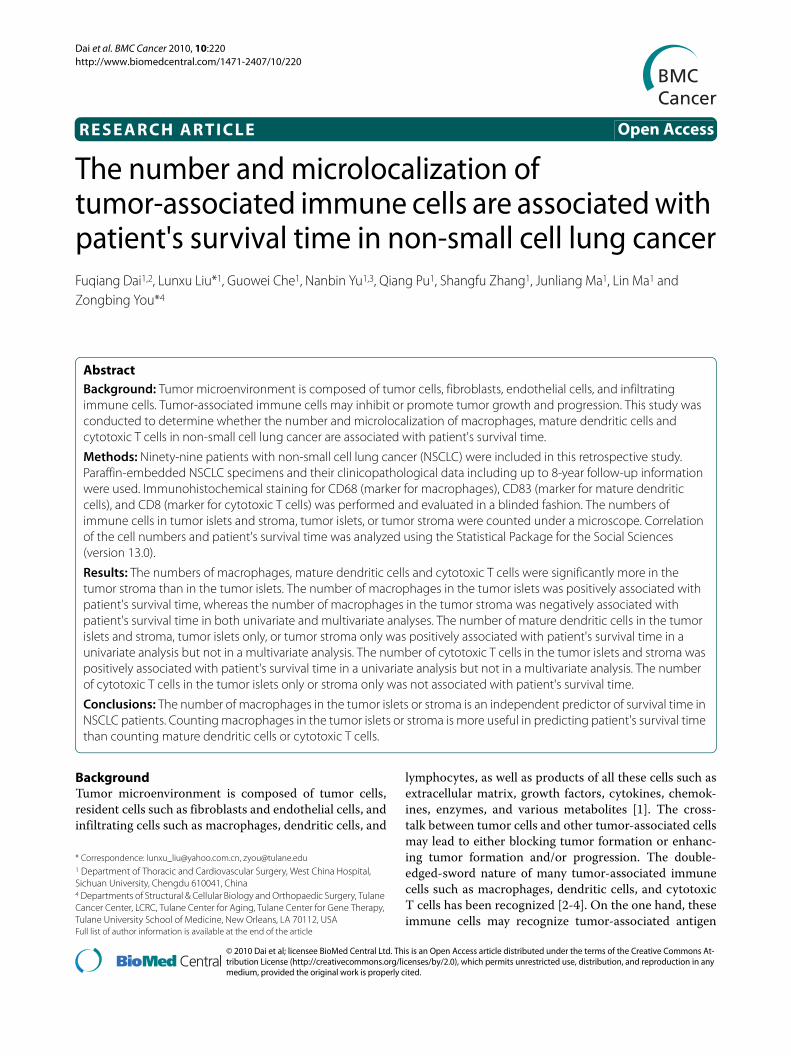
Dai et al. BMC Cancer 2010, 10:220 http://www.biomedcentral.com/1471-2407/10/220 Open Access RESEARCH ARTICLE BioMed Central © 2010 Dai et al; licensee BioMed Central Ltd. This is an Open Access article distributed under the terms of the Creative Commons At- tribution License (http://creativecommons.org/licenses/by/2.0), which permits unrestricted use, distribution, and reproduction in any medium, provided the original work is properly cited. Research article The number and microlocalization of tumor-associated immune cells are associated with patient's survival time in non-small cell lung cancer Fuqiang Dai 1,2 , Lunxu Liu* 1 , Guowei Che 1 , Nanbin Yu 1,3 , Qiang Pu 1 , Shangfu Zhang 1 , Junliang Ma 1 , Lin Ma 1 and Zongbing You* 4 Abstract Background: Tumor microenvironment is composed of tumor cells, fibroblasts, endothelial cells, and infiltrating immune cells. Tumor-associated immune cells may inhibit or promote tumor growth and progression. This study was conducted to determine whether the number and microlocalization of macrophages, mature dendritic cells and cytotoxic T cells in non-small cell lung cancer are associated with patient's survival time. Methods: Ninety-nine patients with non-small cell lung cancer (NSCLC) were included in this retrospective study. Paraffin-embedded NSCLC specimens and their clinicopathological data including up to 8-year follow-up information were used. Immunohistochemical staining for CD68 (marker for macrophages), CD83 (marker for mature dendritic cells), and CD8 (marker for cytotoxic T cells) was performed and evaluated in a blinded fashion. The numbers of immune cells in tumor islets and stroma, tumor islets, or tumor stroma were counted under a microscope. Correlation of the cell numbers and patient's survival time was analyzed using the Statistical Package for the Social Sciences (version 13.0). Results: The numbers of macrophages, mature dendritic cells and cytotoxic T cells were significantly more in the tumor stroma than in the tumor islets. The number of macrophages in the tumor islets was positively associated with patient's survival time, whereas the number of macrophages in the tumor stroma was negatively associated with patient's survival time in both univariate and multivariate analyses. The number of mature dendritic cells in the tumor islets and stroma, tumor islets only, or tumor stroma only was positively associated with patient's survival time in a univariate analysis but not in a multivariate analysis. The number of cytotoxic T cells in the tumor islets and stroma was positively associated with patient's survival time in a univariate analysis but not in a multivariate analysis. The number of cytotoxic T cells in the tumor islets only or stroma only was not associated with patient's survival time. Conclusions: The number of macrophages in the tumor islets or stroma is an independent predictor of survival time in NSCLC patients. Counting macrophages in the tumor islets or stroma is more useful in predicting patient's survival time than counting mature dendritic cells or cytotoxic T cells. Background Tumor microenvironment is composed of tumor cells, resident cells such as fibroblasts and endothelial cells, and infiltrating cells such as macrophages, dendritic cells, and lymphocytes, as well as products of all these cells such as extracellular matrix, growth factors, cytokines, chemok- ines, enzymes, and various metabolites [1]. The cross- talk between tumor cells and other tumor-associated cells may lead to either blocking tumor formation or enhanc- ing tumor formation and/or progression. The double- edged-sword nature of many tumor-associated immune cells such as macrophages, dendritic cells, and cytotoxic T cells has been recognized [2-4]. On the one hand, these immune cells may recognize tumor-associated antigen * Correspondence: [email protected], [email protected] 1 Department of Thoracic and Cardiovascular Surgery, West China Hospital, Sichuan University, Chengdu 610041, China 4 Departments of Structural & Cellular Biology and Orthopaedic Surgery, Tulane Cancer Center, LCRC, Tulane Center for Aging, Tulane Center for Gene Therapy, Tulane University School of Medicine, New Orleans, LA 70112, USA Full list of author information is available at the end of the article
-
Upload
independent -
Category
Documents
-
view
0 -
download
0
Transcript of The number and microlocalization of tumor-associated immune cells are associated with patient's...
Dai et al. BMC Cancer 2010, 10:220http://www.biomedcentral.com/1471-2407/10/220
Open AccessR E S E A R C H A R T I C L E
Research articleThe number and microlocalization of tumor-associated immune cells are associated with patient's survival time in non-small cell lung cancerFuqiang Dai1,2, Lunxu Liu*1, Guowei Che1, Nanbin Yu1,3, Qiang Pu1, Shangfu Zhang1, Junliang Ma1, Lin Ma1 and Zongbing You*4
AbstractBackground: Tumor microenvironment is composed of tumor cells, fibroblasts, endothelial cells, and infiltrating immune cells. Tumor-associated immune cells may inhibit or promote tumor growth and progression. This study was conducted to determine whether the number and microlocalization of macrophages, mature dendritic cells and cytotoxic T cells in non-small cell lung cancer are associated with patient's survival time.
Methods: Ninety-nine patients with non-small cell lung cancer (NSCLC) were included in this retrospective study. Paraffin-embedded NSCLC specimens and their clinicopathological data including up to 8-year follow-up information were used. Immunohistochemical staining for CD68 (marker for macrophages), CD83 (marker for mature dendritic cells), and CD8 (marker for cytotoxic T cells) was performed and evaluated in a blinded fashion. The numbers of immune cells in tumor islets and stroma, tumor islets, or tumor stroma were counted under a microscope. Correlation of the cell numbers and patient's survival time was analyzed using the Statistical Package for the Social Sciences (version 13.0).
Results: The numbers of macrophages, mature dendritic cells and cytotoxic T cells were significantly more in the tumor stroma than in the tumor islets. The number of macrophages in the tumor islets was positively associated with patient's survival time, whereas the number of macrophages in the tumor stroma was negatively associated with patient's survival time in both univariate and multivariate analyses. The number of mature dendritic cells in the tumor islets and stroma, tumor islets only, or tumor stroma only was positively associated with patient's survival time in a univariate analysis but not in a multivariate analysis. The number of cytotoxic T cells in the tumor islets and stroma was positively associated with patient's survival time in a univariate analysis but not in a multivariate analysis. The number of cytotoxic T cells in the tumor islets only or stroma only was not associated with patient's survival time.
Conclusions: The number of macrophages in the tumor islets or stroma is an independent predictor of survival time in NSCLC patients. Counting macrophages in the tumor islets or stroma is more useful in predicting patient's survival time than counting mature dendritic cells or cytotoxic T cells.
BackgroundTumor microenvironment is composed of tumor cells,resident cells such as fibroblasts and endothelial cells, andinfiltrating cells such as macrophages, dendritic cells, and
lymphocytes, as well as products of all these cells such asextracellular matrix, growth factors, cytokines, chemok-ines, enzymes, and various metabolites [1]. The cross-talk between tumor cells and other tumor-associated cellsmay lead to either blocking tumor formation or enhanc-ing tumor formation and/or progression. The double-edged-sword nature of many tumor-associated immunecells such as macrophages, dendritic cells, and cytotoxicT cells has been recognized [2-4]. On the one hand, theseimmune cells may recognize tumor-associated antigen
* Correspondence: [email protected], [email protected] Department of Thoracic and Cardiovascular Surgery, West China Hospital, Sichuan University, Chengdu 610041, China4 Departments of Structural & Cellular Biology and Orthopaedic Surgery, Tulane Cancer Center, LCRC, Tulane Center for Aging, Tulane Center for Gene Therapy, Tulane University School of Medicine, New Orleans, LA 70112, USAFull list of author information is available at the end of the article
BioMed Central© 2010 Dai et al; licensee BioMed Central Ltd. This is an Open Access article distributed under the terms of the Creative Commons At-tribution License (http://creativecommons.org/licenses/by/2.0), which permits unrestricted use, distribution, and reproduction in anymedium, provided the original work is properly cited.
Dai et al. BMC Cancer 2010, 10:220http://www.biomedcentral.com/1471-2407/10/220
Page 2 of 10
and activate cytotoxic T cells, in order to initiate anti-tumor immune responses. On the other hand, the sameimmune cells may establish immune tolerance and evenpromote tumor growth and metastasis through enhanc-ing angiogenesis and invasion of extracellular matrix.
Non-small cell lung cancer (NSCLC) is the most com-mon cause of cancer-related death worldwide. The five-year survival rate is approximately 67% for the patientswith stage IA NSCLC after putatively curative surgery [5].In order to identify new prognostic factors that can guideclinical practice, we have previously found that the num-ber of tumor-associated macrophages (TAMs) in thetumor islets is positively associated with survival time inthe patients with NSCLC [6]. Because TAMs are not theonly tumor-associated immune cells, in this study we fur-ther investigated the prognostic value of mature dendriticcells and cytotoxic T cells in the patients with NSCLC.
MethodsStudy populationThis study was approved by the Institutional ReviewBoard of West China Hospital, Sichuan University. Theprocedures to obtain human lung cancer tissues and fol-low-up information are in accordance with the EthicalPrinciples for Medical Research Involving Human Sub-jects as formulated in the World Medical AssociationDeclaration of Helsinki (revised in 2008). All specimenswere obtained from the archives of formalin-fixed, paraf-fin-embedded tissue blocks in the Department of Tho-racic and Cardiovascular Surgery, West China Hospital,Sichuan University. The lung cancer tissues were col-lected from surgeries performed from August, 1999 toAugust, 2001. The patients were followed up untilDecember, 2007, through outpatient visits and/or corre-spondences to family members. Ninety-nine patientswere included in this retrospective study. Histologicalevaluation was based on the World Health Organizationcriteria [7]. Tumor stage was evaluated according to theInternational Union against Cancer TNM classificationsystem [7]. The clinicopathological characteristics weresummarized in Table 1.
ImmunohistochemistryFour-μm thick tissue sections were de-waxed in xyleneand rehydrated through graded alcohols. Antigenretrieval was carried out using microwave at middle-to-high temperature for 8 min, low-to-high temperature for5 min, and then cooled down at room temperature for 20min. Mouse anti-human CD68 monoclonal antibodies(clone KP1, recognizing macrophages), rabbit anti-human CD8 monoclonal antibodies (clone SP16, recog-nizing cytotoxic T cells), and streptavidin-peroxidaseconjugated secondary antibodies (SP-9002) were
obtained from Zhongshan Goldenbridge BiotechnologyCo., LTD., Beijing, China. Mouse anti-human CD83monoclonal antibodies (clone HB15a, recognizingmature dendritic cells) were obtained from Santa CruzBiotechnology, Inc., Santa Cruz, CA, USA. Diaminobenz-idine (DAB) substrate kit was obtained from Dako NorthAmerica, Inc., Carpinteria, CA, USA. Immunohis-tochemical staining of individual markers was performedaccording to the kit manufacturer's instructions. Sectionswere then counterstained with hematoxylin and mountedin an aqueous mounting medium. Tissue sections previ-ously stained positively were used as positive control,while tissue sections with primary antibodies replaced byphosphate-buffered saline served as negative control.Positive cells showed brown particles on the cellularmembrane and/or in the cytoplasm. Under a microscope,five representative high-power fields (× 400 magnifica-tion) of the tumor islets and stroma per tissue sectionwere selected and counted for positive cell numbers. Theaverage number of these five high-power fields repre-sented the cell number per high-power field. Evaluationof the stained tissue sections was performed by twoinvestigators who were blinded in regard to the clinico-pathological characteristics. An average of the resultsobtained by the two examiners was used to representeach case.
Statistical analysisStatistical analysis was carried out using the StatisticalPackage for the Social Sciences (SPSS, version 13.0, SPSSInc., Chicago, IL, USA). For categorical analysis, themedian number of cells per high-power field was used asa cut-off point to dichotomize the continuous variables.The Mann-Whitney nonparametric test was used tocompare between two groups. In order to assess anypotential relationship between the number of immunecells and patient's survival time, the Spearman's rank cor-relation coefficient (Spearman's rho, or rs) was calculatedin a univariate analysis. The correlation between thenumber of immune cells and the clinicopathologicalcharacteristics was analyzed using the χ2 test. TheKaplan-Meier survival curves were used to look for cor-relation between the cell number and patient's survivaltime, or between the ratio of cells in the tumor islets ver-sus stroma and patient's survival time. Statistical signifi-cance was analyzed using the log-rank test. Amultivariate Cox proportional hazards model was used toestimate adjusted hazard ratios and 95% confidence inter-vals (CI) and to identify which variable was an indepen-dent prognostic factor. The validity of the proportionalhazards assumption was assessed from log (-log [Sur-vival]) curves. For the above comparisons, P < 0.05 wasconsidered statistically significant.
Dai et al. BMC Cancer 2010, 10:220http://www.biomedcentral.com/1471-2407/10/220
Page 3 of 10
ResultsPatient characteristicsAll of the 99 patients (Table 1) had complete follow-upinformation and the pathological diagnosis was verifiedby a pathologist prior to inclusion in this study. The over-all cumulative 5-year survival rate was 32%. Patients withstage I carcinomas, squamous cell carcinomas, well-dif-ferentiated carcinomas, or carcinomas without lymphnode metastasis had better survival rates than the otherpatients (Table 1).
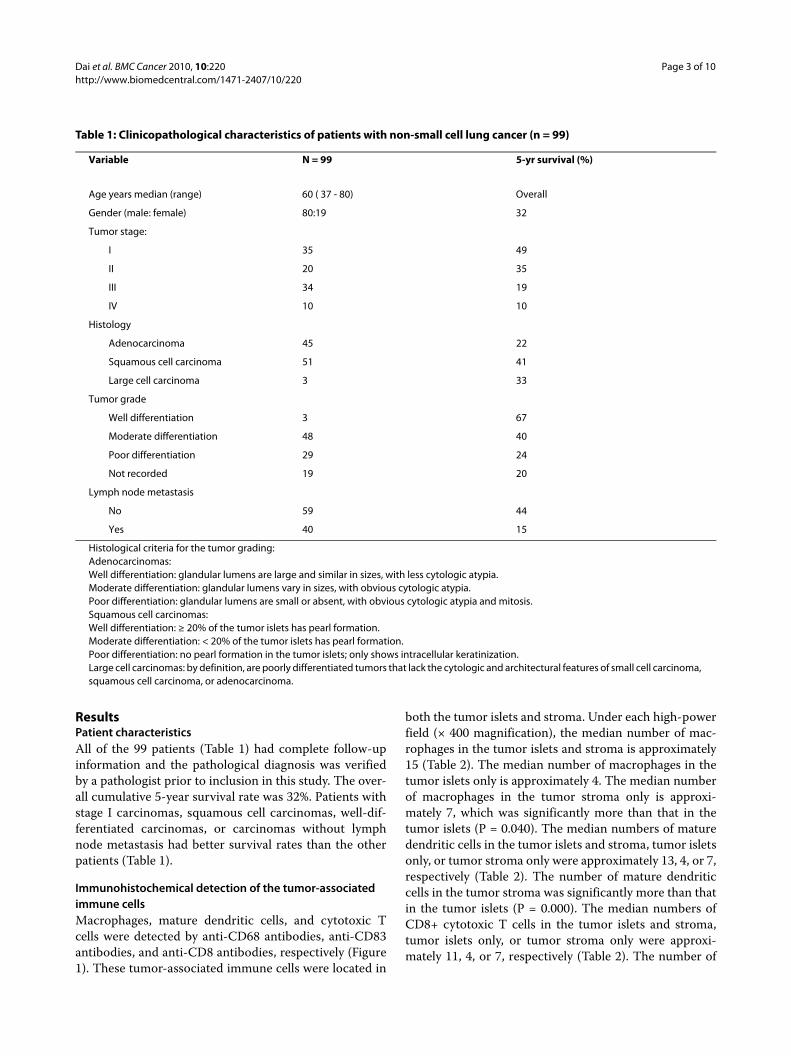
Immunohistochemical detection of the tumor-associated immune cellsMacrophages, mature dendritic cells, and cytotoxic Tcells were detected by anti-CD68 antibodies, anti-CD83antibodies, and anti-CD8 antibodies, respectively (Figure1). These tumor-associated immune cells were located in
both the tumor islets and stroma. Under each high-powerfield (× 400 magnification), the median number of mac-rophages in the tumor islets and stroma is approximately15 (Table 2). The median number of macrophages in thetumor islets only is approximately 4. The median numberof macrophages in the tumor stroma only is approxi-mately 7, which was significantly more than that in thetumor islets (P = 0.040). The median numbers of maturedendritic cells in the tumor islets and stroma, tumor isletsonly, or tumor stroma only were approximately 13, 4, or 7,respectively (Table 2). The number of mature dendriticcells in the tumor stroma was significantly more than thatin the tumor islets (P = 0.000). The median numbers ofCD8+ cytotoxic T cells in the tumor islets and stroma,tumor islets only, or tumor stroma only were approxi-mately 11, 4, or 7, respectively (Table 2). The number of
Table 1: Clinicopathological characteristics of patients with non-small cell lung cancer (n = 99)
Variable N = 99 5-yr survival (%)
Age years median (range) 60 ( 37 - 80) Overall
Gender (male: female) 80:19 32
Tumor stage:
I 35 49
II 20 35
III 34 19
IV 10 10
Histology
Adenocarcinoma 45 22
Squamous cell carcinoma 51 41
Large cell carcinoma 3 33
Tumor grade
Well differentiation 3 67
Moderate differentiation 48 40
Poor differentiation 29 24
Not recorded 19 20
Lymph node metastasis
No 59 44
Yes 40 15
Histological criteria for the tumor grading:Adenocarcinomas:Well differentiation: glandular lumens are large and similar in sizes, with less cytologic atypia.Moderate differentiation: glandular lumens vary in sizes, with obvious cytologic atypia.Poor differentiation: glandular lumens are small or absent, with obvious cytologic atypia and mitosis.Squamous cell carcinomas:Well differentiation: ≥ 20% of the tumor islets has pearl formation.Moderate differentiation: < 20% of the tumor islets has pearl formation.Poor differentiation: no pearl formation in the tumor islets; only shows intracellular keratinization.Large cell carcinomas: by definition, are poorly differentiated tumors that lack the cytologic and architectural features of small cell carcinoma, squamous cell carcinoma, or adenocarcinoma.
Dai et al. BMC Cancer 2010, 10:220http://www.biomedcentral.com/1471-2407/10/220
Page 4 of 10
CD8+ cytotoxic T cells in the tumor stroma was signifi-cantly more than that in the tumor islets (P = 0.032).
Correlation between the number and microlocalization of immune cells and survival timeThe Spearman's rank correlation coefficient (Spearman'srho, or rs) was calculated to assess any potential relation-ship between the tumor-associated immune cells andpatient's survival time. We found that the macrophagenumber in the tumor islets and stroma had no associationwith patient's survival time (rs = -0.105, P = 0.300). Themacrophage number in the tumor islets was positivelyassociated with patient's survival (rs = 0.483, P = 0.000). Incontrast, the macrophage number in the tumor stromawas negatively associated with patient's survival (rs = -
0.541, P = 0.000) (Table 2). The numbers of mature den-dritic cells in the tumor islets and stroma, tumor islets,and tumor stroma were all positively associated withpatient's survival time, with rs = 0.221 (P = 0.028), 0.404(P = 0.001), and 0.284 (P = 0.045), respectively. Similarly,the numbers of CD8+ cytotoxic T cells in the tumor isletsand stroma, tumor islets, and tumor stroma were alsopositively associated with patient's survival time, with rs =0.297 (P = 0.003), 0.247 (P = 0.014), and 0.212 (P = 0.035),respectively (Table 2).
In order to assess whether there is any value in predict-ing prognosis, the median number of the tumor-associ-ated immune cells was used as a cut-off point todichotomize the 99 patients into a group with a cell num-ber above the median and a group with a cell number
Table 2: Correlation between the number and microlocalization of immune cells and patient's survival time by a univariate analysis
Cell type Islets + Stroma Islets Stroma P (islets versus stroma)
Number Spearman's rho & P
Number Spearman's rho & P
Number Spearman's rho & P
Macrophages 15.2(1.8 - 48.4)
rs = -0.105P = 0.300
4.2(0.2 - 40.6)
rs = 0.483P = 0.000
7.2(0.4 - 39.6)
rs = -0.541P = 0.000
0.040
Mature dendritic cells
13.2(1.4 - 137.6)
rs = 0.221P = 0.028
4.2(0 - 21.4)
rs = 0.404P = 0.001
7.0(0.2 - 25.4)
rs = 0.284P = 0.045
0.000
CD8+ T cells 10.8(0.4 - 45.0)
rs = 0.297P = 0.003
4.2(0 - 66.2)
rs = 0.247P = 0.014
7.2(0.2 - 71.4)
rs = 0.212P = 0.035
0.032
The number of immune cells represents median (range) per high-power field. The number of immune cells in the tumor islets and stroma (Islets + Stroma) was obtained by combining the number in the tumor islets and the number in the tumor stroma in each case. Then, the numbers of immune cells in the tumor islets and stroma were ranked and the median number was determined. Therefore, the median number of immune cells in the tumor islets and stroma is not a simple sum of the median number in the tumor islets and the median number in the tumor stroma as shown in this table.
Figure 1 Immunohistochemical detection of tumor-associated immune cells in non-small cell lung cancer tissues. (a) Macrophages (marker CD68) stained brown in the tumor islets (arrow) and tumor stroma (arrowhead). (b) Mature dendritic cells (marker CD83) stained brown in the tumor islets (arrow) and tumor stroma (arrowhead). (c) Cytotoxic T cells (marker CD8) stained brown in the tumor islets (arrow) and tumor stroma (arrow-head). Original magnification, × 400.
Dai et al. BMC Cancer 2010, 10:220http://www.biomedcentral.com/1471-2407/10/220
Page 5 of 10
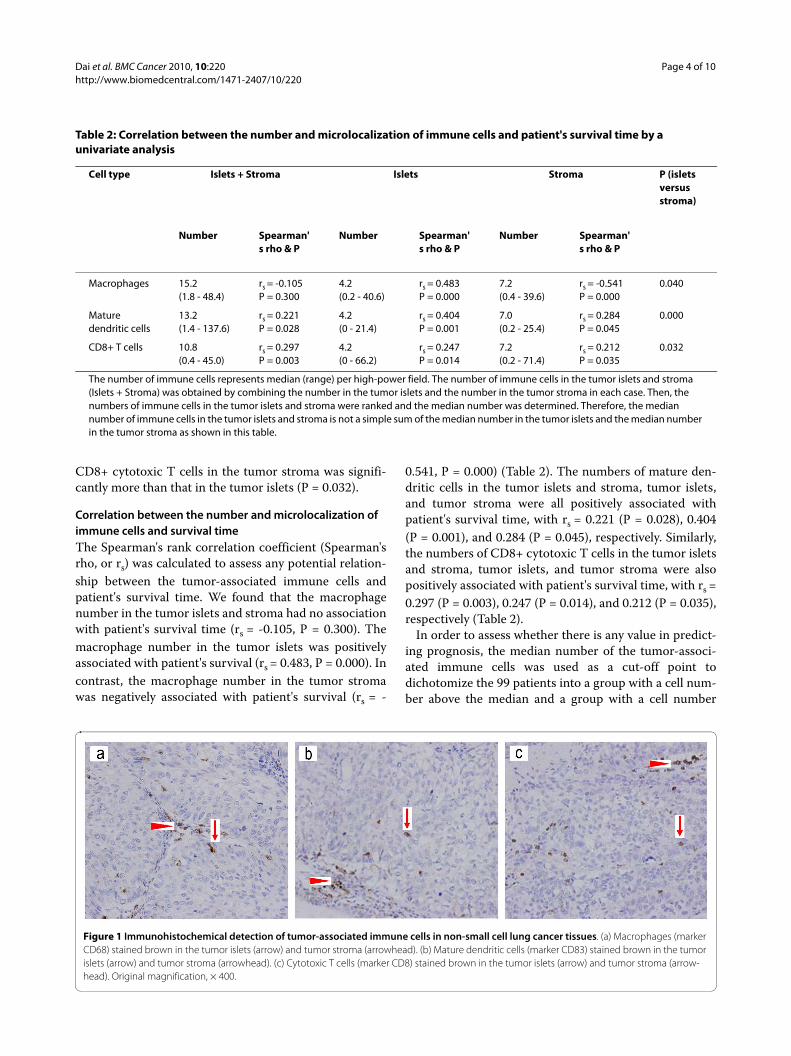
below the median. We found that the patients withabove-the-median macrophage number in the tumorislets and stroma had a similar Kaplan-Meier survivalcurve, compared to those patients with below-the-median macrophage number in the tumor islets andstroma (P = 0.322, Figure 2a). The patients with above-the-median macrophage number in the tumor islets had asignificantly better cumulative survival, compared to thepatients with below-the-median macrophage number inthe tumor islets (P = 0.001, Figure 2b). In contrast, thepatients with above-the-median macrophage number inthe tumor stroma had a significantly worse cumulativesurvival, compared to the patients with below-the-median macrophage number in the tumor stroma (P =0.000, Figure 2c). When the ratio of the macrophagenumber in the tumor islets versus the macrophage num-ber in the tumor stroma was calculated, the patients withabove-the-median ratio (islets/stroma) had a significantlybetter cumulative survival, compared to the patients withbelow-the-median ratio (P = 0.000, Figure 2d).
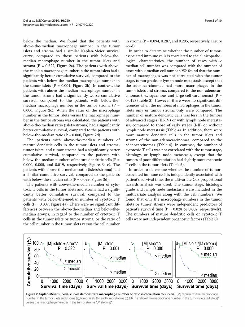
The patients with above-the-median numbers ofmature dendritic cells in the tumor islets and stroma,tumor islets, and tumor stroma had a significantly bettercumulative survival, compared to the patients withbelow-the-median numbers of mature dendritic cells (P =0.000, 0.005, and 0.019, respectively, Figure 3a-c). Thepatients with above-the-median ratio (islets/stroma) hada similar cumulative survival, compared to the patientswith below-the-median ratio (P = 0.099, Figure 3d).
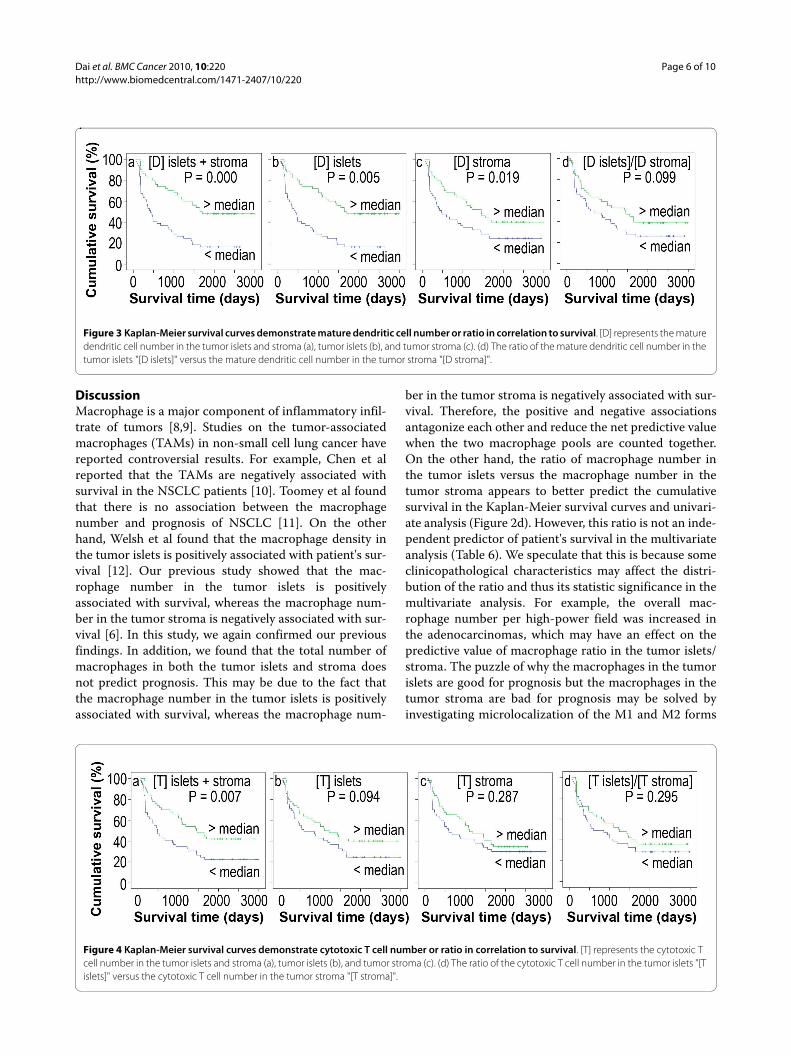
The patients with above-the-median number of cyto-toxic T cells in the tumor islets and stroma had a signifi-cantly better cumulative survival, compared to thepatients with below-the-median number of cytotoxic Tcells (P = 0.007, Figure 4a). There were no significant dif-ferences between the above-the-median and below-the-median groups, in regard to the number of cytotoxic Tcells in the tumor islets or tumor stroma, or the ratio ofthe cell number in the tumor islets versus the cell number
in stroma (P = 0.094, 0.287, and 0.295, respectively, Figure4b-d).
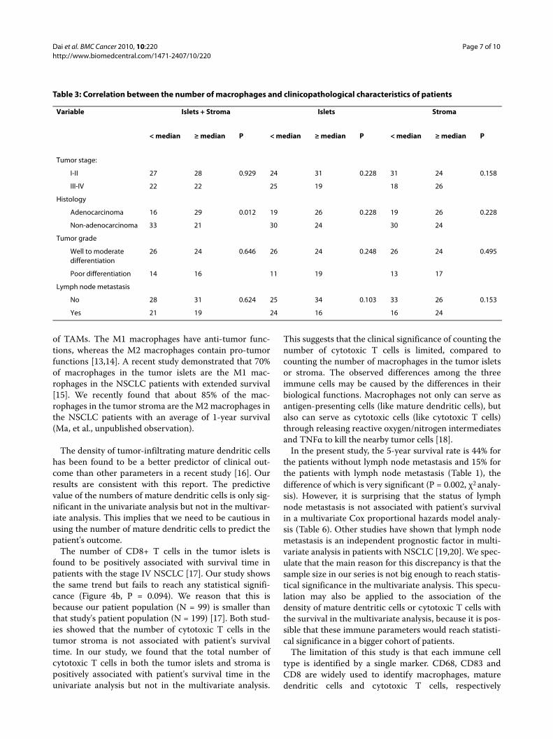
In order to determine whether the number of tumor-associated immune cells is correlated to the clinicopatho-logical characteristics, the number of cases with <median cell number was compared with the number ofcases with ≥ median cell number. We found that the num-ber of macrophages was not correlated with the tumorstage, tumor grade, or lymph node metastasis, except thatthe adenocarcinomas had more macrophages in thetumor islets and stroma, compared to the non-adenocar-cinomas (i.e., squamous and large cell carcinomas) (P =0.012) (Table 3). However, there were no significant dif-ferences when the numbers of macrophages in the tumorislets only or tumor stroma only were compared. Thenumber of mature dendritic cells was less in the tumorsof advanced stages (III-IV) or with lymph node metasta-sis, compared to those of early stages (I-II) or withoutlymph node metastasis (Table 4). In addition, there weremore mature dendritic cells in the tumor islets andstroma of the non-adenocarcinomas, compared to theadenocarcinomas (Table 4). In contrast, the number ofcytotoxic T cells was not correlated with the tumor stage,histology, or lymph node metastasis, except that thetumors of poor differentiation had slightly more cytotoxicT cells in the tumor islets (Table 5).
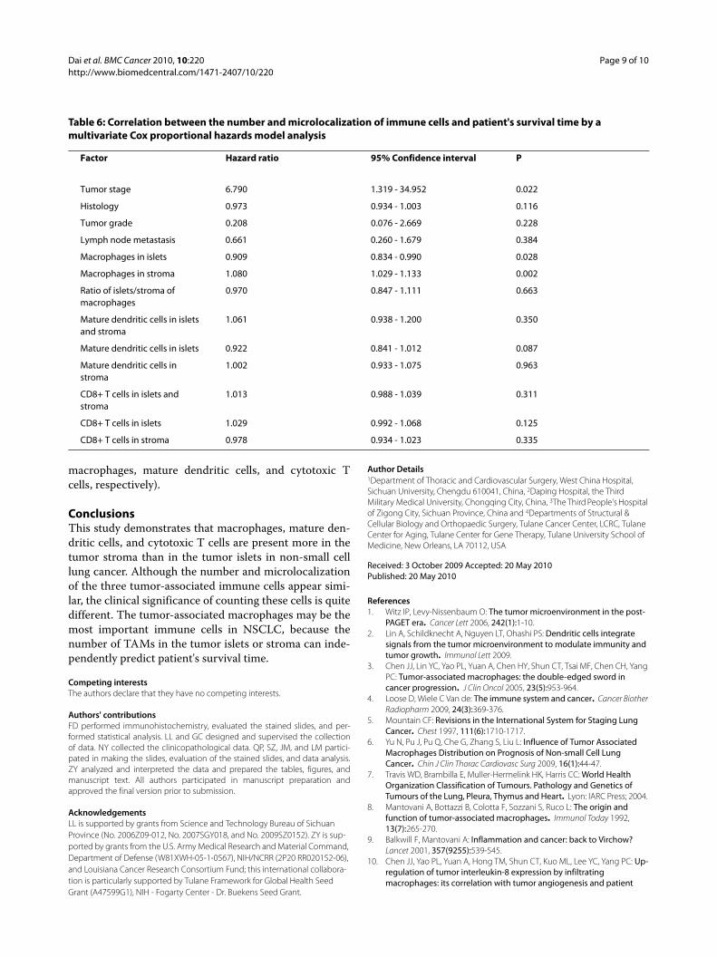
In order to determine whether the number of tumor-associated immune cells is independently associated withpatient's survival time, the multivariate Cox proportionalhazards analysis was used. The tumor stage, histology,grade and lymph node metastasis were included in themultivariate analysis along with the cell numbers. Wefound that only the macrophage numbers in the tumorislets or tumor stroma were independent predictors ofpatient's survival time (P = 0.028 or 0.002, respectively).The numbers of mature dendritic cells or cytotoxic Tcells were not independent prognostic factors (Table 6).
Figure 2 Kaplan-Meier survival curves demonstrate macrophage number or ratio in correlation to survival. [M] represents the macrophage number in the tumor islets and stroma (a), tumor islets (b), and tumor stroma (c). (d) The ratio of the macrophage number in the tumor islets "[M islets]" versus the macrophage number in the tumor stroma "[M stroma]".
Dai et al. BMC Cancer 2010, 10:220http://www.biomedcentral.com/1471-2407/10/220
Page 6 of 10
DiscussionMacrophage is a major component of inflammatory infil-trate of tumors [8,9]. Studies on the tumor-associatedmacrophages (TAMs) in non-small cell lung cancer havereported controversial results. For example, Chen et alreported that the TAMs are negatively associated withsurvival in the NSCLC patients [10]. Toomey et al foundthat there is no association between the macrophagenumber and prognosis of NSCLC [11]. On the otherhand, Welsh et al found that the macrophage density inthe tumor islets is positively associated with patient's sur-vival [12]. Our previous study showed that the mac-rophage number in the tumor islets is positivelyassociated with survival, whereas the macrophage num-ber in the tumor stroma is negatively associated with sur-vival [6]. In this study, we again confirmed our previousfindings. In addition, we found that the total number ofmacrophages in both the tumor islets and stroma doesnot predict prognosis. This may be due to the fact thatthe macrophage number in the tumor islets is positivelyassociated with survival, whereas the macrophage num-
ber in the tumor stroma is negatively associated with sur-vival. Therefore, the positive and negative associationsantagonize each other and reduce the net predictive valuewhen the two macrophage pools are counted together.On the other hand, the ratio of macrophage number inthe tumor islets versus the macrophage number in thetumor stroma appears to better predict the cumulativesurvival in the Kaplan-Meier survival curves and univari-ate analysis (Figure 2d). However, this ratio is not an inde-pendent predictor of patient's survival in the multivariateanalysis (Table 6). We speculate that this is because someclinicopathological characteristics may affect the distri-bution of the ratio and thus its statistic significance in themultivariate analysis. For example, the overall mac-rophage number per high-power field was increased inthe adenocarcinomas, which may have an effect on thepredictive value of macrophage ratio in the tumor islets/stroma. The puzzle of why the macrophages in the tumorislets are good for prognosis but the macrophages in thetumor stroma are bad for prognosis may be solved byinvestigating microlocalization of the M1 and M2 forms
Figure 3 Kaplan-Meier survival curves demonstrate mature dendritic cell number or ratio in correlation to survival. [D] represents the mature dendritic cell number in the tumor islets and stroma (a), tumor islets (b), and tumor stroma (c). (d) The ratio of the mature dendritic cell number in the tumor islets "[D islets]" versus the mature dendritic cell number in the tumor stroma "[D stroma]".
Figure 4 Kaplan-Meier survival curves demonstrate cytotoxic T cell number or ratio in correlation to survival. [T] represents the cytotoxic T cell number in the tumor islets and stroma (a), tumor islets (b), and tumor stroma (c). (d) The ratio of the cytotoxic T cell number in the tumor islets "[T islets]" versus the cytotoxic T cell number in the tumor stroma "[T stroma]".
Dai et al. BMC Cancer 2010, 10:220http://www.biomedcentral.com/1471-2407/10/220
Page 7 of 10
Table 3: Correlation between the number of macrophages and clinicopathological characteristics of patients
Variable Islets + Stroma Islets Stroma
< median ≥ median P < median ≥ median P < median ≥ median P
Tumor stage:
I-II 27 28 0.929 24 31 0.228 31 24 0.158
III-IV 22 22 25 19 18 26
Histology
Adenocarcinoma 16 29 0.012 19 26 0.228 19 26 0.228
Non-adenocarcinoma 33 21 30 24 30 24
Tumor grade
Well to moderate differentiation
26 24 0.646 26 24 0.248 26 24 0.495
Poor differentiation 14 16 11 19 13 17
Lymph node metastasis
No 28 31 0.624 25 34 0.103 33 26 0.153
Yes 21 19 24 16 16 24
of TAMs. The M1 macrophages have anti-tumor func-tions, whereas the M2 macrophages contain pro-tumorfunctions [13,14]. A recent study demonstrated that 70%of macrophages in the tumor islets are the M1 mac-rophages in the NSCLC patients with extended survival[15]. We recently found that about 85% of the mac-rophages in the tumor stroma are the M2 macrophages inthe NSCLC patients with an average of 1-year survival(Ma, et al., unpublished observation).
The density of tumor-infiltrating mature dendritic cellshas been found to be a better predictor of clinical out-come than other parameters in a recent study [16]. Ourresults are consistent with this report. The predictivevalue of the numbers of mature dendritic cells is only sig-nificant in the univariate analysis but not in the multivar-iate analysis. This implies that we need to be cautious inusing the number of mature dendritic cells to predict thepatient's outcome.
The number of CD8+ T cells in the tumor islets isfound to be positively associated with survival time inpatients with the stage IV NSCLC [17]. Our study showsthe same trend but fails to reach any statistical signifi-cance (Figure 4b, P = 0.094). We reason that this isbecause our patient population (N = 99) is smaller thanthat study's patient population (N = 199) [17]. Both stud-ies showed that the number of cytotoxic T cells in thetumor stroma is not associated with patient's survivaltime. In our study, we found that the total number ofcytotoxic T cells in both the tumor islets and stroma ispositively associated with patient's survival time in theunivariate analysis but not in the multivariate analysis.
This suggests that the clinical significance of counting thenumber of cytotoxic T cells is limited, compared tocounting the number of macrophages in the tumor isletsor stroma. The observed differences among the threeimmune cells may be caused by the differences in theirbiological functions. Macrophages not only can serve asantigen-presenting cells (like mature dendritic cells), butalso can serve as cytotoxic cells (like cytotoxic T cells)through releasing reactive oxygen/nitrogen intermediatesand TNFα to kill the nearby tumor cells [18].
In the present study, the 5-year survival rate is 44% forthe patients without lymph node metastasis and 15% forthe patients with lymph node metastasis (Table 1), thedifference of which is very significant (P = 0.002, χ2 analy-sis). However, it is surprising that the status of lymphnode metastasis is not associated with patient's survivalin a multivariate Cox proportional hazards model analy-sis (Table 6). Other studies have shown that lymph nodemetastasis is an independent prognostic factor in multi-variate analysis in patients with NSCLC [19,20]. We spec-ulate that the main reason for this discrepancy is that thesample size in our series is not big enough to reach statis-tical significance in the multivariate analysis. This specu-lation may also be applied to the association of thedensity of mature dentritic cells or cytotoxic T cells withthe survival in the multivariate analysis, because it is pos-sible that these immune parameters would reach statisti-cal significance in a bigger cohort of patients.
The limitation of this study is that each immune celltype is identified by a single marker. CD68, CD83 andCD8 are widely used to identify macrophages, maturedendritic cells and cytotoxic T cells, respectively
Dai et al. BMC Cancer 2010, 10:220http://www.biomedcentral.com/1471-2407/10/220
Page 8 of 10
Table 4: Correlation between the number of mature dendritic cells and clinicopathological characteristics of patients
Variable Islets + Stroma Islets Stroma
< median ≥ median P < median ≥ median P < median ≥ median P
Tumor stage:
I-II 21 34 0.012 23 32 0.107 22 33 0.044
III-IV 28 16 26 18 27 17
Histology
Adenocarcinoma 30 15 0.002 27 18 0.070 27 18 0.070
Non-adenocarcinoma
19 35 22 32 22 32
Tumor grade
Well to moderate differentiation
21 29 0.861 23 27 0.647 26 24 0.066
Poor differentiation
12 18 12 18 9 21
Lymph node metastasis
No 23 36 0.014 24 35 0.041 25 34 0.103
Yes 26 14 25 15 24 16
Table 5: Correlation between the number of cytotoxic T cells and clinicopathological characteristics of patients
Variable Islets + Stroma Islets Stroma
< median ≥ median P < median ≥ median P < median ≥ median P
Tumor stage:
I-II 23 32 0.107 24 31 0.228 27 28 0.840
III-IV 26 18 25 19 23 21
Histology
Adenocarcinoma 24 21 0.547 23 22 0.841 24 21 0.688
Non-adenocarcinoma
25 29 26 28 26 28
Tumor grade
Well to moderate differentiation
26 24 0.248 29 21 0.040 28 22 0.248
Poor differentiation
11 19 10 20 12 18
Lymph node metastasis
No 25 34 0.103 27 32 0.416 29 30 0.838
Yes 24 16 22 18 21 19
[15,17,21-25]. However, these markers are also expressedby other cell types. For example, CD68 has been found inimmature CD1a-positive dendritic cells [26,27]. CD83 isalso expressed by other cell types such as B lymphocytes[28]. A better marker for mature dendritic cells is DC-Lamp (CD208, using rat IgG2a antibodies, clone
SR1010.E1, from Schering-Plough, Kenilworth, NJ) forparaffin-embedded tissue sections [16]. Although mosttumor-infiltrating CD8+ T cells are cytotoxic T cells [25],some CD8+ T cells are regulatory T cells [29]. Therefore,it is possible that a portion of the immune cells identifiedby these markers are not the named immune cells (i.e.,
Dai et al. BMC Cancer 2010, 10:220http://www.biomedcentral.com/1471-2407/10/220
Page 9 of 10
macrophages, mature dendritic cells, and cytotoxic Tcells, respectively).
ConclusionsThis study demonstrates that macrophages, mature den-dritic cells, and cytotoxic T cells are present more in thetumor stroma than in the tumor islets in non-small celllung cancer. Although the number and microlocalizationof the three tumor-associated immune cells appear simi-lar, the clinical significance of counting these cells is quitedifferent. The tumor-associated macrophages may be themost important immune cells in NSCLC, because thenumber of TAMs in the tumor islets or stroma can inde-pendently predict patient's survival time.
Competing interestsThe authors declare that they have no competing interests.
Authors' contributionsFD performed immunohistochemistry, evaluated the stained slides, and per-formed statistical analysis. LL and GC designed and supervised the collectionof data. NY collected the clinicopathological data. QP, SZ, JM, and LM partici-pated in making the slides, evaluation of the stained slides, and data analysis.ZY analyzed and interpreted the data and prepared the tables, figures, andmanuscript text. All authors participated in manuscript preparation andapproved the final version prior to submission.
AcknowledgementsLL is supported by grants from Science and Technology Bureau of Sichuan Province (No. 2006Z09-012, No. 2007SGY018, and No. 2009SZ0152). ZY is sup-ported by grants from the U.S. Army Medical Research and Material Command, Department of Defense (W81XWH-05-1-0567), NIH/NCRR (2P20 RR020152-06), and Louisiana Cancer Research Consortium Fund; this international collabora-tion is particularly supported by Tulane Framework for Global Health Seed Grant (A47599G1), NIH - Fogarty Center - Dr. Buekens Seed Grant.
Author Details1Department of Thoracic and Cardiovascular Surgery, West China Hospital, Sichuan University, Chengdu 610041, China, 2Daping Hospital, the Third Military Medical University, Chongqing City, China, 3The Third People's Hospital of Zigong City, Sichuan Province, China and 4Departments of Structural & Cellular Biology and Orthopaedic Surgery, Tulane Cancer Center, LCRC, Tulane Center for Aging, Tulane Center for Gene Therapy, Tulane University School of Medicine, New Orleans, LA 70112, USA
References1. Witz IP, Levy-Nissenbaum O: The tumor microenvironment in the post-
PAGET era. Cancer Lett 2006, 242(1):1-10.2. Lin A, Schildknecht A, Nguyen LT, Ohashi PS: Dendritic cells integrate
signals from the tumor microenvironment to modulate immunity and tumor growth. Immunol Lett 2009.
3. Chen JJ, Lin YC, Yao PL, Yuan A, Chen HY, Shun CT, Tsai MF, Chen CH, Yang PC: Tumor-associated macrophages: the double-edged sword in cancer progression. J Clin Oncol 2005, 23(5):953-964.
4. Loose D, Wiele C Van de: The immune system and cancer. Cancer Biother Radiopharm 2009, 24(3):369-376.
5. Mountain CF: Revisions in the International System for Staging Lung Cancer. Chest 1997, 111(6):1710-1717.
6. Yu N, Pu J, Pu Q, Che G, Zhang S, Liu L: Influence of Tumor Associated Macrophages Distribution on Prognosis of Non-small Cell Lung Cancer. Chin J Clin Thorac Cardiovasc Surg 2009, 16(1):44-47.
7. Travis WD, Brambilla E, Muller-Hermelink HK, Harris CC: World Health Organization Classification of Tumours. Pathology and Genetics of Tumours of the Lung, Pleura, Thymus and Heart. Lyon: IARC Press; 2004.
8. Mantovani A, Bottazzi B, Colotta F, Sozzani S, Ruco L: The origin and function of tumor-associated macrophages. Immunol Today 1992, 13(7):265-270.
9. Balkwill F, Mantovani A: Inflammation and cancer: back to Virchow? Lancet 2001, 357(9255):539-545.
10. Chen JJ, Yao PL, Yuan A, Hong TM, Shun CT, Kuo ML, Lee YC, Yang PC: Up-regulation of tumor interleukin-8 expression by infiltrating macrophages: its correlation with tumor angiogenesis and patient
Received: 3 October 2009 Accepted: 20 May 2010 Published: 20 May 2010This article is available from: http://www.biomedcentral.com/1471-2407/10/220© 2010 Dai et al; licensee BioMed Central Ltd. This is an Open Access article distributed under the terms of the Creative Commons Attribution License (http://creativecommons.org/licenses/by/2.0), which permits unrestricted use, distribution, and reproduction in any medium, provided the original work is properly cited.BMC Cancer 2010, 10:220
Table 6: Correlation between the number and microlocalization of immune cells and patient's survival time by a multivariate Cox proportional hazards model analysis
Factor Hazard ratio 95% Confidence interval P
Tumor stage 6.790 1.319 - 34.952 0.022
Histology 0.973 0.934 - 1.003 0.116
Tumor grade 0.208 0.076 - 2.669 0.228
Lymph node metastasis 0.661 0.260 - 1.679 0.384
Macrophages in islets 0.909 0.834 - 0.990 0.028
Macrophages in stroma 1.080 1.029 - 1.133 0.002
Ratio of islets/stroma of macrophages
0.970 0.847 - 1.111 0.663
Mature dendritic cells in islets and stroma
1.061 0.938 - 1.200 0.350
Mature dendritic cells in islets 0.922 0.841 - 1.012 0.087
Mature dendritic cells in stroma
1.002 0.933 - 1.075 0.963
CD8+ T cells in islets and stroma
1.013 0.988 - 1.039 0.311
CD8+ T cells in islets 1.029 0.992 - 1.068 0.125
CD8+ T cells in stroma 0.978 0.934 - 1.023 0.335
http://www.ncbi.nlm.nih.gov/entrez/query.fcgi?cmd=Retrieve&db=PubMed&dopt=Abstract&list_uids=9187198

Dai et al. BMC Cancer 2010, 10:220http://www.biomedcentral.com/1471-2407/10/220
Page 10 of 10
survival in non-small cell lung cancer. Clin Cancer Res 2003, 9(2):729-737.
11. Toomey D, Smyth G, Condron C, Kelly J, Byrne AM, Kay E, Conroy RM, Broe P, Bouchier-Hayes D: Infiltrating immune cells, but not tumour cells, express FasL in non-small cell lung cancer: No association with prognosis identified in 3-year follow-up. Int J Cancer 2003, 103(3):408-412.
12. Welsh TJ, Green RH, Richardson D, Waller DA, O'Byrne KJ, Bradding P: Macrophage and mast-cell invasion of tumor cell islets confers a marked survival advantage in non-small-cell lung cancer. J Clin Oncol 2005, 23(35):8959-8967.
13. Mantovani A, Sica A, Locati M: Macrophage polarization comes of age. Immunity 2005, 23(4):344-346.
14. Mantovani A, Sozzani S, Locati M, Allavena P, Sica A: Macrophage polarization: tumor-associated macrophages as a paradigm for polarized M2 mononuclear phagocytes. Trends Immunol 2002, 23(11):549-555.
15. Ohri CM, Shikotra A, Green RH, Waller DA, Bradding P: Macrophages within NSCLC tumour islets are predominantly of a cytotoxic M1 phenotype associated with extended survival. Eur Respir J 2009, 33(1):118-126.
16. Dieu-Nosjean MC, Antoine M, Danel C, Heudes D, Wislez M, Poulot V, Rabbe N, Laurans L, Tartour E, de Chaisemartin L, et al.: Long-term survival for patients with non-small-cell lung cancer with intratumoral lymphoid structures. J Clin Oncol 2008, 26(27):4410-4417.
17. Kawai O, Ishii G, Kubota K, Murata Y, Naito Y, Mizuno T, Aokage K, Saijo N, Nishiwaki Y, Gemma A, et al.: Predominant infiltration of macrophages and CD8(+) T Cells in cancer nests is a significant predictor of survival in stage IV nonsmall cell lung cancer. Cancer 2008, 113(6):1387-1395.
18. Mantovani A, Sica A, Locati M: New vistas on macrophage differentiation and activation. Eur J Immunol 2007, 37(1):14-16.
19. Fukui T, Mori S, Yokoi K, Mitsudomi T: Significance of the number of positive lymph nodes in resected non-small cell lung cancer. J Thorac Oncol 2006, 1(2):120-125.
20. Ueda K, Kaneda Y, Sakano H, Tanaka T, Hayashi M, Li TS, Hamano K: Independent predictive value of the overall number of metastatic N1 and N2 stations in lung cancer. Jpn J Thorac Cardiovasc Surg 2003, 51(7):297-301.
21. Al-Shibli K, Al-Saad S, Donnem T, Persson M, Bremnes RM, Busund LT: The prognostic value of intraepithelial and stromal innate immune system cells in non-small cell lung carcinoma. Histopathology 2009, 55(3):301-312.
22. Kim DW, Min HS, Lee KH, Kim YJ, Oh DY, Jeon YK, Lee SH, Im SA, Chung DH, Kim YT, et al.: High tumour islet macrophage infiltration correlates with improved patient survival but not with EGFR mutations, gene copy number or protein expression in resected non-small cell lung cancer. Br J Cancer 2008, 98(6):1118-1124.
23. van Cruijsen H, Ruiz MG, Valk P Van der, de Gruijl TD, Giaccone G: Tissue micro array analysis of ganglioside N-glycolyl GM3 expression and signal transducer and activator of transcription (STAT)-3 activation in relation to dendritic cell infiltration and microvessel density in non-small cell lung cancer. BMC Cancer 2009, 9:180.
24. Bergeron A, El-Hage F, Kambouchner M, Lecossier D, Tazi A: Characterisation of dendritic cell subsets in lung cancer micro-environments. Eur Respir J 2006, 28(6):1170-1177.
25. Hiraoka K, Miyamoto M, Cho Y, Suzuoki M, Oshikiri T, Nakakubo Y, Itoh T, Ohbuchi T, Kondo S, Katoh H: Concurrent infiltration by CD8+ T cells and CD4+ T cells is a favourable prognostic factor in non-small-cell lung carcinoma. Br J Cancer 2006, 94(2):275-280.
26. Geissmann F, Dieu-Nosjean MC, Dezutter C, Valladeau J, Kayal S, Leborgne M, Brousse N, Saeland S, Davoust J: Accumulation of immature Langerhans cells in human lymph nodes draining chronically inflamed skin. J Exp Med 2002, 196(4):417-430.
27. Caux C, Vanbervliet B, Massacrier C, Dezutter-Dambuyant C, de Saint-Vis B, Jacquet C, Yoneda K, Imamura S, Schmitt D, Banchereau J: CD34+ hematopoietic progenitors from human cord blood differentiate along two independent dendritic cell pathways in response to GM-CSF+TNF alpha. J Exp Med 1996, 184(2):695-706.
28. Kretschmer B, Luthje K, Schneider S, Fleischer B, Breloer M: Engagement of CD83 on B cells modulates B cell function in vivo. J Immunol 2009, 182(5):2827-2834.
29. Karagoz B, Bilgi O, Gumus M, Erikci AA, Sayan O, Turken O, Kandemir EG, Ozturk A, Yaylaci M: CD8+CD28- cells and CD4+CD25+ regulatory T cells in the peripheral blood of advanced stage lung cancer patients. Med Oncol 2009.
Pre-publication historyThe pre-publication history for this paper can be accessed here:http://www.biomedcentral.com/1471-2407/10/220/prepub
doi: 10.1186/1471-2407-10-220Cite this article as: Dai et al., The number and microlocalization of tumor-associated immune cells are associated with patient's survival time in non-small cell lung cancer BMC Cancer 2010, 10:220