Surveying the Evolution of Dr. John H. Watson through 70 Yea
Upload
bensakovichCategory
view
0download
0
SURVEY AND SUMMARY
The non-Watson±Crick base pairs and theirassociated isostericity matricesNeocles B. Leontis*, Jesse Stombaugh and Eric Westhof1
Chemistry Department and Center for Biomolecular Sciences, Overman Hall, Bowling Green State University,Bowling Green, OH 43403, USA and 1Institut de Biologie MoleÂculaire et Cellulaire du CNRS, Mode lisation etSimulations des Acides Nucle iques, UPR 9002, 15 rue Rene Descartes, F-67084 Strasbourg Cedex, France
Received April 30, 2002; Revised June 21, 2002; Accepted July 2, 2002
ABSTRACT
RNA molecules exhibit complex structures in whicha large fraction of the bases engage in non-Watson±Crick base pairing, forming motifs thatmediate long-range RNA±RNA interactions andcreate binding sites for proteins and small moleculeligands. The rapidly growing number of three-dimensional RNA structures at atomic resolutionrequires that databases contain the annotation ofsuch base pairs. An unambiguous and descriptivenomenclature was proposed recently in whichRNA base pairs were classi®ed by the base edgesparticipating in the interaction (Watson±Crick,Hoogsteen/CH or sugar edge) and the orientation ofthe glycosidic bonds relative to the hydrogen bonds(cis or trans). Twelve basic geometric families wereidenti®ed and all 12 have been observed in crystalstructures. For each base pairing family, we presenthere the 4 3 4 `isostericity matrices' summarizingthe geometric relationships between the 16 pairwisecombinations of the four standard bases, A, C, Gand U. Whenever available, a representativeexample of each observed base pair from X-raycrystal structures (3.0 AÊ resolution or better) isprovided or, otherwise, theoretically plausiblemodels. This format makes apparent the recurrentgeometric patterns that are observed and helpsidentify isosteric pairs that co-vary or interchangein sequences of homologous molecules whilemaintaining conserved three-dimensional motifs.
INTRODUCTION
The past 10 years have witnessed an explosion in RNAstructure determination at the atomic level. An increasingnumber of structures of important, functionally diverse RNAmolecules have been determined, including the rRNAs (5S,16S and 23S), many tRNAs, a variety of ribozymes, part of theSRP RNA, portions of viral RNA genomes and a varietyof RNA aptamers bound to their ligands (Table 1). The
complexity of many of these structures challenges the abilityof individual scientists to understand and visualize thediversity of interactions. Nonetheless, careful examinationreveals recurrent motifs (1±4). The most fundamental motif isthe edge-to-edge hydrogen bonding interaction between twobases. The prototype is the standard (canonical) Watson±Crickbase pair, in which two bases interact with their Watson±Crickedges, with the glycosidic bonds oriented cis relative to theaxis of the interaction (Fig. 1). Yet even the early crystallo-graphic studies of nucleic acids revealed other modes ofinteraction (5). In the l980s, with only the atomic structures oftRNAs and crystal packing interactions of small oligonucle-otides to work from, compilations of non-Watson±Crick pairsappeared (6). Such compilations grouped interactions accord-ing to base type (purine±purine, purine±pyrimidine andpyrimidine±pyrimidine) rather than geometry. Recently, weproposed a classi®cation of RNA base pairs based ongeometry (7). This approach is justi®ed by the need to easily(and eventually automatically) identify recurrent structuralmotifs in new crystal structures and to predict the occurrencesof motifs through comparative sequence analysis. Thisapproach will lead in turn to higher quality sequencealignments of homologous RNA molecules. RNA homologymodeling is based on two main assumptions (8±10). The ®rstis that the secondary and tertiary structures are much morehighly conserved than primary sequence. The second is that,just as for Watson±Crick pairs in secondary structures, thosecompensatory base substitutions that retain the non-Watson±Crick pairs in three-dimensional structure elementsand motifs are more likely to be observed than those thatcannot be accommodated. These ideas have been applied byother workers to the problem of identifying non-Watson±Crick interactions and RNA motifs using compara-tive sequence analysis, especially in the context of base triples(11±13).
Here, we present matrices of observed and predicted edge-to-edge interactions based on exhaustive examination ofmedium to high resolution (<3.0 AÊ ) RNA crystal structures,including the recently published structures of the ribosome. Inseveral important cases, NMR structural work has providedthe ®rst observations of non-Watson±Crick base pairs (14±19).However, NMR geometries are not always unambiguouslydetermined and as all base pairs have subsequently been found
*To whom correspondence should be addressed. Tel: +1 419 372 8663; Fax: +1 419 372 9809; Email: [email protected]
ã 2002 Oxford University Press Nucleic Acids Research, 2002, Vol. 30 No. 16 3497±3531

in X-ray crystal structures, we chose our examples from thelatter. These data provide the basis for implementingalgorithms to automatically identify and classify motifs
mediating tertiary interactions in complex RNA structures.The data we present should also assist in the interpretation ofRNA interference (20), modi®cation (21) and instant evolu-tion data (22), i.e. the assignment of possible geometries for agiven interaction identi®ed through these types of experi-ments. Finally, these data are useful and crucial to thegeneration of accurate structural alignments of homologousRNA sequences.
MATERIALS AND METHODS
This work relied on visual examination of high resolutionX-ray crystal structures to determine hydrogen bondingpatterns. Structures were obtained from the Nucleic AcidDatabase (http://ndbserver.rutgers.edu/NDB) and the ProteinData Bank (http://www.rcsb.org/pdb/) and were manipulatedwith the Swiss PDB Viewer program, available from http://www.expasy.ch/spdbv/ (23). Hydrogen bonding diagramswere prepared using the Chem3D and ChemDraw Proprograms (CambridgeSoft Corporation). Diagrams were pre-pared using Canvas (Deneba Software).
Figures 2±13 are available on the Internet at either http://www.bgsu.edu/departments/chem/RNA/pages or http://www-ibmc.u-strasbg.fr/upr9002/westhof/. The BGSU website also pro-vides interactive three-dimensional views of each base pairusing the CHIME plug-in.
RESULTS
In previous work, we showed that RNA bases (purines andpyrimidines) interact edge-to-edge using any one of threeedges (Fig. 1); consequently, all base pairs involving two or
Figure 1. (Left) Identi®cation of edges in the RNA bases. (Right) cis versus trans orientation of glycosidic bonds.
Table 1. X-ray structures from which base pairs shown in Figures 2±14were taken
NDB File Structure description Reference
AR0001 RNA internal loop (35)AR0005 RNA double helix (30)AR0006 RNA 16mer duplex (36)AR0008 14 bp RNA duplex (37)DR0005 Biotin-binding aptamer (38)PR0004 EF-TU±cysteinyl tRNA complex (39)PR0005 HDV ribozyme (40)PR0015 23S rRNA L11 site (E.coli) (41)PR0021 Signal recognition particle (SRP) RNA (42)PR0022 RNA-binding protein NOVA-2/RNA (43)PTR009 Glu-tRNA synthetase mutant±RNA complex (44)PTR012 EF-TU±Phe-tRNA complex (45)RR0009 23S rRNA L11 site (Thermotoga maritima) (46)RR0030 30S ribosomal subunit from T.thermophilus (47)RR0033 50S ribosomal subunit from H.marismortui (48)RR0051 50S ribosomal subunit from D.radiodurans (49)TRNA07 Yeast tRNA (ASP) (8)TRNA09 Yeast tRNA (PHE) (8)UR0001 Leadzyme x-tal contact (50)UR0002 Sarcin loop from rat 28S rRNA (51)UR0003 Group I intron (52)UR0004 Frameshifting pseudoknot (53)UR0008 Cobalamin aptamer (54)URF042 RNA hexamer (55)URL050 RNA dodecamer (56)URL051 Symmetric internal loop (57)URL064 Loop E from 5S RNA (58)URX035 Hammerhead ribozyme (59)URX053 Group I intron (33)
3498 Nucleic Acids Research, 2002, Vol. 30 No. 16
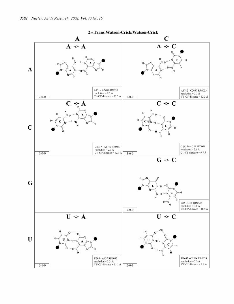
more edge-to-edge hydrogen bonds belong to one of 12geometric families (7). Each family is identi®ed by the edgesinvolved in the interaction and the relative orientations of theglycosidic bonds of the interacting nucleotides, cis or trans(Table 2). When the glycosidic bonds of the two bases assumethe default anti con®guration, the relative strand orientationsare those given in the third column of Table 2 (24). In Figures2±13, representative examples are provided of observed basepairs for each geometrical family. When the two interactingedges are different (for example Watson±Crick andHoogsteen), a historically based priority rule is invoked(Watson±Crick > Hoogsteen > sugar edge) so the baseidenti®ed with each row of a given matrix is the oneinteracting with the higher priority edge. Thus, in Family 3,cis Watson±Crick/Hoogsteen (Fig. 4), all the pairings in the®rst row involve adenine interacting with its Watson±Crickedge while all the pairings in the ®rst column involve adenineinteracting with its Hoogsteen edge. In each panel of Figures2±13, the higher priority base appears to the left, oriented sothat its Watson±Crick edge faces to the right. A list ofreferenced NDB ®les with primary references is provided inTable 1.
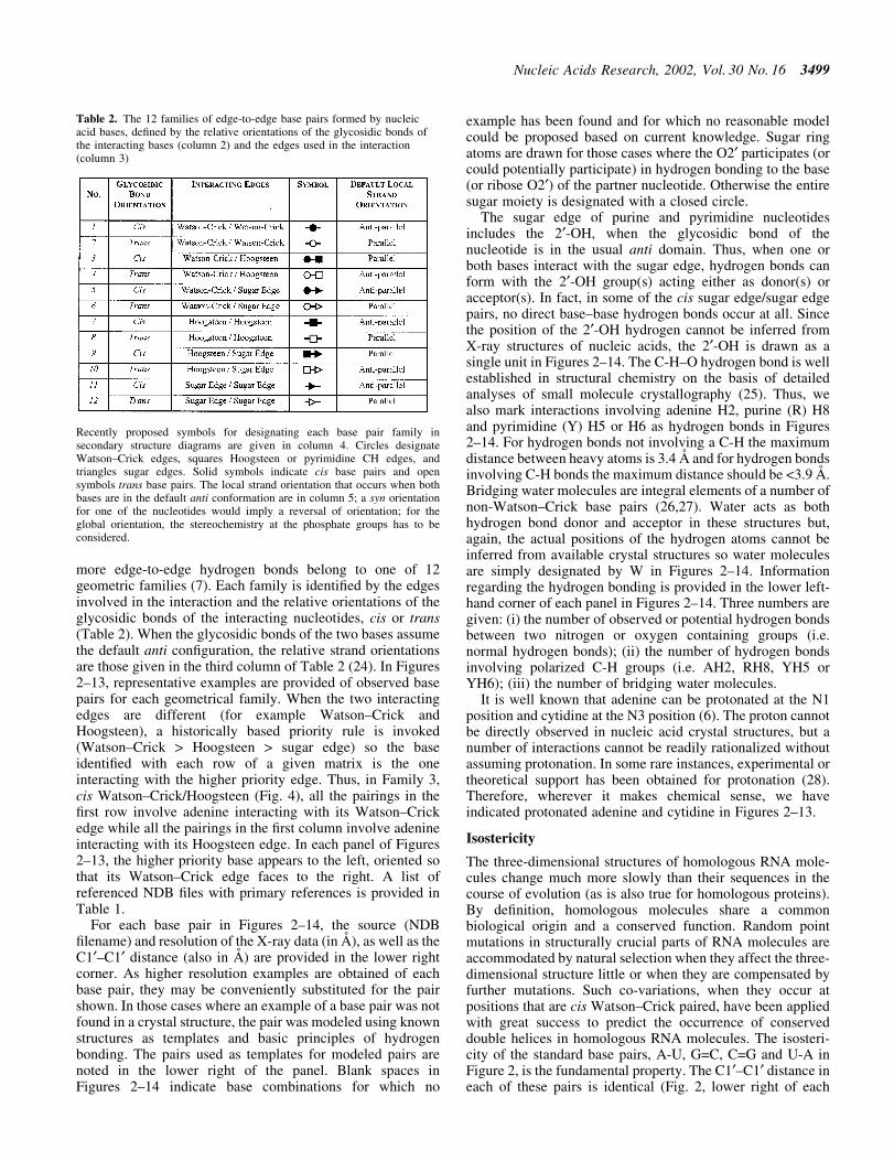
For each base pair in Figures 2±14, the source (NDB®lename) and resolution of the X-ray data (in AÊ ), as well as theC1¢±C1¢ distance (also in AÊ ) are provided in the lower rightcorner. As higher resolution examples are obtained of eachbase pair, they may be conveniently substituted for the pairshown. In those cases where an example of a base pair was notfound in a crystal structure, the pair was modeled using knownstructures as templates and basic principles of hydrogenbonding. The pairs used as templates for modeled pairs arenoted in the lower right of the panel. Blank spaces inFigures 2±14 indicate base combinations for which no
example has been found and for which no reasonable modelcould be proposed based on current knowledge. Sugar ringatoms are drawn for those cases where the O2¢ participates (orcould potentially participate) in hydrogen bonding to the base(or ribose O2¢) of the partner nucleotide. Otherwise the entiresugar moiety is designated with a closed circle.
The sugar edge of purine and pyrimidine nucleotidesincludes the 2¢-OH, when the glycosidic bond of thenucleotide is in the usual anti domain. Thus, when one orboth bases interact with the sugar edge, hydrogen bonds canform with the 2¢-OH group(s) acting either as donor(s) oracceptor(s). In fact, in some of the cis sugar edge/sugar edgepairs, no direct base±base hydrogen bonds occur at all. Sincethe position of the 2¢-OH hydrogen cannot be inferred fromX-ray structures of nucleic acids, the 2¢-OH is drawn as asingle unit in Figures 2±14. The C-H±O hydrogen bond is wellestablished in structural chemistry on the basis of detailedanalyses of small molecule crystallography (25). Thus, wealso mark interactions involving adenine H2, purine (R) H8and pyrimidine (Y) H5 or H6 as hydrogen bonds in Figures2±14. For hydrogen bonds not involving a C-H the maximumdistance between heavy atoms is 3.4 AÊ and for hydrogen bondsinvolving C-H bonds the maximum distance should be <3.9 AÊ .Bridging water molecules are integral elements of a number ofnon-Watson±Crick base pairs (26,27). Water acts as bothhydrogen bond donor and acceptor in these structures but,again, the actual positions of the hydrogen atoms cannot beinferred from available crystal structures so water moleculesare simply designated by W in Figures 2±14. Informationregarding the hydrogen bonding is provided in the lower left-hand corner of each panel in Figures 2±14. Three numbers aregiven: (i) the number of observed or potential hydrogen bondsbetween two nitrogen or oxygen containing groups (i.e.normal hydrogen bonds); (ii) the number of hydrogen bondsinvolving polarized C-H groups (i.e. AH2, RH8, YH5 orYH6); (iii) the number of bridging water molecules.
It is well known that adenine can be protonated at the N1position and cytidine at the N3 position (6). The proton cannotbe directly observed in nucleic acid crystal structures, but anumber of interactions cannot be readily rationalized withoutassuming protonation. In some rare instances, experimental ortheoretical support has been obtained for protonation (28).Therefore, wherever it makes chemical sense, we haveindicated protonated adenine and cytidine in Figures 2±13.
Isostericity
The three-dimensional structures of homologous RNA mole-cules change much more slowly than their sequences in thecourse of evolution (as is also true for homologous proteins).By de®nition, homologous molecules share a commonbiological origin and a conserved function. Random pointmutations in structurally crucial parts of RNA molecules areaccommodated by natural selection when they affect the three-dimensional structure little or when they are compensated byfurther mutations. Such co-variations, when they occur atpositions that are cis Watson±Crick paired, have been appliedwith great success to predict the occurrence of conserveddouble helices in homologous RNA molecules. The isosteri-city of the standard base pairs, A-U, G=C, C=G and U-A inFigure 2, is the fundamental property. The C1¢±C1¢ distance ineach of these pairs is identical (Fig. 2, lower right of each
Table 2. The 12 families of edge-to-edge base pairs formed by nucleicacid bases, de®ned by the relative orientations of the glycosidic bonds ofthe interacting bases (column 2) and the edges used in the interaction(column 3)
Recently proposed symbols for designating each base pair family insecondary structure diagrams are given in column 4. Circles designateWatson±Crick edges, squares Hoogsteen or pyrimidine CH edges, andtriangles sugar edges. Solid symbols indicate cis base pairs and opensymbols trans base pairs. The local strand orientation that occurs when bothbases are in the default anti conformation are in column 5; a syn orientationfor one of the nucleotides would imply a reversal of orientation; for theglobal orientation, the stereochemistry at the phosphate groups has to beconsidered.
Nucleic Acids Research, 2002, Vol. 30 No. 16 3499

3500 Nucleic Acids Research, 2002, Vol. 30 No. 16

Figure 2. (Opposite and above) 4 3 4 matrix displaying observed base pairs belonging to the cis Watson±Crick/Watson±Crick family. The canonicalWatson±Crick pairs comprise the diagonal of the matrix. Symbols used in Figures 2±14 employ circles to designate Watson±Crick edges, squares forHoogsteen or pyrimidine CH edges, and triangles for sugar edges. Solid symbols indicate cis base pairs and open symbols trans base pairs. In the lower left-hand corner of each panel in Figures 2±14, numbers describing the hydrogen bonding are provided. The ®rst is the number of observed or potential hydrogenbonds between two nitrogen- or oxygen-containing groups, i.e. normal hydrogen bonds. The second is the number of hydrogen bonds involving polarized C-Hgroups (i.e. AH2, RH8, YH5 or YH6). The third is the number of bridging water molecules.
Nucleic Acids Research, 2002, Vol. 30 No. 16 3501
3502 Nucleic Acids Research, 2002, Vol. 30 No. 16

Figure 3. (Opposite and above) Observed base pairs of the trans Watson±Crick/Watson±Crick family. The pairing displays a 2-fold rotational symmetry.Thus, the matrix is symmetric.
Nucleic Acids Research, 2002, Vol. 30 No. 16 3503
3504 Nucleic Acids Research, 2002, Vol. 30 No. 16

Figure 4. (Opposite and above) Observed and modeled base pairs of the cis Watson±Crick/Hoogsteen family.
Nucleic Acids Research, 2002, Vol. 30 No. 16 3505
3506 Nucleic Acids Research, 2002, Vol. 30 No. 16

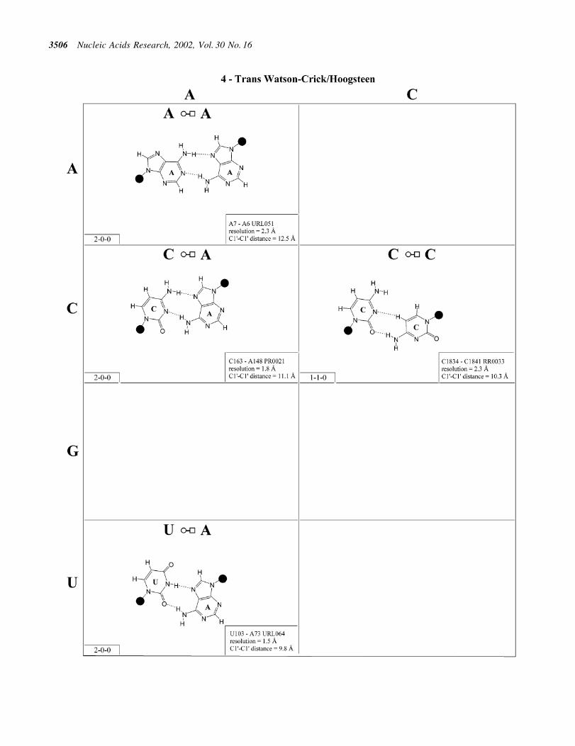
Figure 5. (Opposite and above) Observed base pairs of the trans Watson±Crick/Hoogsteen family.
Nucleic Acids Research, 2002, Vol. 30 No. 16 3507
3508 Nucleic Acids Research, 2002, Vol. 30 No. 16

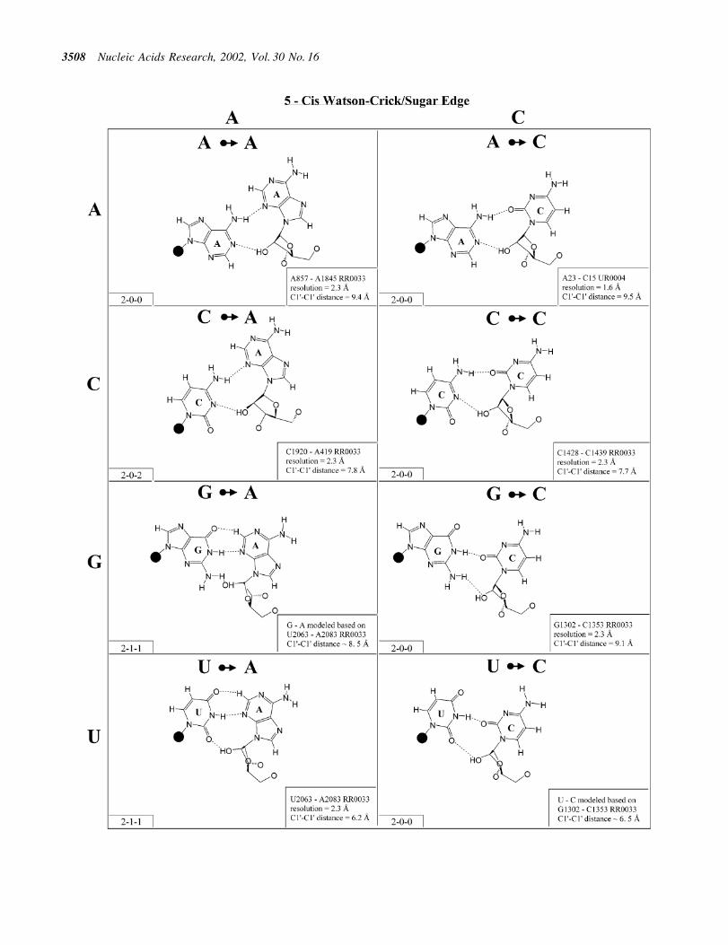
Figure 6. (Opposite and above) Observed and modeled base pairs of the cis Watson±Crick/sugar edge family.
Nucleic Acids Research, 2002, Vol. 30 No. 16 3509
3510 Nucleic Acids Research, 2002, Vol. 30 No. 16

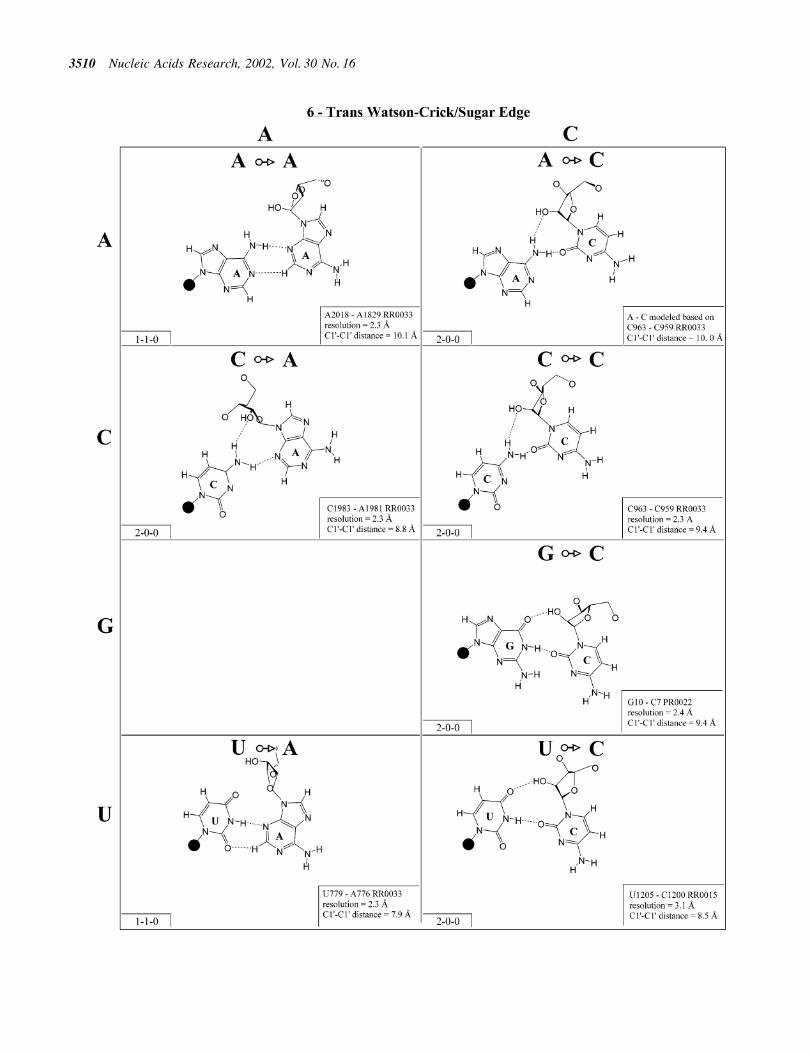
Figure 7. (Opposite and above) Observed and modeled base pairs of the trans Watson±Crick/sugar edge family.
Nucleic Acids Research, 2002, Vol. 30 No. 16 3511

3512 Nucleic Acids Research, 2002, Vol. 30 No. 16

Figure 8. (Opposite and above) Observed base pairs of the cis Hoogsteen/Hoogsteen family.
Nucleic Acids Research, 2002, Vol. 30 No. 16 3513

3514 Nucleic Acids Research, 2002, Vol. 30 No. 16

Figure 9. (Opposite and above) Observed base pairs of the trans Hoogsteen/Hoogsteen family. The pairing displays a 2-fold rotational symmetry. Thus, thematrix is symmetric.
Nucleic Acids Research, 2002, Vol. 30 No. 16 3515

3516 Nucleic Acids Research, 2002, Vol. 30 No. 16

Figure 10. (Opposite and above) Observed and modeled base pairs of the cis Hoogsteen/sugar edge family.
Nucleic Acids Research, 2002, Vol. 30 No. 16 3517
3518 Nucleic Acids Research, 2002, Vol. 30 No. 16

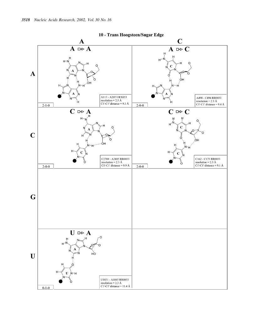
Figure 11. (Opposite and above) Observed base pairs of the trans Hoogsteen/sugar edge family.
Nucleic Acids Research, 2002, Vol. 30 No. 16 3519

3520 Nucleic Acids Research, 2002, Vol. 30 No. 16
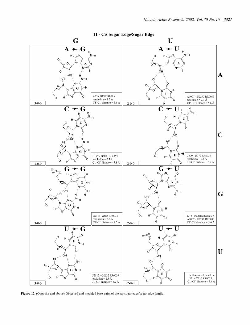
Figure 12. (Opposite and above) Observed and modeled base pairs of the cis sugar edge/sugar edge family.
Nucleic Acids Research, 2002, Vol. 30 No. 16 3521

3522 Nucleic Acids Research, 2002, Vol. 30 No. 16

Figure 13. (Opposite and above) Observed and modeled base pairs of the trans sugar edge/sugar edge family.
Nucleic Acids Research, 2002, Vol. 30 No. 16 3523
3524 Nucleic Acids Research, 2002, Vol. 30 No. 16
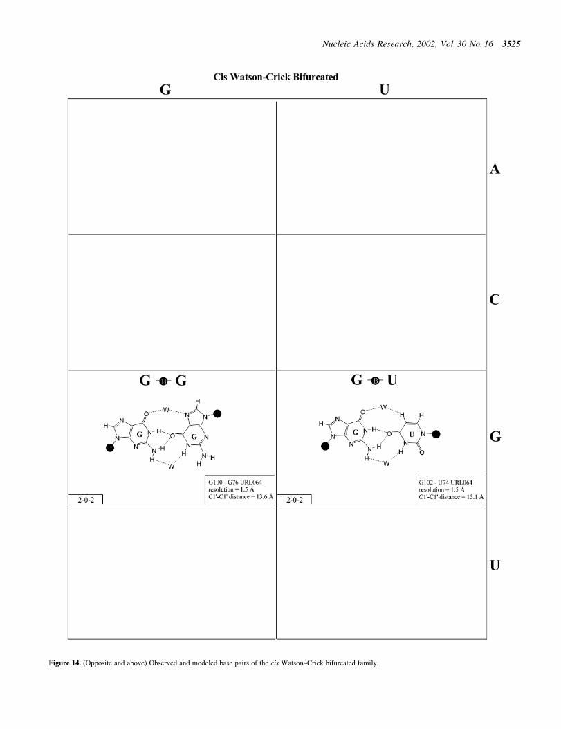
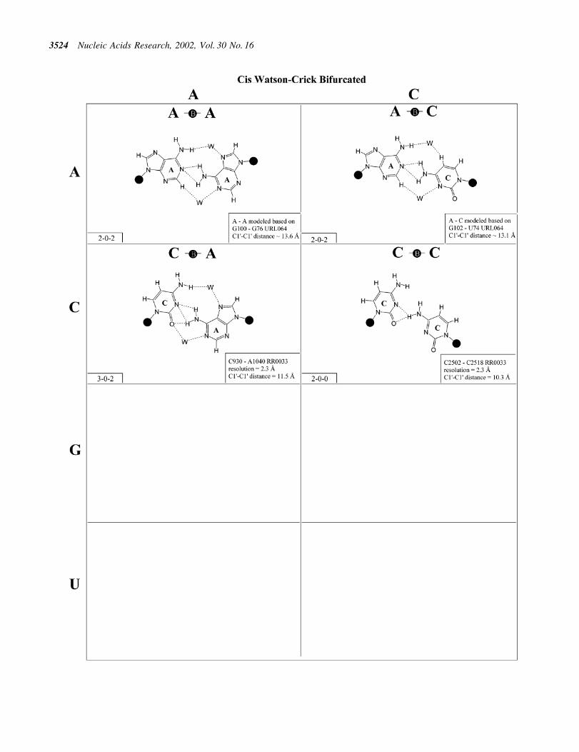
Figure 14. (Opposite and above) Observed and modeled base pairs of the cis Watson±Crick bifurcated family.
Nucleic Acids Research, 2002, Vol. 30 No. 16 3525

panel), as are the relative orientations of the glycosidic bonds,considered as vectors in three-dimensional space. When twobase pairs display nearly the same C1¢±C1¢ distance and havetheir glycosidic bonds oriented in the same way, they canreplace each other without drastically changing the three-dimensional path and relative geometric orientations of thephosphate±sugar backbones. We denote such base pairs as`isosteric', although this does not necessarily imply that thetwo base pairs occupy the same total volume of space, and inmany cases this, in fact, does not hold.
Isostericity matrices
Generally, base pairs belonging to the same geometric familyexhibit very similar relative orientations of their glycosidicbonds, implying the maintenance of the local orientations ofthe strands and thus of the three-dimensional organization.However, in the general case, all possible base pairs belongingto a single geometric family are not isosteric to each otherbecause the C1¢±C1¢ distances may be quite different. Thus,the C1¢±C1¢ distance can be used to group the base pairswithin each geometric family into isosteric subsets orsubfamilies. The recognition of subsets of isosteric basepairs within a family serves the purpose of identifying pairsthat can substitute for each other while preserving the three-dimensional structure, crucial information for three-dimen-sional modeling of tertiary interactions, prediction of motifs,and the generation and re®nement of accurate structuralalignments. In the following, each geometric family isconsidered in turn and the isosteric subsets of base pairs
identi®ed from Figures 2±13 are summarized in the form ofisostericity matrices in Tables 3±5.
Cis Watson±Crick/Watson±Crick (Family 1). We beginwith the base pairs belonging to the cis Watson±Crick/Watson±Crick geometric family, shown in Figure 2. The(canonical) Watson±Crick pairs, A-U, U-A, G=C andC=G, form an isosteric subfamily, which we designate I1
in the isostericity matrix for this family, shown in Table 3(®rst row, left). Likewise the wobble pairs G/U and A(+)/C form an isosteric subgroup I2. However, unlike I1, thewobble pairs are not self-isosteric and, thus, the wobblepairs U/G and C/A(+) comprise a third isosteric subset,which, however, is related to I2 and is thereforedesignated i2. In certain contexts the wobble pairs cansubstitute for canonical cis Watson±Crick/Watson±Crickpairs within a helix. We can say that they are compatiblewith the canonical pairs. However, substitution of a G/U
Table 3. Isostericity matrices for base pairing Families 1±6
First row, cis and trans Watson±Crick/Watson±Crick; second row, cis andtrans Watson±Crick/Hoogsteen; third row, cis and trans Watson±Crick/sugar edge. Parentheses indicate modeled interactions, not yet observed inhigh resolution X-ray structures. In the cis Watson±Crick/Watson±Cricktable i2 is used to designate the wobble pairs G/U and A(+)/C as they arenot isosteric to U/G and C/A(+); unlike the standard Watson±Crick pairs,the wobble pairs are not self-isosteric.
Table 4. Isostericity matrices for base pairing Families 7±12
First row, cis and trans Hoogsteen/Hoogsteen; second row, cis and transHoogsteen/sugar edge; third row, cis and trans sugar edge/sugar edge.Parentheses indicate modeled interactions, not yet observed in highresolution X-ray structures. For the cis Hoogsteen/sugar edge geometry, I1
indicates observed pairs in which the interacting bases are adjacent in thepolynucleotide chain, while I2 indicates observed pairs in which one or twonucleotides separate the interacting bases.
Table 5. Isostericity matrix for the cis bifurcated geometry
3526 Nucleic Acids Research, 2002, Vol. 30 No. 16

or A(+)/C pair for a U/G or C/A(+) results in a largerstructural perturbation in a helical context (29) and thus R/Y are usually not compatible with Y/R wobble pairs.
The pairs A/G and G/A constitute a fourth subfamily,designated I3. Like the canonical pairs (I1) they are self-isosteric. I4 consists solely of the A/A pair, since the G/Gcombination cannot occur in this geometry. C/U and U/C areself-isosteric and comprise subset I5. Interestingly, in highresolution structures this pair is consistently observed with aninserted water molecule, bridging between the imino positionsof the bases, perhaps because of repulsion between the O2atoms of the interacting pyrimidines (30). Consequently theC1¢±C1¢ distance for the water-inserted C/U pair is signi®-cantly larger than expected for a pyrimidine±pyrimidine pair,and close to that of cis Watson±Crick/Watson±Crick A/G.Interestingly, U/C is observed to co-vary with A/G in theanticodon stem of tRNAs (27). Thus, in certain contexts C/Uand A/G are compatible.
The isosteric wobble pairs C(+)/C and U/U, both of whichhave been observed, comprise the ®nal isosteric subgroup ofthe cis Watson±Crick/Watson±Crick geometric family, desig-nated I6. The C1¢±C1¢ distance in this subfamily is signi®-cantly smaller than that of any of the others, including thewater-inserted U/C.
Trans Watson±Crick/Watson±Crick (Family 2). Representa-tive base pairs belonging to the trans Watson±Crick/Watson±Crick geometric family are shown in Figure 3 andthe isostericity matrix is shown in the right panel of the ®rstrow of Table 3. The trans orientation of the glycosidic bondsallows for a possible 2-fold axis perpendicular to and passingthrough the middle of the base pair. Unlike the correspondingcis pairs, the A/U (designated I1) and G/C (designated I2) pairsare not isosteric. However, these and all trans Watson±Crick/Watson±Crick pairs are self-isosteric and thus Table 3 issymmetric with respect to the main diagonal. The pairs A/Cand G/U are isosteric, but not isosteric with A/U or G/C, andthus form a third group, I3. The homopurine pairs A/A andG/G are isosteric (I4) but A/G cannot form with two hydrogenbonds. As for the cis Watson±Crick/Watson±Crick family, allpossible trans Watson±Crick/Watson±Crick pairs have beenobserved in crystal structures.
The trans Watson±Crick/Watson±Crick C/C pair shown inFigure 3 has three hydrogen bonds and requires protonation ofone cytosine at N1. It is from a crystal structure of cysteinyltRNA at 2.6 AÊ resolution (PR0004). An alternative hydrogenbonding pattern can be proposed that does not requireprotonation but involves only two hydrogen bonds (CN1±CN4 and CN4±CN1), which would make C/C isosteric withU/U rather than U/C. This geometry is observed at lowerresolution (3.5 AÊ ) for the tertiary base pair (C1773/C2565) inthe structure of the 23S rRNA of Deinococcus radiodurans(RR0051). This pair corresponds to the tertiary interactionU1838 /U2621 in the 23S rRNA of Haloarcula marismortui(U1782/U2586 in the Escherichia coli sequence) and was ®rstidenti®ed by sequence analysis based on the co-variation of U/U and C/C for these positions (31). Thus we favor grouping U/C and C/U in one isosteric subgroup (I5) and C/C with U/U inanother (I6). The observed U1432/C1394 pair (RR0033) has asodium ion bridging UO4±CO2 (compare with cisWatson±Crick/Watson±Crick).
Cis Watson±Crick/Hoogsteen (Family 3). Representative pairsin this family are shown in Figure 4 and the correspondingisostericity matrix in Table 3. U/A, U/G and C(+)/G have beenobserved and together with C/U (modeled on C/G and A/G)are grouped into the isosteric subfamily I1. Modeled base pairsare indicated in Tables 3 and 4 in parentheses. Cytosinerequires protonation at N3 to form C(+)/G. C/C and U/U haveboth been observed and are grouped into subfamily I2, whichis related to I1 by a lateral shift in the hydrogen bonds. A(+)/Ghas been observed at high resolution (1.9 AÊ ) and requiresprotonation of AN1 to form. A(+)/G is grouped with G/A(observed) and A/U (modeled) in subfamily I3. G/G is relatedto A(+)/G and G/A by a lateral shift in the hydrogen bondingpositions, and thus G/G is grouped separately (I4).
The cis Watson±Crick/Hoogsteen interaction often occursas part of a base triple. The base that interacts with itsHoogsteen edge uses its Watson±Crick edge to pair with thethird base. For example, the isosteric U/U and C/C pairscomprise tertiary interactions in the conserved L11-bindingsite of 23S rRNA as part of such a triple. C1072´C1092=G1099 (E.coli numbering) co-varies with U´U-A inthe 23S rRNAs of all phylogenetic groups. This providesanother example of sequence co-variation re¯ecting isostericsubgroups of the isostericity matrix.
In summary, eight of the 10 pairs expected in this familyhave been observed. The R/R and R/Y pairs exhibit signi®-cantly longer C1¢±C1¢ distances than the Y/R and Y/Y pairs. Inaddition, isolated examples involving single hydrogen bondsand non-planar interactions have been observed (e.g. A2812/A2814 and A378/C271 in RR0033).
Trans Watson±Crick/Hoogsteen (Family 4). As for thecorresponding cis geometry, the R/R and R/Y pairs of thetrans Watson±Crick/Hoogsteen geometry exhibit signi®cantlylonger C1¢±C1¢ distances than the Y/R and Y/Y pairs (Fig. 5and Table 3, second row, right). U/A and U/C are isosteric(subfamily I1) and are related by a lateral shift to C/A, C(+)/Gand U/U (subfamily I2). In fact, I1 and I2 are mutuallycompatible, thus U/A and C/A are observed to co-vary in theloop E motifs of 5S rRNA and SRP (2,32). U/G is placed in itsown group (I3) because it is rarely observed and does not co-vary with U/A or C/A, perhaps because of the repulsionbetween UO2 and GO6, which may destabilize pairing in thestandard geometry and favor hydrogen bonding between UO4and GC8.
Three of the four R/R combinations form base pairs. A/Aand A(+)/G are isosteric and with G/U comprise subfamily I4.G/G is related by a lateral shift to A/A and A(+)/G and is thusnot exactly isosteric and so is grouped separately (I5). A(+)/Grequires protonation of AN1 and has been observed in tRNA(e.g. TRNA07).
In summary, all 10 pairs expected for this family have beenobserved. As for the cis Watson±Crick/Hoogsteen family,isolated examples involving single hydrogen bonds and non-planar interactions also occur (e.g. A2577/C2555 and G345/A305 in RR0033).
Cis Watson±Crick/sugar edge (Family 5). The cis Watson±Crick/sugar edge family (Fig. 6 and Table 3, third row, left)comprises four main isosteric subfamilies that are de®ned by
Nucleic Acids Research, 2002, Vol. 30 No. 16 3527

the base that pairs using its Watson±Crick edge. Thus, all fourA/N pairs are isosteric and all have been observed (subfamilyI1). All four C/N pairs have been observed and comprise asecond group, I2. Three of the G/N pairs have been observed(I3). G/A was modeled using U/A as a template. It should beisosteric to G/C and G/U. G/G displays a signi®cantly longerC1¢±C1¢ distance and is therefore placed in its own subgroup(I5). U/A and U/G have been observed, whereas U/C and U/Uwere modeled based on G/C and G/U. The four U/N pairs arealso expected to form a single isosteric group (I4).
Trans Watson±Crick/sugar edge (Family 6). The base pairsbelonging to the trans Watson±Crick/sugar edge family areshown in Figure 7 and the corresponding isostericity matrix inTable 3 (third row, right). Both A/A and A/G have beenobserved and are isosteric. The A/G pair is more common andprobably more stable as it involves two conventionalbase±base hydrogen bonds and a potential A(N6)±G(O2¢)hydrogen bond. This interaction can occur as part of a basetriple (for example A24´G7=C14 in UR0004) or as an isolatedtertiary base pair (e.g. A629´G2070 or A2018´A1829 inH.marismortui 23S rRNA, RR0033). The A/Y interactionswere modeled based on C/C, but these would only involve onebase±base hydrogen bond (Fig. 7) and are expected to occur inthe context of base triples. All four A´N interactions should beisosteric (I1, Table 3, third row, right).
The C/A, C/G and C/C interactions have been observed andC/U can be modeled using C/C as a template. Like the A/Rinteractions, the C/R interactions can occur as part of basetriples (e.g. C46´G43=C37 in H.marismortui 5S rRNA,RR0033) or as isolated tertiary interactions (e.g. C1981´A1983, RR0033). The C963/C959 pair from 23S rRNAbelongs to a base triple in which C959 is Watson±Crick pairedto A1005. The C/G pair is the only C/N trans Watson±Crick/sugar edge interaction to feature two conventional base±basehydrogen bonds and is the most common. All the C/N and A/Npairs are grouped in a single isosteric subfamily, designated I1.
The G/U pair occurs most commonly as the closing basepair in UUCG-type hairpin loops, with the G in the syncon®guration and the strands antiparallel (see Table 2 legend).The G/C trans Watson±Crick/sugar edge pair can also occurin a hairpin loop (e.g. G10/C7 in PR0022) and is isosteric withG/U, which together form the I2 subfamily. The G/Rinteractions are not expected to occur and have not beenobserved.
Examples of U/A, U/C and U/G have been observedand U/U can be modeled based on U/C (Fig. 7). (An exampleof U/U exists in a low resolution structure, U106/U258 in theGroup I intron, UR0003.) The U/A interaction occurs as partof a base quadruple with C879=G871 in a three-way junctionin 23S rRNA (RR0033). The U/C interaction occurs as part of abase triple in 16S rRNA of Thermus thermophilus (RR0015)and U/G as a tertiary interaction in 23S rRNA (RR0033) thatinvolves a bridging water molecule. The hydrogen bondingpatterns in the U/Y and G/Y pairs are similar but the C1¢±C1¢distances are greater in the G/Y pairs, so these form differentisosteric subgroups. U/A can be grouped with U/Y (I3), butU´G is distinct (I4).
Cis Hoogsteen/Hoogsteen (Family 7). The only examplesfrom this family have been observed in the ribosome (Fig. 8
and Table 4, row one, left). They are very rare. The G2494/C2493 interaction involves adjacent nucleotides. C2493 is inthe rare syn conformation and thus presents its Hoogsteen edgeto interact with the Hoogsteen edge of G2494, thus allowingthe CH6±GO6 hydrogen bond to form in place of theunfavorable CO2±GO6 repulsive interaction. The secondexample, G2616/G2617, also involves adjacent nucleotideswith G2616 also in the syn conformation. A1742/G2033 is atertiary interaction with antiparallel strands. Kinks and sharpturns in the phosphodiester backbones of the antiparallelstrands allow the two bases to approach each other to form thecharacteristic AN6±GO6 hydrogen bond.
Trans Hoogsteen/Hoogsteen (Family 8). The trans Hoogsteen/Hoogsteen pairs are shown in Figure 9 and the isostericitymatrix in Table 4 (®rst row, right). Like the transWatson±Crick/Watson±Crick family, these pairs are self-isosteric due to symmetry. It is interesting to notice that in thisfamily, except for one base pair, all the pairs involve a singlehydrogen bond. This pair occurs in tRNA and in sarcin/ricinmotifs. The sequence variations observed for these motifscorrespond closely to the observed base pairs shown inFigure 8 (2,27).
Cis Hoogsteen/sugar edge (Family 9). The cis Hoogsteen/sugar edge interaction can involve the bases of adjacent ormore distant nucleotides in the polynucleotide chain.Generally, only a single hydrogen bond can form betweenthe interacting bases (Fig. 10). The best known examples arethe A/A `platform' (33) and the U/G `side-by-side' pair of thesarcin/ricin loop motif (18). In addition to these, many otherpairs of this type have been observed. Eleven examplesinvolving immediately adjacent nucleotides have beenobserved and are shown in Figure 10. On the basis of theU/U pair, we can propose a model for U/C, and on the basis ofthe G/G pair we can propose G/A. In fact, cis Hoogsteen/sugaredge G/A is observed at lower resolution (~3.5 AÊ ) in the 23SrRNA of D.radiodurans (G2035/A2034 in NDB ®le rr0051) atthe position corresponding to G2093/G2092. Bases of non-adjacent nucleotides can form similar base pairs, but these arenot isosteric to the adjacent pairs. All the pairs involvingadjacent pairs are essentially isosteric (I1 in Table 4). Non-adjacent pairs form a second isosteric group (I2). Examples ofnon-adjacent cis Hoogsteen/sugar edge pairs exist for many ofthe adjacent pairs shown in Figure 10, but the adjacent pair isshown by preference. Examples of non-adjacent pairsinclude U2527/G2525, C2787/C2785 and C2575/U2473from 23S rRNA (H.marismortui) and A56/A54 from 5SrRNA (H.marismortui).
One of the most remarkable cis Hoogsteen/sugar edge pairsis U832/U831, in which a water bridges between U832(O4)and U831(O2). U/U is observed to co-vary with cisHoogsteen/sugar edge U/G in some sarcin loop motifs(N.B.Leontis and E.Westhof, manuscript in preparation).
Trans Hoogsteen/sugar edge (Family 10). The most commoninteraction of this type is the `sheared' A/G in which theHoogsteen edge of A interacts with the sugar edge of G(Fig. 11). In fact, this is the most commonly occurring A/Gbase pair. This base pair occurs in loop E of 5S rRNA and inthe sarcin/ricin motif of 23S rRNA. Co-variations at these
3528 Nucleic Acids Research, 2002, Vol. 30 No. 16

positions include A/A, A/Y (Y = U or C), C/A (C Hoogsteen,A sugar edge) and C/Y. On the basis of these co-variations andthe structures of the A/G and A/A pairs, models were proposedfor A/Y, C/A and C/Y (32). Subsequently, all these pairs havebeen observed (see Fig. 11), just as modeled, and, interest-ingly, all are isosteric. Thus, the A/N, C/A and C/Y pairs aregrouped into one isosteric subfamily, designated I1 in Table 4(second row, right). The G/G, U/A and U/G pairs form asecond isosteric subgroup (I2) that does not co-vary with the®rst.
Cis sugar edge/sugar edge (Family 11). As shown inFigure 12, examples of almost all possible cis sugar edge/sugar edge pairs have been observed and all 16 combinationsare expected to be isosteric (Table 4, third row, left). Thisinteraction is not symmetric as the O2¢ of one nucleotidehydrogen bonds to the base R(N3) or Y(O2) and to thehydroxyl O2¢ of the other nucleotide. The former nucleotide isgiven priority (7). When that nucleotide is a pyrimidine (Y),there is in fact no direct base±base hydrogen bond. When it is apurine (R), there is a single base±base hydrogen bond (exceptfor A´G, with two). This interaction occurs frequently betweenadjacent nucleotides belonging to two strands (with the 5¢nucleotide of one strand receiving from the hydroxyl group ofthe 3¢ nucleotide of the other). Such a motif is referred to as the`ribose-zipper motif' (33). Furthermore, the cis sugar edge/sugar edge interaction often occurs in combination with thetrans sugar edge/sugar edge pair of the frequent and versatilerecognition motif comprised of adjacent cis and trans sugaredge/sugar edge base pairs (3,27).
Trans sugar edge/sugar edge (Family 12). The trans sugaredge/sugar edge base pair (Fig. 13 and Table 4, third row,right) usually involves at least one adenosine. Generally, suchinteractions occur as part of base triples in which theadenosine (and more rarely guanosine) interacts with thesugar edge of a standard base pair. The A´A, A´G and A´Cexamples are of this type: A306´A340-U325 (RR0033),A867´C880=G870 (RR0033) and A20´G4=C17 (UR0004).Of these, A´G is by far the most common, since it occurs in thefrequent recognition motif made of adjacent cis and transsugar edge/sugar edge pairs. The A´U pair is found in tRNAsas part of a base triple (A21´U8´A14). The other pairsinvolving G are much rarer. Examples of G´G include those inwhich one G is canonically paired as well as isolated tertiarypairs such as G315´G336 and G2428´G2466 (RR0033). TheG´U shown in Figure 13 is a tertiary pair, whereas the G´Cexample is part of a base triple (G2617´C2542=G2617,RR0033). The A´N pairs form one group (I1) and the G´Npairs a second group (I2).
Bifurcated hydrogen bonding patterns. Bifurcated pairs areintermediate between two edge-to-edge geometries (Fig. 14and Table 5). They involve interactions between an exocyclicfunctional group of one base and the edge of another.Bifurcated pairs may also show distinct patterns of co-variation and substitution. For example, the isosteric G´G andG´U cis bifurcated pairs, ®rst observed at high resolution in thestructure of loop E of bacterial 5S rRNA, were found to co-vary with each other and with A´C and A´A, both of whichcould be modeled in the same geometry (32). These pairs are
intermediate to the cis Watson±Crick/Watson±Crick and thetrans Watson±Crick/Hoogsteen families. The isostericitymatrix (Table 5) was proposed for bifurcated pairs of thiskind (27). Additional examples belonging to this family ofbifurcated pairs have been observed in the ribosome, includingC2502/C2518 (also part of a loop E type motif) and C930/A1040.
Bifurcated pairs intermediate to the trans Watson±Crick/Hoogsteen and trans sugar edge/Hoogsteen families occur inloop E-related motifs in 16S rRNA (G581´G760, E.colinumbering), 23S rRNA (G706´G722) and the SRP (G162-G149).
Intermediate and alternative hydrogen bonding patterns. In asmall number of cases, alternative hydrogen bonding patternshave been observed for particular base pair combinations.These may be due to the limited resolution of the experimentaldata or re®nement errors or to the actual existence of distinctpotential energy minima that depend on the local structuralcontext. The symmetrical, cis Watson±Crick (wobble-like)U/U and C/C pairs provide trivial examples of the latter. Forexample, two uridines can pair with UO4±UN3 and UN3±UO2 hydrogen bonds or with UN3±UO4 and UO2±UN3hydrogen bonds. Which set of hydrogen bonds occurs dependson the local context. Alternatively, U/U can open up andincorporate a bridging water molecule (34). Likewise, G andU can form a conventional wobble pair (Fig. 2) or, in certaincontexts, a bifurcated pair, involving two bridging watermolecules (Fig. 14). Two possible hydrogen bonding patternsfor trans Watson±Crick C/C were discussed above. Higherresolution structural work complemented by computation isneeded to determine which pattern is favored and whether thisis context-dependent.
Another example is provided by the cis Watson±Crick(wobble) C´A pair, for which hydrogen bonding may beproposed between C(N4) and A(N1) and between C(N3) andA(C2) in place of hydrogen bonds between C(N1) and A(N6)and C(N3) and protonated A(N3), which are usually observed.An example with the alternative hydrogen bonding pattern isobserved in the context of a base triple in 23S rRNA(C40´A441´A442 in RR0033). The triple consists of theA442´A441 cis Hoogsteen/sugar edge interaction and thealternative C40´A441 cis Watson±Crick/Watson±Crick pair.An additional hydrogen bond is observed between C40(O2)and A442(N6). Higher resolution is required to con®rm thisinteraction.
In conclusion, it must be emphasized that base pairing isdue to multiple weak interactions and thus a considerabledegree of ¯exibility and deformation is expected. Thus, whileone can generally classify base pairs into one of the 12families discussed above, a particular base pair may form witha slightly different combination of hydrogen bonds or with theabsence of one or more hydrogen bonds, depending on thestructural context or on the resolution of the structure.
Interactions of a base with an `edge' de®ned by two bases. Apremise of the approach we have taken has been that complexinteractions (base triples, quadruples, etc.) can be analyzed ascombinations of base pairs. In a few cases this analysis breaksdown and new patterns arise, which again re¯ect synergisticeffects. An example is the interaction of the Watson±Crick
Nucleic Acids Research, 2002, Vol. 30 No. 16 3529

edge of C with the Hoogsteen edge of a (standard) G=C basepair. Four interactions can be anticipated, cis or trans C´GWatson±Crick/Hoogsteen and cis or trans C´C Watson±Crick/Hoogsteen, and are in fact observed (see Tables 4 and 5). A®fth interaction, distinct from these, has also been observed.An example is provided by C113 interacting with theHoogsteen edges of the C15=G66 base pair in 5S rRNA ofH.marismortui (RR0033). This interaction is intermediatebetween the cis C´C Watson±Crick/Hoogsteen interactionseen in base triples such as C1072´C1092=G1099 in the L11-binding site of 23S rRNA (E.coli numbering, NDB ®lePR0015 or RR0009) and the trans C(N1+)´G Watson±Crick/Hoogsteen interaction seen in base triples such as C8(+)´G12=C26 in the frameshifting pseudoknot (UR0004). It canbest be described as an interaction of the Watson±Crick edgeof C113 with the Hoogsteen edge of the C15=G66 base pair, asit involves hydrogen bonds to both G66(O6) and C15(N4).
CONCLUSIONS
The rapidly growing database of RNA crystal structuresprovides examples of nearly every type of base pair. Many ofthe base pairs presented in Figures 2±14 were ®rst proposed ontheoretical grounds and have now been observed by X-raycrystallography at <3.0 AÊ resolution. Generally, the observedbase pairs are as predicted (27,32). The overwhelming numberof base±base interactions observed in the ribosome and theother new structures that have appeared recently can beunambiguously classi®ed into one of the 12 families ofTable 2. A small number of base pairs comprise bifurcatedpairs that are intermediate between two of the 12 families (7).Furthermore, care must be taken so as not to confuse the transsugar edge/sugar edge and trans Watson±Crick/sugar edgeinteractions, because frequently a Watson±Crick/2¢-OHhydrogen bond can also occur in the trans sugar edge/sugaredge geometry.
Other kinds of interactions are observed in complex RNAstructures which need to be analyzed and catalogued, includ-ing additional bifurcated pairs, perpendicular edge-to-edgeinteractions, interactions exclusively involving the ribosemoiety of one or both nucleotides, and base stackinginteractions.
Preliminary analyses, some of which have been presentedhere, indicate that there is a close correspondence between theisosteric subfamilies identi®ed on structural grounds and thepatterns of co-variation and base substitution that are observedin homologous RNA, when they are properly aligned. Theprimary signi®cance of this work is that it provides a basis forevaluating and re®ning structural alignments for homologousRNA molecules. Consideration of the isostericity matrixcorresponding to each base pair is essential for producingcorrect alignments at positions involved in non-Watson±Crickbase pairing or determining that one motif has in fact beenreplaced by another in a set of homologous sequences.
Here, we have emphasized the geometrical aspects of basepairing in order to aid in their classi®cation. Clearly,depending on the edges involved, various groups or siteswill be available for interactions with another RNA segment, aprotein or a small molecule. For example, when theWatson±Crick sites are not engaged, they can be used forinteraction with phosphate groups. Similarly, the Hoogsteen
sites are used for interactions with amino acid side chains incomplexes between proteins and helices. Besides conferringgeometrical similarity, the isostericity matrices contain infor-mation on compensating changes that would occur betweenbase pairs at the level of a given functional group or a set offunctional groups.
ACKNOWLEDGEMENTS
The authors acknowledge fruitful discussions with PascalAuf®nger and Luc Jaeger. This work was supported by NSFREU grant CHE-9732563 and NIH grant 2R15-GM55898.
REFERENCES
1. Woese,C.R., Winker,S. and Gutell,R.R. (1990) Architecture of ribosomalRNA: constraints on the sequence of "tetra-loops". Proc. Natl Acad. Sci.USA, 87, 8467±8471.
2. Leontis,N.B. and Westhof,E. (1998) A common motif organizes thestructure of multi-helix loops in 16 S and 23 S ribosomal RNAs. J. Mol.Biol., 283, 571±583.
3. Nissen,P., Ippolito,J.A., Ban,N., Moore,P.B. and Steitz,T.A. (2001) RNAtertiary interactions in the large ribosomal subunit: the A-minor motif.Proc. Natl Acad. Sci. USA, 98, 4899±4903.
4. Moore,P.B. (1999) Structural motifs in RNA. Annu. Rev. Biochem., 68,287±300.
5. Hoogsteen,K. (1963) The crystal and molecular structure of a hydrogen-bonded complex between 1-methylthymine and 9-methyladenine. ActaCrystallogr., 16, 907±916.
6. Saenger,W. (1984) Principles of Nucleic Acid Structure. Spring-Verlag,New York, NY.
7. Leontis,N.B. and Westhof,E. (2001) Geometric nomenclature andclassi®cation of RNA base pairs. RNA, 7, 499±512.
8. Westhof,E., Dumas,P. and Moras,D. (1988) Restrained re®nement of twocrystalline forms of yeast aspartic acid and phenylalanine transfer RNAcrystals. Acta Crystallogr. A, 44, 112±123.
9. Michel,F., Hanna,M., Green,R., Bartel,D.P. and Szostak,J.W. (1989) Theguanosine binding site of the Tetrahymena ribozyme. Nature, 342,391±395.
10. Michel,F. and Westhof,E. (1990) Modelling of the three-dimensionalarchitecture of group I catalytic introns based on comparative sequenceanalysis. J. Mol. Biol., 216, 585±610.
11. Gautheret,D., Damberger,S.H. and Gutell,R.R. (1995) Identi®cation ofbase-triples in RNA using comparative sequence analysis. J. Mol. Biol.,248, 27±43.
12. Gautheret,D. and Gutell,R.R. (1997) Inferring the conformation of RNAbase pairs and triples from patterns of sequence variation. Nucleic AcidsRes., 25, 1559±1564.
13. Macke,T.J., Ecker,D.J., Gutell,R.R., Gautheret,D., Case,D.A. andSampath,R. (2001) RNAMotif, an RNA secondary structure de®nitionand search algorithm. Nucleic Acids Res., 29, 4724±4735.
14. Varani,G., Cheong,C. and Tinoco,I.,Jr (1991) Structure of an unusuallystable RNA hairpin. Biochemistry, 30, 3280±3289.
15. Heus,H.A. and Pardi,A. (1991) Structural features that give rise to theunusual stability of RNA hairpins containing GNRA loops. Science, 253,191±194.
16. Wimberly,B., Varani,G. and Tinoco,I.,Jr (1993) The conformation ofloop E of eukaryotic 5S ribosomal RNA. Biochemistry, 32, 1078±1087.
17. SantaLucia,J.,Jr and Turner,D.H. (1993) Structure of (rGGCGAGCC)2 insolution from NMR and restrained molecular dynamics. Biochemistry,32, 12612±12623.
18. Szewczak,A.A. and Moore,P.B. (1995) The sarcin/ricin loop, a modularRNA. J. Mol. Biol., 247, 81±98.
19. Butcher,S.E., Allain,F.H. and Feigon,J. (1999) Solution structure of theloop B domain from the hairpin ribozyme. Nature Struct. Biol., 6,212±216.
20. Ryder,S.P., Ortoleva-Donnelly,L., Kosek,A.B. and Strobel,S.A. (2000)Chemical probing of RNA by nucleotide analog interference mapping.Methods Enzymol., 317, 92±109.
3530 Nucleic Acids Research, 2002, Vol. 30 No. 16
21. Ehresmann,C., Baudin,F., Mougel,M., Romby,P., Ebel,J.P. andEhresmann,B. (1987) Probing the structure of RNAs in solution. NucleicAcids Res., 15, 9109±9128.
22. Lee,K., Varma,S., SantaLucia,J.,Jr and Cunningham,P.R. (1997) In vivodetermination of RNA structure-function relationships: analysis of the790 loop in ribosomal RNA. J. Mol. Biol., 269, 732±743.
23. Guex,N. and Peitsch,M.C. (1997) SWISS-MODEL and the Swiss-PdbViewer: an environment for comparative protein modeling.Electrophoresis, 18, 2714±2723.
24. Westhof,E. (1992) Westhof's rule. Nature, 358, 459±460.25. Desiraju,G.R. (1996) The C-H±O hydrogen bond: structural implications
and supramolecular design. Acc. Chem. Res., 29, 441±449.26. Auf®nger,P. and Westhof,E. (1998) Hydration of RNA base pairs.
J. Biomol. Struct. Dyn., 16, 693±707.27. Leontis,N.B. and Westhof,E. (1998) Conserved geometrical base-pairing
patterns in RNA. Q. Rev. Biophys., 31, 399±455.28. Csaszar,K., Spackova,N., Ste¯,R., Sponer,J. and Leontis,N.B. (2001)
Molecular dynamics of the frame-shifting pseudoknot from beet westernyellows virus: the role of non-Watson-Crick base-pairing, orderedhydration, cation binding and base mutations on stability and unfolding.J. Mol. Biol., 313, 1073±1091.
29. Masquida,B. and Westhof,E. (2000) On the wobble GoU and relatedpairs. RNA, 6, 9±15.
30. Tanaka,Y., Fujii,S., Hiroaki,H., Sakata,T., Tanaka,T., Uesugi,S.,Tomita,K. and Kyogoku,Y. (1999) A¢-form RNA double helix in thesingle crystal structure of r(UGAGCUUCGGCUC). Nucleic Acids Res.,27, 949±955.
31. Gutell,R.R., Power,A., Hertz,G.Z., Putz,E.J. and Stormo,G.D. (1992)Identifying constraints on the higher-order structure of RNA: continueddevelopment and application of comparative sequence analysis methods.Nucleic Acids Res., 20, 5785±5795.
32. Leontis,N.B. and Westhof,E. (1998) The 5S rRNA loop E: chemicalprobing and phylogenetic data versus crystal structure. RNA, 4,1134±1153.
33. Cate,J.H., Gooding,A.R., Podell,E., Zhou,K., Golden,B.L., Kundrot,C.E.,Cech,T.R. and Doudna,J.A. (1996) Crystal structure of a group Iribozyme domain: principles of RNA packing. Science, 273, 1678±1685.
34. Vicens,Q. and Westhof,E. (2001) Crystal structure of paromomycindocked into the eubacterial ribosomal decoding A site. Structure(Camb.), 9, 647±658.
35. Jang,S.B., Hung,L.W., Chi,Y.I., Holbrook,E.L., Carter,R.J. andHolbrook,S.R. (1998) Structure of an RNA internal loop consisting oftandem C-A+ base pairs. Biochemistry, 37, 11726±11731.
36. Pan,B., Mitra,S.N. and Sundaralingam,M. (1999) Crystal structure of anRNA 16-mer duplex R(GCAGAGUUAAAUCUGC)2 with nonadjacentG(syn).A+(anti) mispairs. Biochemistry, 38, 2826±2831.
37. Trikha,J., Filman,D.J. and Hogle,J.M. (1999) Crystal structure of a 14 bpRNA duplex with non-symmetrical tandem GxU wobble base pairs.Nucleic Acids Res., 27, 1728±1739.
38. Nix,J., Sussman,D. and Wilson,C. (2000) The 1.3 AÊ crystal structure of abiotin-binding pseudoknot and the basis for RNA molecular recognition.J. Mol. Biol., 296, 1235±1244.
39. Nissen,P., Thirup,S., Kjeldgaard,M. and Nyborg,J. (1999) The crystalstructure of Cys-tRNACys-EF-Tu-GDPNP reveals general and speci®cfeatures in the ternary complex and in tRNA. Struct. Fold Des., 7,143±156.
40. Ferre-D'Amare,A.R., Zhou,K. and Doudna,J.A. (1998) Crystal structureof a hepatitis delta virus ribozyme. Nature, 395, 567±574.
41. Conn,G.L., Draper,D.E., Lattman,E.E. and Gittis,A.G. (1999) Crystalstructure of a conserved ribosomal protein-RNA complex. Science, 284,1171±1174.
42. Batey,R.T., Rambo,R.P., Lucast,L., Rha,B. and Doudna,J.A. (2000)Crystal structure of the ribonucleoprotein core of the signal recognitionparticle. Science, 287, 1232±1239.
43. Lewis,H.A., Musunuru,K., Jensen,K.B., Edo,C., Chen,H., Darnell,R.B.and Burley,S.K. (2000) Sequence-speci®c RNA binding by a Nova KHdomain: implications for paraneoplastic disease and the fragile Xsyndrome. Cell, 100, 323±332.
44. Arnez,J.G. and Steitz,T.A. (1996) Crystal structures of three misacylatingmutants of Escherichia coli glutaminyl-tRNA synthetase complexed withtRNA(Gln) and ATP. Biochemistry, 35, 14725±14733.
45. Nissen,P., Kjeldgaard,M., Thirup,S., Polekhina,G., Reshetnikova,L.,Clark,B.F. and Nyborg,J. (1995) Crystal structure of the ternary complexof Phe-tRNAPhe, EF-Tu, and a GTP analog. Science, 270, 1464±1472.
46. Wimberly,B.T., Guymon,R., McCutcheon,J.P., White,S.W. andRamakrishnan,V. (1999) A detailed view of a ribosomal active site: thestructure of the L11-RNA complex. Cell, 97, 491±502.
47. Carter,A.P., Clemons,W.M., Brodersen,D.E., Morgan-Warren,R.J.,Wimberly,B.T. and Ramakrishnan,V. (2000) Functional insights from thestructure of the 30S ribosomal subunit and its interactions withantibiotics. Nature, 407, 340±348.
48. Ban,N., Nissen,P., Hansen,J., Moore,P.B. and Steitz,T.A. (2000) Thecomplete atomic structure of the large ribosomal subunit at 2.4 AÊ
resolution. Science, 289, 905±920.49. Schlunzen,F., Zarivach,R., Harms,J., Bashan,A., Tocilj,A., Albrecht,R.,
Yonath,A. and Franceschi,F. (2001) Structural basis for the interaction ofantibiotics with the peptidyl transferase centre in eubacteria. Nature, 413,814±821.
50. Wedekind,J.E. and McKay,D.B. (1999) Crystal structure of a lead-dependent ribozyme revealing metal binding sites relevant to catalysis.Nature Struct. Biol., 6, 261±268.
51. Correll,C.C., Munishkin,A., Chan,Y.L., Ren,Z., Wool,I.G. andSteitz,T.A. (1998) Crystal structure of the ribosomal RNA domainessential for binding elongation factors. Proc. Natl Acad. Sci. USA, 95,13436±13441.
52. Golden,B.L., Gooding,A.R., Podell,E.R. and Cech,T.R. (1998) Apreorganized active site in the crystal structure of the Tetrahymenaribozyme. Science, 282, 259±264.
53. Su,L., Chen,L., Egli,M., Berger,J.M. and Rich,A. (1999) Minor grooveRNA triplex in the crystal structure of a ribosomal frameshifting viralpseudoknot. Nature Struct. Biol., 6, 285±292.
54. Sussman,D., Nix,J.C. and Wilson,C. (2000) The structural basis formolecular recognition by the vitamin B12 RNA aptamer. Nature Struct.Biol., 7, 53±57.
55. Wahl,M.C., Rao,S.T. and Sundaralingam,M. (1996) The structure ofr(UUCGCG) has a 5¢-UU-overhang exhibiting Hoogsteen-like trans U.Ubase pairs. Nature Struct. Biol., 3, 24±31.
56. Lietzke,S.E., Barnes,C.L., Berglund,J.A. and Kundrot,C.E. (1996) Thestructure of an RNA dodecamer shows how tandem U-U base pairsincrease the range of stable RNA structures and the diversity ofrecognition sites. Structure, 4, 917±930.
57. Baeyens,K.J., De Bondt,H.L., Pardi,A. and Holbrook,S.R. (1996) Acurved RNA helix incorporating an internal loop with G.A and A.A non-Watson-Crick base pairing. Proc. Natl Acad. Sci. USA, 93, 12851±12855.
58. Correll,C.C., Freeborn,B., Moore,P.B. and Steitz,T.A. (1997) Metals,motifs and recognition in the crystal structure of a 5S rRNA domain.Cell, 91, 705±712.
59. Scott,W.G., Finch,J.T. and Klug,A. (1995) The crystal structure of an all-RNA hammerhead ribozyme: a proposed mechanism for RNA catalyticcleavage. Cell, 81, 991±1002.
Nucleic Acids Research, 2002, Vol. 30 No. 16 3531
Copyright © 2022 FDOKUMEN