The neuroimmune guidance cue netrin-1 promotes atherosclerosis by inhibiting the emigration of...
21
The neuroimmune guidance cue netrin-1 promotes atherosclerosis by inhibiting macrophage emigration from plaques Janine M van Gils 1 , Merran C Derby 2 , Luciana R Fernandes 3 , Bhama Ramkhelawon 1 , Tathagat D Ray 1 , Katey J Rayner 1 , Sajesh Parathath 1 , Emilie Distel 1 , Jessica L Feig 1 , Jacqueline I Alvarez-Leite 3 , Alistair J Rayner 1 , Thomas O McDonald 4 , Kevin D O’Brien 4 , Lynda M Stuart 5 , Edward A Fisher 1,6 , Adam Lacy-Hulbert 5 , and Kathryn J Moore 1,6 1 Marc and Ruti Bell Vascular Biology and Disease Program, Leon H. Charney Division of Cardiology, Department of Medicine, New York University School of Medicine, New York, NY, USA 2 Department of Medicine, Massachusetts General Hospital, Harvard Medical School, Boston, MA, USA 3 Department of Biochemistry and Immunology, Federal University of Minas Gerais, Belo Horizonte, MG, Brazil 4 Division of Cardiology, University of Washington, Seattle, WA, USA 5 Department of Pediatrics, Program for Developmental Immunology, Massachusetts General Hospital, Harvard Medical School, Boston, MA, USA 6 Department of Cell Biology, New York University School of Medicine, New York, NY, USA Abstract Atherosclerotic plaque formation is fueled by the persistence of lipid-laden macrophages in the artery wall. The mechanisms by which these cells become trapped, thereby establishing chronic inflammation, remain unknown. Netrin-1, a neuroimmune guidance cue, was secreted by macrophages in human and mouse atheroma, where it inactivated macrophage migration to chemokines implicated in their egress from plaques. Acting via its receptor UNC5b, netrin-1 inhibited CCL2- and CCL19-directed macrophage migration, Rac1 activation and actin polymerization. Targeted deletion of netrin-1 in macrophagesseverely diminished atherosclerosis progression in Ldlr −/− mice and promoted macrophage emigration from plaques. Thus, netrin-1 promotes atherosclerosis by retaining macrophages in the artery wall and establish a causative role for negative regulators of leukocyte migration in chronic inflammation. Atherosclerosis is a disease of chronic inflammation that is distinguished by the persistence of cholesterol-engorged macrophages in arterial plaques. Arterial inflammation is initiated by the subendothelial retention of plasma low density lipoprotein (LDL), and enhanced by Correspondence should be addressed to K.J.M. ([email protected]), Kathryn J. Moore, Ph.D., New York University School of Medicine, 522 First Avenue, Smilow 705, New York, NY 10016, tel: 212.263.9259, fax: 212.263.9115. AUTHOR CONTRIBUTIONS J.vG. performed migration and atherosclerosis studies; M.D. performed smooth muscle studies and fetal liver cell transplantation; K.R and L.F. performed mouse atherosclerosis studies, J.A-L., S.P. and B.R. performed microscopy; T.R., A.R. and J.F performed biochemical assays; T.M. and K.O. performed immunohistochemical studies of human atheroma; E.D. and E.F. assisted in bead labeling experiments, L.S and A.L-H. contributed to experimental design, data analysis and provided helpful discussion; K.M. designed, analyzed and interpreted the studies and wrote the manuscript with J.vG. The authors have no competing financial interests. NIH Public Access Author Manuscript Nat Immunol. Author manuscript; available in PMC 2012 August 01. Published in final edited form as: Nat Immunol. ; 13(2): 136–143. doi:10.1038/ni.2205. NIH-PA Author Manuscript NIH-PA Author Manuscript NIH-PA Author Manuscript
-
Upload
independent -
Category
Documents
-
view
1 -
download
0
Transcript of The neuroimmune guidance cue netrin-1 promotes atherosclerosis by inhibiting the emigration of...
The neuroimmune guidance cue netrin-1 promotesatherosclerosis by inhibiting macrophage emigration fromplaques
Janine M van Gils1, Merran C Derby2, Luciana R Fernandes3, Bhama Ramkhelawon1,Tathagat D Ray1, Katey J Rayner1, Sajesh Parathath1, Emilie Distel1, Jessica L Feig1,Jacqueline I Alvarez-Leite3, Alistair J Rayner1, Thomas O McDonald4, Kevin D O’Brien4,Lynda M Stuart5, Edward A Fisher1,6, Adam Lacy-Hulbert5, and Kathryn J Moore1,6
1Marc and Ruti Bell Vascular Biology and Disease Program, Leon H. Charney Division ofCardiology, Department of Medicine, New York University School of Medicine, New York, NY,USA2Department of Medicine, Massachusetts General Hospital, Harvard Medical School, Boston, MA,USA3Department of Biochemistry and Immunology, Federal University of Minas Gerais, BeloHorizonte, MG, Brazil4Division of Cardiology, University of Washington, Seattle, WA, USA5Department of Pediatrics, Program for Developmental Immunology, Massachusetts GeneralHospital, Harvard Medical School, Boston, MA, USA6Department of Cell Biology, New York University School of Medicine, New York, NY, USA
AbstractAtherosclerotic plaque formation is fueled by the persistence of lipid-laden macrophages in theartery wall. The mechanisms by which these cells become trapped, thereby establishing chronicinflammation, remain unknown. Netrin-1, a neuroimmune guidance cue, was secreted bymacrophages in human and mouse atheroma, where it inactivated macrophage migration tochemokines implicated in their egress from plaques. Acting via its receptor UNC5b, netrin-1inhibited CCL2- and CCL19-directed macrophage migration, Rac1 activation and actinpolymerization. Targeted deletion of netrin-1 in macrophagesseverely diminished atherosclerosisprogression in Ldlr−/− mice and promoted macrophage emigration from plaques. Thus, netrin-1promotes atherosclerosis by retaining macrophages in the artery wall and establish a causative rolefor negative regulators of leukocyte migration in chronic inflammation.
Atherosclerosis is a disease of chronic inflammation that is distinguished by the persistenceof cholesterol-engorged macrophages in arterial plaques. Arterial inflammation is initiatedby the subendothelial retention of plasma low density lipoprotein (LDL), and enhanced by
Correspondence should be addressed to K.J.M. ([email protected]), Kathryn J. Moore, Ph.D., New York University Schoolof Medicine, 522 First Avenue, Smilow 705, New York, NY 10016, tel: 212.263.9259, fax: 212.263.9115.
AUTHOR CONTRIBUTIONSJ.vG. performed migration and atherosclerosis studies; M.D. performed smooth muscle studies and fetal liver cell transplantation; K.Rand L.F. performed mouse atherosclerosis studies, J.A-L., S.P. and B.R. performed microscopy; T.R., A.R. and J.F performedbiochemical assays; T.M. and K.O. performed immunohistochemical studies of human atheroma; E.D. and E.F. assisted in beadlabeling experiments, L.S and A.L-H. contributed to experimental design, data analysis and provided helpful discussion; K.M.designed, analyzed and interpreted the studies and wrote the manuscript with J.vG. The authors have no competing financial interests.
NIH Public AccessAuthor ManuscriptNat Immunol. Author manuscript; available in PMC 2012 August 01.
Published in final edited form as:Nat Immunol. ; 13(2): 136–143. doi:10.1038/ni.2205.
NIH
-PA Author Manuscript
NIH
-PA Author Manuscript
NIH
-PA Author Manuscript
oxidative modification of these lipoproteins, which triggers an influx of monocytes1. Unlikeother inflammatory states, atherosclerotic inflammation does not readily resolve andcholesterol-laden macrophages persist in the arterial wall. These macrophage “foam cells”cause expansion of the plaque though recruitment of additional leukocytes and vascularsmooth muscle cells, and contribute prominently to plaque instability through the secretionof extracellular matrix-degrading proteases and cytotoxic factors. Notably, atheroscleroticplaques that cause clinical events (ie. myocardial infarction and stroke) are characterized bya high macrophage content2.
While macrophage retention in the artery wall has long been recognized as a fundamentalstep in creating the chronic inflammatory milieu underlying atherosclerosis, the mechanismsregulating this process are not well understood. Resolution of acute inflammation typicallyinvolves emigration of monocyte-derived cells out of the inflamed site through nearbylymphatic vessels3. This process appears to be impaired in atherosclerosis and has beenattributed, in part, to the cholesterol loading of macrophages which shifts these cells to amore sessile phenotype4. Recent studies in transplant-based mouse models of atherosclerosisregression have shown that reducing plasma non-HDL cholesterol and/or increasing high-density lipoproteins (HDL), promotes emigration of macrophages from lesions to regionaland systemic lymph nodes5-9. Macrophage expression of the chemokine receptor CCR7 wasshown to be essential for decreasing the macrophage content of plaques8,9, implicating theCCR7-specific ligands CCL19 and CCL21 in promoting the egress of these cells from theartery wall. These studies indicate that macrophage emigration from the plaque is activelyinhibited during hypercholesterolemia, although the regulatory signals that impair thisprocess remain largely unknown.
A paradigm for inhibitory guidance cues exists in the developing nervous system, whereaxonal migration relies on the integration of both chemorepulsive and chemoattractivesignals to steer the axonal growth cone. One such guidance molecule netrin-1, a secretedlaminin-related molecule, mediates both chemorepulsion and chemoattraction of axonsnavigating the spinal cord midline. This context-dependent response to netrin-1 is regulatedby differential receptor expression by the target cell. For example, neurons expressing theDeleted in Colon Cancer (DCC) receptor or Neogenin are attracted by a diffusible gradientof netrin-1 secreted at the midline10. Conversely, co-expression of the UNC5b receptor withDCC converts netrin-1 attraction to repulsion, whereas expression of UNC5b alone mediatesshort-range repulsion10,11. Previous studies have uncovered instructional roles for netrin-1and its receptors outside the nervous system in organogenesis12,13, angiogenesis14,15 andtumorigenesis16,17, suggesting that netrin-1 regulates cell migration in a broader context.
Work from our group identified netrin-1 as a leukocyte guidance cue expressed by theendothelium that is downregulated during acute infection with Staphylococcus aureus18.These studies established that netrin-1 inhibited migration of monocytes, neutrophils andlymphocytes via its receptor UNC5b. Recent studies in models of hypoxia and reperfusioninjury have extended these findings to show that expression of netrin-1 by epithelial cellsalso attenuates leukocyte accumulation18-21. Given its role in inhibiting leukocyte migration,we sought to determine whether netrin-1 contributed to the retention of macrophages in thechronic inflammatory milieu of the atherosclerotic plaque. Our studies show that netrin-1was abundantly expressed by macrophage foam cells formed in vitro and in vivo, and inatherosclerotic lesions. In functional studies we demonstrate that netrin-1 expressed by foamcells differentially regulated the cellular constituents of atheroma. Netrin-1 inactivatedmacrophage migration and supported chemoattraction of coronary artery smooth musclecells. Thus, expression of netrin-1 in plaques would be predicted to simultaneously preventinflammatory cell egress and induce smooth muscle cell recruitment into the intima, therebypromoting lesion progression. In support of thishypothesis, we demonstrate that deletion of
van Gils et al. Page 2
Nat Immunol. Author manuscript; available in PMC 2012 August 01.
NIH
-PA Author Manuscript
NIH
-PA Author Manuscript
NIH
-PA Author Manuscript
netrin-1 in myeloid cells severely reduced atherosclerosis lesion size and complexity inLdlr−/− mice and was associated with macrophage emigration from plaques.
RESULTSMacrophage foam cells express the guidance cue netrin-1
Given its role in attenuating leukocyte migration18, we first investigated whether netrin-1 isexpressed in atherosclerotic plaques. Immunostaining of serial sections of human coronaryartery atherosclerotic plaques showed expression of netrin-1 and its chemorepulsive receptorUNC5H2 (also called UNC5b) in lesional cells that express the macrophage marker HAM56(Supplementary Fig 1a,b). By contrast, DCC, the chemoattractive netrin-1 receptor, was notdetected in these plaques (data not shown). A similar pattern of lesional netrin-1 stainingwas seen in the Ldlr−/− mouse model of atherosclerosis (Fig. 1a). In aortic sinus plaques ofLdlr−/− mice fed a western diet (WD) for 12 weeks, double staining for netrin-1 and themacrophage marker CD68 showed netrin-1 expression by lesional macrophages. In addition,extracellular netrin-1 staining was apparent in macrophage-rich regions of the plaque,consistent with netrin-1 being a secreted protein that can bind to extracellular matrixcomponents. Analysis of the aortic arch, a second site of lesion predilection in mice, showedthat Ntn1 mRNA was increased in Ldlr−/− mice compared to wild-type C57BL/6 mice, andnetrin-1 expression was further upregulated by feeding these mice a WD (Fig. 1b). Similarresults were obtained in the Apoe−/− mouse model of atherosclerosis (Supplementary Fig.1c), indicating that netrin-1 expression by lesional macrophages is a common characteristicof mouse and human atheroma.
To understand the molecular mechanisms regulating netrin-1 and UNC5b expression, weisolated peritoneal macrophages (pMø) from Ldlr−/− mice fed either a chow or WD. In thiscommonly used model of in vivo foam cell formation, WD-feeding markedly increasedNtn1 and Unc5b mRNAs, whereas expression of CD68 was unchanged (Fig. 1c). Similarresults were observed in hypercholesterolemic Apoe−/− mice (Supplementary Fig. 1d),suggesting that increased cellular lipid accumulation may upregulate expression of netrin-1and UNC5b. To test this, we treated pMø with native LDL, or LDL that had been oxidized(oxLDL), a modification that promotes cholesterol loading of macrophages.
Consistent with our in vivo data, oxLDL, but not LDL, increased macrophage expression ofNtn1 and Unc5b mRNA (Fig. 1d, Supplementary Fig. 1e). Immunoblot analysis confirmedincreased cell-associated netrin-1 protein in oxLDL-treated macrophages (Fig. 1e), whichwas paralleled by an increase in netrin-1 in cell culture supernatants (Fig. 1f). Notably, theinduction of Ntn1 mRNA (Fig. 1g) and netrin-1 protein (Supplementary Fig. 1f) by oxLDLrequired CD36, a scavenger receptor previously implicated in macrophage retention inatherosclerotic plaques22. Because we and others have shown that oxLDL binding to CD36induces NF-κB activation23,24 and the Ntn1 promoter contains an NF-κB binding site20, weinvestigated whether NF-κB contributes to the upregulation of Ntn1. Using a Ntn1promoter-luciferase reporter gene, we demonstrate that Ntn1 promoter activity was inducedby oxLDL and this was reduced by the NF-κB inhibitor BAY 11-7082 (Fig. 1h).Collectively, these data demonstrate that loading of macrophages with cholesterol underphysiologic conditions, or by oxidized lipids via CD36 in vitro, increases expression ofnetrin-1 and its receptor UNC5b.
Netrin-1 blocks the directed migration of macrophagesWe next assessed the impact of netrin-1 on macrophage chemotaxis using transwell Boydenchambers and a real-time detection method (xCelligence). Using both methods, recombinantnetrin-1 potently inhibited the chemotaxis of the macrophage cell line RAW264.7 to CCL2,
van Gils et al. Page 3
Nat Immunol. Author manuscript; available in PMC 2012 August 01.
NIH
-PA Author Manuscript
NIH
-PA Author Manuscript
NIH
-PA Author Manuscript
also known as monocyte chemoattractant protein-1 (MCP-1), but had little effect onmacrophage migration in the absence of chemokine (Fig. 2a-b). The inhibitory effects ofnetrin-1 on chemotaxis were dose dependent (Fig. 2a), with 250 ng/ml netrin-1 inhibitingCCL2-induced migration by >90% (Fig. 2b). Furthermore, pre-treating RAW264.7 cellswith netrin-1 for 1 h rendered the cells refractory to CCL2 (Fig 2c). Similar to its effects onRAW264.7 macrophages, netrin-1 potently inhibited chemotaxis of pMø to CCL2 (Fig 2d).This effect was not chemokine-specific, as netrin-1 also blocked migration of RAW264.7and pMø to the CCR7 ligands CCL19 and CCL21 (Fig 2e, Supplemental Fig. 2a-g),chemokines implicated in the egress of CD68+ cells from plaques8,9. To gain insight into themechanisms by which netrin-1 inhibits macrophage chemotaxis, we measured its effect onorganization of the actin cytoskeleton. Stimulation of pMø with CCL2 or CCL19 induced amarked reorganization of actin, characterized by the appearance of membrane ruffles,lamellipodia and filapodia (Fig. 2f (arrows), Supplementary Fig. 3a), and rapid cellspreading (Fig. 2g, Supplementary Fig. 3b). By contrast, pMø pre-treated withnetrin-1 priorto stimulation with CCL2 or CCL19 maintained a rounded morphology (Fig. 2f(arrowheads), Supplementary Fig. 3a) and showed no increase in mean cell area (Fig. 2g,Supplementary Fig. 3b), consistent with a non-motile status. Quantification of phalloidin-stained actin filaments by flow cytometry confirmed that netrin-1 blocked the increase inpolymerized actin-1 associated with CCL2 stimulation (Fig. 2h). As the Rho GTPase Rac1plays a key role in the reorganization of actin in macrophages, we next investigated whethernetrin-1 altered Rac1 activation following chemotactic stimulation using glutathione-S-transferase (GST) beads conjugated to the PAK1-PBD to detect the activated GTP-boundform of Rac1. Whereas netrin-1 pretreatment of macrophages modestly increased basalamounts of activated Rac1, it inhibited CCL2-induced Rac1 activation (Fig. 2i) andphosphorylation of the focal adhesion kinase FAK (Fig. 2j), which cooperates with Rac1 tolink the actin cytoskeleton to the extracellular matrix during cell spreading and migration.Collectively, these data indicate that netrin-1 inhibits the directional migration ofmacrophages by disrupting the Rac1 signaling cascade, re-organization of the actincytoskeleton and cell polarization. As both netrin-1 and UNC5b were upregulated incholesterol-loaded macrophages we postulated that netrin-1 secreted by macrophages inatherosclerotic plaques may act in an autocrine or paracrine manner via UNC5b toimmobilize these cells in the artery wall. To test the role of UNC5b in the response tonetrin-1, we pre-incubated macrophages with a recombinant UNC5b-Fc fusion protein or anantibody that binds to the extracellular domain of UNC5b. Both the recombinant UNC5b-Fc(Fig. 3a) and Unc5b antibody (Fig. 3b), but not control IgG, reversed the inhibitory effect ofnetrin-1 on CCL19-induced migration. By contrast, no change was seen with an inhibitor ofthe A2B adenosine receptor, another netrin-1 receptor implicated in the attenuation ofneutrophil migration (Fig. 3c). Furthermore, consistent with the secretion of netrin-1 bymacrophage foam cells, conditioned medium from oxLDL-but not LDL-treatedmacrophages, inhibited cell migration to CCL19 (Fig. 3d), and this was reversed byrecombinant UNC5b-Fc (Fig. 3e). To confirm that netrin-1 is the active component secretedby oxLDL-treated Mø, we used pMø isolated from Ntn1−/− bone marrow chimeric mice,which do not express netrin-1 in response to oxLDL (Fig. 3f). Whereas conditioned mediumfrom oxLDL-treated Ntn1+/+ pMø inhibits migration of naïve macrophages to CCL19 by80%, conditioned medium from similarly treated Ntn1−/− macrophages reduces migration byonly 25% (Fig. 3f). Furthermore, Ntn1+/+ conditioned medium incubated with recombinantUNC5b-Fc inhibited migration to a similar extent as Ntn1−/− conditioned medium (≈25%).By contrast, the effects of Ntn1−/− conditioned medium were unchanged by the addition ofrecombinant UNC5b (Fig. 3f). Together, these data suggest that netrin-1 secreted bycholesterol-laden macrophages would promote their accumulation in atherosclerotic plaquesby inhibiting their emigration from this site of inflammation.
van Gils et al. Page 4
Nat Immunol. Author manuscript; available in PMC 2012 August 01.
NIH
-PA Author Manuscript
NIH
-PA Author Manuscript
NIH
-PA Author Manuscript
Studies in a transplant mouse model of atherosclerosis regression have demonstrated thatmacrophages in plaques exit via nearby lymphatics upon aggressive cholesterol lowering6.A similar pathway for macrophage clearance has been reported during resolving peritonitiswhere inflammatory macrophages emigrate to the draining lymphatics of the omentum3,25.To test the effect of netrin-1 on macrophage emigration in vivo we used a well-characterizedthioglycollate-induced peritonitis model in which administration of lipopolysaccharide(LPS) induces the rapid egress of recruited leukocytes from the peritoneum22. As previouslyreported, i.p. injection of LPS reduced the number of leukocytes in the peritoneum by 75%in control mice (Fig. 3g). By contrast, mice pretreated with netrin-1 (i.p.) 45 min prior to thisinflammatory stimulus showed no significant change in peritoneal leukocytes. Flowcytometric analysis of the macrophage marker F4/80 confirmed that macrophages wereretained in the peritoneum of netrin-1 pre-treated mice compared to control mice (0.4 × 10⁶
± 0.1 × 10⁶ control vs. 1.1 × 10⁶ ±0.3 × 10⁶ netrin-1). These data demonstrate that netrin-1can inhibit the emigration of macrophages from a site of active inflammation.
Netrin-1 is a chemoattractant for smooth muscle cellsDuring the progression of atherosclerosis, smooth muscle cells from the underlying mediallayer are recruited to the plaque and participate in promoting plaque growth. To investigatewhether netrin-1 expression in plaque affects other cellular components of theatherosclerotic lesion, we measured its effect on migration of human coronary artery smoothmuscle cells (CaSMCs). Unlike its inhibitory effect on macrophages, netrin-1 dose-dependently induces migration of CaSMCs (Fig. 4a). Furthermore, conditioned mediumfrom oxLDL-, but not LDL-treated macrophages induces migration of CaSMCs (Fig. 4b).To understand how Netrin-1 mediates chemoattraction of CaSMCs, we measured expressionof its known receptors. Whereas CaSMCs had low expression of Dcc and Unc5b, these cellsabundantly expressed the DCC-related receptor neogenin (Fig. 4c-d), as previously reportedfor vascular smooth muscle cells13. Notably, pre-treatment of CaSMCs with a neogeninantibody abrogated the chemoattractive effect of conditioned medium from oxLDL-treatedmacrophages (Fig. 4e), supporting a role for foam cell derived netrin-1 in inducing SMCmigration. Immunohistochemical staining of atherosclerotic plaques of Ldlr−/− mice showedcolocalization of neogenin with smooth muscle actin (Fig. 4f), but not the macrophagemarker CD68 (Supplementary Fig. 4a). Furthermore, neogenin was expressed in medialsmooth muscle cells (arrows) and in smooth muscle cells that had migrated into the intima(arrowheads), as well as some cells that were not smooth muscle actin positive (Fig. 4f).Together these data indicate that through distinct mechanisms netrin-1 modulates CaSMCand macrophages migration.
Hematopoietic deficiency of netrin-1 reduces atherosclerosisBased on the above data we postulated that expression of netrin-1 by macrophage foam cellsmay promote atherosclerotic plaque growth by both blocking macrophage egress andinciting smooth muscle cell recruitment in the intima. To test this hypothesis, we used fetalliver cells from day E14 Ntn1−/− or Ntn1+/+ (WT) pups to reconstitute the bone marrow oflethally irradiated Ldlr−/− mice, thereby generating Ldlr−/− mice with either Ntn1−/− or WTmacrophages. Four weeks post-transplantation, the chimeric Ntn1−/− →Ldlr−/− and WT→Ldlr−/− mice were challenged with a WD for 12 weeks. Analysis of Ntn1 and Ldlr geneexpression in both circulating leukocytes and pMø of recipient mice confirmed a completechange in the genotype to the donor types (Supplementary Table 1, Supplementary Fig. 4b).There were no differences in serum total cholesterol and triglyceride concentrations, orcholesterol distribution in VLDL, LDL, and HDL in chimeric Ntn1−/− →Ldlr−/− and WT→Ldlr−/− mice (Supplementary Table 1, Supplementary Fig. 4c). Despite these similarserum cholesterol profiles, Ldlr−/− mice lacking expression of Ntn1 in bone marrow-derivedcells show a striking reduction in atherosclerosis (Fig. 5).
van Gils et al. Page 5
Nat Immunol. Author manuscript; available in PMC 2012 August 01.
NIH
-PA Author Manuscript
NIH
-PA Author Manuscript
NIH
-PA Author Manuscript
Analysis of the aorta en face showed that Ntn1−/− →Ldlr−/− mice had a 55% reduction inmean atherosclerotic lesion area compared to WT →Ldlr−/− mice (Fig 5a) that was presentthroughout the aorta (Supplemental Fig. 4d). Isolation of mRNA from the aortic arch of theNtn1−/− →Ldlr−/− mice showed reduced expression of the macrophage marker CD68compared to WT →Ldlr−/− mice (Fig. 5b), and correspondingly, lower expression of Ntn1(Fig. 5c). Ntn1−/− →Ldlr−/− mice also had reduced expression of markers for otherleukocyte subsets in the aortic arch, including CD3 (T cell) and CD11c (dendritic cell) (Fig.5b). Furthermore, analysis of atherosclerotic plaques in a second anatomical site, the aorticroot, showed markedly smaller lesion area in Ntn1−/− →Ldlr−/− mice compared to WT→Ldlr−/− mice (Fig. 5d-f). Quantification of lesion burden by cross-sectional analysis of theaortic root established that this decrease in lesion area in Ntn1−/− →Ldlr−/− mice wasconsistently present throughout the 400 μm of the aortic root (Fig. 5e).
Histological characterization of the aortic sinus plaques using the Stary method revealed thatWT →Ldlr−/− mice had more advanced atherosclerosis, typified by a greater proportion ofcomplex lesions (stage 3-4), whereas Ntn1−/− →Ldlr−/− plaques tended to be less progressed(stage 1-3) (Fig. 6a). Immunohistochemical staining of aortic sinus plaques confirmed areduction in both macrophage (MOMA-2, Fig. 6b) and smooth muscle cells (alpha smoothmuscle actin, Fig. 6c) in Ntn1−/− →Ldlr−/− compared to WT →Ldlr−/− mice. During theprogression of atherosclerosis, macrophage persistence contributes to changes in plaquemorphology, particularly the formation of the necrotic core. This critical feature ofdangerous plaques arises from the combination of apoptosis of cholesterol-ladenmacrophages and defective phagocytic clearance of the apoptotic cells by the surroundingmacrophages (efferocytosis). Notably, TUNEL staining revealed that plaques in Ntn1−/−
→Ldlr−/− mice contained significantly fewer apoptotic cells compared to WT →Ldlr−/−
mice (Fig. 6d) and this correlated with a 46% decrease in necrotic area in Ntn1−/− →Ldlr−/−
plaques (Fig. 6e). Together these data suggest that netrin-1 expression by lesionalmacrophages promotes atherosclerotic plaque growth and complexity by enhancingmacrophage retention in the intima.
To test whether netrin-1 expression in plaques inhibits macrophage emigration, we used amacrophage tracking technique in which monocytes can be selectively labeled in vivo withfluorescent beads so that their movement into26 and out of7 plaques can be tracked. WT→Ldlr−/− and Ntn1−/− →Ldlr−/− mice were harvested 72 h after labeled bead injection, atime point previously shown to have peak recruitment of labeled cells to atheroscleroticplaque (“baseline”)26, and after 14 days to measure the number of labeled macrophagesremaining in plaques. The number of bead-labeled macrophages in WT →Ldlr−/− plaqueswas similar at baseline and 14 days, indicating that macrophages did not emigrate duringthis time (Fig. 6f). By contrast, in Ntn1−/− →Ldlr−/− plaques there was a 40% decrease inbeads after 14 days compared to baseline, indicating that fewer macrophages were retainedin the plaque in the absence of netrin-1. As the beads cannot be degraded, and any labeledmacrophages undergoing apoptosis are phagocytosed by surrounding macrophages therebytransferring the bead label, the reduction in beads in the Ntn1−/− →Ldlr−/− mice wasindicative of macrophage movement out of the plaque. Collectively, our data support theretention of macrophages in plaques by netrin-1 and demonstrate that upon the removal ofthis retention signal, macrophages emigrate from this site of chronic inflammation.
DISCUSSIONIt is appreciated that plaques that cause clinical events, so called “vulnerable plaques”, havea high macrophage content2. Thus, strategies targeted at reducing macrophage accumulationand/or directing emigration of these cells from the plaque offer promise as a complement tostandard lipid lowering therapies. However to date, the signals responsible for mediating
van Gils et al. Page 6
Nat Immunol. Author manuscript; available in PMC 2012 August 01.
NIH
-PA Author Manuscript
NIH
-PA Author Manuscript
NIH
-PA Author Manuscript
macrophage retention in the artery wall have been poorly understood. Our data establish thatnetrin-1, a neuronal guidance molecule with recently ascribed immunomodulatory functions,and its receptor UNC5b, are expressed by macrophage foam cells of human and mouseatherosclerotic plaques. Notably, recombinant netrin-1 and netrin-1 secreted by in vitroformed foam cells potently block the directed migration of macrophages to CCL19, achemokine implicated in the emigration of monocyte-derived cells from plaques in atransplant model of regression8,9. Moreover, netrin-1 also blocked migration ofmacrophages to CCL2, which along with its receptor CCR2, has been shown to play roles inmyeloid cell trafficking to the lymph node during inflammation27,28. Thus, netrin-1 inplaques may act to immobilize macrophage foam cells and prevent their egress to the lumenor lymphatic system. Supporting this hypothesis, we find that the selective deletion ofnetrin-1 in bone marrow derived cells markedly reduces atherosclerotic lesion size andcomplexity in Ldlr−/− mice, and is associated with macrophage emigration from plaques.These data establish a causative role for netrin-1 in the persistence of inflammation inatherosclerosis, and highlight the importance of such negative regulators of leukocytemigration in chronic inflammation.
There have been an increasing number of reports that guidance molecules characterized inthe developing nervous system can modulate leukocyte migration in inflammatorystates18,29-31. Such studies have shown that members of the netrin, slit, semaphorin andephrin families of guidance cues can have both chemoattractive and chemorepulsive effectson leukocyte trafficking. Recent studies from our group and others have shown that netrin-1is expressed on endothelial and epithelial cells, where it appears to function in limitingleukocyte transmigration into tissues18-20,32. For example, endothelial expression of netrin-1is downregulated in the lung during acute Staphylococcus aureus induced inflammation,coincident with recruitment of neutrophils to the tissue18. By contrast, netrin-1 isupregulated on gut epithelial cells during transient ischemia and attenuates neutrophilrecruitment to protect from hypoxia-induced inflammation20. These studies showing thatnetrin-1 inhibits migration of circulating leukocytes into tissues, and thus is anti-inflammatory in this capacity, have provided insight into the homeostatic barrier functionsperformed by netrin-1. In this regard, a recent study reported that intravenous viral deliveryof netrin-1 to Ldlr−/− mice reduced atherosclerosis33, presumably by increasing itsexpression on endothelial cells, although no determination of the cell types expressingnetrin-1 were made. Unfortunately, this study was underpowered and the traditionalatherosclerosis measurements of cross-sectional lesion area and en face aortic lesion burdenwere not assessed, limiting its conclusions. By contrast, our data indicate that the expressionof netrin-1 by macrophage foam cells within plaques is pro-atherosclerotic, and itsinactivation of macrophage emigration from this inflamed site likely inhibits the resolutionof inflammation. Thus, as in the nervous system where netrin-1 can have both positive andnegative effects on axonal migration, it may play multifunctional roles in regulatinginflammation depending on the site of its expression.
While expressed at low levels in monocytes and most tissue macrophages, netrin-1 is highlyexpressed by macrophage foam cells in human and mouse atheroma. The mechanisms ofnetrin-1 upregulation in this context may be similar to those in hypoxia, where HIF1-α andNF-κB regulate netrin-1 transcription19,20,34. Hypoxic stress is intimately linked toatherosclerosis35 and these transcription factors are activated in lesional macrophage.Induction of netrin-1 by oxLDL involved activation of NF-κB via CD3623,24, a scavengerreceptor previously implicated in the retention of macrophages in atherosclerosis22,36.Macrophages that infiltrate the arterial intima are thought to exit via lymphatic vessels oregress into the bloodstream, or undergo apopotosis and efferocytotic clearance37-39. All ofthese mechanisms appear to be impaired in atherosclerosis, leading to the progressiveaccumulation of macrophages in plaques. However, recent studies in mouse models suggest
van Gils et al. Page 7
Nat Immunol. Author manuscript; available in PMC 2012 August 01.
NIH
-PA Author Manuscript
NIH
-PA Author Manuscript
NIH
-PA Author Manuscript
that these processes can be restored with aggressive lipid management, providing hope thatatherosclerosis regression can be achieved in humans40. Genetic manipulations that reducedplasma non-HDL cholesterol and/or increased HDL cholesterol, promoted macrophageemigration from plaques to regional and systemic lymph nodes via a mechanism involvingCCR7, the receptor for CCL19/CCL215-9. Notably, as netrin-1 blocked macrophagechemotaxis to CCL19 and CCL21, it would be expected to inhibit the normal migratoryprocesses that bring about resolution of inflammation. Consistent with this, deletion ofnetrin-1 in lesional macrophages in Ldlr−/− mice promotes macrophage emigration andreduces plaque size. Although the exact means by which netrin-1 inhibits migration remainsto be elucidated, our data indicate that netrin-1 inhibits Rac-mediated re-organization of theactin cyctoskeleton, preventing cell spreading and the migration of macrophages out ofplaques.
In addition to its effects on macrophages, netrin-1 is a potent chemoattractant for CaSMC, aprocess dependent on the neogenin receptor. The recruitment of smooth cells by netrin-1represents a second mechanism by which this guidance molecule could promoteatherosclerosis. During plaque progression, the internal elastic lamina can become breachedand smooth muscle cells migrate into the intima to participate in lesion growth. In plaques ofNtn1−/− →Ldlr−/− chimeric mice, we see a reduction in the accumulation of smooth musclecells, consistent with the notion that loss of netrin-1 both allows macrophage emigrationfrom the plaque, and curtails the recruitment of SMC into it. Furthermore, Ntn1−/− →Ldlr−/−
plaques show fewer apoptotic cells and reduced necrotic area indicating that the mechanismsregulating tissue homeostasis are enhanced in the absence of netrin-1. Together these dataunderscore the important role that negative guidance cues may play in the persistence ofinflammation, and indicate that local inhibition of such factors may have therapeutic valuefor the resolution of inflammation in atherosclerosis, and other chronic inflammatorydiseases.
Supplementary MaterialRefer to Web version on PubMed Central for supplementary material.
AcknowledgmentsWe thank J. Goss and W. Groot for technical support, M. Tessier-Lavigne (Genentech) for Ntn1+/− mice, and T.Kinane (Massachusetts General Hospital) for providing anti-UNC5b antiserum. Support for this work came fromthe American Heart Association (Grant-in-aid 0655840T to KM; 09POST2080250 to J.vG), National Institutes ofHealth (RC1HL100815 to KM; R01HL084312 to EF), Heart and Stroke Foundation of Canada (KR), and ConselhoNacional de Desenvolvimento Científico e Tecnológico (01558/2007-6 to LF and JA-L).
APPENDIX
METHODSMice
C57BL/6J, Ldlr−/− and Apoe−/− mice were from Charles River Laboratories or JacksonLaboratories. Ntn1+/− mice were a kind gift from M. Tessier-Lavigne (Stanford University)and were backcrossed 8 generations onto a C57BL/6 background. Cd36−/− mice weregenerated in our laboratory41. All mice were maintained in a pathogen-free facility.Experimental procedures were done in accordance with the USDA Animal Welfare Act andthe PHS Policy for the Human Care and Use of Laboratory Animals and the MassachusettsGeneral Hospital’s and New York University School of Medicine’s Subcommittees onResearch Animal Care and Use.
van Gils et al. Page 8
Nat Immunol. Author manuscript; available in PMC 2012 August 01.
NIH
-PA Author Manuscript
NIH
-PA Author Manuscript
NIH
-PA Author Manuscript
Immunohistochemical stainingHuman left anterior descending coronary artery segments were obtained from hearts excisedat the time of cardiac transplantation, then fixed in neutral buffered formalin and embeddedin paraffin. The use of human coronary artery tissue for these studies was approved by theUniversity of Washington Human Subjects Review Committee. Atherosclerotic plaquemorphology was determined from 6-μm sections stained with Movat’s pentachrome stains.Macrophages were identified using monoclonal antibody HAM-56 (titer: 1:10,DakoCytomation). Rabbit polyclonal antisera were used to identify netrin-1 (titer: 1:500,Calbiochem) or UNC5b (generated by T. B. Kinane against an extracellular domain:SQAGTDSGSEVLPDS)18 and normal rabbit serum was used as a negative control. Novared (Vector Laboratories), which yields a red reaction product, was used as the peroxidasesubstrate and cell nuclei were counterstained with hematoxylin. Macrophages in mouseatherosclerotic lesion were immunostained using rat anti-macrophage antibody (MOMA-2,AbD Serotec), anti-rat HRP-conjugated antibody (AbD Serotec) or control rat IgG2b (AbDSerotec) and DAB Substrate Kit (BD Pharmingen). To detect smooth muscle cells, anti-smooth muscle actin (1A4, AbD Serotec) or mouse IgG2a (DakoCytomation) as a negativecontrol were used together with ARK™ animal research kit peroxidase (DakoCytomation)according to manufacturer’s instructions. Cell nuclei were counterstained with hematoxylin(Sigma-Aldrich).
Immunofluorescence stainingHuman CaSMCs cultured on glass coverslips were fixed with ice-cold methanol for 4 min at−20°C and blocked with 5% FBS in PBS for 20 min at 22°C. Thereafter, cells wereincubated with the indicated primary antibodies for 1 h at 22°C [goat-anti-human neogenin(1 μg/ml, C-20, Santa Cruz Biotechnology), goat-anti-human DCC (4 μg/ml, A-20, SantaCruz Biotechnology), or normal goat IgG (Santa Cruz Biotechnology)], followed byincubation with a donkey-anti-goat-IgG conjugated to Alexa 488 (Invitrogen). Cells weremounted in Vectashield® mounting medium with DAPI (Vector Laboratories). Images weretaken on a Diaphot TMD inverted microscope (Nikon) with a 63x objective and appropriatefilters.
Cross-sections of the aortic roots from Ldlr−/− mice (5 μm) were fixed with 100% acetonefor 10 min and blocked with 4% BSA in PBS. Next, the sections were incubated with theindicated primary antibodies for 2 h at 22°C or overnight at 4°C [rabbit-anti netrin-1 (0.3μg/ml, ab26162, Abcam), rat-anti-mouse CD68 non-conjugated or conjugated to Alexa 488(1:300, FA-11, AbD Serotec), rabbit-anti neogenin (1.6 μg/ml, ab26299, Abcam), goat-anti-smooth muscle actin (2.5 μg/ml, ab21027, Abcam)]. Sections were washed and whennecessary incubated with biotinylated anti-rabbit-IgG (BA-1000, Vector Laboratories) for30 min followed by incubation with strepavadin conjugated to Texas Red (Sa-5006, VectorLaboratories), strepavadin conjugated to Fluorescein (Sa-5001, Vector Laboratories),donkey-anti-goat IgG conjugated to Alexa 488 (Invitrogen) or goat-anti-rat IgG conjugatedto Alexa 568 (Invitrogen) for 15 min. Sections without primary antibody served as negativecontrols. The sections were mounted using Vectashield medium with DAPI and visualizedusing either TCS-SP confocal laser scanning microscope (Leica) or Axioscope 2 Plusfluorescence microscope (Carl Zeiss).
Cell cultureRAW 264.7 and HEK293T cells were obtained from ATCC. Peritoneal macrophages werecollected from mice by peritoneal lavage 4 d after intraperitoneal injection with 3%thioglycollate as previously described41. Cells were washed in PBS and used for migrationor cultured overnight in DMEM (10% FBS) and stimulated for 6-48 h in reduced serummedium (2% FBS) with 50 μg/ml LDL (Biomedical Technologies Inc) or LDL oxidized as
van Gils et al. Page 9
Nat Immunol. Author manuscript; available in PMC 2012 August 01.
NIH
-PA Author Manuscript
NIH
-PA Author Manuscript
NIH
-PA Author Manuscript
previously described42. RNA or protein was isolated as described42. Conditioned mediumfrom 4 × 10⁶ cells were collected in 1 ml and concentrated to 250 μl using a centriconYM-30 (Millipore). Human coronary artery smooth muscle cells (Cambrex Bio Sciences)were cultured in growth factor supplemented SmBM.
RT-qPCRRNA (0.5-1 μg) was reverse transcribed using iScript™ cDNA Synthesis Kit (Biorad)according to manufacturer’s instruction. DNA from mouse blood was isolated usingArchivePureTM DNA Kit (5Prime) according to manufacturer’s protocol. RT-PCR analysiswas conducted using iQ SYBR green Supermix (Biorad) or iTaq Supermix (Biorad) on theiCycler Real-Time Detection System (Biorad). Fold change in mRNA expression wascalculated using the comparative cycle method (ΔΔCt). Sequences of primers used arelisted in Supplementary table 2.
Immunoblot analysisCells were washed in ice-cold PBS and lysed in ice cold RIPA buffer supplemented with 1mM sodium orthovanadate, 1 mM phenylmethanesulfonylfluoride and 1 mg/ml proteaseinhibitor cocktail (Roche). 50 μg of protein was separated on a SDS-PAGE gel, transferredto a nitrocellulose or PVDF membrane and blocked in 5% BSA in Tris-buffered saline pH7.2 containing 0.1% Tween-20 (TBS-T) for 2 hours at 22°C. The membranes wereincubated with 1 μg/ml polyclonal rat anti-mouse netrin-1 (158936, R&D Systems) or 0.1μg/ml phospho-FAK (Tyr576/577, Cell Signaling) overnight at 4°C. Blots were incubatedwith goat anti-rat horseradish peroxidase-conjugated secondary antibody (1:10,000 dilution,Sigma-Aldrich) or goat anti-rabbit horseradish peroxidase-conjugated secondary antibody(1:2000 dilution, Sigma-Aldrich) for 1 h at 22°C and exposed to ECL reagent (AmershamBiosciences). Signal was recorded using Hyperfilm (Amersham Biosciences). Afterchemiluminescence detection, memberanes were stripped with 0.2 M NAOH and reprobedwith antibodies against α-tubulin (B-5-1-2, Sigma-Aldrich) or FAK (77/FAK, BDBiosciences) for normalization. Densitometry analysis of the gels was carried out usingImageJ software from the NIH (http://rsbweb.nih.gov/ij/).
Netrin-1 ELISADetection of netrin-1 by ELISA was performed as previously described34, with minormodifications. Briefly, 96-well plates (EIA/RIA plate, Corning Incorporated) were coatedwith 500 ng/well recombinant UNC5b-Fc protein (R&D Systems). Before adding sample,each well was incubated with 350 μl/well of blocking solution, containing 5% (w/v) BSA(Fisher Scientific) in PBS. Standards made with recombinant netrin-1 protein or conditionedmedium (250 μl) were added to the coated plates and incubated for 16 h at 4°C plus 2 h at37°C. After three washes with 0.5% BSA in PBS, 100 μl/well rat anti-netrin-1 (1 μg/ml in0.5% BSA in PBS, 158936, R&D Systems) was added to each well and incubated for 30min at 37°C. After extensive washing, each well was incubated with 100 μl/well HRP-conjugated rabbit anti-rat (1:2000 in blocking solution, Sigma-Aldrich) for 30 min at 37°C.After very extensive washing the plates were incubated for up to 20 min at 22°C with 200μl/well peroxidase substrate solution, o-Phenylenediamine dihydrochloride(SIGMAFAST™ OPD tablets, Sigma-Aldrich). The reaction was stopped with 50 μl/well3M HCl and plate read at 492 nm using a multi-well plate reader (SPECTRAmax M5,Molecular Devices).
Netrin-1 Promoter-Luciferase assayHEK293T were transfected with a human Netrin-1 promoter luciferase reporter construct(SwitchGear Genomics) using lipofectamineTM 2000 (Invitrogen). After transfection,
van Gils et al. Page 10
Nat Immunol. Author manuscript; available in PMC 2012 August 01.
NIH
-PA Author Manuscript
NIH
-PA Author Manuscript
NIH
-PA Author Manuscript
media was changed and cells were pretreated with the NF-κB inhibitor BAY 11-7082 (20μM) for 1 h prior stimulation with 100 μg/ml oxLDL or PBS as a control for 24 h.Luciferase activity was assessed using the Dual-Glo Luciferase Assay System (Promega)and normalized to a constitutively expressed Renilla reporter.
F-actin stainingFreshly isolated peritoneal macrophages were seeded on 8-well LabTek slides (ThermoScientigfic) for 24 h or used directly. The cells were pretreatment with mouse recombinantnetrin-1 (250 ng/ml, R&D Systems) or PBS for 30 min and stimulated with either CCL19(500 ng/ml, R&D Systems) or CCL2 (10 ng/ml R&D systems) for the indicated times.Macrophages were fixed (4% paraformaldehyde), permeabilized (0.1% Triton X-100) andactin filaments stained with Alexa Fluor 568 or 647 phalloidin (Molecular probes,Invitrogen) at a dilution of 1/40. Cells in suspension were analyzed by flow cytometry (C6Flow Cytometer, Accuri Cytometers, BD Biosciences). The cells on coverslips weremounted after DAPI staining and vizualized with a Nikon Eclipse microscope and imageswere captured using the NIS-elements software. Images were background correctedfollowed by measurement of the average cell area of at least 10 cells per field using theIvision software.
Rac1 activation assaysPeritoneal macrophages were seeded in a 6-well plate overnight in regular growth media.The cells were stimulated with CCL2 (10 ng/ml R&D systems) for 5 min with or without apretreatment with mouse recombinant netrin-1 (250 ng/ml, R&D Systems) for 30 min. Atthe end of stimulation the cells were lysed and Rac1-GTP was measured using Rac1Activation Assay (Cell Biolabs) according manufacturer’s instructions.
Migration assaysChemotaxis of macrophages (pMø, RAW264.7) and CaSMC was measured in 96-wellBoyden chamber with a 5 μm (macrophages) or 8 μm pore size filter (CaSMC) (NeuroProbe) as previously described18. Macrophage chemotaxis was also quantified using a Real-time Cell Invasion and Migration (RT-CIM) xCelligence assay system with monitoringevery 5 min (Roche Applied Science). Cell migration to CCL2 (100 ng/ml; R&D Systems)or CCL19 (500 ng/ml; R&D Systems) was measured in the absence or presence ofrecombinant netrin-1 (mouse or chicken from R&D Systems), recombinant rat UNC5b-Fcchimera (cat# 1006-UN, R&D Systems) or whole IgG (Jackson Lab) or conditioned mediumfrom LDL- or oxLDL-stimulated pMø. In some assays, cells were pretreated netrin-1 orUNC5b blocking antibody (AF1006, R&D Systems) or Neogenin blocking antibody (SantaCruz, sc- 6537) or control goat IgG (Santa Cruz) for 1 h prior to migration. Following cellmigration for 3 or 16 h (CaSMC and macrophages, respectively), 4 random high powerfields from each well were counted to determine the number of cells that had migrated intothe lower chamber. Migration was expressed as chemotactic index, which was calculated bydividing the number of migrating cells in the treated groups by the number of migrating cellsin the lowest control well. Results of chemotaxis assays are representative of at least threeindependent experiments performed on triplicate samples.
In vivo studies of inflammatory leukocyte emigration from the peritoneumAs previously described22,43, mice (n = 6/group) were injected intraperitoneally with 1 ml of3% thioglycollate to incite a sterile peritonitis with macrophage numbers peaking on day 4.After 4 d, the mice were pre-treated with netrin-1 (500 ng i.p.) or PBS for 45 min, and theninjected with an inflammatory stimulus (400 ng LPS) to induce efflux of leukocytes fromthe peritoneum to the draining lymph nodes. After 3 h the peritoneal cells were collected by
van Gils et al. Page 11
Nat Immunol. Author manuscript; available in PMC 2012 August 01.
NIH
-PA Author Manuscript
NIH
-PA Author Manuscript
NIH
-PA Author Manuscript
lavage and counted. The percentage of macrophages in the lavage was quantified by flowcytometry using PE-conjugated anti-F4/80 (CI:A3-1, AbD Serotec).
Fetal liver cell transplantationFemale and male Ntn1+/− mice were mated. On day 14 of gestation, embryos were dissectedfree from the placenta and yolk sac, and a single-cell suspension of fetal liver cell (FLC) wasprepared by flushing through graded sizes of needles. Part of the fetal tissues was used forgenotyping by X-gal staining intensity, the other part to provide independent confirmation ofthe genotype by PCR analysis. FLC (2 × 10⁶) were i.v. injected in lethally irradiated Ldlr−/−
mice, by 2 exposures of 600 cGy. 4 weeks after transplantation, mice were put on a westerndiet (Teklad Adjusted Calories 88137; 21% (w/w) fat; 0.15% (w/w) cholesterol; 19.5% (w/w) casein, no sodium cholate) for 12 weeks prior to atherosclerosis analysis.
Lipoprotein profile analysisPooled plasma from 3 mice per genotype was used to obtain lipoprotein profiles. Profileswere obtained using FLPC gel filtration and Superose 6 column (Amersham Pharmacia),fractions were analyzed for total cholesterol.
Atherosclerotic analysisAfter 12 weeks of WD feeding, mice were anesthetized with Tribomoethanol (0.4mg/g i.p.)and ex-sanguinated by cardiac puncture. Plasma was collected from fasted mice and totalplasma cholesterol and triglyceride concentrations were measured as described44. Aortaswere flushed with PBS and perfused with 10% neutral buffered formalin. Aortic roots wereembedded in OCT medium, and the aortic arch and descending aorta were placed informalin overnight. En face lesion analysis was performed on dissected aortas pinned openand imaged with a dissecting microscope. Lesion area was quantified using IP Lab Spectrumsoftware version 3.9 (Scanalytics) and expressed as a percentage of the total aortic area ordefined regions. For cross-sectional analysis of lesion area in the aortic root, every secondsection (5 μm thick) throughout the aortic sinus (400 μm) was taken for analysis andquantified using IP Lab Spectrum software. Lesional necrotic areas were analyzed as wepreviously described45, from the average of 6 hematoxylin and eosin stained sections (5 μmthick) per mouse, spaced 25 μm apart. The necrotic area was defined as acellular andanuclear white area and quantified using IP Lab Spectrum software45. Apoptotic cells werelabeled after proteinase K treatment by TUNEL (TdT-mediated dUTP nick-end labeling)using the in situ cell death detection kit TMR-red (Roche Diagnostics). TUNEL and DAPIstaining was viewed using an Zeiss Axioplan Inverted fluorescent microscope and onlyTUNEL-positive cells that colocalized with DAPI-stained nuclei were counted as beingpositive.
Labeling and tracking of blood monocytesCirculating blood monocytes of WT →Ldlr−/− and Ntn1−/− →Ldlr−/− chimeric mice werelabeled in vivo by retro-orbital i.v. injection of 1 μm Fluoresbrite green fluorescent (YG)plain microspheres (Polysciences) diluted 1:4 in sterile PBS as previously described by usand others7,8,26. This method predominantly labels Ly6clo monocytes and labelingefficiency (% beads positive blood monocytes) was measured by flow cytometry 1 d afterinjection of beads. One group of mice was harvested after 72 h for baseline measurements,as this time point has previously been shown to have optimal recruitment of labeledmonocytes to atherosclerotic plaques and clearance of the labeled monocytes from theblood. A second group of mice was harvested 14 d later to measure the number of labeledmacrophages remaining in plaques. The number of beads per section were normalized forthe monocyte labeling efficiency by multiplying the mean number of beads per section of
van Gils et al. Page 12
Nat Immunol. Author manuscript; available in PMC 2012 August 01.
NIH
-PA Author Manuscript
NIH
-PA Author Manuscript
NIH
-PA Author Manuscript
mouse A by the % bead positive blood monocytes of all mice/mouse A as previouslydescribed. Over time, the bead content of the plaque will decrease if the beads left in thesame cells that brought them in or if they were transferred to another monocyte-derived cellthat then left. In other words, a decrease in bead number indicates that there was emigrationof monocyte-derived cells from the plaque.
StatisticsThe difference between two groups was analyzed by Student’s t-test or by one-way ANOVAfollowed by the Newman-Keuls multiple comparison test. A P-value of <0.05 wasconsidered significant.
REFERENCES1. Moore KJ, Tabas I. Macrophage in the pathogenesis of atherosclerosis. Cell. 2011; 145:341–355.
[PubMed: 21529710]
2. Libby P, Aikawa M. Stabilization of atherosclerotic plaques: new mechanisms and clinical targets.Nat Med. 2002; 8:1257–1262. [PubMed: 12411953]
3. Bellingan GJ, Caldwell H, Howie SE, Dransfield I, Haslett C. In vivo fate of the inflammatorymacrophage during the resolution of inflammation: inflammatory macrophages do not die locally,but emigrate to the draining lymph nodes. J Immunol. 1996; 157:2577–2585. [PubMed: 8805660]
4. Randolph GJ. Emigration of monocyte-derived cells to lymph nodes during resolution ofinflammation and its failure in atherosclerosis. Curr Opin Lipidol. 2008; 19:462–468. [PubMed:18769227]
5. Rong JX, et al. Elevating high-density lipoprotein cholesterol in apolipoprotein E-deficient miceremodels advanced atherosclerotic lesions by decreasing macrophage and increasing smooth musclecell content. Circulation. 2001; 104:2447–2452. [PubMed: 11705823]
6. Llodra J, et al. Emigration of monocyte-derived cells from atherosclerotic lesions characterizesregressive, but not progressive, plaques. Proc Natl Acad Sci U S A. 2004; 101:11779–11784.[PubMed: 15280540]
7. Feig JE, et al. Reversal of hyperlipidemia with a genetic switch favorably affects the content andinflammatory state of macrophages in atherosclerotic plaques. Circulation. 2011; 123:989–998.[PubMed: 21339485]
8. Feig JE, et al. LXR promotes the maximal egress of monocyte-derived cells from mouse aorticplaques during atherosclerosis regression. J Clin Invest. 2010; 120:4415–4424. [PubMed:21041949]
9. Trogan E, et al. Gene expression changes in foam cells and the role of chemokine receptor CCR7during atherosclerosis regression in ApoE-deficient mice. Proc Natl Acad Sci U S A. 2006;103:3781–3786. [PubMed: 16537455]
10. Cirulli V, Yebra M. Netrins: beyond the brain. Nat Rev Mol Cell Biol. 2007; 8:296–306. [PubMed:17356579]
11. Keleman K, Dickson BJ. Short- and long-range repulsion by the Drosophila Unc5 netrin receptor.Neuron. 2001; 32:605–617. [PubMed: 11719202]
12. Srinivasan K, Strickland P, Valdes A, Shin GC, Hinck L. Netrin-1/neogenin interaction stabilizesmultipotent progenitor cap cells during mammary gland morphogenesis. Dev Cell. 2003; 4:371–382. [PubMed: 12636918]
13. Salminen M, Meyer BI, Bober E, Gruss P. Netrin 1 is required for semicircular canal formation inthe mouse inner ear. Development. 2000; 127:13–22. [PubMed: 10654596]
14. Nguyen A, Cai H. Netrin-1 induces angiogenesis via a DCC-dependent ERK1/2-eNOS feed-forward mechanism. Proc Natl Acad Sci U S A. 2006; 103:6530–6535. [PubMed: 16611730]
15. Wilson BD, et al. Netrins promote developmental and therapeutic angiogenesis. Science. 2006;313:640–644. [PubMed: 16809490]
16. Arakawa H. Netrin-1 and its receptors in tumorigenesis. Nat Rev Cancer. 2004; 4:978–987.[PubMed: 15573119]
van Gils et al. Page 13
Nat Immunol. Author manuscript; available in PMC 2012 August 01.
NIH
-PA Author Manuscript
NIH
-PA Author Manuscript
NIH
-PA Author Manuscript
17. Fitamant J, et al. Netrin-1 expression confers a selective advantage for tumor cell survival inmetastatic breast cancer. Proc Natl Acad Sci U S A. 2008; 105:4850–4855. [PubMed: 18353983]
18. Ly NP, et al. Netrin-1 inhibits leukocyte migration in vitro and in vivo. Proc Natl Acad Sci U S A.2005; 102:14729–14734. [PubMed: 16203981]
19. Mirakaj V, et al. Netrin-1 dampens pulmonary inflammation during acute lung injury. Am J RespirCrit Care Med. 2010; 181:815–824. [PubMed: 20075388]
20. Rosenberger P, et al. Hypoxia-inducible factor-dependent induction of netrin-1 dampensinflammation caused by hypoxia. Nat Immunol. 2009; 10:195–202. [PubMed: 19122655]
21. Wang W, Reeves WB, Ramesh G. Netrin-1 and kidney injury. I. Netrin-1 protects againstischemia-reperfusion injury of the kidney. Am J Physiol Renal Physiol. 2008; 294:F739–747.[PubMed: 18216145]
22. Park YM, Febbraio M, Silverstein RL. CD36 modulates migration of mouse and humanmacrophages in response to oxidized LDL and may contribute to macrophage trapping in thearterial intima. J Clin Invest. 2009; 119:136–145. [PubMed: 19065049]
23. Janabi M, et al. Oxidized LDL-induced NF-kappa B activation and subsequent expression ofproinflammatory genes are defective in monocyte-derived macrophages from CD36-deficientpatients. Arterioscler Thromb Vasc Biol. 2000; 20:1953–1960. [PubMed: 10938017]
24. Stewart CR, et al. CD36 ligands promote sterile inflammation through assembly of a Toll-likereceptor 4 and 6 heterodimer. Nat Immunol. 2010; 11:155–161. [PubMed: 20037584]
25. Bellingan GJ, et al. Adhesion molecule-dependent mechanisms regulate the rate of macrophageclearance during the resolution of peritoneal inflammation. The Journal of experimental medicine.2002; 196:1515–1521. [PubMed: 12461086]
26. Tacke F, et al. Monocyte subsets differentially employ CCR2, CCR5, and CX3CR1 to accumulatewithin atherosclerotic plaques. The Journal of clinical investigation. 2007; 117:185–194.[PubMed: 17200718]
27. Sato N, et al. CC chemokine receptor (CCR)2 is required for langerhans cell migration andlocalization of T helper cell type 1 (Th1)-inducing dendritic cells. Absence of CCR2 shifts theLeishmania major-resistant phenotype to a susceptible state dominated by Th2 cytokines, b celloutgrowth, and sustained neutrophilic inflammation. The Journal of experimental medicine. 2000;192:205–218. [PubMed: 10899907]
28. Jimenez F, et al. CCR2 plays a critical role in dendritic cell maturation: possible role of CCL2 andNF-kappa B. Journal of immunology. 2010; 184:5571–5581.
29. Delaire S, et al. Biological activity of soluble CD100. II. Soluble CD100, similarly to H-SemaIII,inhibits immune cell migration. J Immunol. 2001; 166:4348, 4354. [PubMed: 11254688]
30. Munoz JJ, et al. Expression and function of the Eph A receptors and their ligands ephrins A in therat thymus. J Immunol. 2002; 169:177–184. [PubMed: 12077243]
31. Wu JY, et al. The neuronal repellent Slit inhibits leukocyte chemotaxis induced by chemotacticfactors. Nature. 2001; 410:948–952. [PubMed: 11309622]
32. Wang W, Reeves WB, Pays L, Mehlen P, Ramesh G. Netrin-1 overexpression protects kidneyfrom ischemia reperfusion injury by suppressing apoptosis. Am J Pathol. 2009; 175:1010–1018.[PubMed: 19700747]
33. Khan JA, et al. Systemic human Netrin-1 gene delivery by adeno-associated virus type 8 altersleukocyte accumulation and atherogenesis in vivo. Gene Ther. 2010
34. Paradisi A, et al. NF-kappaB regulates netrin-1 expression and affects the conditional tumorsuppressive activity of the netrin-1 receptors. Gastroenterology. 2008; 135:1248–1257. [PubMed:18692059]
35. Parathath S, et al. Hypoxia is present in murine atherosclerotic plaques and has multiple adverseeffects on macrophage lipid metabolism. Circulation research. 2011; 109:1141–1152. [PubMed:21921268]
36. Curtiss LK. Reversing atherosclerosis? N Engl J Med. 2009; 360:1144–1146. [PubMed: 19279347]
37. Gerrity RG. The role of the monocyte in atherogenesis: II. Migration of foam cells fromatherosclerotic lesions. Am J Pathol. 1981; 103:191–200. [PubMed: 7234962]
van Gils et al. Page 14
Nat Immunol. Author manuscript; available in PMC 2012 August 01.
NIH
-PA Author Manuscript
NIH
-PA Author Manuscript
NIH
-PA Author Manuscript
38. Kling D, Holzschuh T, Betz E. Recruitment and dynamics of leukocytes in the formation of arterialintimal thickening--a comparative study with normo- and hypercholesterolemic rabbits.Atherosclerosis. 1993; 101:79–96. [PubMed: 8216505]
39. Landers SC, Gupta M, Lewis JC. Ultrastructural localization of tissue factor on monocyte-derivedmacrophages and macrophage foam cells associated with atherosclerotic lesions. Virchows Arch.1994; 425:49–54. [PubMed: 7921413]
40. Williams KJ, Feig JE, Fisher EA. Rapid regression of atherosclerosis: insights from the clinicaland experimental literature. Nature clinical practice. Cardiovascular medicine. 2008; 5:91–102.
41. Moore KJ, et al. A CD36-initiated signaling cascade mediates inflammatory effects of beta-amyloid. J Biol Chem. 2002; 277:47373–47379. [PubMed: 12239221]
42. Kunjathoor VV, et al. Scavenger receptors class A-I/II and CD36 are the principal receptorsresponsible for the uptake of modified low density lipoprotein leading to lipid loading inmacrophages. J Biol Chem. 2002; 277:49982–49988. [PubMed: 12376530]
43. Cao C, Lawrence DA, Strickland DK, Zhang L. A specific role of integrin Mac-1 in acceleratedmacrophage efflux to the lymphatics. Blood. 2005; 106:3234–3241. [PubMed: 16002427]
44. Moore KJ, et al. Loss of receptor-mediated lipid uptake via scavenger receptor A or CD36pathways does not ameliorate atherosclerosis in hyperlipidemic mice. J Clin Invest. 2005;115:2192–2201. [PubMed: 16075060]
45. Manning-Tobin JJ, et al. Loss of SR-A and CD36 activity reduces atherosclerotic lesioncomplexity without abrogating foam cell formation in hyperlipidemic mice. Arterioscler ThrombVasc Biol. 2009; 29:19–26. [PubMed: 18948635]
van Gils et al. Page 15
Nat Immunol. Author manuscript; available in PMC 2012 August 01.
NIH
-PA Author Manuscript
NIH
-PA Author Manuscript
NIH
-PA Author Manuscript
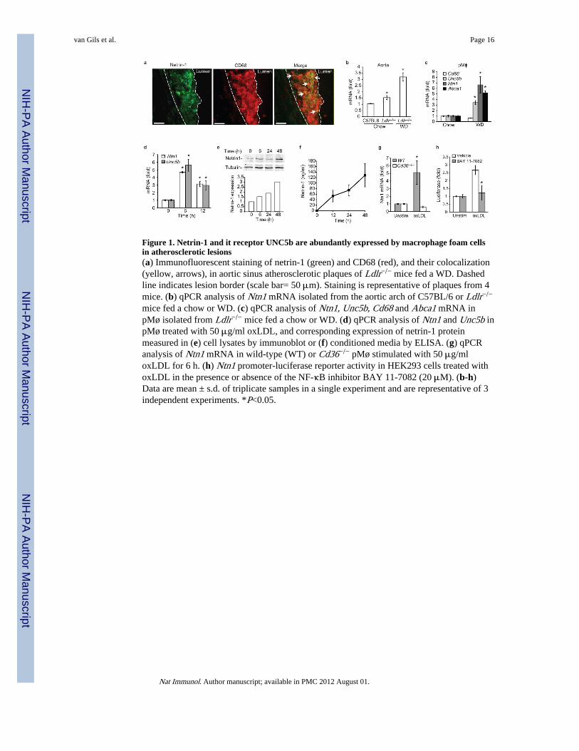
Figure 1. Netrin-1 and it receptor UNC5b are abundantly expressed by macrophage foam cellsin atherosclerotic lesions(a) Immunofluorescent staining of netrin-1 (green) and CD68 (red), and their colocalization(yellow, arrows), in aortic sinus atherosclerotic plaques of Ldlr−/− mice fed a WD. Dashedline indicates lesion border (scale bar= 50 μm). Staining is representative of plaques from 4mice. (b) qPCR analysis of Ntn1 mRNA isolated from the aortic arch of C57BL/6 or Ldlr−/−
mice fed a chow or WD. (c) qPCR analysis of Ntn1, Unc5b, Cd68 and Abca1 mRNA inpMø isolated from Ldlr−/− mice fed a chow or WD. (d) qPCR analysis of Ntn1 and Unc5b inpMø treated with 50 μg/ml oxLDL, and corresponding expression of netrin-1 proteinmeasured in (e) cell lysates by immunoblot or (f) conditioned media by ELISA. (g) qPCRanalysis of Ntn1 mRNA in wild-type (WT) or Cd36−/− pMø stimulated with 50 μg/mloxLDL for 6 h. (h) Ntn1 promoter-luciferase reporter activity in HEK293 cells treated withoxLDL in the presence or absence of the NF-κB inhibitor BAY 11-7082 (20 μM). (b-h)Data are mean ± s.d. of triplicate samples in a single experiment and are representative of 3independent experiments. *P<0.05.
van Gils et al. Page 16
Nat Immunol. Author manuscript; available in PMC 2012 August 01.
NIH
-PA Author Manuscript
NIH
-PA Author Manuscript
NIH
-PA Author Manuscript
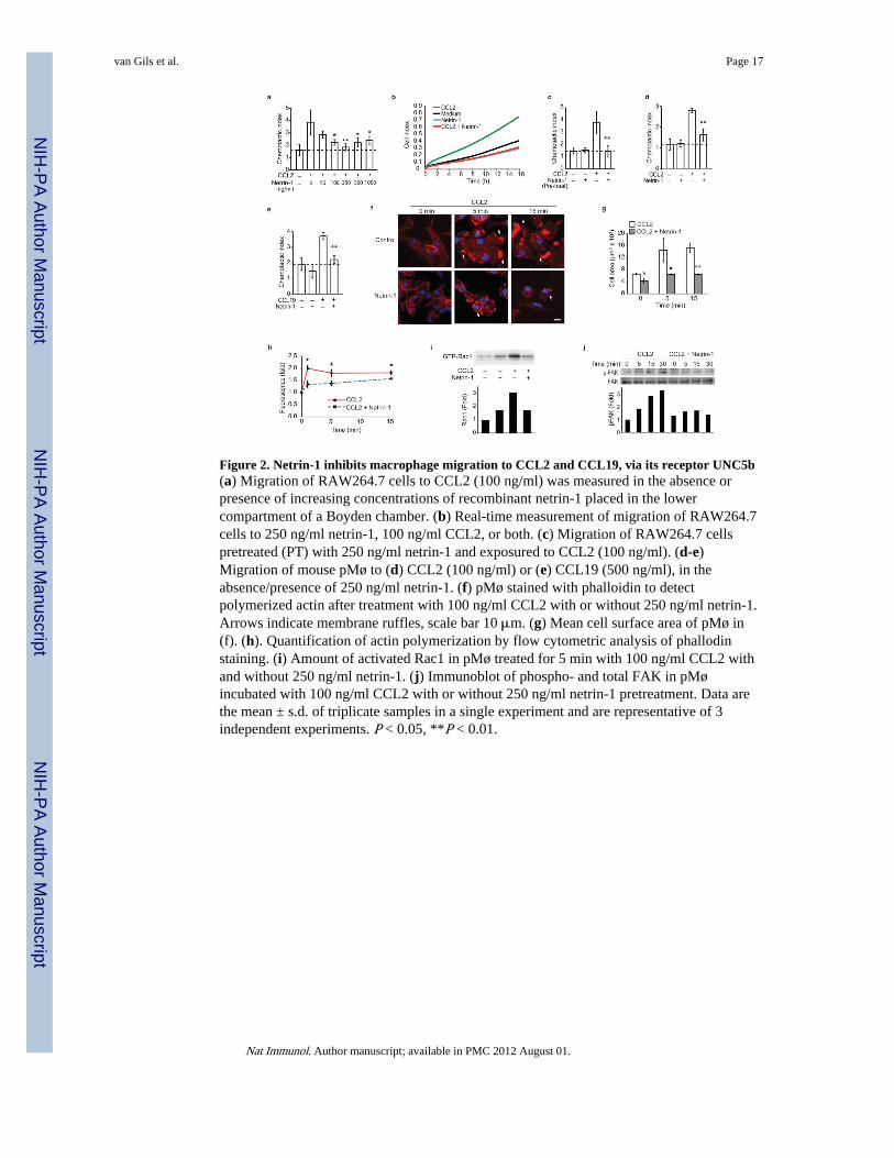
Figure 2. Netrin-1 inhibits macrophage migration to CCL2 and CCL19, via its receptor UNC5b(a) Migration of RAW264.7 cells to CCL2 (100 ng/ml) was measured in the absence orpresence of increasing concentrations of recombinant netrin-1 placed in the lowercompartment of a Boyden chamber. (b) Real-time measurement of migration of RAW264.7cells to 250 ng/ml netrin-1, 100 ng/ml CCL2, or both. (c) Migration of RAW264.7 cellspretreated (PT) with 250 ng/ml netrin-1 and exposured to CCL2 (100 ng/ml). (d-e)Migration of mouse pMø to (d) CCL2 (100 ng/ml) or (e) CCL19 (500 ng/ml), in theabsence/presence of 250 ng/ml netrin-1. (f) pMø stained with phalloidin to detectpolymerized actin after treatment with 100 ng/ml CCL2 with or without 250 ng/ml netrin-1.Arrows indicate membrane ruffles, scale bar 10 μm. (g) Mean cell surface area of pMø in(f). (h). Quantification of actin polymerization by flow cytometric analysis of phallodinstaining. (i) Amount of activated Rac1 in pMø treated for 5 min with 100 ng/ml CCL2 withand without 250 ng/ml netrin-1. (j) Immunoblot of phospho- and total FAK in pMøincubated with 100 ng/ml CCL2 with or without 250 ng/ml netrin-1 pretreatment. Data arethe mean ± s.d. of triplicate samples in a single experiment and are representative of 3independent experiments. P < 0.05, **P < 0.01.
van Gils et al. Page 17
Nat Immunol. Author manuscript; available in PMC 2012 August 01.
NIH
-PA Author Manuscript
NIH
-PA Author Manuscript
NIH
-PA Author Manuscript
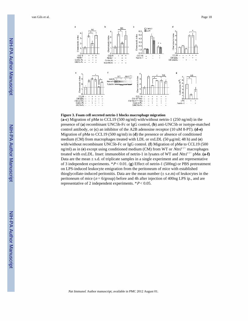
Figure 3. Foam cell secreted netrin-1 blocks macrophage migration(a-c) Migration of pMø to CCL19 (500 ng/ml) with/without netrin-1 (250 ng/ml) in thepresence of (a) recombinant UNC5b-Fc or IgG control, (b) anti-UNC5b or isotype-matchedcontrol antibody, or (c) an inhibitor of the A2B adenosine receptor (10 uM 8-PT). (d-e)Migration of pMø to CCL19 (500 ng/ml) in (d) the presence or absence of conditionedmedium (CM) from macrophages treated with LDL or oxLDL (50 μg/ml; 48 h) and (e)with/without recombinant UNC5b-Fc or IgG control. (f) Migration of pMø to CCL19 (500ng/ml) as in (e) except using conditioned medium (CM) from WT or Ntn1−/− macrophagestreated with oxLDL. Inset: immunoblot of netrin-1 in lysates of WT and Ntn1−/− pMø. (a-f)Data are the mean ± s.d. of triplicate samples in a single experiment and are representativeof 3 independent experiments. *P < 0.01. (g) Effect of netrin-1 (500ng) or PBS pretreatmenton LPS-induced leukocyte emigration from the peritoneum of mice with establishedthioglycollate-induced peritonitis. Data are the mean number (± s.e.m) of leukocytes in theperitoneum of mice (n = 6/group) before and 4h after injection of 400ng LPS ip., and arerepresentative of 2 independent experiments. *P < 0.05.
van Gils et al. Page 18
Nat Immunol. Author manuscript; available in PMC 2012 August 01.
NIH
-PA Author Manuscript
NIH
-PA Author Manuscript
NIH
-PA Author Manuscript
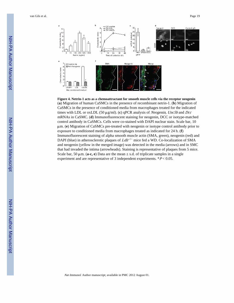
Figure 4. Netrin-1 acts as a chemoattractant for smooth muscle cells via the receptor neogenin(a) Migration of human CaSMCs in the presence of recombinant netrin-1. (b) Migration ofCaSMCs in the presence of conditioned media from macrophages treated for the indicatedtimes with LDL or oxLDL (50 μg/ml). (c) qPCR analysis of Neogenin, Unc5b and DccmRNAs in CaSMC. (d) Immunofluorescent staining for neogenin, DCC or isotype-matchedcontrol antibody in CaSMCs. Cells were co-stained with DAPI nuclear stain. Scale bar, 10μm. (e) Migration of CaSMCs pre-treated with neogenin or isotype control antibody prior toexposure to conditioned media from macrophages treated as indicated for 24 h. (f)Immunofluorescent staining of alpha smooth muscle actin (SMA, green), neogenin (red) andDAPI (blue) in atherosclerotic plaques of Ldlr−/− mice fed a WD. Co-localization of SMAand neogenin (yellow in the merged image) was detected in the media (arrows) and in SMCthat had invaded the intima (arrowheads). Staining is representative of plaques from 5 mice.Scale bar, 50 μm. (a-c, e) Data are the mean ± s.d. of triplicate samples in a singleexperiment and are representative of 3 independent experiments. *P < 0.05.
van Gils et al. Page 19
Nat Immunol. Author manuscript; available in PMC 2012 August 01.
NIH
-PA Author Manuscript
NIH
-PA Author Manuscript
NIH
-PA Author Manuscript
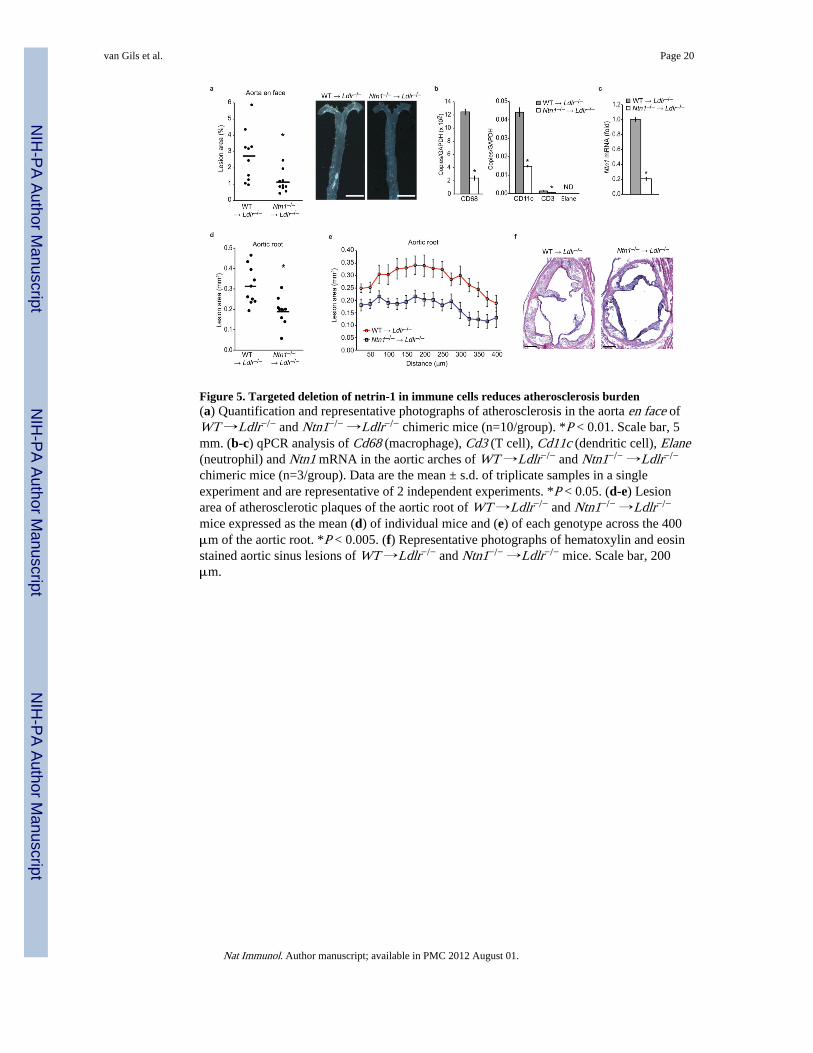
Figure 5. Targeted deletion of netrin-1 in immune cells reduces atherosclerosis burden(a) Quantification and representative photographs of atherosclerosis in the aorta en face ofWT →Ldlr−/− and Ntn1−/− →Ldlr−/− chimeric mice (n=10/group). *P < 0.01. Scale bar, 5mm. (b-c) qPCR analysis of Cd68 (macrophage), Cd3 (T cell), Cd11c (dendritic cell), Elane(neutrophil) and Ntn1 mRNA in the aortic arches of WT →Ldlr−/− and Ntn1−/− →Ldlr−/−
chimeric mice (n=3/group). Data are the mean ± s.d. of triplicate samples in a singleexperiment and are representative of 2 independent experiments. *P < 0.05. (d-e) Lesionarea of atherosclerotic plaques of the aortic root of WT →Ldlr−/− and Ntn1−/− →Ldlr−/−
mice expressed as the mean (d) of individual mice and (e) of each genotype across the 400μm of the aortic root. *P < 0.005. (f) Representative photographs of hematoxylin and eosinstained aortic sinus lesions of WT →Ldlr−/− and Ntn1−/− →Ldlr−/− mice. Scale bar, 200μm.
van Gils et al. Page 20
Nat Immunol. Author manuscript; available in PMC 2012 August 01.
NIH
-PA Author Manuscript
NIH
-PA Author Manuscript
NIH
-PA Author Manuscript
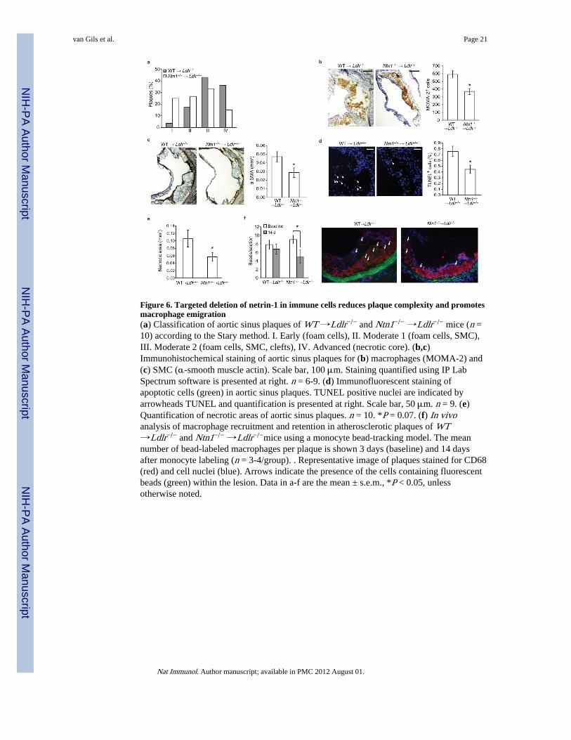
Figure 6. Targeted deletion of netrin-1 in immune cells reduces plaque complexity and promotesmacrophage emigration(a) Classification of aortic sinus plaques of WT →Ldlr−/− and Ntn1−/− →Ldlr−/− mice (n =10) according to the Stary method. I. Early (foam cells), II. Moderate 1 (foam cells, SMC),III. Moderate 2 (foam cells, SMC, clefts), IV. Advanced (necrotic core). (b,c)Immunohistochemical staining of aortic sinus plaques for (b) macrophages (MOMA-2) and(c) SMC (α-smooth muscle actin). Scale bar, 100 μm. Staining quantified using IP LabSpectrum software is presented at right. n = 6-9. (d) Immunofluorescent staining ofapoptotic cells (green) in aortic sinus plaques. TUNEL positive nuclei are indicated byarrowheads TUNEL and quantification is presented at right. Scale bar, 50 μm. n = 9. (e)Quantification of necrotic areas of aortic sinus plaques. n = 10. *P = 0.07. (f) In vivoanalysis of macrophage recruitment and retention in atherosclerotic plaques of WT→Ldlr−/− and Ntn1−/− →Ldlr−/−mice using a monocyte bead-tracking model. The meannumber of bead-labeled macrophages per plaque is shown 3 days (baseline) and 14 daysafter monocyte labeling (n = 3-4/group). . Representative image of plaques stained for CD68(red) and cell nuclei (blue). Arrows indicate the presence of the cells containing fluorescentbeads (green) within the lesion. Data in a-f are the mean ± s.e.m., *P < 0.05, unlessotherwise noted.
van Gils et al. Page 21
Nat Immunol. Author manuscript; available in PMC 2012 August 01.
NIH
-PA Author Manuscript
NIH
-PA Author Manuscript
NIH
-PA Author Manuscript