THE N E U T R A L I Z A T I O N OF A N I M A L V I R U S E S ...
238
THE NEUTRALIZATION OF ANIMAL VIRUSES. A THesis submitted for the Degree of Doctor of Philosophy in the Australian National University.
-
Upload
khangminh22 -
Category
Documents
-
view
0 -
download
0
Transcript of THE N E U T R A L I Z A T I O N OF A N I M A L V I R U S E S ...

T H E N E U T R A L I Z A T I O N O F
A N I M A L V I R U S E S .
A THes is s u b m i t t e d f o r th e
Degree o f D o c to r of P h i lo s o p h y
i n th e
A u s t r a l i a n N a t i o n a l U n i v e r s i t y .

Z 4 JAN 19611^67 2.

A C K N O W L E D G E M E N T S
I would, like to express my thanks to Dr. S. Fazekas de St Groth for the privilege of working in his laboratory and for his help and stimulus in my introduction to, and training in, the field of animal virology. I would also like to thank Professor Fenner and the members of his department for ..iany helpful discussions during my stay in Canberra, and for the typing of this thesis I am indebted to my wife Anne.

S T A T E M E N T .
The work described in Chapter two was directed by Dr, S• Fazekas de St Groth and carried out by Dr. 3. Fazekas ae St Groth,Mr. J. Withell and myself. That described in Chapter three was carried out oy Mr. J. Withell and myself. Chapters one, four and five are wnolly my own work and Chapter six is the result of the «joint work carried out by Dr. S. Fazekas de St Groth, Mr. J. Withell and myself.

C Ö I T E N T S
CHAPTER 1 . PAGE.The Neutralization of Bacterial and
Animal Viruses by Antibody:A Review of the Literature.
Introduction. 1.Neutralization of Bacteriophage. 1.Kinetics of Neutralization. 6.Effect of the Kost Cells on the Reaction Kinetics. 16.Effect of Vp on Kinetics 18.The Neutralization of Animal Viruses. 22.Evidence for a Complex Reaction Mechanism. 27.
CHAPTER 2.The Assay of Neutralizing Antibodies
Against Influenza Virus in the Surviving Allantois.Introduction. 32.The Effect of Normal Animal Sera on the Host Tissue. 35.The Effect of Anticellular Antibodies. 37.The Neutralization of Infectivity. 41.

CHAPTER 2 contd PACEEffect of the Reaction Time on Neutralization Titres. Standardization of the Incubation Period.The Test Volume.Comparison of the method with other in vivo Neutralization Tests and Antihaemagglutenin Titrations•The Accuracy of the Test.Summary and Conclusions.
43.
46.48.
52.55.60.
CHAPTER 5 .The Reaction between Influenza
Virus and Neutralizing Antibody. Introduction.Effect of the Reaction Time on the Reversibility of the Reaction. Aggregation of the Virus Particles. Reactivation by Ultrasonic Vibrations. Discussion.Summary.
62.
63.67.71.73.77.
ALIBRARY r

CHAPTER 4. PAGE.Kinetics of the Virus-Antibody
Interaction.Introduction. 78.Kinetics of the Secondary Reaction. 79.Effect of the Serum Concentration onthe Reaction Rate. 83.The Kinetics of Rabbit-pox VirusInactivation. 86.The Nature of the Protected Fraction. 88.The Reversibly Induced ProtectedFraction. 89.Effect of the Host Cells on theProtected Fraction. 94.Discussion. 99.Summary. 108.
CHAPTER 5.The Mechanism of the Virus-antibody
Interaction.Introduction. 110.Hypotheses. ’ 112.Testing the Hypotheses. 123.Neutralization of Influenza Virus by Papain Digested Antibody. 124.The Protected Fraction. 130.

CHAPTER 5. contd. PACE.Heterogeneity of Antisera. 133.Steric Effects. 136.Discussion. 141.Summary and. Conclusions. 148.
CHAPTER 6.Q.uantal neutralization Tests.
Introduction. 150.Theoretical. 152.Experimental. 164.The Behaviour of normal Antibody:the Dose Response Curve. 164.Effect of the Reaction Time on theDose Response Curve. 170.The Effect of Honinfectious Virus. 174.The Effect of the Test Volume. 176.The Behaviour of Papain DigestedAntibody. 183.Effect of the Reaction Time on thePosition of the Dose Response Curve. 185.Discussion. 187.Summary. 194.

CHAPTER 7 PACE
195.198.
Ceneral Summary Appendix. Bibliography. 204

C H A P T E R
O H E
THE NEUTRALIZATION OF BACTERIAL
AND ANIMAL VIRUSES BY ANTIBODY:
A REVIEW OF THE LITERATURE*
f* L I B R A R Y \ j

1
INTRODUCTION.
The neutralization of virus by antibody has been studied extensively by bacterial and animal virologists, the most comprehensive work being that carried out on bacteriophage. For this reason tnis chapter has been divided into two sections, the first dealing with bacteriophage neutralization and the second with the neutralization of animal viruses.
NEUTRALIZATION OF BAGTSRIQPHAG-E.
Bordet and Ciuca (1921) first demonstrated that the injection of rabbits with phage lysates stimulates the production of phage neutralizing antibodies. Their antisera also contained agglutinins for the host bacterium; however, the injection of host bacteria failed to stimulate the production of phage neutralizing antibody. Moreover in 1922 Otto and Winkler were able to remove all bacterial agglutinins, by absorbtion with host bacteria, without affecting the antiphage properties of the serum. These findings demonstrate that the pha^e is antigenically distinct from its host.
Until recently phage particles were considered to consist of a single antigen, thus producing

on inoculation a single antibody. In an extensive study, Lanni and Lanni (1953), also Rountree (1952); De Liars, Luria, Fisher and Levinthal (1953), presented clear evidence for the existance of at least two distinct antigens in phage Tl. The tail antigen reacts with neutralizing- antibody, and under the correct conditions aggregate on and complement fixation can be demonstrated with this antigen. The second antigen, situated in the phage head, participates in specific aggregation and complement fixation, but does not react measurably with neutralizing antibody.
When a phage preparation is mixed with homologous antiserum there is a progressive decrease in the number of plaque forming particles. The inactivation process can be interrupted at any time by diluting the mixture below the concentration at which collision between phage particles and antibody will proceed at a significant rate. Such a proceedure can be used to study the kinetics of inactivation provided dilution does not bring about any significant reactivation. In general phage neutralization is considered to be an irreversible process (Burnet, Keogh and Lush, 1937), although there is evidence that the reaction mechanism is

3
complex, and a fraction of the particles may bee
reversibly neutralized (Andrew's and Biford, 1933; Jerne and Avegno, 1956).
Before discussing the question of reversibilityk
of the neutralization reaction, it should be pointed out, that in this context the reaction is considered to be irreversible if dilution of the phage antiserum mixture past the point of serum activity does not cause any measurable reactivation of the neutralized particles. Andrews and Elford (1933 b) were able to demonstrate reactivation of phage C36 on dilution. However as these workers were (quick to point out tnis reactivation can only be demonstrated under certain conditions. Namely, provided the reaction time with antiserum is short. Other workers were unable to demonstrate this effect with different viruses (Burnet, 1933;Hersney, 1943; Kalmanson, Hershey and Bronfenorenner, 1942). However in 1956 Jerne and Avegno studied the problem in more detail, and clearly demonstrated that neutralization proceeds in two phases. When phage T4 is mixed with antiserum there is a rapid reversible neutralization of the phage particles.On dilution of the phage-antibody mixture, the amount of virus that can be reactivated is inversely
proportional to the initial reaction time. Thus a

4
phase of irreversible neutralization is proceedingat a slower rate than that of reversibleneutralization. This effect was dependent on thehistory of the antiserum. Antisera produced bybleeding an animal early after inoculation showingthis effect to a much more marked extent than lateantisera. However even when late antisera were
dSused a fraction of the virus particles wsre still reversibly neutralized.
Attempts to dissociate irreversibly neutralized phage by formolized bacteriophage were unsuccessful, (Hershey, 1941b). However Burnet (1935 ) was ableto show that soluble phage antigen had a slight reactivating effect. Although antibody bound irreversibly does not appear to dissociate spontaneously to any great extent, there is evidence that it can be removed by certain methods: Kalmanson and Bronfenbrenner (1943) found that neutralized phage could be completely reactivated by digestion of the complex with papain, provided the virus nad not been over neutralized with an excess of serum. Anderson and Doerman (1952) succeeded in obtaining approximately a thirty fold increase in titre of a neutralized preparation of
T3 by subjecting it to ultrasonic vibrations.

These experiments indicate that the antibody does not damage the phage particle itself but rather that it interferes with some step in the interaction of the intact virus particle with the host cell.
Delbrück (1945) found that following adsorption to the host cell, phage is resistant to the action of immune serum throughout the whole of the latent
period. However, Gross (1954) was able to demonstrate that pha^e was susceptible to inactivation by antiserum during the period of reversible absorption but not once the phage had injected its DUA into the host bacterium.
In summary the following points can be made:
1. Bacteriophage is neutralized when neutralizing antibody combines with the virus particle.
2. This combination can be either readily or hardly reversible, the evidence of Jerne and Avegno (1956) indicating that some particlesin the population can be reversibly neutralized whilst others are irreversibly neutralized.
3. Antibody does not damage the virus particle itself but in some way prevents the phage from
infecting the host bacterium. When the antibody is removed the virus particle regainsits full activity

6 .
4. For the tailed phages, neutralizing antibodies are those directed against the tail antigens.
5. The virus particles can be neutralized whilst they are free in suspension or whilst they are reversibly absorbed to the host bacterium.
KINETICS QF NEUTRALIZATION.
The interpretation of kinetic data is somewhat obscured by the fact that the reaction between phage and antibody is not a simple bimolecular reaction. I shall first consider the factual data that have been collected and then try to reconcile these data with a plausible reaction mechanism.
The technique most commonly adopted for kinetic studies consists of sampling from a phage antibody mixture at various times, rapidly diluting the sample to prevent the continuation of the neutralization reaction, and assaying for residual infectivity.
In 1922 Prausznitz was able to demonstrate that phage neutralization by immune serum was a relatively slow process, and that a proportion of
the phage population resisted inactivation when assayed by this dilution technique. Andrewes and

7
Eiford (1923 a) oarried out a more extensive study of the kinetics of bacteriophage neutralization and summarized their results in the form of the "Percentage Law”: "over a very wide range a givendilution of serum neutralized in a given time an approximately constant percentage, however much phage there was present." Although these authors pointed out that the reaction mechanism was complex— they were able to demonstrate both reversible and irreversible neutralization — they did not attempt to distinguish between these two reactions, Burnet, Keogh and Lush (1937) studied the neutralization of phage C16. These workers were able to confirm the validity of the "Percentage Law". They also demonstrated that when low serum concentrations were used, there was a pronounced lag before the reaction followed a logarithmic course. This lag decreased as the serum concentration increased until at high serum concentrations the reaction appeared to have first order kinetics.Once again approximately of the virus particles were neutralized at a much slower rate than the remainder. This the authors attributed to heterogeneity of the virus population. Kalmanson, Hershey and Bronfenbrenner (1942) found a similar situation when they studied the inactivation phage T2.

8
In 1945 Delbrück demonstrated that T2 and T7 followed first order kinetics until 99$ of the particles were neutralized. Similar behaviour has been observed with phages related to T5 (Adams 1952) , with the M phages of B.megaterium (Friedman and Cowles, 1955) and with phage D20 (Adams and Wade 1955).
The initial lag in the kinetic curve described by Burnet, Keogh and Lush (1957) and Kalmanson, Hershey and Bronfenbrenner (1942) was attributed to a multihit reaction mechanism, the latter authors suggesting that two or three antibody molecules were required to neutralize T2. However Cann and Clark (1954), following up a suggestion of Sagik (1954), demonstrated that the lag in the kinetics of T2 inactivation by antiserum could apparently be removed oy activating the virus stocks before serum treatment. This effect, however, does not completely explain the reason for the lag period since Park (1956) has shown that a Klebsiella phage which cannot be activated (Sagik, 1954) still shows a pronounced lag in its kinetics of inactivation by dilute serum.
Jerne and Avegno (1956) using an early bleed antiserum, demonstrated that when the virus and

9
antibody were mixed, there was a rapid reversible combination between the antibody and virtually all the virus particles. However as the reaction time increased there was a decrease in the amount of virus that could be reactivated on dilution. Thus they suggested that the reaction between the phage and antibody proceeded as follows.
V + A — YA + B — - Vj
Where Y represents free virus particles, YA virus particles that are reversibly neutralized and Yj virus particles that are irreversibly neutralized. They envisiged the secondary reaction occurring when a second antibody molecule (B), directed against the junction area of the YA complex, combined with this complex and stabilized the primary combination. These authors were also able to show that the reversible reaction proceeds in the forward direction with pseudo first order kinetics (antibody being in excess),thus demonstrating that one antibody molecule bound reversibly was sufficient to neutralize the particle•
On dilution of the reaction mixture any reversibly neutralized phage will begin to reactivate and this reactivation will continue

10
on the assay ^late. However the reactivated particles will only form visible plaques if they have reactivated within a certain time after the dilution and plating proceedure. In fact, as Jem e and Avegno (1956) have pointed out, if t is the time within which reactivation must occur for the reactivated particles to score as infective, then fraction f of reversibly neutralized particles that will reactivate in this time is given by,
£ = ( 1 - e "kt ),
where k is the dissociation rate constant,Boyd (1956) nas collected data related to
equilibrium between various antigens and their specific antibodies. These results show that the equilibrium constants for the various reactions can vary over a hundredfold range. If the equilibrium constant varies then either the association constant or the dissociation constant or both must vary for the different reactions.Thus it is not unlikely that the value of k for different phage antibody reactions will be a variable factor, also , the history of the antiserum has a marked effect on the value of k, (Talmage, 1957). Hot only will the value of k

11.
vary with, different antigens and different sera, the value of t will also vary for different phage host systems. Thus the value of f may vary between a figure tending to unity and one much less than unity depending on the value of kt for the system under consideration. Such variation in the value of f would account for the differences in the early stages of the reaction kinetics that have been observed by various workers.
If the value of f —►1, any reversibly neutralized virus will score as infective and the rate of formation of the stable union will be measured. Under these conditions a lag or induction period woulu be observed when the virus is inactivated by relatively dilute antisera, the lag representing the time taken to build up the concentration of the VA intermediate. If however f 1, a large fraction of the reversibly neutralized particles, ( 1 - f ), will score as non infective, and under these conditions no shoulder on the kinetic curves will be observed since the primary reaction proceeds with first order kinetics (Jerne and Avegno, 1956).
Jerne and Avegno envisaged that the stabilization reaction involved the attachment of a second antibody molecule to the virus-antibody complex.

12
However there is no experimental evidence that a second antibody molecule is concerned in the reaction. One could postulate that the slow reaction is due to some type of stabilization of the VA complex that does not involve a second antibody molecule. In wnich case the reaction could be represented diagramatically as
V f A YA Vi
the broken arrow is included because the Vj complex is not, strictly speaking, irreversibly neutralized since it can be reactivated if treated with ultrasonic vibrations. For either of these mechanisms the lag observed when f is close to unity would represent the time taken to build up the intermediate YA.
One might expect that it wo old be possible on kinetic grounds to differentiate between the two mechanisms, that is, to determine whether or not a second antibody molecule is involved in the secondary reaction. This would be the case if one could explicitly describe the kinetics of this type of reaction. This however, cannot beachieved for the following reason.
The schematic representation of the reaction shown above is an over-simplification. Each

13
virus particle has a number of sites at which neutralizing antibody can attach (Hershey, Kalmanson and Bronfenbrenner, 1943; see also Delbrück, 1945). Thus when antibody is mixed with bacteriophage a number of different equilibria will be set up,
and the rate at which virus particles are converted from the reversibly neutralized state to the "irreversibly” neutralized state will be a compound of the rates at which virus particlesof the VA, V A 2 --- VAn classes can be converted to thelatter state. However because of steric and electrostatic effects, the occupation of one site by an antibody molecule will alter the probability of filling neighbouring sites with antibody by an unknown factor. Thus making it virtually impossible to calculate the frequency distribution of virus particles in the various forms,VA, VA2 --- VAn. Without tnis informationone cannot attempt to describe explicitly the kinetics of the secondary reaction.
V + A VAVA + A VA2
VA (n - 1) + A V A n

14
3ven though, the reaction is not amenable to analytical treatment, kinetic studies have shown that the initial stages of the inactivation are adequately described by the equation,
-KtV _ e D
T o -
Where V is the residual virus infectivity at time t, Vo is the initial virus titre, K is a constant and D is the dilution of serum (Delbrück, 1945). This is true provided the serum concentration is not too low (Burnet, Keogh and Lush, 1957; Lark, 1956); under these conditions tnere is an initial lag before the reaction follows an exponential course. As has already been pointed out, this lag is due to the time taken to build up the concentration of the intermediate VA complex. The rate of this reaction will also be dependent on the serum concentration and above some critical concentration this reaction will effectively be instantaneous. However the secondary reaction proceeds at a rate that is some 300 times slower than that of the primary reaction (Jerne and Avegno, 1956). Thus as the concentration of the serum is increased one would expect the lag to become less pronounced until above a certain
uo.
concentration it would be undetectable (see Burnet, Keogh, and Lush, 1937).
The above equation still holds at low serum concentration but only after the induction period has been passed (Burnet Keogh and Lush, 1937).This empirical relationship has proved quite useful in comparing the serum activity of different antiserum preparations (Campbell, 1959). It has also been used to estimate the serological relation between different phages (Luria, 1953).The values of K for a given antiserum and different phages indicates the degree of serological relationship among the phages. An unrelated phage will give a K value of 0; a weakly cross-reacting phage will have a value of K much lower than that of the homologous phage. V
When the value of Vo becomes small, that is at low survival levels the above equation once more oreaks down. The possible explanations of this phenomenon are discussed in the followingsection

16
EFFECTS OF THE HOST CELLS OK THE REACTION KINETICS
As early as 1926 dTHerelle demonstrated that different assay hosts may give different estimates of the fraction of unneutralized phage in a given phage antiserum mixture, Kalmanson and Bronfenhrenner (1942) analysed this host effect with a strain of T2 that plated with almost the same efficiency on strains of Shiga and Flexner dysentry bacteria and E. coli. They found the susceptibility to neutralization by antiserum was the same regardless of the host used to produce the virus stocks, However when the kinetics of neutralization were studied by plating samples on all three hosts, the rate of inactivation was found to be greater when Shiga and Flexner bacteria were used as host than when E. coli was the host. Friedman (1954) demonstrated a similar phenomenon with the phage M5 of B.megaterium.
Kalmanson and Bronfenbrenner (1942) suggested that their results provide evidence for a serological heterogeneity inherent in individual phage particles. However the explanation put foreward by Adams (1959) seems more reasonable,

17
nin the random reaction of antibody molecules with uniform antigen sites on the phage particles, the virus surface could be so altered that certain of the particles would adsorb abortively, or even be unable to absorb, to the dysentry host cells, whereas stronger bonds with E. coli would still allow productive interaction." Regardless of the hypothesis that one invokes to explain these results, the clear fact is that a fraction of the antibody molecules that combine with the virus will behave as neutralizing antibodies if one host system is used for the assay, but will not if a different host system is used. This effect immediately complicates the picture of the reaction mechanism because presumably if a suitable host is chosen (in fact the effect may be present to varying degrees with all hosts) some antibody molecules that do not neutralize the virus particle may combine firmly with its critical sites, thus rendering the particle insusceptible to the action of neutralizing antibodies.
If the stabilization of the union Detween virus and antibody is brought about by a consecutive mecnanism the basic reaction may nowbe written as

J.Ö.
V + A ,---- YA v -~ Yx ( l )
Y + P ----* YP Yp (2)
where Yp r e p r e s e n t s v i r u s p a r t i c l e s p r o t e c t e d
from n e u t r a l i z i n g a n t i b o d y .
EFFECT OF Yp 0If KIISTICS.
I f t h e Yp p a r t i c l e s c o n s t i t u t e a sm a l l
f r a c t i o n o f t h e v i r u s p o p u l a t i o n , th e k i n e t i c s
o f i n a c t i v a t i o n by a n t i b o d y w i l l be d e te r m in e d
by r e a c t i o n 1 u n t i l t h e s u r v i v i n g p a r t i c l e s a r e
o f th e same o r d e r o f m agni tude a s t h e p r o t e c t e d
f r a c t i o n . At t h i s s t a g e th e s p e c i f i c r a t e o f
i n a c t i v a t i o n w i l l a p p a r e n t l y d e c r e a s e and when
th e s u r v i v i n g f r a c t i o n i s Yp/Yq i . e . t h e
p r o t e c t e d f r a c t i o n (Yq b e in g th e t o t a l amount of
v i r u s ) , t h e i n a c t i v a t i o n r a t e w i l l be z e r o .
M o reo v er , i f t h e v a lu e o f Yp i s d e p en d e n t on t h e
h o s t b a c t e r i u m u se d t o a s s a y th e v i r u s , t h e
s u r v i v a l l e v e l a t which th e in h o m o g en e i ty o f th e
v i r u s p o p u l a t i o n becomes e v i d e n t w i l l va ry when
th e same v i r u s a n t i b o d y m i x t u r e s a r e a s s a y e d on
d i f f e r e n t h o s t b a c t e r i a .
F r iedm an (1954) found t h a t t h e s u r v i v a l l e v e l
a t which the k i n e t i c cu rve b e g in s to f l a t t e n o f f
v a r i e s when d i f f e r e n t h o s t b a c t e r i a a r e u sed f o r

19
the survivor assay. However, there is evidence that the formation of protected particles may not completely account for the change in the rate of inactivation observed at low survival levels.
Delbrück (1945) suggested that the inhomogeneity might be inherent in the virus population or, more likely, that it might develop during the course of the reaction as a result of attachment of antibody to noncritical sites on the phage particle. The work of Jerne and Avegno (1956) throws some light on this problem. These workers observed a fall in the rate of the secondary reaction at low survival levels. However when they tested these survivors by the decision tube technique they found that they were reversibly neutralized. Thus the inhomogeneity must be in part due to a fraction of the particles which form a loose union with the antibody molecules but which, either cannot form a stable union with the antibody or if they can, form this stable union at a slower specific rate than the majority of the virus particles in the preparation.

20
This effect could arise in any of three ways.1. There may he an inhomogeneity in the antibody molecules. Thus all the molecules may combine loosely with the virus surface at a relatively rapid rate but a fraction may not be able to stabilize this union at as fast a rateas the other antibody molecules in the antiserum.
2. There may be an inhomogeneity in the union that is formed between the virus and antibody.In this case some particular types of reversible unions, although they neutralize the infectivity of the virus particle, may not be as readily stabilized as others.
3. The effect may be due to the structure of the virus particles. Some particles because of their structure forming a firm union with antibody at a much slower specific rate than the remainder of the particles.
However, regardless of the mechanism, when a virus population reacts with an antiserum three classes of product will be formed,a) "Irreversibly" neutralized particles, (Vj).b) Particles which are reversibly neutralized

21
but which can be converted, to the Vj form at a very slow rate, (Va).c) Protected, particles, that is, particles that have virus specific non-neutralizing antibodies attacned to their surface, (V^)•
Such a reaction scheme would explain the slow rate of inactivation observed at low survival levels. It would also explain the frequency distribution of plaque sizes observed by Burnet, Keogh and Lush (19,37). These authors found that after a prolonged incubation of phage and antiserum, the size of the plaques produced, when the reaction mixture was diluted and plated on a susceptible host bacterium, fell into two distinct groups. One group had a mean plaque size which was slightly smaller than the average plaque size of the untreated phage particle, and tne otner group had an average plague size which was about 1mm. smaller in diameter than the plaques produced by the control virus. According to the above scheme at the time of plating, all the virus particles would fall into one of the three classes, Va, Vj and Vj, provided there was an excess of antiserum.

22
Va virus would, give rise to plaques after reactivation had. occurred, either in the dilution tube, or what is more likely, on the assay plate. Thus one would expect to find a delay in starting time which would tend to produce plaques of a smaller average size than those produced when normal virus is plated and incubated for the same time. Whilst Vp virus should produce plaques of approximately the same size as active virus, or slightly smaller if the antibody bound to tne virus has some steric effect which leads to a slignt delay in the iniation of infection by such particles.
THE NEUTRALIZATION OF ANIMAL VIRUSES.
Interest in the immunological aspects of virus diseases has its origin in the work of Jenner, who was the first to put to practical use the phenomenon of post-infection immunity. However it was not until the close of the nineteenth century that attempts were made to elucidate the nature of this phenomenon. In 1898 Böclere, Chambon and Menard, working with vaccinia virus, studied the transfer of immunity to heifers by

23
the injection of serum, from immunized animals.The nature of the mechanism by which such immunity was transferred was the subject of a subsequent paper, (Beclfere, Chambon and Menard, 1899). The latter paper they concluded with the comment:"On ne saurait encore affirmer si cette substance agit directement sur les agents infecteux comme virulicide, ou si eile agit comme un stimulant sur les cellules de 1 T organisme .,T This proolem proved to be the subject of controversy for the following thirty odd years.
In 1928, Andrewes was able to demonstrate that a mixture of vaccinia virus and antiserum that was neutral when inoculated into rabbits, would give rise to infection if diluted before inoculation. Such results lead him to the conclusion that there was little evidence for any combination between the virus and antibody, and that the antiserum exerted its protection by virtue of the effect that it had on the host cells; a view which was also held by Long and Olitsky (1920}. However during his investigations with herpes virus, Bedson (1928) found that the amount of virus that could be reactivated on dilution decreased when the reagants were allowed

24
to react for several hours before dilution and
inoculation. This observation favoured the hypothesis that virus neutralization was brought about by a combination between the virus and antibody. In the following year the same author confirmed this conclusion by demonstrating that
antibody could be removed from an animalfs serum by absorption of the serum with herpes virus absorbed to collodion, (Bedson, 1929).Andrewes, (1920+, reinvestigated the dilution phenomenon he had observed with vaccinia virus two years earlier. He also found that the amount of virus infectivity that could be recovered on dilution of a neutral mixture decreased as the reaction time is increased. Thus he concluded that an in vitro combination between the virus and antibody could take place , but that it took several days to become established. However the results of experiments carried out by Wilson Smith (1920) indicated that the combination between vaccinia virus and antiserum was a much more rapid process than had been suggested by
Andrewes; the former author found that the
protective antibody could be removed from an
antiserum by absorption with virus-containing tissue.

25
However, even in the light of this evidence there were still some adherents of the view that antibody exerted its protective effect by virtue of the effect that it had on the host ceils (see Sabin, 1935).
In 1957 Burnet, Keogh and Lush compiled a monograph covering all the then known virus- antibody interactions. The striking feature of the work described by these authors was the underlying unity amongst all virus-antioody reactions. In essence, their view on virus neutralization, which constituted orthodox thinking on this subject for the following twenty years, can be stated as follows.1. Virus inactivation by immune serum results primarily from a union of antibody with the virus surface. This union is reversible. However secondary reactions such as aggregation or a secondary irreversible virus-antibody union may occur, particularly when concentrated reagents are used.2. The union of antibody has no intrinsic inactivating effect; the inactivation is the failure of interaction between susceptible cells and antibody-coated virus particles.
It should be emphasized that although these workers were aware of the fact that, under certain

26
c i r c u m s t a n c e s , a seco n d a ry i r r e v e r s i b l e s t a g e
i n t h e n e u t r a l i z a t i o n r e a c t i o n c o u ld be o b s e r v e d ,
th e y c o n s i d e r e d i t o f m inor im p o r t a n c e , a s i s
e v id e n c e d by th e f a c t t h a t t h e i r t h e o r e t i c a l
t r e a t m e n t o f v i r u s n e u t r a l i z a t i o n assumes t h a t
trie r e a c t i o n i s c o m p le t e ly r e v e r s i b l e . Thus
B urne t was l e | . d t o f o r m u l a t e tn e view t h a t
" to a n e a r a p p r o x i m a t i o n i n each c a s e , t h e v i r u s
a n t i b o d y r e a c t i o n r e s u l t i n g i n i n a c t i v a t i o n i s
i r r e v e r s i b l e i n th e case o f t y p i c a l b a c t e r i a l
v i r u s e s and c o m p le t e ly r e v e r s i b l e i n th e case of
a n im a l v i r u s e s . T h is d i f f e r e n c e h a s n e v e r been
a s f u l l y a p p r e c i a t e d a s i t d e s e r v e s t o be n o r
h as any h y p o t h e s i s been p ro p o s e d t o a c c o u n t f o r i t .
I t i s q u i t e c l e a r , how ever , t h a t d i s c u s s i o n s of
phage n e u t r a l i z a t i o n by immune serum can have
l i t t l e b e a r i n g on t h e b e h a v i o u r o f an im a l v i r u s e s
and c o r r e s p o n d i n g immune s e r a , " ( B u r n e t , 1 9 5 5 ) .
The c o n ce p t o f a r e v e r s i b l e r e a c t i o n mechanism
was f u r t h e r s u o s t a n t i a t e d by th e d e m o n s t r a t i o n
t h a t n e u t r a l m i x t u r e s o f i n f l u e n z a v i r u s and
s p e c i f i c a n t ib o d y c o u ld be r e a c t i v a t e d by th e
a d d i t i o n o f homologous b u t no t h e t e r o l o g o u s
n e a t - i n a c t i v a t e d v i r u s , (McKee and H a l e , 1946;
I s a a c s , 1 9 4 8 ) .

27
EVIDENCE FOR A COMPLEX REACTION MECHANISM,
Although the accumulated data clearly pointed to a reversible combination between the virus and antibody, most workers had observed that the amount of virus that could be reactivated on dilution began to decrease when concentrated reactants were allowed to react for a prolonged period of time. This effect was observed with herpes (Bedson, 1928), influenza virus, (Burnet, 1936) louping ill, (GrOyal, 1935), vaccinia virus (Andrews, 1930; Keogh, 1936), Newcastle disease virus, (Rubin and Franklin, 1957), laryngotracheitis virus (Burnet, Keogh and Lush, 1937), myxoma virus (Burnet, Keogh and Lush, 1937), and Eastern equine encephalitis, (Merrill, 1936). This secondary effect was never studied in any great detail and was usually attributed to some type anomalous behaviour observed when nigh concentrations of virus and antibody were used (Burnet, Keogh and Lush 1937; Merrill, 1936).
When Gard investigated the reaction between Theiler!s virus and antioody he coined the term immunoinactivation to describe this secondary phenomenon, (Gard, 1955). In a subsequent

28
publication (Gard, 1957) , it became clear that this phenomenon could also be observed when relatively dilute reagents were used to study the reaction. These studies lead G-ard to postulate that the neutralization reaction proceeded in two distinct stages, the primary reaction oeing the reversible attachment of antioody to the virus surface and the secondary reaction, a reaction leading to the irreversible neutralization of the virus infectivity.The term immunoinactivation was quite apt because what was being measured was the effect of antibody on the virus infectivity. Thus G-ard left open the question of whether this effect was due to an irreversible combination between the virus and antibody or due to some type of inactivation mediated by reversibly bound antibody However his work did clearly demonstrate that it was an oversimplification to consider the interaction between animal viruses and specific antioody as a simple reversible reaction, even when low concentrations of virus and antibody were used to study the reaction.
Dulbecco, Vogt and Strickland completely broke away from the concept that the reaction

29
between animal viruses and antibody was, at least in .part reversible, when in 1956 they postulated that the reaction between animal viruses and antibody was completely irreversible. These workers also postulated that a fraction of the virus particles in any given population were resistant to inactivation by antiserum. This notion of a heterogeneous virus population is reminiscent of the explanation put foreward oy Burnet, Keogh and lush (1937), and Kalmanson,Hershey and Bronfenbrenner (1942) , to explain the slow rate of inactivation of bacteriophage observed at low survival levels. However in 1958 Kazekas de St G-roth and Reid criticised the work of Dulbecco, Vogt and Strickland on the grounds that the experimental data were compatible neither with a completely irreversible reaction mechanism, nor with a "persistant fraction" of non-neutralizable virus. This view has recently been substantiated by Handel (1959) who has demonstrated that some of the particles in a preparation of poliovirus and specific antioody can be reactivated on dilution in a medium of neutral pH. In an earlier publication Mandel (1958) was also aole to demonstrate that

so
the so called "persistant fraction" of Dulbeceo, Vogt and Strickland was not due to particles tnat were resistant to inactivation by antiserum. This author held the view that the "persistant fraction" was due to the reactivation of a fraction of the virus-antibody complexes on the surface of the host cells; an interpretation also suggested by Rubin (1957) and Rubin and Franklin (1957).
Although it has been stated that one of the fundamental differences between bacterial and animal virus it> that whereas bacterial virus can oe irreversibly neutralized by antibody, animal viruses can only be reversibly neutralized (Burnet, 1955), this statement seems unacceptable on general immunological grounds. It is difficult to conclave why one viral antigen when mixed with its specific antibody, should behave in a fundamentally different way from another. Moreover, the above evidence shows that it is an oversimplification to consider the reaction between animal viruses and antioody as a simple reversible reaction, and it is my thesis that the reaction is exactly analogous to that which occurswhen bacterial viruses react with

Accordingly the experimental work presented here has been designed to study the reaction between animal viruses and neutralizing antibody with a view to substantiating and elucidating further the hypothetical reaction scheme presented in the preceding section of this chapter.

C H A P T E R
T W O
THE a SSa Y OF NEUTRALIZING ANTIBODIES
AGAINST INFLUENZA VIRUS IN THE SURVIVING ALLANTOIS.

32.
INTRODUCTION.
Tiie infectivity of preparations of influenza virus and the neutralizing activity of anti- influenza sera have been estimated in a number of different assay systems; ferret lung (Smith, Andrewes and Laidlavv, 1933), mouse lung (Andrewes, Laidlaw and Smith, 19o4; Horsfall 1939), amniotic inoculation of developing chick embryos (Burnet 1941), allantoic inoculation of developing chick embryos (Hirst 1942; Burnet and Beveridge 1943).Of these various assay systems, tne method of allantoic inoculation has been the one most commonly used. However, assays carried out by allantoic inoculation are quantal in nature, and thus require large numbers of replicates if accurate information is to be gained from the assay.Moreover , as was pointed out by Fazekas de St G-roth and Cairns (1952) , inter-egg variability of an unknown amount detracts from the value of this type of assay.
In 1948 ..eller and Binders demonstrated that preparations of minced amniotic membranes, obtained from chicken embryos, would support the growth of influenza virus. The technique of replacing whole

eggs by small pieces of the eg0 membranes was farther developed by Fulton and Armitage. These authors were able to reduce the number of embryonated eggs required for an infectivity assay by replacing the whole egg by a small piece of the chorioallantoic membrane suspended in a suitable maintainence medium,(Fulton and Armitage, 1951).In the following year Fulton (1952) endeavoured to adapt this system to the assay of neutralizing antibodies. However he found that the tissue was
t'Cj\
sensitive to normal animal sera at diluto/s as high as l/lQQO. Thus, to assay sera at dilutions lower than l/lGOO, the pieces of tissue had to be washed after the surviving- virus had absorbed to the tissue. This procedure, apart from enormously adding to the labour involved in carrying out a neutralization test, could also lead to errors of interpretation when the mechanism of neutralization is being investigated. It is apparent from Chapter 1 that when animal and bacterial viruses react with antibody at least a fraction of the particles have antibody reversibly attached to their surface.These reversibly neutralized particles can, under
certain conditions, attach to the surface of
susceptible cells (Hultin and ilcKee, 1952) but due

ö4.
to tiie presence of antibody on their surface, they do not proceed to infect the cell. If, however, the host tissue is washed after the virus nas been allowed to adsorb to the tissue, such reversibly neutralized virus may reactivate and proceed to infect the tissue. Thus some virus particles, although they have been neutralized by antibody, may score as infective when assayed in this way. For this reason, when selecting an assay system for the study of the mechanism of virus neutralization, it is expedient to avoid the use of a system that may obscure certain phases of the neutralization reaction.
Fazekas de St G-roth and White (1958a) described a method of infectivity titration in which small pieces of allantois attached to the egg shell were used in place of whole eggs. As these authors demonstrated in a subsequent paper (Fazekas de St Groth and White 1958b) , this technique eliminates gross variation in suscegtibelity within the host tissue. Because of this and the fact that the technique is more economical in terms of labour
and cost than titrations in whole eggs, this system
of assay was chosen for studies on the neutralizationof influenza virus

35.
The following experiments were designed to determine the limitations of the assay system of Fazekas de St Groth and White (1958a) when adapted to study the neutralization of infectivity.
THE EFFECT OF HQKMAh AFIMAh ShRA OK TEE HOST TISSUE
While the general features of infectivity tests carried out in egg pieces (Fazekas de St Groth and White 1958a, 19585) indicated that the system was well suited to neutralization studies, the experience of Fulton (1952) suggested that the first thing that should be tested was the effect of normal animal sera on this host system.
The type of experiment which will demonstrate whether or not normal animal sera do affect the host tissue is essentially an infectivity test, where the main groups differ only in the constitution of the medium* Sera collected from a number of
different animals with no immunological experience of influenza virus were inactivated by heating at 56°C. for thirty minutes and added to the maintainence medium (Standard Medium, S.il. of
Fazekas de St Groth and White 1958a). The sera
were diluted in 3.16 fold (half log^) steps, such that the final concentration of serum in

56.
FOWL RABBIT MOUSE HUMAN
MOUSE FERRETFOWL RABBIT
Figure 1. The infectivity of two strains of influenza virus in the presence of various concentrations of normal animal sera. The four steps of the abscissa corresponds to final serum concentrations of lO”1 ”'', 10”U'*~, 10 10 ^*w,respectively. The ordinate gives the observed infectivity titres in log^o uni"ts. The 95$ confidence interval of the infectivity titre in
the absence of serum lies between the broken lines.
* library V;L J

37 .
contact -with the host tissue ranged, from 1:31.6 to 1:1000. Two strains of influenza virus were titrated, in these media, MSL (A strain), and LEE (B strain). Dilutions of each strain of virus were made up in bulk, and then 0.025 ml. of each was added to sets of eight replicate cups containing
egg pieces. The results were read after 2 or 3 days incubation under standard conditions (Fazekas de St Groth and White 1958a).
It is clear from Figure 1 that some sera, when present in high concentration, reduced the susceptibility of the host tissue. However, when the dilution of serum in contact with the host tissue is 1:100 or larger, none of the sera had any significant effect on the viral growth.
THE EFFECT OF AHTICELLULAR AMTIBPPISS.
In the preparation of antisera for this work,
rabbits and fowls were inoculated with influenza virus which had been grown in the allantoic cavity of embryonated eggs. This material was concentrated
and presumably separated from a certain amount of
allantoic fluid proteins by a cycle of absorption
and elution from fowl red blood cells, (Hirst 1941,
Francis and Salk 1942).

«3 8 .
However, the possibility regained, that some allantoic cell fragments, large or small, may contaminate the virus preparation. Such fragments, on inoculation, could, lead, to the production of anticellular antibodies which interfere with viral growth in the antigenic cells, (Q,uersin - Thiry, 1955; Kabel et al, 1958). When considering such nonspecific serum activity, the important question is not whether such effects exist or not, but whether they have any significant effect at the levels of serum concentration used in practice.Thus in the early stages of this work it became routine practice, when testing an anriserum for its neutralizing activity against homologous virus, to determine at the same time the effect of concentrated serum on the growth of heterologous virus. The following is a description of a typical experiment.
Inactivated fowl and rabbit anti-LHS sera were diluted in two fold steps in 3.11. , these dilutions were then mixed with equal volumes of stock virus
dilutions (LEE) and allowed to react at 0°c. for
30 minutes. The mixtures were inoculated into 11 day egg pieces, 8 replicates per dilution,
and incubated at 35°C. on the shaking machine for

59
T A B L E 1.
THE EFFECT OF ANTICELLULAR ANTIBODIES.
Test VirusMEL LEE
R anti-LEE serumV T2
1:20 0.26 --
R anti-LEE serum1:400 -- 5.47
Fo anti-LEE serum1:20 -0.01 —
Fo anti-LEE serum1:50 _ _ 6.43
T- is the infeetivity titre of the virus in l°g10 uniliS> after incubation with antiserum.T^ is the infeetivity titre when the serum is replaced by standard medium. R represents rabbit antisera and Fo , fowl antisera.

40.
three days. The two sera diluted 1:20 in S.M. were then mixed with equal volumes of dilutions of heterologous virus (MEL) , left to react at 0°C. for SO minutes, inoculated into 11 day egg pieces (8 replicates per dilution) and shaken at 36°0. for two days. Virus controls consisting of equal volumes of virus dilutions and S.M. were titrated for infectivity at the same time. The results of the experiment are taoulated in table 1.
The highest serum concentration used in these tests is dictated by the highest concentration of virus to be neutralized, which in most of these experiments was of the order of 107 ID^/ml. , that is, about 10^ LD^o/70]-111110 inoculated^ the volume inoculated in the standard test being 0.05 ml. of the virus serum mixture. Thus it is clear that at serum concentrations of the same order or higher than those used in routine tests, there is no nonspecific inhibition of the growth of heterologous virus in egg pieces. These results have been confirmed in other experiments of this kind.
The results of this and the previous experiment together with the fact that the host system is sensitive to all influenza strains (Fazekas de St Groth and White, 1958c), renders the titration ofantibodies feasible, at least in theory.

41
THE HECTTRALIZATION OF INFECTIVITY.
Dilutions of MEL (A) and LEE (B) virus were made in chilled standard medium and mixed with
equal volumes of specific antisera. The mixtures
were held for thirty minutes at 0°C., and then 0.05 ml. of each added to a set of ten cups containing the host tissue. The trays were mounted on a shaking machine and incubated for two days at 36°C. Finally, the pieces of tissue were removed, one drop of 5/(j fowl red cell suspension added to each cup, and the pattern of settled cells observed. Agglutination of the cells was taken as a sign of infection and the end-points were calculated by tne method of Reed and Muench (1958). The results are presented in figure 2; the final concentration of antiserum is shown on the aoscissa, and the number of ID^q neutralized on the ordinate.
Antisera from ooth rabbits and fowls neutralize the infectivity of the homologous virus; the effect is still significant at serum dilutions of the
order of 1:100,000. The dose response curves are
of sigmoid siiape with a central slope much steeper
than unity, and these slopes vary for different
virus-antibody combinations. Thus apart from the

4£
- 4 0 - 3 0 -20 - 5 0 - 4 0 - 3 0
—4-5 — 3-5
LOG SERUM CONC. LOG SERUM CONC
F ig u r e z . Tue n e u t r a l i z a t i o n ol’ t h e
i n f e c t i v i t y o f two s t r a i n s o f i n f l u e n z a v i r u s by
t h e i r s p e c i f i c a n t i s e r a . A, anti-MEL fow l serum;
c e n t r a l s l o p e , 106 *6 6 . B, anti-MEL r a b b i t serum;
c e n t r a l s lo p e , 1 0 5 *9 5 . c , a n t i - L E E fowl serum ;
c e n t r a l s l o p e , X08 , 6 E . D, a n t i - L E E r a b b i t serum;
c e n t r a l s l o p e , i o 4 ' * 2 .

very high titres, the general behaviour is similar to that observed in neutralization tests with influenza viruses in other systems,(Tyrell and Horsfall, 1953).
STANDARDIZATION OF THE TECHNIQUE,
Since the assay of neutralizing' antibodies is merely a modified infectivity test, only those steps need be tested which are peculiar to it, or which are likely to alter the basic conditions of infectivity tests. Such effects were studied of the range expected to be of practical interest.
EFFECT OF THE REACTIOR THUS OH NEUTRALIZATION TITRES.
All strains of influenza virus are heat labile. Thus for quantitative work it is essential to keep all dilutions of virus in an ice bath, where the loss of infectivity is negligible over the period of a standard test. The addition of a small volume (about 10)») of chilled fluid to the medium of the host tissue is without any effect on either its survival or susceptibility to infection.
In a routine test the time between starting' and finishing the stage of inoculation may extend

44.
over an hour or more. It has been shown that such a delay before the tissue preparation is inoculated does not affect the susceptibility of the tissue (Fazekas de St G-roth and White, 1958a); it could, however, influence the outcome of a neutralization test if the final titre depends on
the time allowed for interaction of virus and antibody before corning into contact with the host cells. Such an effect was tested in the following experiment.
Dilutions of inactivated anti-LEE serum were mixed with different doses of LEE virus and held at 0°G. for 10, SO, 40, 80, and 160 minutes.Then 0.05 ml. of the mixtures was inoculated into plastic trays containing eleven day old egg pieces. A virus control consisting of mixtures of virus and normal serum was titrated at the same time.The trays were shaken at 56°C. for three days, and read for haemagglutenin production. Table S summarizes the results of this experiment. An analysis of varience shows that there is no significant difference at the 5rjo level in the
amount of virus neutralized by the various serum
dilutions, when the reaction time is varied from
10 to 160 minutes. Accordingly in all subsequent

45
•00
-P♦HEH
PO
•HPCÖN
•HrHCÖUPP0
P
Po0)S•H
• EH CM
CDffj0)PP
O
Po0)
«H<P
0PEH
rHO r-N P O P IfNP OO M O rH rH l r \ ON
O lOv IN- ON CO CMO • • • • •
0 H lT\ IfN lT\ l r \ Lf\P to P O
•H rH > ^
p -IOI—I
pO
p "IoI— !
p -Iop H
IN*
n oIOrH
0
E P•H *H P £
Po ••H O P O O O 00 Pper p
l r \ CM CM IN CO oCM p - CM CN CM rH
• • • • • ton o CO n o PO PO o
o in lf\ o O •H pp- CM CM P* ON > O• • • • • •HOO PO PO PO p- <HP
o PrH
0 •H0o
■ r0 §0 t->
uP 0P 0
O IfN O0 IfN OON CN IfN N ON 0 P« » • • • CÖCtJp" P* p- p- _H- rH
P P0 o0 •H0 P
oON
oON
oON
oON
oON
P“ P~ P" P~ p-
orH
OC\J
OP*
o o00 NO
rH
'O0CS3•HrH0FhP20P0•H
PO
•H
'S
oIfN
QM
0P
P i P i
0 0P
0 p P0 rN
P00 rH P 0 P > •H 0 P H
O„ IfN
♦H Q
toPCM
•HrH0P
PP0
iP
0PP
OP

46 .
experiments of this type, a reaction time of thirty minutes was taken as sufficient to allow neutralization to proceed to completion.
STANDARDIZATION OF THE INCUBATION PERIOD.
Even in normal infectivity tests some strains of influenza virus have to he incubated for 3 instead of the usual 2 days, as the titres often keep rising up to 60 hours after inoculation,(Fazexas de St G-roth and White, 1958a). In neutralization tests, maximum virus titres may not he reached in the same time as in tests carried out in the absence of serum. Since the neutralizing- activity of an antiserum is expressed as a relative quantity, that is, as the ratio of the observed titre in the presence of serum to that in the absence of serum, the neutralization test should not be read until the virus titres have reached their maximum.If tne test is read before this time the activity of the serum would oe overestimated.
To test this point several dilutions of anti-LIEL and anti-LEE sera were mixed with doses of the homologous virus ranging from 1 to 10^ LD^q , and
inoculated into quadruplicate trays, with ten replicates per dilution. One half of the trays
was taken down after 48 hours, and the remainder

LOG
V
IRU
S N
EUTR
AL
4 7 .
M 4-
o * / >
- 3 0- 4 0- 5 0
LOG SERUM CONCENTRATION LOG SERUM CONCENTRATION
F i g u r e 3 . The n e u t r a l i z a t i o n o f two s t r a i n s
o f i n f l u e n z a v i r u s a f t e r d i f f e r e n t p e r i o d s of
i n c u b a t i o n . The r i g h t - h a n d s c a l e s on t h e o r d i n a t e
r e f e r t o t h e c u rv e s w i t h the n e g a t i v e s l o p e s , and
show t h e i n f e c t i v i t y t i t r e s a t the v a r i o u s
c o n c e n t r a t i o n s o f serum. The l e f t - h a n d s c a l e s
show t h e n e u t r a l i z i n g t i t r e s . The an t i - l IE L and
a n t i LEE s e r a came from hyperimmune r a b b i t s .
The symbols h a l v e d v e r t i c a l l y a r e 48 h o u r r e a d i n g s ,
t h o s e h a l v e d h o r i z o n t a l l y a r e 72 h o u r r e a d i n g s .
OB
SERV
ED
TITRE

48.
after 7£ hoars’ incubation at 36°C. The results are plotted in figure 3.
In the case of LIEL virus, there is no difference "between titres after £ or 3 days. This holds for both neutralization trays and the infectivity controls. LEU virus, however, shows a rise of 0.4 log^Q units between the second and the third day. This rise runs parallel in both the neutralization and the control trays, hence the estimate of virus neutralized is the same whether the test is read on the second or third day.Thus as a routine an estimate of the virus neutralized by a particular serum dilution can be obtained after two days but if absolute measurements of the titre of virus in the presence of serum are required, tests with LEE virus should not be read until after three days incubation.
THE TEST VQLUI1E.
The question whether the volume of the medium, affects the outcome of neutralization tests is of both practical and theoretical importance.Fazekas de St G-roth and white (1958a) demonstrated that the infectivity of a given preparation of influenza virus was the same, when tested in

49 .
s u r v i v i n g p i e c e s o f a l l a n t o i s , r e g a r d l e s s o f
w h e th e r t h e egg p i e c e s were c o n t a i n e d i n a l a r g e
o r s m a l l volume of m a i n t a i n e n c e medium. Hov^ever,
when t h i s sys tem i s u sed f o r n e u t r a l i z a t i o n s t u d i e s
i t i s oov ious t n a t th e outcome of n e u t r a l i z a t i o n
t e s t s can on ly be in d e p e n d e n t o f t h e t e s t volume
i f t h e r e a c t i o n i s i r r e v e r s i b l e , t n a t i s , i f th e
a s s a y would s im ply d e te r m in e th e amount of v i r u s
l e f t f r e e i n t h e tu b e where t h e r e a c t a n t s were
o r i g i n a l l y b ro u g h t t o g e t h e r . I f however th e
n e u t r a l i z a t i o n r e a c t i o n were r e v e r s i b l e , t h a t i s ,
g o v e rn e d by t h e law o f mass a c t i o n , t h e n e u t r a l i z i n g
a c t i v i t y of a serum would be i n v e r s e l y p r o p o r t i o n a l
t o t h e volume o f th e t e s t sy s te m . The p r a c t i c a l
c o n seq u e n ce s o f t n e s e a l t e r n a t i v e s i s t n a t i n th e
f i r s t case t h e n e u t r a l i z i n g a c t i v i t y o f an a n t i s e r u m
would oe u n i q u e l y d e f i n e d by th e number o f v i r u s
p a r t i c l e s i t was a o l e t o r e n d e r n o n i n f e c t i v e ,
i r r e s p e c t i v e o f t h e t e s t c o n d i t i o n s . Whereas in
t h e second c a se such a d e f i n i t i o n would be
m e a n in g l e s s a s the outcome o f an a s s a y would depend
n o t only on th e a b s o l u t e q u a n t i t y o f r e a g e n t s , b u t
a l s o on t h e i r f i n a l c o n c e n t r a t i o n i n th e a s s a y sy s tem .

50
-soLOG SERUM ADDED
-40
Figure 4. The effect of the test volume on neutralization curves. Anti-MEL rabbit serum tested in 2.00ml. volumes of medium (closed circles), and 0.30ml. volumes (open circles).

51.
Neutralization experiments in plastic trays are particularly suited to this type of experiment since the volume of medium in contact with the host tissue can be varied readily. Equal volumes of virus and antiserum, dilutions were mixed and held at 0°C. for thirty minutes, and then 0.05 ml. of the mixtures were inoculated into trays containing 2.00 or 0.30 ml. volumes of S.M. per cup. The size of the egg pieces being the seme in both sets of trays. Figure 4 shows the results of this experiment. The results are plotted as the log-^ of the volume of undiluted serum that must be added to the cups to neutralize a given amount of virus. Clearly, more serum is required to Dring about the same effect in the trays with the larger test volume than tnose with the smaller. Apart from being of theoretical interest, this feature of virus neutralization is of great practical importance in the design of neutralization tests to be used in the quantitative assay of antibody activity. Such practical applications are discussed in more detail in Chapter 6.

52.
CQilPARISOfl OF THE IIETHOD WITH OTHER in vivo HEIITiiAhIZAT10 U TESTS .nlD AATIHAOLLaGGhlJ TE.,; IE TITRaTIQES.
To establish the egg piece neutralization assay in relation to the orthodox techniques, seven representative strains of virus were chosen: two of type A, two of subtype A-prime, two of type B, and a strain of swine influenza. Each of these was testen at two levels (about 10^ and 1C~ ID^q ) against specific antisera prepared in rabbits or fowls. All the rabbit sera were hyperimmune; the fowl sera were tne response to a single intraveneous injection of antigen. Dilutions of the inactivated sera were brought together with an equal volume of the appropriate dose of virus. After a reaction time of thirty minutes at 0°C., 0.05 ml. volumes were inoculated either allantoically into eleven day emoryonated eggs, or into cu^s containing an egg Piece bathed in 0.50 ml. of 3.11* The results were read after 48 hours in the case of A-strains, and after 72 hours in the case of the slow growing 3-strains. Only four of the seven strains were
adapted to grow in mice. These viruses were mixed with similar doses of antibody as used in eggs,

52
T A B L 3 2.
Comparative T i t r a t i o n Of A n t i s e r a .
Stra in of virus A ntiserum
(
Dose
PH 8 (A) Rabbit 1:11:100
M EL (A) Rabbit 1:11:100
Fowl 1:11:100
CAM (A') Rabbit 1:11 :100
R abb it 1:11:100
FM I (A') Rabbit 1:11:100
Rabbit 1:11:100
L E E (B) R abb it 1:11:100
Fowl 1:11:100
BON (B) R abb it 1:11:100
(S\V(swine)
R ab b it 1:11:100
R abb it 1:11:100
N eutra l iza t ion tests
In eggs In tray s
a : « ? } « 3 - 2 1 » > 2 4 > 3 »
2 : ; j } ( 4 - 3 2 )
^ ) < 3 - >
^ o / ' 5 ' 7 7 »
^ o i } « 5 6 7 » i o l } * 5 -7 7 »
? : > » 7 > m " } « 3 ' 5 2 »
m i } ' 3 -9 7 » M l } ' 3' 521
> m } ' 3 - »
m o / < 3 4 8 >
< « : 5 } ' 5 o 7 > mJ}'4'32»M l } ' 3 7 3 » :m « ) < 4 ' 3 3 »
} : 2 « } ' 3 ' 7 3 » is“}«4-33»
A ntihaem -agglutin in
In mice tes t
> 4 7J } < 2 '9 2 »4-22
3-77
— 3-40
— 3-30
— 3 1 6
— 3-75
— 3-88
> 4 4 ) } ' 2 *3 9 »3-89
— 4-04
— 2-75
— 2-75
3-30
All t i tres are in log10 units . T he bracketed figures show the infectivity t i t r e o f th e u nd ilu ted challenging dose.

54 .
and 0,05 ml. of the mixtures was instilled nasally into groups of five animals. Specific deaths were noted, and the survivors autopsied on the seventh day after inoculation, neutralizing titres were calculated by relating the average lung lesions to those found in groups of control mice receiving the dilution of virus only. Antihaemagglutenin titrations were carried out according to the method of Fazekas de St G-roth, lithell and Lafferty (1958) as described in the previous sections of this chapter. Table 3 contains the results of these experiments.
It can be seen from Table 3 that the antihaemagglutenin titres are not simply related to the neutralizing titres. This confirms the general experience of previous workers who compared the two basic techniques for measuring antibodies against influenza virus, (Burnet, 1943; Tyrrell and Horsfall, 1953). Moreover the three in vivo methods give different estimates of the neutralizing activity of the serum. The titres in whole eggs are invariably lower than in surviving egg-pieces taken from the same batch of eggs. The slope of the line passing through the two points of the

55
assay in whole eggs is steeper in every instance in which it could be determined. This finding- renders these two tests incommensurable. The neutralizing titres obtained from the mouse lung- tests lie closer to the tray tests. However, once again differences in end-points and slope set these two apart. The explanation of these and other puzzling- features of the neutralization test such as the difference in slope observed when the same virus preparation is titrated against different antisera, (see figure 1), requires a more thorough understanding- of the mechanism of virus neutralization; these problems will be discussed in more detail in Chapter 6.
TKiC ACCURACY OF THH TEST.
The following factorial experiment was designed to define what sources of variation need be considered when the above neutralization technique is used as a laboratory routine.
Replicate tests were carried out by using the same set of virus-antibody dilutions and delivering them with the same pipette to a number of trays. These tests estimate the inherent error of themethod

56
Parallel tests were conducted by taking several starting dilations of serum, inactivating each separately, and using each to make up a series of virus-antibody mixture. Parallel tests estimate the error due to the variation in the technique of a single operator using the same reagents on the same day. The systematic difference between operators could also be estimated since three operators performed each of the tests; and by repeating the whole set on several days, the possible contribution of factors such as variation between batches of eggs, differences in the rate of shaking, changes in the outer temperature, the condition of the glassware, could also be isolated. Thus the factors tested included operators (5), days of performance (5), parallel tests (£) , and replicate tests (4) , that is seventy two trays in all. Since each tray contained a complete two point assay, the dose of virus may be regarded as a further contrast raising the degrees of freedom to 143; forty cups of tissue contribute to each entry. The results of this experiment are analysed in table 4.

5 7 .
iHO rH rHO O O• • •
0 H o H O OO O O
AÖ O A O Acd • •o o f t 1 o a. i i i i a, i•H«H.1 A. V A A•na a, rH f t IfN ITNho O O O•H • • •co O o o
0OÖ O 0 *h
• •H Pw P 0p 0 PCO >0
EH•
ä 0O P
•H ÖP •Hcd O 0N f t PH
•H 1 Ö 0rH •o 0 2cd C! 0 cr*
• 0 5S wJr P2 Ö
W 0 OtH $25f t 0< O OEh O C
cd 0 g{>s •H 0 OP r~, 0 'O•H as P 0rH > bO 0*H 0 pX> <H Q Cm•H OO2 0TS •Ho 0Phf t rH0 cdf t
50x lEh O
•HPCÖ
f-i0>
<Ho0OPh3Oco
o n ^ itnon o oo Cn ±
• • • •UMTNrH O no CM ON iH
OnO J - ( M cO O rH O ICNoO n o ir \o o r-t onvo QnItn\o Unco• • • • • • • • • • •n O H O H r l n O O O CM rH O
O irOrvsO IN 00 no^CN CM IN CM IN O O O CM• • • •NO o o oCM
OO ITWO H nOJ* no N-IN no CM J- J- noOlTWOOO CMnOVTNH rHnonO rH O «H CM i—I O nOCM rH rH O O O O O O O O O O O O
o o o o o o o o o o o o
iH CM CM H CM CM |H J-C M CM J ” CM CM 00 noO iH H
0OaasS
uouu4)
f t co-P
« W 0
o ft ft ft o p
0 p3 O O CO CO Jh Ch v-'' 0 c
•H fn p O > 0 W P « « « « « 0
p , )L| H * P P<H O 0 O Eh O f t O f t f t Eh Eh O O Eh 0O *H P H CÖ O
O c ö H t i K M M H N H H X M M M H 0 U as 0 rHCO S 0 P ( P Q Q Q M H O Q Q Q E h Q f t O 0 f t 0 Cj 0
Q Q O M H f t

58.
Dae to trie steep slope of the dose response relationship, the determination of neutralization titres is considerably more accurate than the estimate of infectivity in the same system (Fazekas de St G-roth and White, 1958b). Compared to the small basic variance, additional sources of error show up as significant in the analysis of variance. Thus, while there is no difference in the technique of the three operators, each introduced further variation when making up his own series of serum dilutions (OxP). -These differences, although statistically detectable, are of little practical importance, the mean difference between parallel tests being only 0.087 log- Q units. The absolute difference between tests performed on different days is of the same order, but once more
statistically significant. Of more direct practical importance is the error associated with the test carried out under different conditions. Table 5 shows the errors calculated from the data of Table 4.
when an antiserum is titrated by the method
developed above, the neutralizing titre will have
an inherent error of + 0.118. When the same serum

T A B L E j5.
STANDARD ERROR OF NELTTEALIZIRQ TITRES
TOLER DIFFERENT CONDITIONS OF TESTING.
S o u rce o f v a r i a t i o n S ta n d a rd e r r o r
5 9 .
O p e ra to r Lay o f te s t in g - R eag en ts
•rl0 cn1'Ö
Same Same Same 0 .1 1 8
D i f f e r e n t 0 .132
D i f f e r e n t Same 0 .123
D if f e r e n t 0 .1 3 7
D i f f e r e n t Same Same 0 .1 1 8
D i f f e r e n t 0 .1 3 3
D i f f e r e n t Same 0 .124
D i f f e r e n t 0 .1 3 8
Tiie e r r o r s a r e e x p r e s s e d . i n log^Q u n i t s

60.
is tested on another day, by another operator the joint error rises to at most + 0.108.The magnitude of these errors gives an estimate of the accuracy of the test, whilst the ratio of the basic and operative errors gives an estimate of how well the test is standardized. In "both respects this test is superior to the generally used test in the whole allantois.
SUL&AKY AHD CONCLUSIONS
1) The egg-piece host system of Fazekas de St G-roth and white (1958a) is much less sensitive to the detrimental effects of normal animal sera than the assay system of Fulton and Armitage (1951).In the former system, animal sera when diluted by a factor of 100 or more have no effect on the growth of virus in the tissue. Thus since the standard procedure involves a dilution of 1:12 on inoculation, virus serum mixtures can be prepared from dilutions of serum ranging from 1:10.
2) A reaction time of 50 minutes at 0°C. was found to be adequate for this type of neutralization test, the titres being indistinguishable within the range of 10 to 160 minutes preincubation.

61
3} Neutralization tests with MEL virus shouldobe read after 2 days incubation at 36 C.
For LITE virus the absolute amount of neutralization
cannot be estimated until after 3 days incubation.
4) The sera used in this work do not contain
anti-cellular antibodies at a level that would
interfere with the estimation of specific
neutralization.
5) The test volume has a marked effect on the
outcome of neutralization experiments of this kind.
Thus the reaction between virus and antibody must
be governed, at least in part, by the law of
mass action.
6) There seems to be no simple correlation
between different in vivo neutralization tests
and antihaemagglutfnin tests, nor do neutralization
tests carried out in different in vivo systems
appear to be simply related to one another.

C H A P T E R
T H R E E
THE REACTION BETWEEN INFLUENZA VIRUS
AND NEUTRALIZING ANTIBODY

62
INTRODUCTION.
The literature relating to the reaction between animal viruses and neutralizing antibody has been reviewed in Chapter 1. Although there is still some argument as to whether or not the reaction is reversible, the available data seems to indicate that the reaction mechanism is analogous to that which has been proposed for the neutralization of bacterial viruses.
In the preceding chapter it has been shown that the outcome of neutralization tests carried out with influenza virus is dependant on the volume of the assay system. This is evidence of the reversibility of the neutralization reaction. In this chapter the reversibility of the reaction has been studied in detail. The aim of this study being to determine whether the reversibility of the reaction decreases as the reaction time is increased, and if so, whether this effect is due to a stabilization of the union between the virus and antibody or to some side reaction mediated by reversibily bound antibody.

65.
EFFECT OF THE REACTION TIES OH THE REVERSIBILITY OF TEE REACTIOH.
A preparation of MEL virus containing 10® ID^/ml. was mixed with an equal volume of inactivated serum 1 (appendix) diluted 1:40 in S.h., i.e. containing' a large excess of antibody. The virus-serum mixture was left to react at 0°C. and after various time intervals samples were withdrawn from the reaction time, diluted in chilled S.jI. and assayed for residual virus infectivity. Both these and all subsequent infectivity titrations of influenza virus were carried out according to the method of Fazekas de St Groth and White (1958a).A control preparation consisting of a mixture of virus and 3.51. was titrated for infectivity at the beginning and the end of the experiment.The results of this experiment are presented in figure 1.
As the length of the reaction time is increased, the amount of infectivity that can be recovered on dilution of the mixture shows a marked decrease, after an initial period in which the infectivity is quantitatively recovered.

64 .
60TIME IMINj
Figure 1. The effect of the reaction time on the reversibility of the reaction. The shaded, columns show the titre of virus infectivity that can be recovered from the virus-serum mixture on dilution. The open columns show the titre of a control preparation at the beginning and end of the experiment. Fach infectivity estimate is the mean of two titrations. The 95,;;? confidence interval for the infectivity estimates is 0.20 log-^Q units.
LIBRARY *

65
In these experiments, the virus-serum mixtures were inoculated immediately after dilution. Thus any reactivation of infeetivity that occurs must take place either in the dilution tubes or soon after the virus is added to the assay system.The following experiment was designed to determine whether or not the level of reactivation changed when the diluted material was held at 0°C. for various time intervals before inoculation into the assay system.
Equal volumes of AEL virus (approximately 10b ID^/ml.) and serum 1, diluted 1:40 in S.Jl. were mixed and allowed to react at 0°C for one hour. After this tine the reaction mixture was diluted in 5.16 fold (naif log 10) steps in chilled S.L1. A set of control dilutions was prepared at the same time by diluting the virus- antiserum mixture immediately after it had been mixed. The two sets of dilutions were held at 0°C. for the remainder of the experiment. Infeetivity titrations were carried out immediately after dilution and after the dilution had been held at 0°C. for 1 and 2 days. The results of this experiment are tabulated in table 1.

66
T A B L E 1
REACT IVAT TOUT T IT AES .
Log, 0 Virus Titres (ID^/ml) Standard Error
Time of Inoculation 0 hrs 24 hrs 48 hrs
Test Sample 5.93 + 0.22 5.68 +• 0.22 5.83 + 0.22
Control Sample 7.77 + 0.22 7.38 + 0.22 7.86 + 0.22
Difference 1.84 + 0.31 1.70 4- 0.31 2.03 4- 0.31
Each virus titre is the mean of two titrations

67.
Twelve day old eggs were used for the titrations carried out on day one, whilst eleven day old eggs were used for the other two titrations. To correct for the lower sensitivity of the twelve day old eggs, 0.1 log-iQ units were added to the day one titres, (see Fazekas de St. G-roth and White, 1958 b.). The significant fact that arises from, this experiment is that the relative proportion of infective particles present in the diluted mixture is not significantly different, whether the mixture is assayed immediately after „ dilution or after the diluted material has been held at 0°C. for 48 hours before titration.
AGGREGATION OF THE VIRUS PARTICLES.
In 1936 Merrill observed a phenomenon similar to that described above when studying the reaction between Western equine encephalitis virus and antiserum. He interpreted his results by assuming that the serum aggregated the virus particles into small clumps. Each aggregate, he assumed, behaved as a discreet unit during the dilution procedure but on inoculation into the host system such aggregates could give rise to infection. Thus if on the average the virus particles were aggregated

68
into groups of 10, a tenfold fall in titre 'would be observed. It is not impossible that such a mechanism could account for the results so far described.
The following experiment was conducted to test tnis hypothesis. A mixture consisting of 5 ml. of HEL virus (9.10** ID^/ml.) and an equal volume of inactivated serum 1 diluted 1:10 in S.iu was prepared and allowed to react at 0°G. for two hours. A virus control consisting of a similar mixture of virus and normal rabbit serum was also prepared at the same time. The two mixtures were then spun in the preparative ultracentrifuge (Spinco model L) at an average acceleration of 26,400 g for 00 minutes. The supernatants were removed and each deposit resuspended in 10 ml. of S.M. After a second cycle of centrifugation eacn deposit was made up to 0.5 ml. with S.Ii. This washing proceedure was carried out to remove any excess serum wnich interferes with the production of clear electron micrographs.
A suspension of polystyrene latex balls with an average diameter of 075 millimicrons and at a concentration of 2.03 1010/ml. was made up in

69.
T A B L E 2
AGGREGATION OF THE VIRUS PARTICLES.
C ount s of Virus and Latex Particles.
Latex Virus24 10
Test Sample 31 1029 614 18
Total 98 44
27 18Control Sample 38 16
26 1649 10
Total 140 60
Virus Titres
lo«10 ID5<-j/mX. loglO Visible Particles /ml.
Control 8.80 9.56Test 4.44 9.54
Difference 4.36 0.02

70
gelatin saline, 'inis suspension was then treated wich ultrasonic vibrations for 2 minutes to ensure that the polystyrene was not aggregated.0.2 ml. of the latex suspension was then mixed with 0.5 ml. of each virus preparation. The resulting virus latex mixtures were treated in two ways. Samples were withdrawn, diluted in onilled S.M. and titrated for virus infectivity and a second set of samples wd£fe used for electron micrograph counts. The specimens for electron microscopy were prepared according to the agar filtration method of Kelienberger and Arber (1957). The results of this experiment are tabulated in table 2.
4.26Although there is a 10 fold difference in the infectivity of the test and the control preparation there is no significant difference in the number of visible particles present in the two preparations. Thus it is quite obvious that aggregation of the virus particles is not responsible for the secondary reaction observed at the concentrations of virus and antioody used
in these experiments.

71
REACTIVATION 3Y ULTRASONIC VIBRATIONS,
In 1942 Anderson and Doerman demonstrated that so called irreversibly neutralized phage particles could be partially reactivated by treatment with ultrasonic vibrations. Thus demonstrating that the failure to reactivate on dilution was due, at least in part, to a firm combination between the virus particle and the antibody molecule. The effect of ultrasonic vibrations on the influenza virus-antibody complex was examined in the following experiment.
A mixture of MEL virus (approximately 10s ID^/ml.) and inactivated serum 1, diluted 1:40 in S.M. was prepared and allowed to react at 0°C. for 200 minutes. A sample of the reaction mixture was then removed, diluted in chilled S.M. and titrated for virus infectivity. A second sample was treated with ultrasonic vibrations using the Mullard ultrasonic drill. After various intervals of ultrasonic treatment, samples of the mixture were titrated for virus infectivity.A control preparation consisting of a mixture of MEL virus and S.M. was treated in the same way. Figure 2 shows the result of this experiment.

LOG
72
time [MINj
Figure 2. Reactivation by ultrasonic vibrations. The open circles snow the effect of various periods of ultrasonic vibration on the infectivity of a control virus preparation. The closed circles show tue effect of various periods of ultrasonic vibration on a virus preparation of the same titre that nas been allowed to react with specific antibody for 2Ü0 minutes before treatment with ultrasonic waves.

73
The open circles show the effect of ultrasonic vibrations on the control and the closed circles the effect on the neutralized virus preparation.It can be seen that ultrasonic vibrations slowly inactivate the infeotivity of the control preparation.Alien, however, the test sample is treated with ultrasonic vibrations there is a rapid increase in the infeotivity of the preparation which has risen to the control level after 5 minutes treatment. Reneutralization of the reactivated particles was prevented by diluting the samples past the point of detectable serum activity immediately after treatment with ultrasonic vibrations.
It follows from this experiment that the fall in infeotivity observed when a preparation of influenza virus is allowed to react with specific antibody for an extended period of time is not due to inactivation of the virus particles, by reversibly bound antibody molecules.
DISCUSSIONu.r .
The work described in this chapter was not designed to elucidate any novel feature of the reaction between viruses and antibody but merely to clarify and restate a fact that has been

74
obscured by the mass of conflicting literature that has been published on this subject.
Although tne studies of Burnet, Keogh and Lush (1937) had shown that some reaction other than a freely reversible combination between virus and antibody occured when the reaction was allowed to proceed for an extended period of time, these authors considered such reactions to be of only minor importance. Thus they postulated that animal viruses differed from bacterial viruses in so far as the former were reversibly neutralized by antibody whilst the latter combined irreversibly with antibody. However it is clear from the studies reported here that it is an oversimplification to consider the reaction between influenza virus and its specific antibody as a reaction of the type,
V + A =t VADuring the early stage of the reaction the
infectivity of a neutralimixture of virus and antibody can be quantitativly recovered on dilution. However, after extended reaction time, the amount of virus infectivity that can be recovered falls to
approximately 0.1^ of the total virus activity initially present in the mixture.

75
This effect cannot be attributed to aggregation of the virus particles since direct counts of the number of visible particles are the same both before and after the secondary reaction has occured. Also the effect of ultrasonic vibrations rules out the possibility that stresses imposed on the virus particle by reversibly bound antibody molecules cause some permanent damage to the virus particle. Thus the secondary reaction can be attributed to the formation of a relatively firm union between the virus particle and the antibody molecules. Strictly speaking, it is not correct to say that this secondary reaction is irreversible since it is readily reversed by treatment of the virus-antibody complex with ultrasonic vibrations. However on dilution in a medium of neutral pH the rate of reactivation of particles that have undergone this secondary reaction is extremely slow. Thus the proportion of particles that have antibody firmly bound to the surface can be estimated by diluting a virus- antibody mixture and assaying for recoverable
infectivity

76
In general when influenza virus reacts with antibody two types of reaction occur. One which is a rapid, freely reversible combination between the virus and antibody and a second comparitivly slow reaction that leads to the formation of a firm union between the virus particle and the antibody molecule.
This diphasic nature of the reaction between influenza virus and its specific neutralizing antibody is typical of the type of reaction that occurs when both animal and bacterial viruses react with antibody (see Chapter 1). Jerne and Avegno (1956) proposed a consecutive reaction mechanism to account for the results of investigations carried out with the bacterial virus T4. However the possibility that the two reactions are quite distinct and competitive rather than consecutive, must also be considered, The majority of the work described in the following chapters has been designed to elucidate the mechanism of the obviously complex reaction oetween viruses and their specific antibody.

7 7 .
SUMMARY.
The r e a c t i o n "between i n f l u e n z a v i r u s and i t s
s p e c i f i c a n t i b o d y i s com plex . During' t h e e a r l y
s t a g e s o f t h e r e a c t i o n t h e u n i o n be tw een v i r u s
and a n t i b o d y i s f r e e l y r e v e r s i b l e on d i l u t i o n .
However when th e r e a c t i o n t im e i s i n c r e a s e d a
p r o p o r t i o n o f t h e v i r u s p a r t i c l e s have a n t i b o d y
f i r m l y bound t o t h e i r s u r f a c e , t h i s complex does
n o t r e a d i l y d i s s o c i a t e on d i l u t i o n bu t can be
d i s r u p t e d by s o n i c v i b r a t i o n s .

C H A P T E R
F O U R
KINETICS OF TEE VIRUS-ANTIBODY
INTERACTION

78
INTRODUCTION.
When influenza virus is mixed with antibody two types of reaction occur. The primary reaction, which is a freely reversible combination oetween tne virus and antibody, either precedes or competes •with a secondary reaction which leads to the formation of a more stable union between the virus and antibody. Preliminary experiments (Chapter 3) have shown that the rate of the secondary reaction can be followed by diluting a virus-antiserum mixture at various times after mixing and assaying the diluted material for residual virus infectivity. In this chapter the above technique lias been used to study the kinetics of the secondary reaction with a view to testing the hypothesis presented in Chapter 1. This hypothesis predicts that under the correct conditions there should be an initial la^ in the rate of the secondary reaction and that the rate of the reaction should show a marked decline at low survival levels due to the production of a protected fraction during the reaction between the virus and antibody.

79.
KINETICS OF THE SECONDARY REACTION.
MEL virus at a concentration of approximately
10 ID^q / was chilled, to 0 0 and. mixed, with
an equal volume of a 1:10 dilution of serum 1.
Chilled S.M. was used as the diluent in these
experiments. At various time intervals samples
were removed from the reaction mixture, diluted
in chilled S.M# and assayed for residual virus
infectivity. A virus control consisting of a
mixture of LIEL virus and S.1I. was titrated at
the same time as the test samples. The results of
this experiment are plotted in Figure 1 as log^Q
virus survival against time. Where the virus
survival is the ratio of the residual infectivity
to the infectivity of the control mixture.
Inactivation of the virus infectivity proceeds
at a rapid rate until the survival level is low,-3 -410 to 10 . Thereafter the rate of inactivation
is much slower. The change in the reaction rate,
observed at low survival levels could be due
either to the depletion of serum or to some
heterogeneity in the reaction mechanism. The
following experiment was carried out to determine
whether or not this effect was due to the
depletion of serum.

80
O -2-0
-3 0
TIME
Figure 1. The kinetics of inactivation ofIinfluenza virus by specific antibody. Y q is the
ratio of the number of ID^q recovered on dilution
of the virus-serum, mixture to the number of rDc~
initially present in the mixture.

81
A virus-serum mixture of the same composition as that used in the previous experiment was again prepared and allowed to react at 0°G for 2 hours.The virus-antibody complex formed was then sedimented in the Spinco model L ultracentrifuge at an average acceleration of 26,400 g for 30 minutes. The supernatant material was removed and once more mixed with an equal volume of virus (10® 1 ^ 5 0 / aal •) • The kinetics of inactivation at 0°G were then followed.
Figure 2 shows a comparison of two sets of kinetic data. One set is the data obtained when the supernatant material is used, the other is that obtained when serum 1 diluted by an equivalent amount is used.
It is quite clear from this figure that during the reaction there has been no significant depletion of antibody. Thus the slow rate of inactivation observed at low survival levels (fig. 1) must be due to some form of heterogeneity. This heterogeneity may either be inherent in the virus particles themselves or produced when the virus reacts with antibody. Before going on t° discuss possible mechanisms whereby this heterogeneity may arise, it is of interest to

82.
- 0-0
- 1-0
-3-0
TIME (HRSJ
Figure 2. The presence of excess antibody
in the reaction mixture. The closed circles are
the results obtained with the supernatant of the
reaction mixture and the open circles those
obtained with the freshly prepared dilution
of antiserum.

8o.
examine the effect of serum concentration on the kinetics of the secondary reaction.
THE EFFECT OF THE SERUM C CRC ERTRAT I OH OH THE REACTIOR RATS.
To study the effect of the serum concentration on the reaction rate, inactivated serum 1, diluted 1:20 and 1:40 in chilled S.M. , was mixed with an equal volume of MEL virus (approximately 10® ID^/ml.) and the mixture held at 0°C. At various time intervals samples were withdrawn from the mixture, diluted in chilled S.M. and assayed for residual virus infectivity. A virus control consisting of a mixture of virus and S.M. was titrated at the same time. Figure 3 shows the results of this experiment•
It can be seen from this figure that the kinetic curve can be divided into three phases.The first is a lag phase. This phase becomes more pronounced as the serum concentration is lowered.The lag phase is followed by a phase of rapid, roughly exponential inactivation, the rate of inactivation during this phase being proportional to the serum concentration. Finally at low survival levels there is a marked fall in the

84
-io
- 2 0
- 3 0
— 4 0
TIME OHRS.)
Figure 3. The effect of the serum concentration on the reaction rate. The closed circles show the kinetics of inactivation at 0°C. when the virus is mixed with an equal volume of a 1:20 dilution of the serum. The open circles are the kinetics observed when a 1:40 dilution of serumis mixed with the virus preparation.
j <r \ i' ^ LIBRARY T-

8 5 .
r a t e of i n a c t i v a t i o n . The p r e s e n c e o f a s h o u l d e r
on t h e k i n e t i c c u rv e s i s c o m p a t ib le w i t h th e
h y p o t h e s i s t h a t t h e r e a c t i o n be tw een v i r u s and
a n t i b o d y p r o c e e d s i n two s t a g e s , t h e f i r s t s t a g e
be ing th e f o r m a t i o n of a l o o s e c o m b in a t io n be tween
th e v i r u s and a n t i b o d y , and th e second s t a g e
b e in g th e s t a b i l i z a t i o n o f t h i s u n i o n .
The f a c t t h a t t h e f i n a l p h a se of t h e k i n e t i c
cu rve o c c u r s a t low s u r v i v a l l e v e l s makes i t
t e c h n i c a l l y d i f f i c u l t to s t u d y th e cu rv e i n
t h i s r e g i o n . The r e a s o n f o r t h i s becomes obv ious
when one remembers t h a t t h e v i r u s serum m ix t u r e
h as t o be d i l u t e d beyond t h e l e v e l o f serum
a c t i v i t y t o s to p the r e a c t i o n . Fo r t h i s
p a r t i c u l a r serum th e c o n c e n t r a t i o n o f serum added
t o t h e a s s a y sys tem was a lw ays IO” 5 t im e s the
c o n c e n t r a t i o n of u n d i l u t e d serum. However th e
work o f Keogh (1936] i n d i c a t e d t h a t v a c c i n i a
v i r u s showed t h i s change i n th e r a t e o f i n a c t i v a t i o n
by a n t i s e r u m once th e s u r v i v a l l e v e l had f a l l e n
to a p p r o x i m a t e l y 10” ^ . S in ce t h e v i r u s e s o f t h e
pox group a r e a l s o r e l a t i v e l y h e a t s t a b l e th ey
seemed more p r o m is in g v i r u s e s to u se f o r s t u d i e s
r e l a t e d to th e slow p h a se o f i n a c t i v a t i o n .

86.
TEE KINETICS OF RABBIT-POX VIRUS INACTIVATION.
The general features of the secondary reaction between rabbit-pox virus and antibody wacestudied in the following experiment. Equal volumes of inactivated anti rabbit-pox serum (serum 2) diluted 1:10 and rabbit-pox virus at a concentration of approximately 10^ pock forming units per m l . (PFE/dil.) were mixed and held at room temperature (19°C).Samples withdrawn from the reaction mixture at various times, were diluted in gelatin saline and assayed for residual infective virus by the pock counting method of Westwood, Phipps and Boulter (1955). A virus control consisting of a mixture of rabbit- pox virus and gelatin saline was titrated in parallel with the test samples. The resultant kinetic curve is plotted in Figure 4.
The general features of the kinetic curve are similar to those found with influenza virus except that the fall in the rate of inactivation occurs when approximately SO jo of the virus has been inactivated. The presence of excess serum in the mixture was demonstrated by the following experiment.
A mixture of virus and serum of the same
composition as that used in the previous experiment

87
ADDI TI ON OF ACTIVE VIRUS-20
TIME (HRS.)
Figure 4. The kinetics of rabhit-poxvirus inactivation. The open circles show the
Vdecrease in the virus survival Yq when the virus is mixed with an equal volume of antiserum diluted 1:10, at 19°C. The closed circles show the decrease in survival of virus added to the reaction mixture after a three hour reaction period.

88.
was prepared and allowed to react at room temperature for three hours. Stock virus (approximately 10® PFU/ml.) was then diluted 1:10 in this reaction mixture and its rate of inactivation followed. The results of this experiment are superimposed on the straight inactivation curve (fig. 4). Thus once again the change in inactivation rate is not due to the depletion of serum.
THE HATIJKE OF THE PROTECTED FRACTION.
The marked change in the rate of the secondary reaction observed at low survival levels has been described by both bacterial and animal virologists. However the reason for this change has never been satisfactorily explained (see Chapter 1). In Chapter 1 the hypothesis was put foreward that this change in the rate of the secondary reaction may be due to the production of a protected fraction when the virus-antibody reaction takes place. The following experiments were carried out to test this hypothesis.

89
THE REVERSIBLY INDUCED PROTECTED FRACTION.
To test for the presence of a reversibly induced protected fraction, a mixture of rabbit- pox virus (10^ PFU/rnl.) and an equal volume (5 ml.) of a 1:10 dilution of inactivated serum 2 was prepared and allowed to react for four hours at room temperature Figure 4 shows that after such a reaction time the secondary reaction is proceeding at a very slow rate. The virus and any bound antibody were sedimented by ultracentrifugation at an average acceleration of 15,000 g for 15 minutes. The supernatant material was then removed and stored in a sterile test tube. The deposit was washed by two cycles of centrifugation through 30 ml. of phosphate buffered saline (P.B.S.).The final deposit was then resuspended in 5 ml. of
rP.B.3. This preparation shall be refe^ed to as the washed virus preparation. This preparation was then treated in two ways.1. Equal volumes of the first supernatant and the washed virus preparation were mixed and the kinetics of the secondary reaction followed at room temperature.2. The washed virus preparation was mixed with an equal volume of a 1:20 dilution of antiserum 2

90
- 0-0
- 2 0
TIME CHOURSO
Figure 5, Inactivation of tne reversibly induced protected fraction. The open circles show the inactivation of the washed virus preparation by tne supernatant material and the triangles show the inactivation of this preparation by a fresh sample of antiserum. This inactivation is superimposed on the inactivation curve observed when a fresh preparation of rabbit-pox virus is mixed with antiserum (closed circles).

91.
and the kinetics of inactivation again determined.Figure 5 is a plot of the kinetic curves obtained.For the sake of comparison the kinetics of inactivation of the washed virus preparation have been superimposed on the inactivation curve obtained when the stock virus is allowed to react with antioody.
After a mixture of virus and antiserum of the above composition has reacted for four hours, the rate of the secondary reaction is extremely slow.If this were due to the presence in the mixture of a number of virus particles that were resistant to inactivation by antiserum, the washing proceedure would not alter their susceptibility to attack by antioody. However this experiment shows that if the virus antibody complex existing in the mixture at this stage is washed and the mixture reconstituted, there is another phase of rapid inactivation; the kinetics of inactivation being essentially the same whether the washed virus preparation is mixed with the supernatant material or with a fresh preparation of antibody. Thus the change in the rate of inactivation observed at low survival levels is due, at least in part, to some reversibly bound agent that protects the virus particle from becoming firmly combined with the antibody present in the medium.

92.
The inactivation of the washed, virus preparation differs from that of normal virus in that there is a decline in the rate of inactivation after 30 to 40$ of the infectivity has been inactivated; when a fresh preparation of rabbit-pox virus is used this change in rate of inactivation is observed after 90$ of the infectivity has been inactivated. This effect is unlikely to be related to the large proportion of inactive virus in the washed virus preparation since active virus that is added to a virus-serum mixture that has reacted for 3 hours is once again inactivated to the 10$ level (see fig. 4). Thus one must conclude that either this reversible protection does not completely account for the observed results or that the washing proceedure was such that only a fraction of the protected particles had reactivated. The following experiment was carried out to test the latter point.
A washed virus preparation was prepared as described above and allowed to stand at 4°C. for 10 days when it was again given a single cycle of washing. This preparation was then mixed with an equal volume of a 1:10 dilution of serum 2 and assayed for surviving- virus at time zero and after

93.
T A B L E 1.
Inactivation of the washed virus preparation after a reactivation period of 10 days at 4°C.
Sample log 10 Virus log 10 Virustit re P F h / r a l . survival.
Control 5.04 0.004 hours 4.57 t-•
01
5 hours 4.59 -0.45

94.
reaction tidies of fear and five hours. Table 1
contains the results of this experiment.
Even when the washed virus preparation is
left to reactivate for 10 days, 60 to 7 0 of the
residual virus infectivity is still protected from
inactivation by antiserum. This can only mean that
some other factor also contributes to the protected
fraction.
EFFECT OF THE HOST CELLS Oft THE PROTECTS!) FRACTION
The host cells could conci^ev/ably have an
important effect on trie outcome of neutralization
experiments. As was pointed out in Chapter 3,
neutralizing antibodies do not damage the virus
particle but exert their activity by virtue of the
fact that the virus-antibody complex behaves in a
manner different to the free virus particle.
Experimental evidence related to this point
indicates that antibody, provided it does not
saturate the virus surface, does not prevent
adsorption to the host cells but does prevent
penetration into the cells (Hultin and ilcKee, 1952;
Rubin and Franklin, 1957; Rubin, 1957).
Rubin in 1957 put foreward the suggestion that
antibody prevents Newcastle disease virus entering
host cells because it blocks the virus enzyme.

95.
Thus on this hypothesis a virus particle is only neutral if it is attache! to a cellular receptor through a site that is occupied by antibody.This view, however, is difficult to reconcile with the fact that a virus particle that is saturated with antibody cannot absorb to the cell surface, (Hu.ltin and McKee, 1952; Rubin and Franklin, 1957). What seems more likely is that a virus particle with bound antibody can absorb to a cell provided tne site of absorption is not blocked by antibody. Thus the virus particle would only be neutral if the antibody prevented its subsequent uptake by the cell.
In 1948 Fazekas de St Groth put foreward evidence to support the suggestion that influenza virus is taken into susceptible cells by an active engulfment of the virus particle by the cell.It does not seem unreasonable to assume that
66other animal virus enter cells by a process akin to this. On the basis of such a mechanism of virus uptake, antibody may exert its effect in the following way. The attachment of antibody to the virus surface could bring about a redistribution of charge around the region of its attachment.
This affected portion of the virus particle may
now be no longer "wetted” by the cell, due to the

96
change i n z e t a p o t e n t i a l b rough t a b o u t by t h e
a t t a c h m e n t o f a n t i b o d y to t h e v i r u s s u r f a c e .
Thus a n e u t r a l i z e d b u t n o t s a t u r a t e d v i r u s p a r t i c l e
c o u ld a t t a c h to trie c e l l s u r f a c e t h r o u g h a p o r t i o n
of t h e v i r u s p a r t i c l e s t h a t was n o t a f f e c t e d by
th e a n t i b o d y , b u t would n o t be a b l e t o p e n e t r a t e
t h r o u g h th e c e l l membrane.
There i s e v id e n c e t h a t a n t i b o d y m o le c u le s a r e
h e t e r o g e n e o u s i n t h e i r e l e c t r i c a l p r o p e r t i e s ,
(Cann, Brown and Kirkwood, 1 9 4 9 ) , Thus t h e
s i t u a t i o n c o u ld conc i jev^ab iy a r i s e i n which some
a n t i b o d y m o l e c u l e s , t h a t i s v i r u s s p e c i f i c g l o b u l i n
m o l e c u l e s , would p r e v e n t p e n e t r a t i o n o f a v i r u s
p a r t i c l e i n t o one c e l l ty p e b u t n o t i n t o a n o t h e r .
In e s s e n c e , w h e th e r o r n o t an v i r u s a n t i b o d y
i s a n e u t r a l i z i n g a n t i b o d y i s d e te r m in e d by th e
c e l l ty p e i n which t h e n e u t r a l i z i n g a c t i v i t y i s
b e in g s t u d i e d . I f the c e l l ty p e i s such t h a t a
f r a c t i o n o f th e a n t i b o d i e s a r e n o t n e u t r a l i z i n g
a n t i b o d i e s , t h e n t h e s e m o le c u l e s would behave a s
a p r o t e c t i v e a g e n t t h a t c o u ld combine f i r m l y w i t h
th e v i r u s p a r t i c l e .
Such an e f f e c t c o u ld be c o n t r i b u t i n g t o th e
p r o t e c t e d f r a c t i o n found when r a b b i t - p o x v i r u s i s
t i t r a t e d on t h e c h o r i o a l l a n t o i c membrane. The
f o l l o w i n g e x p e r im e n t p r o v i d e s f u r t h e r i n f o r m a t
r e l a t e d to t h i s p ro b le m . L I B R A R Y V i

97
A mixture of rabbit-pox virus (approximately 107 PFtf/ml.) ana a 1:10 dilution of antiserum 2 was prepared and allowed to react at room temperature. After reaction periods of four and five hours samples of the mixture were diluted in 10 fold steps in gelatin saline and assayed for residual virus infectivity. The infectivity titrations were carried out by counting pocks produced on the chorion (Westwood, Phipps and Boulter, 1955), by intradermal inoculations of the dilutions into the rabbit back (Fenner, 1958) and oy intracerebral inoculation of the dilutions into mice (Fenner, 1958). In the latter two assay systems each dilution was inoculated either into six sites on the rabbit back or groups of six mice. A control titration was carried out by assaying a mixture of the virus and gelatin saline in the three assay systems. The results of this experiment are tabulated in table 2.
The influence of the cell system is quite marxed. Approximately 10;o of the virus particles are protected when the reaction mixture is assayed on the chorion or in the mouse brain. However when the same preparation is titrated on the rabbit back the protected fraction is only 0 .1,0 of the virus particles.

98
T A B L E 2.
THE EFFECT OF THE HOST CELLS OH THE PROTECTED FRACTION
Assay System Sample loglO Virus j?FU/ml
Titre•
lo&10 Virus Survival
Control 7.20 —
Chorion 4 hrs 6.02 1.185 hrs 6.00 1.20
Assay System Sample loglO Virus LD50/ml
Titre•
loglO Virus Survival
Control 6.50 -
House Brain 4 hrs 5.21 1.295 hrs 5.50 1.00
Assay System Sample log^Q Virus Titre•
loglO Virus Survival
Control 6.83 -
Rabbit Shin 4 hrs 4.00 2.835 hrs 4*00 2.85

99.
Thus clearly, whether or not a virus particle is neutralized depends not only on whether an antibody molecule is attached to its surface but whether this particular antibody molecule is recognised by the host cell as a neutralizing antibody.However, on this evidence alone it is not possible to decide whether the effect is due to the relation of electrical properties of antibody molecule and cell, or to some form of host cell induced reactivation as postulated by Rubin and Franklin (1957).
DISCUSSION.
The kinetic curves obtained with overneutraiized influenza and rabbit-pox virus reflect the complexity of the neutralization reaction. After an initial lag phase the amount of reactivable virus that can be recovered on dilution falls at an approximately exponential rate. However at low survival levels there is a sharp change in the rate of permanent inactivation. The lag phase is compatible with the hypothesis that the neutralization reaction occurs in two consecutive stages. The primary reaction being the formation of a freely reversible combination between the virus and antibody and the secondary reaction the stabilization of this loose union.

100
The lag or induction period would then representthe time taken to build up the concentration oftne reversibly neutralized intermediate. Although,in the case of bacteriophage, there is a strong-case in favour of the hypothesis that the stabilizationreaction is preceeded by the formation of a loosecombination between the virus and antibody,(see Chapter 1). The situation is not as clear-cut in the case of animal virus neutralization.However since it seems very unlikely that animal viruses will behave in a fundamentally different way to bacterial viruses, the present results will oe discussed in terms of the consecutive hypothesis.
Kinetic studies carried out with other animal viruses (Dulbeeco, Vogt and Strickland 1956;Rubin and Franklin, 1957j failed to show any evidence of a lag period. This is probably due to the fact that as the rate of the secondary reaction is increased the lag period decreases. Thus although Dulbeeco, Vogt and Strickland could find no evidence for a lag period when the reaction between polio virus and antibody was studied at 37°C., their results show definite evidence of a shoulder on the kinetic curves when the reaction is followed at a lower temperature.

101.
That is when the rate of the secondary reaction is reduced.Thus the equation,
(Dulbecco, Vogt ani Strickland 1956), where k is a constant and V is the number of infective particles recovered on dilution of a mixture of V^ virus particles and antiserum at a concentration c, t units of time after mixing, does not describe the early stages of the kinetics. However once the induction period is passed the inactivation does proceed at a uniform exponential rate until the survival level is low and during this phase of the inactivation the above equation is an adequate description of the kinetics. It should, however, be emphasized that this equation is merely an empirical relationship that holds only over a restricted range. This fact does not detract from its usefulness in relative comparisons such as in determining the antigenic relationship between various virus strains (see McBride, 1959).
At low survival levels the rate of inactivation shows a sharp decline. This effect is due to some heterogeneity of the reaction. Duloecco,Vogt and Strickland (1956) attributed this change

102
in rate to a persistant fraction of virus particles. These particles they assumed were not susceptible to inactivation by antiserum,Rubin and Franklin (1957) put foreward the suggestion that the so called persistant fraction was due to particles that although they could combine firmly with antibody, were reactivated on the surface of the host cell, a view to which Dulbecco himself later subscribed, (1958);Handel (1958) came to a similar conclusion.However the results of the experiments carried out with rabbit-pox virus show that this explanation is inadequate. The protected fraction, that is, the fraction of particles that are apparently resistant to inactivation by antiserum, can be divided into two classes. There are those particles that have some agent, presumably antibody, reversibly attached to their surface.If this is removed by washing the protected particles are once more inactivated in the same manner as particles that have not reacted with antiserum. This protected fraction, the reversibly protected fraction, could arise in either of two ways. When the virus is mixed with antibody the primary reaction is the formation of a reversible

10b
union between the virus particle and the antibody molecules. This reversible union is then stabilized by some secondary reaction. If the antibody molecules were not homogeneous and a fraction of them, although able to undergo the primary reaction, were not able to form a stable union with the virus particle, such antibodies could give rise to a reversibly protected fraction since they would prevent the attachment to the virus particle of antibody molecules that could form a stable union. In general terms such a reaction scheme could be represented as follows;
7 + a z = ± 7a
where 7 represents an active virus particle. If the critical antigenic sites on the virus particle are occupied by a-type molecules, these will be reversibly protected (7a). If however an A-type molecule attaches to one of the sites, this can stabilize itself to give 7j which represents an inactive particle with antibody firmly bound to its surface. Thus if 7a is a small fraction of
the virus particles, the slow rate of formation
of 7^ observed at low survival levels may represent
7 + A 7A -f=r=r 7X

104
the rate of conversion of Va to Vj.A similar situation could arise if the a-type
antibodies react in the same way as the A-type, but at a much slower rate*Schematically,
V + a Va v-j-
If k- kg, the rate of formation of Vj will slow down at low survival levels, provided Va is once again a small fraction of the virus particles.
It is not necessary to assume that the antibody molecules are heterogeneous. If the antibody molecules themselves were homogeneous but could form two different types of union with the virus particle, an A-type of union being one that can be stabilized, and an a-type of union, one that either cannot be stabilized or is stabilized at a much slower rate. The rate of formation of virus particles with antibody firmly attached to their surface would again be similar to that described above.
The experiments carried out with rabbit-pox virus show that there is a second type of protected

105.
fraction. This has been called the host cell induced protected fraction and consists of particles that are protected by an agent that is firmly bound to the surface of the virus particles. The fact that the level of the protected fraction varies when the same neutralized material is titrated in different cell systems indicates that what is a neutralizing combination for one cell system is not neutralizing for another. Thus, depending on the cell system used to assay the virus infectivity, a fraction of the antibody molecules in an animalTs serum, although able to combine with the virus surface, may not be able to neutralize the virus infectivity. Such virus specific but non-neutralizing antibodies would give rise to this second type of protected fraction since they would combine firmly with the antigenic sites on the virus particle and so protect the particle from neutralizing antibodies.
The possibility that this effect may be due to the electrical heterogeneity of the antibody molecules has already been discussed. Then again the suggestion of Rubin and Franklin (1957) that a fraction of the neutralized virus particles are reactivated by the host cells may be correct.

106
However with, the data at present available, one can only speculate as to why an antibody molecule should be a neutralizing antibody for one cell system but not for another.
In view of the results the most likely reactions that occur when virus is mixed with antibody under
such conditions that aggregation does not take place, may be represented diagramatically as follows.
V + A ± VA Vjk2
V + a Va -r - - Vj
V P VP V^ (Host cell dependent)
Where P represents antibody molecules that are not recognized by the assay cells as neutralizing antibodies, and Y^ is the host cell induced protected fraction. The forms YA and Va may arise due to a heterogeneity of the antibody molecules or due to the formation of two different types of combination between the virus particles and a homogeneous antibody population. In this case A and a are used to indicate that two different
combinations can be formed. It is not known
whether Ya is converted to Yj- directly or by a displacement of a by A. However whichever

107.
mechanism is operative, the rate of formation of Vj from Va is very much less than the rate of formation of Vj from VA. Whether or not a second, antibody molecule is involved in the stabilisation reaction is unknown.
The kinetics of inactivation of rabbit-pox virus, assayed on the chorion, differ from those observed with influenza virus in two aspects.In the case of rabbit-pox virus the level of the protected fraction is considerably higher than that observed for influenza virus. Moreover, whereas the protected fraction of rabbit-pox virus is inactivated at an extremely slow rate, that of influenza virus is inactivated at a relatively rapid rate. These differences are due to differences in the magnitude of the host cell induced protected fraction. In the case of rabbit-pox virus the host cell induced protected fraction must be relatively large since the protected fraction falls from approximately 6 ? of the total virus population when the chorion is used to assay the virus infectivity, to approximately 0.16$» when the same virus-serum preparation is titrated in the rabbit skin.

108.
However in the case of influenza virus, the fact that the virus .particles are being inactivated at a significant rate when the survival level is as low as 10 4 (see fig. 3), indicates that the host cell induced protected fraction must be less than Q.Olp? of the virus population; it is possible that in this case the host cell induced protected fraction is altogether nonexistent.
SUMMARY.
1. The kinetics of the secondary reaction that occurs when influenza virus and rabbit-pox virus are mixed with specific antibody can be divided into three phases.a) An induction phase.b) A rapid phase of exponential inactivation.c) A slow phase of inactivation.
2. The equation,
describes only the phase of rapid inactivation.
3. The slow phase of inactivation is due to the formation of a protected fraction during the virus-antibody reaction.

109
4. This protected, fraction may have tvso component protected, fractions, the reversibly protected, fraction and. the host cell induced, protected fraction.
5. The magnitude of the host cell induced protected fraction varies according to the assay system used to study the neutralization.

C H A P T E R
F I V E
THE MECHANISE! OF THE VIRUS-ART IB OP Y
INTERACTIOH•
LfBRAMY

110
IhTRODUCTIQH.
The experimental evidence so far presented strongly favours the hypothesis that both bacterial and animal viruses react in the same way with specific antibody. The reaction sequence being a rapid primary reaction which leads to the formation of a loose combination between the virus and antibody, followed by a relatively slow reaction which is the stabilization of the primary union.At the same time competing reactions are taking- place. These latter reactions lead to the production of the protected fraction.
Q/uite a large body of information, related to the structure and shape of both antibody molecules and some virus particles, has been amassed over recent years. Also, studies of the reaction between antibodies and simple haptens have thrown considerable light on the strength of bonds formed when antibodies react with their specific antigen, (see Boyd 1956). Thus at this stage it is of interest to examine the virus-antibody reaction against this background of immunological information, with a view to proposing a physical mechanism that might account for the observedreactions

J .J .X .
The classical picture of an antibody molecule,as determined from viscosity and sedementationstudies, is a prolate ellipsoid with major andminor axes approximately 220 1 and 40 A,(Heurath, 1939). However electron micrographstudies carried out by Valentine (1959) lead thisauthor to conclude that the molecule is either asphere 70 a in diameter, or consists of two suchunits linked oy a tenuous polypeptide chain.Bateman, Galkins and Chambers (1941) studied thethickness of antibody monolayers absorbed to thesurface of specific antigen, and from these studiesconcluded that the antibody molecule was asymetric
ohaving a major axis of about 250 A and a minoraxis of approximately 55 A. This finding is infair agreement with the results of electronmicrograph studies carried out by Anderson andStanley (1941). These authors found that whentobacco mosaic virus reacts with a concentratedsolution of antibody, the diameter of the virusrod is increased from three to four fold.Thus setting “Che length of the antibody molecule
o oat 220 A to 270 A. A recent paper by Hall,Hisonoff and Slater (1959) indicates that the results of Valentine may be due to the disruption

H E .
of the molecule during the preparation of the material for electron microscopy; the former authors were able to show by uirect electron microscope observation that the antibody moleculeis roughly cylindrical in shape with a length
o oof 250 A and a diameter of 30 - 40 A. Thesefigures are consistant with those determined byother physical methods.
Virus particles vary considerably in theirsize and shape, and for tne following discussionthe influenza virus-antibody system will be usedas a model. Influenza A viruses are approximatelyspherical and have a diameter of about 1,000 1.The outer protein coat of the virus particle maybe considered as a mosaic of antigenic sites andthe problem to be discussed is what is the mostlikely mechanism of attachment of antibody to thevirus surface that will account for the observedresults.
HYPOTHESES.
It is convenient to consider first the reaction that leads to the formation of a stable union
between the virus particle and the antibody molecule. The experimental evidence indicates that this

1 1 6 .
r e a c t i o n o c c u r s i n two s t a g e s . The f i r s t b e in g
the r a p i d f o r m a t i o n o f a f r e e l y r e v e r s i b l e u n i o n
be tw een t h e v i r u s and a n t i b o d y and th e s e c o n d ,
th e s t a b i l i z a t i o n o f t h i s u n i o n . The s t a b i l i z a t i o n
r e a c t i o n may or may not i n v o lv e a second a n t i b o d y
m o le c u le . Thus th e r e a c t i o n be tw een a n t i b o d y and
an a n t i g e n i c s i t e on th e v i r u s p a r t i c l e may be
w r i t t e n a s ,
o r ,
f a s t slowY +- A ; = = ± YA Yj
f a s t slowY v A — - :. ± YA + B T - r - r t YAB
where Y r e p r e s e n t s a v i r a l a n t i g e n i c s i t e ,
A th e a n t i b o d y m o l e c u l e , Yj t h e s t a b l e complex
formed i f a n o t h e r a n t i b o d y m o le c u le does n o t t a k e
p a r t i n t h e s t a b i l i z a t i o n r e a c t i o n and YAB th e
s t a b l e complex formed when a second a n t i b o d y
m o lecu le (B) r e a c t s w i th t h e YA complex .
S t u d i e s c a r r i e d ou t on th e h a p t e n - a n t i b o d y
r e a c t i o n show t h a t t h e r e a c t i o n i s f r e e l y r e v e r s i b l e
(Boyd, 1 9 5 6 ) . Thus i t seems l i k e l y t h a t th e
p r im a r y f r e e l y r e v e r s i b l e r e a c t i o n be tw een th e
v i r u s and a n t ib o d y may be th e a t t a c h m e n t o f one
end o f t h e a n t i b o d y m o le c u le to an a n t i g e n i c s i t e
on t h e v i r u s p a r t i c l e . The r e a c t i o n r e s e m b l in g

114.
that which occurs between haptens and antibody. Indeed, Anderson and Stanley have shown that antibody molecules can attach endviise to the surface of tobacco mosaic virus (Anderson and Stanley, 1941). The question that now arises is, what is the mechanism of the secondary reaction ? The fact that no stabilization reaction has ever been observed with haptens would indicate that the size of the antigen may play a part in the secondary reaction.
Several hypotheses can be proposed to account for the secondary reaction. These may be classified as self-stabilization hypotheses, that is, the stabilization reaction does not involve a second antibody molecule or non self-stabilization hypotheses. Self-stabilization might occur in either of two ways and these hypotheses will be considered first.
HYPOTHESIS OHE
In all the following hypotheses the primary reaction is considered to be the attachment of one end of the antibody molecule to an active site on the virus surface. Figure 1 shows diagramatically one way in which self-stabilization may occur.

115 .
F i g u r e 1 . S t a b i l i z a t i o n a c c o rd in g t o
h y p o t h e s i s one . The b roken l i n e r e p r e s e n t s
s e c o n d a ry Dona f o r m a t i o n be tw een a d j a c e n t g ro u p s
on t h e v i r u s and a n t i b o d y . T h is d iag ram i s n o t
drawn t o s c a l e .
* L I B R A R Y £ S

116.
The topography of the virus particle will probably be quite uneven, and once the active site on the antibody molecule has attached to the antigenic site it may be possible to form secondary bonds between reactive groups along the 'if-globulin molecule and adjacent groups on the virus surface. Such secondary bond formation would then stabilize the attachment of the antibody molecule to the virus surface.
HYPOTHESIS TWO
Another reaction that may lead to selfstabilization may be a reaction analogous to the precipitin reaction. When small molecules such as serum albumin react with antibody the reaction once more proceeds in two stages. The primary reaction being the attachment of an antibody molecule with an antigen molecule. Due to the divalent nature of the antibody molecule (Eisen and Karush 1949), the free site on the antibody molecule can combine with another antigen molecule and if the antigen is multivalent such reactions may lead to the formation of large aggregates, which |jhen precipitate. However, as has been pointed out in Chapter 3, the virus-

117.
Figure 2. Stabilization according to hypotnesis two. According to this hypothesis stabilization occurs when the second active site on the antibody molecule combines with another antigenic site on the surface of the virus particle. The broken lines represent secondary bond formation between groups on the virus surface and adjacent groups on the antibody molecule. This figure is drawn roughly to scale for the attachment of an antibody molecule to the surface of an influenza
virus particle.

118.
antibody reaction, as discussed here, is carried out at such a level of concentration that
aggregation does not take place. However, although the concentration of virus particles is low each virus particle itself represents a very high local concentration of antigenic sites.
Thus it is conceivable that once an antibody molecule has combined at one end with an antigenic site on the virus particle, the free site on the antibody molecule may then combine with another site on the same virus particle, Figure 2.
Such a reaction would probably produce some distortion of the tertiary structure of the antibody protein and for this reason the reaction would have a relatively high energy of activation and consequently proceed at a relatively slow rate. Also once the antibody molecule had formed this secondary union, there would be a possibility of secondary bonds being formed between the virus particle and the antibody molecule tnat would now be held close to the virus surface.

119.
HYPOTHESIS THREE
This hypothesis was suggested by Jerne and Avegno (1956) to explain the reaction between bacteriophage and antibody.
When an animal is injected with an antigen, in this case the virus particle, it produces antibodies to the foreign antigenic sites on the virus particle. These antibodies, when they are produced, would combine with any antigen still circulating in the animals serum or any antigen introduced as a booster dose. The junction area of the antigen-antibody complex so formed, may then act as a new antigenic site, stimulating the production of junction-specific antibodies.Thus the antiserum so produced would contain two types of antioody; those directed against the antigenic sites on the virus particle and those directed against the junction of the antigen- antibody complex. When such an antiserum is mixed with influenza virus, the primary reaction may be a reversible combination between the first type of antibody and the virus surface.This combination could then be stabilized by the locking effect brought about by the combination of the junction specific antibody with the complex, Fig 3.

120.
Figure 3. Stabilization according to the locking hypothesis of Jerne and Avegno. This figure is roughly drawn to scale for the attachment of antibody to the surface of an influenza virus particle.
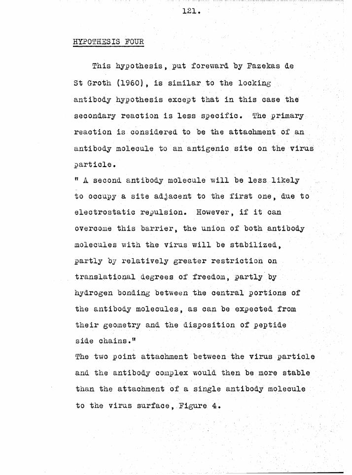
121
HYPOTHESIS FOUR
This hypothesis, pu.t foreward by Fazekas de St G-roth (1960) , is similar to the locking antibody hypothesis except that in this case the secondary reaction is less specific. The primary reaction is considered to be the attachment of an antibody molecule to an antigenic site on the virus particle.” k second antibody molecule will be less likely to occupy a site adjacent to the first one, due to electrostatic repulsion. However, if it can overcome this barrier, the union of both antibody molecules with the virus will be stabilized, partly by relatively greater restriction on translational degrees of freedom, partly by hydrogen bonding between the central portions of the antibody molecules, as can be expected from their geometry and the disposition of peptide side chains.”The two point attachment between the virus particle and the antibody complex would then be more stable tnan the attachment of a single antibody molecule to the virus surface, Figure 4.

122.
Figure 4. Stabilization according to hypothesis four. This hypothesis postulates that stabilization occurs when secondary bonds are formed between two antibody molecules placed adjacent to one another on the surface of the virus particle, This figure is approximately to scale for the attachment of antibody to the surface ofinfluenza virus.

1 Zo.
TESTING THE HYPOTHESES.
Porter (1958) demonstrated that the digestion of rabbit antibody with papain split the molecule into three pieces of approximately equal molecular weight. Although papain digestion completely removed the precipitating activity of the antibody, the digested material could still combine specifically with the antigen, as was witnessed by the fact that the digested antibody would block the precipitation of the antigen by the untreated antibody. Thus Porter concluded that papain digestion split the molecule such that the immunologically active portions were no longer ooth confined to the one molecule. That is, papain digestion artifically produced monovalent antibody. Equilibrium dialysis studies carried out by Uissonoff and woernley (1959) showed that papain digestion had little effect on the capacity of the degraded molecule to bind hapten; thus, the combining sites in the degraded molecule are almost completely intact.
By studying the reactions of papain digested neutralizing antibody, it should be possible to distinguish between some of the above hypotheses.

124
If either hypothesis one or three is correct, papain digested antibody should behave in the sane way as normal antibody, provided the reduction in molecular weight does not affect the neutralizing capacity of the antibody. If hypothesis four is correct, papain digestion may or may not affect the capacity of the antibody to form a stable union with the virus particle; papain digestion reduces the size of the molecule and the shorter molecules may not be able to form secondary oonds with adjacent antibody molecules. The situation is quite clear cut for hypothesis two. In this case stabilization can only occur if the antibody molecule is divalent.
hhJTfLJilZATIQH OF INFLUENZA VIRUS 3Y PAPAIN DIGESTED ANTIBODY.
The X-globulin fraction of a hyperimmune rabbit anti-MEL serum was precipitated with sodium sulphate (Xeckwick, 1940), and the precipitated protein digested with papain according to the method of forter (1958). The digested material was dialyseu against normal saline for 48 hours at 4°C.

125.
T A B L E 1.
The activity of papain digested antibody compared with that of the untreated antibody.
TEST
Ant i -haemagglut$nation
PVR cell agglutination
Neutralization
SERUM TITRES (expressed as dilution of serum)
DIGESTED UNTREATEDANTIBODY ANTIBODY
1:40 1:1,000
<1:2 1:2,000
1:288 1:6,500

126.
The activity of the digested antibody
preparation was compared with a sample of the
untreated serum by titrating both for anti-
haemagglutinin activity (Fazekas de St Groth,
Withe11 and Lafferty, 1958), their ability to
agglutinate P.V.R. cells (Fazekas de St Groth, 1949)
and for their neutralizing activity. The
neutralization test was carried out by mixing
serial two fold dilutions of serum with a constant
amount of virus (10 ID^q ). The serum-virus
mixtures were held at 0°C. for 50 minutes and then
inoculated into plastic trays containing eleven
day old egg pieces. Fach mixture was inoculated
into eight replicate cups. The trays were incubated
at 36°0. for 48 hours and then read for
haemagglutenin production. The results of these
experiments are shown in table 1.9
The agglutination of P.V.R. cells by antibody
is dependant on the divalent nature of the
molecules. Thus trie failure of the digested
material to agglutinate P.V.R. cells indicates
that the molecules are no longer divalent.
However these monovalent antibodies are still
able to neutralize the virus and inhibit
haemagglutination.

12 7.
The neutralization test described above can
he used to assay antibody molecules that are
reversibly attached to the virus particles. The
following experiment was designed to test whether
or not papain digested antibody could form a stable
combination with the virus particle.
A mixture of the undiluted digest material and
an equal volume of MSL virus (approximately 10 ID^/ml.)
was prepared and allowed to react at 0°C. At various
time intervals samples were removed from the reaction
mixture, diluted in chilled S.M. and titrated in
egg pieces for recoverable virus infectivity.
A second mixture consisting of equal volumes of
LH3L virus (approximately 10® ID^/ml.) and the
untreated serum, diluted 1:80 in S.M. , was prepared
and titrated for recoverable virus infectivity at
the same time. The amount of infectious virus
initially present in the mixtures was estimated
by titrating a control mixture, in which the
antiserum was replaced by normal rabbit serum,
at the beginning and end of the experiment. The
control titrations indicated that there was no
significant heat inactivation during the reaction
period. Figure 5 is the plot of the virus survival
against time for the digested and untreated
virus-serum mixture s.

128.
-2 0
-3 0
-4 0 -
TIME IHRSJ
Figure 5. Inactivation of ulEL virus by papain digested antibody (closed circles) and undigested antibody (open circles).
^ l i b r a r t T-t
f /VERStf'*

129.
The mixture containing the digested antibody has approximately three times the serum activity of that containing the untreated serum, as judged by antihaemagglutenin and neutralization titrations (table i). However, whereas the untreated serum rapidly forms a stable combination with the virus particle, the infectivity of the mixture containing the digested antibody is completely recovered on dilution. Thus papain digested antibody can combine reversibly with the virus particle and so neutralize its infectivity, out cannot form a stable union with the virus particle.
These results immediately rule out hypotheses one and three as possible explanations of the mechanism of the stabilization reaction.Hypothesis two, that is, that stabilization is due to the divalent nature of the antibody molecule, is compatible with these results. Hypothesis four is also compatible with these results but only if one postulates tnat the stabilizing bridge can only be formed between that portion of the antibody molecules tnat is split out on papain digestion.

150
THE PROTECTED FRACTION.
The host cell induced protected fraction will vary from zero upwards depending on the cells used to assay the virus infectivity. However this portion of the protected fraction is probably not due to any peculiarity of the combination between the virus and antibody, (see Chapter 4). The reversibly induced protected fraction on the other hand, is due to some abnormality of the reaction between the virus and antibody. This protected fraction might arise in two basically different ways.
A fraction of the antibody molecules present in an antiserum may combine loosely with the virus surface but because of their structure, may be unable to stabilize this union. The presence of monovalent or incomplete antibody in an animalTs serum has been proposed by several workers (Pappenheimer, 1940; Heidelberger and Kendall, 1935). Such antibody could account for the reversibly induced protected fraction if the self-stabilization mechanism were true. Thus if a virus suspension were to react with an antiserum that contained both divalent and monovalent antibody, a proportion

löl
of the virus particles would have their antigenic sites occupied by monovalent antibody. Before the divalent antibody could attach firmly to the surface of these particles some of the protecting antibodies would have to be displaced. Such a reaction sequence could be written,
V * A 7 = ± 7A VIV 4- a tzzz± 7a
Where, in this case, A represents divalent self- stabilizing antibody and a the monovalent protecting antibody.
If such a mechanism is to account for the slow rate of conversion of 7a to 7j, this conversion must be a rate limiting step. That is, slower than the rate of conversion of 7A to 7j_* Whether or not monovalent antibody could account for the protected fraction can be tested by using the digested antibody as a protecting agent. The following experiment was carried out to examine this point.
THE 1RQTBCTI7B EFFECT OF BABAIK DIGESTED MTIBODY
0.1 ml. of a stock preparation of MEL virus was added to 0.9 ml. of the papain digest, the virus titre of the mixture was approximately

132.
TIME IHRSJ
Figure 6. The protective effect of papain digested antibody. The closed circles show the kinetics of the secondary reaction observed when MEL reacts with anti-MEL serum. The open circles show the kinetics observed when the virus has been allowed to react with papain digested antibody before being mixed with the undigested antibody.

10® ID^/ml. This mixture was allowed to react at 0°C. for SO minutes. At the end of this reaction time an equal volume of inactivated serum 1, diluted 1:20 in S.A. was added to the mixture.The final mixture was then held at 0°G. for the remainder of the experiment. Samples were withdrawn from this mixture immediately after mixing and at time intervals up to two hours after mixing. These samples were diluted in chilled S.M. and assayed for recoverable virus infectivity.A parallel experiment was carried out in which the papain digest was replaced by S.L1. The resulting kinetic curves are shown in Figure 6.This figure shows quite clearly that papain digested antibody has a marked protecting effect. Thus, it is possible that the presence of monovalent antibody in an animal’s serum could lead to a reversibly induced protected fraction.
HSTEROGFhFITY OF ANTISERA
If the production of a reversibly induced protected fraction is due to some type of heterogeneity amongst the antibody molecules, one might expect variations in the level of the protected fraction produced when different

IS 4
antisera react with, the same virus stocks.
When discussing tue data of Dul'becco, Vogt and
Strickland, Fazekas de St G-roth and Reid (1958)
pointed out that there were small but significant
changes in the level of the "persistant fraction"
observed when poliovirus and Western equine
encephalitis were neutralized by different sera.
The following experiment was carried out to
determine whether the protected fraction varied
when different sera were used to neutralize the
same preparation of influenza virus. The two
sera used for the experiment were a hyperimmune
rabbit anti-MEL serum (serum 1) and the serum
obtained from a fowl bled seven days after an
intravenous dose of 25,600 agglutinating doses
of MEL virus. Both sera were inactivated by
heating a 1:10 dilution of the sera at 56°C for
50 minutes. The rabbit serum diluted 1:20 in S.M.
and the fowl serum diluted 1:40 in S.M. were then
mixed with an equal volume of influenza virus and
the kinetics of inactivation studied as in
Chapter 4. The results of this experiment are
shown in figure 7.
The projection of the line representing the
rate of inactivation of the protected fraction

135
- I O
- 2 0
- 3 0
TIME CHOURS}
F i g u r e 7 . H e t e r o g e n e i t y of a n t i s e r a .
The k i n e t i c s o f th e s e co n d a ry r e a c t i o n o b se rv e d
when MEL r e a c t s w i th r a b b i t ( c l o s e d c i r c l e s ] and
fowl (ojjen c i r c l e s ) an t i -K S L s e r a .

Ih6
back to the ordinate gives an estimate of the fraction of particles initially in the protected state. This fraction is fortyfold higher for the fowl serum than for the rabbit serum. These results are in agreement with the hypothesis that antisera are heterogeneous, containing what might be called protecting antibodies and inactivating antibodies.
STERIC EFFECTS
The geometry of the combination between the antibody molecule and the virus particle could also contribute to the production of a reversibly induced protected fraction. When a suspension of virus reticles is mixed with a high concentration of antibody, the primary reaction will be the reversible attachment of a large number of antibody molecules to the surface of the virus particle, figure 8. If the stabilization reaction is due to the attachment of one of the free sites of the antibody molecule to a second antigenic site on the virus particle, the crowding of antibody molecules onto the virus surface would sterically hinder the stabilization reaction. Thus one would expect to observe an increase in the level of the

Figure 8. A diagramatic representation of the attachment of antibody to the surface of influenza virus when the virus is mixed with a high concentration of antibody. This figure is approximately to scale.
if/yERStV*

lo8.
protected fraction when the virus reacts with high concentration of antibody.
EFFECT OF THE SERUM CONCENTRATION ON THE LEVEL OF THE PROTECTED FRACTION.
This experiment was carried out with the CL strain of vaccinia virus because the infectivity of this virus can be determined more accurately than the infectivity of a preparation of influenza virus. A rabbit was immunized by inoculating 0.1 ml. of a suspension of the virus (8 * 105 PFXJ/ml.) intradermally into five different sites on the rabbit's bach. Three weeks later another ten 0.1 ml. volumes of the virus were inoculated intradermally. The animal was bled out a week later and the serum obtained inactivated and used in the following experiment.
Two virus-serum mixtures were prepared such that the final dilution of serum in the mixtures was 1:20 and 1 :40, the initial virus titre of the mixtures, as indicated by the titre of a control mixture in which the antibody was replaced by gelatin saline, was 5 * 107PFu/ml. A third virus- serum mixture was prepared by diluting the stock virus 1:10 in undiluted serum; the titre of the

109
TIME
Figure 9. The effect of serum concentration on the level of the protected fraction observed when vaccinia virus reacts with antibody. The closed circles show the kinetics of inactivation when the final dilution of serum is 1:20, the open circles show the kinetics observed when the final serum dilution is 1:40, and the half closed circles the kinetics observed when tne stock virus is diluted 1:10 in undiluted antiserum.

140.
stock virus being such that the initial titre of this mixture was also 5 x lO^PFU/ml. The mixtures were held at room temperature (19°C.) for the duration of the experiment and the kinetics of the secondary reaction followed by diluting samples of the mixtures and assaying on the CALI of eleven day old embryonated eggs (Westwood,
i?Phips and Boulter, 1957). The results of thisuexperiment are shown in figure 9.
When the 1:20 and 1:40 dilutions of antiserum are mixed with the virus there is a phase of rapid inactivation, but in both cases the kinetic curves flatten out at the same level.
However in the case of the concentrated serum the protected fraction is approximately three fold higher.

141.
DISCÜSSIOlf.
The f$ct that papain digested antibody can combine reversibly with the virus particle but is unable to form a stable union, makes it very unlikely that hypotheses one or three will account for the secondary reaction. These results, however, are compatible with the hypothesis that the stabilization is due to the divalent nature of the antibody molecule. Hypothesis four, that is, that the stabilization reaction is the formation of bonds between adjacent antibody molecules, may also be true. However, since papain digestion removes the capacity of antibody to form this stable union, one must assume that in this case, the stabilizing' bonds can only be formed between the portion of the molecule that is split out on papain digestion. Thus in view of the reactions of papain digested antibody one can conclude that hypotheses two and four could offer a likely explanation of the mechanism of the secondary reaction.
There are also some physical data that Dearon the likelihood of these two hypotheses. Bateman, Calkins and Chambers (1941) studied the

142.
increase in optical thickness that occured whenmonolayers of antibody were adsorbed to a surfacelayer of antigen. These authors found that theoptical thickness of the resultant monolayer wasdependant on the concentration of the antibodysolution that was allowed to react with theantigen. When high concentrations of antibodywere used, the optical thickness of the monolayer
owas approximately 250 A. Thus under theseconditions the antibody was probably attached withits major axis normal to the surface. If howevera dilute solution of antiserum was added to theantigen, the increase in optical thickness
oobserved was approximately 55 A. It seemed , therefore, that under these conditions the major axis of the antibody molecule is lying parallel to the surface. Moreover once the antigen surface has been saturated by the addition of dilute antibody, the addition of a concentrated antibody solution will not cause any increase in the optical thickness of the monolayer. Thus antibody molecules that are lying parallel to the antigen surface
must be specifically held in this position.

145
The studies of Ogato et al (1952) indicate that optical thickness, estimated by the Blodgett-Langmuir technique, may not always give a reliable estimate of the actual thickness of the absorbed antioody layer, however the electron microscope studies of Mudd and Anderson (1941) present more concrete evidence relating to the mechanism of attachment of antibody to large antigenic surfaces. These authors investigated the attachment of antibody to bacterial flagella, and came to the conclusion that,"the data then concerning the deposition of antibodies, obtained by electron micrography, suggest that with increasingly strong sensitization the deposition of antibodies may proceed from a patchy distribution (incomplete film) to a thin film corresponding* in order of thickness to the minor axis of the globulin molecule, and with stronger sensitization to a film in which the globulin molecules are radially disposed."
These results indicate that when a large surface of antigenic sites are available, the antibody can combine such that its major axis is perpendicular to the surface or parallel to the surface. However provided the antibody

144
molecules are not hindered by nearby molecules adsorbed to the surface they finally attach in the parallel position. Thus the latter type of combination must be more stable than the former.This evidence and the reactions of papain digested antibody suggest that hypothesis two is the most likely explanation of the stabilization reaction.
Since the level of the reversibly induced protected fraction varies when different sera are mixed with the same virus preparation and titrated in the same assay system, the protected fraction must be due to some form of heterogeneity amongst the antioody molecules. The protecting effect of papain digested antibody shows that the presence of monovalent antioody in a serum could account for the reversibly induced protected fraction. However, although the presence of univalent antibody in animal sera has been suggested, (Heidelberger and Kendall, 19o5; Pappenheimer 1940), monovalent antioody as such has never been isolated and there is still considerable argument as to whether or not it exists (see Kabat 1955). The fact that the rate of neutralization of the artificially produced protected fraction is very much slowerthan the rate observed when the virus is

145.
inactivated, by a "normal" antiserum, may mean that the protecting agent in a "normal" antiserum behaves in a way different from papain digested antibody. However this difference could also be due to the fact that a high concentration of papain digested antibody was added to the virus before the "normal" antiserum was added. Similar kinetics would be observed if the protecting antibody present in antisera could also undergo the secondary reaction but at a much slower rate than "normal" antibody. Thus apart from concluding that antisera are heterogeneous, little can be said about the reactions that lead to the production of this portion of the reversibly induced protected fraction.
One of the predictions that follow from hypothesis two is that the protected fraction should increase when the kinetics of the secondary reaction are studied at very high serum concentrations. The finding that this doee in fact take place is further support for this hypothesis.
Thus when viruses react with specific antisera, three distinct reactions take place.These reactions may oe written as,

146
kl+ A — 1 VA,----Vj a)
k2V + a v.--— " V a ^ -V... (2)
V + P VP V , (3)
Reaction one is the reaction that leads to the formation of a stable union between the virus particle and the antibody molecule. The Va complex most probably represents the attachment of one active site on the antibody molecule to an antigenic site on the virus particle. This combination is then stabilized to give V .. The most likely mechanism by which the stabilization comes about is the attachment of the remaining free active site on the antibody molecule to another antigenic site on the surface of the virus particle, the combination being further stabilized by the formation of secondary bonds between the groupings along the length of the antibody molecule and those on the adjacent surface of the virus particle. If the virus particles are reacted with a high concentration of antiserum the crowding of antibody molecules on the surface of the virus particle may sterically hinder the stabilizationof the loose unions so formed

147
Tiie second, reaction is the reaction that leads to the production of the reversibly protected fraction. This comes about because a fraction of the antibody molecules in an animal’s serum are of the a_type. These molecules are able to form a loose union with the virus particle but either cannot stabilize this union or if they can, do so at a much slower rate than A-type molecules.That is k-j. is much larger than kg. If the a-type molecules cannot stabilize the Va union then the reaction,
Va V-j-is probably a displacement reaction in which a is replaced by A to give Va which is then converted to Vj.
The third reaction represents the formation of the host cell induced protected fraction. The P type molecules differ from the neutralizing- antibodies in that they cannot prevent the virus particles from entering and infecting the host cells. The magnitude of host cell induced protected fraction can show i±uite a marked variation when the same virus-serum preparation is titrated in different cell systems, (Chapter 4).

148
SulüvLvRY Rill) COHCLUSIOflS
1) . Papain digested, antibody will combine reversibly with influenza virus and so neutralize its infectivity but will not form a stable union with the virus particle.
2) . The reversibly induced protected fraction is due in part to the heterogeneity of the antibody molecules in an animal's serum. However when the virus is allowed to react with very high concentrations of antiserum the protected fraction increases. This increase is probably due to the geometry of the union formed between the antibody and the virus particles under these conditions.
3) . When a relatively low concentration of virus particles react with specific antibody, the primary reaction is probably the formation of a reversible union between one active site on the antibody molecule and an antigenic site on the virus particle. The union so formed may then be stabilized by the antibody molecule attaching to
another antigenic site on the virus particle.
This would bring the length of the antibody molecule
into close contact with the virus surface and allow

149.
the formation of weak bonds such as van der Waals bonds between groupings along- the length of the antibody molecule and those on the surface of the virus particle.
4). Papain digested antibody will behave as a protecting antibody. Thus demonstrating that monovalent antibody, if present in an animal’s serum, could account for the reversibly induced protected fraction. However there is insufficient evidence to allow any definite conclusions to be
drawn about the reactions that lead to the production of this protected fraction.

C H A P T E R
S I X
QUARTAL REUTRALIZATIOR TESTS.
' r LI 8 ft A R Y
E K S l^

150
INTRODUCTION.
The work described, in the previous chapters has been concerned with the mechanism of the neutralization reaction and although there is not sufficient evidence to describe the reaction mechanism completely and unequivocally, sufficient conclusions can be drawn to allow a quantitative treatment of the dose-response relationship in quantal neutralization tests. A quantal assay is an assay carried out in a system that can only give an all or nothing response. Although the quantal test is probably the most commonly used neutralization test, it is also true to say that it is tne least understood of all the virus infectivity or infectivity related titrations.In a recent review, Horsfall (1957) discussed the various factors that influence the outcome of neutralization tests and came to the conclusion that;"For the investigator concerned with the measurement of antibody concentration, the practical difficulties introduced by tne effects of slope of the neutralization line, host susceptibility, route of inoculation, and amount of virus used are these:

151.
Neutralizing antibodies may oe found with one set of conditions, but not with another.A high neutralizing titre may emerge under some conditions and a low titre under others. With two sera from the same individual, a large
increase in the amount of ant foody may oe indicated under some conditions and a small increase under other conditions. Comparison of antibody levels under different experimental conditions is not
Ifeasible without extensive and precise information
about the effect of all the variables used,” and in conclusion he goes on to say — "Important and informative though the neutralization test is in
studies on virus infections and diseases, it should be supplanted whenever possible by an in vitro test if this will yield equivalent information."
One must agree witn Horsfall in his criticism
of what might be termed the classical neutralization test, (see Chapter 2). However, whether the neutralization test should be supplanted whenever
possible by in vitro tests is a point of argument.
The purpose of the investigations described in
this chapter is to demonstrate that the difficulties
discussed by Horsfall are not due to any inherent

152
inadequacey of the neutralization test itself
out rather to the awkward use of a very sensitive
technique.
THEORETICAL.
The reaction between an antigenic site on the
surface of influenza virus and antibody may be
written as follows.
kiV + A ; = * V-j-
k2V * a ----Va ,____ 'J1
».here A and a are the two types of neutralizing
antibody present in an animalfs serum (see Chapter 5)
and Vj represents the stable virus-antibody union.
Whether Va is converted directly to Vj or whether
V-£ can only be produced by a displacement of a by
A is not known. However for the purpose of the
present discussion it is sufficient to say that
Vj can be formed from Ya but at a rate which is
very much slower than the rate of formation of
Y x from YA, that is, k^.
Reactions leading to the formation of a host
cell induced protected fraction have not been
included in the above reaction scheme since the

153.
available evidence (see Chapter 4) indicates that when the neutralization of influenza virus is assayed in egg pieces, the host cell induced protected fraction is either very low, less than 0.Q1/Ö of the particles, or nonexistent.
It is convenient to consider first the situation that arises when k^ and kg are both zero. That is the reaction between influenza virus and papain digested antibody. The reaction between papain digested antibody and an antigenic site on the surface of influenza virus may be written as,
V +K
A ---pap — pap
In this case K is most probably an average equilibrium constant for the reaction, since it is very likely that will consist of a groupof molecules with varying affinities for antigenic sites on the surface of the virus particle.
In 1958 Fazekas de St.Groth, Y/atson and Reid put foreward a mathematical model of virus neutralization based on a reversible reaction mechanism. In their analysis they set up
equations to describe situations of varying degrees
of complexity. Jerne (1956) produced experimental evidence that indicated that one antibody

154
molecule reversibly bound to a critical site on bacteriophage T4 was sufficient to neutralize its infectivity. Since there is no evidence to the contrary, the assumption that one antibody molecule reversibly attached to a critical site on the surface of the influenza virus particle will be made for the purpose of the following discussion.
The following two equations have been derived by Fazehas de St G-roth, Watson and Reid.The equilibrium, expression for the reaction is,
yK = [SVQ (1 - n) - yl (A-y) (1)
where K is the equilibrium constant, y the concentration of formed antigen-antibody complexes,3 the number of antigenic sites per virus particle, Vj the initial concentration of infective and n Vq the initial concentration of noninfective particles, and A the initial concentration of antibody molecules; concentrations are expressed as molecules /ml.
If a single antibody molecule bound to a critical site neutralizes the infectivity of a virus particle, the fraction of surviving particles in a virus serum mixture will be given by.

155
Y-To
1y
S' "V0 (1-n) .c
(2 )
Where Y is the concentration of surviving infective particles, and c the number of critical sites per virus particle. These two equations can be used to derive an expression for the quantal dose response relationship in the region of low virus inputs.
In a quantal test Y, by definition, has a value of unity. Thus for a quantal test equation (2) becomes.
i l y"Tq l1 “ O 0 (1-n) (S)
Taking logarithms of both sides of equation (3) we have,
\ ylog v0 = — o log [ 1 - s v0 (1_n)
From equation (1) it can be shown that,
S V0 (1-n) Ay ^
S v0 (l-nl-t-A+i (5)

156
T his a p p r o x i m a t i o n h a s an e r r o r o f 1$ p r o v i d e d
S v0
S u b s t i t u t i n g f o r y i n e q u a t i o n (4) we o b t a i n ,
l o g Vq ^ - c l o gA
S Vn (1 -n ) * A + TK
( 6 )
At low v i r u s i n p u t s t h e v a lu e o f S Vn (1 -n ) w i l l
be s m a l l e r t h a n — by s e v e r a l o r d e r s o f m a g n i t u d e ,K
and can t h e r e f o r e s a f e l y be n e g l e c t e d . T h is
l e a d s t o t h e e q u a t i o n
l o g VQ c l o g (1+ AK). (7)
Expanding th e t e rm l o g (1+AK) we have
l o g VQ * o l o g I AK - i (AK)2 + I ( A K ) - ------ j l o g e
( 8 )
As lo n g a s A 4 — , t h e f i r s t t e rm o f t h e s e r i e s K
o v e r e s t i m a t e s lo g (1-t-AK) by l e s s t h a n 5$ . I n
th e r e g i o n o f low v i r u s i n p u t s AK i s s e v e r a l o r d e r s
o f m ag n i tu d e l e s s t h a n u n i t y , and t h u s t h e h i g h e r
powers o f AK can be n e g l e c t e d .
The a p p r o x i m a t i o n t h e n becom es ,
l o g Vq cs cAK . l o g e (9)
a more c o n v e n i e n t form o f e q u a t i o n 9 i s o b t a i n e d
by t a k i n g l o g a r i t h m s o f b o th s i d e s .{ * L I B R A R Y h s
ERSlV*

157
Thus,log (log Vq ) log A + log (cK log e ) (10)
Since the expression on the LHS of equation (10) is derived from the ratio of the concentration of free to total virus, equation (10) will still hold if Vq is measured in ID^q , provided one neutralizes to the ID^ end point.
In equation (10) the antibody concentration refers to the final concentration in the test system. However the titre of an antiserum is often quoted as the dilution of serum in the reaction mixture, which when inoculated into the host system behaves as one ID^q . Since the inoculation often involves dilution of the reaction mixture the apparent potency of the serum will vary when the same material is estimated in host systems that involve different dilution factors. This effect can be overcome by adding a correction factor to equation (10). Equation (1) can be written in the form,
\ Occupied Sites j M
Where L j- signifies concentration in thereaction mixture

158.
If however daring the assay, this mixture isdilated by a factor — , the equilibrium ischanged such that
K -^Occupied sites . 1
Free sites J . r.l I Free antibody M d
That is:
K -
^Occupied sitesj MjFree sites]— jFree antibody]
* d
and,
K _ d
'^Occupied sites]
iFree sites i, tFree antibody 1Thus if the concentrations are refered to
the reaction mixture, K in equation (10) mustK
oe replaced by & . That is, equation (10) will become,
( &C log e \log (log VQ ) log k d + log V d J
where A-p now refers to the concentration of antibody in the reaction tube.
Equation (11) allows the definition of a
useful unit of antibody concentration. When V q equals 10, that is, when the concentration of antibody in the reaction mixture is such that
(11)
rH I

159.
10 I1>5 q are neutralized to the 1 11)50 ieve1»1log — -log (KC log e) + log d
that is ,&'J<1 ~ (KC log e) _1 (12)
where A^ is the particular value of A^ for which log (log Vq ) - 0.
If tne unit of antibody concentration is defined as (KC log e)” antibody molecules per ml. then when log (log Vq ) is zero, the concentration of antibody in the reaction tube will be d concentration units. Thus if the dilution of serum in the reaction tube is 1Q~X , the antiserum must have a concentration of d. 10 units.
This unit of potency is a functional unit since the actual number of antibody molecules in a solution of unit concentration depends on one value of K for the particular antiserum and tne value of c for the virus under test.Thus this unit is a measure of the neutralizing potency of the serum and does not estimate the absolute number of antibody molecules in an animal*s serum. 3ueh an estimate could, however, be derived from the neutralizing potency ifthe value of c and K were known

160.
The neutralizing potency of a serum can be determined, from the knowledge of the dilution of serum that will neutralize a known dose of virus to the 1 TDejQ level, provided this dose is such tnat equation (11) is a valid approximation. Equation (11) may be written in the form,
log AR — log (log Vq ) — log d ~ log (Kc log e)
The value of the LHS of the above equation is the logarithm of the dilution of serum that has unit concentration. Thus the neutralizing potency of the serum is given by,
Neutralizing potency concentration units.
-[log Ar -log (log VQ) -log dj
TKh EFFECT OF THE SECONDARY REACTION.
The above equations will hold provided the reaction is reversible. However, in the case of "normal” antibody, the reversible combination between virus and antibody is followed by a secondary stabilizing reaction. This reaction may also be a reversible reaction, but in this case theposition of equilibrium favours the formation of the staole complex.

161
The rate of the secondary reaction is very much slower than the rate of the primary reaction (see Chapter 3), thus the primary equilibrium can be established before the secondary reaction nas proceeded to any significant extent. This accounts for the finding that the neutralizing titres of a group of serum dilutions as estimated by a quantantal neutralization test, are the same when the reaction time at 0°C is varied from 10 to 160 minutes (table 1 Chapter 2). Thus one would expect the above equations to approximate closely the behaviour of ’’normal" antioody so long as the reaction time is not extended. If however, the reagents are allowed to react for an extended time before inoculation into the assay system, the secondary reaction will have had time to proceed to a significant extent, and since this reaction is the formation of a more stable union between the virus and antioody, its effect will be to increase the apparent potency of a given dilution of antiserum.
The effect of the secondary reaction can
best be considered as follows

162
As was pointed, out at the beginning of this
chapter, the reaction between influenza virus
sites and. normal antioody may be written,
When a mixture of virus and. antibody is prepared
the equilibrium between V, A, a, VA and Va is
established, and if the reaction period is short,
equation (11) will describe to a close approximation,
the oehaviour of the dose response curve. If
the reaction time is extended a fraction of the
VA complexes will be converted to the V-j- form
at a rate k- . When however, the complexes
remaining are predominantly of the Va type, the
rate of conversion to Vj will fall since the rate
constant kg now controls the rate of formation of
Vj (kj_ > kg) . A consequence of the conversion of
VA to Vj will be a shift in the position of
equilibrium, and the rate at which the position
of equilibrium shifts will be controlled at first
by k^. However after a longer reaction period kg
will control the rate at which the equilibrium
moves in the direction of VA. Thus causing a
marked slowing down of the rate at which tl

löo
equilibrium shifts. Due to the fact that the position of equilibrium is moving very slowly when the reaction is controlled by , even after very long reaction times a fraction of the antibody-virus site unions will still be in the Va form. Thus the reaction will qualitatively behave as a reversible reaction; if the reaction mixture is diluted a fraction of the virus particles will reactivate. However since all the neutralized particles will not dissociate on dilution this fraction will be less than would be expected if the reaction were completely reversible. Also the shift in the position of equilibrium brought about Dy the secondary reaction will mean that more virus particles will be neutralized oy a given amount of serum. Thus although equation (11) will still describe the dose response curve, this shift in the position of equilibrium will show itself as an apparent increase in tne value of the equilibrium constant.

164
EkPERIIIEETAL.
The f o l l o w i n g work i s a d e s c r i p t i o n o f
e x p e r im e n t s c a r r i e d , o a t to t e s t t h e v a l i d i t y o f
th e above t h e o r y .
TILS BEHAVIOUR OF "RChhlAL" ANTIBODY----------
TEE DOSE RESPONSE CURVE.
D i l u t i o n s o f MEL (A) and LEE (B) v i r u s were
made up i n c h i l l e d 3.M. and mixed w i t h e q u a l
volumes o f d i l u t i o n s of s p e c i f i c a n t i s e r a . The
m i x t u r e s were h e l d f o r 30 m in u te s a t 0 ° C . , and
t h e n 0 .0 5 m l . o f e ac h was adued t o a s e t o f t e n
cups c o n t a i n i n g th e h o s t t i s s u e . The t r a y s were
mounted on th e s h a k in g machine and i n c u b a t e d f o r
two days a t b>6°C. F i n a l l y , t h e p i e c e s o f t i s s u eCxfid
were removed one drop o f a 5$ fowl r e d c e l l
s u s p e n s i o n added t o each cup and a l l o w e d to s e t t l e .
A g g l u t i n a t i o n of th e c e l l s was t a k e n as a s i g n
o f i n f e c t i o n , and t h e end p o i n t s were c a l c u l a t e d
by t h e method o f Reed and Muench (1 9 3 8 ) . The
r e s u l t s o f t h i s e x p e r im e n t a r e p l o t t e d i n
F i g u r e 1; th e f i n a l c o n c e n t r a t i o n of a n t i b o d y
( e x p r e s s e d a s th e d i l u t i o n o f serum) i s shown on
the a b s c i s s a , and th e number o f ID^q n e u t r a l i z e d
on th e o r d i n a t e .

165
o i -
-40-20-30-40
< 4 -
_ l O -
- 4-5 - 3-5
LOG SERUM CONC LOG SERUM CONC
F i g u r e 1 . The n e u t r a l i z a t i o n o f th e
i n f e c t i v i t y o f two s t r a i n s o f i n f l u e n z a v i r u s
by t h e i r s p e c i f i c a n t i s e r a , p l o t t e d a s ( l o g Vq , l o g A).
A, anti-MEL fowl serum; c e n t r a l s l o p e , 1 0 ^ * ° ° .
B y a n t i-ltEL r a b b i t serum; c e n t r a l s l o p e ,
C, a n t i - L E E fowl serum; c e n t r a l s l o p e , 1 0 ^ * ^ .
D, a n t i - L E E r a b b i t serum; c e n t r a l s l o p e , i q 4 * 4 2 #

166
When plotted as log serum concentration against log ID^q neutralized the dose response curve is sigmoid. This finding contrasts with the results of Tyrell and Horsfall (195b) , and Clark and Tyrell (1S59). These authors considered that there was a straight line relationship between the serum concentration and the amount of virus neutralized when the results were plotted in this way. Indeed, the central region of the dose response curve does approximate to a straight line, and the above authors confined their investigations to this region. However the fact that the dose response curve is not a straight line is of considerable theoretical importance. The sloge of the central portion of the dose response carve also varies when different sera are titrated against the same virus stocks.This phenomenon will be discussed in more detail in a later section of this chapter. If the above theoretical treatment is correct, in the region of low virus inputs, the log ID^q neutralized should be exponentially related to the log of the serum concentration (equation 9). Thus if log (log Vq ) is plotted against log A, where Vg is the virus input and A the antibody concentration, (in practice expressed as the dilution of serum),

167
oOduo
>oodoo
Figure £. The neutralization of the
infectivity of two strains of influenza virus by
their specific antisera, plotted as jjlog(log Vq J, log Aj
A, anti-MEL fowl serum.
B, anti-MEL rabbit serum.
C, anti-LEE fowl serum.
D, anti-LEE rabbit serum.

168.
in the region of low virus inputs the dose response relationship should he a straight line with unit slope. In figure 2 the results of the above experiment are plotted as log (log V ) log A.
This figure shows quite clearly that for 4 5inputs up to 10 or 10 ID^q , equation 10 is a
good approximation to the dose response curve.In the above experiment the virus-serum
mixtures were inoculated after a reaction period of 50 minutes at 0°C. That is, before the secondary reaction had made any significant contrioution to the neutralization (see table 1 Chapter 2). If however the virus-serum mixtures are allowed to react for a much longer period of time before inoculation into the host system, one would expect to observe the effect of the secondary reaction on the dose response relationship. As was pointed out in the theoretical section, the secondary reaction would bring about a shift in the position of the primary equilibrium thus leading to an apparent increase in the value of the equilibrium constant. This apparent increase would have two effects on the dose response curve. Firstly the straight line portion of the log (log Vh ), log A plot would be shifted along

169.
-40 -3 5 -30 LOG A
Figure 3. The effect of the reaction time on the dose response curve, plotted as [log (log Vq ) , log . The open circles show the dose response curve obtained after a reaction period of 30 minutes at 0°G. The closed circles show the dose response curve obtained after a reaction period of 5 hours at 0°C.

170.
the abscissa such that a given concentration of antibody would now neutralize more virus, and secondly the range of virus inputs over which the approximate equation 10 holds will be reduced since this approximation only holds provided SVQ(l-n) < i .
EFFECT OF REACTION TIME OH TEE DOSE RESPONSE CURVE.
In the following experiment, dilutions of anti-lvlEL antiserum were mixed with equal volumes of stock virus dilutions and allowed to react at 0°C. Llaterial from the reaction tubes was then inoculated into plastic trays containing eleven day old egg pieces, 30 minutes and five hours after mixing. The trays were incubated and the amount of virus neutralized calculated as in the previous experiment. The dose response curves obtained after reaction times of 30 minutes and five hours are shown in figure 3. Clearly the dose response curve behaves as predicted.
Horsfall (1957), when discussing the merits of the neutralization test, pointed out that the slope of the dose response curve (plotted as log VJ, log A) varies when different sera are tested in the same host system, or when the same sera are tested in different host systems,

171.
LOG A
Figure 4. The effect of the reaction time on the dose response curve, plotted as (log Vq , log A). The open circles show the dose response curve obtained after a reaction period of 30 minutes at 0°C. The closed circles show the dose response curve obtained after a reaction period of 5 hoursat 0°C

172.
(see figure 1). Neutralizing litres can tie compared, therefore only by ignoring a basic property of the reaction; moreover, if this property is recognised., no comparison can be made at all. The above experiment provides a clue as to how this effect may come about. In figure 4 th e results of this experiment are plotted as log Vq , log A. The central slope of the dose response curve is much flatter if the virus-serum mixtures are inoculated after a reaction time of five hours than if the same mixtures are inoculated after a thirty minute reaction period. This flattening of the slope is due to the fact that the dose response curve deviates from its exponential course at lower virus inputs wnen the reaction mixtures have been allowed to react for an extended period of time, in this case the effect is due to an apparent increase in the value of K. However, since equation 10 only holds provided SVq (l-n)<i, a similar effect would be observed if the same serum is titrated with virus stocks that vary in the value of n, that is, the proportion of noninfectious particles in the
virus preparation. As the value of n is increased the value of Vq at which the approximation breaks down will correspondingly decrease. This effect is

174.
-4 5 -4 0 - 3-5 -3 0LOG A
Figure 5. The effect of noninfectious virus on the dose response curve, plotted as (log Vq , log A). The closed circles show the dose response curve obtained with stoch virus and the open circles the dose response curve obtained 'when noninfectious particles are added to the stock virus.

174.
illustrated in the following experiment.
THE EFFECT OF LOHIhFECTIOUS VIRUS.
An amount of ultraviolet inactivated MEL virus was mixed with a preparation of active virus such that, if the stock preparation was considered to consist only of infectious virus, that is the value of n for the stock virus is unity, then the second preparation would have a value of n equal to 99. Serial dilutions of each of these preparations were then mixed with an eequal volume of dilutions of a rabbit anti-MEL serum, allowed to react for thirty minutes at 0°C and then inoculated into eleven day egg pieces. The trays were read after an incubation period of 48 hours at 36°C.
In figure 5 the results of this experiment are plotted as log Vq , log A. Clearly when the value of n is increased the central slope shows a marked decline. This experiment provides an explanation for the change in slope observed when an antiserum is titrated against the same virus stock in different host systems, (Burnet, 1943; Tyrell and Horsfall, 1953). In this case the value of n for the particular virus stock is dependant on the host system in which the virus is titrated. Thus the slope will be steepest

175.
- 3.5- 4 0 LOG A
Figure 6. The effect of noninfections virus on the dose response curve , plotted as l^log (log Vq ), log A^. The closed circles show the dose response curve obtained with stoch virus and the open circles the dose response curve obtained when noninfectious particles are addedto the stock virus

176.
when the serum is titrated in the host system which gives the smallest value of n. The results of fyrell and Horsfall (1953) show that this is in fact the case.
It will be noticed that n does not appear in equation 10. Thus in the region where this approximation holds one would not expect the presence of noninfectious virus to have an effect on the log (log Vq ), log A dose response relationship other than to decrease the range of virus inputs over which equation 10 would be a valid approximation. Figure 6 shows that this is in fact the case.In tnis figure the above results are plotted as log (log Vq ), log A. In the linear region of the dose response curve the value of n does not have any significant effect on tne outcome of the neutralization test.
THE EFFECT OF TED TEST VOLUME,
Results presented in Chapter Z show that the test volume has a marked effect on the outcome of neutralization tests. This effect is due to the dissociation that occurs when the reaction mixture is inoculated into the test system. Thus if neutralization tests carried out in assay systems

177
of different volumes are to be compared, one must make allowance for this effect. If the dilution of serum that neutralizes a given amount of antibody is refered to the dilution in the reaction tube, then equation 11 describes the dose response curve in the region of low virus inputs. This equation may be written in the form,
log (log V0) ^ log A h +■ log (KC log e) - log d.(IS)
The plot of log (log Vq ) against log Ag, will be a straight line with unit slope. However the position of this line relative to the axes will depend on the value of log (KC log e) - log a.Thus if a set of virus-serum mixtures are inoculated into host systems of different volume the position of the dose response curve will vary according to the value of log d; under these conditions log (KC log e) will be a constant.
This effect was investigated in the following experiment. Mixtures of MEL virus and its specific antiserum were prepared and allowed to react at 0°C for thirty minutes. At the end of this reaction period 0.05 ml. of the mixtures was inoculated into eleven day egg pieces suspended in 0.5, 1.0 and 2.0 ml. volumes of S.M. The trays

178.
d- 0-4
- 3 5 - 3 0 -2 5 -2 0
Figure 7. The effect of the test volume on the dose response curve, plotted as [log (log Vj), log . The closed triangles show the dose response curve obtained when the test volume is 0.30 ml. The open circles shOYj the dose response curve obtained when the test volume is 1.00 ml., and the closed circles that obtained when the test volume is 2.00 ml.

179
were then incubated and read in the standard
manner. Figure 7 is the plot of log (log Vq ) against log A-.
This figure shows that the dose response
curve behaves qualitatively as predicted. However
in the larger test volumes the shift of the dose
response curve is less than that predicted by the
tneory. This effect is best demonstrated in the
following way.
When log (log Vq) - 0, equation 13 becomes,
log ~ log d - log (KC log e)
Where is the particular concentration of
antiserum at which log (log Vq ) is zero. That is,
the concentration of antiserum at the point where
the dose response curve cuts the abscissa.
If log Al is plotted against log d the points
should fall on a straight line with unit slope.
In figure 8 the results of the previous experiment
are plotted as log A^, log d. The value of log Ai
for each test volume was calculated from the points
along the linear section of the dose response curve ,
Figure 8 shoves a plot of log Ag against l0g a .
the limits on the points are 9Op confidence
limits and the oroken line is a line of unit slope.

180
~ 3-6 - 3'4 “ 3-2 -3 0 - 2-8LOG a'r
Figure 8. The effect of the test volume. The plot of log A_*t against log d. The limits on the points show the 9$j{ confidence interval for the calculation of log A^ and the broken line isa line of unit slope

181.
It can tie seen from this figure that as the value of d increases there is a deviation from the theoretical relationship. The deviation is such that less of the virus antibody complexes dissociate when the reaction mixtures are inoculated into large volumes than would he predicted by equation 13. This effect is most probably due to the secondary reaction. Equation 13 is an accurate description of the reaction betv/een virus and antibody, provided the secondary reaction is not making a significant contribution to the neutralization.That this is the case, after a thirty minute reaction period at 0°C, is illustrated by the fact that neutralization titres estimated after reaction periods ranging from 10 to 160 minutes, are not significantly different but show an increase when determined after a five hour reaction period.Sven so the secondary reaction may still account for the observed results.
In this experiment, after a reaction period of thirty minutes at 0°C. the reaction mixtures are inoculated into the medium containing the host
tissue; the temperature of this medium is 36°C.
When this is done the new position of equilibrium
will be approached at a rate controlled by the rate

182
of the dissociation reaction, and the larger the value of d the longer will he the time taken to reach the new equilibrium position. The results of Dulbecco, Vogt and Strickland (1956), show that in the case of the neutralization of Western equine encephalitis virus by rabbit antibody, the rate of the secondary reaction increases approximately four fold for every 10°G. rise in temperature, and it is not unlikely that the reaction between influenza virus and antibody will have a similar temperature dependance. If this is the case, inoculation of the reaction mixture into the assay system will greatly increase the rate of the secondary reaction. Thus there will be a competition between the primary reaction, tending to establish a new position of equilibrium, and the secondary reaction tending to stabilize the combination formed in the reaction tube.
Increasing the test volume will also increase the time that any free virus is susceptible to antiDOdy activity since its removal from the medium by the tissue would be determined by its rate of diffusion. Thus the overall effect of the
dilution involved in inoculating the reaction
mixtures into the test system would be to increase

183.
the likelihood, of the secondary reaction contributing to the neutralization. Although this effect must take place to some extent whenever the reaction mixture is diluted on inoculation into the assay system, figure 8 shows that the secondary reaction will only make a significant contribution to the neutralization when the value of d is greater than 20. Thus for values of d less than 20 equation 13 describes the effect of the test volume on the position of the dose response curve within the limits of accuracy of the test.
THIS BEHAVIOUR OF lAPAlh DIGESTED AUTI30DY.
The behaviour of papain digested antibody provides a further check on the theoretical treatment. Since papain digestion prevents the antibody from forming a firm union with the virus particle, equation 10 should closely approximate the dose response curve in the region of low virus inputs, regardless of whether the virus and digested antibody are allowed to react for a short or a very long period of time.

184
9> o- 6
0-4
- 2 5 - 2 0 - 1-5
Figure 9* The dose response curve obtained with papain digested antibody, plotted as [log (log V0) , log A].

185.
THE DOSE RESPONSE CURVE*
The digested antibody was prepared as described in Chapter 5. Dilations of MEL virus were mixed with equal volumes of dilutions of the digested antibody and allowed to react at 0°C. for thirty minutes. 0.05 ml. of the mixtures were then inoculated into ten replicate cups of plastic trays containing eleven day old egg pieces.The trays were then incubated and read in the standard manner. Figure 9 shows the resulting dose response curve plotted as log (log Vq) against log A.
The dose response curve is identical with that found when normal antibody reacts with influenza virus. In the region of low virus inputs the points fall on a straight line with unit slope but at high virus inputs, the dose response curve shows the expected marked deviation from this straight line relationship.
EFFECT OF THE REACTION TELE OH THE POSITION OF THE DOSE RESPONSE CURVE.
Papain digested antibody should differ from normal antibody in that the position of the dos«
length of tne reaction period. Thus there shouldresponse curve should not be affected by the

186
I—I
mhiPP<EH
<H •O
'Öß oO P •H *H -P P Cd Ö ß <
•Ha nqPh 00p0
Q
0Pp
p00bO
•HQ
0PpChO
Po0
«M«HW
ßcd0Vr-H
0 0P PEh P
*bOß
N•HrHcdPhp
0 p •H
t>>Po *ß cJ
. 0 ß3 p q 0 o Ü
fc Ph
ß•H ß cö O Q,
CÖ 0 Pha*H PiEH O
ß i>>O Ü •H ß P 0 ü P Cd O 0 Oh
PS ßO •H P cd co •H r—Icd h p pi 0
' dbQ0rH1
PS<S
bOoH
PSc
bO O
•—i
>bJDOHbOOrH
bOorH
tN-!r\CO
OI—I
OO CN lT \ IfV
C\JorH
C\]oCO
1
J -co•rH!
C\JoH
vOlrv
o nrHCOa
levco•rHI
COp -
ov o
•CO
ovO
vOlev
COOrH
oOlev•
COOrH
COlevCOorH
COOv•rHI
oCO•rHI
levlev
O -rHCO
I
OvCO•rHI
rHp i
c olev•
CO
COlev
0a•HP
ßO
•HPÜ00PS
CO0P3ß•HaoCO
COPhßoP
lev
V) p •H
§0ßoÜ
r oP bO
0rH1
O>bOOrH
bO0rH1
PS<bO0
I—Ii—a
1or~lil
Oß0
PO
Ph
bOß•HIVi•HrHcdPhPß0Is:

187.
be no significant difference between the neutralizing potency of the serum determined after reaction periods of SO minutes and five hours.
In the following experiment two known doses of virus were mixed with equal volumes of dilutions of papain digested antibody. After a reaction period of 30 minutes or five hours at 0°G., the neutralizing titre against each dose of virus was determined by inoculating 0.05 ml. of the reaction mixtures into eight replicate cups of plastic trays containing eleven day egg pieces suspended in 0.3 ml. of S.M. The trays were then incubated and read in the standard manner. The results of this experiment are shown in table 1.From this table it can be seen that the neutralizing potency of the digested antibody is the same whether tested after a reaction period of 30 minutes or five hours.
DISCUSSION.
The major drawback of the orthodox neutralization test is the fact that the slope and position of the dose response "line" vary in an unpredictable manner when the same antiserum is titrated in different host systems. Even when the same serum

188.
is titrated against different virus stocks in the same host system the slope of the dose response "line" may vary; the more noninfectious virus there is in a preparation the flatter will be the slope of the dose response "line”. However, the results presented in this chapter show that these difficulties can be overcome.
When papain digested antibody is used as the neutralizing antibody the equations derived from the mathematical model of virus neutralization, proposed by Fazekas de St G-roth, Watson and Reid (1958), accurately describe the dose response curve, regardless of whether the reaction mixture is left to react for a short or a prolonged period of time. However in the case of "normal” antibody, allowance must be made for the effect of the secondary reaction. When the reactants are allowed to react at 0°G. for a prolonged period of time before inoculation into the host system, the position of the lower portion of the dose response curve shifts in such a manner that the antiserum is more potentthan it would be if tested after a short reaction period at 0°G. However the results presented in table 2 of Chapter 1 indicate that within a

189
reaction period, of 10 to 80 minutes at 0°C., the amount of virus neutralized, by a given dilution of antiserum is constant. These findings are compatiole with the proposed reaction mechanism discussed in the previous chapter. That is, when "normal" antibody reacts with influenza virus there is a rapid reaction which is the formation of a readily reversible union between the virus particle and the antioody molecule, and this reaction is followed by a relatively slow stabilization reaction. The fact that the neutralizing titres are not significantly different over a 10 to 80 minute reaction period at 0°C. indicates that within this time the primary equilibrium has been established and the secondary reaction has not proceeded to a sufficient extent to significently change this position of equilibrium. Thus if a standard reaction period of 30 minutes at 0°C. is adopted the equations derived in the theoretical section can be applied to the reaction between "normal" antibody and influenza virus.
Although the secondary reaction leads to the production of a much more stable union between thevirus particle and the antibody molecule, this reaction is most probably still reversible;

190.
Mandel (1958) has been able to demonstrate that poliovirus that has antibody firmly bound to its surface can be readily dissociated by dilution in a medium of low pH. Thus if the secondary reaction were allowed to run to completion a new equilibrium would be established and the above equations could again be applied to the system. However from the point of view of carrying out quantitative neutralization tests it is a technically simpler matter to carry out antibody estimations when the secondary reaction is not contributing to the neutralization.Firstly, the secondary reaction takes a very long time to reach its equilibrium position; kinetic experiments carried out with influenza viruses show that even after a four hour reaction period at 0°C. there are no indications of the reaction coming to equilibrium. Secondly the rate of reversal of the secondary reaction is extremely slow (see Chapter 3). Thus if the position of equilibrium is altered by diluting the reaction mixture during the assay proceedure, the amount of virus that reactivates within a sufficient time to score as infectious will be very much lessthan the amount that must reactivate to establish

191.
the new position of equilibrium. Thirdly, sincethe secondary reaction is the formation of arelatively firm union between the virus andantibody, the equilibrium constant for thereaction will be very much larger than that forthe primary equilibrium. Thus the range of virusinputs over which the approximate equations holdwill be correspondingly decreased. Thus ifquantitative estimations of antibody activityare being carried out it is sounder to carry outneutralization tests after a reaction period of
ooQ minutes at 0 C. However, if one is only interested in whether or not there is antibody present in an animalTs serum, the test is more sensitive if carried out after a prolonged reaction time. This latter proceedure is the oasis of the immune inactivation test described by G-ard (1957).
Before attempting to use a particular assay system for quantitative neutralization tests one must determine whether or not the assay system is suitable. A suitable assay system must fulfill two conditions. First the volume of the test system must be known and secondly equation 10 must hold over a reasonably wide range of virus inputs. This second condition will not hold for all host systems.

192.
The value of n will vary when the same virus
material is titrated in different host systems,
and the larger the value of n the smaller will
be the range of virus inputs over which equation 10
will hold. However if neutralization tests are
carried out in assay systems that fulfill these
conditions, then the dose response relationship
(plotted as log (log Vq ), log A) will be a
straight line with unit slope, in the region of
low virus inputs, and the position of this line
will be defined by the neutralizing potency of
the serum and the volume of the test system,
provided the dilution factor involved in the
inoculation is not large. When the dilution
factor is less than l/20 the equation 11 accurately
describes the effect of dilution on the position
of the dose response curve. However if the
dilution factor is greater than l/20, the
secondary reaction makes a significant contribution
to the neutralization and more virus is neutralized
than would be predicted from equation 11, (see figure 8).
hue to the fact that the amount of virus
neutralized by a given concentration of serum is
exponentially related to the concentration of
serum, equation 9, the most accurate method of

195.
estimating serum activity is to titrate a constant amount of virus against varying dilutions of serum. In practice a set of serum dilutions are titrated against several virus levels in the range where the log (log Vq ), log A plot is linear.The neutralizing potency of the serum can then he estimated from each titration by making use of the relationship,
-[log Aft - log (log V0 )-log djNeutralizing Potency =10
concentration units.
Potency tests carried out in this manner have the following advantages over tests carried out by estimating the dilution of serum that will neutralize a given dose of virus.1) The potency estimate is independant of the
dose of virus used in the assay.2) The effects of dilution on inoculation into
the host system can be accounted for quantitatively. 2} Estimates of potency carried out in this way
are not affected by the presence of non-infectious virus in the challenge dose. Provided the log A, log (log V0 ) plot is linear in the region tested.
4) The results of neutralization tests carried out in different assay systems can be directly compared.

194
SIMfARY.
1) The data of the preceding chapters relating to the mechanism of virus neutralization by antibody has been used to develop a theoretical description of the dose response curve in quantal neutralization.
2) The predictions of the theoretical treatment have been found to be in complete agreement with the experimental results.
3) A new method of evaluating' ^uantal neutralization tests has been developed. This method allows the definition of an absolute unit of neutralizing potency that is independant of the volume of the test system, the number of non-infectious particles in the virus preparation and the host system in which the assay is carried out.

C H A P T E R
S E V E R

195
GENERAL SIEmflARY»
When t h i s work was commenced i n 1957 , o p i n i o n
r e l a t i n g to t h e mechanism o f t h e c o m b in a t io n be tween
a n im a l v i r u s e s and t h e i r s p e c i f i c a n t i b o d y was
d i v i d e d , u n t i l 1956 th e most commonly h e l d view
was t h a t a n im a l v i r u s e s , i n c o n t r a s t t o t h e i r
b a c t e r i a l c o u n t e r p a r t , combined r e v e r s i b l y w i t h
s p e c i f i c a n t i b o d y , (B u rn e t 1 9 5 5 J. I n 1956 D u lb ecco ,
Vogt ana S t r i c k l a n d s t u d i e d t h e k i n e t i c s o f
i n a c t i v a t i o n o f two an im al v i r u s e s by immune serum,
E a s t e r n e q u in e e n c e p h a l i t i s v i r u s and p o l i o v i r u s .
P h is work l e a d t h e s e a u t h o r s t o p o s t u l a t e t h a t ,
a t l e a s t i n th e c a s e o f t h e s e two v i r u s e s , the
a t t a c h m e n t o f a n t i b o d y to th e v i r u s s u r f a c e was
an i r r e v e r s i b l e p r o c e s s , and t h a t a f r a c t i o n of
t h e v i r u s p a r t i c l e s , t h e p e r s i s t a n t f r a c t i o n ,
r e s i s t e d n e u t r a l i z a t i o n by immune serum . C l e a r l y ,
d i f f e r e n c e s o f such a fu n d a m e n ta l n a t u r e w a r r a n t e d
t h e r e i n v e s t i g a t i o n o f th e p rob lem o f v i r u s
n e u t r a l i z a t i o n .
By r e - e x a m i n i n g d a t a r e l e v a n t to t h e n e u t r a l i z a t i o n
o f b a c t e r i o p h a g e by s p e c i f i c a n t i s e r u m , i t was
p o s s i b l e t o p ro d u ce a new t h e o r y o f n e u t r a l i z a t i o n
t h a t had th e e n c o u r a g in g f e a t u r e o f o f f e r i n g a

196
reasonable explanation of all the experimentally observed phenomena. Although it had been generally- maintained that animal and bacterial viruses differ in the mechanism of their reaction with antibody, (Burnet 1955), it seemed difficult, on immunological grounds, to see how this could come about. Both animal and bacterial viruses vary in their fine structure. However^essentially, each consists of an outer protein or lipo-mucoprotein envelope that encloses the genetic material of the virus, and it is this outer envelope that reacts with specific antibody. To say that the origin of such a particle will affect the outcome of its interaction with antibody would seem to be contrary to all the present notions of the generality of antigen-antibody reactions. That bacterial and animal viruses do react in a similar manner with antibody is born out by the results of the experimental studies. When animal viruses react with specific antibody, the primary reaction is the formation of a freely reversible union most probably by the attachment of one end of the antibody molecule with an antigenic site on the surface of the virus particle. This union may then be stabilised by the attachment of the second site

197
on the antibody molecule to another antigenic site on the surface of the virus particle and the
subsequent formation of secondary bonds between groupings along the length of the antibody molecule and adjacent groups on the surface of the virus particle, hue to the heterogeneity of the antisera two other reactions -MR take place. These reactions lead to the production of the reversibly protected fraction and the host cell induced protected fraction.
The elucidation of the mechanism of the reaction between influenza virus ana antibody made it possible to derive equations that describe the behaviour of the dose-response relationship in quantal neutralization tests, and so overcome the difficulties experiencea by earlier workers when trying to use such tests to estimate the neutralizing activity of an antiserum (see Horsfall, 1957). Moreover the correspondence of the experimental results with the theoretical predictions is further evidence in favour of the proposed reaction mechanism.

198.APPENDIX,
CQxvlPQSITIQN OF DILUENTS
Xormal saline 0.85/<? sodiiun chloride in distilled
water. The Saline solution is sterilized by
autoclaving at 115°G. for 30 minutes.
Calcium magnesium saline, 0.85>o sodium chloride,
0.0079/Ö magnesium chloride, 0.0028yj calcium chloride.
The calcium chloride is dissolved in 0.02M borate
buffer at pH 7.2 and the remaining constituents
then added to the solution. The resulting salt
solution is sterilized by autoclaving at 115°C.
for 30 minutes.
G-elatin Saline, 0.5;o gelatin dissolved in calcium
magnesium saline.
Standard Medium. (S.X.)
HaCl 8.0 gm.
KC1 0.6 gm.
CaCl^ 0.8 gm.
xlgCl^ 0.05 gm.
glucose 0.3 gm.
Gelatin (acid free) 2.0 gm.
Chloramphenicol 0.05 gm.
Phenol red 0.0025 gm.
water (glass distilled or deionized)
to 1,000 ml

199.
Red blood cells were obtained from the venous blood of white Leghorn fowls, collected in a
sodium citrate solution. The cells were washed in three changes of saline, packed and
Astored at 4 C. Suspensions of 5fo strength (by volume) were made up daily, ho cells older than three days were used.
Sggs. Fertile eggs were incubated at 38°C. and 55 to 65p> relative humidity. The eggs were mecnanically turned twice a day and candled before use. In most cases the eggs were used after an incubation period of eleven days.
Titration of haemagglutinin. 0.25 mi. volumes of normal saline were delivered from an automatic pipetting machine into each cup of transparent plastic trays. Serial two fold dilutions of virus were prepared using a Takatsy (1955) loop.A standard drop (0.025 ml.) of 5$ fowl red blood cells was then added, the trays shaken and allowed to stand at room temperature. The pattern of settled cells was read thirty five minutes later. Partial agglutination was taken as the end point. Antihaemagglutination titrations were carried out according to the method of Fazekas de St Groth, Lithe11 and Lafferty (1958).

200.
Infectivity titrations. Titration of the infectivity
of preparations of influenza virus were carried out according to the method of Fazekas de St Groth
and ».Trite (1958a).Titrations of the infectivity of vaccinia
virus and rabbit-pox virus were carried out by counting pocks on the chorioallantoic membrane of embryonated eggs according- to the method of Westwood, Phips and Boulter (1957J. Titrations of the infectivity of rabbit-pox virus in the mouse brain and on the rabbit back were carried out according to the method of Fenner (1958).
Virus Strains. Influenza virus strains wereprepared by inoculating eleven day old embryonated
2 4eggs allantoically with 10 to 10 ID^q of virus,
contained on 0.05 ml. volumes of standard medium.The eggs were incubated at 56°C. for 48 or 72 hours, depending on whether stocks or A or B strains were being grown. After incubation the eggs were chilled and the allantoic fluids harvested. Allantoic fluids containing the virus were dispensed in 0.5 or l.J ml. volumes in glass ampoules, snap-frozen
in a dryice-alcohol mixture and stored in a Revco
model SZR-653 deep freeze at -72JC.

201
Influenza strains used were ,Type A strains:
PR8 (Francis, 1934);LL3L (Burnet, 1935);
Type A-prime strains:CALI (Anderson, 1947);F1.I1 prototype of A-prime virus (Rasmussen, Stokes
Smadel, 1948).Type B strains:
L2F prototype of influenza B virus (Francis,1940); BON (Beveridge, Burnet and -Villiams, 1944).
Swine influenza:SV/ "Strain 15" of Shope (1951).Vaccinia virus and rabbit-pox virus stocks
4were prepared by inoculating approximately 10 to 105 xJFU onto the chorioallantoic membrane of eleven day old chick embryos. The eggs were incubated at 36°C. for 48 hours when the chorioallantoic membranes were disected out and tne virus extracted according to the method of Gessler, Bender and Parkinson (1956j. 0.1 ml. volumes of the virusstock were stored in glass capillaries at -72°C.Poxvirus strains used were,
Vaccinia virus, Strain CL (see Fenner 1958) Rabbit-pox virus, Strain RP-XJ (see Fenner 1958).

202.
Preparation of red cell eluates of influenza virus.
Red. olood cell eluates were prepared, by adding approximately lOJb by volume of packed chicken red cells to chilled infected allantoic fluid. The red cell allantoic fluid mixture «as left to stand with occasional stirring in an ice bath for two hours. The red cells and adsorbed virus were then spun out of the allantoic fluid, washed with three changes of chilled normal saline and resuspended in calcium magnesium saline. The suspension of red cells in calcium magnesium saline was held at 37°G. for two hours to allow the virus to elute from the red cells. The cells were then spun out of the suspension and the supernatant material stored at 4°C. The volume of calcium magnesium saline was l/lO original volume of infected allantoic fluid.
Ultraviolet inactivation of influenza virus eluate.20 ml. of the eluate were exposed in a 9cm.
Petri dish for three minutes to the ultraviolet light from a 15 watt Phillips ultraviolet tube.The Petri dish was held at a distance of 15 cm. from the tube and gently rocked during the inactivation.

Preparation of antiseraThe antisera vised in the experiments
described in Chapters two and six were prepared in the following way. Rabbits were given 1.0 ml. of high-titre allantoic fluid into each flank: twice a week for three weeks, and bled one month after the first injection. Fowls recieved a single intraveneous dose, usually 1.0 ml. of an eluate. Depending on the immunizing virus, they were bled out after three to twelve days, alw:ays at the peak of antibody production.
Serum 1 ; anti-ilEL rabbit serum. A rabbit was inoculated intraveneously with 2.0 ml. of an eluate (24,000 agglutenating doses /ml.}, and given a booster injection of one ml. of the eluate one month latex. One week after the booster injection the animal was Died out.
Serum 2; anti-rabbit-pox serum. A rabbit that had recovered from a previous raboit-pox infection was reinfected by inoculating 10 Pi’ll intradermally into four sites on the rabbit’s back. The animal was bled out two weeks later.
Storage of antisera. All antisera were stored att10°C , without any preservative.

"r LIBRARY T-)
^AffVERSV^

204
a DAuIS, , 1952 .
Glassification of bacterial viruses:
Characteristics of the 15 species and of the T2,
C16 species. J. Bacteriol., j64, 387.
If.K. , and E. -.mDE, 1955.
Glassification of bacterial viruses:
Characteristics of the Tl, D20 species of coli-
dysentery phages. J. Bacteriol., 70^ 25-3.
a PaICS, H.H«, 1959.
Bacteriophages. Interscience Publishers, Inc.,
hew York.
AflDSRSOK, S.G-., 1847.
Sporadic and minor epidemic incidence of influenza
A in Victoria, 1945-46. Aust« J. Exptl. Biol, and
.fed. See . , 2£,243.
AADEASOL, T.F., and Y.I1. SI A. ..LAY, 1941.
A study by means of the electron microscope of
the reaction between tobacco mosaic virus and its
antiserum. J. Biol. Chem. 159, 339.
ihCDERSOh, I . F ♦, and A.K. DQE^IAFF, 1952.
Sonic reactivation of antiserum-neutralized
bacteriophage 13. J. Bacteriol. 63, 291.

205
nADREwES, C ,H,, 1928.The action of immune serum on vaccinia and
virus ill in vitro. J. Path, and Bact., Lond. 31, 671.
^-hPRlI.ES, C.H., 1950.Antivaccinial serum 5. Evidence for slow union
with virus in vitro. J. Path, and Bact., Lond. 33, 265.
x.X)hE„ JS , C . H . , and hJ. ELffOKD , 1933 a.Observations on antiphage sera T "The Percentage
Law” . Brit. J. Exptl. Pathol., 14, 367.
nhDRE^ES, O.H., and ,,.J. ELFOKD, 1933 b.Observations on antiphage sera: XT. Properties
of incompletely neutralized phage.Brit. J. Esptl. Pathol., 14, 377.
ARDRSAES , C.H., P.P. LAIPLAV;, and W. SMITH. , 1934.Susceptability of mice to viruses of human and
swine influenza. Lancet 2_, 859.
BATHiAH, J.B., H.E. CALKIHS, and L.A. CHihIBERS, 1941.Optical study of the reactions between transferred
monolayers of LancefieldTs "11" substance and various antisera. J. Immunol., 41, 321.
BpLKRS, a ., CHA..301» and 1EKARD, 1898.Ann. Inst. Pasteur, 12, 837.

206.
BECLESS, A., 'CHAIBOM and lA aHB, 1899.Ibid, 13, 81.
BEDS Oil, S.B., 1928.Observations on the mode of action of a viricidal
serum. Brit. J. Exptl. Bath., _9, £55.
BBBSOB, S.B., 1929»Further observations on the mode of action of
viricidal serum. Brit. J. Exptl. Path., 10, 564.
BBV~BRIDGE, '«7.1.3., F.M. SUREST, and S.E. WILLIAMS, 1944.The isolation of influenza A and B viruses by
chick-embryo inoculations. Aust. J. Exptl. Biol, and Med. Sei., 2_£, 1.
BORDET, J., and M. CIUCA, 1921.Compt. rend. soc. biol., 84, 276.
BOYD, ...G. , and S.B. HOOKER, 1959.The influence of the molecular weight of antigen
on the proportion of antibody to antigen in precipitates. I'll. A statistical analysis of available data, including some previously unpublished.J. Gen. Physiol., 22, 281.
BOYD, «V.C., 1956.Fundamentals of immunology. Interscience
Publishers, Inc., hew York.

£07
BHUiiKSR, K.T., and R« UARP, 1959.Differences in the stability of antigen-antibody
complexes formed with poliovirus and acute and convalescent phase human sera, J. Immunol., 83, 405
BURNET, F.M. , 1933.A specific soluble substance from bacteriophages.
Brit. J. Exptl. Pathol., 14, 100.
BURNET, P.U., 1935.Influenza virus isolated from an Australian
epidemic. Med. J. Aust. ii, 651.
BURNET, P.M. , 1936.Influenza on the developing egg 3. The
"neutralization” of egg virus oy immune serum.Aust. J. Exptl. Biol. Med. Sei. 14, £47.
BURK ET, F MI», 5.'/. KEOGH, and D. LUSH, 1957.Immunological reactions of filterable viruses.
Aust. J. Exptl. Biol. Med. Sei. 15, ££7.
BURNET, ff.M., 1943.Characteristics of the influenza virus-antibody
reaction as tested by the method of allantoic
inoculation. Aust. J. Exptl. Biol. Med. Sei. 21, £31
BURBET, F.M., 1955.
Principles of animal virology. Academic Press Inc
hew York

208
CAlIBBELL, J.G., 1959.
Anamnestic response in rabbits immunized with
two related bacterial viruses. Aust. J. Exptl. Biol,
and Lied. Sei., _37, 245.
CAM, J.A., R.A. 3AQI.IC, and J.G. KIAK.VQQI), 1949.
The fractionation of globulin by electrophoresis
convection. J. Am. Chem. Soc. 2687.
CAM, J.A. , and S.W. CrARA, 1954.
On the kinetics of neutralization of bacteriophage
12 by specific antiserum. J. Immunol., 7j2, 462.
CxiAHKS, S.Ä.R., and D.A.J. TYRELL, 1958.The neutralization of influenza and otner viruses
by homologous immune serum. Studies in roller tube
tissue cultures. Arch. G-es. Virus forsenun,-,, _8, 453.
DELBRÜCK, LI. , 1945.
Effects of specific antisera on the growth of
bacterial viruses. J. Bacteriol., 50, 137.
BE M R S , MI., S.E. LUIIIA, E. FISHER, and C . LEVINTKAL,
1955.
The production of incomplete badteriophage
particles by the action of proflavine and the
properties of the incomplete particles.
Ann. inst. Pasteur, 84, 113.

209.
DÜL3EGCQ, R., M. VOGT, and A ,5,5, STRICKLAND, 1956.
A study of the basic aspects of neutralization
of two animal viruses, Western equine encephalomeylitis
and polio-virus. Virology, _2, 162.
O'uLBSCG0 , R. , 1958.
In symposium on latency and masking.
±>urDess Printing Co., Minneapolis.
nISSH, H.M., and F. KARUSCH, 1949.
Interaction of purified antibody with homologous
napten -- antibody valence and binding constant.
J. Amer. Chem. Soc., 71, 363.
I'AZBKAS OB ST. GROTS, S. 1948.
Viropexis, the mechanism of influenza virus
infection. Mature 162, 294.
TAZMKAS 1)3 ST. GROTS, S., 1949.
Modification of virus receptors by metaperiodate 1.
The properties of IQ^-treated red cells.
Aust. J. Sxptl. Med. Biol. See., 27, 65.
FAZBKAS DE ST. Q-ROTH, S., G.S. WATSQM« , and A,F. REID,
1958.
The neutralization of animal viruses 1. A model
of virus-antibody interaction. J. Immunol. 80, 215.

210
FAZBKiiS DE ST. G-KQTH, S«, and A,F. RSID, 1958.
The neutralization of animal viruses TT.A critical comparison of hypotheses.
J. Immunol. 8£, 225.
FmZEHAS DE ST. GRQTH, S., and D,0. WHITE, 1958 a.
An improved assay for the infectivity of
influenza viruses. J. Hyg. Camb•, J56, 151.
FAZEKAS DB ST. GRQTH, S., and D.Q. WHITE, 1958 b.
The dose response relationship between influenza
virus and the surviving allantois.
J. Hyg. Camb., _56, 523.
Fa ZBKAS OB ST. GROT K , 3. , J . aIIK5LL, and K.J. LAFFBRTY,
1958.
An improved assay metnod for neutralizing-
antibodies against influenza viruses.
J. Hyg. Camb., 56, 415.
FAZBKAS DE ST. G-RQTH, S.t I960.
In press.
FEMSR, F., 1958.
Biological characters of several strains of
vaccinia, cowpox and rabbit-pox viruses.
Virology, 5, 502.

211.
FINDLAY, 1926.The mechanism of immunity in Rift Valley Fever.
Brit. J. Exptl. Path., 17, 89.
FRANCIS, T . , 1984.
Transmitt ion of influenza By a filterable virus.
Science, 80, 457.
FKAHCIS, T., 1940.
A new type of virus from Spedimic Influenza.
Science, £2, 405.
FRANCIS, T., and J .h. SALK, 1942.
Simplified procedure for concentration and
purification of influenza virus. Science, _96, 499.
FAIEIHAK, M. , and B.B. GOV,LBS , 1955.
The bacteriophages of Bacillus megaterium :
1 Serological, physical, and biological properties.
J. Bacteriol., 6jo, 579.
FRIiuDulAH , M. , 1954.
The effect of the host on the properties and
sensitivity of a bacterial virus. J. Bacteriol., j58, 274.
FULTQH, F. and B. ARAITAG-S, 1951.
Surviving tissue suspensions for influenza virus
titration. J. Hyg. Camb., 49, 247.

212
FU1.TQH, F., 1952.The titration of influenza virus neutralizing
antibodies. J. Hyg. Camb., 50, 265.
GARB, 5., 1955.Neutralization of Theiler's virus. Acta. Path,
and micro. Scand. 57, 21.
GARD , S. , 1957.Immuno-inactivation of poliovirus.
Arch. Ges. Virus forschung, j>, 449.
GESShER, A.S« , C.E. BENDER, and iuG. PARKINSON, 1956.A new rapid method for isolating viruses by
selective fluorocarbon deproteinization.Trans. _:.Y. Acad. Sei., 18, 701.
GOYAh, R.K. , 1956.Experiments on the reactivation of virus in
neutral serum-virus mixtures. J. Immunol., 29, 111.
GROSS, S.R., 1954.Abortive infection of a strain of Eschericia
Coli by Goliphage T2. J. Bact. 6j3, 56.
HABEL, K., J.d. HORflIBR00K, n.C. GREGG, R.J. SILVER3ERG, and K.K. TAKSuIOTQ, 1958.
The effect of anticellular sera on virus multiplication in tissue culture. Virology, 5, 7.

HALL, C.E., A. NISQKOFF, and H.5. SLAYTER, 1959Electron microscope observations of rabbit
antibodies. J. Biophys. and Biochem. Cytol., _6, 407
HEIDELBERGER, M. , and F.E. KEHDALL, 1955.A quantitative theory of the precipitin reaction
I'll. The reaction between crystaline egg albumin and its homologous antibody.
dTHERELLE, F., 1926.The bacteriophage and its behaviour.
.Villiams and Wilkins, Baltimore.
HERSHEY, A.D., 1940.Experiments with bacteriophage supporting the
lattice hypothesis. J. Immunol., 47, 77.
HIRST, G.K., 1941.The agglutination of red cells by allantoic
fluid of chick embryos infected with influenza virus Science, _94, 22.
KIr.ST , G.K. , 1942.In Vifo titration of influenza virus and of
neutralizing antibodies in chick embryos.J. Immunol., 45, 285.

HORSFALL, P.L. , Jr., 1957.Virus-neutralization tests: Implications and.
interpretations. Ann. N.Y. Acad.. Sei., 69 , 633.
H'ULIIh, and. A.P. McKEE, 1952.Fixation of "neutralized.” influenza virus by
susceptible cells. J. Bact., 63, 437.
HlHMlIuAR, K, , and A. KSTLSR, 1958.Dissociation of polio-virus from neutralizing
antibody. Virology, _6, 297.
ISAACS, A., 1948.Reactivation of neutral mixtures of influenza virus
and serum by virus inactivated by heat.Brit. J. Exptl. Path., <29, 529.
JARRE, 3 3.K. and P. AVEG-hO, 1956.The development of the phage-inactivating
properties of serum during the course of specific immunization of an animal: reversible and irreversible inactivation. J. Immunol., 7j3, 200.
RABAT, E.A., 1953.The nature and significance of the antibody
response. A .11. Pappenheimer, ed. U.Y. Acad. Med.lew York

215.
imud.IARSOH, G.M., A.I). HERSKEY, and J .BRQIH?KEBREMiER,
1942.
Factors influencing the rate of neutralization
of bacteriophage by antibody. J. Immunol., 45, 1.
KmiAIAKSOE, G.K. , and J. BKOHFEHBREMER, 1943.
Restoration of activity of neutralized biologic
agents Dy removal of the antibody with papain.
J. Immunol. 47 , 387.
K5CIC..TQK, A. A. , 1940.
The serum proteins in multiple myelomatosis.
Biochem. J., 34, 1248.
KSEiLBKBBRGSR, E. , and ~ . AR32R, 1957.
Electron microscope studies of phage multiplication
I. A method for quantitative analysis of particle
suspensions. Virology, 3, 24b.
KEOGH, 35.V . , 1936.
Titration of vaccinia on the chorioallantoic
membrane of the chick embryo and its application to
immunological studies of neurovaccinia.
J. Rath, and Bact., Lond. 43, 441.

216
KJAhuEH, L. , and R,W. SCHLESINGER, 1959,Influence of host cells on the residual
infectivity of neutralized vesicular stomatitis virus. Virology, 7, 256.
luiMI, F. , and Y«T. LaMI, 1953.Antigenic structure of bacteriophage. Gold Spring
Harbour Symposia ^uant. Biol., 18, 159.
LOHG, P.H. and P.K. OLITSKY, 1950.Recovery of vaccinia virus after neutralization
aith immune serum. J. Sxptl. Lied., 51, 209.
hURIAt S.E., 1955.General virology. ..iley New York.
i.ilHBHL, 3., 1958.Studies on the interactions of poliomyelitis
virus, antibody, and host cells in a tissue culture system. Virology, _6, 424.
IlAbPhh, 3., 1959.Reversibility of the poliovirus-antibody reaction.
Red. Proc., 18, 582.
MCBRIDE, ¥.1)», 1959.ujitigenic analysis of poliovirus by kinetic
studies of serum neutralization. Virology, 7, 45.

*17.
^cKEE, A.P., and V/..1. HALS, 1946.Reactivation of overneutralized mixtures of
influenza virus and antibody. J. Immunol., 54, 253.
qfJ^LuL, 1936.Quantitative studies on the neutralization of
equine encephalomeyelitis virus by immune serum.1 Combination of virus and antibody in vitro.J. Immunol., 30, 185.
JUDD, S., and T.F. AIDERSOI, 1941.Demonstration by the electron microscope of the
combination of antibodies with flagellar and somatic antigens. J. Immunol. 42, 251.
REURATK, H. 1939.The apparent shape of protein molecules.
J. Am. Chem.- Soc., 61, 1841.
MISOROFF, A., and P.h. .QERIsLEY, 1959.Effect of hydrolysis by papain on the combining
sites of an antibody, hature , 183, 1525.
OCa TO, T., T. Ta CELBAJA, £. SUZKI, K. FÜKUDA, and S. YAEa QKA, 1952.
Quantitative studies on the antigen-antibody reaction by the monolayer technique. J. Immunol.,69 13.

£18.
OTTO, R. t and .F. wIl.KmBR, 1922.
Dent. Med. Wochschr., 48, 383.
PAPPEKHKIMEh, A,M. , 1940.
Anti-egg albumin antibody in the horse.
J . Sxptl. Med., 71, £63.
PARK, B.H., 1956.
An enzyme produced by a phage-host cell system:
I. The properties of a Klebsiella phage.
Virology, 2 t 711.
POKIER, R.R., 1958.
Separation and isolation of fractions of
rabbit -globulin containing the antibody and
antigenic combining sites, nature, 18£, 670.
PnAUSZKITZ, C., 19££.
Zentr. Bakteriol, Parasitenk. Abt. 1 £9, 187.
QjJERSIK-TKIRY, h. , 1955.
Action de serums anti-tissulaires sur certaines
infections k virus. Institute Pasteur de Bruxelles.
Editions J. Ducuiot, S.A. G-einbloux.
RASMJSSEK, A.P., J,C, STOKES, and J.E. SMADEL, 1948.
The Army experience with influenza, 1946-1947.
II. laboratory aspects. Amer. J. Hyg. , 47, 142.

219.
-USED, L.J., and H. mXIERCH, 1938.A simple method of estimating 50 per cent
end points. Amer. J. Hyg., 27_, 495.
EQURTRES, B.M. , 1952.Serological studies of the multiplication of a
staphylococcal bacteriophage. Aust.J. Sxptl. Biol.Med. Sei., 25, 203.
RUB III, H. , 1957.Interaction between Newcastle disease virus (NDV),
antibody and cell. Virology, _4, 533.
RUBIK, H. and R. FRANKLIK, 1957.On the mechanism of Newcastle disease virus
neutralization by immune serum. Virology, 55 , 587 .
SABIR, A., 1935.The mechanism of immunity to filterable viruses
1. Does the virus combine with the protective substance in immune serum in the absence of tissue.Brit. J. Bjcptl. Rath., 16, 70.
SAGIK, B.R., 1954.A specific reversible inhibition of bacteriophage
T2. J. Bacteriol., 68, 430.

220
SCHULTZ, E.u., L.P, GEBHARDT, and L.T, BOLLOCK, 1921.Studies on the antigenic properties of the
ultraviruses. VII. Nature of the viricidal antibody in antipoliomeyelitis serum. J. Immunol., 21, 171.
SLOPE, R.E., 1931.Swine Influenza. 1. Experimental transmission
and pathology. J. Exptl. lied., 54, 549.
SMITH, W. , 1950.Specific antibody absorption by the viruses of
vaccinia and herpes. J. Path, and 3act. Lond., 55, 275.
SMITH, U. , 0 »H» AKÜRK5ES, and P.P. LAIBLAW, 1955.Virus obtained from influenza patients.
Lancet, _2, 66.
TaIAIAG-E, I?,V/., 1957.The primary equilibrium between antigen and
antibody. Ann. N.Y. Acad. Sei., 7£, 82.
TYRRELL, D.A.J., and ff.L. HORSFALL Jr., 1955.Neutralization of viruses by homologous immune
serum. 1. Quantitative studies on factors which affect the neutralization reaction with Newcastle disease, influenza A, and Bacterial virus,T5.J. Exptl. Med., 97,845.

221.
VALEITIKE, R.C. , 1959.The shape of protein molecules suggested by
electron microscopy. Nature, 184, 1828.
WELLER, T.H., and J.F. SEDERS, 1948.Production of haemagglutenin by mumps and influenza
A viruses in suspended cell tissue cultures.Proc. Soc. Exptl. Biol, and Med., 6>9, 124.
WESTWOOD, J.C.N. , P.H. PE IPS, and E.A. BOULTER, 1957.The titration of vaccinia virus on the corrio-
allantoic membrane of the developing chick embryo.J. Hyg. Camb., 55, 122.