The Morphology of Oxytrema Silicula (Gould)
68
THE MORPHOLOGY OF OXYTREMA SILICULA (GOUIJ)) by HILDA LEI CHING A THESIS submi tte<l to OREGON STATE COLLEGE in partial fUlfillment of the requirements for the degree of MASTER OF SCI ENCE June· 1957
-
Upload
khangminh22 -
Category
Documents
-
view
0 -
download
0
Transcript of The Morphology of Oxytrema Silicula (Gould)

THE MORPHOLOGY OF OXYTREMA SILICULA (GOUIJ))
by
HILDA LEI CHING
A THESIS
submi tteltl to
OREGON STATE COLLEGE
in partial fUlfillment of the requirements for the
degree of
MASTER OF SCIENCE
Junemiddot 1957
AIPB0TED r
Redacted for Privacy
Profcagor of ampoolo6r
In frhnrgc of ilaJor
Redacted for Privacy
0halmra of thr Separtusat of Zoo7ogt
Redacted for Privacy
Sheteraamp sf SEbooL 6rednets Oomlttsc
Redacted for PrivacyDaan of Eraduatr Sohool
Datc thcgh to prcrerfud-fu+14 geuro7
Iypcd by Serol Au0crron
ACKNOWLEDGMENTS
The writer expresses appreciationa to Dr Ivan
Pratt major professor for supervision of the work and
for the use of the research facil1tiesJ to Dr James E
McCauley for helpful criticism and suggestions and to
Dr E J Dornfeld for assistance with the photomicrographs
used in the platesbull
1 IE
d 1 l o oul ot s1l1cula ( Qoulcl) X 8
TABLE OF CONTENTS
INTRODUCTION bullbullbullbullbullbull bull bull bull bull bull bull bull bull bull bull bull bull bull bull bull bull bull bull bull bull bull bull bull bull bull bull bull bull bull bull bull bull bull bull bull
Page
1
NATURAL HISTORY bullbullbull bull bull bull bull bull bull bull bull bull bull bull bull bull bull bull bull bull bull bull bull bull bull bull bull bull bull bull bull bull bull bull bull bull bull bull 4
MATERI ALS AND METHODS bullbullbull bull bull bull bull bull bull bull bull bull bull bull bull bull bull bull bull bull bull bull bull bull bull bull bull bull bull bull bull bull 6
GENERAL FEATURES bullbull bull bull bull bull bull bull bull bull bull bull bull bull bull bull bull bull bull bull bull bull bull bull bull bull bull bull bull bull bull bull bull bull bull bull bull 8
MANTLE bull bullbullbullbullbullbullbullbull bull bullbullbullbullbullbullbullbullbullbullbullbullbullbullbull bull bullbullbullbullbullbullbullbullbullbullbullbullbullbull bull bullbullbullbull bull bull 9
THE RESPIRATORY SYSTEMbullbullbull bull bull bull bull bull bull bull bull bull bull bull bull bull bull bull bull bull bull bull bull bull bull bull bull bull bull bull bull 10
THE DIGESTI VE SYSTEM bullbullbullbullbullbull bull bull bull bull bull bull bull bull bull bull bull bull bull bull bull bull bull bull bull bull bull bull bull bull bull bull bull 12
THE NERVOUS SYSTEMbullbullbull bull bull bull bull bull bull bull bull bull bull bull bull bull bull bull bull bull bull bull bull bull bull bull bull bull bull bull bull bull bull bull bull 20
THE SENSE ORGANS bullbullbullbullbullbullbullbullbullbullbull bull bull bull bull bull bull bull bull bull bull bull bull bull bull bull bull bull bull bull bull bull bull bull bull bull bull 27
THE EXCRETORY SY~ TE~~bullbullbullbullbullbull bull bull bull bull bull bull bull bull bull bull bull bull bull bull bull bull bull bull bull bull bull bull bull bull bull bull bull 29
THE CIRCULATORY SYSTEM bullbullbullbullbullbullbullbullbull bull bull bull bull bull bull bull bull bull bull bull bull bull bull bull bull bull bull bull bull bull bull 30
TID RlPHODUCTIVE SYSTEM bullbullbullbullbullbullbullbullbullbullbullbullbullbullbullbullbullbullbullbullbullbullbullbullbullbullbullbullbullbull 34
SUDARYbullbullbullbull bullbullbullbullbullbullbullbullbullbullbullbullbullbullbullbullbullbullbullbull bull bull bull bull bull bull bull bull bull bull bull bull bull bull bull bull bull bull bull bull 38
BIBLIOGRAPHY bullbullbullbullbullbull bull bull bull bull bull bull bull bull bull bull bull bull bull bull bull bull bull bull bull bull bull bull bull bull bull bull bull bull bull bull bull bull bull bull bull 40
APPENDIX bullbullbullbullbullbullbullbullbullbullbullbullbullbullbullbullbullbullbullbullbullbullbullbullbullbullbullbullbullbullbullbullbullbullbullbullbullbullbullbullbullbullbullbull 43
LIST OF FIGURES
Frontispiece
Figure 1
Figure 2
Figure 3
Figure 4
Figure 5
Figure 6
Figure 7
Figure 8
Figure g
Figure 10
Figure 11
Figure 12
Figure 13
Figure 14
Figure 15
Figure 16
Figure 17
Figure 18
Shell and operculum of Qxytrema silicula Gould)
Photomicrograph of a sagittal section of Oxytrema silioula
Diagram of the digestive system dorsal view
Diagram of the radular teeth
Lateral and cross section views of the crystalline style
Dorsal view of the gastric shield
Dorsal view of stomach with dorsal layerpartly removed
Cross section of the esophagus
Outline of the ctenidial platelets
Photomicrograph of a cross section of the stomach and style sac
Photomicrograph of a sagittal section throughthe heart
Diagram of the dorsal view of the nervous system
Diagram of the cerebral and pedal ganglia their connectives and the statocysts
Cross section of a statocyst
Photomicrograph of a cross section of the eye
Diagram of the circulatory system dorsal view
Diagram of the gonad
Drawings of the spermatozoa
Diagram of the genital apparatus of the male ventral view
LIST OF FIGURES (cont)
Figure 19 Diagram of the genital apparatus of the female ventral view
THE MORPHOLOGY OF OXYTREMA SILICULA (GOULD)
INTRODUCTION
Oxytrema silicula (Gould) a common fresh-water snail
of the Northwest is a gastropod molluao that is placed in
the subclass Prosobranchia It belongs to the order
Taenioglossa because it has a long radula with rows of
three teeth arranged laterally to a median tooth The
snail is further classified into the family Pleuroceridae
since it possesses a conical~ elongate shell a s piral
operculum and sessile eyes
A study of the literature reveals that Gould
(5 p 222-225) first described the snail and gave it the
name Melania silicula His classification and descriptions
were based entirely upon the shell Lea in 1862 (9 p
262-272) defined a new genus Goniobasis which included
among its 82 species G silicula the species described-by Gould Recently Morrison (14) of the Smithsonian
Institute made some biological and taxonomic corrections
for the North American genera of pleurocerids He
suggested that the name Oxytrema be used for all genera
in North America formerly named Pleurooera or Gonioshy
basis The genua Qxytrema now includes therefore all
the living species of Pleurooeridae in North America
Henderson (8 p 274-276) described four sub-species
of Oxytrema silieula They are Qbull silicula silicula
2
(Gould) Qbull silicula bairdiana (Lea) Qbull silicula rudene
(Reeve) and Qbull silicula shastaensis ( Lea ) The relationshy
ships and value s of middotthese sub- specific names have not been
completely de termined and sub-specific names will not be
included in this paper
Pleurocerid snails have been commonly called the
fresh-water Periwinkles it or npennywinklesn although these
names have been given in other sections of the country to
t he marine gastropods of the genus Littorina as well as to
l arval caddis flies
The range of o silicula generally extends from southshy-western Washington southward to the Shasta and Scott
rivera in California and westward from the Willamett e
basin dralnage in Oregon to the coast 1he four subshy
s pe cies occur in this general region
Only a few morphological studies on members of this
family have been made Stimson (21 p 41-53 ) described
some of the structural characters of the family Magruder
(12 p 883-912) made the first complete anatomical study
on a pleurooerid snail from Kentucky streams
Since the descriptions of Qbull silicula have been baaed
primarily upon the shell the purpose of this paper is to
present the internal structure of the snail The general
morphology will be described and histological and cytoshy
logical material will be used only when it contributes to
the general anatomical picture
Morphological studies should be valuable in parasitoshy
logical work for t his snail is the first intermediate host
to at least ten l arval trematodes Location of parasites
in the snail host and parasitological effects on the snail
tissues are so~e of the problems dependent on a knowledge
of t he anatomy of the snail
4
NATURAL HISTORY
Qxytrema ailieula is usually found elinging to rooks
or on organic material in the shallower and more rapidly
moving par ts of streams It is not a snail of still and
stagnant water
Snail collecting in the spring summer and early fall
yielded a large number of animals However during the
winter the snails became more difficult to find
Not much is known about the life history of these
snails Age and growth studies are being made now at
Oregon State College Older l arger snails have been
observed to have eroded ends The younger snails usually
have complete spires
From observations of the digestive system and eating middot
habits the snails were determined to be herbivores In
the streams great numbers of snails would be found on old
oak and alder leaves or on algal material Laboratory
snails thrived when fed on carrots oemiddotlery t and algae
Information on the breeding habits of this snail has
been sparse Some observations have been made on eggs and
egg-laying of snails kept in laboratory aquaria Eggs
pass down the sinus on the right side of the foot and pass
posteriorly along the edge of the foomiddott as the foot is
extended As the shell of the snail moves forward in turn
the eggs adhere to the egg mass which is being formed at
5
------ -----
the rear
The egg masses are oblong convex and attached to
stones or on the sides of the aquaria The eggs are blue
green blue and surrounded by a thick jelly Spiral
cleavage the veliger s tagea and hatching of the snails
were observed
The na~~al enemies of o silicula include fish and-raoeons Snails were found in stomachs of trout
6
UTERIALS AND METHODS
Snails from the Willamette and Alsea basin drainages
were used for dissections and microscopic study The
shells from these areas had been identified to be those of
Oxytrema silioula by w J Eyerdam a malacologist in
Seattle Washington
For dissections large non-parasitized animals were
selected The s hells were carefully broken and removed
after pressure was applied with a pair of pliers The
snails were usually not harmed by this process and could
be studied under a dissecting microscope Dissections
were made on live and preserved snails Preservatives used
were alcohol and Bouins fluid
Immature snails and organs of mature animals were
used for microscopic studies The shells were again
removed from the snails The molluscs were anesthetized
i n dilute alcohol or menthol and then fixed in hot Bouins
solution After dehydration t he snails were placed in a
vacuum chamber for better infiltra tion of paraffin Whole
animals and some organa were serially sectioned at 10
microns and mounted on slides The sections were stained
with Harris hematoxylin and counteratained with eosin
cleared and mounted in Canada balsam
For the study of the circulatory system India ink
was injected through fine glass pipets into the auricles
7
and the animals fixed in neutral formalin
Drawings were made with the aid of a camera luoida and
a mioroprojector
8
GENERAL FBAWRES
From a dorsal view the body of 9Ytrema silicula is
asymmetrical and coiled dextrally As in most gastropods
the body can be divided into the head foot mantle and
visceral mass The mantle and the structures within its
cavity will be discussed later
The head and foot are darkly pigmented and well
supplied with a layer of mucus They are covered with an
epithelium composed of cuboidal cells ~vo kinds of which
are glandular and pigmented The head and foot can be
extended out of the shell although the rest of the animal
remains hidden
The head is projected into a strongly contractile
snout that is dorso-ventrally flattened Ventrally in the
center of the snout is a vertical slit the mouth which
is surrounded by a pair of lips
The head epithelium extends on each side of the head
to cover the tentacles In liVing animals these tentacles
are encircled by narrow yellow bands The tentacles are
highly contractile sensory organs thick at their bases and
tapering to blunt tips They are composed of a core of a
blood vessel and a nerve surrounded by muscular tissue and
epithelium
On the lateral side of the base of each tentacle is
a sessile eye A dorsal view of a living animal reveals
9
the curved cornea and black retina In a preserved snail
however the eyes appear withdrawn from the surface and are
covered with a l ayer of epithelium
The foot predominately muscular tissue is well
supplied with nerves and blood sinuses and is grey-black
with a light grey sole When the animal is extended the
foot is roughly rectangular and flattened The anterior
part of the foot bears a large number of mucous glands
A dense and evenly distributed layer of cilia covers the
sole On the dorsal side of the posterior end of the foot
is a spiral horny operculum that closes the aperture of
the shell The muscles of the foot are continuous with
the columellar muscle posteriorly The columellar muscle
consists of tough white fibers that attach the body to the
shell at the body whorl
Posteriorly the body ends in coils composed of the
digestive gland with the peripheral gonads The coils
turn to the right and form a blunt apex
MANTLE
Behind the head is the thin vascularized mantle The
edge of the mantle is thickened and curved dorsally to fit
the aperture of the shell Ventro-laterally the edge is
continued to about two mm from the operculum where the
mantle is united to the foot The edge projects as a free
fringe In addition to the central blood sinuses the
10
mantle contains several nerves and a thin layer of muscle
fibers beneath the epithelium The parietal epithelium is
continuous with the body epithelium and is pigmented with
cuboidal and gland cells The Visceral epithelium is well
supplied with mucous glands especially on the right side
A number of mucous glands is also found within the mantle
at the anterior end
Several organs and openings are situated in the mantle
cavity On the extreme right is the small opening of the
anus Located immediately below it is the genital
aperture On the extreme left is the osphradium (OS
Fig 15) a low longitudinal ridge of epithelial cells
encircling a nerve The osphradium is located in the
direction of the respiratory current and is thought to be
a water-testing organ To the right of the osphradium is
the ctenidium the l argest organ in the mantle cavity The
ctenidium is slightly visible through the mantle as faint
transverse lines Since the ctenidium is the principal
organ of respiration the respiratory system will now be
described
RESPIRATORY SYSTEM
The otenidium extends anteriorly to posteriorly in a
slightly diagonal direction to the right following the
curvature of the body Because the triangular plates
making up the ctenidium are arranged in a single row the
11
ctenidium is classified as monopectinate The plates come
to rounded points and are longer on the left side
(Figure 2) They taper in length at the ends with the
longest largest plates located in the middle In number
these plates range from 82 to 92
Each plate has an epithelium of tall columnar cells
that are mostly non-ciliated though some are both ciliated
and glandular at the rounded points Beneath the epishy
thelium is a thin layer of muscle fibers and connective
tissue In the center of each plate is a sinus through
which the blood circulates
Since the foot and mantle are highly vascularized it
is probable that these also aid in respiration Respishy
ration can be carried on out of water Snails were kept
at 6deg0 on moist filter paper for two months
12
THE DIGBSTIVE SYSTE
The digestive system of Qbull silicula closely resembles
that of Pleurocera canaliculatum undulatum as described by 1Magruder (12 p 889-896)
The mouth enters a buccal cavity which contains a
pair of mandibles and a long ribbon-like radula (R
Fig 1) The radula is supported ventrally by an
odontophore (OD Pig 1) to which are attached several
muscles These muscles insert anteriorly and inferiorly
on the cartilaginous part of the odontophore The buccal
cavity its contents and the surrounding muscular and
connective tissues compose the buccal mass At the
posterior end of the buccal mass the esophagus passes
through a nerve ring A pair of salivary glands parallels
the long narrow esophagus from the buccal mass until the
esophague turns ventrally The esophagus enters the left
side of the stomach anterior to the entrance of the duct
from the digestive gland Anterior to the stomach a style
sac encloses a crystalline style The tip of the style
weare against a gastric shield located below the style sao
and in the stomach Dorsally the intestine leaves t he
stomach from a groove on the left side bends over the
style sac turns to the left and passes forward and widens
Since Morrison has revised the classification of Pleurooerids the name Pleurooera should probablybe changed to Oxytreman
1
13
to become the rectum The rectum terminates on the right
side of the mantle cavity in a small anus
The organa and cellular detail of the digestive system
will be c onsidered
Mouth The mouth (Mouth Fig 2) is a vertical slit
on the anterior end of the snout that becomes oval in shape
when the animal is feeding Glandular non-pigmented
plicated lips surround the mouth
The mouth has epithelial cells t~iee the length of the
cells lining the body surface The cells are columnar with
a cuticular appearance No pi gment or gland cells are
present
Buccal Mass The mouth expands laterally into a
l arge irregular buccal cavity tha t extends from the level
of the middle of the snout past the level of the tentacles
A pair of mandibles thin cuticular structures are at the
anterior end of the buccal cavity
The buccal cavity contains a long radula that is Sshy
shaped in sagittal sections The radula consists of 88-92
rows of teeth that decrease in size posteriorly The
radular formula is 3-1-3 ie two marginal teeth and a
l ateral tooth are placed on each side of a median tooth
Each tooth bears a number of cusps the number being
characteristic for the type of tooth (Figure 3) The
outer marginal teeth have seven cusps per tooth t he inner
marginal teeth only five per tooth The l ateral teeth
14
have two cusps on the right and left of a large central
cusp The central tooth has four cusps on the right and
left of a large central cusp ~he complete formula is
l 1 l l 1 1 l bull7 + 5 + 2-l-2 + 4-1-4 + 2-1-2 + + 1
The radula is enclosed in a thin sac that is supported
by an odontophore In sagittal sections the odontophore
is located under the anterior curve of the S-shaped radula
and is continuous anteriorly and dorsally but diverges
ventrally and posteriorly It resembles cartilage in being
lightly stained and having large vesicular cells with small
nuclei
Esophagus The buccal cavity opens into a narrow
long esophagus (E Fig 2) which is surrounded by the
ganglionic ring at the junction of the two The esophagus
extends posteriorly following the curvature of the body
passes ventrally into the visceral mass and enters a groove
on the lower left side of the stomach
Externally it does not vary in di ameter throughout
its length There are four longitudinal folds of epishy
thelium in the cavity of the esophagus (Figure 7) These
folds increase in number before the esophagus enters the
stomach The epithelium is weakly ciliated with some
pigment cells and numerous gland cells
Salivary Glands The salivary glands (SG Fig 2)
a pair of highly c onvoluted white tubes attach at the
15
posterior end of the b11cc al cavity and pass posteriorly
through t he nerve ring and the muscles of the buccal mass
They parallel the esophagus one on each side and end
when the esophagus turns ventrally
The left gland is longer t han the right In crossshy
section the glands are round to oval (S Fig 7) The
cells of the one l ayer composing the gland are strongly
basophilic columnar sparsely ciliated and surround a
very small cavi ty
Digestive Gland 1~e duct from the digestive gland
(DD Fig 2) opens into the stomach just below the entrance
of the esophagus and is lined with ciliated columnar cells
epithelium similar to that of the stomach The duct
collects the contents of the tubules on the columellar
side The tubules compose the digestive gland or liver
and form the mass of the coila posterior to the stomach
~1e gonads cover the surface of the gland except on the
columellar side
The digestive gland tubules are yellow-brown with
pigmented spots They are round in cross-section and
surrounded by connective tissue and blood sinuses In
each tubule the glandular unevenly-staining epithelium
rests on a delicate basement membrane Three types of
cells in the gland are dis tinguishable the black pigshy
mented those with brovn inclusions and gland cells withshy
out inclusions The former two cell types are basophilic
16
w1d long with basal nuclei The pigmented cell has a
l arge vacuole containing black pigment The second type
of cell has several vacuoles enclosing brown secretory
bodies ~1e gland cells are round lightly stained and
have small clear vacuoles
Stomach The stomach (S Fig 2) is an irregularshy
shaped organ located on the dorsal surface of the body
coil It is anterior to the gonads and the digestive
gland and posterior to the style sac and intestine
The dorsal roof of the stomach and the lower ventral
portion are patterned into a series of parallel ridges of
epithelium The floor of the stomach is raised into many
folds which may aid in the mixing of the stomach contents
The largest of these folds is the central fold (CF
Fig 6) in the lower middle of the stomach It is pyriform
and largest posteriorly gradually diminishing in size
anteriorly where it covers part of the gastric shield
An outer fold is formed on the right and posterior to
the central fold Be~veen these folds is a groove or
gutter tha t is continuous with the groove of the intestine
and esophagus The outer fold continues posteriorly
bends upon itself and then curves to the left forming a
boundary for a d~ep channel be~veen it and the stomach
wall
The epithelium of the stomach is ciliated except for
the anterior portion which has a cuticular lining The
17
main structure in the lining is the gastric shield
(Figure 5) an irregular partly formed tube against which
the style projects The s hield curves to the left and
bears light crenations The shield is irridescent and
transparent except for the opaque anterior end Biuret
and nitric acid tests showed the shield to be protein in
nature
Style Sao Anterior to and opening into the stomach
is the style sac (ss Fig 9) The style sao is one-third
the size of the stomach and nearly cylindrical As the
name implies the style sao contains a crystalline style
which projects out into the stomach
The style sao is separated from the intestinal groove
on the left by two folds of the intestinal wall the
typhlosoles Both typhlosoles bear very tall columnar
cells with short cilia The dorsal or major typhlosole
(MT Fig 9) is a ridge of cells that fits in the groove
of the minor typhlosole (MI Fig 9) The ventral or
minor typhlosole is composed of two ridges of cells and is
the broader of the two
The epithelium of the style sac is unusual in being
composed of large columnar cells covered at t heir distal
ends with a dense mass of cilia all exactly the same
length These bristle-like c ilia form an even surface
upon which the style rests The nuclei of the epithelium
are within the lower half of the cells and stain heavily
18
with hematoxylin
Crystalline Style In mature animals the crystalline
style (Figure 4) measures about 45 mm long and 10 mm
thick with blunt ends A core opaque and larger at the
anterior end is surrounded by yellow hyaline l ayers In
cross section the style shows the circular core and conshy
centric hyaline layers (Figure 4-B)
The posterior smaller end of the style projects out
of the style sac and wears against the gastric shield of
the stomach Here the style end is spirally wound semishy
liquid and mixed with the stomach contents No attachment
of the style in the style sao can be seen
The color and consistency of the style vary greatly
depending on the environment and health of the animals
In animals that had been previously fed the style was a
light yellow Snails that were starved for two months
possessed soft brown styles Every snail examined
possessed a crystalline style
The style disappeared within an hour after it had been
placed in water The hyaline layers dissolved first with
the l arge end of the core remaining for a longer time
The styles coagula ted in 95~ alcohol and were positive for
the Biuret test indicating that they were proteins of an
albumi noid nature A solution of styles digested a 0 5
starch solution in 2-4 minutes
The styles then are albuminoid proteins containing
19
an amylolytic enzyme capable of starch digestion
Intestine From the dorsal side the intestine leaves
the stomach from a deep groove that is incompletely
separated from the style s ac by the typhlosoles (Plate
3 Fig 6 I) The intestine (I Fig 2) curves to the
right around the style sao loops back and crosses over
the esophagus and passes forward to become the rectum
In cross-section the intestine is a greatly folded
weakly ciliated tube After leaving the stomach it bears
a small typhlosole that disappears as the intestine passes
anteriorly
Rectwn and Anus As the intestine passes forward it
widens and becomes the rectum (R Fig 2) The rectum has
an obliquely folded epithelium composed of low columnar
cells each possessing a few cilia
The rectum ends in a small opening in the right side
of the mantle caVity the anus ( A Fig 2) which is well
supplied with ciliated columnar cella and mucous glands
The fecal pellets formed in the oblique folds of
the rectum are extruded singly or in packets and are
spindle-shaped
20
THE NERVOUS SYS TEM
The nervous system of this s pecies is similar to the
streptoneurous forms described by Spengel (20 p 333-383)
and Bouvier (2 p 1-510)
The terminology of Spengel will be used for the names
of ganglia and nerves The ganglia are termed buccal
cerebral pedal pleural supra and subintestinal and
visceral The nervous connections between members of a
pair of ganglia are named commissurea while t hose between
members of different pairs or unpaired ganglia are called
connectives
The ganglia are mainly located at the anterior end of
the body The components of the circumesophageal ring
are the cereb~al ganglia its connectives and the pedal
ganglia The cerebral ganglia form connections with the
buccal and pleural ganglia The left pleural ganglion is
separated f rom the subintestinal gangl ion by a constriction
A nerve from the right pleural ganglion goes to the suprashy
intestinal ganglion on the left side Nerves from the
supraintestinal and subintestinal ganglia extend posshy
teriorly to form the visceral ganglion
A detailed description of the nervous system follows
(Figures 11-14) Only the major nerves have been followed
since it is not practical to describe t he smaller nerves
which vary in size and location
21
Buccal Gangli a The paired buccal ganglia (BG
Fi g ll) are located postero-dorsally in the buccal mass
and are difficult to dissect because of the sheath of
sphincter muscles covering them These ganglia are small
oval in shape and connected to each other by a long thin
commissure (BC) iwo nerves branch from each buccal
ganglion the lateral nerve passes forward to innervate
the anterior buccal mass and the medial nerve branches
again and innervates the posterior buccal maos A fine
white connective attaches each buccal ganglion at its
postero- lateral end to the antero- ventral end of the
cerebral ganglion on the same side (BCO)
Cerebral gangl iabull The cerebral ganglia (OG Fig 11)
are situated anteriorly on the dorso- lateral side of the
buccal mass They are pyriform in outline with pointed
lateral ends pigmented anteriorly and twice the size of
the buccal ganglia A short thick commissure (CC) lying
over the esophagus connects the two cerebral ganglia The
cerebral ganglion on each side is connected to its pedal
ganglion by a thick long cerebro-pedal connective and to
its pleural ganglion by a short cerebro- pleural connective
The cerebro- pedal connectives arise ventrally from the
inner angle of the cerebral ganglia while the cerebroshy
pleurals lie external to them Fi ve anterior and three
lateral nerves originate from each cerebral ganglion on
the antero- lateral surface and innervate the head Since
22
they are identical for the left and right sides only one
side will be described
Anterior nerves
1 The first nerve (Cl Fig 11) arising from the
antero-dorsal side of the cerebral ganglion is a medium
sized labial nerve which runs laterally on the perishy
visceral cavity wall where it is attached by fine
connective tissue and its swall branching nerves The
nerve finally enters the mouth region
2 The next nerve (02) arises laterally and conshy
tinues parallel to the first along the cavity wall to the
anterior region of the snout Here it branches and inshy
nervates the lip region
3 The next nerve (03) originates so closely to the
preceding nerve that it i s fused to it It also runs forshy
ward to the snout region and innervates the mouth
4bull The pharyngeal nerve (C4) originates on the
antero-ventral surface and goes forward l aterally until it
reaches t he middle of the snout Here it turns sharply
and passes ventrally into the muscles of the buccal mass
It branches and innervates the anterior buccal mass region
5 The fourth labial nerve (Cs) originates very
closely to the pharyngeal nerve It goes forward parallel
to the pharyngeal nerve and then branches into two small
nerves one innervating the posterior buccal mass region
and the other innervating the mouth region
23
Lateral nerves
1 The tentacular nerve (Tbull Fig 11) the largest
nerve arising from the cerebral ganglion originates in
the middle of the dorso-lateral surface It passes
l aterally into the muscles of the body wall and then forshy
ward to the tentacle where it enters and extends to the
tip of the tentacle It is centrally located and gives
off very small branches as i t proceeds forward
2 The small optic nerve (0) starts from the ventroshy
lateral surface and passes obliquvly forward to enter the
eye at the lateral edge of the tentacle
3 The very fine statocyst nerve (STN Fig 12)
originates below the optic nerve and passes ventrally with
the cerebra-pedal connective It terminates in the wall
of the statocyst on the dorsolateral surface of the pedal
ganglia
Pedal ganglia The paired pedal ganglia (PG Fig 12)
are round structures connected by a short thick commissure
They are ventral to the cerebral ganglia and the pleural
gangl ia from which they receive the cerebra-pedal CP) and
pleuro-pedal (PP) connectives respectively The cerebrashy
pedal connectives are thick and almost rounded so that they
appear like ganglia The paired statoeyats (ST) are
attached to the dorso- lateral surfaces of the pedal ganglia
by connective tissue Several nerves arise from the pedal
ganglia Antero-medially is a large nerve that goes
24
forward and innervates the pedal muscles giving off small
branching nerves in an irregular pattern Ventrally a
large nerve trtUlk leaves the pedal ganglia and branches
several times the branches being distributed in the
opercular region of the foot Small lateral and posterior
nerves originate from the pedal ganglia and contribute to
the innervation of the foot
Left pleural ganglion Since the pleural ganglia
are irregular in size and give rise to different nerves
they shall be described separately The left pleural
ganglion (LPG Fig 11) is large constricted in the
middle and appears like two ganglia It is attached to
the cerebral sanglion by a short cerebro-pleural conshy
nec tive Arising from the anterior lobe are two nerves
1 A small anterior nerve (F1 ) innervates the
muscles of the neck region
2 A long left mantle nerve (LM) extends posteriorly
to the left of the esophagus and left salivary gland It
enters the neck muscles and then turns into the mantle
running along the ventral surface toward the right
Branches of this nerve extend to the margin of the mantle
The only nerve seen to originate from the posterior
lobe of the left pleural ganglion is the small columnar
nerve (N) It passes from the middle of the lobe posshy
teriorly and parallel to the subintestinal nerve to be
described below Just past the mantle margin it passes
25
into the muscle
Right pleural ganglion The right pleural ganglion
(RPG Fiampll) is shorter and t hicker than the left pleural
ganglion and not constricted A short vertical connective
joins the right pleural ganglion with the cerebral
ganglion
1 The most anterior nerve (F2 ) of the three nerves
of the right pleural ganglion is located on the median
dorsal surface where it turns to the right and innervates
the muscles of the neck region
2 Posteriorly the right pleural ganglion ends in
a large supraintestinal nerve (R) that passes dorsal to the
esophagus and salivary glands into the left wall of the
body cavity where it joins the supraintestinal ganglion
3 ihe third nerve to originate from the right
pleural ganglion is the short connective between the subshy
intestinal ganglion and the right pleural ganglion (X)
1his small nerve arises ventral to the origin of the suprashy
intestinal nerve
Supraintestinal ganglion In the left wall of the
body cavity the supraintestinal ganglion (SPG) receives
the supraintestinal nerve from the right pleural ganglion
and gives off many small branches tha t innervate the mantle
tissue and the anterior part of the ctenidium One of the
right branches of the supraintes tinal ganglion is the left
visceral connective (LV) It extends along the right of
26
the body wall posteriorly and meets the rieht visceral
corJlective ventrally
Subinteatinal ganglion The subintestinal ganglion
(SUG) is separated from the left pleural ganglion by a
constriction It receives a connective (X) on ita anteroshy
dorsal ourface from the right pleural gangl ion On its
ventro-lateral surface two nerves arise the right mantle
and t he subintestinal
1 According to Magruder (12 p 905) the right
mantle nerve in middot oanaliculatum undulatum originates from
the right pleural ganglion In Oxytrema silicula there
has been an apparent fusiou of the lower part of the right
mantle nerve with the subintestinal ge~glion
The right mantle nerve divides into a small and a
large branch The large branch turns to the right and
enters the muscles of the body wall and curves anteriorly
into the margin of the mantle wh~re it gives off a small
genital branch This genital nerve further divides into
two nerv3s that serve the lateral and median long itudinal
folds of the genital organ
2 The subL~testinal nerve (SU) leaves the subshy
intestinal ganglion posteriorly and gives off m~~y small
branches which turn into the muscles of t he region Posshy
teriorly the main nerve becomes the subintestinal visceral
connective that joins the visceral ganglion
Visceral ganglion The subintestinal visceral (RV)
27
and the supraintestinal visceral (LV) connectives join
posteriorly to form an indistinct visceral ganglion (VG)
The ganglion is anterior to the stomach ventral to the
renal organ and at the right of the pericardial cavity
A small branch from the right side branches irregularly
into the visceral mass The main nerve (K) continues
posteriorly on the inner surface of the visceral mass to
the digestive gland and gonad It is parallel to the
genital duet and visceral artery during its posterior
course
SENSE ORGANS
In addition to the osphradium and tentacles already
described the sense organs of this animal include the
statocysts and eyes
Simroth (19 p 323) described the statocysts as
organs of equilibrium not of hearing In this animal as
in other gastropods the statocysts are paired and closely
associated with the pedal gangli a A thin layer of conshy
nective tissue envelops the statocyst on the posteroshy
lateral surface of each pedal ganglion In cross s ection
the organs are observed to have a single layer of flat
irregular cella that are sparsely ciliated (Figure 13)
The round cavity contains 20 to 25 statocontia which are
clear and almost transparent Upon staining t hey are
shown to be strongly basophilic Three to four statocontia
28
-----------
are larger than the others and placed in the center 1~ese
are barrel- shaped with faint longmiddot itudinal striations The
other statoeontia are minute and vary in shape from oblong
to oblong with center constrictions
The eyes are paired sessile organa on the lateral
edges of the bases of the tentacles The body epithelium
continues over the eyes to form the outer cornea (ltigure
14) bull 1ne cells are tall columnar and lack the pigment and
gland cells that characterize the body epithelium Below
the outer cornea is a thin layer of connective tissue that
extends around the optic vesicle and covers the optic
nerve The optic vesicle i s like an indented cup with themiddot
center optic nerve in the indentation The retina is
crescent-shaped with the deepest layer opposite the optic
nerve The retina is composed of a layer of middot tall cella
with brushlike cilia at their anterior ends The upper
one-third of the cells is filled with black retinal
granules The lower two-thirds of the cells contain the
nuclei and are pointed at their poste1middotior ends so that they
appear continuous with the fibers of the optic nerve The
edges of the retina are continued over the lens as a thin
cornea the cells composing it being amorphous and without
pigment A round yellow lens that does not stain fills
the cavity formed by the retina and inner cornea
29
THE EXCRETORY SYSTEM
The renal organ is divided into a minute anterior
part and a large posterior part The anterior portion is
oval and located to the right of the perioardial cavity
above the posterior portion This anterior portion conshy
sists of thin irregular lamellae that hang from the body
epithelium into a spacious cavity (AR Fig 1) The
lamellae are composed of regular low cuboidal cells
enclosing blood sinuses and extend above the anterior part
of the genital lamallae and below the rectum adjacent to
the ctenidium The anterior renal organ opens anteriorly
into the mantle cavity by means of a pore lined by ciliated
glandular epithelium The lamellae of the anterior portion
are separated from the lamellae of the posterior portion by
a thin wall but the cavities are continuous
The posterior portion of the renal organ is oblong
with tapering ends and covers the dorsal surface of the
pericardial cavity as well as the dorsal part of the
stomach (PR Fig 15) In live animals the posterior
portion is light yellow and s pongy in appearance On its
surface are seen faint striations that in cross section
are found to be regular fused lamellae These fused
lamellae are lined with vacuolated light-staining cells
which give the organ a spongy effect
The blood enters the dorsal surface of the posterior
30
portion through the afferent renal sinus which breaks up
into many small sinuses On the ventral surface the
efferent renal sinus drains the posterior portion directly
into the auricle The posterior portion is connected to
the pericardia cavity through a small opening on the
right
THE OIROULATORY SYSTEM
The vascular plan features a two-chambered heart with
a system of arteries from which blood flows into sinuses
or lacunae and back into the heart again (Figure 15)bull
Heart The heart on the left side of the body whorl
is enclosed by a cubical pericardial cavity Posteriorly
the pericardial cavity extends in a tube-like fashion The
cavity is lined by a layer of epithelial cells and is
covered anteriorly for the most part by the posterior renal
organ
Dorsally the auricle (A) the first chamber of the
heart receives t he blood from the l arge l eft ctenidial
sinus and the small renal sinus The auricle is a thin
square sack about 05 mm in diameter surrounded by a
delicate squamous layer in which only nuclei of cells are
visible Beneath this layer are narrow irregular muscle
bands outlining an irregular cavity The auricle narrows
suddenly and leads to the large ventricle No valves can
be seen in the auriculo-ventricular opening
31
In contrast to the auricle the ventricle (V) is twice
as large and pyriform in shape No ventricular valves are
found and the walls are mainly of muscle Beneath the thin
epithelium there is a layer of uniform muscle bundles and
then large irregular muscle bundles that project into the
lumen
The ventricle opens ventrally into the aortic artery
which divides immediately into the cephalic artery (CA)
going anteriorly and the visceral artery (VA) passing
posteriorly
Cephalic artery The cephalic artery gives off a
branch on the left of the style sao before passing anterishy
orly in an oblique direction to the left It passes forshy
ward dorsal to the esophagus up to the region of the
cerebral ganglia Here the cephalic artery gives off a
branch to the buccal mass and then passes antero-ventral1y
between the pedal ganglia The artery to the buccal mass
sends branches to the tentacles and then divides again to
form an anterior and a posterior branch The anterior
branch circulates blood to the snout lips and anterior
end of the buccal mass while the posterior branch supplies
blood to the radular sac and pharynx As the main artery
passes forward from the pedal ganglia it divides into
many arteries supplying the foot The expansion of the
foot is probably aided by the well-developed arterial
system
32
Visceral artery The visceral artery follov1s the
border of the posterior renal organ and is dorsal to the
anterior style sac It turns to the columellar side where
it accompanies the genital duct and visceral nerve to the
digestive gland and gonads giving off branches until it
finally ends at the apex
Venous system There being no capillaries or veins
a venous system of lacunae with no definite walls allows
the blood to flow to the heart
Anteriorly the foot is well supplied with sinuses
especially at the periphery These empty into a large
perivisceral sinus lying on the floor of the mantle cavity
The perivisceral sinus finally empties into the afferent
ctenidial sinus on the right margin of the otenidium
The l arge rectal sinus (RS ) on t he left of the rectum
collects blood from t he mantle and genital folds and also
empties into the afferen t ctenidial sinus The afferent
ctenidial sinus (AC) also receives blood from t he sinuses
in the mantle
The blood from the afferent ctenidial sinus goes to
each ctenidial leaflet through a central sinus and is
aerated It then passes into the efferent ctenidial sinus
(EO) on the l eft margin of the otenidium The efferent
otenidial sinus collects blood from the osp~middotadium before
entering the auricle dorsally
The peri-intestinal sinus (IS) surrounds the stomach
33
style sao and the anterior part of the intestine and also
collects blood from the gonads and digestive gland The
blood enters the posterior renal organ which transports it
to t he auricle by the efferent renal sinus
The small afferent renal sinus (R) on the dorsal part
of the posterior renal organ collec ts some blood from the
t erminal part of the intestine and passes it to the
efferent renal sinus
Thus the venous blood may pass (1) from the perishy
visceral mantle and rectal sinuses into the afferent
ctenidial sinus become aerated in the ctenidial leaflets
pour into the efferent ctenidial sinus and then to the
auricle or (2) from the peri-intestinal sinuses to the
efferent renal sinus to the auricle The blood passing
into the heart is then mixed
34
THE REPRODUCTIVE SYSTEM
As far as can be determined the sexes in this species
are separate Upon examination of 314 mature animals from
November to March a ratio of 1 0 f emales to 1 2 males was
found This proportion was the same in various size groups
of males and females
In all these animals the only external sex character
was a sinus on t he right side of the female foot No
copul atory apparatus was found in the male
The ovigerous sinus was first described by Stimson
(21 Pbull 41-53) and is one of the bases of subfamily classishy
fication used today (14) The sinus begins at t he genital
aperture continues along the right side of the foot and
disappears in the opercular lobes The sinus of the living
snail has a constant slight rhythmic motion It is gray
in color Upon microscopic examination the sinus is found
to consist of three pockets arranged like a three leaf
clover The epithelium is heavily ciliated with unishy
cellular gland cells and no pigment cells
Actual observations of egg laying demonstrated the
egg- transporting function of the sinus Another function
may be t he transport of the male sex products into the
female since the male has no copulatory organs However
I have found no sperm or spermatophores in the sinuses
The gonads occupy the same location in both sexes and
35
when inactive look very similar (Figure 16) The gonads
are located on the dorsal surface of the digestive gland
and may reach the border of the stomach They do not cover
the apex or the columellar side of the digestive glandbull
On the columellar side are five branches that collect the
contents of the gonads into a main duct
The ovary appears lobulated due to the yolk filled
tubules The tubules are flask shaped and have one layer
of cells The germinal epithelium to which the eggs are
attached is very delicate and often torn when the shell is
broken The color of the ovary varies from yellow to
orange to blue green blue depending on the nature of the
yolk The blue green blue ovaries were judged to be mature
since the eggs laid were of this color
The oviduct on the columellar side transports the
contents of the ovary to the genital laminae The oviduct
is a thin tube of ciliated Gpithelium a few muscle fibers
and connective tissue
The closed oviduct enters two genital l aminae that are
fused dorsally but open ventrally (Figure 19) The
anterior portion of the l ateral lamina (LL is large and
highly folded with glandular epithelium throughout Ihe
medial lamina (ML ) is thin with ciliated epithelium and
increases in size as it reaches the genital opening The
l aminae fuse and open into the mantle cavity on a s mall
papilla surrounded by a l ayer of muscle
36
A small white organ the sperm receptacle (SR) lies
abote the anterior end of the laminae next to and fused
with the nidamental gland A small duct connects the sperm
receptacle with the anterior portion of the l a teral lamina
The sperm receptacle is composed of compartments in which
heads of eupyrene sperm are lodged
The large white nidamental gland (NG) is dorsal to
t he genital folds This gland has glandular epithelium
arranged in lamellae with ducts leading into the spaces
above the genital lruninae
The testis consists of blind tubules that appear
finely lobulated All stages of germ cells from spermashy
togonia to spermatozoa are found in the tubules When
active the testis is a golden yellow
The ciliated thin vas deferens passes from the testis
to the genital l aminae on the columellar side The two
genital laminae are fused anteriorly but are open
ventrally The larger lateral lamina (LL ) folds over the
medial l eft lamina (ML) Between them is the genital sinus
The l aminae have very tall glandular epithelium They
taper to a small opening in the mantle cavity below the
rectum White masses are often seen passing from t his
opening Upon microscopic examination the masses are seen
to consist of eupyrene sperm with their heads embedded in
s mall round spermatophores in a liquid medium
The sperm i n this species and in many gastropods as
37
described by Meves (13 p 1-84) and oodward (24 p 1-23)
are of two types the larger worm-like oligopyrene and
the hair-like eupyrene sperm (Figures 7 A B) The olishy
go pyrene sperm have dark staining centers and five to six
flagella at the posterior end They measure approximately
30 microns in length by three microns in diameter After
being in water for some time they tend to swell and
become round (Fig~e 17 C) They are non-motile while the
eupyrene sperm are motile The eupyrene sperm have
extremely long tails and dark staining heads They measure
about 36 microns in length the head being 1 5 microns
wide These sperm are found attached to spermatophores
and in the sperm receptacles in females
The proportion of eupyrene to oligopyrene sperm varies
with the season In early fall there were greater numbers
of oligopyrene sperm In the spring more eupyrene sperm
were present in the testis
The aut hor agrees with Woodward (24 p 20) in conshy
cluding that the oligopyrene sperm are of no functional
significance since t hey are non-motile and not found in
the spermatophores or in the sperm receptacles of females
38
SUMMARY
Oxytrema silicula (Gould) resembles other pleurocerid
gastropods in the general body plan spiral operculum
monopeetinate ctenidiumt and radular formula
It possesses in addition a style sac enclosing a
crystalline style The style projects into the stomach
and wears against the gastric shield an irridescent
partly- formed tube The style is albuminoid in nature and
possesses an amylolytic enzyme
The respiratory and circulatory systems had no outshy
standing characteristics
The nervous system with its main circumesophageal
ganglionic ring is similar to the streptoneurous forms
described by Spengel and Bouvier However no zygoneurous
connections between nerves could be seen A fusion of the
lower part of the right mantle nerve with the subintestinal
ganglion is a unique character for this species
The sexes in Qbull silicula are separate vith a ratio of
10 females to 1 2 males The only external sexual
characteristic found was an ovigerous sinus on the right
side of the female foot ~gg laying and egg masses were
observed ~ The male produces two kinds of sperm the hairshy
like eupyrene and the worm-like oligopyrene The latter
is believed to be of no functional significance as
evidenced by its absence in the spermatophores and in the
c ~e f M- lte a yi c ~ o f Wt 2 ) s
39
sperm receptacle of females
--------
APPENDIX
40
1
2
j3
BIBLIOGRAPHY
Baker F C The fresh water mollusca of Wisconsin Madison 1928 ( Wisconsin Geologic and Na tural History Survey Bulletin 70 Pt 1-Gastropoda)
Bouvier E L Systeme nerveaux morphologie generale et classification des gastropodes Prosobranches Annal es des sciences naturelles Zoologie 3(7)1-510~ 1887
Fretter v Minute proaobranchs of rock pooleJournal of the arine Biological Association of the United Kingdom 27597-632 1939
Goodrich c Goniobasis of the Coosa River Alabama Ann Arbor Mich 1939 (Michigan University Museum of Zoology Miscellaneous publication no 31)
Gould A A Description o new shells collected bythe u s Exploring Expedition under Capt Wilkes Proceedings of the Boston Society of Natural History 2222-225 1847
Graham A On the structure of the alimentary canal of style-bearing prosobrancha Proceedings of the Zoological Society of London 10975-112 1939
Henderson J Western American species of Goniobasia with descriptions of new forms The Nautilus 48(3) 95-99 1935
Non- marine mollusca of Oregon and Washington supplement University of Colorado Studies 23(4)251-280 271-276 1936
Lea I Description of a new genua (Goniobaeis) of the f amily Melanidae and eighty-two new species Proshyceedings of the Academy of Natural Sciences Philashydelphia 1862 p 262-272
Mackintosh N A The crystalline style in gastropods Quarterly Journal of Microscopical Science 69317-42 1925
agruder S R ecord of a crystalline style in two fresh water gastropods The Nautilus 48(3)100-102 1935middot
41
12 The anatomy of the fresh water prosobranchiate gastropod Pl eurocera canaliculatum undulatum (Say) American 1diand Naturalist 16883shy912 1935
13 Meves F Uber oligopyrene und apyrene Spermien und uber ihre Entstehung nach Beobachtungen an Paludina und ~faera Archiv fur mikroskopi2che Anatomie 611- bull f902
14 Morrison J P E forld relations of the Me1anians Reprinted from the American Malaco1ogical Bulletin and Report December 1951 3 p
15 Munsell A H Munsell book of color Baltimore Munsell Color Co 1929 42 Pbull
16 Prashad B Anatomy of the common Indian Appleshysnails Pila globosa Memoirs of the Indian Museum 8(3)91-1~ 1925
17 Robson G C On the anatomy and affinities of Hysobia nosophor~ Annals and Magazine of Natural History ser 9 7401- 413 1921
18 On the anatomy and affinities of Paludestrina ventrosa Quarterly Journal of Microshysc opical Sci ence 66159- 185 1922a
19 Simroth H Gastropoda Prosobranchia In H G Bronns Klassen und Ordnungen des Tier-Reichs Dritter Band II Abteilung Buch 1 1901 1501 Pbull
20 Spengel J w Die Geruchsor~ann und das Nervensystemder Molluskan Zeitschrift ffir wissenschaftliche Zoologie 35333- 383 1881
21 Stimson w On the s tructural characters of the soshycalled Melanians of North America American Journal of Sc ience 2d ser 3841- 53 middot 1864
22 bull Winsor c P The eggs of Goniobasis virginia Gamelin and Anculosa oarinata Bruginere Journal of the Washington Academy of Sc i ence 23(1)34-36 1933
23 Woodward T M Anatomy of the reproductive systemof Goniob asis lagueata (Say) Journal of the Tennessee Academy of Science 9=243-258 1934
42
24 lagueata (Say) 1935
Spermic dimorphism in Goniobaaia Journal of Morphology 57(1)1-23
25 Van Oleave H J Studies on snails of the genusPleuocera I The e ggs and egg laying habits The Nautilus-46(1) 29-34 1932
43
KEY TO PLATE 1
FIGURE 1
Photomicrograph of a sagittal section of OxYtrema silicula (Gould)X 410
AR-Anterior renal organCG-Cerebral ganglion C Columellar muscle CT-Cross section of the ctenidium E - Es ophagus F - Foot GS-Gaatric shield I -Intestine M- Mantle OD-Odontophore OF- Operculum PG-Pedal ganglion R - Rectum RA- Radula RP- Right pl eural gangl ion S - Stomach ST-StatocystSU- Subintestinal ganglion
44
Fio I
pLATE I
45
KEY TO PLATE 2
FIGURE 2
Diagram of the Digestive System Dorsal View
A -Anus BM- Buccal mass DD-Digestive gland duct E - EsophagusEO-Esophageal opening I - Intestine R - Rectum S - Stomach SG-Salivary gland8S- Style sao
bull
Fig 2
PLATE 2
47 KEY TO PLATE 3
FIGURE 3
Radular teeth
A-Outer marginal tooth B-Inner marginal tooth C-Lateral tooth D-Central tooth
FIGURE 4
Crystalline Style
A-Lateral view B-Cross-section showing
coaxial layers
FIGURE 5
Gastric shield
FIGURE 6
Stomach dorsal view with dorsal layer partly removed
OF- Central fold CS-Crystalline styleEo-Esophageal openingGS-Gastrio shield I -Intestine ss-style sac
FIGURE 7
Cross section of the esophagus
0-Lumen of esophagus S- Salivary gland
FIGURE 8
Outline of otenidial leaflets
Fig 3
Fig4
Fig 5 Fig 6
~
Fit 7 Fig 8
PLATE 3
49
KEY TO PLATE 4
FIGURE 9
Cross section of the stomach and style sao Photomicrograph X 77
CS-Crystalline style I -Intestine MI-Minor typhlosole IT- Major t yphlosole S -Stomach SS-Style sac
FIGURE 10
Sagittal section through the heart Photomicrograph X 155
PR- Posterior renal organA -Auricle V -Ventricle
50
Fig 9 middot
Fig 10
PLATE 4
51shy
KEY TO PLATE 5
FIGURE 11
Diagram of the Nervous System Dorsal View
BC-Buccal commissure BOO-Buccal cerebral connective BG -Buccal ganglion C1 - First l abial nerve C2 -Second labial nerve C3 -Third labial nerve C4 -Pharyngeal nerve 05 - Fourth l abial nerve CC - Cerebral commissure F1 - Left neck nerve F2 - Right neck nerve G -Genita~ nerve K -Visceral nerve LOG-Left cerebral ganglion LM -Left mantle nerve LP - Left pleural ganglionLV - Supraintestinal visceral connective N -Columnar nerve 0 - Optic nerve R - Suprainteatinal nerve RM -Right mantle nerve RPG-Righ t pleural ganglionRV -Subintestinal visceral connective SPG-Supraintestinal ganglionSUG-Subintestinal gangl ion T -Tentacular nerve VG - Visceral ganglion
62
Fig II
PLATE 5
53
KEY TO PLATE 6
PIGURE 12
Diagram of the cerebral and pedal ganglia their connectives and the statooysts
L -Left LOG-Left cerebral ganglion LOP- Left oerebro-pleural connective LPC-Left eerebro-pedal connective ve - Pedal commissure PG - Pedal ganglion PP - Pleuro-pedal connective R -Right ST - statocystSTN-Sta tocyst nerve
-+---LPC ~IM-+--~CP
R 11+-----STN L
-+11+--pp
~~r---vT
-----t---PG
1-~-----r---PC
Figl2
Plate 6
55
KEY TO PLATb 7
FIGURE 13
Cross section of the s t atocystPhotomicrograph X 352
S- Statocontium
FIGURE 14
Cross section of the eye bull Photomicr ograph X 384
L-Lens 0-0ptic nerve R-Retina
56
Fig 13
Fig 14
PLATE 7
57
KEY TO PLATE 8
FIGURE 15
Diagram of the Circulatory System Dorsal View (Sinuses in black arteries outlined)
A - Auricle ABA- Anterior Buccal ArteryAC - Affer ent ctenidial sinus BA - Buccal ar teryCA - Cephalic arteryCT -Ctenidium EC - Efferent otenidial sinus IS - Intestinal sinus OS - Osphradium PA - Pedal arteryPBA-Pos terior buccal arteryPR - Posterior renal organR - Efferent renal s inus RA - Radular sinus RAP- Right anterior pedal arteryRPP- Right posterior pedal artery RS - Rectal sinus TA - Tentacular artery V -Ventricle VA -Visceral artery
~~--4---~~-RA
---+--f-1--Pr--RAP
ll----+-1-------1---+--RPP
~-44~~~~-PA
IS
Fig 15
PLATE 8
59
KlltiY TO PLATE 9
FIGURE 16
Di agram of middotthe gonad
DG-Digestive glandG -Gonad over entire surface
of digestive glandGD-Gonadal duct
FIGURE 17
Spermatozoa
A Eupyrene s permatozoan B Oligopyrene s perma tozoan C Oligopyrene spermatozoan
swollen
FIGURE 18
Genital apparatus of male ven tral view
LL-Lateral lamina ~L-Medial l amina
FIGURE 19
Genital apparatus of female ventral view
LL-Lateral l amina ML-Medial lamina NG-Nidamental glandSR-Sperm receptacle
A
Oimm
Fiq I 8 Fig 19
PLATE 9
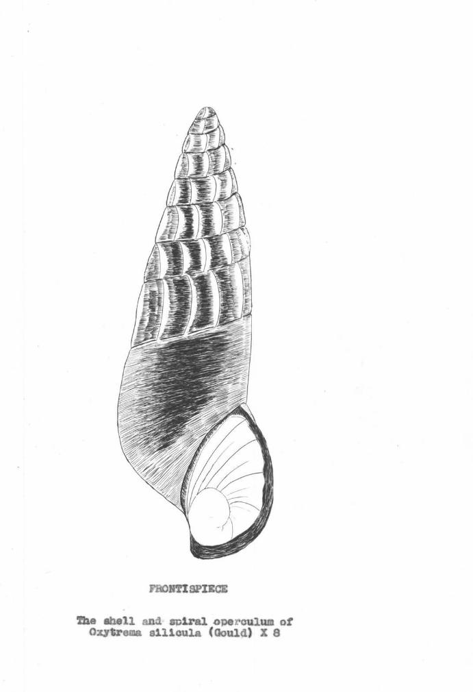
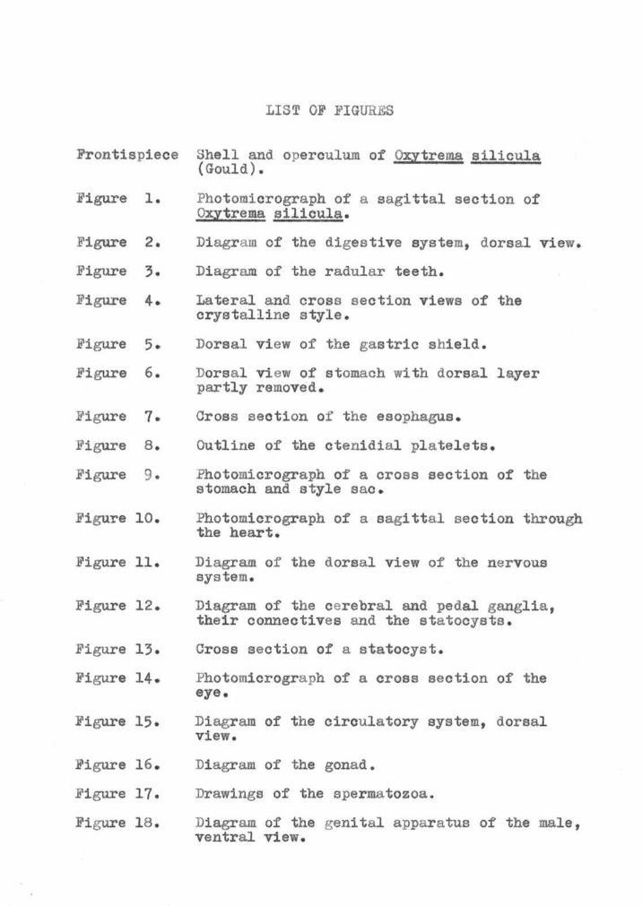

2
(Gould) Qbull silicula bairdiana (Lea) Qbull silicula rudene
(Reeve) and Qbull silicula shastaensis ( Lea ) The relationshy
ships and value s of middotthese sub- specific names have not been
completely de termined and sub-specific names will not be
included in this paper
Pleurocerid snails have been commonly called the
fresh-water Periwinkles it or npennywinklesn although these
names have been given in other sections of the country to
t he marine gastropods of the genus Littorina as well as to
l arval caddis flies
The range of o silicula generally extends from southshy-western Washington southward to the Shasta and Scott
rivera in California and westward from the Willamett e
basin dralnage in Oregon to the coast 1he four subshy
s pe cies occur in this general region
Only a few morphological studies on members of this
family have been made Stimson (21 p 41-53 ) described
some of the structural characters of the family Magruder
(12 p 883-912) made the first complete anatomical study
on a pleurooerid snail from Kentucky streams
Since the descriptions of Qbull silicula have been baaed
primarily upon the shell the purpose of this paper is to
present the internal structure of the snail The general
morphology will be described and histological and cytoshy
logical material will be used only when it contributes to
the general anatomical picture
Morphological studies should be valuable in parasitoshy
logical work for t his snail is the first intermediate host
to at least ten l arval trematodes Location of parasites
in the snail host and parasitological effects on the snail
tissues are so~e of the problems dependent on a knowledge
of t he anatomy of the snail
4
NATURAL HISTORY
Qxytrema ailieula is usually found elinging to rooks
or on organic material in the shallower and more rapidly
moving par ts of streams It is not a snail of still and
stagnant water
Snail collecting in the spring summer and early fall
yielded a large number of animals However during the
winter the snails became more difficult to find
Not much is known about the life history of these
snails Age and growth studies are being made now at
Oregon State College Older l arger snails have been
observed to have eroded ends The younger snails usually
have complete spires
From observations of the digestive system and eating middot
habits the snails were determined to be herbivores In
the streams great numbers of snails would be found on old
oak and alder leaves or on algal material Laboratory
snails thrived when fed on carrots oemiddotlery t and algae
Information on the breeding habits of this snail has
been sparse Some observations have been made on eggs and
egg-laying of snails kept in laboratory aquaria Eggs
pass down the sinus on the right side of the foot and pass
posteriorly along the edge of the foomiddott as the foot is
extended As the shell of the snail moves forward in turn
the eggs adhere to the egg mass which is being formed at
5
------ -----
the rear
The egg masses are oblong convex and attached to
stones or on the sides of the aquaria The eggs are blue
green blue and surrounded by a thick jelly Spiral
cleavage the veliger s tagea and hatching of the snails
were observed
The na~~al enemies of o silicula include fish and-raoeons Snails were found in stomachs of trout
6
UTERIALS AND METHODS
Snails from the Willamette and Alsea basin drainages
were used for dissections and microscopic study The
shells from these areas had been identified to be those of
Oxytrema silioula by w J Eyerdam a malacologist in
Seattle Washington
For dissections large non-parasitized animals were
selected The s hells were carefully broken and removed
after pressure was applied with a pair of pliers The
snails were usually not harmed by this process and could
be studied under a dissecting microscope Dissections
were made on live and preserved snails Preservatives used
were alcohol and Bouins fluid
Immature snails and organs of mature animals were
used for microscopic studies The shells were again
removed from the snails The molluscs were anesthetized
i n dilute alcohol or menthol and then fixed in hot Bouins
solution After dehydration t he snails were placed in a
vacuum chamber for better infiltra tion of paraffin Whole
animals and some organa were serially sectioned at 10
microns and mounted on slides The sections were stained
with Harris hematoxylin and counteratained with eosin
cleared and mounted in Canada balsam
For the study of the circulatory system India ink
was injected through fine glass pipets into the auricles
7
and the animals fixed in neutral formalin
Drawings were made with the aid of a camera luoida and
a mioroprojector
8
GENERAL FBAWRES
From a dorsal view the body of 9Ytrema silicula is
asymmetrical and coiled dextrally As in most gastropods
the body can be divided into the head foot mantle and
visceral mass The mantle and the structures within its
cavity will be discussed later
The head and foot are darkly pigmented and well
supplied with a layer of mucus They are covered with an
epithelium composed of cuboidal cells ~vo kinds of which
are glandular and pigmented The head and foot can be
extended out of the shell although the rest of the animal
remains hidden
The head is projected into a strongly contractile
snout that is dorso-ventrally flattened Ventrally in the
center of the snout is a vertical slit the mouth which
is surrounded by a pair of lips
The head epithelium extends on each side of the head
to cover the tentacles In liVing animals these tentacles
are encircled by narrow yellow bands The tentacles are
highly contractile sensory organs thick at their bases and
tapering to blunt tips They are composed of a core of a
blood vessel and a nerve surrounded by muscular tissue and
epithelium
On the lateral side of the base of each tentacle is
a sessile eye A dorsal view of a living animal reveals
9
the curved cornea and black retina In a preserved snail
however the eyes appear withdrawn from the surface and are
covered with a l ayer of epithelium
The foot predominately muscular tissue is well
supplied with nerves and blood sinuses and is grey-black
with a light grey sole When the animal is extended the
foot is roughly rectangular and flattened The anterior
part of the foot bears a large number of mucous glands
A dense and evenly distributed layer of cilia covers the
sole On the dorsal side of the posterior end of the foot
is a spiral horny operculum that closes the aperture of
the shell The muscles of the foot are continuous with
the columellar muscle posteriorly The columellar muscle
consists of tough white fibers that attach the body to the
shell at the body whorl
Posteriorly the body ends in coils composed of the
digestive gland with the peripheral gonads The coils
turn to the right and form a blunt apex
MANTLE
Behind the head is the thin vascularized mantle The
edge of the mantle is thickened and curved dorsally to fit
the aperture of the shell Ventro-laterally the edge is
continued to about two mm from the operculum where the
mantle is united to the foot The edge projects as a free
fringe In addition to the central blood sinuses the
10
mantle contains several nerves and a thin layer of muscle
fibers beneath the epithelium The parietal epithelium is
continuous with the body epithelium and is pigmented with
cuboidal and gland cells The Visceral epithelium is well
supplied with mucous glands especially on the right side
A number of mucous glands is also found within the mantle
at the anterior end
Several organs and openings are situated in the mantle
cavity On the extreme right is the small opening of the
anus Located immediately below it is the genital
aperture On the extreme left is the osphradium (OS
Fig 15) a low longitudinal ridge of epithelial cells
encircling a nerve The osphradium is located in the
direction of the respiratory current and is thought to be
a water-testing organ To the right of the osphradium is
the ctenidium the l argest organ in the mantle cavity The
ctenidium is slightly visible through the mantle as faint
transverse lines Since the ctenidium is the principal
organ of respiration the respiratory system will now be
described
RESPIRATORY SYSTEM
The otenidium extends anteriorly to posteriorly in a
slightly diagonal direction to the right following the
curvature of the body Because the triangular plates
making up the ctenidium are arranged in a single row the
11
ctenidium is classified as monopectinate The plates come
to rounded points and are longer on the left side
(Figure 2) They taper in length at the ends with the
longest largest plates located in the middle In number
these plates range from 82 to 92
Each plate has an epithelium of tall columnar cells
that are mostly non-ciliated though some are both ciliated
and glandular at the rounded points Beneath the epishy
thelium is a thin layer of muscle fibers and connective
tissue In the center of each plate is a sinus through
which the blood circulates
Since the foot and mantle are highly vascularized it
is probable that these also aid in respiration Respishy
ration can be carried on out of water Snails were kept
at 6deg0 on moist filter paper for two months
12
THE DIGBSTIVE SYSTE
The digestive system of Qbull silicula closely resembles
that of Pleurocera canaliculatum undulatum as described by 1Magruder (12 p 889-896)
The mouth enters a buccal cavity which contains a
pair of mandibles and a long ribbon-like radula (R
Fig 1) The radula is supported ventrally by an
odontophore (OD Pig 1) to which are attached several
muscles These muscles insert anteriorly and inferiorly
on the cartilaginous part of the odontophore The buccal
cavity its contents and the surrounding muscular and
connective tissues compose the buccal mass At the
posterior end of the buccal mass the esophagus passes
through a nerve ring A pair of salivary glands parallels
the long narrow esophagus from the buccal mass until the
esophague turns ventrally The esophagus enters the left
side of the stomach anterior to the entrance of the duct
from the digestive gland Anterior to the stomach a style
sac encloses a crystalline style The tip of the style
weare against a gastric shield located below the style sao
and in the stomach Dorsally the intestine leaves t he
stomach from a groove on the left side bends over the
style sac turns to the left and passes forward and widens
Since Morrison has revised the classification of Pleurooerids the name Pleurooera should probablybe changed to Oxytreman
1
13
to become the rectum The rectum terminates on the right
side of the mantle cavity in a small anus
The organa and cellular detail of the digestive system
will be c onsidered
Mouth The mouth (Mouth Fig 2) is a vertical slit
on the anterior end of the snout that becomes oval in shape
when the animal is feeding Glandular non-pigmented
plicated lips surround the mouth
The mouth has epithelial cells t~iee the length of the
cells lining the body surface The cells are columnar with
a cuticular appearance No pi gment or gland cells are
present
Buccal Mass The mouth expands laterally into a
l arge irregular buccal cavity tha t extends from the level
of the middle of the snout past the level of the tentacles
A pair of mandibles thin cuticular structures are at the
anterior end of the buccal cavity
The buccal cavity contains a long radula that is Sshy
shaped in sagittal sections The radula consists of 88-92
rows of teeth that decrease in size posteriorly The
radular formula is 3-1-3 ie two marginal teeth and a
l ateral tooth are placed on each side of a median tooth
Each tooth bears a number of cusps the number being
characteristic for the type of tooth (Figure 3) The
outer marginal teeth have seven cusps per tooth t he inner
marginal teeth only five per tooth The l ateral teeth
14
have two cusps on the right and left of a large central
cusp The central tooth has four cusps on the right and
left of a large central cusp ~he complete formula is
l 1 l l 1 1 l bull7 + 5 + 2-l-2 + 4-1-4 + 2-1-2 + + 1
The radula is enclosed in a thin sac that is supported
by an odontophore In sagittal sections the odontophore
is located under the anterior curve of the S-shaped radula
and is continuous anteriorly and dorsally but diverges
ventrally and posteriorly It resembles cartilage in being
lightly stained and having large vesicular cells with small
nuclei
Esophagus The buccal cavity opens into a narrow
long esophagus (E Fig 2) which is surrounded by the
ganglionic ring at the junction of the two The esophagus
extends posteriorly following the curvature of the body
passes ventrally into the visceral mass and enters a groove
on the lower left side of the stomach
Externally it does not vary in di ameter throughout
its length There are four longitudinal folds of epishy
thelium in the cavity of the esophagus (Figure 7) These
folds increase in number before the esophagus enters the
stomach The epithelium is weakly ciliated with some
pigment cells and numerous gland cells
Salivary Glands The salivary glands (SG Fig 2)
a pair of highly c onvoluted white tubes attach at the
15
posterior end of the b11cc al cavity and pass posteriorly
through t he nerve ring and the muscles of the buccal mass
They parallel the esophagus one on each side and end
when the esophagus turns ventrally
The left gland is longer t han the right In crossshy
section the glands are round to oval (S Fig 7) The
cells of the one l ayer composing the gland are strongly
basophilic columnar sparsely ciliated and surround a
very small cavi ty
Digestive Gland 1~e duct from the digestive gland
(DD Fig 2) opens into the stomach just below the entrance
of the esophagus and is lined with ciliated columnar cells
epithelium similar to that of the stomach The duct
collects the contents of the tubules on the columellar
side The tubules compose the digestive gland or liver
and form the mass of the coila posterior to the stomach
~1e gonads cover the surface of the gland except on the
columellar side
The digestive gland tubules are yellow-brown with
pigmented spots They are round in cross-section and
surrounded by connective tissue and blood sinuses In
each tubule the glandular unevenly-staining epithelium
rests on a delicate basement membrane Three types of
cells in the gland are dis tinguishable the black pigshy
mented those with brovn inclusions and gland cells withshy
out inclusions The former two cell types are basophilic
16
w1d long with basal nuclei The pigmented cell has a
l arge vacuole containing black pigment The second type
of cell has several vacuoles enclosing brown secretory
bodies ~1e gland cells are round lightly stained and
have small clear vacuoles
Stomach The stomach (S Fig 2) is an irregularshy
shaped organ located on the dorsal surface of the body
coil It is anterior to the gonads and the digestive
gland and posterior to the style sac and intestine
The dorsal roof of the stomach and the lower ventral
portion are patterned into a series of parallel ridges of
epithelium The floor of the stomach is raised into many
folds which may aid in the mixing of the stomach contents
The largest of these folds is the central fold (CF
Fig 6) in the lower middle of the stomach It is pyriform
and largest posteriorly gradually diminishing in size
anteriorly where it covers part of the gastric shield
An outer fold is formed on the right and posterior to
the central fold Be~veen these folds is a groove or
gutter tha t is continuous with the groove of the intestine
and esophagus The outer fold continues posteriorly
bends upon itself and then curves to the left forming a
boundary for a d~ep channel be~veen it and the stomach
wall
The epithelium of the stomach is ciliated except for
the anterior portion which has a cuticular lining The
17
main structure in the lining is the gastric shield
(Figure 5) an irregular partly formed tube against which
the style projects The s hield curves to the left and
bears light crenations The shield is irridescent and
transparent except for the opaque anterior end Biuret
and nitric acid tests showed the shield to be protein in
nature
Style Sao Anterior to and opening into the stomach
is the style sac (ss Fig 9) The style sao is one-third
the size of the stomach and nearly cylindrical As the
name implies the style sao contains a crystalline style
which projects out into the stomach
The style sao is separated from the intestinal groove
on the left by two folds of the intestinal wall the
typhlosoles Both typhlosoles bear very tall columnar
cells with short cilia The dorsal or major typhlosole
(MT Fig 9) is a ridge of cells that fits in the groove
of the minor typhlosole (MI Fig 9) The ventral or
minor typhlosole is composed of two ridges of cells and is
the broader of the two
The epithelium of the style sac is unusual in being
composed of large columnar cells covered at t heir distal
ends with a dense mass of cilia all exactly the same
length These bristle-like c ilia form an even surface
upon which the style rests The nuclei of the epithelium
are within the lower half of the cells and stain heavily
18
with hematoxylin
Crystalline Style In mature animals the crystalline
style (Figure 4) measures about 45 mm long and 10 mm
thick with blunt ends A core opaque and larger at the
anterior end is surrounded by yellow hyaline l ayers In
cross section the style shows the circular core and conshy
centric hyaline layers (Figure 4-B)
The posterior smaller end of the style projects out
of the style sac and wears against the gastric shield of
the stomach Here the style end is spirally wound semishy
liquid and mixed with the stomach contents No attachment
of the style in the style sao can be seen
The color and consistency of the style vary greatly
depending on the environment and health of the animals
In animals that had been previously fed the style was a
light yellow Snails that were starved for two months
possessed soft brown styles Every snail examined
possessed a crystalline style
The style disappeared within an hour after it had been
placed in water The hyaline layers dissolved first with
the l arge end of the core remaining for a longer time
The styles coagula ted in 95~ alcohol and were positive for
the Biuret test indicating that they were proteins of an
albumi noid nature A solution of styles digested a 0 5
starch solution in 2-4 minutes
The styles then are albuminoid proteins containing
19
an amylolytic enzyme capable of starch digestion
Intestine From the dorsal side the intestine leaves
the stomach from a deep groove that is incompletely
separated from the style s ac by the typhlosoles (Plate
3 Fig 6 I) The intestine (I Fig 2) curves to the
right around the style sao loops back and crosses over
the esophagus and passes forward to become the rectum
In cross-section the intestine is a greatly folded
weakly ciliated tube After leaving the stomach it bears
a small typhlosole that disappears as the intestine passes
anteriorly
Rectwn and Anus As the intestine passes forward it
widens and becomes the rectum (R Fig 2) The rectum has
an obliquely folded epithelium composed of low columnar
cells each possessing a few cilia
The rectum ends in a small opening in the right side
of the mantle caVity the anus ( A Fig 2) which is well
supplied with ciliated columnar cella and mucous glands
The fecal pellets formed in the oblique folds of
the rectum are extruded singly or in packets and are
spindle-shaped
20
THE NERVOUS SYS TEM
The nervous system of this s pecies is similar to the
streptoneurous forms described by Spengel (20 p 333-383)
and Bouvier (2 p 1-510)
The terminology of Spengel will be used for the names
of ganglia and nerves The ganglia are termed buccal
cerebral pedal pleural supra and subintestinal and
visceral The nervous connections between members of a
pair of ganglia are named commissurea while t hose between
members of different pairs or unpaired ganglia are called
connectives
The ganglia are mainly located at the anterior end of
the body The components of the circumesophageal ring
are the cereb~al ganglia its connectives and the pedal
ganglia The cerebral ganglia form connections with the
buccal and pleural ganglia The left pleural ganglion is
separated f rom the subintestinal gangl ion by a constriction
A nerve from the right pleural ganglion goes to the suprashy
intestinal ganglion on the left side Nerves from the
supraintestinal and subintestinal ganglia extend posshy
teriorly to form the visceral ganglion
A detailed description of the nervous system follows
(Figures 11-14) Only the major nerves have been followed
since it is not practical to describe t he smaller nerves
which vary in size and location
21
Buccal Gangli a The paired buccal ganglia (BG
Fi g ll) are located postero-dorsally in the buccal mass
and are difficult to dissect because of the sheath of
sphincter muscles covering them These ganglia are small
oval in shape and connected to each other by a long thin
commissure (BC) iwo nerves branch from each buccal
ganglion the lateral nerve passes forward to innervate
the anterior buccal mass and the medial nerve branches
again and innervates the posterior buccal maos A fine
white connective attaches each buccal ganglion at its
postero- lateral end to the antero- ventral end of the
cerebral ganglion on the same side (BCO)
Cerebral gangl iabull The cerebral ganglia (OG Fig 11)
are situated anteriorly on the dorso- lateral side of the
buccal mass They are pyriform in outline with pointed
lateral ends pigmented anteriorly and twice the size of
the buccal ganglia A short thick commissure (CC) lying
over the esophagus connects the two cerebral ganglia The
cerebral ganglion on each side is connected to its pedal
ganglion by a thick long cerebro-pedal connective and to
its pleural ganglion by a short cerebro- pleural connective
The cerebro- pedal connectives arise ventrally from the
inner angle of the cerebral ganglia while the cerebroshy
pleurals lie external to them Fi ve anterior and three
lateral nerves originate from each cerebral ganglion on
the antero- lateral surface and innervate the head Since
22
they are identical for the left and right sides only one
side will be described
Anterior nerves
1 The first nerve (Cl Fig 11) arising from the
antero-dorsal side of the cerebral ganglion is a medium
sized labial nerve which runs laterally on the perishy
visceral cavity wall where it is attached by fine
connective tissue and its swall branching nerves The
nerve finally enters the mouth region
2 The next nerve (02) arises laterally and conshy
tinues parallel to the first along the cavity wall to the
anterior region of the snout Here it branches and inshy
nervates the lip region
3 The next nerve (03) originates so closely to the
preceding nerve that it i s fused to it It also runs forshy
ward to the snout region and innervates the mouth
4bull The pharyngeal nerve (C4) originates on the
antero-ventral surface and goes forward l aterally until it
reaches t he middle of the snout Here it turns sharply
and passes ventrally into the muscles of the buccal mass
It branches and innervates the anterior buccal mass region
5 The fourth labial nerve (Cs) originates very
closely to the pharyngeal nerve It goes forward parallel
to the pharyngeal nerve and then branches into two small
nerves one innervating the posterior buccal mass region
and the other innervating the mouth region
23
Lateral nerves
1 The tentacular nerve (Tbull Fig 11) the largest
nerve arising from the cerebral ganglion originates in
the middle of the dorso-lateral surface It passes
l aterally into the muscles of the body wall and then forshy
ward to the tentacle where it enters and extends to the
tip of the tentacle It is centrally located and gives
off very small branches as i t proceeds forward
2 The small optic nerve (0) starts from the ventroshy
lateral surface and passes obliquvly forward to enter the
eye at the lateral edge of the tentacle
3 The very fine statocyst nerve (STN Fig 12)
originates below the optic nerve and passes ventrally with
the cerebra-pedal connective It terminates in the wall
of the statocyst on the dorsolateral surface of the pedal
ganglia
Pedal ganglia The paired pedal ganglia (PG Fig 12)
are round structures connected by a short thick commissure
They are ventral to the cerebral ganglia and the pleural
gangl ia from which they receive the cerebra-pedal CP) and
pleuro-pedal (PP) connectives respectively The cerebrashy
pedal connectives are thick and almost rounded so that they
appear like ganglia The paired statoeyats (ST) are
attached to the dorso- lateral surfaces of the pedal ganglia
by connective tissue Several nerves arise from the pedal
ganglia Antero-medially is a large nerve that goes
24
forward and innervates the pedal muscles giving off small
branching nerves in an irregular pattern Ventrally a
large nerve trtUlk leaves the pedal ganglia and branches
several times the branches being distributed in the
opercular region of the foot Small lateral and posterior
nerves originate from the pedal ganglia and contribute to
the innervation of the foot
Left pleural ganglion Since the pleural ganglia
are irregular in size and give rise to different nerves
they shall be described separately The left pleural
ganglion (LPG Fig 11) is large constricted in the
middle and appears like two ganglia It is attached to
the cerebral sanglion by a short cerebro-pleural conshy
nec tive Arising from the anterior lobe are two nerves
1 A small anterior nerve (F1 ) innervates the
muscles of the neck region
2 A long left mantle nerve (LM) extends posteriorly
to the left of the esophagus and left salivary gland It
enters the neck muscles and then turns into the mantle
running along the ventral surface toward the right
Branches of this nerve extend to the margin of the mantle
The only nerve seen to originate from the posterior
lobe of the left pleural ganglion is the small columnar
nerve (N) It passes from the middle of the lobe posshy
teriorly and parallel to the subintestinal nerve to be
described below Just past the mantle margin it passes
25
into the muscle
Right pleural ganglion The right pleural ganglion
(RPG Fiampll) is shorter and t hicker than the left pleural
ganglion and not constricted A short vertical connective
joins the right pleural ganglion with the cerebral
ganglion
1 The most anterior nerve (F2 ) of the three nerves
of the right pleural ganglion is located on the median
dorsal surface where it turns to the right and innervates
the muscles of the neck region
2 Posteriorly the right pleural ganglion ends in
a large supraintestinal nerve (R) that passes dorsal to the
esophagus and salivary glands into the left wall of the
body cavity where it joins the supraintestinal ganglion
3 ihe third nerve to originate from the right
pleural ganglion is the short connective between the subshy
intestinal ganglion and the right pleural ganglion (X)
1his small nerve arises ventral to the origin of the suprashy
intestinal nerve
Supraintestinal ganglion In the left wall of the
body cavity the supraintestinal ganglion (SPG) receives
the supraintestinal nerve from the right pleural ganglion
and gives off many small branches tha t innervate the mantle
tissue and the anterior part of the ctenidium One of the
right branches of the supraintes tinal ganglion is the left
visceral connective (LV) It extends along the right of
26
the body wall posteriorly and meets the rieht visceral
corJlective ventrally
Subinteatinal ganglion The subintestinal ganglion
(SUG) is separated from the left pleural ganglion by a
constriction It receives a connective (X) on ita anteroshy
dorsal ourface from the right pleural gangl ion On its
ventro-lateral surface two nerves arise the right mantle
and t he subintestinal
1 According to Magruder (12 p 905) the right
mantle nerve in middot oanaliculatum undulatum originates from
the right pleural ganglion In Oxytrema silicula there
has been an apparent fusiou of the lower part of the right
mantle nerve with the subintestinal ge~glion
The right mantle nerve divides into a small and a
large branch The large branch turns to the right and
enters the muscles of the body wall and curves anteriorly
into the margin of the mantle wh~re it gives off a small
genital branch This genital nerve further divides into
two nerv3s that serve the lateral and median long itudinal
folds of the genital organ
2 The subL~testinal nerve (SU) leaves the subshy
intestinal ganglion posteriorly and gives off m~~y small
branches which turn into the muscles of t he region Posshy
teriorly the main nerve becomes the subintestinal visceral
connective that joins the visceral ganglion
Visceral ganglion The subintestinal visceral (RV)
27
and the supraintestinal visceral (LV) connectives join
posteriorly to form an indistinct visceral ganglion (VG)
The ganglion is anterior to the stomach ventral to the
renal organ and at the right of the pericardial cavity
A small branch from the right side branches irregularly
into the visceral mass The main nerve (K) continues
posteriorly on the inner surface of the visceral mass to
the digestive gland and gonad It is parallel to the
genital duet and visceral artery during its posterior
course
SENSE ORGANS
In addition to the osphradium and tentacles already
described the sense organs of this animal include the
statocysts and eyes
Simroth (19 p 323) described the statocysts as
organs of equilibrium not of hearing In this animal as
in other gastropods the statocysts are paired and closely
associated with the pedal gangli a A thin layer of conshy
nective tissue envelops the statocyst on the posteroshy
lateral surface of each pedal ganglion In cross s ection
the organs are observed to have a single layer of flat
irregular cella that are sparsely ciliated (Figure 13)
The round cavity contains 20 to 25 statocontia which are
clear and almost transparent Upon staining t hey are
shown to be strongly basophilic Three to four statocontia
28
-----------
are larger than the others and placed in the center 1~ese
are barrel- shaped with faint longmiddot itudinal striations The
other statoeontia are minute and vary in shape from oblong
to oblong with center constrictions
The eyes are paired sessile organa on the lateral
edges of the bases of the tentacles The body epithelium
continues over the eyes to form the outer cornea (ltigure
14) bull 1ne cells are tall columnar and lack the pigment and
gland cells that characterize the body epithelium Below
the outer cornea is a thin layer of connective tissue that
extends around the optic vesicle and covers the optic
nerve The optic vesicle i s like an indented cup with themiddot
center optic nerve in the indentation The retina is
crescent-shaped with the deepest layer opposite the optic
nerve The retina is composed of a layer of middot tall cella
with brushlike cilia at their anterior ends The upper
one-third of the cells is filled with black retinal
granules The lower two-thirds of the cells contain the
nuclei and are pointed at their poste1middotior ends so that they
appear continuous with the fibers of the optic nerve The
edges of the retina are continued over the lens as a thin
cornea the cells composing it being amorphous and without
pigment A round yellow lens that does not stain fills
the cavity formed by the retina and inner cornea
29
THE EXCRETORY SYSTEM
The renal organ is divided into a minute anterior
part and a large posterior part The anterior portion is
oval and located to the right of the perioardial cavity
above the posterior portion This anterior portion conshy
sists of thin irregular lamellae that hang from the body
epithelium into a spacious cavity (AR Fig 1) The
lamellae are composed of regular low cuboidal cells
enclosing blood sinuses and extend above the anterior part
of the genital lamallae and below the rectum adjacent to
the ctenidium The anterior renal organ opens anteriorly
into the mantle cavity by means of a pore lined by ciliated
glandular epithelium The lamellae of the anterior portion
are separated from the lamellae of the posterior portion by
a thin wall but the cavities are continuous
The posterior portion of the renal organ is oblong
with tapering ends and covers the dorsal surface of the
pericardial cavity as well as the dorsal part of the
stomach (PR Fig 15) In live animals the posterior
portion is light yellow and s pongy in appearance On its
surface are seen faint striations that in cross section
are found to be regular fused lamellae These fused
lamellae are lined with vacuolated light-staining cells
which give the organ a spongy effect
The blood enters the dorsal surface of the posterior
30
portion through the afferent renal sinus which breaks up
into many small sinuses On the ventral surface the
efferent renal sinus drains the posterior portion directly
into the auricle The posterior portion is connected to
the pericardia cavity through a small opening on the
right
THE OIROULATORY SYSTEM
The vascular plan features a two-chambered heart with
a system of arteries from which blood flows into sinuses
or lacunae and back into the heart again (Figure 15)bull
Heart The heart on the left side of the body whorl
is enclosed by a cubical pericardial cavity Posteriorly
the pericardial cavity extends in a tube-like fashion The
cavity is lined by a layer of epithelial cells and is
covered anteriorly for the most part by the posterior renal
organ
Dorsally the auricle (A) the first chamber of the
heart receives t he blood from the l arge l eft ctenidial
sinus and the small renal sinus The auricle is a thin
square sack about 05 mm in diameter surrounded by a
delicate squamous layer in which only nuclei of cells are
visible Beneath this layer are narrow irregular muscle
bands outlining an irregular cavity The auricle narrows
suddenly and leads to the large ventricle No valves can
be seen in the auriculo-ventricular opening
31
In contrast to the auricle the ventricle (V) is twice
as large and pyriform in shape No ventricular valves are
found and the walls are mainly of muscle Beneath the thin
epithelium there is a layer of uniform muscle bundles and
then large irregular muscle bundles that project into the
lumen
The ventricle opens ventrally into the aortic artery
which divides immediately into the cephalic artery (CA)
going anteriorly and the visceral artery (VA) passing
posteriorly
Cephalic artery The cephalic artery gives off a
branch on the left of the style sao before passing anterishy
orly in an oblique direction to the left It passes forshy
ward dorsal to the esophagus up to the region of the
cerebral ganglia Here the cephalic artery gives off a
branch to the buccal mass and then passes antero-ventral1y
between the pedal ganglia The artery to the buccal mass
sends branches to the tentacles and then divides again to
form an anterior and a posterior branch The anterior
branch circulates blood to the snout lips and anterior
end of the buccal mass while the posterior branch supplies
blood to the radular sac and pharynx As the main artery
passes forward from the pedal ganglia it divides into
many arteries supplying the foot The expansion of the
foot is probably aided by the well-developed arterial
system
32
Visceral artery The visceral artery follov1s the
border of the posterior renal organ and is dorsal to the
anterior style sac It turns to the columellar side where
it accompanies the genital duct and visceral nerve to the
digestive gland and gonads giving off branches until it
finally ends at the apex
Venous system There being no capillaries or veins
a venous system of lacunae with no definite walls allows
the blood to flow to the heart
Anteriorly the foot is well supplied with sinuses
especially at the periphery These empty into a large
perivisceral sinus lying on the floor of the mantle cavity
The perivisceral sinus finally empties into the afferent
ctenidial sinus on the right margin of the otenidium
The l arge rectal sinus (RS ) on t he left of the rectum
collects blood from t he mantle and genital folds and also
empties into the afferen t ctenidial sinus The afferent
ctenidial sinus (AC) also receives blood from t he sinuses
in the mantle
The blood from the afferent ctenidial sinus goes to
each ctenidial leaflet through a central sinus and is
aerated It then passes into the efferent ctenidial sinus
(EO) on the l eft margin of the otenidium The efferent
otenidial sinus collects blood from the osp~middotadium before
entering the auricle dorsally
The peri-intestinal sinus (IS) surrounds the stomach
33
style sao and the anterior part of the intestine and also
collects blood from the gonads and digestive gland The
blood enters the posterior renal organ which transports it
to t he auricle by the efferent renal sinus
The small afferent renal sinus (R) on the dorsal part
of the posterior renal organ collec ts some blood from the
t erminal part of the intestine and passes it to the
efferent renal sinus
Thus the venous blood may pass (1) from the perishy
visceral mantle and rectal sinuses into the afferent
ctenidial sinus become aerated in the ctenidial leaflets
pour into the efferent ctenidial sinus and then to the
auricle or (2) from the peri-intestinal sinuses to the
efferent renal sinus to the auricle The blood passing
into the heart is then mixed
34
THE REPRODUCTIVE SYSTEM
As far as can be determined the sexes in this species
are separate Upon examination of 314 mature animals from
November to March a ratio of 1 0 f emales to 1 2 males was
found This proportion was the same in various size groups
of males and females
In all these animals the only external sex character
was a sinus on t he right side of the female foot No
copul atory apparatus was found in the male
The ovigerous sinus was first described by Stimson
(21 Pbull 41-53) and is one of the bases of subfamily classishy
fication used today (14) The sinus begins at t he genital
aperture continues along the right side of the foot and
disappears in the opercular lobes The sinus of the living
snail has a constant slight rhythmic motion It is gray
in color Upon microscopic examination the sinus is found
to consist of three pockets arranged like a three leaf
clover The epithelium is heavily ciliated with unishy
cellular gland cells and no pigment cells
Actual observations of egg laying demonstrated the
egg- transporting function of the sinus Another function
may be t he transport of the male sex products into the
female since the male has no copulatory organs However
I have found no sperm or spermatophores in the sinuses
The gonads occupy the same location in both sexes and
35
when inactive look very similar (Figure 16) The gonads
are located on the dorsal surface of the digestive gland
and may reach the border of the stomach They do not cover
the apex or the columellar side of the digestive glandbull
On the columellar side are five branches that collect the
contents of the gonads into a main duct
The ovary appears lobulated due to the yolk filled
tubules The tubules are flask shaped and have one layer
of cells The germinal epithelium to which the eggs are
attached is very delicate and often torn when the shell is
broken The color of the ovary varies from yellow to
orange to blue green blue depending on the nature of the
yolk The blue green blue ovaries were judged to be mature
since the eggs laid were of this color
The oviduct on the columellar side transports the
contents of the ovary to the genital laminae The oviduct
is a thin tube of ciliated Gpithelium a few muscle fibers
and connective tissue
The closed oviduct enters two genital l aminae that are
fused dorsally but open ventrally (Figure 19) The
anterior portion of the l ateral lamina (LL is large and
highly folded with glandular epithelium throughout Ihe
medial lamina (ML ) is thin with ciliated epithelium and
increases in size as it reaches the genital opening The
l aminae fuse and open into the mantle cavity on a s mall
papilla surrounded by a l ayer of muscle
36
A small white organ the sperm receptacle (SR) lies
abote the anterior end of the laminae next to and fused
with the nidamental gland A small duct connects the sperm
receptacle with the anterior portion of the l a teral lamina
The sperm receptacle is composed of compartments in which
heads of eupyrene sperm are lodged
The large white nidamental gland (NG) is dorsal to
t he genital folds This gland has glandular epithelium
arranged in lamellae with ducts leading into the spaces
above the genital lruninae
The testis consists of blind tubules that appear
finely lobulated All stages of germ cells from spermashy
togonia to spermatozoa are found in the tubules When
active the testis is a golden yellow
The ciliated thin vas deferens passes from the testis
to the genital l aminae on the columellar side The two
genital laminae are fused anteriorly but are open
ventrally The larger lateral lamina (LL ) folds over the
medial l eft lamina (ML) Between them is the genital sinus
The l aminae have very tall glandular epithelium They
taper to a small opening in the mantle cavity below the
rectum White masses are often seen passing from t his
opening Upon microscopic examination the masses are seen
to consist of eupyrene sperm with their heads embedded in
s mall round spermatophores in a liquid medium
The sperm i n this species and in many gastropods as
37
described by Meves (13 p 1-84) and oodward (24 p 1-23)
are of two types the larger worm-like oligopyrene and
the hair-like eupyrene sperm (Figures 7 A B) The olishy
go pyrene sperm have dark staining centers and five to six
flagella at the posterior end They measure approximately
30 microns in length by three microns in diameter After
being in water for some time they tend to swell and
become round (Fig~e 17 C) They are non-motile while the
eupyrene sperm are motile The eupyrene sperm have
extremely long tails and dark staining heads They measure
about 36 microns in length the head being 1 5 microns
wide These sperm are found attached to spermatophores
and in the sperm receptacles in females
The proportion of eupyrene to oligopyrene sperm varies
with the season In early fall there were greater numbers
of oligopyrene sperm In the spring more eupyrene sperm
were present in the testis
The aut hor agrees with Woodward (24 p 20) in conshy
cluding that the oligopyrene sperm are of no functional
significance since t hey are non-motile and not found in
the spermatophores or in the sperm receptacles of females
38
SUMMARY
Oxytrema silicula (Gould) resembles other pleurocerid
gastropods in the general body plan spiral operculum
monopeetinate ctenidiumt and radular formula
It possesses in addition a style sac enclosing a
crystalline style The style projects into the stomach
and wears against the gastric shield an irridescent
partly- formed tube The style is albuminoid in nature and
possesses an amylolytic enzyme
The respiratory and circulatory systems had no outshy
standing characteristics
The nervous system with its main circumesophageal
ganglionic ring is similar to the streptoneurous forms
described by Spengel and Bouvier However no zygoneurous
connections between nerves could be seen A fusion of the
lower part of the right mantle nerve with the subintestinal
ganglion is a unique character for this species
The sexes in Qbull silicula are separate vith a ratio of
10 females to 1 2 males The only external sexual
characteristic found was an ovigerous sinus on the right
side of the female foot ~gg laying and egg masses were
observed ~ The male produces two kinds of sperm the hairshy
like eupyrene and the worm-like oligopyrene The latter
is believed to be of no functional significance as
evidenced by its absence in the spermatophores and in the
c ~e f M- lte a yi c ~ o f Wt 2 ) s
39
sperm receptacle of females
--------
APPENDIX
40
1
2
j3
BIBLIOGRAPHY
Baker F C The fresh water mollusca of Wisconsin Madison 1928 ( Wisconsin Geologic and Na tural History Survey Bulletin 70 Pt 1-Gastropoda)
Bouvier E L Systeme nerveaux morphologie generale et classification des gastropodes Prosobranches Annal es des sciences naturelles Zoologie 3(7)1-510~ 1887
Fretter v Minute proaobranchs of rock pooleJournal of the arine Biological Association of the United Kingdom 27597-632 1939
Goodrich c Goniobasis of the Coosa River Alabama Ann Arbor Mich 1939 (Michigan University Museum of Zoology Miscellaneous publication no 31)
Gould A A Description o new shells collected bythe u s Exploring Expedition under Capt Wilkes Proceedings of the Boston Society of Natural History 2222-225 1847
Graham A On the structure of the alimentary canal of style-bearing prosobrancha Proceedings of the Zoological Society of London 10975-112 1939
Henderson J Western American species of Goniobasia with descriptions of new forms The Nautilus 48(3) 95-99 1935
Non- marine mollusca of Oregon and Washington supplement University of Colorado Studies 23(4)251-280 271-276 1936
Lea I Description of a new genua (Goniobaeis) of the f amily Melanidae and eighty-two new species Proshyceedings of the Academy of Natural Sciences Philashydelphia 1862 p 262-272
Mackintosh N A The crystalline style in gastropods Quarterly Journal of Microscopical Science 69317-42 1925
agruder S R ecord of a crystalline style in two fresh water gastropods The Nautilus 48(3)100-102 1935middot
41
12 The anatomy of the fresh water prosobranchiate gastropod Pl eurocera canaliculatum undulatum (Say) American 1diand Naturalist 16883shy912 1935
13 Meves F Uber oligopyrene und apyrene Spermien und uber ihre Entstehung nach Beobachtungen an Paludina und ~faera Archiv fur mikroskopi2che Anatomie 611- bull f902
14 Morrison J P E forld relations of the Me1anians Reprinted from the American Malaco1ogical Bulletin and Report December 1951 3 p
15 Munsell A H Munsell book of color Baltimore Munsell Color Co 1929 42 Pbull
16 Prashad B Anatomy of the common Indian Appleshysnails Pila globosa Memoirs of the Indian Museum 8(3)91-1~ 1925
17 Robson G C On the anatomy and affinities of Hysobia nosophor~ Annals and Magazine of Natural History ser 9 7401- 413 1921
18 On the anatomy and affinities of Paludestrina ventrosa Quarterly Journal of Microshysc opical Sci ence 66159- 185 1922a
19 Simroth H Gastropoda Prosobranchia In H G Bronns Klassen und Ordnungen des Tier-Reichs Dritter Band II Abteilung Buch 1 1901 1501 Pbull
20 Spengel J w Die Geruchsor~ann und das Nervensystemder Molluskan Zeitschrift ffir wissenschaftliche Zoologie 35333- 383 1881
21 Stimson w On the s tructural characters of the soshycalled Melanians of North America American Journal of Sc ience 2d ser 3841- 53 middot 1864
22 bull Winsor c P The eggs of Goniobasis virginia Gamelin and Anculosa oarinata Bruginere Journal of the Washington Academy of Sc i ence 23(1)34-36 1933
23 Woodward T M Anatomy of the reproductive systemof Goniob asis lagueata (Say) Journal of the Tennessee Academy of Science 9=243-258 1934
42
24 lagueata (Say) 1935
Spermic dimorphism in Goniobaaia Journal of Morphology 57(1)1-23
25 Van Oleave H J Studies on snails of the genusPleuocera I The e ggs and egg laying habits The Nautilus-46(1) 29-34 1932
43
KEY TO PLATE 1
FIGURE 1
Photomicrograph of a sagittal section of OxYtrema silicula (Gould)X 410
AR-Anterior renal organCG-Cerebral ganglion C Columellar muscle CT-Cross section of the ctenidium E - Es ophagus F - Foot GS-Gaatric shield I -Intestine M- Mantle OD-Odontophore OF- Operculum PG-Pedal ganglion R - Rectum RA- Radula RP- Right pl eural gangl ion S - Stomach ST-StatocystSU- Subintestinal ganglion
44
Fio I
pLATE I
45
KEY TO PLATE 2
FIGURE 2
Diagram of the Digestive System Dorsal View
A -Anus BM- Buccal mass DD-Digestive gland duct E - EsophagusEO-Esophageal opening I - Intestine R - Rectum S - Stomach SG-Salivary gland8S- Style sao
bull
Fig 2
PLATE 2
47 KEY TO PLATE 3
FIGURE 3
Radular teeth
A-Outer marginal tooth B-Inner marginal tooth C-Lateral tooth D-Central tooth
FIGURE 4
Crystalline Style
A-Lateral view B-Cross-section showing
coaxial layers
FIGURE 5
Gastric shield
FIGURE 6
Stomach dorsal view with dorsal layer partly removed
OF- Central fold CS-Crystalline styleEo-Esophageal openingGS-Gastrio shield I -Intestine ss-style sac
FIGURE 7
Cross section of the esophagus
0-Lumen of esophagus S- Salivary gland
FIGURE 8
Outline of otenidial leaflets
Fig 3
Fig4
Fig 5 Fig 6
~
Fit 7 Fig 8
PLATE 3
49
KEY TO PLATE 4
FIGURE 9
Cross section of the stomach and style sao Photomicrograph X 77
CS-Crystalline style I -Intestine MI-Minor typhlosole IT- Major t yphlosole S -Stomach SS-Style sac
FIGURE 10
Sagittal section through the heart Photomicrograph X 155
PR- Posterior renal organA -Auricle V -Ventricle
50
Fig 9 middot
Fig 10
PLATE 4
51shy
KEY TO PLATE 5
FIGURE 11
Diagram of the Nervous System Dorsal View
BC-Buccal commissure BOO-Buccal cerebral connective BG -Buccal ganglion C1 - First l abial nerve C2 -Second labial nerve C3 -Third labial nerve C4 -Pharyngeal nerve 05 - Fourth l abial nerve CC - Cerebral commissure F1 - Left neck nerve F2 - Right neck nerve G -Genita~ nerve K -Visceral nerve LOG-Left cerebral ganglion LM -Left mantle nerve LP - Left pleural ganglionLV - Supraintestinal visceral connective N -Columnar nerve 0 - Optic nerve R - Suprainteatinal nerve RM -Right mantle nerve RPG-Righ t pleural ganglionRV -Subintestinal visceral connective SPG-Supraintestinal ganglionSUG-Subintestinal gangl ion T -Tentacular nerve VG - Visceral ganglion
62
Fig II
PLATE 5
53
KEY TO PLATE 6
PIGURE 12
Diagram of the cerebral and pedal ganglia their connectives and the statooysts
L -Left LOG-Left cerebral ganglion LOP- Left oerebro-pleural connective LPC-Left eerebro-pedal connective ve - Pedal commissure PG - Pedal ganglion PP - Pleuro-pedal connective R -Right ST - statocystSTN-Sta tocyst nerve
-+---LPC ~IM-+--~CP
R 11+-----STN L
-+11+--pp
~~r---vT
-----t---PG
1-~-----r---PC
Figl2
Plate 6
55
KEY TO PLATb 7
FIGURE 13
Cross section of the s t atocystPhotomicrograph X 352
S- Statocontium
FIGURE 14
Cross section of the eye bull Photomicr ograph X 384
L-Lens 0-0ptic nerve R-Retina
56
Fig 13
Fig 14
PLATE 7
57
KEY TO PLATE 8
FIGURE 15
Diagram of the Circulatory System Dorsal View (Sinuses in black arteries outlined)
A - Auricle ABA- Anterior Buccal ArteryAC - Affer ent ctenidial sinus BA - Buccal ar teryCA - Cephalic arteryCT -Ctenidium EC - Efferent otenidial sinus IS - Intestinal sinus OS - Osphradium PA - Pedal arteryPBA-Pos terior buccal arteryPR - Posterior renal organR - Efferent renal s inus RA - Radular sinus RAP- Right anterior pedal arteryRPP- Right posterior pedal artery RS - Rectal sinus TA - Tentacular artery V -Ventricle VA -Visceral artery
~~--4---~~-RA
---+--f-1--Pr--RAP
ll----+-1-------1---+--RPP
~-44~~~~-PA
IS
Fig 15
PLATE 8
59
KlltiY TO PLATE 9
FIGURE 16
Di agram of middotthe gonad
DG-Digestive glandG -Gonad over entire surface
of digestive glandGD-Gonadal duct
FIGURE 17
Spermatozoa
A Eupyrene s permatozoan B Oligopyrene s perma tozoan C Oligopyrene spermatozoan
swollen
FIGURE 18
Genital apparatus of male ven tral view
LL-Lateral lamina ~L-Medial l amina
FIGURE 19
Genital apparatus of female ventral view
LL-Lateral l amina ML-Medial lamina NG-Nidamental glandSR-Sperm receptacle
A
Oimm
Fiq I 8 Fig 19
PLATE 9
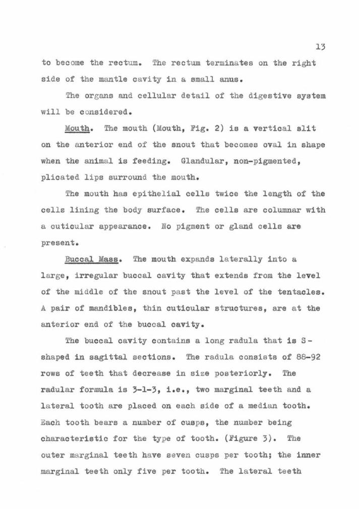
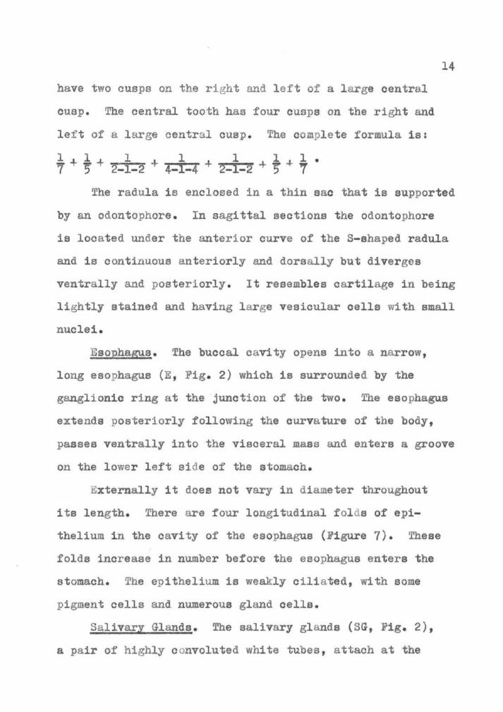
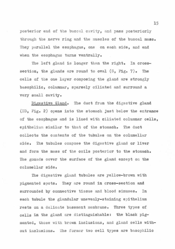
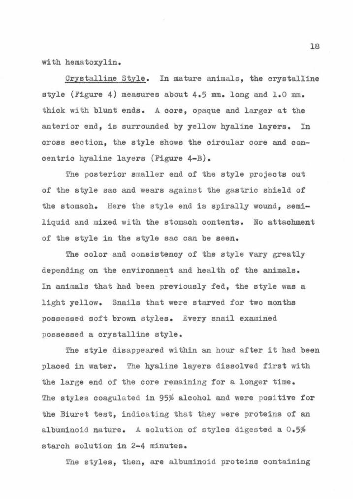
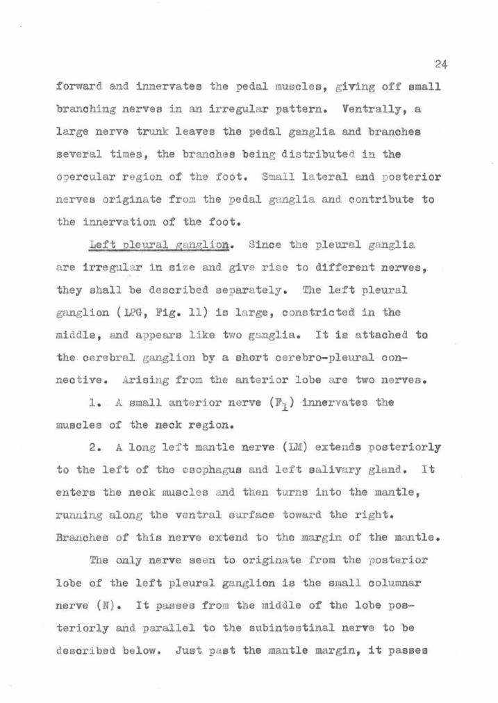
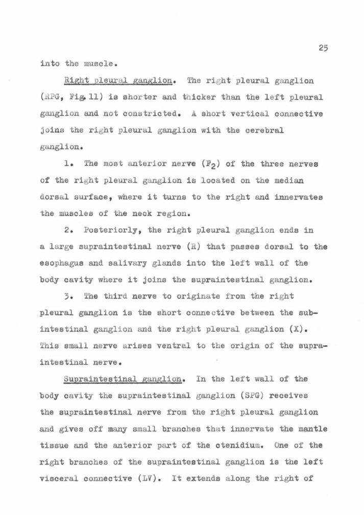
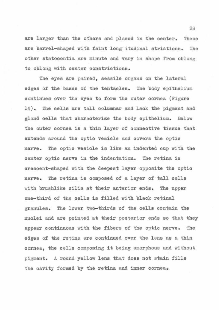
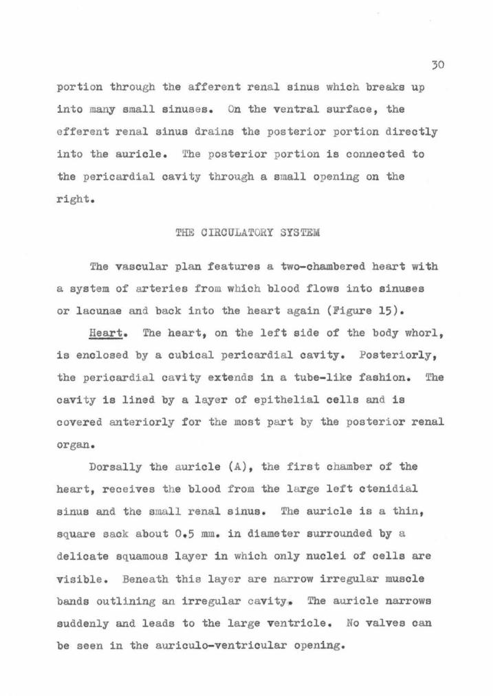
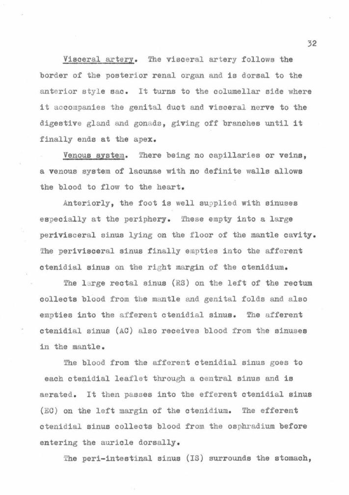
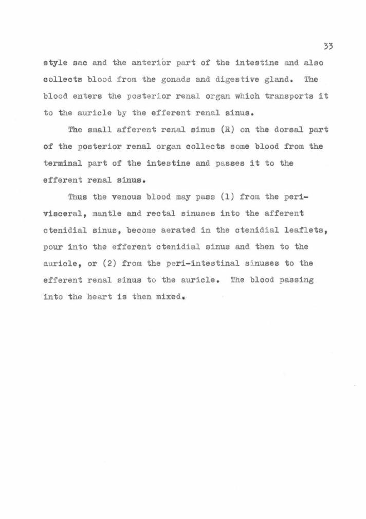
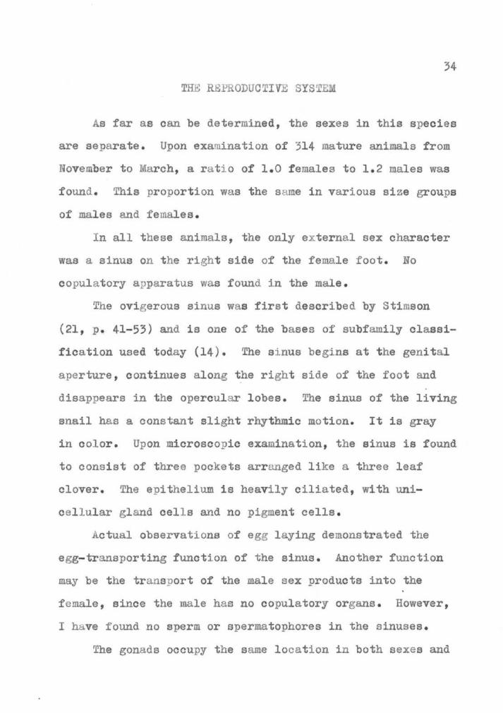
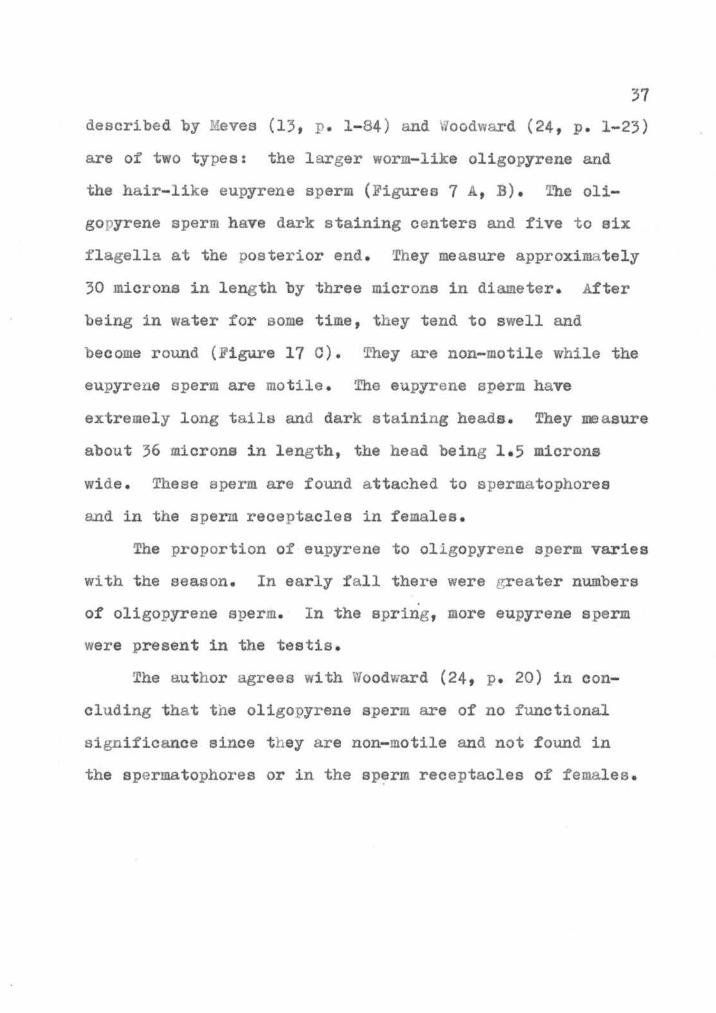
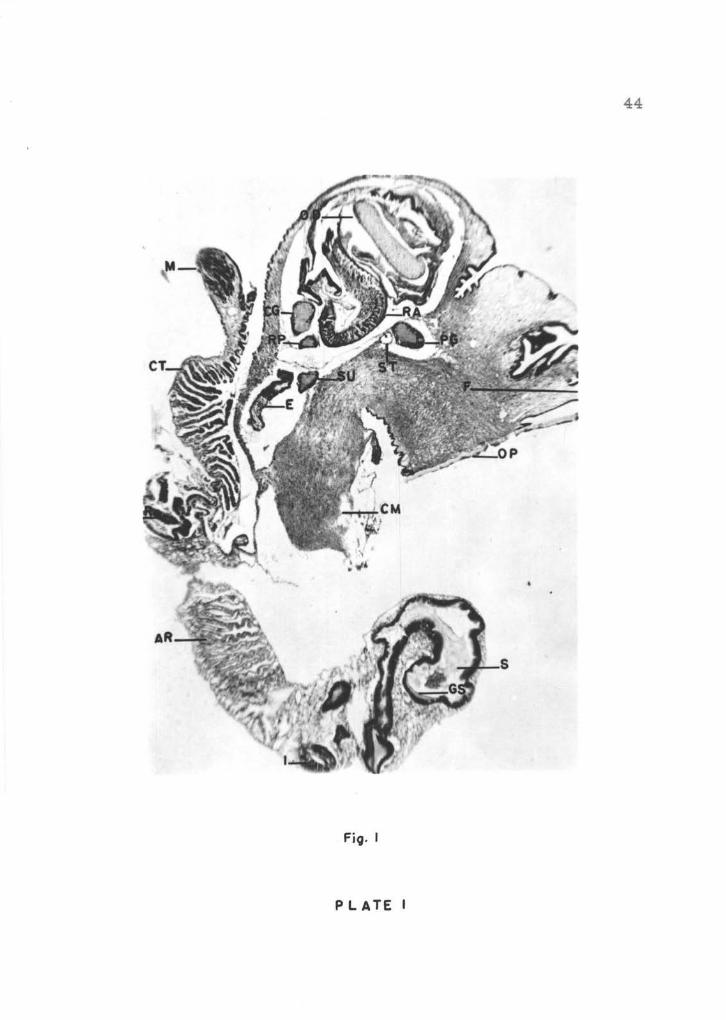
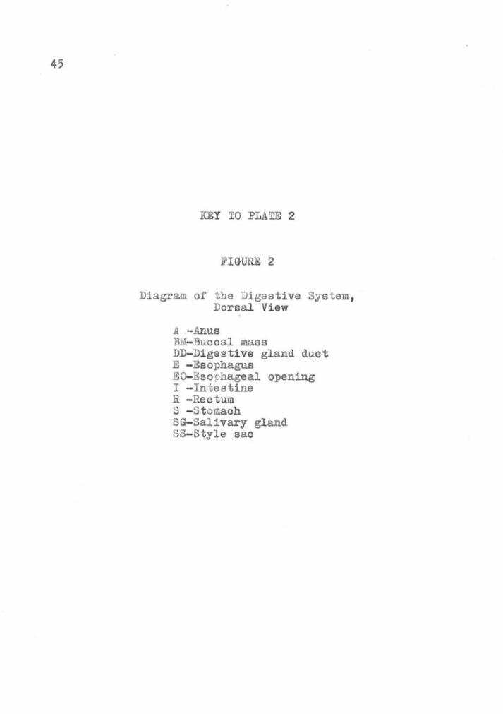
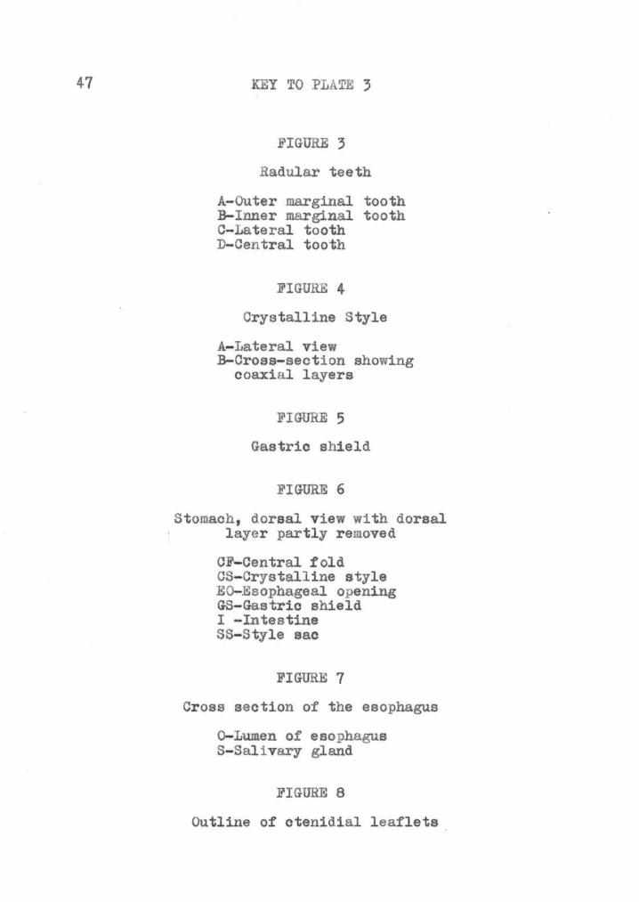
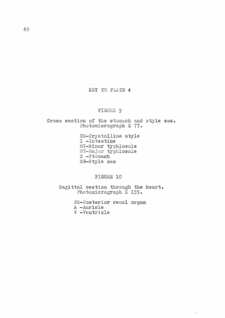
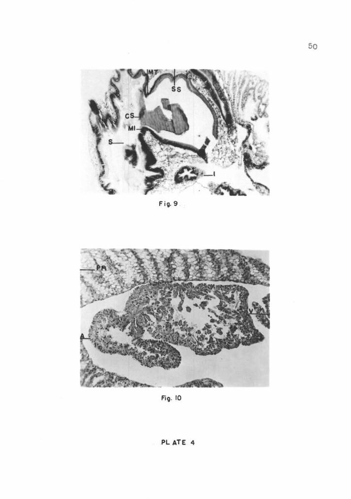
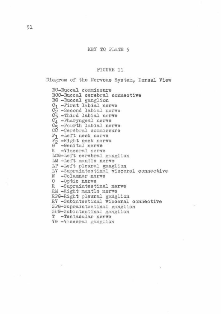
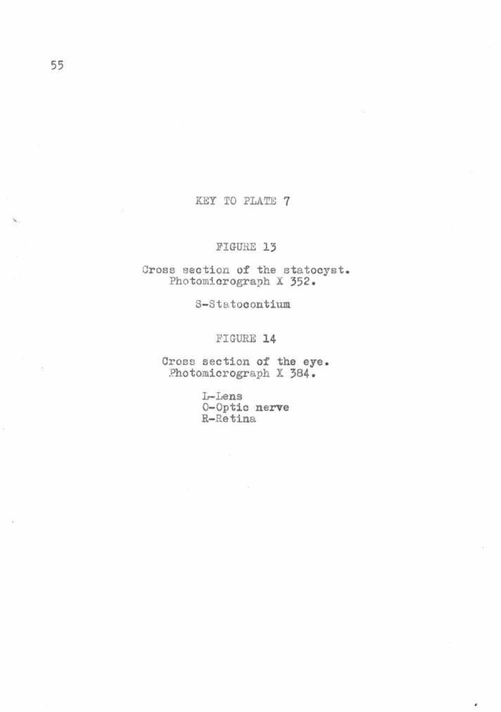
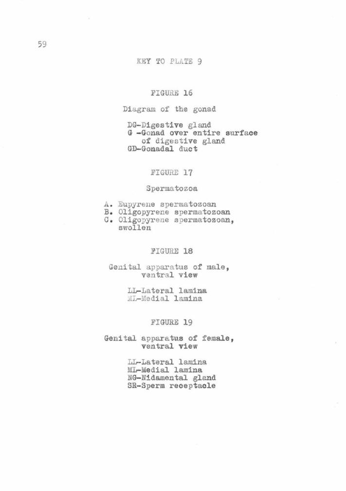
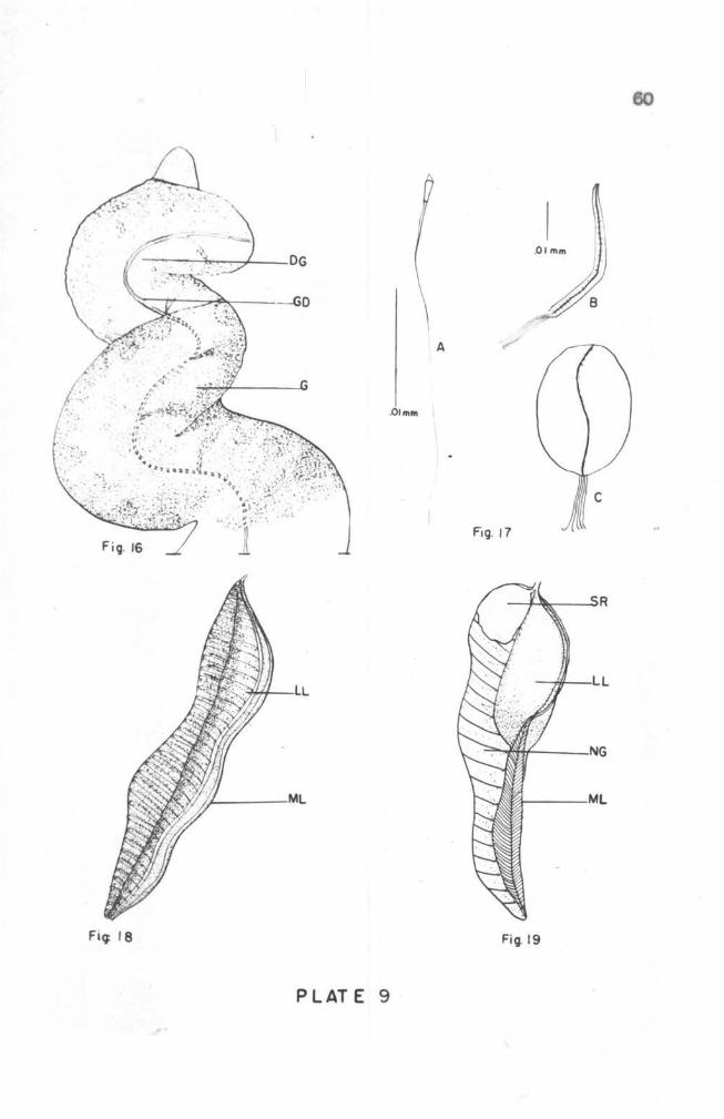
A
Oimm
Fiq I 8 Fig 19
PLATE 9