The molecular structure of an unusual cytochrome c 2 determined at 2.0 å; the cytochrome c h from...
9
The molecular structure of an unusual cytochrome c 2 determined at 2.0 Å; the cytochrome c H from Methylobacterium extorquens JON READ, RAJ GILL, SIMON L. DALES, JON B. COOPER, STEVE P. WOOD, and CHRIS ANTHONY Division of Biochemistry and Molecular Biology, School of Biological Sciences, University of Southampton, Southampton SO16 7PX, United Kingdom ~Received December 15, 1998; Accepted February 25, 1999! Abstract Cytochrome c H is the electron donor to the oxidase in methylotrophic bacteria. Its amino acid sequence suggests that it is a typical Class I cytochrome c, but some features of the sequence indicated that its structure might be of special interest. The structure of oxidized cytochrome c H has been solved to 2.0 Å resolution by X-ray diffraction. It has the classical tertiary structure of the Class 1 cytochromes c but bears a closer gross resemblance to mitochondrial cyto- chrome c than to the bacterial cytochrome c 2 . The left-hand side of the haem cleft is unique; in particular, it is highly hydrophobic, the usual water is absent, and the “conserved” Tyr67 is replaced by tryptophan. A number of features of the structure demonstrate that the usual hydrogen bonding network involving water in the haem channel is not essential and that other mechanisms may exist for modulation of redox potentials in this cytochrome. Keywords: cytochrome c H ; cytochrome c 2 ; Methylobacterium; tryptophan; X-ray structure During growth of methylotrophic bacteria on methane or metha- nol, energy is obtained from the oxidation of methanol catalyzed by a soluble quinoprotein methanol dehydrogenase ~ MDH!. This occurs in the periplasm and initiates a short electron transport chain, which involves cytochrome c L ~the electron acceptor for the dehydrogenase! and cytochrome c H ~the donor to the oxidase! ~Anthony, 1992!: MDH r Cytochrome c L r Cytochrome c H r Oxidase. To study the processes of intraprotein and interprotein electron transport, we are investigating the structures of these soluble pro- teins in Methylobacterium extorquens. The structure of the quin- oprotein methanol dehydrogenase has been published previously ~Ghosh et al., 1995; Anthony & Ghosh, 1998!, and this paper describes the structure of the donor to the oxidase, cytochrome c H . Its amino acid sequence ~Anthony, 1992! is typical of mitochon- drial cytochromes c and bacterial cytochromes c 2 that donate elec- trons to oxidases or photosynthetic reaction centers. These Class I cytochromes c are characteristically low spin with the single haem group attached covalently to the protein through thioether linkages between haem vinyl groups and two cysteine residues located near the N-terminus, the 5th ligand to the haem iron being histidine and the 6th ligand being methionine ~ Moore & Pettigrew, 1990!. They share a common polypeptide fold that leads to the burial of the haem propionate substituents within the protein interior. Of the 96 sequences of cytochrome c listed by Moore and Pettigrew, 26% of residues are invariant and most of these are also conserved in the bacterial cytochromes c 2 . Furthermore, the main features of these cytochromes are superimposable in the X-ray structures. Within the cytochromes c and cytochrome c 2 , the most signif- icant differences occur within the “left-hand” side of the haem cleft in which Met80 is bonded to the haem iron. It is the properties of the residues in this region that are thought to determine differences in redox potential, and small changes in structure in this region between the oxidized and reduced conformations are proposed to have important implications with respect to mechanisms of elec- tron transfer ~ Brayer & Murphy, 1996!. Of particular importance are Tyr67, which is hydrogen bonded to the sulfur atom of Met80 in the ferrocytochrome; Thr78, which is hydrogen bonded to the outer haem propionate; and Arg38, which is hydrogen bonded to the inner propionate. A water molecule in this region is also con- served in most structures in which it is hydrogen bonded to Tyr67 and Thr78. Although the primary sequence of cytochrome c H is very similar to that of other cytochromes c, a few unusual features suggest that it might be of special interest with respect to the currently accepted Reprint requests to: Professor Chris Anthony, Division of Biochemistry and Molecular Biology, School of Biological Sciences, University of Southampton, Southampton SO16 7PX, United Kingdom; e-mail: C.Anthony @soton.ac.uk. Protein Science ~1999!, 8:1232–1240. Cambridge University Press. Printed in the USA. Copyright © 1999 The Protein Society 1232
Transcript of The molecular structure of an unusual cytochrome c 2 determined at 2.0 å; the cytochrome c h from...

The molecular structure of an unusual cytochromec2
determined at 2.0 Å; the cytochromecH fromMethylobacterium extorquens
JON READ, RAJ GILL, SIMON L. DALES, JON B. COOPER, STEVE P. WOOD,and CHRIS ANTHONYDivision of Biochemistry and Molecular Biology, School of Biological Sciences, University of Southampton,Southampton SO16 7PX, United Kingdom
~Received December 15, 1998;Accepted February 25, 1999!
Abstract
CytochromecH is the electron donor to the oxidase in methylotrophic bacteria. Its amino acid sequence suggests thatit is a typical Class I cytochromec, but some features of the sequence indicated that its structure might be of specialinterest. The structure of oxidized cytochromecH has been solved to 2.0 Å resolution by X-ray diffraction. It has theclassical tertiary structure of the Class 1 cytochromesc but bears a closer gross resemblance to mitochondrial cyto-chromec than to the bacterial cytochromec2. The left-hand side of the haem cleft is unique; in particular, it is highlyhydrophobic, the usual water is absent, and the “conserved” Tyr67 is replaced by tryptophan. A number of features ofthe structure demonstrate that the usual hydrogen bonding network involving water in the haem channel is not essentialand that other mechanisms may exist for modulation of redox potentials in this cytochrome.
Keywords: cytochromecH; cytochromec2; Methylobacterium; tryptophan; X-ray structure
During growth of methylotrophic bacteria on methane or metha-nol, energy is obtained from the oxidation of methanol catalyzedby a soluble quinoprotein methanol dehydrogenase~MDH !. Thisoccurs in the periplasm and initiates a short electron transportchain, which involves cytochromecL ~the electron acceptor for thedehydrogenase! and cytochromecH ~the donor to the oxidase!~Anthony, 1992!:
MDH r CytochromecL r CytochromecH r Oxidase.
To study the processes of intraprotein and interprotein electrontransport, we are investigating the structures of these soluble pro-teins inMethylobacterium extorquens. The structure of the quin-oprotein methanol dehydrogenase has been published previously~Ghosh et al., 1995; Anthony & Ghosh, 1998!, and this paperdescribes the structure of the donor to the oxidase, cytochromecH.Its amino acid sequence~Anthony, 1992! is typical of mitochon-drial cytochromesc and bacterial cytochromesc2 that donate elec-trons to oxidases or photosynthetic reaction centers. These Class Icytochromesc are characteristically low spin with the single haem
group attached covalently to the protein through thioether linkagesbetween haem vinyl groups and two cysteine residues located nearthe N-terminus, the 5th ligand to the haem iron being histidine andthe 6th ligand being methionine~Moore & Pettigrew, 1990!. Theyshare a common polypeptide fold that leads to the burial of thehaem propionate substituents within the protein interior. Of the 96sequences of cytochromec listed by Moore and Pettigrew, 26% ofresidues are invariant and most of these are also conserved in thebacterial cytochromesc2. Furthermore, the main features of thesecytochromes are superimposable in the X-ray structures.
Within the cytochromesc and cytochromec2, the most signif-icant differences occur within the “left-hand” side of the haem cleftin which Met80 is bonded to the haem iron. It is the properties ofthe residues in this region that are thought to determine differencesin redox potential, and small changes in structure in this regionbetween the oxidized and reduced conformations are proposed tohave important implications with respect to mechanisms of elec-tron transfer~Brayer & Murphy, 1996!. Of particular importanceare Tyr67, which is hydrogen bonded to the sulfur atom of Met80in the ferrocytochrome; Thr78, which is hydrogen bonded to theouter haem propionate; and Arg38, which is hydrogen bonded tothe inner propionate. A water molecule in this region is also con-served in most structures in which it is hydrogen bonded to Tyr67and Thr78.
Although the primary sequence of cytochromecH is very similarto that of other cytochromesc, a few unusual features suggest thatit might be of special interest with respect to the currently accepted
Reprint requests to: Professor Chris Anthony, Division of Biochemistryand Molecular Biology, School of Biological Sciences, University ofSouthampton, Southampton SO16 7PX, United Kingdom; e-mail: [email protected].
Protein Science~1999!, 8:1232–1240. Cambridge University Press. Printed in the USA.Copyright © 1999 The Protein Society
1232

conclusions concerning the role of the conserved residues in thevicinity of the haem. The X-ray structure of cytochromecH re-ported here confirms that this is indeed the case. For example, theabsence of the “conserved” water, the substitution of Tyr67 bytryptophan, and the substitution of Arg38 by alanine prevent theestablishment of the typical hydrogen bonded network in the haemchannel that is considered to be important in modulating redoxpotentials and electron transfer events in other Class Ic-typecytochromes.
Results and discussion
The primary sequence of cytochrome cH
The primary sequence of cytochromecH ~100 residues! has beenpreviously determined by Ambler and Athalye~cited in Anthony,1992!. During model building of the X-ray structure, it provedpossible to fit the entire sequence within the electron density ex-cept for the loop containing residues 38–42. To resolve this anom-aly, a peptide containing this part of the sequence was produced bydigestion with trypsin, and its sequence shown by Edman degra-dation to be 37-AGEGADGYAFSDA-49, thus reordering residues41 to 43 from GAD to ADG. This sequence correlated well withthe electron density and was used subsequently in model building.The sequence of cytochromecH is presented in Figure 1 alignedwith sequences of mitochondrial cytochromesc from tuna, horse,and yeast, and some cytochromesc2 whose X-ray structures havebeen determined. The alignment shows that between 43 and 45%of residues of cytochromecH are identical to those in cyto-chromesc andc2. Although its sequence shows that cytochromecH
is clearly a typical cytochromec2, for convenience in this paper wewill continue to refer to it as cytochromecH.
The overall structure of cytochrome cH
The structure of cytochromecH was determined by molecular re-placement and refined to anR-factor of 16.5% with a correspond-ing Rfree of 22.2%~see Materials and methods!. The polypeptidechain backbones had continuous electron density for all three mol-ecules in the triclinic unit cell. Side chains were partially disor-dered for all the three molecules in residues Lys9, Glu21, Lys22,Lys30, Lys36, Glu39, Lys52, and Lys63. The average main-chainthermal parameters~B-factors! for all three molecules in the unitcell are shown in Figure 2. The main-chain torsion angles for allthree molecules in the unit cell were used for construction ofthe Ramachandran plot, which showed no residues in disallowedregions, and 88% of residues within the most favored regionsas defined in the PROCHECK program used for this analysis~Laskowski et al., 1993!.
CytochromecH has the classical tertiary structure of the Class 1cytochromesc andc2, as shown in Figure 3. This comprises fivehelices~A to E! and their interconnecting loops, which togetherenclose the haem prosthetic group whose edge breaches the “front”face of the molecule where close approach with redox couplepartners is thought to occur during electron transfer. It is notablethat cytochromecH bears a closer gross resemblance to cyto-chrome c than to the bacterial cytochromec2; the main-chainatoms for cytochromecH and tuna cytochromec superimpose witha root-mean-square deviation~RMSD! of 0.77 Å for 90 residues;for cytochromecH and cytochromec2 of Rhodospirillum rubrum,the RMSD is 0.85 Å for 87 residues.
Fig. 1. The sequence of cytochromecH of M. extorquens. This is aligned with sequences of cytochromes whose structures have beendetermined; these include the mitochondrial cytochromec from horse heart~Bushnell et al., 1990; Qi et al., 1994; Banci et al., 1997b!,tuna~Tanako & Dickerson, 1981a, 1981b!, and yeast~Saccharomyces cerevisiaeiso-I-cytochromec! ~Louie & Brayer, 1990; Baist-rocchi et al., 1996; Benning et al., 1996; Banci et al., 1997a!; and the cytochromesc2 from Rhodopila globiformis~Benning et al., 1996!and R. rubrum ~Salemme et al., 1973!. Sequences are taken from Moore and Pettigrew~1990!. The numbers refer to the tunamitochondrial cytochromec ~upper!, M. extorquens~middle!, andR. rubrum~lower!. The residues marked in bold are those that aredifferent in cytochromecH and which have a major influence on the properties of the left hand side of the haem cleft. The underlinedsequence is the loop that is relatively more rigid in cytochromecH.
Structure of an unusual cytochrome c2 1233

The a-helix A ~residues 4 to 15! is distorted by Pro13~seeFig. 3! that replaces an otherwise conserved lysine~tuna, Lys13!on the surface, and this change allows solvent access to atom CBBon the haem. The sequence of residues 15 through 39 is composedof a series of turns. The loop~18 to 29! immediately after the haembinding sequence~CKACH! is three residues shorter than in cy-tochromesc and c2 and encloses a buried water molecule in allthree cytochromes~Wat4 in cytochromecH!. The base loop~36 to39! has an insertion of one residue and therefore adopts a slightlydifferent conformation from that in cytochromesc andc2; further-more, the otherwise conserved arginine~tuna Arg38! is replaced byAla35 in cytochromecH. Helix B ~41 to 43! is short~about oneturn in length! and is followed by a portion of “random coil”similar in conformation to cytochromec. Helix C ~59 to 67! con-sists of two full turns as seen in cytochromec2 and contains aunique tryptophan substitution~Trp65! toward its C-terminus, re-placing Tyr67 in tuna cytochromec. Helix D ~69 to 72! is only fourresidues long as in cytochromec, and the subsequent extendedchain ~73 to 86! also follows a similar path. The last helix~E!~residues 86 to 97! is intermediate in length~about three turns!between the E helices of cytochromec2 ~2.5 turns! and cyto-chromec ~four turns!. The terminal helices~A and E! are heldtogether by way of a van der Waals interaction between aromaticside chains~Phe10 and Tyr95! on the two helices exactly as seenin tuna cytochromec.
In cytochromesc andc2, the residues immediately surroundingthe haem edge facing the solvent are mainly hydrophobic and thefive closest of these are conserved as hydrophobic residues incytochromecH ~Moore & Pettigrew, 1990; Brayer & Murphy,1996!; these are Ala12~tuna, Val11!, Val25~Val28!, Tyr44~Tyr46!,Val79 ~Ile81!, Phe80~Phe82!, and Pro81~Ala83!. Ala15 is re-placed with Lys15. Only two out of the five acidic residues of tunaare acidic in cytochromecH; one of these is the highly conservedAsp91~tuna, Asp93!. Of the 12 lysine residues present on the front
face of tuna cytochromec, only four are present in cytochromecH
~Lys9, Lys70, Lys71, and Lys77!; these include the three that areconserved in all cytochromesc andc2 and which are thought to beimportant in interactions with redox partners~tuna, Lys72, Lys73,and Lys79!.
Because the overall structure of cytochromecH is generallysimilar to other cytochromesc and c2, we can conclude that in-teraction with its redox partners is by way of bonding betweenhydrophobic residues around the haem edge, following an initialionic interaction involving the conserved lysines, as shown previ-ously by chemical modification and cross linking studies~Anthony,1992; Cox et al., 1992!.
The flexibility of the cytochrome cH backbone
Figure 2 shows the meanB-values for the main-chain atoms ofoxidized cytochromecH for each of the three molecules in the unitcell; the regions showing the greatest flexibility~higherB-values!are, as expected, the loop regions. The loop leading to the methi-onine ligand~Lys71–Met78; indicated by IV in Fig. 2! is relativelyimmobile as seen in other cytochromesc. In these cytochromesthere is a salt bridge between the “invariant arginine”~tuna, Arg38!and the inner haem propionate, the loop carrying this argininebeing relatively rigid and showing little change on reduction. Bycontrast, in cytochromecH, Arg38 is replaced by Ala35 and there-fore, as expected, the loop between Ala35 and Ala41~indicated byI in Fig. 2! is relatively mobile. In cytochromesc three other loopsare relatively mobile~yeast numbering: 47–59, 65–72, 81–85!. IncytochromecH two of these are also mobile; these are region II~Ala45–Trp57!, and region V~Lys79–Ile83!. The third of theseloops ~III; Lys63–Pro70!, however, is relatively rigid; this is theloop that contains the conserved tyrosine in cytochromesc andc2
~tuna, Tyr67!, which is uniquely replaced by tryptophan~Trp65!,leading to a generally more rigid structure in this region in cyto-chromecH.
The hydrophobic environment of the haem in cytochrome cH
In cytochromecH many of the interactions with the haem are verysimilar to those in all other cytochromesc and c2. The haem iscovalently bonded to the protein by thioether linkages from Cys14and Cys17, and the haem iron is ligated to His18 and Met78; thebond length between S of Met78 and the haem iron atom is 2.27 Å,that between NE2 of His18 and the haem iron is 1.91 Å, and theangle formed between these bonds is 174.48. Except for some ofthe interactions with the haem propionates, all other interactionsare predominantly hydrophobic in character including those in-volving Trp57~tuna, Trp59!, Phe80~Phe82!, Pro69~Pro71! on theleft of the haem~with respect to the conventional view as seen inFig. 3!, and Leu66~Leu68! behind the methionine. However, thefollowing replacements near the haem of cytochromecH are morehydrophobic than in cytochromec: Trp65 ~tuna, Tyr67!, Ala35~tuna, Arg38!, Phe46~Tyr48!, and Leu50~Asn52!.
In cytochromecH, as in most cytochromesc andc2 , the outerhaem propionate~HP6, on haem ring D! has no direct interactionwith solvent; it forms hydrogen bonds with side chains of buriedthreonine or serine and the main-chain nitrogen atom of a lysineresidue~Table 1; Fig. 4!. The inner haem propionate~HP7, on ringA! is exceptional in cytochromecH in not being shielded com-pletely from solvent~Fig. 5!. Although the bonding of one oxygenatom~O2A! is similar to that in cytochromec ~Table 1, Fig. 6!, the
A
B
C
Fig. 2. The B-factors for the main-chain atoms of cytochromecH. ThemeanB-factors are represented in each case by a horizontal line. Regionsof the molecule that are referred to in the text are marked as dark bars.
1234 J. Read et al.
other oxygen atom~OA1! is bonded completely differently. Thisatom is usually protected from bulk solvent by hydrogen bondingto one of the conserved waters~tuna, Wat26! and to the conservedTyr48 ~tuna! and Arg38 ~tuna! ~Fig. 6!. In cytochromecH, thepropionate oxygen~O1A! is also hydrogen bonded to water~Wat1;tuna, Wat26!, but Tyr48 and Arg38~tuna! are replaced by thenonbonding Phe46 and Ala35. The space usually taken by thearginine side chain is occupied by a second water~Wat2!, which ishydrogen bonded to the propionate~O1A! and to water in contactwith bulk solvent~Table 1; Figs. 6, 7!. It is possible that dissoci-ation of this exceptionally exposed “inner” propionate contributesto the unusual pH dependence of redox potential previously re-ported for this cytochrome~O’Keeffe & Anthony, 1980!.
A unique feature of the haem cleft of cytochrome cH
Of the three buried conserved water molecules present in cyto-chromesc andc2, only two are present in cytochromecH. One ofthese is Wat4~tuna, Wat2!, adjacent to the histidine ligand; thesecond Wat1~tuna, Wat26! is located toward the outside of thecleft and is bonded to main-chain oxygen and nitrogen atoms andto the inner propionate~HP7! ~Table 1; Fig. 6!. At the entrance ofthe haem cleft in cytochromecH, there is an additional water~Wat2! occupying the position of the side chain of Arg38~in tuna!,which is replaced in cytochromecH by Ala35 ~Fig. 6!. This wateris in direct contact with bulk solvent~Fig. 5! and is also linked tothe inner haem propionate~HP7!.
The majority of cytochromesc and c2 contain a third buriedwater molecule~Wat3 in tuna; Wat166 in yeast! in the left-handside of the haem cleft where it is hydrogen bonded to a conservedtyrosine~tuna, Tyr67! ~Scott & Mauk, 1996; Surridge, 1994!. Re-markably, in cytochromecH this tyrosine is replaced by tryptophan~Trp65! whose bulk precludes the possibility of a buried watermolecule in this location~Table 1; Fig. 7!.
It should be noted that cytochromec2 of R. rubrum is alsounusual in lacking the “conserved” buried water but this is re-placed by a second tyrosine~Tyr52! whose hydroxyl oxygen atomreplaces the water oxygen and forms a hydrogen bonding networkwith Tyr70 ~tuna, Tyr67! and Ser89~tuna, Thr78! in the ferricyto-chrome~Table 1; Fig. 4! ~Salemme et al., 1973!.
The redox potential of cytochrome cH
Factors affecting redox potentials of cytochromes include localdielectric effects arising from the amount of solvent exposed haem,the presence of more or less hydrophobic residues near the haem,the orientation of the haem ligands relative to the haem plane, theextent of hydrogen bonding to these ligands and the haem propi-onates, and solvent structure in the haem binding pocket~Moore &Pettigrew, 1990!. For example, a more hydrophobic environmentin the haem pocket will favor the reduced form and raise the redoxpotential Kassner~1972!. The redox potential of cytochromecH
~294 mV! is 34 mV higher than that of mitochondrial cytochrome~O’Keeffe & Anthony, 1980!, which is consistent with the lack of
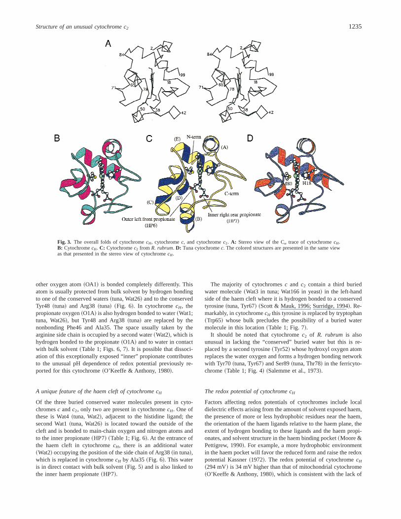
Fig. 3. The overall folds of cytochromecH, cytochromec, and cytochromec2. A: Stereo view of the Ca trace of cytochromecH.B: CytochromecH. C: Cytochromec2 from R. rubrum. D: Tuna cytochromec. The colored structures are presented in the same viewas that presented in the stereo view of cytochromecH.
Structure of an unusual cytochrome c2 1235

water molecules and the remarkable number of hydrophobic sub-stitutions in this region: Thr40 is replaced with Ala37, Ser47 withAla45, Tyr48 with Phe46, Asn52 with Leu50, and Tyr67 withTrp65. CytochromecH is the only cytochromec, which has atryptophan~Trp65! in the position normally occupied by Tyr67,and the only one known to lack the internal water, or hydroxylequivalent, and its associated hydrogen bonding network, in theleft-hand side of the haem cleft.
Considerable speculation has centered around the role of tyro-sine~tuna, Tyr67! in modulating redox potential. Only 2% of cy-tochromesc or c2 have an alternative~phenylalanine! at this position~Moore & Pettigrew, 1990!. No cytochrome has been investigatedin which Tyr67 has been modified to a tryptophan, but a number ofvariants have been investigated in which it has been modified~chemically or by mutagenesis! to the relatively hydrophobic phe-nylalanine. If this had led to no changes except for a change inhydrophobicity then the mutation would be expected to lead to ahigher redox potential as observed in cytochromecH; however, inall cases the redox potential was decreased by 30–60 mV~Luntzet al., 1989; Wallace et al., 1989; Caffrey et al., 1991; Berghuiset al., 1994!. An explanation of this observation has been affordedby determination of the complete structures of oxidized and re-
duced forms of the wild-type~Tyr67! and the Tyr67Phe variant ofthe iso-1-cytochromec of yeast by Berghuis et al.~1994!. Theyhave shown that the mutation does not lead to an overall increasein hydrophobicity because in this variant the usual water~Wat166!is retained and a second water is present in an enlarged internalcavity, in a position approximately equivalent to that of the hy-droxyl group of Tyr67 in the wild-type protein. The mutation alsoleads to alterations in the hydrogen bond network seen in wild-typecytochromec, involving Tyr67, Wat166~tuna, Wat3!, and othernearby residues, and loss of the Tyr67 to Met80 hydrogen bondnormally present in the reduced form. It was concluded that thesealterations lead to a structurally more rigid and stable form of theoxidized protein and hence a substantially lower midpoint poten-tial in the variant~Berghuis & Brayer, 1992!.
A further structural feature of cytochromes that has been impli-cated in the regulation of redox potential is the salt bridge betweenthe inner haem propionate~HP7! and Arg38, replacement of whichby other amino acids leads to a substantial decrease in redox po-tential ~Proudfoot & Wallace, 1987!. This is consistent with thesuggestion of Moore et al.~1984! that the positive charge on theguanidino group of Arg38 effectively neutralizes the negativelycharged inner propionate, thus diminishing its potential electro-
Table 1. The coordination of water and propionates in cytochrome c of tuna, cytochrome c2
of R. rubrum and cytochrome cHa
Coordinating species
CytochromecTuna
Cytochromec2
R. rubrumCytochromecH
M. extorquens
Water; Wat26, Wat1 O2A of HP7~inner haem!
2.6 Å Water replacedby His42
O1A of HP7~inner haem!
2.8 Å
Gln42 N 2.9 Å Glu39 N 2.8 ÅLys39 O 2.8 Å Lys36 O 3.1 Å
Gly40 N 2.7 Å
Water; Wat3 Tyr67 OH 2.9 Å Water replacedby OH of Tyr52
Trp57 excludes water
Thr78 OG1 2.9 ÅAsn52b ND1 3.0 Å
Tyr52 of R. rubrum Tyr70 OH 2.7 Å~This replaces Wat3.! Ser89 OG 2.4 Å
Inner haem propionate~HP7!O1A Wat26 O 2.6 Å Wat1 O 2.8 Å
Tyr48 OH 2.7 Å Tyr48 OH 2.7 Å Phe46 cannot bondArg38 NH1 2.8 Å His42 N 2.9 Å Ala35 cannot bond
His42 ND1 2.2 Wat2 O 2.9 Å
O2A Trp59 NE1 3.0 Å Trp62 NE1 3.0 Å Trp57 NE1 2.9 ÅGly41 N 3.3 Å Ala41 N 2.9 Å Gly38 N 2.8 ÅAsn52b OD 3.2 Å Tyr42 not bonded Leu50 cannot bond
Outer haem propionate~HP-6!O1D Thr49 N 3.0 Å Ser49 N 2.9 Å Ser47 not bonded
O2D Thr49 OG1 2.7 Å Thr49 OG1 2.6 Å Ser47 OG1 2.8 ÅSer47 N 2.7 Å
Thr78 OG1 2.7 Å Ser89 not bonded Thr76 OG 2.6 ÅLys79 N 3.1 Å Lys90 N 2.6 Å Lys77 N 2.9 Å
aIn all the bonds identified in the table, the residues involved are equivalent in the sequence~see Fig. 1!, except that Arg38 incytochromec is replaced by His42 inR. rubrumcytochromec2.
bAsn52 is bonded to Wat3 and propionate only in the reduced cytochrome.
1236 J. Read et al.

static effect at the haem iron and thereby raising the observedmidpoint potential. The propionate~HP7! of cytochromecH isunable to form such a salt bridge, because Arg38 is replaced byAla35, which should lower the redox potential for cytochromecH.We suggest that the observed redox potential of cytochromecH is
achieved by the delicate balance of these opposing influences,between hydrophobicity of the left side of the haem cleft on theone hand, and the absence of Arg38 and increased haem exposureon the other.
The relevance of the cytochrome cH structureto the electron transfer mechanism
In cytochromec the buried water~tuna, Wat3! is part of a hydrogenbonding network involving Thr78, and the outer haem propionate~HP6! ~Fig. 4!. In the reduced state, the water is also bonded toAsn52, and Tyr67 is hydrogen bonded to the sulfur of the Met80ligand. During oxidation these two bonds are broken, with a cor-responding shift in the side chains of Asn52 and Tyr67, and thewater molecule is shifted about 1.2 Å toward the iron. This con-formational change is likely to occur in response to, or alterna-tively to stabilize, the increased positive charge centered at thehaem iron in the oxidized form~Louie & Brayer, 1990!. It has beensuggested that the consistency of these observations across allavailable cytochromec structures strongly implicates this waterand its hydrogen bonding network in their electron transfer mech-anisms~Brayer & Murphy, 1996!. However, in cytochromecH the
Fig. 4. Stereo diagrams of the left-hand channel of the haem binding sitein the oxidized cytochromes. The inner propionate~HP7! is omitted forclarity. A: CytochromecH; although the hydrogen bonding pattern of theouter propionate~HP6! is the same as in the other two cytochromes, Tyr67is replaced by Trp65 and there is no buried water.B: Cytochromec ~tuna!;Tyr67 is hydrogen bonded to the buried water~W3!. Asn52 forms addi-tional bonds with W3 and HP7 in the reduced cytochrome.C: Cyto-chromec2 from R. rubrum. Asn52 in tuna is replaced by a tyrosine whoseside-chain hydroxyl group occupies the same position as the conservedwater molecule. These diagrams were produced using MOLSCRIPT~Krau-lis, 1991!.
Fig. 5. The solvent exposed regions of haem in cytochromecH and tunacytochromec. The solvent exposed regions of the haem~in black! werecalculated using a rolling ball of 1.4 Å radius in GRASP~Nicholls et al.,1993!. Tuna cytochromec has atoms CMC, CBC, and CMD of the haemexposed; in addition to these, cytochromecH also has atom CBB exposedon the front surface and atom OIA of the inner haem propionate~HP7!exposed on the lower right-hand side of the molecule.
Structure of an unusual cytochrome c2 1237

unique structure of the left-hand side of its haem cleft, lackingwater and the associated hydrogen bonding web, precludes such amechanism. Thr76~tuna, Thr78! is bonded to the outer haem pro-pionate~HP6! as in other cytochromes, but Asn52 is replaced byLeu50, and the tryptophan~Trp65! is unable to form hydrogenbonds with the methionine ligand to the haem iron~Fig. 4!.
Any structural changes that take place during oxidation andreduction of cytochromecH must do so in the unusually hydro-phobic environment in its haem cleft, and these are likely to in-volve the novel Trp65, which replaces the conserved Tyr67. Trp65
is unlikely to form a hydrogen bond to the iron ligand~Met78! inthe ferrocytochrome as this would require a large rotation of thetryptophan side chain, which could only occur if there were amajor change in the overall structure in this region. It is much moreprobable that Trp65 is involved in an alternative mechanism. Thering plane of Trp65 is inclined by about 158 from an orthogonaldisposition with respect to ring A of the haem such that the indoleNH is directed toward the pyrrole nitrogen~3.5 Å distant! ~Fig. 7!.Small rotational or translational adjustments to the position ofTrp65 would permit the closer approach of NE1 to the haem planewhere thep cloud is able to fill the role of acceptor for a hydrogenbond. Such hydrogen bond systems were first discussed by Levittand Perutz~1988!, and a number of examples have subsequentlybeen identified~Mitchell et al., 1994!. In the current context, thiswould provide an effective means for modulating the relative sta-bility of the reduced and oxidized states. Furthermore, Trp65 is invan der Waals contact with the Trp57 side chain that in turn hy-drogen bonds to the HP7 propionate~Fig. 7!, thus perhaps con-tributing to the remarkable pH dependence of redox potential.
Other changes that have been observed on reduction of mito-chondrial cytochromec include the conformation of the loops thatinterconnect the inside and outside of the protein in the region ofthe haem pocket cleft. On the basis of work with the yeast cyto-chromec, a model has been proposed that links the changes in thehydrogen bonding network with changes in conformation of themore mobile surface loops~Berghuis & Brayer, 1992; Brayer &Murphy, 1996!. It was suggested that the relatively rigid 73–80segment, by virtue of its surface location next to the solvent ex-posed edge of the haem, can act to trigger the structural changesrequired to stabilize the oxidation state required to facilitate elec-tron transfer. On forming a complex with redox partners, the 75–78b turn residues act as a “push button,” which leads to a change inconformation involving movement of three mobile loops~tuna47–59; 65–72; 81–85!. Although cytochromecH retains the rela-tively immobile region~Lys71–Met78!, only two of the three mo-bile loops are retained; the third is the critical loop~Lys63–Pro70!that carries the conserved tyrosine in cytochromesc andc2 ~tuna,Tyr67!, which is uniquely replaced by tryptophan~Trp65! in cy-tochromecH, leading to a generally more rigid structure in thisregion. Clearly, in cytochromecH, no such mechanism can be
Fig. 6. Stereo diagrams of the hydrogen bonding patterns surrounding theinner haem propionate HP7.A: CytochromecH; the highly conservedArg38 ~tuna! is replaced by Ala35 allowing access of the bulk solvent to theinner propionate HP-7, which is an otherwise relatively hydrophobic en-vironment.B: Tuna cytochromec, in which the inner propionate is shieldedfrom the bulk solvent by Arg38.C: Yeast iso-1 cytochromec; Arg38 isconnected to the propionate via W121~equivalent to tuna Wat26! and anadditional water~W168! forming a rigid network, which shields the pro-pionate from the bulk solvent; in this cytochrome Trp59 and Asn52 do notbond with the inner propionate.D: R. rubrumcytochromec2; the side chainof the highly conserved Arg38 is replaced by the side chain of His42, whichforms an electrostatic link with the inner propionate, protecting it from thebulk solvent and preventing the usually conserved water~Wat26! frombeing present. These diagrams were produced using MOLSCRIPT~Krau-lis, 1991!.
Fig. 7. The packing of Trp57 and Trp65 adjacent to the haem in cyto-chromecH showing hydrogen bond interactions between the indole NE1 ofTrp57 and the inner haem propionate HP7 and a potential hydrogen bondbetween Trp65 and the ring A haemp cloud.
1238 J. Read et al.

involved during its oxidation and reduction, and it should be notedthat such conformational changes are not, in principle, essential forthese redox reactions. Indeed, this is indicated by the observationthat in the Tyr67Phe variant of yeast iso-1-cytochromec systemthere is relatively little difference in the conformations of the ox-idized and reduced forms and yet it is able to fulfill its functionsalmost as well as the wild-type protein~Berghuis et al., 1994!.
Finally, we should consider whether or not there is any reasonfor cytochromecH to have the very special structure that it does. Apossible explanation is that this has evolved to provide a relativelyhigh redox potential, which may be essential for its additionalspecial function in the “methanol oxidase” electron transport chain~Anthony, 1992!. It is similar to other cytochromesc in mediatingelectron transfer between the cytochromebc1 complex and theoxidase, but it has the special additional function of oxidizing thecytochromecL that is the specific electron acceptor from methanoldehydrogenase. This cytochrome has a typical redox potential~254 mV! ~O’Keeffe & Anthony, 1980! and a cytochromec witha higher redox potential will facilitate its oxidation.
Materials and methods
Crystallization and data collection
The protein was purified as previously described~Day & Anthony,1990!. Initial crystallization conditions were found using the Hamp-ton sparse matrix screen and optimized on the initial conditionsfound ~screen 1 no. 43—30% PEG 1500, unbuffered!. Crystalswere grown using the hanging drop vapor diffusion method. Thefinal conditions were 10 mg0mL protein, 18% PEG 1500, 50 mMTris-HCl, pH 7.0. Crystals appeared within 2 days with dimensionsof 200mm 3 200mm 3 100mm. X-ray data were collected on aMar 30 cm Image plate at room temperature. The X-ray source wasan Enraf Nonius rotating anode generator operated at 50 kV and100 mA producing copper Ka radiation via a graphite monochro-mator. The crystal to detector distance was 150 mm giving a max-imum resolution of 2 Å at theedge of the plate. Data processingstatistics are given in Table 2. The space group is P1, with thefollowing cell dimensions:a5 33.76 Å,b5 57.57 Å,c5 50.95 Å,a 5 67.818, b 5 89.338, andg 5 74.408. Assuming three moleculesper triclinic unit cell, a solvent content of 40% was calculated forthe crystal.
Structure determination and refinement
The initial coordinate model used in molecular replacement wasfor cytochromec2 from R. rubrum, having removed those regionswhere the sequence alignment indicated major insertions relativeto cytochromecH. The orientations of the three molecules in thecytochromecH unit cell were determined from a cross-rotationfunction calculated with ALMN where the solutions were the threehighest peaks. An oriented search molecule was positioned at theorigin of the P1 cell for cytochromecH. The translation vector foreach of the other two molecules was calculated using TFFC. Thepeak height for the second and third molecule translations was 9.25~map RMS 4.26! and 10.85~RMS 4.74!. Having applied appro-priate rotations and translations to theR. rubrumcytochromec2
coordinates, the model was subjected to rigid body and subsequentrestrained positional, and thermal parameter, refinement withRESTRAIN~Haneef et al., 1985!. Five percent of data was flagged
for the Free-R set. The first electron density maps were averagedusing density modification. Unless otherwise stated, all calcula-tions were performed using programs in the CCP4 suite~CCP4,1994!. Modeling was carried out using the X-Autofit-Autobuild op-tion of QUANTA ~Molecular Simulations, Inc., Burlington, Massa-chusetts!. The initial difference and double difference maps showedclearly the location of sequence differences from the search modeland regions where main-chain adjustments were required. Althoughgood electron density was found for most regions, the loops 38 to42 and 46 to 55 were less clearly defined. It was possible to tracethe loop from residues 46 to 55 using the superposed coordinatesfor oxidized horse heart cytochromec. The sequence of residues 38to 42 fitted the electron density poorly and so this region wasresequenced. The new sequence fitted the electron density much
Table 2. Data processing and refinement statisticsfor the cytochrome cH structure
Total
Highestresolution shell~2.07–2.01 Å!
Data processingRmerge
a 6.1% 13.4%x2 b 1.52 1.32^s~I !& 740 265^I & 12,043 1,682Redundancy 2.25 1.9Completeness 94.4% 71.5%
Refinement statisticsResolution range~Å! 30–2.01Number of atoms 2,445~2,160 protein,
129 haem, 156 waters!Number of parameters refined 9,951Number of reflections used 16,999R-factorc ~%! 16.1Correlation coefficient~b! 0.96Free-R factord ~%! 22.2Correlation coefficiente ~free set! ~b! 0.92AverageB main chain~Å2! 25.2AverageB protein atoms~Å2! 30.5AverageB waters~Å2! 42.6Overall B ~Å2! 31.4
RMSD from ideal geometryBond lengths~Å! 0.007Bond angle distances~Å! 0.018Van der Waals contacts~Å! 0.028Type I planes~Å! 0.009Type II planes~Å! 0.007Chiral tetrahedra~Å! 0.011
aRmerge 5 (h(i 6~Ihi 2 Ih!60(h(i ~Ihi! where Ih is the mean of scaledobservationsIhi.
bx2 5 ~(h(i @~Ihi 2 Ih!20s2~Ihi!#!•n0~n 2 1! where n 5 number of
observations.cRefinementR-factor 5 (~6Fo6 2 6Fc6!0(Fo whereFo and Fc are the
observed and calculated structure factors.dThe freeR-factors were calculated with 5% of the reflection data.3Correlation coefficient5 A0#~BC! where A 5 n(~6Fo66Fc6! 2
~(6Fo6!~(6Fc6!; B 5 n(6Fo62 2 ~(6Fo6!2; C 5 n(6Fc62 2 ~(6Fc6!2 andn5 number of structure factors included. Fitting of structures was achievedusing the LSQKB program, which uses the Kabsch algorithm~Kabsch,1976!.
Structure of an unusual cytochrome c2 1239

better, although this region still displayed high thermal parameters.After fitting of the main-chain atoms and further positional refine-ment, NCS restraints were removed and waters added.
The structure of cytochromecH was refined to anR-factor of16.5% with a correspondingRfree of 22.2%; the data processingand refinement statistics are given in Table 2. The RMSDs ofmain-chain and side-chain atoms between the three molecules ofcytochromecH in the unit cell were 0.29–0.34 and 0.66–0.78 Å,respectively. Fitting of structures was achieved using the LSQKBprogram, which uses the Kabsch algorithm~Kabsch, 1976!.
Acknowledgments
We acknowledge financial support from BBSRC and The Wellcome Trust.
References
Anthony C. 1992. The c-type cytochromes of methylotrophic bacteria.BiochimBiophys Acta 1099:1–15.
Anthony C, Ghosh M. 1998. The structure and function of the PQQ-containingquinoprotein dehydrogenases.Prog Biophys Mol Biol 69:1–21.
Baistrocchi P, Banci L, Bertini I, Turano P. 1996. Three-dimensional solutionstructure ofSaccharomyces cerevisaereduced Iso-1-cytochromec. Biochem-istry 35:13788–13796.
Banci L, Bertini I, Bren KL, Gray HB, Sompornpisut P, Turano P. 1997a. Solu-tion structure of oxidizedSaccharomyces cerevisaeIso-1-cytochromec.Biochemistry 36:8992–9001.
Banci L, Bertini I, Gray HB, Luchinat C, Reddig T, Rosato A, Turano P. 1997b.Solution structure of oxidized horse heart cytochromec2. Biochemistry36:9867–9877.
Benning MM, Meyer TE, Holden HM. 1996. Molecular structure of a highpotential cytochromec2 from Rhodopila globiformis. Arch Biochem Biophys333:338–348.
Berghuis AM, Brayer GD. 1992. Oxidation state conformational changes incytochromec. J Mol Biol 223:259–276.
Berghuis AM, Guillemette JG, Smith M, Brayer GD. 1994. Mutation oftyrosine-67 to phenylalanine in cytochromec significantly alters the localheme environment.J Mol Biol 235:1326–1341.
Brayer GD, Murphy MP. 1996. Structural studies of eukaryotic cytochromesc.In: Scott RA, Mauk AG, eds.Cytochrome c. A multidisciplinary approach.Sausalito, California: University Science Books.
Bushnell GW, Louie GV, Brayer GB. 1990. High resolution structure of horseheart cytochromec. J Mol Biol 214:585–595.
Caffrey MS, Daldal F, Holden HM, Cusanovitch MA. 1991. Importance ofhydrogen-bonding networks in cytochromesc to their redox potentials andstabilities.Biochemistry 30:4119–4125.
Collaborative Computational Project, No. 4. 1994. The CCP4 suite: Pro-grammes for protein crystallography.Acta Cryst D50:760–763.
Cox JM, Day DJ, Anthony C. 1992. The interaction of methanol dehydrogenaseand its electron acceptor, cytochromecL, in the facultative methylotrophMethylobacterium extorquensAM1 and in the obligate methylotrophMe-thylophilus methylotrophus. Biochim Biophys Acta 1119:97–106.
Day DJ, Anthony C. 1990. Soluble cytochromesc of methanol-utilising bacte-ria. Methods Enzymol 188:298–303.
Ghosh M, Anthony C, Harlos K, Goodwin MG, Blake CCF. 1995. The refinedstructure of the quinoprotein methanol dehydrogenase fromMethylobacte-rium extorquensat 1.94Å.Structure 3:177–187.
Haneef I, Moss DS, Stanford MJ, Borkakoti N. 1985. Restrained structure factorleast-squares refinement of protein structures using a vector processingcomputer.Acta Cryst A41:426–423.
Kabsch W. 1976. A solution for the best rotation to relate two sets of vectors.Acta Cryst A32:922–923.
Kassner RJ. 1972. Effects of nonpolar environments on the edox potentials ofheme complexes.Proc Nat Acad Sci USA 69:2263–2267.
Kraulis PJ. 1991. MOLSCRIPT: A program to produce both detailed and sche-matic plots of protein structures.J Appl Cryst 24:946–950.
Laskowski RA, MacArthur MW, Moss DS, Thornton JM. 1993. PROCHECK:A program to check the quality of protein structures.J Appl Cryst 24:946–950.
Levitt M, Perutz M. 1988. Aromatic rings as hydrogen bond acceptors.J MolBiol 201:751–754.
Louie GV, Brayer GB. 1990. High resolution refinement of yeast-iso-1-cyto-chromec and comparison with other eucaryotic cytochromesc. J Mol Biol214:527–555.
Luntz TL, Schejter A, Garber EAE, Margoliash E. 1989. Structural significanceof an internal water molecule studied by site-directed mutagenesis oftyrosine-67 in rat cytochromec. Proc Nat Acad Sci USA 86:3524–3528.
Mitchell JBO, Nandi CL, McDonald IK, Thornton JM, Price SL. 1994. Amino0aromatic interactions in proteins: Is the evidence stacked against hydrogenbonding.J Mol Biol 239:315–331.
Moore GR, Harris DE, Leitch FA, Pettigrew GW. 1984. Characterisation ofionisations that influence the redox potential of mitochondrial cytochromecand photosynthetic bacterial cytochromec2. Biochim Biophys Acta 754:331–342.
Moore GR, Pettigrew GW. 1990.Cytochromes c; evolutionary, structural andphysicochemical aspects. London: Academic Press. pp 1–478.
Nicholls A, Bharadwaj R, Honig B. 1993. GRASP: A graphical representationand analysis of surface properties.Biophys J 64:A166.
O’Keeffe DT, Anthony C. 1980. The two cytochromes c in the facultativemethylotrophPseudomonasAM1. Biochem J 192:411–419.
Proudfoot AEI, Wallace CJA. 1987. Semisynthesis of cytochromec analogs.The effect of modifying the conserved residue 38 and residue 39.Biochem J248:965–967.
Qi PX, Distefano DL, Wand AJ. 1994. Solution structure of horse heart ferro-cytochromec determined by high-resolution NMR and restrained simulatedannealing.Biochemistry 33:6408–6417.
Salemme FR, Freer ST, Xuong NH, Alden RA, Kraut J. 1973. The structure ofoxidized cytochromec2 of Rhodospirillum rubrum. J Biol Chem 248:3910–3921.
Scott RA, Mauk AG. 1996. Cytochromec. A multidisciplinary approach. Sau-salito, California: University Science Books.
Surridge C. 1994. Watershed for the structure of cytochromec. Nature 369:426.Tanako T, Dickerson RE. 1981a. Conformation change of cytochromec. I.
Ferrocytochromec structure refined at 1.5 Å resolution.J Mol Biol 153:79–94.
Tanako T, Dickerson RE. 1981b. Conformation change of cytochromec. II.Ferricytochromec structure refinement at 1.8 Å resolution and comparisonwith the ferrocytochrome structure.J Mol Biol 153:95–115.
Wallace CJA, Mascagni P, Chait BT, Collawn JF, Paterson Y, Proudfoot AEI,Kent SBH. 1989. Substitutions engineered by chemical synthesis at 3 con-served sites in mitochondrial cytochromec—Thermodynamic and func-tional consequences.J Biol Chem 264:15199–15209.
1240 J. Read et al.