The malaria co-infection challenge: An investigation into the antimicrobial activity of selected...
21
Author's Accepted Manuscript The malaria co-infection challenge: An inves- tigation into the antimicrobial activity of selected Guinean medicinal plants Mohamed Sahar Traore, Mamadou Aliou Balde, Aïssata Camara, Elhadj Saïdou Balde, Sere Diane, Mamadou Saliou Telly Diallo, Abdoulaye Keita, Paul Cos, Louis Maes, Luc Pieters, Aliou Mamadou Balde PII: S0378-8741(15)00152-X DOI: http://dx.doi.org/10.1016/j.jep.2015.03.008 Reference: JEP9368 To appear in: Journal of Ethnopharmacology Received date: 1 December 2014 Revised date: 3 March 2015 Accepted date: 4 March 2015 Cite this article as: Mohamed Sahar Traore, Mamadou Aliou Balde, Aïssata Camara, Elhadj Saïdou Balde, Sere Diane, Mamadou Saliou Telly Diallo, Abdoulaye Keita, Paul Cos, Louis Maes, Luc Pieters, Aliou Mamadou Balde, The malaria co-infection challenge: An investigation into the antimicrobial activity of selected Guinean medicinal plants, Journal of Ethnopharmacology, http://dx.doi. org/10.1016/j.jep.2015.03.008 This is a PDF file of an unedited manuscript that has been accepted for publication. As a service to our customers we are providing this early version of the manuscript. The manuscript will undergo copyediting, typesetting, and review of the resulting galley proof before it is published in its final citable form. Please note that during the production process errors may be discovered which could affect the content, and all legal disclaimers that apply to the journal pertain. www.elsevier.com/locate/jep
-
Upload
independent -
Category
Documents
-
view
0 -
download
0
Transcript of The malaria co-infection challenge: An investigation into the antimicrobial activity of selected...
Author's Accepted Manuscript
The malaria co-infection challenge: An inves-tigation into the antimicrobial activity ofselected Guinean medicinal plants
Mohamed Sahar Traore, Mamadou Aliou Balde,Aïssata Camara, Elhadj Saïdou Balde, SereDiane, Mamadou Saliou Telly Diallo, AbdoulayeKeita, Paul Cos, Louis Maes, Luc Pieters, AliouMamadou Balde
PII: S0378-8741(15)00152-XDOI: http://dx.doi.org/10.1016/j.jep.2015.03.008Reference: JEP9368
To appear in: Journal of Ethnopharmacology
Received date: 1 December 2014Revised date: 3 March 2015Accepted date: 4 March 2015
Cite this article as: Mohamed Sahar Traore, Mamadou Aliou Balde, AïssataCamara, Elhadj Saïdou Balde, Sere Diane, Mamadou Saliou Telly Diallo,Abdoulaye Keita, Paul Cos, Louis Maes, Luc Pieters, Aliou Mamadou Balde, Themalaria co-infection challenge: An investigation into the antimicrobial activityof selected Guinean medicinal plants, Journal of Ethnopharmacology, http://dx.doi.org/10.1016/j.jep.2015.03.008
This is a PDF file of an unedited manuscript that has been accepted forpublication. As a service to our customers we are providing this early version ofthe manuscript. The manuscript will undergo copyediting, typesetting, andreview of the resulting galley proof before it is published in its final citable form.Please note that during the production process errors may be discovered whichcould affect the content, and all legal disclaimers that apply to the journalpertain.
www.elsevier.com/locate/jep

The malaria co-infection challenge: an investigation into the antimicrobial activity of
selected Guinean medicinal plants
Mohamed Sahar TRAORE1,2,3*, Mamadou Aliou BALDE1,2,3, Aïssata CAMARA1,2,3, Elhadj
Saïdou BALDE1, Sere DIANE1, Mamadou Saliou Telly DIALLO1,2, Abdoulaye KEITA,2,
Paul Cos4, Louis MAES4, Luc PIETERS5 and Aliou Mamadou BALDE1,2,3
1-Department of Pharmacy, University Gamal Abdel Nasser of Conakry, Guinea
2-Research and Valorization Center on Medicinal Plants, Dubreka, Guinea
3-Laboratoire Pharmaceutique AMB-Pharma, Guinea
4-Laboratory of Microbiology, Parasitology and Hygiene (LMPH), Faculty of
Pharmaceutical, Biomedical and Veterinary Sciences, University of Antwerp,
Universiteitsplein 1, B-2610 Antwerp, Belgium
5-Natural Products & Food Research and Analysis (NatuRA), Department of Pharmaceutical
Sciences, University of Antwerp, Universiteitsplein 1, B-2610 Antwerp, Belgium

Abstract
Ethnopharmacological relevance. In sub-Saharan Africa, concomitant occurrence of malaria
and invasive infections with micro-organisms such as Gram-positive Staphylococcus aureus,
Gram-negative Escherichia coli and yeasts or fungi such as Candida albicans and Aspergillus
fumigatus is common. Non-tuberculous mycobacteriosis caused by Mycobacterium chelonae
has been recognized as a pulmonary pathogen with increasing frequency without effective
therapy. Although less important, the high incidence of Trichophyton rubrum infections along
with its ability to evade host defense mechanisms, accounts for the high prevalence of
infections with this dermatophyte. Considering the treatment cost of both malaria and
microbial infections, along with the level of poverty, most affected African countries are
unable to cope with the burden of these diseases. In sub-Saharan Africa, many plant species
are widely used in the treatment of these diseases which are traditionally diagnosed through
the common symptom of fever. Therefore it is of interest to evaluate the antimicrobial
activities of medicinal plants reported for their use against malaria/fever.
Materials and Methods. Based on an ethnobotanical survey, 34 Guinean plant species widely
used in the traditional treatment of fever and/or malaria have been collected and evaluated for
their antimicrobial activities. Plants extracts were tested against Candida albicans,
Trichophyton rubrum, Aspergillus fumigatus, Mycobacterium chelonae, Staphylococcus
aureus and Escherichia coli.
Results. The most interesting activities against Candida albicans were obtained for the polar
extracts of Pseudospondias microcarpa and Ximenia americana with IC50 values of 6.99 and
8.12 µg/ml, respectively. The most pronounced activity against Trichophyton rubrum was
obtained for the ethanol extract of Terminalia macroptera (IC50 5.59 µg/ml). Only 7 of the 51
tested extracts were active against Staphylococcus aureus. From these, the methanolic extracts
of the leaves and stem bark of Alchornea cordifolia were the most active with IC50 values of
2.81 and 7.47 µg/ml, respectively. Only Terminalia albida and Lawsonia inermis showed
activity against Mycobacterium chelonae. None of the tested extracts was active against
Escherichia coli.
Conclusion. A number of traditional Guinean plant species used against malaria/fever showed,
in addition to their antiplasmodial properties, antimicrobial activity. The fact that some plant
species are involved in the traditional treatment of malaria/fever without any antiplasmodial
evidence may be justified by their antimicrobial activities.
Keywords: Antimicrobial; malaria; Co-infection; Guinea; plant extracts.

1. Introduction
Areas of the world with high rates of malaria also carry a heavy burden of infectious diseases
which are caused by pathogenic microorganisms, such as parasites, bacteria, viruses or fungi.
Within the poorest and developing countries in Africa, malaria, acute respiratory infections,
diarrhea, tuberculosis and recently Ebola figure among the major infectious killers. Although
having serious consequences through a prolongation of illness along with an increasing
mortality especially to the vulnerable pregnant women and children infected with Plasmodium
falciparum, the magnitude and the impact of malaria co-infection with other pathogenic
microorganisms are still largely unknown. Until now, it was suggested that the most important
cause of death among children in Africa is malaria; however, the methodology of these studies
has been questioned. More recent community-based studies of the incidence of invasive
bacterial infections in rural Gambia and Kenya have all documented a significant contribution
to childhood morbidity and mortality in developing countries. One of the risk factors to
develop invasive bacterial infections in Africa is Plasmodium falciparum malaria [Anthony et
al., 2009; Scott et al., 2011; Bassat et al., 2009; Bronzan et al., 2007; Church et al., 2014].
In sub-Saharan Africa, concomitant occurrence of malaria and invasive infections by micro-
organisms is common. In children with severe Plasmodium falciparum malaria, evidence of
invasive bacterial infections with Gram-positive Staphylococcus aureus, Gram-negative
Escherichia coli, and other enteric Gram-negative bacteria has been reported in many
countries including Tanzania, Kenya, Mozambique, Nigeria and Burkina Faso [Berkley et al.,
1999; 2005; Brent et al., 2006; Chaturvedi et al., 2009; Church et al., 2014; Crawley et al.,
2010; Evans et al., 2004; Graham et al., 2000; Gwer et al., 2007; Keong et al., 2006; Maltha
et al., 2014; Uneke et al., 2008; Walsh et al., 2000]. Gram-negative bacteria like E.coli may
cause amongst others urinary tract infections, pneumonia, neonatal meningitis, diarrhea and
skin infections; while Gram-positive organisms like S. aureus may cause nosocomial
infections, skin infections, respiratory diseases, meningitis, endocarditis, osteomyelitis and
wound infections [Gunaselvi and Kulasekaren, 2010].
The major yeasts and fungi implicated worldwide as a potential cause of invasive fungal
infections include Candida and Aspergillus spp. These produce a wide variety of infections
that are difficult to diagnose as most of the diagnostic tests are non-specific and the culture
takes a long time. C. albicans can cause infections in specific physiological and pathological
conditions such as infancy, pregnancy, diabetes, prolonged broad spectrum antibiotic

administration, steroidal chemotherapy as well as AIDS [Low and Rotstein, 2011].
Aspergillosis is one of many opportunistic fungal infections that mainly affect the lungs
[Silva, 2010] and 90% of invasive aspergillosis is caused by the air-borne opportunistic fungal
pathogen, Aspergillus fumigatus. The mortality rate of this disease is still very high (50-95%),
partly because of diagnostic difficulties, limited antifungal treatment options, and the weak
condition of patients at risk. But also in part because understanding of virulence factors
involved in A. fumigatus pathogenicity and interactions of the pathogen with the host immune
system is still poor [Binder and Lass-Flörl , 2013; McCormick et al., 2010].
Dermatophytic fungal infections are one of the most common infectious diseases and are
among the most commonly diagnosed skin diseases in Africa [Nweze, 2010]. Although the
correlation between malaria and these fungal infections is not documented, they are present
worldwide. Trichophyton rubrum is responsible for the vast majority of chronic
dermatophytoses [Scheers et al., 2013]. Its high infectivity and its ubiquitous presence
account for its high incidence. Together with the ability of T. rubrum to evade host defense
mechanisms, this accounts for the high prevalence of infections with this fungus [Dahl and
Grando, 1994]. Their co-infection with malaria must be common but is poorly documented
particularly in malaria endemic areas.
Non-tuberculous mycobacteriosis with Mycobacterium chelonae is an opportunistic pathogen
which has been recognized as a pulmonary pathogen with increasing frequency. It is an
increasingly recognized cause of disease in immunocompromised patients. M. chelonae is
characterised by a high degree of in vitro resistance to antituberculous drugs and has been
associated with development of drug resistance and treatment failures. Attempts to eradicate
the organism through chemotherapy have been largely unsuccessful. No effective therapy for
M. chelonae lung infections has been established to date, and reported cases of pulmonary
resection for the treatment of M. chelonae infections are extremely rare [Singh and Yu, 1992;
Green et al., 2000; Goto et al., 2012; Wallace et al., 2001].
Since routine antibiotics along with antimalarials are currently recommended for patients with
severe malaria, the indiscriminate use of antibiotics would be both financially costly and
could perpetuate the rise of antimicrobial resistance, which threatens the effective prevention
and treatment of an ever-increasing range of infections caused by bacteria, parasites, viruses
and fungi [WHO, 2014]. Considering the treatment cost of both malaria and microbial
infections, along with the level of poverty, most affected African countries are unable to cope
with the burden of these diseases. For many African people, particularly the rural populations,

traditional medicines continue to be the first and most important source of medical solace
when illness strikes health. Thus, many plant species are widely used in the treatment of
various diseases as a recipe consisting of only one or more medicinal plants. Moreover, the
same plant species or recipe could be frequently and indistinctly employed for the traditional
symptomatic treatment of various diseases such as malaria, bacterial and viral infections. In
Guinean rural areas where diagnosis based on blood cultures is usually unavailable and
antibiotic choice is limited, traditional medicine is the unique way for the management of
most of the diseases. Owing to the fact that it is very difficult for traditional healers to
differentiate between malaria and other infectious diseases, their remedies mainly aim to treat
the fever symptom.
From an ethnobotanical survey on malaria/fever conducted in Guinea, numerous plant species
have been collected [Traoré et al., 2013], but only few of them exhibited an in vitro
antimalarial potency with an IC50 < 64 µg/ml. To justify the “antimalarial” traditional use of
the weakly active or inactive plant species against Plasmodium falciparum (IC50 ≥ 64 µg/ml),
it was assumed that these could possibly act on symptoms of malaria such as febrile illnesses
and/or enhance immunological responses [Traoré et al., 2014]. Upon these considerations, it
is of interest to clarify the biological importance and level of antimicrobial and/or antimalarial
activity of the plant species cited by the Guinean traditional healers in the treatment of
fever/malaria. Nowadays, a worldwide search for new classes of effective antimalarial and
antibacterial drugs is in progress and natural products have been recognized as highly
important candidates for such purpose [Tobinaga et al., 2009]. Therefore the present study
was undertaken.
2. Materials and Methods
2.1. Ethnobotanical investigation
The selected plants were collected during an ethnobotanical survey conducted in the four
main Guinean regions from May 2008 to September 2010. Botanical identification was first
conducted in the field, and confirmed by Dr. S.M. Keita (CERE, University of Conakry), M.S.
Barry and N. Camara (Centre de Recherche et de Valorisation des Plantes Médicinales –
CRVPM, Dubreka). Voucher specimen registration numbers at the Herbarium of the CRVPM
and local names are listed in Traoré et al., 2013. Traditional healers were interviewed in their
homes, and herbalists in front of their stalls (on the roadside or on various market places). The
questionnaire and oral interviews were based on the standardised model which was designed
by CRVPM, Dubreka. The main questions focused on demographic data (age, sex),

educational level, professional experience, knowledge about malaria: local names, cause,
known signs and symptoms of malaria, plants used in the preparation of antimalarial
remedies, plant parts employed, mode of preparation, and mode of administration.
2.2. Preparation of plant extracts
Plant extracts were prepared by macerating 10 g of powdered dried plant material with 50 ml
solvent (hexane, chloroform, methanol, or ethanol 70%) for 24 h. With regard to the aqueous
crude extract, a decoction of 10 g dried plant powder was prepared in 150 ml distilled water
for 30 min, corresponding to the traditional preparation method. The extracts were then
filtered and each filtrate was evaporated under reduced pressure to dryness; 5 mg were
weighed and submitted for antimicrobial testing.
2.3. Biological evaluation
Antimicrobial evaluation was carried out as previously described by Cos et al. [2006a;
2006b]. Extracts were tested on the following microorganisms: the enterobacteriaceae Gram-
negative Escherichia coli ATCC 8739, the Gram-positive cocci Staphylococcus aureus ATCC
6538, the acid-fast bacteria Mycobacterium chelonae, the yeast Candida albicans ATCC
59630, the opportunistic filamentous fungi Aspergillus fumigatus ATCC 16404, the
dermatophyte Trichophyton rubrum ATCC 68183. The level of antimicrobial activity was
arbitrarily ranked according to the following criteria: strong (IC50 ≤ 10 µg/ml); good (10
µg/ml < IC50 ≤ 20 µg/ml); moderate (20 µg/ml < IC50 ≤40 µg/ml); weak (40 µg/ml <IC50 ≤ 64
µg/ml); inactive (IC50 ≥ 64 µg/ml). Positive control substances included flucytosine for
Candida albicans (IC50 0.34 µM), voriconazole for Trichophyton rubrum and Aspergillus
fumigatus (IC50 0.18 µM and 0.75 µM, respectively), doxycycline for Staphylococcus aureus
and Escherichia coli IC50 0.83 µM and 0.82 µM, respectively) and rifampicin for
Mycobacterium chelonae (IC50 0.1 µM).

3. Results and Discussion
Since the clinical features of malaria closely resemble those of other febrile illnesses, such as
typhoid fever, septicemias, urinary tract infections, upper and lower respiratory tract
infections, which are common in most of the tropical countries, antimalarial plant species with
antimicrobial properties are of importance. The plants species were selected on the basis of
their number of citations by traditional healers for their use against malaria which
in fact include febrile illnesses. Of the 51 extracts from 34 plant species tested, only 17
(from 15 plant species) showed strong to weak antimicrobial activity (IC50 < 64µg/ml). The
34 selected plant species are distributed into the following 24 families:
Anacardiaceae (Pseudospondias microcarpa (A.Rich.) Engl.; Spondias mombin L.),
Annonaceae (Cleistopholis patens Engl. & Diels), Apocynaceae (Strophanthus hispidus DC),
Bignoniaceae (Newbouldia laevis (P.Beauv.) Seem.; Markhamia tomentosa (Benth.)
K.Schum), Caesalpiniaceae (Cassia sieberiana DC; Mezoneuron benthamianum Baill.;
Piliostigma thonningii (Schumach) Milne-Redhead), Caricaceae (Carica papayaL.),
Combretaceae (Combretum glutinosum Perr., Guiera senegalensis J.F.Gmel., Terminalia
albida Sc. Elliot, Terminalia macroptera Guill.); Dilleniaceae (Tetracera alnifolia Willd.),
Ebenaceae (Diospyros mespiliformis Hochst.), Euphorbiaceae (Alchornea cordifolia
(Schumach.&Thonn.) Müll.Arg., Bridelia micrantha (Hochst.) Baill), Fabaceae (Erythrina
senegalensis DC), Hymenocardiaceae (Hymenocardia acida Tul.), Hypericaceae (Vismia
guineensis (L.) Choisy), Icacinaceae (Rhaphiostylis beninensis (Hook f.) Planch.), Lythraceae
(Lawsonia inermis L.), Meliaceae (Azadirachta indica A. Juss.; Trichilia emetica Vahl.),
Mimosaceae (Albizia zygia (DC.) J. F. Macbr), Moraceae (Ficus sp; Ficus vallis-choudae
Del.), Olacaceae (Ximenia americana L.), Rubiaceae (Morinda geminata DC), Rutaceae
(Zanthoxylum zanthoxyloides (Lam.) Zepernick& Timber), Sapindaceae (Paullinia pinnata
L.), Tiliaceae (Grewia villosa Willd.), Verbenaceae (Lantana camara L.)
A total of 51 extracts from 34 plant species were evaluated for their antibacterial and
antifungal activity against C. albicans (34 extracts from 31 plant species), T. rubrum (26
extracts from 24 plant species), A. fumigatus (7 extracts from 7 plant species), S. aureus (51
extracts from 34 plant species), E.coli (47 extracts from 30 plant species) and M. chelonae (10
extracts from 9 plant species). The cytotoxicity of all the tested extracts has been established
previously [Traoré et al., 2014].

A total of 26, 17, 6 and 2 plant extracts were prepared from leaves, stem bark, bark, and root
bark, respectively. Among these, 42 were polar (23 methanol, 14 ethanol 70%, 5 aqueous
extracts), and 9 were apolar (6 hexane, 3 chloroform). The maximum concentration tested for
each sample was 64 µg/ml. Since for all anti-effective bioassays of extracts tested against
Gram-negative bacteria, mycobacteria and fungi, IC50 values should be below 100 µg/ml to be
considered as active [Cos et al., 2006], all tested samples with an IC50 > 64µg/ml (S. aureus,
M. chelonae, C. albicans, T. rubrum and A. fumigatus) could not be considered strictly
speaking as inactive. But, based on arbitrarily defined criteria, any IC50 value above 64 µg/ml
was considered as inactive in the present study.
3.1. Antifungal activity:
Candida albicans
Except Ficus spp, H. acida and S. mombin, all the other 31 plant species were tested. This
yeast was inhibited by only 6 plant extracts: P. microcarpa (IC50 6.99 µg/ml; methanol extract
of stem bark) and X. americana (IC50 8.12 µg/ml; methanol extract of leaf), D. mespiliformis
(IC50 21.69 µg/mL; ethanol extract of leaf), C. glutinosum (IC50 28.12 µg/mL; methanol
extract of leaf), T. macroptera (IC50 21.93 µg/ml; ethanol extract of stem bark) and T. albida
(IC50 34.55 µg/ml; methanol extract of root bark). The other 25 tested plant species were not
active at a concentration ≤ 64 µg/ml.
C. albicans is known as a common cause of morbidity and mortality in immune-compromised
individuals [Lee et al., 2002]. Previous investigations indicated a good activity against C.
albicans for the chloroform extract of the leaf of X. Americana, while the methanol and
aqueous extracts were devoid of any activity [Omer and Elnima, 2003]. A weak activity was
also described for the methanol and aqueous extracts of the stem bark [Maikai et al., 2009].
On the other hand, our results with P. microcarpa contrasted with the inactivity reported by
Kisangau et al. [2007]. All the active extracts against C. albicans are also active against T.
rubrum, except C. glutinosum and M. geminata, which were only active against C. albicans
and T. rubrum, respectively.
The antifungal properties of the tested plant species were in agreement with some previous
results, such as the wide antifungal effect of the polar extracts of X. americana (leaf, stem-
bark, or root) against C. albicans, A. niger, Sacchoromyces cerevisiae [Omer and Elnima,
2003], the significant antifungal activity of D. mespiliformis against A. flavus, A. niger,
Microsporum gypseum, T. rubrum and C. albicans [Sadiq et al., 2013], the activity of A.

cordifolia (leaf, stem-bark) against C. albicans [Adeshina et al., 2012]. The antifungal
activities of Terminalia species are related to the presence of tannins and saponins [Baba-
Moussa et al., 1999]. However, our results on the antifungal activity of T. macroptera
contrasted with its inactivity described by Silva et al. [1997].
Aspergillus fumigatus
At the highest tested concentration (64 µg/ml), none of the seven tested extracts from Bridelia
micrantha, Combretum glutinosum, Lantana camara, Pseudospondias microcarpa,
Raphiostylis beninensis, Ximenia americana and Zanthoxylum zanthoxyloides were active.
Nevertheless, for related species such as Bridelia atroviridis and Zanthoxylum gilletii,
antifungal activity against Aspergillus niger has been reported [Agyare et al., 2006].
According to Saadabi (2006), the CHCl3 leaf extract of Grewia villosa was strongly active
against Aspergillus flavus, Candida albicans and slightly active against A. fumigatus and A.
niger. These opportunistic filamentous fungi were considered for years to be weak pathogens.
With an increasing number of immunosuppressed patients, however, there has been a dramatic
increase in severe and usually fatal invasive aspergillosis, which now is the most common
mold infection worldwide [Latgé, 1999].
Trichophyton rubrum
Only the ethanol extract of bark of T. macroptera (IC50 5.59 µg/ml), the ethanol extract of the
root of M. geminata (IC50 19.5 µg/ml), the ethanol extract of leaf of D. mespiliformis (IC50
24.44 µg/ml), the methanol extract of leaf of X. americana (IC50 26.06 µg/ml) and the
methanol extract of bark of P. microcarpa (IC50 28.15 µg/ml) showed a significant activity.
The other 19 tested plant species were devoid of any activity at the highest concentration of
64 µg/ml (A. zygia, A. indica, B. micrantha, C. sieberiana, C. patens, C. glutinosum, E.
senegalensis, F. vallis-choudae, G. villosa, G. senegalensis, L. camara, M. tomentosa, P.
pinnata, P. thonningii, R. beninensis, S. hispidus, T. emetic, V. guineensis, and Z.
zanthoxyloides).
3.2. Antibacterial activities
Esherichia coli
The Gram-negative E. coli was the least susceptible among the 3 tested micro-organisms;
none of the 47 tested extracts were active at the highest tested concentration (64 µg/ml).
However, previous activities against E. coli have been described for A. cordifolia, Bridelia

micrantha, S. mombin, T. albida, L. inermis, L. camara, R. beninensis [Adeyemi et al., 2008;
Corthout et al.,1994; Gull et al., 2013; Ayodole et al., 2010; Ganjewala et al., 2009; Barreto et
al., 2010; Lasisi et al., 2011].
Staphylococcus aureus
All the extracts were tested against the Gram-positive S. aureus and 11/51 (21.56 %) showed
activity with an IC50 less than 64 µg/ml. The best antibacterial activity was observed for the
methanolic extracts of the leaves and stem barks of Alchornea cordifolia (IC50 values 2.18 and
7.47 µg/ml, respectively), the methanol and hexane extracts of the leaves of S. mombin (IC50
11.81, and 19.97 µg/ml, respectively), and the methanol extract of the leaves of L. camara
(IC50 14.35 µg/ml). The hexane extract of the root bark of V. guineensis, the methanol extract
of the leaves of T. alnifolia and the stem-bark of Ficus sp. exhibited a moderate effect against
S. aureus (IC50 24.41, 26.91 and 29.23 µg/ml, respectively), while the extracts of Guiera
senegalensis (ethanol leaf), Hymenocardia acida aqueous stem bark) and Morinda geminata
(ethanol root bark) were weakly active (IC50 46.59, 58.69 and 61.17 µg/ml, respectively).
The antibacterial activity of some of these plant species has extensively been studied. Among
these, A. cordifolia (leaf, stem-bark) showed a wide and significant inhibition of many
pathogenic microorganisms such as S. aureus and Pseudomonas aeruginosa [Okeke et al.,
1999; Igbeneghu et al., 2007], Bacillus subtilis and Klebsiella pneumonia [Ajali, 2000]. Some
A. cordifolia fractions from the most active leaf extract, notably those containing phenolics
and terpenoids, exhibited significant activity against P. aeruginosa, B. subtilis and E. coli
[Ganjewala et al., 2009]. S. mombin showed a moderate activity against, S. aureus, K.
pneumoniae, Salmonella typhosa, Serratia marcescens, Proteus mirabilis and Enterobacter
cloacae [Abo et al., 1999; Corthout et al., 1994; Umeh et al., 2009; da Silva et al., 2012]. T.
albida and L. inermis exhibited an antimicrobial activity against S. aureus, Klebsiella
pneumoniae, E. coli, Streptococcus pyogenes, P. aeruginosa and P. mirabilis [Gull et al.,
2013; Ayodole et al., 2010]. The leaf extract of Lantana camara was found to be effective
against B. subtilis, P. aeruginosa, S. aureus, P. vulgaris and V. cholerae [Ganjewala et al.,
2009; Barreto et al., 2010].
Mycobacterium chelonae
The methanol extract of the root bark of Terminalia albida and the leaf of Lawsonia inermis
were the only samples active against M. chelonae with interesting IC50 values of 11.81 and 16
µg/ml, respectively. This result could be interesting as compared with some recent in vitro

susceptibility studies demonstrating activity of newly developed antimicrobials such as
linezolid which inhibited M. chelonae with a MIC 50% at 8 µg/ml or MIC 90% at 16 µg/ml
[Wallace et al., 2001]. The antimycobacterial activity of the leaf extract of L. inermis (IC50 16
µg/ml) against non-tuberculous mycobacteriosis caused by M. chelonae overlapped with
previous reported activity against M. tuberculosis H37Rv which was inhibited by 6 µg/ml of
L. inermis extract. Moreover, in in vivo studies on guinea pigs and mice at a dose of 5 mg/kg
body weight, the herb showed significant resolution of experimental tuberculosis following
infection with M. tuberculosis H37Rv [Sharma, 1990]. Such activities could be related at least
in part to the presence of lawsone, i.e. 2-hydroxynaphthoquinone, one of the major bioactive
constituents of L. inermis [Lall et al., 2003] and lawsonicin, which showed an IC50 value of
6.25 µg/ml against M. tubeculosis H37Rv [Bhatti et al., 2013]. Moreover, our current findings
on the antimycobacterial properties of T. albida (IC50 11.81 µg/ml) are in line with earlier
reports on Terminalia species particularly T. avicennioides, which inhibited significantly the
growth of M. tuberculosis and Bacillus Calmette Guerin (BCG) at 78 and 200 µg/ml,
respectively [Mann et al., 2008]. Like other oleanane triterpenoids, friedelin isolated from T.
avicennioides exhibited an in vitro antimycobacterial activity against BCG with a MIC value
of 4.9 µg/ml [Abdullahi et al., 2011]. On the other hand, the bark and root are used as an
antibiotic in Nigeria [Ayodele et al., 2010]. Noteworthy, the two Terminalia species viz. T.
albida and T. macroptera figure among the most frequently cited antimalarial plant species by
Guinean traditional healers and herbalists [Traoré et al., 2013]. Although inactive against M.
chelonae at the highest tested concentration (64 µg/ml), the leaf and root bark of Tetracera
alnifolia were active against M. tuberculosis [Lawal et al., 2011]. Neem seed oil and essential
oils from leaves and bark have been shown to inhibit the growth of various genera of
pathogenic microorganisms, such as Mycobacterium and Plasmodium [Habluetzel et al.,
2009].
Based on our previous in vitro antiplasmodial study of Guinean plant species [Traoré et al.,
2014], the relationship between the in vitro antimicrobial and antimalarial activities of the
tested plant species is heterogeneous:
4/34 (12%) were devoid of any antimicrobial or antiplasmodial activity against P. falciparum-
K1 at the highest tested concentration (IC50 >64 µg/ml), i.e. Bridelia sp., Raphiostylis
beninensis, Strophantus hispidus and Zanthoxylum zanthoxyloides;
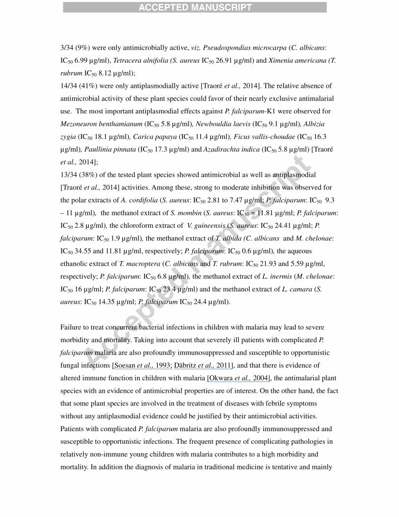
3/34 (9%) were only antimicrobially active, viz. Pseudospondias microcarpa (C. albicans:
IC50 6.99 µg/ml), Tetracera alnifolia (S. aureus IC50 26.91 µg/ml) and Ximenia americana (T.
rubrum IC50 8.12 µg/ml);
14/34 (41%) were only antiplasmodially active [Traoré et al., 2014]. The relative absence of
antimicrobial activity of these plant species could favor of their nearly exclusive antimalarial
use. The most important antiplasmodial effects against P. falciparum-K1 were observed for
Mezoneuron benthamianum (IC50 5.8 µg/ml), Newbouldia laevis (IC50 9.1 µg/ml), Albizia
zygia (IC50 18.1 µg/ml), Carica papaya (IC50 11.4 µg/ml), Ficus vallis-choudae (IC50 16.3
µg/ml), Paullinia pinnata (IC50 17.3 µg/ml) and Azadirachta indica (IC50 5.8 µg/ml) [Traoré
et al., 2014];
13/34 (38%) of the tested plant species showed antimicrobial as well as antiplasmodial
[Traoré et al., 2014] activities. Among these, strong to moderate inhibition was observed for
the polar extracts of A. cordifolia (S. aureus: IC50 2.81 to 7.47 µg/ml; P. falciparum: IC50 9.3
– 11 µg/ml), the methanol extract of S. mombin (S. aureus: IC50 = 11.81 µg/ml; P. falciparum:
IC50 2.8 µg/ml), the chloroform extract of V. guineensis (S. aureus: IC50 24.41 µg/ml; P.
falciparum: IC50 1.9 µg/ml), the methanol extract of T. albida (C. albicans and M. chelonae:
IC50 34.55 and 11.81 µg/ml, respectively; P. falciparum: IC50 0.6 µg/ml), the aqueous
ethanolic extract of T. macroptera (C. albicans and T. rubrum: IC50 21.93 and 5.59 µg/ml,
respectively; P. falciparum: IC50 6.8 µg/ml), the methanol extract of L. inermis (M. chelonae:
IC50 16 µg/ml; P. falciparum: IC50 23.4 µg/ml) and the methanol extract of L. camara (S.
aureus: IC50 14.35 µg/ml; P. falciparum IC50 24.4 µg/ml).
Failure to treat concurrent bacterial infections in children with malaria may lead to severe
morbidity and mortality. Taking into account that severely ill patients with complicated P.
falciparum malaria are also profoundly immunosuppressed and susceptible to opportunistic
fungal infections [Soesan et al., 1993; Däbritz et al., 2011], and that there is evidence of
altered immune function in children with malaria [Okwara et al., 2004], the antimalarial plant
species with an evidence of antimicrobial properties are of interest. On the other hand, the fact
that some plant species are involved in the treatment of diseases with febrile symptoms
without any antiplasmodial evidence could be justified by their antimicrobial activities.
Patients with complicated P. falciparum malaria are also profoundly immunosuppressed and
susceptible to opportunistic infections. The frequent presence of complicating pathologies in
relatively non-immune young children with malaria contributes to a high morbidity and
mortality. In addition the diagnosis of malaria in traditional medicine is tentative and mainly

based on fever symptoms, which is common to many other microbial infections as well.
Although the present antimicrobial investigation was not exhaustive, these preliminary results
may justify at least partly the traditional use of some “antimalarial” plant species without any
in vitro or in vivo antiplasmodial evidence. To the best of our knowledge, this is the first
report on the antimycobacterial activity of T. albida and L. inermis against M. chelonae. This
finding could be of interest since L. inermis and Terminalia spp. have been reported to be
active against M. tuberculosis. Moreover, the antiplasmodial activity previously described for
T. albida (0.6 µg/ml) [Traoré et al., 2014] may be of therapeutic importance in the treatment
of malaria associated with cough in view of its favourable Selectivity Index (SI) >100).
Further studies are required to validate and rationalise such considerations. Future
perspectives include a wide antimicrobial testing of all other antimalarial plant species, the
identification of active antimalarial and/or antimicrobial constituents, the evaluation of the
toxicity of the most interesting plant species along with a clinical trial to evaluate the
efficacity and tolerability of standardised plant extracts.

References
Abdullahi, M., Kolo, I., Adebayo, O.O., Joseph, O.A., Majekodumi, O.F., Joseph, I.O., 2011.
Antimycobacterial Friedelane-terpenoid from the Root Bark of Terminalia
avicennioides. American Journal of Chemistry 1, 52-55.
Abo, K.A., Ogunleye, V.O., Ashidi, J.S. 1999, Antimicrobial potential of Spondias mombin,
Croton zambesicus and Zygotritonia crocea. Phytotherapy Research 13, 494-497.
Adeshina, G.O., Kunle, O.F., Onaolapo, J.A., Ehinmidu, J.O., Odama, L.E., 2012.
Antimicrobial Activity of the Aqueous and Ethyl Acetate Sub-Fractions of Alchornea
cordifolia Leaf. European Journal of Medicinal Plants 2, 31-41.
Adeyemi, A., Omonigbehin, A.E., Stella, S., Oluwatosin, O., Jumoke, S., 2008. Antibacterial
activity of extracts of Alchornea cordifolia (Schum and Thonn) Mull.Arg., Boerhavia
diffusa (L) and Bridelia micrantha (Hoscht) Baill. used in traditional medicine in
Nigeria on Helicobacter pylori and four diarrhoeagenic bacterial pathogens. African
Journal of Biotechnology 7, 3761-3764.
Agyare, C., Mensah, A.Y., Osei-Asante, S., 2006. Antimicrobial activity and phytochemical
studies of some medicinal plants from Ghana. Boletín Latinoamericano y del Caribe
de Plantas Medicinales y Aromáticas 5, 113-117.
Ajali, U., 2000. Antibacterial activity of Alchornea cordifolia stem bark. Fitoterapia 71, 436-
438.
Ayodele, S.M., Alpheus, G., Iruaga, O.M., 2010. Antibacterial screening of the root stem and
leaf extracts of Terminalia albida sc. Elliot on selected pathogenic bacteria. African
Journal of Microbiology Research 4, 1457-1459.
Baba-Moussa, F., Akpagana, K., Bouchet, P., 1999. Antifungal activities of seven West
African Combretaceae used in traditional medicine. Journal of Ethnopharmacology
66, 335-338.
Bajwa, S.J., Kulshrestha A., 2013. Fungal Infections in Intensive Care Unit: Challenges in
Diagnosis and Management. Annales of Medical and Health Sciences Research 3,
238–244.
Barreto, F.S., Sousa, E.O., Campos, A.R., Costa, J.G.M., Rodrigues, F.F.G., 2010.
Antibacterial Activity of Lantana camara Linn and Lantana montevidensis Brig
Extracts from Cariri-Ceará, Brazil. Journal of Young Pharmacists 2(1), 42-44.
Bassat, Q., Guinovart, C., Sigauque, B., Mandomando, I., Aide, P., Sacarlal, J., Nhampossa,
T., Bardaji, A., Morais, L., Machevo, S., Letang, E., Macete, E., Aponte, J.J., Roca, A.,

Menendez, C., Alonso, P.L., 2009. Severe malaria and concomitant bacteraemia in
children admitted to a rural Mozambican hospital. Tropical Medicine & International
Health 14, 1011–1019.
Berkley, J., Mwarumba, S., Bramham, K., Lowe, B., Marsh, K., 1999. Bacteraemia
complicating severe malaria in children. Transactions of the Royal Society of Tropical
Medicine and Hygiene 93, 283–286.
Berkley, J.A., Lowe, B.S., Mwangi, I., Williams, T., Bauni, E., Mwarumba, S., Ngetsa, C.,
Slack, M.P., Njenga, S., Hart, C.A., Maitland, K., English, M., Marsh, K., Scott, J.A.,
2005. Bacteremia among children admitted to a rural hospital in Kenya. New England
Journal of Medicine 352, 39–47.
Bhatti, H.A., Uddin, N., Begum, S., Siddiqui, B.S., 2013. Synthesis of Acyl Analogues of
Coniferyl Alcohol and their Antimycobacterial Activity. Journal of the Chemical
Society of Pakistan 35, 886-889.
Binder, U., Lass-Flörl, C., 2013 .New insights into invasive aspergillosis--from the pathogen
to the disease. Current Pharmaceutical Design 19, 3679-3688.
Brent, A.J., Oundo, J.O., Mwangi, I., Ochola, L., Lowe, B., Berkley, J.A., 2006. Salmonella
bacteremia in Kenyan children. Pediatric Infectious Diseases Journal 25, 230–236.
Bronzan, R.N., Taylor, T.E., Mwenechanya, J., Tembo, M., Kayira, K., Bwanaisa, L., Njobvu,
A., Kondowe, W., Chalira, C., Walsh, A.L., Phiri, A., Wilson, L.K., Molyneux, M.E.,
Graham, S.M., 2007. Bacteremia in Malawian children with severe malaria:
prevalence, etiology, HIV coinfection, and outcome. Journal of Infectious Diseases
195, 895–904.
Chaturvedi, H.K., Mahanta, J., Pandey, A., 2009. Treatment-seeking for febrile illness in
north-east India: an epidemiological study in the malaria endemic zone. Malaria
Journal 8, 301.
Church, J., Maitland, K., 2014. Invasive bacterial co-infection in African children with
Plasmodium falciparum malaria: a systematic review; BMC Medicine 12, 11-16.
Corthout, J., Pieters, L.A., Claeys, M., Vanden Berghe, D.A. and Vlietinck, A. J., 1994.
Antibacterial and molluscicidal phenolic acids from Spondias mombin. Planta Medica
60, 460-463.
Cos, P., Maes, L., Sindambiwe, J.B., Vlietinck, A.J., Vanden Berghe, D., 2006a. Bioassays for
antibacterial and antifungal activities. In: Gupta, M.P., Handa, S.S., Vanish, K., Eds.
Biological Screening of Plant Constituents, International Centre for Science and High
Technology, Trieste; 19-28.

Cos, P., Vlietinck, A.J., Vanden Berghe, D., Maes, L., 2006b. Anti-infective potential of
natural products: how to develop a stronger in vitro proof-of-concept. Journal of
Ethnopharmacology 106, 290-302.
Crawley, J., Chu C., Mtove, G., Nosten, F., 2010. Malaria in children. Lancet 375: 1468–
1481.
Da Silva, A.R., de Morais, S.M., Marques, M.M., de Oliveira, D.F., Barros, C.C., de Almeida,
R.R., Vieira, Í.G., Guedes, M.I., 2012. Chemical composition, antioxidant and
antibacterial activities of two Spondias species from Northeastern Brazil.
Pharmaceutical Biology 50, 740-746.
Däbritz, J., Schneider, M., Just-Nuebling, G., Groll, A.H., 2011. Minireview: Invasive
fungal infection complicating acute Plasmodium falciparum malaria. Mycoses 54,
311–317.
Dahl, M.V., Grando, S.A.,1994. Chronic dermatophytosis: what is special about Trichophyton
rubrum? Advances in Dermatology 9, 97-109.
Evans, J.A., Adusei, A., Timmann, C., May, J., Mack, D., T. Agbenyega, T., Horstmann, R.D.,
Frimpong, E., 2004. High mortality of infant bacteraemia clinically indistinguishable
from severe malaria. QJM – An International Journal of Medicine 97, 591–597.
Ganjewala, D., Sam, S., Hayat Khan, K., 2009. Biochemical compositions and antibacterial
activities of Lantana camara plants with yellow, lavender, red and white flowers.
EurAsian Journal of Biosciences 3, 69-77.
Graham, S.M., Walsh, A.L., Molyneux, E.M., Phiri, A., Molyneux, M.E., 2000. The clinical
presentation of non-typhoidal Salmonella bacteraemia in Malawian children.
Transactions of the Royal Society of Tropical Medicine and Hygiene 94, 310–314.
Green, S.L., Lifland, B.D., Bouley, D.M., Brown, B.A., Wallace, R.J.Jr., Ferrell, J.E.Jr.,
2000. Disease Attributed to Mycobacterium chelonae in South African Clawed Frogs
(Xenopus laevis). Comparative Medicine. 50, 675-679.
Gull, I., Sohail, M., Aslam, M.S., Amin Athar, M., 2013. Phytochemical, toxicological and
antimicrobial evaluation of Lawsonia inermis extracts against clinical isolates of
pathogenic bacteria. Annals of Clinical Microbiology and Antimicrobials 12, 36.
Goto, T., Hamaguchi, R., Maeshima, A., Oyamada, Y., Kato, R., 2012. Pulmonary resection
for Mycobacterium chelonae infection. Annales of Thoracal and Cardiovascular
Surgery 18, 128-131.
Gunaselvi, G., Kulasekaren,V, Gopal, V., 2010. Antibacterial and antifungal activity of

various leaves extracts of Hardwickia binata Roxb. (Caesalpinaceae). International
Journal of Pharmaceutical Technology Research 2, 2183-2187.
Gwer, S., Newton, C.R., Berkley, J.A., 2007. Over-diagnosis and co-morbidity of severe
malaria in African children: a guide for clinicians. American Journal of Tropical
Medicine and Hygiene 77, 6–13.
Habluetzel, A., Lucantoni, L. and Esposito, F., 2009. Azadirachta indica as a public health
tool for the control of malaria and other vector-borne diseases. Indian Journal of
Medical Research 130, 112-114.
Igbeneghu, O.A., Iwalewa, E.O., Lamikanra, A., 2007. A study of the in- vivo activity of the
leaf extract of Alchornea cordifolia against multiply antibiotic resistant S. aureus
isolate in mice. Phytotherapy Research 21, 67-71.
Keong, B.C.M., Sulaiman, W., 2006. Typhoid and Malaria Co-Infection – An Interesting
Finding in the Investigation of a Tropical Fever. Malaysian Journal of Medical
Sciences 13, 74–75.
Kisangau, D.P., Hosea, K.M., Joseph, C.C., Lyaruu, H.V., 2007. In vitro antimicrobial assay of
plants used in traditional medicine in Bukoba Rural district, Tanzania. African Journal
of Traditional, Complementary and Alternative Medicines 4, 510-523.
Lall, N., Das Sarma, M., Hazra, B., Meyer, J.J., 2003. Antimycobacterial activity of
diospyrin derivatives and a structural analogue of diospyrin against Mycobacterium
tuberculosis in vitro. Journal of Antimicrobial Chemotherapy 51, 435–438.
Lasisi, A. A., Folarin, O. M., Dare, E. O., Akinloye, O. A., Fisuyi, M.O., 2011.
Phytochemical, antibacterial and cytotoxic evaluation of Raphiostylis beninensis
[Hook F. ex Planch] stem bark extracts. International Journal of Pharma & Bio
Sciences 2 (3), 489-495.
Latgé, J.-P. 1999. Aspergillus fumigatus and Aspergillosis. Clinical Microbiology Reviews
12, 310–350.
Lawal, T.O., Adeniyi, B.A, Wan, B., Franzblau, S.G. and Mahady, G.B., 2011. In-Vitro
Susceptibility of Mycobacterium Tuberculosis to Extracts of Uvaria Afzelli Scott Elliot
and Tetracera Alnifolia Willd. African Journal of Biomedical Research 14, 17 -21.
Lee, S.J., Gonzalez-Aseguinolaza, G., Nussenzweig, M.C., 2002. Disseminated Candidiasis
and Hepatic Malarial Infection in Mannose-Binding-Lectin-A-Deficient Mice.
Molecular and Cellular Biology 22, 8199–8203.
Low, C.-Y. and Rotstein, C., 2011. Emerging fungal infections in immunocompromised
patients. F1000 Medicine Reports 2011, 3:14 (doi:10.3410/M3-14) .

Maikai, V.A, Maikai, B.V., Kobo, P.I., 2009. Antimicrobial Properties of Stem Bark Extracts
of Ximenia americana. Journal of Agricultural Science 1, 30-34.
Maltha, J., Guiraud, I., Kaboré, B., Lompo, P., Ley, B. Bottieau, E., Van Geet, C., Tinto, H.,
Jacobs, J., 2014. Frequency of severe malaria and invasive bacterial infections among
children admitted to a rural hospital in Burkina Faso. PLoS One 9, e89103.
Mann, A., Amupitan, J.O., Oyewale, A.O., Okogun, J. I., Ibrahim, K., Oladosu, P., Lawson,
L., Olajide, I., Nnamdi, A., 2008. Evaluation of in vitro antimycobacterial activity of
Nigerian plants used for treatment of respiratory diseases. African Journal of
Biotechnology 7, 1630-1636.
McCormick, A., Loeffler, J., Ebel, F., 2010. Aspergillus fumigatus: contours of an
opportunistic human pathogen. Cellular Microbiology 12, 1535-1543.
Mesia, K, Tona, L, Mampunza, M.M., Ntamabyaliro, N, Muanda, T., Muyembe, T.,
Musuamba, T., Mets, T., Cimanga, K., Totté, J., Pieters, L., Vlietinck, A.J., 2012.
Antimalarial efficacy of a quantified extract of Nauclea pobeguinii stem bark in
human adult volunteers with diagnosed uncomplicated falciparum malaria: Part 2: A
clinical phase IIB trial. Planta Medica 78, 853-860.
Okeke, I.N., Ogundaini, A.O., Ogungbamila, F.O., Lamikanra, A., 1999. Antimicrobial
spectrum of Alchornea cordifolia leaf extract. Phytotherapy Research 13, 67-69.
Okwara, F.N., Obimbo, M., Wafula, M., Murila V., 2004. Bacteraemia, urinary tract
infection and malaria in hospitalised febrile Children in Nairobi: is there an
association? East African Medical Journal 81, 47-51.
Omer, M.E.F.A., Elnima E.I., 2003. Antimicrobial activity of Ximenia americana. Fitoterapia
74, 122–126.
Ramana, K.V., Kandi, S., Bharatkumar, V., Sharada, C.H.V., Rao, R., Mani, R., Rao, S.D.,
2013. Invasive Fungal Infections: A Comprehensive Review. American Journal of
Infectious Diseases and Microbiology 1, 64-69.
Saadabi, A.M.A., 2006. Antifungal activity of some Saudi plants used in traditional
medicine. Asian Journal of Plant Sciences. 5, 907-909.
Sadiq, I.S., Dangoggo, S.M., Hassan, L.G., Manga S.B., 2013. Bioactive Isolation and
Antifungal Screening of Leaf and Bark of Diospyros mespiliformis and Ziziphus
spina-christi. International Journal of Traditional and Natural Medicines 2, 104-117.
Scheers, C., Andre, J., Thompson, C., Rebuffat, E., Harag, S., Kolivras, A., 2013. Refractory
Trichophyton rubrum infection in lamellar ichthyosis. Pediatric Dermatology 30,
200-203.

Scott, J.A., Berkley, J.A., Mwangi, I., Ochola, L., Uyoga, S., Macharia, A., Ndila, C., Lowe,
B.S., Mwarumba, S., Bauni, E., Marsh, K., Williams, T.N., 2011. Relation between
falciparum malaria and bacteraemia in Kenyan children: a population based, case–
control study and a longitudinal study. Lancet 378, 1316–1323.
Sharma V.K., 1990. Tuberculostatic activity of henna (Lawsonia inermis Linn.). Tubercle 71,
293-295.
Silva, O., Duarte, A., Pimentel, M., Viegas, S., Barroso, H., Machado, J., Pires, I.,
Cabrita, J., Gomes, E., 1997. Antimicrobial activity of Terminalia macroptera root.
Journal of Ethnopharmacology 57, 203- 207.
Silva, R.F., 2010. Fungal infections in immunocompromised patients. Jornal Brasileiro de
Pneumologia 36, 142-147.
Soesan, M., Kager, P.A, Leverstein-van Hall, A., Van Lieshout, J.J., 1993. Coincidental
severe Plasmodium falciparum infection and disseminated candidiasis. Transactions
of the Royal Society of Tropical Medicine and Hygiene 87, 288–289.
Tobinaga, S., Sharma, M.K., Aalbersberg, W.G., Watanabe, K., Iguchi, K., Narui, K., Sasatsu,
M., Waki, S., 2009. Isolation and identification of a potent antimalarial and
antibacterial polyacetylene from Bidens pilosa. Planta Medica 75, 624-628.
Traoré, M.S., Baldé, M.A., Diallo, M.S.T., Baldé, E.S., Diané, S., Camara, A., Diallo, A.,
Baldé, A., Kéita, A., Kéita, S.M., Oularé, K., Magassouba, F.B., Diakité, I., Diallo, A.,
Pieters, L., Baldé, A.M., 2013. Ethnobotanical survey on medicinal plants used by
Guinean traditional healers in the treatment of malaria. Journal of Ethnopharmacology
150: 1145-1153.
Traore, M.S., Diané, S., Diallo, M.S.T., Balde, E.B, Balde, M.A., Camara, A., Diallo, A.,
Keita, A., Cos, P., Maes, L., Pieters, L, Balde, A.M., 2014. In Vitro Antiprotozoal and
Cytotoxic Activity of Ethnopharmacologically Selected Guinean Plants. Planta
Medica 80, 1340-1344.
Umeh, E., Igoli, J., Agada, E., Usman, S., 2009. Evaluating extracts of Spondias mombin for
antimicrobial activities. Bio-Research 7, 2, http://dx.doi.org/10.4314/br.v7i2.56584
Uneke, C.J., 2008. Concurrent malaria and typhoid fever in the tropics: the diagnostic
challenges and public health implications. Journal of Vector Borne Diseases 45, 133-
142.
Wallace Jr, R.J., Brown-Elliot, B.A., Ward, S.C., Crist, C.J., Mann, L.B., 2001. Activities of
linezolid against rapidly growing mycobacteria. Antimicrobial Agents and
Chemotherapy. 45, 764-767.

Walsh, A.L, Phiri, A.J., Graham, S.M., Molyneux, E.M., Molyneux, M.E., 2000. Bacteremia
in febrile Malawian children: clinical and microbiological features. Pediatric
Infectious Diseases Journal 19, 312–318.
World Health Organization, 2014. Antimicrobial resistance. Global Report on Surveillance.
ISBN 978 92 4 156474 8 (NLM classification: QV 250) .
Grap[hical
Plants used against malaria/fever
Antimicrobial screening
Plants active against malaria
and/or co-infections