The life cycle of Australapatemon magnacetabulum (Digenea, Strigeidae) from Northwestern Argentinae
7
BioOne sees sustainable scholarly publishing as an inherently collaborative enterprise connecting authors, nonprofit publishers, academic institutions, research libraries, and research funders in the common goal of maximizing access to critical research. The Life Cycle of Australapatemon magnacetabulum (Digenea: Strigeidae) from Northwestern Argentina Author(s): Dora Davies and Margarita Ostrowski de Núñez Source: Journal of Parasitology, 98(4):778-783. 2012. Published By: American Society of Parasitologists URL: http://www.bioone.org/doi/full/10.1645/GE-3054.1 BioOne (www.bioone.org ) is a nonprofit, online aggregation of core research in the biological, ecological, and environmental sciences. BioOne provides a sustainable online platform for over 170 journals and books published by nonprofit societies, associations, museums, institutions, and presses. Your use of this PDF, the BioOne Web site, and all posted and associated content indicates your acceptance of BioOne’s Terms of Use, available at www.bioone.org/page/terms_of_use . Usage of BioOne content is strictly limited to personal, educational, and non-commercial use. Commercial inquiries or rights and permissions requests should be directed to the individual publisher as copyright holder.
-
Upload
independent -
Category
Documents
-
view
0 -
download
0
Transcript of The life cycle of Australapatemon magnacetabulum (Digenea, Strigeidae) from Northwestern Argentinae
BioOne sees sustainable scholarly publishing as an inherently collaborative enterprise connecting authors, nonprofit publishers, academic institutions, researchlibraries, and research funders in the common goal of maximizing access to critical research.
The Life Cycle of Australapatemon magnacetabulum (Digenea: Strigeidae) fromNorthwestern ArgentinaAuthor(s): Dora Davies and Margarita Ostrowski de NúñezSource: Journal of Parasitology, 98(4):778-783. 2012.Published By: American Society of ParasitologistsURL: http://www.bioone.org/doi/full/10.1645/GE-3054.1
BioOne (www.bioone.org) is a nonprofit, online aggregation of core research in the biological, ecological, andenvironmental sciences. BioOne provides a sustainable online platform for over 170 journals and books publishedby nonprofit societies, associations, museums, institutions, and presses.
Your use of this PDF, the BioOne Web site, and all posted and associated content indicates your acceptance ofBioOne’s Terms of Use, available at www.bioone.org/page/terms_of_use.
Usage of BioOne content is strictly limited to personal, educational, and non-commercial use. Commercial inquiriesor rights and permissions requests should be directed to the individual publisher as copyright holder.
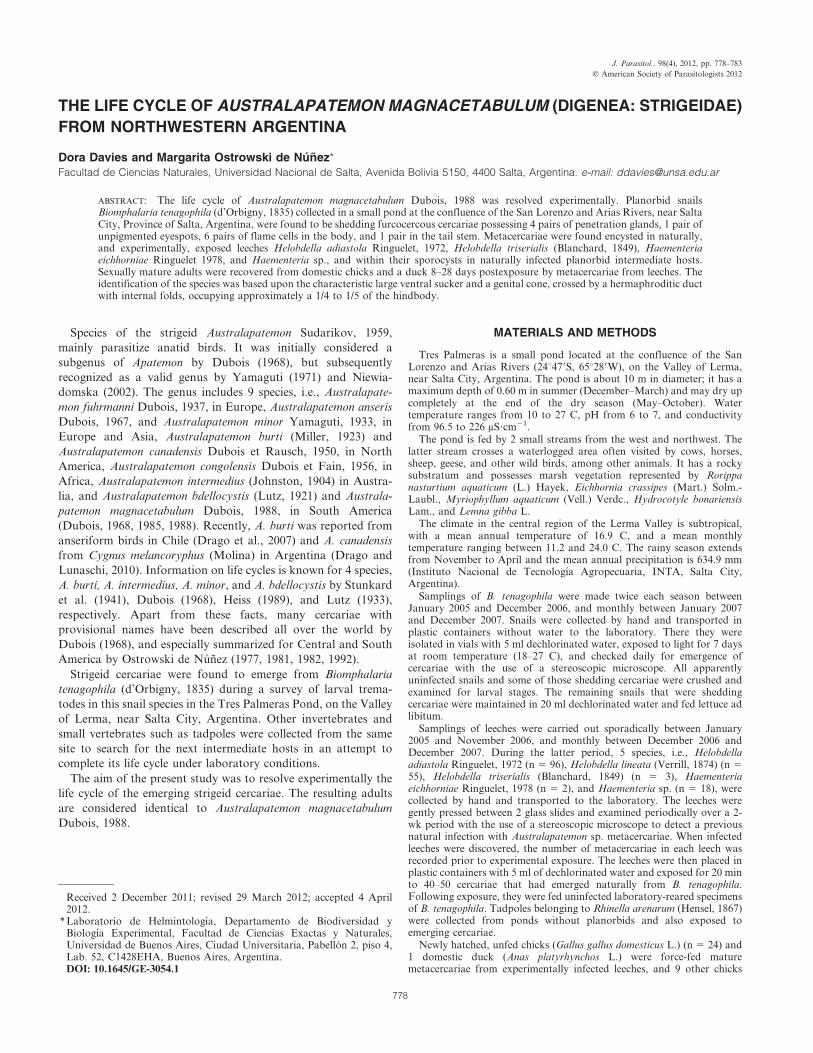
THE LIFE CYCLE OF AUSTRALAPATEMON MAGNACETABULUM (DIGENEA: STRIGEIDAE)
FROM NORTHWESTERN ARGENTINA
Dora Davies and Margarita Ostrowski de Nunez*Facultad de Ciencias Naturales, Universidad Nacional de Salta, Avenida Bolivia 5150, 4400 Salta, Argentina. e-mail: [email protected]
ABSTRACT: The life cycle of Australapatemon magnacetabulum Dubois, 1988 was resolved experimentally. Planorbid snailsBiomphalaria tenagophila (d’Orbigny, 1835) collected in a small pond at the confluence of the San Lorenzo and Arias Rivers, near SaltaCity, Province of Salta, Argentina, were found to be shedding furcocercous cercariae possessing 4 pairs of penetration glands, 1 pair ofunpigmented eyespots, 6 pairs of flame cells in the body, and 1 pair in the tail stem. Metacercariae were found encysted in naturally,and experimentally, exposed leeches Helobdella adiastola Ringuelet, 1972, Helobdella triserialis (Blanchard, 1849), Haementeriaeichhorniae Ringuelet 1978, and Haementeria sp., and within their sporocysts in naturally infected planorbid intermediate hosts.Sexually mature adults were recovered from domestic chicks and a duck 8–28 days postexposure by metacercariae from leeches. Theidentification of the species was based upon the characteristic large ventral sucker and a genital cone, crossed by a hermaphroditic ductwith internal folds, occupying approximately a 1/4 to 1/5 of the hindbody.
Species of the strigeid Australapatemon Sudarikov, 1959,
mainly parasitize anatid birds. It was initially considered a
subgenus of Apatemon by Dubois (1968), but subsequently
recognized as a valid genus by Yamaguti (1971) and Niewia-
domska (2002). The genus includes 9 species, i.e., Australapate-
mon fuhrmanni Dubois, 1937, in Europe, Australapatemon anseris
Dubois, 1967, and Australapatemon minor Yamaguti, 1933, in
Europe and Asia, Australapatemon burti (Miller, 1923) and
Australapatemon canadensis Dubois et Rausch, 1950, in North
America, Australapatemon congolensis Dubois et Fain, 1956, in
Africa, Australapatemon intermedius (Johnston, 1904) in Austra-
lia, and Australapatemon bdellocystis (Lutz, 1921) and Australa-
patemon magnacetabulum Dubois, 1988, in South America
(Dubois, 1968, 1985, 1988). Recently, A. burti was reported from
anseriform birds in Chile (Drago et al., 2007) and A. canadensis
from Cygnus melancoryphus (Molina) in Argentina (Drago and
Lunaschi, 2010). Information on life cycles is known for 4 species,
A. burti, A. intermedius, A. minor, and A. bdellocystis by Stunkard
et al. (1941), Dubois (1968), Heiss (1989), and Lutz (1933),
respectively. Apart from these facts, many cercariae with
provisional names have been described all over the world by
Dubois (1968), and especially summarized for Central and South
America by Ostrowski de Nunez (1977, 1981, 1982, 1992).
Strigeid cercariae were found to emerge from Biomphalaria
tenagophila (d’Orbigny, 1835) during a survey of larval trema-
todes in this snail species in the Tres Palmeras Pond, on the Valley
of Lerma, near Salta City, Argentina. Other invertebrates and
small vertebrates such as tadpoles were collected from the same
site to search for the next intermediate hosts in an attempt to
complete its life cycle under laboratory conditions.
The aim of the present study was to resolve experimentally the
life cycle of the emerging strigeid cercariae. The resulting adults
are considered identical to Australapatemon magnacetabulum
Dubois, 1988.
MATERIALS AND METHODS
Tres Palmeras is a small pond located at the confluence of the SanLorenzo and Arias Rivers (24u479S, 65u289W), on the Valley of Lerma,near Salta City, Argentina. The pond is about 10 m in diameter; it has amaximum depth of 0.60 m in summer (December–March) and may dry upcompletely at the end of the dry season (May–October). Watertemperature ranges from 10 to 27 C, pH from 6 to 7, and conductivityfrom 96.5 to 226 mS?cm21.
The pond is fed by 2 small streams from the west and northwest. Thelatter stream crosses a waterlogged area often visited by cows, horses,sheep, geese, and other wild birds, among other animals. It has a rockysubstratum and possesses marsh vegetation represented by Rorippanasturtium aquaticum (L.) Hayek, Eichhornia crassipes (Mart.) Solm.-Laubl., Myriophyllum aquaticum (Vell.) Verdc., Hydrocotyle bonariensisLam., and Lemna gibba L.
The climate in the central region of the Lerma Valley is subtropical,with a mean annual temperature of 16.9 C, and a mean monthlytemperature ranging between 11.2 and 24.0 C. The rainy season extendsfrom November to April and the mean annual precipitation is 634.9 mm(Instituto Nacional de Tecnologıa Agropecuaria, INTA, Salta City,Argentina).
Samplings of B. tenagophila were made twice each season betweenJanuary 2005 and December 2006, and monthly between January 2007and December 2007. Snails were collected by hand and transported inplastic containers without water to the laboratory. There they wereisolated in vials with 5 ml dechlorinated water, exposed to light for 7 daysat room temperature (18–27 C), and checked daily for emergence ofcercariae with the use of a stereoscopic microscope. All apparentlyuninfected snails and some of those shedding cercariae were crushed andexamined for larval stages. The remaining snails that were sheddingcercariae were maintained in 20 ml dechlorinated water and fed lettuce adlibitum.
Samplings of leeches were carried out sporadically between January2005 and November 2006, and monthly between December 2006 andDecember 2007. During the latter period, 5 species, i.e., Helobdellaadiastola Ringuelet, 1972 (n 5 96), Helobdella lineata (Verrill, 1874) (n 5
55), Helobdella triserialis (Blanchard, 1849) (n 5 3), Haementeriaeichhorniae Ringuelet, 1978 (n 5 2), and Haementeria sp. (n 5 18), werecollected by hand and transported to the laboratory. The leeches weregently pressed between 2 glass slides and examined periodically over a 2-wk period with the use of a stereoscopic microscope to detect a previousnatural infection with Australapatemon sp. metacercariae. When infectedleeches were discovered, the number of metacercariae in each leech wasrecorded prior to experimental exposure. The leeches were then placed inplastic containers with 5 ml of dechlorinated water and exposed for 20 minto 40–50 cercariae that had emerged naturally from B. tenagophila.Following exposure, they were fed uninfected laboratory-reared specimensof B. tenagophila. Tadpoles belonging to Rhinella arenarum (Hensel, 1867)were collected from ponds without planorbids and also exposed toemerging cercariae.
Newly hatched, unfed chicks (Gallus gallus domesticus L.) (n 5 24) and1 domestic duck (Anas platyrhynchos L.) were force-fed maturemetacercariae from experimentally infected leeches, and 9 other chicks
Received 2 December 2011; revised 29 March 2012; accepted 4 April2012.
* Laboratorio de Helmintologıa, Departamento de Biodiversidad yBiologıa Experimental, Facultad de Ciencias Exactas y Naturales,Universidad de Buenos Aires, Ciudad Universitaria, Pabellon 2, piso 4,Lab. 52, C1428EHA, Buenos Aires, Argentina.DOI: 10.1645/GE-3054.1
J. Parasitol., 98(4), 2012, pp. 778–783
F American Society of Parasitologists 2012
778
were force-fed metacercariae in sporocysts from naturally infected B.tenagophila. The animals were housed in cages at room temperature, fedwith oat and bread, and supplied with water ad libitum. They were killedand necropsied from 3 to 28 days postexposure (PE) to recover the adults.
The morphology of the larval and adult stages was examined, preferablyusing live specimens. Neutral red and Nile blue sulphate were used forvital staining of cercariae. Adults were fixed in hot 4% formalin, stored in70% ethyl alcohol, cleared in Amman’s lactophenol, or stained withchlorhydric carmine, dehydrated, cleared in creosote, and mounted inCanada balsam; some specimens were embedded in paraffin, cut at 7 m,stained with hematoxylin–eosin, and mounted in Canada balsam. Twoinfected B. tenagophila and 3 specimens of adults from chicks werepreserved directly in 96 and 70% ethyl alcohol, respectively. Drawingswere made with the aid of a camera lucida. For measurements, livesporocysts and metacercariae from experimentally infected leeches weregently pressed between a coverslip and slide; cercariae were fixed in hot4% formalin, mounted in the same fixative, and placed between a coverslipand slide without pressure. Measurements of the mostly ventrally curvedadult bodies were made from permanent mounts as follows: the hindbodylength was taken as the distance between the middle of the neckimmediately after the proteolytic gland and the posterior end of theworm. The forebody length was taken as the distance between the anteriorend and the middle of the neck behind the proteolytic gland (see Johnstonand Angel, 1951). Measurements are given in micrometers, with thearithmetic mean followed by the range in parentheses. Eggs were measuredalive and in permanent mounts. The type material of A. magnacetabulum(MHNG 985.644) and voucher specimens of A. bdellocystis (MHNG983.1006) deposited in the Museum of Natural History of Geneva,Geneva, Switzerland, were studied for comparison.
RESULTS
A total of 3,078 specimens of B. tenagophila was collected, of
which 45 (1.46%) were infected with the Australapatemon species.
All the exposed leeches, i.e., H. adiastola, H. triserialis,
Haementeria sp., and H. eichhorniae were infected with mature
metacercariae, whereas no evidence of infection was observed in
H. lineata or in R. arenarum tadpoles. Twenty-five specimens were
recovered from the duck 14 days PE; 12 of 24 chicks exposed to
metacercariae from leeches were positive, and 7, 3, 26, 2, 43, and 1
(total 82) adults were recovered at 8, 9, 12, 13, 14, and 28 days PE,
respectively. Five of 9 chicks exposed to metacercariae from B.
tenagophila acquired the infection, and 1, 6, 5, and 2 (total 14)
adults were recovered at 3, 7, 10, and 13 days PE, respectively.
DESCRIPTION
Australapatemon magnacetabulum Dubois, 1988(Figs. 1–8)
Adult (Figs. 1, 2, and 6): Diagnosis based on permanent mounts of 43gravid specimens from experimentally infected domestic duck and chicks.Measurements based on 10 specimens from duck 14 days PE; measure-ments on specimens from chicks in Table I. Body distinctly bipartite,curved ventrally, 1,478 (1,326–1,725) long, with definite neck constriction.Forebody utriculiform, cylindrical, 401 (337–473) long by 305 (236–360)wide. Hindbody subcylindrical, 1,077 (989–1,252) long by 313 (218–432)wide, with rounded truncated end perpendicular to longitudinal axis. Oralsucker subterminal, 83 (60–98) long by 80 (65–92) wide; prepharynx notdiscernible; pharynx small, 39 (37–42) long by 39 (29–49) wide; esophagusshort, bifurcates in front of ventral sucker into 2 ceca ending at level ofgenital cone. Ventral sucker larger than oral sucker, 125 (99–174) long by126 (83–193) wide. Holdfast organ consists of 2 lobes with spinoustegument, at anterior level of pyriform proteolytic gland, 39 (27–57) longby 98 (54–166) wide, situated at limit between forebody and hindbody.
Gonads occupying anterior 2/3 of hindbody. Testes tandem, rounded toirregular in shape, borders slightly lobed; anterior and posterior testis 174(162–185) long by 117 (86–147) wide and 173 (159–185) long by 102 (77–115) wide, respectively. Seminal vesicle dorsal, 81 (78–84) long by 110 (85–135) wide, posterior to testes. Hermaphroditic duct with internal folds
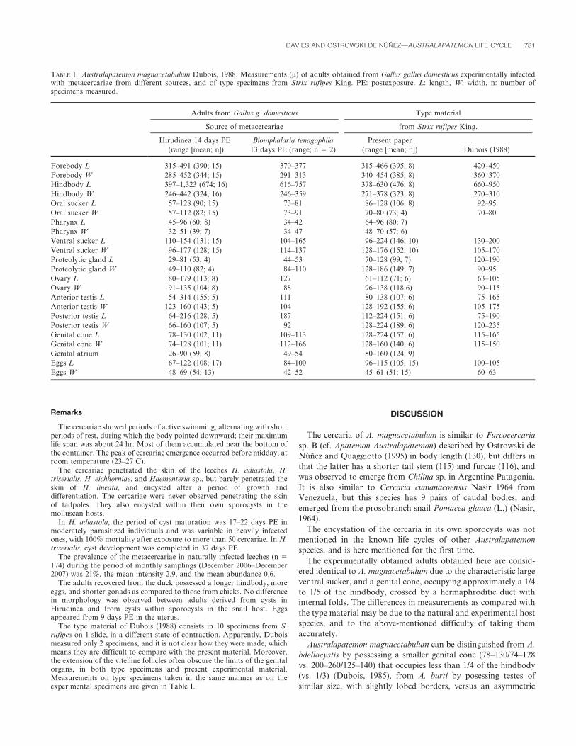
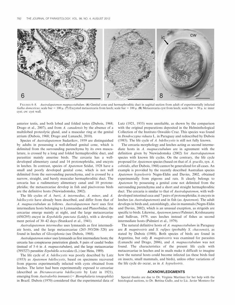
enters spherical genital cone, 134 (91–165) long by 140 (123–184) wide,delimited from surrounding parenchyma by its own musculature, opensinto shallow genital atrium 51 (46–57) long. Ovary oval, 127 (113–140)long by 91 (86–94) wide, with posterior border slightly concave, located infirst third of hindbody, immediately anterior to testes. Laurer’s canalopens dorsally at level of anterior edge of anterior testis. Mehlis’ glandpresent. Vitelline glands numerous, elongate, extend from level of neck toposterior end of hindbody, occupy ventral and ventrolateral regions.Vitelline reservoir intertesticular. Uterus emerging from ootype, runstoward anterior end until level of anterior border of hindbody, then turnsbackward until joining ejaculatory duct; with 1–9 eggs, measuring 90 (79–98) long by 49 (42–58) wide in permanent mounts.
Egg: Live eggs obtained from adults 111 (93–123) long by 67 (59–74)wide (n 5 35). Capsule oval, 1 end operculate, with zygote and severalvitelline cells. Fully developed miracidia first observed after 15 days atroom temperature (23–27 C), most miracidia develop within 20 days.
Mother sporocyst: Not detected in naturally, heavily infected snails.Daughter sporocyst (Fig. 3): Sporocysts in digestive gland and gonad of
B. tenagophila. Body elongated, sac-like, colorless, up to 4 mm long. Largesporocysts with 10–16 fully developed cercariae and numerous germinalmasses. Sporocysts forming tangled masses in digestive gland, gonad,intestine, stomach, kidney, prostate, etc., of heavily infected snails.
Cercaria (Figs. 4 and 5): Measurements based on 30 formalin-fixedspecimens from natural infections. Body oval, 133 (100–191) long by 50(34–73) wide, with 4 pairs of sensory hairs, 1 at level of penetration organ,1 at level of unpigmented eye spots, 1 at posterior level of ventral sucker, 1at level of excretory vesicle. Tegument spines present on ventral side,reaching close to anterior margin of ventral sucker. Tail stem 136 (108–164) long by 36 (25–42) with 5–6 pairs of caudal bodies, approximately 10sensory hairs on lateral borders, tegument without spines; furca 163 (105–201) long, with spinous tegument and several short sensory hairs.Penetration organ 28 (22–37) long by 25 (20–37) wide, densely coveredwith minute spines; anterior tip protrusible, oral opening terminal.Prepharynx short, inconspicuous, pharynx distinct. Esophagus long;intestinal bifurcation approximately halfway between pharynx and ventralsucker, intestinal ceca reaching almost to posterior level of ventral sucker.Ventral sucker 25 (19–37) long by 25 (17–37) wide, situated posterior tomidbody; opening surrounded by 2–3 crowns of spines. Unpigmentedeyespots situated slightly posterior to cecal bifurcation. Four pairs ofpenetration glands difficult to recognize, posterior to ventral sucker.Gland ducts run toward anterior end, dilated slightly on enteringpenetration organ, open in groups of 4 individual pores on each side oforal opening. Excretory system with small bladder, 6 pairs of flame cells inbody and 1 pair in tail stem. Flame cell formula 2 ([1+1] + [2+2] + 1) 5 14.Postacetabular transverse commissure present.
Metacercariae (Figs. 7 and 8): Measurements based on experimentallyobtained, live encysted metacercariae (n 5 10) from leeches, 17–22 daysPE. Inner cyst oval in shape, 172 (157–181) long by 152 (137–159) wide,cyst wall thick, 32 (15–47). Measurements based on cysts (n 5 20) fromlive sporocysts: Inner cyst 205 (191–221) long by 171 (162–189) wide; cystwall 29 (13–49). Measurements based on cysts (n 5 2) from Canadabalsam–mounted leeches: inner cyst 186 (180–191) long by 168 (166–169),cyst wall 20 (9–30).
Taxonomic summary
Natural definitive host: Strix rufipes King., Buteo magnirostris (Gm.)(see Dubois, 1988).
Experimental definitive hosts: Gallus g. domesticus Linnaeus, 1758; A.platyrhynchos Linnaeus, 1758.
Site of infection: Anterior intestine.First intermediate host: Biomphalaria tenagophila (d’Orbigny, 1835).Site of infection: Gonad and hepatopancreas.Second intermediate host: Helobdella adiastola Ringuelet, 1972, H.
triserialis (Blanchard, 1849), Haementeria eichhorniae Ringuelet, 1978, andHaementeria sp.
Locality: Salta, Tres Palmeras Pond (24u479S, 65u289W).Site of infection: Parenchyma.Specimens deposited: MACN-Pa 521/1-9 from A. platyrhynchos,
MACN-Pa 521/10-19, from G. g. domesticus, MACN-Pa 521/20 sporo-cysts from B. tenagophila, MACN-Pa 521/21-24 sagittal sections of adultsfrom G. domesticus; MACN-Pa 521/25 histological sections of leechescontaining metacercariae.
DAVIES AND OSTROWSKI DE NUNEZ—AUSTRALAPATEMON LIFE CYCLE 779
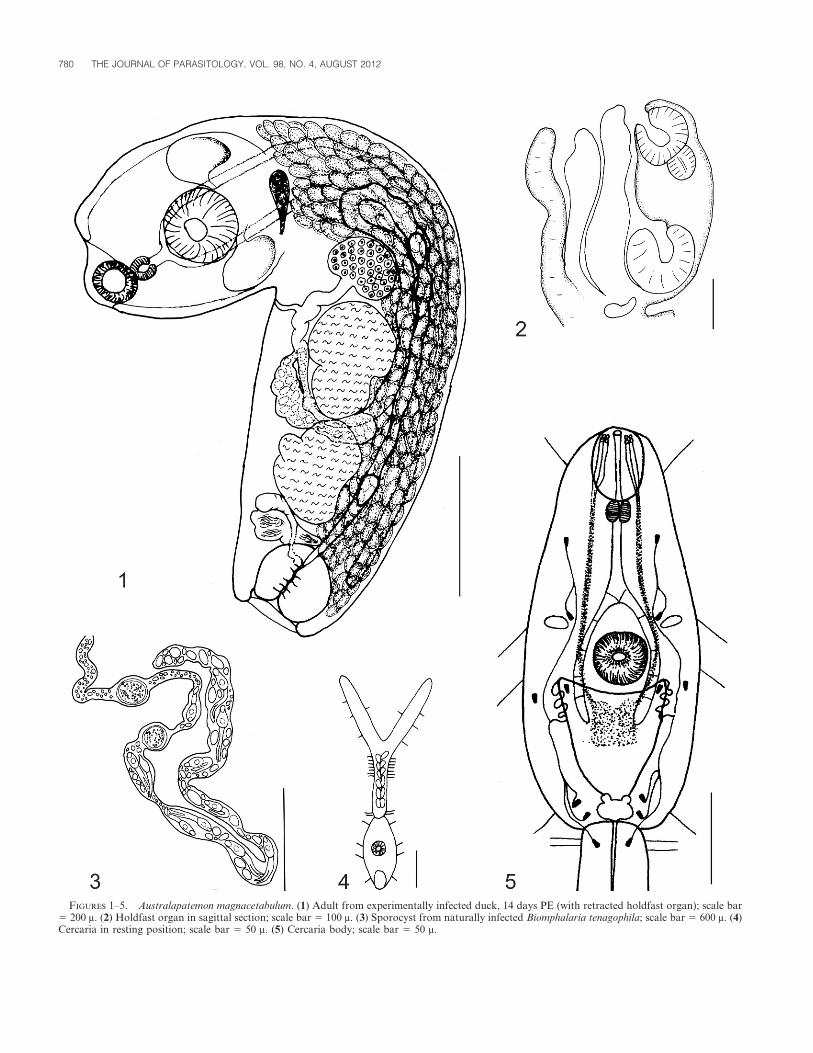
FIGURES 1–5. Australapatemon magnacetabulum. (1) Adult from experimentally infected duck, 14 days PE (with retracted holdfast organ); scale bar5 200 m. (2) Holdfast organ in sagittal section; scale bar 5 100 m. (3) Sporocyst from naturally infected Biomphalaria tenagophila; scale bar 5 600 m. (4)Cercaria in resting position; scale bar 5 50 m. (5) Cercaria body; scale bar 5 50 m.
780 THE JOURNAL OF PARASITOLOGY, VOL. 98, NO. 4, AUGUST 2012
Remarks
The cercariae showed periods of active swimming, alternating with shortperiods of rest, during which the body pointed downward; their maximumlife span was about 24 hr. Most of them accumulated near the bottom ofthe container. The peak of cercariae emergence occurred before midday, atroom temperature (23–27 C).
The cercariae penetrated the skin of the leeches H. adiastola, H.triserialis, H. eichhorniae, and Haementeria sp., but barely penetrated theskin of H. lineata, and encysted after a period of growth anddifferentiation. The cercariae were never observed penetrating the skinof tadpoles. They also encysted within their own sporocysts in themolluscan hosts.
In H. adiastola, the period of cyst maturation was 17–22 days PE inmoderately parasitized individuals and was variable in heavily infectedones, with 100% mortality after exposure to more than 50 cercariae. In H.triserialis, cyst development was completed in 37 days PE.
The prevalence of the metacercariae in naturally infected leeches (n 5
174) during the period of monthly samplings (December 2006–December2007) was 21%, the mean intensity 2.9, and the mean abundance 0.6.
The adults recovered from the duck possessed a longer hindbody, moreeggs, and shorter gonads as compared to those from chicks. No differencein morphology was observed between adults derived from cysts inHirudinea and from cysts within sporocysts in the snail host. Eggsappeared from 9 days PE in the uterus.
The type material of Dubois (1988) consists in 10 specimens from S.rufipes on 1 slide, in a different state of contraction. Apparently, Duboismeasured only 2 specimens, and it is not clear how they were made, whichmeans they are difficult to compare with the present material. Moreover,the extension of the vitelline follicles often obscure the limits of the genitalorgans, in both type specimens and present experimental material.Measurements on type specimens taken in the same manner as on theexperimental specimens are given in Table I.
DISCUSSION
The cercaria of A. magnacetabulum is similar to Furcocercaria
sp. B (cf. Apatemon Australapatemon) described by Ostrowski de
Nunez and Quaggiotto (1995) in body length (130), but differs in
that the latter has a shorter tail stem (115) and furcae (116), and
was observed to emerge from Chilina sp. in Argentine Patagonia.
It is also similar to Cercaria cumanacoensis Nasir 1964 from
Venezuela, but this species has 9 pairs of caudal bodies, and
emerged from the prosobranch snail Pomacea glauca (L.) (Nasir,
1964).
The encystation of the cercaria in its own sporocysts was not
mentioned in the known life cycles of other Australapatemon
species, and is here mentioned for the first time.
The experimentally obtained adults obtained here are consid-
ered identical to A. magnacetabulum due to the characteristic large
ventral sucker, and a genital cone, occupying approximately a 1/4
to 1/5 of the hindbody, crossed by a hermaphroditic duct with
internal folds. The differences in measurements as compared with
the type material may be due to the natural and experimental host
species, and to the above-mentioned difficulty of taking them
accurately.
Australapatemon magnacetabulum can be distinguished from A.
bdellocystis by possessing a smaller genital cone (78–130/74–128
vs. 200–260/125–140) that occupies less than 1/4 of the hindbody
(vs. 1/3) (Dubois, 1985), from A. burti by posessing testes of
similar size, with slightly lobed borders, versus an asymmetric
TABLE I. Australapatemon magnacetabulum Dubois, 1988. Measurements (m) of adults obtained from Gallus gallus domesticus experimentally infectedwith metacercariae from different sources, and of type specimens from Strix rufipes King. PE: postexposure. L: length, W: width, n: number ofspecimens measured.
Adults from Gallus g. domesticus Type material
Source of metacercariae from Strix rufipes King.
Hirudinea 14 days PE
(range [mean; n])
Biomphalaria tenagophila
13 days PE (range; n 5 2)
Present paper
(range [mean; n]) Dubois (1988)
Forebody L 315–491 (390; 15) 370–377 315–466 (395; 8) 420–450
Forebody W 285–452 (344; 15) 291–313 340–454 (385; 8) 360–370
Hindbody L 397–1,323 (674; 16) 616–757 378–630 (476; 8) 660–950
Hindbody W 246–442 (324; 16) 246–359 271–378 (323; 8) 270–310
Oral sucker L 57–128 (90; 15) 73–81 86–128 (106; 8) 92–95
Oral sucker W 57–112 (82; 15) 73–91 70–80 (73; 4) 70–80
Pharynx L 45–96 (60; 8) 34–42 64–96 (80; 7) .
Pharynx W 32–51 (39; 7) 34–47 48–70 (57; 6) .
Ventral sucker L 110–154 (131; 15) 104–165 96–224 (146; 10) 130–200
Ventral sucker W 96–177 (128; 15) 114–137 128–176 (152; 10) 105–170
Proteolytic gland L 29–81 (53; 4) 44–53 70–128 (99; 7) 120–190
Proteolytic gland W 49–110 (82; 4) 84–110 128–186 (149; 7) 90–95
Ovary L 80–179 (113; 8) 127 61–112 (71; 6) 63–105
Ovary W 91–135 (104; 8) 88 96–138 (118;6) 90–115
Anterior testis L 54–314 (155; 5) 111 80–138 (107; 6) 75–165
Anterior testis W 123–160 (143; 5) 104 128–192 (155; 6) 105–175
Posterior testis L 64–216 (128; 5) 187 112–224 (151; 6) 75–190
Posterior testis W 66–160 (107; 5) 92 128–224 (189; 6) 120–235
Genital cone L 78–130 (102; 11) 109–113 128–224 (157; 6) 115–165
Genital cone W 74–128 (101; 11) 112–166 128–160 (140; 6) 115–150
Genital atrium 26–90 (59; 8) 49–54 80–160 (124; 9) .
Eggs L 67–122 (108; 17) 84–100 96–115 (105; 15) 100–105
Eggs W 48–69 (54; 13) 42–52 45–61 (51; 15) 60–63
DAVIES AND OSTROWSKI DE NUNEZ—AUSTRALAPATEMON LIFE CYCLE 781
anterior testis, and both lobed and folded testes (Dubois, 1968;
Drago et al., 2007), and from A. canadensis by the absence of a
multilobed proteolytic gland, and a muscular ring at the genital
atrium (Dubois, 1968; Drago and Lunaschi, 2010).
Species of Australapatemon Sudarikov, 1959 are distinguished
by adults in possessing a well-defined genital cone, which is
delimited from the surrounding parenchyma by its own muscu-
lature, is crossed by a long and folded hermaphroditic duct, and
parasitize mainly anserine birds. The cercaria has a well-
developed alimentary canal and 14 protonephridia, and encysts
in leeches. In contrast, species of Apatemon Szidat, 1928 have a
small and poorly developed genital cone, which is not well
delimited from the surrounding parenchyma, and is crossed by a
narrow, straight, and barely muscular hermaphroditic duct. The
cercaria has a rudimentary alimentary canal and 10 protone-
phridia; the metacercariae develop in fish and piscivorous birds
are the definitive hosts (Niewiadomska, 2002).
The life cycles of A. burti, A. intermedius, A. minor, and A.
bdellocystis have already been described, and differ from that of
A. magnacetabulum as follows. Australapatemon burti uses first
intermediate hosts belonging to Lymnaeidae and Planorbidae; the
cercariae emerge mainly at night, and the large metacercariae
(450/295) encyst in Erpobdella punctata (Leidy), with a develop-
ment period of 30–42 days (Stunkard et al., 1941).
Australapatemon intermedius uses lymnaeids as first intermedi-
ate hosts, and the large metacercariae (265–393/206–328) are
found in leeches of Glossiphonia (see Dubois, 1968).
Australapatemon minor uses lymnaeids as first intermediate hosts; the
cercaria has conspicuous penetration glands, 8 pairs of caudal bodies
(instead of 5–6 in A. magnacetabulum), and the large metacercariae
(395/327) parasitize Erpobdella octoculata (L.) (see Heiss, 1989).
The life cycle of A. bdellocystis was poorly described by Lutz
(1933) as Apatemon bdellocystis, based on specimens recovered
from pigeons experimentally infected with cysts obtained from
leeches. The latter had been experimentally exposed to cercariae
(described as Dicranocercaria bdellocystis by Lutz in 1921),
emerging from Australorbis immunis (5 Biomphalaria tenagophila)
in Brazil. Dubois (1970) considered that the experimental data of
Lutz (1921, 1933) were unreliable, as shown by the comparison
with the original preparations deposited in the Helminthological
Collection of the Instituto Oswaldo Cruz. This species was found
in Dendrocygna viduata L. in Paraguay and redescribed by Dubois
(1985). The life cycle of A. bdellocystis is still not fully known.
The cercaria morphology and leeches acting as second interme-
diate hosts in A. magnacetabulum are in agreement with the
definition given by Niewiadomska (2002) for Australapatemon
species with known life cycles. On the contrary, the life cycle
proposed for Apatemon species (based on that of A. gracilis, syn. A.
cobitidis, after Dubois, 1968) cannot be generalized for all cases. An
example is provided by the recently described Australian species
Apatemon hypseleotris Negm-Eldin and Davies, 2002, obtained
experimentally from pigeons and rats. It clearly belongs to
Apatemon by possessing a genital cone not delimited from the
surrounding parenchyma and a short and straight hermaphroditic
duct. The cercaria is similar to that of Australapatemon, with well-
developed intestinal ceca and 7 pairs of protonephridia; it encysts in
leeches (as Australapatemon) and in fish (as Apatemon). The adult
develops in birds and, astonishingly, also in mammals (Negm-Eldin
and Davies, 2002), which is an unusual exception, as strigeids are
specific to birds. Likewise, Apatemon jamesi Palmieri, Krishnasamy
and Sullivan, 1979, uses leeches instead of fishes as second
intermediate hosts (Palmieri et al., 1979).
The natural definitive hosts of A. magnacetabulum in Paraguay
are B. magnirostris and S. rufipes (probably S. chacoensis), as
stated by Dubois (1988). Both species of birds are found in
Argentina, but only B. magnirostris was examined for parasites
(Lunaschi and Drago, 2006), and A. magnacetabulum was not
found. The characteristics of the present life cycle with
metacercariae in leeches and in snails make it difficult to imagine
how the natural hosts could become infected (as these birds feed
on insects, small mammals, and birds), unless other variations of
the life cycle do occur, as in A. hypseleotris.
ACKNOWLEDGMENTS
Special thanks are due to Dr. Virginia Martınez for her help with thehistological sections, to Dr. Bettina Gullo, and to Lic. Javier Montero for
FIGURES 6–8. Australapatemon magnacetabulum. (6) Genital cone and hermaphroditic duct in sagittal section from adult of experimentally infectedGallus domesticus; scale bar 5 100 m. (7) Excysted metacercaria from leech; scale bar 5 100 m. (8) Metacercaria cyst from leech; scale bar 5 50 m. ic: innercyst; cw: cyst wall.
782 THE JOURNAL OF PARASITOLOGY, VOL. 98, NO. 4, AUGUST 2012
the taxonomic identification of the leeches, to Dr. Olga Martınez for thetaxonomic identification of the plants, to Miss Tomasa Medina for themaintenance of leeches and snails in the laboratory, and to Dr. AlainDeChambrier, Geneva, Switzerland, for the loan of type and voucher specimens.
LITERATURE CITED
DRAGO, F. B., AND L. I. LUNASCHI. 2010. Digenea, Strigeidae, Australa-patemon canadensis Dubois and Rausch, 1950: First record in SouthAmerica and a new host record. Check List 6: 382–384.
———, ———, A. C. HINOJOSA-SAEZ, AND D. GONZALEZ-ACUNA. 2007.First record of Australapatemon burti and Paramonostomum pseu-dalveatum (Digenea) from Anas georgica (Aves, Anseriformes) inChile. Acta Parasitologica 52: 201–205.
DUBOIS, G. 1968. Synopsis des Strigeidae et des Diplostomatidae(Trematoda). Memoires de la Societe Neuchateloise des SciencesNaturelles 10(Premier fascicule): 1–261.
———. 1970. Les Strigeata (Trematoda) de la collection A. Lutz.Memorias do Instituto Oswaldo Cruz 68: 169–196.
———. 1985. Quelques Strigeoidea (Trematoda) recoltes chez des oiseauxdu Paraguay par la Mission Claude Weber, automne 1983, du Museumd’Histoire Naturelle de Geneve. Revue suisse de Zoologie 92: 641–648.
———. 1988. Quelques Strigeoidea (Trematoda) recoltes au Paraguay parles expeditions du Museum d’Histoire Naturelle de Geneve, au coursdes annees 1979, 1982 et 1985. Revue Suisse de Zoologie 95: 521–532.
HEISS, C. 1989. Biologie einer Apatemon-Art (Trematoda) aus Weihern inMittelfranken. Master’s Thesis. University of Erlangen-Nurnberg,Germany, 79 p.
JOHNSTON, T. H., AND L. M. ANGEL. 1951. The morphology and life cycleof the trematode, Apatemon intermedius, from the black swan.Transactions of the Royal Society of Southern Australia 74: 66–78.
LUNASCHI, L. I., AND F. B. DRAGO. 2006. Strigeid parasites of the roadsidehawk, Buteo magnirostris (Aves: Falconiformes) from Argentina.Zootaxa 1106: 25–33.
LUTZ, A. 1921. Zur Kenntnis des Entwicklungszyklus der Holostomiden.Zentralblatt fur Bakteriologie, Originale 86: 124–129.
———. 1933. Notas sobre Dicranocercarias brazileiras. Memorias doInstituto Oswaldo Cruz 27: 349–375.
NASIR, P. 1964. Studies on freshwater larval Trematodes—Part IV. A newvirgulate xiphidiocercaria, and another new longifurcate, pharynge-ate, distome, furcocercaria, from Venezuela. Zoologischer Anzeiger173: 220–226.
NEGM-ELDIN, M., AND R. W. DAVIES. 2002. Morphology and life cycle ofApatemon hypseleotris species novum from Australia includingmetacercariae viability and excystment. Deutsche TierarztlicheWochenschrift 109: 306–314.
NIEWIADOMSKA, K. 2002. Family Strigeidae Railliet, 1919. In Keys to theTrematoda. Vol. 1, D. I. Gibson, A. Jones, and R. A. Bray (eds.).CAB International, London, U.K., p. 231–241.
OSTROWSKI DE NUNEZ, M. 1977. Trematoda (larvas libres de trematodesdigeneos). In Aquatic biota of southern South America, Stuart H.Hurlbert (ed.). San Diego State University, San Diego, California, p.68–72.
———. 1981. Trematoda. Free-living larvae. In Aquatic biota of tropicalSouth America, Stuart H. Hurlbert (ed.). San Diego State University,San Diego, California, p. 110–115.
———. 1982. Trematoda. Free-living larvae. In Aquatic biota of Mexico,Central America and the West Indies, Stuart H. Hurlbert (ed.). SanDiego State University, San Diego, California, p. 85–89.
———. 1992. Trematoda. Familias Strigeidae, Diplostomidae, Clinosto-midae, Schistosomatidae, Spirorchiidae y Bucephalidae. In Fauna deagua dulce de la Republica Argentina, Z. A. de Castellanos (ed.). LaPlata, Buenos Aires, Argentina, 9(fasc.1): 5–55.
———, AND A. QUAGGIOTTO. 1995. Trematodes larvales (Digenea) de lasfamilias Diplostomidae, Strigeidae y Echinostomatidae en la RegionPatagonica Argentina. Boletın Chileno de Parasitologıa 50: 28–33.
PALMIERI, J. R., M. KRISHNASAMY, AND J. T. SULLIVAN. 1979. Strigeoidtrematodes of Malaysia with descriptions of a new genus and threenew species. Journal of Helminthology 53: 51–63.
STUNKARD, H. W., C. H. WILLEY, AND Y. RABINOWITZ. 1941. Cercaria burtiMiller, 1923, a larval stage of Apatemon gracilis (Rudolphi, 1819)Szidat, 1928. Transactions of the American Microscopical Society 60:485–497.
YAMAGUTI, S. 1971. Synopsis of digenetic trematodes of vertebrates.Keigaku Publishing Co., Tokyo, Japan, 1074 p.
DAVIES AND OSTROWSKI DE NUNEZ—AUSTRALAPATEMON LIFE CYCLE 783