The kidney as a target organ in pharmaceutical research
16
REVIEWS Drug Discovery Today Volume 16, Numbers 5/6 March 2011 The kidney as a target organ in pharmaceutical research Marco Prunotto 1,4, , Giulio Gabbiani 2 , Silvia Pomposiello 3 , GianMarco Ghiggeri 4 and Solange Moll 1 1 Institute of Clinical Pathology, University Hospital Geneva, Geneva 1211, Switzerland 2 Pathology Department, University of Geneva, Geneva 1211, Switzerland 3 Pharma Research & Early Development, F. Hoffmann-La Roche Ltd, Hoffmann-La Roche Ltd., Basle 4070, Switzerland 4 Nephrology Unit Laboratory, Giannina Gaslini Children’s Hospital, Genoa 16147, Italy Kidney diseases are a major source of morbidity and mortality in humans. In developed countries, mortality owing to chronic kidney disease (CKD) terminating in end-stage renal failure is comparable with that associated with cancer. A full understanding of the mechanisms implicated in the progression of CKD is needed to achieve its prevention and to delay the need for support strategies based on dialysis and transplantation. Renal fibrosis is the unifying feature of progressive renal alterations. In this review, we discuss the current status of possible mechanisms, tools and targets in CKD. Pathophysiological compound identification, biomarker discovery and accurate selection of clinical validation criteria appear to be three key elements needed to develop a successful innovative pharmaceutical approach to treating kidney diseases. Introduction The kidney is a complex organ involved in whole-body home- ostasis in humans (and other organisms). One of its functions is the production of urine, which results from plasma filtration and modification of the urine composition via reabsorption–secretion mechanisms. Filtration and reabsorption–secretion occur in sepa- rate functional compartments: filtration occurs in special units called glomeruli, which filter low-molecular-weight plasma pro- teins while restricting the passage of larger macromolecules; reab- sorption–secretion occurs in tubules, which extensively modify the glomerular filtrate and account for the final excretion of urine [1]. In addition to urine production, the kidney also has important roles in various physiological processes, including: erythropoiesis [via the production of erythropoietin (EPO)]; hypertension regula- tion (via the production of renin); and phosphocalcic metabolism (via the production of the active hormonal form of vitamin D). Avoiding or repairing tissue injuries involving lesions of the cell types involved in such processes are crucial for preserving renal function. The epidemiology of kidney disease From a clinical point of view, kidney diseases can be grouped into two categories according to the onset of the renal pathology (but irrespective of the etiology of injury): (i) acute kidney injury (AKI); and (ii) chronic kidney disease (CKD). AKI, also known as acute renal failure or acute kidney failure, is a rapid loss of renal func- tion, with variable evolution (full, partial or no recovery of a normal renal function), whereas CKD is defined as a progressive loss of function over a long period of time. Both affect patients worldwide and both are associated with a high morbidity and mortality rate. AKI community-based incidence has increased by 60% in the past few years [2,3] affecting up to 15.3% of all hospitalized patients [4]. CKD affects more than 13% of the population in the USA [5] and in Europe [6]. In addition, recent epidemiological studies have brought to light important links connecting AKI and CKD to progression toward end-stage renal disease (ESRD) [7]. Thus, a large percentage of patients who have AKI do not fully recover their renal function and go on to develop CKD. Moreover, AKI events have been demonstrated to be higher in CKD-affected patients [8], thus having an important, negative impact on the global epidemiology of ESRD [9]. This observation is Reviews POST SCREEN Corresponding author:. Prunotto, M. ([email protected]) 244 www.drugdiscoverytoday.com 1359-6446/06/$ - see front matter ß 2010 Elsevier Ltd. All rights reserved. doi:10.1016/j.drudis.2010.11.011
Transcript of The kidney as a target organ in pharmaceutical research
Review
s�P
OSTSCREEN
REVIEWS Drug Discovery Today � Volume 16, Numbers 5/6 �March 2011
The kidney as a target organ inpharmaceutical researchMarco Prunotto1,4,, Giulio Gabbiani2, Silvia Pomposiello3, GianMarco Ghiggeri4 andSolange Moll1
1 Institute of Clinical Pathology, University Hospital Geneva, Geneva 1211, Switzerland2 Pathology Department, University of Geneva, Geneva 1211, Switzerland3 Pharma Research & Early Development, F. Hoffmann-La Roche Ltd, Hoffmann-La Roche Ltd., Basle 4070, Switzerland4Nephrology Unit Laboratory, Giannina Gaslini Children’s Hospital, Genoa 16147, Italy
Kidney diseases are a major source of morbidity and mortality in humans. In developed countries,
mortality owing to chronic kidney disease (CKD) terminating in end-stage renal failure is comparable
with that associated with cancer. A full understanding of the mechanisms implicated in the progression
of CKD is needed to achieve its prevention and to delay the need for support strategies based on dialysis
and transplantation. Renal fibrosis is the unifying feature of progressive renal alterations. In this review,
we discuss the current status of possible mechanisms, tools and targets in CKD. Pathophysiological
compound identification, biomarker discovery and accurate selection of clinical validation criteria
appear to be three key elements needed to develop a successful innovative pharmaceutical approach to
treating kidney diseases.
IntroductionThe kidney is a complex organ involved in whole-body home-
ostasis in humans (and other organisms). One of its functions is
the production of urine, which results from plasma filtration and
modification of the urine composition via reabsorption–secretion
mechanisms. Filtration and reabsorption–secretion occur in sepa-
rate functional compartments: filtration occurs in special units
called glomeruli, which filter low-molecular-weight plasma pro-
teins while restricting the passage of larger macromolecules; reab-
sorption–secretion occurs in tubules, which extensively modify
the glomerular filtrate and account for the final excretion of urine
[1]. In addition to urine production, the kidney also has important
roles in various physiological processes, including: erythropoiesis
[via the production of erythropoietin (EPO)]; hypertension regula-
tion (via the production of renin); and phosphocalcic metabolism
(via the production of the active hormonal form of vitamin D).
Avoiding or repairing tissue injuries involving lesions of the cell
types involved in such processes are crucial for preserving renal
function.
Corresponding author:. Prunotto, M. ([email protected])
244 www.drugdiscoverytoday.com 1359-6446/06/$ - s
The epidemiology of kidney diseaseFrom a clinical point of view, kidney diseases can be grouped into
two categories according to the onset of the renal pathology (but
irrespective of the etiology of injury): (i) acute kidney injury (AKI);
and (ii) chronic kidney disease (CKD). AKI, also known as acute
renal failure or acute kidney failure, is a rapid loss of renal func-
tion, with variable evolution (full, partial or no recovery of a
normal renal function), whereas CKD is defined as a progressive
loss of function over a long period of time. Both affect patients
worldwide and both are associated with a high morbidity and
mortality rate. AKI community-based incidence has increased by
60% in the past few years [2,3] affecting up to 15.3% of all
hospitalized patients [4]. CKD affects more than 13% of the
population in the USA [5] and in Europe [6]. In addition, recent
epidemiological studies have brought to light important links
connecting AKI and CKD to progression toward end-stage renal
disease (ESRD) [7]. Thus, a large percentage of patients who have
AKI do not fully recover their renal function and go on to develop
CKD. Moreover, AKI events have been demonstrated to be higher
in CKD-affected patients [8], thus having an important, negative
impact on the global epidemiology of ESRD [9]. This observation is
ee front matter � 2010 Elsevier Ltd. All rights reserved. doi:10.1016/j.drudis.2010.11.011
Drug Discovery Today � Volume 16, Numbers 5/6 �March 2011 REVIEWS
[()TD$FIG]
ESRD
Lung can
cer
Colore
ctal
can
cer
Breas
t can
cer
Prost
ate
cance
r
Dea
ths
(in
th
ou
san
ds)
180
160
140
120
100
80
60
40
20
0
Drug Discovery Today
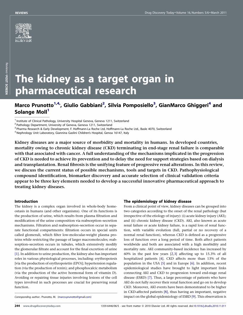
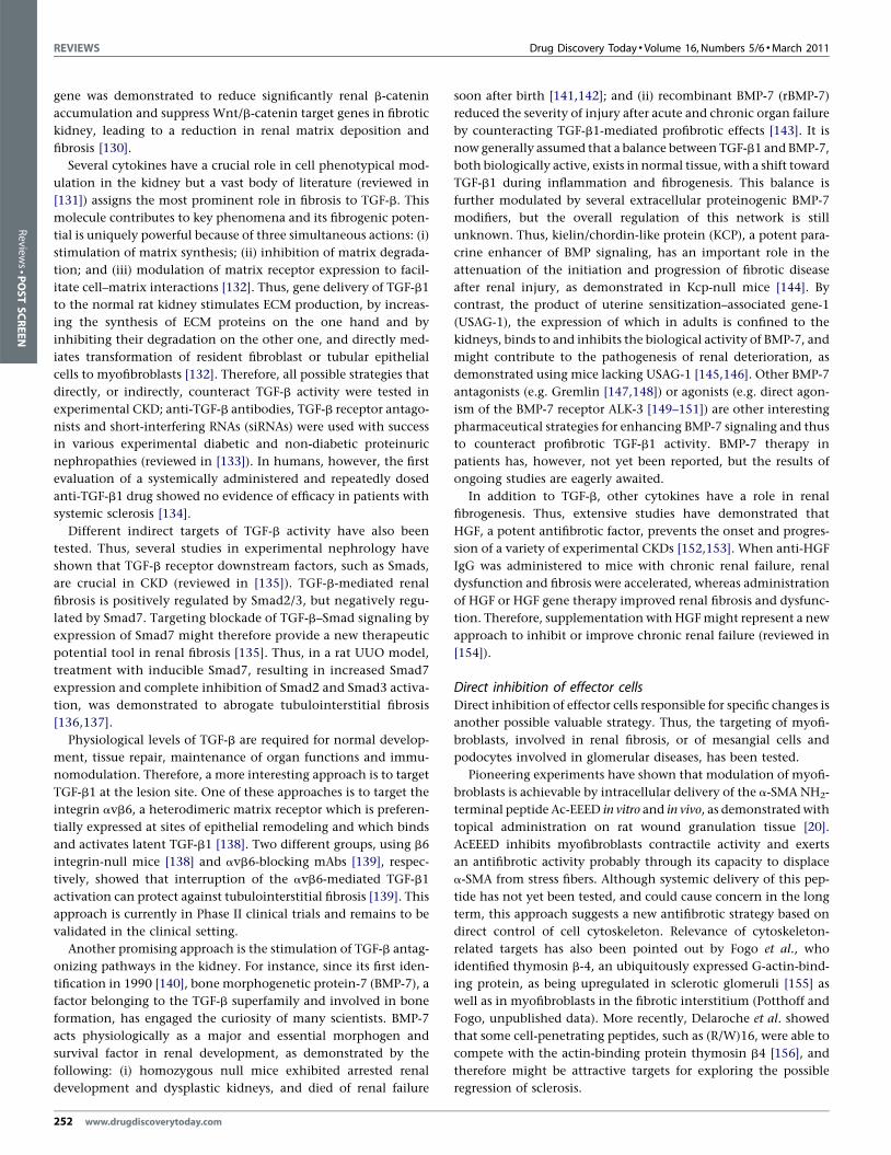
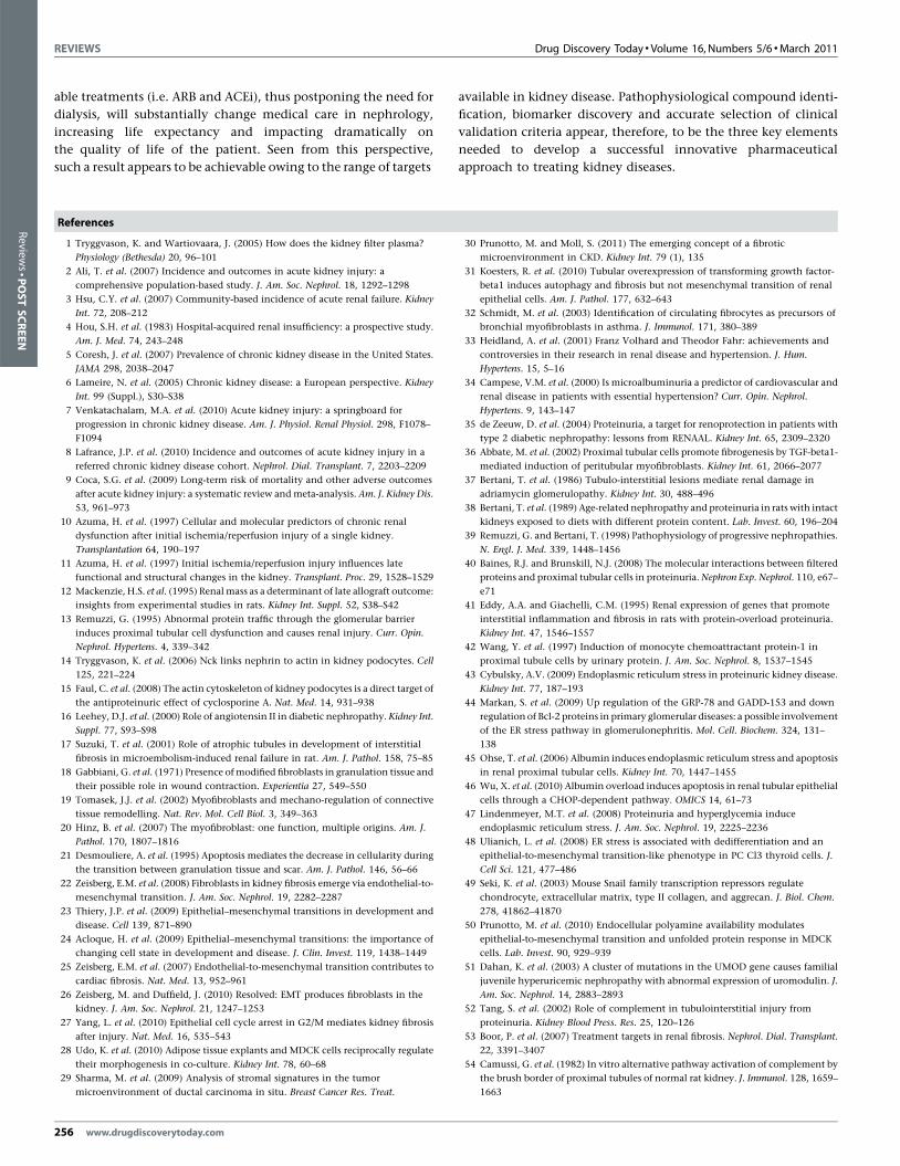
FIGURE 1
ESRD: a leading cause of patient death in developed countries.
Epidemiological studies have shown that ESRD is a major cause of death, its
mortality in developed countries was comparable to that of cancer in the USAin 2004 (Surveillance, Epidemiology, and End Results Program of the National
Cancer Institute; http://seer.cancer.gov/).
Reviews�POSTSCREEN
supported by experimental evidence that shows that a reduced
renal mass negatively influences repair and recovery after ischemic
injury mimicking AKI [10–12]. Thus, similarly to the reduction of
renal mass commonly observed in CKD patients independently of
the cause, diabetic- and age-related nephropathies are expected to
predispose kidneys to develop acute injuries. The global popula-
tion of patients with ESRD treated with renal replacement therapy
(RRT) was estimated to have reached almost 1.7 million at the end
of 2003, and continues to grow at an annual rate of�5–8%, driven
by an aging population, increased incidence of diabetes mellitus,
better means of detecting CKD and better access to treatment [6].
This exponential growth worldwide of patients with ESRD, whose
mortality in developed countries is comparable to that owing to
cancer (Fig. 1), will become a public health problem with unbear-
able costs of substitutive treatments.
From a pathological point of view, kidney diseases can be
grouped into three categories according to the injured renal com-
partment, but irrespective of the etiology of injury: (i) glomeru-
lopathies (e.g. immune complex-mediated glomerulonephritis);
(ii) tubulointerstitial diseases (e.g. interstitial nephritis); and (iii)
vascular diseases (e.g. hypertensive nephroangiosclerosis). How-
ever, in regard to their pathogenic interconnections, the distinc-
tion between these three conditions appears artificial, because
renal fibrosis (i.e. tubulointerstitial fibrosis), is the common and
final histopathological feature.
Characteristics of the glomerular and tubularcompartmentsGlomeruli and tubules, which are functionally and morphologi-
cally distinct, both contribute to renal fibrosis. The filtration
function of the kidney relies upon the unique morphology of
the glomerulus. This filtrating unit consists of a capillary tuft
located inside the Bowman’s capsule. The glomerular filter com-
prises three distinct layers: (i) a fenestrated endothelium; (ii) a
glomerular basement membrane (GBM); and (iii) a slit diaphragm
located between the interdigitating foot processes of the epithelial
podocytes [1]. The glomeruli are affected in a variety of systemic
and primary kidney diseases (e.g. autoimmune processes and
inherited defects of podocytes), leading to proteinuria. Persistent
proteinuria has harmful effects on the kidney and frequently leads
to glomerulosclerosis, tubulointerstitial fibrosis and CKD [13].
Emphasis has been placed during the past few years on the role
of podocytes and mesangial cells as causes of the proteinuria and
in the progression of kidney fibrosis, respectively. Genome map-
ping of inherited nephrotic syndromes that do not respond to
steroid treatment has led to the discovery of proteins of the
podocyte slit-diaphragm [14,15]. In diabetic nephropathy, high
glucose and angiotensin II (AngII) both target mesangial cells
directly, modulating mesangial matrix synthesis degradation
and inducing transforming growth factor (TGF)-b secretion, thus
contributing to glomerular sclerosis [16].
In contrast to the filtration function of glomeruli, tubules are
involved in the modification of urine composition via reabsorp-
tion and secretion processes. Morphologically, tubules are in
direct contact with the interstitial compartment and are com-
posed of extracellular matrix and cells. Injury of the tubular
epithelial cells therefore also affects the interstitium. Thus, dur-
ing ischemic AKI, tubular cells (especially proximal tubule cells,
which are particularly sensitive to hypoxia) are severely injured,
with occasional necrosis. Although patients generally recover
from AKI through an efficient tubular re-epithelialization process,
some of them progress to CKD. In this case, atrophic tubules
contribute to the progression of CKD by eliciting transformation
of interstitial cells into myofibroblasts through a variety of
mechanisms, as demonstrated in the microembolic rat model
of CKD [17]. Bands of fibrosis, with a-smooth muscle actin
(SMA)-expressing myofibroblasts, can arise at the level of prox-
imal tubular cells; the contribution of distal tubules to fibrosis is
still not fully understood.
The fibrosis pathwaysInterstitial fibrosis is a common pathological feature observed in
all kinds of CKD irrespective of their etiology (Fig. 2). An important
advance in the understanding of fibrosis pathogenesis was the
identification of the myofibroblast, initially described in wound
granulation tissue [18], as the major actor in the onset and evolu-
tion of the lesions [19,20]. The myofibroblast, characterized by the
expression of a-SMA [19], contributes crucially to extracellular
matrix (ECM) remodeling by the synthesis of essential matrix
components, such as collagen type I and III, and cellular fibro-
nectin. Fibroblast migration, accumulation, replication and differ-
entiation into myofibroblasts, are observed in injured tissues
under the influence of local inflammatory factors such as TGF-
b1. Although myofibroblasts normally disappear as a result of
apoptosis when a wound heals [21], they might persist in other
situations, leading to fibrosis that, in turn, causes tissue deforma-
tion and loss of organ function.
Myofibroblasts originate mainly from local fibroblasts [18],
although another local source of these cells is epithelial (or endothe-
lial) cells that undergo epithelial-to-mesenchymal transition (EMT)
[22]. EMT has been extensively investigated recently and has been
demonstrated to be a crucial phenomenon in other relevant phy-
siological and pathological situations, such as organ development
www.drugdiscoverytoday.com 245
REVIEWS Drug Discovery Today � Volume 16, Numbers 5/6 �March 2011
[()TD$FIG]
Trichrome staining α-SMA
(a) (a')
(b) (b')
(c) (c')
(d) (d')
Drug Discovery Today
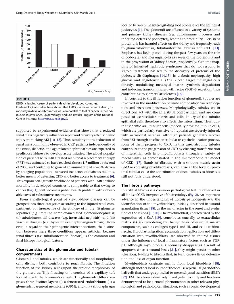
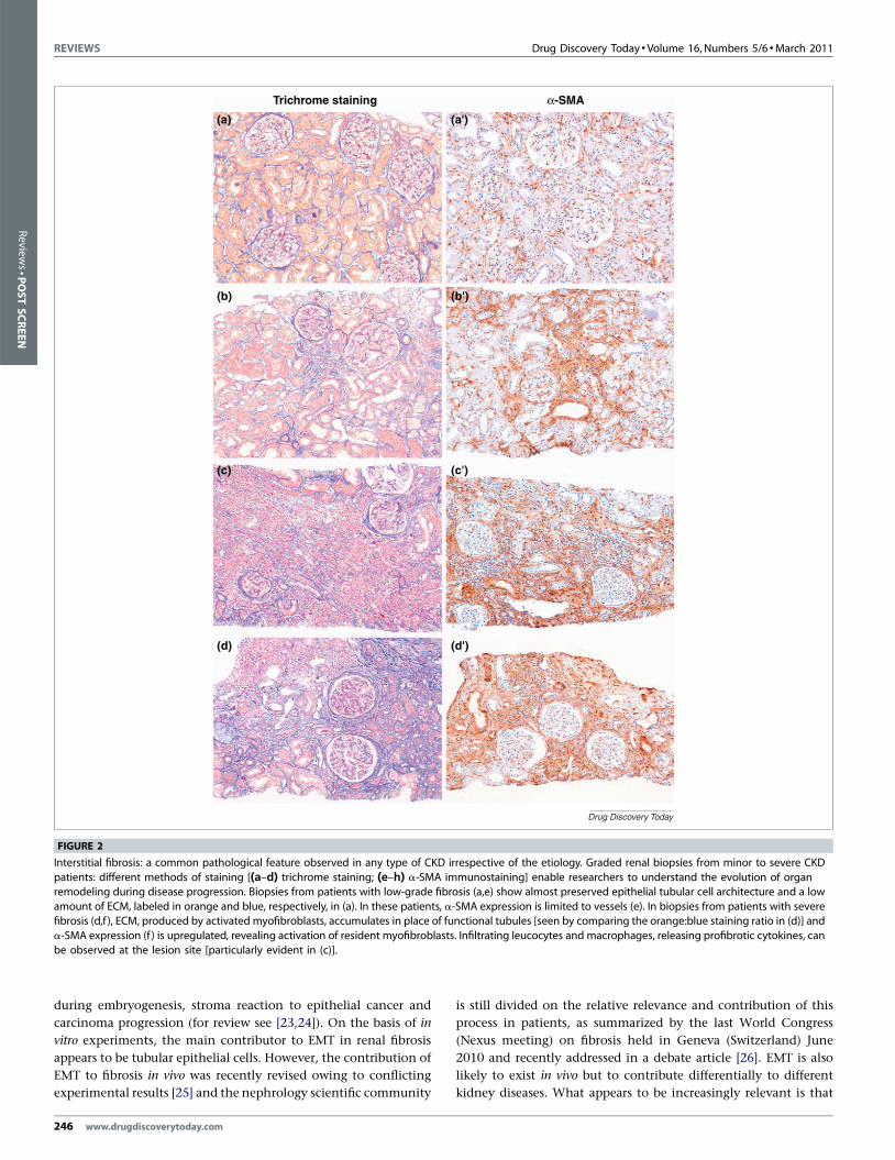
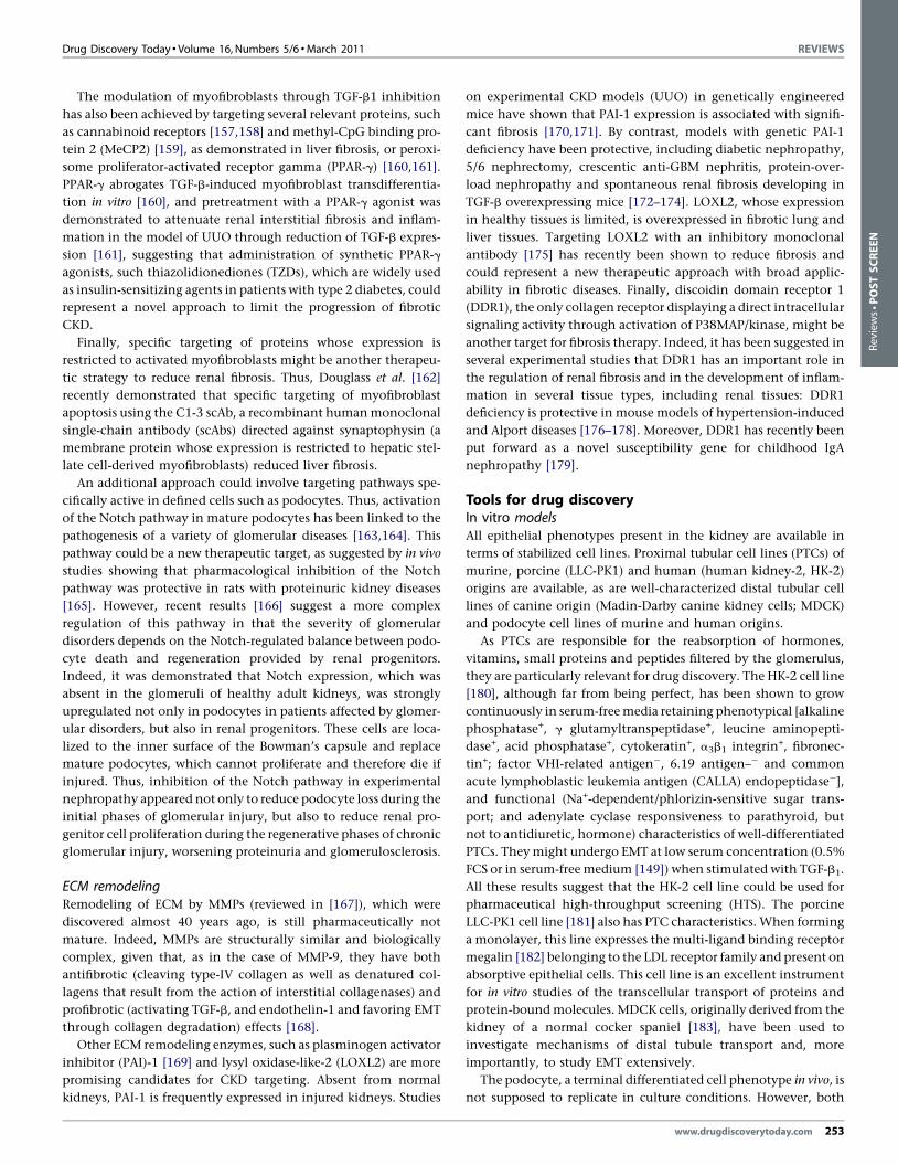
FIGURE 2
Interstitial fibrosis: a common pathological feature observed in any type of CKD irrespective of the etiology. Graded renal biopsies from minor to severe CKD
patients: different methods of staining [(a–d) trichrome staining; (e–h) a-SMA immunostaining] enable researchers to understand the evolution of organ
remodeling during disease progression. Biopsies from patients with low-grade fibrosis (a,e) show almost preserved epithelial tubular cell architecture and a low
amount of ECM, labeled in orange and blue, respectively, in (a). In these patients, a-SMA expression is limited to vessels (e). In biopsies from patients with severefibrosis (d,f ), ECM, produced by activated myofibroblasts, accumulates in place of functional tubules [seen by comparing the orange:blue staining ratio in (d)] and
a-SMA expression (f ) is upregulated, revealing activation of resident myofibroblasts. Infiltrating leucocytes and macrophages, releasing profibrotic cytokines, can
be observed at the lesion site [particularly evident in (c)].
Review
s�P
OSTSCREEN
during embryogenesis, stroma reaction to epithelial cancer and
carcinoma progression (for review see [23,24]). On the basis of in
vitro experiments, the main contributor to EMT in renal fibrosis
appears to be tubular epithelial cells. However, the contribution of
EMT to fibrosis in vivo was recently revised owing to conflicting
experimental results [25] and the nephrology scientific community
246 www.drugdiscoverytoday.com
is still divided on the relative relevance and contribution of this
process in patients, as summarized by the last World Congress
(Nexus meeting) on fibrosis held in Geneva (Switzerland) June
2010 and recently addressed in a debate article [26]. EMT is also
likely to exist in vivo but to contribute differentially to different
kidney diseases. What appears to be increasingly relevant is that
Drug Discovery Today � Volume 16, Numbers 5/6 �March 2011 REVIEWS
Reviews�POSTSCREEN
epithelial cell health might be pivotal in preserving normal kidney
architecture. Injured tubular epithelial cells have recently been
demonstrated to behave as a major source of pro-inflammatory
cytokines in experimental induced kidney fibrosis [27]. In addition,
Udo et al. [28] were able to show that this is also true in physiological
conditions in vitro, wheredifferentiatedMDCK distal tubular epithe-
lial cells, butnot3T3 fibroblasts, inhibited regeneration of mesench-
ymal stem cells from co-cultured adipose tissue fragments, therefore
confirming that renal tubular cells contribute to the general main-
tenance of the static state of mesenchymal cells. Results that suggest
the presence of a pro-fibrotic microenvironment perpetuating the
activation of myofibroblasts are similar to the scenario already
established in cancer biology [29,30].
Mimicking of tubular injury by confined overexpression of TGF-
b1 in renal epithelial cells [31] confirmed the absence of EMT in
vivo in mice and reconfirmed the importance in keeping epithelial
cells healthy. In this model, TGF-b1 drove autophagy in tubular
epithelial cells in the absence of EMT, leading to tubular atrophy,
with concomitant interstitial fibrosis that resulted from myofibro-
blasts derived from local fibroblasts. Neither tracing of injured
tubules in electron micrographs nor genetic tagging of tubular
epithelial cells revealed cells transgressing the tubular basement
membrane, a typical feature observed in EMT.
Another possible myofibroblast precursor is bone marrow-
derived cells (fibrocytes), which have been identified in several
fibrotic organs, such as skin, liver and lung [32]. However, the
contribution of local versus circulating cells to fibrosis process
remains to be established. Different mechanisms, among them
proteinuria, hypoxia and inflammation, might drive myofibro-
blast activation within the kidney.
As early as 1914, Volhard and Fahr [33] found that renal damage
was related to copious protein excretion in the urine and they
suggested that proteinuria could be the result of impaired plasma
protein reabsorption. Epidemiological studies have since con-
firmed the detrimental effect of proteinuria on renal function,
showing a clear correlation between renal failure progression and
proteinuria, which appears as an independent risk factor [34].
Moreover, several recent in vitro studies using proximal tubular
epithelial cells led to renewed interest in proteinuria as a causative
agent of tubular damage, myofibroblast activation and consequent
CKD progression [35,36].
However, it is still unclear how proteinuria produces fibrosis and
worsens renal function. Proteins in the urine are normally
absorbed by endocytosis in the proximal tubules. During periods
of heavy proteinuria, the filtered proteins accumulate in lysosomes
in the proximal tubular cells, causing cell disruption and injury
[37,38]. Proteins might also incite a toxic response through the
stimulation of the expression of proinflammatory cytokines [39].
Tubular reabsorption of albumin has been shown to alter the
biology of proximal tubular cells [40] and to upregulate synthesis
of various inflammatory mediators [41], thus contributing either
to phenotypical modulation of proximal epithelial cells or to
leukocyte interstitial infiltration with consequent release of pro-
fibrotic cytokines. Thus, cultured proximal tubule cells that are
exposed to albumin, transferrin, or immunoglobulin G, produce
increased amounts of monocyte chemotactic protein-1 (MCP-1),
which is preferentially excreted into the basolateral compartment,
suggesting a mechanism for interstitial inflammatory infiltrates in
vivo [42]. Excessive urinary protein excretion and reabsorption has
also been observed to induce rough endoplasmic reticulum (RER)
stress in proximal epithelial tubular cells. Whereas RER-stress
resolves rapidly upon accumulation of misfolded proteins, it
can lead to apoptosis under persistent proteinuria [43], as indi-
cated by numerous reports in experimental and human renal
pathology [44–47].
Interestingly, conditions perturbing the homeostasis of the RER
might induce EMT. This was demonstrated not only in in vitro
studies on epithelial cells [48–50], but also in human studies on
‘uromodulin (UMOD)-related diseases’. These familial forms of
tubulointerstitial fibrosis, reported previously under different
names, including medullary cystic kidney disease type 2 (MCKD2),
familial juvenile hyperuricemic nephropathy (FJHN) and glomer-
ulocystic kidney disease (GCKD), have a common defect in the
gene encoding uromodulin. It was recently demonstrated that
intracellular trapping of uromodulin within the RER was asso-
ciated with the development of typical tubulointerstitial fibrosis
[51].
Complement is also implicated in the pathogenesis of protei-
nuria-associated tubulointerstitial injury [52,53]. During glomer-
ular proteinuria, complement proteins are likely to be present in
the glomerular filtrate, and proximal tubular cells from both rat
and human kidneys activate complement [54,55]. There is also
evidence that tubular cells are an important local source of com-
plement [52]. It was demonstrated that massive proteinuria
induced tubulointerstitial injury associated with a marked deposi-
tion of C5b-9 on the apical membrane of proximal tubular cells
[56], and that membrane attack complex (MAC) formation inhibi-
tion ameliorated tubulointerstitial injury in proteinuric [57], but
not in nonproteinuric [58] animal models. In addition to C5b-9,
complement C5, and in particular C5a, might also become a target
in tubulointerstitial injury [59]. In the mouse unilateral ureteral
obstruction (UUO) model, genetic C5 deficiency or pharmacolo-
gical inhibition of the C5a receptor (C5aR) potently reduced
tubulointerstitial fibrosis [60]. C5aR antagonists and anti-C5 anti-
bodies (eculizumab) have been, or are currently being, evaluated in
clinical trials. Complement regulatory proteins, such as Crry (a
structural and functional rodent analog of human CR1, which
inhibits C3 activation) or CD59 (which inhibits C5b-9 formation)
might also be of interest [61,62].
Endothelin is also an important mediator of proteinuria-asso-
ciated renal fibrosis [63]. Cultured proximal tubule cells that are
exposed to albumin, transferrin, or immunoglobulin G produce
increased amount of endothelin-1 (ET-1) [64]. Studies in a variety
of experimental models demonstrated that gene encoding
endothelin and/or endothelin expression is increased during
nephropathy and colocalizes with fibrotic lesions [63]. Inversely,
pharmacological antagonism of endothelin receptors delayed the
evolution and/or prevented renal failure. Although bosentan, a
dual inhibitor of ETA and ETB, is in clinical use, its benefits versus
side effects (mainly hepatotoxicity and fluid retention) in CKD
patients require additional evaluation [53].
Hypoxia is the second presumed contributor to the progres-
sion of kidney fibrosis. Chronic ischemia in the tubulointersti-
tium space can occur via several mechanisms, including
intrarenal vasoconstriction [secondary to renin angiotensin
aldosterone system (RAAS) local activation or loss of vasodilatory
www.drugdiscoverytoday.com 247
REVIEWS Drug Discovery Today � Volume 16, Numbers 5/6 �March 2011
Review
s�P
OSTSCREEN
nitric oxide (NO)] or structural lesions that impair blood flow
delivery, such as glomerulosclerosis, with impairment of the
glomerular capillary bed and consecutive impairment of the
peritubular perfusion and of the tubular oxygen supply. Tubu-
lointerstitial ischemic damage also results in the loss of peritub-
ular capillaries, as demonstrated in histological studies of human
kidneys [65]. Furthermore, interstitial fibrosis impairs tubular
oxygen supply owing to the increased diffusion distance between
peritubular capillaries and tubular cells. Mechanisms of hypoxia-
induced tubulointerstitial damage are multifactorial. Hypoxia
can activate fibroblasts, change the extracellular matrix meta-
bolism of resident renal cells, and lead to eventual fibrogenesis
[66,67]. Furthermore, mild hypoxia was demonstrated to induce
tubular EMT with the transdifferentiation of cultured tubular
cells into myofibroblasts [68]. Treatments targeting hypoxic
tubulointerstitial damage involve EPO, as anemia is a risk factor
for renal failure, with the subsequent improvement in oxygena-
tion of the kidney.
[()TD$FIG]
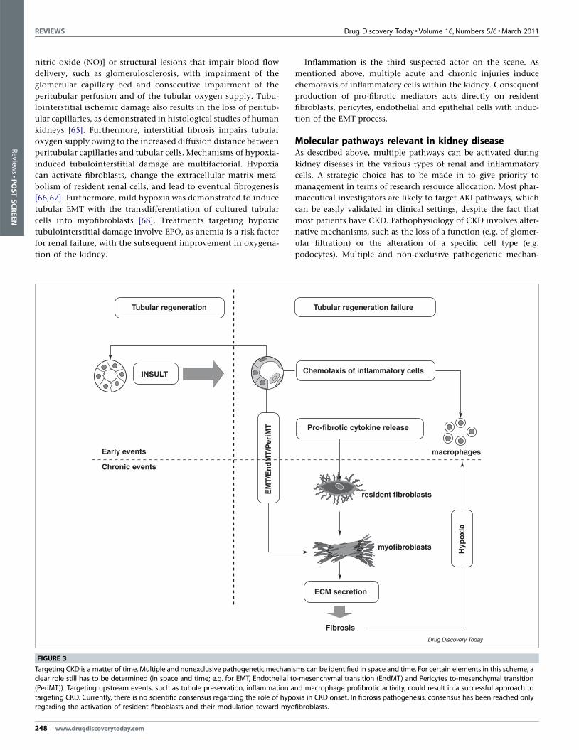
Tubular regeneration
INSULT
Early events
Chronic events
EM
T/E
nd
MT
/Per
iMT
FIGURE 3
Targeting CKD is a matter of time. Multiple and nonexclusive pathogenetic mechani
clear role still has to be determined (in space and time; e.g. for EMT, Endothelial t
(PeriMT)). Targeting upstream events, such as tubule preservation, inflammation a
targeting CKD. Currently, there is no scientific consensus regarding the role of hyporegarding the activation of resident fibroblasts and their modulation toward myo
248 www.drugdiscoverytoday.com
Inflammation is the third suspected actor on the scene. As
mentioned above, multiple acute and chronic injuries induce
chemotaxis of inflammatory cells within the kidney. Consequent
production of pro-fibrotic mediators acts directly on resident
fibroblasts, pericytes, endothelial and epithelial cells with induc-
tion of the EMT process.
Molecular pathways relevant in kidney diseaseAs described above, multiple pathways can be activated during
kidney diseases in the various types of renal and inflammatory
cells. A strategic choice has to be made in to give priority to
management in terms of research resource allocation. Most phar-
maceutical investigators are likely to target AKI pathways, which
can be easily validated in clinical settings, despite the fact that
most patients have CKD. Pathophysiology of CKD involves alter-
native mechanisms, such as the loss of a function (e.g. of glomer-
ular filtration) or the alteration of a specific cell type (e.g.
podocytes). Multiple and non-exclusive pathogenetic mechan-
Tubular regeneration failure
Chemotaxis of inflammatory cells
Pro-fibrotic cytokine release
resident fibroblasts
macrophages
myofibroblasts
ECM secretion
Fibrosis
Hyp
oxia
Drug Discovery Today
sms can be identified in space and time. For certain elements in this scheme, a
o-mesenchymal transition (EndMT) and Pericytes to-mesenchymal transition
nd macrophage profibrotic activity, could result in a successful approach to
xia in CKD onset. In fibrosis pathogenesis, consensus has been reached onlyfibroblasts.
Drug Discovery Today � Volume 16, Numbers 5/6 �March 2011 REVIEWS
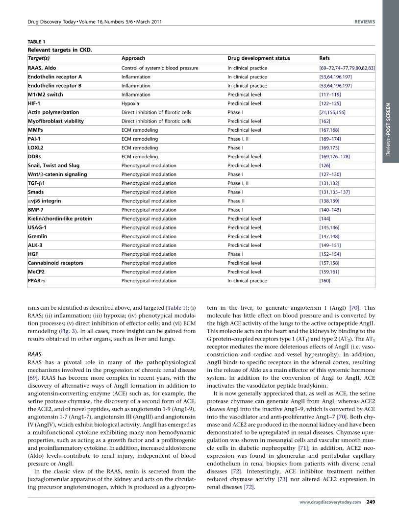
TABLE 1
Relevant targets in CKD.
Target(s) Approach Drug development status Refs
RAAS, Aldo Control of systemic blood pressure In clinical practice [69–72,74–77,79,80,82,83]
Endothelin receptor A Inflammation In clinical practice [53,64,196,197]
Endothelin receptor B Inflammation In clinical practice [53,64,196,197]
M1/M2 switch Inflammation Preclinical level [117–119]
HIF-1 Hypoxia Preclinical level [122–125]
Actin polymerization Direct inhibition of fibrotic cells Phase I [21,155,156]
Myofibroblast viability Direct inhibition of fibrotic cells Preclinical level [162]
MMPs ECM remodeling Preclinical level [167,168]
PAI-1 ECM remodeling Phase I, II [169–174]
LOXL2 ECM remodeling Phase I [169,175]
DDRs ECM remodeling Preclinical level [169,176–178]
Snail, Twist and Slug Phenotypical modulation Preclinical level [126]
Wnt/b-catenin signaling Phenotypical modulation Phase I [127–130]
TGF-b1 Phenotypical modulation Phase I, II [131,132]
Smads Phenotypical modulation Phase I [131,135–137]
avb6 integrin Phenotypical modulation Phase II [138,139]
BMP-7 Phenotypical modulation Phase I [140–143]
Kielin/chordin-like protein Phenotypical modulation Preclinical level [144]
USAG-1 Phenotypical modulation Preclinical level [145,146]
Gremlin Phenotypical modulation Preclinical level [147,148]
ALK-3 Phenotypical modulation Preclinical level [149–151]
HGF Phenotypical modulation Phase I [152–154]
Cannabinoid receptors Phenotypical modulation Preclinical level [157,158]
MeCP2 Phenotypical modulation Preclinical level [159,161]
PPAR-g Phenotypical modulation In clinical practice [160]
Reviews�POSTSCREEN
isms can be identified as described above, and targeted (Table 1): (i)
RAAS; (ii) inflammation; (iii) hypoxia; (iv) phenotypical modula-
tion processes; (v) direct inhibition of effector cells; and (vi) ECM
remodeling (Fig. 3). In all cases, more insight can be gained from
results obtained in other organs, such as liver and lungs.
RAASRAAS has a pivotal role in many of the pathophysiological
mechanisms involved in the progression of chronic renal disease
[69]. RAAS has become more complex in recent years, with the
discovery of alternative ways of AngII formation in addition to
angiotensin-converting enzyme (ACE) such as, for example, the
serine protease chymase, the discovery of a second form of ACE,
the ACE2, and of novel peptides, such as angiotensin 1-9 (Ang1-9),
angiotensin 1-7 (Ang1-7), angiotensin III (AngIII) and angiotensin
IV (AngIV), which exhibit biological activity. AngII has emerged as
a multifunctional cytokine exhibiting many non-hemodynamic
properties, such as acting as a growth factor and a profibrogenic
and proinflammatory cytokine. In addition, increased aldosterone
(Aldo) levels contribute to renal injury, independent of blood
pressure or AngII.
In the classic view of the RAAS, renin is secreted from the
juxtaglomerular apparatus of the kidney and acts on the circulat-
ing precursor angiotensinogen, which is produced as a glycopro-
tein in the liver, to generate angiotensin I (AngI) [70]. This
molecule has little effect on blood pressure and is converted by
the high ACE activity of the lungs to the active octapeptide AngII.
This molecule acts on the heart and the kidneys by binding to the
G protein-coupled receptors type 1 (AT1) and type 2 (AT2). The AT1
receptor mediates the more deleterious effects of AngII (i.e. vaso-
constriction and cardiac and vessel hypertrophy). In addition,
AngII binds to specific receptors in the adrenal cortex, resulting
in the release of Aldo as a main effector of this systemic hormone
system. In addition to the conversion of AngI to AngII, ACE
inactivates the vasodilator peptide bradykinin.
It is now generally appreciated that, as well as ACE, the serine
protease chymase can generate AngII from AngI, whereas ACE2
cleaves AngI into the inactive Ang1–9, which is converted by ACE
into the vasodilator and anti-proliferative Ang1–7 [70]. Both chy-
mase and ACE2 are produced in the normal kidney and have been
demonstrated to be upregulated in renal diseases. Chymase upre-
gulation was shown in mesangial cells and vascular smooth mus-
cle cells in diabetic nephropathy [71]; in addition, ACE2 neo-
expression was found in glomerular and peritubular capillary
endothelium in renal biopsies from patients with diverse renal
diseases [72]. Interestingly, ACE inhibitor treatment neither
reduced chymase activity [73] nor altered ACE2 expression in
renal diseases [72].
www.drugdiscoverytoday.com 249
REVIEWS Drug Discovery Today � Volume 16, Numbers 5/6 �March 2011
Review
s�P
OSTSCREEN
In addition to AngII, other Ang peptides might exert clinically
relevant vasoactive actions. As described previously, Ang1–7, a
heptapeptide derived from AngI and/or from AngII, might antag-
onize AngII, especially in situations of an overactive RAAS, such
as during sodium restriction, an effect that results in renal
vasodilation and increased natriuresis. In addition, digestion
of AngI and AngII by angiotensinases, peptidases, aminopepti-
dases, carboxypeptidases, or endopeptidases, results in different
peptide fragments with functions distinct from those of AngI
and AngII. Thus, AngII is metabolized in the kidney by amino-
peptidase A into AngIII and further into AngIV [74]. AngIV (the
3–8 peptide) binds the AT4 receptor selectively and stimulates
plasminogen activator inhibitor-1 release (PAI-1) [75]. Emerging
evidence suggests that AngII is not only a vasoactive peptide, but
also a true cytokine that regulates cell growth, inflammation and
fibrosis [70]. Non-hemodynamic effects of angiotensin include
increased production of reactive oxygen species; upregulation of
cytokines, cell adhesion molecules and profibrotic growth fac-
tors; induction of TGF-b expression; increased synthesis of extra-
cellular matrix proteins; stimulation of PAI-1 production by
endothelial and vascular smooth muscle cells; and macrophage
activation and infiltration.
Therefore, inhibition of the RAAS is particularly effective in
preventing glomerulosclerosis and tubulointerstitial fibrosis. The
benefit seen with such drugs is in addition to that expected from
their antihypertensive effects alone. Drugs acting on the RAAS
include agents that inhibit the synthesis and release of renin or
AngII directly (i.e. renin and ACE inhibitors), drugs that antag-
onize the receptor effects of AngII (i.e. AngII receptor blockers;
ARBs), the Aldo-receptor antagonists, and a new class of com-
bined ACEI and neutral endopeptidase inhibitors, called vaso-
peptidase inhibitors (VPIs). Renin inhibitors (e.g. aliskiren,
enalkiren and zalkiren) are promising drugs for combating renal
fibrosis, as shown in severely hypertensive rats transgenic for
genes encoding human renin and angiotensinogen [76]. How-
ever, clinical effects on renal fibrosis remain to be determined.
The recently discovered receptor for renin and prorenin [(pro)-
renin receptor; (P)RR] was linked to glomerular fibrosis [77]. The
specific (P)RR activates intracellular signaling and enhances
receptor-bound (pro)renin catalytic activity on the cell surface.
Experimental evidence regarding the role of (P)RR in renal
damage is accumulating and suggests that blocking (P)RR repre-
sents a new therapeutic target for tissue protection. (P)RR blockers
might be more potent than renin inhibitors, because renin inhi-
bitors do not block renin or prorenin binding to, and activation
of, (P)RR. VPIs (e.g. AVE7688 and omapatrilat) block both ACE
and neutral endopeptidase (NEP). NEP catalyzes the hydrolysis of
atrial natriuretic peptide (ANP), brain natriuretic peptide and C-
type natriuretic peptide [78]. Simultaneous inhibition of the
RAAS and NEP system by VPIs results in vasodilation, natriuresis
and diuresis, and decreases peripheral vascular resistance and
blood pressure to a greater extent than does ACE inhibition alone
[53]. In an animal model of Alport syndrome with progressive
renal fibrosis, AVE7688 demonstrated nephroprotection with
antifibrotic, anti-inflammatory, and antiproteinuric effects
[79]. Finding that AVE7688 corrected the imbalanced ratio
between the vasoconstrictor endothelin 1 (ET-1) and the vasodi-
lators NO and ANP, could explain the superior renoprotection of
250 www.drugdiscoverytoday.com
vasopeptidase over ACE inhibitors [80]. At present, several VPIs
are in various stages of development. Omapatrilat has undergone
the most extensive clinical development. However, the high
incidence of angioedema compared with enalapril (as reported
in the Omapatrilat Cardiovascular Treatment Assessment Versus
Enalapril trial) might limit its use [81]. Combination therapy of
ACEI and ARB [82] or high-dosage of ACEI [83] showed potential
in experimental studies to stop or even regress renal fibrosis, at
least in the early stages.
Combined therapy, such as RAAS inhibitors associated with
statin, Vitamin D or Rho kinase inhibitors, has been widely
described in literature and demonstrated to be effective in redu-
cing glomerulosclerosis and inflammation. Statins, or 3-hydroxy-
3-methyl-glutaryl coenzyme A (HMG-CoA) reductase inhibitors,
independent of their ability to lower cholesterol, have pleiotropic
properties, including enhanced endothelial function with
increased endothelial nitric oxide synthase (eNOS) expression
and decreased ET-1 expression, anti-oxidant, anti-inflammatory
and immunomodulatory actions as well as antihypertensive and
antiproliferative effects [84]. Indeed, statins interfere with the
prenylation of Ras and Rho family small GTP98 binding proteins,
thereby blocking the activation of signaling pathways and tran-
scription factors, which regulate inflammatory and fibrogenic
genes that are crucial to renal disease progression [85,86]. In rats
with severe passive Heymann nephritis, the addition of statin to a
chronic background of ACE inhibition and ARB induced remission
of proteinuria and conferred complete protection of the kidney
[87]. In a more recent study, the effects of maximal RAAS inhibi-
tion by ACEI plus ARB in combination with statin were assessed in
rats with overt diabetic nephropathy [88]. Dual RAAS blockade
significantly reduced proteinuria with respect to vehicle, and the
addition of statin further lowered proteinuria to control levels.
Glomerulosclerosis was ameliorated by RAS inhibitors or statin,
and regression was achieved by the addition of statin. Finally,
tubular damage, interstitial inflammation and the expression of
the fibrotic markers TGF-b1 and phosphorylated Smad 2/3 in
tubuli were demonstrated to be significantly reduced by the triple
regimen.
Analogs of vitamin D have been demonstrated to attenuate
renal injury in several models of kidney disease [89,90]. The
activity of 1,25-dihydroxyvitamin D3 [1,25(OH)2D3], the active
hormonal form of vitamin D, is mediated by the vitamin D
receptor (VDR), a member of the nuclear receptor superfamily
[91], which is highly expressed in the kidney [92]. The most well-
known function of the vitamin D endocrine system is to maintain
calcium and phosphorus homoeostasis. However, previous studies
unveiled a range of so-called ‘non-calcemic’ activities, including
regulation of the RAAS and of the nuclear factor (NF)-kB pathway
[92]. 1,25(OH)2D3 and vitamin D analogs were shown to inhibit
renin expression in animals [93,94] by suppressing renin gene
transcription, as demonstrated by in vitro experiments [95]. In
addition, 1,25(OH)2D3 was demonstrated to suppress activation
of NF-kB [96,97], a family of transcription factors that regulates a
range of genes involved in inflammation, proliferation and fibro-
genesis, and which is known to have a key role in kidney disease
[98]. Consistently, plasma vitamin D status was found to be
inversely associated with increased renal inflammation in patients
with a variety of kidney diseases [99]. Moreover, vitamin D ana-
Drug Discovery Today � Volume 16, Numbers 5/6 �March 2011 REVIEWS
Reviews�POSTSCREEN
logs, alone or in combination with RAAS inhibitors, were demon-
strated to prevent or attenuate proteinuria, glomerulosclerosis and
tubulointerstitial fibrosis in several experimental models of kidney
diseases [100–102]. Finally, two recent animal studies using the
UUO model reported an attenuation of renal fibrosis with vitamin
D analog [103] and in VDR-null mice [89]. Together, these data
enforce the suggestion that the 1,25(OH)2D3–VDR system has a
crucial renoprotective role in pathophysiological conditions.
Whether vitamin D analogs could further optimize existing thera-
pies in human renal disease is currently under investigation
[90,104]. Results from clinical trials that have used vitamin D
analogs in patients with CKD [105] and in renal transplant reci-
pients [106] are expected in the near future.
Animal studies have reported that Rho-kinase inhibition has
renoprotective effects in different experimental models, such as
UUO [107], hypertension [108,109] and diabetes [110]. During the
1990s, Rho-kinase was identified as one of the main downstream
effectors of the small G protein RhoA [111,112]. Rho-kinase is
activated by Rho and mediates some of its biological effects. Rho
functions as a molecular switch, in that Rho is inactive in its
guanosine diphosphate (GDP)-bound form and active in its gua-
nosine triphosphate (GTP)-bound form [113], and it participates in
various cellular functions, including formation of stress fibers and
focal adhesions, regulation of calcium ion sensitivity, production
of cytokines and regulation of G1 to S in cell cycle progression
[114]. The discovery of specific Rho-kinase inhibitors advanced the
knowledge of the Rho–Rho-kinase pathway in vitro and in vivo
[115]. Recently, combination therapy including an ACEI (imida-
pril) and a Rho-kinase inhibitor (fasudil) was demonstrated to be
more effective than either monotherapy for the prevention of
renal interstitial fibrosis induced by UUO, with reduction of
TGF-b and/or collagen production, monocyte and/or macrophage
infiltration, myofibroblast differentiation and oxidative stress gen-
eration [116]. Thus, Rho-kinase inhibitor could serve as a novel
strategy for the treatment of renal fibrosis. However, further
translational studies in humans are needed to substantiate these
findings.
Inflammatory cells and myofibroblastsAn exciting field for target discovery in kidney disease is the
crosstalk between inflammatory cells and myofibroblasts. Rephras-
ing the dialog between these partners might lead to the develop-
ment of new therapies. This could be the case for infiltrating
macrophages, which are classically recognized as active players
in progressive renal scarring but might also have a protective role
by preserving renal architecture and limiting progressive renal
scarring, as demonstrated in the late stage of an experimental
kidney fibrosis model after UUO (reviewed in [117]). These appar-
ently paradoxical findings might reflect the differential regulation
of inflammation and fibrosis mediated by two functionally distinct
macrophage phenotypes. Thus, classically activated (M1) and
alternatively activated (M2) macrophages [117–119] mediate con-
trasting and complementary functions in tissue fibrosis. Whereas
M2 macrophages produce large amounts of TGF-b and induce
myofibroblast proliferation, promoting ECM synthesis and fibrosis
development, M1 macrophages produce matrix metalloprotei-
nases (MMPs) by themselves and induce myofibroblasts to produce
MMPs, promoting ECM degradation and fibrosis resolution.
Therefore, new therapeutic agents that specifically target M1,
induce the transformation of M2 to M1 or interfere with M1
and/or M2 and myofibroblast crosstalk, could represent a selective
approach for renal fibrosis treatment.
HypoxiaHypoxia has been documented to have a role in chronic renal
diseases of different etiologies [e.g. 5/6 nephrectomy, diabetic
nephropathy, immunoglobulin A (IgA) nephropathy and UUO].
Retrospective [120] and prospective [121] studies suggested that
improvement of anemia, and thus improvement of renal hypoxia,
by treatment with EPO delayed the progression of renal failure.
Treatments with hypoxia-inducible factor (HIF), one of the most
important factors in the cellular response to hypoxia, and/or with
VEGF, an angiogenic growth factor, might be new strategies
against ischemia-induced tubulointerstitial fibrosis, investigated
currently only in animal models [122,123]. However, experimen-
tal evidence indicated that hypoxia, via HIF activation, might
either accelerate [124] or attenuate [125] renal fibrogenesis. There-
fore, this field of pharmaceutical research appears premature and
many relevant points still have to be addressed by basic research.
Phenotypical modulation processesTargeting phenotypical modulation processes enables potentially
multiple levels of intervention. Thus, inhibition of early tubular
events, such cell detachment, can be achieved by blocking tran-
scriptional factors (e.g. Snail, Twist or Slug), which regulate expres-
sion of relevant adhesion molecules, such as E-cadherins, or by
targeting enzymes (e.g. glycogen synthase kinase-3 b; GSK-3b) that
regulate at the transcriptional factor level within tubular cells:
activation of the Snail gene (SNAI1) has been demonstrated in vitro
to suppress fibrosis via the direct repression of the kidney differ-
entiation factor hepatocyte nuclear factor (HNF)-1b. GSK-3b-con-
ditional-KO mice have elevated collagen production, elevated
levels of profibrotic a-SMA and increased myofibroblast formation
during wound healing, and elevated expression and production of
ET-1 compared with control mice [126]. Antagonizing ET-1 was
demonstrated, in the same study, to reverse the phenotype of
Gsk3b-CKO mice, suggesting that targeting the GSK-3b pathway
or ET-1 is of benefit in controlling tissue repair and fibrogenic
responses in vivo.
Advanced phenotypical transformation of epithelial, endothe-
lial cells or pericytes toward a mesenchymal phenotype can also be
controlled by targeting the Wnt/b-catenin signaling pathway.
Wnt proteins transmit their signal across the plasma membrane
through interactions with serpentine receptors, the Frizzled (Fzd)
family of proteins, and co-receptors, members of the low-density
lipoprotein (LDL) receptor-related protein (e.g. LRP5/6). Upon
binding to their receptors, Wnt proteins induce a series of down-
stream signaling events, inducing stimulation of the transcription
of Wnt target genes, such as those encoding fibronectin [127] and
MMP-7 [128], which are correlated with mesenchymal phenotype
and invasiveness. Kidney disease states characterized by renal
fibrosis exhibit overexpression of multiple Wnt proteins in glo-
meruli and interstitium [129]. Therefore, targeting Wnt/b-catenin
signaling might be an effective strategy to hinder the progression
of renal interstitial fibrosis. Thus, in a mouse model of obstructive
nephropathy, delivery of the Wnt antagonist Dickkopf-1 (DKK1)
www.drugdiscoverytoday.com 251
REVIEWS Drug Discovery Today � Volume 16, Numbers 5/6 �March 2011
Review
s�P
OSTSCREEN
gene was demonstrated to reduce significantly renal b-catenin
accumulation and suppress Wnt/b-catenin target genes in fibrotic
kidney, leading to a reduction in renal matrix deposition and
fibrosis [130].
Several cytokines have a crucial role in cell phenotypical mod-
ulation in the kidney but a vast body of literature (reviewed in
[131]) assigns the most prominent role in fibrosis to TGF-b. This
molecule contributes to key phenomena and its fibrogenic poten-
tial is uniquely powerful because of three simultaneous actions: (i)
stimulation of matrix synthesis; (ii) inhibition of matrix degrada-
tion; and (iii) modulation of matrix receptor expression to facil-
itate cell–matrix interactions [132]. Thus, gene delivery of TGF-b1
to the normal rat kidney stimulates ECM production, by increas-
ing the synthesis of ECM proteins on the one hand and by
inhibiting their degradation on the other one, and directly med-
iates transformation of resident fibroblast or tubular epithelial
cells to myofibroblasts [132]. Therefore, all possible strategies that
directly, or indirectly, counteract TGF-b activity were tested in
experimental CKD; anti-TGF-b antibodies, TGF-b receptor antago-
nists and short-interfering RNAs (siRNAs) were used with success
in various experimental diabetic and non-diabetic proteinuric
nephropathies (reviewed in [133]). In humans, however, the first
evaluation of a systemically administered and repeatedly dosed
anti-TGF-b1 drug showed no evidence of efficacy in patients with
systemic sclerosis [134].
Different indirect targets of TGF-b activity have also been
tested. Thus, several studies in experimental nephrology have
shown that TGF-b receptor downstream factors, such as Smads,
are crucial in CKD (reviewed in [135]). TGF-b-mediated renal
fibrosis is positively regulated by Smad2/3, but negatively regu-
lated by Smad7. Targeting blockade of TGF-b–Smad signaling by
expression of Smad7 might therefore provide a new therapeutic
potential tool in renal fibrosis [135]. Thus, in a rat UUO model,
treatment with inducible Smad7, resulting in increased Smad7
expression and complete inhibition of Smad2 and Smad3 activa-
tion, was demonstrated to abrogate tubulointerstitial fibrosis
[136,137].
Physiological levels of TGF-b are required for normal develop-
ment, tissue repair, maintenance of organ functions and immu-
nomodulation. Therefore, a more interesting approach is to target
TGF-b1 at the lesion site. One of these approaches is to target the
integrin avb6, a heterodimeric matrix receptor which is preferen-
tially expressed at sites of epithelial remodeling and which binds
and activates latent TGF-b1 [138]. Two different groups, using b6
integrin-null mice [138] and avb6-blocking mAbs [139], respec-
tively, showed that interruption of the avb6-mediated TGF-b1
activation can protect against tubulointerstitial fibrosis [139]. This
approach is currently in Phase II clinical trials and remains to be
validated in the clinical setting.
Another promising approach is the stimulation of TGF-b antag-
onizing pathways in the kidney. For instance, since its first iden-
tification in 1990 [140], bone morphogenetic protein-7 (BMP-7), a
factor belonging to the TGF-b superfamily and involved in bone
formation, has engaged the curiosity of many scientists. BMP-7
acts physiologically as a major and essential morphogen and
survival factor in renal development, as demonstrated by the
following: (i) homozygous null mice exhibited arrested renal
development and dysplastic kidneys, and died of renal failure
252 www.drugdiscoverytoday.com
soon after birth [141,142]; and (ii) recombinant BMP-7 (rBMP-7)
reduced the severity of injury after acute and chronic organ failure
by counteracting TGF-b1-mediated profibrotic effects [143]. It is
now generally assumed that a balance between TGF-b1 and BMP-7,
both biologically active, exists in normal tissue, with a shift toward
TGF-b1 during inflammation and fibrogenesis. This balance is
further modulated by several extracellular proteinogenic BMP-7
modifiers, but the overall regulation of this network is still
unknown. Thus, kielin/chordin-like protein (KCP), a potent para-
crine enhancer of BMP signaling, has an important role in the
attenuation of the initiation and progression of fibrotic disease
after renal injury, as demonstrated in Kcp-null mice [144]. By
contrast, the product of uterine sensitization–associated gene-1
(USAG-1), the expression of which in adults is confined to the
kidneys, binds to and inhibits the biological activity of BMP-7, and
might contribute to the pathogenesis of renal deterioration, as
demonstrated using mice lacking USAG-1 [145,146]. Other BMP-7
antagonists (e.g. Gremlin [147,148]) or agonists (e.g. direct agon-
ism of the BMP-7 receptor ALK-3 [149–151]) are other interesting
pharmaceutical strategies for enhancing BMP-7 signaling and thus
to counteract profibrotic TGF-b1 activity. BMP-7 therapy in
patients has, however, not yet been reported, but the results of
ongoing studies are eagerly awaited.
In addition to TGF-b, other cytokines have a role in renal
fibrogenesis. Thus, extensive studies have demonstrated that
HGF, a potent antifibrotic factor, prevents the onset and progres-
sion of a variety of experimental CKDs [152,153]. When anti-HGF
IgG was administered to mice with chronic renal failure, renal
dysfunction and fibrosis were accelerated, whereas administration
of HGF or HGF gene therapy improved renal fibrosis and dysfunc-
tion. Therefore, supplementation with HGF might represent a new
approach to inhibit or improve chronic renal failure (reviewed in
[154]).
Direct inhibition of effector cellsDirect inhibition of effector cells responsible for specific changes is
another possible valuable strategy. Thus, the targeting of myofi-
broblasts, involved in renal fibrosis, or of mesangial cells and
podocytes involved in glomerular diseases, has been tested.
Pioneering experiments have shown that modulation of myofi-
broblasts is achievable by intracellular delivery of the a-SMA NH2-
terminal peptide Ac-EEED in vitro and in vivo, as demonstrated with
topical administration on rat wound granulation tissue [20].
AcEEED inhibits myofibroblasts contractile activity and exerts
an antifibrotic activity probably through its capacity to displace
a-SMA from stress fibers. Although systemic delivery of this pep-
tide has not yet been tested, and could cause concern in the long
term, this approach suggests a new antifibrotic strategy based on
direct control of cell cytoskeleton. Relevance of cytoskeleton-
related targets has also been pointed out by Fogo et al., who
identified thymosin b-4, an ubiquitously expressed G-actin-bind-
ing protein, as being upregulated in sclerotic glomeruli [155] as
well as in myofibroblasts in the fibrotic interstitium (Potthoff and
Fogo, unpublished data). More recently, Delaroche et al. showed
that some cell-penetrating peptides, such as (R/W)16, were able to
compete with the actin-binding protein thymosin b4 [156], and
therefore might be attractive targets for exploring the possible
regression of sclerosis.
Drug Discovery Today � Volume 16, Numbers 5/6 �March 2011 REVIEWS
Reviews�POSTSCREEN
The modulation of myofibroblasts through TGF-b1 inhibition
has also been achieved by targeting several relevant proteins, such
as cannabinoid receptors [157,158] and methyl-CpG binding pro-
tein 2 (MeCP2) [159], as demonstrated in liver fibrosis, or peroxi-
some proliferator-activated receptor gamma (PPAR-g) [160,161].
PPAR-g abrogates TGF-b-induced myofibroblast transdifferentia-
tion in vitro [160], and pretreatment with a PPAR-g agonist was
demonstrated to attenuate renal interstitial fibrosis and inflam-
mation in the model of UUO through reduction of TGF-b expres-
sion [161], suggesting that administration of synthetic PPAR-g
agonists, such thiazolidionediones (TZDs), which are widely used
as insulin-sensitizing agents in patients with type 2 diabetes, could
represent a novel approach to limit the progression of fibrotic
CKD.
Finally, specific targeting of proteins whose expression is
restricted to activated myofibroblasts might be another therapeu-
tic strategy to reduce renal fibrosis. Thus, Douglass et al. [162]
recently demonstrated that specific targeting of myofibroblast
apoptosis using the C1-3 scAb, a recombinant human monoclonal
single-chain antibody (scAbs) directed against synaptophysin (a
membrane protein whose expression is restricted to hepatic stel-
late cell-derived myofibroblasts) reduced liver fibrosis.
An additional approach could involve targeting pathways spe-
cifically active in defined cells such as podocytes. Thus, activation
of the Notch pathway in mature podocytes has been linked to the
pathogenesis of a variety of glomerular diseases [163,164]. This
pathway could be a new therapeutic target, as suggested by in vivo
studies showing that pharmacological inhibition of the Notch
pathway was protective in rats with proteinuric kidney diseases
[165]. However, recent results [166] suggest a more complex
regulation of this pathway in that the severity of glomerular
disorders depends on the Notch-regulated balance between podo-
cyte death and regeneration provided by renal progenitors.
Indeed, it was demonstrated that Notch expression, which was
absent in the glomeruli of healthy adult kidneys, was strongly
upregulated not only in podocytes in patients affected by glomer-
ular disorders, but also in renal progenitors. These cells are loca-
lized to the inner surface of the Bowman’s capsule and replace
mature podocytes, which cannot proliferate and therefore die if
injured. Thus, inhibition of the Notch pathway in experimental
nephropathy appeared not only to reduce podocyte loss during the
initial phases of glomerular injury, but also to reduce renal pro-
genitor cell proliferation during the regenerative phases of chronic
glomerular injury, worsening proteinuria and glomerulosclerosis.
ECM remodelingRemodeling of ECM by MMPs (reviewed in [167]), which were
discovered almost 40 years ago, is still pharmaceutically not
mature. Indeed, MMPs are structurally similar and biologically
complex, given that, as in the case of MMP-9, they have both
antifibrotic (cleaving type-IV collagen as well as denatured col-
lagens that result from the action of interstitial collagenases) and
profibrotic (activating TGF-b, and endothelin-1 and favoring EMT
through collagen degradation) effects [168].
Other ECM remodeling enzymes, such as plasminogen activator
inhibitor (PAI)-1 [169] and lysyl oxidase-like-2 (LOXL2) are more
promising candidates for CKD targeting. Absent from normal
kidneys, PAI-1 is frequently expressed in injured kidneys. Studies
on experimental CKD models (UUO) in genetically engineered
mice have shown that PAI-1 expression is associated with signifi-
cant fibrosis [170,171]. By contrast, models with genetic PAI-1
deficiency have been protective, including diabetic nephropathy,
5/6 nephrectomy, crescentic anti-GBM nephritis, protein-over-
load nephropathy and spontaneous renal fibrosis developing in
TGF-b overexpressing mice [172–174]. LOXL2, whose expression
in healthy tissues is limited, is overexpressed in fibrotic lung and
liver tissues. Targeting LOXL2 with an inhibitory monoclonal
antibody [175] has recently been shown to reduce fibrosis and
could represent a new therapeutic approach with broad applic-
ability in fibrotic diseases. Finally, discoidin domain receptor 1
(DDR1), the only collagen receptor displaying a direct intracellular
signaling activity through activation of P38MAP/kinase, might be
another target for fibrosis therapy. Indeed, it has been suggested in
several experimental studies that DDR1 has an important role in
the regulation of renal fibrosis and in the development of inflam-
mation in several tissue types, including renal tissues: DDR1
deficiency is protective in mouse models of hypertension-induced
and Alport diseases [176–178]. Moreover, DDR1 has recently been
put forward as a novel susceptibility gene for childhood IgA
nephropathy [179].
Tools for drug discoveryIn vitro modelsAll epithelial phenotypes present in the kidney are available in
terms of stabilized cell lines. Proximal tubular cell lines (PTCs) of
murine, porcine (LLC-PK1) and human (human kidney-2, HK-2)
origins are available, as are well-characterized distal tubular cell
lines of canine origin (Madin-Darby canine kidney cells; MDCK)
and podocyte cell lines of murine and human origins.
As PTCs are responsible for the reabsorption of hormones,
vitamins, small proteins and peptides filtered by the glomerulus,
they are particularly relevant for drug discovery. The HK-2 cell line
[180], although far from being perfect, has been shown to grow
continuously in serum-free media retaining phenotypical [alkaline
phosphatase+, g glutamyltranspeptidase+, leucine aminopepti-
dase+, acid phosphatase+, cytokeratin+, a3b1 integrin+, fibronec-
tin+; factor VHI-related antigen�, 6.19 antigen–� and common
acute lymphoblastic leukemia antigen (CALLA) endopeptidase�],
and functional (Na+-dependent/phlorizin-sensitive sugar trans-
port; and adenylate cyclase responsiveness to parathyroid, but
not to antidiuretic, hormone) characteristics of well-differentiated
PTCs. They might undergo EMT at low serum concentration (0.5%
FCS or in serum-free medium [149]) when stimulated with TGF-b1.
All these results suggest that the HK-2 cell line could be used for
pharmaceutical high-throughput screening (HTS). The porcine
LLC-PK1 cell line [181] also has PTC characteristics. When forming
a monolayer, this line expresses the multi-ligand binding receptor
megalin [182] belonging to the LDL receptor family and present on
absorptive epithelial cells. This cell line is an excellent instrument
for in vitro studies of the transcellular transport of proteins and
protein-bound molecules. MDCK cells, originally derived from the
kidney of a normal cocker spaniel [183], have been used to
investigate mechanisms of distal tubule transport and, more
importantly, to study EMT extensively.
The podocyte, a terminal differentiated cell phenotype in vivo, is
not supposed to replicate in culture conditions. However, both
www.drugdiscoverytoday.com 253
REVIEWS Drug Discovery Today � Volume 16, Numbers 5/6 �March 2011
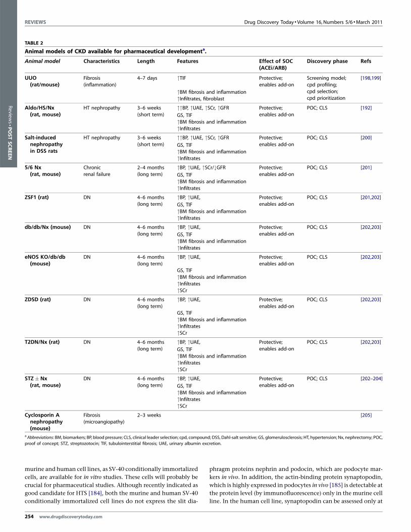
TABLE 2
Animal models of CKD available for pharmaceutical developmenta.
Animal model Characteristics Length Features Effect of SOC(ACEi/ARB)
Discovery phase Refs
UUO(rat/mouse)
Fibrosis
(inflammation)
4–7 days "TIF Protective;
enables add-on
Screening model;
cpd profiling;
cpd selection;cpd prioritization
[198,199]
"BM fibrosis and inflammation
"Infiltrates, fibroblastAldo/HS/Nx
(rat, mouse)HT nephropathy 3–6 weeks
(short term)
""BP, "UAE, "SCr, "GFR Protective;
enables add-on
POC; CLS [192]
GS, TIF
"BM fibrosis and inflammation
"InfiltratesSalt-induced
nephropathyin DSS rats
HT nephropathy 3–6 weeks
(short term)
""BP, "UAE, "SCr, "GFR Protective;
enables add-on
POC; CLS [200]
GS, TIF"BM fibrosis and inflammation
"Infiltrates5/6 Nx
(rat, mouse)Chronic
renal failure
2–4 months
(long term)
"BP, "UAE, "SCr/#GFR Protective;
enables add-on
POC; CLS [201]
GS, TIF
"BM fibrosis and inflammation
"InfiltratesZSF1 (rat) DN 4–6 months
(long term)"BP, "UAE, Protective;
enables add-onPOC; CLS [201,202]
GS, TIF
"BM fibrosis and inflammation"Infiltrates
db/db/Nx (mouse) DN 4–6 months
(long term)
"BP, "UAE, Protective;
enables add-on
POC; CLS [202,203]
GS, TIF"BM fibrosis and inflammation
"InfiltrateseNOS KO/db/db
(mouse)DN 4–6 months
(long term)
"BP, "UAE, Protective;
enables add-on
POC; CLS [202,203]
GS, TIF
"BM fibrosis and inflammation
"Infiltrates"SCr
ZDSD (rat) DN 4–6 months
(long term)
"BP, "UAE, Protective;
enables add-on
POC; CLS [202,203]
GS, TIF
"BM fibrosis and inflammation"Infiltrates"SCr
T2DN/Nx (rat) DN 4–6 months
(long term)
"BP, "UAE, Protective;
enables add-on
POC; CLS [202,203]
GS, TIF
"BM fibrosis and inflammation
"Infiltrates"SCr
STZ � Nx(rat, mouse)
DN 4–6 months
(long term)
"BP, "UAE, Protective;
enables add-on
POC; CLS [202–204]
GS, TIF"BM fibrosis and inflammation
"Infiltrates"SCr
Cyclosporin Anephropathy(mouse)
Fibrosis(microangiopathy)
2–3 weeks [205]
a Abbreviations: BM, biomarkers; BP, blood pressure; CLS, clinical leader selection; cpd, compound; DSS, Dahl-salt sensitive; GS, glomerulosclerosis; HT, hypertension; Nx, nephrectomy; POC,
proof of concept; STZ, streptozotocin; TIF, tubulointerstitial fibrosis; UAE, urinary albumin excretion.
Review
s�P
OSTSCREEN
murine and human cell lines, as SV-40 conditionally immortalized
cells, are available for in vitro studies. These cells will probably be
crucial for pharmaceutical studies. Although recently indicated as
good candidate for HTS [184], both the murine and human SV-40
conditionally immortalized cell lines do not express the slit dia-
254 www.drugdiscoverytoday.com
phragm proteins nephrin and podocin, which are podocyte mar-
kers in vivo. In addition, the actin-binding protein synaptopodin,
which is highly expressed in podocytes in vivo [185] is detectable at
the protein level (by immunofluorescence) only in the murine cell
line. In the human cell line, synaptopodin can be assessed only at
Drug Discovery Today � Volume 16, Numbers 5/6 �March 2011 REVIEWS
Reviews�POSTSCREEN
the mRNA level. The unique possible phenotypic readout for drug
testing in human podocytes therefore appears to be limited to F-
actin reorganization.
Finally, kidney mesenchymal cells are also available. Glomeru-
lar mesangial human [186], mouse [187] and rat [188] SV-40
conditionally immortalized cell lines have been extensively used
in studies of diabetic nephropathy. Renal interstitial cells are
commercially available uniquely as primary cells rather than as
a cell line. Therefore, to reduce cell variability, experiments on
renal interstitial cells can be conducted on foreskin fibroblasts,
such AG01523 (Coriell Institute for Medical Research, Camden,
NJ, USA; http://www.coriell.org) or WS-1 (ATCC-LGC Standards,
Teddington, UK; http://www.lgcstandards-atcc.org/). In basal con-
ditions, these cell lines exhibit a low content of a-SMA, thereby
representing an ideal setting for the induction of myofibroblast
activation through TGF-b1 administration and evaluation of
reporter activity for TGF-b1 signaling. Even if any of these in vitro
renal cells show low levels of differentiation compared with ori-
ginal cells, this does not reduce the relevance of these tools for
pharmaceutical research.
In vivo modelsMonkeys, dogs, sheep, rabbits and particularly rats and mice, have
been used in experimental nephrology. Surgical, nephrotoxic,
immunomediated or metabolic models exist in these species for
AKI and CKD (reviewed in [189,190]). Owing to advantages of
reproducibility, lower interanimal variation, and short time-
course and easy performance, some models are more popular than
others (e.g. the cisplatin model [191] for AKI and the UUO model
[189] for fibrosis/CKD).
The cisplatin model is linked to toxic species arising from drug
metabolism in tubular cells. Tubular nephrotoxicity, owing to
cisplatin absorption and accumulation in proximal tubule cells,
with apoptosis and necrosis of epithelial cells, is similar to the
pathology and tubular dysfunction observed in oncology patients.
This model is inducible in rats or mice by a single intraperitoneal
injection of 6–20 mg cisplatin kg�1 body weight.
Induction of CKD by UUO, although not a common cause of
kidney disease in humans, has proven to be a valuable tool for the
study of tubulointerstitial fibrosis. The immediate consequence of
urinary tract obstruction is an increase in pressure within the
urinary tract, resulting in renal pelvis dilation, tubular dilation,
tubular apoptosis, increased renin–angiotensin system activity
followed by macrophage infiltration, leading to tubular atrophy
and tubulointerstitial fibrosis. A major drawback of this model is
the inability to measure renal function changes accurately because
the remaining unobstructed kidney compensates for the
obstructed kidney loss of function. Induction of tubulointerstitial
fibrosis and glomerulosclerosis can also be also achieved by sub-
total nephrectomy (i.e. 5/6 nephrectomy). These ligation and/or
ablation models have shown good results in the rat, whereas those
in rabbit and mouse have been less extensively studied and appear
less reliable. The variability of the 5/6 nephrectomy model is,
however, greater compared with UUO experiments.
Animal models for diabetic nephropathy share many features
with human diabetic nephropathy, which is characterized by
albuminuria, progressively declining glomerular filtration rate
(GFR) and defined histopathological features, including thicken-
ing of the GBM, mesangial expansion often with nodular glo-
merulosclerosis, arteriolar hyalinosis and tubulointerstitial
fibrosis. These animal models have been delineated by targeting
proteinuria, glomerulosclerosis, glomerulonephritis, glomerular
hypertrophy and tubulointerstitial nephritis (reviewed in [192]).
For example, eNOS�/�db/db mice [193,194] exhibit significant
albuminuria and glomerular pathology that parallel the later
phase of diabetic nephropathy (DN) in patients with type 2
diabetes, including arteriolar hyalinosis, mesangial expansion,
thickening of GBM, and focal segmental and early nodular
glomerulosclerosis. This model should prove useful for studying
the role of endothelial dysfunction in the development of DN,
and to develop new diagnostic tools and therapeutic interven-
tions. (For a complete list of animal model available for CKD see
Table 2.)
In general, although not yet perfect, animal models have pro-
vided valuable insights into the mechanisms of renal diseases, far
more than in other disease areas. The main task of pharmaceutical
scientist resides in the correct model adoption in relation to
compound profile and target indication. What is still poorly used,
and should, in our opinion, be used to a greater extent, is the acute-
on-chronic approach animal model. As shown by epidemiological
studies, this approach would take in account the fact that CKD is
the most important predisposing risk factor for the development
of AKI.
ConclusionsOn the basis of the discussions above, it appears that approaching
kidney disease is all but simple. Multiple processes are activated
during renal diseases, and many cell types are involved. Moreover,
insight from preclinical studies shows that not only resident renal
cells, but also inflammatory cells have a crucial role in kidney
disease progression. To make the scenario more complex, few, if
any, reference drugs devoted to kidney disease exist on the market,
with the exception of ARB and ACE, and clinical target validation
could take several years in the case of CKD. In addition, chronic
and long-term treatment, such as in the case of CKD, exposes
patients to the unknown risks of off-target effects inhibiting key
molecular pathways, such as for TGF-b.
As briefly highlighted in this review, it is still not clear which of
the processes occurring during fibrosis onset (e.g. myofibroblast
activation, ECM deposition, inflammation, hypoxia or ER-stress) is
or are pivotal, and, if so, if this is translatable to patients. The
microenvironment at the lesion site must be more complex so that
the blockade of a single factor, although proved relevant in single-
target knock-down experiments, would fail to slow down, inhibit
or revert fibrosis in humans. Moreover the availability of a sound
and clear panel of biomarkers for early CKD, which should enable
researchers to explore the action of new compounds without
waiting years for a functional response in patients, is still lacking.
By contrast, patients with CKD represent a relevant business
case for pharma companies, as they currently account for more
than 13% of the US population. Treatment of patients with CKD
will also prove beneficial in reducing morbidity and mortality in
high-risk cardiovascular patients, widening the business case even
more [195].
From a theoretical perspective, a drug that is able to slow down
the progression of chronic renal failure, on top of currently avail-
www.drugdiscoverytoday.com 255
REVIEWS Drug Discovery Today � Volume 16, Numbers 5/6 �March 2011
Review
s�P
OSTSCREEN
able treatments (i.e. ARB and ACEi), thus postponing the need for
dialysis, will substantially change medical care in nephrology,
increasing life expectancy and impacting dramatically on
the quality of life of the patient. Seen from this perspective,
such a result appears to be achievable owing to the range of targets
256 www.drugdiscoverytoday.com
available in kidney disease. Pathophysiological compound identi-
fication, biomarker discovery and accurate selection of clinical
validation criteria appear, therefore, to be the three key elements
needed to develop a successful innovative pharmaceutical
approach to treating kidney diseases.
References
1 Tryggvason, K. and Wartiovaara, J. (2005) How does the kidney filter plasma?
Physiology (Bethesda) 20, 96–101
2 Ali, T. et al. (2007) Incidence and outcomes in acute kidney injury: a
comprehensive population-based study. J. Am. Soc. Nephrol. 18, 1292–1298
3 Hsu, C.Y. et al. (2007) Community-based incidence of acute renal failure. Kidney
Int. 72, 208–212
4 Hou, S.H. et al. (1983) Hospital-acquired renal insufficiency: a prospective study.
Am. J. Med. 74, 243–248
5 Coresh, J. et al. (2007) Prevalence of chronic kidney disease in the United States.
JAMA 298, 2038–2047
6 Lameire, N. et al. (2005) Chronic kidney disease: a European perspective. Kidney
Int. 99 (Suppl.), S30–S38
7 Venkatachalam, M.A. et al. (2010) Acute kidney injury: a springboard for
progression in chronic kidney disease. Am. J. Physiol. Renal Physiol. 298, F1078–
F1094
8 Lafrance, J.P. et al. (2010) Incidence and outcomes of acute kidney injury in a
referred chronic kidney disease cohort. Nephrol. Dial. Transplant. 7, 2203–2209
9 Coca, S.G. et al. (2009) Long-term risk of mortality and other adverse outcomes
after acute kidney injury: a systematic review and meta-analysis. Am. J. Kidney Dis.
53, 961–973
10 Azuma, H. et al. (1997) Cellular and molecular predictors of chronic renal
dysfunction after initial ischemia/reperfusion injury of a single kidney.
Transplantation 64, 190–197
11 Azuma, H. et al. (1997) Initial ischemia/reperfusion injury influences late
functional and structural changes in the kidney. Transplant. Proc. 29, 1528–1529
12 Mackenzie, H.S. et al. (1995) Renal mass as a determinant of late allograft outcome:
insights from experimental studies in rats. Kidney Int. Suppl. 52, S38–S42
13 Remuzzi, G. (1995) Abnormal protein traffic through the glomerular barrier
induces proximal tubular cell dysfunction and causes renal injury. Curr. Opin.
Nephrol. Hypertens. 4, 339–342
14 Tryggvason, K. et al. (2006) Nck links nephrin to actin in kidney podocytes. Cell
125, 221–224
15 Faul, C. et al. (2008) The actin cytoskeleton of kidney podocytes is a direct target of
the antiproteinuric effect of cyclosporine A. Nat. Med. 14, 931–938
16 Leehey, D.J. et al. (2000) Role of angiotensin II in diabetic nephropathy. Kidney Int.
Suppl. 77, S93–S98
17 Suzuki, T. et al. (2001) Role of atrophic tubules in development of interstitial
fibrosis in microembolism-induced renal failure in rat. Am. J. Pathol. 158, 75–85
18 Gabbiani, G. et al. (1971) Presence of modified fibroblasts in granulation tissue and
their possible role in wound contraction. Experientia 27, 549–550
19 Tomasek, J.J. et al. (2002) Myofibroblasts and mechano-regulation of connective
tissue remodelling. Nat. Rev. Mol. Cell Biol. 3, 349–363
20 Hinz, B. et al. (2007) The myofibroblast: one function, multiple origins. Am. J.
Pathol. 170, 1807–1816
21 Desmouliere, A. et al. (1995) Apoptosis mediates the decrease in cellularity during
the transition between granulation tissue and scar. Am. J. Pathol. 146, 56–66
22 Zeisberg, E.M. et al. (2008) Fibroblasts in kidney fibrosis emerge via endothelial-to-
mesenchymal transition. J. Am. Soc. Nephrol. 19, 2282–2287
23 Thiery, J.P. et al. (2009) Epithelial–mesenchymal transitions in development and
disease. Cell 139, 871–890
24 Acloque, H. et al. (2009) Epithelial–mesenchymal transitions: the importance of
changing cell state in development and disease. J. Clin. Invest. 119, 1438–1449
25 Zeisberg, E.M. et al. (2007) Endothelial-to-mesenchymal transition contributes to
cardiac fibrosis. Nat. Med. 13, 952–961
26 Zeisberg, M. and Duffield, J. (2010) Resolved: EMT produces fibroblasts in the
kidney. J. Am. Soc. Nephrol. 21, 1247–1253
27 Yang, L. et al. (2010) Epithelial cell cycle arrest in G2/M mediates kidney fibrosis
after injury. Nat. Med. 16, 535–543
28 Udo, K. et al. (2010) Adipose tissue explants and MDCK cells reciprocally regulate
their morphogenesis in co-culture. Kidney Int. 78, 60–68
29 Sharma, M. et al. (2009) Analysis of stromal signatures in the tumor
microenvironment of ductal carcinoma in situ. Breast Cancer Res. Treat.
30 Prunotto, M. and Moll, S. (2011) The emerging concept of a fibrotic
microenvironment in CKD. Kidney Int. 79 (1), 135
31 Koesters, R. et al. (2010) Tubular overexpression of transforming growth factor-
beta1 induces autophagy and fibrosis but not mesenchymal transition of renal
epithelial cells. Am. J. Pathol. 177, 632–643
32 Schmidt, M. et al. (2003) Identification of circulating fibrocytes as precursors of
bronchial myofibroblasts in asthma. J. Immunol. 171, 380–389
33 Heidland, A. et al. (2001) Franz Volhard and Theodor Fahr: achievements and
controversies in their research in renal disease and hypertension. J. Hum.
Hypertens. 15, 5–16
34 Campese, V.M. et al. (2000) Is microalbuminuria a predictor of cardiovascular and
renal disease in patients with essential hypertension? Curr. Opin. Nephrol.
Hypertens. 9, 143–147
35 de Zeeuw, D. et al. (2004) Proteinuria, a target for renoprotection in patients with
type 2 diabetic nephropathy: lessons from RENAAL. Kidney Int. 65, 2309–2320
36 Abbate, M. et al. (2002) Proximal tubular cells promote fibrogenesis by TGF-beta1-
mediated induction of peritubular myofibroblasts. Kidney Int. 61, 2066–2077
37 Bertani, T. et al. (1986) Tubulo-interstitial lesions mediate renal damage in
adriamycin glomerulopathy. Kidney Int. 30, 488–496
38 Bertani, T. et al. (1989) Age-related nephropathy and proteinuria in rats with intact
kidneys exposed to diets with different protein content. Lab. Invest. 60, 196–204
39 Remuzzi, G. and Bertani, T. (1998) Pathophysiology of progressive nephropathies.
N. Engl. J. Med. 339, 1448–1456
40 Baines, R.J. and Brunskill, N.J. (2008) The molecular interactions between filtered
proteins and proximal tubular cells in proteinuria. Nephron Exp. Nephrol. 110, e67–
e71
41 Eddy, A.A. and Giachelli, C.M. (1995) Renal expression of genes that promote
interstitial inflammation and fibrosis in rats with protein-overload proteinuria.
Kidney Int. 47, 1546–1557
42 Wang, Y. et al. (1997) Induction of monocyte chemoattractant protein-1 in
proximal tubule cells by urinary protein. J. Am. Soc. Nephrol. 8, 1537–1545
43 Cybulsky, A.V. (2009) Endoplasmic reticulum stress in proteinuric kidney disease.
Kidney Int. 77, 187–193
44 Markan, S. et al. (2009) Up regulation of the GRP-78 and GADD-153 and down
regulation of Bcl-2 proteins in primary glomerular diseases: a possible involvement
of the ER stress pathway in glomerulonephritis. Mol. Cell. Biochem. 324, 131–
138
45 Ohse, T. et al. (2006) Albumin induces endoplasmic reticulum stress and apoptosis
in renal proximal tubular cells. Kidney Int. 70, 1447–1455
46 Wu, X. et al. (2010) Albumin overload induces apoptosis in renal tubular epithelial
cells through a CHOP-dependent pathway. OMICS 14, 61–73
47 Lindenmeyer, M.T. et al. (2008) Proteinuria and hyperglycemia induce
endoplasmic reticulum stress. J. Am. Soc. Nephrol. 19, 2225–2236
48 Ulianich, L. et al. (2008) ER stress is associated with dedifferentiation and an
epithelial-to-mesenchymal transition-like phenotype in PC Cl3 thyroid cells. J.
Cell Sci. 121, 477–486
49 Seki, K. et al. (2003) Mouse Snail family transcription repressors regulate
chondrocyte, extracellular matrix, type II collagen, and aggrecan. J. Biol. Chem.
278, 41862–41870
50 Prunotto, M. et al. (2010) Endocellular polyamine availability modulates
epithelial-to-mesenchymal transition and unfolded protein response in MDCK
cells. Lab. Invest. 90, 929–939
51 Dahan, K. et al. (2003) A cluster of mutations in the UMOD gene causes familial
juvenile hyperuricemic nephropathy with abnormal expression of uromodulin. J.
Am. Soc. Nephrol. 14, 2883–2893
52 Tang, S. et al. (2002) Role of complement in tubulointerstitial injury from
proteinuria. Kidney Blood Press. Res. 25, 120–126
53 Boor, P. et al. (2007) Treatment targets in renal fibrosis. Nephrol. Dial. Transplant.
22, 3391–3407
54 Camussi, G. et al. (1982) In vitro alternative pathway activation of complement by
the brush border of proximal tubules of normal rat kidney. J. Immunol. 128, 1659–
1663
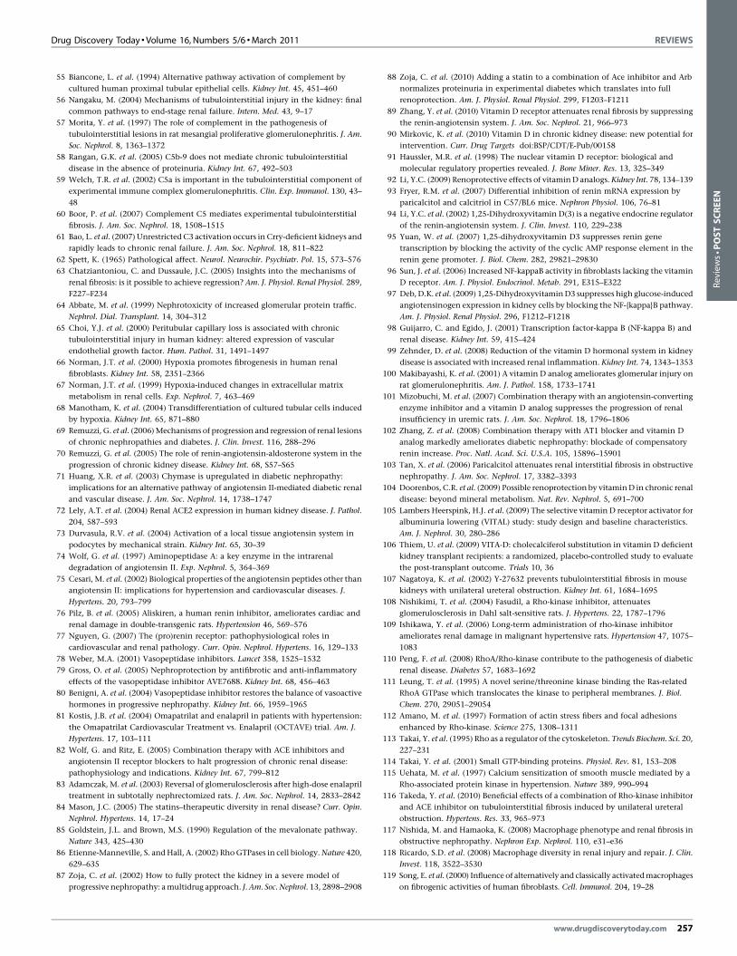
Drug Discovery Today � Volume 16, Numbers 5/6 �March 2011 REVIEWS
Reviews�POSTSCREEN
55 Biancone, L. et al. (1994) Alternative pathway activation of complement by
cultured human proximal tubular epithelial cells. Kidney Int. 45, 451–460
56 Nangaku, M. (2004) Mechanisms of tubulointerstitial injury in the kidney: final
common pathways to end-stage renal failure. Intern. Med. 43, 9–17
57 Morita, Y. et al. (1997) The role of complement in the pathogenesis of
tubulointerstitial lesions in rat mesangial proliferative glomerulonephritis. J. Am.
Soc. Nephrol. 8, 1363–1372
58 Rangan, G.K. et al. (2005) C5b-9 does not mediate chronic tubulointerstitial
disease in the absence of proteinuria. Kidney Int. 67, 492–503
59 Welch, T.R. et al. (2002) C5a is important in the tubulointerstitial component of
experimental immune complex glomerulonephritis. Clin. Exp. Immunol. 130, 43–
48
60 Boor, P. et al. (2007) Complement C5 mediates experimental tubulointerstitial
fibrosis. J. Am. Soc. Nephrol. 18, 1508–1515
61 Bao, L. et al. (2007) Unrestricted C3 activation occurs in Crry-deficient kidneys and
rapidly leads to chronic renal failure. J. Am. Soc. Nephrol. 18, 811–822
62 Spett, K. (1965) Pathological affect. Neurol. Neurochir. Psychiatr. Pol. 15, 573–576
63 Chatziantoniou, C. and Dussaule, J.C. (2005) Insights into the mechanisms of
renal fibrosis: is it possible to achieve regression? Am. J. Physiol. Renal Physiol. 289,
F227–F234
64 Abbate, M. et al. (1999) Nephrotoxicity of increased glomerular protein traffic.
Nephrol. Dial. Transplant. 14, 304–312
65 Choi, Y.J. et al. (2000) Peritubular capillary loss is associated with chronic
tubulointerstitial injury in human kidney: altered expression of vascular
endothelial growth factor. Hum. Pathol. 31, 1491–1497
66 Norman, J.T. et al. (2000) Hypoxia promotes fibrogenesis in human renal
fibroblasts. Kidney Int. 58, 2351–2366
67 Norman, J.T. et al. (1999) Hypoxia-induced changes in extracellular matrix
metabolism in renal cells. Exp. Nephrol. 7, 463–469
68 Manotham, K. et al. (2004) Transdifferentiation of cultured tubular cells induced
by hypoxia. Kidney Int. 65, 871–880
69 Remuzzi, G. et al. (2006) Mechanisms of progression and regression of renal lesions
of chronic nephropathies and diabetes. J. Clin. Invest. 116, 288–296
70 Remuzzi, G. et al. (2005) The role of renin-angiotensin-aldosterone system in the
progression of chronic kidney disease. Kidney Int. 68, S57–S65
71 Huang, X.R. et al. (2003) Chymase is upregulated in diabetic nephropathy:
implications for an alternative pathway of angiotensin II-mediated diabetic renal
and vascular disease. J. Am. Soc. Nephrol. 14, 1738–1747
72 Lely, A.T. et al. (2004) Renal ACE2 expression in human kidney disease. J. Pathol.
204, 587–593
73 Durvasula, R.V. et al. (2004) Activation of a local tissue angiotensin system in
podocytes by mechanical strain. Kidney Int. 65, 30–39
74 Wolf, G. et al. (1997) Aminopeptidase A: a key enzyme in the intrarenal
degradation of angiotensin II. Exp. Nephrol. 5, 364–369
75 Cesari, M. et al. (2002) Biological properties of the angiotensin peptides other than
angiotensin II: implications for hypertension and cardiovascular diseases. J.
Hypertens. 20, 793–799
76 Pilz, B. et al. (2005) Aliskiren, a human renin inhibitor, ameliorates cardiac and
renal damage in double-transgenic rats. Hypertension 46, 569–576
77 Nguyen, G. (2007) The (pro)renin receptor: pathophysiological roles in
cardiovascular and renal pathology. Curr. Opin. Nephrol. Hypertens. 16, 129–133
78 Weber, M.A. (2001) Vasopeptidase inhibitors. Lancet 358, 1525–1532
79 Gross, O. et al. (2005) Nephroprotection by antifibrotic and anti-inflammatory
effects of the vasopeptidase inhibitor AVE7688. Kidney Int. 68, 456–463
80 Benigni, A. et al. (2004) Vasopeptidase inhibitor restores the balance of vasoactive
hormones in progressive nephropathy. Kidney Int. 66, 1959–1965
81 Kostis, J.B. et al. (2004) Omapatrilat and enalapril in patients with hypertension:
the Omapatrilat Cardiovascular Treatment vs. Enalapril (OCTAVE) trial. Am. J.
Hypertens. 17, 103–111
82 Wolf, G. and Ritz, E. (2005) Combination therapy with ACE inhibitors and
angiotensin II receptor blockers to halt progression of chronic renal disease:
pathophysiology and indications. Kidney Int. 67, 799–812
83 Adamczak, M. et al. (2003) Reversal of glomerulosclerosis after high-dose enalapril
treatment in subtotally nephrectomized rats. J. Am. Soc. Nephrol. 14, 2833–2842
84 Mason, J.C. (2005) The statins–therapeutic diversity in renal disease? Curr. Opin.
Nephrol. Hypertens. 14, 17–24
85 Goldstein, J.L. and Brown, M.S. (1990) Regulation of the mevalonate pathway.
Nature 343, 425–430
86 Etienne-Manneville, S. and Hall, A. (2002) Rho GTPases in cell biology. Nature 420,
629–635
87 Zoja, C. et al. (2002) How to fully protect the kidney in a severe model of
progressive nephropathy: a multidrug approach. J. Am. Soc. Nephrol. 13, 2898–2908
88 Zoja, C. et al. (2010) Adding a statin to a combination of Ace inhibitor and Arb
normalizes proteinuria in experimental diabetes which translates into full
renoprotection. Am. J. Physiol. Renal Physiol. 299, F1203–F1211
89 Zhang, Y. et al. (2010) Vitamin D receptor attenuates renal fibrosis by suppressing
the renin-angiotensin system. J. Am. Soc. Nephrol. 21, 966–973
90 Mirkovic, K. et al. (2010) Vitamin D in chronic kidney disease: new potential for
intervention. Curr. Drug Targets doi:BSP/CDT/E-Pub/00158
91 Haussler, M.R. et al. (1998) The nuclear vitamin D receptor: biological and
molecular regulatory properties revealed. J. Bone Miner. Res. 13, 325–349
92 Li, Y.C. (2009) Renoprotective effects of vitamin D analogs. Kidney Int. 78, 134–139
93 Fryer, R.M. et al. (2007) Differential inhibition of renin mRNA expression by
paricalcitol and calcitriol in C57/BL6 mice. Nephron Physiol. 106, 76–81
94 Li, Y.C. et al. (2002) 1,25-Dihydroxyvitamin D(3) is a negative endocrine regulator
of the renin-angiotensin system. J. Clin. Invest. 110, 229–238
95 Yuan, W. et al. (2007) 1,25-dihydroxyvitamin D3 suppresses renin gene
transcription by blocking the activity of the cyclic AMP response element in the
renin gene promoter. J. Biol. Chem. 282, 29821–29830
96 Sun, J. et al. (2006) Increased NF-kappaB activity in fibroblasts lacking the vitamin
D receptor. Am. J. Physiol. Endocrinol. Metab. 291, E315–E322
97 Deb, D.K. et al. (2009) 1,25-Dihydroxyvitamin D3 suppresses high glucose-induced
angiotensinogen expression in kidney cells by blocking the NF-{kappa}B pathway.
Am. J. Physiol. Renal Physiol. 296, F1212–F1218
98 Guijarro, C. and Egido, J. (2001) Transcription factor-kappa B (NF-kappa B) and
renal disease. Kidney Int. 59, 415–424
99 Zehnder, D. et al. (2008) Reduction of the vitamin D hormonal system in kidney
disease is associated with increased renal inflammation. Kidney Int. 74, 1343–1353
100 Makibayashi, K. et al. (2001) A vitamin D analog ameliorates glomerular injury on
rat glomerulonephritis. Am. J. Pathol. 158, 1733–1741
101 Mizobuchi, M. et al. (2007) Combination therapy with an angiotensin-converting
enzyme inhibitor and a vitamin D analog suppresses the progression of renal
insufficiency in uremic rats. J. Am. Soc. Nephrol. 18, 1796–1806
102 Zhang, Z. et al. (2008) Combination therapy with AT1 blocker and vitamin D
analog markedly ameliorates diabetic nephropathy: blockade of compensatory
renin increase. Proc. Natl. Acad. Sci. U.S.A. 105, 15896–15901
103 Tan, X. et al. (2006) Paricalcitol attenuates renal interstitial fibrosis in obstructive
nephropathy. J. Am. Soc. Nephrol. 17, 3382–3393
104 Doorenbos, C.R. et al. (2009) Possible renoprotection by vitamin D in chronic renal
disease: beyond mineral metabolism. Nat. Rev. Nephrol. 5, 691–700
105 Lambers Heerspink, H.J. et al. (2009) The selective vitamin D receptor activator for
albuminuria lowering (VITAL) study: study design and baseline characteristics.
Am. J. Nephrol. 30, 280–286
106 Thiem, U. et al. (2009) VITA-D: cholecalciferol substitution in vitamin D deficient
kidney transplant recipients: a randomized, placebo-controlled study to evaluate
the post-transplant outcome. Trials 10, 36
107 Nagatoya, K. et al. (2002) Y-27632 prevents tubulointerstitial fibrosis in mouse
kidneys with unilateral ureteral obstruction. Kidney Int. 61, 1684–1695
108 Nishikimi, T. et al. (2004) Fasudil, a Rho-kinase inhibitor, attenuates
glomerulosclerosis in Dahl salt-sensitive rats. J. Hypertens. 22, 1787–1796
109 Ishikawa, Y. et al. (2006) Long-term administration of rho-kinase inhibitor
ameliorates renal damage in malignant hypertensive rats. Hypertension 47, 1075–
1083
110 Peng, F. et al. (2008) RhoA/Rho-kinase contribute to the pathogenesis of diabetic
renal disease. Diabetes 57, 1683–1692
111 Leung, T. et al. (1995) A novel serine/threonine kinase binding the Ras-related
RhoA GTPase which translocates the kinase to peripheral membranes. J. Biol.
Chem. 270, 29051–29054
112 Amano, M. et al. (1997) Formation of actin stress fibers and focal adhesions
enhanced by Rho-kinase. Science 275, 1308–1311
113 Takai, Y. et al. (1995) Rho as a regulator of the cytoskeleton. Trends Biochem. Sci. 20,
227–231
114 Takai, Y. et al. (2001) Small GTP-binding proteins. Physiol. Rev. 81, 153–208
115 Uehata, M. et al. (1997) Calcium sensitization of smooth muscle mediated by a
Rho-associated protein kinase in hypertension. Nature 389, 990–994
116 Takeda, Y. et al. (2010) Beneficial effects of a combination of Rho-kinase inhibitor
and ACE inhibitor on tubulointerstitial fibrosis induced by unilateral ureteral
obstruction. Hypertens. Res. 33, 965–973
117 Nishida, M. and Hamaoka, K. (2008) Macrophage phenotype and renal fibrosis in
obstructive nephropathy. Nephron Exp. Nephrol. 110, e31–e36
118 Ricardo, S.D. et al. (2008) Macrophage diversity in renal injury and repair. J. Clin.
Invest. 118, 3522–3530
119 Song, E. et al. (2000) Influence of alternatively and classically activated macrophages
on fibrogenic activities of human fibroblasts. Cell. Immunol. 204, 19–28
www.drugdiscoverytoday.com 257
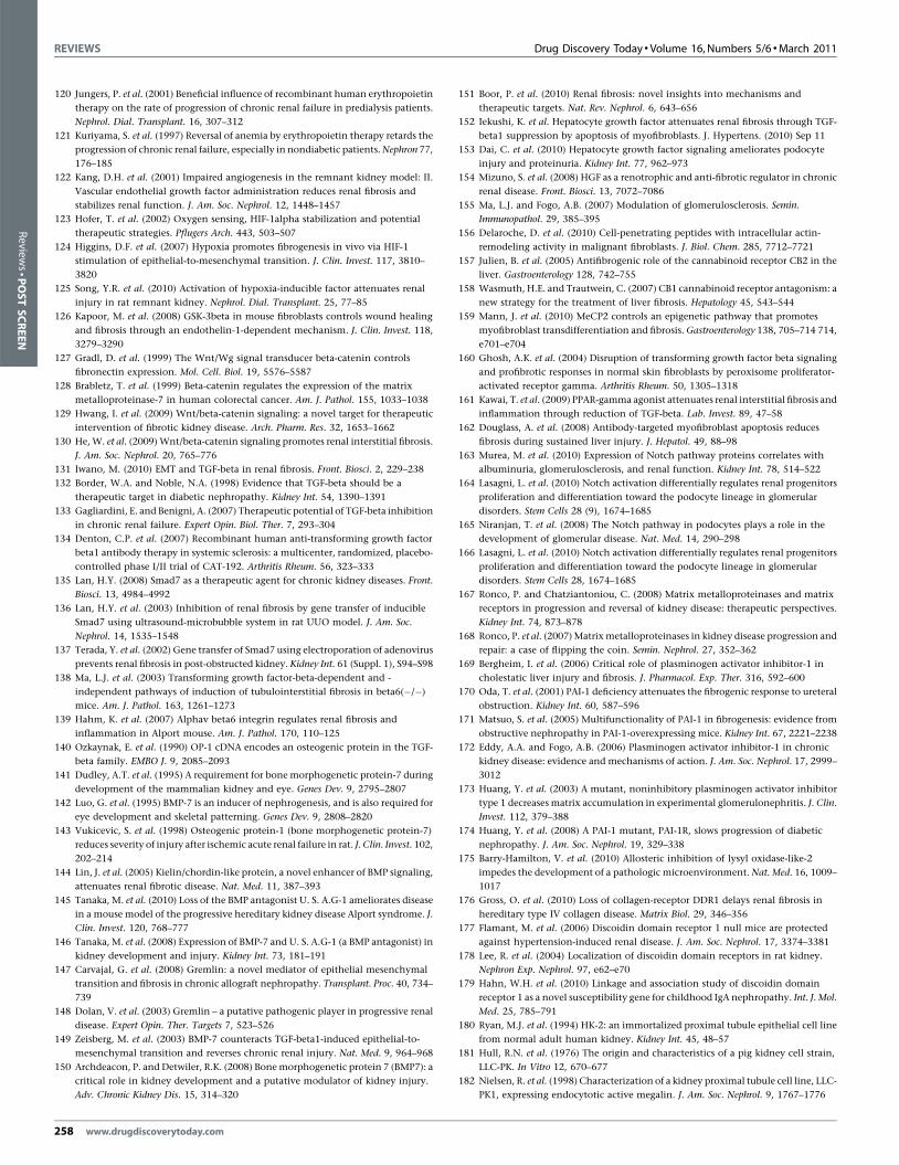
REVIEWS Drug Discovery Today � Volume 16, Numbers 5/6 �March 2011
Review
s�P
OSTSCREEN
120 Jungers, P. et al. (2001) Beneficial influence of recombinant human erythropoietin
therapy on the rate of progression of chronic renal failure in predialysis patients.
Nephrol. Dial. Transplant. 16, 307–312
121 Kuriyama, S. et al. (1997) Reversal of anemia by erythropoietin therapy retards the
progression of chronic renal failure, especially in nondiabetic patients. Nephron 77,
176–185
122 Kang, D.H. et al. (2001) Impaired angiogenesis in the remnant kidney model: II.
Vascular endothelial growth factor administration reduces renal fibrosis and
stabilizes renal function. J. Am. Soc. Nephrol. 12, 1448–1457
123 Hofer, T. et al. (2002) Oxygen sensing, HIF-1alpha stabilization and potential
therapeutic strategies. Pflugers Arch. 443, 503–507
124 Higgins, D.F. et al. (2007) Hypoxia promotes fibrogenesis in vivo via HIF-1
stimulation of epithelial-to-mesenchymal transition. J. Clin. Invest. 117, 3810–
3820
125 Song, Y.R. et al. (2010) Activation of hypoxia-inducible factor attenuates renal
injury in rat remnant kidney. Nephrol. Dial. Transplant. 25, 77–85
126 Kapoor, M. et al. (2008) GSK-3beta in mouse fibroblasts controls wound healing
and fibrosis through an endothelin-1-dependent mechanism. J. Clin. Invest. 118,
3279–3290
127 Gradl, D. et al. (1999) The Wnt/Wg signal transducer beta-catenin controls
fibronectin expression. Mol. Cell. Biol. 19, 5576–5587
128 Brabletz, T. et al. (1999) Beta-catenin regulates the expression of the matrix
metalloproteinase-7 in human colorectal cancer. Am. J. Pathol. 155, 1033–1038
129 Hwang, I. et al. (2009) Wnt/beta-catenin signaling: a novel target for therapeutic
intervention of fibrotic kidney disease. Arch. Pharm. Res. 32, 1653–1662
130 He, W. et al. (2009) Wnt/beta-catenin signaling promotes renal interstitial fibrosis.
J. Am. Soc. Nephrol. 20, 765–776
131 Iwano, M. (2010) EMT and TGF-beta in renal fibrosis. Front. Biosci. 2, 229–238
132 Border, W.A. and Noble, N.A. (1998) Evidence that TGF-beta should be a
therapeutic target in diabetic nephropathy. Kidney Int. 54, 1390–1391
133 Gagliardini, E. and Benigni, A. (2007) Therapeutic potential of TGF-beta inhibition
in chronic renal failure. Expert Opin. Biol. Ther. 7, 293–304
134 Denton, C.P. et al. (2007) Recombinant human anti-transforming growth factor
beta1 antibody therapy in systemic sclerosis: a multicenter, randomized, placebo-
controlled phase I/II trial of CAT-192. Arthritis Rheum. 56, 323–333
135 Lan, H.Y. (2008) Smad7 as a therapeutic agent for chronic kidney diseases. Front.
Biosci. 13, 4984–4992
136 Lan, H.Y. et al. (2003) Inhibition of renal fibrosis by gene transfer of inducible
Smad7 using ultrasound-microbubble system in rat UUO model. J. Am. Soc.
Nephrol. 14, 1535–1548
137 Terada, Y. et al. (2002) Gene transfer of Smad7 using electroporation of adenovirus
prevents renal fibrosis in post-obstructed kidney. Kidney Int. 61 (Suppl. 1), S94–S98
138 Ma, L.J. et al. (2003) Transforming growth factor-beta-dependent and -
independent pathways of induction of tubulointerstitial fibrosis in beta6(�/�)
mice. Am. J. Pathol. 163, 1261–1273
139 Hahm, K. et al. (2007) Alphav beta6 integrin regulates renal fibrosis and
inflammation in Alport mouse. Am. J. Pathol. 170, 110–125
140 Ozkaynak, E. et al. (1990) OP-1 cDNA encodes an osteogenic protein in the TGF-
beta family. EMBO J. 9, 2085–2093
141 Dudley, A.T. et al. (1995) A requirement for bone morphogenetic protein-7 during
development of the mammalian kidney and eye. Genes Dev. 9, 2795–2807
142 Luo, G. et al. (1995) BMP-7 is an inducer of nephrogenesis, and is also required for
eye development and skeletal patterning. Genes Dev. 9, 2808–2820
143 Vukicevic, S. et al. (1998) Osteogenic protein-1 (bone morphogenetic protein-7)
reduces severity of injury after ischemic acute renal failure in rat. J. Clin. Invest. 102,
202–214
144 Lin, J. et al. (2005) Kielin/chordin-like protein, a novel enhancer of BMP signaling,
attenuates renal fibrotic disease. Nat. Med. 11, 387–393
145 Tanaka, M. et al. (2010) Loss of the BMP antagonist U. S. A.G-1 ameliorates disease
in a mouse model of the progressive hereditary kidney disease Alport syndrome. J.
Clin. Invest. 120, 768–777
146 Tanaka, M. et al. (2008) Expression of BMP-7 and U. S. A.G-1 (a BMP antagonist) in
kidney development and injury. Kidney Int. 73, 181–191
147 Carvajal, G. et al. (2008) Gremlin: a novel mediator of epithelial mesenchymal
transition and fibrosis in chronic allograft nephropathy. Transplant. Proc. 40, 734–
739
148 Dolan, V. et al. (2003) Gremlin – a putative pathogenic player in progressive renal
disease. Expert Opin. Ther. Targets 7, 523–526
149 Zeisberg, M. et al. (2003) BMP-7 counteracts TGF-beta1-induced epithelial-to-
mesenchymal transition and reverses chronic renal injury. Nat. Med. 9, 964–968
150 Archdeacon, P. and Detwiler, R.K. (2008) Bone morphogenetic protein 7 (BMP7): a
critical role in kidney development and a putative modulator of kidney injury.
Adv. Chronic Kidney Dis. 15, 314–320
258 www.drugdiscoverytoday.com
151 Boor, P. et al. (2010) Renal fibrosis: novel insights into mechanisms and
therapeutic targets. Nat. Rev. Nephrol. 6, 643–656
152 Iekushi, K. et al. Hepatocyte growth factor attenuates renal fibrosis through TGF-
beta1 suppression by apoptosis of myofibroblasts. J. Hypertens. (2010) Sep 11
153 Dai, C. et al. (2010) Hepatocyte growth factor signaling ameliorates podocyte
injury and proteinuria. Kidney Int. 77, 962–973
154 Mizuno, S. et al. (2008) HGF as a renotrophic and anti-fibrotic regulator in chronic
renal disease. Front. Biosci. 13, 7072–7086
155 Ma, L.J. and Fogo, A.B. (2007) Modulation of glomerulosclerosis. Semin.
Immunopathol. 29, 385–395
156 Delaroche, D. et al. (2010) Cell-penetrating peptides with intracellular actin-
remodeling activity in malignant fibroblasts. J. Biol. Chem. 285, 7712–7721
157 Julien, B. et al. (2005) Antifibrogenic role of the cannabinoid receptor CB2 in the
liver. Gastroenterology 128, 742–755
158 Wasmuth, H.E. and Trautwein, C. (2007) CB1 cannabinoid receptor antagonism: a
new strategy for the treatment of liver fibrosis. Hepatology 45, 543–544
159 Mann, J. et al. (2010) MeCP2 controls an epigenetic pathway that promotes
myofibroblast transdifferentiation and fibrosis. Gastroenterology 138, 705–714 714,
e701–e704
160 Ghosh, A.K. et al. (2004) Disruption of transforming growth factor beta signaling
and profibrotic responses in normal skin fibroblasts by peroxisome proliferator-
activated receptor gamma. Arthritis Rheum. 50, 1305–1318
161 Kawai, T. et al. (2009) PPAR-gamma agonist attenuates renal interstitial fibrosis and
inflammation through reduction of TGF-beta. Lab. Invest. 89, 47–58
162 Douglass, A. et al. (2008) Antibody-targeted myofibroblast apoptosis reduces
fibrosis during sustained liver injury. J. Hepatol. 49, 88–98
163 Murea, M. et al. (2010) Expression of Notch pathway proteins correlates with
albuminuria, glomerulosclerosis, and renal function. Kidney Int. 78, 514–522
164 Lasagni, L. et al. (2010) Notch activation differentially regulates renal progenitors
proliferation and differentiation toward the podocyte lineage in glomerular
disorders. Stem Cells 28 (9), 1674–1685
165 Niranjan, T. et al. (2008) The Notch pathway in podocytes plays a role in the
development of glomerular disease. Nat. Med. 14, 290–298
166 Lasagni, L. et al. (2010) Notch activation differentially regulates renal progenitors
proliferation and differentiation toward the podocyte lineage in glomerular
disorders. Stem Cells 28, 1674–1685
167 Ronco, P. and Chatziantoniou, C. (2008) Matrix metalloproteinases and matrix
receptors in progression and reversal of kidney disease: therapeutic perspectives.
Kidney Int. 74, 873–878
168 Ronco, P. et al. (2007) Matrix metalloproteinases in kidney disease progression and
repair: a case of flipping the coin. Semin. Nephrol. 27, 352–362
169 Bergheim, I. et al. (2006) Critical role of plasminogen activator inhibitor-1 in
cholestatic liver injury and fibrosis. J. Pharmacol. Exp. Ther. 316, 592–600
170 Oda, T. et al. (2001) PAI-1 deficiency attenuates the fibrogenic response to ureteral
obstruction. Kidney Int. 60, 587–596
171 Matsuo, S. et al. (2005) Multifunctionality of PAI-1 in fibrogenesis: evidence from
obstructive nephropathy in PAI-1-overexpressing mice. Kidney Int. 67, 2221–2238
172 Eddy, A.A. and Fogo, A.B. (2006) Plasminogen activator inhibitor-1 in chronic
kidney disease: evidence and mechanisms of action. J. Am. Soc. Nephrol. 17, 2999–
3012
173 Huang, Y. et al. (2003) A mutant, noninhibitory plasminogen activator inhibitor
type 1 decreases matrix accumulation in experimental glomerulonephritis. J. Clin.
Invest. 112, 379–388
174 Huang, Y. et al. (2008) A PAI-1 mutant, PAI-1R, slows progression of diabetic
nephropathy. J. Am. Soc. Nephrol. 19, 329–338
175 Barry-Hamilton, V. et al. (2010) Allosteric inhibition of lysyl oxidase-like-2
impedes the development of a pathologic microenvironment. Nat. Med. 16, 1009–
1017
176 Gross, O. et al. (2010) Loss of collagen-receptor DDR1 delays renal fibrosis in
hereditary type IV collagen disease. Matrix Biol. 29, 346–356
177 Flamant, M. et al. (2006) Discoidin domain receptor 1 null mice are protected
against hypertension-induced renal disease. J. Am. Soc. Nephrol. 17, 3374–3381
178 Lee, R. et al. (2004) Localization of discoidin domain receptors in rat kidney.
Nephron Exp. Nephrol. 97, e62–e70
179 Hahn, W.H. et al. (2010) Linkage and association study of discoidin domain
receptor 1 as a novel susceptibility gene for childhood IgA nephropathy. Int. J. Mol.
Med. 25, 785–791
180 Ryan, M.J. et al. (1994) HK-2: an immortalized proximal tubule epithelial cell line
from normal adult human kidney. Kidney Int. 45, 48–57
181 Hull, R.N. et al. (1976) The origin and characteristics of a pig kidney cell strain,
LLC-PK. In Vitro 12, 670–677
182 Nielsen, R. et al. (1998) Characterization of a kidney proximal tubule cell line, LLC-
PK1, expressing endocytotic active megalin. J. Am. Soc. Nephrol. 9, 1767–1776
Drug Discovery Today � Volume 16, Numbers 5/6 �March 2011 REVIEWS
Reviews�POSTSCREEN
183 Rindler, M.J. et al. (1979) Retention of differentiated properties in an established
dog kidney epithelial cell line (MDCK). J. Cell Biol. 81, 635–648
184 Reiser, J. et al. (2010) Toward the development of podocyte-specific drugs. Kidney
Int. 77, 662–668
185 Mundel, P. et al. (1997) Synaptopodin: an actin-associated protein in telencephalic
dendrites and renal podocytes. J. Cell Biol. 139, 193–204
186 Sraer, J.D. et al. (1996) Stable cell lines of T-SV40 immortalized human glomerular
mesangial cells. Kidney Int. 49, 267–270
187 MacKay, K. et al. (1988) Glomerular epithelial, mesangial, and endothelial cell
lines from transgenic mice. Kidney Int. 33, 677–684
188 Nitta, K. and Horiba, N. (1997) An immortalized rat mesangial cell line. In Vitro
Cell. Dev. Biol. Anim. 33, 156–157
189 Hewitson, T.D. et al. (2009) Small animal models of kidney disease: a review.
Methods Mol. Biol. 466, 41–57
190 Heyman, S.N. et al. (2002) Animal models of acute tubular necrosis. Curr. Opin.
Crit. Care 8, 526–534
191 Yao, X. et al. (2007) Cisplatin nephrotoxicity: a review. Am. J. Med. Sci. 334, 115–124
192 Balakumar, P. et al. (2008) Experimental models for nephropathy. J. Renin
Angiotensin Aldosterone Syst. 9, 189–195
193 Zhao, H.J. et al. (2006) Endothelial nitric oxide synthase deficiency produces
accelerated nephropathy in diabetic mice. J. Am. Soc. Nephrol. 17, 2664–2669
194 Nakagawa, T. (2009) A new mouse model resembling human diabetic
nephropathy: uncoupling of VEGF with eNOS as a novel pathogenic mechanism.
Clin. Nephrol. 71, 103–109
195 Astor, B.C. et al. (2008) Glomerular filtration rate, albuminuria, and risk of
cardiovascular and all-cause mortality in the US population. Am. J. Epidemiol. 167,
1226–1234
196 Hocher, B. and Paul, M. (2000) Transgenic animal models for the analysis of the
renal endothelin system. Nephrol. Dial. Transplant. 15, 935–937
197 Kohan, D.E. (2010) Endothelin, hypertension and chronic kidney disease: new
insights. Curr. Opin. Nephrol. Hypertens. 19, 134–139
198 Truong, L.D. et al. (1996) Cell apoptosis and proliferation in experimental chronic
obstructive uropathy. Kidney Int. 50, 200–207
199 Klahr, S. (2001) Urinary tract obstruction. Semin. Nephrol. 21, 133–145
200 Wang, P.X. and Sanders, P.W. (2005) Mechanism of hypertensive nephropathy in
the Dahl/Rapp rat: a primary disorder of vascular smooth muscle. Am. J. Physiol.
Renal Physiol. 288, F236–F242
201 Ma, L.J. and Fogo, A.B. (2003) Model of robust induction of glomerulosclerosis in
mice: importance of genetic background. Kidney Int. 64, 350–355
202 Schlondorff, D. (2010) Choosing the right mouse model for diabetic nephropathy.
Kidney Int. 77, 749–750
203 Brosius, F.C., III et al. (2009) Mouse models of diabetic nephropathy. J. Am. Soc.
Nephrol. 20, 2503–2512
204 Tesch, G.H. and Allen, T.J. (2007) Rodent models of streptozotocin-induced
diabetic nephropathy. Nephrology 12, 261–266
205 Kang, D.H. et al. (2001) Post-cyclosporine-mediated hypertension and
nephropathy: amelioration by vascular endothelial growth factor. Am. J. Physiol.
Renal Physiol. 280, F727–F736
www.drugdiscoverytoday.com 259