The K domain mediates heterodimerization of the Arabidopsis floral organ identity proteins, APETALA3...
13
The K domain mediates heterodimerization of the Arabidopsis floral organ identity proteins, APETALA3 and PISTILLATA Yingzhen Yang, Laura Fanning and Thomas Jack Department of Biological Sciences, Dartmouth College, Hanover, NH 03755, USA Received 7 June 2002; revised 28 August 2002; accepted 5 September 2002. For correspondence (Tel. þ1 603 646 3367, fax þ1 603 646 1347; e-mail [email protected]). Summary MADS genes in plants encode key developmental regulators of vegetative and reproductive development. The majority of well-characterized plant MADS proteins contain two conserved domains, the DNA-binding MADS domain and the K domain. The K domain is predicted to form three amphipathic a-helices referred to as K1, K2, and K3. In this report, we define amino acids and subdomains important for heterodimerization between the two Arabidopsis floral organ identity MADS proteins APETALA3 (AP3) and PISTILLATA (PI). Analysis of mutants defective in dimerization demonstrates that K1, K2 and the region between K1 and K2 are critical for the strength of AP3/PI dimerization. The majority of the critical amino acids are hydrophobic indicating that the K domain mediates AP3/PI interaction primarily through hydrophobic interactions. Specially, K1 of AP3 and PI resembles a leucine zipper motif. Most mutants defective in AP3/PI heterodi- merization in yeast exhibit partial floral organ identity function in transgenic Arabidopsis. Our results also indicate that the motif containing Asn-98 and specific charged residues in K1 (Glu-97 in PI and Arg-102 in AP3) are important for both the strength and specificity of AP3/PI heterodimer formation. Keywords: MADS, dimerization, Arabidopsis, APETALA3, PISTILLATA, K domain, leucine zipper. Introduction MADS domain transcription factors function as important plant developmental regulators (Causier et al., 2002; Jack, 2001; Kieffer and Davies, 2001; Riechmann and Meyerowitz, 1997b). MADS genes in Arabidopsis function to regulate diverse developmental events such as flowering time con- trol, meristem identity, floral organ development and fruit development (Alvarez-Buylla et al., 2000b; Initiative, 2000; Riechmann et al., 2000; Theißen et al., 2000). MADS pro- teins function as dimeric DNA-binding proteins. A subset of plant MADS proteins contains two conserved domains, the MADS domain and the K domain (MIKC type) (Alvarez- Buylla et al., 2000b). The 56-amino acid MADS domain possesses both DNA-binding and dimerization functions as demonstrated by X-ray structures of the MADS domains of the mammalian proteins SRF and MEF2 and the yeast protein MCM1. The amino terminal end of the MADS domain contacts DNA while the C-terminal end contains two anti-parallel b sheets that function as a dimerization interface (Figure 1) (Hassler and Richmond, 2001; Pellegrini et al., 1995; Santelli and Richmond, 2000; Tan and Rich- mond, 1998). The 80-amino acid K domain contains several (abcdefg) n heptad repeats in which a and d positions are occupied by hydrophobic amino acids suggesting that K domain forms a series of amphipathic a-helices (Ma et al., 1991; Riechmann and Meyerowitz, 1997b). Between the MADS and K domains is an intervening region (the I domain) that varies in length. Domain swapping studies suggest that I domain is necessary for dimerization and functional specificity of the Arabidopsis MADS proteins APETALA3 (AP3), PISTILLATA (PI), AGAMOUS (AG) and APETALA1 (AP1) (Krizek and Meyerowitz, 1996b; Riech- mann and Meyerowitz, 1997a; Riechmann et al., 1996a). The C-terminal domain is variable in length and sequence among different plant MADS proteins. The C domain of some MADS proteins, but not all, contains a transcriptional activation domain (Cho et al., 1999; Honma and Goto, 2001; Moon et al., 1999). In some MADS proteins, the C domain is postulated to mediate formation of multimers of MADS proteins (Egea-Cortines et al., 1999; Honma and Goto, 2001). The Plant Journal (2003) 33, 47–59 ß 2003 Blackwell Publishing Ltd 47
Transcript of The K domain mediates heterodimerization of the Arabidopsis floral organ identity proteins, APETALA3...

The K domain mediates heterodimerization of theArabidopsis floral organ identity proteins,APETALA3 and PISTILLATA
Yingzhen Yang, Laura Fanning and Thomas Jack�
Department of Biological Sciences, Dartmouth College, Hanover, NH 03755, USA
Received 7 June 2002; revised 28 August 2002; accepted 5 September 2002.�For correspondence (Tel. þ1 603 646 3367, fax þ1 603 646 1347; e-mail [email protected]).
Summary
MADS genes in plants encode key developmental regulators of vegetative and reproductive development.
The majority of well-characterized plant MADS proteins contain two conserved domains, the DNA-binding
MADS domain and the K domain. The K domain is predicted to form three amphipathic a-helices referred to
as K1, K2, and K3. In this report, we define amino acids and subdomains important for heterodimerization
between the two Arabidopsis floral organ identity MADS proteins APETALA3 (AP3) and PISTILLATA (PI).
Analysis of mutants defective in dimerization demonstrates that K1, K2 and the region between K1 and K2
are critical for the strength of AP3/PI dimerization. The majority of the critical amino acids are hydrophobic
indicating that the K domain mediates AP3/PI interaction primarily through hydrophobic interactions.
Specially, K1 of AP3 and PI resembles a leucine zipper motif. Most mutants defective in AP3/PI heterodi-
merization in yeast exhibit partial floral organ identity function in transgenic Arabidopsis. Our results also
indicate that the motif containing Asn-98 and specific charged residues in K1 (Glu-97 in PI and Arg-102 in
AP3) are important for both the strength and specificity of AP3/PI heterodimer formation.
Keywords: MADS, dimerization, Arabidopsis, APETALA3, PISTILLATA, K domain, leucine zipper.
Introduction
MADS domain transcription factors function as important
plant developmental regulators (Causier et al., 2002; Jack,
2001; Kieffer and Davies, 2001; Riechmann and Meyerowitz,
1997b). MADS genes in Arabidopsis function to regulate
diverse developmental events such as flowering time con-
trol, meristem identity, floral organ development and fruit
development (Alvarez-Buylla et al., 2000b; Initiative, 2000;
Riechmann et al., 2000; Theißen et al., 2000). MADS pro-
teins function as dimeric DNA-binding proteins. A subset of
plant MADS proteins contains two conserved domains, the
MADS domain and the K domain (MIKC type) (Alvarez-
Buylla et al., 2000b). The 56-amino acid MADS domain
possesses both DNA-binding and dimerization functions
as demonstrated by X-ray structures of the MADS domains
of the mammalian proteins SRF and MEF2 and the yeast
protein MCM1. The amino terminal end of the MADS
domain contacts DNA while the C-terminal end contains
two anti-parallel b sheets that function as a dimerization
interface (Figure 1) (Hassler and Richmond, 2001; Pellegrini
et al., 1995; Santelli and Richmond, 2000; Tan and Rich-
mond, 1998). The 80-amino acid K domain contains several
(abcdefg)n heptad repeats in which a and d positions are
occupied by hydrophobic amino acids suggesting that K
domain forms a series of amphipathic a-helices (Ma et al.,
1991; Riechmann and Meyerowitz, 1997b). Between the
MADS and K domains is an intervening region (the I
domain) that varies in length. Domain swapping studies
suggest that I domain is necessary for dimerization and
functional specificity of the Arabidopsis MADS proteins
APETALA3 (AP3), PISTILLATA (PI), AGAMOUS (AG) and
APETALA1 (AP1) (Krizek and Meyerowitz, 1996b; Riech-
mann and Meyerowitz, 1997a; Riechmann et al., 1996a).
The C-terminal domain is variable in length and sequence
among different plant MADS proteins. The C domain of
some MADS proteins, but not all, contains a transcriptional
activation domain (Cho et al., 1999; Honma and Goto, 2001;
Moon et al., 1999). In some MADS proteins, the C domain is
postulated to mediate formation of multimers of MADS
proteins (Egea-Cortines et al., 1999; Honma and Goto,
2001).
The Plant Journal (2003) 33, 47–59
� 2003 Blackwell Publishing Ltd 47
The role of K domain in mediating the strength and
specificity of dimerization between MADS proteins has
not been clearly elucidated. Previous studies demonstrate
that the K domain mediates specific protein/protein inter-
actions in yeast two-hybrid assays in the absence of the
MADS and I domains (Davies et al., 1996; Fan et al., 1997;
Moon et al., 1999). However, the K domain is dispensable
for the formation of DNA-binding dimers of some Arabi-
dopsis MADS proteins such as AP1 and AG (Huang et al.,
1996; Mizukami et al., 1996; Riechmann et al., 1996b). For
AP3 and PI, in vitro DNA binding requires, at a minimum,
the MADS domain, the I domain and the first 16–30 amino
acids of the K domain (Riechmann et al., 1996b; Zachgo
et al., 1995).
To characterize the role of the K domain in the protein/
protein interaction among plant MADS proteins, we have
focused on two Arabidopsis floral organ identity MADS
proteins AP3 and PI. Both genetic and biochemical data
suggest that AP3 and PI function as an obligate heterodi-
mer. Analysis of ap3 and pi mutants reveals that both AP3
and PI are necessary for specification of petal and stamen
identity in the flower (Bowman et al., 1989; Goto and
Meyerowitz, 1994; Jack et al., 1992). Arabidopsis MADS
proteins such as AG and AP1 are typical in that they bind
to DNA as homo- or heterodimers (Huang et al., 1993; 1996;
Mizukami et al., 1996). By contrast, AP3 and PI do not bind
to DNA as homodimers but only as an AP3/PI heterodimer
(Davies et al., 1996; Riechmann et al., 1996b).
MADS proteins in Arabidopsis have similar DNA-binding
specificities (Riechmann et al., 1996b). In vivo functional
specificity of plant MADS proteins is not dependent on
DNA-binding specificity as demonstrated by domain swap-
ping experiments among AP3, PI, AG, AP1, SRF and MEF2
(Krizek and Meyerowitz, 1996b; Riechmann and Meyero-
witz, 1997a). The independence of functional specificity
from DNA-binding specificity suggests the potential impor-
tance of dimerization among MADS proteins. At present,
the rules of dimerization specificity among plant MADS
proteins are not well understood. In Arabidopsis, there
are approximately 40 putative MIKC-type MADS proteins
and understanding the rules of dimerization specificity will
be a key to elucidating its function. In this report, we define
subdomains and specific amino acids in the K domain
important for the strength and specificity of AP3/PI hetero-
dimerization. The features of the K domain elucidated here
likely are applicable to all MIKC-type MADS proteins in
plants.
Results
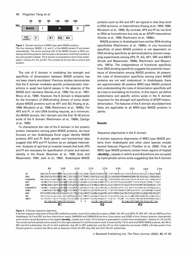
Sequence alignments in the K domain
K domain sequence alignments of MIKC-type MADS pro-
teins from Arabidopsis and other plant species reveals
several features (Figure 2) (Theißen et al., 2000). First, all
MIKC-type MADS proteins contain three regions of heptad
(abcdefg)n repeats in which a and d positions are occupied
by hydrophobic amino acids suggesting that the K domain
Figure 1. Domain structure of MIKC-type plant MADS proteins.The four domains, MADS, I, K, and C, of the MADS protein PI are shownschematically. The amino terminal half of the MADS domain is DNA con-tacting while the posterior half encodes a dimerization motif that consists oftwo anti-parallel b sheets. The K domain is postulated to form three amphi-pathic a-helices, K1, K2, and K3. The numbers at the top refer to amino acidposition.
Figure 2. K Domain sequence alignment.K domain sequence alignment of three DEF-subfamily proteins, three GLO-subfamily proteins, GGM2, AG, AP1 and SEP3. PI, AP3, AP1, AG and SEP3 are fromArabidopsis, GLO and DEF are from Antirrhinum majus, OsMADS4 and OSMADS16 are from Oryza sativa, and GGM2 is from Gnetum gnemon. Hydrophobicamino acids in a and d positions in the heptad (abcdefg)n repeats are in bold. The K domain is postulated to contain three amphipathic a-helices: K1, K2 and K3.Two conserved hydrophobic amino acids (h) in the region between K1 and K2 are also in bold. Conserved Gly-110 (in almost all MIKC MADS proteins), Asn-98 (inDEF and GLO subfamilies), Glu-97 (in GLO subfamily), Arg-102 (in DEF subfamily), and Gly-131 (in GLO subfamily) are boxed. GGM2, a B-class protein fromGnetum gnemon, contains Asn-98 as well as features of both the DEF (Arg-102) and GLO (Glu-97) subfamilies.
48 Yingzhen Yang et al.
� Blackwell Publishing Ltd, The Plant Journal, (2003), 33, 47–59

forms three amphipathic a-helices (Ma et al., 1991; Riech-
mann and Meyerowitz, 1997b). The first putative amphi-
pathic a-helix, K1, spans the region from position 87–108
(numbering as in PI); the second putative amphipathic a-
helix, K2, spans the region from position 121–135; and the
third putative amphipathic a-helix, K3, spans the region
from position 143–170 (Figure 2). Second, a conserved gly-
cine is present at position 110 in all MIKC-type MADS
proteins except a few FLC-like proteins such as FLM/
MAF1/AGL27 (Alvarez-Buylla et al., 2000a; Ratcliffe et al.,
2001; Scortecci et al., 2001). Third, in the region between K1
and K2, hydrophobic amino acids are conserved at posi-
tions 113 and 116 in most MIKC-type MADS subfamilies;
these two hydrophobic amino acids are not spaced with the
proper periodicity to be components of either the K1 or K2
amphipathic a-helices.
More than 70 AP3- and PI-like genes have been cloned
from various plant species and have been categorized as
members of the DEFICIENS (DEF) and GLOBOSA (GLO)
subfamilies (DEF and GLO are the Antirrhinum majus
AP3 and PI orthologs) (Kramer et al., 1998; Kramer, 2000;
Theißen et al., 1996, 2000). DEF- and GLO-subfamily pro-
teins possess several features in addition to the common
features of MIKC-type MADS proteins (Figure 2). First, K1 is
more conserved while K3 is less conserved in hydropho-
bicity at a and d positions in DEF and GLO subfamilies
compared to other MADS subfamilies (Kramer, 2000;
Theißen et al., 2000). Second, 44 of 46 DEF-subfamily pro-
teins and all GLO-subfamily proteins contain asparagine at
position 98, a putative hydrophobic a position in K1. In
other subfamilies, position 98 is occupied most often by a
hydrophobic amino acid. Third, glycine is conserved at
position 131 in all GLO-subfamily members, but not in
DEF-subfamily members. Fourth, charged amino acids are
conserved at specific positions in DEF and GLO subfamilies,
e.g. position 97 is negatively charged in GLO-subfamily
members while position 102 is positively charged in DEF-
subfamily members (Figure 2) (Theißen et al., 2000).
Interaction of AP3 and PI in yeast two-hybrid assays
To study the dimerization of AP3 and PI, we utilized yeast
two-hybrid assays. We failed to detect a protein/protein
interaction above background levels utilizing either full-
length versions of AP3 and PI (MIKC) or versions missing
the first 26 amino acids of the MADS domain (1/2MIKC)
(Figure 3a). However, a strong interaction was detected
between AP3 and PI lacking the entire MADS domain (i.e.
AP3(IKC) and PI(IKC)). Neither AP3 nor PI interacts with
itself or with AP1 or AG demonstrating that AP3 and PI
interact strongly, and specifically in yeast two-hybrid
assays (Figure 3a, data not shown).
Versions of AP3 and PI lacking both the MADS and I do-
mains (i.e. AP3(KC) and PI(KC)) also interact in yeast, but
less well than versions containing the I domain (Figure 3a,b).
The interaction of AP3(KC)/PI(IKC) is 50–90% of the
AP3(IKC)/PI(IKC) interaction suggesting the contribution
of I domain to AP3/PI interaction is very limited in yeast
two-hybrid assays (Figure 3b). The interaction of AP3(IK)/
PI(IKC) is similar to AP3(IKC)/PI(IKC); thus, deletion of the C
domain reveals that the C domain does not contribute to the
strength of AP3/PI interaction (Figure 3b). By contrast, when
K3 is deleted together with the C domain, the interaction of
AP3(IþK1þK2)/PI(IKC) is 15–30% of the AP3(IKC)/PI(IKC)
interaction suggesting that K3 is more important for the
strength of AP3/PI than the I or C domains (Figure 3b).
A reverse yeast two-hybrid screen identifies AP3 and PI
mutants defective in AP3/PI dimerization
To identify the critical amino acids for AP3/PI heterodimer-
ization, we performed reverse yeast two-hybrid screens to
identify mutants with weakened protein/protein interac-
tions (Li and Fields, 1993; Vidal, 1997; White, 1996). Spe-
cially, we screened for AP3 and PI mutants that interact less
well with its wild-type partner. As a source of mutants, we
constructed random AP3 and PI mutant libraries by error-
prone PCR. We then screened the AP3 and PI libraries for
mutants defective in protein/protein interaction based on
color screens of yeast colonies using lacZ as a reporter
gene (Li and Fields, 1993). In yeast, there is a correlation
between the strength of protein/protein interaction and the
color of the yeast colonies on an X-gal plate. Dark blue
colonies on an X-gal plate indicate strong protein/protein
interactions while light blue and white colonies indicate
weakened or disrupted protein/protein interactions. We
focused our efforts on characterizing light blue colonies
rather than white colonies, reasoning that many of the
white colonies may contain catastrophic mutations, for
example resulting in the production of truncated proteins,
while the light blue colonies more likely contain mutations
resulting in specific defects in AP3/PI protein/protein inter-
action.
We sequenced approximately 100 mutant AP3 and PI
genes from the light blue colonies that exhibited weakened
AP3/PI interaction. Ten per cent of the sequenced genes did
not contain a mutation in the encoded protein, 45%
encoded multiple amino acid changes, 5% contained a
single-base deletion or introduced a stop codon, and
40% encoded a single amino acid change. Some mutations
were identified multiple times; for example, 10 independent
mutations in Ile-128 of PI were identified; six single muta-
tions (three PII128T and three PII128F), three double mutations
(PII94T,I128T, PII128T,Q166R, and PII128F,H130Y), and one multiple
mutation (PII113M,K119R,I128T,S145P,M152V,H183R).
Among the mutants with amino acid changes, 95% con-
tain a mutation in one of the following categories: (i) change
in amino acids at the a or d positions in the heptad repeats
� Blackwell Publishing Ltd, The Plant Journal, (2003), 33, 47–59
Dimerization of AP3 and PI 49

in K1 or K2 (e.g. PII94T); (ii) change in conserved hydro-
phobic amino acids (e.g. PIL116R); (iii) change in conserved
glycine residues (e.g. AP3G110S), or (iv) change at positions
other than a and d to either a proline or glycine (e.g. PIE104G).
Since all of the single mutants that we sequenced contained
mutations in one of these four categories (Table 1; Figure 4),
we focused our analysis on the mutants containing single
amino acid changes. Interaction between these mutant
proteins and its wild-type partner is between 3 and 50%
of wild-type levels (Table 1). All mutations except PIS145P are
located between position 91 and 135; this region includes
K1, K2 and the region between K1 and K2.
Mutant genes isolated in the reverse yeast two-hybrid
screen were cloned in the context of full-length AP3 and PI
containing the MADS domain. Protein/protein interaction
of full-length mutant proteins with its wild-type partner
were measured directly by co-immunoprecipitation assays
and indirectly by EMSA DNA-binding assays (data not
Figure 3. Interaction of wild-type and mutant AP3 and PI in yeast two-hybrid assays (error bars indicate SD).(a) MADS-deleted AP3 and PI interact strongly, and specifically in yeast two-hybrid assays. M (MADS), I, K, C stand for the four domains in AP3, PI, AG and AP1.1/2MIKC indicates proteins with the N-terminal half of the MADS domain deleted. IKC indicates proteins that lack the entire MADS domain. KC indicates proteinsthat lack both MADS and I domains.(b) Interaction between versions of AP3 and PI containing the I and K domains (IK), the K and C domains (KC), and the I domain plus the first two K domainamphipathic alpha helices K1 and K2, but lacking the K3 helix (I–K1–K2).(c) Interaction between AP3N98V, AP3N98L, PIN98V, and PIN98L in two-hybrid assays.(d) Interaction between PIwt and AP3E96A, AP3R99A, and AP3R102A in two-hybrid assays.
� Blackwell Publishing Ltd, The Plant Journal, (2003), 33, 47–59
50 Yingzhen Yang et al.

shown). For mutant proteins that interact very poorly with
wild-type partners (below 10% of wild-type levels), defects
in co-immunoprecipitation and DNA binding were con-
sistently observed. For mutant proteins that were less
defective in AP3/PI interaction (between 10 and 50% of
wild-type levels), defects were more variable. By contrast,
defects in protein/protein interactions in yeast two-hybrid
assays, as measured by b-galactosidase liquid assays, were
reproducible.
To ascertain if dimerization-defective mutants exhibit
functional defects in vivo, we ecotopically expressed the
mutant AP3 and PI genes in Arabidopsis. The function of the
mutant gene was determined by examining both the flower
phenotype of the ectopic expression lines (Table 1) and the
ability of ectopic expression lines to rescue petal and sta-
men defects in strong ap3 or pi mutants. Most transgenic
plants expressing mutant AP3 or PI genes exhibit inter-
mediate or weak 35S phenotypes and fail to fully rescue
petals and stamens when crossed to a strong ap3 or pi
mutant (Table 1, Figure 5). These assays indicate that most
mutant AP3 and PI genes possess only partial activity in
planta. In summary, the in vivo function of most dimeriza-
tion-defective AP3 and PI mutants correlates with the inter-
action strength between the mutant protein and its wild-
type partner as measured in yeast two-hybrid assays.
Hydrophobic amino acids in K1 and K2
Among the 26 single mutants isolated from the reverse
yeast two-hybrid screen, 20 contain mutations in hydro-
phobic amino acids. 18 of 20 hydrophobic mutations are in
K1 and K2 (Figure 4a, Table 1). Mutations (16 out of 18) are
located at a or d positions in the postulated heptad repeats;
the two exceptions are PIL103P and PIL132P, both of which
contain proline mutations that likely disrupt a-helical sec-
ondary structure rather than directly interfering with the
interaction interface in AP3/PI heterodimer. In PI, almost all
a and d positions in K1 and K2 were identified as critical
positions for AP3/PI interaction (Figure 4a). By contrast, no
single mutation was identified at a or d positions in K3. In
summary, the fact that most mutations are located in con-
served hydrophobic positions in K1 and K2 suggests that
Table 1 Phenotypes of AP3 and PI mutants
MutationyYeast two–hybridinteraction (% wild type)z
35S T1phenotype§
AP3WT 100 �������
PIWT 100 �������
PIL87P,K146R 14 nd�PIL87A 40–50 ����
PII91T 30–44 �����
PII94T 32–43 ����
PIN98D 3–4 ��
PIN98S 40–60 ������
PIN98L 6–10 �����
PIN98V 10–15 �����
AP3N98D 5 �����
AP3N98K 50 �������
AP3N98L 5–10 �����
AP3N98V 3–6 ����
AP3L101R 18 �����
PIL101A 11–20 �����
PIL103P 10–11 ����
PIE104G 42–54 ����
PIL105A 10–20 �����
PIL105H 4–5 ��
PIL101A,L105A 1–3 ��
AP3I105F 22 �����
PIL108S 4–6 ����
PIL108A 5–10 �����
AP3G110S 10–13 ��
PIG110A 52–67 ������
PIG110P 4–5 �
PIL113R 20–30 ����
AP3L113E 15–25 ���
AP3L116F 24 ndAP3L116E 15–25 �����
PIL116R 25 �����
PIL116P,K146R 22 ��
AP3I118E 100 �������
PIL121A 50–60 ������
PIL121P 10 �
PIL121R 34 ��
AP3L124F 20 �����
AP3L124P 1–2 �
PIV124D 10 ��
PIV124A 30 ����
PIL121A,V124A 10–20 ����
AP3M128V 30 �����
AP3M128K 13 �
PII128T 20–31 ����
PII128F 17–26 ����
PII94T,I128T 1–4 �
PIG131V 4–8 ��
PIG131A 33–44 ������
PIG131P 6–10 �
PIL132P 17 ����
PIV135A 39–52 �����
Table 1 continued
MutationyYeast two–hybridinteraction (% wild type)z
35S T1phenotype§
PIL142P 40–50 ��
PIL142A 90 ������
PIS145P 40–50 ��
yReverse two-hybrid mutants are in bold, others are site-specificmutants.zThree to five independent colonies were assayed for each mu-tant. Ranges indicate that mutants were assayed multiple times.§Phenotype of 35S flowers: (�), wild type (no organ identityconversion); (��), very weak; (���), weak; (����), weak-intermediate;(�����), intermediate; (������), intermediate–strong; (�������), strong(equivalent to 35S::AP3 or 35S::PI).�Not determined.
� Blackwell Publishing Ltd, The Plant Journal, (2003), 33, 47–59
Dimerization of AP3 and PI 51

AP3 and PI interact primarily via interactions between the
hydrophobic faces of K1 and K2.
The involvement of both K1 and K2 in AP3/PI heterodi-
merization is further demonstrated by the severe dimeriza-
tion defects of double mutants containing mutations in
both K1 and K2. For example, both PII94T and PII128T single
mutants interact with AP3 at 25–35% of wild-type levels, but
the interaction between PII94T,I128T and AP3wt is only 1–4% of
wild-type levels (Table 1). The effects of double mutations
on AP3/PI interaction can also be mimicked by analyzing K1
mutants in one partner together with K2 mutants in the
other partner (e.g. AP3I101R/PIV135A and AP3M128V/PII94T). The
interactions between these pairs of mutants are much
weaker than the interaction between the single mutant
and its wild-type partner (data not shown). In summary,
our studies on double mutants in K1 and K2 demonstrate
that both K1 and K2 contribute to the strength of AP3/PI
dimerization.
In addition to the reverse yeast two-hybrid screen, we
also utilized a partial alanine-scanning strategy as a com-
plementary approach to study the importance of hydro-
phobic amino acids at a and d positions in K1 and K2.
Specially, we constructed several single PI mutants (PIL87A,
PIL101A, PIL105A, PIL108A, PIL121A, PIV124A and PII142A) and two
PI double mutants (PIL101A,L105A and PIL121A,V124A). All single
mutants except PII142A exhibit decreased AP3/PI interaction,
further suggesting the critical role of K1 and K2 in AP3/PI
dimerization (Table 1). Compared to the single alanine
mutants, the double alanine mutants exhibit more severe
defects in AP3/PI interaction (Table 1).
Conserved hydrophobic amino acids in the region
between K1 and K2
The region between K1 and K2, from position 109–120,
contains hydrophobic amino acids at positions 113 and
116 that are conserved in most MIKC-type MADS subfami-
lies (Figure 2) (Theißen et al., 2000). A hydrophobic amino
acid is also present at position 118 in most DEF-subfamily
proteins but not in GLO-subfamily proteins. To investigate
the role of these conserved hydrophobic amino acids in
AP3/PI dimerization, we constructed several site-specific
mutants (AP3L113E, AP3L116E, AP3I118E and PII113R) in addi-
tion to the mutants isolated in the reverse yeast two-hybrid
screen (AP3L116F, PII113T,S145L, PIL116R and PIL116P,K146R)
(Table 1 and data not shown). AP3L113E, AP3L116E, PII113R
and PIL116R are defective in AP3/PI dimerization (15–30% of
wild-type levels) (Table 1). Consistent with the defects in
yeast, these four mutants exhibit partial AP3 and PI activity
in planta. The importance of hydrophobicity at positions
113 and 116 is in contrast to position 118 of AP3; AP3I118E
interacts with PI at wild-type levels and functions similarly
to wild-type AP3 in planta (Table 1). In summary, hydro-
phobic side groups at positions 113 and 116 of AP3 and PI
are required for strong AP3/PI interaction.
Asparagine 98 in AP3 and PI
Asn-98 is located at a putative a position in one of the
heptad repeats in K1. Among plant MADS proteins, position
98 is conserved as asparagine only in the DEF and GLO
subfamilies (Figure 2) (Kramer, 2000; Theißen et al., 2000).
To study the role of Asn-98 in AP3/PI interaction, we
mutated Asn-98 to the hydrophobic amino acids leucine
and valine (AP3N98L, AP3N98V, PIN98V and PIN98L), and mea-
sured protein/protein interactions by yeast two-hybrid
assays (Table 1, Figure 3c). When Asn-98 is mutated to
leucine, the interaction of AP3N98L/PIN98L is slightly stronger
than that of AP3N98L/PIwt or AP3wt/PIN98L, but in all three
cases, the interaction is only 5–10% of wild-type levels.
When Asn-98 is mutated to valine, the interaction of
AP3N98V/PIN98V is much higher (30–40% of wild-type levels)
than the interaction of AP3N98V/PIwt or AP3wt/PIN98V (3–5 and
10–15% of wild-type levels, respectively) (Figure 3c). The
compensatory interactions detected in AP3N98V/PIN98V and
AP3N98L/PIN98L suggest that Asn-98 in AP3 interacts directly
Figure 4. Reverse yeast two-hybrid mutants inAP3 and PI.(a) Mutants isolated in reverse yeast two-hybridscreen are indicated. Mutations in AP3 areshown below the AP3 sequence and mutationsin PI are shown above the PI sequence. Themajority of the reverse yeast two-hybrid muta-tions are located in conserved hydrophobicamino acids.(b) Comparison of K1 of AP3 and PI with leucinezipper motifs from GCN4, FOS, and JUN.
� Blackwell Publishing Ltd, The Plant Journal, (2003), 33, 47–59
52 Yingzhen Yang et al.

Figure 5. In planta functional assays of AP3 and PI mutants defective in AP3/PI heterodimerization.Flowers from 35S over-expression and mutant rescue for two PI mutants (PII128T [e–h] and PIL105H [i–l]) and two AP3 mutants (AP3L124F [q–t] and AP3G110S [u–x]). PImutants were analyzed by over-expression (i.e. 35S::PI) alone and in combination with 35S::AP3. Over-expression lines were also utilized to ascertain the degreeof pi-4 mutant rescue. Similarly, AP3 mutants were analyzed by over-expression (i.e. 35S::AP3) alone and in combination with 35S::PI. Over-expression lineswere also utilized to ascertain the degree of ap3 -3 mutant rescue.(a) 35S::PI. First whorl organs are sepaloid petals.(b) 35S::PI pi-4. Petals are fully rescued in whorl 2 and stamens are partially rescued in whorl 3.(c) 35S::PI 35S::AP3. Whorls 1 and 2 develop as petals, whorls 3 and 4 develop as stamens.(d) 35S::PI 35S::AP3 pi-4. pi-4 mutants are completely rescued by 35S::AP3 35S::PI.(e) 35S::PII128T. First whorl organs are sepals with some petaloid sectors.(f) 35S::PII128T pi-4. Second whorl organs develop as short petals and third whorl organs develop as staminoid carpels demonstrating partial rescue of the pi-4phenotype.(g) 35S::PII128T 35S::AP3. The first whorl develops as short sepaloid petals, and the fourth whorl develops as stamens, carpelloid stamens or multichamberedcarpels.
� Blackwell Publishing Ltd, The Plant Journal, (2003), 33, 47–59
Dimerization of AP3 and PI 53

with Asn-98 in PI. The difference in interaction strength
between AP3N98V/PIN98V and AP3N98L/PIN98L also suggests
that size of the R group is critical in this interaction; the
larger R group of leucine might result in steric hindrance in
the interaction interface. This is consistent with the fact that
the interactions of AP3N98V/PIN98L and AP3N98L/PIN98V are
stronger than that of AP3N98L/PIN98L (Figure 3c).
To test if polar amino acids other than asparagine could
function at position 98, we constructed AP3N98K and
AP3N98D in addition to PIN98D and PIN98S, isolated in the
reverse yeast two-hybrid screen. Both AP3N98D and PIN98D
are severely defective in AP3/PI dimerization in yeast two-
hybrid assays (3–5% of wild-type levels). By contrast,
AP3N98K and PIN98S interact at 40–60% of wild-type levels.
AP3N98K and PIN98S also function much better than AP3N98D
and PIN98D in planta demonstrating that certain charged/
polar amino acids other than asparagine can function at
position 98 (Table 1).
Role of charged amino acids in K1 in AP3/PI dimerization
Charged amino acids are conserved at certain positions in
DEF- and GLO-subfamily proteins (e.g. positions 96, 97, 99,
and 102) (Figure 2). However, no mutations were identified
in these conserved charged amino acids in the reverse two-
hybrid screens. To study the role of charged amino acids in
AP3/PI dimerization, we mutated positions 96, 99, and 102
in AP3 to alanine. In the AP3(IKC)/PI(IKC) context, AP3E96A
and AP3R99A interact with PI at 90–120% of wild-type levels,
while AP3R102A interacts with PI at 55–70% of wild-type
levels. When these interactions were tested in the
AP3(KC)/PI(KC) context, the interaction defect of AP3R102A
is enhanced (20–35% of wild-type levels) (Figure 3d). These
results suggest that Arg-102 of AP3 contributes more to the
strength of AP3/PI dimerization than Glu-96 or Arg-99.
Conserved glycine residues in K domain
Glycine is conserved at position 110 in almost all MIKC-type
MADS proteins (Theißen et al., 2000). In all GLO-subfamily
proteins, but not in DEF-subfamily proteins, a second gly-
cine is conserved at position 131. AP3G110S and PIG131V were
isolated from the reverse yeast two-hybrid screen. Both
AP3G110S and PIG131V exhibit very limited function in yeast
and in planta (Table 1). Additionally, PIG110A, PIG110P, PIG131A
and PIG131P were constructed to study the role of conserved
glycines in AP3/PI interaction. While PIG110A and PIG131A
exhibit modest defects in both yeast (30–60% of wild-type
levels) and in planta (Table 1), PIG110P and PIG131P exhibit
severe defects both in yeast and in planta (Table 1). In
summary, our studies demonstrate that Gly-110 in AP3
and PI and Gly-131 in PI are critical for strong AP3/PI
interaction.
Discussion
Critical amino acids for the strength of AP3/PI interaction
are located in the K domain, from position 87–135. This
region includes the first two putative amphipathic a-helices
in the K domain (K1 and K2) and the region between K1 and
K2. The majority of critical amino acids for AP3/PI interac-
tion are located on the putative hydrophobic faces of these
a-helices suggesting that the strength of AP3/PI dimeriza-
tion is mediated primarily via hydrophobic interactions.
By contrast, no hydrophobic amino acids in K3 were iden-
tified as critical for AP3/PI dimerization. This is consistent
with the results of DNA-binding assays utilizing truncated
versions of AP3 and PI (as well as DEF and GLO) that
demonstrate that both K1 and K2, but not K3, are necessary
for strong DNA binding (Riechmann et al., 1996b; Zachgo
et al., 1995). Although no single mutants were identified
Figure 5. continued(h) 35S::PII128T 35S::AP3 pi-4. Whorls 1 and 2 develop as sepaloid petals and whorls three and four develop primarily as staminoid carpels demonstrating partialrescue.(i) 35S::PIL105H. Whorl 1 develops as sepals demonstrating that PIL105H possesses very limited function in planta.(j) 35S::PIL105H pi-4. Second whorl organs are petaloid sepals and third whorl organs are most often missing.(k) 35S::PIL105H 35S::AP3. First whorl organs develop as short sepaloid petals and fourth whorl organs as multichambered carpels demonstrating that PIL105H
activity is enhanced by over-expression of AP3.(l) 35S::PIL105H 35S::AP3 pi-4. Organs in whorls 1 and 2 are petaloid sepals, whorl 3 is either missing or develops as filaments, and whorl 4 develops asmultichambered carpels.(m) 35S::AP3. Fourth whorl organs develop as stamens or carpelloid stamens.(n) 35S::AP3 ap3-3. Stamens are fully rescued in whorl 3, petals are partially rescued in whorl 2 (short petals with some green sectors).(o) 35S::AP3 35S::PI. Whorls 1 and 2 develop as petals, whorls 3 and 4 develop as stamens.(p) 35S::AP3 35S::PI ap3-3. ap3-3 mutants are completely rescued by 35S::AP3 35S::PI.(q) 35S::AP3L124F. Whorl 4 develops as multichambered carpels demonstrating partial function.(r) 35S::AP3L124F ap3-3. Second whorl organs are sepals and third whorl organs are stamens or carpelloid stamens demonstrating partial rescue of ap3-3.(s) 35S::AP3L124F 35S::PI. Whorl 1 develops as short petals, whorl 2 as petals, whorl 3 as stamens, and whorl 4 as carpelloid stamens.(t) 35S::AP3L124F 35S::PI ap3-3. Whorls 1 and 2 develop as petals, and whorl 3 and 4 develop as stamens or carpelloid stamens.(u) 35S::AP3G110S. The fourth whorl develops as wild-type carpels demonstrating very limited function of AP3G110S in planta.(v) 35S::AP3G110S ap3-3. Whorl 2 develops as sepals and whorl three as carpels (organ number, but not organ identity, is rescued) demonstrating that AP3G110S
exhibits only very slight rescue of ap3-3.(w) 35S::AP3G110S 35S::PI. Whorl 1 develops as petaloid sepals and whorl 4 develops as a multichambered carpels indicating that AP3G110S activity is enhanced byover-expression of PI.(x) 35S::AP3G110S 35S::PI ap3-3. Whorls 1 and 2 develop as sepals or petaloid sepals and whorls 3 and 4 develop as carpels or staminoid carpels.
� Blackwell Publishing Ltd, The Plant Journal, (2003), 33, 47–59
54 Yingzhen Yang et al.

in hydrophobic positions in K3, deletion of K3 affects the
strength of AP3/PI dimerization more than deletion of the I
or C domains.
MIKC-type MADS proteins can be categorized into a
number of subfamilies. The demonstration that K1, K2
and the region between K1 and K2 are important for AP3/
PI dimerization is generally applicable to other MIKC sub-
families. K2 and the hydrophobic amino acids between K1
and K2 are highly conserved in all MIKC-type MADS sub-
families, thus, it is likely that these regions play an impor-
tant role in dimerization of all MIKC-type MADS proteins. By
contrast, K1 is more strongly conserved in the DEF and GLO
subfamilies; thus, the identification of important amino
acids in K1 is more applicable to the B class MADS proteins
(i.e. the GLO and DEF subfamilies) than to other MIKC
subfamilies.
High-resolution X-ray structures of the DNA-binding
cores of SRF, MCM1, MEF2 demonstrate that subdomains
of the MADS domain mediate dimerization. The N-terminal
DNA-contacting half of the MADS domain forms an amphi-
pathic a-helix that contacts DNA and contributes to the
strength of dimerization. The C-terminal half of MADS
domain forms a series of anti-parallel b-sheets that function
as a dimerization interface (Hassler and Richmond, 2001;
Pellegrini et al., 1995; Santelli and Richmond, 2000; Tan and
Richmond, 1998). We did not address critical amino acids in
the MADS domain in the reverse yeast two-hybrid screens
due to the fact that MADS domain-containing versions of
AP3 and PI do not interact strongly in yeast two-hybrid
assays. However, we did construct site-specific mutants in
key amino acids in the b-sheet region and these mutants do
not function in planta and do not bind to DNA demonstrat-
ing that these positions are essential for function (unpub-
lished).
The I domain is plant-specific and high-resolution struc-
tures of plant MADS proteins have not been obtained.
Domain-swapping experiments suggest that I domain par-
ticipates in dimerization specificity (Krizek and Meyerowitz,
1996b). Yeast two-hybrid assays demonstrate that the
interaction strength of versions of AP3 and PI that lack
the I domain (AP3(KC)/PI(KC)) is half that of versions that
contain the I domain (AP3(IKC)/PI(IKC)) indicating that I
domain contributes to the strength of AP3/PI interaction
(Figure 3a). Heterodimers that consist of one monomer with
and one without an I domain (i.e. AP3(KC)/PI(IKC)) interact
at 50–90% of AP3(IKC)/PI(IKC) levels suggesting the con-
tribution of I domain to AP3/PI interaction is limited in yeast
two-hybrid assays. Consistent with this, many single
mutations in the K domain have a more dramatic effect
on the strength of AP3/PI interaction (reduced to 3–50% of
wild-type levels) than truncation of the entire I domain
(Table 1).
Previous experiments did not suggest a role for the C
domain in MADS protein dimerization. In agreement with
this, reverse yeast two-hybrid screens did not identify any
critical amino acids in the C-terminal domain required for
strong AP3/PI interaction. In addition, when the entire C
domain of AP3 is deleted, AP3 still interacts with PI(IKC) at
levels equivalent to AP3(IKC)/PI(IKC) (Figure 3b).
The first amphipathic a-helix in the K domain mediates
AP3/PI heterodimerization in a manner similar to the
leucine zipper motif
The leucine zipper is a short coiled coil of two parallel
helices comprised of three to six (abcdefg)n heptad repeats,
in which the d-positions are occupied by leucine (Land-
schulz et al., 1988). Hydrophobic amino acids at a and d
positions from each helix form the core of the hydrophobic
interaction interface. The aliphatic chains of polar/charged
residues on e and g positions also participate in forming the
edges of the hydrophobic interaction interface. Addition-
ally, some polar/charged amino acids at a, e, and g posi-
tions form hydrogen bonds or salt bridges to strengthen
protein/protein interaction and also to provide dimerization
specificity (Glover and Harrison, 1995; O’Shea et al., 1991;
Vinson et al., 1993).
In PI, all a and d positions in K1 are occupied by hydro-
phobic amino acids except position 98 (Figure 2). All hydro-
phobic amino acids at a and d positions in K1 are critical for
AP3/PI dimerization (Figure 4a, Table 1) demonstrating that
K1 contributes to the strength of AP3/PI interaction mainly
via hydrophobic interactions.
The leucine zipper proteins GCN4 and Jun contain aspar-
agine at an a position in the central region of the leucine
zipper motif (Figure 4b) (Zeng et al., 1997). Similarly, AP3
and PI encode asparagine at position 98 in the central
region of K1. Asn-98 is highly conserved in DEF- and
GLO-subfamily proteins, but not in other MADS subfami-
lies. Mutation of Asn-98 to leucine or valine reduces AP3/PI
interaction to 3–15% of wild-type levels (Table 1). The com-
pensatory interaction between PIN98V and AP3N98V suggests
that Asn-98 in AP3 directly interacts with Asn-98 in PI
(Figure 3c). On the other hand, AP3N98K and PIN98S, but
not AP3N98D or PIN98D, maintain 50–60% of wild-type inter-
action suggesting that certain polar/charged amino acids
other than asparagine can function at position 98 (Table 1).
Two asparagines can form hydrogen bonds via the protons
of the gNH2 group and the carbonyl oxygen. In the high
resolution structures of GCN4 and Jun homodimers, an a-
position asparagine in one monomer forms hydrogen
bonds with an a-position asparagine in the other monomer
(Junius et al., 1995; O’Shea et al., 1991). Similarly, we
hypothesize that Asn-98 in AP3 hydrogen bonds with
Asn-98 in PI contributing to the strength of AP3/PI interac-
tion. This hypothesis is supported by the fact that serine
and lysine are able to function at position 98 in place of
asparagine. In the Fos/Jun heterodimer, the corresponding
� Blackwell Publishing Ltd, The Plant Journal, (2003), 33, 47–59
Dimerization of AP3 and PI 55

a position in Fos is occupied by lysine (Glover and Harrison,
1995). We suspect that AP3N98K interacts with PI in a manner
similar to Fos and Jun. In the case of PIN98S, the hydroxyl
group in serine can function as both a hydrogen bond
donor and acceptor, similar to asparagine. Interestingly,
the recently categorized B-sister genes contain serine or
threonine at position 98 (Becker et al., 2002).
In addition, Asn-98 may also contribute to the specificity
of heterodimer formation, as is the case in leucine zipper
proteins. In GCN4, an a-position asparagine is proposed to
impart specificity for formation of a two-stranded, parallel
coiled coil (Gonzalez et al., 1996; Harbury et al., 1993; Junius
et al., 1995; Zeng et al., 1997). The a-position asparagine
helps position the helices in a parallel orientation and
disfavors anti-parallel arrangements that necessitate aspar-
agine side chains packing against non-polar chains. We
propose that Asn-98 is one of the key determinants of AP3/
PI heterodimer specificity.
In dimers formed by leucine zipper proteins, amino acids
at g positions in one monomer form salt bridges with amino
acids on e0 positions in the next heptad repeat in the other
monomer to strengthen the protein/protein interaction (the
iþ 5 ionic interaction) (Glover and Harrison, 1995; Lumb
and Kim, 1995; O’Shea et al., 1991; Vinson et al., 1993).
Our comparative study on Glu-96, Arg-99, Arg-102 in AP3
suggests that Arg-102 contributes more to the strength
of AP3/PI interaction than Glu-96 and Arg-99 (Figure 3d).
We hypothesize that the e-position Arg-102 in AP3 forms
a salt bridge with g position Glu-97 in PI. Position 97 is
highly conserved as a negatively charged amino acid
in GLO-subfamily proteins, but not in other MADS sub-
families (Figure 2). Similarly, position 102 is conserved as
positively charged amino acid in DEF-subfamily pro-
teins, but not in other MADS subfamilies. The postulated
interaction between Arg-102 in AP3 and Glu-97 in PI resem-
bles the iþ 5 ionic interaction observed in leucine zipper
dimers.
In leucine zipper dimers, the iþ 5 ionic interaction also
promotes the formation of specific dimer pairs and pre-
vents the formation of non-specific dimer pairs (O’Shea
et al., 1992; Vinson et al., 1993; Zeng et al., 1997). We
propose that the interaction between Glu-97 in PI and
Arg-102 in AP3 facilitates specific heterodimerization
between AP3 and PI and prevents formation of homodi-
mers and other AP3- and PI-containing heterodimers. Inter-
estingly, in the gymnosperm Gnetum gnemon, there is a
single DEF/GLO-like gene, GGM2 (Winter et al., 2002).
Unlike DEF and GLO proteins from angiosperms, GGM2
binds to DNA as a homodimer. GGM2 contains both Glu-97
and Arg-102 suggesting to us that an iþ 5 ionic interaction
could take place to stabilize the GGM2 homodimer. We
propose that the subdomain from position 97–102 is critical
for heterodimerization specificity of DEF- and GLO-like
proteins.
The second amphipathic a-helix in the K domain of
PI has unique features
The second amphipathic a-helix in the K domain (K2) spans
the region from position 121–135. Similar to K1, all con-
served hydrophobic amino acids at a and d positions in K2
of PI were identified as critical residues for AP3/PI interac-
tion suggesting that K2 mediates AP3/PI dimerization
mainly via hydrophobic interactions.
Interestingly, at position 131, a putative hydrophobic d
position, non-hydrophobic amino acids are present in most
MIKC-type MADS subfamilies. Specially, glycine is con-
served at position 131 in all GLO-subfamily proteins while
serine or threonine is present in most DEF-subfamily pro-
teins. Glycine is rarely found in a-helices and most likely
does not contribute to helix stabilization. However, the fact
that PIG131V and PIG131A interact with AP3 less well than
wild-type PI suggests that hydrophobicity is not favored at
this putative d position and that a small R group at position
131 may be important (Table 1). Though glycine residues
are rare in helical segments of soluble proteins, glycine
residues are not uncommon in transmembrane helices
(Senes et al., 2001). In transmembrane proteins, such as
the glycerol facilitator from E. coli, which contains eight
transmembrane a-helices, glycine residues on one a-helix
form inter-helical Ca–H� � �O hydrogen bonds with the
carbonyl or hydroxyl oxygen on other helices. In the col-
lagen triple helix, glycine also forms inter-helical Ca–H� � �Ohydrogen bonds (Bella and Berman, 1996). These uncon-
ventional interhelical Ca–H� � �O hydrogen bonds are pro-
posed to be a determinant of stabilization and specificity in
transmembrane helix interactions (Senes et al., 2001). It is
possible that the d-position Gly-131 in PI participates in
formation of Ca–H� � �O bonds in a similar way to glycine
residues in collagen triple helix and transmembrane
helices.
The region between K1 and K2
The region between K1 and K2 contains 12 amino acids
from position 109–120. This region is conserved at several
positions in most MIKC-type MADS subfamilies, including
Gly-110 and positions 113 and 116, which are highly con-
served as hydrophobic amino acids. Mutations at Gly-110 in
both AP3 and PI dramatically affect AP3/PI dimerization
(Table 1). Based both on the fact that the heptad repeats
are interrupted in this region and the flexibility of glycine in
adapting many conformations, we propose that Gly-110
plays an important structural role at the end of the first
amphipathic a-helix, e.g. forming a loop between helices.
Though positions 113 and 116 are highly conserved as
hydrophobic amino acids, these two positions are not
spaced with the proper periodicity to be components of
the heptad repeats in either K1 or K2. Mutational analysis of
� Blackwell Publishing Ltd, The Plant Journal, (2003), 33, 47–59
56 Yingzhen Yang et al.

positions 113 and 116 in AP3 and PI suggests that hydro-
phobicity is necessary at these two positions for strong
AP3/PI interaction (Table 1). These two conserved hydro-
phobic amino acids could comprise a short hydrophobic
face that directly mediates AP3/PI interaction. It is also
possible that this region functions indirectly by ensuring
that the K1 and K2 a-helices adopt the correct three-dimen-
sional conformation.
Experimental procedures
Yeast two-hybrid plasmids
The yeast two-hybrid vector pGAD-C1 contains GAL4 activationdomain (AD) and has leu2 as the nutritional selection marker. Thetwo-hybrid vector pGBDU-C1 contains GAL4 DNA-binding domain(DB) and has ura3 as the nutritional selection marker (James et al.,1996). Both pGAD-C1 and pGBDU-C1 utilize the truncated ADH1promoter to drive expression of GAL4 fusions. The yeast reporterstrain is pJ69-4a (MATa trp1–901 leu2–3, 112 ura3-52 his3-200 gal4DLYS2:: GAL1-HIS3 GAL2-ADE2 met2::GAL7-lacZ). pJ69–4a, isogenicto pJ69-4a except that it is of opposite mating type, is used to bringdifferent plasmid constructs together viamating (James et al., 1996).Plasmids expressing six different versions of both AP3 and PI wereconstructed:(i)entireopenreadingframecontainingtheMADS(M), I,K and C domains (MIKC); (ii) deletion of the first 26 amino acids of theMADS domain (1/2 MIKC); (iii) versions lacking the entire MADSdomain (IKC); (iv) versions lacking both MADS and I domains (KC);(v) deletion of both MADS domain and C domain (IK); (vi) deletion ofMADS domain, C domain, and the third putative amphipathica-helixof the K domain (IþK1þK2). The IKC and the KC regions of both AP1and AG were also cloned into yeast vectors.
Construction and screening of AP3 and PI mutant
libraries
Error-prone PCR with unbalanced dNTP concentration in the pre-sence of MnCl2 was used to generate random mutations inAP3(IKC) and PI(IKC) (Fromant et al., 1995; Vidal, 1997). PCR pro-ducts were transformed together with the linearized yeast vectorsinto pJ69-4A a (for AP3 mutants) or pJ69-4A a (for PI mutants) togenerate mutant libraries by homologous recombination (Vidal,1997). The AP3 and PI mutant libraries contain about 10 000 inde-pendent colonies in 15 sub-libraries. Each sublibrary was screenedindependently by mating to a yeast strain containing the wild-typepartner. About 2–10% of the colonies exhibited a light-blue coloron X-gal plates (i.e. weak interactors) in the primary screen and asubset were analyzed by quantitative b-galactosidase liquid assays(Reynolds et al., 1998) to confirm the weakened AP3/PI interaction.For each mutant, three to four independent colonies were assayedby b-galactosidase liquid assays. Mutant plasmids were rescuedand sequenced. Mutant AP3 and PI genes were sub-cloned into thefull-length cDNA context for ectopic expression in planta and forDNA-binding and co-immunoprecipitation assays.
Construction of site-specific AP3 and PI mutants
Single-stranded DNA mutagenesis (Yukenberg et al., 1991) andoverlap extension PCR (Horton and Pease, 1991) were used toconstruct site-specific mutants.
DNA-binding and co-immunoprecipitation assays
The DNA-binding ability of the mutant protein with its wild-typepartner protein was tested by electrophoretic mobility shift assay(EMSA) using a 49-bp probe containing CArG3 from the AP3promoter (Riechmann et al., 1996b; Tilly et al., 1998). At leasttwo independent assays were conducted for each mutant protein.Co-immunoprecipitation assays were performed using FLAG-tagged AP3 or PI protein as described in Riechmann et al., (1996b).
In vivo function of AP3 and PI mutants
The mutant genes were cloned into pCGN18, the ectopic expres-sion vector which contains the broadly expressed 35S promoterfrom cauliflower mosaic virus (Krizek and Meyerowitz, 1996a;Sieburth et al., 1995). 35S::AP3� and 35S::PI� constructs wereintroduced into Arabidopsis Columbia ecotype via Agrobacter-ium-mediated vacuum transformation (Bechtold and Pelletier,1998). For each mutant, 10–60 T1 plants were generated. Sevenclasses of function were assigned to 35S::AP3� based on the whorl4 phenotype in the flowers of most T1 transgenic plants. In flowersof 35S::AP3wt transgenic plants, carpels in whorl 4 are converted tostamens or carpelloid stamens (Jack et al., 1994). ‘Strong’ indicatesflower phenotype similar to 35S::AP3wt. ‘Intermediate’ describesflowers with gynoecium of more than two carpels and most oftencarpels are not completely fused together. ‘Weak’ describes plantswith occasional flowers that have a gynoecium of more than twocarpels. ‘Wild type’ stands for flowers with two fused carpels inwhorl 4 for all T1 plants. In 35S::PIwt transgenic plants, sepals inwhorl 1 are petaloid, and the flower buds are bulged due to thepetaloid sepals (Krizek and Meyerowitz, 1996a). The phenotypes of35S::P5� were assigned based on the degree of conversions ofsepals to petals in whorl 1. The percentage of co-suppressed plantsin T1 (i.e. those that exhibit an ap3 or pi mutant phenotype) wasgreater for 35S::AP3 (up to 50%) than 35S::PI (5–25%).
The ability of mutant AP3 or PI proteins to rescue petal andstamen organ identity was ascertained in two ways. For AP3�,representative 35S::AP3� lines were crossed to ap3-3, a strong ap3mutant allele. 35S::AP3wt can fully rescue stamen organ identity inap3-3, but can only partially rescue petal organ identity in ap3-3(Figure 5) (Jack et al., 1994). A second approach is to examine theability of 35S:AP3� to rescue ap3-3 together with 35S::PI; both petaland stamen organ identities of ap3-3 are fully rescued in35S::AP3wt 35S::PIwt ap3-3 (Krizek and Meyerowitz, 1996a). Simi-larly, for PI�, mutant rescue was assayed by crossing a represen-tative 35S:PI� line to pi-4, a strong pi allele. 35S::PIwt fully rescuespetal identity of pi-4, and largely rescues stamen identity. Mutantrescue was also assayed by examining the ability of 35S:PI� torescue pi-4 together with 35S::AP3; both petal and stamen organidentities are fully rescued 35S::AP3wt 35S::PIwt pi-4.
35S::AP3 35S::PI double transgenic plants exhibit a vegetativephenotype that is not observed in 35S::AP3 or 35S::PI singletransgenics (Krizek and Meyerowitz, 1996a). Specially, the rosetteand cauline leaves are curved and are much smaller than leaves inwild-type plants and cauline leaves that develop in apical-mostpositions are often petaloid. The severity of the leaf phenotype wasfound to be indicative of the interaction strength between AP3and PI.
Acknowledgements
We thank Jess Goris for excellent technical assistance and Kank-shita Swaminathan for comments on the manuscript. This work is
� Blackwell Publishing Ltd, The Plant Journal, (2003), 33, 47–59
Dimerization of AP3 and PI 57

supported by grants from the NSF (IBN-9405884 and MCB-0090742).
References
Alvarez-Buylla, E.R., Liljegren, S.J., Pelaz, S., Gold, S.J., Burgeff,C., Ditta, G.S., Vergara-Silva, F. and Yanofsky, M.F. (2000a)MADS-box gene evolution beyond flowers: expression inpollen, endosperm, guard cell, roots, and trichomes. Plant J.24, 1–11.
Alvarez-Buylla, E.R., Pelaz, S., Liljegren, S.J., Gold, S.E., Burgeff,C., Ditta, G.S., de Pouplana, L.R., Martinez-Castilla, L. andYanofsky, M.F. (2000b) An ancestral MADS-box gene duplica-tion occurred before the divergence of plants and animals. Proc.Natl Acad. Sci. USA, 97, 5328–5333.
Bechtold, N. and Pelletier, G. (1998) In planta Agrobacterium-mediated transformation of adult Arabidopsis thaliana plantsby vacuum infiltration. Meth Mol. Biol. 82, 259–266.
Becker, A., Kaufmann, K., Freialdenhoven, A., Vincent, C., Li, M.-A.,Saedler, H. and Theißen, G. (2002) A novel MADS-box genesubfamily with a sister group relationship to class B floralhomeotic genes. Mol. Genet. Genomics, 266, 942–950.
Bella, J. and Berman, H.M. (1996) Crystallographic evidence forCa–� � �H¼C hydrogen bonds in a collagen triple helix. J. Mol.Biol. 264, 734–742.
Bowman, J.L., Smyth, D.R. and Meyerowitz, E.M. (1989) Genesdirecting flower development in Arabidopsis. Plant Cell, 1, 37–52.
Causier, B., Kieffer, M. and Davies, B. (2002) MADS-box genesreach maturity. Science, 296, 275–276.
Cho, S., Jang, S., Chae, S., Chung, K.M., Moon, Y.H., An, G. andJang, S.K. (1999) Analysis of the C-terminal region of Arabidop-sis thaliana APETALA1 as a transcription activation domain.Plant Mol. Biol. 40, 419–429.
Davies, B., Egea-Cortines, M., de Andrade Silva, E., Saedler, H. andSommer, H. (1996) Multiple interactions amongst floral homeo-tic MADS box proteins. EMBO J. 16, 4330–4343.
Egea-Cortines, M., Saedler, H. and Sommer, H. (1999) Ternarycomplex formation between the MADS-box proteins SQUA-MOSA, DEFICIENS, and GLOBOSA is involved in the controlof floral architecture in Antirrhinum majus. EMBO J. 18, 5370–5379.
Fan, H.-Y., Hu, Y., Tudor, M. and Ma, H. (1997) Specific interactionsbetween K domains of AG and AGLs, members of the MADSdomain family of DNA binding proteins. Plant J. 12, 999–1010.
Fromant, M., Blanquet, S. and Plateau, P. (1995) Direct randommutagenesis of gene-sized DNA fragments using polymerasechain reaction. Anal. Biochem. 224, 347–353.
Glover, J.N.M. and Harrison, S.C. (1995) Crystal structure of theheterodimeric bZIP transcription factor c-Fos-c-Jun bound toDNA. Nature, 373, 257–261.
Gonzalez, L., Woolfson, D.N. and Alber, T. (1996) Buried polarresidues and structural specificity in the GCN4 leucine zipper.Nature Struct. Biol. 3, 1011–1018.
Goto, K. and Meyerowitz, E.M. (1994) Function and regulation ofthe Arabidopsis floral homeotic gene PISTILLATA. Genes Dev. 8,1548–1560.
Harbury, P.B., Zhang, T., Kim, P.S. and Alber, T. (1993) A switchbetween two-, three-, and four-stranded coiled coils in GCN4leucine zipper mutants. Science, 262, 1401–1407.
Hassler, M. and Richmond, T.J. (2001) The B-box dominates SAP-1–SRF interactions in the structure of the ternary complex.EMBO J. 20, 3018–3028.
Honma, T. and Goto, K. (2001) Complexes of MADS-box proteinsare sufficient to convert leaves into floral organs. Nature, 409,525–529.
Horton, R.M. and Pease, L.R. (1991) Recombination and mutagen-esis of DNA sequences using PCR. In Directed Mutagenesis,(M.J. McPherson ed.). New York, USA: Oxford University Press,IRL Press, pp. 217–247.
Huang, H., Mizukami, Y. and Ma, H. (1993) Isolation and character-ization of the binding sequence for the product of the Arabi-dopsis floral homeotic gene AGAMOUS. Nucl Acids Res. 21,4769–4776.
Huang, H., Tudor, M., Su, T., Zhang, Y., Hu, Y. and Ma, H. (1996)DNA binding properties of two Arabidopsis MADS domainproteins: binding consensus and dimer formation. Plant Cell,8, 81–94.
Initiative, Arabidopsis Genome (2000) Analysis of the genomesequence of the flowering plant Arabidopsis thaliana. Nature,408, 796–815.
Jack, T. (2001) Plant development going MADS. Plant Mol. Biol. 46,515–520.
Jack, T., Brockman, L.L. and Meyerowitz, E.M. (1992) The homeo-tic gene APETALA3 of Arabidopsis thaliana encodes a MADSbox and is expressed in petals and stamens. Cell, 68, 683–697.
Jack, T., Fox, G.L. and Meyerowitz, E.M. (1994) Arabidopsishomeotic gene APETALA3 ectopic expression: transcriptionaland post-transcriptional regulation determine floral organ iden-tity. Cell, 76, 703–716.
James, P., Halladay, J. and Craig, E.A. (1996) Genomic libraries anda host strain designed for highly efficient two-hybrid selection inyeast. Genetics, 144, 1425–1436.
Junius, F.K., Mackay, J.P., Bubb, W.A., Jensen, S.A., Weiss, A.S.and King, G.F. (1995) Nuclear magnetic resonance characteriza-tion of the Jun leucine zipper domain: unusual properties ofcoiled-coil interfacial polar residues. Biochemistry, 34, 6164–6174.
Kieffer, M. and Davies, B. (2001) Developmental programmes infloral organ formation. Seminars Cell Dev. Biol. 12, 373–380.
Kramer, E.M. (2000) Evolution of Genetic Mechanisms ControllingPetal and Stamen Development. PhD Thesis, Yale University,New Haven, Connecticut.
Kramer, E.M., Dorit, R.L. and Irish, V.F. (1998) Molecular evolutionof genes controlling petal and stamen development: duplicationand divergence within the APETALA3 and PISTILLATA MADS-box gene lineages. Genetics, 149, 765–783.
Krizek, B.A. and Meyerowitz, E.M. (1996a) The Arabidopsishomeotic genes APETALA3 and PISTILLATA are sufficient toprovide the B class organ identity function. Development, 112,11–22.
Krizek, B.A. and Meyerowitz, E.M. (1996b) Mapping the proteinregions responsible for the functional specificities of the Arabi-dopsis MADS domain organ-identity proteins. Proc. Natl Acad.Sci. USA, 93, 4063–4070.
Landschulz, W.H., Johnson, P.F. and McKnight, S.L. (1988) Theleucine zipper: a hypothetical structure common to a new classof DNA binding proteins. Science, 240, 1759–1764.
Li, B. and Fields, S. (1993) Identification of mutations in p53 thataffect its binding to SV40 large T antigen by using the yeast two-hybrid system. FASEB J. 7, 957–963.
Lumb, K.J. and Kim, P.S. (1995) A buried polar interaction impartsstructural uniqueness in a designed heterodimeric coiled coil.Biochemistry, 34, 8642–8648.
Ma, H., Yanofsky, M.F. and Meyerowitz, E.M. (1991) AGL1–AGL6,an Arabidopsis gene family with similarity to floral homeoticand transcription factor genes. Genes Dev. 5, 484–495.
� Blackwell Publishing Ltd, The Plant Journal, (2003), 33, 47–59
58 Yingzhen Yang et al.

Mizukami, Y., Huang, H., Tudor, M., Hu, Y. and Ma, H. (1996)Functional domains of the floral regulator AGAMOUS: charac-terization of the DNA binding domain and analysis of dominant-negative mutations. Plant Cell, 8, 831–845.
Moon, Y.-H., Jung, J.-Y., Kang, H.-G. and An, G. (1999) Identifica-tion of a rice APETALA3 homologue by yeast two-hybrid screen-ing. Plant Mol. Biol. 40, 167–177.
O’Shea, E.K., Klemm, J.D., Kim, P.S. and Alber, T. (1991) X-raystructure of the GCN4 leucine zipper, a two-stranded, parallelcoiled coil. Science, 254, 539–544.
O’Shea, E.K., Rutkowski, R. and Kim, P.S. (1992) Mechanismof specificity in Fos-Jun oncoprotein heterodimer. Cell, 68,699–708.
Pellegrini, L., Tan, S. and Richmond, T.J. (1995) Structure of serumresponse factor core bound to DNA. Nature, 376, 490–498.
Ratcliffe, O.J., Nadzan, G.C., Reuber, T.L. and Riechmann, J.L.(2001) Regulation of flowering in Arabidopsis by an FLC homo-logue. Plant Physiol. 126, 122–132.
Reynolds, A., Lundblad, V., Dorris, D. and Keaveney, M. (1998)Yeast vectors and assays for expression of cloned genes. InCurrent Protocols in Molecular Biology, Vol. 2 (Ausubel, F.M.,Brent, R., Kingston, R.E., Moore, D.D., Seidman, J.G., Smith, J.A.and Struhl K., eds). New York: John Wiley & Sons, 13.6.1–13.6.6.
Riechmann, J.L., Heard, J., Martin, G., Reuber, L., Jiang, C.-Z.,Keddie, J., Adam, L., Pineda, O., Ratcliffe, O.J., Samaha, R.R.et al. (2000) Arabidopsis transcription factors: genome-widecomparative analysis among eukaryotes. Science, 290,2105–2110.
Riechmann, J.L., Krizek, B.A. and Meyerowitz, E.M. (1996a) Dimer-ization specificity of Arabidopsis MADS domain homeotic pro-teins APETALA1, APETALA3, PISTILLATA, and AGAMOUS.Proc. Natl Acad. Sci. USA, 93, 4793–4798.
Riechmann, J.L. and Meyerowitz, E.M. (1997a) Determination offloral organ identity by Arabidopsis MADS domain homeoticproteins AP1, AP3, PI, and AG is independent of their DNA-binding specificity. Mol. Biol. Cell, 8, 1243–1259.
Riechmann, J.L. and Meyerowitz, E.M. (1997b) MADS domainproteins in plant development. Biol. Chem. 378, 1079–1101.
Riechmann, J.L., Wang, M. and Meyerowitz, E.M. (1996b) DNA-binding properties of Arabidopsis MADS domain homeoticproteins APETALA1, APETALA3, PISTILLATA and AGAMOUS.Nucl Acids Res. 24, 3134–3141.
Santelli, E. and Richmond, T.J. (2000) Crystal structure of MEF2Acore bound to DNA at 1.5 A resolution. J. Mol. Biol. 297, 437–449.
Scortecci, K.C., Micheals, S.D. and Amasino, R.M. (2001) Identifi-cation of a MADS-box gene, FLOWERING LOCUS M, thatrepresses flowering. Plant J. 26, 229–236.
Senes, A., Ubarretxena-Belandia, I. and Engelman, D.M. (2001) TheCa–H� � �O hydrogen bond: a determinant of stability and speci-ficity in transmembrane helix interactions. Proc. Natl Acad. Sci.USA, 98, 9056–9061.
Sieburth, L.E., Running, M.P. and Meyerowitz, E.M. (1995) Geneticseparation of third and fourth whorl functions of AGAMOUS.Plant Cell, 7, 1249–1258.
Tan, S. and Richmond, T. (1998) Crystal structure of the yeastMATa2/MCM1/DNA ternary complex. Nature, 391, 660–666.
Theißen, G., Becker, A., Di Rosa, A., Kanno, A., Kim, J.T.M., nster,T., Winter, K.-U. and Saedler, H. (2000) A short history of MADS-box genes in plants. Plant Mol. Biol. 42, 115–149.
Theißen, G., Kim, J. and Saedler, H. (1996) Classification andphylogeny of the MADS-box multigene family suggest definedroles of MADS-box gene subfamilies in the morphological evo-lution of eukaryotes. J. Mol. Evol. 43, 484–516.
Tilly, J., Allen, D.W. and Jack, T. (1998) The CArG boxes in thepromoter of the Arabidopsis floral organ identity gene APE-TALA3 mediate diverse regulatory effects. Development, 125,1647–1657.
Vidal, M. (1997) The reverse two-hybrid system. In The Yeast Two-Hybrid System (Bartel P.L. and Fields S., eds). New York: OxfordUniversity Press, pp. 109–147.
Vinson, C.R., Hai, T. and Boyd, S.M. (1993) Dimerization specificityof the leucine zipper-containing bZIP motif on DNA binding:prediction and rational design. Genes Dev. 7, 1047–1058.
White, M.A. (1996) The yeast two-hybrid system: forward andreverse. Proc. Natl Acad. Sci. USA, 93, 10001–10003.
Winter, K.-U., Weiser, C., Kaufmann, K., Bohne, A., Kirchner, C.,Kanno, A., Saedler, H. and Theißen, G. (2002) Evolution of classB floral homeotic proteins: obligate heterodimerization origi-nated from homodimerization. Mol. Biol. Evol. 19, 587–596.
Yang, Y., Xiang, H.M. and Jack, T. (2003) pistillata-S, an Arabi-dopsis B class mutant with strong defects in petal but not instamen development. Plannt J. 33, 177–188.
Yukenberg, P.D., Witney, F., Geisselsoder, J. and McClary, J. (1991)Site-directed in vitro mutagenesis using uracil-containing DNAand phagemid vectors. In Directed Mutagenesis (McPhersonM.J., ed.). New York, USA: Oxford IRL Press, 27–48.
Zachgo, S., de Andrade Silva, E., Motte, P., Trobner, W., Saedler, H.and Schwarz-Sommer, Z. (1995) Functional analysis of theAntirrhinum floral homeotic DEFICIENS gene in vivo and in vitroby using a temperature-sensitive mutant. Development, 121,2861–2875.
Zeng, X., Herndon, A.M. and Hu, J.C. (1997) Buried asparaginesdetermine the dimerization specificities of leucine zippermutants. Proc. Natl Acad. Sci. USA, 94, 3673–3678.
� Blackwell Publishing Ltd, The Plant Journal, (2003), 33, 47–59
Dimerization of AP3 and PI 59