Identification and functional analysis of SKA2 interaction with the glucocorticoid receptor
Upload
independentCategory
view
1download
0
RESEARCH ARTICLE
The Interaction of FABP with KapαOrtal Amber-Vitos, Nataly Kucherenko, Esther Nachliel, MenachemGutman,Yossi Tsfadia*
Department of Biochemistry and Molecular Biology, Tel Aviv University, Ramat Aviv, Tel Aviv, Israel
AbstractGene-activating lipophilic compounds are carried into the nucleus when loaded on fatty-
acid-binding proteins (FABP). Some of these proteins are recognized by the α-Karyopherin
(Kapα) through its nuclear localization signal (NLS) consisting of three positive residues
that are not in a continuous sequence. The Importin system can distinguish between FABP
loaded with activating and non-activating compounds. In the present study, we introduced
molecular dynamics as a tool for clarifying the mechanism by which FABP4, loaded with
activating ligand (linoleate) is recognized by Kapα. In the first phase, we simulated the com-
plex between KapαΔIBB (termed “Armadillo”) that was crystallized with two NLS hepta-pep-
tides. The trajectory revealed that the crystal-structure orientation of the peptides is rapidly
lost and new interactions dominate. Though, the NLS sequence of FABP4 is cryptic, since
the functional residues are not in direct sequence, implicating more than one possible
conformation. Therefore, four possible docked conformations were generated, in which
the NLS of FABP4 is interacting with either the major or the minor sites of Kapα, and the
N! C vectors are parallel or anti-parallel. Out of these four basic starting positions, only the
FABP4-minor site complex exhibited a large number of contact points. In this complex, the
FABP interacts with the minor and the major sites, suppressing the self-inhibitory interaction
of the Kapα, rendering it free to react with Kapβ. Finally, we propose that the transportable
conformation generated an extended hydrophobic domain which expanded out of the
boundary of the FABP4, allowing the loaded linoleate to partially migrate out of the FABP
into a joint complex in which the Kapα contributes part of a combined binding pocket.
IntroductionFatty acids and their related compounds are often associated with gene regulation [1, 2]. Trans-port of these compounds across the nuclear membrane is mediated by fatty acid binding proteins,FABPs, which are identified by the Karyopherin system (Importin system) [3, 4]. The FABPmol-ecules share a common structure: ten β-strands, forming a β-clam configuration that enclosesan inner space in which the fatty acid ligand is embedded. The hydrophobic β-clam domain iscovered by two α helices, connected by a short loop, forming a “lid”. In order to transport theseligands across the nuclear membrane, the FABPs generate a homodimeric structure that exposesthe lid and its NLS residues to the bulk, thus allowing them to interact with Kapα [5].
PLOSONE | DOI:10.1371/journal.pone.0132138 August 18, 2015 1 / 24
OPEN ACCESS
Citation: Amber-Vitos O, Kucherenko N, Nachliel E,Gutman M, Tsfadia Y (2015) The Interaction of FABPwith Kapα. PLoS ONE 10(8): e0132138. doi:10.1371/journal.pone.0132138
Editor: Yaakov Koby Levy, Weizmann Institute ofScience, ISRAEL
Received: March 18, 2015
Accepted: June 10, 2015
Published: August 18, 2015
Copyright: © 2015 Amber-Vitos et al. This is anopen access article distributed under the terms of theCreative Commons Attribution License, which permitsunrestricted use, distribution, and reproduction in anymedium, provided the original author and source arecredited.
Data Availability Statement: All relevant data arewithin the paper.
Funding: These authors have no support or fundingto report.
Competing Interests: The authors have declaredthat no competing interests exist.

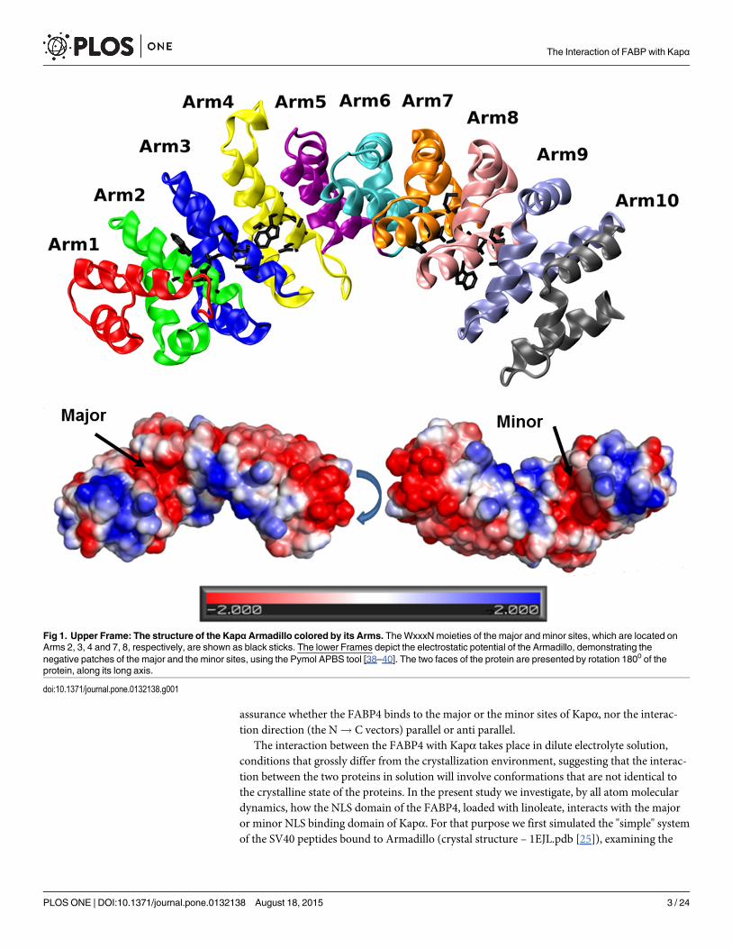
Usually, the residues forming the NLS recognition site are composed of three positiveamino acids. In the "classical signal" case, the residues are KK/RXK/R, typically appear as acontinuous section. However, some proteins are marked by a cryptic signal when a section of8–10 residues [6], is inserted in the middle of the NLS. Some members of the FABP familycarry an NLS sequence, which is also cryptic in nature [7–9]. In the case of FABP4, it consistsof one residue K21 located near the C terminal side of helix 1, while the other two moieties(R30 and K31) are near the N terminal section of helix 2, with a short unstructured loopbetween them (as well as K24, R33, K34 in FABP5)[10]. The proteins that recognize the NLSand offer a transport mechanism into the nucleus are members of the Karyopherin system(Kapα and Kapβ) [3, 11–13]. Kapα is a long protein made of an N terminal section termed IBB(Importin β Binding domain) and a set of 10 repeating structures, each consisting of 3 α-helices(H1, H2, H3),[4], that are assembled in a banana-shaped structure termed the "Armadillo". Onthe Armadillo there are two sites that can identify an NLS sequence termed the major and theminor sites, each having a specific sequence WxxxN that is involved in the binding of the NLS.The major site is located on Arms 2, 3 and 4 interacts with the three moieties of the NLS (seeFig 1). The minor site is located on Arms 7 and 8, and interacts with only two residues of theNLS, as demonstrated in Fig 1. Generally, the major site has a higher affinity to form complexes[3, 4], yet there are some proteins that prefer the minor site [11]. Kapα proteins bind theircargo via an NLS signal and react through their IBB domain with Kapβ. The whole complex iscarried into the nucleus through the nucleoporins.
The IBB has another function; due to an NLS sequence located on the IBB, it can cover themajor site of the Armadillo. In this folded conformation, a "self-inhibited" state, Kapα is notrecognized by Kapβ and cannot be transported into the nucleus. This feature is instrumental inidentifying a putative functional FABP4-Kapα complex; its structure should prevent the forma-tion of the self-inhibited shape.
The FABP4-Kapα import system is very selective. The complex is formed only with FABP4loaded with a certain substrate molecule. Thus, FABP4 will be carried into the nucleus whenloaded with linoleate but not if the ligand is palmitate. This selectivity is attributed to the shapeof the FABP4 dimer; in the case of the non-transportable compounds, the contact between twoFABP4 molecules in the dimer is at the lid zone, a conformation that masks the NLS domain ofthe FABP. FABP molecules that are loaded with transportable ligand form the dimer by joiningthe other pole of the molecule, leaving the lid fully exposed and available for reacting with theArmadillo [5]. Once the loaded FABP4 molecule is located in the nucleus, it releases its cargothat reacts with the PPAR (Peroxisome Proliferator Activator receptor) RXR (Retinoic XReceptor) system, which is a cardinal gene expression machinery that controls the carbohy-drate-lipid metabolic balance [14–18]. Considering that the affinity of FABP to a fatty acid is inthe sub-micromolar range [19–21], the mechanism of the fatty acid release inside the nucleusis also an intriguing problem.
The complexes of the NLS peptides with Kapα were investigated through crystallization ofthe KapαΔIBB (only the Armadillo) with short peptides, bearing the NLS sequence. In the com-plex of Armadillo with the SV40 large T antigen (PKKKRKV), two peptides are bound at themajor site (the positive residues of the peptide are termed P1 to P5 [22]) and at the minor site(termed P1’ to P4’, starting from the second lysine [22]) (1EJL.pdb). The peptide molecules ateither site have no secondary structure and their interactions with the Armadillo are boththrough their backbone atoms and side-chain moieties.
The identification of the NLS residues on FABP4 [8–10, 23, 24] was confirmed by thereplacement of all the three moieties by alanine. This gross replacement experiment cannotexclude the possibility that the FABP4 may interact with only two residues with the minor siteof Kapα, as in the case of the human phospholipid scramblase [11]. Consequently, we have no
The Interaction of FABP with Kapα
PLOSONE | DOI:10.1371/journal.pone.0132138 August 18, 2015 2 / 24
assurance whether the FABP4 binds to the major or the minor sites of Kapα, nor the interac-tion direction (the N! C vectors) parallel or anti parallel.
The interaction between the FABP4 with Kapα takes place in dilute electrolyte solution,conditions that grossly differ from the crystallization environment, suggesting that the interac-tion between the two proteins in solution will involve conformations that are not identical tothe crystalline state of the proteins. In the present study we investigate, by all atom moleculardynamics, how the NLS domain of the FABP4, loaded with linoleate, interacts with the majoror minor NLS binding domain of Kapα. For that purpose we first simulated the "simple" systemof the SV40 peptides bound to Armadillo (crystal structure – 1EJL.pdb [25]), examining the
Fig 1. Upper Frame: The structure of the KapαArmadillo colored by its Arms. TheWxxxNmoieties of the major and minor sites, which are located onArms 2, 3, 4 and 7, 8, respectively, are shown as black sticks. The lower Frames depict the electrostatic potential of the Armadillo, demonstrating thenegative patches of the major and the minor sites, using the Pymol APBS tool [38–40]. The two faces of the protein are presented by rotation 1800 of theprotein, along its long axis.
doi:10.1371/journal.pone.0132138.g001
The Interaction of FABP with Kapα
PLOSONE | DOI:10.1371/journal.pone.0132138 August 18, 2015 3 / 24

rigidity of the peptide's structure and the contribution of each of the residues to the stability ofthe complex.
The results indicate that even when the simulations were initiated from a very stable crystal-line structure, within a short time the peptides in both sites deviated from their original confor-mations. Moreover, even that the overall intensity of the interactions (Lennard-Jones andelectrostatic) between the two reactants was rather stable with time, the relative contribution ofeach of the 7 residues of the peptides alternated with time. The recognition that even the mini-mal system has more than one stable form suggests that the FABP-Armadillo complex mayalso be stabilized by more than a single set of contacts. The strategy selected to study the FAB-P4-Armadillo complex was based on the biological evidence that established the role of theNLS moieties of FABP4 as the recognition elements that interact with the Armadillo [8–10, 23,24], and looked also for interactions between residues that are not bona fidemembers of theNLS or the WxxxN sequences. Based on these observations, we reconstructed a possible modeof interaction between the FABP4 with the Armadillo, testing both major and minor sites inparallel and anti-parallel directions. The results presented below imply that the most likelylocation for the FABP4 to interact with Kapα is at the minor site of the Armadillo, where the N! C vectors are parallel. In this conformation, we observed many non-NLS moieties thatmaintain stable contacts with the residues on both major and minor sites, preventing the IBBfrom folding over, and rendering the complex unrecognizable by Kapβ.
MethodsThe simulations of the Armadillo complex with NLS of the SV40 large T antigen (PKKKRKV)were based on the crystal structure from the Protein Data Bank (pdb code 1EJL [25]).
For the complex of the Armadillo with the FABP4 loaded with linoleate, we replaced theshort NLS peptide, using the crystal structure of the FABP4 loaded with linoleate (2Q9S.pdb[5]). We set the positive moieties (K21, R30, K31) near Arms 2, 3 and 4 (for interaction withthe major site) and near Arms 7 and 8 (for interaction with the minor site) of the Armadillo.Care was taken to avoid clashes between the two proteins. The procedure was repeated twice,where the N! C vectors of the two proteins were in parallel and anti-parallel orientations.Following this initial setting, the systems were relaxed and simulated as described below.
Molecular Dynamics and Simulation ProgramsStandard MD simulations were carried out using of the GROMACS 4.0.7 package [26] with theGROMOS96 force field, with a 53a6 parameter set [27].
The parameters of the linoleate molecules (bond length, angles and dihedrals, improper andpartial charges) were taken from the GROMACS standard building blocks for glutamate. Theparameters of the double bond are GROMACS parameters for retinol as present in RTOL. Thenet charge of the ligands was -1 electron charge.
Prior to the MD step of each simulation, a dodecahedron box was built, with dimensionsextended at least 12 Å from the nearest molecule, and was filled with water using the SPC216model. Charge neutralization, while maintaining an ionic strength of approximately 100 mM,was obtained by adding Na+ and Cl- ions. During the MD simulations, the LINCS algorithm[28] was used to constrain the lengths of all bonds; the water molecules were restrained withthe use of the SETTLE algorithm [29]. All simulations were carried out under NPT conditionsof constant number of moles, pressure and temperature, using the v-rescale coupling algorithmfor keeping the temperature constant and Berendsen’s coupling algorithm for keeping the pres-sure constant (P = 1 bar; τp = 0.5 ps; T = 300 K; τT = 0.1 ps) [30]. A 12 Å cutoff was used forthe Van der Waals (VdW) interactions. The long-range electrostatic interactions were treated
The Interaction of FABP with Kapα
PLOSONE | DOI:10.1371/journal.pone.0132138 August 18, 2015 4 / 24
by the Particle mesh Ewald (PME) [31]. Each system was relaxed by energy minimizationusing the steepest descent and the Conjugated Gradient methods (as embedded in the GRO-MACS package). The resulting structures then underwent 40 ps of simulation with the pro-tein's position restrained, allowing the solvent (water) molecules to reach equilibrium. Thesystem was simulated further for 0.2 ns of unconstrained equilibration simulation. After theequilibration it was possible to start generating the MD simulations with a 2 fs time-step, bywhich Newton's equations of motion were solved. Every simulation had a different initialvelocity for each atom that was randomly generated from the Maxwell-Boltzmann distributionat 300 K. Snapshots were saved at 2 ps intervals. Root Mean Square Deviation (RMSD) andRoot Mean Squared Fluctuation (RMSF) calculations were made for the backbone atoms.
Docking of FABP4 on the ArmadilloThe Armadillo (1EJL.pdb) and FABP (2Q9S.pdb) were docked using the PatchDock server[32–34]. The structures with the highest score were used for the simulations. The strategy fordocking was set to fulfill the following requirements: at least two residues (K21, K30, R31) ofthe FABP4 must be docked with the Armadillo's WxxxN stretches (142–146, 184–188, 231–235 for the major site), or 357–361, 399–403 (for the minor site), while these residues shouldbe within 5 Ǻ from the tryptophan moieties of these stretches. At this range the supposedlyinteracting residues are close enough to establish electrostatic interactions, yet are separated byat least one water molecule, which ensures structural flexibility as the two proteins interact.
Structural and Energetic AnalysisThe first step of this analysis was the generation of a full dataset of minimum distances betweeneach pair of residues of the two structures, by the command, “g_mindist” of the GROMACS4.0 package. The set was filtered to remove all pairs whose geometric mean value, calculatedover a given time interval (few tens of nanoseconds) exceeded a cutoff of 4 Å.
Cluster analysis was performed for all the simulations by the command, “g_cluster” of theGROMACS 4.0 package. The analysis was performed using the Gromos algorithm with a cutoffvalue set to generate a sufficient number of clusters while maintaining statistical significance[35].
For calculating the interacting energies of the proteins or residues-proteins, "g_energy" com-mand of the GROMACS package was applied. The Constrain Network Analysis (CNA) wasused for the calculation of the Flexibility index parameters. The data was executed on the CNAweb-server [36, 37].The electrostatic potential mapping of the protein's surface was calculatedby the PYMOL–APBS plugin [38–40].
Hot Spots analysis was carried out using the KFC Server [41].Principle Component Analysis (PCA) [42] was carried out by using the "g_covar" command
of the GROMACS package. For extracting the Eigen vectors and analysis, we applied the"g_anaeig" command of the GROMACS package.
The analysis of Hydrogen bonds was performed using the web server of Tina and coworkers[43].
Results
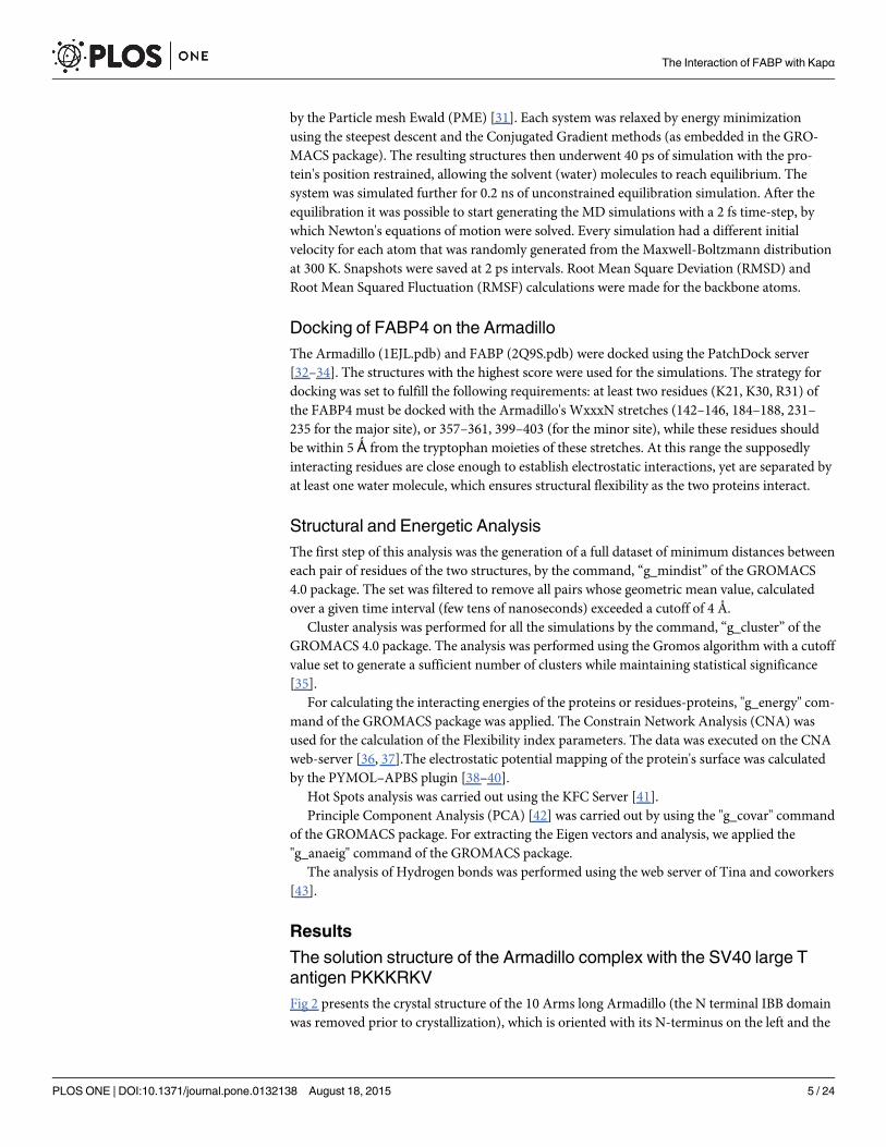
The solution structure of the Armadillo complex with the SV40 large Tantigen PKKKRKVFig 2 presents the crystal structure of the 10 Arms long Armadillo (the N terminal IBB domainwas removed prior to crystallization), which is oriented with its N-terminus on the left and the
The Interaction of FABP with Kapα
PLOSONE | DOI:10.1371/journal.pone.0132138 August 18, 2015 5 / 24
two NLS peptides (PKKKRKV). The peptide on the left interacts with the major site of theArmadillo and the peptide on the right is bound to the minor site. The N! C terminal vectorof the peptides is anti-parallel to that of the Armadillo. This complex was simulated three timesfor ~ 50 ns. For comparison, we also simulated twice the Armadillo in the absence of the NLSpeptides. Examination of the RMSD and RMSF traces indicates that the binding of the peptideshad a minor effect on the stability of the Armadillo, mostly decreasing the RMSF of the resi-dues with which the peptides interact. However, the conformation of the hepta-peptide isaffected by its interactions with the Armadillo during the simulation time. Under the stressapplied by the crystallization setup, only one conformation is prevailing, but once the complexis placed in less restrictive medium, the hepta-peptide is fluctuating between seemingly equipo-tential conformations.
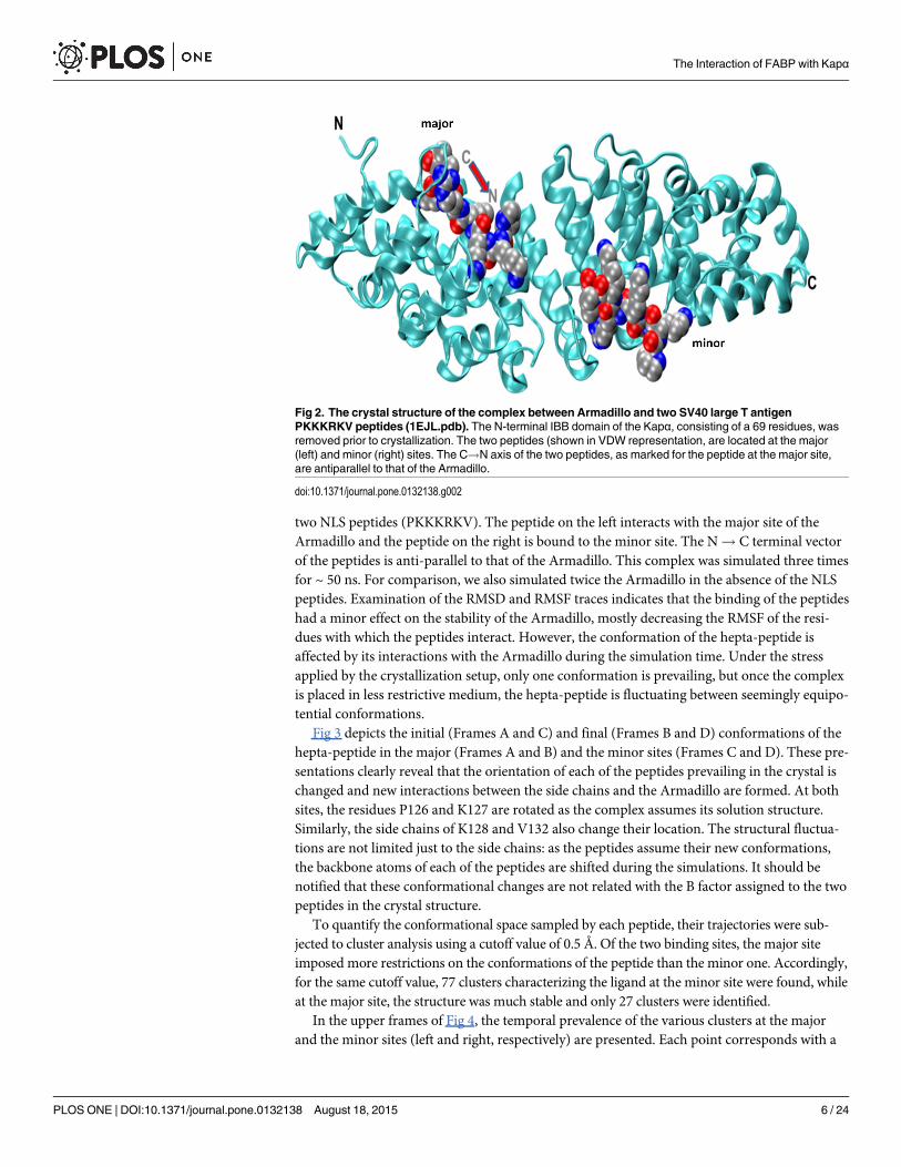
Fig 3 depicts the initial (Frames A and C) and final (Frames B and D) conformations of thehepta-peptide in the major (Frames A and B) and the minor sites (Frames C and D). These pre-sentations clearly reveal that the orientation of each of the peptides prevailing in the crystal ischanged and new interactions between the side chains and the Armadillo are formed. At bothsites, the residues P126 and K127 are rotated as the complex assumes its solution structure.Similarly, the side chains of K128 and V132 also change their location. The structural fluctua-tions are not limited just to the side chains: as the peptides assume their new conformations,the backbone atoms of each of the peptides are shifted during the simulations. It should benotified that these conformational changes are not related with the B factor assigned to the twopeptides in the crystal structure.
To quantify the conformational space sampled by each peptide, their trajectories were sub-jected to cluster analysis using a cutoff value of 0.5 Å. Of the two binding sites, the major siteimposed more restrictions on the conformations of the peptide than the minor one. Accordingly,for the same cutoff value, 77 clusters characterizing the ligand at the minor site were found, whileat the major site, the structure was much stable and only 27 clusters were identified.
In the upper frames of Fig 4, the temporal prevalence of the various clusters at the majorand the minor sites (left and right, respectively) are presented. Each point corresponds with a
Fig 2. The crystal structure of the complex between Armadillo and two SV40 large T antigenPKKKRKV peptides (1EJL.pdb). The N-terminal IBB domain of the Kapα, consisting of a 69 residues, wasremoved prior to crystallization. The two peptides (shown in VDW representation, are located at the major(left) and minor (right) sites. The C!N axis of the two peptides, as marked for the peptide at the major site,are antiparallel to that of the Armadillo.
doi:10.1371/journal.pone.0132138.g002
The Interaction of FABP with Kapα
PLOSONE | DOI:10.1371/journal.pone.0132138 August 18, 2015 6 / 24

The Interaction of FABP with Kapα
PLOSONE | DOI:10.1371/journal.pone.0132138 August 18, 2015 7 / 24
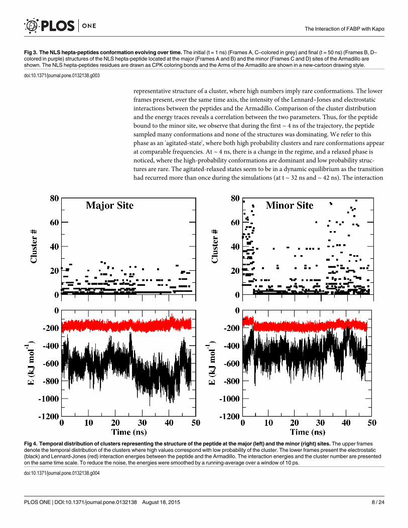
representative structure of a cluster, where high numbers imply rare conformations. The lowerframes present, over the same time axis, the intensity of the Lennard–Jones and electrostaticinteractions between the peptides and the Armadillo. Comparison of the cluster distributionand the energy traces reveals a correlation between the two parameters. Thus, for the peptidebound to the minor site, we observe that during the first ~ 4 ns of the trajectory, the peptidesampled many conformations and none of the structures was dominating. We refer to thisphase as an 'agitated-state', where both high probability clusters and rare conformations appearat comparable frequencies. At ~ 4 ns, there is a change in the regime, and a relaxed phase isnoticed, where the high-probability conformations are dominant and low probability struc-tures are rare. The agitated-relaxed states seem to be in a dynamic equilibrium as the transitionhad recurred more than once during the simulations (at t ~ 32 ns and ~ 42 ns). The interaction
Fig 3. The NLS hepta-peptides conformation evolving over time. The initial (t = 1 ns) (Frames A, C–colored in grey) and final (t = 50 ns) (Frames B, D–colored in purple) structures of the NLS hepta-peptide located at the major (Frames A and B) and the minor (Frames C and D) sites of the Armadillo areshown. The NLS hepta-peptides residues are drawn as CPK coloring bonds and the Arms of the Armadillo are shown in a new-cartoon drawing style.
doi:10.1371/journal.pone.0132138.g003
Fig 4. Temporal distribution of clusters representing the structure of the peptide at the major (left) and the minor (right) sites. The upper framesdenote the temporal distribution of the clusters where high values correspond with low probability of the cluster. The lower frames present the electrostatic(black) and Lennard-Jones (red) interaction energies between the peptide and the Armadillo. The interaction energies and the cluster number are presentedon the same time scale. To reduce the noise, the energies were smoothed by a running-average over a window of 10 ps.
doi:10.1371/journal.pone.0132138.g004
The Interaction of FABP with Kapα
PLOSONE | DOI:10.1371/journal.pone.0132138 August 18, 2015 8 / 24

of the peptides with the major site is more intensive in terms of energy (notice the differencebetween the major and minor sites in the lower frames) and the total number of clusters (usingthe same cutoff value) was significantly smaller (27 vs. 77). The same pattern of alternatingbetween relaxed and agitated phases is also common for the peptide attached to the major site;the peptide assumes its relaxed state between ~27 and ~41 ns of the simulation.
The variation in the intensity of the peptide-Armadillo interactions and the rapid shiftbetween the conformations suggest that the charged moieties of the peptide fluctuate betweenmore than one location. This feature was investigated by calculating the interacting energies ofeach one of the residues of the peptide with the Armadillo. The electrostatic interactionbetween the various residues of the peptide and the Armadillo can be as high as ~ -150 ± 20 kJ/mol per-residue (Fig 5). Yet, the interactions are not constant in time; at the major site (Fig 5,frame A) all of the residues contribute to the binding, but at each time point the identity of thestabilizing moiety is different. Generally, the interactions at p1, p3 and p4 are rather weak,while p2 and p5 are well binding, yet the intensity is subjected to large fluctuations. Surpris-ingly, both proline and valine, which are not a part of the conserved NLS sequence, contributedsubstantially to the stability. Two of the charged residues, K127 and R130, hardly contribute tothe stability and the intensity of the electrostatic component of their contribution is not muchlarger than that of the Lennard-Jones interaction. The other charged moieties participate inintensive interactions, but at certain time frames the positive moieties exchange the intensity ofthe electrostatic interactions with the Armadillo by exposure to the solvent without reducingthe total interactions with the Armadillo.
At the minor site (Fig 5, frame B) we also observe temporal fluctuations in the intensity ofthe electrostatic interactions. The interactions of the Armadillo with K128, p1’, p2’ and p4’ areintense and fluctuate with time, alternating from ~ -10 kJ/mol (comparable with the level ofLennard-Jones attraction) to ~ -200 kJ/mol. The interaction with p3' is stable but less intensive.
The fluctuations in the interaction energies, as calculated for the specific residues of the peptidesreflect the motion of the charged moieties with respect to the Armadillo. To evaluate the extent ofthe motion of the backbone atoms we calculated the (geometric) average distance between eachamino acid of the hepta-peptide and the residues of the Armadillo. Figs 6 and 7 represent thosemoieties where the average distance was less than 4 Å (marked in red) or closer, almost in touch,not to let a water molecule squeeze in (colored in blue). The analysis of the complex at the majorsite (Fig 6, frame A) implies that the moieties comprising the conserved NLS sequence and otherresidues maintain a close contact only with the Arms having theWxxxN repeats (Arms 2, 3 and4). When attached to the minor site (Fig 6, frame B), the peptide experienced higher freedom andinteracted with Arms 5 and 6 that do not carry the NLS recognizable domain in addition to Arms7 and 8. Namely, at the major site, the interactions are only with the Arms that carry theWxxxNrepeats, while at the minor site the NLS peptide is also stabilized by other Arms.
Based on these observations, the hepta-peptides-Armadillo complex seems to provide a"poor blueprint" for structuring the FABP4-Kapα complex. The structure of the hepta-peptidescomplex is constantly wiggling, the intensity of the interactions between the charged moietiesof the NLS with the Armadillo fluctuate over time. Furthermore, residues that are not a part ofthe consensus sequence can make, in some time intervals, major contribution to the stability ofthe complex. Apparently, the freedom of motion while monitoring in aqueous solution at non-cryogenic temperature is much higher than what appears in the crystalline structure.
The interaction of FABP4 with ArmadilloProtein-protein interaction differs from peptide-protein interaction by a lower density ofhydrogen bonds. Upon binding, the short peptides lose their initial structure and expose their
The Interaction of FABP with Kapα
PLOSONE | DOI:10.1371/journal.pone.0132138 August 18, 2015 9 / 24

The Interaction of FABP with Kapα
PLOSONE | DOI:10.1371/journal.pone.0132138 August 18, 2015 10 / 24

backbone atoms for hydrogen bonding with the protein [44]. Proteins, on the other hand,retain their internal structure and fewer hydrogen bonds can be made. Thus, the interaction ofthe FABP4 with the Armadillo is not a simple replication of the NLS peptide-Armadillo com-plex. To select which of the putative structures of the FABP4-Armadillo complex representsthe transportable complex, we based our analysis on the conclusions resulting from the simula-tions of the Armadillo-SV40 NLS peptide: i. The contact between the two proteins can be asflexible as was noticed with the peptide. ii. The interactions may involve residues that are notbona fidemembers of the NLS sequence of the FABP4 (K21, K30 and R31) [8, 24] and theinteracting Arms might include those not bearing the WxxxN sequence.
At present, as there is no direct evidence to which site on the Armadillo the FABP4 isbound, thus we had to consider both major and minor sites for their ability to bind the FABP4.The major site is generally considered to be the more attractive one [3, 4]. Yet it is known thatin the case of the human phospholipid scramblase [11], the NLS sequence is made of only twobasic residues and its binding site is the minor one. Considering the fact that one residue of theNLS sequence is separated from the others by a non-structured loop (K21, R30, K31 in FABP4or K24, R33, K34 in FABP5 [10]), we could not exclude the possibility that actually only tworesidues participate in the binding and their preferred locus will be the minor site. What is
Fig 5. The interaction energies between each residue of the NLS peptide and the Armadillo. (A) depicts the interactions with the major site. (B) depictsthe interactions with the minor site. The residues' identity is marked in the figure.
doi:10.1371/journal.pone.0132138.g005
Fig 6. Identification of the stable residue pairs holding the NLS peptide at the major site. The last 30ns of the trajectory were analyzed in a search forpairs that maintained close contact with each other. The residues of the Armadillo and the NLS are grouped according to the Arm number. Residues locatedon the WxxxN repeat are marked in orange. Residues less than 4 Å or 2.8 Å apart are colored in red and blue, respectively. (A) depicts the interactions withthe major site. (B) depicts the interactions with the minor site.
doi:10.1371/journal.pone.0132138.g006
The Interaction of FABP with Kapα
PLOSONE | DOI:10.1371/journal.pone.0132138 August 18, 2015 11 / 24

more, there is no assurance that on forming a complex, the two proteins orient their N! Cvectors in the anti-parallel mode as do the short NLS peptides. Consequently, both sites andtwo orientations of the vectors should be investigated to evaluate how the FABP4 protein inter-acts with the Kapα.
Four initial conformations were generated by the PatchDock server [23–25]. The setting ofthe proteins was such that at least two positive residues of the NLS of FABP4 were located closeto the WxxxN bearing Arms (2, 3 and 4 for the major site) or Arms 7 and 8 (for the minorsite), at a distance of ~ 5 Ǻ from the WxxxN moiety. We reasoned that such a separation dis-tance can accommodate water molecules in-between the proteins, which ensures structuralflexibility before tight contact is formed. Yet, at this proximity, the long-range non-bondedinteractions between the proteins can still attract them one to the other and to form a tightcontact.
The Docking process generated conformations where the N! C vectors of the two proteinsare either in the parallel or the anti-parallel orientations. The highest scoring conformation ofeach orientation was selected as a starting structure for un-biased all atom molecular dynamics
Fig 7. The RMSD of the Armadillo bound to FABP and the NLS-peptide in different orientation and location (major andminor site). (A) FABP4 boundin the major site parallel to the Armadillo (2 simulations—left and right); (B) FABP4 bound in the major site anti-parallel to the Armadillo (2 simulations); (C)FABP bound in the minor site parallel to the Armadillo (2 simulations); (D) FABP bound in the minor site anti-parallel to the Armadillo (2 simulations); (E) NLSpeptide bound in both sites (major and minor) to the Armadillo; (F)–free Armadillo.
doi:10.1371/journal.pone.0132138.g007
The Interaction of FABP with Kapα
PLOSONE | DOI:10.1371/journal.pone.0132138 August 18, 2015 12 / 24

simulations, allowing the two proteins to adjust to the presence of each other. Each structurewas simulated twice for 50 to 96 ns (Table 1).
Interestingly, some of the lower-scored structures generated by the PatchDock programwere sampled during the early phases of the MD simulations.
Evaluation of the Stability and the Orientation of the Complexes. Fig 7 depicts theRMSD traces for the four simulated orientations of the FABP4 Armadillo complexes. Thesetraces (A-D) are higher than the RMSD calculated for the SV40 NLS-Armadillo complex(Frame E) or the Armadillo without the peptides (Frame F). Thus, the FABP4 seems to lowerthe stability of the Armadillo.
For a large protein made of repeating units (Arms) the variance of RMSD is less informativethan examination of the RMSF of the specific residues (Fig 8). Frame A depicts the RMSF ofthe free Armadillo (blue trace), its complex with two SV40 NLS peptides (red trace) and thecomplex with parallel FABP4 at the major site (green trace). The most stable structure appearsto be the Armadillo-NLS peptide complex. In this complex the RMSF values calculated at themajor site are lower than those calculated for the free Armadillo. The complex with the FABP4at the major site (green trace) shows similar behavior to that of the Armadillo-peptide complex,but this effect is limited only to the Arms forming the major site. From Arm 6 and on, theRMSF of the Armadillo with the FABP4 at the major site is significantly higher than the levelset by the free Armadillo or its NLS complex. Frame B in Fig 8 (the same color code as inFrame A) depicts the RMSF calculated for the Armadillo with the parallel FABP4 docked tothe minor site. When bound to the minor site, the RMSF trace (green) of the Armadillo exhib-its stabilization both at the minor and the major sites, suggesting that the contact of the FABP4with the Armadillo is not limited just to the minor binding site.
For the FABP4 complex with the Armadillo in the anti-parallel orientations, the RMSF val-ues were much higher (data not shown).
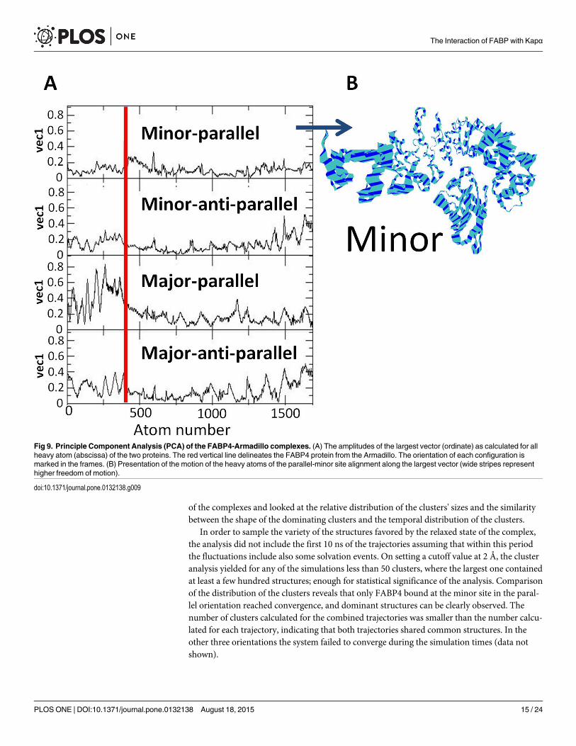
The trajectories of the four initial orientations were subjected to Principal Component anal-ysis [42]; looking for the concerted motions that characterize the putative complexes. Fig 9,frame A depicts the amplitudes of the largest vector as calculated for the heavy atoms of theFABP4 and the Armadillo (drown sequentially on the abscissa). The upper panel, correspond-ing with the parallel-minor site orientation has the smallest amplitudes for both the FABP4and the Armadillo. Frame B in Fig 9 presents the motion of the two proteins along the largestvector, as calculated for the parallel-minor configuration. This figure demonstrates how theinteractions between the two proteins stabilize the contact region between them, while residuesthat are remote from the junction have higher freedom of motion. Comparison of the first vec-tor of the four orientations reveals that the parallel alignment of the FABP4 next to the minorsite generated the most stable complex and a visual projection of the first vector motion of thewhole complex indicates that this configuration has the smallest structural fluctuation. Thus,the PCA calculations corroborate the conclusion that the most probable interaction betweenFABP4 and the Armadillo is the parallel docking at the minor site.
Table 1. The simulations setup used for testing the interactions between the Armadillo with FABP loaded with linoleate. The length of the simula-tions is in units of nanoseconds. The directions of the N terminal!C terminal vectors of FABP4/Armadillo were either parallel or anti-parallel.
simulation Major Minor
parallel anti-parallel parallel anti-parallel
1 50 50 96 60
2 60 50 60 60
doi:10.1371/journal.pone.0132138.t001
The Interaction of FABP with Kapα
PLOSONE | DOI:10.1371/journal.pone.0132138 August 18, 2015 13 / 24
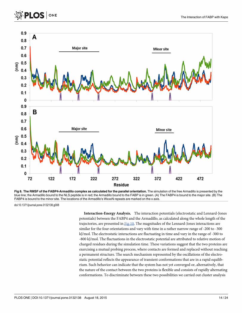
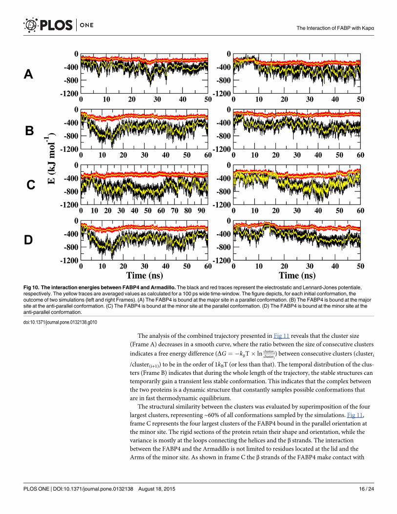
Interaction-Energy Analysis. The interaction potentials (electrostatic and Lennard-Jonespotentials) between the FABP4 and the Armadillo, as calculated along the whole length of thetrajectories, are presented in Fig 10. The magnitudes of the Lennard-Jones interactions aresimilar for the four orientations and vary with time in a rather narrow range of -200 to -300kJ/mol. The electrostatic interactions are fluctuating in time and vary in the range of -300 to-800 kJ/mol. The fluctuations in the electrostatic potential are attributed to relative motion ofcharged residues during the simulation time. These variations suggest that the two proteins areexercising a mutual probing process, where contacts are formed and replaced without reachinga permanent structure. The search mechanism represented by the oscillations of the electro-static potential reflects the appearance of transient conformations that are in a rapid equilib-rium. Such behavior can indicate that the system has not yet converged or, alternatively, thatthe nature of the contact between the two proteins is flexible and consists of rapidly alternatingconformations. To discriminate between these two possibilities we carried out cluster analysis
Fig 8. The RMSF of the FABP4-Armadillo complex as calculated for the parallel orientation. The simulation of the free Armadillo is presented by theblue line; the Armadillo bound to the NLS peptide is in red; the Armadillo bound to the FABP is in green. (A) The FABP4 is bound to the major site. (B) TheFABP4 is bound to the minor site. The locations of the Armadillo's WxxxN repeats are marked on the x axis.
doi:10.1371/journal.pone.0132138.g008
The Interaction of FABP with Kapα
PLOSONE | DOI:10.1371/journal.pone.0132138 August 18, 2015 14 / 24
of the complexes and looked at the relative distribution of the clusters' sizes and the similaritybetween the shape of the dominating clusters and the temporal distribution of the clusters.
In order to sample the variety of the structures favored by the relaxed state of the complex,the analysis did not include the first 10 ns of the trajectories assuming that within this periodthe fluctuations include also some solvation events. On setting a cutoff value at 2 Å, the clusteranalysis yielded for any of the simulations less than 50 clusters, where the largest one containedat least a few hundred structures; enough for statistical significance of the analysis. Comparisonof the distribution of the clusters reveals that only FABP4 bound at the minor site in the paral-lel orientation reached convergence, and dominant structures can be clearly observed. Thenumber of clusters calculated for the combined trajectories was smaller than the number calcu-lated for each trajectory, indicating that both trajectories shared common structures. In theother three orientations the system failed to converge during the simulation times (data notshown).
Fig 9. Principle Component Analysis (PCA) of the FABP4-Armadillo complexes. (A) The amplitudes of the largest vector (ordinate) as calculated for allheavy atom (abscissa) of the two proteins. The red vertical line delineates the FABP4 protein from the Armadillo. The orientation of each configuration ismarked in the frames. (B) Presentation of the motion of the heavy atoms of the parallel-minor site alignment along the largest vector (wide stripes representhigher freedom of motion).
doi:10.1371/journal.pone.0132138.g009
The Interaction of FABP with Kapα
PLOSONE | DOI:10.1371/journal.pone.0132138 August 18, 2015 15 / 24
The analysis of the combined trajectory presented in Fig 11 reveals that the cluster size(Frame A) decreases in a smooth curve, where the ratio between the size of consecutive clusters
indicates a free energy difference (DG ¼ �kBT� ln clustericlusterj
) between consecutive clusters (clusteri
/cluster(i+1)) to be in the order of 1kBT (or less than that). The temporal distribution of the clus-ters (Frame B) indicates that during the whole length of the trajectory, the stable structures cantemporarily gain a transient less stable conformation. This indicates that the complex betweenthe two proteins is a dynamic structure that constantly samples possible conformations thatare in fast thermodynamic equilibrium.
The structural similarity between the clusters was evaluated by superimposition of the fourlargest clusters, representing ~60% of all conformations sampled by the simulations. Fig 11,frame C represents the four largest clusters of the FABP4 bound in the parallel orientation atthe minor site. The rigid sections of the protein retain their shape and orientation, while thevariance is mostly at the loops connecting the helices and the β strands. The interactionbetween the FABP4 and the Armadillo is not limited to residues located at the lid and theArms of the minor site. As shown in frame C the β strands of the FABP4 make contact with
Fig 10. The interaction energies between FABP4 and Armadillo. The black and red traces represent the electrostatic and Lennard-Jones potentiale,respectively. The yellow traces are averaged values as calculated for a 100 ps wide time-window. The figure depicts, for each initial conformation, theoutcome of two simulations (left and right Frames). (A) The FABP4 is bound at the major site in a parallel conformation. (B) The FABP4 is bound at the majorsite at the anti-parallel conformation. (C) The FABP4 is bound at the minor site at the parallel conformation. (D) The FABP4 is bound at the minor site at theanti-parallel conformation.
doi:10.1371/journal.pone.0132138.g010
The Interaction of FABP with Kapα
PLOSONE | DOI:10.1371/journal.pone.0132138 August 18, 2015 16 / 24

Arms 1–5, where the major site is located. These interactions may explain how the binding atthe minor site enhances the stability of the major site (Fig 8 above).
Fig 11, frame D depicts the four largest clusters of the FABP4 attached in the parallel orien-tation at the major site. In this case, there are no interactions with the minor site, accountingfor the higher RMSF of the minor site in this complex (Fig 8 above).
Upon simulations of the FABP4 complex at the minor-parallel orientation, we noticed apeculiar behavior that was never observed before: the linoleate molecule was gradually migrat-ing from its original binding site inside the FABP4 molecule to a new location at the interfacebetween the two proteins. In this conformation, the hydrophobic section of the fatty acid isembedded in the crevice between Arms 8 and 7 of the Armadillo (Fig 12). This sharing ofligand between FABP4 and Armadillo was not noticed in any other trajectory of the FABP4-Armadillo complex. It should be mentioned that simulations of the FABP4 by itself, loaded
Fig 11. Cluster analysis of the combined trajectory of twominor parallel simulations of the FABP4-Armadillo complex. (A) depicts the sizedistribution of the clusters. (B) depicts the time distribution of the clusters. The data relates the cluster number with the time points along the trajectory. (C)presents superposition of the four largest clusters (each cluster has a different color) of the minor parallel simulation. Please note that even after the dominantstructure had converged there are brief appearances of low probability states. (D) depicts, for comparison, the four largest clusters (each cluster has adifferent color) of the major parallel simulation.
doi:10.1371/journal.pone.0132138.g011
The Interaction of FABP with Kapα
PLOSONE | DOI:10.1371/journal.pone.0132138 August 18, 2015 17 / 24
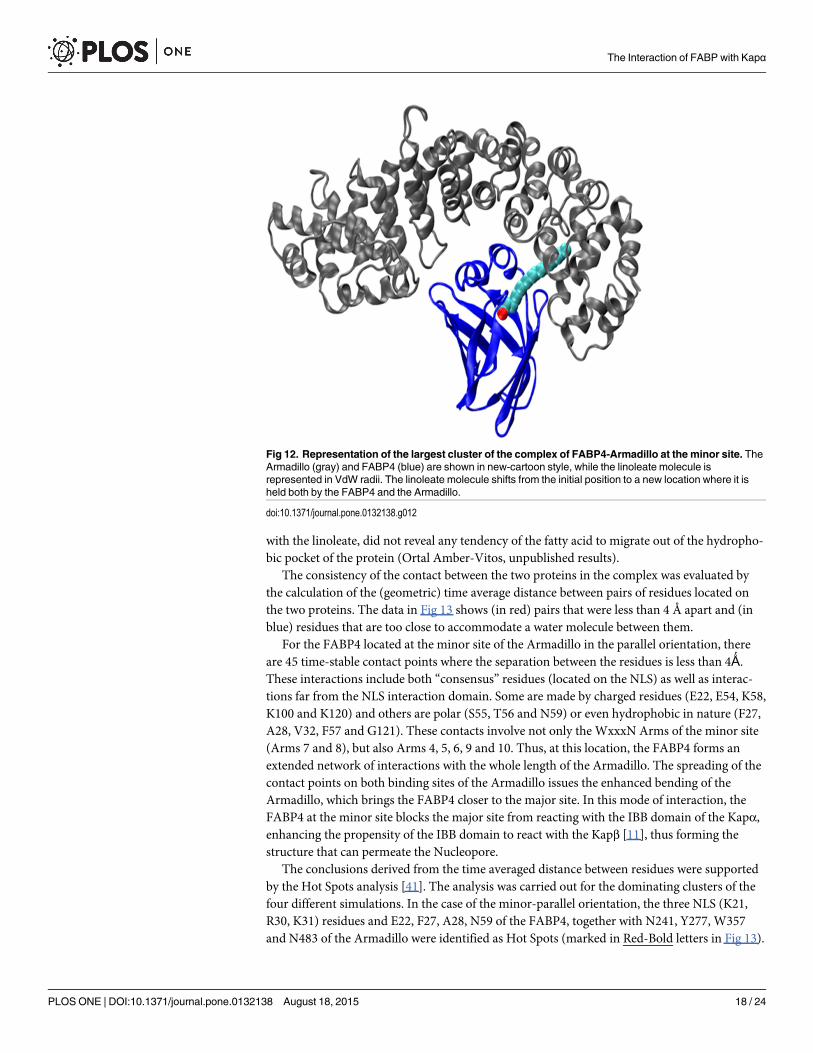
with the linoleate, did not reveal any tendency of the fatty acid to migrate out of the hydropho-bic pocket of the protein (Ortal Amber-Vitos, unpublished results).
The consistency of the contact between the two proteins in the complex was evaluated bythe calculation of the (geometric) time average distance between pairs of residues located onthe two proteins. The data in Fig 13 shows (in red) pairs that were less than 4 Å apart and (inblue) residues that are too close to accommodate a water molecule between them.
For the FABP4 located at the minor site of the Armadillo in the parallel orientation, thereare 45 time-stable contact points where the separation between the residues is less than 4Ǻ.These interactions include both “consensus” residues (located on the NLS) as well as interac-tions far from the NLS interaction domain. Some are made by charged residues (E22, E54, K58,K100 and K120) and others are polar (S55, T56 and N59) or even hydrophobic in nature (F27,A28, V32, F57 and G121). These contacts involve not only the WxxxN Arms of the minor site(Arms 7 and 8), but also Arms 4, 5, 6, 9 and 10. Thus, at this location, the FABP4 forms anextended network of interactions with the whole length of the Armadillo. The spreading of thecontact points on both binding sites of the Armadillo issues the enhanced bending of theArmadillo, which brings the FABP4 closer to the major site. In this mode of interaction, theFABP4 at the minor site blocks the major site from reacting with the IBB domain of the Kapα,enhancing the propensity of the IBB domain to react with the Kapβ [11], thus forming thestructure that can permeate the Nucleopore.
The conclusions derived from the time averaged distance between residues were supportedby the Hot Spots analysis [41]. The analysis was carried out for the dominating clusters of thefour different simulations. In the case of the minor-parallel orientation, the three NLS (K21,R30, K31) residues and E22, F27, A28, N59 of the FABP4, together with N241, Y277, W357and N483 of the Armadillo were identified as Hot Spots (marked in Red-Bold letters in Fig 13).
Fig 12. Representation of the largest cluster of the complex of FABP4-Armadillo at the minor site. TheArmadillo (gray) and FABP4 (blue) are shown in new-cartoon style, while the linoleate molecule isrepresented in VdW radii. The linoleate molecule shifts from the initial position to a new location where it isheld both by the FABP4 and the Armadillo.
doi:10.1371/journal.pone.0132138.g012
The Interaction of FABP with Kapα
PLOSONE | DOI:10.1371/journal.pone.0132138 August 18, 2015 18 / 24
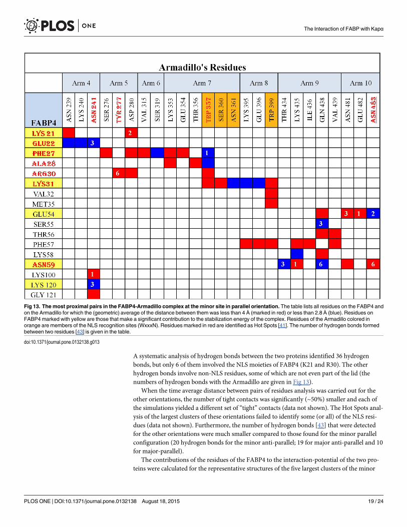
A systematic analysis of hydrogen bonds between the two proteins identified 36 hydrogenbonds, but only 6 of them involved the NLS moieties of FABP4 (K21 and R30). The otherhydrogen bonds involve non-NLS residues, some of which are not even part of the lid (thenumbers of hydrogen bonds with the Armadillo are given in Fig 13).
When the time average distance between pairs of residues analysis was carried out for theother orientations, the number of tight contacts was significantly (~50%) smaller and each ofthe simulations yielded a different set of “tight” contacts (data not shown). The Hot Spots anal-ysis of the largest clusters of these orientations failed to identify some (or all) of the NLS resi-dues (data not shown). Furthermore, the number of hydrogen bonds [43] that were detectedfor the other orientations were much smaller compared to those found for the minor parallelconfiguration (20 hydrogen bonds for the minor anti-parallel; 19 for major anti-parallel and 10for major-parallel).
The contributions of the residues of the FABP4 to the interaction-potential of the two pro-teins were calculated for the representative structures of the five largest clusters of the minor
Fig 13. The most proximal pairs in the FABP4-Armadillo complex at the minor site in parallel orientation. The table lists all residues on the FABP4 andon the Armadillo for which the (geometric) average of the distance between them was less than 4 Å (marked in red) or less than 2.8 Å (blue). Residues onFABP4marked with yellow are those that make a significant contribution to the stabilization energy of the complex. Residues of the Armadillo colored inorange are members of the NLS recognition sites (WxxxN). Residues marked in red are identified as Hot Spots [41]. The number of hydrogen bonds formedbetween two residues [43] is given in the table.
doi:10.1371/journal.pone.0132138.g013
The Interaction of FABP with Kapα
PLOSONE | DOI:10.1371/journal.pone.0132138 August 18, 2015 19 / 24
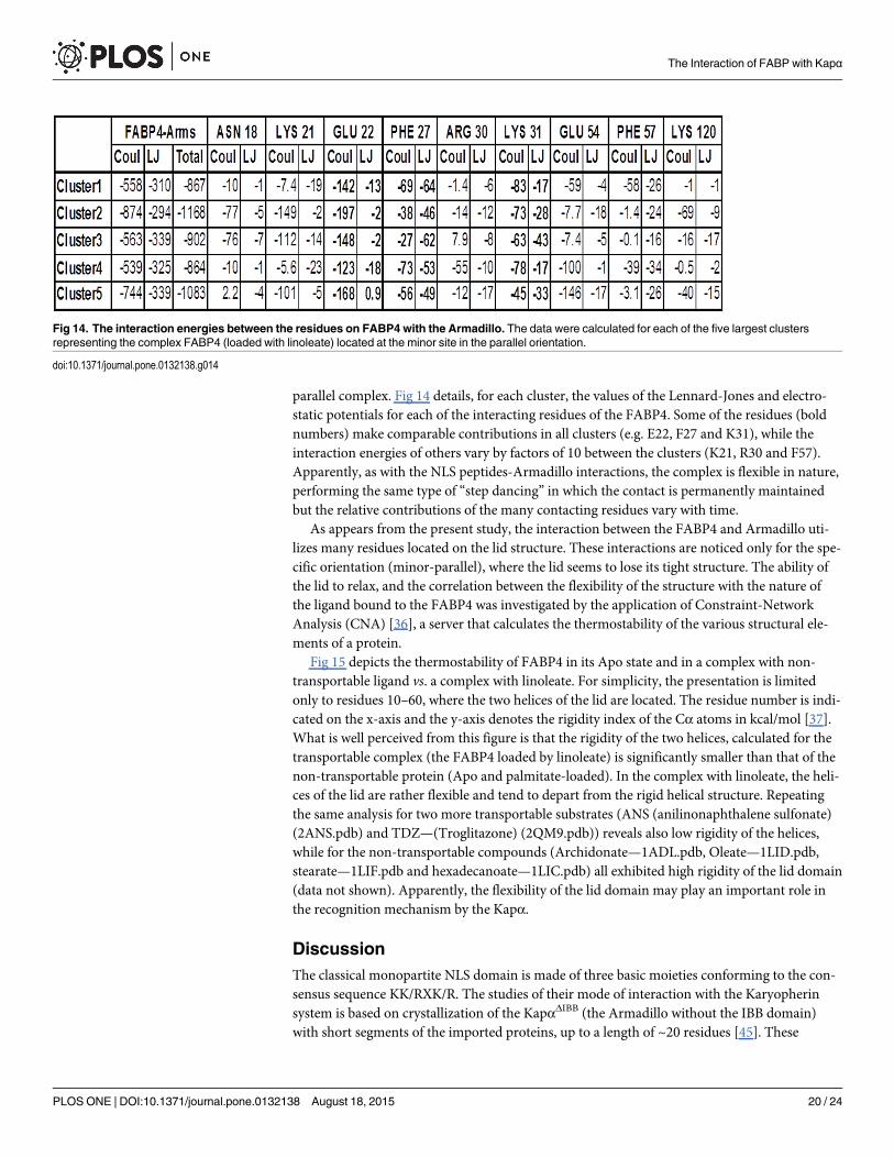
parallel complex. Fig 14 details, for each cluster, the values of the Lennard-Jones and electro-static potentials for each of the interacting residues of the FABP4. Some of the residues (boldnumbers) make comparable contributions in all clusters (e.g. E22, F27 and K31), while theinteraction energies of others vary by factors of 10 between the clusters (K21, R30 and F57).Apparently, as with the NLS peptides-Armadillo interactions, the complex is flexible in nature,performing the same type of “step dancing” in which the contact is permanently maintainedbut the relative contributions of the many contacting residues vary with time.
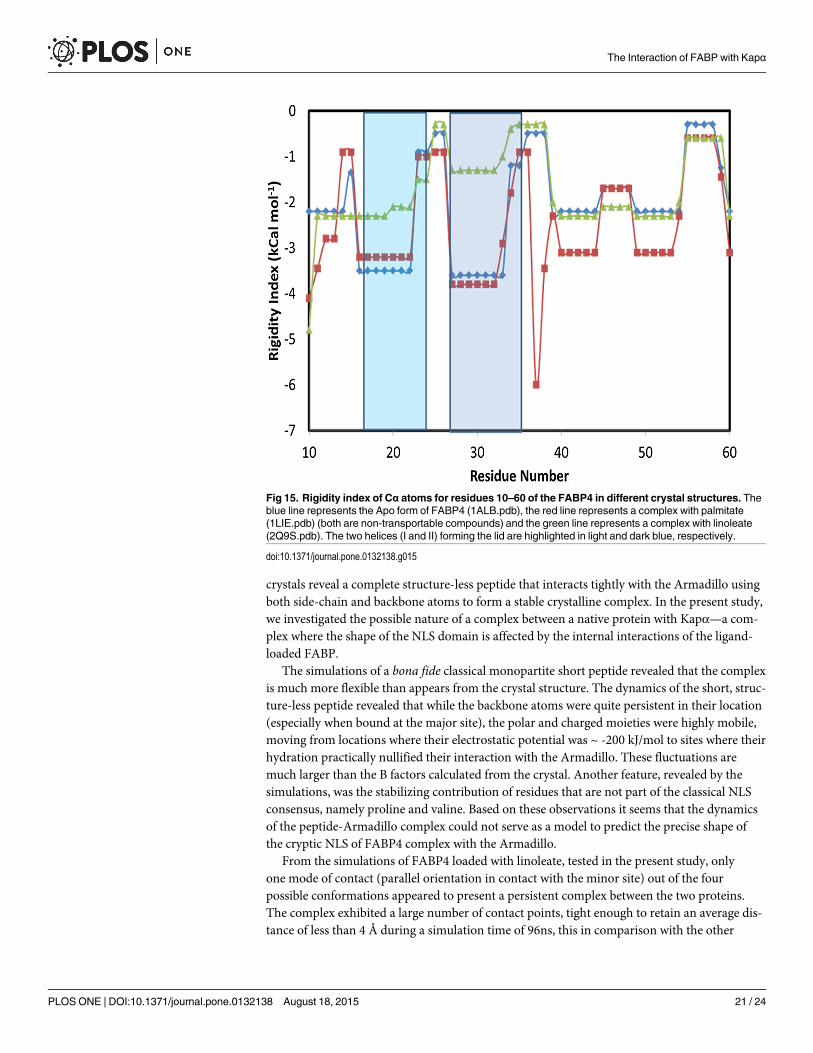
As appears from the present study, the interaction between the FABP4 and Armadillo uti-lizes many residues located on the lid structure. These interactions are noticed only for the spe-cific orientation (minor-parallel), where the lid seems to lose its tight structure. The ability ofthe lid to relax, and the correlation between the flexibility of the structure with the nature ofthe ligand bound to the FABP4 was investigated by the application of Constraint-NetworkAnalysis (CNA) [36], a server that calculates the thermostability of the various structural ele-ments of a protein.
Fig 15 depicts the thermostability of FABP4 in its Apo state and in a complex with non-transportable ligand vs. a complex with linoleate. For simplicity, the presentation is limitedonly to residues 10–60, where the two helices of the lid are located. The residue number is indi-cated on the x-axis and the y-axis denotes the rigidity index of the Cα atoms in kcal/mol [37].What is well perceived from this figure is that the rigidity of the two helices, calculated for thetransportable complex (the FABP4 loaded by linoleate) is significantly smaller than that of thenon-transportable protein (Apo and palmitate-loaded). In the complex with linoleate, the heli-ces of the lid are rather flexible and tend to depart from the rigid helical structure. Repeatingthe same analysis for two more transportable substrates (ANS (anilinonaphthalene sulfonate)(2ANS.pdb) and TDZ—(Troglitazone) (2QM9.pdb)) reveals also low rigidity of the helices,while for the non-transportable compounds (Archidonate—1ADL.pdb, Oleate—1LID.pdb,stearate—1LIF.pdb and hexadecanoate—1LIC.pdb) all exhibited high rigidity of the lid domain(data not shown). Apparently, the flexibility of the lid domain may play an important role inthe recognition mechanism by the Kapα.
DiscussionThe classical monopartite NLS domain is made of three basic moieties conforming to the con-sensus sequence KK/RXK/R. The studies of their mode of interaction with the Karyopherinsystem is based on crystallization of the KapαΔIBB (the Armadillo without the IBB domain)with short segments of the imported proteins, up to a length of ~20 residues [45]. These
Fig 14. The interaction energies between the residues on FABP4 with the Armadillo. The data were calculated for each of the five largest clustersrepresenting the complex FABP4 (loaded with linoleate) located at the minor site in the parallel orientation.
doi:10.1371/journal.pone.0132138.g014
The Interaction of FABP with Kapα
PLOSONE | DOI:10.1371/journal.pone.0132138 August 18, 2015 20 / 24
crystals reveal a complete structure-less peptide that interacts tightly with the Armadillo usingboth side-chain and backbone atoms to form a stable crystalline complex. In the present study,we investigated the possible nature of a complex between a native protein with Kapα—a com-plex where the shape of the NLS domain is affected by the internal interactions of the ligand-loaded FABP.
The simulations of a bona fide classical monopartite short peptide revealed that the complexis much more flexible than appears from the crystal structure. The dynamics of the short, struc-ture-less peptide revealed that while the backbone atoms were quite persistent in their location(especially when bound at the major site), the polar and charged moieties were highly mobile,moving from locations where their electrostatic potential was ~ -200 kJ/mol to sites where theirhydration practically nullified their interaction with the Armadillo. These fluctuations aremuch larger than the B factors calculated from the crystal. Another feature, revealed by thesimulations, was the stabilizing contribution of residues that are not part of the classical NLSconsensus, namely proline and valine. Based on these observations it seems that the dynamicsof the peptide-Armadillo complex could not serve as a model to predict the precise shape ofthe cryptic NLS of FABP4 complex with the Armadillo.
From the simulations of FABP4 loaded with linoleate, tested in the present study, onlyone mode of contact (parallel orientation in contact with the minor site) out of the fourpossible conformations appeared to present a persistent complex between the two proteins.The complex exhibited a large number of contact points, tight enough to retain an average dis-tance of less than 4 Å during a simulation time of 96ns, this in comparison with the other
Fig 15. Rigidity index of Cα atoms for residues 10–60 of the FABP4 in different crystal structures. Theblue line represents the Apo form of FABP4 (1ALB.pdb), the red line represents a complex with palmitate(1LIE.pdb) (both are non-transportable compounds) and the green line represents a complex with linoleate(2Q9S.pdb). The two helices (I and II) forming the lid are highlighted in light and dark blue, respectively.
doi:10.1371/journal.pone.0132138.g015
The Interaction of FABP with Kapα
PLOSONE | DOI:10.1371/journal.pone.0132138 August 18, 2015 21 / 24

conformations in which the number of contacts was 50% or less. In this conformation, all resi-dues of the NLS (K21, R30, K31) were identified as Hot Spots, two of them (K21 and R30)together with some non-NLS moieties (E22, F27, K100, K120 and G121) formed tight lastingcontact with the Armadillo including Arms 4, 5 and 6 that are components of the major sitedomain. This special conformation, in which the FABP4 interacts with both the minor and themajor sites, prevents the IBB domain from folding over the major site and rendering it free toreact with Kapβ.
At first glance, there seems to be some internal contradiction in the present study: the struc-ture of the NLS peptide-Armadillo complex utilizes many more interaction sites than the NLSdomain of the FABP. This apparent discrepancy is a direct reflection of the basic differencebetween the mode of a short peptide binding vs. the binding of a well folded protein [44]. Thepeptide has the freedom to sample a large variety of conformations that are almost equipoten-tial [35]. Thus, it can easily assume a shape that will mostly fit into a rigid binding site of a pro-tein. As a results, the protein-peptide complexes are richer in hydrogen-bond interactions(most of them with the backbone atoms) than the protein-protein complexes [44]. This ability,to fit tightly into a binding site, is denied from a protein due to its inherent structure. In thecase of FABP4, where the NLS site consists of residues located on two adjacent helices, thenature of the complex will diverge from that of the peptides-Armadillo complex. The positivelycharged residues; K21, R30 and K31, do serve as primers of the docking that are attracted tothe crevice of the binding site. But the nature of the mature complex has more features of a pro-tein-protein complex than of a protein-peptide one.
Recently, Pang and Zhou [46] investigated the interaction of short peptides with the minorsite, demonstrating the significance of the interaction of two adjacent positive moieties to thebinding. Their modeling indicated the role of non-polar interaction of other moieties of theNLS sequence with nearby Arms, even with those of the major site. Their study, with a modelpeptide, clearly confirm our suggestion that in the case of protein-protein interaction, wherethe NLS sequence is located on a well-structured domain, the stabilization is gained by interac-tions that are beyond the formalistic NLS residues.
Finally, the minor-parallel conformation generated an extended hydrophobic domainwhich expanded out of the boundary of the FABP4, letting the load carried by the FABP4 (lino-leate) to partially migrate out of the FABP4 protein into a mutual complex, in which the Arma-dillo contributes part of a joint binding pocket. It might be suggested that such a complex isinstrumental in the liberation of the cargo into the nucleus space, once the loaded FABP4reaches the inner space of the nucleus.
Author ContributionsConceived and designed the experiments: YT MG OAV. Performed the experiments: OAV.Analyzed the data: OAV EN. Contributed reagents/materials/analysis tools: NK. Wrote thepaper: OAVMG YT.
References1. Glatz JF, Luiken JJ: Fatty acids in cell signaling: Historical perspective and future outlook. Prostaglan-
dins Leukot Essent Fatty Acids 2014.
2. Moise AR, Noy N, Palczewski K, Blaner WS: Delivery of retinoid-based therapies to target tissues. Bio-chemistry 2007, 46(15):4449–4458. PMID: 17378589
3. Marfori M, Mynott A, Ellis JJ, Mehdi AM, Saunders NF, Curmi PM, et al. Molecular basis for specificityof nuclear import and prediction of nuclear localization. Biochimica et biophysica acta 2011, 1813(9):1562–1577. doi: 10.1016/j.bbamcr.2010.10.013 PMID: 20977914
4. Goldfarb DS, Corbett AH, Mason DA, Harreman MT, Adam SA: Importin alpha: a multipurpose nuclear-transport receptor. Trends Cell Biol 2004, 14(9):505–514. PMID: 15350979
The Interaction of FABP with Kapα
PLOSONE | DOI:10.1371/journal.pone.0132138 August 18, 2015 22 / 24

5. Gillilan RE, Ayers SD, Noy N: Structural basis for activation of fatty acid-binding protein 4. Journal ofmolecular biology 2007, 372(5):1246–1260. PMID: 17761196
6. Pedraza L, Fidler L, Staugaitis SM, Colman DR: The active transport of myelin basic protein into thenucleus suggests a regulatory role in myelination. Neuron 1997, 18(4):579–589. PMID: 9136767
7. Boulikas T: Nuclear localization signals (NLS). Crit Rev Eukaryot Gene Expr 1993, 3(3):193–227.PMID: 8241603
8. Ayers SD, Nedrow KL, Gillilan RE, Noy N: Continuous nucleocytoplasmic shuttling underlies transcrip-tional activation of PPARgamma by FABP4. Biochemistry 2007, 46(23):6744–6752. PMID: 17516629
9. Morgan E, Kannan-Thulasiraman P, Noy N: Involvement of Fatty Acid Binding Protein 5 and PPAR-beta/delta in Prostate Cancer Cell Growth. PPAR research 2010, 2010.
10. Armstrong EH, Goswami D, Griffin PR, Noy N, Ortlund EA: Structural basis for ligand regulation of theFatty Acid Binding Protein 5, Peroxisome Proliferator Activated Receptor beta/delta (FABP5-PPAR-beta/delta) signaling pathway. The Journal of biological chemistry 2014.
11. Lott K, Bhardwaj A, Sims PJ, Cingolani G: A minimal nuclear localization signal (NLS) in human phos-pholipid scramblase 4 that binds only the minor NLS-binding site of importin alpha1. The Journal of bio-logical chemistry 2011, 286(32):28160–28169. doi: 10.1074/jbc.M111.228007 PMID: 21690087
12. Ge Q, Nakagawa T, Wynn RM, Chook YM, Miller BC, Uyeda K: Importin-alpha protein binding to anuclear localization signal of carbohydrate response element-binding protein (ChREBP). The Journalof biological chemistry 2011, 286(32):28119–28127. doi: 10.1074/jbc.M111.237016 PMID: 21665952
13. Chook YM, Suel KE: Nuclear import by karyopherin-betas: recognition and inhibition. Biochimica et bio-physica acta 2011, 1813(9):1593–1606. doi: 10.1016/j.bbamcr.2010.10.014 PMID: 21029754
14. Chandra V, Huang P, Hamuro Y, Raghuram S, Wang Y, Burris TP, et al.: Structure of the intact PPAR-gamma-RXR- nuclear receptor complex on DNA. Nature 2008, 456(7220):350–356. doi: 10.1038/nature07413 PMID: 19043829
15. Barish GD, Narkar VA, Evans RM: PPAR delta: a dagger in the heart of the metabolic syndrome. TheJournal of clinical investigation 2006, 116(3):590–597. PMID: 16511591
16. Castelein H, Declercq PE, Baes M: DNA binding preferences of PPAR alpha/RXR alpha heterodimers.Biochemical and biophysical research communications 1997, 233(1):91–95. PMID: 9144402
17. Motojima K, Peters JM, Gonzalez FJ: PPAR alpha mediates peroxisome proliferator-induced transcrip-tional repression of nonperoxisomal gene expression in mouse. Biochemical and biophysical researchcommunications 1997, 230(1):155–158. PMID: 9020034
18. Plutzky J: The PPAR-RXR transcriptional complex in the vasculature: energy in the balance. Circula-tion research 2011, 108(8):1002–1016. doi: 10.1161/CIRCRESAHA.110.226860 PMID: 21493923
19. Huang H, McIntosh AL, Martin GG, Landrock KK, Landrock D, Gupta S, et al.: Structural and functionalinteraction of fatty acids with human liver fatty acid-binding protein (L-FABP) T94A variant. FEBS J2014, 281(9):2266–2283. doi: 10.1111/febs.12780 PMID: 24628888
20. Richieri GV, Ogata RT, Kleinfeld AM: Thermodynamics of Fatty Acid Binding to Fatty Acid-binding Pro-teins and Fatty Acid Partition betweenWater and Membranes Measured Using the Fluorescent ProbeADIFAB. Journal of Biological Chemistry 1995, 270(25):15076–15084. PMID: 7797491
21. Evans RM: The nuclear receptor superfamily: a rosetta stone for physiology. Mol Endocrinol 2005, 19(6):1429–1438. PMID: 15914712
22. Chang CW, Counago RL, Williams SJ, Boden M, Kobe B: Crystal structure of rice importin-alpha andstructural basis of its interaction with plant-specific nuclear localization signals. The Plant cell 2012, 24(12):5074–5088. doi: 10.1105/tpc.112.104422 PMID: 23250448
23. Noy N: Ligand specificity of nuclear hormone receptors: sifting through promiscuity. Biochemistry 2007,46(47):13461–13467. PMID: 17983246
24. Sessler RJ, Noy N: A ligand-activated nuclear localization signal in cellular retinoic acid binding protein-II. Molecular cell 2005, 18(3):343–353. PMID: 15866176
25. Fontes MR, Teh T, Riell RD, Park SB, Standaert RF, Kobe B: Crystallization and preliminary X-ray dif-fraction analysis of importin-alpha complexed with NLS peptidomimetics. Biochimica et biophysica acta2005, 1750(1):9–13. PMID: 15878698
26. Hess B, Kutzner C, van der Spoel D, Lindahl E: GROMACS 4: Algorithms for highly efficient, load-bal-anced, and scalable molecular simulation. Journal of Chemical Theory and Computation 2008, 4(3):435–447.
27. Oostenbrink C, Villa A, Mark AE, van GunsterenWF: A biomolecular force field based on the freeenthalpy of hydration and solvation: the GROMOS force-field parameter sets 53A5 and 53A6. J Com-put Chem 2004, 25(13):1656–1676. PMID: 15264259
The Interaction of FABP with Kapα
PLOSONE | DOI:10.1371/journal.pone.0132138 August 18, 2015 23 / 24
28. Hess B, Bekker H., Berendsen H. J. C., Fraaije J. G. E. M.: LINCS: A linear constraint solver for molecu-lar simulations. J Comp Chem 1997, 18:1463–1472.
29. Miyamoto S, Kollman PA: SETTLE: An Analytical Version of the SHAKE and RATTLE Algorithms forRigid water models. J Comp Chem 1992, 13:952–962.
30. Berendsen HJC, Postma J. P. M., DiNola A., Haak J. R.: Molecular dynamics with coupling to an exter-nal bath. J Chem Phys 1984, 81:3684–3690.
31. Essmann U, Perera L, Berkowitz ML, Darden T, Lee H, Pedersen LG: A smooth particle mesh Ewaldmethod. J Chem Phys 1995, 103(19):8577–8593.
32. Duhovny D NR, Wolfson HJ.: Efficient Unbound Docking of Rigid Molecules.Gusfield et al, Ed Pro-ceedings of the 2'ndWorkshop on Algorithms in Bioinformatics(WABI) 2002, Lecture Notes in Com-puter Science 2452, pp. 185–200, Springer Verlag, 2002
33. Schneidman-Duhovny D IY, Nussinov R, Wolfson HJ: PatchDock and SymmDock: servers for rigid andsymmetric docking. Nucl Acids Res 2006, 33:: W363–367.
34. Duhovny D, Nussinov R, Wolfson HJ: Efficient unbound docking of rigid molecules. Lect Notes ComputSc 2002, 2452:185–200.
35. Daura X, Antes I, van GunsterenWF, Thiel W, Mark AE: The effect of motional averaging on the calcu-lation of NMR-derived structural properties. Proteins 1999, 36(4):542–555. PMID: 10450095
36. Kruger DM, Rathi PC, Pfleger C, Gohlke H: CNA web server: rigidity theory-based thermal unfoldingsimulations of proteins for linking structure, (thermo-)stability, and function. Nucleic acids research2013, 41(Web Server issue):W340–348. doi: 10.1093/nar/gkt292 PMID: 23609541
37. Pfleger C, Radestock S, Schmidt E, Gohlke H: Global and local indices for characterizing biomolecularflexibility and rigidity. Journal of computational chemistry 2013, 34(3):220–233. doi: 10.1002/jcc.23122PMID: 23007873
38. Konecny R, Baker NA, McCammon JA: iAPBS: a programming interface to Adaptive Poisson-Boltz-mann Solver (APBS). Computational science & discovery 2012, 5(1).
39. Unni S, Huang Y, Hanson RM, Tobias M, Krishnan S, Li WW, Nielsen JE, et al.: Web servers and ser-vices for electrostatics calculations with APBS and PDB2PQR. Journal of computational chemistry2011, 32(7):1488–1491. doi: 10.1002/jcc.21720 PMID: 21425296
40. Baker NA, Sept D, Joseph S, Holst MJ, McCammon JA: Electrostatics of nanosystems: application tomicrotubules and the ribosome. Proceedings of the National Academy of Sciences of the United Statesof America 2001, 98(18):10037–10041. PMID: 11517324
41. Darnell SJ, LeGault L, Mitchell JC: KFC Server: interactive forecasting of protein interaction Hot Spots.Nucleic acids research 2008, 36(Web Server issue):W265–269. doi: 10.1093/nar/gkn346 PMID:18539611
42. David CC, Jacobs DJ: Principal component analysis: a method for determining the essential dynamicsof proteins. Methods Mol Biol 2014, 1084:193–226. doi: 10.1007/978-1-62703-658-0_11 PMID:24061923
43. Tina KG, Bhadra R, Srinivasan N: PIC: Protein Interactions Calculator. Nucleic acids research 2007,35(Web Server issue):W473–476. PMID: 17584791
44. London N, Movshovitz-Attias D, Schueler-Furman O: The Structural Basis of Peptide-Protein BindingStrategies. Structure 2010, 18(2):188–199. doi: 10.1016/j.str.2009.11.012 PMID: 20159464
45. Roman N, Christie M, Swarbrick CM, Kobe B, Forwood JK: Structural Characterisation of the NuclearImport Receptor Importin Alpha in Complex with the Bipartite NLS of Prp20. PloS one 2013, 8(12):e82038. doi: 10.1371/journal.pone.0082038 PMID: 24339986
46. Dukuzumuremyi JM, Rosqvist R, Hallberg B, Akerstrom B, Wolf-Watz H, Schesser K: The Yersinia pro-tein kinase A is a host factor inducible RhoA/Rac-binding virulence factor. The Journal of biologicalchemistry 2000, 275(45):35281–35290. PMID: 10950948
The Interaction of FABP with Kapα
PLOSONE | DOI:10.1371/journal.pone.0132138 August 18, 2015 24 / 24
Copyright © 2022 FDOKUMEN