Lipase-mediated desymmetrization of glycerol with aromatic and aliphatic anhydrides
Upload
independentCategory
view
1download
0
The Impact of the Absence of Aliphatic Glucosinolates onInsect Herbivory in ArabidopsisJules Beekwilder1.*, Wessel van Leeuwen2., Nicole M. van Dam3, Monica Bertossi4, Valentina Grandi4,
Luca Mizzi5, Mikhail Soloviev6, Laszlo Szabados7, Jos W. Molthoff1, Bert Schipper1, Hans Verbocht1,
Ric C. H. de Vos1, Piero Morandini4, Mark G. M. Aarts2, Arnaud Bovy1
1 Plant Research International, Wageningen, The Netherlands, 2 Laboratory of Genetics, Wageningen University, Wageningen, The Netherlands, 3 Netherlands Institute of
Ecology (NIOO-KNAW), Heteren, The Netherlands, 4 Department of Biology, University of Milan, CNR Biophysics Institute, Milano, Italy, 5 Department of Biomolecular
Sciences and Biotechnology, University of Milan, CNR Biophysics Institute, Milano, Italy, 6 School of Biological Sciences, Royal Holloway University of London, Egham,
United Kingdom, 7 Biological Research Center, Szeged, Hungary
Abstract
Aliphatic glucosinolates are compounds which occur in high concentrations in Arabidopsis thaliana and other Brassicaceaespecies. They are important for the resistance of the plant to pest insects. Previously, the biosynthesis of these compoundswas shown to be regulated by transcription factors MYB28 and MYB29. We now show that MYB28 and MYB29 are partiallyredundant, but in the absence of both, the synthesis of all aliphatic glucosinolates is blocked. Untargeted and targetedbiochemical analyses of leaf metabolites showed that differences between single and double knock-out mutants and wildtype plants were restricted to glucosinolates. Biosynthesis of long-chain aliphatic glucosinolates was blocked by the myb28mutation, while short-chain aliphatic glucosinolates were reduced by about 50% in both the myb28 and the myb29 singlemutants. Most remarkably, all aliphatic glucosinolates were completely absent in the double mutant. Expression ofglucosinolate biosynthetic genes was slightly but significantly reduced by the single myb mutations, while the doublemutation resulted in a drastic decrease in expression of these genes. Since the myb28myb29 double mutant is the firstArabidopsis genotype without any aliphatic glucosinolates, we used it to establish the relevance of aliphatic glucosinolatebiosynthesis to herbivory by larvae of the lepidopteran insect Mamestra brassicae. Plant damage correlated inversely to thelevels of aliphatic glucosinolates observed in those plants: Larval weight gain was 2.6 fold higher on the doublemyb28myb29 mutant completely lacking aliphatic glucosinolates and 1.8 higher on the single mutants with intermediatelevels of aliphatic glucosinolates compared to wild type plants.
Citation: Beekwilder J, van Leeuwen W, van Dam NM, Bertossi M, Grandi V, et al. (2008) The Impact of the Absence of Aliphatic Glucosinolates on InsectHerbivory in Arabidopsis. PLoS ONE 3(4): e2068. doi:10.1371/journal.pone.0002068
Editor: Ivan Baxter, Purdue University, United States of America
Received December 21, 2007; Accepted March 21, 2008; Published April 30, 2008
Copyright: � 2008 Beekwilder et al. This is an open-access article distributed under the terms of the Creative Commons Attribution License, which permitsunrestricted use, distribution, and reproduction in any medium, provided the original author and source are credited.
Funding: The authors have no support or funding to report.
Competing Interests: The authors have declared that no competing interests exist.
* E-mail: [email protected]
. These authors contributed equally to this work.
Introduction
Plants resist insect herbivory by producing a wide variety of
toxic and deterrent chemicals. In Arabidopsis thaliana (Arabi-
dopsis) and other Crucifer species, the chemical defense arsenal
against insect herbivores comprises glucosinolates, alongside with
protease inhibitors, phenolics and terpenoid volatiles [1,2].
Glucosinolates constitute a large family of secondary metabo-
lites with over 120 different chemical structures known [3]. All
glucosinolates have a core structure, composed of a b-thioglucose
and an N-hydroxyiminosulphate group (Fig. 1), and an aglycone
side-chain, which is structurally highly diverse. Upon tissue
disruption (e.g. during herbivory), glucosinolates (which are stored
in the plant vacuole) are mixed with myrosinase, a glucosidase that
is spatially separated from its substrate [4]. The myrosinase
activates the glucosinolates by removal of the glucose moiety. This
results in the production of nitriles and (iso)thiocyanates, that are
toxic and deterrent to generalist insect herbivores. A number of
studies have indicated that Arabidopsis lines with high glucosino-
late content show a delayed larval development of lepidopteran
insects [5,6]. Aliphatic glucosinolates may even reduce survival
and growth of insects specialized in feeding on Crucifers [5].
In Arabidopsis, 36 different glucosinolates have been identified,
mostly with aliphatic or indolic side-chains [7,8]. The indolic
glucosinolates are derived from tryptophane, while aliphatic
glucosinolates are derived from methionine. Leaves of many A.
thaliana accessions are very rich in aliphatic glucosinolates carrying
a methylsulfinylalkyl side-chain, of which the alkyl group varies in
length from 3 to 8 carbons (Fig. 1) [9].
Biosynthesis of glucosinolates involves a long series of enzymatic
conversions [10]. The pathway to aliphatic glucosinolates
comprises three phases, starting with deamination of methionine,
followed by elongation of the side chain by sequential condensa-
tion reactions with acetyl-CoA, isomerization and decarboxyl-
ation, and finally synthesis of the core structure. Subsequently,
side-chains may undergo secondary transformations, for instance
to sulfinyl groups. Elongation reactions are carried out by
methylthioalkylmalate synthases (MAM), an aconitase and an
isopropylmalate dehydrogenase [11]. Subsequently, the glucosi-
nolate core structure is synthesized, involving cytochrome P450
PLoS ONE | www.plosone.org 1 April 2008 | Volume 3 | Issue 4 | e2068

enzymes, a C-S lyase and a glucosyltransferase [10,12]. Glucosi-
nolate profiles are specific for species, accessions and tissues [9,13].
Recent research has focussed on factors controlling (parts of) the
glucosinolate pathway. The ability to selectively manipulate
glucosinolate biosynthesis allows new opportunities in both applied
and fundamental research. For applied purposes, one could aim at
breeding crop plants with increased levels of glucoraphanin
(4MSOB), a compound associated with lower risk of lung and
colorectal cancer [14], or in increasing the total levels of
glucosinolates in plants for application in biofumigation [15].
For increasing our understanding of the importance of glucosino-
late biosynthetic pathways, regulating this pathway can allow to
understand its role in the ecological interactions of the plant with
insects and other life forms.
Recently two MYB transcription factors of the R2R3 sub-family
(MYB28 and MYB29) have been identified to be involved in the
regulation of the aliphatic glucosinolate biosynthetic pathway [16–
18]. A knock-out mutation in the Arabidopsis MYB28 gene leads
to strongly reduced expression of aliphatic glucosinolate biosyn-
thesis genes, and accordingly, the levels of long-chain aliphatic
glucosinolates are significantly reduced in this mutant. For a
knock-out mutation in the MYB29 gene, no such effects were
observed, suggesting that this gene was not essential for
constitutive glucosinolate biosynthesis, but rather plays a role in
methyl jasmonate induced glucosinolate biosynthesis [17,19].
Over-expression of MYB28 in Arabidopsis plants resulted in
elevated levels of aliphatic glucosinolates and reduced weight-gain
of Spodoptera exigua larvae feeding on these plants [16]. This
suggests that the activity of the closely related MYB28 and MYB29
transcription factors is important for aliphatic glucosinolate
synthesis and, consequently, insect resistance.
In this work, a double knock-out mutant of MYB28 and MYB29
was constructed in Arabidopsis. This mutant was compared to the
wild type and single-mutant plants on the level of glucosinolates,
gene-expression and resistance to herbivory by the generalist
Lepidopteran insect Mamestra brassicae. The results allow a detailed
insight in the role of MYB28 and MYB29 in the absolute
regulation of aliphatic glucosinolate biosynthesis and their impact
on the ecology of Cruciferae.
Results
myb28 and myb29 single and double knock-out linesThe function of the MYB28 gene (At5g61420) was probed using
different knock-out T-DNA insertion lines. The BRC_H161b line
insertion maps in the second exon of this gene (at +242 bp from
the startcodon), whereas the SALK_136312 line insertion maps
183 bp upstream of the startcodon (Fig. 2). A transposon insertion
in the MYB29 gene (At5g07690) is present in line SM3.34316. The
insertion maps 44 bp upstream of the MYB29 gene startcodon
(Fig. 2).
The BRC_H161b (myb28) line was crossed with the SM3.34316
(myb29) line and the progeny was self-fertilized to generate
homozygous double knock-outs (myb28myb29). The single knock-
out lines did not show any visible phenotype, whereas the double
knock-out line showed a marginal delay in seed germination and
initial growth. In later growth phases, there was no visible
phenotypic difference between wild type Col-0 and any of the
mutant lines.
Double knock-out of MYB28 and MYB29 leads tocomplete absence of aliphatic glucosinolates
The effect of the myb mutations at the biochemical level was
assessed using an untargeted LC-QTOF-MS metabolic profiling
approach with methanol/water extracts from mature rosette
leaves. From each line, five individual replicates were analyzed.
The resulting data matrix (samples vs. mass peaks) contained
intensity values for 2615 mass signals (roughly representing 400
compounds) aligned across all samples. To visualize the effect of
each mutation, principal components analysis (PCA) of the dataset
was performed. As shown in the score plot (Fig. 3), the five
Figure 1. Chemical structures of the major glucosinolates inArabidopsis thaliana Col-0.doi:10.1371/journal.pone.0002068.g001
Figure 2. Position of the insertions in knock-out mutants ofMYB28 (top) and MYB29 (bottom). Black areas represent translatedregions, white areas represent untranslated regions and introns.Numbers indicate the position from the startcodon.doi:10.1371/journal.pone.0002068.g002
Glucosinolate-Less Plant
PLoS ONE | www.plosone.org 2 April 2008 | Volume 3 | Issue 4 | e2068

biological replicates of each mutant cluster together. The plot also
shows that the myb29 mutant is relatively closely related to the wild
type, while the myb28 mutant is more distinct in the plot.
Remarkably, the double mutant is even more distant from the
myb28 mutant than would be anticipated from the effect of myb29
alone.
To analyze which components lead to the separation of the
mutants from the wild type, mass signals were selected that
significantly differed (p,0.01; n = 5) more than two-fold in
intensity between the Col-0 wild type and myb28myb29. In the
double mutant, 159 mass signals representing 24 different
compounds were found to be down-regulated: 11 compounds
could be identified as glucosinolates, from the sulphinylalkyl,
methylthioalkyl, phenyl and alkyl classes, while the other 13 could
not be properly identified due to very low signals (,10-fold
background), which do not allow accurate mass calculation and
subsequent deduction of the elemental formula. In fact, in the double
mutant, the identified downregulated compounds were all reduced
to levels that couldn’t be detected in the MS. The identified
compounds are listed in Table 1. In addition, six compounds (45
mass peaks) were found to be significantly up-regulated by more than
two-fold in the double mutant. Among these compounds were two
indole glucosinolates (Table 1) and four unidentified compounds
with very low intensity signals. Phenolic compounds such as
flavonoids and sinapates, which can also affect insect resistance,
were specifically assessed, but no strong changes could be observed
for e.g. sinapoylmalate and kaempferol-glucoside-rhamnoside
(Table 1). Apparently, the myb28 and myb29 mutations do not
lead to pleiotropic phenotypes.
The glucosinolate content of the rosette leaf material was
further quantified using a dedicated HPLC analysis. The HPLC
chromatograms of the wild type and mutant plants are shown in
Fig 4A. The total amount of glucosinolates was quantified from the
chromatograms and plotted in Fig 4B. The quantification of each
specific glucosinolate is shown in Fig 5. This quantitative analysis
confirms the results obtained by the untargeted metabolomics
analysis. Short-chain aliphatic methylsulphinylalkyl glucosinolates,
such as glucoiberin (3MSOP), glucoraphanin (4MSOB) and
glucoalyssin (5MSOP), are reduced by about 50% in both myb28
and myb29 mutants, but are completely absent from the double
mutant. The long-chain aliphatic methylsulphinylalkyl glucosino-
late glucohirsutin (8MSOO) is not significantly affected in myb29,
but has completely disappeared in myb28 and in the double
mutant. Glucohesperin (6MSOH) and glucoibarin (7MSOH)
showed relative changes similar to glucohirsutin (8MSOO) in
the LC-MS analysis, but were below the detection level in the
dedicated HPLC analysis (Fig. 5). Indolyl glucosinolates, such as
glucobrassicin (I3M) and neoglucobrassicin (1MO-I3M), show a
slight increase in both single mutants, and are two- to three-fold
increased in the double mutant, while 4-methoxyglucobrassicin
(4MO-I3M) is not significantly increased. Knock-out of both
MYB28 and MYB29 thus completely suppressed synthesis of
aliphatic glucosinolates below detection level.
Characterization of the myb28myb29 mutant by geneexpression analysis
To further understand the mechanism by which double knock-
out mutation of MYB28 and MYB29 genes leads to complete
collapse of aliphatic glucosinolate biosynthesis, real-time RT-PCR
assays were performed. Gene expression levels of MYB28, MYB29
and several genes involved in aliphatic glucosinolate biosynthesis
(MAM1, MAM3, CYP83A1 and an aconitase) or indolic glucosi-
nolate biosynthesis (CYP83B1) were monitored in mature expand-
ed rosette leaf material from the Col-0 wild type, the myb28 mutant
(BRC_H161b), the myb29 mutant, and the myb28myb29 double
mutant.
Compared to the wild type Col-0, the levels of MYB28
transcripts were strongly affected (60 to 100-fold reduced) in the
myb28 and the myb28myb29 mutants (Fig. 6). The MYB29 transcript
levels were 4 to 6-fold reduced in the myb29 and myb28myb29
mutant, respectively. Apparently, myb29 is not a knock-out but a
knock-down mutant, since there still is some residual expression of
MYB29 in the mutant. The MYB28 and MYB29 genes hardly
affect each others expression in leaves.
Biosynthetic genes are dramatically more reduced in expression
in the myb29myb28 double mutant, as compared to the single
mutants. Expression of MAM3 was already strongly (.10-fold)
reduced in the myb28 mutant, but even more (.100-fold) reduced
in the myb28myb29 mutant, although the expression in the myb29
mutant was comparable to that in the wild type. The MAM1,
CYP83A1 and Aconitase transcripts were hardly affected (,2-fold) in
the single mutants, but strongly reduced (140-fold, 30-fold and
300-fold, respectively) in the myb28myb29 double mutant. On the
other hand, the levels of the CYP83B1 gene, which participates in
the indolic glucosinolate pathway, were not significantly changed
in any of knockout lines (data not shown). Thus, knocking out both
MYB28 and MYB29 interfered much more severely with
expression of aliphatic glucosinolate biosynthesis genes than was
anticipated from the analysis of both single mutants. This suggests
a strong redundancy of these transcription factors for the
downstream genes tested. MAM3 is an exception, as it is largely
controlled by MYB28 and its regulation by MYB29 is epistatic to
MYB28.
Insect feedingThe myb28myb29 double mutant is to our knowledge the first
Arabidopsis genotype without aliphatic glucosinolates and it
provides the first possibility to assess the relevance of aliphatic
glucosinolates on herbivore insect performance. We therefore
Figure 3. Score plot from principal component analysis of LC-MS metabolic profiles of wild type and mutant Arabidopsislines. Contributions of principal components to separation of thesamples are indicated on the axes.doi:10.1371/journal.pone.0002068.g003
Glucosinolate-Less Plant
PLoS ONE | www.plosone.org 3 April 2008 | Volume 3 | Issue 4 | e2068

compared the performance of larvae of the lepidopteran insect
Mamestra brassicae on the myb28, myb29 and myb28myb29 Arabi-
dopsis mutants. Mamestra was chosen because its larvae are among
the most frequently found pest insects on cabbages [20]. Mamestra
is a generalist, which prefers cruciferous species, but has been
found feeding on many different plant species, including non-
cruciferae [21,22]. There is some evidence for sensitivity of Mamestra
to glucosinolates [23].
In an initial experiment, neonate larvae were transferred to
detached leaves of two wild type lines (Col-0 and progeny of a wild
type segregant from a MYB28myb28 BRC_H161b heterozygote
plant), and knock-out mutants myb28-BRC_H161, myb28-
SALK_136312 and myb29. For each experiment, individual larvae
were reared separately in Petri dishes, and leaves were refreshed at
least every two days. Larvae were weighed after 14 days of feeding.
Mutations in MYB28 and MYB29 resulted in enhanced growth
rates of Mamestra (Fig. 7A). On day 14, the average weight of larvae
raised on wild type plants was two to three times lower than on
leaves from knock-out plants (Fig. 7A, ANOVA F4,76 = 17.5,
p,0.001)
In a second experiment, Mamestra larvae were tested on groups
of hydroponically-grown intact plants. Three replicate groups of
25 neonates were confined to trays with 50 plants of wild type Col-
0, myb28-H161, myb29 or myb28myb29. After 12 days, the larvae
were weighed individually. Again we found a clear effect of plant
genotype on larval mass (Fig. 7B). The body mass of larvae raised
on wild type Col-0 plants was significantly lower than that of
larvae raised on each of the single mutants (1.7–1.8 times lower),
whereas larvae on the double mutant had the highest body mass
(2.6 times higher than Col-0; Fig 7C, nested ANOVA genotype
effect F3,158 = 33.18, p,0.001). Knocking out both MYB28 and
MYB29 genes in Arabidopsis had a significant positive effect on
growth of Mamestra larvae, most prominently if both genes were
knocked-out.
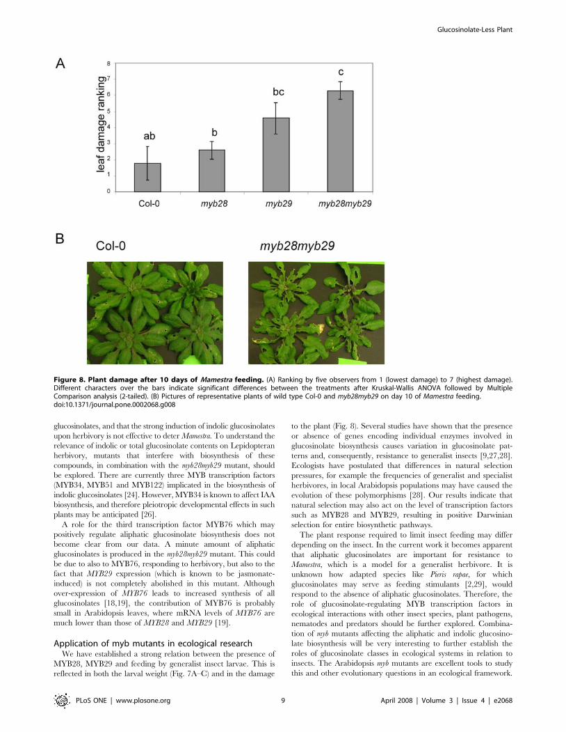
In a third experiment, the effect of Mamestra herbivory on plants
of wild type Col-0, myb28-H161, myb29 or myb28myb29 was
compared. Four replicate groups of eight plants of each line were
separately grown in hydroponic trays. On two of the replicates, 16
Mamestra neonates were positioned, while the other two replicates
were not exposed to insects. After 10 days, the damage to each of
the insect-treated replicates was ranked by five observers after
double-blind visual inspection. As shown in Fig. 8, Mamestra
herbivory resulted in higher damage levels in the myb28myb29
mutant, as compared to the Col-0 wild type, while both single
mutants had intermediate damage levels. These data are consistent
with the higher weight of larvae feeding on the myb28myb29
mutant.
In the same experiment, the content of glucosinolates was
measured for the leaves, with and without Mamestra herbivory. In
Table 2, the effects of herbivory for all four tested lines are shown.
The values in this table were obtained by setting the concentra-
tions of all glucosinolates in Col-0 (without herbivory) at value
1.00, and comparing the concentrations found in the other
samples (with and without herbivory) to this value. In the Col-0
wild-type, three effects of Mamestra herbivory can be observed.
Firstly, most glucosinolates (aliphatic and others) were increased by
a factor 1.5 to 2. Secondly, there is a pronounced increase of the
indolic glucosinolates glucobrassicin (I3M; .2 fold) and neogluco-
brassicin (1MO-I3M; .6 fold). Thirdly, herbivory results in a
strong decrease of glucoerucin (4MTB; a methylthioglucosinolate),
which is a precursor of glucoraphanin (4MSOB; a methylsulfo-
nylglucosinolate). In the myb28 and myb29 mutants, these three
Table 1. Metabolites detected by LC-QTOF-MS (ESI negative mode) that were significantly different (student t-test, p,0.05, n = 5)between myb28myb29 double mutant and wild-type, and relative levels of some other relevant compounds.
Retentiontime
Measuredmass (m/z)
Calculatedmass (m/z)
Elementalcomposition
Difference measuredvs. calulated (ppm)a Compound identity
Ratio doublemutant / WTb
P-value(n = 5)c
3.53 422.0259 422.0255 C11H21O10NS3 1.0 glucoiberin (3MSOP) 0.00028e 8.7E-13
3.92 436.0414 436.0411 C12H23O10NS3 0.6 glucoraphanin (4MSOB) 0.00027e 2.7E-15
5.29 450.0560 450.0568 C13H25O10NS3 21.7 glucoalyssin (5MSOP) 0.00082e 3.7E-09
8.80 464.0716 464.0724 C14H27O10NS3 21.8 glucohesperin (6MSOH) 0.0056e 6.9E-08
13.84 478.0885 478.0881 C15H24O10NS3 0.9 glucoibarin (7MSOH) 0.00040e 4.1E-07
16.67 420.0460 420.0462 C12H23O9NS3 20.5 glucoerucin (4MTB) 0.00034e 2.4E-06
19.56 492.1052 492.1037 C16H31O10NS3 3.0 glucohirsutin (8MSOO) 0.000030e 4.8E-11
23.85 422.0564 422.0585 C15H21O9NS2 24.9 gluconasturtiin (2PE) 0.0042e 6.8E-09
29.37 402.0903 402.0898 C13H25O9NS2 1.3 hexylglucosinolate Id 0.0039e 6.9E-08
30.82 402.0887 402.0898 C13H25O9NS2 2.7 hexylglucosinolate IId 0.010e 4.6E-07
39.20 416.1075 416.1049 C14H27O9NS2 6.3 heptylglycosinolated 0.0041e 4.5E-08
19.38 447.0542 447.0537 C16H20O9N2S2 1.0 glucobrassicin (I3M) 2.74 6.0E-08
31.66 477.0645 477.0643 C17H22O10N2S2 0.4 neoglucobrassicin (1MO-I3M) 2.06 8.2E-05
4.50 565.0491 565.0477 C15H24N2O17P2 2.4 UDP-glucose 1.36 0.0021
23.29 593.1506 593.1512 C27H30O15 21.0 kaempferol-glucoside-rhamnoside 0.82 0.17
16.81 385.1152 385.1140 C17H22O10 3.1 sinapoyl-glucoside 1.28 0.0018
26.36 339.0711 339.0722 C15H16O9 23.1 sinapoyl-malate 1.19 0.034
aaccuracy of the mass measurement, as represented by the difference between the calculated and the measured accurate mass, expressed in ppm of calculated mass.bratio of the mean mass signal intensities of the compound in both genotypes.csignificance value in Students t-test.dAlkylglucosinolates not identified by comparison to standard but predicted from elemental composition.emasses not detected in the double mutant: ratios have been calculated relative to the noise level.doi:10.1371/journal.pone.0002068.t001
Glucosinolate-Less Plant
PLoS ONE | www.plosone.org 4 April 2008 | Volume 3 | Issue 4 | e2068

trends persist. For example in the myb28 mutant, the concentration
of glucoraphanin (4MSOB) is 41% of that in Col-0, and in the
insect-damaged plants glucoraphanin (4MSOB) doubles to 98% of
the undamaged Col-0 value (Table 2). In case of the myb28myb29
double mutant, some traces of short-chain aliphatic glucosinolates
could be observed after herbivory, up to 3% of the undamaged Col-0
levels. In this mutant, long-chain aliphatic glucosinolates such as
glucohirsutin (8MSOO) could not be detected, even after herbivory,
while the herbivory-induced increase in indolic glucosinolates was
very pronounced (5–10 fold). These observations, made with LC-MS
analysis, were confirmed by a targeted HPLC analysis (data not
shown). Apparently, the myb28myb29 double mutation strongly
inhibits aliphatic glucosinolate biosynthesis, even when Arabidopsis
is severely damaged by Mamestra larvae.
Discussion
The transcription factors MYB28 and MYB29, together with
MYB76, are known to play an important role in the regulation of
aliphatic glucosinolate biosynthesis [16,17,19]. So far this was
established by ectopically (over)expressing individual transcription
factors or by studying the phenotypes of individual insertion mutants
or RNAi-silenced plants. Information on redundancy of these
transcription factors comes from a recent study on a double mutant
in which both MYB28 and MYB29 are disrupted, which completely
lacks aliphatic glucosinolates [18]. Our results show that MYB28 and
MYB29 are largely complementary and only partially redundant
with respect to the regulation of aliphatic glucosinolate biosynthesis.
The presence of a functional MYB76 gene is not sufficient to
compensate for the loss of MYB28 and MYB29 function, which
leads to complete absence of aliphatic glucosinolates.
Regulation of glucosinolate biosynthesisThe double mutant myb28myb29 has, compared to the single
mutants, a unique and un-anticipated feature. All aliphatic
glucosinolates (long- and short-chain) drop below the detection
level, even in sensitive LC-MS analyses. In the single myb28 line,
only longer aliphatic glucosinolates are absent, while shorter
aliphatic glucosinolates are only reduced to maximal 40% of the
wild type level (Fig. 5). The effect of the myb29 mutation on the
biochemical level involves even less compounds (Fig. 3, 4 and 5), as
was also observed by others [17,19]. Only the shorter-chain
aliphatic glucosinolates are somewhat reduced (largely to the same
extent as in the myb28 mutant), but no effect on long-chain
aliphatic glucosinolates is observed. These striking differences
between the double mutant and the single mutants at the
biochemical level parallel those observed at the level of expression
of biosynthetic genes, and have also been described in a recent
publication [18]. Both the single myb insertion mutations lead to
modest reductions in expression of structural genes in the
glucosinolate pathway (up to 50% reduction; Fig. 6). There is a
large overlap in the activity of MYB28 and MYB29, as also
observed by others [16–19]. However, they are not redundant.
The dramatic reduction in the expression of the glucosinolate
biosynthesis genes by the myb28myb29 double mutation indicates
that MYB28 and MYB29 contribute equally to activation of these
genes. Thus, the gene expression and biochemical characteristics
of the double mutant show us the quantitative role of MYB28 and
MYB29. The sum of the concentration of MYB28 and MYB29
quantitatively determines the level of aliphatic glucosinolates, both
accounting for about 50% of this level. Our results suggest a linear
correlation between the total concentration of MYB28 + MYB29
on one side and the expression of biosynthetic genes and the
aliphatic glucosinolate concentration on the other.
The concentration of long-chain aliphatic glucosinolates
depends mostly on the expression of the MAM3 gene. MAM3 is
essential to the biosynthesis of long-chain glucosinolates [11]. A
previous report describes that MAM3 expression is not affected in
a myb28myb29 double mutant, and is therefore probably part of a
different regulatory network [18]. Our results do not confirm this,
and indicate that MAM3 expression is predominantly regulated by
MYB28. The single myb28 mutant shows a strong reduction of
MAM3 expression. Possibly, the difference between our observa-
tions on myb28 mutants (line BRC_H161b) and those of Sønderby
Figure 4. Glucosinolate contents of Arabidopsis lines. (A) HPLCprofiles of glucosinolate extracts recorded at 229 nm. Numbers indicateglucosinolates: 1: glucoiberin (C3); 2: glucoraphanin (C4); 3: glucoalyssin(C5); 6: glucobrassicin (indole); 5: glucohirsutin (C8); 6: 4-methoxyglu-cobrassicin (indole); 7: neoglucobrassicin (indole); i.s.: internal standard(glucotropaeolin). (B) Total glucosinolate concentration in leaves of wildtype (Col-0) and mutant plants (myb28, myb29, myb28myb29). Differentletters on top of the bars indicate significance difference at p,0.05(Tukey post hoc test).doi:10.1371/journal.pone.0002068.g004
Glucosinolate-Less Plant
PLoS ONE | www.plosone.org 5 April 2008 | Volume 3 | Issue 4 | e2068

(SALK_136312) is the result of differences in the position of the T-
DNA insertion relative to the MYB28 open reading frame (Fig. 2).
We observe no reduction of MAM3 expression in the single myb29
mutant. Although this suggests that MYB29 is not at all involved in
regulation of MAM3, knocking out MYB29 in the absence of
MYB28 expression drastically reduced MAM3 expression to well
below levels in the single myb28 mutant (Fig. 6). Apparently the
action of MYB28 on MAM3 expression is genetically epistatic: the
effect on MAM3 of the loss of MYB29 can only be seen in the
absence of MYB28 expression. In molecular terms, MYB29
enhances expression of MAM3 (as also observed by [19]), but in
the myb28 knock-out mutant, it cannot sufficiently compensate for
the absence of MYB28. In contrast, in the myb29 knock-out
mutant, MYB28 can readily compensate for the reduction of
MYB29 with respect to MAM3 expression.
Interestingly, the complete down-regulation of aliphatic gluco-
sinolate biosynthesis (by knocking out both MYB28 and MYB29)
leads to a significant increase in the content of indolic
glucosinolates (Fig. 5). Although no significant effect on expression
of CYP83B1 was observed, the increase of glucobrassicin
concentration suggests cross-talk between the biosynthetic path-
ways for indolic and aliphatic glucosinolates [24,25].
Myb mutants and insect performanceThe performance of Arabidopsis-eating insects has never been
tested before in the absence of aliphatic glucosinolates. In Table 2,
it is clear that the phenotype of myb28myb29, being devoid of
aliphatic glucosinolates, persists under herbivory, although some
traces of short-chain aliphatic glucosinolates like glucoraphanin
(4MSOB) were observed in this mutant after 10 days of Mamestra
feeding. The results shown in Fig. 7 clearly show that Mamestra
larvae grow faster consuming myb28myb29 Arabidopsis, and
consequently the myb28myb29 plants suffer the highest amount of
leaf damage from Mamestra feeding (Fig. 8). Likely, MYB28 and
MYB29 contribute thereby to the plants fitness.
Mamestra larvae appear to particularly benefit from the reduction
in short-chain glucosinolates, such as glucoraphanin (4MSOB),
which is the dominant glucosinolate in Arabidopsis. In Col-0 leaves,
the short-chain glucosinolate glucoraphanin accumulates to about
1200 nmol g21 freshweight, which is more than 60% of the total
glucosinolate content (Fig. 4). This particular compound is reduced
to about 700 nmol g21 in both myb28 and myb29 single mutants,
and completely annihilated in the double mutant. Our results do not
indicate a significant contribution of long-chain aliphatic glucosino-
lates to resistance to Mamestra in Arabidopsis. Comparison of myb28
Figure 5. Concentration of individual glucosinolates in leaves from different Arabidopsis genotypes. Error bars indicate standarddeviations (n = 5). Characters on the error bars indicate significance groups (p,0.05, Tukey post hoc test). All values were determined as nmol per gfresh weight, except for glucoibarin and glucohesperin. The latter compounds were below the detection limit in the dedicated HPLC analysis, butcould be analyzed from the LC-MS analysis. Therefore they are represented as ion counts (arbitrary units; a.u.).doi:10.1371/journal.pone.0002068.g005
Glucosinolate-Less Plant
PLoS ONE | www.plosone.org 6 April 2008 | Volume 3 | Issue 4 | e2068

with myb29 mutants, which differ only in the content of these long-
chain molecules, revealed no significant difference with respect to
larval weight gain or leaf damage (Fig. 7 and 8). These compounds
are present in low concentrations relative to glucoraphanin
(4MSOB) (Fig. 5), which apparently results in a low quantitative
contribution to insect resistance.
Possibly, resistance to Mamestra is correlated with total glucosino-
late content (compare Fig. 4B to 7C), rather than with the
concentration of a particular subclass. If total glucosinolate level
would be a relevant parameter, one would expect the indolic
glucosinolate level, which is quite substantial and significantly
increased in the double mutant (Fig. 5), also to be important for
insect resistance. This would be in keeping with the observation that
mainly these indolic glucosinolates are increased (up to 6 fold) upon
Mamestra herbivory (Table 2). Indeed, it has been observed that over-
expression of MYB51, which leads specifically to higher contents of
indolic glucosinolates, has a deterrent effect on larvae of S. exigua
[24]. However, the strong increase in indolic glucosinolates in the
myb28myb29 mutant upon herbivory is not able to compensate for the
absence of aliphatic glucosinolates, for which reason the Mamestra
larvae grow much better on this mutant. This indicates that
resistance to Mamestra in Arabidopsis is mainly mediated by aliphatic
Figure 6. Gene expression analysis of MYB genes and glucosinolate biosynthesis genes. Indicated are the expression levels relative tothose in the wild type on a logarithmic scale. Error bars indicate standard deviations (n = 3). Characters on the error bars indicate significance groups(p,0.05, Tukey post hoc test). The wild type values were always significance group ‘‘a’’.doi:10.1371/journal.pone.0002068.g006
Glucosinolate-Less Plant
PLoS ONE | www.plosone.org 7 April 2008 | Volume 3 | Issue 4 | e2068

Figure 7. The effect of mutations in MYB genes on the interaction of Arabidopsis with Mamestra brassicae. (A) Average larval weights onday 14 of the detached leaf experiment. Error bars indicate standard errors. Different characters over the bars indicate significant differences betweenthe treatments after Tukey’s unequal N HSD analysis (p,0.05). Col-0: n = 12; WT BRC_H161: n = 15; myb28 BRC_H161: n = 17; myb28 SALK_136312:n = 17; myb29: n = 18. WT BRC_H161 is a wild type segregant obtained from the self fertilized progeny of a heterozygous MYB28myb28 (BRC_H161)plant. myb28 BRC_H161 and myb28 SALK_H636 are homozygous myb28 mutants carrying the BRC_H161b or the SALK_136312 T-DNA insert. myb29is a homozygous myb29 mutant carrying the SM3.34316 transposable element insert. (B) Pictures of representative larvae captured from differentmutant lines on day 12 of the whole-leaf experiment. (C) Average larval weights on day 12 of the whole plant experiment. Error bars indicate standarderrors. Different characters over the bars indicate significant differences between the treatments after Tukey’s unequal N HSD analysis (p,0.05). Col-0:n = 24; myb28: n = 43; myb29: n = 51; myb28myb29: n = 53.doi:10.1371/journal.pone.0002068.g007
Glucosinolate-Less Plant
PLoS ONE | www.plosone.org 8 April 2008 | Volume 3 | Issue 4 | e2068
glucosinolates, and that the strong induction of indolic glucosinolates
upon herbivory is not effective to deter Mamestra. To understand the
relevance of indolic or total glucosinolate contents on Lepidopteran
herbivory, mutants that interfere with biosynthesis of these
compounds, in combination with the myb28myb29 mutant, should
be explored. There are currently three MYB transcription factors
(MYB34, MYB51 and MYB122) implicated in the biosynthesis of
indolic glucosinolates [24]. However, MYB34 is known to affect IAA
biosynthesis, and therefore pleiotropic developmental effects in such
plants may be anticipated [26].
A role for the third transcription factor MYB76 which may
positively regulate aliphatic glucosinolate biosynthesis does not
become clear from our data. A minute amount of aliphatic
glucosinolates is produced in the myb28myb29 mutant. This could
be due to also to MYB76, responding to herbivory, but also to the
fact that MYB29 expression (which is known to be jasmonate-
induced) is not completely abolished in this mutant. Although
over-expression of MYB76 leads to increased synthesis of all
glucosinolates [18,19], the contribution of MYB76 is probably
small in Arabidopsis leaves, where mRNA levels of MYB76 are
much lower than those of MYB28 and MYB29 [19].
Application of myb mutants in ecological researchWe have established a strong relation between the presence of
MYB28, MYB29 and feeding by generalist insect larvae. This is
reflected in both the larval weight (Fig. 7A–C) and in the damage
to the plant (Fig. 8). Several studies have shown that the presence
or absence of genes encoding individual enzymes involved in
glucosinolate biosynthesis causes variation in glucosinolate pat-
terns and, consequently, resistance to generalist insects [9,27,28].
Ecologists have postulated that differences in natural selection
pressures, for example the frequencies of generalist and specialist
herbivores, in local Arabidopsis populations may have caused the
evolution of these polymorphisms [28]. Our results indicate that
natural selection may also act on the level of transcription factors
such as MYB28 and MYB29, resulting in positive Darwinian
selection for entire biosynthetic pathways.
The plant response required to limit insect feeding may differ
depending on the insect. In the current work it becomes apparent
that aliphatic glucosinolates are important for resistance to
Mamestra, which is a model for a generalist herbivore. It is
unknown how adapted species like Pieris rapae, for which
glucosinolates may serve as feeding stimulants [2,29], would
respond to the absence of aliphatic glucosinolates. Therefore, the
role of glucosinolate-regulating MYB transcription factors in
ecological interactions with other insect species, plant pathogens,
nematodes and predators should be further explored. Combina-
tion of myb mutants affecting the aliphatic and indolic glucosino-
late biosynthesis will be very interesting to further establish the
roles of glucosinolate classes in ecological systems in relation to
insects. The Arabidopsis myb mutants are excellent tools to study
this and other evolutionary questions in an ecological framework.
Figure 8. Plant damage after 10 days of Mamestra feeding. (A) Ranking by five observers from 1 (lowest damage) to 7 (highest damage).Different characters over the bars indicate significant differences between the treatments after Kruskal-Wallis ANOVA followed by MultipleComparison analysis (2-tailed). (B) Pictures of representative plants of wild type Col-0 and myb28myb29 on day 10 of Mamestra feeding.doi:10.1371/journal.pone.0002068.g008
Glucosinolate-Less Plant
PLoS ONE | www.plosone.org 9 April 2008 | Volume 3 | Issue 4 | e2068

Materials and Methods
Plant materialAll plant material was derived from Arabidopsis thaliana Columbia
(Col-0). A myb28 insertion-mutation (SALK_136312) was identified
in the Salk Institute T-DNA insertion collection (http://signal.salk.
edu/cgi-bin/tdnaexpress). Another myb28 insertion-mutation
(BRC_H161b) was identified in the BRC collection ([30]; http://
www.szbk.u-szeged.hu/,arabidop/mappingoftdnalines.htm). The
insertion in MYB29 (by an En/Spm transposable element) was from
the John Innes collection (SM3.34316) and obtained through NASC
(N121027). Populations from the stock centers were screened for
homozygous insertion by PCR with allele-specific primer pairs. To
obtain a double mutant, myb28 (BRC_H161b) was crossed with the
myb29 line. In the F2 population, double homozygous knock-outs
(myb28myb29) were identified by PCR with allele-specific primer
pairs. These plants were self-crossed, and further progeny from a
homozygous line was used for experiments.
Insect feedingDetached leaf experiment: Arabidopsis plants (Col-0 and
mutants) were grown in climate rooms with an 8 h light / 16 h
darkness regime (light intensity 120 mmol m22 s21) at 20uC in
soil. From 30-day old plants, leaves were detached with a sharp
razor, and gently inserted pair-wise into 0.5 ml semi-solid water
with 0.5% agar in a 0.5-ml reaction tube. For each line, twenty
neonates of M. brassicae (Cabbage moth; Laboratory of Entomol-
ogy, Wageningen University) were individually combined with
leaves in sealed Petri dishes with ventilation holes, kept at room
temperature and under natural daylight conditions. Every second
day, leaf material was refreshed. Individual insects were weighed
to the nearest 0.1 mg at day 14. Larval masses were log-
transformed to meet assumptions of normality and homogeneity
of variance. The log-transformed data were analyzed by ANOVA,
followed by Tukey unequal N HSD analyses to identify significant
differences between treatment groups.
Whole-plant experiment: Seeds were sown in Petri dishes on
water-saturated filter paper followed by a 4-days cold treatment at
4uC. They were then transferred to agar filled tubes and grown on
hydroponics solution [31] in trays of 50 plants. Plants were grown
in a growth chamber with a 12 h light period at 20uC, 70%
relative humidity and a light intensity of 35 W m22. After 24 days
of plant growth, Mamestra neonates were transferred to each tray of
50 plants. Insects weight was determined individually after 12
days. The larval mass data were log-transformed and analyzed
with a nested ANOVA (tray nested in genotype) and Tukey
unequal N HSD analysis. For statistical analyses Statistica 7.1
(Statsoft Inc., Tusla, OK, USA) software was used. Plant damage
was determined by photographing the insect-exposed trays after
10 days. Photos were visually inspected for damage by five
Table 2. Relative glucosinolate concentrations upon feeding of Mamestra brassicae caterpillars.
Compound Col-0Col-0 +insect myb28
myb28 +insect myb29
myb29 +insect myb28myb29
myb28myb29 +insect
short-chain aliphatic glucosinolates
glucoiberin (3MSOP) 1 1.47* 0.52 0.98** 0.32 0.51** n.d.a 0.03*
glucoraphanin (4MSOB) 1 1.93** 0.41 0.98** 0.44 0.70** n.d. 0.03**
glucoalyssin (5MSOP) 1 1.96** 0.41 1.13** 0.56 0.92** n.d. 0.02
long-chain aliphatic glucosinolates
glucohesperin (6MSOH) 1 1.61* 0.00 0.00 0.20 1.15 n.d. n.d.
glucoibarin (7MSOH) 1 1.83* 0.07 0.37** 0.99 1.95** n.d. n.d.
glucohirsutin (8MSOO) 1 2.05* 0.02 0.10** 1.04 2.35** n.d. n.d.
other glucosinolates
glucoerucin (4MTB) 1 0.13** 0.75 0.17** 0.29 0.05* n.d. n.d.
gluconasturtiin (2PE) 1 1.35 0.50 0.84* 0.40 0.52 n.d. n.d.
hexylglucosinolate I 1 1.54* 0.53 0.97** 0.47 0.96** n.d. 0.13
hexylglucosinolate II 1 1.75** 0.33 0.84** 0.60 1.18* n.d. n.d.
heptylglucosinolate 1 1.36** 0.41 0.57* 0.59 0.95* n.d. 0.04
indolic glucosinolates
glucobrassicin (I3M) 1 2.63** 1.19 3.41** 1.36 4.18** 1.98 4.82**
4-methoxyglucobrassicin (4MO-I3M) 1 1.05 0.90 0.87 0.83 0.84 0.61 0.56
neoglucobrassicin (1MO-I3M) 1 6.08** 0.67 6.57** 0.84 7.51** 1.88 10.07**
non-glucosinolates
UDP-glucose 1 0.93 0.91 0.75** 0.89 0.72** 0.95 0.78
kaempferol-glucoside-rhamnoside 1 0.78 0.81 0.55 0.96 0.64 0.79 0.44**
sinapoyl-glucoside 1 1.62* 0.74 1.31* 1.01 1.32 1.23 1.61
sinapoyl-malate 1 1.01 0.85 0.76 0.91 0.85 0.95 0.66*
Shown are ratios of the mass signals relative to those in control Col-0. The significance (n = 4) of concentration change due to insect feeding within the same plant line isindicated.*: p,0.05;**: p,0.01.an.d.: not detectable.doi:10.1371/journal.pone.0002068.t002
Glucosinolate-Less Plant
PLoS ONE | www.plosone.org 10 April 2008 | Volume 3 | Issue 4 | e2068

experienced observers and double blind ranked from low (value 1)
to high (value 7) damage (10 replicates: two trays per line, five
observers). The differences in ranks per plant line were analyzed
by non-parametric Kruskal-Wallis ANOVA.
Untargeted biochemical analysisLeaves from five plants per line were snap-frozen in liquid
nitrogen, snap frozen and ground to a fine powder, under
continuous cooling. For metabolite profiling using LC-MS,
500 mg material was extracted using 5.0 ml 0.1% formic acid
(v/v) in 75% aqueous-methanol, as described before [32].
Extracts (3 ml) were subjected to a non-targeted LC-MS based
metabolomics approach [33], using an Alliance HPLC system, a
PDA detector and a high resolution quadrupole time-of-flight
(QTOF) MS (Waters). Electrospray ionization in negative mode was
used to ionize compounds separated by the reversed phase C18
column. Data were processed by extracting mass signals and aligning
them across all samples in an unbiased manner using the dedicated
MetalignTM software (www.metalign.nl), and a data matrix of
intensities of all mass signals 6 samples was created. Mass signals
with an intensity ,10 times the local noise in all samples were filtered
out. All analyses were performed using 5 biological replicates. For
multivariate analysis, the LC-MS data were read into GeneMaths
software (Applied Maths, Belgium) after 2log-transformation of mass
signal intensities. Mass signals (variables) were normalized by
dividing by the mean of each variable.
Targeted glucosinolate extraction and quantificationGlucosinolate extraction was basically performed as described
before [9,34]. All rosette leaves of five 23-day old plants were pooled
and frozen in liquid nitrogen, in five portions per plant line. The
frozen leaf material was ground in a pre-cooled metal container with
a 10 mm glass bead in a Braun Mikrodismembrator U for 90 sec at
2000 rpm. Subsequently, 100 mg (fresh weight) of frozen ground
leaves were weighed and 50 ml of 3 mM glucotropaeolin was added
as an internal standard. Glucosinolates were extracted by adding
1 ml boiling 80% methanol, vigorous vortexing and 5 minutes
incubation in an 80uC heat block. Samples were centrifuged for
1 min at 16,000 g after which extraction was repeated. Supernatants
were collected and glucosinolates were absorbed on diethylami-
noethyl Sephadex A-25 (equilibrated with water) in 96-well filter
plates (Millipore, Tempe, AZ, catalogue no. MAHVN4550).
Columns were washed twice with 0.5 ml 20 mM NaAc (pH 4),
after which glucosinolates were desulphated on column by addition
of 75 ml of a fresh sulphatase (25 mg ml21) solution and overnight
incubation at room temperature. The desulphated glucosinolates
were eluted using 2 times 100 ml milliQ water, and 20 ml of each
sample was analyzed with a Novapack C18 column on a Spectra
Physics HPLC. Compounds were detected at 229 nm after
separation using a gradient from 0% to 20% acetonitrile gradient
in 0.05% tetramethylammoniumchloride in water in 20 minutes at a
flow of 1 ml min21. Glucosinolates were identified based on
comparison to reference material. Peak area was calculated and
converted to nanomoles per gram fresh weight using the internal
standard peak area as a reference.
Gene expression analysisTotal RNA was isolated from 100 mg Arabidopsis leaves (3
batches per line) using 1.5 ml Trizol reagent (Invitrogen) according
to the manufacturer’s instructions. RNA was treated with DNaseI
(Invitrogen), and subsequently repurified using RNeasy (Qiagen).
RNA concentrations were determined and 1 mg RNA was used for
cDNA synthesis, using the iScript cDNA synthesis kit (BioRad).
Subsequently, equal amounts of each cDNA were used in triplicate
for PCR amplification using iQ SYBR Green Supermix (BioRad) on
a MyiQ iCycler (BioRad), with primer pairs shown in Table 3. Data
were analyzed using IQ5 Optical System software (2.0; BioRad).
Threshold values (Ct) were determined in the different samples. Ct
values from the beta-actin primer pair were used as reference, and
subtracted from the test-gene Ct values (DCt). In wild type Col-0
samples, Ct values for all tested genes were between 20 and 25.
Relative gene expression levels, compared to Col-0, were calculated
according to Livak [35]. Technical variation between gene
expression levels remained below 5% within one sample.
Acknowledgments
We are grateful to Frans van Aggelen (Laboratory of Entomology,
Wageningen University) for providing us with M. brassicae eggs and advice
on the culture of larvae.
Table 3. Genes and primer pairs used for quantitative RT-PCR analysis.
Gene ID Oligo name Sequence Position from ATG Length
At5g61420 MYB28 F AGACTTCTTGGGAAACATCGG 732 241
MYB28 R CACTGAGCAGATTCGCAATG 973
At5g07690 MYB29 F CAATACTGGAGGAGGATATAACC 1166 163
MYB29 R AGTTCTTGTCGTCATAATCTTGG 1329
At5g23010 MAM1 F TTGAGGAGGTCGTGATGG 992 187
MAM1 R CTGATGAATGCCGCTCTC 1179
At5g23020 MAM3 F TCTGAAGGCATTAGTGGTGAAC 1407 95
MAM3 R GCGGAAATCTGAGGGCTTG 1502
At4g13770 CYP83A1 F ATAGTATATGTTCCTCCAGTGTATTC 1547 78
CYP83A1 R GAGAAAGATAGAGAGACGATTGC 1625
At4g31500 CYP83B1 F TAAAGGCAGTCATCAAGG 1049 178
CYP83B1 R CTCATTAGGGTTGTCTCC 1227
At2g43100 Aconitase F GTTTGTTTGATTTGGTATTGTTGTTG 864 96
Aconitase R ACACTCACCATATTCACATATCTTG 960
doi:10.1371/journal.pone.0002068.t003
Glucosinolate-Less Plant
PLoS ONE | www.plosone.org 11 April 2008 | Volume 3 | Issue 4 | e2068

Author Contributions
Conceived and designed the experiments: MA JB Wv Nv MS Rd PM AB.
Performed the experiments: JB Wv Nv MB VG LM LS JM BS HV PM.
Analyzed the data: MA JB Wv Nv MS JM BS HV Rd PM AB.
Contributed reagents/materials/analysis tools: JB Wv MB VG LM MS LS
JM. Wrote the paper: MA JB Wv Nv PM AB.
References
1. Kliebenstein DJ (2004) Secondary metabolites and plant/environment interac-
tions: a view through Arabidopsis thaliana tinged glasses. Plant Cell Environ 27:
675–684.2. Wittstock U, Kliebenstein DJ, Lambrix V, Reichelt M, Gershenzon J (2003)
Glucosinolate hydrolysis and its impact on geenralist and specialist insectherbivores. In: Romeo JT, ed. Integrative Phytochemistry: from Ethnobotany to
Molecular Ecology. Amsterdam: Pergamon. pp 101–125.3. Fahey JW, Zalcmann AT, Talalay P (2001) The chemical diversity and
distribution of glucosinolates and isothiocyanates among plants. Phytochem 56:
5–51.4. Kelly PJ, Bones A, Rossiter JT (1998) Sub-cellular immunolocalization of the
glucosinolate sinigrin in seedlings of Brassica juncea. Planta 206: 370–377.5. Agrawal AA, Kurashige NS (2003) A role for isothiocyanates in plant resistance
against the specialist herbivore Pieris rapae. J Chem Ecol 29: 1403–1415.
6. Kliebenstein D, Pedersen D, Barker B, Mitchell-Olds T (2002) Comparativeanalysis of quantitative trait loci controlling glucosinolates, myrosinase and insect
resistance in Arabidapsis thaliana. Genetics 161: 325–332.7. Brown PD, Tokuhisa JG, Reichelt M, Gershenzon J (2003) Variation of
glucosinolate accumulation among different organs and developmental stages ofArabidopsis thaliana. Phytochem 62: 471–481.
8. Reichelt M, Brown PD, Schneider B, Oldham NJ, Stauber E, et al. (2002)
Benzoic acid glucosinolate esters and other glucosinolates from Arabidopsis
thaliana. Phytochem 59: 663–671.
9. Kliebenstein DJ, Kroymann J, Brown P, Figuth A, Pedersen D, et al. (2001)Genetic control of natural variation in Arabidopsis glucosinolate accumulation.
Plant Phys 126: 811–825.
10. Halkier BA, Gershenzon J (2006) Biology and biochemistry of glucosinolates.Ann Rev Plant Biol 57: 303–333.
11. Textor S, de Kraker JW, Hause B, Gershenzon J, Tokuhisa JG (2007) MAM3catalyzes the formation of all aliphatic glucosinolate chain lengths in
Arabidopsis. Plant Phys 144: 60–71.12. Grubb CD, Abel S (2006) Glucosinolate metabolism and its control. Tr Plant Sci
11: 89–100.
13. Windsor AJ, Reichelt M, Figuth A, Svatos A, Kroymann J, et al. (2005)Geographic and evolutionary diversification of glucosinolates among near
relatives of Arabidopsis thaliana (Brassicaceae). Phytochem 66: 1321–1333.14. Higdon JV, Delage B, Williams DE, Dashwood RH (2007) Cruciferous
vegetables and human cancer risk: epidemiologic evidence and mechanistic
basis. Pharmacol Res 55: 224–236.15. Gimsing AL, Sorensen JC, Tovgaard L, Jorgensen AM, Hansen HC (2006)
Degradation kinetics of glucosinolates in soil. Environ Toxicol Chem 25:2038–2044.
16. Gigolashvili T, Yatusevich R, Berger B, Muller C, Flugge UI (2007) The R2R3-MYB transcription factor HAG1/MYB28 is a regulator of methionine-derived
glucosinolate biosynthesis in Arabidopsis thaliana. Plant J 51: 247–261.
17. Hirai MY, Sugiyama K, Sawada Y, Tohge T, Obayashi T, et al. (2007) Omics-based identification of Arabidopsis Myb transcription factors regulating aliphatic
glucosinolate biosynthesis. Proc Natl Acad Sci U S A 104: 6478–6483.18. Sønderby IE, Hansen BG, Bjarnholt N, Ticconi C, Halkier BA, et al. (2007) A
systems biology approach Identifies a R2R3 MYB gene subfamily with distinct
and overlapping functions in regulation of aliphatic glucosinolates. PlosOne 2:e1322.
19. Gigolashvili T, Engqvist M, Yatusevich R, Muller C, Flugge UI (2007) HAG2/
MYB76 and HAG3/MYB29 exert a specific and coordinated control on the
regulation of aliphatic glucosinolate biosynthesis in Arabidopsis thaliana. NewPhytol 177: 627–42.
20. Gratwick M (1992) Crop Pests in the UK: Collected edition of MAFF leaflets.London: Chapman & Hall.
21. Popova TA (1993) A study of antibiotic effects of cabbage cultivars on thecabbage moth Mamestra Brassicae L. (Lepidoptera, Noctuidae). Entomol Rev 72:
125–132.
22. Rojas JC, Wyatt TD, Birch MC (2000) Flight and oviposition behavior towarddifferent host plant species by the cabbage moth, Mamestra brassicae (L.)
(Lepidoptera : Noctuidae). J Insect Behav 13: 247–254.23. McCloskey C, Isman MB (1993) Influence of foliar glucosinolates in oilseed rape
and mustard on feeding and growth of the Bertha Armyworm, Mamestra
configurata Walker. J Chem Ecol 19: 249–266.24. Gigolashvili T, Berger B, Mock HP, Muller C, Weisshaar B, et al. (2007) The
transcription factor HIG1/MYB51 regulates indolic glucosinolate biosynthesisin Arabidopsis thaliana. Plant J 50: 886–901.
25. Hemm MR, Ruegger MO, Chapple C (2003) The Arabidopsis ref2 mutant isdefective in the gene encoding CYP83A1 and shows both phenylpropanoid and
glucosinolate phenotypes. Plant Cell 15: 179–194.
26. Celenza JL, Quiel JA, Smolen GA, Merrikh H, Silvestro AR, et al. (2005) TheArabidopsis ATR1 Myb transcription factor controls indolic glucosinolate
homeostasis. Plant Phys 137: 253–262.27. Barth C, Jander G (2006) Arabidopsis myrosinases TGG1 and TGG2 have
redundant function in glucosinolate breakdown and insect defense. Plant J 46:
549–562.28. Benderoth M, Textor S, Windsor AJ, Mitchell-Olds T, Gershenzon J, et al.
(2006) Positive selection driving diversification in plant secondary metabolism.Proc Natl Acad Sci U S A 103: 9118–9123.
29. Li Q, Eigenbrode SD, Stringam GR, Thiagarajah MR (2000) Feeding andgrowth of Plutella xylostella and Spodoptera eridania on Brassica juncea with varying
glucosinolate concentrations and myrosinase activities. J Chem Ecol 26:
2401–2419.30. Szabados L, Kovacs I, Oberschall A, Abraham E, Kerekes I, et al. (2002)
Distribution of 1000 sequenced T-DNA tags in the Arabidopsis genome. Plant J32: 233–242.
31. Tocquin P, Corbesier L, Havelange A, Pieltain A, Kurtem E, et al. (2003) A
novel high efficiency, low maintenance, hydroponic system for synchronousgrowth and flowering of Arabidopsis thaliana. BMC Plant Biol 3.
32. Keurentjes JJB, Fu JY, de Vos CHR, Lommen A, Hall RD, et al. (2006) Thegenetics of plant metabolism. Nat Genetics 38: 842–849.
33. De Vos RC, Moco S, Lommen A, Keurentjes JJ, Bino RJ, et al. (2007)Untargeted large-scale plant metabolomics using liquid chromatography
coupled to mass spectrometry. Nat Protocols 2: 778–791.
34. Hogge LR, Reed DW, Underhill EW, Haughn GW (1988) HPLC separation ofglucosinolates from leaves and seeds of Arabidopsis thaliana and their identification
using thermospray liquid chromatography/mass spectrometry. J Chrom Sci 26:551–556.
35. Livak KJ, Schmittgen TD (2001) Analysis of relative gene expression data using
real-time quantitative PCR and the 2(T)(-Delta Delta C) method. Methods 25:402–408.
Glucosinolate-Less Plant
PLoS ONE | www.plosone.org 12 April 2008 | Volume 3 | Issue 4 | e2068
Copyright © 2022 FDOKUMEN