The Impact of Repeated Stress on Neuropeptidergic ...
193
The Impact of Repeated Stress on Neuropeptidergic Regulation of Monoamine Systems Julia Cristine Lemos A dissertation submitted in partial fulfillment of the requirements for the degree of Doctor of Philosophy University of Washington 2012 Reading Committee: Charles Chavkin, Co-Chair Paul Phillips, Co-Chair David Perkel John Neumaier Program Authorized to Offer Degree: Neurobiology and Behavior
-
Upload
khangminh22 -
Category
Documents
-
view
3 -
download
0
Transcript of The Impact of Repeated Stress on Neuropeptidergic ...

The Impact of Repeated Stress on Neuropeptidergic Regulation of Monoamine Systems
Julia Cristine Lemos
A dissertation
submitted in partial fulfillment of the
requirements for the degree of
Doctor of Philosophy
University of Washington
2012
Reading Committee:
Charles Chavkin, Co-Chair
Paul Phillips, Co-Chair
David Perkel
John Neumaier
Program Authorized to Offer Degree:
Neurobiology and Behavior

University of Washington
Abstract
The Impact of Repeated Stress on Neuropeptidergic Regulation of Monoamine Systems
Julia Cristine Lemos
Co-Chairs of Supervisory Committee:
Professor Charles Chavkin, Department of Pharmacology
Professor Paul Phillips, Department of Psychiatry and Behavioral Science
To promote an organism’s evolutionary fitness, neurocircuitry has developed that
encodes and responds to stressors. Stress motivates appropriate responding to
environmental challenges through a diverse array of mechanisms that integrate
cognitive, emotional and motor functions. Neuropeptides are released in an activity-
dependent fashion throughout the brain in response to acute stressors and other
arousing environmental stimuli and impinge on monoaminergic systems to allow
processing of stimuli and produce appropriate behavioral responses. Here I show that
corticotropin releasing factor (CRF) causes the release of dynorphin, the endogenous
ligand for kappa opioid receptors, in limbic brain regions including the basolateral
amygdala (BLA) and dorsal raphe nucleus (DRN) to produce a negative affective state.
The DRN is the major serotonergic projection nucleus in the brain. We demonstrate
that the dynorphin-KOR system has net inhibitory regulation of serotonergic DRN
neuronal excitability. While CRF acting in some limbic regions produces negative
affect, when CRF acts specifically in the nucleus accumbens, a subcortical region that is
critical for interfacing cognitive, emotional and motor inputs, it promotes appetitive
behaviors. I found that intra-nucleus accumbens infusions of CRF produce conditioned

place preference and promotes exploration of a novel stimulus. CRF does this by
potentiating dopamine release within the nucleus accumbens through co-activation of
CRF R1 and R2.
Severe or chronic stress produces an affective shift in responses to subsequent
stressors from engagement to withdrawal, a hallmark symptom of Major Depressive
Disorder. Stressful trauma may lead to this shift by amplifying the negative affective
component of stress and ablating the motivation component. Repeated stress leads to
several neuroadaptations, many of which target the functionality of stress-related
neuropeptides themselves. I discovered that repeated swim stress dysregulates both
CRF and dynorphin-KOR regulation of dopamine and serotonin systems respectively.
Stress exposure causes a net reduction in KOR-mediated inhibitory regulation of
serotonergic neuronal excitability through a p38α MAPK-dependent mechanism. While
stress-induced alterations in KOR regulation of serotonin cells was relatively transient,
stress-induced ablation of CRF’s ability to potentiate dopamine release was remarkably
long-lasting, not recovering for at least 90 days. The behavioral consequence of this
long-term dysregulation of CRF-dopamine interactions was a switch in the subjective
perception of CRF actions in the nucleus accumbens such that CRF was now
experienced as aversive. This dysregulation of CRF signaling in the nucleus
accumbens is dependent on stress-induced glucocorticoid activity. The following body
of work characterizes how neuropeptides impinge on monoamine systems to produce
affect and motivated behaviors in responses to arousing environmental stimuli.
Importantly, offers biological substrates that underlie the etiology of stress-induced
psychopathologies such as Major Depressive Disorder.

i
Table of Contents List of Figures .................................................................................................................. iii
List of Tables ................................................................................................................... v
Chapter 1: Introduction .................................................................................................... 1
Depression and Anxiety Disorders: Etiology and evolution .......................................... 1
Early conceptual framework of Depression .............................................................. 2
Stress-induced Depression ....................................................................................... 4
Models of stress-induced depression ....................................................................... 7
Molecules and systems that underlie stress and depression ..................................... 10
The HPA axis .......................................................................................................... 10
Norepinephrine, Serotonin and Dopamine .............................................................. 12
Stress-related neuropeptides .................................................................................. 23
Chapter 2: CRF receptor activation engages the Dynorphin-KOR system to produce a negative affective state .................................................................................................. 38
Introduction ................................................................................................................ 38
Methods ..................................................................................................................... 40
Results ....................................................................................................................... 42
Discussion .................................................................................................................. 43
Chapter 3: Stress exposure produces a switch from appetitive to aversive signaling by CRF in the nucleus accumbens .................................................................................... 49
Introduction ................................................................................................................ 49
Results and Discussion .............................................................................................. 50
Methods ..................................................................................................................... 61
Chapter 4: Repeated stress dysregulates kappa opioid receptor signaling in the dorsal raphe through a p38 MAPK dependent mechanism ...................................................... 90
Introduction ................................................................................................................ 90
Methods ..................................................................................................................... 92
Results ....................................................................................................................... 98
Discussion ................................................................................................................ 106
Chapter 5: Bi-directional alteration of 5-HT1A autoreceptor function following different stress-exposure paradigms requires activation of the kappa opioid system. ............... 122

ii
Introduction .............................................................................................................. 122
Methods ................................................................................................................... 123
Results ..................................................................................................................... 126
Discussion ................................................................................................................ 128
Chapter 6: Dissertation Conclusions ........................................................................... 133
References .................................................................................................................. 145

iii
List of Figures Figure 2. 1. CRF-induced KORp-ir in limbic brain regions is blocked by norBNI pre-treatment. ...................................................................................................................... 45 Figure 2.2. CRF-induced KORp-ir is absent in animals lacking the gene for preprodynorphin (Dyn -/-). ............................................................................................. 46 Figure 2.3. CRF-induced KORp-ir can be prevented by blockade of either CRF R1 or CRF R2. ........................................................................................................................ 47
Figure 3.1.Cellular localization of CRF peptide, CRF R1 and CRF R2 in the nucleus accumbens. ................................................................................................................... 67 Figure 3.2. CRF peptide is localized cholinergic interneurons throughout the nucleus accumbens. ................................................................................................................... 68 Figure 3.3. CRF R1 co-localization to TH positive fibers. .............................................. 69 Figure 3.4. CRF R2 antibody validation and localization in the nucleus accumbens of WT littermate and R2 KO mice. ..................................................................................... 70 Figure 3.5. CRF increases dopamine release in the nucleus accumbens through co-activation of CRF R1 and R2. ........................................................................................ 71 Figure 3.6. Evoked electrical currents detected by carbon fiber electrodes placed in the nucleus accumbens core are solely attributable to dopamine release. ......................... 72 Figure 3.7. Time course of CRF or vehicle effect on evoked dopamine release. .......... 73 Figure 3.8. Cannula placements for naïve used in the conditioned place preference assay. ............................................................................................................................ 74 Figure 3.9. Intra-nucleus accumbens infusion of CRF produces a conditioned place preference. .................................................................................................................... 75 Figure 3.10. Pre- and post-test times for CRF (5 ng) bilateral injections and CRF (500 ng) unilateral injections. ................................................................................................. 76 Figure 3.11. Intra-accumbens dopamine depletion with 6-OHDA blocks conditioned place preference for Intra-accumbens CRF microinfusion. ........................................... 77 Figure 3.12. CRF is endogenously released in the nucleus accumbens to mediate exploration of a novel object. ......................................................................................... 78 Figure 3.13. Stress exposure abolishes the CRF mediated increase in evoked dopamine release without recovery for at least 90 days. ............................................... 79 Figure 3.14. CRF is abolished following one- or two-day swim stress exposure. .......... 80 Figure 3.15. Kappa opioid regulation of dopamine release in the nucleus accumbens is unaffected in mice exposed to swim stress. .................................................................. 81 Figure 3.16. Animals displayed enhanced depression-like behavior compared to naïve animals even up to 90 days following initial stressor exposure. .................................... 82 Figure 3.17. Basal evoked dopamine release was not affected by stress exposure. ... 83 Figure 3.18. Loss of CRF response following stress exposure is not age related. ........ 84

iv
Figure 3.19. Pre-treatment with glucocorticoid receptor antagonist mifepristone prior to swim stress session protects CRF response. ................................................................ 85 Figure 3.20. Stress exposure switches CRF action in the nucleus accumbens from appetitive to aversive in a conditioned place preference paradigm. .............................. 86 Figure 3.21. Pre- and post-test times for CRF conditioned place preference in mice exposed to two-day FSS and allowed to recover for 90 days. ....................................... 87 Figure 3.22. Stress exposure abolishes CRF-dependent component of novel object exploration. .................................................................................................................... 88
Figure 4.1. KOR activation by U69,593 depresses evoked glutamatergic EPSCs recorded in 5-HT neurons of the DRN. ........................................................................ 112 Figure 4.2. U69,593 produces a norBNI-sensitive decrease in mEPSC frequency and amplitude. .................................................................................................................... 113 Figure 4.3. KOR activation by U69,593 has no effect of evoked GABAergic IPSCs or mIPSCs. ...................................................................................................................... 114 Figure 4.4. KOR activation increases GIRK currents post-synaptically. ...................... 115 Figure 4.5. Repeated forced swim stress causes the release of dynorphin and KOR activation. .................................................................................................................... 116 Figure 4.6. Repeated forced swim stress causes a reduction of KOR-activated GIRK current. ........................................................................................................................ 117 Figure 4.7. Repeated stress exposure does not alter KOR mediated depression of glutamatergic synaptic transmission. ........................................................................... 118 Figure 4.8. p38MAPk mediates the stress-induced reduction in KOR-activated GIRK current. ........................................................................................................................ 119 Figure 4.9. Repeated stressor exposure produces a KOR-dependent increase in phosphorylation of the tyrosine 12 residue of KIR 3.1 in the DRN. ............................. 120 Figure 4.10. Excision of p38α from 5-HT neurons blocks stress-induced phosphorylation of GIRK, but not KOR. ................................................................................................. 121
Figure 5.1. Activation of 5-HT1A by 5-CT produces a concentration dependent increase in GIRK current in 5-HT neurons of the DRN. ............................................................. 130 Figure 5.2. Two-day and seven-day stress exposure causes bi-directional adaptations to 5-HT1A receptor function. ......................................................................................... 131 Figure 5.3. NorBNI pre-treatment prior to stress-exposure blocks stress-induced alterations in 5-HT1A function. .................................................................................... 132

v
List of Tables Table 2.1. Relative CRF induced KORp-ir across limbic brain regions. ........................ 48
Table 3.1. Quantification of EM labeling in 100-nm sections through the rostro-caudal axis of the nucleus accumbens to assess co-localization of CRF receptors and TH immunoreactivity. .......................................................................................................... 89

vi
Acknowledgements
I would like to thank Dr. Charles Chavkin and Dr. Paul Phillips for their outstanding
mentorship and support. Charley, you have inspired me to always demand rigor from
my data, to not just be descriptive, but mechanistic in my observations and fight for the
experiments I believe in and get them funded. Paul, you have taught me to never
undersell my data or myself, to be at the cusp of innovation and in fact, to lead the
charge and to always put my initial scientific observations to task. I appreciate that both
of you have always advocated for me throughout my graduate career. I would also like
to thank my supervisory committee, especially my reading committee, for their helpful
comments and advice. I have great admiration for the post-docs and grad students I
have worked with over the years; I have learned so much from all of you. I would like to
thank Dr. Michael Bruchas and Dr. Ben Land for not only being great scientific
colleagues, but also great friends and making me laugh all day long. I would also like to
thank the entire Chavkin and Phillips lab members past and present, especially those
who contributed to my studies, for their scientific input and friendship. Thank you to my
electrophysiology pals, Karina Leal, Marta Soden, Frank Kalume, John Meitzen and
Sam Gale for their technical help and honest input, especially at the start of my
graduate school career. Thank you to my former mentors at University of Pennsylvania
for their encouragement to pursue this career. Finally, I would like to extend a
particularly warm thank you to my friends and family for their support during the
graduate school process.

vii
Dedication
To the memory of
David Alan Stahly
1969-1994

1
Chapter 1: Introduction
Depression and Anxiety Disorders: Etiology and evolution
Anxiety-related (i.e. Generalized anxiety disorder, PTSD, phobias, OCD, panic
disorders) and mood-related (i.e. Major Depressive Disorder, bipolar disorder,
dysthymia) disorders have a life-time prevalence of 28.8% and 20.6% respectively1. As
these disorders are often co-morbid, it is likely that approximately 35-40% of individuals
will experience some anxiety or mood related disorder at some point in his or her life.
Major Depressive Disorder (MDD) has the single largest lifetime prevalence (17%) of
any mood or anxiety-related disease (21% of women and 13% of men) with an onset of
23-35 years of age1. Following initial diagnosis and treatment, 80% of patients will have
a recurrence of the disease or experience “subthreshold” depressive symptoms (i.e.
dysthymia), and 20-25% of patients will have a chronic unremitting course that requires
continued intervention throughout their lifetime2,3. These statistical realities signal an
urgency to better understand and treat MDD, especially since at present there are still
some major pitfalls in both our current diagnosis and treatment of MDD. While
diagnosis of neurological diseases has become increasingly more objective and
quantitative, our diagnostic criteria for MDD (as well as other psychiatric diseases) still
remains inherently subjective and qualitative being heavily reliant on self-report. For
example, out the nine symptoms in the Diagnostic and Statistical Manual of Mental
Disorders (DSM IV-TR)4, the two principal symptoms are “depressed mood nearly every
day for a period of two weeks” and “loss or diminished interest or pleasure.”

2
While pharmaceutical treatment has proved more efficacious than placebo,
typical SSRIs for example, achieve remission in only approximately 33% of the
patients2. Moreover, a more recent review and critique of clinical trials asserted that
typical antidepressant treatment had only a 16% efficacy in patients with severe
depression, 12% efficacy for moderate depression and had no efficacy for mild
depression5. In contrast to neurological disorders, there is still a public stigmatization of
mental disorders like MDD. In order to improve both diagnosis and treatment, there
needs to be a deeper understanding of the biological basis of mood disorders. As
neuroscientists interested in studying mood and anxiety disorders, we are charged with
rooting psychological descriptions of these disorders in biology. To do this we have
turned to animal models in which a mood disorder such as MDD has been highly
compartmentalized such that one small component of the disease can specifically be
linked to a biological adaptation or abnormality.
Early conceptual framework of Depression
Cognitive behavioral theory and therapy as well as pharmaceutical therapies
have guided pre-clinical animal modeling of mood disorders. One of the most pervasive
forms of non-pharmaceutical therapy for mood disorders is Cognitive-Behavioral
Therapy (CBT) made famous by Aaron T. Beck (c.1963). Cognitive theory is rooted in
four components of human interaction with the environment: cognition (perception of a
stimulus), affect (fear, joy, euphoria, dysphoria), motivation (to approach/avoid the
stimulus) and behavior (action taken in response to the stimulus) 6,7. Dysfunction in any
one of these systems, Beck believed, affected all other systems and was a principal
cause of psychopathology. Beck described individuals with depression as having a

3
“shift to a negative perceptual bias” 8. From its initial inception, Beck developed his
cognitive theory further to describe schemas. Schemas are essentially internal
categorizations of cues that inform a set of interpretations and actions that may first
develop during early childhood.8,9 If the schemas become negatively biased or rigid,
Beck believed that could develop into mental pathology. Importantly, Beck posited that
maladaptive schemas developed in childhood could lay dormant until activated by
specific triggers (stressful life events)8,9. Cognitive therapy relies on the individual to
recognize cues or triggers that lead to a depressive state and intercept them cognitively
and behaviorally to break habitual patterns of stimulus and response.
Another major theory of depression in modern psychology is the theory of
learned helplessness or hopelessness first proposed and studied by Martin Seligman10.
The theory of learned helplessness posits that individuals exposed to inescapable
aversive situations will learn that their behavioral responding does not produce any
desired outcome. In essence, there is a learned dissociation between response and
outcome such that in subsequent situations in which responding could influence
outcome (i.e. escapable situations) the subject does not respond or becomes
increasingly passive in coping with the environment stimulus. In other words, learned
helplessness is characterized by an undermining of motivation to actively respond to
stimuli in the environment (particularly negative or aversive stimuli)11-13. Our animal
models of “depression-like” behavior either directly strive to mirror one of these theories
(the most prominent being learned helplessness) or try to reduce these theories into
individual components.

4
Perhaps most influential in guiding our modeling and hypotheses of mood
disorders has been the clinical efficacy of antidepressants and antianxiety medications.
Nearly every clinically efficacious drug for treating MDD targets one or multiple
monoaminergic systems: serotonin, norepinephrine and dopamine systems; all of these
antidepressants elevate extracellular levels of one or two of these neurochemicals by
blocking their respective transporters or by inhibiting monoamine oxidase (MAO) 14-16.
Clinicians treating tuberculosis patients with iproniazid, a MAO inhibitor (which
increases monoamine tone), found that patients reported elevated mood17. In contrast,
in the 1950s, patients being given reserpine (which depletes monoamine stores) for
hypertension reported depressed mood17. The efficacy of these pharmaceutical
therapies has provided evidence that dysregulation of these three neurochemical
systems is critical to the etiology of mood disorders. However, while both the pre-clinical
and clinical communities both agree and recognize that these three monoaminergic
systems are involved in the etiology of MDD, how they each contribute, what
dysfunctions occur in the systems, how they occur and what the biological
consequences are of elevating monoamine levels in the brain remains unclear. As
such, the vast majority of pre-clinical focus on the etiology of MDD and other related
mood disorders has focused on monoaminergic systems and has given rise to the
“monoamine depletion” hypothesis of depression14,18-22.
Stress-induced Depression
A core principle of all psychological theories of MDD and related mood disorders
is that a particularly stressful life event or a stressful period of one’s life pushes the
individual into a chronic pathological state8,23. In the mid-1930s, Hans Selye, a

5
pioneering stress neuroendocrinologist, defined stress in the following manner: “Stress
refers to a condition and stressor to the stimulus causing it. It covers a wide range of
phenomena, from mild irritation to drastic dysfunction that may cause severe health
breakdown.” In the 1950s, Selye developed the idea of the “General Adaptation
Syndrome” in which an organism undergoes three stages: “alarm reaction”, “stage of
resistance” and “stage of exhaustion”. Selye asserted that the adaptive ability of the
organism was finite24,25. Finally in the 1970s, Selye coined the terms eustress and
distress that first defined the basic concepts of adaptive and pathological stress
respectively26-28. McEwen and Sapolsky (1995) suggested that stress operated on an
inverted U shaped curve, in particular, with regards to cognition. Their work clearly
demonstrated that mild to moderate levels of stress positively enhance cognitive ability
to a point; passed that point increasing levels of stress were correlated with poor
cognitive performance 29. Selye26 and later McEwen and Sapolsky29 eluded to this
continuum of stress, to put it more simply as a range of “good” stress to “bad” stress.
Korte and colleagues (2005) wrote “Indeed, the functional aspects of stress have been
neglected too often”30. This is an important point because probably one of the most
elusive questions with the largest number of inconsistent answers is: Where does
“good” stress stop and “bad” stress begin? For example, are all neuroadaptations
caused by even a single stress exposure pathological? Biologically, how do you
distinguish “good” neuroadaptations (i.e. compensatory) from “bad” neuroadaptations
(i.e. lead to vulnerability)?
Evolutionary biologists and behavioral ecologists have studied this question
through the conceptual development and investigation of allostasis as a framework for

6
understanding stress-induced neuroadaptations 30. While it is possibly true that MDD as
a syndrome is uniquely human, elements of depression have evolved over time as a
strategy for coping with periods of high environmental turmoil. Allostasis refers to the
mechanism by which an organism maintains stability in a changing environment through
physiological adaption (in other words, changing its own set point). The fundamental
principle of allostasis suggests that not all stress-induced adaptions are pathological, in
fact, most are adaptive 30,31. The “Hawk-Dove” theory of coping strategies suggests
that even within a species there are animals that cope with the environment using a
“Hawk” strategy which includes active/pro-active engagement with the environment,
“fight-flight” reaction to threat and greater risk taking. In contrast, a “Dove” strategy
entails a passive withdrawal from the environment, “freeze-hide” reactions and thorough
and cautious investigation of the world. Importantly these two different strategies can
be observed within the same species as evidenced by experimental studies in rodent
species. The social defeat stress model (see below) capitalizes on displays of
aggression and submission from different individual mice or rats. Furthermore,
individuals can switch their strategies in stressful situations from active to passive
coping (i.e. Hawk to Dove). Interestingly, there are physiological correlates in animals
displaying “dove” or passive coping strategies that are consistent with biomarkers of
depression in humans (see below for further detail). One hypothesis of how
depression-like behavior may have evolved is that repeated or severe stress may cause
individuals to allostatically shift into a more permanent “dove” or “prey” mode within their
environment. In contrast, individuals that have been continually reinforced when they
displayed “Hawkish” strategies may engage in riskier behaviors. It is likely adaptive to

7
exploit both coping strategies in a context-dependent fashion. However, in higher order
mammals, being forced into chronic “Dove” or “Hawk” mode may present as
diagnosable pathologies such as depression or mania respectively.
Models of stress-induced depression
Below are descriptions and evaluations of four commonly used animal models of
stress-induced depression-like behavior: learned helplessness (LH), forced swim stress
(FSS), chronic mild stress (CMS) and social defeat stress (SDS). It should be noted
that this is not a complete list of assays and particularly leaves out early life stress
assays such as maternal separation and prenatal stress. These models have been
developed and are continually assessed based on three dimensions of validity:
predictive, face and construct validity.
Following Seligman’s initial development of the learned helplessness model in
dogs and later rodents10, Maier and Watkins extended this model greatly by adding
yoked and unyoked conditions and in doing so illuminated the importance of stress
predictability and controllability32. Here the animals are given inescapable foot shocks
in rodent shuttle boxes. In this design, sometimes there are pairs of rodents that are
yoked. One animal is given a shock that it can either avoid or escape, while the yoked
counterpart receives shocks randomly based on the behavior displayed by the first
subject. Assessment of learned helplessness is carried out after a certain period of time
(e.g. 24 hours) and includes assessment of avoidance and escape behavior when given
a “controllable” foot shock 11, with failure to display these behaviors as a marker of LH.
In both straightforward inescapable shock (IS) as well as in the yoked animal condition
there is evidence of depression-like behaviors and in fact alterations in behavior are

8
evident in contexts that are vastly different than that associated with the stress
administration 13. LH is sensitive to antidepressant treatment and thus has predictive
validity.
Forced swim stress (FSS) is considered another type of learned helplessness.
FSS was first developed by Porsolt (1977) and has since been modified in several
ways33. Generally, rats or mice are placed in a large bucket of water without any
possible method of escape for a period of time. Porsolt et al. (1977) observed that
given a certain period of time animals transition from active escape behaviors (e.g.
swimming, climbing) to passive coping behaviors (e.g. immobility). Moreover, after one
or many exposures, the transition from active to passive coping behaviors is faster such
that animals with a prior history of FSS are overall more immobile than naïve animals 34.
Additionally, animals that have been exposed to FSS also show depression-like
behaviors in other tasks (e.g. hypophagia). This assay is the most commonly used
model of stress-induced depression both in academic and industry settings largely due
to the fact that it has a very strong predictive validity. That is, antidepressants have
robust enhancement of active behaviors despite prior swim exposure34,35. Moreover,
from a practical standpoint it is a relatively easy and inexpensive assay that can be
conducted very quickly. A major weakness of this assay is that it is prone to false
positives. Other agents that are not clinically used as antidepressants such as the
stimulants like cocaine or caffeine can also enhance active behaviors and decrease
immobility35.
CMS is an experimental procedure in which an animal is subjected to mild,
variable stress over a period of several days to weeks that consists of parameters such

9
as constant light, change of cage-mate and cage rotation. Following the end of the
CMS protocol, depression-like behaviors are assessed using various endpoints
including: Immobility in a forced swim test (FST), LH potentiation, sexual behavior,
aggression, grooming and REM sleep36,37. The greatest strength of this assay is its
face validity, meaning that it “looks like” the human experience at face value. Also, it
provides a way of studying the effects of unpredictable stress, similar to that in the
yoked foot shock experiments described above, yet without the same physical distress.
Moreover, one can assess the effects of chronic antidepressant treatment in this
assay35-38. The most prominent weakness in this assay is the variability in results
between labs. Since all the stressors used are relatively mild stressors, one can
imagine that this assay is more sensitive to extraneous environmental stressors or other
influences. However, in a recent review, Willner describes a series of recent studies in
which CMS produced similar effects on both behavioral and physiological endpoints
across several different groups of researchers 37.
In the Social Defeat Stress (SDS) model, a smaller “intruder” male is placed in
the home cage of an aggressive, larger resident male that has been conditioned to
attack intruders (housed with receptive females) 39,40. The intruder male will often
display defensive postures. Sometimes experimenters will allow the intruder male to be
continually attacked. In other situations, as soon as the intruder displays defensive
posture, he is placed in a mesh enclosure within the resident home cage for a certain
period of time. Using similar behavioral endpoints as described in the “CMS” section,
depression-like behaviors can be assessed and are present following this protocol. The
major strength of this assay is its clear face validity 39. Moreover, the construction of

10
dominant and subordinate interactions is clearly ecologically relevant to rodent
behavior. While rats and mice are often used, hamsters, voles and non-human
primates could also be used in modified versions of this assay to assess the generality
of these behavioral manipulations in producing depression across species. All of these
models in addition to the ones not fully described have obvious advantages and
disadvantages. In this body of work we chose to use one mode of stress exposure,
forced swim stress, in one species to assess stress-induced neuroadaptations.
Molecules and systems that underlie stress and depression The HPA axis
Activation of the hypothalamic-pituitary-adrenal (HPA) axis results in the release of
corticosterone (rodents) or cortisol (primates) from the adrenal cortex 41. HPA axis
activation occurs in a pulsatile and circadian fashion, preceding periods of activity.
Corticosterone or cortisol is also released in response to stressful or arousing stimuli
including predator encounter, but also food anticipation 41-43. Corticotropin-releasing
factor (CRF; also known as corticotropin-releasing hormone) is a 41 amino acid
neuropeptide 44 that is highly concentrated in parvocellular of the neurons
paraventricular nucleus of the hypothalamus and in concert with arginine vasopressin
(AVP) is released into the anterior pituitary to cause the release of adrenocorticotropic
hormone (ACTH), which in turn causes the release of corticosterone/cortisol from the
adrenal glands 41. Corticosterone negatively feeds back on the brain by crossing the
blood-brain barrier, activating glucocorticoid receptors, which inhibits hormone secretion
from the hypothalamus and pituitary gland, reduces CRF and AVP secretion and
reduces the cleavage of proopiomelanocortin (POMC) into ACTH and β-endorphin,

11
effectively returning the HPA system back to homeostatic levels. Glucocorticoids can
have extra-hypothalamic actions as well. Sapolsky and McEwen have shown that the
actions of corticosterone in the brain lie on an inverted U shaped curve where
increasing levels of corticosterone can enhance cognitive performance, primarily
through its actions in the hippocampus. However, accumulating or chronically elevated
levels of corticosterone can lead to hippocampal/pyramidal cell atrophy and inhibition of
neurogenesis 29,45-47.
Dysregulation of the HPA axis is now recognized as a biomarker of depressive
disorders and may be specifically responsible for the cognitive impairments typical in
depression. In animal models of stress-induced psychopathology, HPA axis responsivity
is either exaggerated or prolonged and in some models of chronic stress, glucocorticoid
negative feedback is disrupted48 45,49-54. The effects of glucocorticoid receptor (GRs)
activation in the brain have been investigated for several decades; however, the
molecular actions of glucocorticoids, particularly during chronic stress exposure, remain
poorly understood. Upon binding corticosterone, GRs dimerize, then translocate to the
nucleus and directly bind to DNA at sequences called glucocorticoid response elements
producing transcriptional changes in protein expression. GRs can also have protein-
protein interactions with other transcription factors that can also lead to de novo
transcriptional changes55,56. In fact the majority of GR’s repressive actions come from
these protein-protein interactions rather than direct DNA binding. It has been
demonstrated that one way in which GR-chaperone complexes exert their repressive
actions is by blocking the ability of other transcription factors, notably factors such as
NF-κB, from having their transcriptional effects 56. GRs can also have non-genomic

12
actions that are independent of transcription. For example, GRs can reduce the
production of arachidonic acid by inhibiting the EGF signaling pathway56. This diversity
in downstream signaling mechanisms positions the glucocorticoid receptor complex in a
way in which it can cause vast and long-term adaptations in response to chronic
stressor exposure.
Norepinephrine, Serotonin and Dopamine
While perhaps an oversimplification of our current understanding of the disparate
roles of these three transmitters, generally it is thought that changes in tone and release
properties of these three transmitters translate to bi-directional changes in arousal,
affect and motivation respectively. Importantly, it is the disruption in homeostatic
balance of any or all these three monoaminergic neurotransmitters in either direction
that has been associated with depression and anxiety.
Norepinephrine
Norepinephrine and epinephrine (also known as noradrenaline and adrenaline
respectively) have been studied extensively regarding their role in stress-responding
and are thought to act as acute first responders to cause immediate behavioral action
and heighten cognitive arousal and attention57-62. Norepinephrine (NE) has two
receptor classes, α- and β-receptors and is released peripherally in response to acute
stressors as part of the “fight or flight” response to cause vasoconstriction and
increased heart rate primarily through β-receptor coupling to calcium channels63-66.
Centrally NE is released throughout the forebrain from two major NE containing nuclei
located in the hindbrain, the locus coeruleus (LC) and nucleus tractus solitarius (NTS).

13
These nuclei are typically identified neuroantomically as being positive for tyrosine
hydroxylase, the enzyme responsible for converting tyrosine to DOPA (also used a
marker for dopamine). Generally the LC is thought to have a broad noradrenergic
innervation throughout the forebrain, particularly to the hippocampus and prefrontal
cortex 67-69. Aston-Jones and colleagues described two distinct types of
electrophysiological modes of LC neurons; one in which the tonic activity is shifted to a
burst-pause pattern that focuses attention and one in which there is low level sustained
tonic activity that is hypothesized to enable scanning of the environment and flexibility
70. It is therefore thought that acute elevation in norepinephrine throughout both the
central and peripheral nervous is critical for acute responding to acute stressors.
However, in pathological states such as depression there appears to be a hypo-arousal
that may be due to basally reduced levels of synaptic norepinephrine. Indeed, there are
disparate pharmacological treatments for acute anxiety and depression that target the
noradrenergic system. Patients are often prescribed β-blockers ( β1,2,3-adrenergic
receptor antagonist) for acute anxiety; however, the first generation of tricyclic
antidepressants (i.e. desipramine, imipramine) inhibited the noradrenergic (as well as
serotoninergic and dopaminergic) transporter to effectively elevate noradrengeric tone
in the brain 19,65,66. Moreover, some of the newer antidepressant compounds, including
venlafaxine and duloxetine are serotonin-norepinephrine reuptake inhibitors. This
dichotomy in noradrenergic tone in instances of acute stress compared to a depression-
like state presumably brought on by chronic or severe stress is a feature also shared by
the serotonin and dopamine systems and in all three cases is poorly understood in
terms of mechanism.

14
Serotonin Serotonergic innervation originates from the B1-B9 areas of the midbrain, with
the largest number of serotonergic projections to the forebrain originating from the
dorsal and median raphe nuclei. These areas are typically identified neuroantomically
for being positive for tryptophan hydroxylase, the enzyme that converts tryptophan to 5-
hydroxytryptophan (5-HTP), which then gets converted to serotonin otherwise known as
5-hydroxytryptamine (5-HT). Understanding the actions of serotonin in the brain under
normal conditions has been challenging due to the ever-growing number of cloned
receptors and wide distribution of serotonergic innervation. Unlike both
catecholaminergic systems, the serotonin system has 14 cloned receptors: 5-HT1A,B,D,E,F
(coupled to Giα class of G-protein), 5-HT2A,B,C (Gq), 5-HT3 (non-selective cation channel),
5-HT4 (Gs), 5-HT5A,B (Gs), 5-HT6 (Gs), 5-HT7 (Gs)71. Of the monoamines, the
serotonergic system has arguably the most ubiquitous innervation in the central nervous
system and has receptors and/or transporters on virtually every cell type within the brain
(i.e. neurons, astrocytes, microglia, myelin)71. As such, serotonin has been
demonstrated to play a role in affective state, cognition, circadian rhythm/sleep-wake
cycle, temperature regulation, appetite, gut motility, blood pressure, cerebrovascular
perfusion, nociception, sexual behavior and reproduction, social aggression,
sensorimotor gating and responses to stress 72,73.
There are two key questions that remain elusive with regards to the serotonin system:
1. What is the function of serotonin in mediating behavioral actions?
2. What are the cellular and molecular mechanisms that underlie chronically lower
serotonin levels in depressed patients?

15
There have been two leading theories of serotonin’s function in motivated
behaviors that have emerged over the last few decades. First is the hypothesis that
reduction in serotonin tone/release below normal levels enhances the perception of
threat or punishment and promotes a negative affective state or avoidance behavior72,74.
This hypothesis has been supported experimentally through serotonin depletion
experiments in rats, monkeys and humans18,20 and is also supported by clinical
evidence that enhancement of serotonin through selective serotonin reuptake inhibitors
(SSRIs) ameliorates depression 15,72. The second theory, also supported by preclinical
evidence, posits that there is a direct relationship between serotonin levels and
behavioral or response inhibition (i.e. increase in serotonin would cause an increase in
behavioral inhibition) 73. These two processes could translate to the relative impulsivity
of the individual and how much the individual weights certain operant costs. Serotonin
depletion in the forebrain produces an increase in premature actions (impulsivity) as
well as greater delay discounting (enhanced perception of punishment)75,76. It is likely
that serotonin has a role in both behavioral inhibition and negative affect. However it is
also possible that these two processes of negative affect encoding and response
inhibition are supported by serotonin acting in disparate regions. For example, acute
stress exposure causes elevation in serotonin in the dorsolateral striatum, a region
involved in action selection and habit formation (behavioral inhibition) and
simultaneously decreases serotonin in the lateral septum and the amygdala, regions
involved in negative affect and fear learning (aversive learning) 77. Importantly,
dysregulation of either or both of these behavioral processes can lead to stress

16
vulnerability and depression by producing behavioral hyperresponsivity/enhanced
passive coping and enhanced perception of punishment or negativity.
The role of serotonin in depression was serendipitously discovered when patients
with “unrelated” chronic diseases such as hypertension were treated with agents that
manipulated monoamine stores and levels. Since then, the efficacy of second and third
generation SSRIs in alleviating symptoms of depression has provided strong evidence
that somehow the serotonin system is critically involved in the etiology of depression.
Throughout the 70s and 80s it was shown that depressed patients had lower plasma
tryptophan levels, lower CSF 5-HTIAA (5-HT metabolite) and decreased platelet 5-HT
uptake78. It has been demonstrated that at least a subset of depressed/suicidal patients
(~40%) had low serotonin content in post-mortem tissue and that tryptophan depletion
can precipitate depression in normal adults and cause rapid recurrance in depressed
patients being treated with SSRIs14,15,17,18,20,79-81.
Importantly, both the studies conducted on serotonin content in depressed
patients as well as the efficacy of SSRIs do not provide concrete proof of a causal
relationship (as opposed to compensatory) between serotonin tone and depression.
The studies examining serotonin and tryptophan content only provide a correlation with
depression. Furthermore, an important caveat of SSRIs is that they take approximately
28 days to take effect despite immediate elevation of serotonin, suggesting that chronic
elevation of serotonin is necessary and may rely on distal as well as proximal signaling
effects. It has been posited that the ameliorative effects of SSRIs come from their
ability to stimulate neurogenesis in the hippocampus 82,83; this treatment may
compensate for glucocorticoid hippocampal atrophy described by Sapolsky, McEwen

17
and others29,45. Indeed, while inhibiting adult neurogenesis in the hippocampus is not
sufficient to cause a depression-like phenotype, it does seem to be required, in part, for
achieving maximal efficacy of SSRIs82,83. Nevertheless, it seems plausible that
chronically lower levels of 5-HT in the forebrain may lead to increased susceptibility to
stress insult.
As such, a large amount of research effort has focused on mechanisms
underlying the depression in 5-HT tone and release in depressed patients. Specifically,
research has focused on 5-HT neuronal excitability and firing, 5-HT clearance and
turnover by the 5-HT transporter and TPH2 production. (There are two isoforms of TPH,
1 and 2, TPH2 is the isoform that is most strongly expressed in the brain.) There is
significant evidence to suggest that the reduction is serotonin levels may be due to an
increased number of 5-HT1A autoreceptors at the cell membrane of 5-HT containing
neurons and therefore, increased suppression of tonic firing of 5-HT neurons. In both
depressed patients and animal models of depression, there is evidence that 5-HT1A
autoreceptors, but not post-synaptic 5-HT1A heteroreceptors are upregulated 84-86.
Moreover, the C(−1019)G (rs6295) promoter polymorphism in the 5-HT1A receptor that
confers increased binding potential is associated with higher vulnerability to depression
and anxiety 87. In a transgenic mouse model of this polymorphism in which animals had
high or low expression of 5-HT1A autoreceptors, with unaltered heteroreceptors, the
investigators demonstrated that high 5-HT1A autoreceptor activity conferred increased
depression-like behavior, and the transgenic mice were also less responsive to
antidepressant treatment. While the role of the 5-HT1A autoreceptor in mood regulation
is compelling, it has also been demonstrated that 5-HT neurons in the dorsal raphe of

18
stress-hyperresponsive Wistar Kyoto rats have a significantly hyperpolarized basal
resting membrane potential and are therefore, less excitable88. These animals also
have significantly reduced TPH2 present within the dorsal raphe consistent with what
has been shown in clinically depressed humans 88. For obvious reasons, experimenters
have focused on 5-HT transporter function (5-HTT) in depressed patients. Notably,
there was evidence to suggest that variations in a polymorphic 44 base pair
insertion/deletion region of the SLC6A4 gene that encodes 5-HTT allele, specifically the
short homozygous form of the transporter, can lead to anxiety-related behaviors and
susceptibility to depression 89. Interestingly, a recent study demonstrated that this
transporter polymorphism was significantly predictive of stress perception and
depressed mood in women, but not for men 90.
An important question that remains is what exactly causes the transition from
normal serotonin functioning to a pathological state in which multiple loci of serotonergic
regulation are altered? It is generally accepted that given a certain level of intrinsic
vulnerability of an individual, the individual must go through some traumatic or chronic
life event that precipitates pathological neuroadaptations, but what are the biological
mechanisms at work during these life events? Moreover, of the many neuroadaptations
that occur during periods of chronic or traumatic stress, which adaptations are causal
and which are compensatory?
Dopamine
The dopamine system has recently been given more attention for its potential role in
mood disorders91 and has been a more recent target of antidepressant pharmaceutical
treatment. Since the late 90s, learning theory, economics and neuroscience have

19
converged on the dopamine system. In terms of neuroeconomic theory, it has become
the single most referenced neurochemical system in the brain92. The dopaminergic
system is quite different from the noradrenergic and serotonergic systems in that its
projections to the forebrain are relatively restricted to a few target regions. Dopamine
containing neurons are located in A9 and 10 of the midbrain in the ventral tegmental
area (VTA) and substantia nigra pars compacta (SNc). The densest dopaminergic
innervation in the brain is in the ventral and dorsal striatum of the basal ganglia.
Dopaminergic neurons also innervate the prefrontal cortex, basolateral amygdala,
hippocampus and hypothalamus, with some reciprocal connections with dorsal raphe.
The dopamine system is also a tractable system to study since, similarly to the
noradrenergic system, but to a greater extent, it has only two classes of receptors, D1-
like receptors (D1 and D5, Gs coupled) and D2-like receptors (D2,D3 and D4, Gi-
coupled).
In the late 70s, the pioneering work of Roy Wise and others was the first to
suggest that dopamine was critical in reward learning. A seminal study demonstrated
that the dopamine blocking agent pimozide prevented operant lever pressing for
amphetamine, which was not blocked by norepinephrine blocking agents 93. This was
followed by a subsequent study in 1978 that demonstrated that neuroleptics blocked the
rewarding qualities of food94. These two studies spawned the next three decades of
work on the role of the dopamine system in behavior, much of which supported the
concept that dopamine was somehow involved in reward learning. In doing so, they
also sparked a lot of controversy and competing theories regarding the exact role of
dopamine in reward learning. Initially Wise and others posited that dopamine caused

20
an increase in hedonic state and “euphoria-like” sensation95. This assertion was
supported by some of the early work done on cocaine and opiate self-administration
and intracranial self-stimulation (ICSS)96. Cocaine, though a non-selective monoamine
transporter blocker (and local anesthetic), robustly enhances dopamine levels
systemically, and animals will perform operant tasks to receive cocaine infusions.
Likewise, during ICSS, a stimulating electrode is targeted at the medial forebrain
bundle, the dopaminergic fiber tract from the midbrain to the striatum, and an animal will
robustly press a lever for stimulation 96.
Since these initial observations, several other theories have been posited that
have moved away from the idea that dopamine is a hedonic signal per se, but rather,
suggest that dopamine is critical for learning about reward value, approaching reward
and obtaining reward. One theory that has received a huge amount of attention since
the seminal publication by Wolfram Schulz, Peter Dayan and P. Reed Montague is that
dopamine acts as a prediction error signal 97, a component of both the Rescorla-Wagner
model of associative learning and the temporal difference reinforcement model of
instrumental learning. Schulz and colleagues (1997) observed during a basic Pavlovian
task, presentation of a positive unconditioned stimulus (US) (fruit juice) to a non-human
primate elicited an elevation in VTA dopamine cell firing that was time-locked to the
presentation of the reward. When the US was paired with a conditioned stimulus (CS),
two things happened. First, once the reward was expected or fully predicted, VTA
neurons no longer elevated their firing to the presentation of the reward. Additionally,
the value of the reward had transferred to the CS, such that dopamine cells would now
fire to presentation of the CS. If a reward was given that was greater than predicted or

21
unexpected (i.e. given at another time interval), that would cause greater firing of
dopamine cells consistent with a positive prediction error. In contrast, when a reward
was predicted, but not delivered there was a suppression of firing rate indicative of a
negative prediction rate 97. These findings were also supported by more recent work
using fast scan cyclic voltammetry with chronic microsensors, which demonstrated that
dopamine release in the nucleus accumbens followed in similar pattern to changes in
dopamine firing rate during Pavlovian conditioning described by Schultz98. In addition,
it has been demonstrated that, during Pavlovian conditioning, both firing rates and
subsequent dopamine release, scale with increasing reward value99,100.
Paralleling this work the work of Schulz and others was the work of Salamone
and colleagues that showed that dopamine not only encodes reward value, but it is also
necessary for increasing effort associated with obtaining a reward101. In other words,
elevation in dopamine allows individuals to overcome cost to obtain better rewards101.
Therefore, if dopamine scales with both increasing cost and increasing value, one could
surmise that dopamine acts to facilitate an individual working harder for greater
reward102. Interestingly, dopamine does not appear to be particularly sensitive to
temporal costs (i.e. delays prior to rewards). Again, using fast scan cyclic voltammetry
in an operant conditioning task, it was shown that more dopamine is released when a
reward requires an increasing amount of effort 99,103-105. However, importantly, when
presented the option of a high-cost or low-cost option to receive a fixed reward, there is
more dopamine released to the low-cost option under some circumstances; this only
occurred when contingencies were novel99. These data suggest that the encoding of

22
predicted outcome by dopamine is more complicated than the expected value, cost or
even net utility (value - cost).
The subjective value or utility of an action or state can be modulated by the
internal state of the individual. Internal states such as stress level or relative satiety can
shift the utility curve. It is at this point where neuroendocrine systems tightly modulate
the dopamine system to shift behavior. For example, it has been shown that both acute
stress and glucocorticoid activity can increase dopamine levels in the striatum.
Increases in the activity of dopamine neurons as well as an elevation in tone parallels
the circadian activity of glucocorticoids as well as the rise in glucocorticoid levels
immediately prior to food intake104 and also occurs during acute stress exposure in a
glucocorticoid-sensitive fashion. It has been shown that rats that demonstrate higher
responses to novelty show greater changes in glucocorticoid dependent dopamine
release105.
From a neuroethological perspective one challenge has been to connect the
work done with operant and Pavlovian conditioning in an artificial environment with how
the rodent deals with a far more complex environmental landscape, and moreover, what
dopamine does in “the real world.” One could imagine that within a complex
environmental landscape there is one basic tenet, “everything you do should ensure
your reproductive success,” meaning you need sufficient nutrition, sufficient mate
acquisition and not to get eaten by predators. To do this with any success, particularly
as a prey species, there should be a constant analysis of costs and benefits that can be
shifted in times of high predation or poor food resources. However, it is necessary for
the individual to explore and engage in its environment in order to acquire food or mates

23
despite some level of risk. Therefore, in line with the ideas of Salamone and
colleagues, perhaps a critical neuroethological role of dopamine is to overcome certain
costs or even more specifically certain fears in order to effectively engage in the
environment102.
What is particularly useful about the constructs that have been imposed on the
dopamine system is that they offer more tractable predictions that can be experimentally
tested compared to the serotonin and norepinephrine system. Indeed, for both
catecholaminergic systems, despite their complexity, there is still a more simplified set
of rules that can be ascribed to the behavior of the system upon presentation of and
interactions with certain environmental stimuli. One disadvantage of the serotonin
system is that its inherent complexity causes there to be an increasing number of
exceptions to the predictions made by the two prevailing theories outlined above.
Stress-related neuropeptides
Neuropeptides are proteins that are stored in dense core vesicles and are
released in an activity-dependent manner to act as modulatory transmitters throughout
the brain. There are a large number of neuropeptides present both in the hypothalamus
as part of the neuroendocrine system, but also present in every cognitive and limbic
brain structure. However, study of neuropeptides is particularly difficult, primarily due to
the lack of spatial and temporal resolution currently available in measuring their release.
There are electrophysiological, electrochemical and microdialysis methods that can
measure the release of small molecule neurotransmitters both directly and indirectly.
There are far fewer instances in which endogenous neuropeptide release has been
measured and most of these studies have been through indirect proxies of neuropeptide

24
release. There are even fewer instances in which the peptide itself has been measured
through a microdialysis probe. Despite its lack of temporal resolution, direct
measurements of extracellular outflow of neuropeptide have been carried out, but have
presented challenges due to the size of the molecule. Indeed the release of larger
neuropeptides such as CRF (with a total of 41 amino acids) have been especially
challenging to measure due to technical constraints. Because of this, the vast majority
of research on neuropeptides as neurotransmitters have relied on exogenous
application of the peptide, since in this kind of experimental manipulation the
experimenter can artificially control the temporal and spatial resolution of the actions of
the neuropeptide.
Neuropeptide release
Neurotransmission of neuropeptides is remarkably different compared to the release
and actions of small molecule neurotransmitters. Most small molecules are packaged in
small clear vesicles that are approximately 50 nm in size; 5-HT being a notable
exception. In contrast, neuropeptides are packaged in dense core vesicles that are
100-300 nm, and are manufactured in the Golgi and trafficked to release sites. The
kinetics and requirements for dense core vesicle exocytosis are fundamentally different
from release of small molecule neurotransmitters from small clear vesicles.
In the seminal work of Lily and Yuh Nung Jan, it was demonstrated using the
bullfrog sympathetic ganglia that neuropeptides (i.e. LHRH) can co-exist and be co-
released along with classic neurotransmitters (i.e. transmitters contained in small clear
vesicles). They found that both neuropeptide release and the functional consequence

25
of that release required a greater stimulation (excitatory drive) and had a much slower
onset and offset respectively 106-110. It was later demonstrated in the hippocampus of
guinea pigs that the neuropeptide dynorphin could be endogenously released from the
hilus of the dentate gyrus using high frequency stimulation111-113. This was both in
contrast to the much weaker stimulation required to evoke glutamate release from
synaptic terminals and also produced direct inhibitory modulation (see below) of
glutamatergic synaptic activity111-115.
Behaviorally-induced stimulation of neuropeptide release coupled with
pharmacology is the most common approach in determining the modulatory role of
endogenously released neuropeptides. One of the first demonstrations of behaviorally
evoked release of endogenous neuropeptides came from the seminal work of Huda Akil
and colleagues. First, they demonstrated that analgesia produced by focal stimulation
of the periaqueductal grey could be prevented by the non-selective opioid antagonist
naloxone116. Akil and colleagues went on to show that stress-induced analgesia (the
suppression of pain perception under stressful conditions) was mediated by the release
of the endogenous opioid agonist β-endorphin117. These first studies laid the
groundwork for all of the behavioral pharmacology that has been used to advance our
understanding of neuropeptide regulation of the brain.
CRF as a neurotransmitter
CRF was first discovered by Vale and colleagues in 1981 to be the critical
initiation factor of the HPA response44. CRF has two cloned receptors, CRF R1118 and
R2119, both of which are promiscuous receptors in that they can couple to both GSα and

26
Gqα120,121
and at high concentrations have even been shown to couple to Giα in vitro121.
R1 and R2 are approximately 80% homologous, with the major distinction between the
two receptors occurring at the hydrophobic core. Very little is known about the
downstream signaling cascades that are initiated following CRF-receptor binding. As is
typical of GPCRs, agonist stimulation evokes ERK 1/2 activity and recruits β-Arrestin
2120,121.
While CRF is expressed at high abundance in the hypothalamus, it has
subsequently been shown that CRF is present in several extra-hypothalamic limbic
nuclei including the central nucleus of the amygdala (CeA) and BNST122. Importantly,
along with CRF, which binds preferentially to CRF R1 at lower concentrations, but can
also bind to CRF R2 at higher concentrations, the urocortins (Urocortin I, II, III) are
neuropeptides that belong to the CRF family of neuropeptides that preferentially activate
CRF R2s123. Both CRF R1 and CRF R2 have a wide distribution through the CNS 124-
126. Activation of CRF R1s drives the initiation of the HPA axis response41,123. The first
formulations of the relative roles CRF R1 and CRF R2 came from the development of
mice in which the gene encoding for R1 or R2 had been deleted. The initial conclusions
from a broad range of classic behavioral measures of depression and anxiety-like
behavior suggested that CRF R1 mediated stress-induced anxiety and CRF R2
produced anxiolysis123. This led to the assertion that CRF R1 acts to initiate stress
responding both by activating the HPA axis and anxiety-related limbic structures and
CRF R2 acted to return the animal to behavioral homeostatsis. These findings were
also corroborated and extended with behavioral pharmacology that has shown that both
the activating and aversive qualities of stressor exposure can be blocked with CRF R1

27
antagonists127,128 and recapitulated with CRF administration via the
intracerebroventricular (i.c.v) route129-131. There is also an abundance of evidence
suggesting that CRF has a critical role stress-induced reinstatement132 and the aversive
components of protracted withdrawal133. A caveat to these types of studies is that they
presume that under physiological conditions CRF administered i.c.v. is released broadly
throughout the brain, which may or may not be the case. They also presume that CRF
infused i.c.v. is then made available to receptors relatively equally across brain regions,
where clearly CRF i.c.v. will be more abundant in regions that are more proximal to the
ventricular system. These studies were extremely influential and still remain, in large
part, the accepted dogma in the field. However, with the rise of region-specific analysis
of the role of CRF R1 and CRF R2 and the use of conditional knock-outs, it is clear that
the actions of CRF R1 and R2 are far more complicated. Moreover, the behavioral
consequences of CRF receptor activation are diverse and dependent on where and
when this occurs.
Most of our understanding of how CRF acts in disparate brain regions to
modulate activity and produce behavioral effects comes from a combination of
electrophysiology in brain slices and micro-infusion of CRF into discrete nuclei. Using
slice electrophysiology it has been demonstrated that CRF modulates both synaptic
transmission and cell excitability. An interesting feature of CRF (and related peptides)
is that the regulation of synaptic plasticity is often bidirectional. In some limbic brain
regions including the CeA, lateral septum and dorsal raphe, CRF has a bidirectional
effect on neuronal excitability which is either dependent on concentration/dose of ligand
or on the specific CRF-like ligand used (i.e. CRF vs. Urocortins) 134-136. In both cases,

28
biased activation of either CRF R1 or CRF R2 is suggested as the mechanism of this
bidirectionality. While generally CRF is an “activating” neurotransmitter insofar as it
stimulates the release of neurotransmitter it can have a net inhibitory effect on cell
excitability through increased release of inhibitory neurotransmitters such as GABA.
The electrophysiological actions of CRF have been studied in several brain regions.
Importantly, CRF impinges on all three major monoaminergic nuclei to regulate cell
excitability. CRF increases LC neuronal firing through activation of CRF R1 receptors.
More specifically, CRF transitions LC firing from a phasic to tonic labile mode62,137-140.
This has been shown to lead to elevated cortical activation62. It has been suggested
that CRF regulation of LC noradrenergic neurons enhances cognitive arousal and
flexibility during periods of acute stress. Interestingly, following i.c.v. administration of
CRF, animals display an impairment in an attentional set shifting task (an assay of
cognitive flexibility), however, when CRF is administered directly into the LC, animals
displayed an improvement in set shifting suggesting an enhancement of cognitive
flexibility141.
In the serotonergic DRN, CRF inhibits neuronal firing at lower
concentrations/doses via activation of CRF R1 receptors and produces its effects
primarily through enhanced release of GABA from pre-synaptic terminals synapsing
onto 5-HT neurons135. It has also been shown that CRF R1 activation increases inward
current on non 5-HT (presumed GABAergic) neurons. However, at higher
concentrations, CRF increases 5-HT neuronal firing via CRF R2 activation through
direct increase in inward current post-synaptically135. The behavioral consequences of
CRF regulation of serotonin neurons is not well understood. Direct infusions of CRF

29
into the dorsal raphe produce an inhibition of exploratory behavior of novel objects142
and potentiates acquisition and expression of social defeat behavior (i.e. submissive
posture and escape/fleeing)143. Thus CRF modulates serotonin neuron activity to
encode both the perception and reaction of the aversive component of a stressor. By
inhibiting serotonin release in discrete brain regions CRF can produce a negative
affective state that can be associated with a given stimulus and simultaneously cause
behavioral activation enabling the organism to escape or avoid the stimulus.
The actions of CRF in the dopaminergic VTA have been the most enigmatic due
to potentially conflicting results from various studies. It has been demonstrated that
CRF R1 activation can both enhance tonic firing rates through coupling to HCN
channels144, but also can enhance slow D2-IPSCs (GIRK currents) thought to depress
tonic firing145. Similarly confusing, activation of CRF R2 enhances NMDA receptor-
mediated excitatory currents known to facilitate bursting146, and also mobilizes calcium
post-synaptically to open small calcium activated potassium currents shown to depress
bursting147. It is unclear if these finding are in opposition or if these CRF mediated
effects occur in sequence. Behaviorally, intra-VTA infusions of CRF stimulate locomotor
activity.130 However, it has also been shown that CRF administered i.c.v can stimulate
locomotor activity independent of dopamine129.
Dynamic regulation of CRF receptors
CRF and CRF receptor levels and function are dynamically regulated by prior
stress exposure as well as drug exposure. Interestingly, it has been shown in
heterologous systems that CRF R1 is more readily internalized by agonist stimulation

30
than R2, whereas agonist stimulation causes R2 to rapidly diffuse and redistribute121. It
has also been demonstrated in heterologous systems that glucocorticoid receptor
activation suppresses synthesis for both CRF R1 and R2 mRNAs 148,149. In vivo, CRF
treatment to the whole animal as well as swim stress exposure causes R1
internalization in the LC.150,151 Functionally, stress exposure has a biphasic effect on
CRF receptor sensitivity: CRF R1 function at lower doses (30-300 ng) is sensitized,
while CRF R1 function at higher doses (1 µg) is desensitized152. Consistent with what
has been reported in heterologous systems, swim stress exposure causes a
redistribution of CRF R1 and R2 receptors in 5-HT neurons of the dorsal raphe such
that R1 is internalized and R2 present in the cytoplasm redistributes to the cell
surface153. In several different models of stress-hyperresponsivity and depression
(WKY rat, swim exposure), R1 inhibition of 5-HT neuronal excitability are abolished, yet
the excitatory effects of R2 are retained88,154,155. Interestingly, chronic cocaine exposure
has been shown to redistribute CRF receptors to alter the functionality of CRF at the
synapse156-158. In the VTA, chronic cocaine potentiates the excitatory effects of the
CRF159. Notably, chronic morphine treatment has similar sensitizing effects on CRF’s
ability to increase LC discharge as swim stress160. CRF receptor distribution could be
one molecular target of stress-drug cross-sensitization.
Dynorphin-KOR
The dynorphins are a family of neuropeptides sharing common structural
features that confer kappa opioid receptor selectivity and that are derived from a
common precursor (preprodynorphin). The principal biologically active forms include:
dynorphin A(1-17), dynorphin A(1-8), dynorphin B(1-13), alpha-neo endorphin, beta-

31
neo-endorphin and big-dynorphin161-164. It has recently been demonstrated that the
dynorphin-KOR system is a critical mediator of the aversive and analgesic
consequences of stress. While CRF has primarily been studied for its role in stress, the
dynorphin-KOR system plays a role in several other processes including learning and
memory and pain perception and has been implicated in neurological diseases such as
epilepsy and neuropathic pain as well as addiction, anxiety and depression. These
pathologies share the common feature of disruption of normal synaptic
neurotransmission and disruption of the induction of neuroplasticity. There is compelling
evidence to suggest that dynorphin, the endogenous ligand for KOR 161, is released in
response to stressor exposure as well as during pathological hyperexcitability typical of
seizure, neural injury and CNS inflammation. What appears as a common theme in
these different diseases is disruption of dynorphin homeostasis. In both psychiatric and
neurological disease models it is the imbalance of dynorphin-KOR function in either
direction that appears to be critical to the etiology of these diseases.
The specific relationships between stress exposure, dynorphin release, and
subsequent changes in synaptic functioning remain unclear. Pfeiffer et al. (1986)
reported that in male human subjects, KOR agonists produced feelings of dysphoria,
aversion and anxiety. Some subjects also report psychotomimetic actions such as
racing thoughts and feelings of body distortion as well, while others report
pseudohallucinations (hallucinations that are recognized by the individual as a
hallucination and not something that is really occurring), loss of self-control and inability
to focus attention, and these responses were blocked by naloxone 165. Salvinorin A (Sal
A), the active component of the hallucinogen salvia divinorum, is a potent and selective

32
KOR agonist, and cognitive-emotional alterations have been reported following human
recreational use of salvia divinorum (for review see 166). In pre-clinical models of
anxiety, depression and addiction like behavior it has been shown that KOR antagonism
blocks anxiety-like behavior on the elevated plus maze, immobility in the forced swim
stress assay and both stress-induced reinstatement and potentiation of cocaine
seeking167-173.
KOR is a member of the opioid family of receptors which also include the mu and
delta opioid receptors. All three opioid receptors are inhibitory in that they are all Giα
coupled and suppress neuronal excitability. Interestingly, while they have the same
coupling, KOR and MOR often have opposite functional effects and behavioral effects.
MORs are often present on GABAergic inhibitory interneurons and therefore, when
activated produce a disinhibition which behaviorally results in locomotor activation and
feelings of euphoria, not dysphoria.
The acute inhibitory actions of the KOR activation have been well-studied.
Furthermore, there is more known about the functional consequences of downstream
activation of MAP kinases. Generally, KOR activation causes negative regulation of
calcium channels and positive regulation of potassium channels through Gβγ-gating
mechanisms. More specifically, KORs are commonly localized to pre-synaptic terminals
and upon activation reduce release probability. Indeed, at nearly every synapse
examined, KOR activation has been shown to reduce glutamatergic release probability.
Interestingly, in some regions it has also been shown simultaneously to inhibit
GABAergic release probability174. In the hippocampus, KOR activation prevents the
induction of LTP113,175,176. Post-synaptically, KOR activation couples to G-protein

33
activated inward rectifying potassium channels (GIRK)177-181, known to suppress
membrane excitability and in the hippocampus has been shown to positively couple to
both M-channels (voltage-gate potassium channel Kv7 or KCNQ named for its coupling
to muscarinic acetylcholine recptors)182 and Shaker-type potassium channels. It has
been demonstrated more recently that KOR has an acute inhibitory influence on all
three monoamine systems and does so by directly inhibiting pre-synaptic release of
monoamines through actions at the terminals, and suppressing the neuronal excitability
of monoamine-containing neurons in the midbrain through both suppression of
glutamatergic drive and direct post-synaptic inhibition via activation of GIRK channels.
Alterations in Dynorphin-KOR function in disease states
There is a body of evidence to suggest that dynorphin peptide and message
expression is upregulated in both epileptic humans and animal models of epilepsy 183-
187. Normally, dynorphin immunoreactivity in the hippocampus is confined to the granule
cell layer of the dentate gyrus and mossy fiber terminal region in the hilus and CA3
regions 185,188. In contrast, hippocampi taken from epileptic humans show expanded
dynorphin immunoreactivity in the inner molecular layer of the dentate gyrus, suggesting
that dynorphin is expressed in sprouted recurrent collaterals that may underlie some of
the hyperexcitability evident in this syndrome 185,186. In a rodent pilocarpine-induced
model of epilepsy, prior administration of the KOR agonist U50,488 attenuated
pilocarpine induced seizure activity by increasing seizure latency, decreasing seizure
duration, decreasing mossy fiber sprouting and increasing hilar neuronal survival 183.
There are also significant increases in dynorphin expression in the hippocampus
following amygdala kindled seizures 189,190. Along the same line, Dyn -/- animals have a
34
lower threshold for onset of seizure, which can be rescued by pretreatment with
U50,488 in a GNTI-sensitive (KOR-dependent) manner 184. This study is particularly
interesting since there have been reports that polymorphisms in the preprodynorphin
gene promoter are associated with temporal lobe epilepsy 191,192. These studies suggest
that in instances of pathological hyperexcitability dynorphin might be upregulated as a
compensatory mechanism.
In humans, the brains of Alzheimer’s disease patients have significant increases
in dynorphin expression compared to age matched controls 193. In more recent studies,
using standard behavioral models of learning, intra-hippocampal injection of U50,488
impaired contextual freezing (contextual fear conditioning) and increased swim path
length in the Morris water maze assay, both of which indicate that acute KOR activation
in the hippocampus produces learning deficits 194.
With regard to the role of the dynorphin-KOR system in psychiatric disease the
majority of attention has been very focused on the inhibitory effects of KOR activation
on the dopamine system. The notion that was put forward by Wise in the late 70s
suggests that if dopamine is the “euphoria” or reward molecule, then dynorphin’s
inhibitory effect on dopamine would underlie its dysphoric effects. Psychostimulants
increase extracellular dopamine by inhibiting dopamine uptake (inhibition of DAT).
Through activation of D1-like receptors, dopamine increases CREB activation, which in
turn up-regulates dynorphin gene expression 91,195. It has been proposed that this
upregulation of dynorphin expression would result in increased dynorphin release,
which would cause increased inhibition of the dopamine system91,196. As stated above,

35
stress exposure similarly increases dynorphin expression and could also exert its
dysphoric effects by inhibiting the dopamine system.
However, there has been more recent evidence that the aversive component of
stress exposure (assayed by conditioned avoidance experiments) as well as stress-
induce analgesia is, in part, due to KOR actions in the dorsal raphe and regulation of
serotonin tone. Moreover, many of these KOR-dependent stress-related behaviors,
particularly the aversive aspects, are mediated by KOR-induced downstream activation
of p38MAP kinase, not its acute ion channel coupling. KOR activates ERK 1/2, p38
MAPK and c-Jun kinase (JNK). KOR forms a complex with β-arrestin 2 that is
dependent on GRK3 phosphorylation of the Ser369 residue of the C-terminal tail of
KOR. While U50,488 induced p38 MAPK activity is dependent on GRK3, β-arrestin 2
and Ser369 phosphorylation, the early phase of increased ERK1/2 activity was not
dependent on this complex. Members of the MAPK family have been shown to
influence both long-term and short-term synaptic plasticity. Briefly, ERK can modulate
the conductance of dendritic voltage-gated potassium channels, which in turn fine tunes
membrane excitability. ERK regulates AMPA receptor insertion into the membrane and
promotes dendritic spine growth and stabilization in CA1 pyramidal cells 197,198. ERK
also phosphorylates learning related transcription factors CREB and elk which go on to
regulate the expression of several learning/memory related genes. Recent studies have
suggested a distinct role of p38 MAPK in regulating neuroplasticity. ERK and p38 MAPK
have opposing effects on AMPA-R surface expression and function in the same cell
199,200 suggesting divergent pathways for regulating plasticity. Additionally, p38 MAPK
phosphorylates sodium channels (specifically NaV 1.8) in the dorsal root ganglia,

36
increasing current density in these neurons 201. Bruchas et al. (2007) demonstrated that
repeated swim stress increases phospho-p38 MAPK expression in the hippocampus,
striatum and cortex. Importantly, repeated swim stress is required to activate p38
MAPK; there is no appreciable increase in activity above basal levels following a single
swim stress. This increase in phospho-p38 MAPK expression was blocked by norBNI,
and dependent on β-Arrestin2 and GRK3 expression 202. Inhibition of p38 MAPK by
infusion of SB203580 attenuated U50,488 induced conditioned place aversion and
decreased immobility in the FSS assay, but did not attenuate stress-induced analgesia.
This study suggests that KOR activation of p38 MAPK in key brain regions plays a
critical role in associative learning of context with aversive stimuli in specific behavioral
assays.
The serotonergic DRN has recently been identified as a critical site for U50,488
induced aversion and analgesia. Blockade of KORs in the DRN via microinfusion of
norBNI blocked U50,488-mediated aversion and analgesia. Likewise, virally rescued
KORs specifically in the DRN of KOR -/- animals rescued both of these behaviors.
Interestingly, when the Ser369 residue of KOR, necessary for GRK 3 recruitment was
mutated to an alanine, the U50,488-induced conditioned aversion was absent, yet the
U50,488 induced analgesia was retained. Combining these two lines of research, the
investigators hypothesized that KOR-induced activation of p38α MAPK in serotonin
neurons of the DRN mediates, at least in part, stress-induced aversion or dysphoria-like
mood states. SERT-Cre, ePet-Cre and p38α-floxed transgenic mouse line were
crossed to derive transgenic mice in which p38α was excised specifically from SERT or
Pet-containing neurons (i.e. serotonin neurons). These animals showed significant

37
reduction in swim stress immobility, a blockade of conditioned place aversion to the
KOR agonist and a blockade of stress-induced reinstatement of cocaine place
preference. This study demonstrated that it is the downstream activation of MAPK
signaling cascades, and not the acute Gβγ effects of KOR per se, that underlies KOR
induced conditioned aversion. Moreover, acute KOR-mediated stress-related behaviors
such as anxiety-like responding and analgesia may still be dependent of acute
regulation of DRN excitability and not as dependent on downstream signaling.
Summary
We have come very far in identifying molecules involved in stress responding
and psychiatric pathology and have taken strides to reduce certain aspects of behavior
to individual molecules. While we understand a great deal about these discrete
biological modules, we also recognize that the brain is not compartmentalized in this
fashion. Rather, all of the molecules that have been described above interact with one
another within the microcircuitry of limbic brain regions. One of the primary goals of this
thesis work was to better understand how monoamines and neuropeptides interact with
each other under different environmental conditions to produce behavioral outcomes.

38
Chapter 2: CRF receptor activation engages the Dynorphin-KOR system to produce a negative affective state
A subset of data and figures displayed here are adapted from:
Land BB, Bruchas MR, Lemos JC, Melief EJ, Xu M, Chavkin C. (2008) Stress-induced Dysphoria is Encoded by the Dynorphin-Kappa Opioid System. J Neurosci. 28:407-414.
and
Bruchas MR, Land BB, Lemos JC, Chavkin C. (2009). CRF1-R activation of the Dynorphin/Kappa Opioid system in the mouse basolateral amygdale mediates anxiety-like behavior. PLOS One. 4(12): 1-9.
Specific contributions:
J.C.L aided in cannulation surgeries, conducted immunohistochemistry experiments and
collected fluorescent images using epifluorescent and confocal microscopes. M.R.B.
and B.B.L. also conducted cannulation surgeries and performed the microinfusion.
Introduction
There are several neuropeptides that are released during periods of enhanced arousal
such as exposure to an acute stressor. Corticotropin releasing factor (CRF) is a 41 a.a.
neuropeptide that is initiates the HPA axis response following its release from
hypothalamus, but also acts extra-hypothalamically to regulate the excitability of
different limbic regions41,125. It is not well understand how CRF interacts with other
neuropeptides also implicated in mediating stress and arousal responses. A recent set
of studies demonstrated that the negative affective state produced by opiate withdrawal
is absent or significantly reduced in mice deficient for the gene encoding either CRF R1

39
or CRF R2203,204. Moreover, the dysphoria-like effects of opiate withdrawal observed in
mice require endogenous dynorphin release and activation of KORs. It is unclear if the
CRF and the dynorphin systems act in parallel or sequentially. We hypothesized that
CRF causes the release of dynorphin to produce a negative affective component of the
stress response. To test this we conducted a series of behavioral studies using both
pharmacological and genetic methods to elucidate the relationship between CRF and
the dynorphin-KOR system in mediating the behavioral reaction to stress. Using the
conditioned place aversion assay, we found that CRF administered
intracerebroventricularly (i.c.v.) produced a conditioned place aversion previously
described in rats131, that could be prevented by pre-treatment with the KOR antagonist
norBNI administered intraperitoneally (i.p.) and was absent in mice lacking the gene for
preprodynorphin (see Land et al. 2008 for a full description of the results). CRF place
aversion was mediated by CRF R2, but not CRF R1. Importantly conditioned place
aversion produced by KOR agonism with U50,488 was not prevented by pre-treatment
with CRF receptors antagonists suggesting that CRF is upstream of dynorphin 205. The
authors concluded that the aversive component of CRF acting throughout the brain
requires the dynorphin-KOR system. In a subsequent set of behavioral experiments
(see Bruchas et al. 2009 for a full description results), the authors also found that CRF
i.c.v increased anxiety-like behavior assayed with the elevated plus maze, this time
through activation of CRF R1 that also required the dynorphin-KOR system 167. These
studies provided strong behavioral evidence that CRF causes the release of dynorphin
and activation of KORs, yet lack anatomical resolution. To characterize the anatomical
substrates underlying KOR-mediated aversion and anxiety-like behavior we conducted

40
fluorescent immunohistochemistry and measured immunoreactivity for the
phosphorylated Ser369 residue of KOR (KORp). This antibody is a good indication of
activated KOR as it requires agonist binding and GRK3 recruitment 206. Moreover, this
antibody is a proxy endogenous dynorphin release since elevation of KORp-
immunoreactivity (ir) produced by neuropathic pain is absent in Dynorphin -/- mice 207.
We assessed CRF-induced KORp-ir in several limbic brain regions and identified which
CRF receptors were responsible for any observed effects.
Methods Subjects
Male C57BL/6 mice, age >50 days, were maintained under a 12-h light–dark cycle
(7a.m. to 7p.m. light) with access to standard food and water ad libitum. All procedures
on animal subjects were approved by the University of Washington IACUC committee.
Mice housed together (2 - 4 per cage) were subjected to the same behavioral treatment.
Prodynorphin (Dyn -/-) knock-out mice and CRF R1 -/- mice were derived as described
in 208 and 118 respectively and have been backcrossed to C57BL/6 background >10
generations.
Cannulation Surgeries
Isoflurane-anesthetized mice were mounted on a stereotaxic alignment system (David
Kopf Instruments, Tujunga, CA), guide cannulas (26GA, 2.4 mm from pedestal)
(Plastics One, Roanoke, VA) were placed in the lateral ventricle at 1.0 mm lateral, 0.4
mm posterior from bregma, and 3.0 mm below the skull. Cannulas were anchored with
screws using dental cement. After surgery, mice recovered in their home cage for 6–8

41
d. Mice were injected intracerebroventricularly by placing a 33 gauge internal cannula
(Plastics One) into the guide cannula and attached to a flared PE20 tube connected to a
10 μl Hamilton syringe. Injection volumes (1 or 5 μl) of saline or drug CRF (1 µg) were
performed during a 2 min period. Some animals were pre-treated with norBNI or the
selective CRF R1 antagonist, Antalarmin (10 mg/kg) i.p. or the selective CRF R2
antagonist anti-sauvagine 30 (1 nmol) i.c.v. 30 minutes following CRF injections the
animals were intracardially perfused with 4%PFA in 0.1M PB.
Immunohistochemistry
Methods for immunohistochemistry experiments were similar to those reported
previously (Bruchas et al. 2007). Mice were intracardially perfused with 4%
paraformaldehyde in 0.1M PB. It has been our experience that phospho-protein
immunoreactivity (ir) is of higher quality if PBS perfusion prior to 4% PFA is omitted.
Brains were dissected and cryoprotected with 30% sucrose in 0.1M PB at 4°C overnight
(or until the brains submerge), frozen and cut into 30 μm sections with a Leica SM200R
microtome and placed in 0.1M PB with 0.1% sodium azide until processing. We had
previously generated a rabbit polyclonal antibody against the phosphorylated Ser369
residue of KOR 206 that is now available commercially at Abcam. Briefly, floating
sections are washed 3 x 10 min in PBS and then blocked for 60 min in 5% normal goat
serum, 0.3% Triton-X in PBS. Importantly, we have found that for this phospho-
antibody use of floating sections produces higher quality staining than thaw mounted
sections. Sections were incubated in primary antibody (1:200) for 72 hr at 4°C. Often
slices are co-labeled with mouse anti-GAD 67(Millipore) was used at 1:1000. Slices
were then washed in PBS 10-15 min x 3 and incubated in goat Alexa Fluor antibody

42
(mouse or rabbit, respectively) at 1:500 to 1:750 for 2 hr at RT. Following secondary
antibody incubation, slices were washed 3 x 10 min in PBS, then 2 x 10 min in 0.1M PB.
Sections were then mounted on Superfrost plus slides and converslipped with
Vectashield (Vector Laboratories).
Standard epifluorescent and confocal microscopy techniques (Nikon Eclipse
E600 and Leica SL, respectively) were used to image sections for phospho-antibody
staining. For comparisons of phospho-ir from different treatment groups, three animals
from each group were perfused, sectioned and processed in parallel. Imaging of
sections from animals exposed to different treatments were done at the same time with
the same camera settings. As it is typical for different rounds of staining to yield different
immunoreactive intensities, images from each treatment group displayed in this study
came from the same round of staining.
Results
Brain tissue sections were co-labeled with KORp and GAD67. GAD67, the pre-cursor
for GABA, was used as a regional marker and also to assess co-localization of KOR
with GABA-containing neurons. CRF administration to the lateral ventricle produced a
robust increase in KORp-ir compared to saline injected animals in several limbic brain
regions (Figure 2.1 and Table 2.1). Notably, KORp labeling over baseline was highest
in the basolateral amygdala (BLA), dorsal raphe nucleus (DRN) and CA3 region of the
dorsal hippocampus. CRF-induced elevation of KORp-ir was blocked by pre-treatment
with the KOR selective antagonist norBNI (10 mg/kg) given i.p. 1 hr prior to the CRF
injection (Figure 2.1). CRF produces similar elevations in KORp-ir as U50,488 (20

43
mg/kg) i.p. injection compared to saline injected (either i.c.v. or i.p.) animals. In the BLA,
KORp-ir was present on primary neurons as well as GAD67-positive GABAergic
interneurons. Importantly, CRF does not increase KORp-ir in Dynorphin -/- animals
indicating that CRF causes endogenous dynorphin release which in turn activates
KORs. U50,488 does enhance KORp-ir in Dynorphin -/- demonstrating that KORs have
the capacity to bind agonist and become phosphorylated in these animals (Figure 2.2).
Interestingly, we found that CRF elicited increases in KORp-ir were blocked by pre-
treatment with either the selective CRF R1 antagonist Antalarmin (Anta) or the selective
CRF R2 antagonist anti-sauvagine 30 (ASVG 30) (Figure 2.3). Furthermore, as in
wildtype animals, CRF increased KORp-ir in CRF R1 littermates compared to saline
injected littermates, yet this increase was absent in CRF R1 -/- mice (Figure 2.3).
Taken together, these data indicate that CRF can activate KORs by causing the release
of dynorphin in several limbic brain regions implicated in emotional responding.
Moreover, it can do so through either CRF R1 or CRF R2.
Discussion
The primary goal of these studies was to directly test behaviorally the sequential
interaction of CRF and dynorphin systems and characterize this interaction in terms of
anatomical substrates. CRF evokes dynorphin release and KOR activation to varying
degrees in several limbic brain regions. CRF mediated release of dynorphin in some of
these brain regions may encode an aversive component of stress, but it some regions
may also serve as a way in which a region (i.e. the hippocampus) is returned back to
homeostasis after periods of heightened excitability. Interestingly, this CRF-dynorphin
interaction is common to both anxiety-like and dysphoria-like behavior despite utilizing

44
disparate CRF receptors to elicit dynorphin release. In addition to the initial
characterization of CRF-dynorphin interactions in behavioral assays that model
dysphoria-like and anxiety-like behavior, Bruchas et al. (2009) also characterized the
role of KOR activation specifically in the BLA in CRF mediated aversion and anxiety.
NorBNI injected directly into the BLA, blocked both CRF (icv)-mediated anxiety-like
behavior167 and CRF (icv) mediated aversion (unpublished observations). CRF acts
through CRF R1 to produce anxiety-like behavior and R2 to produce dysphoria-like
behavior, yet blockade of either CRF receptor blocks KOR activation in the BLA. These
data suggest that CRF evoked release of dynorphin in the BLA through either CRF R1
or CRF R2 produces a negative affective state that can effect both measures of anxiety
and dysphoria assayed by elevated plus maze and condition place aversion
respectively.
Several studies have suggested that both CRF and dynorphin play a critical role
in mediating behavioral responses to stress. However, this set of studies was the first
to elucidate a direct sequential interaction of these two neuropeptide systems in which
CRF causes the release of dynorphin to produce negative affect. Understanding how
these systems are intertwined is critical to our understanding of how complex arousing
stimuli are processed and in turn responded to by an organism.

45
Figure 2. 1. CRF-induced KORp-ir in limbic brain regions is blocked by norBNI pre-treatment.
Fluorescent images of KORp-ir (green) and GAD 67 (red) co-localization in the BLA (scale bar = 200 µm), hippocampus and dorsal raphe (scale bar = 100 µm) of animals injected with vehicle (saline) or CRF i.c.v. Animals pre-treated with the KOR antagonist norBNI did not show CRF-induced elevation of KORp-ir over saline condition (right column).
NorBNI + CRFCRFSaline
Dorsal Raphe
Hippocampus
KORp GAD67
BLA

46
Figure 2.2. CRF-induced KORp-ir is absent in animals lacking the gene for preprodynorphin (Dyn -/-).
Fluorescent images of KORp-ir (green) and GAD 67 (red) co-localization in the BLA at 10x (left) and 40x (right) magnification in WT or Dyn -/- animals injected with U50,488 (20 mg/kg) i.p. or CRF i.c.v.
WT Dyn -/-
U50
CRF
Saline
WT Dyn -/-
10x 40x
40x40x
50 µm200 µm

47
Figure 2.3. CRF-induced KORp-ir can be prevented by blockade of either CRF R1 or CRF R2.
A. 10x fluorescent images of KORp-ir in the BLA. The CRF elicited increases in KORp-ir over saline condition was blocked by pre-treatment with either the selective CRF R1 antagonist antalarmin (10 mg/kg) i.p. or the selective CRF R2 antagonist ASVG-30 (1 nmol) i.c.v. administered 15 minutes prior to CRF injection. B. 20x fluorescent images of KORp-ir in the BLA. The CRF elicited increase in KORp-ir over saline condition was absent in CRF R1 -/- animals.
Saline CRF Anta + CRF ASV30 + CRF
10x 200μm
CRF R1 lt + saline CRF R1 lt + CRF CRF R1 KO + CRF
20x100μm
A
B

48
Table 2.1. Relative CRF induced KORp-ir across limbic brain regions.
Region CRF-induced KORp-ir
Dorsal raphe nucleus +++
Basolateral amygdala +++
Dorsal Hippocampus +++
Ventral Pallidum ++
Ventral tegmental area ++
Nucleus accumbens ++
Bed nucleus of the Stria Terminalis
+
Caudate putamen Not detected
Central nucleus of the amygdala Not detected

49
Chapter 3: Stress exposure produces a switch from appetitive to aversive signaling by CRF in the nucleus accumbens
The following chapter has been adapted from:
Lemos JC, Wanat MJ, Smith JS, Reyes BAS, Hollon NG, Van Bockstaele EJ, Chavkin C, Phillips PEM. Stress exposure produces a switch from appetitive to aversive signaling by CRF in the nucleus accumbens . Under review at Nature
Specific contributions:
J.C.L. performed immunohistochemistry. J.C.L. and N.G.H. carried out fast-scan cyclic
voltammetry experiments. J.C.L., M.J.W. and J.S.S. performed the behavioral
experiments. B.A.S.R. and E.J.V. provided transmission electron microscopy
data. J.C.L., M.J.W., C.C. and P.E.M.P. developed the conceptual and experimental
framework.
Introduction
Stressors motivate an array of adaptive responses ranging from “fight or flight”
to an internal urgency signal facilitating long-term goals30,31. However, traumatic
or chronic uncontrollable stress promotes the onset of Major Depressive
Disorder where acute stressors lose their motivational properties and are
perceived as insurmountable impediments8. Consequently, stress-induced
depression is a debilitating human condition characterized by an affective shift
from engagement of the environment to withdrawal209. An emerging
neurobiological substrate of depression and associated pathology is the nucleus
accumbens, a region with the capacity to mediate a diverse range of stress
responses by interfacing limbic, cognitive and motor circuitry41. Here we report
50
that corticotropin releasing factor (CRF), a neuropeptide released in response to
acute stressors210,211 and other arousing environmental stimuli212,213, acts in the
nucleus accumbens of naïve mice to increase dopamine release through co-
activation of CRF R1 and R2 receptors. Remarkably, severe stress exposure that
produces a depression-like phenotype10 completely abolishes this effect without
recovery for at least 90 days. This loss of CRF’s capacity to regulate dopamine
release in the nucleus accumbens is accompanied by a switch in the reaction to
CRF from appetitive to aversive, indicating a diametric change in the emotional
response to acute stressors. Thus, the current findings offer a biological
substrate for the switch in affect which is central to stress-induced depressive
disorders.
Results and Discussion
CRF activates the hypothalamic-pituitary-adrenal axis and regulates the excitability of
limbic brain regions214. It signals via two receptor subtypes, CRF R1 and CRF R2,
which are distributed widely throughout the brain125,214. In the nucleus accumbens, CRF
facilitates appetitive processes including motivation for cued rewards215 and social
bonding behavior216. Since these processes are, at least in part, mediated by
dopamine217,218, we hypothesized that CRF acts within the nucleus accumbens to
enhance dopamine transmission. To test the validity of this hypothesis, we investigated
the proximity of CRF-containing axonal fibers to those containing dopamine using
fluorescent immunohistochemistry. Dense CRF immunoreactivity was present
throughout the rostro-caudal axis of the nucleus accumbens core and lateral shell and
in the most rostral portion of the medial shell. It displayed dense punctal staining

51
characteristic of fiber terminals consistent with previous studies122, as well as staining in
sparsely located large cell bodies (cholinergic interneurons, Figure 3.1 and
3.2) Tyrosine hydroxylase (TH) immunoreactivity was used as a marker for dopamine-
containing terminals which were interdigitated with CRF-positive fibers (Figure 3.1a).
These findings indicate that the configuration of terminals in the nucleus accumbens is
amenable for interactions between CRF and dopamine transmission.
To test the capacity for direct regulation of dopamine transmission by CRF, we
used immunohistochemistry to co-label the CRF receptors, R1 or R2, with TH. CRF R1
immunoreactivity displayed punctate staining with co-localization of TH
immunoreactivity on fiber segments in addition to localization on cell bodies within the
nucleus accumbens (Figure 3.1b and 3.3). CRF R2 immunoreactivity had a more
diffuse, but still, punctate pattern of staining similar to that in other regions153, with some
co-localization with TH-immunoreactivity (Figure 3.1c and Figure 3.4). Expression of
CRF receptors on subcellular profiles in the nucleus accumbens, including TH-positive
terminals, was confirmed at higher spatial resolution using transmission electron
microscopy (Figure 3.1d; quantified in Table 3.1). These data demonstrate a
subcellular localization of CRF receptors that is well suited for modulation of dopamine
release, establishing anatomical evidence for CRF-dopamine interactions in the nucleus
accumbens.
Next, we tested the functional effect of CRF on dopamine release in the nucleus
accumbens in acute coronal brain slices. We used fast-scan cyclic voltammetry at
carbon-fiber microelectrodes to monitor dopamine release evoked by a single biphasic

52
electrical pulse (2 ms/phase, 100-500 µA) delivered once per minute via a local bipolar
stimulating electrode (Figure 3.5a). This stimulation produced a transient increase in
the detectable current which was absent in mice lacking the gene encoding TH in
dopamine-containing, but not other catecholamine-containing, neurons (Thfs/fs; DbhTh/+
mice219) demonstrating that the signal is unequivocally attributable to dopamine
transmission (Figure 3.6). Vehicle or CRF (10, 100 or 1000 nM) was applied to the slice
for 15 minutes following five minutes of stable baseline and the resultant effect was
quantified by averaging the evoked dopamine current in the last ten minutes. Following
application of vehicle, there was a modest decrease (~7 %) in dopamine release (Figure
3.5b). In contrast, CRF increased dopamine release in a concentration-dependent
manner with similar affinity to its electrophysiological actions in limbic brain regions214.
This effect was significantly greater than vehicle at 100 and 1000 nM (27.8 ± 6.7 and
30.0 ± 8.4 % respectively, mean ± s.e.m.; F3, 49 = 5.026, p < 0.01, one-way ANOVA with
Dunnett’s post-hoc t-tests; Figure 3.5). At these concentrations of CRF, the increase in
dopamine release was greatest in the first two minutes of application and thereafter the
effect remained relatively stable (Figure 3.7a). A similar increase in dopamine release
was observed following 100-nM CRF with a five-minute inter-stimulus interval that did
not produce the decline in dopamine release during vehicle application (Figure 3.7b and
c).
To characterize the receptor subtype(s) mediating this action of CRF on
dopamine release, we applied the selective CRF R1 antagonist, antalarmin (1 µM), or
the selective CRF R2 antagonist, anti-sauvagine 30 (ASVG 30; 250 nM) to the slice,
beginning 20 minutes before CRF application. Interestingly, either antagonist alone was

53
sufficient to block the CRF-mediated increase in evoked dopamine release (F2, 50 =
5.142, p < 0.01, one-way ANOVA with Dunnett’s post-hoc t-tests; Figure 3.5c) indicating
that co-activation of both receptors is required. In concordance with this
pharmacological evidence, CRF (10, 100, 1000 nM) failed to increase dopamine release
in the nucleus accumbens of mice with deletion of either the CRF R1220 or R2119 gene
(Figure 3.5d and e). In further support of a mechanism requiring co-activation of both
receptor subtypes, application of the selective CRF R1 agonist Stressin 1 (100 or 300
nM) or the selective CRF R2 agonist Urocortin 3 (100 or 300 nM) failed to significantly
increase dopamine release when applied individually (p > 0.05 compared to respective
vehicles; Figure 3.5f and g), but significantly increased dopamine release when co-
applied (F3,36 = 3.528, p < 0.05 vs vehicle, one-way ANOVA with Dunnett’s post-hoc t-
tests). The effect of the agonists together could be blocked by pre-treatment with
Antalarmin and ASVG 30 (unpaired t-test, p > 0.05; Figure 3.5h). Together these data
provide convergent evidence that the CRF-elicited increase in dopamine release is
mediated through co-activation of CRF R1 and R2.
The ability for CRF to positively regulate dopamine in the nucleus accumbens is
parsimonious with its capacity to enhance appetitive behaviors215,216. However,
administration of CRF by the ICV route produces conditioned place aversion131,205
which, on the surface, is at odds with our findings as most other agents that act in the
nucleus accumbens to increase or mimic dopamine produce conditioned place
preference221. Conversely, we would predict CRF would cause a conditioned place
preference when restricted to the nucleus accumbens if the increase in dopamine by
CRF in the nucleus accumbens brain slice has relevance to the behaving animal.
54
Therefore, we used a place-conditioning assay to assess the subjective qualities of CRF
infused directly into the nucleus accumbens (cannula placement shown in Figure
3.8). The place-conditioning apparatus consisted of two visually distinct test chambers
separated by a smaller neutral compartment. On day 1, mice were allowed to freely
roam the apparatus and the time they spent in each chamber was recorded (Figure
3.9a). On days 2 and 3, mice received CRF bilaterally into the nucleus accumbens (500
ng per side in 200 nl artificial cerebrospinal fluid) or vehicle infusions and then were
isolated in one of the test chambers for 30 minutes. Four hours later they received the
alternative infusion and were isolated in the other test chamber for 30 minutes. On day
4, mice were again allowed free access to the apparatus. Mice exhibited a significant
preference for the CRF-paired context (see representative trace in Figure 3.9b),
demonstrating that intra-nucleus-accumbens CRF (500 ng) was an appetitive stimulus
to these animals (conditioning by drug, F1,12 = 6.435, p < 0.001 two-way repeated-
measures ANOVA, Figure 3.9c). This dose of CRF is within the range that produces
selective effects in vivo130,215,222-227, but it is difficult to ascertain the steady-state
concentration at receptors as CRF undergoes both radial diffusion and active
clearance228. Nonetheless, even at a lower dose of CRF (5 ng/ 200 nl) where the
injected concentration prior to diffusion is only about twice the highest steady-state
concentration applied to the brain slice, conditioned place preference was observed
(conditioning by drug, F1,14 = 5.415, p < 0.05, two-way repeated measures ANOVA;
Figure 3.9d and Figure 3.10). Taken together, these data indicate that CRF acts in the
nucleus accumbens to produce a positive affective state.
55
To test whether this positive affective state is dependent upon CRF’s ability to
increase dopamine release, we used the catecholaminergic-neuron-selective
neurotoxin, 6-OHDA. Unilateral manipulations were used to avoid a potential confound
of general deficits in performance or associative processes from bilateral dopamine
depletion. Therefore, we first confirmed that infusions of CRF (500 ng/200 nl) into the
nucleus accumbens of one hemisphere produced a conditioned place preference in
control animals (conditioning by drug, F1,12 = 11.77, p < 0.001 two-way repeated-
measures ANOVA; Figure 3.9d and Figure 3.10b). We then compared unilateral CRF
place conditioning in animals that had received infusions (500 nl) of vehicle (0.09%
NaCl,0.1% ascorbate) or 6-OHDA (2 µg) into the nucleus accumbens of the same side
seven days earlier. 6-OHDA treatment produced a significant dopamine depletion on
the side of the injection (p < 0.001; Figure 3.11a), but did not alter locomotor activity (p
> 0.05, unpaired t-test, Figure 3.11b), demonstrating that the unilateral lesions did not
produce a general deficit in motor function. CRF (500 ng in 200 nl) produced place
preference in sham animals (conditioning by drug, F1,18 = 6.95, p < 0.05 two-way
repeated-measures ANOVA; Figure 3.11c), of similar magnitude to controls (treatment
by drug, F1,30 = 0.35, p > 0.05, two-way ANOVA). However, the subjective effects of
intra-accumbens CRF in 6-OHDA-treated animals was significantly altered (p < 0.05,
unpaired t-test, Figure 3.9d) to the extent where place preference was absent
(conditioning by drug, F1,18 = 0.00, p > 0.05, two-way repeated-measures ANOVA;
Supplementary Figure 3.11d). These data demonstrate that the positive affective state
produced by CRF in the nucleus accumbens is dependent upon its ability to increase
dopamine release.

56
This capacity for exogenously applied CRF to produce robust behavioral effects
through regulation of dopamine transmission has important implications for the way the
brain normally processes the environment to engage arousing stimuli. Therefore, we
tested the role of endogenously released CRF in mediating the response to an arousing
stimulus by assaying novel object exploration, a behavior that requires intact dopamine
transmission229,230. We bilaterally infused the CRF antagonist, α-helical CRF (500 ng in
200 nl per side), into the nucleus accumbens, placed animals into an arena and then 15
minutes later, introduced a novel object into the center (Figure 3.12a). While α-helical
CRF had no effect on baseline exploration of the center of the arena compared to
vehicle (lactated ringer’s with 1% acetic acid), it significantly attenuated the appetitive
effects (i.e., eliciting of approach and exploration) of the novel object (treatment by
stimulus, F1,18 = 4.62, p < 0.05, two-way repeated-measures ANOVA; Figure 3.12b).
These data demonstrate that endogenous CRF in the nucleus accumbens is utilized
under physiological conditions to mediate appetitive responses to arousing
environmental stimuli.
Exposure to severe or chronic stress can produce profound alterations in normal
stress signaling that can be detrimental to physical and mental health, predisposing
individuals to depression23,53. To model this phenomenon, we employed a modified
Porsolt paradigm in which mice are exposed to two days of repeated swim stress
(Figure 3.13a). Animals were placed in a vessel of water (29.0 - 31.0 °C) for 15 minutes
followed by four additional 6-minute swim sessions (separated by 6-minute recovery
periods) 24 hours later. This protocol has been shown to produce escalating immobility
across sessions indicating a depression-like phenotype10. We prepared coronal slices
57
of the nucleus accumbens from these animals thirty minutes after the final stress
exposure and found that the ability for CRF to potentiate dopamine release was
completely abolished (stress exposure by drug, F4,116 = 12.61, p < 0.001 two-way
ANOVA, Figure 3.13b). This loss of CRF’s action was also observed with the canonical
Porsolt33 paradigm (single 15-min swim; Figure 3.14). Notably, we established that this
change in the ability of CRF to regulate dopamine release was not a generalized
change in stress-related peptide signaling as the effect of a kappa-opioid agonist to
reduce dopamine release was unaffected by the two-day stress-exposure paradigm
(Figure 3.15). Therefore, these data demonstrate that severe stress selectively
abolishes CRF’s ability to modulate dopamine release in the nucleus accumbens.
Surprisingly, there was no recovery of the action of CRF on dopamine release in the
nucleus accumbens 7, 30 or even 90 days after stress exposure in animals that
underwent the two-day forced swim stress paradigm (stress exposure by drug, F4,116 =
4.852, p < 0.01, two-way ANOVA; Figure 3.13b). This time period is consistent with the
protracted course of stress-induced depressive disorders231, and indeed, a depression-
like phenotype was maintained across this 90-day post-stress period as assessed by
swim immobility (Figure 3.16). Importantly, the loss of the CRF response was neither
due to a baseline change in evoked dopamine release (Figure 3.17) nor simply an age-
related phenomenon (Figure 3.18). Therefore, we have demonstrated that severe
stress produces a persistent dysregulation of CRF-dopamine interactions that normally
produces a positive affective state.
Stress-induced depressive disorders are associated with altered levels of several
neurochemicals that interact with the CRF system, including serotonin232,233,

58
dynorphin234-236 and glucocorticoids41,53,56,148,149. Therefore, we targeted these systems
to gain mechanistic insight into the stress-induced loss of CRF’s regulation of dopamine
release. We pre-treated animals (10 ml/kg intraperitoneal) with vehicle, fluoxetine
(selective serotonin-reuptake inhibitor; 10 mg/kg), norBNI (kappa-opioid-receptor
antagonist; 10 mg/kg) or RU486 (glucocorticoid-receptor antagonist; 30 mg/kg) prior to
stress exposure on each of the swim-stress days. The animals were allowed to recover
for seven days, then slices were prepared and the CRF response was tested (Figure
3.13c). The abolition of CRF modulation of dopamine release by stress was not
significantly affected by fluoxetine or norBNI (p > 0.05; Figure 3.19) but was prevented
by RU486 (p < 0.001; Figure 3.13d and Figure 3.19). Even at a lower dose (10 mg/kg),
RU486 significantly reduced the effects of stress exposure on dopamine regulation by
CRF (p < 0.01; Figure 3.19). These data demonstrate that glucocorticoid signaling is a
critical component of the profound stress-induced dysregulation of CRF-dopamine
interactions in the nucleus accumbens.
This robust loss of the neurochemical response to CRF in the nucleus
accumbens following severe stress suggests a long-lasting alteration in its subjective
qualities. To test this notion, we utilized the place conditioning paradigm in animals that
had been exposed to the two-day swim-stress regimen. Mice that underwent repeated
swim stress seven days prior to conditioning spent significantly less time in the CRF-
paired chamber compared to the vehicle-paired chamber following conditioning,
establishing that CRF in the nucleus accumbens is now aversive to these animals
(conditioning by drug, F1, 10 = 5.824, p < 0.01, two-way ANOVA , Figure 3.20a-
b). Therefore, severe stress produces a diametric shift in the subjective qualities of

59
CRF in the nucleus accumbens from positive to negative (Fig. 6c). Consistent with the
enduring loss of CRF regulation of dopamine observed in vitro, the absence of CRF
conditioned place preference persisted for at least 90 days following repeated stress
exposure (F2,20 = 6.870, p < 0.05, one way ANOVA with Dunnett’s post-hoc; Figure
3.20c and Figure 3.21). Likewise, endogenously released CRF no longer stimulated
exploration of a novel object when tested seven days after stress exposure (stimulus by
drug, F1,16 = 0.004, p > 0.05, two-way repeated measures ANOVA; Figure 3.22)
demonstrating that severe stress abolished the function of CRF in the nucleus
accumbens to stimulate appetitive responses to arousing stimuli (unpaired t-test, p <
0.05, Figure 3.20d). Therefore, these findings demonstrate the long-term loss of a
regulatory mechanism of motivated behavior following severe stress.
Major Depressive Disorder has a lifetime prevalence of 17 %, making it one the
largest public health concerns in the US1; yet, its molecular foundation has been
elusive. Patients suffering from this disorder present with constellations of symptoms
including loss of affect, cognitive impairment and homeostatic imbalance237, symptoms
that are presumably precipitated by dysregulation of multiple brain regions41. It is
established that glucocorticoid-dependent hippocampal atrophy is a critical mediator of
cognitive impairment in depression such as memory loss41,48. More recently, disruption
of nucleus accumbens function has been implicated in the affective symptoms of
depression41,91. In the current work, we studied the actions of CRF on
neurotransmission within this brain region in an attempt to connect pathological-stress-
related neuroadaptation with the shift in affect observed in depressed patients.

60
CRF receptors are distributed widely throughout the brain125 and mediate
disparate effects. For instance, CRF increases motor activity when administered locally
into ventral tegmental area130 or nucleus accumbens shell223, but not the prefrontal
cortex227, nucleus accumbens core223 or bed nucleus of the stria terminalis225, and can
even elicit freezing behavior when injected into the periaqueductal gray224, basolateral
or central nucleus of the amygdala226. Likewise CRF produces conditioned place
aversion when infused into the bed nucleus of the stria terminalis225 or following ICV
administration131,205, yet we demonstrate that direct application to the nucleus
accumbens produces conditioned place preference in naïve animals. This local effect
of CRF in the nucleus accumbens is not surprising given its regulatory role on dopamine
that we characterized, as dopamine agonist administration alone is sufficient to produce
conditioned place preference238-240. Indeed, it was abolished by local dopamine
depletion confirming the requirement for CRF to regulate dopamine in mediating this
behavior. Furthermore, we demonstrate that endogenous CRF is present in the nucleus
accumbens and promotes appetitive behavior towards arousing stimuli. Therefore, our
data highlight the specificity of the local action of both exogenously applied and
endogenously released CRF in the nucleus accumbens in producing a positive, rather
than negative, subjective state.
Importantly, we demonstrate that severe stress disables this capacity of CRF to
positively regulate dopamine, removing CRF’s appetitive qualities, leaving a negative
perceptual bias. This dysregulation is mediated by glucocorticoid, but not kappa,
receptors. Glucocorticoid signaling has been shown to have genomic repressive effects
of the CRF system56, in particular the downregulation of CRF R1148. Indeed, genetic

61
deletion of the CRF R1 gene selectively from dopamine neurons increases anxiety-like
behavior241, further demonstrating that disruption of CRF-dopamine interactions alone is
sufficient to produce a negative affective state similar to that following severe stress242.
While acute regimens of the selective-serotonin reuptake inhibitor, fluoxetine, do not
alleviate pre-existing depression-related symptoms in patients or animal models, they
have been shown to prevent the induction of some depression-like responses to
stress243. Nonetheless, acute administration of fluoxetine prior to stress exposure did
not prevent the perturbation.
Collectively, we demonstrate a specific defect in the regulation of dopamine
transmission in the nucleus accumbens as a consequence of exposure to stress that
induces depression-like behavior. Indeed, depressive disorders produce a profound
change in the perception and behavioral response to acute stressors and other arousing
environmental stimuli that elicit CRF signaling. Taken together, our findings provide a
neurobiological mechanism for the affective shift from engagement of the environment
to withdrawal following severe stress, central to the manifestation of Major Depressive
Disorder.
Methods
Subjects: Male C57BL/6 mice age > 50 days were maintained under a 12-h light–dark
cycle (7a.m. to 7p.m. light) with access to standard food and water ad libitum. All
procedures on animal subjects were approved by the University of Washington or
Thomas Jefferson University IACUC committee. Mice housed together (2 - 4 per cage)
were subjected to the same behavioral treatments.

62
Immunohistochemistry: We used perfusion, cryosectioning and
immunohistochemistry procedures as previously described202. Sections (30 µm) were
then incubated with a mixture of mouse anti-tyrosine hydroxylase 1:1000 (Sigma, St.
Louis, MO) and either rabbit anti-CRF (peptide) 1:150 (Sigma, St. Louis, MO) and
chicken anti-ChAT antibody 1:150 (Invitrogen, Carlsbad, CA) or rabbit anti-CRF R1 or
CRF R2 (Novus Biologicals, Littleton, CO) in blocking buffer for 24-36 hours at room
temperature. Sections were then washed with PBS, and detection was performed using
the fluorescent secondary antibody Alexa Fluor 488 goat anti-mouse IgG 1:500, Alexa
Fluor 555 goat anti-rabbit IgG and Alexa Fluor 633 goat anti-chicken IgG (Invitrogen,
Carlsbad, CA) in blocking buffer for 2 hours at room temperature. Sections were
washed in PBS 3 x 10 minutes and PB 2 x 10 minutes and mounted on Superfrost plus
slides. Sections were imaged with epifluorescence (Nikon) and confocal microscopes
(Leica).
Transmission electron microscopy: Mice were perfused and brains were sectioned
as previously described. 100-nm sections were processed using standard transmission
electron microscopy procedures244. Sections were incubated in mouse anti-TH
(1:1,000; Immunostar Inc., Hudson, WI, USA) and rabbit anti-corticotropin-releasing
factor receptor (1:1,000; Santa Cruz Biotechnology, Inc., Santa Cruz, CA) overnight at
RT. Immunoperoxidase detection of TH and silver-intensified immunogold localization of
CRFr followed standard procedures151. Digital images were captured using the AMT
advantage HR/HR-B CCD camera system (Advance Microscopy Techniques Corp.,
Danvers, MA). Only tissue sections with good preservation of ultrastructural morphology
and with both TH and CRFr immunoreactivity clearly apparent in the tissue were used

63
for the analysis. For immunogold labeling, profiles with at least two immunogold-silver
particles within a cellular compartment in a single thin section were considered
immunolabeled150,151. The cellular elements were classified according to the description
by Peters and colleagues245,246.
Fast scan cyclic voltammetry: Mice were rapidly decapitated and the head placed in
preoxygenated ice-cold artificial cerebrospinal fluid (aCSF) in which sucrose (248 mM)
was substituted for NaCl. The brain was rapidly removed and blocked to isolate the
anterior forebrain. 250-µm coronal slices containing the nucleus accumbens were
prepared using methods previously described 167, placed in a recording chamber, and
continuously perfused (1.5-2.0 ml/min) with oxygenated aCSF (in mM), NaCl 124, KCl
2.5, NaH2PO4 1.25, MgSO4 2.0, CaCl2 2.0, Dextrose 10 and NaHCO3 26) maintained at
31-33 oC. Carbon fiber electrodes were fabricated using a Sutter P-97 puller. Carbon
fiber electrodes (working electrodes) were hand cut to approximately 100-150 µm past
the capillary tip. The potential at a carbon-fiber electrode was held at -0.4 V versus
Ag/AgCl, ramped to +1.3 V and back to -0.4 V (400V/s) every 100 ms. A single bi-
phasic electrical pulse (2 ms/phase, 100-500 µA) was applied to the slice to evoke
dopamine release247.
Swim stress: Mice were subjected to either a single 15 minute swim with a 24 hour
recovery period, or a two-day swim stress in which they were exposed to a 15-minute
swim session on day one, then 24 hours later on day two, were exposed to four 6-
minute swim sessions separated by 6 minutes conducted under bright light (690-700
lux) conditions. Water temperature was maintained at 29.0 – 31.0 °C. Animals were

64
removed from the water if they became completely submerged for >1 sec at any time
during the paradigm. Some animals were sacrificed at 30 minutes, 7, 30 or 90 days
following the final swim session of the two-day protocol and nucleus accumbens slices
were prepared.
Cannulations: Animals were anesthetized with isoflurane and cannulation surgeries
were performed using a stereotaxic alignment system similarly to methods previously
described202. Double guide cannulas (26 gauge, 3.5 mm from pedestal, 2 mm
separation; Plastics One, Roanoke, VA) were placed in the nucleus accumbens core at
1.0 mm/-1.0 mm lateral, 1.0 mm posterior from bregma, and 3.5 mm below the skull.
Guide cannulas were anchored with screws (CMA/Microdialysis, Stockholm, Sweden)
using dental cement, and dummy internal cannulas were placed inside until injection. In
some cases, screws were replaced by a C & B Metabond priming solution (Parkel).
Mice were injected intracerebroventricularly by placing a 33 gauge internal cannula
(Plastics One, Roanoke, VA) into the guide cannula.
Conditioned place preference: Animals were allowed to recover from surgery for at
least four days. All animals were handled for four days prior to the pre-test day.
Animals assigned to the stress-exposed group were subjected to the two-day swim
stress paradigm following recovery; animals were not included if they did not show
normal swimming responses. Stress-exposed animals began CRF conditioning 7 or 90
days following the final swim session. A three-compartment place-conditioning
apparatus was used to measure preference as previously described202. On days 2 and
3, mice received two injections per day: one injection of aCSF and one injection of CRF

65
(500ng/200nl) paired with different chambers at 125 nl/min. On day 4, mice were once
again allowed free access to the entire apparatus for 30 minutes. Following the
conclusion of behavioral testing, cannulae placements were assessed. Mice with
cannula placements outside the accumbens were excluded from the study.
6-OHDA lesion and HPLC: Mice were injected with either 6-OHDA (Sigma) (2 µg/500
nl) or vehicle (0.9% NaCl, 0.1% ascorbate. Following the conclusion of behavioral
testing, a tissue core (approximately 2 x 2 x 1mm) of the ipsilateral and contralateral
accumbens of each animal was microdissected out, rapidly frozen in liquid nitrogen and
stored in microcentrifuge tube at -80°C until processed for tissue dopamine content.
High-performance liquid chromatography (HPLC) was used to measure monoamine
content by the Neurochemistry Core Lab at Vanderbilt University’s Center for Molecular
Neuroscience Research219.
Novel object exploration: Mice were cannulated, allowed to recover from surgery and
handled for four days prior to being subjected to a novel object exploration assay similar
to previously described241. Briefly, on test day 1, mice were given bilateral intra-
accumbens microinfusions of either vehicle (lactated ringer’s with 1% acetic acid) and
were allowed to habituate in an open field for 15 minutes. Subsequently, a novel object
was introduced and exploratory behavior of the novel object was measured for an
additional 15 minutes. On test day 2, the animals received the alternative
pharmacological treatment to what they received on day 1, were allowed to habituate
again the open field and then exposed to a second novel object. Both pharmacological
treatment and novel were counter-balanced across test days. Identically to the place

66
conditioning experiments, one group of mice were exposed to swim stress 7 days prior
to test day 1.

67
Figure 3.1.Cellular localization of CRF peptide, CRF R1 and CRF R2 in the nucleus accumbens.
a. CRF peptide immunoreactivity (red) was present in large cell bodies located sparsely throughout the nucleus accumbens, as well as in small puncta apposed to TH positive fibers (green) (top panel). CRF R1 immunoreactivity had a diffuse, punctate pattern of staining. Notably, CRF R1 immunoreactivity (red) was localized to some TH positive fibers (arrows, yellow fluorescence) evidenced by both fluorescent co-labeling and congruent morphology (white arrows) (middle panel). CRF R2 immunoreactivity was localized primarily in the cytoplasm of medium sized cell bodies throughout the nucleus accumbens in addition to diffuse punctate staining, some of which was localized to TH positive puncta (white arrows, bottom panel). Scale bar = 10 µm. b. Transmission electron microscopy photomicrographs demonstrating the co-localization of CRFr (immunogold particles) with immunoperoxidase labeled TH positive profiles. CRFr (black arrows) were present on both TH positive and TH negative profiles. Left scale bar = 0.5 µm; right scale bars = 1 µm.

CRF TH Mergea.
CRF R2
CRF R1
TH
TH Mergeb.
c. Merge
d..
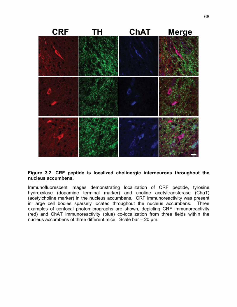
68
Figure 3.2. CRF peptide is localized cholinergic interneurons throughout the nucleus accumbens.
Immunofluorescent images demonstrating localization of CRF peptide, tyrosine hydroxylase (dopamine terminal marker) and choline acetyltransferase (ChaT) (acetylcholine marker) in the nucleus accumbens. CRF immunoreactivity was present in large cell bodies sparsely located throughout the nucleus accumbens. Three examples of confocal photomicrographs are shown, depicting CRF immunoreactivity (red) and ChAT immunoreactivity (blue) co-localization from three fields within the nucleus accumbens of three different mice. Scale bar = 20 µm.
THCRF ChAT Merge

69
Figure 3.3. CRF R1 co-localization to TH positive fibers.
Additional 60x images from two additional animals demonstrating co-localization of CRF R1-IR to TH-IR in the nucleus accumbens core. Scale bar = 10 µm.
CRF R1

70
Figure 3.4. CRF R2 antibody validation and localization in the nucleus accumbens of WT littermate and R2 KO mice.
The CRF R2 KO animals used in the study were derived from the original R2 KO line generated by Bale & Vale (2000) in which exons 10-12 have been deleted creating a functional CRF R2 KO mouse. Non-specific antibody staining in R2 KO mice may be a result of antibody recognition of an epitope on a non-functional truncated protein that has been translated or cross-reactivity of the antibody to another protein. Thus, we used antibodies that recognized an epitope of the c-terminal tail of the R2 protein (Santa Cruz sc-20550) that should be not translated and a Novus Biologicals antibody (NBP1-00768). Sections were prepared, processed and imaged in parallel (i.e. on the same days). a. Low power (20x) epifluorescent images of the nucleus accumbens of WT (left) and R2 KO (right) using the c-terminal R2 antibody (Santa Cruz sc-20550). Scale bar = 100 µm. b. High power (60x) confocal merged images demonstrating CRF R2 (red) and TH (green) localization in the nucleus accumbens of WT (left) and KO (right) animals. Using confocal imaging, there is virtually no detectable CRF R2 red immunofluorescence in the nucleus accumbens of KO mice. Scale bar = 10 µm. c. Exclusively for this set of images, both images were thresholded so that only the top half of red fluorescent pixels are visible. Under this stringent thresholding, red fluorescent puncta remain in the WT image and are not apparent in the R2 KO image. Scale bar = 10 µm d. Low power (20x) epifluorescent images of the nucleus accumbens of WT (left) and R2 KO (right) using the Novus Biologicals R2 antibody (Novus Biologicals NBP1-00768). Scale bar = 100 µm. e. High power image (100x) demonstrating demonstrating co-localization of CRF R2 (Novus Biologicals NBP1-00768) with TH staining. The same pattern of staining seen in the c-terminal Santa Cruz antibody is also seen with the Novus Biologicals R2 antibody. Scale bar = 10 µm.
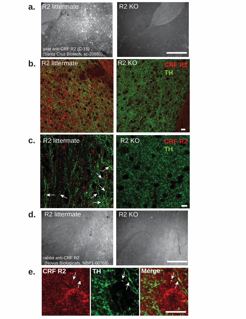
R2 littermate
R2 littermate R2 KO
R2 KO
R2 littermate R2 KO
rabbit anti-CRF R2 (Novus Biologicals, NBP1-00768)
goat anti-CRF R2 (C-15)(Santa Cruz Biotech, sc-20550)
THCRF R2 Merge
CRF R2TH
CRF R2TH
R2 littermate R2 KO
a.
b.
c.
d.
e.

71
Figure 3.5. CRF increases dopamine release in the nucleus accumbens through co-activation of CRF R1 and R2.
a. Representative evoked dopamine release before (grey line) and after (blue line) bath application of 100-nM CRF (top). Corresponding two-dimensional plots depicting changes in peak dopamine oxidation current (pseudocolor) with time as the abscissa and voltage potential as the ordinate (bottom). Dashed lines indicate the time at which stimulation was delivered. b. The mean + s.e.m. response to CRF (10, 100 or 1000 nM) or vehicle (0.07% acetic acid) in the last 10 min of 15-min application, normalized to pre-drug baseline. CRF (100, 1000 nM) increased dopamine release (Dunnet’s post-hoc t-test, p < 0.001, n = 11-18). c. Pre-treatment with either the selective CRF R1 antagonist (antalarmin, 1000 nM) or the selective CRF R2 antagonist (anti-sauvagine 30, 250 nM) blocked the CRF mediated increase in dopamine release (Dunnett’s post-hoc t-test, p < 0.05, n = 18-20). D & e. CRF (10,100, 1000 nM) did not did not increase evoked dopamine release in mice lacking the CRF R1 or CRF R2 receptor gene (gene by drug, p > 0.05, two-way ANOVA, n = 7-13). f. The selective CRF R1 agonist, stressin 1 (100 or 300 nM) did not increase evoked dopamine release (F2,32 = 1.833, p < 0.05 vs acetic acid vehicle, one-way ANOVA, n = 9-15). g. The selective CRF R2 agonist, urocortin 3 (100 or 300 nM) did not increase evoked dopamine release (F2,18 = 1.036, p < 0.05 vs DMSO vehicle, one-way ANOVA, n = 5-8). h. Co-application of stressin 1 and urocortin 3 (100 nM or 300 nM) significantly increased evoked dopamine release but not in the presence of antalarmin (1000 nM) and anti-sauvagine 30 (250 nM) together (Dunnett’s post-hoc test, p < 0.05 vs acetic acid/DMSO vehicle, n = 8 - 15). * p< 0.05, ** p < 0.01, ***p< 0.001.

100 nM D A
1s
1s1s
0
16
24-0.4
-0.4
+1.3E
app
(V a
gain
st A
g/A
gCl)
5 s 5 s
Baseline CRFa.
0 10 100 1000 80
90
100
110
120
130 ** **
CRF (nM)
[DA
] (%
Bas
elin
e)
CRF 100 nMAntalarmin/ASVG 30/
80
90
100
110
120
130 * *
CRF (nM)Antalarmin (nM)
ASVG 30 (nM)
100 1001001000
25000
00
[DA
] (%
Bas
elin
e)
0 10 100 100080
90
100
110
120
130CRF R1 KO
CRF (nM)
[DA
] (%
Ba
seli
ne
)
0 10 100 100080
90
100
110
120
130CRF R2 KO
CRF (nM)
[DA
] (%
Ba
se
line
)
0 100 300 30080
90
100
110
120
130
* *
Stressin 1 + Ucn 3 (nM)
+Anta+ASVG 30
[DA
] (%
Bas
elin
e)
0 100 300 80
90
100
110
120
130
Stressin 1 (nM)
[DA
] (%
Ba
se
line
)
0 100 30080
90
100
110
120
130
Ucn 3 (nM)
[DA
] (%
Bas
elin
e)
b.
e.
c.
d.
f. g.
h.

72
Figure 3.6. Evoked electrical currents detected by carbon fiber electrodes placed in the nucleus accumbens core are solely attributable to dopamine release. Nucleus accumbens slices were prepared from “dopamine deficient” (Thfs/fs; DbhTh/+) mice or littermate control mice in parallel. Following a baseline input-output curve, a dopamine recovery experiment was carried out. Following this recovery experiment, another input-output curve was obtained. Following five baseline stimulations, stimulations once every minute continued while L-DOPA (50 µM) was applied to slice. a. Averaged evoked responses before and after bath application of L-DOPA (50 µM). The insets are average cyclic voltammegrams (CV) corresponding to the averaged evoked response. As L-DOPA washes over the slice, the CV of the evoked response increasingly correlates with the stereotyped electrochemical fingerprint of dopamine. b. Input (electrical stimulation amplitude) – Output (subsequent current measured at the carbon fiber) for a single pulse stimulation in littermate control slices compared to slices from dopamine deficient mice before and after L-DOPA bath application. c. Evoked current elicited by a 20-p stimulation at 300 µA in slices prepared from littermate controls or slices from dopamine deficient mice either before or after L-DOPA bath application.
a.
b. c.
0 100 200 300 400 500
0
10
20
30
40DD WT littermate
DD w/ L-DOPADD - no L-DOPA
Single pulse (A)
Cu
rre
nt
(nA
)
0
20
40
60
80DD WT littermateDD - no L-DOPADD w/ L-DOPA
20 pulse (300 A)
Cu
rre
nt
(nA
)
1 s1 nA
L-DOPA (50 M)
-5 to 0 minutes 0 to 5 minutes 5 to 10 minutes 10 to 15 minutes

73
Figure 3.7. Time course of CRF or vehicle effect on evoked dopamine release.
a. & b. A single bi-phasic stimulating pulse (100-500 µA) was applied to the slice once every minute and dopamine release was measured at the carbon fiber electrode placed in the nucleus accumbens core. Following a five stable baseline currents, CRF (100 nM, 1000 nM) or vehicle (0.07% acetic acid) was bath applied to the slice. There was a small (5-10%) depression in dopamine release over time apparent in the vehicle group (p < 0.01 vs. 100%). In contrast, CRF increased evoked dopamine release by 20% above baseline (p < 0.001 vs. 100%) and 27-30% above vehicle, n = 13 and 18 for vehicle and CRF experiments respectively (drug by time, F20,560 = 2.994, p < 0.001, two-way ANOVA). c. When a single pulse stimulation to the slice every 300 seconds which eliminated the run down demonstrated in the original vehicle group done with a 60 second inter-stimulus interval (ISI). Compared to the time-matched vehicle group, CRF (100 nM) significantly increased evoked dopamine release by 20.3% in the nucleus accumbens (p < 0.01, t-test, n = 8-10).
-5 0 5 10 1570
80
90
100
110
120
130
140
150 VehicleCRF 100 nM
CRF or Vehicle
*
**
** ** **CRF (1000 nM)
Time (min)
[DA
] (%
Bas
elin
e)
VEH CRF 100 nM80
90
100
110
120
130 **
60 sec ISI
[DA
](%
Bas
elin
e)
VEH CRF 100 nM80
90
100
110
120
130 **
300 sec ISI
[DA
](%
Bas
elin
e)
a.
b. c.

74
Figure 3.8. Cannula placements for naïve used in the conditioned place preference assay.
a. Representative cresyl violet image from naïve cohort and cannula placements for each individual animal in the naïve group. b. Representative cresyl violet image from stress-exposed cohort and cannula placements for each individual animal in the stress-exposed group.

Naive
+1.34
+0.74
+0.86
+0.98
+1.10
+1.22
+1.46
+0.62
+1.58

75
Figure 3.9. Intra-nucleus accumbens infusion of CRF produces a conditioned place preference.
a. Schematic of the CPP paradigm. b. Representative trace of time allocation on the CRF and VEH paired sides during the post-conditioning test. c. Mice spent significantly more time in the CRF-paired chamber post-conditioning than the VEH-paired chamber compared to pre-conditioning (conditioning by drug, F1,12 = 6.435, p < 0.001 two-way repeated-measures ANOVA, n = 7 animals). d. Difference score from the post-conditioning test (CRF-paired chamber – VEH paired chamber) for intra-nucleus accumbens injections of CRF (500 ng/200 nl) bilateral, unilateral and CRF (5 ng/200 nl) bilateral. All three conditions yielded a significant preference (p < 0.05 for all conditions, one sample t-test) yet were not significantly different from each other (F2,19 = 1.198, p = 0.3235, one way ANOVA, n = 7-8) . Sham vehicle injected animals a significant preference for CRF similar to control animals, an effect that was absent in animals given a 6-OHDA unilateral lesion (p < 0.05, unpaired t-test, n = 10 for both groups).

CRFVehicle
Day 1PRE-TEST(30 min)
AM: ACSF or CRF(30 min)
PM: ACSF or CRF(30 min)
AM: ACSF or CRF(30 min)
PM: ACSF or CRF(30 min)
POST-TEST(30 min)
Day 2 Day 3 Day 4Day 1
PRE-TEST(30 min)
AM: ACSF or CRF(30 min)
PM: ACSF or CRF(30 min)
AM: ACSF or CRF(30 min)
PM: ACSF or CRF(30 min)
POST-TEST(30 min)
Day 2 Day 3 Day 4
a.
b.
PRE POST0
300
600
900
VEHCRF
#
**
Tim
e i
n c
ha
mb
er
(s)
500 ng 5 ng 500 ng (uni)
-100
0
100
200
300
400
Dif
fere
nc
e s
co
re (
CR
F c
ha
mb
er-
VE
H c
ha
mb
er,
s)
NS
Sham 6-OHDA-100
0
100
200
300
400*
500 ng (uni)
c.
d.

76
Figure 3.10. Pre- and post-test times for CRF (5 ng) bilateral injections and CRF (500 ng) unilateral injections. a. CRF (5 ng) or VEH were administered bilaterally into the nucleus accumbens during conditioning days. Mice spent significantly more time in the CRF-paired chamber post-conditioning than the VEH paired chamber compared to pre-conditioning (conditioning by drug, F1,14 = 5.415, p < 0.05 two-way repeated measures ANOVA, N= 8 mice). b. CRF (500 ng) or VEH were administered unilaterally into the nucleus accumbens during conditioning days. Mice spent significantly more time in the CRF-paired chamber post-conditioning than the VEH paired chamber compared to pre-conditioning (conditioning by drug, F1,12 = 11.77, p < 0.001 two-way repeated measures ANOVA, N= 7 mice).
Unilateral 500ng
PRE POST0
300
600
900
VEHCRF
***##
Tim
e s
pe
nt
in c
ha
mb
er
(s)
Bilateral 5ng
PRE POST0
300
600
900
VEHCRF
*
#
Tim
e s
pe
nt
in c
ha
mb
er
(s)
a.
b.

77
Figure 3.11. Intra-accumbens dopamine depletion with 6-OHDA blocks conditioned place preference for Intra-accumbens CRF microinfusion. a. Unilateral injection of 6-OHDA (2 µg/500 nl) into the nucleus accumbens significantly decreased tissue dopamine content compared to both the uninjected contralateral side assessed with HPLC on fresh frozen tissue in contrast to Sham vehicle (0.9% NaCl, 0.1% ascorbate) animals that did not show significant dopamine depletion (Drug by side, F1,18= 4.475, p < 0.05, two-way ANOVA with Bonferroni post-hoc tests). b. Unilateral 6-OHDA lesion did not effect locomotor activity compared to Sham injected animals (p >0.05, unpaired t-test). c. Sham vehicle injected animals significantly preferred the CRF paired chamber following conditioning (conditioning by drug, F1,18 = 6.954, p < 0.05, two-way repeated measures ANOVA with Bonferroni post-hoct tests). d. 6-OHDA injected animals did not demonstrate a preference for the CRF paired chamber following conditioning (conditioning by drug, F1,18 = 0.004, p > 0.05, two-way repeated measures ANOVA). # = p < 0.05 for the interaction, * p < 0.05, ** p < 0.01 post-hoc tests. n = 10 for both Sham and 6-OHDA groups.
PRE POST0
300
600
900
SHAM
**#
VEHCRF
Tim
e i
n c
ha
mb
er
(s)
PRE POST0
300
600
900
NS
6-OHDA
Tim
e i
n c
ha
mb
er
(s)
Sham 6-OHDA0
20
40
60
80
100
ContraIpsi
NS ***#
Tis
sue
do
pa
min
e c
on
ten
t(n
g/m
g p
rote
in)
SHAM 6-OHDA0
2000
4000
6000
8000NS
To
tal
dis
tan
ce
mo
ved
(c
m)
a. b.
c. d.

78
Figure 3.12. CRF is endogenously released in the nucleus accumbens to mediate exploration of a novel object.
a. Schematic of novel object exploration assay. b. Animals spent significantly more time investigating a novel object placed in the center of an open field when treated with intra-accumbens bilateral microinfusions of vehicle (lactated ringer’s with 1% acetic acid) compared to the non-selective CRF receptor antagonist, α-helical CRF (500 ng) (treatment by stimulus, F1,18 = 4.62, p < 0.05, two-way repeated-measures ANOVA, n = 10).
TEST 1
Object 1or
Object 2
VEH or α-Helical
TEST 2
Habituation
15 min 15 min
48 hr Object 2or
Object 1
α-Helical or VEH
Habituation
15 min 15 min
VEH -Helical0
100
200
300
BaselineNovel object
***#
Tim
e i
nc
en
ter
(s)
a.
b.

79
Figure 3.13. Stress exposure abolishes the CRF mediated increase in evoked dopamine release without recovery for at least 90 days.
a. Schematic of the swim stress paradigm and recovery periods. Nucleus accumbens slices were prepared 30 minutes, 7 days, 30 days or 90 days after the final swim session (black arrows). b. In contrast to naïve mice (blue bars), CRF (100 nM; closed bars) did not increase evoked dopamine release compared to vehicle (open bars) in nucleus accumbens slices from mice exposed to swim stress (red bars). The CRF response did not recover at any of the time points tested compared to naïve mice (stress exposure by drug, F4,116 = 4.852, p < 0.01, two-way ANOVA, n = 8 - 18). c. Time-line schematic of rescue experiments. Mice were pre-treated with either VEH (5% DMSO, 20% chremafor in saline) or RU 486 (30 mg/kg) 30 minutes prior to each swim stress day (arrows). Mice were allowed to recover for seven days and then slices were prepared. d. CRF significantly increased dopamine release compared to VEH in nucleus accumbens slices prepared from animals that had been pre-treated with RU486 (30 mg/kg) compared to VEH (i.p.) prior to each stress session (pre-treatment by drug, F1,27 = 4.841, p < 0.01, two way ANOVA with Bonferonni post-hoc-tests, n = 6-10).

a.
b.
6’15’
Day 1 Day 2
6’6’ 30’24 hr
15’ 30 days7 days90 days
c.
d.
1-V 1-C 2-V 2-C 3-V 3-C 4-V 4-C 5-V 5-C
80
90
100
110
120
130
0 90307
Days following FSS
Naive
**##
###
VEHCRF
###
###
NS NS
NSNS
[DA
] (%
Bas
elin
e)
VEH RU 486 80
90
100
110
120 VEH CRF
NS
**#
i.p. 30 mg/kg i.p.
[DA
] (%
Bas
elin
e)
VEH/RU
15’ 24 h 30 6’ 6’ 7 d
Sac
15’ 24 h
VEH/RU
30’ 6’ 6’ 7 d
Sac
30’ Day 1 Day 2

80
Figure 3.14. CRF is abolished following one- or two-day swim stress exposure.
In comparison to slices from naïve mice, exposure to either the modified two-day Porsolt paradigm used for all studies as well as the canonical one-day Porsolt paradigm significantly abolished CRF-mediated increase in evoked dopamine release. Animals exposed to one 15-minute swim stress were sacrificed 24 hours to time-match this set of animals with the two-day swim stress paradigm (stress exposure by drug, F2,67 = 4.588, p < 0.05, two way ANOVA, n = 5-18).
N-V N-C FSS-V FSS-C FSS1-V FSS2-C
80
90
100
110
120
130
Naive FSS (2 day) FSS (1 day)+ 24 hr
##
NS
**VEHCRF (100 nM)
NS
##
[DA
] (%
Ba
se
lin
e)

81
Figure 3.15. Kappa opioid regulation of dopamine release in the nucleus accumbens is unaffected in mice exposed to swim stress. a. Time course demonstrating the effect of the kappa opioid receptor agonist U69,593 (1µM) and subsequent reversal by the kappa opioid receptor antagonist norBNI (1 µM) on stimulated dopamine release in the nucleus accumbens core of naïve and stress-exposed mice. There were no significant differences in kappa opioid receptor mediated inhibition of dopamine release in slices from naïve versus stress-exposed animals (time by stress exposure, F1,14 = 0.3508, p > 0.05, two-way repeated measures ANOVA). b. Mean data showing the percent change in dopamine release from baseline in the last ten minutes following U69,593 application and the last ten minutes of norBNI reversal. U69,593 produced on average a 44.08-% inhibition in nucleus accumbens animals in naïve animals and a 41.12-% in the nucleus accumbens of stress-exposed animals and in both cases was fully reversed by norBNI. There was no significant difference in mean responses between naïve and stressed groups (p > 0.05, Bonferroni post-hoc t-test, n = 6-10).
U69,593 norBNI
-60
-50
-40
-30
-20
-10
0
10
NaiveFSS
+ U69,593
[DA
](
%c
ha
ng
efr
om
Ba
selin
e)
-5 0 5 10 15 20 25 300
20
40
60
80
100
120 U69,593 (1M)
norBNI (1M)
FSSNaive
Time (min)
[DA
](%
Ba
seli
ne
)
a.
b.

82
Figure 3.16. Animals displayed enhanced depression-like behavior compared to naïve animals even up to 90 days following initial stressor exposure.
Mice were given a five-minute forced swim test either in the absence of prior stress history or 7 or 90 days following exposure to an initial 2-day swim stress. Compared to stress-naïve animals, animals exposed to swim stress both 7 and 90 days prior showed significantly higher immobility during the FST indicating persistent depression-like behavior (F2,25 = 8.287, p < 0.01, one way ANOVA with Neuman-Keuls.post-hoc t-test, n = 8-11).
Naive 7 900
100
200
300
Time (d) following FSS
** **
Imm
ob
ilit
y(s
ec
)

83
Figure 3.17. Basal evoked dopamine release was not affected by stress exposure.
Data were collected from nucleus accumbens slices prepared from naïve mice and stress-exposed mice that were allowed to recover for 30 days. A single-pulse electrical stimulation was delivered to three distinct sites in a nucleus accumbens slice and dopamine current was measured at each site. The evoked dopamine current for three sites was averaged. Following the conclusion of data collection, carbon fiber electrodes were calibrated using a flow cell system to 1 µM dopamine. Averaged current responses were converted to dopamine concentration. There were no differences in evoked dopamine release between naïve and stress-exposed animals (unpaired t-test, p < 0.05, n = 6-7).
1 pulse stimulation
Naive FSS (2 day)0
50
100
150
200
250
30 d recovery
NS
[DA
] n
M

84
Figure 3.18. Loss of CRF response following stress exposure is not age related.
For all other experiments, animals were 60 -150 days old. However, stress-exposed animals allowed to recover for 90 days were >180 days old. To control for possible age-related effects on CRF responsivity, naïve-aged matched animals were interleaved (sacrificed and CRF response tested every other day) with stress-exposed animals allowed to recover for 90 days. Mice assigned to either the naïve or stress-exposed group were shipped on the same date and acclimated to the vivarium for the same amount of time. CRF significantly increased evoked dopamine release in nucleus accumbens slices from naïve age-matched mice compared to vehicle application, but had no effect on evoked dopamine release in the nucleus accumbens from stress-exposed animals that had recovered for 90 days compared to vehicle application (stress exposure by drug, F1,42 = 10.97, p < 0.01, two way ANOVA, n = 7-15).
80
90
100
110
120
NaiveAge-matched
FSS - 90 dayrecovery
** VEHCRF (100 nM)
##
[DA
] (%
Bas
elin
e)
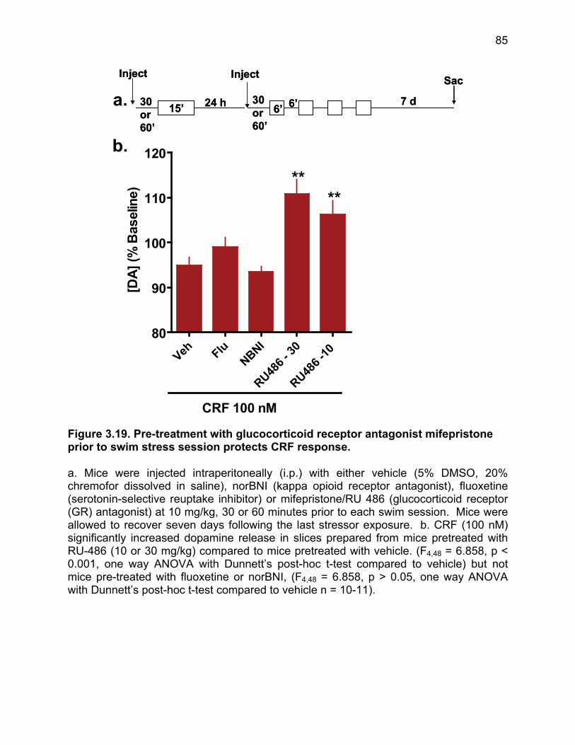
85
Figure 3.19. Pre-treatment with glucocorticoid receptor antagonist mifepristone prior to swim stress session protects CRF response.
a. Mice were injected intraperitoneally (i.p.) with either vehicle (5% DMSO, 20% chremofor dissolved in saline), norBNI (kappa opioid receptor antagonist), fluoxetine (serotonin-selective reuptake inhibitor) or mifepristone/RU 486 (glucocorticoid receptor (GR) antagonist) at 10 mg/kg, 30 or 60 minutes prior to each swim session. Mice were allowed to recover seven days following the last stressor exposure. b. CRF (100 nM) significantly increased dopamine release in slices prepared from mice pretreated with RU-486 (10 or 30 mg/kg) compared to mice pretreated with vehicle. (F4,48 = 6.858, p < 0.001, one way ANOVA with Dunnett’s post-hoc t-test compared to vehicle) but not mice pre-treated with fluoxetine or norBNI, (F4,48 = 6.858, p > 0.05, one way ANOVA with Dunnett’s post-hoc t-test compared to vehicle n = 10-11).
Inject
30or60’
15’ 24 h
Inject
30or60’
6’ 6’ 7 d
SacInject
30or60’
15’ 24 h
Inject
30or60’
6’ 6’ 7 d
Sac
a.
b.
Veh FluNBNI
RU486
- 30
RU486
-10
80
90
100
110
120
****
CRF 100 nM
[DA
](%
Bas
elin
e)

86
Figure 3.20. Stress exposure switches CRF action in the nucleus accumbens from appetitive to aversive in a conditioned place preference paradigm.
Seven days prior to conditioning, one group of animals was exposed to the same two-day stress paradigm used in Figure 4. Both stress-naïve and stress-exposed animals were handled daily for four days prior to conditioning. a. Representative trace of time allocation on the CRF and VEH paired sides during the post-conditioning test for stress-exposed animals. Stress-exposed mice spent significantly less time in the CRF-paired chamber post-conditioning than the VEH-paired chamber compared to pre-conditioning (conditioning by drug, F1,10 = 5.824, p < 0.01 two-way repeated-measures ANOVA with post-hoc Bonferonni t-tests, n = 6 animals). d. Difference in time spent in CRF – vehicle paired chambers during the post-test for naïve (blue) or stress-exposed animals allowed to recover for 7 or 90 days (red). Difference scores were significantly different between naïve and stress-exposed animals (F2,18 = 6.870, p < 0.01, one-way ANOVA, n = 6-8). e. Stress exposure significantly abolished the stimulation of investigatory behavior of a novel object by CRF (p < 0.05, unpaired t-test, n = 9-10).
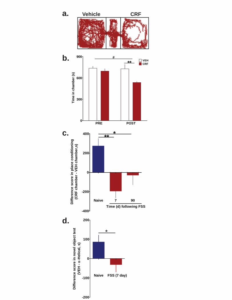
a.
b.
c.
CRFVehicle
d.
Naive 7 90
-400
-200
0
200
400 ***
Time (d) following FSS
Dif
fere
nc
e s
co
re i
n p
lac
e c
on
dit
ion
ing
(CR
F c
ha
mb
er
- V
EH
ch
am
be
r,s)
PRE POST0
300
600
900VEHCRF
#
**
Tim
e in
ch
amb
er (
s)
Naive FSS (7 day)
-200
-100
0
100
200
*
Dif
fere
nc
e s
co
re i
n n
ove
l o
bje
ct
test
(VE
H -
-Hel
ical
, s)

87
Figure 3.21. Pre- and post-test times for CRF conditioned place preference in mice exposed to two-day FSS and allowed to recover for 90 days.
Mice that had been exposed to 2-day swim stress 90 days prior to conditioning did not show a conditioned place preference to the CRF-paired context compared to naïve animals (conditioning by drug, F1,14 = 0.1035, p >0.05, two-way repeated measures ANOVA, n = 8).
PRE POST0
300
600
900 VEHCRFNS
FSS (90 d recovery)
Tim
e i
n c
ha
mb
er
(s)

88
Figure 3.22. Stress exposure abolishes CRF-dependent component of novel object exploration.
Animals were exposed to two-day repeated swim stress 7 days prior to the first test day of the novel object exploration task. Identically to naïve animals, stress-exposed mice were given infusions of vehicle or α-helical CRF (500 ng) in a counter-balanced fashion across test days, prior to placement in an open field. While introduction of a novel object significantly increased center time in both drug conditions, there was not a significant drug interaction, indicating that stress exposure abolished the CRF-dependent component of novel object exploration (stimulus by drug, F1,16 = 0.004, p > 0.05, two-way repeated measures ANOVA, n = 9). ** p < 0.01 for Bonferroni post-hoc t-tests.
Veh -Helical0
100
200
300BaselineNovel object
****
NS
Tim
e i
n c
en
ter
(se
c)

89
Table 3.1. Quantification of EM labeling in 100-nm sections through the rostro-caudal axis of the nucleus accumbens to assess co-localization of CRF receptors and TH immunoreactivity.
To estimate the proportion of dopamine terminals that express at least one CRF receptor, we used the equation P = 1 - (1 - p)n where p is the probability of observing a CRF-receptor immunogold particle per section of a TH-positive terminal and n is the number of sections per terminal. CRF receptor immunoreactivity was observed on 14.58 % of section of TH-positive terminals (i.e., p = 0.1458). Given that dopamine terminals are approximately 1 µm in diameter, each total three-dimensional terminal profile occupies approximately ten 100-nm sections (i.e., n = 10). Therefore, we estimate that 80 % of dopamine terminals in the nucleus accumbens express CRF receptors.
TH-axon terminals CRFr in TH-at Percentage
Mouse 1 299 41 13.71%
Mouse 2 367 38 10.35%
Mouse 3 390 75 19.23%
Total 1056 154 14.58%

90
Chapter 4: Repeated stress dysregulates kappa opioid receptor signaling in the dorsal raphe through a p38 MAPK dependent mechanism
The following chapter has been adapted from:
Lemos JC, Roth CA, Messinger DI, Gill HK, Phillips PEM, Chavkin C. Repeated stress dysregulates kappa opioid receptor signaling in the dorsal raphe through a p38 MAPK dependent mechanism. Under review at Journal of Neuroscience
Specific contributions:
J.C.L. carried out the electrophysiology experiments. J.C.L. and C.A.R conducted
immunohistochemistry experiments. C.A.R. conducted quantitative analysis of
immunohistochemistry experiments and conducted analgesia experiments. D.I.M. bred
all transgenic mice lines. H.K.G. analyzed behavioral data. J.C.L., P.E.M.P. and C.C.
developed the conceptual and experimental framework.
Introduction Adaptive responses to stress exposure are critical to an organism’s survival and
rely upon a precisely tuned constellation of neurotransmitters and neurohormones
acting both centrally and peripherally 30. Moreover, the etiology of stress-induced mood
disorders is thought to involve a combination of pathological neuroadaptations that
include alterations in both stress-related neurocircuitry and signaling 23. Dynorphins, the
endogenous ligands for the kappa opioid receptor (KOR) 161, are released and up-
regulated during stress exposure 202,236,248, and these endogenous opioid neuropeptides
have emerged as critical mediators of several stress-related behaviors196,249. For
example, genetic deletion of either prodynorphin or KOR or pharmacological blockade

91
by KOR antagonists reduce stress-induced anxiety-like, depression-like, and
proaddictive behaviors (see250). However, the cellular and molecular mechanisms by
which the dynorphin-KOR system regulates neuronal excitability in limbic regions and
ultimately produces its behavioral effects remain unclear.
Activation of KOR either pharmacologically or following release of endogenous
dynorphins results in both membrane-delimited G regulation of ion channel
conductance and mitogen activated protein kinase (MAPK) signaling (Bruchas et al.,
2010). Prolonged or repeated activation of KOR also stimulates the G-protein kinase 3 /
-arrestin signaling cascade that subsequently recruits and activates p38 MAPK
202,251,252, and p38 MAPK activation has been shown to be required for the aversive
qualities of stress as well as promoting passive coping strategies 202,253. Recently, we
demonstrated that KOR-dependent activation of p38 MAPK in 5-HT neurons in the
DRN is both necessary and sufficient to induce a negative affective state 253,254.
The importance of the DRN for stress-related behaviors is well established 13.
The DRN is a principal serotonergic nucleus in the brain that sends afferents to many
forebrain limbic regions 255-258 and is specifically activated during exposure to
uncontrollable stress 13,86,254,259,260. Therefore, elucidating the regulation of DRN activity
by stress-related neuropeptides in naïve and stress-exposed individuals is important to
understand how the pathological effects of stress impact brain functioning.
In the present study, we measured the effects of KOR activation in DRN using
whole-cell voltage clamp electrophysiological techniques to isolate glutamatergic and
GABAergic fast synaptic activity as well as isolating post-synaptic G-protein gated

92
inwardly rectifying potassium (GIRKs, Kir3) currents. We then investigated how these
processes were modified by repeated exposure to stress and specifically sought to
identify p38 MAPK-dependent defects in this regulation caused by repeated stress. We
hypothesized that repeated stress would result in sustained dynorphin release that
would in turn disrupt KOR regulation of 5-HT neuronal excitability in the DRN via a p38
MAPK-dependent process.
Methods
Subjects: Male C57BL/6 mice, age >50 days, were maintained under a 12-h light–dark
cycle (7a.m. to 7p.m. light) with access to standard food and water ad libitum. All
procedures on animal subjects were approved by the University of Washington IACUC
committee. Mice that were housed together (2 - 4 per cage) were subjected to the
same behavioral treatment. Prodynorphin (Dyn -/-) knock-out mice and p38αCKOSERT
mice were derived as described in 208 and 253 respectively and have been backcrossed
to C57BL/6 background >10 generations.
Swim stress: Mice were subjected to a two-day swim stress procedure in which they
were exposed to a 15-min swim session on day one, then were exposed to four six-min
swim sessions in 29.0 – 31.0 °C water, separated by six min and conducted under
bright light (690-700 lux) conditions on day 2 as previously described 170.
Tail flick: Stress-induced analgesia was assessed using the warm water (52.5°C) tail
withdrawal assay as described 261,262.
Immunohistochemistry

93
Tryptophan hydroxylase (TPH) Immunohistochemistry staining and intracellular labeling
During electrophysiological recordings, cells were filled with biocytin (0.1%)
(Sigma-Aldrich) present in the recording electrode. After recording, slices were fixed by
submersion in 4% paraformaldehyde prepared in 0.1M phosphate buffer (PB; pH 7.4).
Sections were incubated with mouse anti-TPH antibody (1:500, Sigma-Aldrich) for 12-
16 hr at room temperature (RT). Subsequently, immunohistochemical labeling was
visualized using Alexa Fluor 488 (1:500; Invitrogen) conjugated goat anti-mouse
secondary antibody for 90-120 min at RT. Biocytin was visualized using streptavidin
conjugated Alexa Fluor 647 (1:500, Invitrogen) contained in the same secondary
antibody mixture. Between incubations, slices were rinsed with PBS (3 x 10 min) and
all incubations were done with mild agitation on an orbital shaker.
Phospho-antibody Immunohistochemistry
Methods for immunohistochemistry experiments were similar to those reported
previously (Bruchas et al. 2007). Mice were intracardially perfused with 4%
paraformaldehyde in 0.1M PB. It has been our experience that phospho-protein
immunoreactivity (ir) is of higher quality if PBS perfusion prior to 4% PFA is omitted.
Brains were dissected and cryoprotected with 30% sucrose in 0.1M PB at 4°C overnight
(or until the brains submerged), frozen and cut into 30 μm sections using a Leica
SM200R microtome, and placed in 0.1M PB with 0.1% sodium azide until processing.
We had previously generated a rabbit polyclonal antibody against the phosphorylated
Ser369 residue of KOR 206 that is now available commercially at Abcam. We also
generated a rabbit polyclonal antibody against the phosphorylated-Tyr 12 residue of KIR
3.1 (GIRKp) as described in Ippolito et al. (2005). Standard immunohistochemical

94
procedures were performed to obtain phospho-KOR (KORp) (Abcam, ab63511),
phospho-p38 MAPK (mouse monoclonal sc-7973) and p38α (rabbit polyclonal sc-535)
(Santa Cruz) and pY12-KIR 3.1staining. Briefly, floating sections were washed 3 x 10
min in PBS and then blocked for 60 min in 5% normal goat serum, 0.3% Triton-X in
PBS. Importantly, we have found that for these phospho-antibodies use of floating
sections produces higher quality staining than thaw mounted sections. Sections were
incubated in primary antibody at different concentrations, temperatures and incubations
for each antibody: KORp (1:200, 72 hr at 4°C); mouse anti-phospho-p38 MAPK/ rabbit
anti-p38α (Santa Cruz) (1:50, 36 hr RT or 72 hr at 4°C); affinity purified GIRK-p (1:100
for 72 hr at 4°C; the concentration is based on a 750 µg protein/ml yield as described
(Ippolito et al., 2005)). Often slices were co-labeled with mouse anti-TPH (Sigma) at
1:1000; guinea pig anti-Dynorphin B (Peninsula Laboratories) was used at 1:200.
Slices are then washed in PBS 10-15 min x 3 and incubated in goat Alexa Fluor
antibody (mouse or rabbit, respectively) at 1:500 to 1:750 for 2 hr at RT. Following
secondary antibody incubation, slices were washed 3 x 10 min in PBS, then 2 x 10 min
in 0.1M PB. Sections were then mounted on Superfrost plus slides and converslipped
with Vectashield (Vector Laboratories).
Standard epifluorescent and confocal microscopy techniques (Nikon Eclipse
E600 and Leica SL, respectively) were used to image sections for both TPH
confirmation and phospho-antibody staining. For comparisons of phospho-ir from
different behavioral treatment groups, three animals for each group were perfused,
sectioned and processed in parallel. Imaging of sections from animals exposed to
different treatments were done at the same time with the same camera settings. As it is

95
typical for different rounds of staining to yield different immunoreactive intensities,
images from each treatment group displayed in this study came from the same round of
staining. The pattern of KORp-ir and GIRKp-ir across treatment groups seen in the
dorsal raphe was similar to that previously reported both in vitro and in vitro
181,202,251,263,264.
Phospho-p38-ir quantification
The selectivities of both the p38α and phospho-p38 MAPK antibodies were previously
established using both viral vector expression and conditional gene knock-out
approaches 253. We quantified phospho-p38 MAPK-ir using Metamorph software as a
ratio of phospho-p38 MAPK positive cells to p38α positive cells (i.e. co-localized).
Similar quantification of GIRKp-ir and KORp-ir could not be performed since the
antibodies against the unphosphorylated forms of GIRK and KOR were also raised in
from rabbits. We have previously confirmed that p38α is the isoform that is
phosphorylated following KOR activation 253. Cells are considered positive for the
protein if the intensity of the immunoreactivity is one-standard deviation above the
average background pixel intensity.
Electrophysiology
Procedures were similar to those previously reported 88,265. Mice were
decapitated, head placed in ice cold sucrose buffer during the dissection, and the brain
rapidly removed and blocked. The blocked tissue was mounted on the stage of a Leica
VT1000 S vibratome and surrounded in oxygenated sucrose buffer. Sections (200 m
thick) were cut through the raphe nuclei in sucrose and then placed in oxygenated

96
95%O2/5%CO2 ACSF incubated in a 35-37˚C bath for 1 hr. The slices were then
removed from the bath and kept in oxygenated ACSF at RT. ACSF was composed of
(mM): NaCl 124, KCl 2.5, NaH2PO4 2, CaCl2 2.5, Dextrose 10 and NaHCO3 26. The
sucrose buffer was ACSF in which NaCl was replaced by 248 mM sucrose.
Slices were placed in a recording chamber (Warner Instruments, Hamden, CT)
and continuously perfused with oxygenated ACSF at approximately 1.5 ml/min,
maintained at 30-32˚C by an inline solution heater (TC-324, Warner Instruments). For
recording EPSCs or GIRK currents, the resistance of the electrodes was 5-10 Mwhen
filled with an internal solution of (mM) K-gluconate 130, NaCl 5, Na phosphocreatine 10,
MgCl2 1, EGTA 0.02, HEPES 10, MgATP 2, Na2GTP 0.5, 0.1% Biocytin, pH 7.3. For
measuring IPSCs, the resistance of the electrodes was 4-8 Mwhen filled with an
intracellular solution of (mM) K-gluconate 70, KCl 70, NaCl 2, Na phosphocreatine 10,
EGTA 4, HEPES 10, MgATP 2, Na2GTP 0.3, 0.1% Biocytin, pH 7.3. Whole cell
recordings in voltage clamp were performed on cells in the ventromedial and
dorsomedial aspect of the DRN. The cell was voltage clamped at -70 mV using an
Axopatch 200B amplifier (Molecular Devices, Foster City, CA). Signals were digitized
by a Digidata 1440 A/D converter (Molecular Devices) and stored using pClamp 10.2
software (Molecular Devices). EPSCs were isolated using picrotoxin (10 µM) (Sigma-
Aldrich) and CGP 55845 (1 µM) (Tocris Biosciences). IPSCs were isolated using DNQX
(20 µM) and APV (50 µM) (Tocris Biosciences). For evoked recordings, the stimulating
electrode was placed 150-200 µm dorsolateral of the recording site. Paired pulse
stimulation (1 msec pulse width, 50 msec inter-stimulus interval, 100-1000 µA) was
delivered every 10 sec. A mid-range output response was used as baseline for each

97
cell (e.g. approximately the middle of an input-output curve) to allow for observation of
either positive or negative regulation by U69,593 (National Institute of Drug Abuse Drug
Supply Program). For mPSCs, TTX (1 µM) (Calbiochem) was added to the bath. For
GIRK current experiments, once a recording had been established, the slice was
bathed in 5.5 mM K+ ACSF (from 2.5 mM K+) to enhance the currents in the cells at
hyperpolarized potentials. As expected, when the slice was incubated in high [K+], the
cells became more depolarized and inward rectification was more apparent. This
increased current was blocked by 100 µM BaCl2 266. The cells were subjected to a
voltage ramp protocol in which they were brought from -120 mV to -50 mV over 10 sec.
After three stable ramp measurements (the average of three sweeps) were obtained,
U69,593 (1000 nM) was bath applied and the slice was allowed to equilibrate for 3 min.
Subsequently three ramp measurements were taken every 3 min for 12 additional min
and the last three ramp measurements (after 15 min of U69,593 application) were
averaged. Following U69,593 application and stable current responses, BaCl2 (100 µM)
was added to distinguish Ba2+-sensitive GIRK currents.
Data analysis and statistics
Electrophysiological data were analyzed using Clampfit 10.2 (Molecular Devices)
and MiniAnalysis 6.0.7 software (Synaptosoft, Inc.). One-way repeated measure
ANOVAs or paired t-tests were used for within-cell experiments (pre-drug baseline vs.
post-drug effect). We used unpaired t-tests as well as one-way and two-way ANOVAs
as specified in the Results, for comparisons made across behavioral treatment groups
and genotypes. The responses of individual cells were considered separate N, as is
standard for electrophysiology experiments, however, when comparing differences

98
between different behavioral treatment groups (i.e. naïve vs. stress), a minimum of 3
animals from at least two different cages were used per group.
Results The dorsal raphe is a heterogeneous nucleus containing approximately 70% 5-HT and
30% non 5-HT containing neurons 267. Recordings were confined to the dorso- and
ventro-medial aspect of the dorsal raphe (see representative low power image; Figure
1a, left panel), which has the highest density of the 5-HT-containing neurons.
Additionally, during electrophysiological recordings, cells were filled with biocytin and
subsequently slices were post-fixed and processed using standard
immunohistochemical procedures to distinguish serotonergic (TPH-ir positive) and non-
serotonergic cells (Figure 4.1a). Only 12% of cells were not positively identified as
serotonergic neurons, and these were not included in the subsequent analysis.
Kappa opioid activation in serotonergic dorsal raphe neurons has a net inhibitory
effect
Following 5-10 min of stable baseline evoked EPSC amplitude responses, the kappa
opioid receptor agonist, U69,593 (500 nM, then 1000 nM) was bath applied to the slice;
both eEPSC amplitude and paired paired-pulse ratio were measured. Representative
traces are provided demonstrating that KOR activation by U69,593 decreased eEPSC
amplitude of the first evoked EPSC (red line) compared to baseline responses (black
line) (Figure 4.1b, inset). U69,593 at 500 nM and 1000 nM significantly inhibited
eEPSC amplitudes by 31 ± 6% and 37 ± 8% respectively (F2,29 =13.53, p< 0.0001, one-
way repeated measures ANOVA with Dunnett’s post-hoc t-test, Figure 4.1b and c). We
verified that cumulative dosing did not result in KOR desensitization by showing that 5

99
µM U69,593 added to a previously untreated slice did not produce a greater inhibition
than 500 or 1000 nM added cumulatively (data not shown). Inhibition of eEPSC
amplitude significantly correlated with the increase in paired pulse ratio (r2 = 0.39, p <
0.05) (Figure 4.1d), indicating that KOR activation reduced probability of glutamate
release via a presynaptic mechanism.
To further assess the site of KOR action, we also measured the effect of U69,593
(1000 nM) on miniature EPSC (mEPSC) frequency and amplitude, and representative
traces are shown (Figure 4.2a). Cumulative histograms of both the inter-event interval
(Figure 4.2b) and amplitude (Figure 2c) corresponding to these traces are also shown.
U69,593 significantly decreased mEPSC frequency from baseline (from 7.1 ± 1.8 Hz to
4.1± 1.1 Hz, p <0.05), which was blocked by pre-treatment with the KOR antagonist
norBNI (1 µM) (from 7.6 ± 1.3 Hz to 6.1± 1 Hz, p > 0.05) (norBNI treatment by time, two-
way repeated measures ANOVA, Bonferroni post-hoc, * p <0.05, Figure 4.2d). U69,593
did not significantly affect mEPSC amplitude in either the presence or absence of
norBNI (norBNI treatment by time, two-way repeated measures ANOVA, Bonferroni
post-hoc, p > 0.05, Figure 4.2e). These data confirm that acute activation of KOR
presynaptically depresses excitatory synaptic transmission.
Because KORs regulate both glutamatergic and GABAergic transmission with
similar efficacies in other brain regions 111,113,174,268,269, we next assessed the effects of
U69,593 on both evoked inhibitory post-synaptic current (eIPSC) amplitude and
miniature IPSC frequency and amplitude. As in the eEPSC experiments, a paired pulse
stimulation was delivered to the slice and baseline recordings of pharmacologically
isolated eIPSCs (e.g. in the presence of DNQX and APV) were obtained. KOR

100
activation by U69,593 had no significant effect on eIPSC amplitude (representative
traces, Figure 4.3a; time course, Figure 4.3b; p > 0.05) nor did it significantly affect
paired pulse ratio (p > 0.05). Similarly, U69,593 had no effect on mIPSC (representative
traces, Figure4. 3c), frequency (paired t-test, p > 0.05, Figure 4.3d), or amplitude
(paired t-test, p > 0.05, Figure 4.3e). These data demonstrate that KORs
presynaptically inhibit excitatory synaptic input without affecting GABAergic
transmission in DRN 5-HT neurons, producing a net reduction in excitability.
KOR activation has also been shown to have direct, post-synaptic effects in the
cells of the spinal trigeminal nucleus 270. However, this has not previously been
examined in DRN. Both the conductance (slope) and peak inward currents were
determined for pre-drug and post-drug voltage ramp measurements (see representative
traces, Figure 4.4a). U69,593 significantly increased both the conductance (3.7± 0.4 nS
to 4.5 ± 0.4 nS, paired t-test, p < 0.01, Figure 4.4b) and the peak Ba2+-sensitive inward
current (87.8 ± 24.1 pA to 145 ± 24.0 pA, paired t-test, p < 0.01, Figure 4.4c). U69,593
increased the GIRK current 48.7 pA above baseline, and this effect was blocked by 1
µM norBNI pretreatment (unpaired t-test, p < 0.001, Figure 4.4d). These data suggest
that in stress-naïve animals, KOR activation has an inhibitory effect on serotonergic
neuronal excitability through both pre- and post-synaptic sites of action.
Repeated stress exposure reduces KOR inhibitory regulation on 5-HT neuronal
excitability post-synaptically
We found evidence for dynorphin B-ir within the DRN, but in contrast to a
previous study by Fu et al. (2010), this staining was present in both TPH-ir positive and

101
TPH-ir negative cells (Figure 4.5a, yellow and white arrows, respectively), suggesting
that dynorphin may be locally released from a heterogeneous population of cells that
includes serotonergic neurons. To evoke endogenous dynorphin release in DRN, we
used the previously validated two-day repeated force swim stress (FSS) paradigm 170,271
(Figure 4.5b). This modified Porsolt procedure produced escalating immobility within the
first 15-min session as well as enhanced immobility over the second day of swim
sessions (F6,54 = 40.59, p < 0.0001, one-way repeated measures ANOVA, Figure 4.5a),
indicative of increased passive coping behavior 34. In agreement with our previous
findings 170, this two-day swim paradigm produced significant stress-induced analgesia
(as measured with the warm water tail immersion assay) that could be prevented by
norBNI pre-treatment (time by behavioral treatment, F2,36 = 9.969, p < 0.001, two-way
repeated measure ANOVA, Figure 4.5d), indicating that this stress exposure evoked
dynorphin release. We previously showed that dynorphin release can be detected by
an increase in KOR phosphorylation (KORp-ir) (McLaughlin et al., 2003b); agonist
activated KOR is a substrate for G-protein Receptor Kinase (GRK3) which
phosphorylates ser369 in the carboxy-terminal tail of KOR. Endogenous dynorphin
released by neuropathic pain stress or administration of the stress-related peptide
corticotropin releasing factor intracerebroventricularly increased KORp-ir in wild type
mice but not in mice lacking the gene for preprodynorphin (Dyn -/-) 205,207. Using
increases in KORp-ir to detect sites of endogenous dynorphin action in the present
study, we found that KORp-ir was significantly increased in the dorsal raphe nucleus
evident 30 min following the cessation of the last swim (Figure 4.5e). This elevation in
KORp-ir returned to basal levels in animals that were allowed to recover for 24 hr

102
following the last swim session (Figure 4.5e). At higher magnification (60 and 100 x),
we observed that KORp-ir puncta were robustly localized to the somata of DRN cells
and to a lesser extent outside of the somata, interdigitated with synaptophysin or vGlut1
postive puncta (data not shown). However, even at this resolution we could not
distinguish KORp-ir in presynaptic from post-synaptic subcellular profiles. These results
confirmed that the two-day swim stress paradigm produced escalating depression-like
behaviors, robust and transient activation of KORs through release of endogenous
dynorphins, and dynorphin-KOR-dependent stress induced analgesia.
Repeated swim stress exposure significantly reduced subsequent in vitro
postsynaptic responses to U69,593 in slices prepared 30 min after the final swim
session (representative traces, Figure 4.6a). There was no significant difference in
basal GIRK current between naïve and swim-stressed groups (Naïve: 76.4 ± 23 pA;
FSS (2d): 66 ± 19.5 pA, unpaired t-test, p > 0.05). The increase in GIRK current caused
by U69,593 (1 µM) was 51% smaller in serotonergic neurons recorded in slices from
FSS-treated compared with those from naïve mice (Figure 4.6b). The reduction in KOR
stimulated GIRK currents was not evident in slices from mice allowed to recover for 24
hr after the final swim session (F2,19 = 4.432, p < 0.05, one way ANOVA, Figure 4.6b).
Mice exposed to additional five-minute daily swim sessions for 5 more days (FSS 7d)
did not further decrease subsequent in vitro KOR responses (Naïve: 48.7 ± 8.6 pA; FSS
(2d): 23.7 ± 6.4 pA; FSS (7d): 25.7 ± 3.6 pA) (F2,18 = 4.217, p < 0.05, one-way ANOVA).
The dysregulation of the KOR response caused by forced swim was not evident in Dyn-
/- mice (p >0.05, unpaired t-test, Figure 4.6b). Thus, although multiple neurotransmitters
were likely released during swim stress exposure 34, the most parsimonious explanation

103
for stress-induced dysregulation of KOR responsivity is that dynorphins were being
repeatedly released to activate KORs. While this does not preclude the involvement of
other stress-related neurotransmitters, these data demonstrate that the reduction in
KOR regulation of inward current required stress-induced endogenous dynorphin
release.
Interestingly, repeated stress exposure had no significant effect on KOR
regulation of excitatory presynaptic transmission recorded in serotonergic neurons
(Figure 4.7). U69,593 had a similar inhibitory effects on evoked EPSC amplitude in
slices from naïve and FSS exposed mice (Naïve: 38% inhibition for 1000 nM; FSS: 43%
inhibition for 1000 nM; p > 0.05, two-way repeated measure ANOVA, Figure 4.7a).
Cells recorded from stress-exposed mice had a significantly larger mEPSC amplitudes
compared to naïve animals (two-way repeated measure ANOVA, main effect of
behavioral treatment, F1,10 = 16.08, p < 0.01) suggesting that these cells were more
excitable following FSS. The inhibition of both mEPSC frequency (behavioral treatment
by time, F1,10 = 0.3210, p > 0.05, two-way repeated measures ANOVA) and amplitude
(behavioral by time, F1,10 = 3.024, p > 0.05, two-way repeated measures ANOVA) by
U69,593 was not significantly different in 5-HT neurons from naïve compared with
stress-exposed animals (Figure 4.7b and c). The trend for U69,593 to slightly decrease
mEPSC amplitude observed in naïve animals was significant in stress-exposed animals
(post-hoc t-test, p < 0.01). The lack of a sustained effect of dynorphin release on the
excitatory inputs suggests that either the presynaptic KORs were not desensitized or
that their desensitization quickly recovered. Taken together, these data demonstrate
that repeated stress exposure selectively disrupts the inhibitory actions of KOR on

104
excitability post-synaptically while keeping the pre-synaptic inhibition of glutamatergic
synaptic drive intact (represented schematically in Figure 4.7d).
Stress-induced reduction in KOR signaling in 5-HT neurons of the DRN is p38
MAPK dependent
Animals perfused 30 min following the final repeated stress exposure had significantly
increased phospho-p38-ir compared to naïve animals in DRN (Figure 4.8a and b). The
increased phospo-p38-ir recovered to baseline levels 24 hr following the swim stress,
and the increase was blocked by norBNI administered prior to the swim stress
(representative images, Figure 4.8a; F3,12 = 4.630, p < 0.05, one way ANOVA, Figure
4.8b). Stress exposure did not change the number of p38α positive cells within the
DRN (p > 0.05, Figure 4.8c).
Using a conditional knock-out animal (p38αCKOSERT) in which p38α is specifically
deleted from SERT-containing neurons253 , we assessed whether p38α MAPK activation
following swim stress mediated the stress-induced reduction in KOR activation of GIRK
current. Compared to wild type (WT) littermates that have p38α expressed in both TPH-
ir positive and negative neurons (Figure 4.8d, top panel), in p38αCKOSERT animals,
p38α-ir was only detected in TPH-ir negative neurons. These mice also ubiquitously
express a ROSA-eYFP stop-floxed reporter, where Cre recombinase expression
induces a YFP signal; we also used this signal as an indirect proxy of the floxed p38α
gene excision. Thus, to ensure that the analyzed cells from CKO animals were both
TPH-ir positive and had p38α MAPK excised, we only included biocytin filled cells that
were both TPH-ir positive and YFP-ir positive (Figure 4.8e).

105
Prior studies using in vitro transfected cell culture systems had shown that opioid
receptor activation of p38 MAPK caused Src phosphorylation of tyrosine-12 (Y12) in the
amino terminal domain of Kir3.1 that enhances GIRK channel deactivation263,272. GIRK
channel phosphorylation by this mechanism might have been responsible for the
reduction in response to U69,593 evident in slices from FSS-exposed mice. Consistent
with this hypothesis, p38α MAPK excision from serotonergic neurons prevented the
stress-induced reduction in KOR-activated GIRK current (stress exposure by genotype,
F1,34 = 4.344, p < 0.05, two-way ANOVA, Figure 4.8f). To further test this hypothesis,
we used a previously characterized antibody, GIRKp, able to detect phosphorylation of
the Y12 residue of KIR3.1 263 after either kappa agonist treatment with U50,488 (20
mg/kg, i.p.) or repeated swim stress. Both of these manipulations produced robust
increases in GIRKp-ir in TPH-ir positive neurons compared to basal levels (Figure 4.9a).
The increase in GIRp-ir caused by FSS was blocked by pretreatment with 10 mg/kg
norBNI (Figure 4.9a). Stress-induced phosphorylation of p38 MAPK and KIR3.1 co-
localized in individual cells within the DRN (Figure 4.9b).
To determine whether p38 MAPK activation also promoted phosphorylation at
the KOR ser-369 site important for receptor desensitization we conducted IHC
experiments in which WT or p38αCKOSERT animals were exposed to repeated swim
stress and compared to naïve controls. Serial sections of the DRN from these different
animals were stained for either KORp-ir or GIRKp-ir. As previously shown, repeated
swim stress increased both KORp-ir and GIRKp-ir with the DRN compared to naïve WT
animals (Figure 4.10a, left side). Interestingly, in p38αCKOSERT animals, the stress-
induced increase in KORp-ir was still apparent compared to naïve CKO animals (Figure

106
4.10a, top panel). In contrast, stress-induced GIRKp-ir was markedly blunted in CKO
animals when compared to both naïve CKO animals and stress-exposed WT animals
(Figure 4.10a, bottom panel). These results are an in vivo demonstration that p38α
MAPK activation is required for the phosphorylation of the Y12 residue of KIR3.1, but
p38α MAPK deletion does not affect the phosphorylation of the Ser369 residue of KOR
202,252. A schematic summary of these results is shown (Figure 4.10b).
Discussion In this study, we demonstrate that KOR activation produces a net inhibitory regulation of
5-HT neuronal excitability through pre-synaptic inhibition of glutamatergic synaptic
activity onto serotonergic neurons coupled with an increase in post-synaptic GIRK
current activation. We found that the post-synaptic, but not the pre-synaptic inhibitory
effects of KOR activation were reduced following repeated stress-exposure,
demonstrating a stress-induced change in the regulation of serotonergic neuron
excitability. We interrogated the mechanism underlying this stress-induced
dysregulation of KOR function through use of functional neuroanatomy (i.e. phospho-
antibody immunohistochemistry) as well as p38αCKOSERT transgenic animals and found
that stress-induced activation of p38MAPK was responsible for the reduction in acute
KOR-activated GIRK current seen in 5-HT cells in the DRN of stress-exposed animals.
Consistent with our previous work 181,252,263, we found that p38α MAPK-dependent
inhibition of KOR function was likely a consequence of the tyrosine phosphorylation of
the GIRK channel initiated by p38α MAPK, following p38α MAPK’s activation by KOR.
Mechanism of agonist-induced reduction of KOR activated GIRK current ex vivo

107
There are several possible mechanisms that could underlie our observation that
KOR activation of GIRK current was reduced in stress-exposed animals. First, stress
may desensitize KOR signaling through a β-arrestin dependent mechanism. KOR
desensitization and internalization caused by phosphorylation of Ser369 by GRK3 can
result in sustained analgesic tolerance that can take weeks to recover273However, we
found that within the DRN, stress-induced KORp-ir as well as reduction of KOR
mediated GIRK current recovered to initial baseline levels in 24 hr, suggesting that
phosphorylation of KOR itself was not responsible for the observed dysregulation of
response.
Alternatively, there is evidence suggesting that tyrosine phosphorylation at the N-
terminal cytoplasmic domain of the GIRK channel is a potential mechanism for the p38-
dependent reduction of GIRK current. In previous in vitro work, we showed that
phosphorylation of two obligatory/cooperative tyrosine residues (demonstrated through
BDNF exposure and activation of TrkB receptors) on KIR3.1 (Y12 and Y67) or KIR3.4
(Y32 and Y53) inhibit U69,693 evoked GIRK current through accelerated deactivation
272,274. Phosphorylation of these residues does not cause basal reduction in current,
consistent with our observations that basal GIRK currents were not affected by stress
272,274. Moreover, suppression of U69,593-induced GIRK current was dependent on p38
MAPK activation of Src kinase 263. In vivo, neuropathic pain or swim stress caused an
increase in phospho-GIRK-ir in the spinal cord that was absent in mice lacking GRK3
181,263, which would in turn prevent p38 MAPK recruitment. Here we show p38 MAPK
excision from serotonergic neurons was sufficient to block both stress-induced reduction
of KOR-induced GIRK as well as stress-induced phosphorylation of KIR 3.1, while

108
having no appreciable effect of phosphorylation of KOR. Since p38 is a ser/thr-selective
kinase, tyrosine phosphorylation of the channel likely indirectly follows from the MAPK
activation of a src-like kinase in serotonergic neurons, as previously demonstrated in
transfected AtT20 cells263 Thus, our findings provide a functional consequence of
repeated stress exposure in vivo that is consistent with prior work in heterologous
systems.
Disparate actions of stress on pre- versus post-synaptic KOR function
The disparate effect of stress exposure on pre-synaptic versus post-synaptic
KOR actions points to a fundamental difference in the molecular machinery regulating
these two subcellular compartments. Recently, agonist sensitivity and differences in
regulatory mechanisms of opioid receptors based on subcellular localization has been
examined in pro-opiomelanocortin (POMC) neurons. Pennock and Hentges (2011)
found that agonist-induced desensitization occurs post-synaptically, but not pre-
synaptically. Moreover, these investigators found that there was greater efficacy of
opioid agonism at pre-synaptic receptors compared to post-synaptic receptors,
indicating that there may be more spare receptors available pre-synaptically. While
clearly there are temporal differences between these findings on acute desensitization
and the current study, the results of Pennock and Hentges underscores differences in
agonist-induced regulation of a receptor that are dependent on subcellular localization.
Furthermore, in the current work, it is likely that the differences in pre- versus
post-synaptic regulation by stress are related to subcellular differences in expression of
GRK3, p38α MAPK and Src kinase. For example, we only observe p38 MAPK-ir in the
soma and dendrites of serotonin-containing neurons. Moreover, it is likely that KOR-

109
dependent GIRK current activation did not regulate glutamatergic release probability. It
has been shown that KOR inhibition of excitatory transmission in the hippocampus is
dependent on dendrotoxin-sensitive Shaker-type voltage gated potassium channels 275.
There has been one prior report of KOR regulation of EPSPs in the DRN in putative rat
5-HT neurons using current clamp techniques with the selective KOR agonist enadoline
276. However, it was important to further characterize the pre- and post-synaptic
regulation by KOR using voltage clamp techniques and a combination of evoked and
miniature PSCs to advance our understanding of stress-induced alterations of KOR
function. Here we found that behaviorally evoked endogenous ligand release can have
different effects on pre- versus post-synaptic receptors within the same cell type of a
brain region.
Serotonin, stress coping and mood disorder
While the role of serotonin in cognition and emotion is generally well appreciated,
the mechanisms of serotonin action have been difficult to define because of its wide
regional distribution and large number of cloned receptors 15,72,73. There have been two
prevailing theories of serotonin function that have been posited and tested. First, is the
hypothesis that reduction in serotonin tone/release below basal levels enhances the
perception of threat or punishment and promotes a negative affective bias or aversion
72,74. This hypothesis has been supported experimentally through serotonin depletion
experiments in rats, monkeys and humans and is also supported by clinical evidence
that enhancement of serotonin through selective serotonin reuptake inhibitors (SSRIs)
ameliorates depression 15,72. The second theory, also supported by preclinical
evidence, posits that there is a direct relationship between serotonin levels and

110
behavioral or response inhibition (i.e. increase in serotonin would cause an increase in
behavioral inhibition) 73. Furthermore, it is plausible that both decreases or increases in
behavioral inhibition in stressful situations translates to active versus passive coping
strategies 277. It is likely that serotonin has a role in both these processes, however it
also possible that they are supported by serotonin acting disparate regions. For
example, it has been shown that an acute stress exposure causes elevation in serotonin
the dorsolateral striatum (behavioral inhibition) and simultaneous decreases in serotonin
in the lateral septum and the amygdala (aversive learning) 77. Importantly,
dysregulation of either or both of these behavioral processes can lead to stress
vulnerability by producing behavioral hyper-responsivity/enhanced passive coping and
enhanced perception of punishment or aversion. Understanding the regulation of dorsal
raphe firing is critical in order to disentangle the role of serotonin in both adaptive
behaviors and psychiatric disease.
The current work demonstrated that in naïve animals, KOR activation in the
dorsal raphe caused in net reduction in 5-HT neuronal excitability, which may act to
return firing back to homeostatic levels after heightened periods of activation. KOR
regulation of excitatory synaptic input in DRN is consistent with similar inhibitory effects
on glutamatergic transmission in the hippocampus 183,191. Importantly, KOR activation in
DRN of naïve animals may simultaneously encode the negative affective properties of a
stressor through acute reduction in serotonin tone in limbic regions as a transient
teaching signal. Stress-induced dynorphin release is likely to produce a net reduction in
serotonergic transmission by both presynaptic inhibition of excitatory drive and post-
synaptic hyperpolarization of TPH-ir neurons. Sustained dynorphin action caused a

111
p38 MAPK-dependent reduction in the postsynaptic inhibitory regulation by
dynorphin/KOR; however presynaptic inhibition of excitatory drive was not attenuated.
We also found that stress-exposed mice had an increased basal glutamatergic drive
onto 5-HT neurons similarly to what has been reported in stress exposed rats 86. While
these observations suggest enhanced serotonergic neuronal excitability following
stress, further study is necessary to define net effect on DRN circuit excitability,
However, p38-mediated dysregulation of the serotonergic output has been previously
shown to contribute to the dysphoric effects of stress205,253 and is likely to involve local
circuit adaptations in DRN.
We have shown that in addition to mediating the dysphoric properties of stress,
p38 MAPK activation within the DRN contributes to the increased immobility or passive
coping behaviors evident in the swim paradigm following prior stress exposure 253. Our
current data are consistent with the notion that prior stress experience promotes a shift
to passive coping/behavioral inactivation to subsequent stress exposures caused by an
enhanced stress-induced release of 5-HT into the forebrain. This shift resulting from
pathogenic stress exposure may lead to an enhanced aversive state in the presence of
mild stressors or even non-noxious stimuli. In conclusion, these data offer a
mechanism for stress-induced dysregulation of the excitability of neurons in the DRN, a
critical locus of stress neurocircuitry and identify a functional target of stress-induced
p38MAPK activation that may mediate some of the negative aspects of pathological
stress.

112
Figure 4.1. KOR activation by U69,593 depresses evoked glutamatergic EPSCs recorded in 5-HT neurons of the DRN. A. 20x and 40x (inset) confocal fluorescent images of a representative recording site within the DRN. Cells were filled with biocytin and post-fixed following the conclusion of recording to assess for the presence of TPH. Cells were considered 5-HT positive if the biocytin-filled cell (green) co-localized with TPH (red). Recordings were confined to the dorsal and ventromedial aspect of the mid to caudal DRN. Scale bar = 20 µm. B. Representative traces (inset; scale bar = 25 msec, 100 pA) and time course of normalized evoked EPSC amplitude prior to and following U69,593 (500 nM, 1000 nM) application. Electrical stimulation (100-1000 µA) was delivered in a pairwise fashion to the slice with an inter-stimulus interval of 50 msec (arrows). C. Four responses from baseline, U69,593 500 nM and 1000 nM were averaged to get mean drug responses relative to baseline (**p < 0.01). Both concentrations of significantly decreased the normalized eEPSC amplitude compared to baseline. D. There was a significant inverse correlation between the % inhibition of eEPSC amplitude and the % increase in paired pulse ratio (r2 = 0.3912, p < 0.05) N = 12 for all graphs.
A B
C D
-5 0 5 10 15 200
20
40
60
80
100
120
U69,593 (500 nM)
U69,593 (1000 nM)
Time (min)
No
rma
lize
de
EP
SC
Am
pli
tud
e
Baseline 500 nM 1000 nM0
20
40
60
80
100
120
**
U69,593
**
No
rma
lize
de
EP
SC
am
plit
ud
e
20 40 60 80 100
-40
-20
0
20
40
60
80
% Inhibition
%C
han
ge
inP
PR
1
2
1
2

Figure 4and am
A. ReprDRN ne1 sec). Bafter U6histograthe reprby 43% baselineeffect ongroups).
4.2. U69,59plitude. resentative eurons priorB. Cumulat69,593 wasm of mEPS
resentative on averag
e Bonferron mEPSC .
93 produce
traces of r to and folltive histogrash-in correSC amplitutraces in Ae that was ni post-hocamplitude
es a norBN
pharmacololowing U69am of the isponding t
udes prior tA. D. U69,blunted in
c, * p < 0.0in slices w
NI-sensitive
ogically iso9,593 (1000nter-event to represento and follo,593 signifislices incu
05, N = 7 fowith or witho
e decrease
olated mEP0 nM) applicinterval of ntative tracowing drug cantly decr
ubated withor both groout norBNI
e in mEPSC
PSCs recorcation (Scabaseline m
ces in A. wash-in co
reases mE norBNI fol
oups). E. UI (p < 0.05
C frequenc
rded from 5ale bar = 40
mEPSC andC. Cumul
orrespondinEPSC frequllowing a st
U69,593 ha, N =7 for
113
cy
5-HT 0 pA, d that ative ng to ency table
ad no both

114
Figure 4.3. KOR activation by U69,593 has no effect of evoked GABAergic IPSCs or mIPSCs. A. Representative traces of eIPSCs prior to and following U69,593 (1000 nM) application to the slice (Scale bar = 50 msec, 200 pA). B. Time course of normalized evoked IPSC amplitude responses to U69,593 (1000 nM) relative to baseline demonstrating no effect of KOR activation on GABAergic synaptic transmission (N = 9). C. Representative traces of mIPSCs before and after U69,593 (1000 nM) bath application.(Scale bar = 40 pA, 1 sec). D. & E. U69,593 had no effect of mIPSC frequency or amplitude (N = 6).
-5 0 5 10 150
20
40
60
80
100
120
140
U69,593 (1000 nM)
21
Time (min)
eIP
SC
Am
plit
ude
(% B
asel
ine)
Baseline U69,5930
10
20
30
mIP
SC
Am
pli
tud
e(p
A)
Baseline U69,5930
2
4
6
8
10
12
mIP
SC
Freq
uen
cy(H
z)
12
A C
B D E
mIPSC
mIPSC+ U69,593

115
Figure 4.4. KOR activation increases GIRK currents post-synaptically. Representative traces of baseline inward rectifying currents and KOR-activated (U69,593-induced) currents in which the Ba2+-insensitive current has been subtracted. B. and C. U69,593 increases significantly increases the conductance (slope) and the peak inward current (measured at -120 mV) compared to baseline measurements (** p < 0.01, N = 7). D. For both untreated slices and slices pre-incubated with norBNI (1000 nM), the baseline current was subtracted from the current following U69,593 application to get an absolute change in peak inward current (denoted as GIRK current). U69,593 produced a 48.7 pA GIRK current that was absent in cells that had been pre-incubated with norBNI (** p < 0.01, N =7 for both groups).
A B
C D
-100 -80 -60 -40
-200
-150
-100
-50
50
BaselineU69,593
Voltage (mV)
Cu
rre
nt
(pA
)
Baseline U69,5930
100
200
300**
Pe
ak
inw
ard
curr
en
t(p
A)
Baseline U695930
1
2
3
4
5
6**
Co
nd
uct
anc
e(n
S)
Control NorBNI-20
0
20
40
60
80
***
U69,593 (1000 nM)
GIR
Kcu
rren
t(p
A)

116
Figure 4.5. Repeated forced swim stress causes the release of dynorphin and KOR activation. A. 20x fluorescent images of dynorphin and TPH co-localization with the DRN. There was evidence of dynorphin positive cells in both TPH negative (white arrows) and TPH positive cells (yellow arrows) within the DRN. Scale bar = 40 µm. B. Schematic of the two-day repeated forced swim paradigm used in the study. C. Mice displayed significantly escalating immobility within the first 15 min session on day 1, from day 1 to day 2 and also across sessions on day 2 (**p < 0.01, *** p < 0.001, N =10). D. Mice showed a significant stress-induced increase in tail-withdrawal latency following exposure to the two-day swim paradigm that could be blocked by norBNI (10 mg/kg) pretreatment (### p < 0.001 interaction; *** p < 0.001 post-hoc Bonferonni, N = 7-21). E. Two-day repeated swim stress produces an increase in phospho-KOR-ir compared to naïve animals that returns to basal levels 24 hr after the last swim session (scale bar = 100 µm).

B.100um
Naive FSS (2d) FSS (2d) + 24 h
KORp-ir
24 h
Day 130’
6’15’
6’24 h
Day 2
A
B
C
Dyn-ir TPH-ir Merge
5 10 15 1 2 3 40
100
200
300 *** **
Day 1 Day 2
Imm
ob
ility
(se
c)
Naive FSS FSS+NBNI0
1
2
3
Pre-treatmentPost-treatment
***### ###
Lat
ency
to
tai
l flic
k (s
)
D
E

117
Figure 4.6. Repeated forced swim stress causes a reduction of KOR-activated GIRK current. A. Representative traces of baseline and KOR-induced GIRK currents in 5-HTcells recorded from the DRN of a naïve or stress-exposed mouse. B. In WT animals, repeated swim stress causes a significant decrease in KOR-activated GIRK current that recovered 24 hr following the final swim session (*p < 0.05, N =7-8). In animals lacking the preprodynorphin gene (Dyn -/-), stress exposure did not significantly decrease KOR mediated GIRK current relative to naïve animals (N = 7-9).
-100 -80 -60 -40
-200
-150
-100
-50
50
U69,593Baseline
Voltage (mV)
Cu
rre
nt
(pA
)
-100 -80 -60 -40
-200
-150
-100
-50
50Voltage (mV)
Cu
rre
nt
(pA
)
Naive FSS (2 d) FSS (2 d)0
10
20
30
40
50
60 * *
+24 hrs
WT
U6
9,5
93
-de
pe
nd
en
tG
IRK
cu
rre
nt
(pA
)
Naive FSS (2 d)
Dyn -/-
Naive FSS (2d)A
B

118
Figure 4.7. Repeated stress exposure does not alter KOR mediated depression of glutamatergic synaptic transmission. A. Repeated swim stress did not alter KOR inhibition of normalized eEPSC amplitude at either 500 or 1000 nM relative to measurements obtained in stress-naïve animals (N = 10-12). B. U69,593 (1000 nM) caused a similar inhibition of mEPSC frequency in 5-HT cells recorded from naïve versus stress-exposed mice (behavioral treatment by time, F1,10 = 0.3210, p > 0.05, two-way repeated measures ANOVA) (Naïve: 62 ± 7.0% of baseline; FSS: 58 ± 7.8 % of baseline, Bonferroni post-hoc tests, * p < 0.05, ** p < 0.01, N = 5-7. C. 5-HT cells recorded from stress-exposed animals had significantly larger mEPSC amplitudes (two-way repeated measure ANOVA, main effect of behavioral treatment, F1,10 = 16.08, p < 0.01). There was not a significant difference in U69,593-induced inhibition of mEPSC amplitude between naïve and stress-exposed mice (behavioral by time, F1,10 = 3.024, p > 0.05, two-way repeated measures ANOVA) (Naïve: 92 ± 3.3 % of baseline; FSS: 89 ± 2.4 % of baseline). There was a trend for a small (8%) decrease in mEPSC amplitude by U69,593 in naïve animals, but this was not significant (Bonferroni post-hoc test, p > 0.05). U69,593 did produce a small (10%), but significant decrease in mEPSC amplitude in 5-HT cells of stress-exposed animals (Bonferroni post-hoc t-test, ** p < 0.01) , N = 5-7. D. Summary of KOR regulation of DRN 5-HT neuronal excitability pre- and post-synaptically in stress-naïve and stress-exposed mice.

Naive RepeatedStress
D
5-HT
GABA
Glutamate
5-HT
GABA
Glutamate
κκPR PR
Excitability Excitability
κ κκ
Baseline 500 1000 0
20
40
60
80
100
120
NaiveFSS
U69,593 (nM)
eE
PS
C A
mp
litu
de
(%
Ba
se
lin
e)
B U69 B U690
10
20
30
40 NaiveFSS
**NS
mE
PS
C A
mp
litu
de
(pA
)
B U69 B U690
5
10
15NaiveFSS
***
NS
mE
PS
C F
req
uen
cy (
Hz)
A B
C
NS
NS

119
Figure 4.8. p38MAPk mediates the stress-induced reduction in KOR-activated GIRK current. A. Representative greyscale fluorescent images of phospho-p38 MAPK-ir in the DRN in naive animals, stress-exposed animals, stress exposed animals allowed to recover for 24 hr and stress-exposed animals pre-treated with norBNI (scale bar = 200 µm). B. Two-day swim stress significantly increased the ratio of phospho-p38 MAPK-ir: p38α-ir cells compared to naïve animals sampled from a 40x image of the medial DRN. Following 24 hr of recovery, this ratio returned to baseline levels. norBNI pre-treatment significantly blocked stress-induced activation of phospho-p38MAPK-ir (*p < 0.05, N = 3-5 animals, triplicate sampling per N). C. There was no significant difference in total number of p38α positive cells across groups (N = 3-5 animals, triplicate sampling per N). D. Representative fluorescent images of p38α-ir (red fluorescence) and TPH-ir (green fluorescence) co-localization in the DRN of WT and p38αCKOSERT animals. Co-localization of both proteins is indicated by the presence of yellow fluorescence above background. p38α-ir was present in both TPH positive (yellow fluorescence, yellow arrows) and TPH negative (red fluorescence only, white arrows) within the DRN of WT animals. In contrast, p38α-ir was only present above background staining in TPH negative cells in p38αCKOSERT (red fluorescence only, white arrows) (scale bar = 100 µm). E. Slices obtained from p38αCKOSERT animals used for electrophysiological recording were post-fixed following recordings and triple-labeled for biocytin (blue), TPH (red) and YFP (green) to confirm that the recorded cells were both TPH positive and YFP positive, indicating p38α had been excised (scale bar = 50 µm). F. Stress-induced reduction in KOR-activated GIRK current in 5-HT cells was absent in 5-HT neurons from stress-exposed p38αCKOSERT animals compared to their naïve counterparts (# p < 0.05 interaction, N = 7-12).

p-p38-ir
Naive FSS (2d) FSS (2d) + 24 hr norBNI + FSS
WT p38CKOSERT0
10
20
30
40
50
60
70
NaiveFSS
#
U69,593 (1000 nM)
GIR
K (
pA
)
p38αCKOSERT
WT TPH
Biocytin
YFP
Merge
A
ED
CB
F
Naive FSS (2d) FSS (2d) norBNI0.0
0.2
0.4
0.6
0.8
1.0
1.2
+24 hr +FSS (2d)
*
p-p
38-i
r/p
38
-ir
Naive FSS (2d) FSS (2d) norBNI0
10
20
30
40
50
+24 hr +FSS (2d)p
38
- p
osi
tive
cel
ls (
40x
imag
e)
TPH-ir
p38 α-ir

120
Figure 4.9. Repeated stressor exposure produces a KOR-dependent increase in phosphorylation of the tyrosine 12 residue of KIR 3.1 in the DRN. A. Fluorescent images of phospho-GIRK-ir and TPH labeling in the DRN of a stress-naïve, U50,488 (20 mg/kg) injected, stress-exposed and norBNI + stress exposed animal. Relative to basal levels of phospho-GIRK-ir present in naïve animals, U50,488 or repeated swim stress causes robust increases in GIRKp-ir in TPH positive cells that was blocked by norBNI pre-treatment (scale bar = 200 µm). B. 40x fluorescent images demonstrating co-localization of phospho-p38MAPK-ir and phospho-GIRK-ir. Stress exposure increased phospho-p38MAPK-ir and phospho-GIRK-ir within the same cells in the DRN (scale bar = 50 µm).
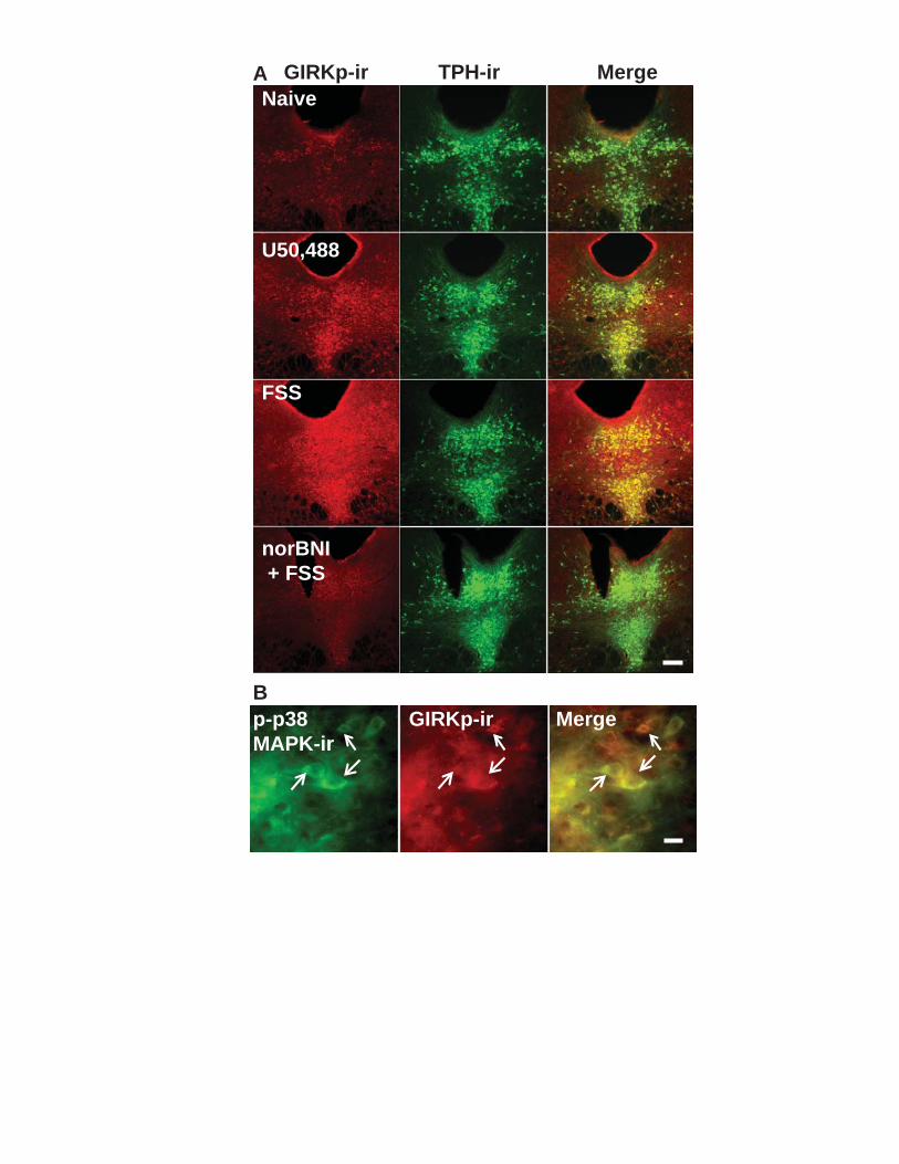
Naive
U50,488
FSS
norBNI + FSS
GIRKp-ir
p-p38 MAPK-ir
MergeGIRKp-ir
A
B
TPH-ir Merge

121
Figure 4.10. Excision of p38α from 5-HT neurons blocks stress-induced phosphorylation of GIRK, but not KOR. A, top panel. Repeated swim stress increases phospho-KOR-ir in the DRN compared to naïve animals in both WT and p38αCKOSERT animals. A, bottom panel. Excision of p38α reduced stress-induced phospho-GIRK-ir in the DRN compared to WT animals (scale bar = 100 µm). B. Schematic depicting indirect tyrosine phosphorylation of the GIRK channel by p38α MAPK following stress-induced dynorphin release and subsequent KOR activation.

WT p38αCKOSERT
Naive NaiveFSS FSS
KORp-ir
GIRKp-ir
A
B
Dynorphin
COOH
NH2
p38P
Y12NH2
COOH
P
Stress
β
Υα
GRK3/Beta-arrestin 2
KIR 3.1
122
Chapter 5: Bi-directional alteration of 5-HT1A autoreceptor function following different stress-exposure paradigms requires activation of the kappa opioid system.
These data are unpublished.
Specific contributions:
J.C.L. collected all of the data presented in this chapter.
Introduction
The 5-HT1A autoreceptor is present on serotonin-containing neurons of the dorsal and
median raphe nucleus (DRN and MRN respectively) and upon activation, dampens
firing of these neurons and subsequent release of serotonin into the forebrain278. 5-
HT1A autoreceptors are not tonically active, but rather are engaged during heightened
periods of neuronal excitability278. The 5-HT1A receptor is a Gi/o coupled receptor that
activates G-protein activated inward rectifying potassium (GIRK) channels through Gβγ
gating mechanisms266. Alterations in 5-HT1A receptor levels and function have been
found in patients with PTSD, anxiety and depression2,15,84,278,279. Likewise, pre-clinical
roden models of stress-induced anxiety and depression have also described alterations
in 5-HT1A receptor function. However, these studies have yielded conflicting results.
For example, studies have shown that 5-HT1A autoreceptor function in DRN neurons
can either be sensitized or desensitized as a result of a stress challenge to the animal
depending on the type of stress insult (e.g. footshock, swim stress, restraint stress,
social defeat stress) and the chronicity and contrallability of the stress. In some of these
studies, acute or sub-chronic stress has no effect on 5-HT1A function, whereas chronic
123
stress causes desensitization of receptor function280,281. In contrast, other studies report
5-HT1A receptor sensitization with both an acute and chronic stress challenge 85,86. Part
of the problem when trying to directly compare these studies is that across studies,
investigators use different pharmacological agents at a broad range of concentrations to
activate 5-HT1A receptors, use different species (rat versus mouse) and use either in
vitro or in vivo electrophysiological techniques to measure 5-HT1A activation of GIRK
and suppression of cell firing. Therefore, I sought to conduct a parametric set of
experiments examining the effect of chronicity on 5-HT1A autoreceptor function using
the same type of stress exposure (i.e. forced swim stress or FSS) using the same 5-
HT1A agonist in the same species.
In addition to this ambiguity, even less is known about the mechanisms
underlying stress-induced regulation of 5-HT1A in the DRN. The dynorphin-kappa opioid
receptor system is engaged during stress-exposure, dynorphin is released in the DRN
and KOR activation regulates 5-HT neuronal excitability (see chapter 4). I hypothesized
that stress induced activation of the dynorphin-KOR system may be, in part, responsible
5-HT1A adaptations caused by stress.
Methods
Subjects: Male C57BL/6 mice, age >50 days, were maintained under a 12-h light–dark
cycle (7a.m. to 7p.m. light) with access to standard food and water ad libitum. All
procedures on animal subjects were approved by the University of Washington IACUC
committee. Mice housed together (2 - 4 per cage) were subjected to the same
behavioral treatment.

124
Swim stress: Mice were subjected to a two-day swim stress procedure in which they
were exposed to a 15-min swim session on day one, then on day two, were exposed to
four six-min swim sessions in 29.0 – 31.0 °C water, separated by six min and
conducted under bright light (690-700 lux) conditions as previously described 170. A
subset of mice were given a six minute swim stress exposure every day, for five
additional days. Another group of animals was treated with norBNI (10 mg/kg) i.p. 24
hours prior to the stress exposure. These groups had accompanying control animals
that were injected with norBNI (10 mg/kg) and were sacrificed in a time matched fashion
to the two stress conditions.
Electrophysiology
Procedures were similar to those previously reported and described in chapter 4
88,265. Sections that were 200 m thick were cut through the raphe nuclei in sucrose and
then placed in oxygenated 95%O2/5%CO2 ACSF incubated in a 35-37˚C bath for 1 hr.
The slices were then removed from the bath and kept in oxygenated ACSF at RT.
ACSF was composed of (in mM): NaCl 124, KCl 2.5, NaH2PO4 2, CaCl2 2.5, Dextrose
10 and NaHCO3 26. The sucrose buffer was ACSF in which NaCl was replaced by 248
mM sucrose.
Slices were placed in a recording chamber (Warner Instruments, Hamden, CT)
and continuously perfused with oxygenated ACSF at approximately 1.5 ml/min,
maintained at 30-32˚C by an inline solution heater (TC-324, Warner Instruments). The
resistance of the electrodes was 5-10 Mwhen filled with an internal solution of (in mM)
K-gluconate 130, NaCl 5, Na phosphocreatine 10, MgCl2 1, EGTA 0.02, HEPES 10,

125
MgATP 2, Na2GTP 0.5, 0.1% Biocytin, pH 7.3. The cells were voltage clamped at -70
mV using an Axopatch 200B amplifier (Molecular Devices, Foster City, CA). Signals
were digitized by a Digidata 1440 A/D converter (Molecular Devices) and stored using
pClamp 10.2 software (Molecular Devices). To measure GIRK currents, once a
recording had been established, the slice was bathed in 5.5 mM K+ ACSF (from 2.5
mM K+) to enhance the currents in the cells at hyperpolarized potentials. As expected,
when the slice incubated in high [K+], the cells became more depolarized and inward
rectification was more apparent. This increase in conductance was sensitive to 100 µM
barium chloride 266. The cells were subjected to a ramp protocol in which they were
brought from -120 mV to -50 mV over 10 sec. After three stable ramp measurements
(the average of three sweeps) were obtained, the 5-HT1A agonist 5-
carboxamidotryptamine (5-CT) (1 or 30 nM) was bath applied and the slice was allowed
equilibrate for 3 min. Subsequently three ramp measurements were taken every 3 min.
For cumulative concentration response experiments, each concentration was applied to
the slice for 8-10 minutes or until the cell reached a stable new equilibrium (i.e. three
stable GIRK current responses). Following 5-CT application and stable current
responses, BaCl2 (100 µM) was added to distinguish Ba2+-sensitive currents.
TPH Immunohistochemistry staining and intracellular labeling
During electrophysiological recordings, cells were filled with biocytin (0.1%)
(Sigma-Aldrich) present in the recording electrode. After recording, slices were fixed by
submersion in 4% paraformaldehyde prepared in 0.1M phosphate buffer (PB; pH 7.4).
Sections were incubated with mouse anti-TPH antibody (1:500, Sigma-Aldrich) for 12-
16 hr at room temperature (RT). Subsequently, immunohistochemical labeling was

126
visualized using Alexa Fluor 488 (1:500; Invitrogen) conjugated goat anti-mouse
secondary antibody for 90-120 min at RT. Biocytin was visualized using streptavidin
conjugated Alexa Fluor 647 (1:500, Invitrogen) contained in the same secondary
antibody mixture. Between incubations, slices were rinsed with PBS (3 x 10 min) and
all incubations were done with mild agitation on an orbital shaker.
Results
Baseline and 5-CT induced inward currents (measured at -120 mV) were
obtained and Ba2+-insensitive currents were subtracted from the total current. 5-CT (30
nM) significantly increased GIRK current from baseline in TPH positive serotonin
containing neurons (paired t-test, p <0.01, Figure 5.1a) as previously described266. 5-
CT activated GIRK currents at concentrations of 1-30 nM applied cumulatively to the
slice. There was no significant difference between 5-CT stimulated GIRK responses at
30 nM applied cumulatively, 30 nM applied directly (non-cumulatively) and 100 nM
applied directly (non-cumulatively) (F2,24 = 0.1728, p> 0.05, one-way ANOVA, Figure
5.1b) indicating that there was no acute desensitization apparent in the cumulative
concentration response experiment and that 30 nM 5-CT produced maximal activation
of GIRK currents.
DRN 5-HT cells were recorded from animals exposed to two-day swim stress
(schematized in Figure 5.2a) and 5-CT (30 nM) activated GIRK current responses were
measured. Compared to that obtained from naïve animals, FSS (2d) did not
significantly alter 5-CT activated GIRK current responses when 30 nM was administered
either non-cumulatively (unpaired t-test, p > 0.05, Figure 5.2a) or cumulatively

127
(Bonferroni post-hoc test, p > 0.05, Figure 5.2b). However, we did find that at 1 nM, a
submaximal concentration, 5-CT responses were sensitized in 5-HT cells recorded from
the DRN of FSS (2d) exposed animals (time by behavioral treatment, F3,51 = 5.358, p <
0.01, two-way repeated measures ANOVA, Figure 5.2b). In contrast, in animals that
were exposed to the same two-day swim stress and then exposed to one six-minute
swim session every day for five additional days had desensitized 5-CT responses at
maximal concentrations (10 and 30 nM) compared to naïve animals (time by behavioral
treatment, F3,42 = 6.814, p < 0.001, two-way repeated measures ANOVA, Figure 5.2b).
We were curious if the sensitization observed in animals exposed to two-day swim
stress returned to naïve levels 24 hrs after the cessation of the final swim stress
exposure. Surprisingly, the sensitization observed after two-day stress exposure, was
also observed following a 24-h recovery period (F2,22 = 4.086, p <0.05, one-way
ANOVA, Figure 5.2c). Moreover, though not significantly different, the sensitization
observed at 24 hrs following stress exposure, was qualitatively larger than that
observed when the animals were sacrificed 30 minutes after the final swim.
The dynorphin-KOR system is activated during swim stress and regulates
serotonin containing neurons of in the DRN. We assessed the effects of stress
exposure both at two days and seven days on 5-CT activation of GIRK currents in
animals that were pre-treated with the selective KOR antagonist norBNI (10 mg/kg) i.p.
We have previously shown that norBNI is a long-lasting antagonist (up to 21
days)261,282,283 and thus we only administered norBNI once, 24 hrs prior to the first
stress exposure. Compared to time-matched animals treated with norBNI, but not
exposed to stress, norBNI treated animals exposed to two-day swim stress did not have

128
sensitized 5-HT1A responses at a submaximal (1 nM) concentration of 5-CT (time by
behavioral treatment, F3,36 = 0.004, p > 0.05, two-way repeated measures ANOVA,
Figure 5.3a). Similarly, seven-day stress exposure did not cause significant
desensitization of 5-HT1A responses at maximal (10 and 30 nM) concentrations of 5-CT
in norBNI treated animals compared to time-matched norBNI treated animals (time by
behavioral treatment, F3,33 = 0.2120, p > 0.05, two-way repeated measures ANOVA,
Figure 3b). Interestingly, norBNI pre-treatment seven days prior to slice preparation
significantly elevated 5-HT1A responses at all concentrations compared to naïve animals
(main effect, F2,63 = 4.325, p < 0.05, Figure 3b) suggesting that the long-lasting impact
of norBNI administration seven days prior to testing sensitizes 5-HT1A receptors.
Collectively this data suggests that the dynorphin-KOR system in 5-HT neurons of the
DRN regulates 5-HT1A responsivity.
Discussion
The principal conclusion from the current work is that stress exposure can bi-
directionally alter 5-HT1A autoreceptor responsivity in serotonin containing neurons of
the DRN that is dependent on the chronicity of the stress exposure. Importantly, we
used the same mode of stress exposure and only varied the number of days the animal
was exposed to stress. Two-day swim stress exposure produces a sensitization of 5-
HT1A activation of GIRK currents at submaximal concentrations of the 5-HT1A agonist 5-
CT. In contrast, seven-day stress exposure produces desensitization of 5-HT1A
activation of GIRK currents at maximal concentrations of 5-CT. Interestingly, stress-
induced sensitization of 5-HT1A induced GIRK current did not recover in 24 hours after
two-day stress exposure and in fact was more robust. A similar sensitization of 5-HT1A
129
response has been observed in mice exposed to social defeat stress for 10 days85. The
stress-induced sensitization of 5-HT1A in the DRN may, in part, lead to increased
anxiety-like and despair-like behavior observed in stress-exposed animals. Enhanced
autoinhibition would lead to chronically lower serotonin tone in the forebrain. Indeed,
transgenic animals that have higher 5-HT1A autoreceptor levels demonstrate enhanced
behavioral despair-like behavior 284. The desensitization of 5-HT1A activation of GIRK
currents has also been reported in animals exposed to chronic variable stress280,285 or
animals exposed to uncontrollable foot-shock281. Interestingly, chronic SSRI treatment
also causes 5-HT1A autoreceptor desensitization286,287 suggesting that stress-induced
desensitization of 5-HT1A responsivity may be a compensatory response to stress insult.
Very little is known about the mechanisms underlying stress-induced regulation
of 5-HT1A function. Recently, it has been demonstrated that that stress-induced
activation of glucocorticoid receptors in the DRN and subsequent activation of HDAC6
mediates 5-HT1A autoreceptor sensitization. In the current work, we show that stress-
induced alterations in 5-HT1A function are prevented by pre-treatment with norBNI to the
whole animal prior to stress-exposure. These data indicate that the dynorphin-KOR
system is also involved in stress-induced regulation 5-HT1A autoreceptor sensitivity.
Interestingly, norBNI administration in and of itself, when administered seven days prior
to testing, robustly increased 5-HT1A sensitivity at all concentrations tested. Previous
work from the laboratory has shown that norBNI, like stress and KOR agonism with
U50,488, activates c-Jun kinase (JNK)261,282. Since stress, KOR agonism and norBNI
all lead to enhanced activation of JNK, it is possible that both stress-induced
sensitization of 5-HT1A and norBNI-sensitization of 5-HT1A require JNK activation.
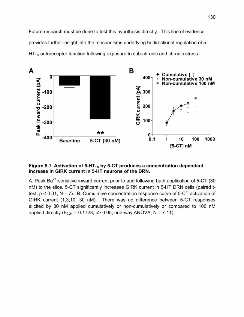
130
Future research must be done to test this hypothesis directly. This line of evidence
provides further insight into the mechanisms underlying bi-directional regulation of 5-
HT1A autoreceptor function following exposure to sub-chronic and chronic stress.
Figure 5.1. Activation of 5-HT1A by 5-CT produces a concentration dependent increase in GIRK current in 5-HT neurons of the DRN.
A. Peak Ba2+-sensitive inward current prior to and following bath application of 5-CT (30 nM) to the slice. 5-CT significantly increases GIRK current in 5-HT DRN cells (paired t-test, p < 0.01, N = 7). B. Cumulative concentration response curve of 5-CT activation of GIRK current (1,3,10, 30 nM). There was no difference between 5-CT responses elicited by 30 nM applied cumulatively or non-cumulatively or compared to 100 nM applied directly (F2,24 = 0.1728, p> 0.05, one-way ANOVA, N = 7-11).
Baseline 5-CT (30 nM)-400
-300
-200
-100
0
**Pea
k in
wa
rdc
urr
en
t(p
A)
0.1 1 10 100 10000
100
200
300
400Cumulative [ ]Non-cumulative 30 nMNon-cumulative 100 nM
[5-CT] nM
GIR
Kcu
rren
t(p
A)
A B
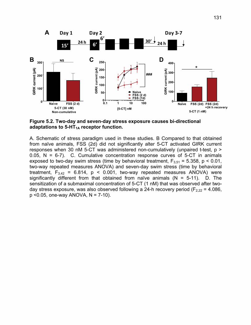
131
Figure 5.2. Two-day and seven-day stress exposure causes bi-directional adaptations to 5-HT1A receptor function.
A. Schematic of stress paradigm used in these studies. B Compared to that obtained from naïve animals, FSS (2d) did not significantly alter 5-CT activated GIRK current responses when 30 nM 5-CT was administered non-cumulatively (unpaired t-test, p > 0.05, N = 6-7). C. Cumulative concentration response curves of 5-CT in animals exposed to two-day swim stress (time by behavioral treatment, F3,51 = 5.358, p < 0.01, two-way repeated measures ANOVA) and seven-day swim stress (time by behavioral treatment, F3,42 = 6.814, p < 0.001, two-way repeated measures ANOVA) were significantly different from that obtained from naïve animals (N = 5-11). D. The sensitization of a submaximal concentration of 5-CT (1 nM) that was observed after two-day stress exposure, was also observed following a 24-h recovery period (F2,22 = 4.086, p <0.05, one-way ANOVA, N = 7-10).
Day 1
30’6’15’
6’24 h
Day 2 Day 3‐7
24 h
Naive FSS (2 d)0
100
200
300NS
5-CT (30 nM)Non-cumulative
GIR
K c
urre
nt(
pA)
0.1 1 10 1000
50
100
150
200
250
NaiveFSS (2 d)FSS (7d)
###
[5-CT] nM
GIR
Kc
urr
en
t(p
A)
Naive FSS (2d) FSS (2d)0
100
200
300
400
+24 h recovery
*
5-CT (1 nM)
GIR
Kc
urr
ent
(pA
)
A
B C D

132
Figure 5.3. NorBNI pre-treatment prior to stress-exposure blocks stress-induced alterations in 5-HT1A function.
A. The cumulative concentration response curve of 5-CT in norBNI treated animals exposed to two-day swim stress was not significantly different from norBNI treated time matched control animals (time by behavioral treatment, F3,36 = 0.004, p > 0.05, two-way repeated measures ANOVA, N = 6-8). B. Similarly, seven-day stress exposure did not cause significant desensitization of 5-HT1A responses at maximal (10 and 30 nM) concentrations of 5-CT in norBNI treated animals compared to time-matched norBNI treated animals (time by behavioral treatment, F3,33 = 0.2120, p > 0.05, two-way repeated measures ANOVA, N = 5-11). norBNI pre-treatment seven days prior to slice preparation significantly elevated 5-HT1A responses at all concentrations compared to naïve animals (main effect, F2,63 = 4.325, p < 0.05).
0.1 1 10 1000
50
100
150
200
250
NBNI + no stressNBNI + FSSNaive
FSS (2d)
[5-CT] nM
GIR
Kcu
rren
t(p
A)
0.1 1 10 1000
100
200
300
400
500
NBNI + no stressNBNI + FSSNaive
FSS (7d)
[5-CT] nM
GIR
Kc
urr
en
t(p
A)
A B

133
Chapter 6: Dissertation Conclusions
Over the last several decades, basic scientists have taken on the challenge of
identifying biological substrates that underlie complex psychiatric diseases such as
anxiety, depression and addiction. A basic strategy used to address this challenge was
to dissect complex syndromes such as Major Depressive Disorder (MDD) into small
discrete components that might be modeled in animals. In doing so, we have been able
to link discrete neurochemicals with specific aspects of psychiatric disease such as
negative affect, withdrawal, hyperarousal and anhedonia. We have now identified
several neuropeptides, small molecule neurotransmitters and downstream signaling
molecules as well as specific brain regions that are critically involved in arousal, affect
and motivation in the normal state and become altered or engaged in the pathological
states. This reductionist approach taken up until the early 2000s has provided a wealth
of information regarding which molecules and which brain regions are involved in
certain aspects of behavior, yet have been studied in a modular fashion. Another
population of scientists has taken a ‘top-down’ approach by attempting to study the
activity of whole brain regions both in human and non-human primates under different
theoretical frameworks that include learning and economic theories. The next wave of
research as well as a primary goal of this thesis work is the merger of these two
different scientific approaches. We sought to understand how these molecules interact
with each other within discrete limbic brain regions to produce behavioral responses;
how they feed forward and feedback on one another and how they are released
temporally in response to stress and arousing environmental stimuli. From this line of

134
work as well as many other studies we are beginning to more fully understand behavior
and psychiatric diseases on the microcircuit level.
New Insights on neuropeptidergic regulation of monoamines
Hans Selye was the first to describe the concepts of eustress and distress26,
which has since been expounded on by McEwen and Sapolsky29,47,48. Here we have
tried to tease these two concepts apart biologically by examining first, acute
neuropeptidergic regulation of monoamines and then examining adaptations of those
interactions following exposure to severe stress. Adaptive responses to acute stressors
in the environment first require the engagement of arousal systems. In order to
adaptively and actively cope with a stressor, there must be a motivational component
that allows the individual to respond to the stimulus. This motivational component may
be positive or negative meaning that it enables an individual to either actively approach
the stimulus in spite of potential risk or actively avoid the stimulus. Finally, it is useful to
the organism to learn that some stressors are dangerous or threatening. In order to do
this, stressors must elicit some negative affective state, whether it be dysphoria, anxiety
or fear, that allows the animal to associate certain stimuli with negative outcomes. It
has been shown that acute stressors elicit the release of CRF extra-
hypothalamically210,288. CRF administered i.c.v and therefore, made available to several
brain regions, is aversive to the animal and causes the animal to avoid riskier
environments such as the open arms of an elevated plus maze or an open
field131,167,205,250. However, it is possible that these effects are mediated by CRF acting
on a subset of brain regions. When CRF is administered directly into the BNST, a brain
region that has been implicated in anxiety responses70, it also produces an aversion225

135
indicating that this brain region is critical in encoding CRF-induced aversion. We have
established that CRF causes the release of dynorphin and activation of KORs in several
limbic brain regions; most notably the BLA and DRN205. It is this engagement of the
dynorphin-KOR system by CRF that underlies both the aversive and anxiogenic
qualities of CRF167,205,250. Importantly, when KOR activation is blocked in the BLA via
site-specific injection of norBNI, that negative affective state assayed by place aversion
and elevated plus maze are blocked250 suggesting that CRF-mediated KOR activation in
the BLA is a critical for informing the animal that stressful stimuli are threatening or
should be avoided. We have also found that conditioned place aversion produced by
direct KOR agonism with U50,488 is blocked by prior norBNI injection into the DRN254
demonstrating that the aversive component of KOR activation also requires the DRN.
The DRN is also of interest since it has one of the densest concentrations of CRF R2125
receptors in the brain and the reader may recall that the CRF-induced aversion was
mediated by CRF R2205. Interestingly, we also found that KOR activation in the DRN
was also necessary to suppress nociceptive responding during stress events254. These
two pieces of evidence suggested that KOR acting acutely in the DRN acts as a
“negative affect” signal. I were particularly interested in the KOR regulation of the DRN
because of the large body of evidence suggesting that serotonin is instrumental in
regulating mood or emotional states, however, KOR regulation of serotonergic DRN cell
excitability has never been fully characterized. As part of this thesis work, we surveyed
the functional actions of KOR in serotonin-containing cells of the DRN using
electrophysiological techniques. I measured the actions of KOR activation on fast
excitatory and inhibitory synaptic transmission as well as activation of post-synaptic

136
potassium channels. I found that KOR agonism by U69,593 has a net inhibitory
regulation of DRN neuronal excitability by inhibiting glutamatergic release probability
pre-synaptically and activating GIRK channels post-synaptically. It was known that
increases in dynorphin and decreases in serotonin in limbic regions both produced
negative affect. This set of studies elucidates how these two neurochemicals interact
with each to produce this change in emotional state. Moreover dynorphin-KOR
regulation of the DRN may promote increased behavioral flexibility or activity by
decreasing serotonin release and inactivating behavioral inhibition. Collectively this
data supports a working model in which stressors elicit CRF release which then
engages the dynorphin-KOR system to negatively regulate serotonergic neuronal firing
and release into the forebrain.
Importantly, CRF is not only released in response to acute stressors210,288, but
also is released extra-hypothalamically in response to arousing stimuli that at face value
do not seem inherently stressful or negative such as cues that are associated with
palatable food212,213. Recently, it has been shown that when CRF is administered
specifically into the nucleus accumbens, a region that is important for controlling
motivated behavior, promotes appetitive behaviors including social pair bonding and
responding to cued rewards215,216. Both of these behaviors are mediated by accumbal
dopamine release217,218. While this may seem paradoxical given what we know about
the behavioral consequences of the CRF i.c.v. administration, it is intuitive that arousing
environmental stimuli may elicit appetitive or approach behaviors; this is how we are
able to engage in the environment. Here we show that CRF acts at CRF receptors
(CRF R1 and R2) on dopamine terminals within the nucleus accumbens to increase

137
dopamine release and that this cellular action causes a place preference (i.e. promotes
approach to environment in which the animal has experience CRF-elicited dopamine
release). Importantly, we also show that endogenous CRF release causes animals to
approach and interact with a novel object. Exposure to novelty has been characterized
as inherently stressful or anxiogenic in rodents, yet also elicits exploratory and
investigatory behavior by the animal. In other words, animals will overcome the
anxiogenic component of novelty and explore the novel object. It is obvious how this
mechanism would be adaptive for an organism as it would allow the organism to
interrogate new environments in search of food and mates despite some inherent risk.
As discussed above, a critical feature of accumbens dopamine release is that it allows
organisms to overcome obstacles to obtain rewards (see Salamone et al. 2003). I
would assert that CRF positive regulation of dopamine release in the nucleus
accumbens is a mechanism by which an organism can engage in the environment and
overcome obstacles that may have some risk associated with them.
The body of evidence presented in this thesis describes and characterizes how
the stress-related neuropeptides CRF and dynorphin impinge on the serotonin and
dopamine systems to regulate behavioral responding to environmental stimuli with
varying saliencies and valences associated with them.
The impact of severe or repeated stress on neuropeptidergic regulation of
monoamine systems
Even though behavioral responses to acute stressors are undoubtedly adaptive
for an organism’s survival, one could argue that even acute or mild stressors have an

138
inherent negative subjective quality. While these acute stress responses may protect
against threat and motivate an individual to achieve long-term goals, they do have an
intrinsic negative affective quality. It is, therefore, intuitive that normal stress responding
is particularly fragile to dysregulation or adaptation following severe or repeated stress
exposure. Following stress trauma, as Beck asserted in his initial theoretical framework
of depression, there is a shift to a negative perceptual bias. To be more specific, there
seems to be an amplification of the negative affective qualities of stressors and an
ablation of the motivational qualities of stressors. In concert, this trauma leads to a shift
in the organism’s behavioral responding such that it now passively copes with
environmental challenges (i.e. Dove mode) and withdraws from the environment.
Many of these studies examine behaviors during the actual stress challenge, but
fewer go on to look at behavioral responding following stress exposure. Having
established the manner in which CRF and dynorphin regulate the dopamine and
serotonin systems respectively in naïve animals, we then exposed another group of
mice to repeated swim stress and asked how this normal regulation is altered or
becomes dysregulated. Here we report that normal dynorphin-KOR negative regulation
of the DRN is blunted following repeated stressor exposure, specifically by reducing the
effectiveness of KOR activation of post-synaptic GIRK currents. Interestingly, this
neuroadaptation persists as long as the stress exposure is reinforced (i.e. the animal is
given additional exposures to swim stress), but returns when the animals are given a 24
hour recovery period. Interestingly, the reduction in KOR activated GIRK current
appears to be fairly moderate. Even though KOR activation of GIRK currents is
reduced by half, it is still present and KOR inhibition of excitatory transmission pre-

139
synaptically remains intact. I were also struck at how dynamic the dynorphin-KOR
system, at least in the DRN, appears to be in that its functionality can shift fairly rapidly
depending on the specific temporal kinetics of the stress exposure, especially when
contrasted to the effect of stress exposure on CRF signaling in the DRN. Swim stress
exposure completely ablates CRF regulation of DRN neuronal excitability154. It is
unknown if CRF signaling in the DRN ever recovers, but it is absent in stress vulnerable
WKY rats suggesting that this adaptation does not necessarily require a severe stress
exposure88. These findings suggest that the dynorphin-KOR system is a critical system
to the animal’s survival in that is fairly resilient and is in large part retained even after
severe stress exposure. Indeed, I found another example of this when assessing KOR
regulation of dopamine release in the nucleus accumbens. I found that stress exposure
had absolutely no effect on KOR inhibition of dopamine release in the nucleus
accumbens. Taken together, the data suggest that dynorphin-KOR inhibition of
monoamine systems is preserved following severe repeated stress exposure and may
underlie the amplification a negative perceptual bias. This notion is corroborated by our
findings that stress exposure completely ablates CRF mediated potentiation of
dopamine release which we have shown underlies appetitive or approach behavior.
As stated in the introduction, Korte and colleagues (2005) have asserted that it is
likely that severe or chronic stress exposure produces of number of neuroadaptations.
How do we know what adaptations are compensatory and which are at the root of
maladaptive behavioral responses that lead to the actual pathology. One hint may
come from the actual diagnostic criterion for MDD. Part of the diagnostic criterion for
MDD is that the individual must experience a preponderance of systems nearly every

140
day for a period of two weeks4. Moreover, in a study examining the protracted course of
MDD, Coryell and colleagues (1994) found that only 25% of patients recovered from a
depressive episode within two months of the initiation of the episode and even more
alarming, 20% of patients had not significantly recovered after two years231.
Additionally, with an 80% reoccurrence rate, it is very likely that patients suffer from sub-
threshold depressive symptoms even after remission from a major depressive episode.
With all that in mind, what has seemed to be relatively lacking in pre-clinical research
modeling stress-induced depression is investigation of the persistence of stress-induced
neuroadaptations. Most studies have not assessed the recovery or persistence of
stress-induced neuroadaptations passed 24 hours after the final stress exposure and no
studies assess these adaptations similar to a time course that maps onto a protracted
course of depression. We were struck by the persistence of the stress-induced ablation
of the CRF potentiation of dopamine release in the nucleus accumbens. We found that
even 90 days after the swim stress, CRF regulation of dopamine release was still
ablated. Moreover, this long-lasting cellular dysregulation was accompanied by an
ablation of the CRF induced place preference when assessed at 7 and 90 days
following stress exposure. Similarly, endogenous CRF release no longer promoted
exploratory behavior. In animals tested at 7 days, there was not only an ablation of the
CRF place preference, but there was a switch to a CRF aversion. Unlike many other
neuroadaptations that are precipitated by stress exposure, this alteration does not
recover and therefore, may be a critical locus for the etiology of protracted depression.
141
Future directions
Epigenetics
The stress-induced ablation of CRF potentiation of dopamine release was
mediated by glucocorticoid receptors. Glucocorticoid signaling has been shown to have
genomic repressive effects of the CRF system56, in particular it causes the
downregulation of both CRF R1148 and CRF R2149 mRNA. This finding suggests that
stress-induced long-lasting changes in brain function may due to epigenetic and
transcriptional regulation. Recently it has been shown that glucocorticoid-mediated
potentiation of anxiety behavior as well as glucocorticoid-induced alterations in DRN
neuronal function were dependent on glucocorticoid-mediated activation of the
cytoplasmic lysine deacetylase HDAC6, which controls Hsp90 acetylation85. One
direction that research on stress-induced psychiatric diseases is heading in is to further
examine stress-induced epigenetic changes289. Long-lasting changes in brain function
that lead to chronic recurring or unremitting depression may be highly dependent on
stress-induced epigenetic changes (i.e. histone modifications and DNA methylation). A
recent set of studies has demonstrated a role for DNA methytransferases (Dmnts)
(catalyzes DNA methylation which is inversely related to gene expression)290 and
histone methyltransferases (promotes increased gene expression)291 in mediating pro-
depressive- and pro-addictive-like behaviors produced by chronic social defeat stress.
The main technological limitation in studying the causal relationship between epigenetic
modifications and depression is the lack of specificity of our current tools. There are
very few, if any pharmacological tools that are selective for specific HDACs or Dmnts.
Development of virally mediated overexpression techniques as well as transgenic tools

142
have improved the specificity of the manipulation of the epigenetic effector, yet has not
addressed the lack the specificity of the epigenetic substrate. In other words, if we
wanted to test hypothesis that glucocorticoids cause epigenetic changes in DNA
transcription that leads to long-term downregulation of CRF R1 in dopaminergic
neurons, we could not because there is currently no way to examine that specific
interaction. Rather, with our current tools, we can only assess the epigenetic actions of
glucocorticoids in the VTA on all of its downstream targets, not CRF R1 specifically.
Glucocorticoid receptors have both genomic and non-genomic actions that are
incredibly diverse and must select for different repressive or activating effects on the
large number of substrates that are regulated by GRs56. Certainly for enzymes like
Dmnts there are recognition sites along the DNA strand (i.e. C-6 position of adenines),
but does this mean that when a stress-related receptor activates Dmnts there is a global
repression of gene transcription or is there an additional “zip code” or different protein
complex formations that allows Dmnts to target specific adenines that would lead to one
gene being repressed over another? Further study needs to be done to understand if
these “zip codes” exist and how they work, which in turn will aid in better
pharmacological and genetic tools to understand behavioral questions.
Temporal dynamics of neuropeptide release and regulation
One of the major gaps in our understanding of neuropeptide signaling is the
temporal dynamics of neuropeptide release and regulation. There have been very few
studies that examine the release of endogenous neuropeptides at all and even fewer
that have done so in awake behaving animals. Moreover, until recently the only tool
available to directly measure extracellular neuropeptides is microdialysis with HPLC.

143
The limitation with microdialysis is that it has relatively slow temporal dynamics (on the
order of 10s and 20s of minutes) and causes substantial damage to the surrounding
tissue due to the size of the probe. However, work from Robert Kennedy’s lab
revolutionized old microdialysis techniques by resolving both of these pitfalls. Using a
combination of microfluidics/capillary segmentation and mass spectrometry, the
Kennedy group has been able to resolve neurochemical measurements, even in awake
behaving animals to seconds for small molecules292-295. Using a method called capillary
liquid chromatography-triple-stage mass spectrometry, they have been able to measure
endogenous opioid peptides, including dynorphin (1-8), in the extracellular space on the
order of minutes and have also immensely improved the feasibility of measuring these
peptides at all296,297. Very large peptides such as CRF(41 aa) still pose technical
challenges due to its impermeability of the microdialysis probe membrane. However,
these new techniques will promote more in vivo monitoring of the neurochemicals with
temporal and spatial resolution and is the start of a very exciting and new direction.
Accompanying this technique is the development of neuropeptide-cre transgenic
animals. In 2010, Martin and colleagues developed and published a CRF-Cre
transgenic mouse 298. Like other Cre-lines, this new transgenic tool opens up a huge
amount of new research on the dynamics of the CRF system. With this tool,
investigators can electrophysiologically characterize CRF containing neurons,
genetically remove proteins from these neurons and also manipulate the firing of these
neurons with both optogenentic techniques and virally-expressed designer receptors
(i.e. DREDDs). This will allow investigators to really assess the temporal relationship
between the engagement of the CRF system within discrete brain regions and

144
subsequent behavioral consequences. Along the same line there has been the
development of floxed CRF R1 mice in which CRF R1 is excised from different neuronal
populations. (Floxed KOR mice are also being developed, but have not been published
yet). Refojo and colleagues (2010) demonstrated that CRF R1 excision from
glutamatergic neurons produced anxiolysis whereas CRF R1 excision from dopamine
neurons produced anxiety-like behaviors in mice241. These data support one of the
main assertions from our own body of work that neuropeptide regulation of disparate
neurochemical systems leads mediates different behavioral responses and that CRF
does not act globally to produce one behavioral or emotional consequence. Similar
mouse lines that interrogate the dynorphin-KOR system are currently in development.
Clearly we have come a long way from the eras of Freud, Selye and Beck in
understanding the biological basis of psychiatric pathology, though their contributions
cannot be overstated. However, even with the advancement of new tools, the real goal
for the next 50 years of research is to take everything we know about the biology of
depression and use it, not to treat MDD, but to treat individuals suffering from
depression. The words “treatment-resistance” and “unremitting” are perhaps the two
most alarming realities about the current state of MDD. If we continue to use more
sophisticated and higher resolution tools to interrogate depression-like behavior using
the same behavioral models we are not going to advance as far as we could. In the
future we have to create better animal models that model different constellations of
symptoms that fall into the larger diagnostic criterion of the disease so that we can learn
how to better tailor treatments.

145
References 1 Kessler, R. C. et al. Lifetime prevalence and age-of-onset distributions of DSM-IV
disorders in the National Comorbidity Survey Replication. Archives of general psychiatry 62, 593-602 (2005).
2 Samuels, B. A. et al. Modeling treatment-resistant depression. Neuropharmacology 61, 408-413, doi:10.1016/j.neuropharm.2011.02.017 (2011).
3 Fava, M. & Kendler, K. S. Major depressive disorder. Neuron 28, 335-341 (2000). 4 American Psychiatric Association. Diagnostic criteria from DSM-IV-TR.
(American Psychiatric Association, 2000). 5 Vohringer, P. A. & Ghaemi, S. N. Solving the antidepressant efficacy question:
effect sizes in major depressive disorder. Clinical therapeutics 33, B49-61, doi:10.1016/j.clinthera.2011.11.019 (2011).
6 Beck, A. T. Thinking and Depression. I. Idiosyncratic Content and Cognitive Distortions. Archives of general psychiatry 9, 324-333 (1963).
7 Beck, A. T. Thinking and Depression. Ii. Theory and Therapy. Archives of general psychiatry 10, 561-571 (1964).
8 Beck, A. T. The evolution of the cognitive model of depression and its neurobiological correlates. The American journal of psychiatry 165, 969-977 (2008).
9 Beck, A. T. & Dozois, D. J. Cognitive therapy: current status and future directions. Annual review of medicine 62, 397-409, doi:10.1146/annurev-med-052209-100032 (2011).
10 Seligman, M. E. Learned helplessness. Annual review of medicine 23, 407-412, doi:10.1146/annurev.me.23.020172.002203 (1972).
11 Chourbaji, S. et al. Learned helplessness: validity and reliability of depressive-like states in mice. Brain Res Brain Res Protoc 16, 70-78 (2005).
12 Henkel, V., Bussfeld, P., Moller, H. J. & Hegerl, U. Cognitive-behavioural theories of helplessness/hopelessness: valid models of depression? Eur Arch Psychiatry Clin Neurosci 252, 240-249 (2002).
13 Maier, S. F. & Watkins, L. R. Stressor controllability and learned helplessness: the roles of the dorsal raphe nucleus, serotonin, and corticotropin-releasing factor. Neuroscience and biobehavioral reviews 29, 829-841, doi:10.1016/j.neubiorev.2005.03.021 (2005).
14 Charney, D. S. Monoamine dysfunction and the pathophysiology and treatment of depression. The Journal of clinical psychiatry 59 Suppl 14, 11-14 (1998).
15 Graeff, F. G., Guimaraes, F. S., De Andrade, T. G. & Deakin, J. F. Role of 5-HT in stress, anxiety, and depression. Pharmacology, biochemistry, and behavior 54, 129-141 (1996).
16 Carlsson, A. The contribution of drug research to investigating the nature of endogenous depression. Pharmakopsychiatrie, Neuro-Psychopharmakologie 9, 2-10 (1976).
17 Owens, M. J. & Nemeroff, C. B. The serotonin transporter and depression. Depression and anxiety 8 Suppl 1, 5-12 (1998).
18 Delgado, P. L. Depression: the case for a monoamine deficiency. The Journal of clinical psychiatry 61 Suppl 6, 7-11 (2000).

146
19 Delgado, P. L. & Moreno, F. A. Role of norepinephrine in depression. The Journal of clinical psychiatry 61 Suppl 1, 5-12 (2000).
20 Heninger, G. R., Delgado, P. L. & Charney, D. S. The revised monoamine theory of depression: a modulatory role for monoamines, based on new findings from monoamine depletion experiments in humans. Pharmacopsychiatry 29, 2-11, doi:10.1055/s-2007-979535 (1996).
21 Moreno, F. A., Heninger, G. R., McGahuey, C. A. & Delgado, P. L. Tryptophan depletion and risk of depression relapse: a prospective study of tryptophan depletion as a potential predictor of depressive episodes. Biological psychiatry 48, 327-329 (2000).
22 Moreno, F. A. et al. Tryptophan depletion selectively reduces CSF 5-HT metabolites in healthy young men: results from single lumbar puncture sampling technique. Int J Neuropsychopharmacol 3, 277-283, doi:10.1017/S1461145700002133 (2000).
23 Bale, T. L. Stress sensitivity and the development of affective disorders. Horm Behav 50, 529-533 (2006).
24 Selye, H. Stress and the general adaptation syndrome. British medical journal 1, 1383-1392 (1950).
25 Selye, H. & Fortier, C. Adaptive reaction to stress. Psychosomatic medicine 12, 149-157 (1950).
26 Selye, H. Stress and distress. Comprehensive therapy 1, 9-13 (1975). 27 Selye, H. Implications of stress concept. New York state journal of medicine 75,
2139-2145 (1975). 28 Selye, H. Confusion and controversy in the stress field. Journal of human stress
1, 37-44, doi:10.1080/0097840X.1975.9940406 (1975). 29 McEwen, B. S. & Sapolsky, R. M. Stress and cognitive function. Curr Opin
Neurobiol 5, 205-216 (1995). 30 Korte, S. M., Koolhaas, J. M., Wingfield, J. C. & McEwen, B. S. The Darwinian
concept of stress: benefits of allostasis and costs of allostatic load and the trade-offs in health and disease. Neuroscience and biobehavioral reviews 29, 3-38 (2005).
31 Koolhaas, J. M. et al. Stress revisited: A critical evaluation of the stress concept. Neuroscience and biobehavioral reviews.
32 Maier, S. F. Stressor controllability and stress-induced analgesia. Annals of the New York Academy of Sciences 467, 55-72 (1986).
33 Porsolt, R. D., Le Pichon, M. & Jalfre, M. Depression: a new animal model sensitive to antidepressant treatments. Nature 266, 730-732 (1977).
34 Cryan, J. F., Valentino, R. J. & Lucki, I. Assessing substrates underlying the behavioral effects of antidepressants using the modified rat forced swimming test. Neuroscience and biobehavioral reviews 29, 547-569 (2005).
35 Nestler, E. J. et al. Preclinical models: status of basic research in depression. Biological psychiatry 52, 503-528 (2002).
36 Willner, P. Validity, reliability and utility of the chronic mild stress model of depression: a 10-year review and evaluation. Psychopharmacology 134, 319-329 (1997).

147
37 Willner, P. Chronic mild stress (CMS) revisited: consistency and behavioural-neurobiological concordance in the effects of CMS. Neuropsychobiology 52, 90-110 (2005).
38 Willner, P. & Mitchell, P. J. The validity of animal models of predisposition to depression. Behav Pharmacol 13, 169-188 (2002).
39 Bjorkqvist, K. Social defeat as a stressor in humans. Physiol Behav 73, 435-442 (2001).
40 Huhman, K. L. Social conflict models: can they inform us about human psychopathology? Horm Behav 50, 640-646 (2006).
41 Nestler, E. J. et al. Neurobiology of depression. Neuron 34, 13-25 (2002). 42 Kalsbeek, A. et al. Circadian rhythms in the hypothalamo-pituitary-adrenal (HPA)
axis. Molecular and cellular endocrinology 349, 20-29, doi:10.1016/j.mce.2011.06.042 (2012).
43 Papadimitriou, A. & Priftis, K. N. Regulation of the hypothalamic-pituitary-adrenal axis. Neuroimmunomodulation 16, 265-271, doi:10.1159/000216184 (2009).
44 Spiess, J., Rivier, J., Rivier, C. & Vale, W. Primary structure of corticotropin-releasing factor from ovine hypothalamus. Proc Natl Acad Sci U S A 78, 6517-6521 (1981).
45 Sapolsky, R. M. Stress and plasticity in the limbic system. Neurochem Res 28, 1735-1742 (2003).
46 Sapolsky, R. M., Krey, L. C. & McEwen, B. S. The adrenocortical axis in the aged rat: impaired sensitivity to both fast and delayed feedback inhibition. Neurobiology of aging 7, 331-335 (1986).
47 Sapolsky, R. M., Krey, L. C. & McEwen, B. S. The neuroendocrinology of stress and aging: the glucocorticoid cascade hypothesis. Endocrine reviews 7, 284-301 (1986).
48 McEwen, B. S. Glucocorticoids, depression, and mood disorders: structural remodeling in the brain. Metabolism: clinical and experimental 54, 20-23 (2005).
49 Gold, P. W., Licinio, J., Wong, M. L. & Chrousos, G. P. Corticotropin releasing hormone in the pathophysiology of melancholic and atypical depression and in the mechanism of action of antidepressant drugs. Annals of the New York Academy of Sciences 771, 716-729 (1995).
50 Rittenhouse, P. A., Lopez-Rubalcava, C., Stanwood, G. D. & Lucki, I. Amplified behavioral and endocrine responses to forced swim stress in the Wistar-Kyoto rat. Psychoneuroendocrinology 27, 303-318 (2002).
51 Scott, L. V. & Dinan, T. G. Vasopressin and the regulation of hypothalamic-pituitary-adrenal axis function: implications for the pathophysiology of depression. Life sciences 62, 1985-1998 (1998).
52 Scott, L. V., Medbak, S. & Dinan, T. G. Blunted adrenocorticotropin and cortisol responses to corticotropin-releasing hormone stimulation in chronic fatigue syndrome. Acta psychiatrica Scandinavica 97, 450-457 (1998).
53 Young, E. A., Akana, S. & Dallman, M. F. Decreased sensitivity to glucocorticoid fast feedback in chronically stressed rats. Neuroendocrinology 51, 536-542 (1990).

148
54 Young, E. A., Haskett, R. F., Murphy-Weinberg, V., Watson, S. J. & Akil, H. Loss of glucocorticoid fast feedback in depression. Archives of general psychiatry 48, 693-699 (1991).
55 Chrousos, G. P. & Kino, T. Glucocorticoid signaling in the cell. Expanding clinical implications to complex human behavioral and somatic disorders. Annals of the New York Academy of Sciences 1179, 153-166, doi:10.1111/j.1749-6632.2009.04988.x (2009).
56 Revollo, J. R. & Cidlowski, J. A. Mechanisms generating diversity in glucocorticoid receptor signaling. Annals of the New York Academy of Sciences 1179, 167-178, doi:10.1111/j.1749-6632.2009.04986.x (2009).
57 Aston-Jones, G. & Bloom, F. E. Norepinephrine-containing locus coeruleus neurons in behaving rats exhibit pronounced responses to non-noxious environmental stimuli. J Neurosci 1, 887-900 (1981).
58 Aston-Jones, G. & Bloom, F. E. Activity of norepinephrine-containing locus coeruleus neurons in behaving rats anticipates fluctuations in the sleep-waking cycle. J Neurosci 1, 876-886 (1981).
59 Aston-Jones, G., Rajkowski, J. & Cohen, J. Role of locus coeruleus in attention and behavioral flexibility. Biological psychiatry 46, 1309-1320 (1999).
60 Usher, M., Cohen, J. D., Servan-Schreiber, D., Rajkowski, J. & Aston-Jones, G. The role of locus coeruleus in the regulation of cognitive performance. Science 283, 549-554 (1999).
61 Foote, S. L., Berridge, C. W., Adams, L. M. & Pineda, J. A. Electrophysiological evidence for the involvement of the locus coeruleus in alerting, orienting, and attending. Prog Brain Res 88, 521-532 (1991).
62 Page, M. E., Berridge, C. W., Foote, S. L. & Valentino, R. J. Corticotropin-releasing factor in the locus coeruleus mediates EEG activation associated with hypotensive stress. Neuroscience letters 164, 81-84 (1993).
63 Funkenstein, D. H. & Meade, L. W. Norepinephrine-like and epinephrine-like substances and the elevation of blood pressure during acute stress. The Journal of nervous and mental disease 119, 380-397 (1954).
64 Silverberg, A. B., Shah, S. D., Haymond, M. W. & Cryer, P. E. Norepinephrine: hormone and neurotransmitter in man. The American journal of physiology 234, E252-256 (1978).
65 Bremner, J. D., Krystal, J. H., Southwick, S. M. & Charney, D. S. Noradrenergic mechanisms in stress and anxiety: II. Clinical studies. Synapse 23, 39-51, doi:10.1002/(SICI)1098-2396(199605)23:1<39::AID-SYN5>3.0.CO;2-I (1996).
66 Bremner, J. D., Krystal, J. H., Southwick, S. M. & Charney, D. S. Noradrenergic mechanisms in stress and anxiety: I. Preclinical studies. Synapse 23, 28-38, doi:10.1002/(SICI)1098-2396(199605)23:1<28::AID-SYN4>3.0.CO;2-J (1996).
67 Gresch, P. J., Sved, A. F., Zigmond, M. J. & Finlay, J. M. Stress-induced sensitization of dopamine and norepinephrine efflux in medial prefrontal cortex of the rat. Journal of neurochemistry 63, 575-583 (1994).

149
68 Loughlin, S. E., Foote, S. L. & Bloom, F. E. Efferent projections of nucleus locus coeruleus: topographic organization of cells of origin demonstrated by three-dimensional reconstruction. Neuroscience 18, 291-306 (1986).
69 Loughlin, S. E., Foote, S. L. & Fallon, J. H. Locus coeruleus projections to cortex: topography, morphology and collateralization. Brain research bulletin 9, 287-294 (1982).
70 Aston-Jones, G., Delfs, J. M., Druhan, J. & Zhu, Y. The bed nucleus of the stria terminalis. A target site for noradrenergic actions in opiate withdrawal. Annals of the New York Academy of Sciences 877, 486-498 (1999).
71 Barnes, N. M. & Sharp, T. A review of central 5-HT receptors and their function. Neuropharmacology 38, 1083-1152 (1999).
72 Cools, R., Roberts, A. C. & Robbins, T. W. Serotoninergic regulation of emotional and behavioural control processes. Trends in cognitive sciences 12, 31-40, doi:10.1016/j.tics.2007.10.011 (2008).
73 Lucki, I. The spectrum of behaviors influenced by serotonin. Biological psychiatry 44, 151-162 (1998).
74 Deakin, J. F. & Graeff, F. G. 5-HT and mechanisms of defence. J Psychopharmacol 5, 305-315, doi:10.1177/026988119100500414 (1991).
75 Miyazaki, K., Miyazaki, K. W. & Doya, K. Activation of dorsal raphe serotonin neurons underlies waiting for delayed rewards. J Neurosci 31, 469-479, doi:10.1523/JNEUROSCI.3714-10.2011 (2011).
76 Miyazaki, K. W., Miyazaki, K. & Doya, K. Activation of the central serotonergic system in response to delayed but not omitted rewards. The European journal of neuroscience 33, 153-160, doi:10.1111/j.1460-9568.2010.07480.x (2011).
77 Kirby, L. G., Allen, A. R. & Lucki, I. Regional differences in the effects of forced swimming on extracellular levels of 5-hydroxytryptamine and 5-hydroxyindoleacetic acid. Brain Res 682, 189-196 (1995).
78 Neumeister, A., Young, T. & Stastny, J. Implications of genetic research on the role of the serotonin in depression: emphasis on the serotonin type 1A receptor and the serotonin transporter. Psychopharmacology 174, 512-524, doi:10.1007/s00213-004-1950-3 (2004).
79 Fallon, J. H. & Loughlin, S. E. Monoamine innervation of the forebrain: collateralization. Brain research bulletin 9, 295-307 (1982).
80 Reneric, J. P. & Lucki, I. Antidepressant behavioral effects by dual inhibition of monoamine reuptake in the rat forced swimming test. Psychopharmacology 136, 190-197 (1998).
81 Owens, M. J. & Nemeroff, C. B. Role of serotonin in the pathophysiology of depression: focus on the serotonin transporter. Clinical chemistry 40, 288-295 (1994).
82 Samuels, B. A. & Hen, R. Neurogenesis and affective disorders. The European journal of neuroscience 33, 1152-1159, doi:10.1111/j.1460-9568.2011.07614.x (2011).
83 Sahay, A. & Hen, R. Adult hippocampal neurogenesis in depression. Nature neuroscience 10, 1110-1115, doi:10.1038/nn1969 (2007).

150
84 Albert, P. R., Le Francois, B. & Millar, A. M. Transcriptional dysregulation of 5-HT1A autoreceptors in mental illness. Molecular brain 4, 21, doi:10.1186/1756-6606-4-21 (2011).
85 Espallergues, J. et al. HDAC6 regulates glucocorticoid receptor signaling in serotonin pathways with critical impact on stress resilience. J Neurosci 32, 4400-4416, doi:10.1523/JNEUROSCI.5634-11.2012 (2012).
86 Kirby, L. G. et al. Cellular effects of swim stress in the dorsal raphe nucleus. Psychoneuroendocrinology 32, 712-723, doi:10.1016/j.psyneuen.2007.05.001 (2007).
87 Le Francois, B., Czesak, M., Steubl, D. & Albert, P. R. Transcriptional regulation at a HTR1A polymorphism associated with mental illness. Neuropharmacology 55, 977-985, doi:10.1016/j.neuropharm.2008.06.046 (2008).
88 Lemos, J. C. et al. Stress-hyperresponsive WKY rats demonstrate depressed dorsal raphe neuronal excitability and dysregulated CRF-mediated responses. Neuropsychopharmacology 36, 721-734, doi:10.1038/npp.2010.200 (2011).
89 Canli, T. & Lesch, K. P. Long story short: the serotonin transporter in emotion regulation and social cognition. Nature neuroscience 10, 1103-1109, doi:10.1038/nn1964 (2007).
90 Beaver, K. M., Vaughn, M. G., Wright, J. P. & Delisi, M. An interaction between perceived stress and 5HTTLPR genotype in the prediction of stable depressive symptomatology. The American journal of orthopsychiatry 82, 260-266, doi:10.1111/j.1939-0025.2012.01148.x (2012).
91 Nestler, E. J. & Carlezon, W. A., Jr. The mesolimbic dopamine reward circuit in depression. Biological psychiatry 59, 1151-1159, doi:10.1016/j.biopsych.2005.09.018 (2006).
92 Glimcher, P. W. Neuroeconomics : decision making and the brain. 1st edn, (Academic Press, 2009).
93 Yokel, R. A. & Wise, R. A. Increased lever pressing for amphetamine after pimozide in rats: implications for a dopamine theory of reward. Science 187, 547-549 (1975).
94 Wise, R. A., Spindler, J., deWit, H. & Gerberg, G. J. Neuroleptic-induced "anhedonia" in rats: pimozide blocks reward quality of food. Science 201, 262-264 (1978).
95 Wise, R. A. & Bozarth, M. A. Brain mechanisms of drug reward and euphoria. Psychiatric medicine 3, 445-460 (1985).
96 Wise, R. A., Bauco, P., Carlezon, W. A., Jr. & Trojniar, W. Self-stimulation and drug reward mechanisms. Annals of the New York Academy of Sciences 654, 192-198 (1992).
97 Schultz, W., Dayan, P. & Montague, P. R. A neural substrate of prediction and reward. Science 275, 1593-1599 (1997).
98 Clark, J. J. et al. Chronic microsensors for longitudinal, subsecond dopamine detection in behaving animals. Nature methods 7, 126-129, doi:10.1038/nmeth.1412 (2010).
99 Gan, J. O., Walton, M. E. & Phillips, P. E. Dissociable cost and benefit encoding of future rewards by mesolimbic dopamine. Nature neuroscience 13, 25-27, doi:10.1038/nn.2460 (2010).
151
100 Tobler, P. N., Fiorillo, C. D. & Schultz, W. Adaptive coding of reward value by dopamine neurons. Science 307, 1642-1645, doi:10.1126/science.1105370 (2005).
101 Salamone, J. D., Correa, M., Mingote, S. & Weber, S. M. Nucleus accumbens dopamine and the regulation of effort in food-seeking behavior: implications for studies of natural motivation, psychiatry, and drug abuse. The Journal of pharmacology and experimental therapeutics 305, 1-8, doi:10.1124/jpet.102.035063 (2003).
102 Phillips, P. E., Walton, M. E. & Jhou, T. C. Calculating utility: preclinical evidence for cost-benefit analysis by mesolimbic dopamine. Psychopharmacology 191, 483-495, doi:10.1007/s00213-006-0626-6 (2007).
103 Piazza, P. V. & Le Moal, M. Glucocorticoids as a biological substrate of reward: physiological and pathophysiological implications. Brain Res Brain Res Rev 25, 359-372 (1997).
104 Piazza, P. V. et al. Glucocorticoids have state-dependent stimulant effects on the mesencephalic dopaminergic transmission. Proc Natl Acad Sci U S A 93, 8716-8720 (1996).
105 Rouge-Pont, F., Deroche, V., Le Moal, M. & Piazza, P. V. Individual differences in stress-induced dopamine release in the nucleus accumbens are influenced by corticosterone. The European journal of neuroscience 10, 3903-3907 (1998).
106 Jan, L. Y. & Jan, Y. N. Role of an LHRH-like peptide as a neurotransmitter is sympathetic ganglia of the frog. Federation proceedings 40, 2560-2564 (1981).
107 Jan, L. Y. & Jan, Y. N. Peptidergic transmission in sympathetic ganglia of the frog. J Physiol 327, 219-246 (1982).
108 Jan, L. Y., Jan, Y. N. & Brownfield, M. S. Peptidergic transmitters in synaptic boutons of sympathetic ganglia. Nature 288, 380-382 (1980).
109 Jan, Y. N., Jan, L. Y. & Kuffler, S. W. A peptide as a possible transmitter in sympathetic ganglia of the frog. Proc Natl Acad Sci U S A 76, 1501-1505 (1979).
110 Jan, Y. N., Jan, L. Y. & Kuffler, S. W. Further evidence for peptidergic transmission in sympathetic ganglia. Proc Natl Acad Sci U S A 77, 5008-5012 (1980).
111 Wagner, J. J., Caudle, R. M. & Chavkin, C. Kappa-opioids decrease excitatory transmission in the dentate gyrus of the guinea pig hippocampus. J Neurosci 12, 132-141 (1992).
112 Wagner, J. J., Evans, C. J. & Chavkin, C. Focal stimulation of the mossy fibers releases endogenous dynorphins that bind kappa 1-opioid receptors in guinea pig hippocampus. Journal of neurochemistry 57, 333-343 (1991).
113 Wagner, J. J., Terman, G. W. & Chavkin, C. Endogenous dynorphins inhibit excitatory neurotransmission and block LTP induction in the hippocampus. Nature 363, 451-454 (1993).
114 Castillo, P. E., Salin, P. A., Weisskopf, M. G. & Nicoll, R. A. Characterizing the site and mode of action of dynorphin at hippocampal mossy fiber synapses in the guinea pig. J Neurosci 16, 5942-5950 (1996).
115 Weisskopf, M. G., Zalutsky, R. A. & Nicoll, R. A. The opioid peptide dynorphin mediates heterosynaptic depression of hippocampal mossy fibre synapses and modulates long-term potentiation. Nature 362, 423-427 (1993).

152
116 Akil, H., Mayer, D. J. & Liebeskind, J. C. Antagonism of stimulation-produced analgesia by naloxone, a narcotic antagonist. Science 191, 961-962 (1976).
117 Akil, H., Shiomi, H. & Matthews, J. Induction of the intermediate pituitary by stress: synthesis and release of a nonopioid form of beta-endorphin. Science 227, 424-426 (1985).
118 Turnbull, A. V. et al. CRF type I receptor-deficient mice exhibit a pronounced pituitary-adrenal response to local inflammation. Endocrinology 140, 1013-1017 (1999).
119 Bale, T. L. et al. Mice deficient for corticotropin-releasing hormone receptor-2 display anxiety-like behaviour and are hypersensitive to stress. Nature genetics 24, 410-414 (2000).
120 Dautzenberg, F. M. & Hauger, R. L. The CRF peptide family and their receptors: yet more partners discovered. Trends Pharmacol Sci 23, 71-77 (2002).
121 Milan-Lobo, L. et al. Subtype-specific differences in corticotropin-releasing factor receptor complexes detected by fluorescence spectroscopy. Mol Pharmacol 76, 1196-1210, doi:10.1124/mol.109.059139 (2009).
122 Merchenthaler, I. Corticotropin releasing factor (CRF)-like immunoreactivity in the rat central nervous system. Extrahypothalamic distribution. Peptides 5 Suppl 1, 53-69 (1984).
123 Bale, T. L. & Vale, W. W. CRF and CRF receptors: role in stress responsivity and other behaviors. Annu Rev Pharmacol Toxicol 44, 525-557 (2004).
124 Millan, M. A., Jacobowitz, D. M., Hauger, R. L., Catt, K. J. & Aguilera, G. Distribution of corticotropin-releasing factor receptors in primate brain. Proc Natl Acad Sci U S A 83, 1921-1925 (1986).
125 Steckler, T. & Holsboer, F. Corticotropin-releasing hormone receptor subtypes and emotion. Biological psychiatry 46, 1480-1508 (1999).
126 Van Pett, K. et al. Distribution of mRNAs encoding CRF receptors in brain and pituitary of rat and mouse. The Journal of comparative neurology 428, 191-212 (2000).
127 Zorrilla, E. P., Valdez, G. R., Nozulak, J., Koob, G. F. & Markou, A. Effects of antalarmin, a CRF type 1 receptor antagonist, on anxiety-like behavior and motor activation in the rat. Brain Res 952, 188-199 (2002).
128 Deak, T. et al. The impact of the nonpeptide corticotropin-releasing hormone antagonist antalarmin on behavioral and endocrine responses to stress. Endocrinology 140, 79-86 (1999).
129 Swerdlow, N. R. & Koob, G. F. Separate neural substrates of the locomotor-activating properties of amphetamine, heroin, caffeine and corticotropin releasing factor (CRF) in the rat. Pharmacology, biochemistry, and behavior 23, 303-307 (1985).
130 Kalivas, P. W., Duffy, P. & Latimer, L. G. Neurochemical and behavioral effects of corticotropin-releasing factor in the ventral tegmental area of the rat. The Journal of pharmacology and experimental therapeutics 242, 757-763 (1987).
131 Cador, M., Ahmed, S. H., Koob, G. F., Le Moal, M. & Stinus, L. Corticotropin-releasing factor induces a place aversion independent of its neuroendocrine role. Brain Res 597, 304-309 (1992).

153
132 Shalev, U., Erb, S. & Shaham, Y. Role of CRF and other neuropeptides in stress-induced reinstatement of drug seeking. Brain Res 1314, 15-28, doi:10.1016/j.brainres.2009.07.028 (2010).
133 Koob, G. F. The role of CRF and CRF-related peptides in the dark side of addiction. Brain Res 1314, 3-14, doi:10.1016/j.brainres.2009.11.008 (2010).
134 Liu, J. et al. Corticotropin-releasing factor and Urocortin I modulate excitatory glutamatergic synaptic transmission. J Neurosci 24, 4020-4029, doi:10.1523/JNEUROSCI.5531-03.2004 (2004).
135 Kirby, L. G. et al. Corticotropin-releasing factor increases GABA synaptic activity and induces inward current in 5-hydroxytryptamine dorsal raphe neurons. J Neurosci 28, 12927-12937, doi:10.1523/JNEUROSCI.2887-08.2008 (2008).
136 Kirby, L. G., Rice, K. C. & Valentino, R. J. Effects of corticotropin-releasing factor on neuronal activity in the serotonergic dorsal raphe nucleus. Neuropsychopharmacology 22, 148-162 (2000).
137 Curtis, A. L., Lechner, S. M., Pavcovich, L. A. & Valentino, R. J. Activation of the locus coeruleus noradrenergic system by intracoerulear microinfusion of corticotropin-releasing factor: effects on discharge rate, cortical norepinephrine levels and cortical electroencephalographic activity. The Journal of pharmacology and experimental therapeutics 281, 163-172 (1997).
138 Page, M. E. & Valentino, R. J. Locus coeruleus activation by physiological challenges. Brain research bulletin 35, 557-560 (1994).
139 Valentino, R. J., Foote, S. L. & Aston-Jones, G. Corticotropin-releasing factor activates noradrenergic neurons of the locus coeruleus. Brain Res 270, 363-367 (1983).
140 Valentino, R. J. & Van Bockstaele, E. Convergent regulation of locus coeruleus activity as an adaptive response to stress. European journal of pharmacology 583, 194-203, doi:10.1016/j.ejphar.2007.11.062 (2008).
141 Snyder, K., Wang, W. W., Han, R., McFadden, K. & Valentino, R. J. Corticotropin-releasing factor in the norepinephrine nucleus, locus coeruleus, facilitates behavioral flexibility. Neuropsychopharmacology 37, 520-530, doi:10.1038/npp.2011.218 (2012).
142 Clark, M. S., McDevitt, R. A., Hoplight, B. J. & Neumaier, J. F. Chronic low dose ovine corticotropin releasing factor or urocortin II into the rostral dorsal raphe alters exploratory behavior and serotonergic gene expression in specific subregions of the dorsal raphe. Neuroscience 146, 1888-1905, doi:10.1016/j.neuroscience.2007.03.032 (2007).
143 Lukkes, J., Vuong, S., Scholl, J., Oliver, H. & Forster, G. Corticotropin-releasing factor receptor antagonism within the dorsal raphe nucleus reduces social anxiety-like behavior after early-life social isolation. J Neurosci 29, 9955-9960, doi:10.1523/JNEUROSCI.0854-09.2009 (2009).
144 Wanat, M. J., Hopf, F. W., Stuber, G. D., Phillips, P. E. & Bonci, A. Corticotropin-releasing factor increases mouse ventral tegmental area dopamine neuron firing through a protein kinase C-dependent enhancement of Ih. J Physiol 586, 2157-2170 (2008).

154
145 Beckstead, M. J. et al. CRF enhancement of GIRK channel-mediated transmission in dopamine neurons. Neuropsychopharmacology 34, 1926-1935 (2009).
146 Ungless, M. A. et al. Corticotropin-releasing factor requires CRF binding protein to potentiate NMDA receptors via CRF receptor 2 in dopamine neurons. Neuron 39, 401-407 (2003).
147 Riegel, A. C. & Williams, J. T. CRF facilitates calcium release from intracellular stores in midbrain dopamine neurons. Neuron 57, 559-570 (2008).
148 Iredale, P. A. & Duman, R. S. Glucocorticoid regulation of corticotropin-releasing factor1 receptor expression in pituitary-derived AtT-20 cells. Mol Pharmacol 51, 794-799 (1997).
149 Chen, A. et al. Mouse corticotropin-releasing factor receptor type 2alpha gene: isolation, distribution, pharmacological characterization and regulation by stress and glucocorticoids. Molecular endocrinology (Baltimore, Md 19, 441-458 (2005).
150 Reyes, B. A., Fox, K., Valentino, R. J. & Van Bockstaele, E. J. Agonist-induced internalization of corticotropin-releasing factor receptors in noradrenergic neurons of the rat locus coeruleus. The European journal of neuroscience 23, 2991-2998, doi:10.1111/j.1460-9568.2006.04820.x (2006).
151 Reyes, B. A., Valentino, R. J. & Van Bockstaele, E. J. Stress-induced intracellular trafficking of corticotropin-releasing factor receptors in rat locus coeruleus neurons. Endocrinology 149, 122-130, doi:10.1210/en.2007-0705 (2008).
152 Curtis, A. L., Pavcovich, L. A. & Valentino, R. J. Long-term regulation of locus ceruleus sensitivity to corticotropin-releasing factor by swim stress. The Journal of pharmacology and experimental therapeutics 289, 1211-1219 (1999).
153 Waselus, M., Nazzaro, C., Valentino, R. J. & Van Bockstaele, E. J. Stress-induced redistribution of corticotropin-releasing factor receptor subtypes in the dorsal raphe nucleus. Biological psychiatry 66, 76-83 (2009).
154 Lamy, C. M. & Beck, S. G. Swim stress differentially blocks CRF receptor mediated responses in dorsal raphe nucleus. Psychoneuroendocrinology 35, 1321-1332, doi:10.1016/j.psyneuen.2010.03.003 (2010).
155 McEuen, J. G., Beck, S. G. & Bale, T. L. Failure to mount adaptive responses to stress results in dysregulation and cell death in the midbrain raphe. J Neurosci 28, 8169-8177, doi:10.1523/JNEUROSCI.0004-08.2008 (2008).
156 Liu, J. et al. Chronic cocaine administration switches corticotropin-releasing factor2 receptor-mediated depression to facilitation of glutamatergic transmission in the lateral septum. J Neurosci 25, 577-583 (2005).
157 Orozco-Cabal, L., Pollandt, S., Liu, J., Shinnick-Gallagher, P. & Gallagher, J. P. Regulation of synaptic transmission by CRF receptors. Reviews in the neurosciences 17, 279-307 (2006).
158 Pollandt, S. et al. Cocaine withdrawal enhances long-term potentiation induced by corticotropin-releasing factor at central amygdala glutamatergic synapses via CRF, NMDA receptors and PKA. The European journal of neuroscience 24, 1733-1743 (2006).
159 Hahn, J., Hopf, F. W. & Bonci, A. Chronic cocaine enhances corticotropin-releasing factor-dependent potentiation of excitatory transmission in ventral tegmental area dopamine neurons. J Neurosci 29, 6535-6544 (2009).

155
160 Xu, G. P., Van Bockstaele, E., Reyes, B., Bethea, T. & Valentino, R. J. Chronic morphine sensitizes the brain norepinephrine system to corticotropin-releasing factor and stress. J Neurosci 24, 8193-8197, doi:10.1523/JNEUROSCI.1657-04.2004 (2004).
161 Chavkin, C., James, I. F. & Goldstein, A. Dynorphin is a specific endogenous ligand of the kappa opioid receptor. Science 215, 413-415 (1982).
162 Chavkin, C. & Goldstein, A. Specific receptor for the opioid peptide dynorphin: structure--activity relationships. Proc Natl Acad Sci U S A 78, 6543-6547 (1981).
163 Schwarzer, C. 30 years of dynorphins--new insights on their functions in neuropsychiatric diseases. Pharmacol Ther 123, 353-370, doi:10.1016/j.pharmthera.2009.05.006 (2009).
164 Wittmann, W. et al. Prodynorphin-derived peptides are critical modulators of anxiety and regulate neurochemistry and corticosterone. Neuropsychopharmacology 34, 775-785, doi:10.1038/npp.2008.142 (2009).
165 Pfeiffer, A., Brantl, V., Herz, A. & Emrich, H. M. Psychotomimesis mediated by kappa opiate receptors. Science 233, 774-776 (1986).
166 Sheffler, D. J. & Roth, B. L. Salvinorin A: the "magic mint" hallucinogen finds a molecular target in the kappa opioid receptor. Trends Pharmacol Sci 24, 107-109 (2003).
167 Bruchas, M. R., Land, B. B., Lemos, J. C. & Chavkin, C. CRF1-R activation of the dynorphin/kappa opioid system in the mouse basolateral amygdala mediates anxiety-like behavior. PloS one 4, e8528, doi:10.1371/journal.pone.0008528 (2009).
168 McLaughlin, J. P., Land, B. B., Li, S., Pintar, J. E. & Chavkin, C. Prior activation of kappa opioid receptors by U50,488 mimics repeated forced swim stress to potentiate cocaine place preference conditioning. Neuropsychopharmacology 31, 787-794 (2006).
169 McLaughlin, J. P., Li, S., Valdez, J., Chavkin, T. A. & Chavkin, C. Social defeat stress-induced behavioral responses are mediated by the endogenous kappa opioid system. Neuropsychopharmacology 31, 1241-1248 (2006).
170 McLaughlin, J. P., Marton-Popovici, M. & Chavkin, C. Kappa opioid receptor antagonism and prodynorphin gene disruption block stress-induced behavioral responses. J Neurosci 23, 5674-5683 (2003).
171 Redila, V. A. & Chavkin, C. Stress-induced reinstatement of cocaine seeking is mediated by the kappa opioid system. Psychopharmacology 200, 59-70, doi:10.1007/s00213-008-1122-y (2008).
172 Schindler, A. G., Li, S. & Chavkin, C. Behavioral stress may increase the rewarding valence of cocaine-associated cues through a dynorphin/kappa-opioid receptor-mediated mechanism without affecting associative learning or memory retrieval mechanisms. Neuropsychopharmacology 35, 1932-1942, doi:10.1038/npp.2010.67 (2010).
173 Beardsley, P. M., Howard, J. L., Shelton, K. L. & Carroll, F. I. Differential effects of the novel kappa opioid receptor antagonist, JDTic, on reinstatement of cocaine-seeking induced by footshock stressors vs cocaine primes and its antidepressant-like effects in rats. Psychopharmacology 183, 118-126 (2005).

156
174 Hjelmstad, G. O. & Fields, H. L. Kappa opioid receptor activation in the nucleus accumbens inhibits glutamate and GABA release through different mechanisms. J Neurophysiol 89, 2389-2395 (2003).
175 Castillo, P. E., Weisskopf, M. G. & Nicoll, R. A. The role of Ca2+ channels in hippocampal mossy fiber synaptic transmission and long-term potentiation. Neuron 12, 261-269 (1994).
176 Terman, G. W., Wagner, J. J. & Chavkin, C. Kappa opioids inhibit induction of long-term potentiation in the dentate gyrus of the guinea pig hippocampus. J Neurosci 14, 4740-4747 (1994).
177 Henry, D. J., Grandy, D. K., Lester, H. A., Davidson, N. & Chavkin, C. Kappa-opioid receptors couple to inwardly rectifying potassium channels when coexpressed by Xenopus oocytes. Mol Pharmacol 47, 551-557 (1995).
178 Ulens, C., Daenens, P. & Tytgat, J. The dual modulation of GIRK1/GIRK2 channels by opioid receptor ligands. European journal of pharmacology 385, 239-245 (1999).
179 Torrecilla, M. et al. G-protein-gated potassium channels containing Kir3.2 and Kir3.3 subunits mediate the acute inhibitory effects of opioids on locus ceruleus neurons. J Neurosci 22, 4328-4334 (2002).
180 Costa, A. C., Stasko, M. R., Stoffel, M. & Scott-McKean, J. J. G-protein-gated potassium (GIRK) channels containing the GIRK2 subunit are control hubs for pharmacologically induced hypothermic responses. J Neurosci 25, 7801-7804 (2005).
181 Ippolito, D. L., Xu, M., Bruchas, M. R., Wickman, K. & Chavkin, C. Tyrosine phosphorylation of K(ir)3.1 in spinal cord is induced by acute inflammation, chronic neuropathic pain, and behavioral stress. J Biol Chem 280, 41683-41693 (2005).
182 Madamba, S. G., Schweitzer, P. & Siggins, G. R. Dynorphin selectively augments the M-current in hippocampal CA1 neurons by an opiate receptor mechanism. J Neurophysiol 82, 1768-1775 (1999).
183 Bausch, S. B., Esteb, T. M., Terman, G. W. & Chavkin, C. Administered and endogenously released kappa opioids decrease pilocarpine-induced seizures and seizure-induced histopathology. The Journal of pharmacology and experimental therapeutics 284, 1147-1155 (1998).
184 Loacker, S., Sayyah, M., Wittmann, W., Herzog, H. & Schwarzer, C. Endogenous dynorphin in epileptogenesis and epilepsy: anticonvulsant net effect via kappa opioid receptors. Brain 130, 1017-1028 (2007).
185 de Lanerolle, N. C. et al. Dynorphin and the kappa 1 ligand [3H]U69,593 binding in the human epileptogenic hippocampus. Epilepsy Res 28, 189-205 (1997).
186 Houser, C. R. et al. Altered patterns of dynorphin immunoreactivity suggest mossy fiber reorganization in human hippocampal epilepsy. J Neurosci 10, 267-282 (1990).
187 De Sarro, G. B. & De Sarro, A. Anticonvulsant properties of non-competitive antagonists of the N-methyl-D-aspartate receptor in genetically epilepsy-prone rats: comparison with CPPene. Neuropharmacology 32, 51-58 (1993).
188 McGinty, J. F., Henriksen, S. J., Goldstein, A., Terenius, L. & Bloom, F. E. Dynorphin is contained within hippocampal mossy fibers: immunochemical

157
alterations after kainic acid administration and colchicine-induced neurotoxicity. Proc Natl Acad Sci U S A 80, 589-593 (1983).
189 Romualdi, P. et al. Early changes in prodynorphin mRNA and ir-dynorphin A levels after kindled seizures in the rat. The European journal of neuroscience 7, 1850-1856 (1995).
190 Iadarola, M. J., Shin, C., McNamara, J. O. & Yang, H. Y. Changes in dynorphin, enkephalin and cholecystokinin content of hippocampus and substantia nigra after amygdala kindling. Brain Res 365, 185-191 (1986).
191 Stogmann, E. et al. A functional polymorphism in the prodynorphin gene promotor is associated with temporal lobe epilepsy. Ann Neurol 51, 260-263 (2002).
192 Gambardella, A. et al. Prodynorphin gene promoter polymorphism and temporal lobe epilepsy. Epilepsia 44, 1255-1256 (2003).
193 Mathieu-Kia, A. M., Fan, L. Q., Kreek, M. J., Simon, E. J. & Hiller, J. M. Mu-, delta- and kappa-opioid receptor populations are differentially altered in distinct areas of postmortem brains of Alzheimer's disease patients. Brain Res 893, 121-134 (2001).
194 Daumas, S. et al. Transient activation of the CA3 Kappa opioid system in the dorsal hippocampus modulates complex memory processing in mice. Neurobiol Learn Mem 88, 94-103 (2007).
195 Carlezon, W. A., Jr. et al. Regulation of cocaine reward by CREB. Science 282, 2272-2275 (1998).
196 Knoll, A. T. & Carlezon, W. A., Jr. Dynorphin, stress, and depression. Brain Res 1314, 56-73, doi:10.1016/j.brainres.2009.09.074 (2010).
197 Sweatt, J. D. Mitogen-activated protein kinases in synaptic plasticity and memory. Curr Opin Neurobiol 14, 311-317 (2004).
198 Thomas, G. M. & Huganir, R. L. MAPK cascade signalling and synaptic plasticity. Nat Rev Neurosci 5, 173-183 (2004).
199 Zhu, J. J., Qin, Y., Zhao, M., Van Aelst, L. & Malinow, R. Ras and Rap control AMPA receptor trafficking during synaptic plasticity. Cell 110, 443-455 (2002).
200 Rumbaugh, G., Adams, J. P., Kim, J. H. & Huganir, R. L. SynGAP regulates synaptic strength and mitogen-activated protein kinases in cultured neurons. Proc Natl Acad Sci U S A 103, 4344-4351 (2006).
201 Hudmon, A. et al. Phosphorylation of sodium channel Na(v)1.8 by p38 mitogen-activated protein kinase increases current density in dorsal root ganglion neurons. J Neurosci 28, 3190-3201 (2008).
202 Bruchas, M. R. et al. Stress-induced p38 mitogen-activated protein kinase activation mediates kappa-opioid-dependent dysphoria. J Neurosci 27, 11614-11623 (2007).
203 Contarino, A. & Papaleo, F. The corticotropin-releasing factor receptor-1 pathway mediates the negative affective states of opiate withdrawal. Proc Natl Acad Sci U S A 102, 18649-18654 (2005).
204 Ingallinesi, M., Rouibi, K., Le Moine, C., Papaleo, F. & Contarino, A. CRF(2) receptor-deficiency eliminates opiate withdrawal distress without impairing stress coping. Molecular psychiatry, doi:10.1038/mp.2011.119 (2011).

158
205 Land, B. B. et al. The dysphoric component of stress is encoded by activation of the dynorphin kappa-opioid system. J Neurosci 28, 407-414 (2008).
206 McLaughlin, J. P., Xu, M., Mackie, K. & Chavkin, C. Phosphorylation of a carboxyl-terminal serine within the kappa-opioid receptor produces desensitization and internalization. J Biol Chem 278, 34631-34640 (2003).
207 Xu, M. et al. Neuropathic pain activates the endogenous kappa opioid system in mouse spinal cord and induces opioid receptor tolerance. J Neurosci 24, 4576-4584 (2004).
208 Sharifi, N., Diehl, N., Yaswen, L., Brennan, M. B. & Hochgeschwender, U. Generation of dynorphin knockout mice. Brain research. Molecular brain research 86, 70-75 (2001).
209 Clark, D. A. & Beck, A. T. Cognitive theory and therapy of anxiety and depression: convergence with neurobiological findings. Trends in cognitive sciences 14, 418-424.
210 Wang, B. et al. Cocaine experience establishes control of midbrain glutamate and dopamine by corticotropin-releasing factor: a role in stress-induced relapse to drug seeking. J Neurosci 25, 5389-5396 (2005).
211 Richter, R. M. & Weiss, F. In vivo CRF release in rat amygdala is increased during cocaine withdrawal in self-administering rats. Synapse 32, 254-261 (1999).
212 Merali, Z., McIntosh, J. & Anisman, H. Anticipatory cues differentially provoke in vivo peptidergic and monoaminergic release at the medial prefrontal cortex. Neuropsychopharmacology 29, 1409-1418 (2004).
213 Ohmura, Y. et al. Corticotropin releasing factor enhances attentional function as assessed by the five-choice serial reaction time task in rats. Behavioural brain research 198, 429-433 (2009).
214 Gallagher, J. P., Orozco-Cabal, L. F., Liu, J. & Shinnick-Gallagher, P. Synaptic physiology of central CRH system. European journal of pharmacology 583, 215-225 (2008).
215 Pecina, S., Schulkin, J. & Berridge, K. C. Nucleus accumbens corticotropin-releasing factor increases cue-triggered motivation for sucrose reward: paradoxical positive incentive effects in stress? BMC biology 4, 8 (2006).
216 Lim, M. M. et al. CRF receptors in the nucleus accumbens modulate partner preference in prairie voles. Horm Behav 51, 508-515 (2007).
217 Aragona, B. J. et al. Nucleus accumbens dopamine differentially mediates the formation and maintenance of monogamous pair bonds. Nature neuroscience 9, 133-139 (2006).
218 Lex, A. & Hauber, W. Dopamine D1 and D2 receptors in the nucleus accumbens core and shell mediate Pavlovian-instrumental transfer. Learning & memory (Cold Spring Harbor, N.Y 15, 483-491 (2008).
219 Hnasko, T. S. et al. Cre recombinase-mediated restoration of nigrostriatal dopamine in dopamine-deficient mice reverses hypophagia and bradykinesia. Proc Natl Acad Sci U S A 103, 8858-8863, doi:10.1073/pnas.0603081103 (2006).

159
220 Timpl, P. et al. Impaired stress response and reduced anxiety in mice lacking a functional corticotropin-releasing hormone receptor 1. Nature genetics 19, 162-166 (1998).
221 Tzschentke, T. M. Measuring reward with the conditioned place preference paradigm: a comprehensive review of drug effects, recent progress and new issues. Progress in neurobiology 56, 613-672 (1998).
222 Carvalho-Netto, E. F., Litvin, Y., Nunes-de-Souza, R. L., Blanchard, D. C. & Blanchard, R. J. Effects of intra-PAG infusion of ovine CRF on defensive behaviors in Swiss-Webster mice. Behavioural brain research 176, 222-229, doi:10.1016/j.bbr.2006.10.003 (2007).
223 Holahan, M. R., Kalin, N. H. & Kelley, A. E. Microinfusion of corticotropin-releasing factor into the nucleus accumbens shell results in increased behavioral arousal and oral motor activity. Psychopharmacology 130, 189-196 (1997).
224 Miguel, T. T. & Nunes-de-Souza, R. L. Anxiogenic and antinociceptive effects induced by corticotropin-releasing factor (CRF) injections into the periaqueductal gray are modulated by CRF1 receptor in mice. Hormones and behavior 60, 292-300, doi:10.1016/j.yhbeh.2011.06.004 (2011).
225 Sahuque, L. L. et al. Anxiogenic and aversive effects of corticotropin-releasing factor (CRF) in the bed nucleus of the stria terminalis in the rat: role of CRF receptor subtypes. Psychopharmacology 186, 122-132, doi:10.1007/s00213-006-0362-y (2006).
226 Donatti, A. F. & Leite-Panissi, C. R. Activation of corticotropin-releasing factor receptors from the basolateral or central amygdala increases the tonic immobility response in guinea pigs: an innate fear behavior. Behavioural brain research 225, 23-30, doi:10.1016/j.bbr.2011.06.027 (2011).
227 Zieba, B. et al. The behavioural and electrophysiological effects of CRF in rat frontal cortex. Neuropeptides 42, 513-523, doi:10.1016/j.npep.2008.05.004 (2008).
228 Oldfield, E. H. et al. Active clearance of corticotropin-releasing factor from the cerebrospinal fluid. Neuroendocrinology 40, 84-87 (1985).
229 Fink, J. S. & Smith, G. P. Mesolimbicocortical dopamine terminal fields are necessary for normal locomotor and investigatory exploration in rats. Brain Res 199, 359-384 (1980).
230 Fink, J. S. & Smith, G. P. Mesolimbic and mesocortical dopaminergic neurons are necessary for normal exploratory behavior in rats. Neuroscience letters 17, 61-65 (1980).
231 Coryell, W. et al. The time course of nonchronic major depressive disorder. Uniformity across episodes and samples. National Institute of Mental Health Collaborative Program on the Psychobiology of Depression--Clinical Studies. Archives of general psychiatry 51, 405-410 (1994).
232 Torres, G., Horowitz, J. M., Laflamme, N. & Rivest, S. Fluoxetine induces the transcription of genes encoding c-fos, corticotropin-releasing factor and its type 1 receptor in rat brain. Neuroscience 87, 463-477 (1998).
233 Wang, H. T., Han, F. & Shi, Y. X. Activity of the 5-HT1A receptor is involved in the alteration of glucocorticoid receptor in hippocampus and corticotropin-

160
releasing factor in hypothalamus in SPS rats. International journal of molecular medicine 24, 227-231 (2009).
234 Bruchas, M. R. & Chavkin, C. Kinase cascades and ligand-directed signaling at the kappa opioid receptor. Psychopharmacology 210, 137-147, doi:10.1007/s00213-010-1806-y (2010).
235 Dautzenberg, F. M., Braun, S. & Hauger, R. L. GRK3 mediates desensitization of CRF1 receptors: a potential mechanism regulating stress adaptation. American journal of physiology. Regulatory, integrative and comparative physiology 280, R935-946 (2001).
236 Shirayama, Y. et al. Stress increases dynorphin immunoreactivity in limbic brain regions and dynorphin antagonism produces antidepressant-like effects. Journal of neurochemistry 90, 1258-1268, doi:10.1111/j.1471-4159.2004.02589.x (2004).
237 Gelenberg, A. J. Depression symptomatology and neurobiology. The Journal of clinical psychiatry 71, e02.
238 Beninger, R. J., Hoffman, D. C. & Mazurski, E. J. Receptor subtype-specific dopaminergic agents and conditioned behavior. Neuroscience and biobehavioral reviews 13, 113-122 (1989).
239 Hoffman, D. C. & Beninger, R. J. Selective D1 and D2 dopamine agonists produce opposing effects in place conditioning but not in conditioned taste aversion learning. Pharmacology, biochemistry, and behavior 31, 1-8 (1988).
240 Hoffman, D. C. & Beninger, R. J. The effects of selective dopamine D1 or D2 receptor antagonists on the establishment of agonist-induced place conditioning in rats. Pharmacology, biochemistry, and behavior 33, 273-279 (1989).
241 Refojo, D. et al. Glutamatergic and dopaminergic neurons mediate anxiogenic and anxiolytic effects of CRHR1. Science 333, 1903-1907, doi:10.1126/science.1202107 (2011).
242 Chaki, S. et al. Anxiolytic- and antidepressant-like profile of a new CRF1 receptor antagonist, R278995/CRA0450. European journal of pharmacology 485, 145-158 (2004).
243 Cryan, J. F. & Mombereau, C. In search of a depressed mouse: utility of models for studying depression-related behavior in genetically modified mice. Molecular psychiatry 9, 326-357, doi:10.1038/sj.mp.4001457 (2004).
244 Van Bockstaele, E. J., Sesack, S. R. & Pickel, V. M. Dynorphin-immunoreactive terminals in the rat nucleus accumbens: cellular sites for modulation of target neurons and interactions with catecholamine afferents. The Journal of comparative neurology 341, 1-15 (1994).
245 Peters, A., Palay, S. L. & Webster, H. d. The Fine Structure of the Nervous System. (Oxford University Press, 1991).
246 Peters, A. & Palay, S. L. The morphology of synapses. J Neurocytol 25, 687-700 (1996).
247 Ford, C. P., Gantz, S. C., Phillips, P. E. & Williams, J. T. Control of extracellular dopamine at dendrite and axon terminals. J Neurosci 30, 6975-6983.
248 Panerai, A. E., Bianchi, M., Brini, A. & Sacerdote, P. Endogenous opioids and their receptors in stress-induced analgesia. Polish journal of pharmacology and pharmacy 39, 597-607 (1987).

161
249 Wee, S. & Koob, G. F. The role of the dynorphin-kappa opioid system in the reinforcing effects of drugs of abuse. Psychopharmacology 210, 121-135, doi:10.1007/s00213-010-1825-8 (2010).
250 Bruchas, M. R., Land, B. B. & Chavkin, C. The dynorphin/kappa opioid system as a modulator of stress-induced and pro-addictive behaviors. Brain Res 1314, 44-55, doi:10.1016/j.brainres.2009.08.062 (2010).
251 Xu, M., Bruchas, M. R., Ippolito, D. L., Gendron, L. & Chavkin, C. Sciatic nerve ligation-induced proliferation of spinal cord astrocytes is mediated by kappa opioid activation of p38 mitogen-activated protein kinase. J Neurosci 27, 2570-2581 (2007).
252 Bruchas, M. R., Macey, T. A., Lowe, J. D. & Chavkin, C. Kappa opioid receptor activation of p38 MAPK is GRK3- and arrestin-dependent in neurons and astrocytes. J Biol Chem 281, 18081-18089 (2006).
253 Bruchas, M. R. et al. Selective p38alpha MAPK deletion in serotonergic neurons produces stress resilience in models of depression and addiction. Neuron 71, 498-511, doi:10.1016/j.neuron.2011.06.011 (2011).
254 Land, B. B. et al. Activation of the kappa opioid receptor in the dorsal raphe nucleus mediates the aversive effects of stress and reinstates drug seeking. Proc Natl Acad Sci U S A 106, 19168-19173, doi:10.1073/pnas.0910705106 (2009).
255 Azmitia, E. C. & Gannon, P. J. The primate serotonergic system: a review of human and animal studies and a report on Macaca fascicularis. Advances in neurology 43, 407-468 (1986).
256 Kosofsky, B. E. & Molliver, M. E. The serotoninergic innervation of cerebral cortex: different classes of axon terminals arise from dorsal and median raphe nuclei. Synapse 1, 153-168, doi:10.1002/syn.890010204 (1987).
257 O'Hearn, E. & Molliver, M. E. Organization of raphe-cortical projections in rat: a quantitative retrograde study. Brain research bulletin 13, 709-726 (1984).
258 Vasudeva, R. K., Lin, R. C., Simpson, K. L. & Waterhouse, B. D. Functional organization of the dorsal raphe efferent system with special consideration of nitrergic cell groups. Journal of chemical neuroanatomy 41, 281-293, doi:10.1016/j.jchemneu.2011.05.008 (2011).
259 Adell, A., Casanovas, J. M. & Artigas, F. Comparative study in the rat of the actions of different types of stress on the release of 5-HT in raphe nuclei and forebrain areas. Neuropharmacology 36, 735-741 (1997).
260 Roche, M., Commons, K. G., Peoples, A. & Valentino, R. J. Circuitry underlying regulation of the serotonergic system by swim stress. J Neurosci 23, 970-977 (2003).
261 Melief, E. J., Miyatake, M., Bruchas, M. R. & Chavkin, C. Ligand-directed c-Jun N-terminal kinase activation disrupts opioid receptor signaling. Proc Natl Acad Sci U S A 107, 11608-11613, doi:10.1073/pnas.1000751107 (2010).
262 Vaught, J. L. & Takemori, A. E. Differential effects of leucine and methionine enkephalin on morphine-induced analgesia, acute tolerance and dependence. The Journal of pharmacology and experimental therapeutics 208, 86-90 (1979).
263 Clayton, C. C., Xu, M. & Chavkin, C. Tyrosine phosphorylation of Kir3 following kappa-opioid receptor activation of p38 MAPK causes heterologous desensitization. J Biol Chem 284, 31872-31881 (2009).

162
264 Land, B. B. et al. The Dysphoric Component of Stress Is Encoded by Activation of the Dynorphin {kappa}-Opioid System. J Neurosci 28, 407-414 (2008).
265 Lemos, J. C. et al. Selective 5-HT receptor inhibition of glutamatergic and GABAergic synaptic activity in the rat dorsal and median raphe. The European journal of neuroscience 24, 3415-3430, doi:10.1111/j.1460-9568.2006.05222.x (2006).
266 Williams, J. T., Colmers, W. F. & Pan, Z. Z. Voltage- and ligand-activated inwardly rectifying currents in dorsal raphe neurons in vitro. J Neurosci 8, 3499-3506 (1988).
267 Kirby, L. G., Pernar, L., Valentino, R. J. & Beck, S. G. Distinguishing characteristics of serotonin and non-serotonin-containing cells in the dorsal raphe nucleus: electrophysiological and immunohistochemical studies. Neuroscience 116, 669-683 (2003).
268 Halasy, K., Racz, B. & Maderspach, K. Kappa opioid receptors are expressed by interneurons in the CA1 area of the rat hippocampus: a correlated light and electron microscopic immunocytochemical study. Journal of chemical neuroanatomy 19, 233-241 (2000).
269 Hjelmstad, G. O. & Fields, H. L. Kappa opioid receptor inhibition of glutamatergic transmission in the nucleus accumbens shell. J Neurophysiol 85, 1153-1158 (2001).
270 Grudt, T. J. & Williams, J. T. kappa-Opioid receptors also increase potassium conductance. Proc Natl Acad Sci U S A 90, 11429-11432 (1993).
271 Mague, S. D. et al. Antidepressant-like effects of kappa-opioid receptor antagonists in the forced swim test in rats. J Pharmacol Exp Ther 305, 323-330 (2003).
272 Ippolito, D. L., Temkin, P. A., Rogalski, S. L. & Chavkin, C. N-terminal tyrosine residues within the potassium channel Kir3 modulate GTPase activity of Galphai. J Biol Chem 277, 32692-32696 (2002).
273 McLaughlin, J. P. et al. Prolonged kappa opioid receptor phosphorylation mediated by G-protein receptor kinase underlies sustained analgesic tolerance. J Biol Chem 279, 1810-1818 (2004).
274 Rogalski, S. L., Appleyard, S. M., Pattillo, A., Terman, G. W. & Chavkin, C. TrkB activation by brain-derived neurotrophic factor inhibits the G protein-gated inward rectifier Kir3 by tyrosine phosphorylation of the channel. J Biol Chem 275, 25082-25088, doi:10.1074/jbc.M000183200 (2000).
275 Simmons, M. L. & Chavkin, C. k-Opioid receptor activation of a dendrotoxin-sensitive potassium channel mediates presynaptic inhibition of mossy fiber neurotransmitter release. Mol Pharmacol 50, 80-85 (1996).
276 Pinnock, R. D. Activation of kappa-opioid receptors depresses electrically evoked excitatory postsynaptic potentials on 5-HT-sensitive neurones in the rat dorsal raphe nucleus in vitro. Brain Res 583, 237-246 (1992).
277 Waselus, M., Valentino, R. J. & Van Bockstaele, E. J. Collateralized dorsal raphe nucleus projections: a mechanism for the integration of diverse functions during stress. Journal of chemical neuroanatomy 41, 266-280, doi:10.1016/j.jchemneu.2011.05.011 (2011).

163
278 Hjorth, S. et al. Serotonin autoreceptor function and antidepressant drug action. J Psychopharmacol 14, 177-185 (2000).
279 Sanchez, C. & Meier, E. Behavioral profiles of SSRIs in animal models of depression, anxiety and aggression. Are they all alike? Psychopharmacology 129, 197-205 (1997).
280 Bambico, F. R., Nguyen, N. T. & Gobbi, G. Decline in serotonergic firing activity and desensitization of 5-HT1A autoreceptors after chronic unpredictable stress. European neuropsychopharmacology : the journal of the European College of Neuropsychopharmacology 19, 215-228, doi:10.1016/j.euroneuro.2008.11.005 (2009).
281 Rozeske, R. R. et al. Uncontrollable, but not controllable, stress desensitizes 5-HT1A receptors in the dorsal raphe nucleus. J Neurosci 31, 14107-14115, doi:10.1523/JNEUROSCI.3095-11.2011 (2011).
282 Bruchas, M. R. et al. Long-acting kappa opioid antagonists disrupt receptor signaling and produce noncompetitive effects by activating c-Jun N-terminal kinase. J Biol Chem 282, 29803-29811 (2007).
283 Melief, E. J. et al. Duration of action of a broad range of selective kappa-opioid receptor antagonists is positively correlated with c-Jun N-terminal kinase-1 activation. Mol Pharmacol 80, 920-929, doi:10.1124/mol.111.074195 (2011).
284 Richardson-Jones, J. W. et al. 5-HT1A autoreceptor levels determine vulnerability to stress and response to antidepressants. Neuron 65, 40-52, doi:10.1016/j.neuron.2009.12.003 (2010).
285 Laaris, N., Le Poul, E., Hamon, M. & Lanfumey, L. Stress-induced alterations of somatodendritic 5-HT1A autoreceptor sensitivity in the rat dorsal raphe nucleus--in vitro electrophysiological evidence. Fundamental & clinical pharmacology 11, 206-214 (1997).
286 Stahl, S. M. Mechanism of action of serotonin selective reuptake inhibitors. Serotonin receptors and pathways mediate therapeutic effects and side effects. Journal of affective disorders 51, 215-235 (1998).
287 Le Poul, E., Laaris, N., Hamon, M. & Lanfumey, L. Fluoxetine-induced desensitization of somatodendritic 5-HT1A autoreceptors is independent of glucocorticoid(s). Synapse 27, 303-312, doi:10.1002/(SICI)1098-2396(199712)27:4<303::AID-SYN4>3.0.CO;2-G (1997).
288 Wang, B., You, Z. B., Rice, K. C. & Wise, R. A. Stress-induced relapse to cocaine seeking: roles for the CRF(2) receptor and CRF-binding protein in the ventral tegmental area of the rat. Psychopharmacology 193, 283-294 (2007).
289 Heim, C. & Binder, E. B. Current research trends in early life stress and depression: review of human studies on sensitive periods, gene-environment interactions, and epigenetics. Experimental neurology 233, 102-111, doi:10.1016/j.expneurol.2011.10.032 (2012).
290 LaPlant, Q. et al. Dnmt3a regulates emotional behavior and spine plasticity in the nucleus accumbens. Nature neuroscience 13, 1137-1143, doi:10.1038/nn.2619 (2010).
291 Covington, H. E., 3rd et al. A role for repressive histone methylation in cocaine-induced vulnerability to stress. Neuron 71, 656-670, doi:10.1016/j.neuron.2011.06.007 (2011).

164
292 Wang, M., Hershey, N. D., Mabrouk, O. S. & Kennedy, R. T. Collection, storage, and electrophoretic analysis of nanoliter microdialysis samples collected from awake animals in vivo. Analytical and bioanalytical chemistry 400, 2013-2023, doi:10.1007/s00216-011-4956-9 (2011).
293 Wang, M., Roman, G. T., Perry, M. L. & Kennedy, R. T. Microfluidic chip for high efficiency electrophoretic analysis of segmented flow from a microdialysis probe and in vivo chemical monitoring. Analytical chemistry 81, 9072-9078, doi:10.1021/ac901731v (2009).
294 Wang, M., Roman, G. T., Schultz, K., Jennings, C. & Kennedy, R. T. Improved temporal resolution for in vivo microdialysis by using segmented flow. Analytical chemistry 80, 5607-5615, doi:10.1021/ac800622s (2008).
295 Wang, M., Slaney, T., Mabrouk, O. & Kennedy, R. T. Collection of nanoliter microdialysate fractions in plugs for off-line in vivo chemical monitoring with up to 2 s temporal resolution. Journal of neuroscience methods 190, 39-48, doi:10.1016/j.jneumeth.2010.04.023 (2010).
296 Li, Q., Zubieta, J. K. & Kennedy, R. T. Practical aspects of in vivo detection of neuropeptides by microdialysis coupled off-line to capillary LC with multistage MS. Analytical chemistry 81, 2242-2250, doi:10.1021/ac802391b (2009).
297 Mabrouk, O. S., Li, Q., Song, P. & Kennedy, R. T. Microdialysis and mass spectrometric monitoring of dopamine and enkephalins in the globus pallidus reveal reciprocal interactions that regulate movement. Journal of neurochemistry 118, 24-33, doi:10.1111/j.1471-4159.2011.07293.x (2011).
298 Martin, E. I. et al. A novel transgenic mouse for gene-targeting within cells that express corticotropin-releasing factor. Biological psychiatry 67, 1212-1216, doi:10.1016/j.biopsych.2010.01.026 (2010).

165
Julia Cristine Lemos Ph.D. 2012 Program in Neurobiology and Behavior Mailing Address Home Address Department of Pharmacology 504 E. Denny Way University of Washington School of Medicine Apt. 309 Box 357280 Seattle, WA 98122 1959 NE Pacific Street Seattle, WA 98195-7280 [email protected] 267.253.0461 (cell) 206.543.1127 (lab) 206.685.3822 (fax) Education Year University DEGREE FIELD OF STUDY 2006-2012 University of Washington Ph.D. Neurobiology & Behavior 2000-2004 University of Pennsylvania B.A. Biological Basis of (with distinction) Behavior Honors & Awards 2012 Irwin J. Kopin Traveling Fellowship for Excellence in Catecholamine research 2010 Serotonin Club Conference Travel Awardee 2009-2011 Ruth L. Kirschstein National Research Service Award
(F31MH086269): Cellular mechanisms underlying kappa opioid regulation of stress responses
2008-2011 National Scholars Program Awardee, Society for Neuroscience 2008 International Narcotics Research Council Pre-doctoral Travel
Awardee 2007 Carl Storm Underrepresented Minority (URM) Fellowship 2006-2009 Institutional training grant for Neurobiology (5T32GM007108-33) 2002-2004 University of Pennsylvania Dean’s list

166
Research Experience 2006-2012 Ph.D., University of Washington
Regulation of monoamingergic systems by stress-related neuropeptides: Investigation of the cellular actions and interactions of stress-related neuropeptides such as corticotropin releasing factor and dynorphin on the excitability of monoaminergic systems/limbic brain regions. Using a combination of behavior, fluorescent immunohistochemistry and in vitro slice electrophysiology and voltammetry I investigate the ability of stress-related neuropeptides to adaptively regulate monoamine (serotonin and dopamine) function. Furthermore, I investigate the dysregulation of these systems following exposure to severe or repeated stressor exposure. Advisors: Charles Chavkin Ph.D. & Paul Phillips Ph.D.
2004-2006 Research Technician, Children’s Hospital of Philadelphia
Regulation of serotonergic raphe nuclei excitability in Sprague Dawley and WKY rats. Investigation of modulatory effects of 5-HT1B activation, benzodiazapines and CRF on excitatory and inhibitory synaptic currents in 5-HT and non 5-HT containing neurons of the dorsal and median raphe nuclei using whole cell voltage clamp techniques in an in vitro slice preparation. This research focused on differences in neuromodulation of synaptic currents between control Sprague-Dawley rats and stress hyperresponsive Wistar-Kyoto rats. Advisor: Sheryl Beck Ph.D.
2003-2004 Senior Honors Thesis Student/Research Assistant, University of
Pennsylvania AnkyrinG dependent localization of KCNQ2/3 to axon initial
segment: Examination of subcellular localization of KCNQ2/3 channels in WT and AnkyrinG site specific (cerebellum) -/- mouse. Primarily using immunohistochemical techniques, this research examined co-localization of KCNQ2/3 and NaV to the axon initial segment in an AnkyrinG dependent fashion. Changes in subcellular localization of KCNQ2/3 through development and in pilocarpine treated animals were also investigated. Advisor: Edward Cooper, M.D., Ph.D.
Fall 2003 Independent Study Student, University of Pennsylvania
Neural correlates of semantic memory: My independent study examined top-down control of semantic retrieval and storage by the

167
left inferior prefrontal cortex (BA 44/45) using fMRI and behavioral techniques. Advisor: Sharon Thompson-Schill Ph.D.
Summer 2003 Independent Study Student, Estacion Biologica Monteverde,
Costa Rica Feeding behavior in the predatory tiger beetle: My independent study looked at differences in predatory feeding behavior of Pseudoxychila tarsalis in relation to prey distribution and composition. Advisors: Mauricio Garcia Ph.D. & Carlos Guindon Ph.D.
2001-2002 Research Assistant University of Pennsylvania
Efficacy of Cognitive-Behavioral Therapy for Drug Resistant Depression: Assisted in data collection, session transcription, literature searches and organization of study subjects. Advisor: Karla Moras Ph.D.
Publications Published works
Lemos JC, Roth CA, Messinger DI, Gill HK, Phillips PEM, Chavkin C. Repeated stress dysregulates kappa opioid receptor signaling in the dorsal raphe through a p38 MAPK dependent mechanism. Accepted at J Neuroscience.
Bruchas MR, Schindler AG, Shankar H, Messinger DI, Miyatake M, Land BB, Lemos JC, Hagan CE, Neumaier JF, Quintana A, Palmiter RD, Chavkin C. (2011). Selective p38α MAPK Deletion in Serotonergic Neurons Produces Stress Resilience in Models of Depression and Addiction. Neuron, Aug 11;71(3):498-511
Calizo LH, Ma X, PanY, Akanwa A, Lemos JC, Craige C, Heemstra L, Beck SG.
Raphe serotonin neurons are not homogenous: Electrophysiological, morphological and neurochemical evidence. Neuropharmacology, Sep;61(3):524-43. Epub 2011 Apr 16. Lemos JC, Roth C, Chavkin C. (2011). Signaling events initiated by kappa opioid receptor activation: quantification and immunolocalization using phospho-selective KOR, p38-MAPK and K
IR 3.1 antibodies. Methods Mol Biol. 717:197-219.
Lemos JC, Chavkin C. (2011). “Functional analysis of Kappa receptor circuits” In Opioid Receptors, 2nd Edition. Eds. G. Pasternak. Lemos JC, Zhang G, Walsh T, Kirby LG, Akanwa AC, Brooks-Kayal A, Beck SG. (2011). Stress hyperresponsive WKY rats demonstrate depressed dorsal raphe neuronal excitability and dysregulated CRF mediated responses. Neuropsychopharmacology. Mar;36(4):721-34. Epub 2010 Dec 15.

168
Bruchas MR, Land BB, Lemos JC, Chavkin C. (2009). CRF1-R activation of the Dynorphin/Kappa Opioid system in the mouse basolateral amygdale mediates anxiety-like behavior. PLOS One. 4(12): 1-9. Kirby LG, Freedman-Daniels E, Lemos JC, Nunan JD, Akanwa AC, Beck SG. (2008). Corticotropin-releasing factor increases GABA synaptic activity and induces inward current in 5-hydroxytryptamine dorsal raphe neurons. J Neurosci. 28: 12927-12937, Land BB, Bruchas MR, Lemos JC, Melief EJ, Xu M, Chavkin C. (2008) Stress-induced Dysphoria is Encoded by the Dynorphin-Kappa Opioid System. J Neurosci. 28:407-414. Lemos JC, Pan YZ, Lamy C, Akanwa AC, Beck, SG. (2006) Selective 5-HT1B receptor inhibition of glutamatergic and GABAergic synaptic activity in rat dorsal and median raphe. European J Neurosci. 24: 3415-3430. Pan Z, Kao TC, Horvath, Z, Lemos J, Sul JY, Cranstoun SD, Bennett V, Scherer SS, Cooper EC. (2006) A common Ankyrin-G-based mechanism retains KCNQ and NaV channels at electrically active domains of the axon. J Neurosci. 26(10): 2599-2613.
Works in progress Lemos JC, Wanat MJ, Smith JS, Reyes BAS, Hollon NG, Van Bockstaele EJ, Chavkin C, Phillips PEM. Stress exposure produces a switch from appetitive to aversive signaling by CRF in the nucleus accumbens . In revision at Nature. Wang W, Nitulescu I, Lemos JC, Phillips PEM, Bamford IJ, Posielski NM, Bamford NS. Prenatal cocaine exposure causes synaptic plasticity at corticostriatal synapses. Submitted at Annals of Neurology. Schindler AG, Messinger DI, Smith JS, Shankar H, Gustin RM, Schattauer SS, Lemos JC, Chavkin NW, Hagan CE, Neumaier JF, Chavkin C. Stress produces aversion and potentiates cocaine reward by releasing endogenous dynorphins in the ventral striatum to locally stimulate serotonin reuptake. Submitted at J Neuroscience. Abstracts/Poster Presentations
Lemos JC, Reyes BAS, Van Bockstaele EJ, Chavkin C, Phillips PE. (2011) Repeated stress causes persistent disruption of CRF mediated increases in dopamine in the nucleus accumbens. Winter Conference on Brain Research, Keystone, CO.
Lemos JC, Phillips PE, Chavkin C. (2010). Stress-induced dysregulation of kappa opioid modulation of serotonergic DRN neuronal excitability. Program No. 905.13. Abstract Viewer/Itinerary Planner. San Diego, CA. Society for Neuroscience. Online.

169
Phillips PE, Chavkin C, Lemos JC. (2010). Corticotropin releasing factor (CRF) increases mesolimbic dopamine release in naïve, but not stressor exposed mice. Program No. 711.9. Abstract Viewer/Itinerary Planner. San Diego, CA. Society for Neuroscience. Online. Lemos JC, Chavkin C, Phillips, PEM. (2010). Corticotropin releasing factor (CRF) increases mesolimbic dopamine release in naïve, but not stressor exposed mice. Winter Conference on Brain Research. Breckenridge, CO. Lemos JC, Roth C, Phillips PE, Chavkin C. (2009). Stress-induced dysregulation of Kappa opioid modulation of serotonergic DRN neuronal excitability. Program No. 469.2. Abstract Viewer/Itinerary Planner. Chicago, IL. Society for Neuroscience. Online. Lemos JC, Phillips PE, Chavkin C. (2009). KOR modulation of Dorsal Raphe Nucleus (DRN) neuronal excitability. International Narcotics Research Conference. Portland, OR Hart AS, Wanat M, Lemos JC, Phillips PEM. (2007). Stress, drugs, dopamine and risk taking. Society for Neuroeconomics, Hull, MA. Land BB, Bruchas MR, Lemos JC, Melief EJ, Chavkin C. (2007). The dynorphin-kappa opioid system mediates the aversive properties of CRF. International Narcotics Research Conference, Berlin, Germany. Lemos JC, Zhang G, Akanwa AC, Beck SG. (2006). GABA-A synaptic activity and its modulation by EtOH are altered in 5-HT neurons of the dorsal raphe of stress hyperresponsive WKY rats. Program No. 58.5. Abstract Viewer/Itinerary Planner. Atlanta, GA. Society for Neuroscience. Online. Lemos JC, Walsh T, Nunan J, Kirby LG, Beck SG (2005). Biphasic modification of CRF effects on GABA-A synaptic activity in median and dorsal raphe neurons in WKY rats. Program No. 186.4. Abstract Viewer/Itinerary Planner. Washington D.C. Society for Neuroscience. Online. Lamy CM, Lemos JC. Akanwa AC, Nunan J, Kirby LG, Beck SG. (2005). Swim stress alters GABAergic synaptic activity in dorsal raphe serotoninergic neurons. Program No. 185.12. Abstract Viewer/Itinerary Planner. Washington, D.C. Society for Neuroscience. Online. Pan ZM, Lemos J, Kao TC, Horvath Z, Scherer SS, Bennett V, Cooper EC. (2005). A common sequence motif mediates the concentration of voltage-gated Na and KCNQ 2/3 K channels at axon initial segments. Society for Biophysics, Long Beach, CA. Hirshorn EA, Lemos JC, Thompson-Schill SL. (2004). Role of the left inferior frontal gyrus in covert word retrieval: Neural correlates of switching during verbal fluency. No. TH 45. Human Brain Mapping Conference Abstract, Budapest, Hungary.

170
Techniques
In vitro electrophysiology (visualized whole cell voltage/current patch clamp) in acute slice preparations (expertise in dorsal/median raphe, nucleus accumbens and hippocampus). I also have some experience in blind whole-cell patching.
In vitro and in vivo voltammetry. My expertise is in in vitro slice voltammetry in
the dorsal and ventral striatum, however I also have some experience in in vivo voltammetry using chronic electrodes.
Fluorescent immunohistochemistry and neuroanatomy
Epifluorescent and confocal microscopy
Stereotaxic intra-cranial survival surgeries. I have moderate experience with
i.c.v. and intra-region cannula implantation as well microinjections.
Behavior. I have experience with various stress paradigms including swim stress and social defeat stress. I have some minimal experience with operant/pavlovian conditioning and conditioned place preference assays.
Service 2006-2009 Co-chair, Neurobiology and Behavior Seminar Series Organizing Committee 2006-present Brain awareness week volunteer Membership in Professional Organizations 2010-present Serotonin Club 2007-2010 International Narcotics Research Council 2005-present Society for Neuroscience Teaching
Autumn 2010 University of Washington – Guest lecturer – Molecular Basis of
Drug addiction, Charles Chavkin Ph.D. “Cellular mechanisms of stress-induced relapse to addiction”
Winter 2007 University of Washington- TA- Undergraduate Introduction to Neurbiology (NBio 301), William Moody Ph.D). This course

171
included a weekly lab portion that included both intracellular and extracellular recording protocols.