The Impact of Environmental and Genetic Factors on Neonatal Late-Onset Sepsis
15
The Impact of Environmental and Genetic Factors on Neonatal Late-Onset Sepsis Matthew J. Bizzarro, MD a , Yuan Jiang, PhD b , Naveed Hussain, MD c , Jeffrey R. Gruen, MD a,d , Vineet Bhandari, MD, DM a , and Heping Zhang, PhD b a Department of Pediatrics, Yale University School of Medicine, New Haven, CT b Department of Epidemiology and Public Health, Yale University School of Medicine, New Haven, CT c Department of Pediatrics, University of Connecticut School of Medicine, Farmington, CT d Department of Genetics, Investigative Medicine and the Yale Child Health Research Center, New Haven, CT Abstract Objective—To assess the genetic contribution to late-onset sepsis in twins in the newborn intensive care unit (NICU). Study design—A retrospective cohort analysis of twins born from 1994 to 2009 was performed on data collected from the NICUs at Yale University and the University of Connecticut. Sepsis concordance rates were compared between monozygotic and dizygotic twins. Mixed effects logistic regression (MELR) analysis was performed to determine the impact of selected non- genetic factors on late-onset sepsis. The influence of additive genetic and common and residual environmental effects were analyzed and quantified. Results—170 monozygotic and 665 dizygotic twin pairs were analyzed and sepsis identified in 8.9%. Mean gestational age and birth weight of the cohort was 31.1 weeks and 1637 grams, respectively. MELR determined birth weight (regression coefficient=−0.001; 95% CI: −0.003– 0.000; p=0.028), respiratory distress syndrome (regression coefficient=1.769; 95% CI: 0.943– 2.596; p<0.001) and duration of total parenteral nutrition (regression coefficient=0.041; 95% CI: 0.017–0.064; p<0.001) as significant non-genetic factors. Further analysis determined 49.0% (p=0.002) of the variance in liability to late-onset sepsis was due to genetic factors alone, and 51.0% (p=0.001) the result of residual environmental factors. Conclusions—Our data support significant genetic susceptibility to late-onset sepsis in the NICU population. Keywords premature newborn; infection; twins © 2010 Mosby, Inc. All rights reserved. * Address correspondence to: Vineet Bhandari, Department of Pediatrics, Yale University School of Medicine, 333 Cedar Street, P. O. Box 208064, New Haven, CT 06520-8064, USA. Tel: (203) 785-2613; Fax: (203) 785-6974; [email protected]. Publisher's Disclaimer: This is a PDF file of an unedited manuscript that has been accepted for publication. As a service to our customers we are providing this early version of the manuscript. The manuscript will undergo copyediting, typesetting, and review of the resulting proof before it is published in its final citable form. Please note that during the production process errors may be discovered which could affect the content, and all legal disclaimers that apply to the journal pertain. The authors declare no conflicts of interest. NIH Public Access Author Manuscript J Pediatr. Author manuscript; available in PMC 2012 February 1. Published in final edited form as: J Pediatr. 2011 February ; 158(2): 234–238.e1. doi:10.1016/j.jpeds.2010.07.060. NIH-PA Author Manuscript NIH-PA Author Manuscript NIH-PA Author Manuscript
Transcript of The Impact of Environmental and Genetic Factors on Neonatal Late-Onset Sepsis

The Impact of Environmental and Genetic Factors on NeonatalLate-Onset Sepsis
Matthew J. Bizzarro, MDa, Yuan Jiang, PhDb, Naveed Hussain, MDc, Jeffrey R. Gruen,MDa,d, Vineet Bhandari, MD, DMa, and Heping Zhang, PhDbaDepartment of Pediatrics, Yale University School of Medicine, New Haven, CTbDepartment of Epidemiology and Public Health, Yale University School of Medicine, New Haven,CTcDepartment of Pediatrics, University of Connecticut School of Medicine, Farmington, CTdDepartment of Genetics, Investigative Medicine and the Yale Child Health Research Center,New Haven, CT
AbstractObjective—To assess the genetic contribution to late-onset sepsis in twins in the newbornintensive care unit (NICU).
Study design—A retrospective cohort analysis of twins born from 1994 to 2009 was performedon data collected from the NICUs at Yale University and the University of Connecticut. Sepsisconcordance rates were compared between monozygotic and dizygotic twins. Mixed effectslogistic regression (MELR) analysis was performed to determine the impact of selected non-genetic factors on late-onset sepsis. The influence of additive genetic and common and residualenvironmental effects were analyzed and quantified.
Results—170 monozygotic and 665 dizygotic twin pairs were analyzed and sepsis identified in8.9%. Mean gestational age and birth weight of the cohort was 31.1 weeks and 1637 grams,respectively. MELR determined birth weight (regression coefficient=−0.001; 95% CI: −0.003–0.000; p=0.028), respiratory distress syndrome (regression coefficient=1.769; 95% CI: 0.943–2.596; p<0.001) and duration of total parenteral nutrition (regression coefficient=0.041; 95% CI:0.017–0.064; p<0.001) as significant non-genetic factors. Further analysis determined 49.0%(p=0.002) of the variance in liability to late-onset sepsis was due to genetic factors alone, and51.0% (p=0.001) the result of residual environmental factors.
Conclusions—Our data support significant genetic susceptibility to late-onset sepsis in theNICU population.
Keywordspremature newborn; infection; twins
© 2010 Mosby, Inc. All rights reserved.*Address correspondence to: Vineet Bhandari, Department of Pediatrics, Yale University School of Medicine, 333 Cedar Street, P. O.Box 208064, New Haven, CT 06520-8064, USA. Tel: (203) 785-2613; Fax: (203) 785-6974; [email protected]'s Disclaimer: This is a PDF file of an unedited manuscript that has been accepted for publication. As a service to ourcustomers we are providing this early version of the manuscript. The manuscript will undergo copyediting, typesetting, and review ofthe resulting proof before it is published in its final citable form. Please note that during the production process errors may bediscovered which could affect the content, and all legal disclaimers that apply to the journal pertain.The authors declare no conflicts of interest.
NIH Public AccessAuthor ManuscriptJ Pediatr. Author manuscript; available in PMC 2012 February 1.
Published in final edited form as:J Pediatr. 2011 February ; 158(2): 234–238.e1. doi:10.1016/j.jpeds.2010.07.060.
NIH
-PA Author Manuscript
NIH
-PA Author Manuscript
NIH
-PA Author Manuscript

Bloodstream infections (BSI) are a relatively common problem in the newborn intensivecare unit (NICU) population, particularly in premature neonates.1–4 Late-onset sepsis (BSIoccurring at >72 hours of life) comprises the majority of episodes in this population, with ahigh rate of associated morbidity and mortality, longer hospital stay and increased costs.1–10
Traditionally, neonatologists attribute the high prevalence of late-onset sepsis in the NICUpopulation to a combination of environmental and host factors including, but not limited to,the immature neonatal immune system, a compromised skin barrier, the need for invasiveprocedures, the prolonged use of invasive life-support apparatus such as endotracheal tubesand central venous catheters, and prolonged hospital stay.9,11 There is significant individualvariability amongst the NICU population with respect to the susceptibility, response to, andoutcome associated with late-onset sepsis that may not be explained by these factors alone.2,11 We hypothesized that, in addition to environmental effects, genetic factors play a majorrole in predisposing neonates toward developing late-onset sepsis. Using data from a largecohort of monozygotic (MZ) and dizygotic (DZ) twins, we analyzed and quantified thegenetic and environmental contributions to late-onset sepsis.
METHODSData on all twin pairs born from January 1, 1994 to December 31, 2009 were collected fromtwo medical centers: the University of Connecticut and Yale University. The InstitutionalReview Boards of each participating center approved the contribution of data to this study.
The zygosity of each twin pair was determined by histopathologic examination of theplacenta with additional confirmation using sex concordance or discordance. Late-onsetsepsis was specifically chosen as the outcome of interest because, in our (and in most)NICUs, it describes the vast majority of BSI.1–4 Late-onset sepsis was defined as a bloodculture obtained at >72 hours of life that yielded a traditional neonatal pathogen (e.g.Escherichia coli) or a commensal species (e.g. Staphylococcus epidermidis).4 Blood culturesthat yielded common skin flora such as coagulase-negative staphylococci, which comprisedthe majority of commensal species-related BSI, were reviewed using specific inclusioncriteria from the Centers for Disease Control and Prevention.12 Although this definition hasrecently been modified13, the previous definition12 was utilized to maintain consistencythroughout the study period. Because late-onset sepsis was analyzed as a dichotomousoutcome, if an infant had multiple episodes only the first was included.
Respiratory distress syndrome (RDS) was defined as the presence of respiratory distresswith an oxygen requirement in the first six hours of life, accompanied by a characteristicchest radiograph. Duration of mechanical ventilation (VENT) was defined as the totalnumber of days that an infant required invasive (i.e. via an endotracheal tube) positivepressure ventilation while in the NICU. Positive pressure ventilation included highfrequency ventilation and/or intermittent mandatory ventilation. Duration of total parentalnutrition (TPN) was defined as the total number of days that the infant required intravenousnutrition while in the NICU. Treating institutions (INST) were the two medical centerswhere the data were collected: the University of Connecticut and Yale University.
Statistical analysesDemographic data were analyzed using Student t test, Wilcoxon rank sum test, or chi-squareanalysis where appropriate.
Chi-square analyses of the zygosity data were performed to compare sepsis concordancerates between MZ and DZ twins. The observed numbers of twin pairs with both infants
Bizzarro et al. Page 2
J Pediatr. Author manuscript; available in PMC 2012 February 1.
NIH
-PA Author Manuscript
NIH
-PA Author Manuscript
NIH
-PA Author Manuscript
affected, with only one infant affected, and with neither infant affected were determined forMZ and DZ groups. These observed numbers formed a 2×3 contingency table and theanalog expected numbers of twin pairs were calculated from the corresponding marginaltotals. The observed to expected distributions of concordance were compared using chi-square analysis.
Mixed effect logistic regression (MELR) analysis was next performed to identify the impactof selected putative risk factors on sepsis. The covariates utilized in the model included birthsequence, male sex, gestational age (GA), birth weight (BW), RDS, VENT, TPN and INST.The status of the outcomes from twin pairs was treated as a correlated event. A MELRmodel was fitted to assess the relationship between the covariates listed and the outcome ofinterest (sepsis), and to incorporate the correlation between twin pairs.
The A (additive genetic) C (common environment) E (unique environment) model describedin Feng et al.14 was then used to estimate the variance in liability for sepsis. This mixedeffects probit model included covariate effects, an additive genetic effect, a commonenvironmental effect shared by a twin pair (no matter which zygosity it has), and a residualenvironmental effect. Unique “environmental” effects specific to the NICU populationincluded non-genetic risk factors for late-onset sepsis, such as birth weight, invasivemechanical ventilation, and TPN use. The additive genetic effect, the commonenvironmental effect, and the residual environmental effect were assumed to be independentand normally distributed. Because MZ twins are genetically identical, their additive geneticeffects are equal. For DZ twins, the covariance of the additive genetic effects is half that forMZ twins.15 The covariates adjusted in the ACE model included all significant covariatesutilized in the MELR analysis. Genetic heritability could then be estimated using the ratio ofestimated genetic variance and the total variance of the trait. To confirm the results of theACE model, we fitted the AE model (without the shared environmental factors) to comparethe genetic effects and the residual environmental effects.
An empirical power calculation of the genetic effect analysis was performed to assess thereliability of the model and results. We randomly simulated 100 datasets with the samesample size as our collected data, using the estimates for covariate effects, genetic effects,and environmental effects obtained from the above AE model. Then we used the AE modelagain to fit each simulated dataset and recorded whether the genetic effect can besignificantly identified. An empirical power is the percentage of the times of significantidentifications out of these 100 inferences.
Statistical analyses were performed using SAS 9.1 (PROC NLMIXED). A p-value of lessthan 0.05 was considered statistically significant.
RESULTSLate-onset sepsis was diagnosed in 149 of 1670 (8.9%) infants from our cohort, whichrepresented 59 of 800 (7.4%) infants from the University of Connecticut and 90 of 870(10.3%) from Yale University. The incidence of sepsis was determined to be inverselyproportional to BW. In the infants with BW < 1000g, 84 of 296 (28.4%) were diagnosedwith late-onset sepsis, as compared with 47 of 362 (13.0%) in those with BW 1000 –1499g,and 12 of 540 (2.2%) in infants with BW 1500 –1999g. We also noted that only 6 of 469(1.3%) infants were diagnosed in the subpopulation with BW ≥ 2000g.
There were 139 (93.3%) episodes of monomicrobial and 10 (6.7%) episodes ofpolymicrobial late-onset sepsis identified in our cohort. Coagulase-negative staphylococciwere the most common organisms isolated (63 of 149 episodes; 42.2%), followed by
Bizzarro et al. Page 3
J Pediatr. Author manuscript; available in PMC 2012 February 1.
NIH
-PA Author Manuscript
NIH
-PA Author Manuscript
NIH
-PA Author Manuscript

Staphylococcus aureus (18 of 149 cases; 12.1%), Enterococcus species (13 of 149; 8.7%)and Klebsiella pneumoniae (8 of 149; 5.4%).
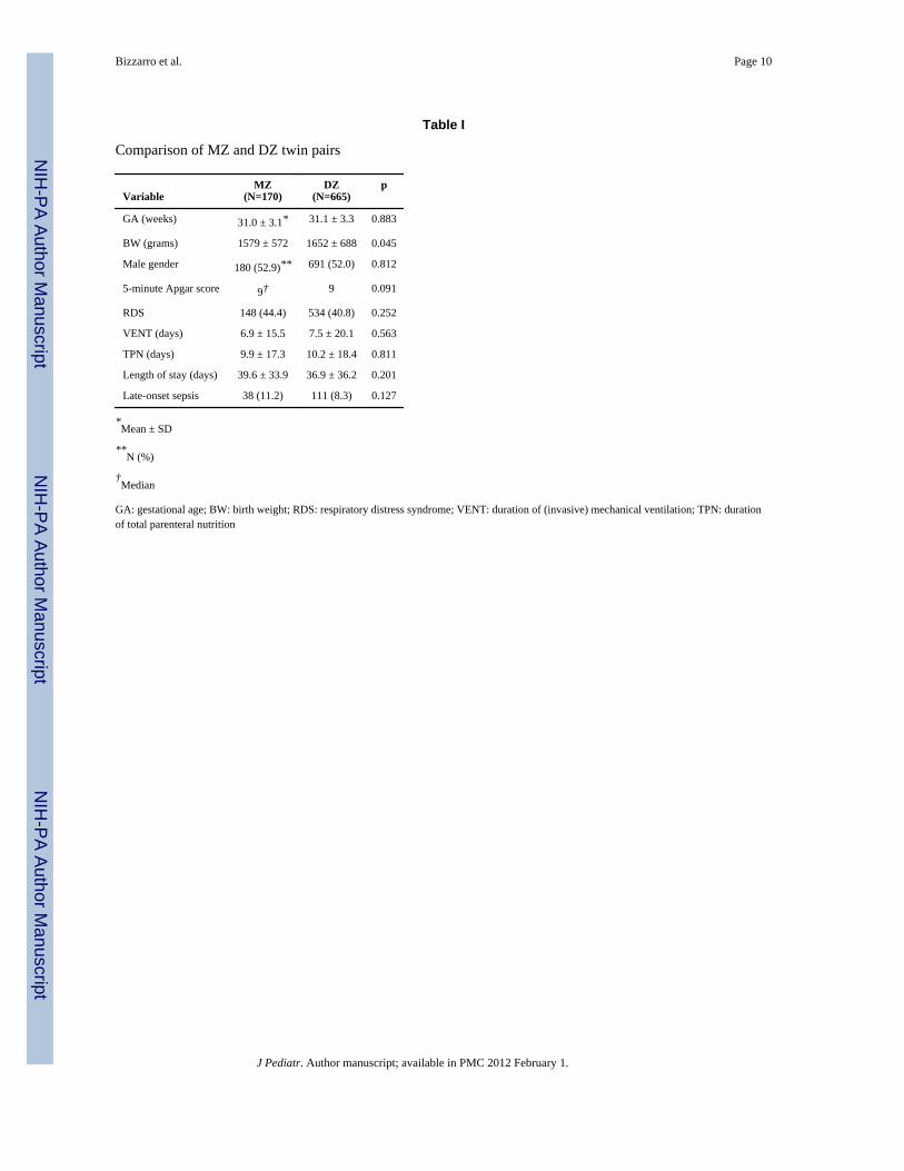
Zygosity data comprised of 170 MZ and 665 DZ twin pairs were utilized for analysis. The835 twin pairs had a mean GA and BW of 31.1 weeks and 1637 grams, respectively. Despitea discrepancy in the overall number of twin pairs in each group, no statistically significantdifferences were observed between MZ and DZ twins with respect to GA, sex, 5-minuteApgar score, the incidence of RDS, duration of mechanical ventilation, duration of TPN, andlength of hospital stay (Table I). However, significant difference was found between MZand DZ twins with respect to BW (Table I).
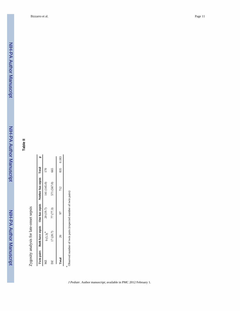
We initially performed an unadjusted concordance analysis to identify if a genetic effectexisted or not for sepsis in our newborn population. The analysis did not reveal a significantdifference of concordance distributions between MZ and DZ twin pairs. Table II shows thatthe concordance rate of the MZ twins was not significantly higher than that of the DZ twins(p=0.183).
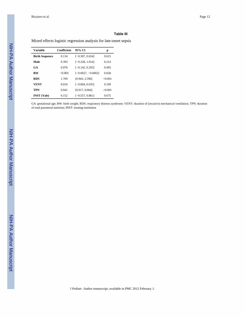
Next, a MELR analysis was performed using late-onset sepsis as the dependent variable inan attempt to identify significant covariates in our cohort that may have contributed to theoutcome of interest. The analysis determined BW, RDS and TPN to be significant predictorsfor late-onset sepsis (Table III). Excluding cases of polymicrobial sepsis, the significantvariables identified by MELR became RDS, VENT and TPN (Table IV; available atwww.jpeds.com).
Once significant non-genetic cofactors for sepsis were identified by the MELR analysis, anACE model which included BW, RDS and TPN as adjusted covariates was employed toestimate the genetic susceptibility to late-onset sepsis. It was determined that 49.8% of thevariance in liability to sepsis was the result of genetic factors alone, and 50.2% of thevariance in liability to sepsis was the result of residual environmental factors. We alsoobserved that shared environmental factors did not contribute to the variance in liability tosepsis. Therefore we fitted an AE model (without the shared environmental factors) tocompare the genetic effects and the residual environmental effects. The AE modeldetermined that 49.0% (p=0.002) of the variance in liability to sepsis was the result ofgenetic factors alone, and 51.0% (p=0.001) of the variance in liability to sepsis was theresult of residual environmental factors (Table V). Excluding cases of polymicrobial sepsis,using the AE model with the three covariates identified above by MELR, we could stillidentify significant genetic susceptibility (53.8%, p-value=0.001; Table VI; available atwww.jpeds.com). This confirmed the results of the ACE model that about a half of thevariance in liability to late-onset sepsis was the result of genetic factors, and the other halfwas due to residual environmental factors.
The empirical power was calculated to assess the reliability of the model and results. Itshowed that as 49.0% of the variance in liability to sepsis was according to genetic factors,this genetic effect could be significantly identified 77 times out of 100 replications, based onthe sample size of our collected data. This revealed that our sample size was reasonable todetect the degree of genetic effect that we found.
DISCUSSIONWe assessed and quantified the influence of additive genetic, common environmental, andresidual environmental effects on the host susceptibility to late-onset sepsis and revealedresidual (unshared) environmental and genetic factors as nearly equal contributors to thedisease process. Lavoie et al assessed the genetic contribution to sepsis in the NICUpopulation via analysis of a cohort of 158 premature twin pairs.16 As a secondary outcome
Bizzarro et al. Page 4
J Pediatr. Author manuscript; available in PMC 2012 February 1.
NIH
-PA Author Manuscript
NIH
-PA Author Manuscript
NIH
-PA Author Manuscript

of an investigation designed to assess the genetic contribution to bronchopulmonarydysplasia, the authors reported a negligible genetic influence on the susceptibility tobacterial infection (defined as the number of positive blood cultures) in their cohort. In alarger cohort of 835 twin pairs, employing criteria specific for BSI, and utilizing a statisticalmodel which included known potential confounding variables for late-onset sepsis, we wereable, for the first time, to quantify both the genetic and environmental effects on this diseaseprocess.
In our analysis, the environmental influences on late-onset sepsis were separated into sharedand unshared (residual) components. Shared environmental effects (e.g. in utero; maternaland intrapartum variables) did not have a significant influence on the susceptibility to late-onset sepsis. These results support the belief that these variables are generally consideredrisk factors for early (≤ 72 hours of life), but not late-onset sepsis. Residual environmentaleffects (e.g. co-morbidities and other variables related to the hospital course) weredetermined to account for a significant component of the variance in liability to late-onsetsepsis. This finding supports the previous understanding of late-onset sepsis as a hospital-acquired disease process in a highly vulnerable patient population. The major independentrisk factors previously identified for late-onset sepsis include, but are not limited to, the useand duration of use of central venous catheters, TPN, and mechanical ventilation.2,17,18
Despite knowledge of these and other risk factors, there is significant variability in the ratesof late-onset sepsis, even in NICUs that provide comparable levels of care.2 The NationalInstitute of Child Health and Human Development Neonatal Research Network, comprisedof 16 tertiary NICUs, reported center-to-center rates of late-onset sepsis ranging from 10.6%to 31.7%.2 Similar patient-to-patient variability is observed within individual NICUssuggesting other factors may play a role.
A genetic component to infection has long been hypothesized in both the adult and pediatricpatient populations and supported through various investigative approaches. In a landmarkepidemiologic investigation, Sorensen et al19, utilizing the Danish Adoption Registry,determined that an adoptee had a near six-fold increased risk of an infectious disease-relateddeath if their biologic parent had died from an infection prior to age 50. No such increasedrisk existed if a similar death occurred in the adoptive parent. The authors concluded thatuntimely deaths of an infectious etiology carry a strong genetic component.19 The geneticcontribution to specific infectious diseases has also been investigated in MZ and DZ twins.Higher rates of disease concordance among MZ twin pairs have suggested a geneticcomponent to chronic infectious diseases such as tuberculosis20, poliomyelitis,21 andhepatitis B.22,23 These epidemiologic investigations have served as the foundation formolecular studies aimed at determining the specific role of single nucleotide polymorphisms(SNP) in the variability in onset and response to infection. Some of these investigations haveyielded positive associations between polymorphisms in the genes which encode pro-inflammatory cytokines such as tumor necrosis factor (TNF)-α24–26, TNF-β27,interleukin-628, and angiotensin I converting enzyme29 and an increase in the severity in theresponse to, and outcome from, sepsis.30 Others have linked genetic polymorphisms in theanti-inflammatory cytokine interleukin receptor antagonist with an increase in thesusceptibility to infection.31,32
Although these data would seem to support a genetic contribution to sepsis, the majority ofinvestigations to date have been conducted in subjects from demographically,geographically, and developmentally diverse patient populations. In fact, the majority ofepidemiologic data on the subject comes from cohorts of adults and older children. There aresimilar pitfalls inherent to SNP association studies. Single-gene associations detected inunderpowered investigations conducted in relatively homogeneous patient populations maybe unreliable.33,34 Furthermore, candidate genes associations identified in adults and older
Bizzarro et al. Page 5
J Pediatr. Author manuscript; available in PMC 2012 February 1.
NIH
-PA Author Manuscript
NIH
-PA Author Manuscript
NIH
-PA Author Manuscript

children may not correspond with similar findings in premature neonates due todevelopmental regulations in gene expression.29,35–37 Genetic polymorphism forangiotensin I converting enzyme, for example, have been associated with a more severeoutcome in older children with sepsis29, but no such association was found to exist inpremature neonates.36,37 In order to identify genetic factors specific to this disease processand population, future investigations will likely need to focus on a developmentallyappropriate population.
There are several limitations to our data that may limit interpretation. One potential pitfallrelates to our definition of BSI, particularly in the setting of commensal species relatedsepsis. Despite efforts to minimize inclusion of false-positive blood cultures into our data, itis possible that some cases of BSI included were not true cases of late-onset sepsis, but wereinstead contaminants. However, all episodes of commensal species related sepsis met strictinclusion criteria that incorporated clinical signs and symptoms of infection, a positive bloodculture or blood cultures, and treatment with appropriate antimicrobial agents. Additionally,excluding the episodes of polymicrobial sepsis did not significantly impact our results. It isalso possible that the genetic effect on late-onset sepsis may be species specific and bygrouping all late-onset sepsis together, we may have overestimated the genetic contributionin some cases and underestimated it in others. Unfortunately, this investigation was notpowered for additional, species-specific subgroup analyses. In addition, our cohort wasrestricted to twin pairs with available zygosity information and therefore represented alimited, but still a fairly substantial, number of infants. The zygosity was determined usingplacental histopathology and sex, as we did not have DNA for confirmation. Amonochorionic placenta was assumed to represent MZ twins. About 9% of similar sexdichorionic placentas are MZ.38 Our conclusions were not affected when adjustments weremade for such worst-case scenarios. Lastly, the covariates collected and analyzed in ourdataset did not include all potential contributing factors related to late-onset sepsis.Complete data were not available, for example, on central versus peripheral venous catheteruse, although we were able to account for the effects of BW, mechanical ventilation, and theuse of TPN (a marker for the need for intravascular access).
Our data indicate that genetic and environmental factors play a near equal role in this diseaseprocess. At the present time, strategies to control late-onset sepsis should continue to focuson limiting the risk of environmental factors such as central lines. In the future, moresophisticated knowledge of, and approaches to, the human genome may further improve ourability to identify and properly manage those at risk.
AcknowledgmentsSupported by National Institutes on Drug Abuse (R01 DA016750 to Y.J and H.Z.), National Institute ofNeurological Disorders and Stroke (R01 NS43530 to J.G.), and National Heart, Lung and Blood Institute (K08 HL074195 to V.B.).
ABBREVIATIONS
BSI bloodstream infections
NICU newborn intensive care unit
VLBW very low birth weight
GA gestational age
BW birth weight
MZ monozygotic
Bizzarro et al. Page 6
J Pediatr. Author manuscript; available in PMC 2012 February 1.
NIH
-PA Author Manuscript
NIH
-PA Author Manuscript
NIH
-PA Author Manuscript

DZ dizygotic
RDS respiratory distress syndrome
VENT duration of (invasive) mechanical ventilation
TPN duration of total parenteral nutrition
INST treating institutions
MELR mixed effects logistic regression
SNP single nucleotide polymorphisms
TNF tumor necrosis factor
REFERENCES1. Stoll BJ, Gordon T, Korones SB, Shankaran S, Tyson JE, Bauer CR, et al. Late-onset sepsis in very
low birth weigh neonates: a report from the National Institute of Child Health and HumanDevelopment Neonatal Research Network. J Pediatr 1996;129:63–71. [PubMed: 8757564]
2. Stoll BJ, Hansen N, Fanaroff AA, Wright LL, Carlo WA, Ehrenkranz RA, et al. Late-onset sepsis invery low birth weight neonates: the experience of the NICHD Neonatal Research Network.Pediatrics 2002;110:285–291. [PubMed: 12165580]
3. Jiang JH, Chiu NC, Huang FY, Kao HA, Hsu CH, Hung HY, et al. Neonatal sepsis in the neonatalintensive care unit: characteristics of early versus late onset. J Microbiol Immunol Infect2004;37:301–306. [PubMed: 15497012]
4. Bizzarro MJ, Raskind C, Baltimore RS, Gallagher PG. Seventy-five years of neonatal sepsis at Yale:1928–2003. Pediatrics 2005;116:595–602. [PubMed: 16140698]
5. Makhoul IR, Sujov P, Smolkin T, Lusky A, Reichman B. Pathogen-specific early mortality in verylow birth weight infants with late-onset sepsis: a national survey. Clin Infect Dis 2005;40:218–224.[PubMed: 15655738]
6. Bhandari A, Bhandari V. Pathogenesis, pathology and pathophysiology of pulmonary sequelae ofbronchopulmonary dysplasia in premature infants. Front Biosci 2003;8:e370–e380. [PubMed:12700058]
7. Akram Khan M, Kuzma-O'Reilly B, Brodsky NL, Bhandari V. Site-specific characteristics ofinfants developing bronchopulmonary dysplasia. J Perinatol 2006;26:428–435. [PubMed:16724120]
8. Kaufman D, Fairchild KD. Clinical microbiology of bacterial and fungal sepsis in very-low-birth-weight infants. Clin Microbiol Rev 2004;17:638–680. [PubMed: 15258097]
9. Stoll BJ, Hansen N. Infections in VLBW infants: studies from the NICHD neonatal researchnetwork. Sem Perinatol 2003;27:293–301.
10. Payne NR, Carpenter JH, Badger GJ, Horbar JD, Rogowski J. Marginal increase in cost and excesslength of stay associated with nosocomial bloodstream infections in surviving very low birthweight infants. Pediatrics 2004;114:348–355. [PubMed: 15286215]
11. Del Vecchio A, Laforgia N, Capasso M, Iolascon A, Latini G. The role of molecular genetics in thepathogenesis and diagnosis of neonatal sepsis. Clin Perinatol 2004;31:53–67. [PubMed:15183656]
12. Garner JS, Jarvis WR, Emori TG, Horan TC, Hughes JM. CDC definitions for nosocomialinfections, 1988. Am J Infect Control 1988;16:128–140. [PubMed: 2841893]
13. Horan TC, Andrus M, Dudeck MA. CDC/NHSN surveillance definition of health-care associatedinfection and criteria for specific types of infection in the acute care setting. Am J Infect Control2008;36:309–332. [PubMed: 18538699]
14. Feng R, Zhou G, Zhang M, Zhang H. Analysis of Twin Data Using SAS. Biometrics 2010;65:584–589. [PubMed: 18647295]
Bizzarro et al. Page 7
J Pediatr. Author manuscript; available in PMC 2012 February 1.
NIH
-PA Author Manuscript
NIH
-PA Author Manuscript
NIH
-PA Author Manuscript

15. Falconer, DS.; Mackay, TFC. Introduction to Quantitative Genetics. 4th ed.. Harlow, U.K: PrenticeHall; 1996.
16. Lavoie PM, Pham C, Jang KL. Heritability of bronchopulmonary dysplasia, defined according tothe consensus statement of the National Institute of Health. Pediatrics 2008;122:479–485.[PubMed: 18762515]
17. Perlman SE, Saiman L, Larson EL. Risk factors for late-onset health care-associated bloodstreaminfections in patients in neonatal intensive care units. Am J Infect Control 2007;35:177–182.[PubMed: 17433941]
18. Távora AC, Castro AB, Militão MA, Girão JE, Ribeiro Kde C, Távora LG. Risk factors fornosocomial infection in a Brazilian neonatal intensive care unit. Braz J Infect Dis 2008;12:75–79.[PubMed: 18553019]
19. Sørensen TI, Nielsen GG, Andersen PK, Teasdale TW. Genetic and environmental influences onpremature death in adult adoptees. N Engl J Med 1988;318:727–732. [PubMed: 3347221]
20. Comstock GW. Tuberculosis in twins: a re-analysis of the Prophit survey. Am Rev Respir Dis1978;117:621–624. [PubMed: 565607]
21. Herdon CN, Jennings RG. A twin-family study of susceptibility to poliomyelitis. Am J HumanGenet 1951;3:17–46. [PubMed: 13171372]
22. Cooke GS, Hill AV. Genetics of susceptibility to human infectious disease. Nat Rev Genet2001;2:967–977. [PubMed: 11733749]
23. Lin TM, Chen CJ, Wu MM, et al. Hepatitis B virus markers in Chinese twins. Anticancer Res1989;9:737–741. [PubMed: 2764519]
24. Nadel S, Newport MJ, Booy R, Levin M. Variation in the tumor necrosis factor-alpha genepromoter region may be associated with death from meningococcal disease. J Infect Dis1996;174:878–880. [PubMed: 8843235]
25. Mira JP, Cariou A, Grall F, Delclaux C, Losser MR, Heshmati F, et al. Association of TNF2, aTNF-alpha promoter polymorphism, with septic shock susceptibility and mortality: a multicenterstudy. JAMA 1999;282:561–568. [PubMed: 10450718]
26. Stüber F, Petersen M, Bokelmann F, Schade U. A genomic polymorphism within the tumornecrosis factor locus influences plasma tumor necrosis factor-alpha concentrations and outcome ofpatients with severe sepsis. Crit Care Med 1996;24:381–384. [PubMed: 8625623]
27. McArthur JA, Zhang Q, Quasney MW. Association between the A/A genotype at the lymphotoxin-alpha+250 site and increased mortality in children with positive blood cultures. Pediatr Crit CareMed 2002;3:341–344. [PubMed: 12780951]
28. Harding D, Dhamrait S, Millar A, Humphries S, Marlow N, Whitelaw A, et al. Is interleukin-6–174 genotype associated with the development of septicemia in preterm infants? Pediatrics2003;112:800–803. [PubMed: 14523169]
29. Harding D, Baines PB, Brull D, Vassiliou V, Ellis I, Hart A, et al. Severity of meningococcaldisease in children and the angiotensin-converting enzyme insertion/deletion polymorphism. Am JRespir Crit Care Med 2002;165:1103–1106. [PubMed: 11956052]
30. Holmes CL, Russell JA, Walley KR. Genetic polymorphisms in sepsis and septic shock. Chest2003;124:1103–1115. [PubMed: 12970043]
31. Fang XM, Schröder S, Hoeft A, Stüber F. Comparison of two polymorphisms of the interleukin-1gene family: interleukin-1 receptor antagonist polymorphism contributes to susceptibility to severesepsis. Crit Care Med 1999;27:1330–1334. [PubMed: 10446828]
32. Carrol ED, Mobbs KJ, Thomson AP, Hart CA. Variable number tandem repeat polymorphism ofthe interleukin-1 receptor antagonist gene in meningococcal disease. Clin Infect Dis 2002;35:495–497. [PubMed: 12145738]
33. Harding D. Impact of common genetic variation on neonatal disease and outcome. Arch Dis ChildFetal Neonatal Ed 2007;92:F408–F413. [PubMed: 17712190]
34. Chauhan M, McGuire W. Interleukin-6 (−174 C) polymorphism and the risk of sepsis in very lowbirth weight infants: meta-analysis. Arch Dis Child Fetal Neonatal Ed 2008;93:F427–F429.[PubMed: 18375611]
35. Viemann D, Dubbel G, Schleifenbaum S, Harms E, Sorg C, Roth J. Expression of toll-likereceptors in neonatal sepsis. Pediatr Res 2005;58:654–659. [PubMed: 16189189]
Bizzarro et al. Page 8
J Pediatr. Author manuscript; available in PMC 2012 February 1.
NIH
-PA Author Manuscript
NIH
-PA Author Manuscript
NIH
-PA Author Manuscript

36. Yanamandra K, Loggins J, Baier RJ. The Angiotensin Converting Enzyme Insertion/Deletionpolymorphism is not associated with an increased risk of death or bronchopulmonary dysplasia inventilated very low birth weight infants. BMC Pediatr 2004;4:26. [PubMed: 15610555]
37. Dahmer MK, Randolph A, Vitali S, Quasney MW. Genetic polymorphisms in sepsis. Pediatr CritCare Med 2005;6:S61–S73. [PubMed: 15857562]
38. Bhandari V, Bizzarro MJ, Shetty A, Zhong X, Page GP, Zhang H, et al. Neonatal Genetics StudyGroup. Familial and genetic susceptibility to major neonatal morbidities in premature twins.Pediatrics 2006;117:1901–1906. [PubMed: 16740829]
Bizzarro et al. Page 9
J Pediatr. Author manuscript; available in PMC 2012 February 1.
NIH
-PA Author Manuscript
NIH
-PA Author Manuscript
NIH
-PA Author Manuscript
NIH
-PA Author Manuscript
NIH
-PA Author Manuscript
NIH
-PA Author Manuscript
Bizzarro et al. Page 10
Table I
Comparison of MZ and DZ twin pairs
VariableMZ
(N=170)DZ
(N=665)p
GA (weeks) 31.0 ± 3.1* 31.1 ± 3.3 0.883
BW (grams) 1579 ± 572 1652 ± 688 0.045
Male gender 180 (52.9)** 691 (52.0) 0.812
5-minute Apgar score 9† 9 0.091
RDS 148 (44.4) 534 (40.8) 0.252
VENT (days) 6.9 ± 15.5 7.5 ± 20.1 0.563
TPN (days) 9.9 ± 17.3 10.2 ± 18.4 0.811
Length of stay (days) 39.6 ± 33.9 36.9 ± 36.2 0.201
Late-onset sepsis 38 (11.2) 111 (8.3) 0.127
*Mean ± SD
**N (%)
†Median
GA: gestational age; BW: birth weight; RDS: respiratory distress syndrome; VENT: duration of (invasive) mechanical ventilation; TPN: durationof total parenteral nutrition
J Pediatr. Author manuscript; available in PMC 2012 February 1.
NIH
-PA Author Manuscript
NIH
-PA Author Manuscript
NIH
-PA Author Manuscript
Bizzarro et al. Page 11
Tabl
e II
Zygo
sity
ana
lysi
s for
late
-ons
et se
psis
Tw
in p
airs
Bot
h ha
ve se
psis
One
has
seps
isN
eith
er h
as se
psis
Tot
alp
MZ
9 (5
.3)*
20 (1
9.7)
141
(145
.0)
170
DZ
17 (2
0.7)
77 (7
7.3)
571
(567
.0)
665
Tot
al26
9771
283
50.
183
* Obs
erve
d nu
mbe
r of t
win
pai
rs (e
xpec
ted
num
ber o
f tw
in p
airs
)
J Pediatr. Author manuscript; available in PMC 2012 February 1.
NIH
-PA Author Manuscript
NIH
-PA Author Manuscript
NIH
-PA Author Manuscript
Bizzarro et al. Page 12
Table III
Mixed effects logistic regression analysis for late-onset sepsis
Variable Coefficient 95% CI p
Birth Sequence 0.134 [−0.387, 0.654] 0.615
Male 0.393 [−0.228, 1.014] 0.214
GA 0.076 [−0.142, 0.293] 0.495
BW −0.001 [−0.0027, −0.0002] 0.028
RDS 1.769 [0.943, 2.596] <0.001
VENT 0.016 [−0.004, 0.035] 0.109
TPN 0.041 [0.017, 0.064] <0.001
INST (Yale) 0.152 [−0.557, 0.861] 0.675
GA: gestational age; BW: birth weight; RDS: respiratory distress syndrome; VENT: duration of (invasive) mechanical ventilation; TPN: durationof total parenteral nutrition; INST: treating institution
J Pediatr. Author manuscript; available in PMC 2012 February 1.

NIH
-PA Author Manuscript
NIH
-PA Author Manuscript
NIH
-PA Author Manuscript
Bizzarro et al. Page 13
Table IV
Mixed effects logistic regression analysis for late-onset sepsis (excluding cases of polymicrobial sepsis)
Variable Coefficient 95% CI p
Birth Sequence 0.807 [−0.044, 1.658] 0.063
Male 0.627 [−0.410, 1.664] 0.235
GA 0.317 [−0.090, 0.725] 0.127
BW −0.002 [−0.0039, 0.0004] 0.103
RDS 2.224 [0.740, 3.709] 0.003
VENT 0.044 [0.012, 0.076] 0.008
TPN 0.103 [0.051, 0.154] <0.001
INST (Yale) −0.182 [−1.557, 1.193] 0.795
GA: gestational age; BW: birth weight; RDS: respiratory distress syndrome; VENT: duration of (invasive) mechanical ventilation; TPN: durationof total parenteral nutrition; INST: treating institution
J Pediatr. Author manuscript; available in PMC 2012 February 1.

NIH
-PA Author Manuscript
NIH
-PA Author Manuscript
NIH
-PA Author Manuscript
Bizzarro et al. Page 14
Table V
AE model analysis for late-onset sepsis
Variables/Effects Estimate p
BW −0.001 <0.001
RDS 1.122 <0.001
TPN 0.025 <0.001
Genetic 0.490 0.002
ResidualEnvironmental
0.510 0.001
BW: birth weight; RDS: respiratory distress syndrome; TPN: duration of total parenteral nutrition
J Pediatr. Author manuscript; available in PMC 2012 February 1.

NIH
-PA Author Manuscript
NIH
-PA Author Manuscript
NIH
-PA Author Manuscript
Bizzarro et al. Page 15
Table VI
AE model analysis for late-onset sepsis (excluding cases of polymicrobial sepsis)
Variables/Effects Estimate p
RDS 1.235 <0.001
VENT 0.014 0.009
TPN 0.026 <0.001
Genetic 0.538 0.001
ResidualEnvironmental
0.462 0.003
RDS: respiratory distress syndrome; VENT: duration of (invasive) mechanical ventilation; TPN: duration of total parenteral nutrition
J Pediatr. Author manuscript; available in PMC 2012 February 1.