The Immunomodulatory Role of Syncytiotrophoblast Microvesicles
10
The Immunomodulatory Role of Syncytiotrophoblast Microvesicles Jennifer Southcombe*, Dionne Tannetta, Christopher Redman, Ian Sargent Nuffield Department of Obstetrics and Gynaecology, University of Oxford, John Radcliffe Hospital, Oxford, United Kingdom Abstract Immune adaptation is a critical component of successful pregnancy. Of primary importance is the modification of cytokine production upon immune activation. With the discovery that normal pregnancy itself is a pro-inflammatory state, it was recognised that the classical Th1/Th2 cytokine paradigm, with a shift towards ‘type 2’ cytokine production (important for antibody production), and away from ‘type 1’ immunity (associated with cell mediated immunity and graft rejection), is too simplistic. It is now generally agreed that both arms of cytokine immunity are activated, but with a bias towards ‘type 2’ immunity. Many factors are released from the placenta that can influence the maternal cytokine balance. Here we focus on syncytiotrophoblast microvesicles (STBM) which are shed from the placenta into the maternal circulation. We show that STBM can bind to monocytes and B cells and induce cytokine release (TNFa, MIP-1a, IL-1a, IL-1b, IL-6, IL-8). Other cytokines are down-modulated, such as IP-10 which is associated with ‘type 1’ immunity. Therefore STBM may aid the ‘type 2’ skewed nature of normal pregnancy. We also observed that PBMC from third trimester normal pregnant women produce more TNFa and IL-6 in response to STBM than PBMC from non-pregnant women, confirming that maternal immune cells are primed by pregnancy, possibly through their interaction with STBM. Citation: Southcombe J, Tannetta D, Redman C, Sargent I (2011) The Immunomodulatory Role of Syncytiotrophoblast Microvesicles. PLoS ONE 6(5): e20245. doi:10.1371/journal.pone.0020245 Editor: Carlos Penha-Goncalves, Instituto Gulbenkian de Cie ˆncia, Portugal Received January 24, 2011; Accepted April 25, 2011; Published May 25, 2011 Copyright: ß 2011 Southcombe et al. This is an open-access article distributed under the terms of the Creative Commons Attribution License, which permits unrestricted use, distribution, and reproduction in any medium, provided the original author and source are credited. Funding: This study was supported by a grant from the Wellcome Trust (http://www.wellcome.ac.uk), Ref GR079862MA, and by the Oxford Partnership Comprehensive Biomedical Research Centre (http://www.oxfordbrc.org) with funding from the Department of Health’s National Institute for Health Research Biomedical Research Centres funding scheme. The views expressed in this publication are those of the authors and not necessarily those of the Department of Health. The funders had no role in study design, data collection and analysis, decision to publish, or preparation of the manuscript. Competing Interests: The authors have declared that no competing interests exist. * E-mail: [email protected] Introduction A pregnant woman’s immune system is carefully controlled and adapted to accommodate the developing semi-allogenic fetus. Failure to appropriately adapt is associated with pregnancy problems such as spontaneous abortion or preeclampsia. The adaptation can be seen by studying maternal cytokine responses to antigens throughout pregnancy. Cytokine responses are often described as being of type 1 or type 2; type 1 cytokines such as Interferon gamma (IFNc) and Tumor Necrosis Factor alpha (TNFa) promote cellular mediated immune responses, and type 2 cytokines such as IL-4 and IL-6 promote humoral immunity. A bias towards type 2 immunity was proposed to prevent cell mediated rejection of the fetus [1], and such changes in cytokine immunity can be observed. Often during pregnancy classical type 1 syndromes alleviate, whereas type 2 syndromes worsen. Over recent years this concept has been shown to be too simplistic [2,3] and the inflammatory nature of normal pregnancy has become more apparent [4]. It is now generally agreed that both arms of cytokine immunity are activated, but with a bias towards ‘type 2’ immunity [5]. It is proposed that factors from the placenta can induce these essential modifications [6]. Possible modulating factors include cytokines, growth factors and enzymes [7]. These factors can often be detected in the maternal peripheral blood and are present at varying levels throughout pregnancy, and therefore have the potential to modify maternal immunity. In addition, it is known that microvesicles (,1 mm) are shed from the syncytiotrophoblast into the maternal blood [8]. These are termed syncytiotrophoblast microvesicles (STBM) and are also thought to affect maternal immunity systemically. Many cell types release vesicles of which there are three main types: vesicles that bud directly from the cell membrane, exosomes that are derived from multivesicular bodies within the cell and apoptotic bodies, small sealed membrane vesicles that are produced from cells undergoing cell death by apoptosis [9]. They are encapsulated by a lipid bilayer, and can contain various cytoplasmic molecules, such as cytoskeletal proteins, signalling molecules, DNA and micro RNAs. The precise nature of the placental vesicles has yet to be defined, with respect to the content and proportion of vesicles, exosomes and apoptotic bodies. We, and others, can detect STBM in the circulation of women in the first trimester of pregnancy and increasing as pregnancy progresses [10,11]. Cellular vesicles are an integral part of various immunological systems, as they carry proteins, lipids and miRNAs from their cell of origin to other target cells. They can be immune activating, for example they can carry antigens which directly stimulate T cells, transfer antigens to dendritic cells for indirect immune cell stimulation, or act independently of antigens by exposing immune cells to stimulatory factors, such as heat shock protein-70 or NKG2D ligands [12]. In contrast, they can be inhibitory, for example they can cause T cell death, inhibit dendritic cell maturation or prevent T cell killing activity, reviewed by Thery et al (2009) [12]. PLoS ONE | www.plosone.org 1 May 2011 | Volume 6 | Issue 5 | e20245
Transcript of The Immunomodulatory Role of Syncytiotrophoblast Microvesicles

The Immunomodulatory Role of SyncytiotrophoblastMicrovesiclesJennifer Southcombe*, Dionne Tannetta, Christopher Redman, Ian Sargent
Nuffield Department of Obstetrics and Gynaecology, University of Oxford, John Radcliffe Hospital, Oxford, United Kingdom
Abstract
Immune adaptation is a critical component of successful pregnancy. Of primary importance is the modification of cytokineproduction upon immune activation. With the discovery that normal pregnancy itself is a pro-inflammatory state, it wasrecognised that the classical Th1/Th2 cytokine paradigm, with a shift towards ‘type 2’ cytokine production (important forantibody production), and away from ‘type 1’ immunity (associated with cell mediated immunity and graft rejection), is toosimplistic. It is now generally agreed that both arms of cytokine immunity are activated, but with a bias towards ‘type 2’immunity. Many factors are released from the placenta that can influence the maternal cytokine balance. Here we focus onsyncytiotrophoblast microvesicles (STBM) which are shed from the placenta into the maternal circulation. We show thatSTBM can bind to monocytes and B cells and induce cytokine release (TNFa, MIP-1a, IL-1a, IL-1b, IL-6, IL-8). Other cytokinesare down-modulated, such as IP-10 which is associated with ‘type 1’ immunity. Therefore STBM may aid the ‘type 2’ skewednature of normal pregnancy. We also observed that PBMC from third trimester normal pregnant women produce moreTNFa and IL-6 in response to STBM than PBMC from non-pregnant women, confirming that maternal immune cells areprimed by pregnancy, possibly through their interaction with STBM.
Citation: Southcombe J, Tannetta D, Redman C, Sargent I (2011) The Immunomodulatory Role of Syncytiotrophoblast Microvesicles. PLoS ONE 6(5): e20245.doi:10.1371/journal.pone.0020245
Editor: Carlos Penha-Goncalves, Instituto Gulbenkian de Ciencia, Portugal
Received January 24, 2011; Accepted April 25, 2011; Published May 25, 2011
Copyright: � 2011 Southcombe et al. This is an open-access article distributed under the terms of the Creative Commons Attribution License, which permitsunrestricted use, distribution, and reproduction in any medium, provided the original author and source are credited.
Funding: This study was supported by a grant from the Wellcome Trust (http://www.wellcome.ac.uk), Ref GR079862MA, and by the Oxford PartnershipComprehensive Biomedical Research Centre (http://www.oxfordbrc.org) with funding from the Department of Health’s National Institute for Health ResearchBiomedical Research Centres funding scheme. The views expressed in this publication are those of the authors and not necessarily those of the Department ofHealth. The funders had no role in study design, data collection and analysis, decision to publish, or preparation of the manuscript.
Competing Interests: The authors have declared that no competing interests exist.
* E-mail: [email protected]
Introduction
A pregnant woman’s immune system is carefully controlled and
adapted to accommodate the developing semi-allogenic fetus.
Failure to appropriately adapt is associated with pregnancy
problems such as spontaneous abortion or preeclampsia. The
adaptation can be seen by studying maternal cytokine responses to
antigens throughout pregnancy. Cytokine responses are often
described as being of type 1 or type 2; type 1 cytokines such as
Interferon gamma (IFNc) and Tumor Necrosis Factor alpha
(TNFa) promote cellular mediated immune responses, and type 2
cytokines such as IL-4 and IL-6 promote humoral immunity. A
bias towards type 2 immunity was proposed to prevent cell
mediated rejection of the fetus [1], and such changes in cytokine
immunity can be observed. Often during pregnancy classical type
1 syndromes alleviate, whereas type 2 syndromes worsen. Over
recent years this concept has been shown to be too simplistic [2,3]
and the inflammatory nature of normal pregnancy has become
more apparent [4]. It is now generally agreed that both arms of
cytokine immunity are activated, but with a bias towards ‘type 2’
immunity [5].
It is proposed that factors from the placenta can induce these
essential modifications [6]. Possible modulating factors include
cytokines, growth factors and enzymes [7]. These factors can often
be detected in the maternal peripheral blood and are present at
varying levels throughout pregnancy, and therefore have the
potential to modify maternal immunity. In addition, it is known
that microvesicles (,1 mm) are shed from the syncytiotrophoblast
into the maternal blood [8]. These are termed syncytiotrophoblast
microvesicles (STBM) and are also thought to affect maternal
immunity systemically.
Many cell types release vesicles of which there are three main
types: vesicles that bud directly from the cell membrane, exosomes
that are derived from multivesicular bodies within the cell and
apoptotic bodies, small sealed membrane vesicles that are produced
from cells undergoing cell death by apoptosis [9]. They are
encapsulated by a lipid bilayer, and can contain various cytoplasmic
molecules, such as cytoskeletal proteins, signalling molecules, DNA
and micro RNAs. The precise nature of the placental vesicles has yet
to be defined, with respect to the content and proportion of vesicles,
exosomes and apoptotic bodies. We, and others, can detect STBM
in the circulation of women in the first trimester of pregnancy and
increasing as pregnancy progresses [10,11].
Cellular vesicles are an integral part of various immunological
systems, as they carry proteins, lipids and miRNAs from their cell
of origin to other target cells. They can be immune activating, for
example they can carry antigens which directly stimulate T cells,
transfer antigens to dendritic cells for indirect immune cell
stimulation, or act independently of antigens by exposing immune
cells to stimulatory factors, such as heat shock protein-70 or
NKG2D ligands [12]. In contrast, they can be inhibitory, for
example they can cause T cell death, inhibit dendritic cell
maturation or prevent T cell killing activity, reviewed by Thery
et al (2009) [12].
PLoS ONE | www.plosone.org 1 May 2011 | Volume 6 | Issue 5 | e20245

STBM can interact with various target cells. In vitro, STBM can
cause disruption to endothelium [13], induce pro-inflammatory
cytokine production by immune cells [10] and inhibit T cell
proliferation [14,15]. In addition, the observation that increased
levels of STBM can be detected in preeclamptic women has led to
the hypothesis that STBM could contribute to the vascular
endothelial disruption and exaggerated inflammation observed in
this disease [6].
To study the effects of STBM on target cells sufficiently large
amounts of relatively pure vesicles are needed. Ideally vesicles
would be obtained from the peripheral blood of pregnant women
through centrifugation, however the yield is very low and
preparations would be heavily contaminated with vesicles from
other cell types; STBM represent only a very small proportion of
the total plasma vesicle population, the predominant form being
platelet derived [11]. Therefore in this study STBM have been
prepared from term placentas delivered by caesarean section using
three different methods designed to mimic the STBM generated in
vivo. These are mSTBM (derived from mechanical dissection of the
placenta), eSTBM (shed from placenta explants in culture) and
pSTBM (perfused from the maternal side of a placenta lobe). As
normal pregnancy is associated with a systemic inflammatory
response we have investigated the stimulatory capacity of the three
STBM preparations. We used a cytokine array panel to identify a
range of cytokines and chemokines that are produced, or
inhibited, when PBMC are treated with STBM. Further studies
focused on seven cytokines: TNFa, Macrophage Inflammatory
Protein-1alpha (MIP-1a), IL-1a, IL-1b, IL-6, IL-8 and interferon-
inducible protein 10 KDa (IP-10). Intracellular cytokine staining
revealed that the monocytes were solely responsible for the
production of TNFa, IL-6, IL-8 and IL-1b. We have previously
shown that PBMC from pregnant women are primed to produce
more TNF-a in response to LPS/IFN-c stimulation than PBMC
from non-pregnant women [10]. Here, we explore if peripheral
blood mononuclear cells (PBMC) from third trimester normally
pregnant women are more responsive to STBM than PBMC from
non-pregnant women. In addition, we show that the STBM bind
to monocytes, which also phagocytose the STBM, and to a lesser
extent to B cells, but only small numbers of T and NK cells are
able to bind STBM.
Methods
SubjectsHealthy women were recruited in the third trimester of
pregnancy. The mean age of the women was 32 years (range 27
to 36), 70% were nulliparous, and mean gestation was 37+3 (range
35+3 to 39+6). Ten normal pregnant women were recruited and
matched to ten non pregnant women for age (+/24 years) and
parity (0, 1–3). None of the women were in labour at the time of
sampling, and all had singleton pregnancies, with no known fetal
abnormalities. None of the women had any significant medical
history, current or recent illnesses, or were taking medication.
These studies were approved by the Oxfordshire Research Ethics
Committee C and informed written consent was obtained.
STBM preparationThree preparations of STBM were made for this study,
mechanically derived STBM (mSTBM), STBM from placenta
perfusion (pSTBM) and explant derived STBM (eSTBM). All
placentas were from normally pregnant, healthy women under-
going elective caesarean section, without labor, and were
processed immediately. Protein content was determined using a
Pierce BCA protein assay kit (Thermo Scientific, Illinois, USA),
and aliquots of microvesicles were stored at 280uC. STBM were
cultured in antibiotic free media for five days to confirm that the
preparations were not contaminated with bacteria.
mSTBM were prepared by a modification of the method of
Smith et al. [16], as outlined previously [13]. Briefly, placenta
tissue is washed in ice cold 100 mM CaCl2 followed by PBS and
scraped from villi and then stirred in 0.9% NaCl buffer for 1 hour.
Cell debris was removed by centrifugation in a Beckman J6-M
centrifuge at 6006g for 10 min at 4uC and 10,0006g for 10 min ,
then the supernatant was centrifuged at 48,0006g for 45 min at
4uC in a Beckman L8-80M ultracentrifuge. The resultant pellets
were pooled and washed in PBS before finally being resuspended
in PBS. Typical mSTBM preparations yield 25–100 mg of
vesicles. A pool of nine mSTBM preparations was made for use
in all experiments.
pSTBM were prepared using a modified dual placental
perfusion system as described by Eaton and Oakey [17]. An
individual lobule was isolated and firstly the fetal circulation
perfused with 0.1 mM filtered modified M-199 tissue culture
medium (Medium 199 with L-glutamine and Earle’s salts,
containing 0.8% Dextran 20, 0.5% BSA, 5000 U/L sodium
heparin, and 2.75 g/L sodium bicarbonate, pH 7.4) containing a
20 ml bolus of 100,000 IU streptokinase to promote clot removal,
at a rate of 5 mL/min. The whole placenta was turned upside
down and laid inside a Perspex water jacket maintained at 37uC.
The maternal circulation was then perfused with medium
(Medium 199 with L-glutamine and Earle’s salts, containing
0.5% BSA, 5000 U/L sodium heparin, and 2.75 g/L sodium
bicarbonate, pH 7.4) through eight 1.7 mm fetal feeding tubes at a
controlled rate of 20 ml/min. Perfusion media were warmed in a
37uC water bath and the maternal perfusion medium was
oxygenated with 95% O2, 5%CO2. The lobule was perfused for
20 min to equilibrate the system, after which time the maternal
circuit was closed with a total volume of perfusion medium of
600 mL. The volume of fetal effluent was measured every 2 min
and the oxygen concentration of the maternal side perfusate
monitored to ensure the stability of the preparation. Pressure
monitors were used to ensure no significant deviations from
baseline during the experimental period. At the end of the 3 hr
perfusion period, the maternal perfusate was centrifuged in a
Beckman J6-M centrifuge at 6006g for 10 min at 4uC. The
supernatant was centrifuged at 150,0006g for 1 hour at 4uC in a
Beckman L8-80M ultracentrifuge. The resultant pellets were
pooled and washed in PBS before finally being resuspended in
PBS to give a final protein content of 5 mg/ml. Typical pSTBM
preparations yield 25–50 mg of vesicles. Five pSTBM prepara-
tions were pooled for use in all experiments.
eSTBM were prepared as follows. Freshly delivered placentae
were first rinsed in ice cold Hanks balanced salt solution and
placed into a glove box maintained at 8% oxygen. Placental
pieces, cut from undamaged lobules that appeared healthy, were
rinsed in 8% O2 equilibrated ice cold explant culture medium
(DMEM/F12 containing 10% foetal bovine serum (PAA Labora-
tories GmbH, Austria), 1% antibiotic and antimycotic solution
(Sigma Aldrich, UK) and L-glutamine) before being placed into ice
cold fresh equilibrated explant culture medium. Placental pieces of
approximately 2 mm in diameter were then dissected and
distributed equally between Costar Netwell (24 mm diameter,
500 mm mesh) supports in 6-well plates containing 4 ml/well
equilibrated explant culture medium (10 explants/well). Placental
explants were finally washed again with a medium change before
being incubated under ‘normoxic’ conditions (8% O2/87% N2/
5% CO2) [18]. After 24 hr the explant supernatant was collected
and centrifuged at 6006g for 10 min to remove cell debris. Rinsed
STBM and Immunity in Pregnancy
PLoS ONE | www.plosone.org 2 May 2011 | Volume 6 | Issue 5 | e20245

explants and ten 0.5 mL aliquots of pooled supernatant were then
stored at 280uC. The remaining supernatant was centrifuged at
150,0006g for 1 hour at 4uC in a Beckman L8-80M ultracentri-
fuge. The resultant pellets were pooled and washed in PBS before
finally being resuspended in 0.5 mL of PBS. Typical eSTBM
preparations yield 0.2–0.4 mg vesicles. Five eSTBM preparations
were pooled for use in all experiments.
Preparation of red blood cell microvesiclesPeripheral blood taken from non-pregnant women was
centrifuged to pellet red blood cells. Cells were resuspended in
PBS/2 mM CaCl2 and treated with calcium ionophore (Sigma
Aldrich, UK) for 1.5 hours at 37uC. Cellular debris was then
removed by centrifugation at 20006g for 10 minutes. Superna-
tants were centrifuged at 150,0006g and microvesicles washed in
PBS and protein concentrations determined by a Pierce BCA
assay and resuspended at 5 mg/ml in PBS prior to freezing at
280uC for use in stimulation assays.
Blood Collection20–30 ml blood samples, from pregnant and non-pregnant
donors, were collected into sodium heparin anti-coagulant (10 U/
ml) and PBMC were prepared by density gradient centrifugation
over lymphoprep (Axis Shield Diagnostics, Cambridgeshire, UK).
Cells were washed twice with PBS and consecutive centrifugation
of 8006g and 2006g for 10 minutes.
PBMC stimulation Assays106/ml PBMC in human serum media (RPMI 1640 supple-
mented with 10% human serum (Sera Laboratories International,
West Sussex, UK), 1% penicillin-streptomycin (50 IU/ml and
50 mg/ml), 1% glutamine, MEM NEAA, sodium pyruvate and
50 nM 2-mercaptoethanol (Gibco Invitrogen, Paisley, UK)) were
incubated with varying concentrations of STBM preparations for
20 hours at 37uC/5% CO2. Samples were centrifuged at
10,0006g for 30 seconds to remove cellular debris and superna-
tants frozen at 280uC until assayed.
Cytokine ArraysCytokine arrays were performed using the Human Cytokine
Array Panel A Proteome Profiler (R&D Systems, Minneapolis,
USA). Briefly, 0.5 ml supernatant from stimulated PBMC was
incubated with the array and cytokines detected following
manufacturer’s instructions. Dots were detected using X-ray film
and films scanned for pixel density analysis using ImageJ software.
Cytokine ELISAIL-1a, IP-10, TNFa, MIP-1a, IL-8, G-CSF and IL-6 ELISA
kits were from Peprotech (NJ, USA) and IL-1b from BD
Biosciences (Oxford, UK) and used following the manufacturer’s
instructions. 100 ml supernatant was analysed in duplicate, and for
the IL-8, IL-6 and IL-1b ELISAs some samples were diluted in
human serum media between 5- and 100-fold in order for the
analyte concentration to fall within the standard curve range.
Standards were prepared in human serum media, and matched
non pregnant and normal pregnant samples were run on the same
96-well MaxiSorp Plate (Nunc, Denmark). In addition, 50 mg/ml
concentrations of mSTBM and pSTBM were analysed, neither of
the preparations contained detectable levels of cytokines. ELISA
were developed using 2-29-Azino-bis(3-ethylbenzthiazoline-6-sul-
phonic acid) Liquid Substrate System for ELISA (Sigma Aldrich,
UK) and absorbance at 405 nm detected using a FLUOstar
Optima (BMG Labtech) plate reader.
Intracellular Cytokine Staining for Flow Cytometry106 PBMC from non-pregnant donors (n = 3) were stimulated
for 20 hours with 50 mg/ml pSTBM in the presence of 3 mg/ml
Brefeldin A (eBioscience, San Diego, U.S.A.), with 1 mg/ml LPS
(Sigma Aldrich, UK) or left untreated. Cells were harvested and
washed 63 in PBS/2% FCS then fixed using Fixation buffer
(eBioscience) prior to staining for 20 minutes at 4uC with CD14-
Alexa647 (BioLegend, San Diego, U.S.A.) or CD19-PeCy7 (BD
Biosciences, Oxford, UK). Cells were then washed and treated
with Permeabilization buffer (eBioscience) following manufactur-
ers instructions prior to incubation for 20 minutes at 4uC with
FITC conjugated antibodies towards IL-6, IL-8 and IL-1b or PE
conjugated antibody towards TNFa (BD Bioscience) or appropri-
ate isotype control. Cells were washed and analysed immediately
by flow cytometry on a LSR-II flow cytometer (BD Biosciences,
Oxford, UK) and data analysed using FACS DIVA software (BD
Biosciences, Oxford, UK).
STBM Binding Assay106/ml PBMC in human serum media were incubated with
50 mg/ml pSTBM for 1 hour at 37uC, cells were then washed 62
with PBS/2% FCS by centrifugation at 2006g for 5 minutes.
Cells were stained with the following fluorescently labelled
antibodies CD14-APC (eBiosciences), CD3-PeCy5 (Biolegend),
CD19-FITC (Serotech), CD56-Pe-Cy7 and CD16-APC-Cy7 (BD
Pharmingen) and NDOG-2-PE or IgG-PE isotype control.
NDOG-2 is a trophoblast specific antibody that recognises
placental alkaline phosphatase [19]. NDOG-2 or IgG control
antibodies were conjugated to phycoerythrin (PE) using a
lightning-link kit (Innova Biosciences, Cambridge, UK). Data
was acquired immediately.
STBM Internalisation study by Image Stream – imagingflow cytometry
5 mM stock solution of BODIPY FL Maleimide (Invitrogen,
Paisley, UK) was filtered through a 0.5 ml 10 K Amicon Ultra
Centrifugal Filter (Millipore, MA, U.S.A.) and used to label
pSTBM. 0.5 ml pSTBM (5 mg/ml) were incubated with BOD-
IPY FL Maleimide for 15 minutes at room temperature, 98%
labelling of pSTBM was confirmed by flow cytometry. pSTBM
were washed with 11 ml PBS with ultracentrifugation at
100,0006g, to remove free BODIPY FL Maleimide. As a control
the same amount of BODIPY FL Maleimide and 100 ml 300 nm
polystyrene beads (Duke Scientific, Palo Alto, U.S.A.) was
subjected to the same ultracentrifugation wash. Post ultracentri-
fugation supernatant was removed and beads or pSTBM were
suspended in 0.5 ml PBS, and a BCA assay performed to
determine the pSTBM yield. To test all free BODIPY FL
Maleimide was removed, 50 mg pSTBM or corresponding volume
of bead control was incubated with 106/ml PBMC. No labelling of
the cells was detected with the control (data not shown).
106 PBMC/ml were incubated with 50 mg/ml pSTBM-
BODIPY FL Maleimide for t = 1, 6 or 20 hours then washed
and fixed in PBS/1% paraformaldehyde. Monocytes were
identified by size, and B cells were stained with directly conjugated
PE-fluorescent antibody towards CD19 before analysis by Image
Stream (Amnis, Seattle, U.S.A.). Data was analysed with IDEAS
4.0 software, using the internalisation application. Briefly, single
cell, in focus images and BODIPY FL positive images alone were
analysed (i.e.: only cells that have bound pSTBM were analysed).
External versus internal BODIPY FL was plotted on a histogram
with values less than zero deemed external pSTBM and greater
than 1 considered to be internalised pSTBM.
STBM and Immunity in Pregnancy
PLoS ONE | www.plosone.org 3 May 2011 | Volume 6 | Issue 5 | e20245

STBM and Immunity in Pregnancy
PLoS ONE | www.plosone.org 4 May 2011 | Volume 6 | Issue 5 | e20245

HLA-DR Staining106 PBMC/ml were stimulated with 50 mg/ml pSTBM for
20 hours, or left untreated, and then cells stained with directly
conjugated fluorescent antibodies HLA-DR-PeCy7 (BD Biosci-
ence), CD19-PE and CD14-Alexa647 (BioLegend). Mean fluores-
cence intensity of HLA-DR was determined by flow cytometry.
Statistical AnalysisDifferences in cytokine production between non-pregnant and
normal pregnant women were determined by a Wilcoxon signed
rank test. Significant differences between cytokines produced in
response to varying levels of STBM within the same non-pregnant
or normal pregnant group were sought using a non-parametric
ANOVA Kruskal-Wallis test with Dunns post test. Differences in
the amount of internalisation over time were found by a repeated
measures ANOVA with Dunns post hoc test. Analyses were
performed with Prism software.
Results
PBMC from a non pregnant woman were incubated with
50 mg/ml mSTBM, pSTBM or eSTBM for 20 hours at 37uC, and
supernatants used for cytokine array analysis, figure 1A. 36
cytokines were analysed and although the array is not quantitative
the plots obtained indicate the trends of cytokine production. Pixel
density intensity was determined and used to identify the fold
change in cytokine concentration, comparing untreated PBMCs to
treatment with each STBM preparation, figure 1B. Several
cytokines were upregulated when incubated with pSTBM or
eSTBM, but not mSTBM. Of note, MIP-1a, MIP-1b, IL-1a, IL-
1b, IL-6 and G-CSF were increased by more than 20-fold. Smaller
changes were also seen for TNFa, IL-10, I-309 and IL-5. Also, two
cytokines were inhibited by incubation with pSTBM or eSTBM –
IP-10 and IL-8. The array was repeated to determine the effect of
pSTBM on an additional non-pregnant and a normal, third
trimester pregnant blood donor, figure 1C. Consistent induction of
TNFa, MIP-1a, MIP-1b, IL-1a, IL-1b, IL-6 and G-CSF was
detected. The PBMC from the pregnant donor consistently
produced greater levels of most cytokines than from the non-
pregnant donor. IP-10 and IL-8 cytokine release was also inhibited
several fold in the presence of pSTBM. As pSTBM and eSTBM
induced a similar profile of cytokine production, we chose to use
pSTBM in further experiments as only this method of preparation
gives the high yield of microvesicles required for the study.
mSTBM were included as a negative control.
Analysis of pSTBM alone showed the presence of RANTES,
MIF, Serpin E1 and sICAM-1 on the vesicles, figure 2H. ELISAs
were also performed on the pSTBM alone for the seven cytokines
chosen for further study. None contained detectable levels of these
cytokines (data not shown). Incubation of PBMC with 50 mg/ml
microvesicles derived from red blood cells did not induce any
changes to cytokines expression profiles (data not shown).
From analysis of the array results TNFa, MIP-1a, IL-1a, IL-1b,
IL-6, IP-10 and IL-8 were chosen for further investigation as these
cytokines were consistently altered in the three cytokine array
experiments. The number of cytokines that could be studied was
limited by constraints on volumes of blood that could be taken,
therefore IL-10, I-309 and IL-5 which showed the smallest
changes were not be included. Technical difficulties with an
ELISA for G-CSF meant that this cytokine could not be analysed
further, and we studied only one of the MIP proteins as they have
similar roles, MIP-1a was chosen as it has a more potent
chemoattractant activity [20]. Ten matched non pregnant and
normal pregnant women in their third trimester of pregnancy
(range 35+3 to 39+6) were recruited and PBMC isolated for
stimulation with mSTBM (50 mg/ml) or pSTBM at varying
concentrations ranging from 50 to 0.1 mg/ml. Cells were
incubated with STBMs, or LPS as a positive control (data not
shown), for 20 hours at 37uC and supernatants harvested for
ELISAs. TNFa, MIP-1a, IL-1a, IL-1b and IL-6 were all
significantly induced by 50 mg/ml pSTBM in both non pregnant
and normal pregnant groups, figure 2A, C, E–G. In addition, IL-6
and MIP-1a were significantly induced at 10 mg/ml; lower
concentrations were not significantly stimulatory, figure 2C and
2E. pSTBM induced IL-8 cytokine production when detected by
ELISA, in contrast to the cytokine array profile. This may be due
to the high dose ‘‘hook effect’’ where a high target protein level
combined with insufficient quantities of antibodies gives a false
negative result. IL-8 was significantly induced at 10 mg/ml, but
not 50 mg/ml, in the non pregnant group and 50 mg/ml in the
normal pregnant group, figure 2B. IP-10 production was however
inhibited by the presence of pSTBM, figure 2D. We found that the
basal production of IP-10 was greater in the normal pregnant
group, perhaps highlighting the heightened inflammatory nature
of pregnancy, and a dose of 50 mg/ml pSTBM was able to
significantly inhibit IP-10. A trend of inhibition is noted for the
non pregnant group, however as the basal levels are low this did
not achieve statistical significance. mSTBM were also incubated
with PBMC but even at a dose of 50 mg/ml there was no
significant effect on any of the cytokines studied, in both non
pregnant or normal pregnant women, table 1. pSTBM (50 mg/ml)
stimulation, above basal production, of TNF-a, IL-6, IL-8, IL-1a,
MIP-1a and IL-1b in both the non pregnant and normal pregnant
groups is shown in figure 3. For all cytokines, except IL-1a, a trend
of greater cytokine induction is noted within the normal pregnant
group, while induced levels of both TNFa and IL-6 were
significantly higher from normal pregnant women’s PBMC
compared to non-pregnant.
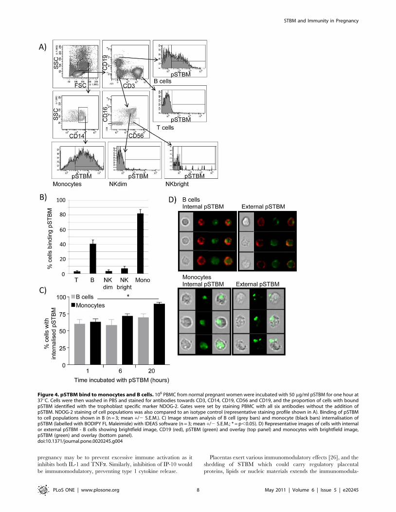
Next, we determined the binding profile of pSTBM to PBMC
by using six colour flow cytometry. We examined the T cell, B cell,
NKdim, NKbright and monocyte cell populations of normal
pregnant women after incubation with pSTBM for 1 hour. STBM
were identified using the trophoblast specific antibody NDOG2.
PBMC were stained with antibodies towards CD14, CD3, CD19,
CD56 and CD16 to identify T cells, B cells, monocytes, NKdim
(CD56+CD16+) and NK bright (CD56brightCD16-) cells,
figures 4A. pSTBM binding was detected on small numbers of
T and NK cells, and to a greater extent to B cells (40%) and
Monocytes (82%), figure 4B. Next we used Image Stream (imaging
flow cytometry) and IDEAS analysis software to determine if
pSTBM were phagocytosed by the monocytes and B cells. pSTBM
were labelled with BODIPY FL maleimide dye and pSTBM
Figure 1. Regulation of cytokine production by mSTBM, pSTBM and eSTBM. PBMC from a non pregnant or normal pregnant donor weretreated with STBM and cytokine production analysed using arrays. Briefly, 106 PBMC/ml were treated with 50 mg/ml mSTBM, pSTBM or eSTBM for20 hours at 37uC. Supernatants were harvested and cytokine production assessed by cytokine array analysis (R&D systems). A) Representative dotblots are shown for untreated cells and with STBM treatments, IP-10 (*) and IL-8 (‘) are highlighted. B) Graphical display of cytokines produced byPBMC from a non pregnant donor with treatment with mSTBM, pSTBM or eSTBM. C) Treatment of PBMC from a non pregnant or normal pregnantdonor with pSTBM. Graphs display cytokines in the order on the dot blots, from top left to bottom right, excluding the positive control three standardpairs of dots on the top left, bottom left and top right.doi:10.1371/journal.pone.0020245.g001
STBM and Immunity in Pregnancy
PLoS ONE | www.plosone.org 5 May 2011 | Volume 6 | Issue 5 | e20245

incubated with 36106 PBMC from non-pregnant donors (n = 3).
Cells were incubated with pSTBM for 1, 6 or 20 hours then
images acquired by Image Stream. Briefly, image stream captures
fluorescence microscopy images of cells as the suspension of cells
passes through the machine, which enables statistical analysis of
pSTBM internalisation on a large number of cells, something
which is not possible by conventional confocal microscopy. We
found that after one hour of incubation with pSTBM, approxi-
mately 60% of B cells and monocytes that had bound pSTBM had
phagocytosed the vesicles. Over time, monocytes continued to
internalise pSTBM, 90% of monocytes had internalised the
pSTBM after 20 hours incubation, whereas no more B cells
internalised vesicles, figure 4C. Representative images of B cells
and monocytes with internalised or external pSTBM are shown in
figure 4D.
Finally, to identify which of the PBMC were producing the most
abundant cytokines, we performed intracellular cytokine staining
on pSTBM stimulated PBMC from non-pregnant donors. The
Figure 2. pSTBM alter cytokine production by PBMC from non and normal pregnant women. 106/ml PBMC from 10 non pregnant (lightbars) or 10 pregnant women (black bars) were treated with varying concentrations of pSTBM for 20 hours at 37uC. Supernatants were harvested andcytokine production assessed by ELISA for A) TNFa, B) IL-8, C) MIP-1a, D) IP-10, E) IL-6, F) IL-1a, and G) IL-1b (pg/ml). H) Cytokines and chemokinesexpressed by the pSTBM alone were determined by cytokine array, cytokines present above background levels in the human serum media were 1-RANTES, 2-MIF, 3-sICAM-1 and 4-Serpin E1.doi:10.1371/journal.pone.0020245.g002
STBM and Immunity in Pregnancy
PLoS ONE | www.plosone.org 6 May 2011 | Volume 6 | Issue 5 | e20245

monocytes were the major producers of IL-6, IL-8, IL-1b and
TNFa, whereas only a few B cells (2–6%) were primed to produce
IL-8 and TNFa, figure 5A. HLA-DR expression was also analysed
as up-regulation indicates activation of antigen presentation
capacity. Surprisingly, HLA-DR expression was downmodulated
on the surface of monocytes, but not B cells, after 20 hours
incubation with pSTBM, figure 5B.
Discussion
Previously, we have shown that pSTBM, but not mSTBM,
induced PBMC from non-pregnant donors to produce inflamma-
tory cytokines (TNF-a, IL-18, and minimal amounts of IL-12p70
and IFN-c) [10]. Here, we have used cytokine arrays to extend this
study and have identified that a wider range of pro-inflammatory
cytokines are induced by pSTBM as well as one (IP-10) which is
inhibited. Furthermore, for some of the cytokines, there were
significant differences in their production in pregnant compared to
non-pregnant women. IL-18 is not included on this array, therefore
previous observations of IL-18 induction were not confirmed. Also,
neither IL-12p70 nor IFN-c were altered on the cytokine arrays,
which may be due to the sensitivity of the array, however on the
basis of this result we decided not to include them in the further
experiments in favour of those cytokines more greatly altered.
Interestingly, we showed that for the 36 cytokines studied the
profile was very similar for eSTBM and pSTBM, but mSTBM
were not as stimulatory. This is consistent with a recent report that
pSTBM and eSTBM but not mSTBM induce monocyte
production of IL-6, IL-8 and IL-1b [21]. We found that pSTBM
significantly induce PBMC production of TNFa, MIP-1a, IL-1a,
IL-1b, IL-6, and IL-8, whether the donors were pregnant or not.
However, mSTBM were not stimulatory. To generate mSTBM
trophoblast tissue is mechanically removed from villi, tissue is
suspended in salt buffer at 4uC and released microvesicles are
collected from the supernatant. We believe this is less likely to
produce biologically normal vesicles than placental perfusion or
explant cultures, which are produced under more physiological
conditions (O2, temperature, buffered solutions). We are currently
performing proteomic analysis to compare mSTBM and pSTBM
and this will be reported in a subsequent paper.
Detecting circulating levels of cytokines is notoriously difficult
and often conflicting reports have been published citing the
increased or decreased quantities, or absence, in pregnancy. The
six cytokines investigated here are shown in some studies to be
elevated in the peripheral blood of pregnant women, compared to
non pregnant women, or are found at increased concentrations
locally at the placenta and are therefore considered important for
successful pregnancy. Total IL-1 concentrations have been
reported to increase in the serum of pregnant women during the
second and third trimester and are significantly higher than in non
pregnant women, but whether this was IL-1a or IL-1b was not
defined [22]. Together with TNFa, the IL-1 proteins IL-1a and
IL-b are pro-inflammatory, and TNFa can also be detected at
increased levels in pregnant women [23]. MIP-1a is a chemokine,
also known as CCL3, and is produced by first trimester placenta
cytotrophoblast to aid recruitment of monocytes and natural killer
cells to the decidua to enable efficient placentation [24]. MIP-1ainstigates acute and chronic inflammatory host responses by
recruiting pro-inflammatory cells expressing its receptor CCR5 to
the sites of injury or infection [25]. Finally, IL-8, which is also a
chemokine, functions as a chemoattractant and angiogenic factor,
and hence helps drive immune responses. Taken together the
induction of all these cytokines (TNFa, IL-1a, IL-b and MIP-1a)
in a pregnant woman would lead to a generalised inflammatory
status. To counteract this we have noted that anti-inflammatory
cytokines are also modulated by STBM, which may prevent
excessive inflammation. IL-6 levels are raised in the peripheral
blood of pregnant women [22], IL-6 has both pro-inflammatory
and anti-inflammatory functions and therefore its presence in
Table 1. Cytokine production is regulated by pSTBM but not mSTBM.
Cytokine production
Donors Treatment TNFa IL-6 IL-8 IL-1a MIP-1a IP-10 IL-1b
Non-pregnant pSTBM 979.1 (88.5) 8487 (835.7) 15390 (4467) 255.8 (79.1) 812.8 (135.1) 2184 (68.2) 6919 (2070)
Non-pregnant mSTBM 27.373 (8.8) 2132.7 (89.5) 1040 (762) 22.026 (1.1) 29.609 (62.5) 5.25 (21.6) 2102.8 (82.8)
Pregnant pSTBM 583.9 (110.9) 5980 (495.5) 10160 (3494) 365.6 (29.1) 497.5 (129.8) 236.24 (22.8) 5184 (1053)
Pregnant mSTBM 2.184 (49.6) 2140.8 (363) 2339.9 (1182) 22.981 (4.0) 2211.3 (119.2) 216.53 (12.0) 244.5 (74.8)
106/ml PBMC from non pregnant or pregnant women were treated with 50 mg/ml pSTBM or mSTBM for 20 hours at 37uC. Supernatants were harvested and cytokineproduction assessed by ELISA for TNFa, IL-6, IL-8, IL-1a, MIP-1a, IP-10 and IL-1b (pg/ml). Values shown are change from mean background production, from 10 nonpregnant or 10 pregnant women, standard error in brackets.doi:10.1371/journal.pone.0020245.t001
Figure 3. PBMC from normal pregnant women produce moreTNFa and IL-6 in response to pSTBM than non pregnantwomen’s PBMC. 106/ml PBMC from non pregnant (light bars) orpregnant women (black bars) (n = 10) were treated with 50 mg/mlpSTBM for 20 hours at 37uC. Supernatants were harvested and cytokineproduction assessed by ELISA for TNFa, IL-6, IL-8, IL-1a, MIP-1a and IL-1b (pg/ml). Values calculated as the mean of value from cells treatedwith 50 ug/ml pSTBM minus background cytokine production.doi:10.1371/journal.pone.0020245.g003
STBM and Immunity in Pregnancy
PLoS ONE | www.plosone.org 7 May 2011 | Volume 6 | Issue 5 | e20245
pregnancy may be to prevent excessive immune activation as it
inhibits both IL-1 and TNFa. Similarly, inhibition of IP-10 would
be immunomodulatory, preventing type 1 cytokine release.
Placentas exert various immunomodulatory effects [26], and the
shedding of STBM which could carry regulatory placental
proteins, lipids or nucleic materials extends the immunomodula-
Figure 4. pSTBM bind to monocytes and B cells. 106 PBMC from normal pregnant women were incubated with 50 mg/ml pSTBM for one hour at37uC. Cells were then washed in PBS and stained for antibodies towards CD3, CD14, CD19, CD56 and CD19, and the proportion of cells with boundpSTBM identified with the trophoblast specific marker NDOG-2. Gates were set by staining PBMC with all six antibodies without the addition ofpSTBM. NDOG-2 staining of cell populations was also compared to an isotype control (representative staining profile shown in A). Binding of pSTBMto cell populations shown in B (n = 3; mean +/2 S.E.M.). C) Image stream analysis of B cell (grey bars) and monocyte (black bars) internalisation ofpSTBM (labelled with BODIPY FL Maleimide) with IDEAS software (n = 3; mean +/2 S.E.M.; * = p,0.05). D) Representative images of cells with internalor external pSTBM - B cells showing brightfield image, CD19 (red), pSTBM (green) and overlay (top panel) and monocytes with brightfield image,pSTBM (green) and overlay (bottom panel).doi:10.1371/journal.pone.0020245.g004
STBM and Immunity in Pregnancy
PLoS ONE | www.plosone.org 8 May 2011 | Volume 6 | Issue 5 | e20245

tory capacity into the maternal system. Immunomodulation of T
cell responses have been previously described [15]; STBM
inhibition of allogeneic immune responses (MLR) is attributed to
syncytiotrophoblast membrane glycoproteins [14] and inhibition
of PHA/ionomycin activation of T cells is dependent upon
microvesicle expression of FasL (where T cell Fas expression is
high, for example on the Jurkat cell line) and PD-L1, the
‘programmed cell death 1 ligand 1’ immunosuppressive molecule
[27,28]. In our study we observe that pSTBM are able to inhibit
IP-10 production. IP-10 is a member of the CXC chemokine
family, also known as CXCL10, and is involved with monocyte, T
cell, natural killer (NK) cell and dendritic cell chemoattraction, as
well as promotion of T cell adhesion to endothelium [29]. It is
induced by pro-inflammatory stimuli and is associated with the
pathogenesis of various diseases, such as diabetes mellitus type 1
[30] as well as allograft rejection [31]. It is also an anti-angiogenic
factor [32] and is associated with the development and
continuation of Th1 responses; as its name suggests, the
interferon-inducible protein 10 KDa (IP-10) is induced by the
classical type 1 cytokine IFNc, and its receptor, CXCR3, has
higher expression on Th1 than Th2 cells [33,34]. Concentrations
of serum IP-10 have been found to be significantly higher in
normal pregnant women than non pregnant women [35], and
even higher levels circulate in pre-eclamptic women. In line with
this, we detect more IP-10 production by PBMC from pregnant
women than non-pregnant women. Gotsch et al (2007) propose
that elevated maternal serum IP-10 contributes to the anti-
angiogenic state of pre-eclampsia (along with sFlt-1 and endoglin).
Here we suggest that in normal pregnancy STBM are able to
reduce the levels of IP-10 produced by PBMC, thereby enabling
angiogenesis and skewing of immunity to type 2 responses that are
important for healthy pregnancy. Finally, the cytokine arrays
indicate STBM induction of G-CSF. This factor aids pregnancy
success in patients with recurrent miscarriage or repetitive failed
implantation in IVF cycles [36,37] and has been shown to be
induced by trophoblast derived microvesicles in vitro [38], further
indicating that STBMs may be important for healthy pregnancy.
We have shown that pSTBM bind monocytes, and to a lesser
extent B cells. In addition, we have shown that monocytes and B
cells are able to rapidly phagocytose pSTBM. This suggests that
the receptor for pSTBM on monocytes and B cells may be
involved with phagocytosis as well as binding. Possible receptors
include Toll-like Receptors, Receptor for Advanced Glycation
End Products (RAGE) and Integrins. Further work is in progress
to assess the nature of this interaction.
While pSTBM bind to both monocytes and B cells, it appears
that the monocytes are responsible for the production of the
majority of the most abundantly produced cytokines (TNFa, IL-6,
IL-8 and IL-1b). Even though monocytes are stimulated to
produce cytokines they do not up-regulate HLA-DR expression,
and HLA-DR expression is reduced by monocytes stimulated by
pSTBM. Increased HLA-DR expression upon antigen stimulation
aids antigen presentation and hence T cell stimulation. T cells are
not normally activated in pregnancy, and no placental derived
antigenic peptide has been reported. However down-modulation
of class II molecules would reduce the potential for antigen
presentation and prevent T helper cell stimulation.
The minimal binding capacity of pSTBM to T cell and NK
cells, may suggest that only a small proportion of these cells may
be subjected to direct immunomodulation by placental microves-
icles. As mentioned above, allogenic or mitogen induced T cell
responses are inhibited by STBM, and NK cells are also reported
to be subjected to STBM derived immunosuppression through
MIC/ULBP protein binding to their NK receptor NKG2D [39].
It is unclear why our preparations are not binding to NK cells at
greater levels.
PBMC from normal pregnant women showed a significantly
increased production of TNFa and IL-6 when compared to the
responses from non-pregnant women. IL-8, IL-1b and MIP-1aalso showed increased production but not to levels of significance.
This suggests immune cells are primed to respond to STBM by
pregnancy which supports our previous observations of enhanced
monocyte activity in pregnant compared to non-pregnant women
[40]. The mechanism of this is unknown, but it could be due to the
increased levels of pro-inflammatory cytokines and hormones
released by the placenta, or prior exposure to STBM in vivo.
In conclusion, we have shown immune cells produce of a variety
of proinflammatory cytokines in response to STBM stimulation,
which may contribute to the increased inflammation seen in
normal pregnancy. Interestingly we have also found that
constitutive IP-10 production is inhibited by STBMs, which
would encourage skewing of the immune system away from
excessive type 1 cytokine responses in normal pregnancy.
Figure 5. Monocytes, not B cells, produce the cytokines IL-6, IL-8, TNFa and IL-1b and downregulate HLA-DR in response topSTBM. A) 106/ml PBMC from non-pregnant donors (n = 3) werestimulated with 50 mg/ml pSTBM, or left untreated, for 20 hours in thepresence of Brefeldin A. Intracellular cytokine analysis was performed todetect production of TNFa, IL-6, IL-8 and IL-1b from either B cells (greybars) or monocytes (black bars). Data shown is the increase inproportion of cells expressing each cytokine above cytokine productionin untreated samples, mean (+/2 S.D.). B) pSTBM binding to monocytes,but not B cells, caused down-regulation of HLA-DR, shown by medianfluorescence intensity of HLA-DR antibody staining (C), (n = 3; mean+/2 S.D.).doi:10.1371/journal.pone.0020245.g005
STBM and Immunity in Pregnancy
PLoS ONE | www.plosone.org 9 May 2011 | Volume 6 | Issue 5 | e20245

Acknowledgments
The authors would like to thank Carol Simms, Hazel Meacher, Nicola
Higgins, Ali Chevassut, Libby Searle, Tess Norris, Katherine Edwards,
Cristina Winter and Linda Holden for their help in recruiting patients and
collecting blood samples for this study. Image stream analysis was
performed in the BRC Translational Immunology Lab with help from
Dr Helen Ferry and Dr Kanchan Phadwal.
Author Contributions
Conceived and designed the experiments: JS DT CR IS. Performed the
experiments: JS DT. Analyzed the data: JS. Wrote the paper: JS DT CR
IS.
References
1. Wegmann TG, Lin H, Guilbert L, Mosmann TR (1993) Bidirectional cytokine
interactions in the maternal-fetal relationship: is successful pregnancy a TH2
phenomenon? Immunol Today 14: 353–356.
2. Chaouat G, Ledee-Bataille N, Dubanchet S, Zourbas S, Sandra O, et al. (2004)
TH1/TH2 paradigm in pregnancy: paradigm lost? Cytokines in pregnancy/
early abortion: reexamining the TH1/TH2 paradigm. Int Arch Allergy
Immunol 134: 93–119.
3. Mor G, Cardenas I (2010) The immune system in pregnancy: a unique
complexity. Am J Reprod Immunol 63: 425–433.
4. Sacks GP, Studena K, Sargent K, Redman CW (1998) Normal pregnancy and
preeclampsia both produce inflammatory changes in peripheral blood leukocytes
akin to those of sepsis. Am J Obstet Gynecol 179: 80–86.
5. Redman CW, Sargent IL (2003) Pre-eclampsia, the placenta and the maternal
systemic inflammatory response–a review. Placenta 24 Suppl A: S21–27.
6. Redman CW, Sargent IL (2010) Immunology of pre-eclampsia. Am J Reprod
Immunol 63: 534–543.
7. Chaouat G, Petitbarat M, Dubanchet S, Rahmati M, Ledee N (2010) Tolerance
to the foetal allograft? Am J Reprod Immunol 63: 624–636.
8. Knight M, Redman CW, Linton EA, Sargent IL (1998) Shedding of
syncytiotrophoblast microvilli into the maternal circulation in pre-eclamptic
pregnancies. Br J Obstet Gynaecol 105: 632–640.
9. Cocucci E, Racchetti G, Meldolesi J (2009) Shedding microvesicles: artefacts no
more. Trends Cell Biol 19: 43–51.
10. Germain SJ, Sacks GP, Sooranna SR, Sargent IL, Redman CW (2007) Systemic
inflammatory priming in normal pregnancy and preeclampsia: the role of
circulating syncytiotrophoblast microparticles. J Immunol 178: 5949–5956.
11. Lok CA, Van Der Post JA, Sargent IL, Hau CM, Sturk A, et al. (2008) Changes
in microparticle numbers and cellular origin during pregnancy and preeclamp-
sia. Hypertens Pregnancy 27: 344–360.
12. Thery C, Ostrowski M, Segura E (2009) Membrane vesicles as conveyors of
immune responses. Nat Rev Immunol 9: 581–593.
13. Smarason AK, Sargent IL, Starkey PM, Redman CW (1993) The effect of
placental syncytiotrophoblast microvillous membranes from normal and pre-
eclamptic women on the growth of endothelial cells in vitro. Br J Obstet
Gynaecol 100: 943–949.
14. Arkwright PD, Rademacher TW, Boutignon F, Dwek RA, Redman CW (1994)
Suppression of allogeneic reactivity in vitro by the syncytiotrophoblast
membrane glycocalyx of the human term placenta is carbohydrate dependent.
Glycobiology 4: 39–47.
15. Gupta AK, Rusterholz C, Holzgreve W, Hahn S (2005) Syncytiotrophoblast
micro-particles do not induce apoptosis in peripheral T lymphocytes, but differ
in their activity depending on the mode of preparation. J Reprod Immunol 68:
15–26.
16. Smith NC, Brush MG, Luckett S (1974) Preparation of human placental villous
surface membrane. Nature 252: 302–303.
17. Eaton BM, Oakey MP (1994) Sequential preparation of highly purified
microvillous and basal syncytiotrophoblast membranes in substantial yield from
a single term human placenta: inhibition of microvillous alkaline phosphatase
activity by EDTA. Biochim Biophys Acta 1193: 85–92.
18. Hung TH, Chen SF, Liou JD, Hsu JJ, Li MJ, et al. (2008) Bax, Bak and
mitochondrial oxidants are involved in hypoxia-reoxygenation-induced apopto-
sis in human placenta. Placenta 29: 565–583.
19. Davies JO, Davies ER, Howe K, Jackson P, Pitcher E, et al. (1985) Practical
applications of a monoclonal antibody (NDOG2) against placental alkaline
phosphatase in ovarian cancer. J R Soc Med 78: 899–905.
20. Schall TJ, Bacon K, Camp RD, Kaspari JW, Goeddel DV (1993) Human
macrophage inflammatory protein alpha (MIP-1 alpha) and MIP-1 beta
chemokines attract distinct populations of lymphocytes. J Exp Med 177:
1821–1826.
21. Messerli M, May K, Hansson SR, Schneider H, Holzgreve W, et al. (2010) Feto-maternal interactions in pregnancies: placental microparticles activate peripheral
blood monocytes. Placenta 31: 106–112.22. Montagnana M, Lippi G, Albiero A, Salvagno GL, Franchi M, et al. (2008)
Serum pro-inflammatory cytokines in physiological and pre-eclamptic pregnan-
cies. Gynecol Endocrinol 24: 113–116.23. Kraus TA, Sperling RS, Engel SM, Lo Y, Kellerman L, et al. (2010) Peripheral
Blood Cytokine Profiling During Pregnancy and Post-partum Periods.Am J Reprod Immunol.
24. Drake PM, Gunn MD, Charo IF, Tsou CL, Zhou Y, et al. (2001) Human
placental cytotrophoblasts attract monocytes and CD56(bright) natural killercells via the actions of monocyte inflammatory protein 1alpha. J Exp Med 193:
1199–1212.25. Maurer M, von Stebut E (2004) Macrophage inflammatory protein-1.
Int J Biochem Cell Biol 36: 1882–1886.26. Chaouat G (1987) Placental immunoregulatory factors. J Reprod Immunol 10:
179–188.
27. Gercel-Taylor C, O’Connor SM, Lam GK, Taylor DD (2002) Shed membranefragment modulation of CD3-zeta during pregnancy: link with induction of
apoptosis. J Reprod Immunol 56: 29–44.28. Sabapatha A, Gercel-Taylor C, Taylor DD (2006) Specific isolation of placenta-
derived exosomes from the circulation of pregnant women and their
immunoregulatory consequences. Am J Reprod Immunol 56: 345–355.29. Neville LF, Mathiak G, Bagasra O (1997) The immunobiology of interferon-
gamma inducible protein 10 kD (IP-10): a novel, pleiotropic member of the C-X-C chemokine superfamily. Cytokine Growth Factor Rev 8: 207–219.
30. Christen U, Von Herrath MG (2004) IP-10 and type 1 diabetes: a question oftime and location. Autoimmunity 37: 273–282.
31. Hancock WW, Gao W, Csizmadia V, Faia KL, Shemmeri N, et al. (2001)
Donor-derived IP-10 initiates development of acute allograft rejection. J ExpMed 193: 975–980.
32. Angiolillo AL, Sgadari C, Taub DD, Liao F, Farber JM, et al. (1995) Humaninterferon-inducible protein 10 is a potent inhibitor of angiogenesis in vivo. J Exp
Med 182: 155–162.
33. Bonecchi R, Bianchi G, Bordignon PP, D’Ambrosio D, Lang R, et al. (1998)Differential expression of chemokine receptors and chemotactic responsiveness
of type 1 T helper cells (Th1s) and Th2s. J Exp Med 187: 129–134.34. Sallusto F, Lenig D, Mackay CR, Lanzavecchia A (1998) Flexible programs of
chemokine receptor expression on human polarized T helper 1 and 2
lymphocytes. J Exp Med 187: 875–883.35. Gotsch F, Romero R, Friel L, Kusanovic JP, Espinoza J, et al. (2007) CXCL10/
IP-10: a missing link between inflammation and anti-angiogenesis inpreeclampsia? J Matern Fetal Neonatal Med 20: 777–792.
36. Scarpellini F, Sbracia M (2009) Use of granulocyte colony-stimulating factor forthe treatment of unexplained recurrent miscarriage: a randomised controlled
trial. Hum Reprod 24: 2703–2708.
37. Wurfel W, Santjohanser C, Hirv K, Buhl M, Meri O, et al. (2010) Highpregnancy rates with administration of granulocyte colony-stimulating factor in
ART-patients with repetitive implantation failure and lacking killer-cellimmunglobulin-like receptors. Hum Reprod 25: 2151–2152; author reply 2152.
38. Atay S, Gercel-Taylor C, Suttles J, Mor G, Taylor DD (2011) Trophoblast-
derived exosomes mediate monocyte recruitment and differentiation.Am J Reprod Immunol 65: 65–77.
39. Hedlund M, Stenqvist AC, Nagaeva O, Kjellberg L, Wulff M, et al. (2009)Human placenta expresses and secretes NKG2D ligands via exosomes that
down-modulate the cognate receptor expression: evidence for immunosuppres-sive function. J Immunol 183: 340–351.
40. Sacks GP, Redman CW, Sargent IL (2003) Monocytes are primed to produce
the Th1 type cytokine IL-12 in normal human pregnancy: an intracellular flowcytometric analysis of peripheral blood mononuclear cells. Clin Exp Immunol
131: 490–497.
STBM and Immunity in Pregnancy
PLoS ONE | www.plosone.org 10 May 2011 | Volume 6 | Issue 5 | e20245


![[Neutralizing antibodies to immunomodulatory therapies in MS-part II] Неутрализиращи антитела към имуномодулиращи терапии при множествена](https://static.fdokumen.com/doc/165x107/633338aaa290d455630a0a17/neutralizing-antibodies-to-immunomodulatory-therapies-in-ms-part-ii-neutralizirashchi.jpg)