The Hypervariable Region of K-Ras4B Is Responsible for Its Specific Interactions with Calmodulin
18
The hypervariable region of K-Ras4B is responsible for its specific interactions with Calmodulin Sherwin J. Abraham § , Ryan P. Nolet § , Richard J. Calvert †,∂ , Lucy M. Anderson † , and Vadim Gaponenko §,* § Department of Biochemistry and Molecular Genetics, University of Illinois, Chicago, IL 60607 † Laboratory of Comparative Carcinogenesis, National Cancer Institute, Frederick, MD 21702 ∂ Division of Bioanalytical Chemistry, U.S. Food and Drug Administration, College Park, MD 20740 Abstract K-Ras4B belongs to the family of p21 Ras GTPases, which play an important role in cell proliferation, survival and motility. The p21 Ras proteins such as K-Ras4B, K-Ras4A, H-Ras, and N-Ras, share 85% sequence homology and activate very similar signaling pathways. Only the C-terminal hypervariable regions differ significantly. A growing body of literature demonstrates that each Ras isoform possesses unique functions in normal physiological processes as well as in pathogenesis. One of the central questions in the field of Ras biology is how these very similar proteins achieve such remarkable specificity in protein-protein interactions that regulate signal transduction pathways. Here we explore specific binding of K-Ras4B to calmodulin. Using NMR techniques and isothermal titration calorimetry we demonstrate that the hypervariable region of K-Ras contributes in a major way to the interaction with calmodulin while the catalytic domain of K-Ras4B provides a way to control the interaction by nucleotide binding. The hypervariable region of K-Ras4B binds specifically to the C-terminal domain of Ca 2+ -loaded calmodulin with micromolar affinity, while the GTP-γ-S loaded catalytic domain of K-Ras4B may interact with the N-terminal domain of calmodulin. Keywords K-Ras4B; calmodulin; hypervariable region; catalytic domain Members of the Ras family of proto-oncogenes are mutated in up to a third of human malignancies(1,2). These are small p21 GTPases that cycle between the GDP-bound inactive and the GTP-bound active states to transmit intracellular signals. How Ras proteins contribute to cancer development is not fully understood, in spite of much study. They have many signaling partners and regulate a variety of cellular processes including proliferation, transformation, differentiation, metastasis, and apoptosis. There are four isoforms in the Ras family, and of these K-Ras, almost exclusively, is mutated in common epithelial cancers including those of the pancreas, colon and lung. Two forms of K-Ras are generated by alternate mRNA splicing, namely K-Ras4A and K-Ras4B. K-Ras4B is more abundant in most tissues, and has been demonstrated to cause tumor formation in studies with genetically engineered mice(3–5). Noonan syndrome, a developmental disorder, is caused by a mutation specifically in K-Ras4B(6). *To whom correspondence should be addressed: Vadim Gaponenko, Department of Biochemistry and Molecular Genetics, University of Illinois at Chicago, 900 S. Ashland Ave., Chicago, IL 60607, Tel. 312-355-4965, Fax 312-413-0353, [email protected]. NIH Public Access Author Manuscript Biochemistry. Author manuscript; available in PMC 2010 August 18. Published in final edited form as: Biochemistry. 2009 August 18; 48(32): 7575–7583. doi:10.1021/bi900769j. NIH-PA Author Manuscript NIH-PA Author Manuscript NIH-PA Author Manuscript
-
Upload
independent -
Category
Documents
-
view
0 -
download
0
Transcript of The Hypervariable Region of K-Ras4B Is Responsible for Its Specific Interactions with Calmodulin
The hypervariable region of K-Ras4B is responsible for its specificinteractions with Calmodulin
Sherwin J. Abraham§, Ryan P. Nolet§, Richard J. Calvert†,∂, Lucy M. Anderson†, and VadimGaponenko§,*§ Department of Biochemistry and Molecular Genetics, University of Illinois, Chicago, IL 60607† Laboratory of Comparative Carcinogenesis, National Cancer Institute, Frederick, MD 21702∂ Division of Bioanalytical Chemistry, U.S. Food and Drug Administration, College Park, MD 20740
AbstractK-Ras4B belongs to the family of p21 Ras GTPases, which play an important role in cell proliferation,survival and motility. The p21 Ras proteins such as K-Ras4B, K-Ras4A, H-Ras, and N-Ras, share85% sequence homology and activate very similar signaling pathways. Only the C-terminalhypervariable regions differ significantly. A growing body of literature demonstrates that each Rasisoform possesses unique functions in normal physiological processes as well as in pathogenesis.One of the central questions in the field of Ras biology is how these very similar proteins achievesuch remarkable specificity in protein-protein interactions that regulate signal transduction pathways.Here we explore specific binding of K-Ras4B to calmodulin. Using NMR techniques and isothermaltitration calorimetry we demonstrate that the hypervariable region of K-Ras contributes in a majorway to the interaction with calmodulin while the catalytic domain of K-Ras4B provides a way tocontrol the interaction by nucleotide binding. The hypervariable region of K-Ras4B binds specificallyto the C-terminal domain of Ca2+-loaded calmodulin with micromolar affinity, while the GTP-γ-Sloaded catalytic domain of K-Ras4B may interact with the N-terminal domain of calmodulin.
KeywordsK-Ras4B; calmodulin; hypervariable region; catalytic domain
Members of the Ras family of proto-oncogenes are mutated in up to a third of humanmalignancies(1,2). These are small p21 GTPases that cycle between the GDP-bound inactiveand the GTP-bound active states to transmit intracellular signals. How Ras proteins contributeto cancer development is not fully understood, in spite of much study. They have manysignaling partners and regulate a variety of cellular processes including proliferation,transformation, differentiation, metastasis, and apoptosis. There are four isoforms in the Rasfamily, and of these K-Ras, almost exclusively, is mutated in common epithelial cancersincluding those of the pancreas, colon and lung. Two forms of K-Ras are generated by alternatemRNA splicing, namely K-Ras4A and K-Ras4B. K-Ras4B is more abundant in most tissues,and has been demonstrated to cause tumor formation in studies with genetically engineeredmice(3–5). Noonan syndrome, a developmental disorder, is caused by a mutation specificallyin K-Ras4B(6).
*To whom correspondence should be addressed: Vadim Gaponenko, Department of Biochemistry and Molecular Genetics, Universityof Illinois at Chicago, 900 S. Ashland Ave., Chicago, IL 60607, Tel. 312-355-4965, Fax 312-413-0353, [email protected].
NIH Public AccessAuthor ManuscriptBiochemistry. Author manuscript; available in PMC 2010 August 18.
Published in final edited form as:Biochemistry. 2009 August 18; 48(32): 7575–7583. doi:10.1021/bi900769j.
NIH
-PA Author Manuscript
NIH
-PA Author Manuscript
NIH
-PA Author Manuscript

Thus, K-Ras4B has a particularly important role in human cancer as well as humandevelopment. It differs from the other highly homologous Ras isoforms in the C-terminal regionwhere the alternate 4B exon provides a polylysine region in addition to posttranslationalfarnesylation. The other Ras isoforms lack the polylysine tail and are modified with a palmitoylgroup in addition to the farnesyl moiety. Some of the unique properties of K-Ras4B have beenrevealed in studies of comparative physiology. These include induced Raf-1 activation intransfected cells(7), expression of matrix metalloproteinase 2 in fibroblasts(8), inhibition ofapoptosis in stem cells(9), nucleolar localization and interaction with nucleolin in several celltypes(10), and AKT activation and cell migration of fibroblasts in response to a growth factor(11). In the latter study, interaction of K-Ras4B with calmodulin (CaM) was demonstrated andfound to be necessary for the K-Ras4B-dependent growth factor responses.
In fact, CaM is the only protein found thus far to bind to K-Ras4B uniquely; the many otherknown binding partners interact with all four isoforms(12). Direct binding of CaM to K-Ras4Bbut not to the highly homologous H-Ras, N-Ras or K-Ras4A has been demonstrated infibroblasts(13), platelets and the MCF-7 breast cancer cell line(14), and Hela cells(15). Thisbinding has also been demonstrated in a yeast two-hybrid assay(14). Functionally importantconsequences found for the K-Ras4B-CaM interaction include regulation of cell cycle throughan ERK2 pathway(16), prevention of Ras activation by PKC in fibroblasts(17), and reversibleintracellular translocation of K-Ras4B in hippocampal neurons in response to glutamate(15).
CaM is a ubiquitous Ca2+-binding protein. Its dumbbell-like architecture is comprised ofglobular N- and C-terminal domains connected by a flexible linker. The general function ofCaM is to mediate Ca2+ signaling through interactions with numerous diverse CaM bindingproteins. Calcium induces a conformational change in CaM rendering it competent for protein-protein interactions(18). CaM binding sites primarily contain basic and hydrophobic aminoacids. In many cases the N-and C-terminal domains of CaM participate in intermolecularinteractions and involve bending CaM into a globular structure(19).
Given the crucial role of K-Ras4B in oncogenesis, and the apparent uniqueness of itsassociation with CaM, detailed understanding of the specifics of this interaction is essentialand could lead to development of highly specific anticancer pharmaceutical agents. Currently,the precise role of K-Ras4B-CaM interaction in cancer is not well defined. However, CaMclearly regulates K-Ras4B membrane association(14) that is crucial for K-Ras4B function incancer. In addition, CaM binding to K-Ras4B activates AKT(11), a protein kinasehyperactivated in many cancer types. AKT plays a role in cell survival and resistance to cancertherapy(20,21). Phenotypically AKT activation by K-Ras4B-CaM interaction results inincreased cell migratory behavior that is characteristic of cells transformed by oncogenic K-Ras4B(11). There have been several reports on characterization of K-Ras4B-CaM interactions,with variable results. The binding event has been shown to have a strong dependence onCa2+ implying that K-Ras4B binds CaM in its Ca2+-loaded form(13,14). CaM association withK-Ras4B was largely independent of the bound guanine nucleotide in the membranes ofstimulated hippocampal neurons(15), but in cell-free assays CaM bound preferentially to theGTP-bound active form of K-Ras4B(13). A nanomolar dissociation constant for K-Ras4B-CaM binding was measured using surface plasmon resonance(22), while CaM antagonists withonly micromolar affinity for CaM inhibited its binding to K-Ras4B(15,23).
Particularly variable results have been reported with regard to the domains of K-Ras4B thatphysically interact with CaM. The hypervariable region (HVR) at the C-terminal tail of K-Ras4B, expressed by itself in neurons, apparently bound CaM(15). However, in pull-downassays and surface plasmon resonance experiments the truncated glutathione S-transferase(GST) tagged catalytic domain (residues 1-166) of K-Ras4B also bound CaM(13,22), as didresidues 1-166 of H-Ras(13) and the 151-amino acid sequence in an alpha helix immediately
Abraham et al. Page 2
Biochemistry. Author manuscript; available in PMC 2010 August 18.
NIH
-PA Author Manuscript
NIH
-PA Author Manuscript
NIH
-PA Author Manuscript

adjacent to the HVR(22). On the other hand, a hemaglutinin (HA) tagged catalytic domain ofK-Ras4B did not(22). CaM, with its extensive capacity to interact with a wide diversity ofproteins, is a particularly promiscuous binding partner(24). It may be that interactions in cell-free systems with truncated and/or tagged peptides may include non-specific associations.
Nuclear magnetic resonance (NMR) allows discernment of interaction patterns for individualamino acid residues and protein domains and thus is a good technique to study details of specificmolecular interactions. Because NMR chemical shifts are sensitive to the rate of dissociationof weakly interacting molecules, it is possible to estimate the strength of protein-proteininteractions and to define the binding interfaces. Addition of isothermal titration calorimetry(ITC) makes it possible to evaluate the energy of protein-protein interactions quantitatively.Using these approaches we demonstrate that the HVR of K-Ras4B is the primary binding sitefor CaM and is responsible for binding specificity. It interacts with the C-terminal domain andlinker region of CaM with micromolar affinity. In addition, we show that the HVR peptide isunable to mimic completely the interactions between the full-length K-Ras4B and CaM. Thisobservation suggests that the weak association with the catalytic domain of K-Ras4B isnecessary for the higher affinity binding. The facts that the HVR constitutes the major bindingsite for CaM, and that no CaM binding could be observed with the inactive GDP-bound K-Ras4B imply that the HVR may be influenced by the context of the full-length protein in thenucleotide-dependent manner.
EXPERIMENTAL PROCEDURESProtein expression, purification, and activity
The full-length human K-Ras4B cDNA was purchased from Invitrogen and cloned into thepET42a bacterial expression plasmid using the NdeI and XhoI restriction sites, adding an N-terminal 6x histidine tag to ease protein purification. The plasmids were transformed into OneShot BL21-A1 cells (Invitrogen), grown overnight in LB media with kanamycin for antibioticselection at 37 °C, and shaking at 250 rpm. For induction, the cells were diluted 1:20 into M9media (with 0.4 M NaCl) and grown to an OD of 0.5 to 0.8 at 600 nm at 37 °C and 250 rpm.The cells were induced at 18 °C with 0.2 mM isopropyl β-D-1-thiogalactopyranoside (IPTG),0.2% arabinose in the presence of 2% ethanol and grown with shaking at 250 rpm for 22–28hours. We also found that K-Ras4B may also be produced at 37 °C in the absence of ethanol.Cells were harvested at 6000 rpm for 20 min at 4 °C in a GS-3 rotor. The supernatant wasdiscarded, and the cell pellets frozen at −80 °C. The cell pellets were lysed using B-PERbacterial extraction reagent (Pierce) with 10 mM MgCl2, 50 μg/mL DNaseI, 2 mMphenylmethanesulfonyl fluoride (PMSF), 1 to 2 tablets of EDTA-free Complete (Roche), and10 mg of lysozyme. The cells were lysed for 20–30 minutes at room temperature on an orbitalshaker. The lysates were centrifuged for 30 minutes at 18,000 rpm in an SS34 rotor at 4 °C.The supernatant was collected, GDP added to 100 uM, sodium citrate to 20 mM, KCl to 50mM and β-mercaptoethanol to 5 mM. The solution was stirred for approximately 60 min. Thepellets were resuspended in 10 mM Tris/HCl pH 7.6, 20 mM Na-Citrate, 50 mM KCl, 5 mMMgCl2, 100 μM GDP, and 2 mM β-mercaptoethanol. The lysate was incubated on an orbitalshaker for 30 minutes, and then spun in a centrifuge the same way as the first lysate.Supernatants from both extractions were dialyzed into His-Bind 1x Binding Buffer (Novagen)with 10 % glycerol overnight at 4 °C. For purification, each of the two extractions was incubatedwith Ni2+-charged His-Bind resin (Novagen) for 2 hours with gentle shaking. The slurry wastransferred to a disposable column (Bio-Rad) and the column was washed extensively with 1xHis-Bind Binding Buffer (no imidazole or glycerol), until no more protein was detectable byBradford analysis. The column was washed with 1x His-Bind Wash Buffer (Novagen)containing 10 mM imidazole. Elutions were performed stepwise (60 mM, 100 mM, 200 mM,and 1M imidazole). All fractions were analyzed on an SDS-PAGE gel. The purified protein
Abraham et al. Page 3
Biochemistry. Author manuscript; available in PMC 2010 August 18.
NIH
-PA Author Manuscript
NIH
-PA Author Manuscript
NIH
-PA Author Manuscript
was dialyzed into Tris-Citrate buffer (50 mM Tris-citrate pH 6.5, 50 mM NaCl, 5 mMMgCl2, 10 mM β-mercaptoethanol, and 10 mM CaCl2) for storage. For nucleotide loading, theprotein solution was dialyzed into nucleotide loading buffer (25 mM Tris/HCl pH 7.6, 150 mMNaCl, 10 mM MgCl2, 5% glycerol, 1 mM EDTA, and 1 mM β-mercaptoethanol). Prior to theaddition of GTP-γ-S to a final concentration of 0.1 mM, EDTA was added to a concentrationof 10 mM to the protein solution. The reaction mix was incubated for 30 min at 30°C. Thereaction was stopped by adding MgCl2 to a final concentration of 65 mM. Similar protocolswere also used in the cloning and purification of truncated K-Ras4B (1-166) (K-Ras4Btr).
To demonstrate the identity and activity of the purified protein, an enzymatic GTP hydrolysisassay and Raf-1 binding activity assay were performed. The hydrolysis assay was performedusing the EnzChek Phosphate Assay Kit (Invitrogen). Ras protein and GTP concentrationswere 15 μM and 500 μM, respectively. Purine Nucleoside Phosphorylase (PNP)phosphorylates 2-amino-6-mercapto-7-methyl-purine riboside (MESG) with the releasedphosphate causing an increase in absorbance at 360 nm. The reaction was monitored for 16hours using a Shimadzu spectrophotometer. Activity of GDP- and GTP-γ-S-loaded K-Ras4Bproteins were also determined using Raf-1 Ras Binding Domain (RBD) agarose beads [RasAssay Reagent (Raf-1 RBD Agarose), Millipore]. The assay is based on the principle that GTP-γ-S-bound K-Ras4B undergoes a conformational change that allows it to bind to the RBD ofRaf-1 while GDP-bound K-Ras4B does not. 200 ng of GTP-γ-S-bound and GDP-bound K-Ras4B proteins was added to 30 μL of Raf-1 RBD agarose beads and incubated in 500 μL ofbuffer containing 25 mM HEPES pH 7.5, 1 mM EDTA, 10 mM MgCl2, 1% NP-40, 0.25%sodium deoxycholate, 150 mM NaCl, and 10% glycerol. Proteinase inhibitors were added asfollows just prior to use: 1 nM PMSF, 0.2 TIU aprotinin, 0.01 M NaF, and 0.2 mM sodiumorthovanandate. Samples were incubated for 2 hours at 4 °C on a tube rotator. Beads were thencollected by a 30 sec centrifugation at 14,000g (Eppendorf 5415D). The supernatant wasremoved by aspiration, and 400 μL of fresh buffer was added and the centrifugation, rinsing,and aspiration were repeated 3 times. 30 μL of 2X Laemelli loading buffer was added to thebeads, and the tube heated to 80 °C for 13 min. Tubes were centrifuged as above, and 15 μLof supernatant was loaded onto a 12% polyacrylamide 1.0 mm thick bis-tris NuPage gel(Invitrogen). The gel was subjected to electrophoresis at 120 V for approximately 1.5 hours,then electro-blotted onto a PVDF membrane (Bio-Rad) at 30 V for 1.5 hours. The blot wasincubated in a primary K-Ras4B antibody (Ab-1, Oncogene Sciences) diluted 1:5000 in 3%milk in PBS-T (phosphate-buffered saline pH 7.4 with 0.1% Tween 20) for 4 hours at roomtemperature. After rinsing, the blot was treated with an anti-mouse secondary antibody coupledto horse radish peroxidase (GE Healthcare UK) diluted 1:2000 in 3% milk in PBS-T. Bandswere visualized by chemiluminescence using the ECL reagent kit (GE Healthcare) withphotographic film. Western blots to show total K-Ras4B protein were done as above, exceptthat 200 ng of each sample in Laemelli buffer was loaded directly onto the gel, without the useof the raf-1 RBD agarose beads.
Human CaM cDNA (Invitrogen) was cloned into the pET42a expression vector (Novagen).The protein was expressed in BL21-A1 E. coli cells at 37 °C overnight after induction with 1mM IPTG and 0.2% arabinose in M9 media containing 15N and 13C stable isotopes (CambridgeIsotope Laboratories). The cell pellets were resuspended in the equilibration buffer (10 mMTris-HCl (pH 7.6), 10 mM CaCl2 and 10 mM β-mercaptoethanol) and heated to 65° C in awater-bath for 30 min. The suspension was then centrifuged at 7000 rpm for 30 min. Thesupernatant was applied to a Phenyl-Sepharose column pre-equilibrated in the equilibrationbuffer. The column was extensively washed with the equilibration buffer. The protein waseluted using elution buffer (10 mM Tris-HCl pH 7.6, 5 mM EDTA and 10 mM β-mercaptoethanol). The purity of the protein was assessed by SDS-PAGE electrophoresis. Thepure fractions were pooled and concentrated. The CaM used in all experiments was Ca2+
loaded.
Abraham et al. Page 4
Biochemistry. Author manuscript; available in PMC 2010 August 18.
NIH
-PA Author Manuscript
NIH
-PA Author Manuscript
NIH
-PA Author Manuscript
NMR experimentsThe 1H-15N HSQC NMR experiments were performed on 600 and 800 MHz Bruker Avancespectrometers equipped with cryogenic probes. All experiments were carried out at 25 °C. Thebuffer conditions were as follows: 10% D2O, 50 mM Tris-Citrate (pH 6.5), 50 mM NaCl, 5mM MgCl2, 10 mM BME, and 10 mM CaCl2. The CaM assignments were confirmed usingCBCACONH, NHCA, and NHCACB experiments. All the data were processed and analyzedusing NMRPipe(25). The assignments were performed using Sparky (UCSF). For NMRtitration experiments 1H-15N HSQC spectra were recorded at different 15N CaM:K-Ras4B/K-Ras4Btr/HVR molar ratios. The mean chemical shift difference (ΔδNH) of the 1H and 15N nucleiwas calculated as previously described(26):
The chemical shift changes above average plus one standard deviation were consideredstatistically significant as commonly done in NMR studies.
ITC ExperimentsK-Ras4B-GTP-γ-S, the HVR peptide, K-Ras4Btr-GTP-γ-S, K-Ras4B-GDP and CaM weredialyzed into a buffer consisting of 50 mM Tris-citrate pH 6.5, 50 mM NaCl, 5 mM MgCl2, 1mM Tris(2-carboxy-ethyl)phosphine hydrochloride (TCEP) and 10 mM CaCl2. The titrationexperiments were performed using a VP-ITC Microcalorimeter (MicroCal LLC, Northampton,MA) at 25°C. For the titration, 25–27 aliquots (10 μL each) of 1 mM CaM were injected intothe ITC cell containing 1.4 mL of 70 μM K-Ras4B-GTP-γ-S, the HVR peptide, K-Ras4Btr-GTP-γ-S, or K-Ras-GDP. Each titration was preceded by a single 2-μL injection to eliminatethe diffusion artifacts. A reference titration was run, with only buffer in the cell instead of K-Ras4B proteins or the HVR, to offset thermodynamic effects of protein dilution. The referencedata were subtracted from the protein titration data points. The initial 2-μL titration point wasdiscarded. The integrated heat values were analyzed using the ‘Origin 7.0’-software as well asthe Scientist 3.0 software (Micromath Scientific Software). The data were fit using the ‘Oneset of sites’ model, to yield the dissociation constant (Kd), the molar ratio, and otherthermodynamic parameters.
RESULTSRecombinant K-Ras4B and K-Ras4Btr are biologically active
The first requirement for investigation of relevant protein-protein interactions between K-Ras4B and CaM is the availability of biologically active proteins. Since the ability to producerecombinant K-Ras4B protein has been established in many laboratories including our own,here we concentrate on testing the activity of K-Ras4B. First, we tested the ability of K-Ras4Band K-Ras4Btr proteins to interact with the Ras Binding Domain (RDB) of Raf-1. The Raf-1RBD binding test is commonly used to assess K-Ras4B activity and is based on the principlethat GTP-bound Ras undergoes a conformational change that allows it to bind to the Raf-1RBD(27). Western blots in Figure 1A show that both K-Ras4B and K-Ras4Btr, when boundto GTP-γ-S, were able to bind Raf-1 RBD with high affinity, while the GDP-loaded negativecontrols exhibited weak binding (12%–15% band intensity of GTP-γ-S loaded K-Ras4B asmeasured by gel scanning). These results are consistent with the literature reports showing thatGTP-bound Ras binds Raf-1 RBD with high affinity, while GDP-loaded Ras displays lowmicromolar affinity towards Raf-1 RBD(28). Alternatively, the samples of GDP-loaded K-Ras4B may have contained a trace amount of GTP-bound protein. However, in our preparations
Abraham et al. Page 5
Biochemistry. Author manuscript; available in PMC 2010 August 18.
NIH
-PA Author Manuscript
NIH
-PA Author Manuscript
NIH
-PA Author Manuscript
of 15N K-Ras4B-GDP using the same nucleotide loading procedure we did not observe signalscorresponding to K-Ras4B-GTP in 1H-15N HSQC NMR experiments. Four separate K-Ras4Bactivity assays were performed to establish a linear range for the GTP-γ-S samples, all withsimilar results (data not shown). The data presented here demonstrate that recombinant K-Ras4B and K-Ras4Btr proteins were both Raf-1 binding competent.
Next, the ability of K-Ras4B and K-Ras4Btr to hydrolyze GTP was tested in a GTP hydrolysisassay. The results shown in Figure 1B represent a typical time course for GTP hydrolysis(29)and indicate that both K-Ras4B and K-Ras4Btr are able to hydrolyze GTP. The determinedinitial rates of GTP hydrolysis measured at the same protein concentration were 51.03 pmole/min and 67.56 pmole/min for K-Ras4B and K-Ras4Btr, respectively. The 32% difference inrates of GTP hydrolysis between K-Ras4B and K-Ras4Btr is in agreement with previouslypublished results(30). Together, we demonstrate that recombinant K-Ras4B and K-Ras4Btr areboth enzymatically active and Raf-1 binding competent.
The N- and C-terminal domains of CaM define the interaction sites with K-Ras4BUpon titration of K-Ras4B loaded with GTP-γ-S into a 0.15 mM solution of 15N CaMsignificant K-Ras4B concentration dependent chemical shift changes in the signals of CaMwere observed in NMR 1H-15N Heteronuclear Single Quantum Coherence (HSQC)experiments (Figure 2A). The fact that the titration produced chemical shift changes asillustrated in Figure 2B indicates fast exchange on the NMR time scale. The fast exchangephenomenon is observed when the binding affinity is relatively low allowing rapid inter-conversion between the free and the bound species. Analysis of chemical shift perturbationsindicates that the most significant chemical shift changes induced by K-Ras4B-GTP-γ-S occurin the regions comprising residues A15, T29, L32, L48, I52, and E67 in the N-terminal β-sheetregion, residues D78 and T79 in the linker region, and the C-terminal F92, R106, M109-N111,and Q143-M145 residues of CaM (Figure 2C). Mapping of chemical shift perturbations ontothe structure of CaM(31) revealed that the K-Ras4B-GTP-γ-S binding interface includes theN- and C-terminal domains and the linker region (Figure 2D).
The HVR of K-Ras4B provides specificity for K-Ras4B binding to CaMTo test if the HVR of K-Ras4B binds to CaM, a 22 amino acid synthetic peptide representingthe HVR of K-Ras4B was purchased from Biopeptide, Inc. Titration of 15N CaM with the HVRpeptide was followed by 1H-15N HSQC spectra recorded on the peptide-protein mixtures atdifferent molar ratios. The overlay of spectra for 15N CaM alone and 15N CaM saturated withthe HVR peptide is shown in Figure 3A. Similar to the K-Ras4B-GTP-γ-S binding to CaM,the fast exchange phenomenon indicative of low affinity binding was observed in NMRtitration experiments (Figure 3B). Addition of the HVR peptide induced significant chemicalshift changes in the linker region (residues D78, T79, E82-E84) and the C-terminal domain ofCaM (residues F92, D93, R106, M109, T110, G113, E123, D129, M144, A147, and K148)(Figure 3C). Only limited chemical shift perturbations are observed in the N-terminal domainof CaM in residues I63 and F68, presumably due to their proximity to the linker region.Mapping of chemical shift perturbations onto the structure of CaM (Figure 3D) indicates thatthe binding interface is primarily comprised of negatively charged amino acids in the linkerregion and those flanking the hydrophobic pockets in CaM, such as D78, E82, E84, D93, D129.Thus, the HVR of K-Ras4B can directly participate in specific interactions with CaM.
To test whether the catalytic domain of K-Ras4B is involved in binding CaM we performedan NMR titration of 15N CaM with K-Ras4Btr loaded with GTP-γ-S (Figure 4A). The presenceof fast chemical exchange indicates weak binding of K-Ras4Btr to CaM (Figure 4B). Thebinding interface in CaM involves small areas in both the N- and C-terminal domainscomprised of residues T5, I27, T28, L32, A57, and E67 in the N-terminal domain and residues
Abraham et al. Page 6
Biochemistry. Author manuscript; available in PMC 2010 August 18.
NIH
-PA Author Manuscript
NIH
-PA Author Manuscript
NIH
-PA Author Manuscript
F92, R106, M109, and N111 in the C-terminal domain (Figures 4C and 4D). Binding involvespredominantly non-polar and hydrophobic amino acids. Together, the results of theseexperiments delineate the K-Ras4B-CaM binding interface. While the HVR may preferentiallybind to the C-terminal domain of CaM, the catalytic domain of K-Ras4B alone weakly bindsto both the N- and C-terminal domains.
The HVR of K-Ras4B may contain the major CaM binding siteTo further investigate the contributions of K-Ras4B domains to binding CaM we measureddissociation constants using ITC experiments. Consistent with our NMR data, K-Ras4B loadedwith GTP-γ-S and the HVR of K-Ras4B bound CaM with low micromolar affinity.Endothermic binding is observed for K-Ras4B and the HVR as judged by the sign of the heatof the reaction. K-Ras4B-GTP-γ-S bound CaM with 1:1 stoichiometry (Figure 5A) with adissociation constant (Kd) of 3.4±1.2 μM. The other fitted parameters were ΔH=2.5±0.8 kcal/mol, and ΔS=30.5 cal/deg/mol. In the case of the HVR peptide titrated with CaM (Figure 5B),the titration curve was fit to the ‘one set of binding sites’ model yielding a dissociation constantof Kd=11.2±1.6 μM, ΔH=0.65±0.1 kcal/mol, and ΔS=25.1 cal/deg/mol. The fit revealed onebinding site for the HVR peptide on CaM. Initially K-Ras4B interaction with CaM was reportedto be nucleotide independent(15). However, subsequent reports demonstrated a strongpreference for GTP-bound K-Ras4B by CaM(13, 22). To test the nucleotide dependence of K-Ras4B binding to CaM we performed an ITC experiment with K-Ras4B-GDP and CaM.Consistent with literature reports(13), ITC experiments for GDP-bound K-Ras4B showed nosignificant binding (Figure 5C).
In addition to the determination of dissociation constants by ITC, we obtained corroboratingresults using non-linear regression analysis of concentration dependent NMR chemical shiftperturbations. Chemical shift perturbations for five amino acids in CaM responding to titrationwith either K-Ras4B-GTP-γ-S, K-Ras4Btr-GTP-γ-S, or the HVR peptide were used in thefitting procedure. Examples of these fits are shown in Figure 6. While saturation with K-Ras4B-GTP-γ-S and the HVR peptide occurs between 1:1.5 to 1:2 molar ratio (Figures 6A and 6C),saturation with K-Ras4Btr-GTP-γ-S occurs at around 1:8 molar ratio (Figure 6B) and isindicative of a much weaker binding affinity. The corresponding dissociation constant (Kd)values are 7.2±4.2 μM, 17.5±10.1 μM, and 307.6±122.3 μM for K-Ras4B-GTP-γ-S, the HVRpeptide, and K-Ras4BtrGTP-γ-S respectively. The dissociation constants (Kd) obtained for K-Ras4B-GTP-γ-S and the HVR peptide binding to CaM from NMR titration experiments arenot statistically different from those obtained by ITC. Due to the very weak binding affinity,we could not use ITC to measure the dissociation constant for the K-Ras4Btr-GTP-γ-S-CaMbinding. However, since the dissociation constant (Kd) values measured using NMR titrationand ITC are in agreement, we could use the Kd value determined using NMR titration toestimate binding affinity of CaM to K-Ras4Btr-GTP-γ-S.
In conclusion, both the HVR and the GTP-γ-S-loaded catalytic domain of K-Ras4B contributeto CaM binding affinity with the HVR providing the major binding site. Interactions betweenK-Ras4B and CaM are significantly weaker upon loading with GDP. Since K-Ras4B-GDPexhibits little or no binding affinity for CaM as compared to the HVR, we propose thatinteractions between the HVR and the catalytic domain of K-Ras4B-GDP may sequester theHVR to prevent it from binding CaM or alter its structure.
DISCUSSIONOur results suggest that specific binding of K-Ras4B to CaM occurs through the unique HVR,and show that CaM interacts with non-farnesylated human K-Ras4B with micromolar affinity.The apparent disagreement of this result with the previously measured nanomolar affinity bysurface plasmon resonance of the GST-tagged K-Ras4B catalytic domain for CaM may be
Abraham et al. Page 7
Biochemistry. Author manuscript; available in PMC 2010 August 18.
NIH
-PA Author Manuscript
NIH
-PA Author Manuscript
NIH
-PA Author Manuscript

explained by differences in protein constructs and experimental approaches(22). The K-Ras4Bbinding site on CaM resides in the N- and C-terminal domains (Figure 2). Both the HVR andthe GTP-γ-S-loaded catalytic domain are involved in interaction with CaM.
Binding the HVR peptide and K-Ras4Btr-GTP-γ-S to the C-terminal domain of CaM involvesthe same regions, namely residues F92 and D93, and the R106, M109-N111 region (Figures3 and 4). This suggests that either the HVR or the catalytic domain of K-Ras4B may interactwith the C-terminal domain of CaM. However, due to its higher binding affinity, the HVR maypredominantly interact with the linker region and the C-terminal domain of CaM (Figures 5and 6), while the GTP-γ-S loaded catalytic domain (1-166) of K-Ras4B may bind the N-terminal domain of CaM. Specific binding of the highly positively charged HVR to the linkerregion and the C-terminal domain is justified by fact that the negatively charged residuesconcentrate in the C-terminal domain of CaM, while the N-terminal domain presents a mixtureof negative and positive charges. Specific interaction of a positively charged peptide with theC-terminal domain of CaM has been observed in the structure of CaM-CaMKIIa complex(32). We hypothesize that the farnesyl group on the C-terminus of the HVR may furtherenhance the binding affinity of K-Ras4B for CaM through interactions with the hydrophobicresidues in the C-terminal domain of CaM. The importance of K-Ras4B farnesylation forbinding to CaM has been previously demonstrated(15, 22).
Binding of the K-Ras4B catalytic domain to the N-terminal domain of CaM is also notconventional. Instead of employing the binding interfaces commonly found in CaM whichincludes the methionine-lined hydrophobic patch, the catalytic domain of K-Ras4B interactswith hydrophobic T27-L32 (spanning the β1-strand of the central β-sheet and the beginningof α2-helix) and L48-I52 (part of α3-helix) regions. This is consistent with the involvement ofthe hydrophobic helix 5 region of the catalytic domain of K-Ras4B in binding CaM(22). Theapparent binding to CaM of the hydrophobic α–helix 5 peptide (amino acids 151–166) whenindependently expressed may indicate that this region participates in binding when in thecontext of the full length protein and/or enhances the binding of the HVR(22). Identificationof chemical shift perturbations in negatively charged E67 of CaM upon binding K-Ras4B isindicative of possible electrostatic contributions to CaM interactions with the catalytic domainof K-Ras4B. Participation of positively charged R68 and R73 from the Switch II region of K-Ras4B in the CaM binding interface was recently discovered using point mutagenesisexperiments(22).
The parallel arrangement of interacting partners in the K-Ras4B-CaM complex is an unusualway of binding CaM. It significantly differs from the common CaM binding mode that involvesbending of the linker region to accommodate the hydrophobic pockets in the N- and C-terminaldomains forming one extensive interface with the target(18,19). Parallel binding modes havebeen previously observed in the complex of CaM with the hydrophobic IQ domain of thecardiac Cav1.2 calcium channel(33) and in the complex of CaM with CaM dependent kinasekinase peptide(34).
Although the HVR is a major CaM binding site, in the context of GDP-loaded K-Ras4B it isunable to efficiently interact with CaM. We hypothesize that the HVR is modulated by the K-Ras4B catalytic domain in a nucleotide dependent manner. Our model that shows how theHVR may be involved in nucleotide dependent binding to CaM is depicted in Figure 7. In theGDP-bound state the HVR may be sequestered by the catalytic domain of K-Ras4B while GTPbinding induces a conformational change that may release the HVR from the catalytic domainand make it available to interact with CaM. Farnesylation at the C-terminus of the HVR of K-Ras4B may further enhance this mechanism by increasing the affinity of the HVR for the GDP-bound catalytic domain. This model is consistent with all of our data and currently availableliterature reports. For example, the HVR of H-Ras has been shown to transiently interact with
Abraham et al. Page 8
Biochemistry. Author manuscript; available in PMC 2010 August 18.
NIH
-PA Author Manuscript
NIH
-PA Author Manuscript
NIH
-PA Author Manuscript
catalytic domain of H-Ras(26). However further testing of the proposed mechanism of K-Ras4B engagement in protein-protein interactions is required.
In conclusion, we show that non-farnesylated GTP-γ-S loaded K-Ras4B interacts with boththe N- and C-terminal domains of CaM with micromolar affinity. The HVR of K-Ras4Bprovides specificity and most of the affinity for this interaction. While the role of the HVR ofK-Ras4B as a membrane anchor is well documented, its participation in protein-proteininteractions is not yet fully explored. Since the HVR of K-Ras4B mediates specific interactionswith CaM, it is possible that it modulates binding to other proteins in a specific manner. Thus,in addition to targeting of K-Ras4B to unique membrane compartments the HVR may alsoachieve specificity of K-Ras4B signaling by its involvement in protein-protein interactions.
AcknowledgmentsFunding: American Cancer Society, Illinois Division Grant ACS 08-14 to Vadim Gaponenko and the U.S. Departmentof Health and Human Services.
ABBREVIATIONSCaM
calmodulin
K-Ras4Btr K-Ras4B truncated (amino acid residues1-166 only)
HVR hypervariable region
NMR nuclear magnetic resonance
ITC isothermal titration calorimetry
HSQC Heteronuclear Single Quantum Coherence
References1. Barbacid M. ras genes. Annual review of biochemistry 1987;56:779–827.2. Kumar R, Sukumar S, Barbacid M. Activation of ras oncogenes preceding the onset of neoplasia.
Science New York, NY 1990;248:1101–1104.3. Malkinson AM. Primary lung tumors in mice: an experimentally manipulable model of human
adenocarcinoma. Cancer research 1992;52:2670s–2676s. [PubMed: 1562998]4. Meuwissen R, Linn SC, van der Valk M, Mooi WJ, Berns A. Mouse model for lung tumorigenesis
through Cre/lox controlled sporadic activation of the K-Ras oncogene. Oncogene 2001;20:6551–6558.[PubMed: 11641780]
5. Janssen KP, el-Marjou F, Pinto D, Sastre X, Rouillard D, Fouquet C, Soussi T, Louvard D, Robine S.Targeted expression of oncogenic K-ras in intestinal epithelium causes spontaneous tumorigenesis inmice. Gastroenterology 2002;123:492–504. [PubMed: 12145803]
6. Carta C, Pantaleoni F, Bocchinfuso G, Stella L, Vasta I, Sarkozy A, Digilio C, Palleschi A, Pizzuti A,Grammatico P, Zampino G, Dallapiccola B, Gelb BD, Tartaglia M. Germline missense mutationsaffecting KRAS Isoform B are associated with a severe Noonan syndrome phenotype. Americanjournal of human genetics 2006;79:129–135. [PubMed: 16773572]
Abraham et al. Page 9
Biochemistry. Author manuscript; available in PMC 2010 August 18.
NIH
-PA Author Manuscript
NIH
-PA Author Manuscript
NIH
-PA Author Manuscript

7. Voice JK, Klemke RL, Le A, Jackson JH. Four human ras homologs differ in their abilities to activateRaf-1, induce transformation, and stimulate cell motility. The Journal of biological chemistry1999;274:17164–17170. [PubMed: 10358073]
8. Liao J, Wolfman JC, Wolfman A. K-ras regulates the steady-state expression of matrixmetalloproteinase 2 in fibroblasts. The Journal of biological chemistry 2003;278:31871–31878.[PubMed: 12805379]
9. Plowman SJ, Berry RL, Bader SA, Luo F, Arends MJ, Harrison DJ, Hooper ML, Patek CE. K-ras 4Aand 4B are co-expressed widely in human tissues, and their ratio is altered in sporadic colorectal cancer.J Exp Clin Cancer Res 2006;25:259–267. [PubMed: 16918139]
10. Birchenall-Roberts MC, Fu T, Kim SG, Huang YK, Dambach M, Resau JH, Ruscetti FW. K-Ras4Bproteins are expressed in the nucleolus: Interaction with nucleolin. Biochemical and biophysicalresearch communications 2006;348:540–549. [PubMed: 16889753]
11. Liao J, Planchon SM, Wolfman JC, Wolfman A. Growth factor-dependent AKT activation and cellmigration requires the function of c-K(B)-Ras versus other cellular ras isoforms. The Journal ofbiological chemistry 2006;281:29730–29738. [PubMed: 16908523]
12. Rodriguez-Viciana P, Sabatier C, McCormick F. Signaling specificity by Ras family GTPases isdetermined by the full spectrum of effectors they regulate. Molecular and cellular biology2004;24:4943–4954. [PubMed: 15143186]
13. Villalonga P, Lopez-Alcala C, Bosch M, Chiloeches A, Rocamora N, Gil J, Marais R, Marshall CJ,Bachs O, Agell N. Calmodulin binds to K-Ras, but not to H- or N-Ras, and modulates its downstreamsignaling. Molecular and cellular biology 2001;21:7345–7354. [PubMed: 11585916]
14. Sidhu RS, Clough RR, Bhullar RP. Ca2+/calmodulin binds and dissociates K-RasB from membrane.Biochemical and biophysical research communications 2003;304:655–660. [PubMed: 12727204]
15. Fivaz M, Meyer T. Reversible intracellular translocation of KRas but not HRas in hippocampalneurons regulated by Ca2+/calmodulin. The Journal of cell biology 2005;170:429–441. [PubMed:16043511]
16. Bosch M, Gil J, Bachs O, Agell N. Calmodulin inhibitor W13 induces sustained activation of ERK2and expression of p21(cip1). The Journal of biological chemistry 1998;273:22145–22150. [PubMed:9705360]
17. Villalonga P, Lopez-Alcala C, Chiloeches A, Gil J, Marais R, Bachs O, Agell N. Calmodulin preventsactivation of Ras by PKC in 3T3 fibroblasts. The Journal of biological chemistry 2002;277:37929–37935. [PubMed: 12151388]
18. Trewhella J. The solution structures of calmodulin and its complexes with synthetic peptides basedon target enzyme binding domains. Cell calcium 1992;13:377–390. [PubMed: 1505003]
19. Ikura M, Clore GM, Gronenborn AM, Zhu G, Klee CB, Bax A. Solution structure of a calmodulin-target peptide complex by multidimensional NMR. Science New York, NY 1992;256:632–638.
20. Qiao M, Sheng S, Pardee AB. Metastasis and AKT activation. Cell cycle Georgetown, Tex2008;7:2991–2996.
21. Steelman LS, Stadelman KM, Chappell WH, Horn S, Basecke J, Cervello M, Nicoletti F, Libra M,Stivala F, Martelli AM, McCubrey JA. Akt as a therapeutic target in cancer. Expert opinion ontherapeutic targets 2008;12:1139–1165. [PubMed: 18694380]
22. Lopez-Alcala C, Alvarez-Moya B, Villalonga P, Calvo M, Bachs O, Agell N. Identification ofessential interacting elements in K-Ras/calmodulin binding and its role in K-Ras localization. TheJournal of biological chemistry 2008;283:10621–10631. [PubMed: 18182391]
23. Hidaka H, Sasaki Y, Tanaka T, Endo T, Ohno S, Fujii Y, Nagata T. N-(6-aminohexyl)-5-chloro-1-naphthalenesulfonamide, a calmodulin antagonist, inhibits cell proliferation. Proceedings of theNational Academy of Sciences of the United States of America 1981;78:4354–4357. [PubMed:6945588]
24. Clapham DE. Calcium signaling. Cell 2007;131:1047–1058. [PubMed: 18083096]25. Delaglio F, Grzesiek S, Vuister GW, Zhu G, Pfeifer J, Bax A. NMRPipe: a multidimensional spectral
processing system based on UNIX pipes. Journal of biomolecular NMR 1995;6:277–293. [PubMed:8520220]
Abraham et al. Page 10
Biochemistry. Author manuscript; available in PMC 2010 August 18.
NIH
-PA Author Manuscript
NIH
-PA Author Manuscript
NIH
-PA Author Manuscript
26. Thapar R, Williams JG, Campbell SL. NMR characterization of full-length farnesylated and non-farnesylated H-Ras and its implications for Raf activation. Journal of molecular biology2004;343:1391–1408. [PubMed: 15491620]
27. de Rooij J, Bos JL. Minimal Ras-binding domain of Raf1 can be used as an activation-specific probefor Ras. Oncogene 1997;14:623–625. [PubMed: 9053862]
28. Herrmann C, Martin GA, Wittinghofer A. Quantitative analysis of the complex between p21ras andthe Ras-binding domain of the human Raf-1 protein kinase. The Journal of biological chemistry1995;270:2901–2905. [PubMed: 7852367]
29. Schlichting I, Rapp G, John J, Wittinghofer A, Pai EF, Goody RS. Biochemical and crystallographiccharacterization of a complex of c-Ha-ras p21 and caged GTP with flash photolysis. Proceedings ofthe National Academy of Sciences of the United States of America 1989;86:7687–7690. [PubMed:2682619]
30. John J, Schlichting I, Schiltz E, Rosch P, Wittinghofer A. C-terminal truncation of p21H preservescrucial kinetic and structural properties. The Journal of biological chemistry 1989;264:13086–13092.[PubMed: 2502546]
31. Chattopadhyaya R, Meador WE, Means AR, Quiocho FA. Calmodulin structure refined at 1.7 Aresolution. Journal of molecular biology 1992;228:1177–1192. [PubMed: 1474585]
32. Wall ME, Clarage JB, Phillips GN. Motions of calmodulin characterized using both Bragg and diffuseX-ray scattering. Structure 1997;5:1599–1612. [PubMed: 9438860]
33. Fallon JL, Halling DB, Hamilton SL, Quiocho FA. Structure of calmodulin bound to the hydrophobicIQ domain of the cardiac Ca(v)1.2 calcium channel. Structure 2005;13:1881–1886. [PubMed:16338416]
34. Osawa M, Tokumitsu H, Swindells MB, Kurihara H, Orita M, Shibanuma T, Furuya T, Ikura M. Anovel target recognition revealed by calmodulin in complex with Ca2+-calmodulin-dependent kinasekinase. Nature structural biology 1999;6:819–824.
Abraham et al. Page 11
Biochemistry. Author manuscript; available in PMC 2010 August 18.
NIH
-PA Author Manuscript
NIH
-PA Author Manuscript
NIH
-PA Author Manuscript

Figure 1.(A) Western blot showing the Raf-1 RBD binding activity of recombinant K-Ras4B and K-Ras4Btr. The GDP-bound K-Ras4B and K-Ras4Btr does not bind the RBD of Raf-1, but theGTP-bound form of the proteins binds the RBD of Raf-1 indicative of protein activity. (B)Enzymatic activity of K-Ras4B and K-Ras4Btr proteins was tested by measuring GTPhydrolysis rates using the EnzChek Phosphate Assay kit (Invitrogen). Recombinant K-Ras4Band K-Ras4Btr proteins are able to hydrolyze GTP indicating that they are enzymatically active.
Abraham et al. Page 12
Biochemistry. Author manuscript; available in PMC 2010 August 18.
NIH
-PA Author Manuscript
NIH
-PA Author Manuscript
NIH
-PA Author Manuscript
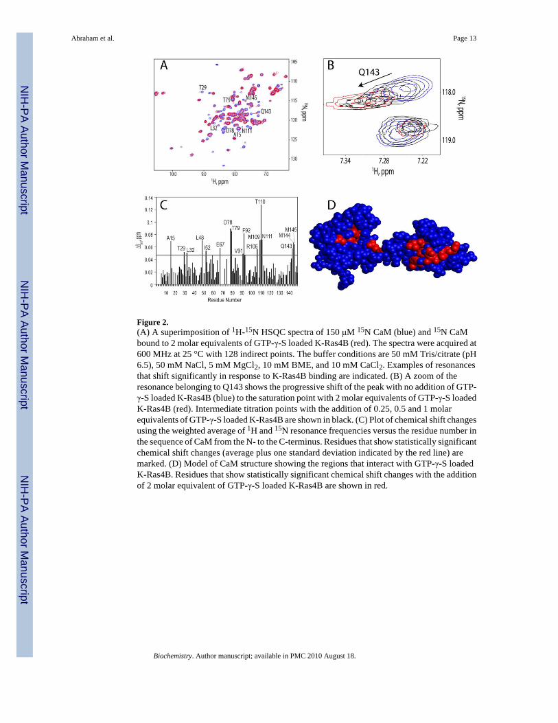
Figure 2.(A) A superimposition of 1H-15N HSQC spectra of 150 μM 15N CaM (blue) and 15N CaMbound to 2 molar equivalents of GTP-γ-S loaded K-Ras4B (red). The spectra were acquired at600 MHz at 25 °C with 128 indirect points. The buffer conditions are 50 mM Tris/citrate (pH6.5), 50 mM NaCl, 5 mM MgCl2, 10 mM BME, and 10 mM CaCl2. Examples of resonancesthat shift significantly in response to K-Ras4B binding are indicated. (B) A zoom of theresonance belonging to Q143 shows the progressive shift of the peak with no addition of GTP-γ-S loaded K-Ras4B (blue) to the saturation point with 2 molar equivalents of GTP-γ-S loadedK-Ras4B (red). Intermediate titration points with the addition of 0.25, 0.5 and 1 molarequivalents of GTP-γ-S loaded K-Ras4B are shown in black. (C) Plot of chemical shift changesusing the weighted average of 1H and 15N resonance frequencies versus the residue number inthe sequence of CaM from the N- to the C-terminus. Residues that show statistically significantchemical shift changes (average plus one standard deviation indicated by the red line) aremarked. (D) Model of CaM structure showing the regions that interact with GTP-γ-S loadedK-Ras4B. Residues that show statistically significant chemical shift changes with the additionof 2 molar equivalent of GTP-γ-S loaded K-Ras4B are shown in red.
Abraham et al. Page 13
Biochemistry. Author manuscript; available in PMC 2010 August 18.
NIH
-PA Author Manuscript
NIH
-PA Author Manuscript
NIH
-PA Author Manuscript
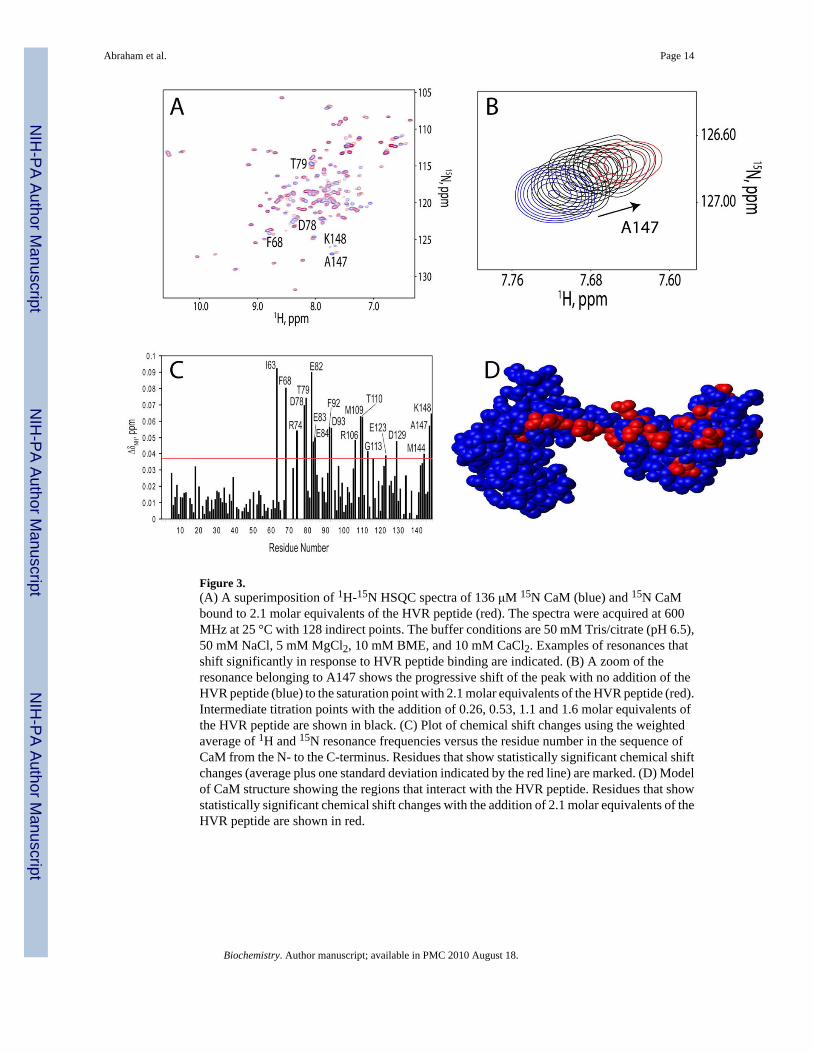
Figure 3.(A) A superimposition of 1H-15N HSQC spectra of 136 μM 15N CaM (blue) and 15N CaMbound to 2.1 molar equivalents of the HVR peptide (red). The spectra were acquired at 600MHz at 25 °C with 128 indirect points. The buffer conditions are 50 mM Tris/citrate (pH 6.5),50 mM NaCl, 5 mM MgCl2, 10 mM BME, and 10 mM CaCl2. Examples of resonances thatshift significantly in response to HVR peptide binding are indicated. (B) A zoom of theresonance belonging to A147 shows the progressive shift of the peak with no addition of theHVR peptide (blue) to the saturation point with 2.1 molar equivalents of the HVR peptide (red).Intermediate titration points with the addition of 0.26, 0.53, 1.1 and 1.6 molar equivalents ofthe HVR peptide are shown in black. (C) Plot of chemical shift changes using the weightedaverage of 1H and 15N resonance frequencies versus the residue number in the sequence ofCaM from the N- to the C-terminus. Residues that show statistically significant chemical shiftchanges (average plus one standard deviation indicated by the red line) are marked. (D) Modelof CaM structure showing the regions that interact with the HVR peptide. Residues that showstatistically significant chemical shift changes with the addition of 2.1 molar equivalents of theHVR peptide are shown in red.
Abraham et al. Page 14
Biochemistry. Author manuscript; available in PMC 2010 August 18.
NIH
-PA Author Manuscript
NIH
-PA Author Manuscript
NIH
-PA Author Manuscript

Figure 4.(A) A superimposition of 1H-15N HSQC spectra of 150 μM 15N CaM (blue) and 15N CaMbound to 8 molar equivalents of GTP-γ-S loaded K-Ras4Btr (red). The spectra were acquiredat 600 MHz at 25 °C with 128 indirect points. The buffer conditions are 50 mM Tris/citrate(pH 6.5), 50 mM NaCl, 5 mM MgCl2, 10 mM BME, and 10 mM CaCl2. Examples of resonancesthat shift significantly in response to K-Ras4Btr binding are indicated. (B) A zoom of theresonance belonging to T28 showing the progressive shift of the peak with no addition of GTP-γ-S loaded K-Ras4Btr (blue) to the saturation point with 8 molar equivalents of GTP-γ-S loadedK-Ras4Btr (red). Intermediate titration points with the addition of 0.5, 1, 2 and 4 molarequivalents of GTP-γ-S loaded K-Ras4Btr are shown in black. (C) Plot of chemical shiftchanges using the weighted average of 1H and 15N resonance frequencies versus the residuenumber in the sequence of CaM from the N- to the C-terminus. Residues that show statisticallysignificant chemical shift changes (average plus one standard deviation indicated by the redline) are marked. (D) Model of CaM structure showing the regions that interact with GTP-γ-S loaded K-Ras4Btr. Residues that show statistically significant chemical shift changes withthe addition of 8 molar equivalent of GTP-γ-S loaded K-Ras4Btr are shown in red.
Abraham et al. Page 15
Biochemistry. Author manuscript; available in PMC 2010 August 18.
NIH
-PA Author Manuscript
NIH
-PA Author Manuscript
NIH
-PA Author Manuscript
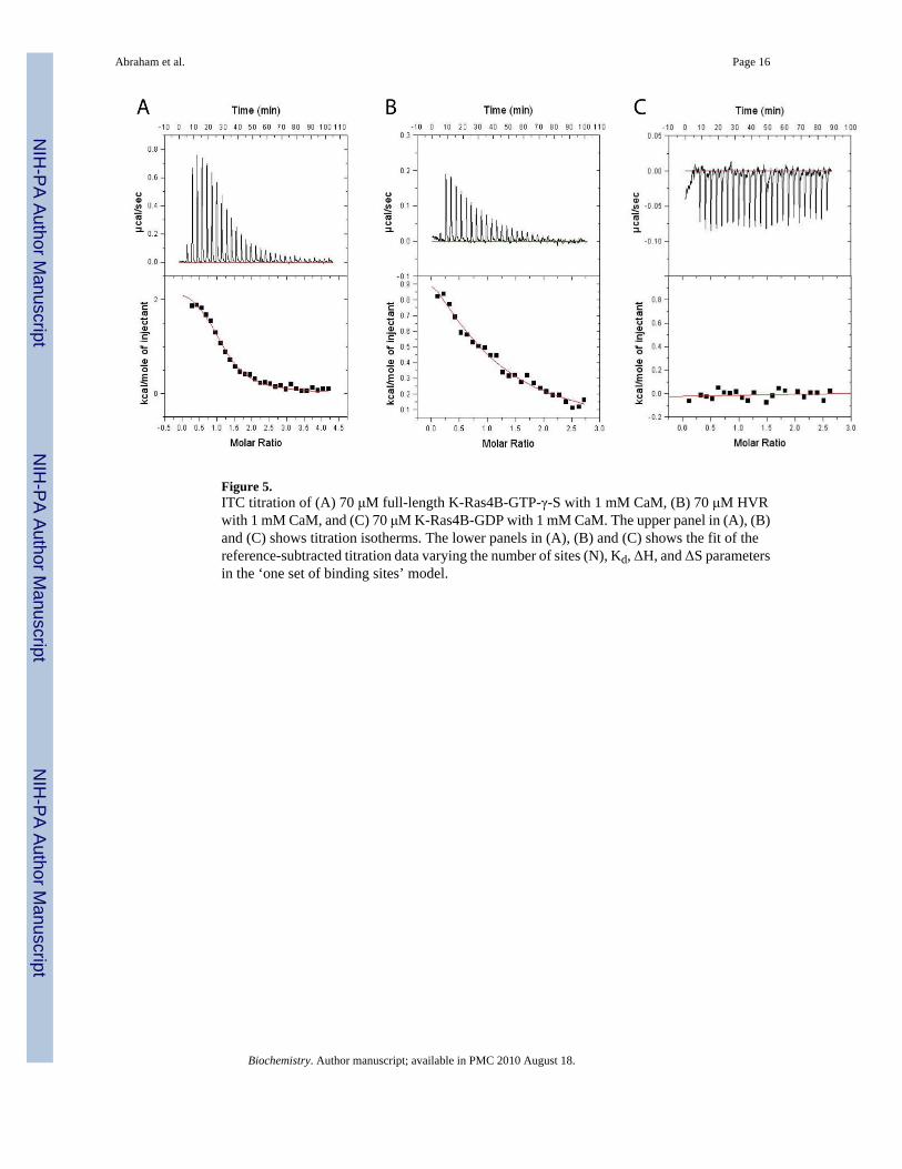
Figure 5.ITC titration of (A) 70 μM full-length K-Ras4B-GTP-γ-S with 1 mM CaM, (B) 70 μM HVRwith 1 mM CaM, and (C) 70 μM K-Ras4B-GDP with 1 mM CaM. The upper panel in (A), (B)and (C) shows titration isotherms. The lower panels in (A), (B) and (C) shows the fit of thereference-subtracted titration data varying the number of sites (N), Kd, ΔH, and ΔS parametersin the ‘one set of binding sites’ model.
Abraham et al. Page 16
Biochemistry. Author manuscript; available in PMC 2010 August 18.
NIH
-PA Author Manuscript
NIH
-PA Author Manuscript
NIH
-PA Author Manuscript

Figure 6.Examples of non-linear fits for chemical shift perturbations in CaM responding to titration with(A) GTP-γ-S loaded K-Ras4B, (B) GTP-γ-S loaded K-Ras4Btr, and (C) the HVR peptide. BothK-Ras4B-GTP-γ-S and the HVR peptide show saturation between 1.5 and 2 molar equivalentswhile K-Ras4Btr-GTP-γ-S shows saturation only at 8 molar equivalents.
Abraham et al. Page 17
Biochemistry. Author manuscript; available in PMC 2010 August 18.
NIH
-PA Author Manuscript
NIH
-PA Author Manuscript
NIH
-PA Author Manuscript

Figure 7.Hypothetical model showing the role of the HVR in the interaction of K-Ras4B with CaM.When GTP is bound to the catalytic domain, the HVR of K-Ras4B interacts specifically withthe C-terminal domain and the linker region of CaM, while the catalytic domain (1-166)interacts with the N-terminal domain giving a parallel binding pattern. When GDP is bound tothe catalytic domain (1-166), the HVR would be sequestered thereby preventing the interactionwith CaM.
Abraham et al. Page 18
Biochemistry. Author manuscript; available in PMC 2010 August 18.
NIH
-PA Author Manuscript
NIH
-PA Author Manuscript
NIH
-PA Author Manuscript