The Gaseous Ozone Therapy as a Promising Antiseptic ...
14
Citation: Rapone, B.; Ferrara, E.; Santacroce, L.; Topi, S.; Gnoni, A.; Dipalma, G.; Mancini, A.; Di Domenico, M.; Tartaglia, G.M.; Scarano, A.; et al. The Gaseous Ozone Therapy as a Promising Antiseptic Adjuvant of Periodontal Treatment: A Randomized Controlled Clinical Trial. Int. J. Environ. Res. Public Health 2022, 19, 985. https://doi.org/10.3390/ ijerph19020985 Academic Editor: Kelvin Afrashtehfar Received: 27 November 2021 Accepted: 12 January 2022 Published: 16 January 2022 Publisher’s Note: MDPI stays neutral with regard to jurisdictional claims in published maps and institutional affil- iations. Copyright: © 2022 by the authors. Licensee MDPI, Basel, Switzerland. This article is an open access article distributed under the terms and conditions of the Creative Commons Attribution (CC BY) license (https:// creativecommons.org/licenses/by/ 4.0/). International Journal of Environmental Research and Public Health Article The Gaseous Ozone Therapy as a Promising Antiseptic Adjuvant of Periodontal Treatment: A Randomized Controlled Clinical Trial Biagio Rapone 1, * , Elisabetta Ferrara 2 , Luigi Santacroce 2 , Skender Topi 3 , Antonio Gnoni 4 , Gianna Dipalma 1 , Antonio Mancini 1 , Marina Di Domenico 5 , Gianluca Martino Tartaglia 6 , Antonio Scarano 7,† and Francesco Inchingolo 1,† 1 Interdisciplinary Department of Medicine, “Aldo Moro” University of Bari, 70121 Bari, Italy; [email protected] (G.D.); [email protected] (A.M.); [email protected] (F.I.) 2 Complex Operative Unit of Odontostomatology, Hospital S.S. Annunziata, 66100 Chieti, Italy; [email protected] (E.F.); [email protected] (L.S.) 3 Department of Clinical Disciplines, School of Technical Medical Sciences, University A. Xhuvani, 3001 Elbasan, Albania; [email protected] 4 Department of Basic Medical Sciences, Neurosciences and Sense Organs, “Aldo Moro” University of Bari, 70121 Bari, Italy; [email protected] 5 Department of Precision Medicine, University of Campania Luigi Vanvitelli, 80138 Naples, Italy; [email protected] 6 UOC Maxillo-Facial Surgery and Dentistry, Department of Biomedical, Surgical and Dental Sciences, School of Dentistry, Fondazione IRCCS Ca Granda, Ospedale Maggiore Policlinico, University of Milan, 20100 Milan, Italy; [email protected] 7 Department of Oral Science, Nano and Biotechnology and CeSi-Met University of Chieti-Pescara, 66100 Chieti, Italy; [email protected] * Correspondence: [email protected]; Tel.: +39-347-7619-817 † These authors contributed equally to this work. Abstract: Background: the establishment of periodontitis is regulated by the primary etiological factor and several individual conditions including the immune response mechanism of the host and individual genetic factors. It results when the oral homeostasis is interrupted, and biological reactions favor the development and progression of periodontal tissues damage. Different strategies have been explored for reinforcing the therapeutic effect of non-surgical periodontal treatment of periodontal tissue damage. Gaseous ozone therapy has been recognized as a promising antiseptic adjuvant, because of its immunostimulating, antimicrobial, antihypoxic, and biosynthetic effects. Then, we hypothesized that the adjunct of gaseous ozone therapy to standard periodontal treatment may be leveraged to promote the tissue healing response. Methods: to test this hypothesis, we conducted a prospective randomized study comparing non-surgical periodontal treatment plus gaseous ozone therapy to standard therapy. A total of 90 healthy individuals with moderate or severe generalized periodontitis were involved in the study. The trial was conducted from September 2019 to October 2020. Forty-five patients were randomized to receive scaling and root-planning (SRP) used as conventional non-surgical periodontal therapy plus gaseous ozone therapy (GROUP A); forty-five were allocated to standard treatment (GROUP B). The endpoint was defined as the periodontal response rate after the application of the ozone therapy at 3 months and 6 months, defined as no longer meeting the criteria for active periodontitis. Statistical analysis was performed employing SPSS v.18 Chicago: SPSS Inc. Results: periodontal parameters differed significantly between patients treated with the two distinct procedures at 3 months (p ≤ 0.005); a statistically significant difference between groups was observed from baseline in the CAL (p ≤ 0.0001), PPD (p ≤ 0.0001) and BOP (p ≤ 0.0001) scores. Conclusions: The present study suggests that SRP combined with ozone therapy in the treatment of periodontitis revealed an improved outcome than SRP alone. Int. J. Environ. Res. Public Health 2022, 19, 985. https://doi.org/10.3390/ijerph19020985 https://www.mdpi.com/journal/ijerph
-
Upload
khangminh22 -
Category
Documents
-
view
0 -
download
0
Transcript of The Gaseous Ozone Therapy as a Promising Antiseptic ...

�����������������
Citation: Rapone, B.; Ferrara, E.;
Santacroce, L.; Topi, S.; Gnoni, A.;
Dipalma, G.; Mancini, A.; Di
Domenico, M.; Tartaglia, G.M.;
Scarano, A.; et al. The Gaseous
Ozone Therapy as a Promising
Antiseptic Adjuvant of Periodontal
Treatment: A Randomized
Controlled Clinical Trial. Int. J.
Environ. Res. Public Health 2022, 19,
985. https://doi.org/10.3390/
ijerph19020985
Academic Editor:
Kelvin Afrashtehfar
Received: 27 November 2021
Accepted: 12 January 2022
Published: 16 January 2022
Publisher’s Note: MDPI stays neutral
with regard to jurisdictional claims in
published maps and institutional affil-
iations.
Copyright: © 2022 by the authors.
Licensee MDPI, Basel, Switzerland.
This article is an open access article
distributed under the terms and
conditions of the Creative Commons
Attribution (CC BY) license (https://
creativecommons.org/licenses/by/
4.0/).
International Journal of
Environmental Research
and Public Health
Article
The Gaseous Ozone Therapy as a Promising AntisepticAdjuvant of Periodontal Treatment: A Randomized ControlledClinical TrialBiagio Rapone 1,* , Elisabetta Ferrara 2 , Luigi Santacroce 2 , Skender Topi 3, Antonio Gnoni 4,Gianna Dipalma 1 , Antonio Mancini 1 , Marina Di Domenico 5, Gianluca Martino Tartaglia 6,Antonio Scarano 7,† and Francesco Inchingolo 1,†
1 Interdisciplinary Department of Medicine, “Aldo Moro” University of Bari, 70121 Bari, Italy;[email protected] (G.D.); [email protected] (A.M.); [email protected] (F.I.)
2 Complex Operative Unit of Odontostomatology, Hospital S.S. Annunziata, 66100 Chieti, Italy;[email protected] (E.F.); [email protected] (L.S.)
3 Department of Clinical Disciplines, School of Technical Medical Sciences, University A. Xhuvani,3001 Elbasan, Albania; [email protected]
4 Department of Basic Medical Sciences, Neurosciences and Sense Organs, “Aldo Moro” University of Bari,70121 Bari, Italy; [email protected]
5 Department of Precision Medicine, University of Campania Luigi Vanvitelli, 80138 Naples, Italy;[email protected]
6 UOC Maxillo-Facial Surgery and Dentistry, Department of Biomedical, Surgical and Dental Sciences,School of Dentistry, Fondazione IRCCS Ca Granda, Ospedale Maggiore Policlinico, University of Milan,20100 Milan, Italy; [email protected]
7 Department of Oral Science, Nano and Biotechnology and CeSi-Met University of Chieti-Pescara,66100 Chieti, Italy; [email protected]
* Correspondence: [email protected]; Tel.: +39-347-7619-817† These authors contributed equally to this work.
Abstract: Background: the establishment of periodontitis is regulated by the primary etiologicalfactor and several individual conditions including the immune response mechanism of the hostand individual genetic factors. It results when the oral homeostasis is interrupted, and biologicalreactions favor the development and progression of periodontal tissues damage. Different strategieshave been explored for reinforcing the therapeutic effect of non-surgical periodontal treatment ofperiodontal tissue damage. Gaseous ozone therapy has been recognized as a promising antisepticadjuvant, because of its immunostimulating, antimicrobial, antihypoxic, and biosynthetic effects.Then, we hypothesized that the adjunct of gaseous ozone therapy to standard periodontal treatmentmay be leveraged to promote the tissue healing response. Methods: to test this hypothesis, weconducted a prospective randomized study comparing non-surgical periodontal treatment plusgaseous ozone therapy to standard therapy. A total of 90 healthy individuals with moderate or severegeneralized periodontitis were involved in the study. The trial was conducted from September 2019to October 2020. Forty-five patients were randomized to receive scaling and root-planning (SRP) usedas conventional non-surgical periodontal therapy plus gaseous ozone therapy (GROUP A); forty-fivewere allocated to standard treatment (GROUP B). The endpoint was defined as the periodontalresponse rate after the application of the ozone therapy at 3 months and 6 months, defined as nolonger meeting the criteria for active periodontitis. Statistical analysis was performed employingSPSS v.18 Chicago: SPSS Inc. Results: periodontal parameters differed significantly between patientstreated with the two distinct procedures at 3 months (p ≤ 0.005); a statistically significant differencebetween groups was observed from baseline in the CAL (p ≤ 0.0001), PPD (p ≤ 0.0001) and BOP(p ≤ 0.0001) scores. Conclusions: The present study suggests that SRP combined with ozone therapyin the treatment of periodontitis revealed an improved outcome than SRP alone.
Int. J. Environ. Res. Public Health 2022, 19, 985. https://doi.org/10.3390/ijerph19020985 https://www.mdpi.com/journal/ijerph

Int. J. Environ. Res. Public Health 2022, 19, 985 2 of 14
Keywords: gaseous ozone therapy; ozone; non-surgical periodontal treatment; moderate periodonti-tis; severe periodontitis; periodontal disease
1. Introduction
Periodontal disease (PD) is one of the most common inflammatory illnesses affectingthe individuals, and the global burden of periodontal disorders, as measured in prevalence,is between 20 and 50%, with severe periodontitis affecting 11.2% worldwide [1–3]. Theterm encompasses a wide spectrum of pathological conditions, ranging from reversiblegingival inflammation to severe form, characterized by progressive destruction of alveolarbone [3]. All clinical manifestations have the same pathogenic pathway, with a dramatic in-crease in bacterial pathogens aggregation (bacterial plaque) as a mainly etiologic factor andimportant genetic and immunoregulatory individual determinants of the severity of thedisease [4–9]. In general, the conventional treatment for periodontal lesions is a mechanicaland manual non-surgical procedure, named scaling and root planning (SRP), aimed at elim-inating supra and sub-gingival bacterial plaque and calculus [10,11]. Several studies haveexamined the application of add-on therapy in the treatment of periodontitis (e.g., laser orphotodynamic therapies) to improve immunogenic responses [12–17], and inter-individualvariability of response to various adjuvant treatments and therapeutic procedures has beenwidely reported [18–21]. Recently, the treatment with gaseous ozone has been studied as asupport for SRP for its important effects of immune modulation and healing [22–24]. Ozonetherapy has been extensively studied in medicine because of its physicochemical propertiesand its unbelievable versatility for many biomedical applications, specifically degenerative,neurological, orthopaedic and genitourinary disorders [25–30]. Also in dentistry, it hasan extensive application which fluctuates from endodontia to conservative as well as thetreatment of tooth sensitivity [31–34]. In the context of periodontal infection, oxygen/ozonegas can act as a powerful device for the targeted antiseptic action, potentially reducingthe impact of microbial burden, and contemporary increasing the immune system capabil-ity [18,27–35]. The efficacy of ozone (O3) is greatly related to the beneficial chemical andphysical properties, that makes it eligible for employment in periodontal area [12,19,30].As extensively documented, the ozone has an immunomodulatory, anti-inflammatory andbiocide action [36–38]. Its antiseptic activity is mediated by disruption of bacterial cellmembrane integrity, resulting in their lysis and death [39,40]. In addition, the ozone exertsa double damage: on sulfhydryl groups of specific enzymes, disrupting the normal cellularenzymatic activity and diminishing their function; on the base components of nucleic acids,the purines and pyrimidine, resulting in damage to DNA [41,42]. The anti-inflammatoryproperty is due to the disruption of the self-perpetuating inflammatory cycle altering thebreakdown of Arachidonic acid-derived prostaglandins that contribute to the developmentof inflammation [43–45]. Furthermore, O3 contributes to activate the immune cells and itis involved in the production of cytokines [19,25,40]. According to these pharmacologicalproperties, the purpose of this trial was to determine the effectiveness of gaseous ozonetherapy in patients with periodontitis, by assuming the superiority of treatment respect tothe SRP only. To test this hypothesis, we conducted a prospective randomized controlledstudy investigating the effectiveness of gaseous ozone therapy in patients with moderateand severe periodontitis.
2. Materials and Methods2.1. Ethical Considerations
The protocol was conducted in compliance with the Ethics Committee ApprovalINTL_ALITMKCOOP/HealthMicroPath/HMM2019_IPM and according to Good ClinicalPractice and the Declaration of Helsinki Declaration of 1975, as revised in 2013 [46]. Writteninformed consent was obtained from all patients before the study.

Int. J. Environ. Res. Public Health 2022, 19, 985 3 of 14
2.2. Study Design and Participants
The trial was conducted from September 2020 to October 2021 at School of TechnicalMedical Sciences, University A. Xhuvani, Elbasan, Albania. This randomized double-masked clinical trial was carried out to test the hypothesis that the gaseous ozone therapyapplication as adjunct to SRP leads to significant improvements of periodontal parameterscompared with SRP alone. Periodontal examinations were performed by one blindedexaminer (BR), and three operators (dentist, AS; dentist, FI, dental hygienist, EF) carriedout the treatment at each time point. One blinded statistician (AG) performed the dataanalysis. Based on limited data available at the time, a sample size of 80 participants (40 foreach group) were required to achieve 90% study power using a two-group t-test assumingan α-level of 0.05. Considering a drop-out rate of 10% total sample size, was planned arecruitment of 90 participants.
2.3. Inclusion and Exclusion Criteria
Adult patients were considered eligible for inclusion in the trial if they had a diagnosisfor moderate-to-severe periodontitis. Periodontitis diagnosis was determined accordingto the new criteria presented in the World Workshop on the Classification of Periodontaland Peri-implant Diseases and Conditions [2]. Qualifying patients met all the followinginclusion criteria: having a periodontitis diagnosis; having at least 16 teeth with a minimumof four teeth in each quadrant; men and women, aged ≥18 years; could provide informedconsent. Reasons for non-enrolment were the following: unable to meet the inclusioncriteria (1) underwent administration of any systemic antibiotic regimen within the previous6 months before enrolment (2); having undergone periodontal therapy within the 12 monthsprior to the randomization (3); history of systemic diseases (4); medical conditions thatcontraindicated ozone therapy (e.g., respiratory diseases) (5); current daily smokers with anumber of >10 cigarettes/day (6); cognitive or serious mental illness; and pregnancy (7).The source population for this study consisted of subjects with a mean age of 51.56 ± 10.35.The follow-up started on the date of the baseline and ended at 6 months; an interim analysisreport addressing the impact of ozone therapy on periodontal outcomes was conducted at14 days.
2.4. Randomization and Blinding
Eligible patients were randomly assigned, in a 1:1 ratio, to following groups: SRP + OZONE(Test Group A, n = 45); SRP (Control Group B, n = 45). Examiners and statistician wereblinded to group assignment. Randomization was performed with computer generatedrandom number list. At baseline, at 3 and 6 months after SRP each patient received peri-odontal examination by two calibrated examiners, blinded to the treatment group. Theexaminers calibration was conducted before the study. The alignment exercise resulted in80% inter-examiner reliability and 90% intra-examiner reproducibility [41].
2.5. Periodontal Clinical Parameters Measurement
To assess the periodontal status before and subsequent the intervention and infer thedifference inter-groups, the following clinical outcome parameters were revealed: Bleedingon probing (BOP) was recorded to assess gingival inflammation and it was registered asthe percentage at four sites per tooth showing bleeding 30 s after probing [47]; Probingpocket depth (PPD), which is established by calculating the distance from the gingivalmargin (GM) to the base of the sulcus/pocket with a calibrated periodontal probe; andclinical attachment level (CAL), which is determined by measuring the distance from thecemento-enamel-junction (CEJ) to the base of the sulcus/pocket [41]. Probing pocket depthand CAL were recorded at six sites per tooth. The assessment of clinical status was carriedout employing the standard probing measurements using a marked periodontal probe(UNC15 probe, Hu Friedy, Chicago, IL, USA).

Int. J. Environ. Res. Public Health 2022, 19, 985 4 of 14
2.6. Outcomes
The outcomes were the probing pocket depth reduction, clinical attachment levelimprovement at 3 and 6 months.
2.7. Treatment
After enrolment, for each patient of both groups, scaling and root planning (SRP)treatment was performed. The objective of scaling is to remove supra- and sub-gingivalcalculus deposits and root planning to smooth root surfaces.
Each participant received hygiene education session and appropriate motivation andunderwent supra- and subgingival prophylaxis with ultrasonic instruments. All sites withprobing pocket depth (PPD) ≥4 mm were root planed by using manual instruments (Graceycurets, Hu Friedy, Chicago, IL, USA), under local anaesthesia. No rinsing with chlorhexi-dine digluconate solution was recommended so as not to affect the results. Follow up wasplanned in three- and six-months’ time. In the group assigned to SRP + OZONE, gaseousozone treatment was performed in three steps after instrumentation by ultrasonic instru-ments employing an ozone generator (Ozone DTA, Sweden & Martina Company; CarraraSan Giorgio, Veneto, Italy), according to manufacturer instructions, as follows: Step 1.2-min rinse with ozonated water at a ratio of 1:3; Full-mouth decontamination; Topicalirrigation with ozonated water; 1–2 cycles of ozone gas at 8–10 power in correspondence ofpathological pockets, under local anaesthesia. Step 2. Quadrant root planning; 2-min rinsewith ozonated water at a ratio of 1:3; Deplaquing; 1–2 cycles of ozone gas at 8–10 power incorrespondence of pathological pockets for each quadrant, under local anaesthesia. Step 3.Maintenance: 2-min rinse with ozonated water at a ratio of 1:3; Deplaquing; 1–2 cycles ofozone gas at 4–5 power in correspondence of pathological pockets for all quadrants, twoweeks after completion of treatment.
To maintain a state of optimal periodontal health for a correct view of the periodontalligament, each patient was motivated to a correct home management using a roto oscil-lating or sonic toothbrush and toothpastes to keep the periodontium intact and avoid theprogression of the disease with the destruction of the tissue itself [48,49].
2.8. Statistical Analysis
The Shapiro-Wilk test was used to confirm normal distribution of the data relatedto each numerical variable for each follow-up time point. Continuous variables werepresented as mean ± standard deviation (SD) and all categorical data are expressed as afrequency or a percentage. The comparison of data from the two groups at each time pointwas performed by using the unpaired 2-sample t test. A mixed model multivariate analysisof covariance (MANCOVA) with two within-subjects factors and one between-subjectsfactor was conducted to determine whether significant differences exist among the timepoints for PPD and CAL between the levels of treatment (SRP+ OZONE or SRP) aftercontrolling for stage of disease (moderate or severe), age, and sex as covariates. A p value of<0.05 was considered statistically significant. Statistical analysis was performed employingSPSS Statistics for Windows, version 18 (SPSS Inc., Chicago, IL, USA).
3. Results
The enrollment was started in September 2019 and ended in December 2019. Duringthis phase, 232 patients were screened. Figure 1 shows the trial profile.
A total of 90 patients were included in the study. Baseline demographic characteristicsand clinical periodontal parameters of the 90 patients included in our analysis are illustratedin Tables 1 and 2, respectively.
Mean age was 51.62 ± 14.42 for Group A and 49.88 ± 10.54 for the Control Group.Twenty-two percentage of the patients had moderate periodontitis, 78% had diagnosis ofsevere periodontitis. As shown in Table 2, no significant difference was detected betweenthe two groups in mean score of two periodontal parameters at baseline (PPD 5.39 vs. 5.37,p = 0.81; BOP 49 vs. 50.83, p = 0.62). However, the Control group showed a higher mean

Int. J. Environ. Res. Public Health 2022, 19, 985 5 of 14
CAL score than the Test group (5.78 vs. 5.53, p ≤ 0.0002). At 3 months a statisticallysignificant difference in the PPD (p ≤ 0.0001), CAL (p ≤ 0.003) and BOP (p ≤ 0.0001) wasobserved between the groups, as shown in Table 3.
Int. J. Environ. Res. Public Health 2022, 19, x FOR PEER REVIEW 5 of 15
Figure 1. Consort diagram showing the screening, enrolment and randomization of study patients.
A total of 90 patients were included in the study. Baseline demographic characteris-tics and clinical periodontal parameters of the 90 patients included in our analysis are illustrated in Table 1 and Table 2, respectively.
Table 1. Baseline demographic characteristics of participants.
Group A * Group B ** Age (mean ± SD) 51.62 ± 9.56 49.88 ± 10.54
Sex M 87% F 13%
M 78% F 22%
Prevalence of Moderate Periodontitis (%)
78 83
Prevalence of Severe Periodontitis (%) 22 17 Group A *: Test Group (SRP + OZONE); Group B **: Control Group (SRP).
Figure 1. Consort diagram showing the screening, enrolment and randomization of study patients.
Table 1. Baseline demographic characteristics of participants.
Group A * Group B **
Age (mean ± SD) 51.62 ± 9.56 49.88 ± 10.54
Sex M 87%F 13%
M 78%F 22%
Prevalence of Moderate Periodontitis (%) 78 83Prevalence of Severe Periodontitis (%) 22 17
Group A *: Test Group (SRP + OZONE); Group B **: Control Group (SRP).
Table 2. Baseline clinical periodontal parameters of both groups.
PPD (mm)Group A *
PPD (mm)Group B ** p Value CAL (mm)
Group ACAL (mm)Group B p Value BOP (%)
Group ABOP (%)Group B p Value
Mean 5.39 5.37 0.81 5.53 5.78 <0.05 49 50.83 0.62Std. Deviation 0.31 0.2 - 0.27 0.3 - 14.74 18.11 -
Group A *: Test Group (SRP + OZONE); Group B **: Control Group (SRP); PPD: Probing pocket depth; CAL:Clinical attachment level; BOP: Bleeding on probing. p Value: statistically significant at <0.05.
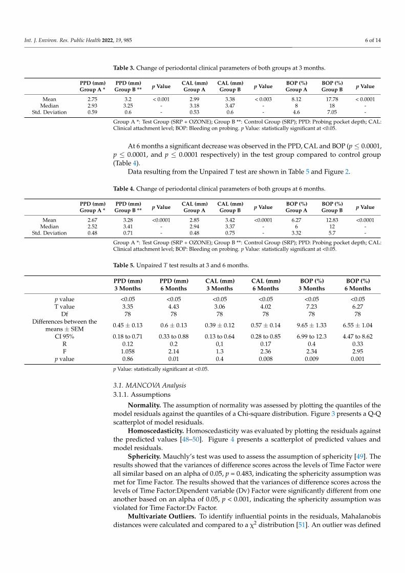
Int. J. Environ. Res. Public Health 2022, 19, 985 6 of 14
Table 3. Change of periodontal clinical parameters of both groups at 3 months.
PPD (mm)Group A *
PPD (mm)Group B ** p Value CAL (mm)
Group ACAL (mm)Group B p Value BOP (%)
Group ABOP (%)Group B p Value
Mean 2.75 3.2 < 0.001 2.99 3.38 < 0.003 8.12 17.78 < 0.0001Median 2.93 3.25 - 3.18 3.47 - 8 18 -
Std. Deviation 0.59 0.6 - 0.53 0.6 - 4.6 7.05 -
Group A *: Test Group (SRP + OZONE); Group B **: Control Group (SRP); PPD: Probing pocket depth; CAL:Clinical attachment level; BOP: Bleeding on probing. p Value: statistically significant at <0.05.
At 6 months a significant decrease was observed in the PPD, CAL and BOP (p ≤ 0.0001,p ≤ 0.0001, and p ≤ 0.0001 respectively) in the test group compared to control group(Table 4).
Data resulting from the Unpaired T test are shown in Table 5 and Figure 2.
Table 4. Change of periodontal clinical parameters of both groups at 6 months.
PPD (mm)Group A *
PPD (mm)Group B ** p Value CAL (mm)
Group ACAL (mm)Group B p Value BOP (%)
Group ABOP (%)Group B p Value
Mean 2.67 3.28 <0.0001 2.85 3.42 <0.0001 6.27 12.83 <0.0001Median 2.52 3.41 - 2.94 3.37 - 6 12 -
Std. Deviation 0.48 0.71 - 0.48 0.75 - 3.32 5.7 -
Group A *: Test Group (SRP + OZONE); Group B **: Control Group (SRP); PPD: Probing pocket depth; CAL:Clinical attachment level; BOP: Bleeding on probing. p Value: statistically significant at <0.05.
Table 5. Unpaired T test results at 3 and 6 months.
PPD (mm)3 Months
PPD (mm)6 Months
CAL (mm)3 Months
CAL (mm)6 Months
BOP (%)3 Months
BOP (%)6 Months
p value <0.05 <0.05 <0.05 <0.05 <0.05 <0.05T value 3.35 4.43 3.06 4.02 7.23 6.27
Df 78 78 78 78 78 78Differences between the
means ± SEM 0.45 ± 0.13 0.6 ± 0.13 0.39 ± 0.12 0.57 ± 0.14 9.65 ± 1.33 6.55 ± 1.04
CI 95% 0.18 to 0.71 0.33 to 0.88 0.13 to 0.64 0.28 to 0.85 6.99 to 12.3 4.47 to 8.62R 0.12 0.2 0,1 0.17 0.4 0.33F 1.058 2.14 1.3 2.36 2.34 2.95
p value 0.86 0.01 0.4 0.008 0.009 0.001
p Value: statistically significant at <0.05.
3.1. MANCOVA Analysis3.1.1. Assumptions
Normality. The assumption of normality was assessed by plotting the quantiles of themodel residuals against the quantiles of a Chi-square distribution. Figure 3 presents a Q-Qscatterplot of model residuals.
Homoscedasticity. Homoscedasticity was evaluated by plotting the residuals againstthe predicted values [48–50]. Figure 4 presents a scatterplot of predicted values andmodel residuals.
Sphericity. Mauchly’s test was used to assess the assumption of sphericity [49]. Theresults showed that the variances of difference scores across the levels of Time Factor wereall similar based on an alpha of 0.05, p = 0.483, indicating the sphericity assumption wasmet for Time Factor. The results showed that the variances of difference scores across thelevels of Time Factor:Dipendent variable (Dv) Factor were significantly different from oneanother based on an alpha of 0.05, p < 0.001, indicating the sphericity assumption wasviolated for Time Factor:Dv Factor.
Multivariate Outliers. To identify influential points in the residuals, Mahalanobisdistances were calculated and compared to a χ2 distribution [51]. An outlier was defined

Int. J. Environ. Res. Public Health 2022, 19, 985 7 of 14
as any Mahalanobis distance that exceeds 22.46, the 0.999 quantile of a χ2 distribution with6 degrees of freedom [52]. There were no outliers detected in the model.
Homogeneity of regression slopes. The assumption for homogeneity of regressionslopes was assessed by rerunning the mixed model MANCOVA, but this time includinginteraction terms between each independent variable and covariate [49,50]. The model withcovariate-independent variable interactions did not explain significantly more variance inthe dependent variables than the original model, F(18, 207) = 1.1, p = 0.356. This impliesthat none of the covariates interacted with the independent variables and the assumptionof homogeneity of regression slopes was met.
Int. J. Environ. Res. Public Health 2022, 19, x FOR PEER REVIEW 7 of 15
Data resulting from the Unpaired T test are shown in Table 5 and Figure 2.
Table 5. Unpaired T test results at 3 and 6 months.
PPD (mm) 3 Months
PPD (mm) 6 Months
CAL (mm) 3 Months
CAL (mm) 6 Months
BOP (%) 3 Months
BOP (%) 6 Months
P value <0.05 <0.05 <0.05 <0.05 <0.05 <0.05 T value 3.35 4.43 3.06 4.02 7.23 6.27
Df 78 78 78 78 78 78 Differences between the
means ± SEM
0.45 ± 0.13 0.6 ± 0.13 0.39 ± 0.12 0.57 ± 0.14 9.65 ± 1.33 6.55 ± 1.04
CI 95% 0.18 to 0.71 0.33 to 0.88 0.13 to 0.64 0.28 to 0.85 6.99 to 12.3 4.47 to 8.62 R 0.12 0.2 0,1 0.17 0.4 0.33 F 1.058 2.14 1.3 2.36 2.34 2.95
P value 0.86 0.01 0.4 0.008 0.009 0.001 p Value: statistically significant at <0.05.
(a)
(b)
Int. J. Environ. Res. Public Health 2022, 19, x FOR PEER REVIEW 8 of 15
(c)
Figure 2. The difference between means at baseline (a), 3 (b) and 6 (c) months.
3.1. MANCOVA Analysis 3.1.1. Assumptions
Normality. The assumption of normality was assessed by plotting the quantiles of the model residuals against the quantiles of a Chi-square distribution. Figure 3 presents a Q-Q scatterplot of model residuals.
Figure 3. Q-Q scatterplot for normality of the residuals for the regression model.
Homoscedasticity. Homoscedasticity was evaluated by plotting the residuals against the predicted values [48–50]. Figure 4 presents a scatterplot of predicted values and model residuals.
Figure 2. The difference between means at baseline (a), 3 (b) and 6 (c) months.

Int. J. Environ. Res. Public Health 2022, 19, 985 8 of 14
Covariate-IV independence. An ANOVA was conducted for each pair of numericcovariates and independent variables to assess independence [49]. A multinomial regres-sion model was conducted and compared to the null model for each pair of categoricalcovariates and independent variables to assess independence. There were no significantmodels for any combination of covariates and independent variables based on an alphaof 0.05, indicating the assumption of independence between covariates and independentvariables was met.
Int. J. Environ. Res. Public Health 2022, 19, x FOR PEER REVIEW 8 of 15
(c)
Figure 2. The difference between means at baseline (a), 3 (b) and 6 (c) months.
3.1. MANCOVA Analysis 3.1.1. Assumptions
Normality. The assumption of normality was assessed by plotting the quantiles of the model residuals against the quantiles of a Chi-square distribution. Figure 3 presents a Q-Q scatterplot of model residuals.
Figure 3. Q-Q scatterplot for normality of the residuals for the regression model.
Homoscedasticity. Homoscedasticity was evaluated by plotting the residuals against the predicted values [48–50]. Figure 4 presents a scatterplot of predicted values and model residuals.
Figure 3. Q-Q scatterplot for normality of the residuals for the regression model.
Int. J. Environ. Res. Public Health 2022, 19, x FOR PEER REVIEW 9 of 15
Figure 4. Residuals scatterplot testing homoscedasticity.
Sphericity. Mauchly’s test was used to assess the assumption of sphericity (49). The results showed that the variances of difference scores across the levels of Time Factor were all similar based on an alpha of 0.05, p = 0.483, indicating the sphericity assumption was met for Time Factor. The results showed that the variances of difference scores across the levels of Time Factor:Dipendent variable (Dv) Factor were significantly different from one another based on an alpha of 0.05, p < 0.001, indicating the sphericity assumption was violated for Time Factor:Dv Factor.
Multivariate Outliers. To identify influential points in the residuals, Mahalanobis distances were calculated and compared to a χ2 distribution [51]. An outlier was defined as any Mahalanobis distance that exceeds 22.46, the 0.999 quantile of a χ2 distribution with 6 degrees of freedom [52]. There were no outliers detected in the model.
Homogeneity of regression slopes. The assumption for homogeneity of regression slopes was assessed by rerunning the mixed model MANCOVA, but this time including interaction terms between each independent variable and covariate [49,50]. The model with covariate-independent variable interactions did not explain significantly more vari-ance in the dependent variables than the original model, F(18, 207) = 1.1, p = 0.356. This implies that none of the covariates interacted with the independent variables and the as-sumption of homogeneity of regression slopes was met.
Covariate-IV independence. An ANOVA was conducted for each pair of numeric covariates and independent variables to assess independence [49]. A multinomial regres-sion model was conducted and compared to the null model for each pair of categorical covariates and independent variables to assess independence. There were no significant models for any combination of covariates and independent variables based on an alpha of 0.05, indicating the assumption of independence between covariates and independent variables was met.
3.1.2. Mixed Model MANCOVA Results The results were examined based on an alpha of 0.05. Table 6 presents the MAN-
COVA results.
Table 6. Mixed Model MANCOVA Results.
Figure 4. Residuals scatterplot testing homoscedasticity.
3.1.2. Mixed Model MANCOVA Results
The results were examined based on an alpha of 0.05. Table 6 presents the MANCOVA results.The p-values for and any interaction with these within-subjects factors were calculated us-
ing the Greenhouse-Geisser corrections to adjust for the violation of the sphericity assumption.Between-Subjects. The main effect for Treatment was significant F(1, 75) = 23.28,
p < 0.001, indicating that there were significant differences in PPD and CAL between the

Int. J. Environ. Res. Public Health 2022, 19, 985 9 of 14
levels of Treatment after controlling for stage of disease, age, and sex. The covariate,Stage_of_disease, was significantly related to PPD and CAL, F(1, 75) = 4.73, p = 0.033. Thecovariate, age, was significantly related to PPD and CAL, F(1, 75) = 7.69, p = 0.007. Thecovariate, sex, was not significantly related to PPD and CAL, F(1, 75) = 2.43, p = 0.123.
Table 6. Mixed Model MANCOVA Results.
Source df SS MS F p η2p
Between-SubjectsTreatment 1 8.76 8.76 23.28 <0.001 0.24
Stage_of_disease 1 1.78 1.78 4.73 0.033 0.06Age 1 2.90 2.90 7.69 0.007 0.09sex 1 0.92 0.92 2.43 0.123 0.03
Residuals 75 28.24 0.38Within-Subjects
Time Factor 2 4.41 2.20 8.11 <0.001 0.10Treatment:Time Factor 2 13.82 6.91 25.45 <0.001 0.25
Stage_of_disease:Time Factor 2 0.99 0.49 1.82 0.166 0.02Age:Time Factor 2 0.40 0.20 0.73 0.482 0.01sex:Time Factor 2 0.71 0.36 1.32 0.271 0.02
Time Factor Residuals 150 40.74 0.27Dv Factor 1 0.23 0.23 0.86 0.358 0.01
Treatment:Dv Factor 1 2.38 2.38 8.74 0.004 0.10Stage_of_disease:Dv Factor 1 0.07 0.07 0.26 0.611 0.00
Age:Dv Factor 1 0.04 0.04 0.15 0.698 0.00sex:Dv Factor 1 0.13 0.13 0.47 0.494 0.01
Dv Factor Residuals 75 20.43 0.27Time Factor:Dv Factor 2 0.11 0.06 0.20 0.778 0.00
Treatment:Time Factor:Dv Factor 2 5.43 2.71 9.57 <0.001 0.11Stage_of_disease:Time Factor:Dv Factor 2 0.01 0.01 0.03 0.955 0.00
Age:Time Factor:Dv Factor 2 0.24 0.12 0.42 0.617 0.01sex:Time Factor:Dv Factor 2 0.73 0.37 1.29 0.276 0.02
Time Factor:Dv Factor Residuals 150 42.56 0.28
Degrees of Freedom (df ): Refers to the number of values used to compute a statistic; an F-test has two valuesfor df : the first is determined by the number of groups being compared—1, and the second is approximatelythe number of observations in the sample; used with the F to determine the p-value; F Ratio (F): The ratio ofexplained variance to error variance; used with the two df values to determine the p-value; Partial Eta Squared(η2
p): Effect size for the ANOVA/MANOVA and determines the strength of the differences among the groups;p-value: The probability of obtaining the observed results if the null hypothesis is true; Residuals: Refers to thedifference between the predicted value for the dependent variable and the actual value of the dependent variable.
Within-Subjects. The main effect for Time Factor was significant F(2, 150) = 8.11,p < 0.001, indicating there were significant differences in PPD and CAL across the levels ofTime Factor ignoring Dv Factor after controlling for stage of disease, age, and sex. The maineffect for Dv Factor was not significant F(1, 75) = 0.86, p = 0.358, indicating the values foracross the levels of Dv Factor, PPD and CAL, were all similar regardless of Time Factor aftercontrolling for stage of disease, age, and sex. The main effect for Time Factor and Dv Factorwas not significant F(2, 150) = 0.20, p = 0.778, indicating that the relationships between thelevels of Dv Factor were similar across the levels of Time Factor after controlling for Stageof disease, age, and sex.
Within-Between Interactions. The interaction effect between Time Factor and treat-ment was significant F(2, 150) = 25.45, p < 0.001, indicating that the relationships betweenthe levels of Time Factor differed significantly between the levels of treatment ignoring DvFactor after controlling for stage of disease, age, and sex.
The interaction effect between Dv Factor and Treatment was significant F(1, 75) = 8.74,p = 0.004, indicating that the relationships between the levels of Dv Factor differed signifi-cantly between the levels of treatment regardless of Time Factor after controlling for stageof disease, Age, and sex.

Int. J. Environ. Res. Public Health 2022, 19, 985 10 of 14
The interaction effect between Time Factor, Dv Factor, and treatment was significantF(2, 150) = 9.57, p < 0.001, indicating that the relationships between the combinations ofTime Factor and Dv Factor differed significantly between the levels of treatment aftercontrolling for stage of disease, age, and sex.
Within-Covariate Interactions. The interaction effect between Time Factor and stageof disease was not significant, F(2, 150) = 1.82, p = 0.166, indicating that the relationshipsbetween the levels of Time Factor were similar for all values of stage of disease. Theinteraction effect between Time Factor and age was not significant, F(2, 150) = 0.73, p = 0.482,indicating that the relationships between the levels of Time Factor were similar for allvalues of age. The interaction effect between Time Factor and sex was not significant,F(2, 150) = 1.32, p = 0.271, indicating that the relationships between the levels of TimeFactor were similar between the levels of sex.
The interaction effect between Dv Factor and stage of disease was not significant,F(1, 75) = 0.26, p = 0.611, indicating that the relationships between the levels of Dv Factorwere similar for all values of stage of disease. The interaction effect between Dv Factor andage was not significant, F(1, 75) = 0.15, p = 0.698, indicating that the relationships betweenthe levels of Dv Factor were similar for all values of age. The interaction effect between DvFactor and sex was not significant, F(1, 75) = 0.47, p = 0.494, indicating that the relationshipsbetween the levels of Dv Factor were similar between the levels of sex.
The interaction effect between Time Factor, Dv Factor, and Stage of disease wasnot significant, F(2, 150) = 0.03, p = 0.955, indicating that the relationships between thecombinations of Time Factor and Dv Factor were similar for all values of stage of dis-ease. The interaction effect between Time Factor, Dv Factor, and age was not significant,F(2, 150) = 0.42, p = 0.617, indicating that the relationships between the combinations ofTime Factor and Dv Factor were similar for all values of age. The interaction effect betweenTime Factor, Dv Factor, and sex was not significant, F(2, 150) = 1.29, p = 0.276, indicatingthat the relationships between the combinations of Time Factor and Dv Factor were similarbetween the levels of sex.
4. Discussion
The key to onset and progression of periodontitis consists of two canonical pathways:the oral microbial subversion, the central stimulus, resulting in the expression of proin-flammatory cytokines to eradicate pathogens and repair the damage tissues; in parallel,the genetic, environmental and systemic health status which contribute cumulatively tothe disease etiology and development. The goal of periodontal therapy is based on theeradication of pathogenic bacteria responsible for the onset of the disease to control theinflammatory. The aim of this clinical trial was to determine the impact of gaseous ozonetherapy in conjunction to conventional periodontal treatment on conditions and severity ofperiodontal disease in healthy subjects diagnosed with moderate or severe periodontitis, incomparison with standard treatment. To provide a compelling comparison be-tween thetwo therapies, a randomized controlled trial was designed. Ozone therapy is a practice ofcomplementary medicine and its effects have been widely confirmed [14,48–50]. Beginningin the 1960 [51], multiple trials assessed the safety and efficacy of ozone in medicine forseveral therapeutic indications. Humans’ studies have exposed the biological plausibility ofozone-induced beneficial impact on several pathological conditions [43–49] and describedthe mechanism of action of ozone, which encompasses the capacity to inactivate bacteria,viruses, fungi, yeast and protozoa by disrupting the integrity of the bacterial cell; the abilityto stimulate the increase in the red blood cell glycolysis rate; the capacity to activate the im-mune response by causing the increase in the production of interleukin-2 which determinesa cascade of subsequent immunological reactions [17,52–54]. Application methods includein-direct and direct procedures, such as the intramuscular injection, ozone bag and others.In dentistry, the indirect technical methods including the ozonated water, ozonated oil andgaseous ozone generator are employed. The aqueous (1.25–20 µmgL−1) and gaseous ozone(1–53 g m−3) are predominantly employed against periodontopathogenic and endodon-

Int. J. Environ. Res. Public Health 2022, 19, 985 11 of 14
tic bacteria, including the Enterococcus faecalis, the mainly endodontic pathogen [55,56].Boch et al., reported 85.38% reduction of bacterial count after gaseous ozone application onEnterococcus faecalis biofilm in root canals and 99.5% eradication of bacteria when the ozonewas combined with NaOCl [56]. Case et al., demonstrated the efficacy of ozone combinedwith ultrasonic agitation and ozone alone on E. fecalis [57]. Further, the antimicrobial activityof the ozone has been documented against the Staphylococcus aureus and Staphylococcusepidermidis [13], registering a significantly de-crease in absolute counts of microorganisms.Emerging studies have examined gaseous ozone therapy in addition to non-surgical pe-riodontal therapy. The rationale behind the use of ozone therapy is based on the conceptof the specifically inflammatory target pathway, as well as the antimicrobial activity. Themicrobial pathogenesis of periodontitis and the immune response are the two determinantsof this choice. Our results support previous recent studies showing that patients who havebeen treated with ozone exhibited statistically and clinically significant improvement inperiodontal inflammation after gaseous ozone treatment [22,28]. In our previous study ofdiabetic patients with periodontitis, we observed a sensitive improvement of periodontalstatus after the application of gaseous ozone [28]. The significant difference between thetwo groups in a decrease in periodontal outcomes at 3 months in the gaseous ozone-treatedgroup rationalize the improvement of the periodontal stability condition at 6 months intest groups. We hypothesized that the significant reduction displayed in the test groupmay reflect the biological activity on periodontal tissues and hence an improvement ondisease [15,16,58,59]. We theorized that the antimicrobial activity is a key step. Periodontitisis a disease whose course essentially feeds on the presence of pathogenic bacteria thatalter homeostasis and induce the establishment of the disease. Then, we assumed thatthe ozone therapy might promote the healing consequent improvement of the state of thedisease by stimulating the immune response and a more rapid lowering of the microbialload. Our results are in contrast with findings reported by Tasdemir et al. [60], which arealso based on topical gaseous ozone application into periodontal pockets. They reportedno significant differences between the two groups during the follow up in periodontalparameters. Although significant differences in CAL between the groups, some factors inour study could be considered, plausible interindividual differences and the time betweenozone treatment and the subsequent 3 months could reveal a different healing patternin these individuals. The addition of ozone treatment showed a marked improvementin periodontal conditions compared with the test group, while both groups manifesteda significant reduction in pocket probing at 3 months. Each patient received the sameoral hygiene instruction and motivation, and this may influence long-term outcomes. Thecurrent study has some limitations, first, the use of ozone therapy was only granted duringthe SRP phase, without any recall, and the follow-up was limited to explore the potentialbenefit of recall. The limitation of this study could be related to the use of criteria to definesuccess as changes in PD and CAL [61–67], because of the potential limited representa-tiveness of the effectiveness of ozone therapy, which includes additional benefits. Furtherstudies investigating biochemical parameters of oxidative stress might be useful for a morein-depth evaluation on periodontal tissue healing restoration.
5. Conclusions
This randomized clinical trial suggests that gaseous ozone therapy in conjunctionwith the conventional periodontal treatment may reduce the likelihood of periodontitisadvancing. Based on previous research we hypothesized that gaseous ozone treatment ofperiodontitis, as adjuvant of SRP, may have encouraging therapeutic effects.
Author Contributions: Conceptualization, B.R.; methodology, A.G.; software, B.R. and E.F.; valida-tion, B.R., E.F., L.S., S.T., G.D., A.M., M.D.D., G.M.T., A.S., F.I. and A.G.; formal analysis, A.G. andE.F.; investigation, B.R. and E.F.; resources, B.R.; data curation, B.R., F.I. and A.S.; writing—originaldraft preparation, B.R.; writing—review and editing, B.R., E.F., F.I. and A.S.; visualization, B.R. andG.M.T.; supervision, B.R. and G.M.T.; project administration B.R., F.I. and A.S. All authors have readand agreed to the published version of the manuscript.

Int. J. Environ. Res. Public Health 2022, 19, 985 12 of 14
Funding: This research received no external funding.
Institutional Review Board Statement: The study was conducted according to the guidelines of theDeclaration of Helsinki, and approved by the Institutional Review Board of Albania (INTL_ALITMKCOOP/HealthMicroPath/HMM2019_IPM).
Informed Consent Statement: Informed consent was obtained from all subjects involved in the study.
Conflicts of Interest: The authors declare no conflict of interest.
References1. Nazir, M.A. Prevalence of periodontal disease, its association with systemic diseases and prevention. Int. J. Health Sci. 2017, 11,
72–80.2. Caton, J.G.; Armitage, G.; Berglundh, T.; Chapple, I.L.C.; Jepsen, S.; Kornman, K.S.; Mealey, B.L.; Papapanou, P.N.; Mariano Sanz,
M.; Tonetti, M.S. A new classification scheme for periodontal and peri-implant diseases and conditions—Introduction and keychanges from the 1999 classification. J. Clin. Periodontol. 2018, 45, S1–S8. [CrossRef]
3. Ng, E.; Tay, J.R.H.; Ong, M.M.A. Minimally Invasive Periodontology: A Treatment Philosophy and Suggested Approach. Int. J.Dent. 2021, 2021, 2810264. [CrossRef]
4. Kaur, G.; Grover, V.; Bhaskar, N.; Kaur, R.K.; Jain, A. Periodontal infectogenomics. Inflamm. Regen. 2018, 38, 8. [CrossRef][PubMed]
5. Loos, B.G.; Van Dyke, T.E. The role of inflammation and genetics in periodontal disease. Periodontology 2000 2020, 83, 26–39.[CrossRef] [PubMed]
6. Dowsett, S.A.; Archila, L.; Foroud, T.; Koller, D.; Eckert, G.J.; Kowolik, M.J. The effect of shared genetic and environmental factorson periodontal disease parameters in untreated adult siblings in Guatemala. J. Periodontol. 2002, 73, 1160–1168. [CrossRef]
7. Taba, M., Jr.; Souza, S.L.; Mariguela, V.C. Periodontal disease: A genetic perspective. Braz. Oral Res. 2012, 26, 32–38. [CrossRef]8. Barros, S.P.; Offenbacher, S. Modifiable risk factors in periodontal disease: Epigenetic regulation of gene expression in the
inflammatory response. Periodontology 2000 2014, 64, 95–110. [CrossRef] [PubMed]9. Montemurro, N.; Perrini, P.; Rapone, B. Clinical Risk and Overall Survival in Patients with Diabetes Mellitus, Hyperglycemia and
Glioblastoma Multiforme. A Review of the Current Literature. Int. J. Environ. Res. Public Health 2020, 17, 8501. [CrossRef]10. Quaglia, E.; Moscufo, L.; Corsalini, M.; Coscia, D.; Sportelli, P.; Cantatore, F.; De Rinaldis, C.; Rapone, B.; Carossa, M.; Carossa, S.
Polyamide vs. silk sutures in the healing of postextraction sockets: A split mouth study. Oral Implantol. 2018, 11, 115–120.11. Cobb, C.M.; Sottosanti, J.S. A re-evaluation of scaling and root planing. J. Periodontol. 2021, 92, 1370–1378. [CrossRef]12. Suvan, J.; Leira, Y.; Moreno Sancho, F.M.; Graziani, F.; Derks, J.; Tomasi, C. Subgingival instrumentation for treatment of
periodontitis. A systematic review. J. Clin. Periodontol. 2020, 47, 155–175. [CrossRef]13. Salvi, G.E.; Stähli, A.; Schmidt, J.C.; Ramseier, C.A.; Sculean, A.; Walter, C. Adjunctive laser or antimicrobial photodynamic therapy
to non-surgical mechanical instrumentation in patients with untreated periodontitis: A systematic review and meta-analysis. J.Clin. Periodontol. 2020, 47, 176–198. [CrossRef]
14. Corsalini, M.; Di Venere, D.; Carossa, M.; Ripa, M.; Sportelli, P.; Cantatore, F.; De Rinaldis, C.; Di Santantonio, G.; Lenoci, G.;Barile, G.; et al. Comparative clinical study between zirconium-ceramic and metal-ceramic fixed rehabilitations. Oral Implantol.2018, 11, 150–160.
15. Gandhi, K.K.; Pavaskar, R.; Cappetta, E.G.; Drew, H.J. Effectiveness of Adjunctive Use of Low-Level Laser Therapy andPhotodynamic Therapy After Scaling and Root Planing in Patients with Chronic Periodontitis. Int. J. Periodontics Restor. Dent.2019, 39, 837–843. [CrossRef]
16. Grassi, F.R.; Grassi, R.; Rapone, B.; Gianfranco, A.; Balena, A.; Kalemaj, Z. Dimensional changes of buccal bone plate in immediateimplants inserted through open flap, open flap and bone grafting, and flapless technique. A CBCT randomized controlled clinicaltrial. Clin. Oral Implant. Res. 2019, 30, 1155–1164. [CrossRef] [PubMed]
17. Ren, C.; McGrath, C.; Jin, L.; Zhang, C.; Yang, Y. The effectiveness of low-level laser therapy as an adjunct to non-surgicalperiodontal treatment: A meta-analysis. J. Periodontal Res. 2017, 52, 8–20. [CrossRef]
18. Grassi, F.R.; Rapone, B.; Scarano Catanzaro, F.; Corsalini, M.; Kalemaj, Z. Effectiveness of computer-assisted anesthetic deliverysystem (STA™) in dental implant surgery: A prospective study. Oral Implantol. 2017, 10, 381–389. [CrossRef]
19. Di Venere, D.; Corsalini, M.; Nardi, G.M.; Laforgia, A.; Grassi, F.R.; Rapone, B.; Pettini, F. Obstructive site localization in patientswith Obstructive Sleep Apnea Syndrome: A comparison between otolaryngologic data and cephalometric values. Oral Implantol.2017, 10, 295–310. [CrossRef]
20. Gou, H.; Fan, R.; Chen, X.; Li, L.; Wang, X.; Xu, Y.; Svensson, P.; Wang, K. Adjunctive effects of laser therapy on somatosensoryfunction and vasomotor regulation of periodontal tissues in patients with periodontitis: A randomized controlled clinical trial. J.Periodontol. 2020, 91, 1307–1317. [CrossRef]
21. Mestnik, M.J.; Feres, M.; Figueiredo, L.C.; Soares, G.; Teles, R.P.; Fermiano, D.; Duarte, P.M.; Faveri, M. The effects of ad-junctive metronidazole plus amoxicillin in the treatment of generalized aggressive periodontitis: A 1-year double-blinded,placebocontrolled, randomized clinical trial. J. Clin. Periodontol. 2012, 39, 955–961. [CrossRef]
Int. J. Environ. Res. Public Health 2022, 19, 985 13 of 14
22. Corsalini, M.; Di Venere, D.; Rapone, B.; Stefanachi, G.; Laforgia, A.; Pettini, F. Evidence of signs and symptoms of Craniomandibu-lar Disorders in Fibromyalgia patients. Open Dent. J. 2017, 11, 91–98. [CrossRef]
23. Marconcini, S.; Giammarinaro, E.; Cosola, S.; Oldoini, G.; Genovesi, A.; Covani, U. Effects of Non-Surgical Periodontal Treatmenton Reactive Oxygen Metabolites and Glycemic Control in Diabetic Patients with Chronic Periodontitis. Antioxidants 2021, 10, 1056.[CrossRef]
24. Azarpazhooh, A.; Limeback, H. The application of ozone in dentistry: A systematic review of literature. J. Dent. 2008, 36, 104–116.[CrossRef]
25. Cosola, S.; Giammarinaro, E.; Genovesi, A.M.; Pisante, R.; Poli, G.; Covani, U.; Marconcini, S. A short-term study of the effects ofozone irrigation in an orthodontic population with fixed appliances. Eur. J. Paediatr. Dent. 2019, 20, 15–18.
26. Gupta, G.; Mansi, B. Ozone therapy in periodontics. J. Med. life 2012, 5, 59.27. Di Venere, D.; Nardi, G.M.; Lacarbonara, V.; Laforgia, A.; Stefanachi, G.; Corsalini, M.; Grassi, F.R.; Rapone, B.; Pettini, F. Early
mandibular canine-lateral incisor transposition: Case Report. Oral Implantol. 2017, 10, 181–189. [CrossRef]28. Rapone, B.; Ferrara, E.; Corsalini, M.; Converti, I.; Grassi, F.R.; Santacroce, L.; Topi, S.; Gnoni, A.; Scacco, S.; Scarano, A.; et al. The
Effect of Gaseous Ozone Therapy in Conjunction with Periodontal Treatment on Glycated Hemoglobin Level in Subjects withType 2 Diabetes Mellitus: An Unmasked Randomized Controlled Trial. Int. J. Environ. Res. Public Health 2020, 17, 5467. [CrossRef]
29. Saini, R. Ozone therapy in dentistry: A strategic review. J. Nat. Sci. Biol. Med. 2011, 2, 151–153. [CrossRef]30. Di Paolo, N.; Bocci, V.; Gaggiotti, E. Ozone therapy. Int. J. Artif. Organs 2004, 27, 168–175. [CrossRef]31. Hodson, N.; Dunne, S.M. Using ozone to treat dental caries. J. Esthet. Restor. Dent. 2007, 19, 303–305. [CrossRef] [PubMed]32. Iliadis, D.; Millar, B. Ozone and its use in periodontal treatment. Open J. Stomatol. 2013, 3, 197–202. [CrossRef]33. Scarano, A.; Inchingolo, F.; Rapone, B.; Festa, F.; Tari, S.R.; Lorusso, F. Protective Face Masks: Effect on the Oxygenation and Heart
Rate Status of Oral Surgeons during Surgery. Int. J. Environ. Res. Public Health 2021, 18, 2363. [CrossRef]34. Rapone, B.; Corsalini, M.; Converti, I.; Loverro, M.T.; Gnoni, A.; Trerotoli, P.; Ferrara, E. Does Periodontal Inflammation Affect
Type 1 Diabetes in Childhood and Adolescence? A Meta-Analysis. Front. Endocrinol. 2020, 11, 27. [CrossRef]35. Hoffman, R.K. Ozone. In Inhibition and Destruction of the Microbial Cell; Hugo, W.B., Ed.; Academic Press: London, UK, 1971;
pp. 251–253.36. Korich, D.G.; Mead, J.R.; Madore, M.S.; Sinclair, N.A.; Sterling, C.R. Effects of ozone, chlorine dioxide, chlorine and monochlo-
ramine on Cryptosporidium parvuum oocyst viability. Appl. Environ. Microbiol. 1990, 56, 1423–1428. [CrossRef]37. Kim, J.; Yousef, A.; Dave, S. Application of ozone for enhancing the microbiological safety and quality of foods: A review. J. Food
Prot. 1999, 62, 1071–1087. [CrossRef]38. Rapone, B.; Nardi, G.M.; Di Venere, D.; Pettini, F.; Grassi, F.R.; Corsalini, M. Oral hygiene in patients with oral cancer undergoing
chemotherapy and/or radiotherapy after prosthesis rehabilitation: Protocol proposal. Oral Implantol. 2016, 9, 90–97. [CrossRef]39. Di Venere, D.; Pettini, F.; Nardi, G.M.; Laforgia, A.; Stefanachi, G.; Notaro, V.; Rapone, B.; Grassi, F.R.; Corsalini, M. Correlation
between parodontal indexes and orthodontic retainers: Prospective study in a group of 16 patients. Oral Implantol. 2017, 10, 78–86.[CrossRef]
40. Kalemaj, Z.; Scarano, A.; Valbonetti, L.; Rapone, B.; Grassi, F.R. Bone response to four dental implants with different surfacetopography: A histologic and histometric study in minipigs. Int. J. Periodontics Restor. Dent. 2016, 36, 745–754. [CrossRef]
41. Michalowicz, B.S.; Hodges, J.S.; Pihlstrom, B.L. Is change in probing depth a reliable predictor of change in clinical attachmentloss? J. Am. Dent. Assoc. 2013, 144, 171–178. [CrossRef]
42. Bocci, V.A. Scientific and medical aspects of ozone therapy. State Art Arch. Med. Res. 2006, 37, 425–435. [CrossRef] [PubMed]43. Corsalini, M.; Di Venere, D.; Sportelli, P.; Magazzino, D.; Ripa, M.; Cantatore, F.; Cagnetta, C.; De Rinaldis, C.; Montemurro, N.;
De Giacomo, A.; et al. Evaluation of prosthetic quality and masticatory efficiency in patients with total removable prosthesis:Study of 12 cases. Oral Implantol. 2018, 11, 230–240.
44. Rapone, B.; Ferrara, E.; Santacroce, L.; Cesarano, F.; Arazzi, M.; Di Liberato, L.; Scacco, S.; Grassi, R.; Grassi, F.R.; Gnoni, A.; et al.Periodontal Microbiological Status Influences the Occurrence of Cyclosporine-A and Tacrolimus-Induced Gingival Overgrowth.Antibiotics 2019, 8, 124. [CrossRef] [PubMed]
45. Lorusso, F.; Noumbissi, S.; Inchingolo, F.; Rapone, B.; Khater, A.G.A.; Scarano, A. Scientific Trends in Clinical Research on ZirconiaDental Implants: A Bibliometric Review. Materials 2020, 13, 5534. [CrossRef]
46. Holt, G.R. Declaration of Helsinki—The World’s Document of Conscience and Responsibility. South Med. J. 2014, 107, 407.[CrossRef] [PubMed]
47. Mühlemann, H.R.; Son, S. Gingival sulcus bleeding—A leading symptom in initial gingivitis. Helv. Odontol. Acta 1971, 15,107–113.
48. Bates, D.; Mächler, M.; Bolker, B.; Walker, S. Fitting linear mixed-effects models using lme4. arXiv 2014, arXiv:1406.5823.49. Field, P.A.; Wilcox, R.R. Robust statistical methods: A primer for clinical psychology and experimental psychopathology
researchers. Behav. Res. Ther. 2017, 98, 19–38. [CrossRef]50. Osborne, W.J.; Waters, E. Four assumptions of multiple regression that researchers should always test. Pract. Assess. Res. Eval.
2002, 8, 2.51. Newton, R.R.; Rudestam, K.E. Your Statistical Consultant, 2nd ed.; Sage Publications, Inc.: Newbury Park, CA, USA, 2013.52. Kline, R.B. Principles and Practice of Structural Equation Modeling, 4th ed.; Guilford Publications: New York City, NY, USA, 2015.
Int. J. Environ. Res. Public Health 2022, 19, 985 14 of 14
53. Butera, A.; Gallo, S.; Maiorani, C.; Preda, C.; Chiesa, A.; Esposito, F.; Pascadopoli, M.; Scribante, A. Management of GingivalBleeding in Periodontal Patients with Domiciliary Use of Toothpastes Containing Hyaluronic Acid, Lactoferrin, or Paraprobiotics:A Randomized Controlled Clinical Trial. Appl. Sci. 2021, 11, 8586. [CrossRef]
54. Preda, C.; Butera, A.; Pelle, S.; Pautasso, E.; Chiesa, A.; Esposito, F.; Oldoini, G.; Scribante, A.; Genovesi, A.M.; Cosola, S. TheEfficacy of Powered Oscillating Heads vs. Powered Sonic Action Heads Toothbrushes to Maintain Periodontal and Peri-ImplantHealth: A Narrative Review. Int. J. Environ. Res. Public Health 2021, 18, 1468. [CrossRef] [PubMed]
55. Fuccio, C.; Luongo, C.; Capodanno, P.; Giordano, C.; Scafuro, M.A.; Siniscalco, D.; Lettieri, B.; Rossi, F.; Maglione, S.; Berrino, L. Asingle subcutaneous injection of ozone prevents allodynia and decreases the over-expression of pro-inflammatory caspases in theorbito-frontal cortex of neuropathic mice. Eur. J. Pharmacol. 2008, 603, 42–49. [CrossRef] [PubMed]
56. Johansson, E.; Claesson, R.; van Dijken, J.W. Antibacterial effect of ozone on cariogenic bacterial species. J. Dent. 2009, 37, 449–453.[CrossRef]
57. Di Filippo, C.; Cervone, C.; Rossi, C.; Di Ronza, C.; Marfella, R.; Capodanno, P.; Luongo, C.; Rossi, F.; D’Amico, M. Antiarrhythmiceffect of acute oxygen-ozone administration to rats. Eur. J. Pharmacol. 2010, 629, 89–95. [CrossRef]
58. Stoker, G. Ozone in chronic middle ear deafness. Lancet 1902, 160, 1187–1188. [CrossRef]59. Oldoini, G.; Ricci Frabattista, G.; Saragoni, M.; Cosola, S.; Giammarinaro, E.; Genovesi, A.M.; Marconcini, S. Ozone Therapy for
Oral Palatal Ulcer in a Leukaemic Patient. Eur. J. Case Rep. Intern. Med. 2020, 7, 001406.60. Baysan, A.; Whiley, R.; Lynch, E. Antimicrobial effects of a novel ozone generating device on microorganisms associated with
primary root carious lesion in vitro. Caries Res. 2000, 34, 498–501. [CrossRef]61. Elvis, A.M.; Ekta, J.S. Ozone therapy: A clinical review. J. Nat. Sci. Biol. Med. 2011, 2, 66–70. [CrossRef]62. Balmes, J.R.; Arjomandi, M.; Bromberg, P.A.; Costantini, M.G.; Dagincourt, N.; Hazucha, M.J.; Hollenbeck-Pringle, D.; Rich, D.Q.;
Stark, P.; Frampton, M.W. Ozone effects on blood biomarkers of systemic inflammation, oxidative stress, endothelial function,and thrombosis: The Multicenter Ozone Study in older Subjects (MOSES). PLoS ONE 2019, 14, e0222601.
63. Boch, T.; Tennert, C.; Vach, K.; Al-Ahmad, A.; Hellwig, E.; Polydorou, O. Effect of gaseous ozone on Enterococcus faecalisbiofilm-an in vitro study. Clin. Oral Investig. 2016, 20, 1733–1739. [CrossRef]
64. Case, P.D.; Bird, P.S.; Kahler, W.A.; George, R.; Walsh, L.J. Treatment of Root Canal Biofilms of Enterococcus faecalis with OzoneGas and Passive Ultrasound Activation. J. Endod. 2012, 38, 523–526. [CrossRef] [PubMed]
65. Tasdemir, Z.; Oskaybas, M.N.; Alkan, A.B.; Cakmak, O. The effects of ozone therapy on periodontal therapy: A randomizedplacebo-controlled clinical trial. Oral Dis. 2019, 25, 1195–1202. [CrossRef] [PubMed]
66. Seydanur Dengizek, E.; Serkan, D.; Abubekir, E.; Aysun Bay, K.; Onder, O.; Arife, C. Evaluating clinical and laboratory effects ofozone in non-surgical periodontal treatment: A randomized controlled trial. J. Appl. Oral Sci. 2019, 27, e20180108. [CrossRef][PubMed]
67. Isler, S.C.; Unsal, B.; Soysal, F.; Ozcan, G.; Peker, E.; Karaca, I.R. The effects of ozone therapy as an adjunct to the surgical treatmentof peri-implantitis. J. Periodontal Implant Sci. 2018, 48, 136–151. [CrossRef] [PubMed]