Strontium rich hybrid injectable system for bone regeneration

n engl j med
350;5
www.nejm.org january
29, 2004
The
new england journal
of
medicine
459
original article
The Effects of Strontium Ranelate on theRisk of Vertebral Fracture in Womenwith Postmenopausal Osteoporosis
Pierre J. Meunier, M.D., Christian Roux, M.D., Ph.D., Ego Seeman, M.D., Sergio Ortolani, M.D., Janusz E. Badurski, M.D., Tim D. Spector, M.D., Jorge Cannata, M.D., Adam Balogh, M.D., Ernst-Martin Lemmel, M.D.,
Stig Pors-Nielsen, M.D., René Rizzoli, M.D., Harry K. Genant, M.D., and Jean-Yves Reginster, M.D.*
From the Department of Rheumatology andBone Diseases, Edouard Herriot Hospital,Lyons, France (P.J.M.); the Department ofRheumatology, Cochin Hospital, René Des-cartes University, Paris (C.R.); the EndocrineUnit, Austin Hospital, University of Mel-bourne, Melbourne, Australia (E.S.); theCenter for Metabolic Bone Disease, IstitutoAuxologico Italiano, Milan, Italy (S.O.); theCenter for Osteoporosis and Osteo-Artic-ular Diseases, Bialystok, Poland (J.E.B.); theDepartment of Rheumatology, St. Thomas’Hospital, London (T.D.S.); the Mineral andBone Department, Central Hospital As-turias, Oviedo, Spain (J.C.); the Departmentof Obstetrics and Gynecology, Universityof Debrecen, Medical and Health SciencesCenter, Debrecen, Hungary (A.B.); MaxGrundig Klinik, Innere Medizin–Rheuma-tology, Bühl, Germany (E.-M.L.); the De-partment of Clinical Physiology, HillerødHospital, Hillerød, Denmark (S.P.-N.); theDivision of Bone Diseases, Departmentof Internal Medicine, University Hospital,Geneva (R.R.); the Osteoporosis and Arthri-tis Group, University of California, San Fran-cisco (H.K.G.); and the World Health Or-ganization Collaborating Center for PublicHealth Aspects of Osteoarticular Disorders,Department of Epidemiology and PublicHealth, University of Liège, Liège, Belgium(J.Y.R.). Address reprint requests to Dr. Meu-nier at the Department of Rheumatologyand Bone Disease, Pavillon F, Edouard Her-riot Hospital, 69437 Lyons CEDEX 03, France,or at [email protected].
*Investigators participating in the studyare listed in the Appendix.
N Engl J Med 2004;350:459-68.
Copyright © 2004 Massachusetts Medical Society.
background
Osteoporotic structural damage and bone fragility result from reduced bone formationand increased bone resorption. In a phase 2 clinical trial, strontium ranelate, an orallyactive drug that dissociates bone remodeling by increasing bone formation and decreas-ing bone resorption, has been shown to reduce the risk of vertebral fractures and to in-crease bone mineral density.
methods
To evaluate the efficacy of strontium ranelate in preventing vertebral fractures in a phase3 trial, we randomly assigned 1649 postmenopausal women with osteoporosis (lowbone mineral density) and at least one vertebral fracture to receive 2 g of oral strontiumranelate per day or placebo for three years. We gave calcium and vitamin D supplementsto both groups before and during the study. Vertebral radiographs were obtained annu-ally, and measurements of bone mineral density were performed every six months.
results
New vertebral fractures occurred in fewer patients in the strontium ranelate group thanin the placebo group, with a risk reduction of 49 percent in the first year of treatmentand 41 percent during the three-year study period (relative risk, 0.59; 95 percent confi-dence interval, 0.48 to 0.73). Strontium ranelate increased bone mineral density at month36 by 14.4 percent at the lumbar spine and 8.3 percent at the femoral neck (P<0.001 forboth comparisons). There were no significant differences between the groups in theincidence of serious adverse events.
conclusions
Treatment of postmenopausal osteoporosis with strontium ranelate leads to early andsustained reductions in the risk of vertebral fractures.
abstract
Copyright © 2004 Massachusetts Medical Society. All rights reserved. Downloaded from www.nejm.org by JEAN-YVES REGINSTER on February 20, 2009 .

n engl j med
350;5
www.nejm.org january
29
,
2004
The
new england journal
of
medicine
460
ertebral fractures, a serious con-
sequence of osteoporosis, lead to acute andchronic back pain, spinal deformity, and
a reduction in survival equivalent to that caused byhip fractures.
1
The health care burden in financialterms is substantial.
2
Vertebral deformities predictfurther vertebral fracture, even within one year, andalso predict nonvertebral fractures.
1,3,4
The bone fragility that characterizes osteoporo-sis after menopause results both from an imbalancein bone remodeling at the cellular level, which caus-es bone resorption to exceed bone formation, andfrom an increase in the rate of remodeling at thetissue level.
5
Antiresorptive therapies reduce the rateof bone remodeling and lower the fracture rate by30 to 50 percent. Antiresorptive agents, however, donot increase bone tissue mass. Instead, the increasein bone mineral density observed in clinical trialsof antiresorptive drugs is the result of a more com-plete secondary mineralization of the existing (butreduced) bone tissue mass.
6-10
Restoration of bonetissue mass and bone structure is not achieved withantiresorptive drugs and requires the use of anabol-ic agents.
11,12
Strontium ranelate is a new orally active agentconsisting of two atoms of stable strontium and anorganic moiety (ranelic acid). It stimulates the for-mation of new bone tissue and decreases bone re-sorption, as has been shown in vitro and in experi-ments in animals.
13-16
Strontium ranelate preventsbone loss in ovariectomized rats, increases bonemass in osteopenic animals, and increases bonestrength in normal animals.
13,17,18
To date, no del-eterious effects on the primary or secondary miner-alization of bone in laboratory animals
18,19
orhumans
20
have been reported. Results from a two-year placebo-controlled, phase 2, dose–responsestudy involving 353 postmenopausal women withosteoporosis suggested that ingestion of 2 g a dayof oral strontium ranelate reduced the incidence ofvertebral fractures during the second year of treat-ment and simultaneously increased bone mineraldensity.
20
In order to confirm these results, we de-signed the Spinal Osteoporosis Therapeutic Inter-vention study to test the safety of strontium rane-late and its efficacy against vertebral fracture whengiven orally at a dose of 2 g per day for three years inpostmenopausal women with osteoporosis and ahistory of vertebral fracture.
study subjects
We recruited postmenopausal women from Novem-ber 1996 through July 1998 at 72 centers in 11 Euro-pean countries and Australia for this prospective,randomized, double-blind, placebo-controlled trial.Women were eligible for the study if they were atleast 50 years old, had been postmenopausal for atleast five years, had had at least one fracture con-firmed by spinal radiography (after minimal trau-ma), and had a lumbar-spine bone mineral densityof 0.840 g per square centimeter or less (measuredwith Hologic instruments). Women were ineligibleif they had severe diseases or conditions that couldinterfere with bone metabolism or if they used anti-osteoporotic treatments (fluoride salts and bis-phosphonates taken for more than 14 days withinthe previous 12 months, or estrogen, calcitonin, orcalcitriol taken for more than 1 month in the previ-ous 6 months). All participants provided written in-formed consent before enrollment in the study; thestudy was approved by the institutional review boardat each center.
treatment protocol and follow-up
Throughout the study, subjects received daily cal-cium supplements at lunchtime (up to 1000 mg ofelemental calcium, depending on their dietary cal-cium intake), to maintain a daily calcium intakeabove 1500 mg, and vitamin D (400 to 800 IU, de-pending on the base-line serum concentration of25-hydroxyvitamin D). After a run-in period of 2 to24 weeks, depending on the severity of the defi-ciency of calcium and vitamin D, the subjects wererandomly assigned to receive 2 g a day of strontiumranelate (two packets a day of a powder that theymixed with water) or placebo powder for 3 years.Subjects were instructed to take the study drug oncedaily, at bedtime, or twice daily (one packet 30 min-utes before breakfast, and one at bedtime). Mostsubjects (87 percent) chose the once-daily regimen.
Three lateral radiographs of the spine (thoracicand lumbar radiographs and an image of the thora-columbar junction) were obtained at base line andannually, according to standardized procedures, andif there were indications of symptomatic vertebralfracture (acute back pain, a decrease in body heightof at least 1 cm, or both). At base line, anteroposte-
vmethods
Copyright © 2004 Massachusetts Medical Society. All rights reserved. Downloaded from www.nejm.org by JEAN-YVES REGINSTER on February 20, 2009 .

n engl j med
350;5
www.nejm.org january
29, 2004
strontium ranelate and vertebral fracture in postmenopausal osteoporosis
461
rior radiographs of the spine were also obtained.All radiographs were assessed at a central facility;radiologists were told the time sequence of each ra-diograph but were unaware of the treatment as-signment.
Two methods of assessing vertebral fracturewere used. First, a semiquantitative visual assess-ment of each vertebra, from T4 to L4, was performedby the same reader throughout the study. The semi-quantitative grading scale was as follows: grade 0,normal; grade 1, a decrease in the height of any ver-tebra of 20 to 25 percent; grade 2, a decrease of 25to 40 percent; and grade 3, a decrease of 40 percentor more.
21,22
For primary analysis, a new fracturewas defined by a change in the score of a vertebrafrom grade 0 at base line to a subsequent grade of1 or more. Second, quantitative assessment was alsoperformed: anterior, middle, and posterior verte-bral heights were measured for each vertebra, fromT4 to L4. A new fracture was defined by a decreasein height of at least 15 percent (or 3 mm) on a ver-tebra graded 0 at base line and with a grade on thesemiquantitative scale of 1, 2, or 3.
23
Nonvertebral fractures were confirmed by a ra-diologic evaluation or by a report from a hospital-ization. (The study did not have sufficient power foradequate statistical comparison of the two groups.)Skull, face, finger, toe, and coccygeal fractures werenot considered to be osteoporotic fractures. Stand-ing height was measured at base line and every sixmonths with a Harpenden stadiometer, accordingto a standardized procedure.
Bone mineral density at the lumbar spine andproximal femur was measured by dual-energy x-rayabsorptiometry at base line and at six-month inter-vals (Hologic). All the scans were analyzed centrally.A quality-control program was conducted through-out the study.
24
Lumbar-spine bone mineral densi-ty, adjusted for the strontium content of the bone,was calculated at month 36 according to the fol-lowing formula
20
: adjustment factor = 1÷[1+(esti-mated bone strontium content ¬ 0.61)], with bonestrontium content defined as the value measured inbone-biopsy samples obtained in some subjects atmonth 36.
Blood and urine samples were collected at baseline, three months, and six months, and then ev-ery six months; the samples were stored at ¡80°Cand analyzed centrally. Biochemical tests were per-formed by Bio Analytical Research Corporation(BARC) according to standard methods. Serum con-
centrations of bone-specific alkaline phosphatase(a marker of bone formation) were measured withan immunoradiometric assay (Tandem-R Ostase,Hybritech), serum concentrations of C-telopep-tide cross-links (a marker of bone resorption) withan enzyme-linked immunosorbent assay (SerumCrossLaps, Osteometer Biotech), parathyroid hor-mone with an immunoradiometric assay (N-tact,DiaSorin), 25-hydroxyvitamin D with a radioimmu-noassay (DiaSorin), and calcitonin with an immu-noradiometric assay (Biosource). Also measuredwas 1,25-dihydroxyvitamin D, by means of a radi-
Figure 1. Study Populations.
Randomized sample, 1649 patientsStrontium ranelate, 828
Placebo, 821
Safety sample, 1640 patientsStrontium ranelate, 826
Placebo, 814
9 Excluded: no treatment received
198 Excluded: no radiograph after base line
Analysis at 1 yr, 1385 patientsStrontium ranelate, 686
Placebo, 699
Completed follow-up at 3 yr, 1260 patientsStrontium ranelate, 628
Placebo, 632
Full analyzed sample (intention-to-treat population), 1442 patients
Strontium ranelate, 719Placebo, 723
Copyright © 2004 Massachusetts Medical Society. All rights reserved. Downloaded from www.nejm.org by JEAN-YVES REGINSTER on February 20, 2009 .
n engl j med
350;5
www.nejm.org january
29
,
2004
The
new england journal
of
medicine
462
oreceptor assay (DiaSorin). The strontium con-tent of serum and bone was measured with induc-tively coupled plasma-emission spectrophotometry(BARC), and the results were released only after therandomization code had been broken.
Biopsies of transiliac bone were carried out aftertetracycline double labeling in 20 consenting pa-tients to assess bone strontium content and safety-related histomorphometric variables.
statistical analysis
The main efficacy analysis was performed on an in-tention-to-treat basis and included patients whounderwent randomization, who had taken at leastone packet of treatment, and for whom at least onespinal radiograph was obtained after base line. Kap-lan–Meier product-limit estimates of the incidenceof new vertebral fractures were calculated at the timeeach year when radiography was performed. An un-adjusted Cox model served as the main statisticalanalysis for the comparison of groups and the esti-mation of relative risks and 95 percent confidenceintervals.
Two-sided Student’s t-tests were used to com-
pare percent changes from base line in independentsamples, the Pearson chi-square test was used tocompare the numbers of patients with at least twonew vertebral fractures and the numbers of patientswith a specific adverse event, and 95 percent confi-dence intervals were determined for the differencesbetween the groups with respect to mean changesin serum calcium, phosphorus, and parathyroid hor-mone levels. For percent changes from the base-linevalue in bone mineral density at each subsequentvisit, a step-down hierarchical procedure was per-formed on the basis of the increasing treatment ef-fect over time. The mean values in the two groupsat each visit were compared with the use of one-sid-ed Student’s t-tests with a type I error rate of 2.5 per-cent. The P values presented correspond to a two-sided test at the 5 percent threshold. (One-sidedP values were doubled.)
The data and assessments collected in this studywere held by Servier, and statistical analyses wereperformed by Servier. Data concerning bone min-eral density, biochemical markers and other bio-chemical variables, and the evaluation of spinal ra-diographs were collected centrally by independentinvestigators and then transferred to Servier for sta-tistical analysis. The authors had access to all thedata and take responsibility for the veracity of theanalyses.
This study was coordinated and organized un-der the control of an independent advisory com-mittee, whose members were not directly involvedin the study, and the international coordinator (Dr.Meunier), who monitored the scientific quality ofthe studies, patient compliance and adherence tothe protocol, results, and conclusions. A steeringcommittee planned the study, its conduct, and thestatistical analysis, and it oversaw any scientific ortechnical issues. The members of an adverse-eventscommittee were independent of the sponsor and ofother committees.
study subjects
Of 1649 women who underwent randomization,87.4 percent (1442 women) made up the popula-tion for the intention-to-treat analysis (Fig. 1). Thebase-line characteristics of the two groups weresimilar both in the intention-to-treat population(Table 1) and among all patients randomly assignedto treatment groups (data not shown). In the inten-tion-to-treat population, 87.4 percent of the place-
results
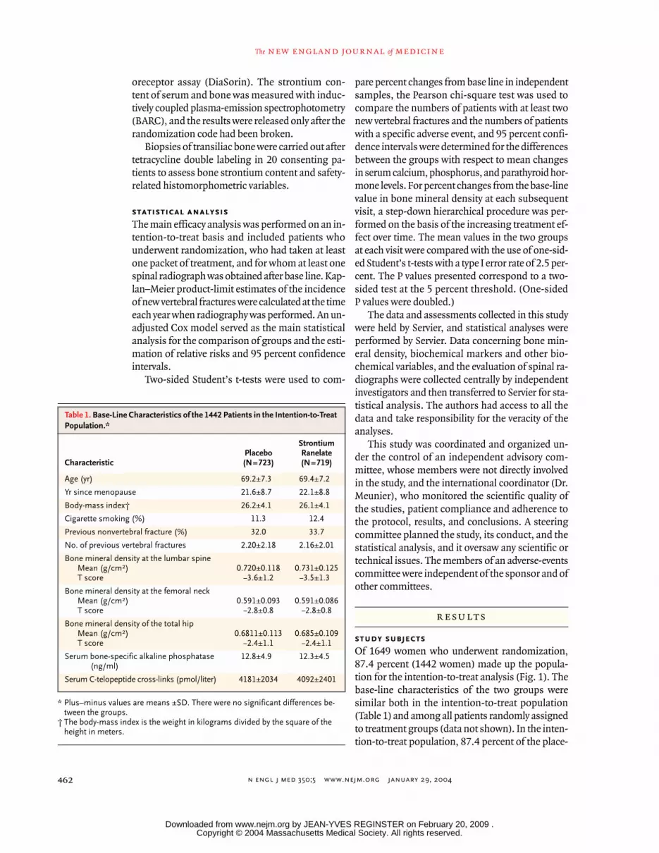
* Plus–minus values are means ±SD. There were no significant differences be-tween the groups.
† The body-mass index is the weight in kilograms divided by the square of the height in meters.
Table 1. Base-Line Characteristics of the 1442 Patients in the Intention-to-Treat Population.*
CharacteristicPlacebo(N=723)
StrontiumRanelate(N=719)
Age (yr) 69.2±7.3 69.4±7.2
Yr since menopause 21.6±8.7 22.1±8.8
Body-mass index† 26.2±4.1 26.1±4.1
Cigarette smoking (%) 11.3 12.4
Previous nonvertebral fracture (%) 32.0 33.7
No. of previous vertebral fractures 2.20±2.18 2.16±2.01
Bone mineral density at the lumbar spineMean (g/cm
2
)T score
0.720±0.118¡3.6±1.2
0.731±0.125¡3.5±1.3
Bone mineral density at the femoral neckMean (g/cm
2
)T score
0.591±0.093¡2.8±0.8
0.591±0.086¡2.8±0.8
Bone mineral density of the total hipMean (g/cm
2
)T score
0.6811±0.113¡2.4±1.1
0.685±0.109¡2.4±1.1
Serum bone-specific alkaline phosphatase (ng/ml)
12.8±4.9 12.3±4.5
Serum C-telopeptide cross-links (pmol/liter) 4181±2034 4092±2401
Copyright © 2004 Massachusetts Medical Society. All rights reserved. Downloaded from www.nejm.org by JEAN-YVES REGINSTER on February 20, 2009 .

n engl j med
350;5
www.nejm.org january
29, 2004
strontium ranelate and vertebral fracture in postmenopausal osteoporosis
463
bo group and 87.3 percent of the strontium ranelategroup completed three years of follow-up.
vertebral fractures, body height, and nonvertebral fractures
At the end of the first year of treatment, there was a49 percent lower risk of a new vertebral fracture inthe strontium ranelate group than in the placebogroup (incidence, 6.4 percent vs. 12.2 percent; rela-tive risk, 0.51; 95 percent confidence interval, 0.36to 0.74; P<0.001), and a 52 percent lower risk ofsymptomatic fracture (3.1 percent vs. 6.4 percent;relative risk, 0.48; 95 percent confidence interval,0.29 to 0.80; P=0.003). Over the entire three-yearstudy period, the strontium ranelate group had a 41percent lower risk of a new vertebral fracture thanthe placebo group (20.9 percent vs. 32.8 percent;relative risk, 0.59; 95 percent confidence interval,0.48 to 0.73; P<0.001) (Fig. 2). On the basis of thesedata, 9 patients would need to be treated for threeyears with strontium ranelate in order to prevent1 patient from having a vertebral fracture (95 per-cent confidence interval, 6 to 14).
Quantitative assessment confirmed by the semi-quantitative evaluation of vertebral fractures showedthat 17.7 percent of patients receiving strontiumranelate for three years and 28.4 percent of patientsreceiving placebo had one new vertebral fracturethat met the study criteria (relative risk in the stron-tium ranelate group, 0.58; 95 percent confidenceinterval, 0.46 to 0.73; P<0.001). The proportion ofpatients with more than one new vertebral fractureover the three-year period was 6.4 percent in thestrontium ranelate group and 9.8 percent in the pla-cebo group (relative risk, 0.64; 95 percent confi-dence interval, 0.44 to 0.93; P=0.02). Symptomaticvertebral fractures were detected in 192 patients(11.3 percent of the strontium ranelate group and17.4 percent of the placebo group), correspondingto a 38 percent lower risk over a period of three yearsin the strontium ranelate group than in the controlgroup (relative risk, 0.62; 95 percent confidence in-terval, 0.47 to 0.83; P<0.001).
Over the three-year treatment period, fewer pa-tients had height loss of at least 1 cm in the stron-tium ranelate group (30.1 percent) than in the pla-cebo group (37.5 percent, P=0.003). Back pain wasreported by 17.7 percent of the women in the stron-tium ranelate group and by 21.3 percent in the pla-cebo group (P=0.07). Nonvertebral fractures oc-curred in 234 women (112 in the strontium ranelategroup and 122 in the placebo group) over the study
period (relative risk, 0.90; 95 percent confidenceinterval, 0.69 to 1.17).
bone mineral density, serum strontium levels, and markers of bone turnover
Bone mineral density was similar at base line in thetwo groups and increased continuously at the spine,femoral neck, and total hip in the strontium rane-late group over the three-year period, with no trendtoward a plateau (Fig. 3). Over a period of threeyears, bone mineral density in the strontium rane-late group had increased from base line by 12.7 per-cent at the lumbar spine, 7.2 percent at the femoralneck, and 8.6 percent at the total hip (P<0.001 forall three comparisons with base-line values), corre-sponding to differences between the placebo andthe treatment groups at three years of 14.4 percent,8.3 percent, and 9.8 percent, respectively. At threeyears, the bone mineral density at the lumbar spine,adjusted for the strontium content, showed an in-crease over the base-line value of 6.8 percent in the
Figure 2. Proportion of Patients in the Intention-to-Treat Population Who Had One or More New Vertebral Frac-tures, Assessed According to the Semiquantitative Method.
Analysis at month 12 was restricted to patients with as-sessable radiographs at base line and at month 12 (686 patients in the strontium ranelate group and 699 in the placebo group). The relative risk of fracture in the treat-ment group at 12 months was 0.51 (95 percent confidence interval, 0.36 to 0.75; P<0.001). Analysis for the three-year period was restricted to patients with assessable radio-graphs at base line and after base line (719 patients in the strontium ranelate group and 723 in the placebo group). The relative risk of fracture over 36 months was 0.59 (95 percent confidence interval, 0.48 to 0.73; P<0.001).
Placebo Strontium ranelate
Prob
abili
ty o
f Ver
tebr
al F
ract
ure
(%)
35
40
30
10
5
15
20
25
012
Months
36
Copyright © 2004 Massachusetts Medical Society. All rights reserved. Downloaded from www.nejm.org by JEAN-YVES REGINSTER on February 20, 2009 .

n engl j med
350;5
www.nejm.org january
29
,
2004
The
new england journal
of
medicine
464
strontium ranelate group and a decrease of 1.3 per-cent in the placebo group (P<0.001); these changescorrespond to a treatment-related increase of 8.1percent.
At base line, the median serum strontium con-centration was 0.3 µmol per liter in both groups. Atthree months, the median serum strontium con-centration in the treated group had risen to 117.9
µmol per liter and then reached a plateau. At thethird month of therapy, the serum concentration ofbone-specific alkaline phosphatase was higher inthe strontium ranelate group than in the placebogroup (a treatment-related increase of 8.1 percent,P<0.001), and this difference persisted at each eval-uation during the three years (Fig. 4A and 4C). Theconcentration of serum C-telopeptide cross-linkswas lower in the strontium ranelate group than inthe placebo group at month 3 (a treatment-relateddifference of 12.2 percent, P<0.001) and at each sub-sequent evaluation during the three years (P<0.001)(Fig. 4B and 4C).
histomorphometric features of bone
Bone biopsies were performed at 24, 36, or 48months in 20 consenting patients, resulting in 14samples that could be assessed. All biopsy speci-mens consisted of lamellar bone. None of the biop-sies showed evidence of osteomalacia or any sign ofa primary mineralization defect. As compared withthe placebo group, the strontium ranelate group hadno increase in osteoid thickness (P=0.64) or in themineralization lag time (P=0.76) and no decreasein the mineral apposition rate (P=0.93).
adverse events and metabolic results
During the three years of the study, the rate of com-pliance with therapy was 83 percent in the stron-tium ranelate group and 85 percent in the placebogroup. The rates of adverse events, serious adverseevents, and withdrawal due to adverse events weresimilar in the two groups. Diarrhea was the mostfrequent gastrointestinal adverse event (occurringin 6.1 percent of the strontium ranelate group and3.6 percent of the placebo group, P=0.02). How-ever, this effect disappeared after the first threemonths. There was a lower incidence of gastritis,as diagnosed clinically by the investigators, in thestrontium ranelate group than in the placebo group(3.6 percent vs. 5.5 percent, P=0.07). Serum calci-um concentrations were lower and serum phos-phate concentrations higher in the strontium rane-late group than in the control group at month 3, witha plateau thereafter (95 percent confidence intervalfor the difference between the groups, ¡0.32 to ¡0.20mg per deciliter [¡0.08 to ¡0.05 mmol per liter] forcalcium and 0.25 to 0.37 mg per deciliter [0.08 to0.12 mmol per liter] for phosphorus). There was aslight reduction in serum parathyroid hormonefrom the level at month 6 in both groups (decrease
Figure 3. Effects of Strontium Ranelate on Bone Mineral Density in All Pa-tients Receiving 2 g a Day of Oral Strontium Ranelate.
P<0.001 for all comparisons, with the use of a step-down hierarchical procedure.
Lumbar Spine
Femoral Neck
Total Hip
Mea
n (±
SD) P
erce
nt C
hang
e in
Bon
e M
iner
al D
ensi
ty
12
8
0
16
4
¡40 6 12 18 24 30 36
0 6 12 18 24 30 36
0 6 12 18 24 30 36
Placebo
Strontium ranelate
12
8
0
16
4
¡4
Strontium ranelate
12
8
0
16
4
¡4
Months
Strontium ranelate
Placebo
Placebo
+14.4%
+8.3%
+9.8%
Copyright © 2004 Massachusetts Medical Society. All rights reserved. Downloaded from www.nejm.org by JEAN-YVES REGINSTER on February 20, 2009 .

n engl j med
350;5
www.nejm.org january
29, 2004
strontium ranelate and vertebral fracture in postmenopausal osteoporosis
465
Figure 4. Strontium Ranelate–Induced Changes in Serum Biochemical Markers of Bone Metabolism.
Panel A shows absolute changes from base-line values in bone-specific alkaline phosphatase, Panel B shows absolute changes from base-line values in C-telopeptide cross-links, and Panel C shows differences over time in biochemical markers between the two groups. Data shown are mean (±SE) values in the strontium ranelate group minus mean val-ues in the placebo group. Comparisons were performed with analyses of covariance in which base-line values were used as covariates.
Cha
nge
from
Bas
e Li
ne (n
g/m
l)
2.0
2.5
1.5
1.0
0.5
0.00 6 12 18 24 30 36
Months
Strontium ranelate
Placebo
3.0
Bon
e-Sp
ecifi
c A
lkal
ine
Phos
phat
ase
(ng/
ml)
C-T
elop
eptid
e C
ross
-Lin
ks(p
mol
/lite
r)
1.0
1.2
0.8
0.6
0.4
0.2
¡100
¡200
¡300
¡400
¡500
¡600
¡700
¡800
0.0
0 6 12 18 24 30 36
Months
1.4
Cha
nge
from
Bas
e Li
ne (p
mol
/lite
r)
200
400
0
¡200
¡400
¡6000 6 12 18 24 30 36
Months
Strontium ranelate
Placebo600
A B
C
Bone-Specific Alkaline Phosphatase C-Telopeptide Cross-Links
Difference over Time between Groups
P=0.003
P=0.02
P=0.01
P<0.001
P<0.001
P<0.001
P<0.001
P<0.001
P<0.001
P<0.001P<0.001
P=0.003
P=0.003
P=0.01
P=0.01
P=0.01
P=0.01
P=0.003
P=0.003
P=0.006
Copyright © 2004 Massachusetts Medical Society. All rights reserved. Downloaded from www.nejm.org by JEAN-YVES REGINSTER on February 20, 2009 .

n engl j med
350;5
www.nejm.org january
29
,
2004
The
new england journal
of
medicine
466
from base-line to six-month values, ¡3.4
±
10.7 pgper milliliter in the strontium ranelate group and¡1.3
±
15.8 pg per milliliter in the placebo group).No changes were observed for serum 25-hydroxyvi-tamin D, 1,25-dihydroxyvitamin D, or calcitonin.
Serum creatine kinase concentrations in theskeletal muscle increased in some patients; with ahigh concentration defined as a value that was atleast twice the upper limit of the normal range (145IU per liter), high levels were detected in 3.4 per-cent of patients in the strontium ranelate group atleast once during the study and in 1.8 percent ofpatients in the placebo group. No increase in mus-cular symptoms or other biologic abnormalitieswere observed. Most of the increases in creatinephosphokinase were transient, and in more than88 percent of the patients who had high concentra-tions, the values returned to the normal range dur-ing treatment.
Prevention of fractures is the primary aim of anti-osteoporotic treatment. In the present study, stron-tium ranelate ingested daily reduced the risk of newvertebral fractures by 49 percent at one year and by41 percent over a three-year period among post-menopausal women with osteoporosis. Althoughdata from direct comparisons with other antios-teoporotic treatments are lacking, the reduction inthe risk of vertebral fracture seems similar to thereduction reported with alendronate (47 percent),
6
5 mg of risedronate (49 percent),
7
60 mg of ralox-ifene (30 percent),
8
and parathyroid hormone (65percent after 21 months of treatment).
12
The meth-ods of assessing vertebral fractures, involving bothsemiquantitative and morphometric evaluations,were similar to the methods used in these other re-ports.
6-8,12
Strontium ranelate also reduced the riskof multiple vertebral fractures and the risk of symp-tomatic fractures. In this group of women, who hadestablished osteoporosis and a history of fractures,the number needed to treat to prevent one fracturewas nine.
Most antiresorptive agents prevent bone destruc-tion by reducing the rate of bone remodeling, asreflected by a decrease in both markers of bone re-sorption (more than 50 percent with bisphospho-nates and about 30 percent with raloxifene) andmarkers of bone formation (about 50 percent withbisphosphonates and 20 percent with raloxifene).
25
Treatment with parathyroid hormone increases both
bone formation and bone resorption.
26
When para-thyroid hormone and alendronate are combined,there is, unexpectedly, no potentiation of their ef-fects on biochemical bone markers.
27
The mecha-nism of action of strontium ranelate is probably dif-ferent from those of these drugs. Each time thepatients were evaluated during our study, bone for-mation had increased in the group assigned to stron-tium ranelate, on the basis of serum concentrationsof bone-specific alkaline phosphatase, and bone re-sorption had decreased, on the basis of serum con-centrations of C-telopeptide cross-links, as com-pared with the values in the placebo group. Thechanges in biochemical markers of bone resorptionand formation were most pronounced during thefirst six months; the dissociation between the bonemarkers was evident throughout the study. Themechanisms for the apparent dissociation betweenreduced bone resorption and increased bone forma-tion are not yet understood, but they probably dif-fer from the mechanisms of current treatments.
6-9
Any metal with an atomic number higher than
that of calcium can be expected to influence bonemineral density.
28
In experiments, strontium inbone correlated with strontium in plasma.
29
In thisstudy, strontium ranelate increased bone mineraldensity at three years by 14.4 percent at the lumbarspine, by 8.3 percent at the femoral neck, and by9.8 percent at the total hip, and bone strength isdirectly proportional to bone mineral density. Al-though data from directly comparable trials are notavailable, the treatment effect after adjustment forthe strontium content of bone was an 8.1 percentincrease in bone mineral density at the lumbar spineat 3 years, which compares favorably with a 9 per-cent increase with parathyroid hormone (20 µg) at21 months and with the increases with other drugs(5.9 percent with risedronate, 6.2 percent with alen-dronate, and 2.6 percent with raloxifene). Moreover,this increase is consistent with the results of pre-vious phase 2 studies involving strontium rane-late.
20,30
In summary, strontium ranelate given oral-ly at a dose of 2 g daily appears to reduce the riskof vertebral fractures rapidly, effectively, and safelyamong postmenopausal women with osteoporosis.
Supported by Servier.Drs. Balogh, Meunier, Nielsen, Ortolani, Reginster, Roux, See-
man, and Spector report having received consulting fees from Servi-er; Drs. Meunier and Roux consulting fees from Eli Lilly; Dr. Baloghlecture fees from Lilly Hungaria; Dr. Genant consulting and lecturefees from Servier, Wyeth Laboratories, Eli Lilly, GlaxoSmithKline,Novartis, Pfizer, and Procter & Gamble and grant support from Pfi-zer, Procter & Gamble, and Eli Lilly; Dr. Ortolani consulting feesfrom Roche and Janssen, lecture fees from Italfarmaco, and grant
discussion
Copyright © 2004 Massachusetts Medical Society. All rights reserved. Downloaded from www.nejm.org by JEAN-YVES REGINSTER on February 20, 2009 .

n engl j med
350;5
www.nejm.org january
29, 2004
strontium ranelate and vertebral fracture in postmenopausal osteoporosis
467
support from Servier, Novartis, and TEVA Pharmaceutical Indus-tries; Dr. Meunier lecture fees from Servier, Eli Lilly, Merck, Procter& Gamble, and Aventis; Dr. Reginster consulting fees from Novar-tis, Negma Laboratories, Eli Lilly, Wyeth Laboratories, Amgen,GlaxoSmithKline, Roche, Merck Pharmaceuticals, Nycomed, NPSPharmaceuticals, and Theramax, lecture fees from Merck, Eli Lilly,Rotta Pharmaceuticals, Institut Biochimique, Novartis, Servier,
Merck, Teijin, Anelis, Theramex, Nycomed, and Novo Nordisk,and grant support from Bristol-Myers Squibb; Dr. Rizzoli consult-ing fees from Novartis and Wyeth Laboratories, lecture fees fromMerck, Servier, and Aventis, and grant support from Proskalia andServier; Dr. Roux consulting and lecture fees from Procter & Gam-ble and lecture fees from Novartis and Eli Lilly; and Dr. Spector lec-ture fees from Procter & Gamble.
appendix
The Spinal Osteoporosis Therapeutic Intervention study investigators were as follows:
Australia
: J. Graham, Ashford Specialist Centre, Ash-ford; K.W. Ng, St. Vincent’s Hospital, Fitzroy; R. Prince, Sir Charles Gairdner Hospital, Nedlands; J. Prins, Princess Alexandra Hospital,Wooloongabba; E. Seeman, Austin and Repatriation Medical Centre, Heidelberg; J. Wark, Royal Melbourne Hospital, Parkville;
Belgium
: J.Y.Reginster, Brull Hospital, Liege; J.P. Devogelaer, Catholic University of Louvain, Brussels; J.M. Kaufman, University of Gent, Gent; F. Rae-man, Jan Palfijin Ziekenhuis, Merksem; M. Walravens, Medical Centre, Tessenderloo;
Denmark
: S. Pors-Nielsen, Hillerød Hospital, Hill-erød; H. Beck-Nielsen, University Hospital Osteoporosis Centre, Odense; P. Charles, Aarhus Amtssygehus, Aarhus; O.H. Sorensen,Osteoporosis Research Center, Hvidovre;
France
: P.J. Meunier, Edouard Herriot Hospital, Lyons; J.P. Aquino, Medical Clinic La Porte Verte,Versailles; C. Benhamou, La Madeleine Hospital, Orleans; F. Blotman, Lapeyronie Hospital, Montpellier; O. Bonidan, E. Muller Hospital,Mulhouse; P. Bourgeois, Pitié-Salpétrière Hospital, Paris; M.C. De Vernejoul Lariboisière Hospital, Paris; J. Dehais, Pellegrin-Tondu Hospi-tal, Bordeaux; P. Fardellone, Nord Hospital, Amiens; A. Kahan, Cochin Hospital, Paris; J.L. Kuntz, Hautepierre Hospital, Strasbourg; C.Marcelli, Regional Hospital and University Centre, Caen; A. Prost, Hôtel Dieu Hospital, Nantes; B. Vellas, La Grave-Casselardit Hospital,Toulouse; G. Weryha, Brabois Hospital, Vandoeuvre;
Germany
: E.M. Lemmel, Max Grundig Clinic, Buhl; D. Felsenberg, Benjamin FranklinClinical University, Berlin; J. Hensen, Medical Clinic 1, Erlangen; H.P. Kruse, Medical Clinical University, Hamburg; W. Schmidt, St. JosefHospital, Bochum; J. Semler, Immanuel Hospital, Berlin; G. Stucki, Clinic for Physical Medicine and Rehabilitation, Munich;
Greece
: C.Phenekos, Red Cross Hospital, Athens;
Hungary
: A. Balogh, University of Debrecen, Medical and Health Sciences Centre, Debrecen; R. DeChatel, Semmelweis Orvostudomanyi Egyetem, Budapest;
Italy
: S. Ortolani, Centre Auxologico Italiano, Milan; S. Adami, Hospital andClinical Centre of Medecine, Valeggio sul Mincio; G. Bianchi, Hospital La Colletta, Arenzano; M.L. Brandi, General Hospital Careggi, Flo-rence; D. Cucinotta, S. Orsola Malpighi Hospital, Bologna; C. Fiore, Medical Clinic, Vittorio Emanuel Hospital, Catania; C. Gennari, Insti-tute of Medicine Clinic, New General Hospital Le Scotte, Siena; G. Isaia, University of General Medicine, Turin; G. Luisetto, University In-stitute of Semeiotica Medica, Padua; R. Passariello, Institute of Radiology, La Sapienza University, Rome; M. Passeri, Parma University,Parma; G. Rovetta, E. Bruzzone Institute, Genoa; L. Tessari, San Raffaele Hospital, Milan;
Poland
: J.E. Badurski, Center for Osteoporosisand Osteo-Articular Diseases, Bialystok; K. Hoszowski, Medical Centre, Warsaw; R.S. Lorenc, Osteoporosis Centre of Krajowe, Warsaw; A.Sawicki, Warsawian Center of Osteoporosis and Calcium Metabolism Osteomed, Warsaw;
Spain
: A. Díez, El Mar Hospital, Barcelona; J.B.Cannata, General Hospital of Asturias, Oviedo; M. Díaz Curiel and A. Rapado, Jiménez Díaz Foundation, Madrid; J. Gijon and A. Torrijos,La Paz Hospital/Hospital de Rehabilitacion y Traumatologia, Madrid; J.M. Padrino, Hospital 12 de Octubre, Madrid; A. Roces Varela, Hos-pital Nuestra Sra de la Candelaria, Santa Cruz de Tenerife, Canary Islands;
Switzerland:
J.P. Bonjour and R. Rizzoli, Geneva University Hospi-tal, Geneva;
United Kingdom:
T. D. Spector, Department of Rheumatology, St. Thomas’ Hospital, London; M. Clements, North London Clin-ical Studies Centre, Mount Vernon Hospital, Northwood; D.V. Doyle, Silverthorn Centre, Chingford; P. Ryan, Medway Hospital Trust,Gillingham; I.G. Smith, Synexus Ltd. Clinical Research Centre, Manchester; R. Smith, Banbury Clinical Studies Centre, Banbury.
references
1.
Nevitt MC, Ettinger B, Black DM, et al.The association of radiographically detectedvertebral fracture with back pain and func-tion: a prospective study. Ann Intern Med1998;128:793-800.
2.
Ray NF, Chan JK, Thamer M, Melton LJIII. Medical expenditures for the treatmentof osteoporotic fractures in the United Statesin 1995: report from the National Osteoporo-sis Foundation. J Bone Miner Res 1997;12:24-35.
3.
Lindsay R, Silverman SL, Cooper C, et al.Risk of new vertebral fracture in the year fol-lowing a fracture. JAMA 2001;285:320-3.
4.
Kotowicz MA, Melton LJ III, Cooper C,Atkinson EJ, O’ Fallon WM, Riggs BL. Riskof hip fracture in women with vertebral frac-ture. J Bone Miner Res 1994;9:599-605.
5.
Seeman E. Pathogenesis of bone fragil-ity in women and men. Lancet 2002;359:1841-50.
6.
Black DM, Cummings SR, Karpf DB, etal. Randomised trial of effect of alendro-nate on risk of fracture in women with ex-isting vertebral fractures. Lancet 1996;348:1535-41.
7.
Reginster JY, Minne HW, Sorensen OH,et al. Randomised trial of the effects of ris-edronate on vertebral fractures in womenwith established postmenopausal osteoporo-sis. Osteoporos Int 2000;11:83-91.
8.
Ettinger B, Black DM, Mitlak BH, et al.Reduction of vertebral fracture risk in post-menopausal women with osteoporosis treat-ed with raloxifene: results from a 3-year ran-domized clinical trial. JAMA 1999;282:637-45. [Erratum, JAMA 1999;282:2124.]
9.
Lufkin EG, Wahner HW, O’Fallon WM,et al. Treatment of postmenopausal osteopo-sosis with transdermal estrogen. Ann InternMed 1992;117:1-9.
10.
Boivin GY, Chavassieux PM, Santora AC,Yates J, Meunier PJ. Alendronate increasesbone strength by increasing the mean de-gree of mineralization of bone tissue in os-teoporotic women. Bone 2000;27:687-94.
11.
Chavassieux PM, Arlot ME, Reda C, WeiL, Yates AJ, Meunier PJ. Histomorphometricassessment of the long-term effects of alen-dronate on bone quality and remodeling inpatients with osteoporosis. J Clin Invest 1997;100:1475-80.
12.
Neer RM, Arnaud CD, Zanchetta JR, etal. Effect of parathyroid hormone (1-34) onfractures and bone mineral density in post-menopausal women with osteoporosis.N Engl J Med 2001;344:1434-41.
13.
Marie PJ, Amman P, Boivin G, Rey C.Mechanisms of action and therapeutic po-tential of strontium in bone. Calcif TissueInt 2001;69:121-9.
14.
Marie PJ, Garba MT, Hott M, Miravet L.Effect of low doses of stable strontium onbone metabolism in rats. Miner ElectrolyteMetab 1985;11:5-13.
15.
Canalis E, Hott M, Deloffre P, Tsou-deros Y, Marie PJ. The divalent strontiumsalt S12911 enhances bone cell replicationand bone formation in vitro. Bone 1996;18:517-23.
16.
Buehler J, Chappuis P, Saffar JL, Tsou-deros Y, Vignery A. Strontium ranelate in-hibits bone resorption while maintainingbone formation in alveolar bone in monkeys(Macaca fascicularis). Bone 2001;29:176-9.
17.
Marie PJ, Hott M, Modrowski D, et al.An uncoupling agent containing strontiumprevents bone loss by depressing bone re-
Copyright © 2004 Massachusetts Medical Society. All rights reserved. Downloaded from www.nejm.org by JEAN-YVES REGINSTER on February 20, 2009 .

n engl j med
350;5
www.nejm.org january
29
,
2004
468
strontium ranelate and vertebral fracture in postmenopausal osteoporosis
sorption and maintaining bone formationin estrogen-deficient rats. J Bone Miner Res1993;8:607-15.
18.
Grynpas MD, Marie PJ. Effects of lowdoses of strontium on bone quality and quan-tity in rats. Bone 1990;11:313-9.
19.
Boivin G, Deloffre P, Perrat B, et al. Stron-tium distribution and interactions with bonemineral in monkey iliac bone after strontiumsalt (S 12911) administration. J Bone MinerRes 1996;9:1302-11.
20.
Meunier PJ, Slosman DO, Delmas PD, etal. Strontium ranelate: dose-dependent ef-fects in established postmenopausal verte-bral osteoporosis — a 2-year randomizedplacebo controlled trial. J Clin EndocrinolMetab 2002;87:2060-6.
21.
Genant HK, Wu CY, van Kuijk C, NevittMC. Vertebral fracture assessment using asemiquantitative technique. J Bone Miner Res1993;8:1137-48.
22.
Genant HK, Jergas M, Palermo L, et al.
Comparison of semiquantitative visual andquantitative morphometric assessment ofprevalent and incident vertebral fracturesin osteoporosis. J Bone Miner Res 1996;11:984-96.
23.
Wu CY, Li J, Jergas M, Genant HK. Com-parison of semiquantitative and quantitativetechniques for the assessment of prevalentand incident vertebral fractures. OsteoporosInt 1995;5:354-70.
24.
Slosman DO, Provvedini DM, MeunierPJ, et al. The use of different dual X-ray ab-sorptiometry brands in a multicenter clini-cal trial. J Clin Densitom 1999;2:37-44.
25.
Delmas PD. Markers of bone turnover formonitoring treatment of osteoporosis withantiresorptive drugs. Osteoporos Int 2000;11:Suppl 6:S66-S76.
26.
Lindsay R, Nieves J, Formica C, et al. Ran-domised controlled study of effect of para-thyroid hormone on vertebral-bone mass andfracture incidence among postmenopausal
women on oestrogen with osteoporosis. Lan-cet 1997;350:550-5.
27.
Black DM, Greenspan SL, Ensrud KE,et al. The effects of parathyroid hormoneand alendronate alone or in combination inpostmenopausal osteoporosis. N Engl J Med2003;349:1207-15.
28.
Nielsen SP, Slosman D, Sorensen OH,et al. Influence of strontium on bone miner-al density and bone mineral content measure-ments by dual X-ray absorptiormetry. J ClinDensitom 1999;2:371-9.
29.
Dahl SG, Allain P, Marie PJ, et al. Incor-poration and distribution of strontium inbone. Bone 2001;28:446-53.
30.
Reginster JY, Deroisy R, Dougados M,Jupsin I, Colette J, Roux C. Prevention of ear-ly postmenopausal bone loss by strontiumranelate: the randomized, two-year, double-masked, dose-ranging, placebo-controlledPREVOS trial. Osteoporos Int 2002;13:925-31.
Copyright © 2004 Massachusetts Medical Society.
Copyright © 2004 Massachusetts Medical Society. All rights reserved. Downloaded from www.nejm.org by JEAN-YVES REGINSTER on February 20, 2009 .
Copyright © 2022 FDOKUMEN