Polyunsaturated fatty acid status in individuals with Poly Cystic ...
Upload
independentCategory
view
0download
0
ORIGINAL RESEARCH
The effects of resting and exercise serum from childrenwith cystic fibrosis on C2C12 myoblast proliferation in vitroThanh Nguyen1, Jeff M. Baker2, Joyce Obeid1, Sandeep Raha3, Gianni Parise2, Linda Pedder3,4 &Brian W. Timmons1
1 Child Health & Exercise Medicine Program, McMaster University, Hamilton, Ontario, Canada
2 Exercise Metabolism Research Group, Department of Kinesiology, McMaster University, Hamilton, Ontario, Canada
3 Department of Pediatrics, McMaster University, Hamilton, Ontario, Canada
4 Cystic Fibrosis Clinic, McMaster Children’s Hospital, Hamilton, Ontario, Canada
Keywords
C2C12 cells, children, exercise.
Correspondence
Brian W. Timmons, Child Health & Exercise
Medicine Program, McMaster University and
McMaster Children’s Hospital, 1280 Main
Street West, HSC 3N27G Hamilton, Ontario,
Canada L8S 4K1.
Tel: 905-521-2100, ext. 77218 or 77615
Fax: 905-521-7936
E-mail: [email protected]
Funding Information
Funding for this study was made possible by
a Discovery Grant from the Natural Sciences
and Engineering Research Council of Canada
(NSERC) to B. W. Timmons. B. W. Timmons
is supported by a CIHR New Investigator
Salary Award. T. Nguyen was supported by
an NSERC Postgraduate Scholarship.
Received: 22 April 2014; Revised: 12 May
2014; Accepted: 15 May 2014
doi: 10.14814/phy2.12042
Physiol Rep, 2 (6), 2014, e12042,
doi: 10.14814/phy2.12042
Abstract
Chronic systemic inflammation is a clinical symptom in children with cystic
fibrosis (CF), but the effects on skeletal muscle development are unknown.
The aims of this study were to determine (1) the effects of systemic factors
from children with CF and healthy controls on myoblast proliferation, and
(2) whether exercise serum can have an effect on proliferation in vitro. Eleven
children with CF and 11 biological age-matched controls completed two
30-min bouts of cycling at an intensity set at 50% peak mechanical power.
Serum samples were collected before exercise (REST), immediately following
exercise (EX), and after 60 min of recovery (REC). Serum samples prepared
in group-specific pools were used for cell culture experiments. C2C12 myo-
blasts were incubated in 5% serum and media for 1 h and then immediately
harvested for protein and mRNA analysis, or incubated in growth media for
2 days to examine proliferation. C2C12 myoblasts treated with CF serum dis-
played greater proliferation phenotype than myoblasts treated with control
serum. Proliferation did not change with EX or REC serum from children
with CF compared to CF REST serum, while proliferation was increased with
EX and REC serum from control compared to control REST serum. These
findings suggest that systemic factors from children with CF at rest and after
exercise can alter myoblast proliferation responses when compared to systemic
factors from healthy children, which may have implications on skeletal muscle
development.
Introduction
Children with cystic fibrosis (CF) suffer from chronic sys-
temic inflammation (Tirakitsoontorn et al. 2001; Nguyen
et al. 2012) that may negatively impact whole body skele-
tal muscle mass via increased protein catabolism (van
Heeckeren et al. 2000; Ionescu et al. 2002, 2004). In
patients with CF, those who have increased protein
breakdown as measured by urinary pseudouridine, tend
to have increased IL-6 and TNF-a (Ionescu et al. 2002),
and lower fat-free mass (Ionescu et al. 2002, 2004). This
systemic inflammation observed in patients with CF may
stem from chronic pulmonary infection (Valletta et al.
1992; van Heeckeren et al. 2000). Indeed, mice infected
with Pseudomonas aeruginosa localized in the lungs have
increased pulmonary inflammation in the days following
infection. This was mirrored by an increase in the inflam-
matory cytokine, IL-6, whereby the concentration of IL-6
ª 2014 The Authors. Physiological Reports published by Wiley Periodicals, Inc. on behalf of
the American Physiological Society and The Physiological Society.
This is an open access article under the terms of the Creative Commons Attribution License,
which permits use, distribution and reproduction in any medium, provided the original work is properly cited.
2014 | Vol. 2 | Iss. 6 | e12042Page 1
Physiological Reports ISSN 2051-817X

observed in the lungs correlated with the amount of
weight loss observed in these infected mice, with evidence
of reduced skeletal muscle leg mass (van Heeckeren et al.
2000). Collectively, the available evidence suggests that
factors in the systemic circulation may negatively affect
whole body skeletal muscle development. However, few
studies have addressed this important issue in children
with CF by studying the specific effects of the systemic
environment on molecular and signaling pathways of
skeletal muscle development.
Systemic inflammatory profiles can be transiently
altered with acute bouts of exercise, even in children with
CF (Nguyen et al. 2012). Often an increase in inflamma-
tory cytokines is observed with exercise; however, an exer-
cise-induced increase in IL-6, for example, is viewed by
many as being anti-inflammatory (Petersen and Pedersen
2005). Given that chronic levels of systemic inflammation
may have a negative impact on whole body skeletal mus-
cle, but that specific episodes of exercise may create an
anti-inflammatory systemic environment, we wanted to
examine the effects of these different systemic environ-
ments on indices of skeletal muscle development. Specifi-
cally, we wanted to compare the effects of serum from
healthy children with serum from children with CF, in
rest, exercise, and recovery conditions, on markers of
skeletal muscle development.
Since skeletal muscle from children is difficult to
obtain for ethical reasons, we used the well-established
C2C12 skeletal muscle murine cell line as the target cells
to examine the effects of systemic environments on indi-
ces of muscle development. C2C12 cells are a subclone of
myoblasts from muscle in the leg of a C3H mouse (Yaffe
and Saxel 1977; Burattini et al. 2004) and are responsive
to human factors (Sadowski et al. 2001; Frost et al.
2002). Muscle development encompasses several stages,
including proliferation, differentiation, and fusion. We
examined aspects of myoblast proliferation as it repre-
sents the early stages of muscle development (Peault et al.
2007). Specifically, we determined how different systemic
environments affected proliferation signaling pathways
and gene expression, and whether these molecular
changes translated into an altered phenotype of prolifera-
tion, as measured by an increased number of cells. To
provide additional insight into the regulation of skeletal
muscle development, we also examined markers of differ-
entiation. Since inflammation can induce greater C2C12
myoblasts proliferation at the cost of reduced differentia-
tion into myotubes (Dogra et al. 2006), we hypothesized
that compared with healthy controls, serum from children
with CF would induce greater signaling of proliferation
pathways due to their chronic systemic inflammation
(Nguyen et al. 2012), resulting in a greater proliferation
phenotype. Moreover, given that IL-6 is associated with
increasing myoblast proliferation (Toth et al. 2011) and
exercise is known to increase circulating IL-6 (Nguyen
et al. 2012), we hypothesized that changes in the systemic
environment induced by a specific episode of exercise
would enhance proliferation.
Methods
Participants
Participants’ characteristics are shown in Table 1. Eleven
children with CF (two females) with complete blood sam-
ples were included in this study. Patients were recruited
from the Cystic Fibrosis Clinic at the McMaster Chil-
dren’s Hospital (Hamilton, Ontario, Canada). Children
with CF who could not perform reproducible pulmonary
function tests were excluded from the study. Six patients
with CF were taking nonsteroidal anti-inflammatory
drugs (NSAIDs) and/or inhaled or nasal spray corticoster-
oids. One participant was diagnosed with cirrhosis of the
liver. Eleven sex- and biological age-matched (Mirwald
et al. 2002) healthy control children were recruited from
the surrounding community. Healthy control children
were included in the study only if they had no known
physical, mental, metabolic, and inflammatory diseases.
All parents/guardians and children provided written
informed consent and assent, respectively, prior to enroll-
ment in this study, which was approved by the Hamilton
Health Science/Faculty of Health Science Research Ethics
Board, with consent and assent forms signed by parents/
guardian and children, respectively.
Table 1. Participants’ characteristics.
CF (n = 11)
Matched
controls (n = 11) P value
Age (years) 14.4 � 2.2 13.9 � 2.2 0.59
Estimated years from
predicted age of PHV
0.56 � 1.71 0.72 � 1.57 0.83
FEV1 (% predicted) 90.3 � 22.0 93.4 � 7.4 0.73
Height (m) 1.62 � 0.13 1.68 � 0.13 0.33
Height (&) 45.5 � 22.31 73.7 � 29.6 0.02
Weight (kg) 49.5 � 11.9 54.6 � 9.4 0.27
Weight (&) 37.2 � 18.11 62.2 � 25.2 0.02
BMI (&) 35.6 � 19.4 48.9 � 23.2 0.16
% Body fat 20.0 � 4.8 19.2 � 9.62 0.79
FFM (kg) 39.8 � 10.2 44.3 � 9.72 0.31
Values are expressed in mean � SD. PHV, peak height velocity;
FEV1, forced expiratory volume in 1 sec; &, percentile; BMI, body
mass index; FFM, fat-free mass.1Significant difference between groups.2n = 10 for age-matched controls.
2014 | Vol. 2 | Iss. 6 | e12042Page 2
ª 2014 The Authors. Physiological Reports published by Wiley Periodicals, Inc. on behalf of
the American Physiological Society and The Physiological Society.
CF Serum and Myoblast Proliferation T. Nguyen et al.

Exercise and blood sampling
Participants completed two visits. During the first visit,
anthropometric variables (height, weight, body composi-
tion using bioelectrical impedance analysis) and FEV1
were measured along with peak mechanical power
(PMP), assessed using the McMaster All-Out Progressive
Continuous Cycling Test on a cycle ergometer (Fleisch-
Metabo, Geneva, Switzerland). Height, weight, and body
mass index (BMI) percentiles were calculated using refer-
ence values of weight-for-age and stature-for-age from the
Centers for Disease Control and Prevention (2009). Fat-
free mass (FFM) was calculated using an age-specific BIA
equation from Schaefer et al. (1994). Percent body fat
was calculated as ([Body weight � FFM]/body
weight) 9 100. Reference data for FEV1 were obtained
from Wang et al. (1993) and were used to calculate per-
cent predicted.
The second visit was scheduled a minimum of 2–3 days
after the first. Participants were asked to refrain from
consuming any food or liquid, with the exception of
water, 3 h prior to the visit. They also refrained from par-
ticipating in any strenuous physical activity for at least
24 h before the visit. The second visit consisted of
2 9 30-min bouts of cycling at a constant pace of
~60 rpm and an intensity equivalent to 50% PMP. We
chose to study the effects of 60 min of moderate-intensity
exercise as this reflects the internationally accepted rec-
ommendation for daily physical activity for children
(Tremblay et al. 2011; World Health Organization 2013).
Blood samples were collected using an in-dwelling cathe-
ter placed in the antecubital region of the arm. Blood
samples were collected before exercise (REST), at the end
of the 2 9 30-min bouts of cycling (EX) and after
60 min of recovery (REC). Blood was collected into vacu-
tainers that were either placed on ice (plasma) or allowed
to clot for 30 min at room temperature (serum). Samples
were then centrifuged for 20 min at 2000g and 4°C. Allplasma and serum samples were aliquoted and stored at
�80°C for future analysis. Plasma samples were analyzed
for the inflammatory cytokine IL-6 and serum samples
were used for cell culture experiments.
Cell culture experiments
To provide a sufficient volume of serum to execute the
proposed cell culture experiments, pooling of serum was
necessary (i.e., the amount of blood required from one
child surpassed what we considered to be ethically justi-
fied). Hence, we prepared group-specific pools of serum
(i.e., CF and healthy controls), representing equal volume
from each individual within a group, for each of the
REST, EX, and REC time points.
C2C12 myoblasts
C2C12 myoblast cell line was purchased from American
Type Culture Collection (Rockville, MD). Myoblasts were
grown on 100-mm cell culture dishes in growth media
(GM) consisting of Dulbecco’s Modified Eagle’s Medium
(DMEM) supplemented with 10% fetal bovine serum
(FBS) and 1% penicillin–streptomycin and incubated at
37°C in 5% CO2.
Protein and gene expression
Cells at passage 8 were grown until 70% confluence was
reached at which point the cells were used in the experi-
mental setup. For treatment, GM was removed and the
cells were washed with PBS. Cells were then given 7 mL
of treatment consisting of DMEM supplemented with 5%
serum from children with CF or from healthy controls,
with each time point represented (i.e., REST, EX and
REC) and 1% penicillin–streptomycin. Cells were incu-
bated at 37°C in 5% CO2 for 1 h, after which treatment
media was removed, cells were washed with PBS, and
then harvested with 1 mL of TRIzol� Reagent (Life Tech-
nologies, Burlington, Ontario, Canada). Plates were
placed on ice and samples were collected and aliquoted
into 2-mL tubes. Samples were vortexed and stored at
�80°C for further analyses.
Protein isolation
Samples in TriZol reagent were thawed and treated with
0.2 mL of chloroform, shaken for 15 sec, incubated at
room temperature for 5 min, and centrifuged at 12,000g
and 4°C for 15 min. The upper aqueous phase was
placed in 2.0-mL tubes and stored at �80°C for RNA
isolation. The interphase was discarded and the phenol–chloroform phase was placed in a 2.0-mL tube for pro-
tein isolation. Protein isolation was completed using the
protein precipitation method as per manufacturer’s
instruction (TriZol reagent, Life Technologies). Protein
pellets were resuspended using 200 lL of 1% SDS with
protease inhibitors (Complete mini; Protease inhibitor
cocktail tablets; Roche Diagnostics, Laval, Quebec, Can-
ada) and phosphatase inhibitors (PhosSTOP, Roche
Diagnostics, Laval, Quebec, Canada). Pellets were incu-
bated at room temperature in resuspension reagents for
20–40 min and then heated at 50°C in a heat block
until completely resuspended. Samples were centrifuged
at 10,000g for 10 min at 4°C and transferred to a new
tube. Protein concentration was assessed using Pierce�BCA protein assay kit (Thermo Fisher Scientific Inc.,
Rockford, IL), and samples stored at �20°C until fur-
ther analyses.
ª 2014 The Authors. Physiological Reports published by Wiley Periodicals, Inc. on behalf ofthe American Physiological Society and The Physiological Society.
2014 | Vol. 2 | Iss. 6 | e12042Page 3
T. Nguyen et al. CF Serum and Myoblast Proliferation

Western blotting
We chose to study proteins involved in the JAK/STAT3
pathway, as it has been identified to result in increased
C2C12 myoblast proliferation when activated (Spangen-
burg and Booth 2002). Specifically, STAT3 and its acti-
vated form (p-STAT3) were measured. Suppressor of
cytokine signaling (SOCS3) is a key regulator of inflam-
mation and its upregulation inhibits the JAK/STAT3
pathway (Croker et al. 2008; Diao et al. 2009). STAT3,
pSTAT3, and SOCS3 protein expressions were analyzed
with actin used as a loading control. Equal amounts of
protein (5 mg for STAT3; 25 mg for p-STAT3 and
SOCS3) and Laemmli buffer were boiled at 95°C for
5 min. Samples were loaded on in the wells of a 12.5%
gel and run at 120 V for approximately 2 h and trans-
ferred to polyvinylidene fluoride (PVDF; Millipore, Etobi-
coke, Canada) membranes at 120 V on ice for 1 h.
Membranes were blocked with 5% nonfat powdered milk
in 1x TBST at room temperature for 1 h, then incubated
overnight in primary antibody (STAT3 rabbit antibody,
dilution 1:2000, catalog number #4904. pSTAT3 Tyr705
rabbit antibody, dilution 1:500, catalog number #9145,
Cell Signaling Technology, Boston, MA; SOCS3 rabbit
antibody, dilution 1:500, catalog number #ab16030,
Abcam Inc., Cambridge, MA; Actin rabbit antibody, dilu-
tion 1:1000, catalog number # A2066. Sigma-Aldrich Co.,
St. Louis, MO) in either 5% BSA (BSA, Santa Cruz
Biotechnology, Santa Cruz, CA) in 1x TBST for STAT3,
p-STAT3, SOCS3 or 5% nonfat powdered milk for actin
at 4°C. Following multiple washes with 1x TBST, blots
were incubated in goat anti-rabbit HRP (dilution 1:2000,
catalog number #7074, Cell Signaling Technology) in 5%
nonfat powdered milk in 1x TBST for 60 min at room
temperature. Following multiple washes with 1x TBST,
proteins were detected with ECL (SuperSignal West Dura,
Thermo Fisher Scientific) using FluorChem SP (Alpha
Innotech Corporation, San Leandro, CA). Protein bands
corresponding to the predicted molecular weight of
STAT3 and p-STAT3 (94 kDa), SOCS3 (27 kDa), and
actin (42 kDa) were quantified using the FluorChem SP
Software with background correction.
RNA isolation and reverse transcription
Ribonucleic acid was isolated with E.Z.N.A Total RNA
Kit I (Omega Bio-Tek, Norcross, GA) using the previ-
ously isolated upper aqueous phase. Total RNA isolation
was carried out using the Omega protocol. RNA was
transcribed to 500 ng of cDNA using the Applied Bio-
sciences High Capacity cDNA reverse Transcription Kit
(Applied Bioscience, Foster City, CA) and the Eppendorf
Mastercycler epigradient thermal cycler (Eppendorf, Miss-
issauga, Ontario, Canada).
Quantitative real-time polymerase chainreaction
Quantitative real-time polymerase chain reaction (qRT-
PCR) was carried out using SYBR Green PCR master
mix (AP Applied Biosystems, Warrington, UK) and
25-lL reactions. Primers were custom made and pur-
chased from the MOBIX Lab (DNA sequencing and Ol-
oigo Synthesis Facility, McMaster University, Hamilton,
Canada). Markers of proliferation measured were
SOCS3 and Pax7. Pax7 was measured as it is expressed
by proliferating myoblasts (Buckingham 2007). In addi-
tion to proliferation markers, a differentiation marker
was measured to gain additional insight into aspects of
muscle development. The differentiation marker mea-
sured was myogenin, which is an early marker of differ-
entiation that signifies commitment to myotube
development (Andres and Walsh 1996). The primer
sequences used are shown in Table 2. GAPDH was used
as the housekeeping gene. Primers were reconstituted
using 1x TE buffer of pH 8.0 to make 100 lmol/L and
stored at �20°C until further analyses. In PCR 0.2-mL
tubes (Axygen Inc., Union City, CA), 12.5 lL of SYBR
green, 2 lL forward primer, 2 lL reverse primer,
7.5 lL of H2O, and 1 lL (25 ng) of cDNA were com-
bined to give a total volume of 25 lL for all mRNA
markers except GAPDH. For GAPDH, 6.5 lL of H2O
and 2 lL (50 ng) of cDNA was used instead. qRT-PCR
was performed using a Eppendorf Mastercycler ep real-
plex2 real-time PCR system (Eppendorf). GAPDH
Table 2. Primer sequences.
Gene name Forward sequence Reverse sequence
SOCS3 50-TGCAGGAGAGCGGATTCTAC-30 50-TGACGCTCAACGTGAAGAAG-30
Pax 7 50-GCTACCAGTACAGCCAGTATG-30 50-GTCACTAAGCATGGGTAGATG-30
myogenin 50-CTACAGGCCTTGCTCAGCTC 50-AGATTGTGGGCGTCTGTAGG-30
GAPDH 50-TGCACCACCAACTGCTTAG 50-GGATGCAGGGATGATGTTC-30
2014 | Vol. 2 | Iss. 6 | e12042Page 4
ª 2014 The Authors. Physiological Reports published by Wiley Periodicals, Inc. on behalf of
the American Physiological Society and The Physiological Society.
CF Serum and Myoblast Proliferation T. Nguyen et al.

expression was not different between conditions.
Changes in gene expression over time were expressed as
fold change from REST values, using the ΔΔCT method
(Schmittgen and Livak 2008). ΔCT values were used for
statistical analyses.
Proliferation phenotype experiment usinghuman serum
To determine the effects of systemic factors on C2C12
myoblasts proliferation at a phenotypic level, passage 8
cells were seeded in cell-treated 96-well plates at a con-
centration of 1000 cells per well in 100 lL GM. Plates
were incubated at 37°C in 5% CO2 environment for 24 h
to allow myoblasts to adhere and grow. GM was removed
and cells were treated with 200 lL of treatment media
consisting of DMEM supplemented with 5% serum from
children with CF or from healthy controls, with each time
point represented (i.e., REST, EX, and REC) and 1% pen-
icillin–streptomycin. Treated plates were incubated at
37°C in 5% CO2 for 1 h, after which serum was removed
and replaced with GM. Plates were then incubated at
37°C in 5% CO2 for 2 days. After 2 days of incubation,
plates were washed with 200 lL of PBS, then treated with
2% PFA at a volume of 100 lL, and incubated at room
temperature for 30 min. Plates were then washed with
200 lL of PBS, treated with 50 lL of DAPI, and incu-
bated for 10 min in the dark. Plates were washed with
200 lL of PBS, air dried, and stored at 4°C until analysis.
The number of nuclei was assessed using a Nikon Eclipse
Ti (Nikon Instrument Inc. Melville, NY) at 109 magnifi-
cation. Five random fields of view were captured from
each well and the number of nuclei was determined using
the NIS-Element AR 3.2 64-bit (Nikon Instrument Inc.)
program. The sum of the nuclei from all five photographs
was used to represent the total number of nuclei for each
well.
Plasma analysis
Given the possible implication of IL-6’s involvement in
muscle proliferation (Toth et al. 2011) plasma samples
were analyzed for IL-6 using enzyme-linked immunosor-
bent assays (ELISA) from R&D systems (Human IL-6
Quantikine HS ELISA kits, Minneapolis, MN). All exer-
cise concentrations were corrected for changes in plasma
volume (Dill and Costill 1974).
Statistical analyses
Statistical analyses were completed using SPSS version
17.0 (PASW Statistics version 17.0, SPSS Inc., Chicago,
IL), unless otherwise stated. Data were tested for normal-
ity using Shapiro–Wilk test. All variables were normally
distributed except for height percentile and IL-6. To
determine differences in participants’ characteristics, an
independent T-test was used for normally distributed data
and the Mann–Whitney U-test was used for non-normally
distributed data. To determine differences between groups
in protein expression, gene expression and proliferation
phenotype and signaling two-way repeated measure ANO-
VAs were performed. The two factors examined were
group (CF, Control) 9 time (REST, EX, REC). To deter-
mine the effects of exercise, one-way repeated measure
ANOVA was preformed separately for children with CF
and controls. Tukey’s HSD post hoc analyses were per-
formed to test for differences between means. All ANOVA
analyses were completed using STATISTICA version 5.0
(StatSoft Inc., Tulsa, OK). To determine difference
between groups in plasma IL-6, the Mann–Whitney
U-test was used for each time point (i.e., REST, EX, and
REC). To determine the effects of exercise on plasma IL-6
concentration, Kruskal–Wallis H test with Wilcoxon post
hoc was used. Values are expressed as mean � SD unless
stated otherwise. Significance was set at P ≤ 0.05.
Results
Protein expression
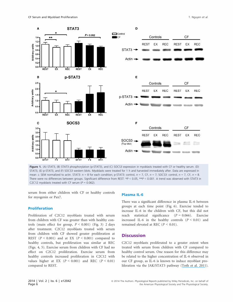
There was no significant difference in STAT3, p-STAT3,
or SOCS3 between C2C12 myoblasts treated with serum
from children with CF or healthy controls (Fig. 1). There
was no exercise effect observed with any of the protein
markers in C2C12 myoblasts treated with serum from
children with CF, although a trend toward a decrease in
STAT3 at REC compared to REST was observed
(P = 0.062). An exercise effect was observed for STAT3
and p-STAT3 expression in C2C12 myoblasts treated
with healthy serum; STAT3 decreased with EX (P < 0.01)
and REC (P < 0.05) compared to REST. Expression of
p-STAT3 decreased in C1C12 myoblasts treated with
healthy serum at REC (P < 0.05) compared with REST,
while EX had no effect of p-STAT3.
mRNA expression
Similar gene expression of SOCS3, Pax7, and myogenin
were observed in C2C12 myoblasts treated with serum
from children with CF or healthy controls (Fig. 2). In
C2C12 myoblasts treated with serum from children with
CF, a trend for an exercise effect for SOCS3 was observed
(one-way ANOVA, time effect, P = 0.065). In addition, a
reduction in gene expression of SOCS3 at REC from
REST (P = 0.058) (Fig. 2A) was observed. No exercise
effect was observed in C2C12 myoblasts treated with
ª 2014 The Authors. Physiological Reports published by Wiley Periodicals, Inc. on behalf ofthe American Physiological Society and The Physiological Society.
2014 | Vol. 2 | Iss. 6 | e12042Page 5
T. Nguyen et al. CF Serum and Myoblast Proliferation
serum from either children with CF or healthy controls
for myogenin or Pax7.
Proliferation
Proliferation of C2C12 myoblasts treated with serum
from children with CF was greater than with healthy con-
trols (main effect for group, P < 0.001) (Fig. 3) 2 days
after treatment. C2C12 myoblasts treated with serum
from children with CF showed greater proliferation at
REST (P < 0.001) and at EX (P < 0.001) compared to
healthy controls, but proliferation was similar at REC
(Figs. 4, 5). Exercise serum from children with CF had no
effect on C2C12 proliferation. Exercise serum from
healthy controls increased proliferation in C2C12 with
values higher at EX (P < 0.001) and REC (P < 0.01)
compared to REST.
Plasma IL-6
There was a significant difference in plasma IL-6 between
groups at each time point (Fig. 6). Exercise tended to
increase IL-6 in the children with CF, but this did not
reach statistical significance (P = 0.066). Exercise
increased IL-6 in the healthy controls (P < 0.01) and
remained elevated at REC (P < 0.01).
Discussion
C2C12 myoblasts proliferated to a greater extent when
treated with serum from children with CF compared to
healthy control serum. One reason for this difference may
be related to the higher concentration of IL-6 observed in
our CF group, as IL-6 is known to induce myoblast pro-
liferation via the JAK/STAT3 pathway (Toth et al. 2011).
Figure 1. (A) STAT3, (B) STAT3 phosphorylation (p-STAT3), and (C) SOCS3 expression in myoblasts treated with CF or healthy serum. (D)
STAT3, (E) p-STAT3, and (F) SOCS3 western blots. Myoblasts were treated for 1 h and harvested immediately after. Data are expressed in
mean � SEM normalized to actin. STAT3: n = 8 for each condition; p-STAT3: control, n = 7, CF, n = 7; SOCS3: control, n = 7, CF, n = 8.
There were no differences between groups. Significant difference from REST: *P < 0.05, **P < 0.001. A trend was observed with STAT3 in
C2C12 myoblasts treated with CF serum (P = 0.062).
2014 | Vol. 2 | Iss. 6 | e12042Page 6
ª 2014 The Authors. Physiological Reports published by Wiley Periodicals, Inc. on behalf of
the American Physiological Society and The Physiological Society.
CF Serum and Myoblast Proliferation T. Nguyen et al.

According to the literature, other systemic factors may be
playing a role in myoblast proliferation. IL-1 has the
capacity to induce greater myoblast proliferation (Otis
et al. 2014) and while we did not measure IL-1 in our
participants other studies have reported higher systemic
levels in patients with CF (Greally et al. 1993). Therefore,
although a higher concentration of IL-6 in our CF serum
may be responsible for inducing greater proliferation,
other inflammatory mediators may also be involved.
While some mediators can induce greater myoblast
proliferation, others can inhibit proliferation. Reduced
oxidative stress has been shown to increased myoblast
proliferation (Zaccagnini et al. 2007), suggesting that high
oxidative stress would result in lower myoblast prolifera-
tion. In addition, NSAIDs (Mikkelsen et al. 1985) can
inhibit myoblast proliferation and corticosteroid (te Pas
et al. 2000) can reduce the rate of proliferation during
the early stages of proliferation. We did not measure oxi-
dative stress in our CF patients, however, increased oxida-
tive stress is observed in CF patients compared to healthy
subjects (Reid et al. 2007). In addition, six of our patients
with CF were on NSAIDs and/or inhaled or nasal spray
corticosteroids, and the effects of NSAIDs were not
accounted for in our study. Despite the known effects of
oxidative stress, NSAIDs and corticosteroids on reducing
proliferation, our myoblasts treated with CF had greater
proliferation. Pooling our samples may have diluted the
concentrations of NSAIDs and corticosteroids and
reduced the inhibitory effects on proliferation. Another
scenario may be that the systemic factors that stimulate
myoblast proliferation were much more potent than the
inhibitory effects of oxidative stress, NSAIDs, and corti-
costeroids.
The effects of exercise on circulating inflammatory
cytokines in children with and without CF (n = 12 for
Figure 2. Gene expression expressed as fold change � SD from
REST for (A) SOCS3, (B) Pax7, and (C) myogenin in C2C12
myoblasts treated with CF serum or control serum. Myoblasts were
treated for 1 h and harvested immediately after. C2C12 myoblasts
were treated with serum from REST: before exercise, EX: after
60 min of cycling, REC: after 60 min of recovery. n = 3 for each
condition. In C2C12 myoblasts treated with CF serum a trend
(P = 0.058) toward reduced SOCS3 gene expression at REC from
REST was observed.
Figure 3. Myoblast proliferation. Average number of C2C12 cells
treated with either CF or control serum. Data are expressed as
mean � SD. Wells were seeded with 1000 myoblasts in 100-lL
growth media and allowed to adhere for 24 h. Myoblasts were
then treated for 1 h with treatment media and allowed to
proliferate in growth media for 2 days. *Significant difference
between groups, P < 0.001.
ª 2014 The Authors. Physiological Reports published by Wiley Periodicals, Inc. on behalf ofthe American Physiological Society and The Physiological Society.
2014 | Vol. 2 | Iss. 6 | e12042Page 7
T. Nguyen et al. CF Serum and Myoblast Proliferation

each group) from this study has been published elsewhere
(Nguyen et al. 2012). While there were no exercise-related
changes in proliferation for myoblast treated with CF
serum, differences were observed with healthy control
serum. More specifically, the EX and REC serum from
controls caused greater proliferation compared to REST
serum. Given that IL-6 concentrations were higher at
these time points in healthy controls only, it is plausible
that the proliferative property of IL-6 may also explain
increased C2C12 myoblasts proliferation in this group.
Conversely, the lack of increase in myoblast proliferation
with EX and REC serum from children with CF may be
attributed to a ceiling effect. Systemic concentrations of
IL-6 were already much higher in the CF group at REST,
and did not substantially increase at EX or REC. Addi-
tional experiments are required to determine the true role
of IL-6 in myoblast proliferation in this context. How-
ever, our data are novel as they highlight that serum from
patients suffering from a systemic inflammatory disease
can cause greater C2C12 myoblast proliferation. Further-
more, EX and REC serum from children with CF does
Figure 4. Photographs of myoblasts treated with serum from children with CF or healthy controls. Wells were seeded with 1000 myoblasts in
100-lL growth media and allowed to adhere for 24 h. Myoblasts were then treated for 1 h with treatment media and allowed to proliferate in
growth media for 2 days. Myoblasts nuclei were stained with DAPI. REST: before exercise, EX: after 60 min of cycling, REC: after 60 min of
recovery. n = 12 for each conditions.
Figure 5. Effects of serum from children with CF and healthy
controls on myoblast proliferation. Wells were seeded with 1000
myoblasts in 100-lL growth media and allowed to adhere for 24 h.
Myoblasts were then treated with treatment media for 1 h and
allowed to proliferate in growth media for 2 days. Mean � SD
number of nuclei. REST: before exercise, EX: after 60 min of
cycling, REC: after 60 min of recovery. n = 12 for each conditions.
*Significant difference between groups, P < 0.001. **Significant
difference compared to REST, P < 0.01.
Figure 6. Effects of exercise on systemic IL-6 in children with CF
and controls. Data are expressed in mean � SEM. REST: before
exercise, EX: after 60 min of cycling, REC: after 60 min of recovery.
n = 11 for each condition. *Significant difference between groups,
P < 0.05. **Significant difference compared to REST, P < 0.01.
2014 | Vol. 2 | Iss. 6 | e12042Page 8
ª 2014 The Authors. Physiological Reports published by Wiley Periodicals, Inc. on behalf of
the American Physiological Society and The Physiological Society.
CF Serum and Myoblast Proliferation T. Nguyen et al.

not alter this proliferative response, while EX and REC
serum from healthy controls enhances it.
In this study, C2C12 myoblasts proliferated to a similar
extent when exposed to serum from children with CF
(either with REST, EX, or REC) and REC serum from
healthy controls. This suggests that in healthy children,
the systemic environment created during the immediate
recovery period following exercise induces similar effects
on C2C12 myoblast proliferation as CF serum. An
increase in myoblast proliferation is required for differen-
tiation (Thomas et al. 2000); however, an increase in pro-
liferation in C2C12 myoblasts when treated with an
inflammatory stimulus results in reduced capacity to dif-
ferentiate (Dogra et al. 2006). In children with CF, given
their chronic inflammatory state (Tirakitsoontorn et al.
2001; Nguyen et al. 2012), it is conceivable that the
increased proliferation observed with CF serum would be
conducive to impaired differentiation leading to impaired
muscle development. Indeed, young rats given chronic
IL-6 exposure experienced 13% reduced muscle growth
(Bodell et al. 2009). However, it is difficult to speculate
as to whether the observed increase in proliferation with
recovery serum from healthy children is conducive to
greater or impaired muscle development given that (1)
the levels of IL-6 observed in healthy children were signif-
icantly lower than the CF group at all time points includ-
ing at the recovery time point, (2) the elevated
inflammatory state is acute, and (3) the inflammatory
state was induced by exercise and not by a chronic infec-
tion or disease. Thus, to further investigate the effects of
increased proliferation observed on muscle development,
examining the effects of myoblast differentiation after
serum exposure would be the next logical step. Although
in our study, mRNA markers of proliferation and differ-
entiation (Pax7, SOCS3, and myogenin) were unaffected
by exposure to serum, further work should measure dif-
ferentiation phenotype (e.g., myoblast fusion index) of
C2C12 myoblasts exposed to different systemic environ-
ments. On the basis of our data, we would expect C2C12
differentiation to decrease upon exposure to serum from
children with CF.
Systemic factors from healthy children following exer-
cise decreased protein signaling involved in C2C12 myo-
blast proliferation. Specifically, STAT3 and p-STAT3
decreased in myoblasts treated with EX and REC serum
from healthy controls, compared to REST serum. We
expected these results to translate into less proliferation
with myoblasts treated with EX and REC serum in our
phenotype experiments. However, we observed the oppo-
site effect with an increase in proliferation. These discor-
dant findings may be due to timing, since protein
samples were collected immediately following serum treat-
ment, while the phenotype experiments were performed
on samples collected 2 days after serum treatment. Our
results may suggest that either (1) systemic factors pro-
moted an acute reduction in proliferation signaling in
C2C12 myoblasts, with a later increase in proliferation
phenotype; or (2) the JAK/STAT3 pathway is not respon-
sible for the increased proliferation phenotype observed.
Our study focused on comparing the effects of systemic
factors in children with CF and healthy controls by using
a common target tissue (C2C12 myoblasts). We acknowl-
edge that the use of human serum on a mouse cell line
may limit the application of our results since we investi-
gated the effects of systemic factors from one species on
another species’ tissue. Because the use of human serum
on tissue is a novel approach, we sought to insure the use
of serum on a tissue was feasible and that differences
would be apparent. We used the C2C12 cell line because
relative to human primary or human cell lines C2C12
myoblasts are more proficient in growth, proliferation,
and relatively inexpensive. Human cells are known to be
difficult to grow, slow to proliferation, and expensive.
Additionally, it is common to grow and proliferate
C2C12 myoblasts using fetal bovine serum, and to differ-
entiate using horse serum. Thus, using serum from a spe-
cies other than from a mouse on C2C12 myoblasts is
common practice. Given the feasibility of our study, we
plan to pursue the use of a human cell line or human
primary cells in the future. Furthermore, since the CFTR
protein is expressed in skeletal muscle (Lamhonwah et al.
2010), future work should also include skeletal muscle
from a CF model to provide clearer insight into the rela-
tive roles of the systemic environment and local muscle
factors in skeletal muscle development in children with
CF. Finally, we pooled our serum samples in order to
conserve sample volume and to insure the completion of
all other analyses set forth, since pediatric blood samples
are difficult to obtain and standard ethical procedure
inhibits the collection of blood to a certain amount. Pool-
ing our samples is a limitation of this study.
Conclusion
We took the novel approach of exposing C2C12 myo-
blasts to serum obtained from children with CF and
healthy controls. We found that C2C12 myoblast prolifer-
ation was greater when treated with CF serum than con-
trol serum. In addition, proliferation did not differ
between REST, EX, or REC CF serum, while an exercise
effect was observed in healthy controls. Protein and
mRNA markers of proliferation did not increase in
C2C12 myoblasts treated with serum from children with
CF or healthy controls. This work highlights the ability of
systemic factors from patients with an inflammatory dis-
ease to alter aspects of skeletal muscle development.
ª 2014 The Authors. Physiological Reports published by Wiley Periodicals, Inc. on behalf ofthe American Physiological Society and The Physiological Society.
2014 | Vol. 2 | Iss. 6 | e12042Page 9
T. Nguyen et al. CF Serum and Myoblast Proliferation

Acknowledgment
We would like to express our sincere appreciation to the
participants and families involved for their dedication to
the study. A special thanks to Valerie Carroll (Nurse
Coordinator) and to the McMaster Pediatric CF Clinic
for their assistance with patient recruitment.
Conflict of Interest
None declared.
References
Andres, V., and K. Walsh. 1996. Myogenin expression, cell
cycle withdrawal, and phenotypic differentiation are
temporally separable events that precede cell fusion upon
myogenesis. J. Cell Biol. 132:657–666.
Bodell, P. W., E. Kodesh, F. Haddad, F. P. Zaldivar,
D. M. Cooper, and G. R. Adams. 2009. Skeletal muscle
growth in young rats is inhibited by chronic exposure to
IL-6 but preserved by concurrent voluntary endurance
exercise. J. Appl. Physiol. 106:443–453.
Buckingham, M. 2007. Skeletal muscle progenitor cells and the
role of Pax genes. C. R. Biol. 330:530–533.
Burattini, S., P. Ferri, M. Battistelli, R. Curci, F. Luchetti, and
E. Falcieri. 2004. C2C12 murine myoblasts as a model of
skeletal muscle development: morpho-functional
characterization. Eur. J. Histochem. 48:223–233.
Centers for Disease Control and Prevention. 2009. Percentile
Data Files with LMS. Available at http://www.cdc.gov/
growthcharts/percentile_data_files.htm (cited April 30,
2014)
Croker, B. A., H. Kiu, and S. E. Nicholson. 2008. SOCS
regulation of the JAK/STAT signalling pathway. Semin. Cell
Dev. Biol. 19:414–422.
Diao, Y., X. Wang, and Z. Wu. 2009. SOCS1, SOCS3, and
PIAS1 promote myogenic differentiation by inhibiting the
leukemia inhibitory factor-induced JAK1/STAT1/STAT3
pathway. Mol. Cell. Biol. 29:5084–5093.
Dill, D. B., and D. L. Costill. 1974. Calculation of percentage
changes in volumes of blood, plasma, and red cells in
dehydration. J. Appl. Physiol. 37:247–248.
Dogra, C., H. Changotra, S. Mohan, and A. Kumar. 2006.
Tumor necrosis factor-like weak inducer of apoptosis
inhibits skeletal myogenesis through sustained activation of
nuclear factor-kappaB and degradation of MyoD protein.
J. Biol. Chem. 281:10327–10336. 4-14-2006.
Frost, R. A., G. J. Nystrom, and C. H. Lang. 2002. Regulation
of IGF-I mRNA and signal transducers and activators of
transcription-3 and -5 (Stat-3 and -5) by GH in C2C12
myoblasts. Endocrinology 143:492–503.
Greally, P., M. J. Hussain, D. Vergani, and J. F. Price. 1993.
Serum interleukin-1 alpha and soluble interleukin-2
receptor concentrations in cystic fibrosis. Arch. Dis. Child.
68:785–787.
van Heeckeren, A. M., J. Tscheikuna, R. W. Walenga,
M. W. Konstan, P. B. Davis, B. Erokwu, et al. 2000. Effect
of Pseudomonas infection on weight loss, lung mechanics,
and cytokines in mice. Am. J. Respir. Crit. Care Med.
161:271–279.
Ionescu, A. A., L. S. Nixon, S. Luzio, V. Lewis-Jenkins,
W. D. Evans, M. D. Stone, et al. 2002. Pulmonary function,
body composition, and protein catabolism in adults with
cystic fibrosis. Am. J. Respir. Crit. Care Med. 165:495–500.
Ionescu, A. A., L. S. Nixon, and D. J. Shale. 2004. Cellular
proteolysis and systemic inflammation during exacerbation
in cystic fibrosis. J. Cyst. Fibros. 3:253–258.
Lamhonwah, A. M., C. E. Bear, L. J. Huan, C. P. Kim,
C. A. Ackerley, and I. Tein. 2010. Cystic fibrosis
transmembrane conductance regulator in human muscle:
dysfunction causes abnormal metabolic recovery in exercise.
Ann. Neurol. 67:802–808.
Mikkelsen, U. R., H. Langberg, I. C. Helmark, D. Skovgaard,
L. L. Andersen, M. Kjaer, et al. 1985. Local NSAID infusion
inhibits satellite cell proliferation in human skeletal muscle
after eccentric exercise. J. Appl. Physiol. 107:2009.
Mirwald, R. L., A. D. Baxter-Jones, D. A. Bailey, and
G. P. Beunen. 2002. An assessment of maturity from
anthropometric measurements. Med. Sci. Sports Exerc.
34:689–694.
Nguyen, T., J. Obeid, H. E. Ploeger, T. Takken, L. Pedder, and
B. W. Timmons. 2012. Inflammatory and growth factor
response to continuous and intermittent exercise in youth
with cystic fibrosis. J. Cyst. Fibros. 11:108–118.
Otis, J. S., S. Niccoli, N. Hawdon, J. L. Sarvas, M. A. Frye,
A. J. Chicco, et al. 2014. Pro-inflammatory mediation of
myoblast proliferation. PLoS One 9:e92363.
te Pas, M. F., P. R. de Jong, and F. J. Verburg. 2000.
Glucocorticoid inhibition of C2C12 proliferation rate and
differentiation capacity in relation to mRNA levels of the
MRF gene family. Mol. Biol. Rep. 27:87–98.
Peault, B., M. Rudnicki, Y. Torrente, G. Cossu, J. P. Tremblay,
T. Partridge, et al. 2007. Stem and progenitor cells in
skeletal muscle development, maintenance, and therapy.
Mol. Ther. 15:867–877.
Petersen, A. M., and B. K. Pedersen. 2005. The
anti-inflammatory effect of exercise. J. Appl. Physiol.
98:1154–1162.
Reid, D. W., N. Misso, S. Aggarwal, P. J. Thompson, and
E. H. Walters. 2007. Oxidative stress and lipid-derived
inflammatory mediators during acute exacerbations of cystic
fibrosis. Respirology 12:63–69.
Sadowski, C. L., T. T. Wheeler, L. H. Wang, and H. B. Sadowski.
2001. GH regulation of IGF-I and suppressor of cytokine
signaling gene expression in C2C12 skeletal muscle cells.
Endocrinology 142:3890–3900.
2014 | Vol. 2 | Iss. 6 | e12042Page 10
ª 2014 The Authors. Physiological Reports published by Wiley Periodicals, Inc. on behalf of
the American Physiological Society and The Physiological Society.
CF Serum and Myoblast Proliferation T. Nguyen et al.

Schaefer, F., M. Georgi, A. Zieger, and K. Scharer. 1994.
Usefulness of bioelectric impedance and skinfold
measurements in predicting fat-free mass derived from
total body potassium in children. Pediatr. Res. 35:
617–624.
Schmittgen, T. D., and K. J. Livak. 2008. Analyzing real-time
PCR data by the comparative C(T) method. Nat. Protoc.
3:1101–1108.
Spangenburg, E. E., and F. W. Booth. 2002. Multiple signaling
pathways mediate LIF-induced skeletal muscle satellite cell
proliferation. Am. J. Physiol. Cell Physiol. 283:C204–C211.
Thomas, M., B. Langley, C. Berry, M. Sharma, S. Kirk, J. Bass,
et al. 2000. Myostatin, a negative regulator of muscle
growth, functions by inhibiting myoblast proliferation.
J. Biol. Chem. 275:40235–40243.
Tirakitsoontorn, P., E. Nussbaum, C. Moser, M. Hill, and
D. M. Cooper. 2001. Fitness, acute exercise, and anabolic
and catabolic mediators in cystic fibrosis. Am. J. Respir.
Crit. Care Med. 164:1432–1437.
Toth, K. G., B. R. McKay, L. M. De, J. P. Little,
M. A. Tarnopolsky, and G. Parise. 2011. IL-6 induced
STAT3 signalling is associated with the proliferation of
human muscle satellite cells following acute muscle damage.
PLoS One 6:e17392.
Tremblay, M. S., D. E. Warburton, I. Janssen, D. H. Paterson,
A. E. Latimer, R. E. Rhodes, et al. 2011. New Canadian
physical activity guidelines. Appl. Physiol. Nutr. Metab.
36:36–46.
Valletta, E. A., A. Rigo, L. Bonazzi, L. Zanolla, and
G. Mastella. 1992. Modification of some markers of
inflammation during treatment for acute respiratory
exacerbation in cystic fibrosis. Acta Paediatr. 81:227–230.
Wang, X., D. W. Dockery, D. Wypij, M. E. Fay, and
B. G. Ferris Jr. 1993. Pulmonary function between 6 and
18 years of age. Pediatr. Pulmonol. 15:75–88.
World Health Organization. 2013. Global Strategy on Diet,
Physical Activity and Health: Physical Activity and Young
People. Available at http://www.who.int/dietphysicalactivity/
factsheet_young_people/en/index.html (cited August 30,
2013).
Yaffe, D., and O. Saxel. 1977. Serial passaging and
differentiation of myogenic cells isolated from dystrophic
mouse muscle. Nature 270:725–727.
Zaccagnini, G., F. Martelli, A. Magenta, C. Cencioni,
P. Fasanaro, C. Nicoletti, et al. 2007. p66(ShcA) and
oxidative stress modulate myogenic differentiation and
skeletal muscle regeneration after hind limb ischemia.
J. Biol. Chem. 282:31453–31459.
ª 2014 The Authors. Physiological Reports published by Wiley Periodicals, Inc. on behalf ofthe American Physiological Society and The Physiological Society.
2014 | Vol. 2 | Iss. 6 | e12042Page 11
T. Nguyen et al. CF Serum and Myoblast Proliferation
Copyright © 2022 FDOKUMEN