The Effectiveness of Hand Splinting to Prevent Muscle ...
270
I The Effectiveness of Hand Splinting to Prevent Muscle Contracture Following Acquired Brain Impairment N.A. Lannin Doctorate of Philosophy 2006 University of Western Sydney
-
Upload
khangminh22 -
Category
Documents
-
view
3 -
download
0
Transcript of The Effectiveness of Hand Splinting to Prevent Muscle ...

I
The Effectiveness of Hand Splinting to
Prevent Muscle Contracture Following
Acquired Brain Impairment
N.A. Lannin Doctorate of Philosophy
2006
University of Western Sydney

II
Acknowledgements I owe so much to my supervisors, Professor Anne Cusick, Dr Annie McCluskey, and Dr
Robert Herbert who have been the ‘godfathers’ of this research from its beginning. Their
sympathetic encouragement and good judgement have both inspired me and steadied the
project from start to finish and their acceptance of this thesis is its best recommendation.
I am grateful to the participants who took part in the trials. Your commitment and
enthusiasm encourages me still. Thank you.
I wish to express my appreciation to my colleagues for their assistance during the clinical
trials. In particular I would like to recognise the roles of Sally Horsley during Study 2 and of
Rachael McQueen, Belinda Armstrong, Annemieke Clark, Nadia Downey, Merilyn Ferris,
Glade Vyslysel, Michelle Turnbull, Valerie Webster, Melani Boyce, Joanne Glinsky, Jan
Hancock, Jenni Johnson, Alison Pearce, Natalie Fairbairn and Lauren Wade during Study 3.
I thank them for their commitment and professionalism during their involvement with this
project.
The financial assistance of a University of Western Sydney Postgraduate Research Award
towards this research is hereby acknowledged. Financial support for the studies was also
provided through part-time employment during my candidature by the Dean’s Unit (College
of Health and Science) and the School of Exercise and Health Science, University of
Western Sydney, and by the School of Physiotherapy, University of Sydney.
The faculties of the School of Exercise and Health Science at University of Western Sydney
and the School of Physiotherapy at University of Sydney, and my colleagues have provided
ongoing interest and support throughout this project. I thank them for helping place this
work in context and their generosity with time. A special word of thanks must go to Julia
Bowman for her friendship and practical assistance throughout this journey.
Finally, I acknowledge the ongoing support of my family; thank you for encouraging me
through all my academic efforts. This is the culmination. Thank you seems inadequate,
however, to show my appreciation for my husband, Ben, for the advice he gave me on points
about which I consulted him, for allowing me to spend countless hours in the office, and for
never doubting that the end was in sight.

III
Statement of Authentication
The work presented in this thesis is, to the best of my knowledge and belief, original except
as acknowledged in the text. I hereby declare that I have not submitted this material, either
in full or in part, for a degree at this or any other institution.
……… ………
(Signature)

IV
Table of Contents
Chapter One: Introduction ...............................................................................................1 1.1 Background................................................................................................2 1.2 Context of the Study..................................................................................2 1.3 Research Aims and Objectives..................................................................6 1.4 Scope of the Study.....................................................................................8 1.5 Definition of Terms ...................................................................................9 1.6 Synopsis...................................................................................................13
Chapter Two: Literature Review ...................................................................................15
2.1 Introduction .............................................................................................16 2.2 Adults with Acquired Brain Impairment.................................................17 2.3 Neuromusculoskeletal changes after acquired brain impairment............18 2.4 The Wrist .................................................................................................22 2.5 Occupational Therapy for adults with contracture following acquired
brain impairment .....................................................................................23 2.6 The Prevention of Contracture ................................................................28 2.7 Measurement Issues.................................................................................43 2.8 Key Issues to Emerge from the Literature Review .................................53 2.9 Synopsis...................................................................................................57 Chapter Three: A Systematic Review ............................................................................58 3.1 Chapter Overview....................................................................................59 3.2 Background to the Study .........................................................................59 3.3 Rationale for the Study Design................................................................61 3.4 Aim of the Study .....................................................................................62 3.5 Method.....................................................................................................62 3.6 Results .....................................................................................................69 3.7 Discussion................................................................................................71 3.8 Synopsis...................................................................................................76
Chapter Four: A Randomised Controlled Trial Comparing Splinting plus Stretch to
Stretch Alone in Adults with Hemiplegia following Acquired Brain
Impairment ............................................................................................82 4.1 Chapter Overview....................................................................................83 4.2 Background..............................................................................................83 4.3 Description and Rationale for Study Design ...........................................84 4.4 Aim..........................................................................................................85 4.5 Research Question ...................................................................................85 4.6 Method.....................................................................................................85 4.7 Results .....................................................................................................96 4.8 Discussion..............................................................................................103 4.9 Conclusion from the Study....................................................................109 4.10 Synopsis.................................................................................................109

V
Chapter Five: Effectiveness of two types of hand splints for preventing contractures
in adults who are not receiving hand stretches post-stroke: a single
blind, randomised controlled trial .....................................................111 5.1 Chapter Overview..................................................................................112 5.2 Background............................................................................................112 5.3 Aim........................................................................................................113 5.4 Research Questions ...............................................................................113 5.5 Description and Rationale for Study Design .........................................113 5.6 Method...................................................................................................114 5.7 Results ...................................................................................................128 5.8 Discussion..............................................................................................140 5.9 Conclusion.............................................................................................147 5.10 Synopsis.................................................................................................147
Chapter Six: Discussion .................................................................................................148 6.1 Introduction ...........................................................................................149 6.2 Key Findings .........................................................................................151 6.3 Strengths and Limitations......................................................................154 6.4 Recommendations and Implications......................................................159 6.5 Conclusion.............................................................................................169 References 170 Appendices 218

VI
List of Tables
Table 3.1 Electronic search strategy........................................................................63 Table 3.2 Oxford Centre for Evidence-Based Medicine levels of evidence ...........65 Table 3.3 PEDro scoring criteria and implementation guidelines...........................68 Table 3.4 Methodological rating of randomised controlled trials...........................70 Table 3.5 Summary of studies – interventions and participants..............................78 Table 3.6 Summary of studies – research methods .................................................80 Table 4.1 Upper Limb Subscale of the Motor Assessment Scale (UL-MAS) item 6, 7 and 8......................................................................................................95 Table 4.2 Baseline characteristics ...........................................................................97 Table 4.3 Numbers of participants in each group who did not adhere to the protocol
.................................................................................................................99
Table 4.4 Baseline performance of groups on dependent variables ........................99 Table 4.5 Compliance with splinting program ......................................................103 Table 5.1 Baseline characteristics of groups .........................................................130 Table 5.2 Baseline performance of groups on dependent variables ......................133 Table 5.3 Numbers of participants in each group who did not adhere to the protocol
...............................................................................................................134 Table 5.4 Compliance with splinting protocol ......................................................139

VII
Figures and Illustrations
Figure 2.1 Wrist splint illustrating three points of pressure......................................39
Figure 2.2 Static Palmar-Mitt Splint.........................................................................40
Figure 2.3 Device used to measure extensibility of the long finger flexor muscles .46
Figure 4.1 A seated weight-bearing stretch of the upper limb..................................89
Figure 4.2 A seated stretch of the upper limb demonstrating the use of an air splint.................................................................................................................90
Figure 4.3 Static palmar-mitt splint with the wrist and fingers positioned in the
‘functional position’ ................................................................................91
Figure 4.4 Participant flow diagram .......................................................................100
Figure 4.5 Mean (95% confidence intervals) of differences between torque-controlled wrist extension range of motion of the splint and control groups. ...................................................................................................101
Figure 5.1 Static palmar-mitt splint which positioned the wrist and fingers in the
‘functional position’ ..............................................................................119
Figure 5.2 Static palmar-mitt splint with the wrist and fingers positioned in extension................................................................................................120
Figure 5.3 Participant flow diagram .......................................................................132
Figure 5.4 Mean (95% Confidence Interval) of differences between torque-
controlled wrist extension range of motion of the control, functional splint and extension splint groups .........................................................135

VIII
Abbreviations
Acronym/Abbreviation Meaning
AAOS American Academy of Orthopedic Surgery
ANCOVA Analysis of Covariance
CEBP Centre for Evidence Based Physiotherapy
CI Confidence Interval
cm Centimetre
CVA Cerebrovascular Accident; stroke
DASH Disabilities of the Arm, Shoulder, and Hand Questionnaire
FMA Fugl-Meyer Assessment
ICC Intraclass Correlation Coefficient
ICIDH International Classification of Impairments, Disabilities and Handicaps
LOCF Last observation carried forward
MAS Motor Assessment Scale
MMSE Mini Mental State Examination
n Number (sample size)
OT Australia Australian occupational therapy professional association
OTA American Occupational Therapy Association
p Probability
PEDro Physiotherapy Evidence Database
r Pearson’s product-moment correlation coefficient
ROM Range of Motion
SD Standard Deviation
UL-MAS Upper Limb subscale of Motor Assessment Scale
US United States of America

IX
Abstract
Aim: The aim of the thesis was to evaluate the effectiveness of static hand splints for the
prevention of muscle contracture during early rehabilitation following acquired brain
impairment. Design: Three studies were undertaken and are reported in this thesis. The aim
of the first study was to appraise the existing research on the effects of hand splinting for
adults with hemiplegia following acquired brain impairment. A systematic review and
methodological critique of published scientific literature were conducted. The aim of the
second study was to evaluate the effectiveness of static hand splints which position the wrist
and fingers in the common ‘functional position’ when provided in conjunction with a
rehabilitation program which included daily motor training and prolonged stretches. This
study was an assessor-blinded randomised controlled trial. Study 3 was also an assessor-
blinded randomised trial. The aim of the final study was to evaluate the effectiveness of two
hand splinting positions, the ‘functional position’ and a position of wrist and finger
extension, in comparison to a control group that did not receive prolonged stretches.
Setting: Studies two and three were conducted in inpatient rehabilitation centres in
Australia. Participants: In the second study, 28 adults who had hemiplegia following
acquired brain impairment were randomly allocated to receive either a static palmar hand
splint which positioned their wrist in a ‘functional position’, or the control group. Both
groups in Study 2 received one hour of upper limb stretches and approximately 30 minutes
of upper limb motor training five days per week. In the third study, 63 adults with
hemiplegia following stroke were randomly allocated to receive a hand splint which
positioned their wrist and fingers in either a ‘functional position’ or with their wrist and
fingers in an extended position, or to be in the control group. In Study 3, none of the groups
received prolonged stretches to their wrist or finger flexor muscles for the duration of the
study. Outcome measures: The primary outcome across both randomised controlled trials

X
(studies two and three) was extensibility of the wrist and finger flexor muscles as estimated
by torque-controlled range of movement of the wrist joint with the fingers held in extension.
A device designed to measure extensibility of the wrist and finger flexor muscles was used
in both studies. Secondary outcome measures included the upper limb subscale of the Motor
Assessment Scale (UL-MAS), visual analogue scale of pain, the Tardieu scale, and the
Disabilities of Arm, Shoulder and Hand (DASH) questionnaire. Results: The results of the
systematic review indicated that there were no randomised controlled trials which
investigated the effect of static hand splints on the prevention of contracture in the finger
and wrist flexor muscles, and that the majority of evidence to date has been of low quality.
Study 2, a randomised controlled trial, indicated that wearing a hand splint in addition to
participating in upper limb stretches and motor training for four weeks did not improve wrist
extensibility. Compared to the control group (stretch alone), wearing the splint increased
wrist extension by a mean of 1.0º (95% CI -3.7º to 6.1º). Study 3, also a randomised
controlled trial, indicated that wearing a hand splint with the wrist and fingers in either the
‘functional position’ or maximum extension for four weeks did not improve wrist
extensibility. Compared to the control group (no stretch), wearing the ‘functional’ splint
increased wrist extension by a mean of 1.5º (95% CI -5.7º to 8.7º); and wearing the
extension splint reduced wrist extension by a mean of 2.5º (95% CI -9.6º to 4.6º).
Conclusion: Findings indicate that splinting the hand in the ‘functional’ position or in a
position of greater wrist extension did not prevent contracture following acquired brain
impairment over the course of the study periods (four weeks with follow up at 4 and 6 weeks
in studies 2 and 3 respectively).

Chapter 1 Introduction
1
CHAPTER ONE
Introduction

Chapter 1 Introduction
2
Chapter One: Introduction
1.1 Background
Hand splints are often used to provide a stretch to hemiplegic muscles, however therapists
disagree about the value of providing this intervention. What little research is available
represents low-level evidence which cannot answer the question of whether or not splints are
worthwhile. This research was motivated by a desire to evaluate the efficacy of hand
splinting after brain impairment.
1.2 Context of the Study
Acquired brain impairment is one of a number of terms used to describe impairment due to
injury to the brain (Collins & Dean, 2002; Stanton, Jessop, & Henstridge, 1994). Acquired
brain impairment caused by a stroke or injury and may lead to a range of disabilities, one of
the most common being loss of hand and arm movement (Scottish Intercollegiate Guidelines
Network, 2002). Some recovery of movement typically occurs in people who survive
acquired brain impairment, but often the secondary complication of muscle contracture
(Gracies, 2001) limits rehabilitation and reduces functional outcomes.
Both the Commonwealth Department of Health (Priorities and Quality Branch
Commonwealth Department of Health and Ageing, 2002) and the World Health
Organisation (1982) have highlighted a growing concern about the management of adults
with acquired brain impairment. One particular area of concern is the use of allied health
treatments to address hemiplegia (National Health and Medical Research Council, 2000).
Approximately 70 to 85% of people with acquired brain impairment experience hemiplegia

Chapter 1 Introduction
3
(Sivenius, 1982; Kotila, Waltimo, Niemi, Laaksonen, & Lempinen, 1986) and this
proportion is higher among the elderly (Kalra, Smith, & Crome, 1993). An estimated 48 to
95% of people never regain functional arm or hand use as a result of hemiplegia (Gowland,
1982; Mayo et al., 1999). This loss of hand use is particularly devastating as everyday
activities of life become uniquely challenging.
The scientific literature on management of hemiplegia is inconclusive and provides limited
guidance for practice (Dobkin, 1989; Dromerick, 2003; Jutai & Teasell, 2003; Kwakkel,
Wagenaar, Koelman, Lankhorst, & Koetsier, 1997; Scottish Intercollegiate Guidelines
Network, 2002). Existing studies also lack the statistical power needed to guide clinical
practice (for example, when deciding whether or not to as splint the hemiplegic hand).
Research reported in this thesis was undertaken to address this gap in knowledge. In
particular, research was planned which would examine the efficacy of common
interventions used during rehabilitation to mitigate finger and wrist contractures after brain
impairment.
1.2.1 Contractures Post-Brain Impairment
Apart from the loss of movement, hemiplegia predisposes people to contracture (Farmer &
James, 2001; O’Dwyer & Ada, 1996; Pandyan & Granat, 1997). Contractures are a
shortening of the muscle accompanied by increased resistance to passive stretch (Gordon,
1990; Pandyan & Granat, 1997). Contractures can occur in the elbow, but contractures in the
wrist and fingers are thought to be more common and often more disabling (Pandyan &
Granat, 1997; Yarkony & Sahgal, 1987). Wrist and finger flexor muscle contracture can
limit the range of extension at the wrist, increasing the amount of resistance when the hand
and fingers are stretched. These collective impairments lead to reduced function in the upper
limb.

Chapter 1 Introduction
4
When the wrist and finger joints become contracted following acquired brain impairment, it
becomes more difficult for a person to remain independent (Carr & Shepherd, 1987). For
example, wrist contractures make it difficult to dress a hemiplegic arm independently.
Contractures limit the ability to passively straighten the fingers and place the hand into the
sleeve, and reduce the ability to actively assist to don a shirt, a movement requiring wrist
extension (Bukowski, 2000). Contractures also create unsightly deformities and are thought
to predispose people to spasticity, pressure areas, sleep disruption (Scott & Donovan, 1981;
Yarkony & Sahgal, 1987) and unnecessary pain and distress (Horin & Reardon, 2000).
Contracture is thus considered one of the main complications of acquired brain impairment
and treatment is aimed at preventing contracture in those people with full range of
movement, and reducing the amount in those people with contracture.
1.2.2 Handsplinting for the treatment of Contractures
A number of interventions are used to prevent and treat contracture. These interventions
include: positioning, pharmacology, therapeutic exercise, serial casting and splinting
(Atwood, 1999). Currently, the evidence base for these interventions is limited. It is
therefore open to conjecture whether any of these interventions benefit people with
hemiplegia. This thesis focuses on one intervention in particular: hand splinting.
Hand splinting has been recommended since 1911 to prevent and treat contracture (Neuhaus
et al., 1981). This intervention is used by occupational therapists, orthotists and
physiotherapists, and its use is endorsed by rehabilitation physicians and neurologists (Fess,
2002). Over the past 100 years, therapists have disagreed about whether or not splints are
effective. Even those therapists who promote the use of splints hold differing opinions
regarding mechanisms by which splints prevent and treat contracture (Milazzo & Gillen,
1998; Neuhaus et al., 1981; Ryerson, 2001).

Chapter 1 Introduction
5
1.2.3 Theories which Underpin Hand Splinting
Neuhaus and colleagues (1981) identified two different approaches to treatment for
hemiplegia which have influenced current splinting practices. The first is the biomechanical
approach, which emphasises the prevention or correction of deformity by mechanical
application of splints. The second is the neurophysiological approach which concentrates on
the use of movement and handling to reduce spasticity. With the introduction of the
neurophysiological approach, splinting became ‘inhibitory’ in design and studies began to
test the central tenet of this theoretical approach, which was the ‘inhibition’ of spasticity.
Brennan (1959) stated that splinting decreased flexor tone. Kaplan (1962) stated that dorsal
splinting decreased flexor spasticity. Charait (1968) reported dorsal splinting to be more
effective than volar splinting in decreasing muscle tone. More recently Rose and Shah
(1987) reported that both dorsal and volar splints reduce spasticity. Unfortunately various
limitations in research methodology threatened the internal validity of all of these results, an
issue investigated and discussed in Chapter 3: A Systematic Review.
In recent times allied health professionals have returned to the biomechanical approach
toward splinting. The biomechanical approach is one where a splint is prescribed to reduce
soft-tissue contractures and increase the range of movement. Linden and Trombly (1995),
Gutman (2001), and Milazzo and Gillen (1998) advocate splinting to prevent deformity of
the upper limb and to “maintain or lengthen soft tissues” (Milazzo & Gillen, 1998, 173).
Although leading allied health professionals assert that splints can hold the hand in a
position which prevents contracture by providing a gentle stretch to the muscles and
tendons, no clinical trials have tested this theory (Ashburn, Cornall, Melville, Simpson, &
Wright, 1998).

Chapter 1 Introduction
6
1.2.4 The Gap in Empirical Evidence for Hand Splinting
One major assumption of occupational therapists working in rehabilitation, irrespective of
the underlying theoretical approach, is that contracture can be managed by using a hand
splint (Hill, 1988; Pedretti, Smith & McHugh-Pendleton, 1996; Scott & Dow, 1995; Wilton,
1997; Woodson, 2002). Although the biomechanical theory has gained favour amongst
therapists (Neuhaus et al., 1981), there is no definitive support for the efficacy of this
approach. There have been no randomised controlled trials on contracture splinting for
people with hemiplegia following acquired brain impairment (Milazzo & Gillen, 1998).
Further, no research with this population has focused on muscle contracture as the primary
outcome despite clear evidence that contracture has a major effect on function and is the
primary reason that hand splints are prescribed (Neuhaus et al., 1981). The lack of research
in this area constitutes a major gap in knowledge about the use of hand splints which this
thesis aims to remedy.
1.3 Research Aims and Objectives
The purpose of this program of research is to test the hypothesis that hand splinting produces
a positive effect on the wrist and finger flexor muscles of adults in acute rehabilitation
following acquired brain impairment. The theoretical framework underlying this study is the
biomechanical approach for splinting, first proposed in early 1900s and extended in recent
years by a greater understanding of the neural and mechanical bases for movement (Milazzo
& Gillen, 1998). Central to this theoretical framework is the acceptance that the hand splint
applies a stretch which is thought to prevent contracture in those people who have yet to
develop one and prevent further contracture in those people whose joints have begun to
contract (and potentially reduce the amount of contracture in such cases). The program of
research aims to systematically review and meta-analyse relevant literature (Chapter 3), and

Chapter 1 Introduction
7
then investigate the effectiveness of hand splinting both with and without prolonged
stretching (Chapters 4 and 5).
The program of research will address three studies and five research questions. The studies
will be planned to provide evidence that will fill a knowledge gap and guide clinical practice
decisions regarding the use of hand splints for people with hemiplegia following acquired
brain impairment.
Study 1: A systematic review of splinting for adults with hemiplegia following stroke.
Question 1. Do adults with acquired brain impairment who participate in a splinting
program experience greater function, less contracture, less spasticity and/or
less pain, than those who do not participate in a splinting program?
Study 2: A randomised, controlled trial of splinting for adults with hemiplegia following
acquired brain impairment who receive hand stretches
Question 2. Do adults with acquired brain impairment who wear a hand splint nightly for
four weeks in addition to receiving routine daily upper limb stretches,
experience less contracture and pain, and more function than those who
receive only routine daily stretches?
Study 3: A randomised, controlled trial of splinting in two wrist positions for adults with
hemiplegia following acquired brain impairment who are not receiving hand stretches.
Question 3. Do adults with a recent stroke who wear a hand splint nightly for four weeks
in the ‘functional position’ experience less contracture, spasticity and pain,
and more function than those who do not wear a splint?

Chapter 1 Introduction
8
Question 4. Do adults with a recent stroke who wear a hand splint nightly for four weeks
in a position of greater wrist extension experience less contracture, spasticity
and pain, and more function than those who do not wear a splint?
Question 5. Do adults with a recent stroke experience less contracture, spasticity and
pain, and more function when their wrist is positioned in increased extension
in a splint compared to a ‘functional’ resting splint?
1.4 Scope of the Study
The first study (Chapter 3) is a systematic review designed to summarise the evidence
supporting the use of hand splinting for the stroke population. The systematic review
employs methods to minimise bias in its conclusions (Cook, Mulrow & Haynes, 1997;
Mulrow, 1994; Oxman & Guyatt, 1988). The quality of trials is assessed in an objective
way (Maher, Sherrington, Herbert, Moseley & Elkins, 2003) and the results of studies are
collated. A critical appraisal of the results of the systematic review provides the opportunity
to refine hypotheses, recognise and avoid pitfalls of previous work, estimate sample sizes,
and identify important covariates that warrant consideration in the planned studies (Mulrow,
1994). This thesis and one of its subsequent publications provide an original synthesis and
appraisal of current research in this area (Lannin & Herbert, 2003).
Study 2 (Chapter 4) is a randomised clinical trial designed to determine the effectiveness of
hand splinting in conjunction with prolonged stretching. Specifically, this study examines
the effect of four weeks of nightly hand splinting. The value of this study is that it
investigates a typical clinical question common to therapists working in Australian acute
care hospitals: “if patients are receiving stretches and upper limb rehabilitation daily, is it

Chapter 1 Introduction
9
still beneficial to prescribe a hand splint?” In this thesis (and another subsequent
publication), Study 2 provides evidence that splinting is not clinically beneficial when
provided in conjunction with prolonged stretching (Lannin, Horsley, Herbert, McCluskey, &
Cusick, 2003).
The third study (Chapter 5) is also a randomised controlled trial. The results from study 2
refined questions about the efficacy of hand splinting, and identified that both wrist position
when splinting and use of stretching were factors that need to be controlled. To achieve this,
study 3 used a three-group design that compared not wearing a splint with the effect of four
weeks of night hand splinting in either neutral or maximal wrist extension. Participants in
study 3 did not receive upper limb stretches. In this thesis (and in the final manuscript
submitted for publication), Study 3 provides evidence that splinting in either a neutral
position or a position of wrist extension is not clinically beneficial when compared to a
control group who received no stretching or splinting (Lannin, Cusick, Herbert, &
McCluskey, 2006).
The randomised controlled trials (Chapters 4 and 5) used hand splints and the position of the
wrist in the splints as independent variables. The dependent variables, in order of priority,
were: contracture of the wrist and finger flexor muscles, functional movement of the hand
and arm, spasticity and pain. Time post-impairment was a covariate.
1.5 Definition of Terms
One of the challenges in research related to contracture is the lack of clarity in the definition
of specific terms. Terminology used in this thesis has been defined in the following glossary
and provides an orientation and overview.

Chapter 1 Introduction
10
Acquired Brain Impairment
Acquired brain impairment is non-congenital damage to the brain which results from head
trauma, and non-traumatic brain injuries such as those caused by strokes, tumours, hypoxia,
and neurodegenerative diseases such as Parkinsons’ disease and multiple sclerosis (Collins
& Dean, 2002). Although acquired brain impairment may arise from these multiple causes,
the focus within this thesis will be restricted to people with stroke and head trauma. Since
acquired brain impairment can lead to hemiplegia, which predisposes adults to contracture,
no delineation between stroke and head trauma is made within the literature review.
Head trauma
A traumatic brain injury is an acquired brain impairment caused by a traumatic event,
for example a blow to the head, or from a motor vehicle accident (Sahgal, 1988; Scott
& Dow, 1995). Brain tissue damage results from both the direct injury and from
secondary reactions that contribute to delayed tissue damage and cell death (Frantseva,
Kokarovtseva, Naus, Carlen, MacFabe, & Velazquez, 2002; Laurer, Lenzlinger, &
McIntosh, 2000).
Stroke
A stroke, also known as a cerebrovascular accident (CVA), occurs due to disease of
the cerebral blood supply (Bierman & Atchison, 2000; Heitzner & Teasell, 1998). A
stroke occurs when an artery supplying blood to the brain becomes blocked, cutting
off the blood supply (ischaemic stroke), or when the blood leaks from arteries and into
the brain (haemorrhagic stroke) (Bierman & Atchison, 2000; Stroke Australia
Taskforce, 1996; Woodson, 2002). Although there are varied causes and types of
stroke, all lead to tissue anoxia due to a cessation of cerebral blood flow (Pedretti,
Smith & McHugh-Pendleton, 1996; Woodson, 2002).

Chapter 1 Introduction
11
Hemiplegia
Damage to the brain may disrupt the function of several descending neurological pathways,
including the corticospinal pathway, which is responsible for executing muscle movement.
Such damage results in a reduced ability of the brain to activate the muscles and leaves the
muscles immobilised (Gracies, 2001). Such muscle immobilisation is called paralysis. When
paralysis arises from acquired brain impairment it commonly affects only one side of the
body contralateral to the side of the brain damaged, and is known as hemiplegia (Trombly,
1995).
Hypertonia
Muscle tone consists of several distinct components: (1) physical inertia of the limb, (2)
mechanical-elastic characteristics of muscles and connective tissues, and (3) reflex muscle
activity (Katz, 1999). After acquired brain impairment, the tone of a muscle may change.
An increase in tone is known as hypertonia (Gans & Glenn, 1990), meaning an increased
resistance to passive stretch of a muscle (Boyd & Ada, 2001). Since tone is a reflection of
different components working together, it stands to reason that hypertonia may thus arise
from several causes. The two most common causes of hypertonia are a decrease in the
elastic characteristics of the muscles and connective tissue (commonly known as
contracture), and an increase in reflex muscle activity (of which the most common form is
spasticity).
Contracture
The term contracture defines the lack of mobility of a joint, and is caused by stiffening and
shortening of soft tissues (Pandyan & Granat, 1997). Immobilisation of a muscle in its
shortened position as a consequence of hemiplegia may lead to changes in muscle fibre and
surrounding connective tissues, and cause contracture (Ada & Canning, 1990; Goldspink &

Chapter 1 Introduction
12
Williams, 1990; Gracies, 2001; Herbert, 1988). The detrimental effects of contracture on
hand function and the reasons why therapists use interventions to manage contracture will be
discussed in Chapter 2 (Literature Review). Because of the importance placed on managing
contracture within current rehabilitation settings, contracture is viewed as the primary
dependent variable in the following studies that form this thesis.
Spasticity
Spasticity is characterised by a velocity-dependent increase in muscle resistance, in response
to a passive stretch which results from hyperexcitability of the stretch reflex (Lance, 1990).
In this thesis, spasticity is considered to be only one component of hypertonia. The terms
spasticity and hypertonia are not synonymous, since spasticity is felt only when moving a
limb passively through range at a high velocity (Boyd & Ada, 2001). The potential role of
spasticity on upper limb performance is discussed in Chapter 2 (Literature Review). The
decision by a therapist to splint in order to prevent contracture may not, however, be
associated with spasticity (Feldman, 1990). Therefore, spasticity is viewed as a secondary
dependent variable in the following studies that form this thesis.
Stretch
Stretch refers to the sustained and uninterrupted positioning of a muscle in its most
elongated position (Ada & Canning, 1990; DeDeyne, 2001). Stretch of the wrist and long-
finger flexor muscles requires both the wrist and fingers to be positioned in extension, thus
elongating the flexor muscles that cross these joints. The use of the modality of stretch by
occupational therapists will be extensively discussed in Chapter 2 (Literature Review).

Chapter 1 Introduction
13
Hand Splint
Throughout this thesis a hand splint refers to an external device designed to apply, distribute
or remove forces to or from the body in a controlled manner (Edwards & Charlton, 2002).
When the term ‘splint’ is used in this thesis it specifically means a static hand splint. These
devices do not allow motion and serve as a rigid support to the wrist and fingers. In this
manner the splint is able to act as a positioning device (Ashburn et al., 1998). The use of
hand splinting by occupational therapists is discussed in Chapter 2 (Literature Review). The
evidence for and against splinting is discussed in Chapter 3 (A Systematic Review).
Joint range of motion
Range of movement refers to the amount of movement available at a single joint and is
influenced by the associated bony structure, connective tissue, and the length of muscles
crossing the joint (Norkin & White, 1995).
Muscle Length
Muscle length refers to the ability of the muscle surrounding a joint to lengthen, allowing
one or more joints to move through the available joint range of motion. The term muscle
length is used in this thesis to refer to the end of range of the muscle across a joint
(Berryman Reese & Bandy, 2002). Muscle length is expressed as and measured by degrees
of joint motion (Kendall, McCreary & Provance, 1993).
1.6 Synopsis
This chapter has introduced the problem of wrist contracture following acquired brain
impairment and the need for contracture prevention. The literature on hand splinting for
contracture prevention in the wrist lacks comparative studies. The scientific evidence upon

Chapter 1 Introduction
14
which therapists base their treatment decisions is lacking. For many years, decisions about
whether or not to splint the wrist, and which splint position to use have been based more on
personal experience and theoretical principles than on scientific evidence. Accordingly,
there is a need for a better understanding of the efficacy of hand splinting for adults who
have experienced acquired brain impairment.

Chapter 2 15 Literature review
CHAPTER TWO
Literature Review

Chapter 2 16 Literature review
Chapter Two: Literature Review
2.1 Introduction
This thesis seeks to determine the effectiveness of a common occupational therapy
intervention for people after acquired brain impairment: hand splinting for contracture
prevention. Anecdotal reports suggest therapists are highly variable in the way they treat
people with splints following brain impairment. Such variation leads to different hand
splinting practices between therapists, which may potentially result in different outcomes for
people with acquired brain impairment. To help reduce some of these inconsistencies, there
is a need to better understand the effectiveness of hand splinting for the prevention of
contracture following acquired brain impairment. Using two cornerstones of evidence-based
practice (Craig, Irwig, & Stockler, 2001), the systematic review and randomised controlled
trial, the management of contracture in the wrist following acquired brain impairment will
be investigated. Findings will help to answer important questions about the effectiveness of
hand splinting post-brain impairment and provide high quality evidence to guide clinicians
during rehabilitation.
In this Chapter, three important topics relevant to the study aim will be reviewed. First,
background literature on acquired brain impairment in Australia will be summarised,
providing a context for the study. The impact of acquired brain impairment on motor
performance will also be considered. Second, the development of muscle contracture in this
population will be reviewed. Fundamental characteristics of contracture and models of
contracture prevention will be considered. Finally, the use of therapy interventions to
address contracture following acquired brain impairment will be reviewed. The chapter will

Chapter 2 17 Literature review
demonstrate inconsistencies in practice, gaps in the evidence and a need for evidence-based
recommendations about the treatment of contracture following acquired brain impairment.
2.2 Adults with Acquired Brain Impairment
Throughout the western world, acquired brain impairment is the most common cause of
long-term disability (American Heart Association, 2005; Australian Bureau of Statistics,
1993; The Stroke Association, n.d.). Between 70,000 and 100,000 Australians experience
acquired brain impairment each year (Australian Institute of Health and Welfare, 2003;
Fortune & Wen, 1999). Causes include stroke, head trauma, neurodegenerative conditions,
hypoxia and tumours (Department of Human Services and Health, 1994).
Acquired brain impairment leads to socio-economic consequences for the people affected
and their families and health services. Little is known about the cost of care for various sub-
groups of people with acquired brain impairment, however, it is estimated that every year
the Australian Commonwealth and State Governments spend over $700 million on the
management of acquired brain impairments (Fortune & Wen, 1999). Furthermore, the
present value of total lifetime costs for all strokes is AUS $1.3 billion (US $985 million)
with the average lifetime cost of treatment and ongoing care for a disabled stroke patient
exceeding AUS $44,428.00 (Dewey, et al., 2003). Clearly this population represents a large
proportion of the health-spending budget. In addition to the substantial cost, acquired brain
impairment is the most common diagnostic client group seen by occupational therapists in
Australia (Griffin & McConnell, 2001), the United States of America (Trombly & Ma,
2002) and Europe (Rijken & Dekker, 1998).
Adults with acquired brain impairment experience changes in body structure and function as
a result of damage to their cortex. For example, many experience limb paralysis, hemiplegia,

Chapter 2 18 Literature review
and other secondary consequences such as contracture due to immobilisation. These changes
in body function affect the performance of tasks and activities such as eating and driving.
Occupational therapy and physiotherapy interventions aim to target impairments and activity
limitations arising from acquired brain impairment.
2.3 Neuromusculoskeletal changes after acquired brain impairment.
Impaired upper limb function is one of the most common and challenging sequelae of
acquired brain impairment (Cirstea & Levin, 2000; Hiroka, 2001; Shelton & Reding, 2001).
Although it is difficult to estimate the extent of disability in this population, between 48 and
95% of people do not regain functional upper limb use (Gowland, 1982; Williams, Galea, &
Winter, 2001). Considerable expense and therapy time is dedicated to upper limb
rehabilitation and to understanding the causes of movement problems. For occupational
therapists to effectively treat upper limb dysfunction, a thorough understanding of the motor
problems following acquired brain impairment is required (Cirstea & Levin, 2000).
Movement problems affecting the upper limb are typically caused by muscle weakness
(Burke, 1988; Carr & Shepherd, 1987; Ding, Yao, Lai, & McAllister, 2001; Poole &
Whitney, 1992), abnormal muscle tone (Lance, 1990; Burke, 1988; Ding, et al., 2001;
Gracies, 2001; Poole & Whitney, 1992), incorrect timing of movement (Carr & Shepherd,
1987) and loss of dexterity (Ada & Canning, 1990; Ada, Canning, & Dwyer, 2000). Many
people with acquired brain impairment experience upper limb paralysis followed by gradual
recovery of some isolated upper limb movements. Many, however, are left with considerable
disability (Dobkin, 2005).

Chapter 2 19 Literature review
2.3.1 Positive and Negative Symptoms of Abnormal Motor Function
Motor impairments following acquired brain damage can be classified as either positive or
negative (Ada, Canning, & Dwyer, 2000; Becher, Harlaar, Lankhorst, & Vogelaar, 1998;
Gardiner, 1996; Jackson, 1873 (as cited in Pearce, 2004); O’Dwyer, Ada, & Neilson, 1996;
Reynolds, 1861 (as cited in Pearce, 2004); Sheean, 2001). Positive symptoms include
abnormal postures; exaggerated proprioceptive reflexes producing spasticity; and
exaggerated cutaneous reflexes of the limbs producing flexion withdrawal spasms, extensor
spasms, and the Babinski response (Ada & O’Dwyer, 2001; Burke, 1988; Landau, 1980;
O’Dwyer, et al., 1996). Negative symptoms include cerebral and spinal shock, weakness,
loss of coordination and loss of dexterity (Burke, 1988; Landau, 1980; O’Dwyer, et al.,
1996; Sheean, 2001).
Traditionally, the positive symptoms associated with acquired brain impairment were
believed to be the principle underlying cause of movement disorders (Gardiner, 1996). This
belief led occupational therapists to adopt what is known as the neurophysiological approach
to treatment (Milazzo & Gillen, 1998; Stockmeyer, 1967). That approach will be discussed
in 2.6.1. Therapists using this approach choose therapy interventions aimed at reducing
positive symptoms, and in particular spasticity.
In this thesis, spasticity is viewed as one of several impairments that may be experienced
following acquired brain impairment. The most widely used definition of spasticity is that of
Lance (1990): a “motor disorder characterised by a velocity-dependent increase in muscle
tone with exaggerated tendon jerks, resulting from hyperexcitability of the stretch reflex, as
one component of the upper motor neurone syndrome” (Lance, 1980; p. 485). This
definition indicates that spasticity results from an abnormality of the reflex system. Further,
it indicates that spasticity is not the whole problem, rather only one feature of a motor

Chapter 2 20 Literature review
disorder. In fact in 1990, Lance reiterated this definition and added that “spasticity does not
include impaired voluntary movement and an abnormal posture” (p. 606). While therapists
commonly argue that spasticity and hypertonicity are interchangeable terms, Lance’s
definition argues that spasticity is a separate phenomenon, produced only during high-
velocity movement (O’Dwyer, et al., 1996; Sheean, 2001). In this thesis spasticity is
differentiated from contracture in line with the biomechancial approach (Boyd and Ada,
2001; Wilton, 1997). This distinction will be discussed in 2.6.2.
Despite spasticity historically being seen as an important focus for treatment by
occupational therapists, research suggests that spasticity is usually not the limiting factor for
the production of active movement. There is no high quality research available to
demonstrate that ‘treating’ spasticity improves functional recovery from hemiplegia; indeed
there is much evidence to the contrary, both theoretical (Burke, 1988; Landau, 2004) and
clinical (Ada, Vattanaslip, O'Dwyer, & Crosbie, 1998; Davies, Mayston, & Newham, 1996;
Landau & Hunt, 1990; O'Dwyer, et al., 1996; Sommerfeld, Eek, Svensson, Holmqvist, &
von Arbin, 2004). For most people post-brain impairment, the major functional deficits are
largely due to negative impairments such as weakness and dexterity (Ada, et al., 2000; Ada,
Canning, Low, 2003; Burke, 1988; Landau, 1980, 2004; O’Dwyer, et al., 1996).
2.3.2 Changes in the Mechanical-Elastic Properties of the Muscle and Connective
Tissue due to immobilisation
Muscle weakness and loss of dexterity following acquired brain impairment may immobilise
the upper limb. While the upper limb is immobile, muscle stiffness and contractures
frequently develop which often compromise recovery of movement. Contracture is
recognised when a loss of range of motion and increased resistance to passive movement are

Chapter 2 21 Literature review
present (Ada, Goddard, McCully, Stavrinos, & Bampton, 2005). Studies of people with
acquired brain impairment have confirmed that resistance to passive movement is due to
changes in mechanical-elastic properties of the muscle and connective tissue (Ada, et al.,
1998; Berger, Quintern, & Dietz, 1982; Dietz, Trippel, & Berger, 1991; Hufschmidt &
Mauritz, 1985; O’Dwyer, et al., 1996; Pandyan, Cameron, Powell, Stott, & Granat, 2003;
Sahrmann & Norton, 1977); and not due to spasticity alone, as previously believed.
Animal studies have shown that adaptations of muscle length and extensibility occur in
response to immobilisation depending upon the position in which the muscle is immobilised
(Booth, 1982; Herring, Grimm, & Grimm, 1984; Wren, 2003). It has been demonstrated that
animal muscles increase in length when immobilised in a lengthened position and decrease
in length when immobilised in a shortened position through the loss or addition of
sarcomeres (Tabary, Tabary, Tardieu, Tardieu, & Goldspink, 1972; Tardieu, Tabary,
Tabary, & Tardieu, 1982; Williams & Goldspink, 1978). When a muscle is immobilised in a
shortened position the amount of tension applied to the muscle is reduced (Herbert, 1988).
This has been shown to be the first mechanism towards contracture (Gracies, 2001). A
muscle which no longer has tension applied will have structural changes in contractile and
connective tissue which leads to the muscle generating tension at a new, shorter resting
length (Herbert & Balnave, 1993; Herbert & Crosbie, 1997). Observable structural changes
include muscle shortening, shortening of the connective tissue within a muscle (Goldspink
& Williams, 1990) and disruption of the workings of the synovial fluid, membrane and
articular cartilage (Trudel, Himori, Goudreau, Uhthoff, 2003). These changes in the
mechanical-elastic properties of the muscle and connective tissue cause limited range of
movement (McClure, Blackburn, & Dusold, 1994; Vattanasilp, Ada, & Crosbie, 2000).

Chapter 2 22 Literature review
Although no studies have been performed on time course development of contractures in the
wrist after acquired brain impairment in humans, anecdotal evidence is compelling
(Gossman, Sahrmann, & Rose, 1982; Pandyan et al., 2003). Loss of wrist extension has been
documented following acquired brain impairment (Pandyan, et al., 2003; Pandyan & Granat,
1997; Twitchell, 1951). On average, people without wrist movement after stroke lost on
average 14º of wrist extension over 8 weeks (Pandyan et al., 2003). Muscles vulnerable to
contracture at the wrist (causing wrist flexion contractures) include the long-finger flexor
muscles in addition to wrist flexor muscles.
2.4 The Wrist
The central nervous system plans and regulates movement, but it cannot do so without
taking into account the mechanical properties of the muscles and skeletal system (Winters &
Crago, 2000). The wrist comprises of eight carpal bones with numerous articular surfaces,
ligaments, tendons, and neurovascular structures (Hamill & Knutzen, 1995; Moojen, Snel,
Ritt, Venema, Kauer & Bos, 2003; Sennwald, Zdravkovic, Kern, & Jacob, 1993). Whilst
available range will vary with torque, on average, the wrist flexes from 0º to between 65 and
80º, and extends from 0º to between 55 and 70º (Neumann, 2002; Norkin & White, 1995;
Sarrafian, Melamed, & Goshgarian, 1977).
When a person is asked to perform grip tasks without specifying the wrist angle, a joint
position is automatically selected for optimal performance (such as comfort or maximal
force production). On average, able-bodied people require a minimum of 60° of wrist
extension to perform activities of daily living such as personal care tasks, food preparation,
and activities such as turning a key or doorknob and using a telephone (Brumfield &
Champoux, 1984; Ryu et al., 1991). A minimum of 25º of wrist extension is also necessary

Chapter 2 23 Literature review
for grip strength (O’Driscoll, Horii, Ness, Cahalan, Richards & An, 1992). People use their
hands and fingers extensively during daily activities and many tasks require a person to be
able to attain and sustain wrist extension (Brumfield & Champoux, 1984; Bulthaup,
Cipriani, & Thomas, 1999; Hazelton, Smidt, Flatt, & Stephens, 1975; Safaee-Rad, Shwedyk,
Quanbury, & Cooper, 1990). Loss of wrist extension as a result of contracture thus impairs
performance of everyday tasks (Atwood, 1999; Cooper, Shwedyk, Quanbury, Miller, &
Hildebrand, 1993; Doubilet & Polkow, 1977; Grover, Gellman, & Waters, 1996; Herbert &
Balnave, 1993; MacKay-Lyons, 1989).
Therapists are concerned with the loss of wrist extension because of the subsequent
limitation to performance of tasks which impact on a person’s ability to participate in
meaningful activities. For example, wrist flexor contractures and the subsequent limitation
in wrist extension make it difficult to perform simple activities of dressing (Bukowski,
2000). Contractures also create deformities in the appearance of the limb and are thought to
predispose people to spasticity, pressure areas resulting in skin breakdown, sleep
disturbances and pain (Atwood, 1999; Dalyan, Sherman, & Cardenas, 1998; Scott &
Donovan, 1981). Limitations in task performance are less severe if the extensibility of
muscles can be maintained – that is, if contractures can be prevented (Perry, 1980).
2.5 Occupational Therapy for adults with contracture following
acquired brain impairment
Australians require access to health interventions that are based on sound scientific evidence
(Wooldridge, 1998). The practical use of such evidence by health professionals promises
better health outcomes by delivering healthcare that is effective, appropriate, feasible and
meaningful to consumers (Pearson, 1998; Sackett & Rosenberg, 1995; Sackett, Rosenberg,

Chapter 2 24 Literature review
Muir Gray, Haynes, & Richardson, 1996). The last decade has been characterised by calls
for continued improvement of treatment effectiveness (Greener & Langhorne, 2002; Koes &
Hoving, 1998; Lloyd, King, & Bassett, 2002; Mulrow & Lohr, 2001; Rodwin, 2001; Taylor,
1997; Van Der Weyden & Armstrong, 2004). Treatment improvement is required in all
health professions: medicine, the allied health professions, and nursing. The focus of this
study is occupational therapy and an intervention (hand splinting) commonly used by
occupational therapists. There is a need for an increase in the base of clinical decisions in
occupational therapy (Bennett & Bennett, 2000; Brown & Rodger, 1999; Cusick, 2001; Law
& Baum, 1998; Lloyd-Smith, 1997). Evidence-based practice is the use of current best
evidence when making clinical decisions, usually evidence from high quality clinical
research (Herbert, Sherrington, Maher, & Moseley, 2001; Sackett, Richardson, Rosenberg,
& Haynes, 1997). Occupational therapists have been urged to provide evidence-based
practice for some time now (Cusick, 2001; Dubouloz, Egan, Vallerand, von Zweck, 1999;
Taylor, 1997).
The penetration of evidence-based practice into clinical practice is increasing (McKenna,
Bennett, Dierselhuis, Hoffmann, Tooth & McCluskey, 2005) although it has not been
uniform across occupational therapy practice (Walker, Drummond, Gatt, & Sackley, 2000).
There are still underdeveloped areas of practice. One such area is neurological occupational
therapy, which focuses on the treatment of impairments and disability arising from acquired
brain impairment (Steultjens, Dekker, Bouter, van de Nes, Cup, & van den Ende, 2003).
Research suggests that occupational therapists working in neurology do not make the choice
between rehabilitation techniques and approaches based on relevant research, published
papers or scientific theory (Walker et al., 2000). Instead, therapists appear to rely more on
habits and tradition to guide their clinical reasoning (Eakin, 1997). This reliance may be
related to the limited profession-specific evidence available to occupational therapists

Chapter 2 25 Literature review
working in neurological rehabilitation. Convincing evidence concerning the usefulness of
specific rehabilitation interventions for adults following acquired brain impairment remains
limited (Dobkin, 2005; Langhorne, Legg, Pollock, & Sellars, 2002; Pomeroy & Tallis, 2003;
Teasell, Foley, Bhogal, & Speechley, 2003; Teasell, Jutai, Bhogal, & Foley, 2003). More
research is required to enable the profession to become evidence-based (Steultjens et al.,
2003).
Therapy provided to someone with wrist contracture following acquired brain impairment is
generally determined by individual occupational therapists’ assumptions about motor control
and their understanding of factors contributing to motor control problems (Cirstea & Levin,
2000; Gordon, 2000). Interventions thus vary considerably depending on the occupational
therapist’s preferred theoretical approach (Woldag & Hummelsheim, 2002).
Occupational therapists use a number of approaches to prevent wrist contracture in adults
with hemiplegia. These approaches are usually based on one of two main frames of
reference (Poole, Whitney, Hangeland, & Baker, 1990), known as neurophysiological and
biomechanical approaches.
2.5.1 The Neurophysiological Approach
Rehabilitation beliefs underlying the neurophysiological approach are that recovery takes
place from proximal to distal in the body, stability and control of the shoulder are necessary
before the hand can be used, and spasticity must be inhibited before active use of the upper
limb can occur (Milazzo & Gillen, 1998; Stockmeyer, 1967). This approach uses
neuromuscular facilitation, sensory input and developmental sequences to facilitate change
in the organisation of the central nervous system, and thereby improve the overall function

Chapter 2 26 Literature review
of the person with acquired brain impairment (Morgans & Gething, 2002). Techniques
common to the neurophysiological approach include the “Bobath” or neurodevelopmental
approaches (Bobath, 1990), proprioceptive neuromuscular facilitation (Knott & Voss, 1968),
“Brunnstrom” approach (Brunnstrom, 1970; Perry, 1967); the “Rood” approach (Rood,
1954; Stockmeyer, 1967), and the “Ayres” approach (Davies, 1985).
Therapies based on the neurophysiological approach have been developed through clinical
experience and have a tendency to be based on now outdated beliefs about what does and
what does not promote neurological recovery (Burgess, 1989; Milazzo & Gillen, 1998). The
Bobaths (1990), perhaps the most important proponents of the neurophysiological approach,
considered spasticity to always coexist with hemiparesis after stroke. However, as already
discussed in section 2.4, no relationship has been found between the degree of functional
movement and the level of spasticity (Ada, et al., 1998; Fellows & Thilmann, 1994;
Sommerfeld, Eek, Svensson, Holmqvist, & von Arbin, 2004; Yelnik, Albert, Bonan, &
Laffont, 1999). This body of research contradicts the assumptions of the neurophysiological
theory. Another example of practice contrary to research that underlies the use of facilitation
and inhibition techniques, is the assumption that both posture and movement are primarily
controlled by reflexes. Controlled outcome studies have also failed to demonstrate that the
neurophysiological treatment approach is more effective than other approaches to stroke
rehabilitation (Langhammer & Stanghelle, 2000; Luke, Dodd, & Brock, 2004; van Vliet,
Lincoln, & Foxall, 2005). Despite such scientific evidence, the neurophysiological approach
is still commonly used by occupational therapists working with people after acquired brain
impairment (Milazzo & Gillen, 1998; Pierson, 2002).
Occupational therapists who choose to apply the neurophysiological theory to practice do
not do so based on research evidence or scientific theory (Walker et al., 2000). If they did

Chapter 2 27 Literature review
therapists would find that the theory behind the treatment approach is unsupported by
evidence. In contrast to the neurophysiological theory underlying treatment, the
biomechanical theory has been influenced by modern research findings. The biomechanical
approach will now be described.
2.5.2 The Biomechanical Approach to Therapy
The biomechanical approach is so named because of the adherence to principles of normal
alignment, mobility and stability (Wilton, 1997). The theoretical basis of the biomechanical
approach includes a need to maintain soft tissue length (including muscle, tendon, and
ligament) and train movement. The biomechanical approach builds on research findings that
demonstrate that impaired movement is a result of the negative features of brain impairment
(such as weakness and loss of dexterity). Further, it is these features which lead to the
secondary complications, including mechanical-elastic changes in the muscle and
contracture. This approach assumes that the clinical manifestation of ‘spasticity’, being
resistance to passive stretch, occurs primarily because of mechanical-elastic changes in
muscles and not spasticity (Carr & Shepherd, 1998; Boyd & Ada, 2001). This is in line with
research evidence (Davies, Mayston, & Newham, 1996; O'Dwyer et al., 1996). As
immobilisation frequently results in mechanical-elastic changes in muscles, it is appropriate
for occupational therapists to use treatments that will address immobilisation and contracture
(Carr & Shepherd, 1998; Gracies, 2001; Lincoln, Parry, & Vass, 1999).
Irrespective of the treatment approach used, most therapists accept that contracture
formation indicates poor prognosis for upper limb function. The maintenance of passive
range of movement and prevention of deformity are thus important early goals in
rehabilitation (Pedretti et al., 1996). Prevention or reduction of shortening of muscle and

Chapter 2 28 Literature review
non-contractile tissue must therefore be included in rehabilitation management of people
with acquired brain impairment (Gardiner, 1996).
2.6 The Prevention of Contracture
Prevention of contracture is one of many challenges facing occupational therapists and
physiotherapists working with adults following acquired brain impairment. To improve
upper limb movement and task performance, therapists favour treatments which are thought
to selectively lengthen muscles at risk of contracture (Carey, 1990). Stretch is the main
physical modality used to selectively lengthen muscles (Farmer & James, 2001; Gracies,
2001; Harvey & Herbert, 2002; Hepburn, 1987; Hill, 1988). Stretch has been traditionally
used by occupational therapists (Atwood, 1999; Hill, 1988).
2.6.1 Stretch Interventions
Clinically, two broad types of stretch interventions are used to prevent and treat contracture.
The first involves placing ‘at-risk’ muscles (such as the wrist and long finger flexor
muscles) in a position of stretch for lengthy periods of time. The proposed benefit of stretch
interventions are that they will result in decreased hypertonicity (resistance) in the
hemiplegic muscles, thus preventing loss of joint range of motion. Prolonged low-load
stretches, using positioning, casting and splinting, are the most common way of
administering stretches (Dean, Mackey, & Katrak 2000; Milazzo & Gillen, 1998; Moseley,
2002; Schultz-Johnson, 1996). The second approach to stretching involves stretching ‘at
risk’ muscles for short periods of time. High-load brief stretches, passive ranging
movements, and proprioceptive neuromuscular facilitation exercises are examples of short-
duration stretches (Moseley, 2002). The best available evidence suggests that prolonged
stretches are more effective than brief stretches in preventing and treating contractures in

Chapter 2 29 Literature review
people with acquired brain impairment (Light, Nuzik, Personius, & Barstrom, 1984; Milazzo
& Gillen, 1998; Steffen & Mollinger, 1995). The remaining discussion will therefore focus
on long-duration stretch.
2.6.2 Research on prolonged low-load stretching in animals and humans
Several studies provide evidence that the use of prolonged low-load stretch can produce
positive and beneficial effects on muscle lengths. This type of stretching has been
investigated both in animal and human muscle.
1. Animal studies have found that the length of muscle can be increased as the result of
stretch. Studies have also demonstrated that lower intensity stretch, applied for
longer periods of time (known as low-load, long-duration stretch) can effect change
in muscles which have been immobilised in a shortened position (Goldspink 1977;
Williams, 1988; Williams, Catanese, Lucey, & Goldspink, 1988; Williams &
Goldspink, 1978).
2. The use of long-duration stretch to treat and prevent contractures is usually justified
by animal studies, as there is an absence of high quality human studies (Goldspink,
1999; Goldspink & Williams, 1990; Herbert, 1988; Williams, 1990).
3. Stretch has been shown to produce additional actin and myosin filaments and new
sarcomeres in series and in parallel in animal muscles (Goldspink, 1999; Cox et al.,
2000).
4. An animal study comparison of stretching methods and durations also revealed that
stretch for a minimum period of 30 minutes a day was sufficient to prevent the loss
of sarcomeres and the muscle atrophy characteristically associated with
immobilisation of mice muscles in the shortened position (Williams, 1988; Williams,
1990).

Chapter 2 30 Literature review
Human studies have investigated the effect of long-duration stretch on the extensibility
of soft tissues, particularly around the knees of older adults. These studies examined the
effects of long duration stretch on joint mobility and range of motion in people with
musculoskeletal conditions. These human studies provide support for the theory that
increasing the amount of time that a muscle spends positioned in its lengthened end-of-
range will improve passive range of movement (Flowers & LaStayo, 1994; Light, Nuzik,
Personius, & Barstrom, 1984). These studies, however, were conducted on orthopaedic
or able-bodied adults and not people with acquired brain impairment.
For people with brain impairment, evidence that long-duration stretch lengthens
shortened muscles or maintains their length is limited (Harvey, Herbert, & Crosbie,
2002). Limitations in study design limit the usefulness of much of the data for therapists
wanting to apply results to their clinical practice. For example, many studies have
measured the effect of stretch on joint mobility and range of motion within minutes of
stopping the stretch intervention and without follow-up (MacKay-Lyons, 1989; Rose &
Shah, 1987). Such studies are problematic because increases in range of motion
observed soon after stopping stretching are primarily due to changes in viscoelastic
properties (Herbert, 1993; Magnusson, Simonsen, Dhyre-Poulsen, Aagard, Mohr, &
Kyaer, 1996; Muir, Chesworth, & Vandervoort, 1999) rather than real structural changes
to muscle and other soft tissue (Harvey & Herbert, 2002). Without structural change
there is little likelihood of lasting increases in muscle length.
More recent studies are promising, but even these have failed to provide a conclusive
answer on the efficacy of stretch, as there were methodological problems apparent in
these study designs. For example, Ada and colleagues (2005) addressed the issue of
timing of measurement but failed to use torque-controlled range of movement in their

Chapter 2 31 Literature review
investigation of the effect of stretch. If torque is not standardised, changes in the range
of motion and joint angle may simply be due to an increased tolerance to stretch, rather
than real changes in the muscle structure. Changes in joint angle measured manually
with a goniometer may give the illusion of an increase in range of movement without
lasting structural adaptations occurring within the muscle (Folpp, Deall, Harvey, &
Gwin, 2005). Another recent trial controlled for torque during range of movement
testing, but had poor compliance with the study protocol and a small sample size (Turton
& Britton, 2005). These limitations reduce confidence in the study’s main finding that
there was no effect after prolonged stretching in people with stroke. There is clearly still
a gap in the knowledge about the effect of stretch after acquired brain impairment.
2.6.3 Optimal Timing of Stretch
For optimal efficacy, several authors have recommended that long-duration stretch be
implemented as early as possible after acquired brain impairment (Ada & Canning, 1990;
Feldman, 1990). Because muscle changes are detectable a few hours after a muscle is
immobilised in its shortened position, implementation of prolonged stretch in muscles at risk
of shortening may be beneficial early after acquiring a brain impairment. High-level
evidence, such as that provided by well-designed randomised controlled trials, is required
before firm conclusions can be drawn about the benefits of early prolonged stretch of the
upper limb after acquired brain impairment. The application of prolonged stretch proved
successful in a randomised controlled trial of serial leg casts, which were applied within 14
days of traumatic brain injury for the prevention of foot deformity (Moseley, 1997). There
were, however, some methodological flaws in that study (such as non-blinded measurement
of outcomes) which again reduce confidence in results.

Chapter 2 32 Literature review
2.6.4 Therapeutic Interventions used to apply Low-Load Prolonged Duration Muscle
Stretch
The literature regarding various stretch modalities used to prevent and manage contracture
lacks large, well-designed randomised controlled trials. For the many years, decisions to use
stretch interventions have been based on clinical opinion and habit rather than on scientific
evidence (Gracies, 2001). Common to all stretch interventions is the assumption that joints
should be positioned with at-risk muscles in their most lengthened positions (Farmer &
James, 2001). Both the neurophysiological and biomechanical approaches agree that
continuous stretching of muscles is a key factor in producing decreased resistance to
movement and increasing range of movement (Anderson, Snow, Dorey, & Kabo, 1988;
Barnes, 1998; Booth, Doyle, & Montgomery, 1983; Gossman, Sahrmann, & Rose, 1982;
Heart and Stroke Foundation of Ontario, 2000; Kaplan, 1962). Often only relatively simple
equipment is required to apply low-load, prolonged duration muscle stretch. Common
stretch interventions include passive stretching, plaster casting, and hand splints (Farmer &
James, 2001; Gracies, 2001; Harvey & Herbert, 2002; Schultz-Johnson, 1996). Each of
these will now be described in relation to the intervention choices made for the studies in
this thesis.
Passive Stretching
Passive stretching, or positioning, is a technique frequently used by therapists to
address contracture (Ada et al., 2005; Akeson, Amiel, Abel, Garfin, & Woo, 1987;
Atwood, 1999; Cherry, 1980). To passively stretch a joint, the joint is held by the
therapist (or client) at the limit of range of movement and tension is sustained (Leong,
2002). Currently, animal physiology studies and lesser quality studies on humans
provide the majority of evidence to inform clinical decisions. Recent animal models

Chapter 2 33 Literature review
suggest that the critical stimulus for stretch is strongly related to time – that is, muscles
will respond only to the average amount of stretch experienced in a 24 hour period
(Wren, 2003). Periods of longer stretching appear to lead to positive structural
changes. In a non-randomised, cross-sectional study of passive stretching of the lower
limb in children with cerebral palsy, Tardieu and colleagues (1988) found that
contractures did not occur when muscles were stretched using positioning for more
than six hours a day. In a randomised trial comparing low-load, prolonged stretch (one
hour duration) with high-load brief stretch (one minute), Light and colleagues (1984)
demonstrated a preference for low-load, long duration stretch conducted with elderly
musculoskeletal participants. This length of time exceeds that normally used for
passive stretching of adults with acquired brain impairment. Thus, based on the limited
human studies available, it would seem that for passive stretching to be effective it
must be maintained for a long period of time (Farmer & James, 2001). For this reason,
therapists frequently consider using other modalities which prolong the stretching
time, such as casting and splinting.
In a recent clinical trial of adults following stroke (Ada et al., 2005), 30-minutes of
passive stretching to shoulder internal rotator muscles using limb positioning
significantly reduced the development of contracture post-stroke. The group who did
not receive stretches lost on average 11.8º more range of movement over the study
period than those who did receive stretches (Ada et al., 2005). In that same study,
however, positioning or stretching the shoulder for 30 minutes into 90° of flexion did
not prevent contractures. Measurements taken in this study were not torque-controlled
which introduces the possibility that the positive finding was simply an increase in
tolerance to stretch, rather than physiological changes to the mechanical-elastic
properties of the shoulder internal rotator muscles.

Chapter 2 34 Literature review
Ada and colleagues (2005), in their randomised controlled trial, found a small effect at
low torque when stretching human shoulder muscles for 30 minutes. This finding is
consistent with earlier animal studies by Williams (1988; 1990). In their lower quality
study, however, Tardieu and colleagues (1988) found that much longer periods of
stretch were required, consistent with more recent work by Wren (2003). Wren (2003)
hypothesised that average muscle length and minimum tendon strain govern muscle
and tendon length adaptations. Wren’s model predicts that muscle length increases
when a muscle is immobilised in a lengthened position and decreases when the muscle
is immobilised in a shortened position. Wren’s model contrasts to earlier models (such
as those proposed by Goldspink and Williams) in its suggestion that it is the average
muscle length that governs tissue adaptation, and not a shorter period of time spent in
the most lengthened position.
The limited number of studies and conflicting results regarding the duration of stretch
to improve mechanical-elastic properties are reflected in clinical practice, with little
consistency amongst clinicians. Currently there are no published clinical guidelines
on stretching following acquired brain impairment to prevent contracture. In the
absence of high quality scientific evidence on stretch duration, studies such as those
presented in this thesis must rely on lower level studies (Tardieu et al., 1988; Wren,
2003). Findings from both these studies suggest that the prolonged duration of stretch
appears to be a critical stimulus for preventing contracture. Consequently the options
of casting and splinting were further explored as potential study interventions.
Casting
Casting is one treatment option for applying a stretch to muscles. Casts are typically
made from plaster or synthetic ‘fiberglass’, and used to hold a joint in a specific

Chapter 2 35 Literature review
position (Copley & Kuipers, 1999; Mortenson & Eng, 2003). To achieve a low-load,
prolonged duration muscle stretch, a plaster cast is applied to a joint at the limit of
range of movement (Cherry, 1980; Farmer & James, 2001; Feldman, 1990; Hill, 1988;
Pierson, 2002; Tardieu, Tardieu, Colbeau-Justin, & Lespargot, 1982). The most
common wearing time (or stretch duration) for a cast is three to seven days
(Mortenson & Eng, 2003). In some instances, the casts are changed sequentially every
few days (i.e., serial casting) to incrementally increase joint range of movement
(Gracies, 2001; Hill, 1988; Mortenson & Eng, 2003; Teplicky, Law & Russell, 2002).
Casts have been used to prevent contractures in patients with acquired brain
impairments, mainly in lower but also in upper limbs (Barnard et al., 1984; Garland,
Thompson, & Waters, 1980).
In a prospective, randomised controlled trial, Moseley (1997) reported that casting the
ankle soleus muscle combined with stretching the gastrocnemius muscle resulted in
significant increases in ankle range of movement. The effect of casting on muscles
and joints of the upper limb has not been researched using a randomised controlled
trial design. Much of the research on casting in the upper limb of adults with acquired
brain impairment uses single case study designs (Copley, Watson-Will, & Dent, 1996;
Freehafer, 1977; King, 1982; Steer, 1989). Casts applied by Freehafer (1977), King
(1982) and Steer (1989) to elbow flexion contractures for 24 hours per day over
several weeks resulted in significant gains in range of motion and functional
movement. Although there is hardly any more evidence for hand splinting than
casting, the difficulties inherent in applying casts to the fingers and hand have meant
that occupational therapists have historically favoured splinting over casting to prevent
contracture.

Chapter 2 36 Literature review
Splinting
A hand splint offers a therapeutic means of maintaining a static position and thus
applying a prolonged stretch to muscles (Duncan, 1989; Farmer & James, 2001; Fess,
1995; Hill, 1988; Lowe, 1989; Milazzo & Gillen, 1998; Trombly, 1989; Wilton,
1997). Hand splints, like casts, are able to place a joint in any position that the
therapist chooses, allowing for customised stretch positions between individuals.
Splints differ from casts with regard to the materials used and the wearing regime;
hand splints are generally made from thermoplastic material and are designed to be
removed daily whereas casts are generally not. Hand splints are the most frequently
used stretch intervention following acquired brain impairment by occupational
therapists in Australia (Griffin & McConnell, 2001). This is in spite of a paucity of
research to guide practice decisions. Although the research base for splinting is no
more or less convincing than that available for casting, the use of splinting is far more
widespread particularly with people after stroke. What little evidence is available will
be reviewed in the next section.
2.6.5 Use of Hand Splinting to Prevent Contracture Following Acquired Brain
Impairment
Splinting and Contracture:
The use of hand splints in rehabilitation following acquired brain impairment developed
largely from the clinical experiences of therapists (Kogler, 2002). As already highlighted,
many controversies exist regarding the theoretical basis for splinting following acquired
brain impairment, and regarding their influence on muscle length (Neuhaus et al., 1981). In
general, however, many therapists apply a splint to the hemiplegic hand and wrist to achieve
one or more of the following aims:

Chapter 2 37 Literature review
a. To protect joint integrity by immobilising the joint which is believed to decrease
mechanical irritation caused by overstretching of a joint. Overstretching is thought to
occur due to decreased proprioception within the joint following acquired brain
impairment (Neuhaus et al., 1981; Milazzo & Gillen, 1998).
b. To maintain correct joint alignment. Static splinting in a functional position is
usually considered to maintain correct alignment and increase the patient’s
functional hand use while more controlled movement is being regained (Duncan,
1989; Milazzo & Gillen, 1998; Rose, 1986).
c. To prevent or correct developing contractures. The wrist and fingers assume a
‘relaxed’ position of flexion following acquired brain impairment, which contributes
to the formation of a contracture. The splint is thought to act as an opposing force
against the flexion contracture by providing a sustained stretch (Duncan, 1989;
Lowe, 1989; Milazzo & Gillen, 1998; Rose, 1986).
d. To achieve improved function at a joint. For example, positioning a flexed wrist in
more extension may place the fingers at a better functional advantage for active
movement (Milazzo & Gillen, 1998; Wilton, 1997; Woolcott, 1966).
e. To reduce pain (Neuhaus et al., 1981; Pedretti et al., 1996; Wilton, 1997).
f. To decrease spasticity (Kogler, 2002; Milazzo & Gillen, 1998; Neuhaus et al., 1981;
Pedretti et al., 1996; Wilton, 1997).
There is a shortage of published data pertaining to the efficacy of hand splinting to prevent
contracture following acquired brain impairment (Duncan, 1989; Fess, 1995; Milazzo &
Gillen, 1998; Paternostro-Sluga & Steiger, 2004). The existing body of literature on hand
splinting in this population will be systematically reviewed in Study 1 of this dissertation
(Chapter 3). In brief, most published studies involve case reports or case series, with only
three high quality studies identified (randomised controlled trials). All three of these trials

Chapter 2 38 Literature review
were of poor methodological quality. The limited research and lack of a no-splint control
group in all trials to date limits the usefulness of these results for therapists. This gap in
knowledge will be addressed in the program of studies for this thesis.
There has been much debate about the mechanism by which splinting appears to be effective
(Barnes, 1998; Carlson, 1984; Edwards & Charlton, 2002; Paternostro-Sluga & Steiger,
2004; Ryerson, 2001). Different mechanisms are proposed based on the neurophysiological
or biomechanical approaches (Feldman, 1990; Hill, 1988; Milazzo & Gillen, 1998). Both
biomechanical and neurophysiological approaches have their advocates (Pierson, 2002) and
opponents (Barnes, 1998; Kogler, 2002). There is also a lack of consensus about splint
designs, hand position, duration and wearing compliance. In short, the use of hand splints
for people post-brain impairment remains controversial. The following sub-section discusses
the variety of opinions for splinting and examines theories that govern splint design, wrist
positions within the splint, and splinting compliance in the management of contracture. In
keeping with the overall design and focus of this program of research, evidence on the
efficacy of splinting will be rigorously examined in Chapter 3: A Systematic Review, rather
than via a narrative literature review.
The most common reason for splinting following acquired brain impairment, and the one
which is central to this thesis, is to prevent contractures and deformity (Milazzo & Gillen,
1998; Neuhaus et al., 1981). Although splints are widely used to provide stretch, there is
limited empirical evidence to support their use. Instead, tradition and underlying logic of
related theories provides justification. While splints offer distinct practical advantages to
apply stretch over passive range of motion and casting, splinting is only a modality used to
achieve prolonged stretch. The splint in and of itself is not a ‘cure’. The underlying
principle is that muscles must be placed under constant tension for a period of time if they

Chapter 2 39 Literature review
are to lengthen. Consequently, clinical choices regarding the modality for applying stretch
using a hand splint are important.
Type of Hand Splint Design:
Static splinting is used to apply stretch for the prevention of contracture. Regardless of their
shape, splints applied to people following acquired brain impairment generally use three
points of pressure to control joint motion (Fess, 1995; Fess & Kiel, 1998; Duncan, 1989).
These three points of pressure are arranged with a middle force directed opposite to the
proximal and distal forces (Figure 2.1). The palmar aspect of the splint acts as the resistance
arm to the weight of the hand and the pull of the flexors on the wrist and fingers (Duncan,
1989). Contracture of the wrist and finger flexor muscles means that these muscles are
shorter and generate greater tensions at shorter lengths. Contracted flexor muscles thus
increase their pull on the wrist and fingers, forcing the fingers to rest in flexion. The theory
behind splinting considers that the hand splint applies the lever force as a counterforce
against this flexion pull, and thus, the flexor muscles are lengthened (stretched).
Figure 2.1 Wrist splint illustrating three points of pressure

Chapter 2 40 Literature review
The splint provides the greatest mechanical advantage (i.e. least force to produce greatest
torque force at the wrist) if the forearm component of the splint is lengthened. For any
given level of stretch (torque), this results in decreased force on the proximal anterior
forearm thereby creating a more comfortable splint. Lengthening the forearm component
also decreases the magnitude of the middle reciprocal force, making the splint more durable
and more comfortable at the wrist strap (Fess, 1995; Fess & Philips, 1987). For these
reasons, the splints used in this study series were constructed with a long forearm
component.
Occupational therapists can choose between many splint designs all of which position the
hand similarly; alternative designs are described in the literature while others remain
unpublished (Tenney & Lisak, 1986; Copley & Kuipers, 1999). A ‘static palmar-mitt splint’
(Figure 2.2) is a common design used to reduce abnormal posturing of the wrist and hand
following acquired brain impairment (Copley & Kuipers, 1999). The aim of the splint is to
provide muscle stretch and positioning to the hemiplegic wrist and hand, thereby preventing
contracture (Copley & Kuipers, 1999; Tenney & Lisak, 1986). As no hand function is
possible when wearing the splint, it is also known as a ‘resting splint’. The static palmar-
mitt splint design was used in the study series to maintain wrist and finger positions.
Figure 2.2 Static Palmar-Mitt Splint

Chapter 2 41 Literature review
Positioning the wrist in the splint:
The wrist position in splints has been inadequately described in published splinting
literature. No clear identification of the amount of muscle tension required to induce muscle
length changes has been documented (Gracies, 2001; Ryerson, 2001). The optimal amount
of extension that the wrist should be ‘stretched’ to when it is positioned in the splint is not
known. The choice of wrist position in a splint appears to be more a matter of clinical
tradition.
Historically, the hand is splinted in a ‘functional’ position post-brain impairment. The
‘functional’ position is where the wrist is positioned between 0 and 10º extension (Hill,
1988; Ryerson, 2001; Tenney & Lisak, 1986; Wilton, 1997). The ‘functional’ position is a
notion that applies to orthopaedic therapy, where the preferred resting position is one that
provides balance of soft tissue on either side of the joint (Gracies, 2001). It has long been
assumed that immobilising the hand in the ‘functional position’ post-brain impairment offers
the best opportunity for function, if and when the patient is able to move their hand again.
There is, however, still no evidence to either support or refute the effectiveness of hand
splinting in the ‘functional’ position. As the ‘functional’ position is currently the most
common position that therapists choose for splints to immobilise a person’s hand following
acquired brain impairment, this position was selected for the study series.
Despite the lack of evidence, preference for the choice of splint position has been influenced
by respected opinion leaders in rehabilitation. Bobath (1979; 1990), for example, insisted
that splinting on full stretch (or maximal range of wrist extension) increased spasticity thus
leading to further muscle contracture. Consequently, the ‘functional’ position was preferred.

Chapter 2 42 Literature review
The use of the ‘functional’ position has not been supported scientifically and appears to
contradict the beneficial effects of full stretch in animal studies discussed earlier. An
unwillingness to provide full stretch when splinting has meant that a potentially effective
treatment has not been offered to a patient by therapists. This rejection may deprive people
following acquired brain impairment of an opportunity to apply stretch at its optimal muscle
lengthening efficacy if, in fact, stretch to end of joint range is effective (Flowers & LaStayo,
1994; Gracies, 2001).
In Study 3 (Chapter 5) I therefore hypothesise that to achieve the necessary intensity of
stretch and prevent contracture, the amount of wrist extension must go beyond the
‘functional position’ (Gracies, 2001). Few authors have experimented with positions of
maximal muscle stretch; this intervention is relatively new. At this stage, there have been no
high-quality trials investigating the efficacy of splinting for contracture prevention in a
position that provides greater stretch intensity at the wrist (Gracies, 2001). The impact of
altering stretch intensity by changing the amount of extension at the wrist within the splint is
one focus of this study series (Chapter 5). As variables of frequency and duration are also
able to alter the amount of stretch delivered by a splint, these were kept constant.
Splinting Compliance:
The oft-cited shortcomings of using splinting to provide stretch following acquired brain
impairment include poor patient compliance, low acceptance of the splint and problems with
the wearing regime (Gracies, 2001). Casts optimise compliance to stretch because they are
difficult to remove, whereas splints are easily removed by the wearer and others. Since
wearing compliance directly affects frequency and duration of stretch (and thus, the dose of

Chapter 2 43 Literature review
stretch received), researchers and clinicians need to consider wearing compliance strategies
when planning a splinting program.
Patient compliance with splint wearing is thought to improve when the splint’s purpose and
goals have been explained, when the splint fits comfortably and can be applied
independently, and when the splint meets the needs of the person wearing it (Lowe, 1989;
Tenney & Lisak, 1986). Wearing tolerance may be enhanced if the benefits of wearing the
splint are discussed (Fess & Kiel, 1998). In this study series, participant education was
considered pivotal to the success of any splinting intervention (Collins, 1999; Fess & Kiel,
1998; Lowe, 1989). Strategies proposed in the literature to enhance wearing compliance will
be used in the study series, such as monitoring splint wearing. Compliance strategies are
reported in both studies (Chapters 4 and 5).
2.7 Measurement Issues
2.7.1 Measurement of Passive Range of Movement
As previously discussed, wrist range of motion is an integral part of human movement; in
order for an individual to move efficiently and with minimal effort, full range of motion
across all joints is imperative. The amount of motion available at a joint depends on the
length and extensibility of both muscles and periarticular connective tissues (Sanders, 1990).
Contracture is characterised by a loss of range of motion and increased resistance to passive
movement (Ada, et al., 2005). The extent of contracture can be measured by finding the
angle at which passive muscle stretch is limited (i.e. end of passive joint range of
movement).

Chapter 2 44 Literature review
To explore methods for measuring passive wrist extension range of movement, factors
related to joint range of movement, muscle length, and measurement technologies will now
be discussed. Traditionally, muscle length has been measured by measuring passive range
of motion (Berryman Reese & Bandy, 2002; Roberson & Giurintano, 1995; Singer, Dunne,
& Allison, 2001). Passive range is a more appropriate measure than active range because
splinting aims to increase passive range, by lengthening tissues that passively restrict
movement. Active range of movement may be limited by other additional factors such as
impaired motor function or cognition and thus will not necessarily provide a measure of
contracture.
The goniometer remains the most widely used instrument for measuring muscle length,
however limitations in the application of the device have been found. Goniometric
measurements are regarded clinically to be objective since they give numerical data about a
joint, but there are concerns about reliability. The amount of change necessary to decide
that a true change has occurred (rather than simply measurement error) is a function of
measurement reliability. A change of more than 5º in goniometric measurements is often
considered a reflection of an actual change in range rather than simply measurement error
(McClure, Blackburn, & Dusold, 1994). Studies of wrist goniometric reliability have
demonstrated high intrarater and interrater reliability (LaStayo, Wheeler, & Flowers, 1994;
Horger, 1990). Studies of goniometry have, however, trended towards intrarater reliability
being slightly better than interrater reliability, meaning that measurements made by different
therapists may need to be interpreted more conservatively than measurements made by the
same therapist.
In addition to concerns about interrater reliability, there are also concerns about the
application of forces during measurement. The main criticism is that consistent forces are

Chapter 2 45 Literature review
not typically applied when assessing passive range of movement (Breger-Lee, Bell-
Krotoski, & Brandsma, 1990; Breger-Lee, Voelker, Giurintano, Novick, & Browder, 1993;
Roberson & Giurintano, 1995; Rothstein, Miller, & Roettger, 1983; Singer et al., 2001).
Goniometric measurement of range of movement involves joint movement by the examiner,
and is subject to variation in the amount of force applied. Since joint angle is dependent on
the force applied and distance from the axis where the force is applied (i.e. joint angle is
dependent on torque), then control of these variables is necessary for reliable passive range
of movement measurement. Alternative measurement technologies address these force-
related problems while retaining the numeric qualities of goniometry.
In contrast to traditional goniometry methods, torque-controlled methods apply a constant,
known force when moving a joint passively through its range of motion (Breger-Lee et al.,
1990). This method records both the torque applied and the position of the joint (Roberson
& Giurintano, 1995). Torque-controlled measurement of range of motion is not only
objective but is also a non-invasive method for evaluating the mechanical properties of
tissues that resist movement (Breger-Lee et al., 1990; Breger-Lee et al., 1993). In this study
series, torque-controlled measurements of passive wrist extension will therefore be used.
Torque-controlled measurement of passive wrist extension, with the metacarpophalangeal
and interphalangeal joints maintained in extension, is an appropriate means of evaluating the
extensibility of the extrinsic finger flexor muscles. For muscles that pass over two joints,
such as the extrinsic finger flexor muscles, normal muscle length is typically less than the
total range of joint motion over which the muscle passes (Kendall, McCreary, & Provance,
1993). It is therefore appropriate for extrinsic finger flexor muscles to be placed in their
most elongated position across all finger and wrist joints when measuring range (Berryman
Reese & Bandy, 2002). This position ensures that the observed range reflects limited

Chapter 2 46 Literature review
extensibility in the extrinsic finger flexor muscles, rather than other muscles and soft tissues
that span the flexor aspect of the wrist. A device for measuring the extensibility of the
extrinsic finger flexor and wrist flexor muscles (Harvey, King, & Herbert, 1994) that
addresses these factors will therefore be used throughout this series of studies (see Figure
2.3).
Figure 2.3 Device used to measure extensibility of the long finger flexor muscles
(Harvey et al., 1994).
The device consists of a hand board hinged to a base. The hand and forearm are fastened to
the hand board and base using Velcro straps. The metacarpophalengeal and interphalangeal
joints are held in extension, with the wrist crease overlying the hinge. Since wrist extension
is at a maximum during neutral deviation (Marshall, Mozrall, & Shealy, 1999), neutral wrist
deviation needs to be checked and maintained when positioning the hand. The forearm is
stabilised in pronation whilst the wrist is slowly pulled into extension using a spring balance
attached perpendicular to the distal end of the hand board. By applying a constant moment
Spring Balance
24cm Constant
Moment Arm
Goniometer

Chapter 2 47 Literature review
arm of 24 cm (distance between the wrist joint and point of contact of the spring load
balance, see Figure 2.3), the wrist extensor torque is standardised (Harvey et al., 1994). The
angle of wrist extension is then measured either from a goniometer attached to the side of
the device, or from photographs taken at the point of maximum passive wrist extension. The
wrist angle is defined according to the American Academy of Orthopedic Surgery (AAOS),
where 0º wrist extension indicates a neutral wrist position (and therefore limited
extensibility of the extrinsic finger flexor muscles) (AAOS, 1965).
The wrist extensibility measuring device has been tested on patients with spinal cord lesions,
another diagnostic group who experience loss of movement in the hand/wrist (Harvey et al.,
1994). The wrist extensibility measurement procedure used in the thesis study series, has
previously been found to have strong test-retest reliability (intraclass correlation coefficient
(ICC) 0.85) (Harvey et al., 1994). Measurements taken by Harvey et al., (1994) on day one
were within 5º of the measurements taken on day two 83% of the time indicating that a
change greater than 5º in muscle extensibility represents real change rather than
measurement error. The device can thus be used to produce reliable measures of wrist
extensibility, allowing for a 5º measurement error.
There are other potential sources of error when using any torque-angle measure, including
the device used in this series of studies. One source of error is the effect of active resistance
by the participant. Active resistance to wrist extension movement introduces an opposing
force and which can reduce the range of passive movement recorded (Ellis & Bruton, 1998).
This source of potential error needs to be minimised in study designs to ensure measurement
validity. In this study series, such error will be minimised though exclusion criteria for both
studies 2 and 3; participants were not included in the studies if they were capable of wrist
flexor muscle contractions.

Chapter 2 48 Literature review
Accurate measurement of muscle length across the wrist requires stabilisation of the
proximal segment of the joint being measured, in this case stabilisation of the forearm.
When using the measurement device (Harvey et al., 1994) in supine or when seated, the
forearm is stabilised in full pronation helping to increase the reliability of the measurement
of muscle length (Berryman Reese & Bandy, 2002). Research has shown that the extrinsic
finger flexor muscles also contribute significant force to movement of the wrist (Bawa,
Chalmers, Jones, Segaard, & Walsh, 2000). Consequently when evaluating the torque-range
relationship at the wrist in this study series, the extrinsic finger flexor muscles will be held
in maximum extension.
2.7.2 Measurement of Spasticity
Muscle overactivity also influences the available joint range and muscle length (Becher,
Harlaar, Lankhorst, & Vogelaar, 1998; Gracies, Elovic, McGuire, & Simpson, 1997; Katz &
Rymer, 1989; Tardieu, Huet de la Tour, Bret, & Tardieu, 1982;; Tardieu, Tardieu, Colbeau-
Justin, & Lespargot, 1982). As previously mentioned, the most common reason that
therapists provide a hand splint following acquired brain impairment is to address
contracture. As studies in this series address the issue of contracture, both trials assess the
relative contribution of contracture to upper limb dysfunction through the measurement of
muscle length (as outlined above). However, because therapists also aim to reduce
spasticity through splinting, it was considered important to measure spasticity in the second
randomised controlled trial (Study 3).
Spasticity is typically measured using rating scales such as the Ashworth (Ashworth, 1964)
and the modified Ashworth (Bohannon & Smith, 1987) scales. The Ashworth scale involves

Chapter 2 49 Literature review
rating an individual’s spasticity by moving the affected limb quickly from maximum flexion
to extension. The clinician rates muscle tone based on the response to quick stretch on a
scale from 0 to 4. Due to frequent groupings of lower scores, the original Ashworth scale
was modified to include a grade of 1+, which denotes a slight increase in tone; this scale is
known as the modified Ashworth scale (Bohannon & Smith, 1987). Since neither of these
common scales standardise the velocity at which to test the limb, and as spasticity can only
be distinguished from contracture by the increase in resistance at high imposed speeds of
movement, neither scale has the capacity to distinguish between spasticity and contracture
(Bakheit et al., 2003; Pandyan, 1999; Pandyan, Price, Barnes & Johnson, 2003; Pomeroy, et
al., 2000).
Unlike the Ashworth and modified Ashworth scales, the Tardieu scale has both a velocity
and movement quality component, thus explicitly comparing the occurrence of a catch at
low and high speeds (Ivanhoe & Reistetter, 2004). Given the addition of velocity, the
Tardieu is effective in measuring the velocity-dependent component of hypertonia, that is,
spasticity.
The Tardieu scale was originally developed by Tardieu and colleagues (Tardieu, Shentoub,
& Delarue, 1954), and more recently published in English by Gracies and colleagues
(Gracies, Marosszeky, Renton, Sandanam, Gandevia, & Burke, 2000). Whilst psychometric
studies of the Tardieu Scale involving post-brain impairment adults are lacking, studies
involving children with cerebral palsy have shown promising results. One study by Boyd
and colleagues (Boyd, Barwood, Baillieu, & Graham, 1998) compared the Tardieu Scale as
a spasticity measurement with the Ashworth Scale and found that the Tardieu Scale
demonstrated better repeatability, and therefore greater reliability than the modified
Ashworth score. Another study demonstrated both interrater reliability (ICC 0.7) and

Chapter 2 50 Literature review
acceptable standard error of measurement (SEM) for repeated measures of the Tardieu Scale
(SEM 5º) in children with cerebral palsy (Fosang, Galea, McCoy, Reddihough, & Story,
2003).
When administering the Tardieu rating scale, the stretch reflex of wrist flexors is elicited by
passively extending the wrist at various speeds (once at a speed faster than gravity, and once
at a speed slower than gravity). The angular displacement of the wrist is then measured from
photographs taken at the point of end of range using a slow velocity, and then at the point of
catch using a fast velocity. As the scale can assess range and velocity of passive movement,
the Tardieu will be used in study 3 (a randomised controlled trial) to measure the
relationship between passive torque, velocity, and displacement at the wrist.
2.7.3 Measurement of Motor Function
Since contractures can affect task performance, it is also important to measure the effect of
splinting on motor performance and function. Outcome measures used to assess motor
function post-brain impairment vary considerably in their focus and rigor (Duncan,
Goldstein, Matcharr, Divine, & Fussner, 1992; Heller et al., 1987; Nakayama, Jorgensen,
Raaschou, & Olsen, 1994; Olsen 1990; Williams, Galea, & Winter, 2001). Some measures
focus on body structure and function (that is, the motor recovery and function of the
hemiplegic upper limb), and others focus on task performance (Williams et al., 2001). It has
been recommended that outcome studies for people with hemiplegia following stroke should
use measures that take into account both upper limb motor function and task performance
(Williams et al., 2001).
The Motor Assessment Scale (MAS) takes into account both function and task performance
(Carr, Shepherd, Nordholm, & Lynne, 1985). It is the only upper limb measure

Chapter 2 51 Literature review
recommended by the Post-Stroke Rehabilitation Clinical Practice Guideline (Gresham,
Duncan, Stason, et al., 1995). The MAS contains true functional test items, that is,
participants are asked to perform functional as opposed to simulated tasks (Okkema &
Culler, 1998). The MAS is commonly used in Australian rehabilitation centres (Williams et
al., 2001) and will be used across this study series to measure upper limb motor function.
The MAS incorporates active movement, speed of performance and functional ability to
provide a thorough assessment of the affected upper limb (Williams et al., 2001). The MAS
consists of nine items, three of which relate to upper limb movements (Item 6: Upper Arm
Function, Item 7: Hand Movements, and Item 8: Advanced Hand Activities). Each upper
limb item contains six tasks, with hierarchical scoring for each item ranging from 0 (unable
to perform the first task) to 6 (optimal performance as patient can perform all six tasks)
(Carr et al., 1985).
The upper limb items of the MAS (the UL-MAS subscale) provide an objective measure of
patient progress and can be used to study the effects of a treatment program. Previous
studies have shown that the UL-MAS is sensitive to change in motor function post-stroke
(Bernhardt, Ellis, Denisenko, & Hill, 1998; Brock, Goldie & Greenwood, 2002; Dean &
Mackay, 1992) and is suitable for use as an outcome measure in research studies (Bernhardt
et al., 1998; Kjendahl, Sallstrom, Osten, Stanghelle & Borchgrevink, 1997; Langhammer &
Stanghelle, 2000; Weiss, Suzuki, Bean, & Fielding, 2000) The UL-MAS subscale items also
discriminate between varying abilities of people with stroke (Dean & Mackay, 1992;
Williams et al., 2001), demonstrating the ability of the UL-MAS subscale to provide clinical
outcome data for a range of ability levels.

Chapter 2 52 Literature review
Two studies (Malouin, Pichard, Bonneau, Durand, & Corriveau, 1994; Poole & Whitney,
1988) have examined the concurrent validity of the UL-MAS subscale against the upper
limb section of the Fugl-Meyer Assessment (Sanford, Moreland, Swanson, Stratford, &
Gowland, 1993) as the criterion test. The FMA is a commonly used assessment of upper
limb movement after stroke (Filiatrault, Arsenault, Dutil, & Bourbonnais, 1992; Loewen &
Anderson, 1988) with recognised validity and reliability (Malouin et al., 1994). Both studies
computed the correlations between the individual items of the UL-MAS subscale and the
corresponding items of the FMA (using Spearman’s rho), and found that they ranged from
0.89 to 0.92 (Malouin et al., 1994; Poole & Whitney, 1988) with a median correlation of
0.90. The correlation (Spearman’s rho) between the total score of the UL-MAS subscale
and the FMA (upper limb items) was 0.91 (p<0.001) in chronic stroke patients (Poole &
Whitney, 1988) and 0.93 (p<0.001) in acute stroke patients (Malouin et al., 1994). In
summary, UL-MAS has excellent concurrent validity with other, well-known rehabilitation
measures of upper limb function.
The UL-MAS has also been shown to be reliable for the purposes of assessing the functional
outcome of the upper limb following acquired brain impairment. Test-retest reliability of the
MAS was first established using a group of 14 people post-stroke, each rated by a single,
experienced physiotherapist. Rating was carried out on two occasions one month apart. This
yielded correlation coefficients (Pearson’s r) for all items ranging from 0.87 to 1.00 with an
average correlation of 0.98 (unfortunately which of these correlation scores relate to upper
limb items of the scale was not reported) (Carr et al., 1985). The items of the MAS were
then further tested by Loewen and Anderson (1988) who tested test-retest reliability using
videotaped performances of seven people post-stroke (videos re-evaluated after one month).
Items included in the UL-MAS subscale yielded a mean Kendall’s rank order correlation

Chapter 2 53 Literature review
coefficient of 0.94, 1.0 and 1.0 for items 6, 7, and 8 respectively (Loewen & Anderson,
1988).
Testing of the MAS using five stroke patients and 20 physical therapists and students has
also indicated interrater reliability (ICC) of 0.95 (Carr et al., 1985). In a subsequent study,
two therapists observed 24 chronic stroke patients and yielded an interrater reliability
coefficient (Spearman’s Rho) of 0.99 for the total MAS, and 1.00 (p<.001) for each upper
limb item (Poole & Whitney, 1988). The finding of high interrater reliability has since been
replicated (Loewen & Anderson, 1988).
The UL-MAS subscale is thus a valid and reliable measure of the motor function of the wrist
and hand following acquired brain impairment. It was therefore chosen for use throughout
this series of studies.
2.8 Key Issues to Emerge from the Literature Review
Adults who experience acquired brain impairment (from stroke, traumatic brain injury, or
one of the less common causes such as hypoxia) represent the greatest proportion of
Australians living with a permanent disability. Many are at risk of limited muscle
extensibility. Limited muscle extensibility can result in disabling contractures. Even a small
decrease in extensibility can have profound effects on task performance post-brain
impairment. For example, loss of extensibility in the wrist flexor muscles can prevent wrist
extension with fingers extended, which is essential during reach to grasp. In a similar way,
loss of extensibility can also have profound implications for those people who do not regain
movement after acquired brain impairment, making provision of dependent care difficult as

Chapter 2 54 Literature review
well. Clearly limited muscle extensibility is an important and common problem for this
population. However, there is currently limited available evidence to inform best practice
regarding the prevention of contracture.
The rationale for splinting adults following acquired brain impairment is that muscle
contractures and their subsequent deformities can be corrected passively, and the desired
position can be maintained with a static hand splint. This rationale forms the basis for the
planned series of studies to be reported in this thesis. Controlled studies are urgently needed
to investigate the effectiveness of splinting (Steultjens, Dekker, Bouter, van de Nes, Cup, &
van den Ende, 2003). There is a lack of evidence regarding the side-effects and possible
harm, and rigorous evidence about the usefulness of this intervention is needed.
Rationale for Study 1:
Hand splinting to prevent contractures and deformity following acquired brain impairment is
common in practice (Neuhaus et al., 1981). This practice occurs despite the limited
evidence to inform decisions regarding the intensity, frequency and duration of hand
splinting. Clinical decisions regarding splint type, wearing regime, and whether or not to
use splints at all need to be underpinned by quality evidence. Whilst doctors and therapists
have published on the topic for years, this literature review has demonstrated that there is
little high, quality literature on the effectiveness of hand splinting to guide clinical practice.
The existing body of research will be systematically reviewed in Study 1: A systematic
review of splinting for adults with hemiplegia.
In this chapter, the theoretical perspectives on hand splinting and the relationship between
contracture and spasticity were critiqued. While the neurophysiological theory is the most
common approach used by occupational therapists working with adults following acquired

Chapter 2 55 Literature review
brain impairment, this approach is scientifically flawed. Recent research has disproven many
of the theories upon which theneurophysiological approach is based, for example, those
relating to movement and brain impairment. An alternate theory, the biomechanical
approach, was also critiqued. This theory has more scientific support. The biomechanical
approach therefore undepinned the study series with the primary aim to examine the efficacy
of hand splinting for the management of contracture following acquired brain impairment.
The biomechanical approach was considered most appropriate as there is theoretical support
for the mechanism of contracture assumed in this approach. Throughout this dissertation
study series the primary dependent variable will be the presence of contracture in the
hemiplegic wrist. Earlier in this chapter, the clinical importance of range of movement was
discussed and the limitations of the common use of goniometry for assessing this were
highlighted. In the study series the amount of force producing the extension range being
measured was controlled via the consistent use of a device purpose built for the
measurement, thus allowing the presence of contracture in the wrist and long finger flexor
muscles to be accurately measured. This permitted comparability and discussion of results
across studies.
Whilst the primary dependent variable was muscle contracture, secondary variables of
function and spasticity were also deemed clinically important. The role of the occupational
therapist in the treatment of acquired brain impairment revolves around improving
functional use of the hemiplegic upper limb and restoring the person to his or her maximum
level of independence (Pedretti et al., 1996). The variable of functional upper limb
movement should thus be monitored across studies. Finally, whilst spasticity has been
shown to be only weakly (if at all) related to upper limb movement (Ada et al., 1998; Berger
et al., 1982; Dietz et al., 1981; Hufschmidt & Mauritz, 1985; O’Dwyer & Ada, 1996;

Chapter 2 56 Literature review
Sahrmann & Norton, 1977), the prevalence of its use as a variable in previous research and
its perceived importance to clinicians (Neuhaus et al., 1981; Dobkin, 2005) means that it too
was measured as a secondary dependant variable (Study 3).
Rationale for Study 2:
Hand splinting is often used concomitantly with various methods of stretch such as motor
training and prolonged muscle stretching. There is thus a need to compare the effects of
people with acquired brain impairment who are receiving routine rehabilitation interventions
(including movement training and prolonged stretches to the wrist) with those who receive
routine rehabilitation plus wear a hand splint. This is the premise for Study 2: a
randomised, controlled trial of splinting for adults with hemiplegia following acquired
brain impairment who are receiving hand stretches. The results of this study will
provide much needed guidance for therapists who work in rehabilitation with adults
following brain impairment.
Rationale for Study 3:
As will be demonstrated in Study 1, literature is unable to provide guidance, not only on the
basic question of efficacy of hand splinting, but also on the issue of which position the wrist
joint should be placed in to effect stretch (discussed earlier in this chapter). This is the
premise for Study 3: a randomised, controlled trial of splinting for adults with
hemiplegia following stroke who are not receiving hand stretches. This study will not
only allow comparison of outcomes with a control group, but will also provide guidance on
the optimal position for splinting in light of the biomechanical approach since it randomly
allocates those subjects who wear a hand splint into one of two splinting positions.
Contribution of the study series:

Chapter 2 57 Literature review
Do splints prevent contracture after acquired brain impairment? If people are receiving daily
stretches, does wearing a splint at night further prevent contracture? And, in what position
should the wrist be stretched in the splint? This study series will help fill a gap in the
literature by systematically answering these important questions. In addition to filling the
gaps in existing literature, this study series should also stimulate further research on the
response of muscles to intensive stretch
2.9 Synopsis
This chapter has presented a critique of the literature on upper limb dysfunction following
acquired brain impairment. The need for evidence-based interventions for use with adults
post-acquired brain impairment has been discussed. Factors which contribute to the
experience of disability have been presented, with specific emphasis on the development of
contracture and the role that occupational therapists and physiotherapists can potentially
play in the prevention of contractures. Finally, measurement issues in the management of
contracture were discussed. The following chapter will present Study 1 which aimed to
systematically review the literature on hand splinting.

Chapter 3 58 Study 1: A Systematic Review
CHAPTER THREE
Study 1: A Systematic Review
[This study was published as: Lannin, N.A. & Herbert, R. D. (2003). Is hand splinting effective for adults
following stroke? A systematic review and methodological critique of published research. Clinical
Rehabilitation, 17, 807-816]

Chapter 3 59 Study 1: A Systematic Review
Chapter Three: A Systematic Review
3.1 Chapter Overview
A number of studies have reported the effects of hand splinting with adults who have
hemiplegia following acquired brain impairment. However, there is little consensus on the
prescription of hand splints and whether or not this treatment is associated with reduced
impairment. Furthermore, few studies have attempted to use rigorous research
methodologies, such as the randomised controlled trial design, which would provide
evidence for use in the clinical setting. The overall aim of this study series is to investigate
management of contracture in the wrist and hand after acquired brain impairment, primarily
quantifying the effect of prescribing hand splints. This first study was therefore designed to
appraise the existing literature base for splinting post-stroke and the effect on impairments.
3.2 Background to the Study
After stroke, people often experience motor deficits that interrupt normal upper extremity
function, as discussed in Chapter 2. Hand splints are a common component of rehabilitation
programs (Kogler, 2002; Pedretti, Smith & McHugh-Pendleton, 1996; Woodson, 2002),
however splinting with the acquired brain impairment population has historically been
controversial (Edwards & Charlton, 2002; Langlois, Pederson, & MacKinnon, 1991;
McPherson, Kreimeyer, Aalderks, & Gallagher, 1982; Milazzo & Gillen, 1998). Such
controversy has continued unabated, largely as a result of the lack of scientific evidence on
hand splinting (Ashburn, Cornall, Melville, Simpson, & Wright, 1998; McPherson et al.,
1982; Perry, 1980; Wilton, 1997). Despite the controversies, splints continue to be used
widely in the clinical setting.

Chapter 3 60 Study 1: A Systematic Review
A survey of therapists about their use of upper extremity splinting following stroke revealed
a range of design principles, wearing schedules, splinting materials and clinical aims
(Neuhaus et al., 1981). As discussed in Chapter 2, the clinical aims for splinting following
stroke include reduction in spasticity (Kogler, 2002; Pedretti et al., 1996; Neuhaus et al.,
1981; Wilton, 1997), reduction in pain (Neuhaus et al., 1981; Wilton, 1997), improvement
of functional movement (Kogler, 2002; Milazzo & Gillen, 1998; Wilton, 1997), prevention
of contracture (Kogler, 2002; Pedretti et al., 1996; Milazzo & Gillen, 1998; Wilton, 1997)
and prevention of oedema (Pedretti et al., 1996; Neuhaus et al., 1981; Wilton, 1997).
Despite the variety of aims, there are only two theoretical rationales for splinting after
acquired brain impairment: the biomechanical (Neuhaus et al., 1981; Langlois et al., 1991;
Wilton, 1997; Gossman, Sahrmann, & Rose, 1982) and the neurophysiological approaches
(Neuhaus et al., 1981; Langlois et al., 1991; Wilton, 1997). Therapists who apply the
biomechanical approach recommend splinting to prevent length-associated changes in
muscles and connective tissue, whereas therapists who apply the neurophysiological
approach recommend splinting to inhibit reflexive contraction of muscles.
With varied clinical aims, splint designs and opposing theoretical rationales, it is not
surprising that there is much controversy surrounding this common rehabilitation practice.
The research literature on splinting to date has not resolved such controversies, and
knowledge on the absolute and relative efficacy of splinting is incomplete (Ma & Trombly,
2002). Published research was therefore systematically reviewed to understand the current
level of evidence on hand splinting for adults following stroke, and randomised controlled
trials (RCTs) of any hand or wrist splint for hemiplegia were appraised to assess the
evidence of efficacy.

Chapter 3 61 Study 1: A Systematic Review
3.3 Rationale for the Study Design
An essential component of evidence-based practice has been the production of systematic
reviews, to integrate existing scientific literature and provide data appropriate for both
clinical and research decision-making (Mulrow, 1994). Systematic reviews of rigorous
studies provide the best evidence of the effectiveness of health care intervention (Greener &
Langhorne, 2002; National Health and Medical Research Council, 2000). A systematic
review is a synthesis of the results of multiple primary studies (Cook, Mulrow, & Haynes,
1997). Reviews are produced using a prospective formal methodology which aims to
eradicate systemic and random errors as well as minimise bias (Cook et al., 1997; Cook,
Sackett, & Spitzer, 1995). A systematic review must first begin with a comprehensive
search of all potentially relevant articles which are then appraised, results synthesised and
interpreted.
The benefit of a systematic review is that it can establish whether findings are consistent,
and can be generalised across populations, settings and treatment variations, or can
determine whether findings vary across groups (Mulrow, 1994). The outcomes from a
review are therefore useful both in clinical practice (Garfield, 1987) and in research, since
reviews may highlight the need for additional studies (Mulrow, 1987; 1994). A systematic
review methodology was chosen by the candidate for this program of research to first
identify and refine hypotheses for future studies in the program of research, and second, to
recognise and avoid the pitfalls of the existing literature base, ensuring that the proposed
program of research filled a clinical gap in the knowledge base.

Chapter 3 62 Study 1: A Systematic Review
3.4 Aim of the Study
The aim of the review was to identify and critically appraise all experimental research on the
effects of hand splinting for adults with hemiplegia. This objective led to a content analysis
of published literature, and a systematic review of the evidence obtained from randomised
trials.
3.5 Method
3.5.1 Data Collection Procedures
This review used the search strategy developed by the Cochrane Stroke Review Group to
identify studies involving people who have experienced a stroke. This search strategy was
combined with key words specific to the topic (see Table 3.1). Terms were exploded where
possible then combined. The Cochrane Central Register of Controlled Trials1 was used to
identify any controlled trials not published on the topic of hand-splinting. The following
sources were searched on 26 May 2003. Databases were searched up to 26 May 20032.
a) Bibliographical databases: MEDLINE on SilverPlatter (1966–1998), EMBASE
(1980–1998), CINAHL (1983–1998), and PEDro3
b) Citation index databases: Science Citation Index (SCI), and Social Sciences Citation
Index (SSCI)
c) Websites of 15 relevant professional organisations, and
d) Citation-tracking of primary studies, review articles and books.
1 www.cochrane.org 2 It is acknowledged that there is a time delay between completion of the systematic review and submission of the thesis. A Medline search conducted November 2005 yielded no additional published RCTs of hand splinting in adults with hemiplegia following stroke. Although there were additional lower levels studies, these do not refute the consistency or direction of effect of the results of this systematic review. Chapter 2 reviewed the breadth of all recent work, and therefore includes these later studies. 3 www.pedro.fhs.usyd.edu.au

Chapter 3 63 Study 1: A Systematic Review
Table 3.1 Electronic Search Strategy 1 exp cerebrovascular disorders/
2 stroke$.tw.
3 cerebrovascular$.tw.
4 (cerebral or cerebellar or brainstem or vertebrobasilar).tw.
5 (infarct$ or isch?emi$ or thrombo$ or emboli$).tw.
6 4 and 5
7 carotid$.tw.
8 (cerebral or intracerebral or intracranial or parenchymal).tw.
9 (brain or intraventricular or brainstem or cerebellar).tw.
10 (infratentorial or supratentorial or subarachnoid).tw.
11 8 or 9 or 10
12 (haemorrhage or hemorrhage or haematoma or hematoma).tw.
13 (bleeding or aneurysm).tw.
14 12 or 13
15 11 and 14
16 thrombo$.tw.
17 (intracranial or (venous adj5 sinus$) or (sagittal adj5 venous) or (sagittal adj5 vein)).tw
18 16 and 17
19 transient isch?emic attack$.tw.
20 reversible isch?emic neurologic$ deficit$.tw.
21 venous malformation$.tw.
22 arteriovenous malformation$.tw.
23 21 or 22
24 11 and 23
25 exp aphasia/
26 hemianopsia/
27 hemiplegia/
28 (aphasi$ or dysphasi$ or hemianop$).tw.
29 (hemiplegi$ or hemipar$).tw.
30 25 or 26 or 27 or 28 or 29
31 or/1-3,6-7,15,18-20,24,30
32 leukomalacia, periventricular/
33 cerebral anoxia/
34 exp dementia, vascular/
35 exp vascular headache/
36 migrain$.tw.
37 32 or 33 or 34 or 35 or 36
38 31 not 37
39 physical therapy techniques [MESH]
40 splint$[exp]
41 splint$[tw]
42 splints[MESH]
43 cast$[exp]
44 cast$[tw]
45 Casts, Surgical [MESH]
46 orthotic$.tw.
47 thermoplastic$.tw.
48 orthos$.tw
49 brace.tw.
50 or/39-49

Chapter 3 64 Study 1: A Systematic Review
51 upper.tw
52 arm.tw
53 hand.tw
54 wrist.tw
55 finger$.tw
56 or/51-55
57 50 and 56
58 38 and 57
59 animal/
60 human/
61 59 and 60
62 60 not 61
63 (58 and 60) not 62
64 Limit 63 English
3.5.2 Data Collection
3.5.2.1 Inclusion Criteria
English language studies on the effects of splinting that fulfilled the following
criteria were included in the review:
a. Type of study: studies which generated level IV evidence or higher (Phillips et
al., 1988). While it is acknowledged that the best available evidence is that
which is least susceptible to bias, such as that provided by Levels 1a and 1b
evidence (See Table 3.2), a broader search strategy was used to also identify
studies more prone to bias (Levels 2, 3 and 4) for content analysis.
b. Type of intervention: studies that involved the administration of upper extremity
splinting programs for adults following stroke. For the purpose of this review,
splints were defined as an external, removable device “designed to apply,
distribute or remove forces to or from the body in a controlled manner,” to
control body motion and alteration or prevention in the shape of body tissue
(Ashburn et al., 1998, p. 1).

Chapter 3 65 Study 1: A Systematic Review
c. Types of subjects: studies that involved humans in which more than 50% of the
subjects were adults who had experienced a stroke.
d. Types of outcome measures: Studies that included a measure of functional hand
use, joint range of motion, tone, spasticity, oedema, or pain.
Table 3.2 Oxford Centre for Evidence-Based Medicine levels of evidence (abbreviated)
Level of Evidence Study Design
Level 1a Systematic reviews and meta-analyses of randomised controlled trials
Level 1b Randomised controlled trials (RCTs)
Level 2a Systematic reviews and meta-analyses of randomised and non-randomised controlled trials
Level 2b Controlled trials, cohort and poor quality RCTs
Level 3 Case-control studies
Level 4 Case series
Level 5 Expert opinion including literature/narrative reviews, consensus statements, descriptive studies and individual case studies
Note: Abbreviated from Phillips et al., 1988. Used with permission.
3.5.2.2 Exclusion Criteria
The following study designs and publications were excluded from the review:
a. Diagnostic, or prognostic study,
b. Less than 50% of the splints were applied to the wrist or hand,
c. A second publication of the same study presented the same results.
A single reviewer applied these criteria when determining whether to
include/exclude studies. Abstracts from conference proceedings were excluded
because of the paucity of information provided. Studies published in non-English
language journals were not included because of potential error in the translation and
interpretation of findings (Kelley, 1999).

Chapter 3 66 Study 1: A Systematic Review
3.5.3 Data Analysis
A single reviewer extracted data from the selected studies. Data extracted included study
methodology, outcome measures used, key results, and study conclusions. First, each study
was assigned a level of evidence using the system developed by the Oxford Centre for
Evidence-based Medicine (Phillips et al., 1988) (Table 3.2). The second part of the process
included an internal and external validity rating. Studies were rated according to criteria
developed by AOTA’s Evidence-Based Literature Review Project as ‘high’ (no threats
present), ‘moderate’ (one or two threats exist), or ‘low’ (more than two threats to validity
exist) validity (Trombly & Ma, 2002).
3.5.3.1 Content Analysis
A content analysis of published research located through the search strategy was
made (Weber, 1990) by grouping studies which produced similar levels of evidence
into common categories with subcategories of splint designs. Each study was only
counted once to ensure robust findings and minimal interpretation by the
investigator.
3.5.3.2 Systematic Review
In line with published recommendations for reviews of treatment efficacy, the
systematic review excluded non-randomised trials (Bandolier, 2000; Khan, Ter Riet,
Glanville, Sowden, & Kleijnen, 2000). Two independent reviewers, both with
previous training and experience in study design and critical appraisal, assessed the
methodological quality of all randomised controlled trials. The systematic review
was conducted with multiple reviewers to limit bias, minimise error and improve the
reliability of findings (Chalmers, Levin, Sacks, Reitman, Berrier, & Nagtalingam,

Chapter 3 67 Study 1: A Systematic Review
1987; Magee, Oborn-Barrett, Turner & Fenning, 2000). There were no rating
discrepancies between reviewers.
The tool chosen for quality rating was the PEDro scale, developed by the Centre for
Evidence-Based Physiotherapy (CEBP) in Australia (Maher, Sherrington, Herbert, Moseley
& Elkins, 2003). The PEDro scale consists of 10 criteria each receiving either a ‘yes’ or ‘no’
score (providing a total score maximum of 10). The scale and implementation of the scoring
system are detailed in Table 3.3 (taken from Teasell, Doherty, Speechley, Foley, & Bhogal,
2003). The PEDro scale score has acceptable reliability, and has been shown to be
appropriate for rating randomised controlled trials of physical therapy interventions, such as
required for this review of hand splinting (Maher, Sherrington, Herbert, Moseley, & Elkins,
2003). In relation to methodological quality and susceptability to bias, it was established a
priori that studies which attained a particular PEDro score would be considered as follows:
seven or more= ‘high quality’, five or six= ‘moderate quality’, and those four or less= ‘poor
quality’ (Harvey, Herbert, & Crosbie, 2002).
The intention was to conduct a meta-analysis on the located data if there was sufficient
clinical and statistical homogeneity between studies (Laird & Mosteller, 1990).

Chapter 3 68 Study 1: A Systematic Review
Table 3.3 PEDro Scoring Criteria and Implementation Guidelines
1. Subjects were randomly allocated to groups.
A point for random allocation was awarded if random allocation of patients was stated in its methods. In a crossover study, subjects were randomly allocated an order in which treatments were received. The precise method of randomisation need not be specified; quasi-randomisation allocation procedures did not satisfy this criterion.
2. Allocation was concealed.
A point was awarded for concealed allocation if this was explicitly stated in the methods section or if there was reference that allocation was by sealed opaque envelopes or that allocation involved contacting the holder of the allocation schedule who was “offsite”.
3. The groups were similar at baseline regarding the most important prognostic indicators.
A point for baseline comparability was given if at least one key outcome measure at baseline was reported for the study and control groups. This criterion was satisfied even if only baseline data of study completed-only subjects were presented.
4. There was blinding of all subjects.
The subject was considered blinded if he/she did not know which group they had been allocated to. In addition, subjects were only considered to be “blind” if it could be expected that they would have been unable to distinguish between the treatments applied to different groups.
5. There was blinding of all therapists who administered the therapy.
The therapist was considered blinded if he/she did not know which group the subject had been allocated to. In addition, therapists were only considered to be “blind” if it could be expected that they would have been unable to distinguish between the treatments applied to different groups.
6. There was blinding of all assessors who measured at least one key outcome. The assessor was considered blinded if he/she did not know which group the subject had been allocated to.
7. Adequacy of follow-up.
Follow-up was considered adequate if 85% of the subjects that had been originally randomised were measured on the main outcome at the end of the study.
8. Intention to treat.
All subjects for whom outcome measures were available received the treatment or control condition as allocated or, where this was not the case, data for at least one key outcome was analyzed by “intention to treat”. For purpose of the present evidence-based review, a trial was awarded a point for intention-to-treat if the trial explicitly stated that an intention-to-treat analysis was performed.
9. The results of between-group statistical comparisons are reported for at least one key
outcome.
The analysis was considered a between-group analysis if either a simple comparison of outcomes measured after the treatment was administered was made, or a comparison of the change in one group with the change in another was made. The comparison may be in the form of hypothesis testing (e.g. p-value) or in the form of an estimate (e.g. the mean, median difference, difference in proportion, number needed to treat, relative risk or hazard ratio) and its confidence interval. A trial was awarded a point for this criterion if between group comparison on at least one outcome measure was made and its analysis of comparison was provided.
10. The study provides both point measures and measures of variability for at least one key
outcome.
A point measure was referred as to the measure of the size of the treatment effect. The treatment effect was described as being either a difference in group outcomes, or as the outcome in (each of) all groups. Measures of variability included standard deviations, standard errors, confidence intervals, interquartile ranges (or other quartile ranges), and ranges. Point measures and/or measures of variability that were provided graphically (for example, SDs may be given as error bars in a Figure) were awarded a point as long as it was clear what was being graphed (e.g. whether error bars represent SDs or SEs). For those outcomes that were categorical, this criterion was considered to have been met if the number of subjects in each category was given for each group.
Note: Although the identification of eligibility criteria is also considered under the PEDro scoring system, it is not used to calculate PEDro scores.

Chapter 3 69 Study 1: A Systematic Review
3.6 Results
Of the 106 papers retrieved using the above search strategy, 18 studies met the criteria for
appraisal. Reasons for exclusion of papers were: less than 50% or non-described proportion
of population were adults who had experienced a stroke (20%); the splint was applied to a
body part other than the hand/wrist (10%); the study did not investigate effectiveness of
splinting (17%); and the paper was a narrative opinion/literature review without explicit
appraisal (44%). A further eight papers were excluded because the purpose of the splint was
to provide functional electrical stimulation and not to apply forces to the muscles of the
hand/wrist.
3.6.1 Results of Content Analysis
Eighteen studies were appraised, rated for method and quality, and underwent content
analysis (Tables 3.3 and 3.4). There was a scarcity of randomised controlled trials. Overall,
one study provided Level 5 evidence (5%), 12 studies (63%) provided Level 4 evidence, 2
(10%) provided Level 3 evidence, and 4 (21%) provided Level 2 evidence (RCTs with
methodological limitations). No studies identified using the search strategy provided Level I
evidence (high quality randomised controlled trial or systematic review). Moreover, all
studies were found to have threats to internal validity on appraisal: 17 (90%) rated as having
low internal validity and 2 (10%) as having moderate internal validity. A common threat to
the validity of statistical conclusions was the lack of power. A common threat to statistical
conclusion validity was the lack of power. Appraisal of external validity provided better
results, with 5 (26%) rated as high, 13 (69%) rated as moderate, and 1 (5%) rated as low.
3.6.2 Results of Systematic Review
Four randomised controlled trials were included in the systematic review. The trials differed
in relation to the type of intervention used (such as splint designs and wearing regimes), the

Chapter 3 70 Study 1: A Systematic Review
outcome measures used, and the duration of the intervention period. This heterogeneity
precluded meta-analysis.
The methodological quality and susceptibility to bias of each included randomised
controlled trial is presented in Table 3.4. All papers were of either ‘moderate’ (Poole,
Whitney, Hangeland & Baker, 1990) or ‘poor’ quality (Langlois et al., 1991; McPherson et
al., 1982; Rose & Shah, 1987). None of the randomised controlled trials met the criteria for
a ‘high’ quality study. PEDro scores ranged from two to six (median score = 3.5). While all
studies used random allocation, none used concealed allocation and only one study (Poole,
Whitney, Hangeland & Baker, 1990) used blinded assessors.
Table 3.4
Methodological Rating of Randomised Controlled Trials
PEDro criterion score Study
1 2 3 4 5 6 7 8 9 10 11 TOTAL
McPherson et al., 1982
Y Y N N N N N Y N Y Y 4
Rose et al., 1987 Y Y N N N N N N N Y N 2
Poole et al., 1990 N Y N Y N N Y Y N Y Y 6
Langlois et al., 1991 Y Y N N N N N N N Y Y 3
Note: Explanation of score items: Item score N=absent or not clearly present, Y=present; The PEDro scale
criteria are: (1) specification of eligibility criteria; (2) random allocation; (3) concealed allocation; (4) prognostic similarity at baseline; (5) subject blinding; (6) therapist blinding; (7) assessor blinding; (8) >85% follow-up of at least one key outcome; (9) intention-to-treat analysis; (10) between group statistical comparison for at least one key outcome; and (11) point estimates of variability provided for at least one key outcome. Only items 2–11 are summed to provide total score. NB Item (1) is not included in the calculation of PEDro score.
Data from the one study of ‘moderate’ methodological quality (Poole, Whitney, Hangeland,
& Baker, 1990) showed no difference in motor function of the wrist and hand after wearing
an inflatable pressure splint which positioned the shoulder in 90° of flexion and maximum
external rotation with full elbow extension (hand and wrist not enclosed in air splint) for 30
minutes/day. The mean difference on the 57-point Fugl-Meyer Assessment was -0.12, 95%
CI -9.8 to 9.6 (where a negative difference indicates a beneficial effect).

Chapter 3 71 Study 1: A Systematic Review
The three remaining studies, which were all rated as being of ‘poor’ methodological quality,
investigated the effect of thermoplastic hand splints. One study reported that hand splinting
in the functional position for two hours (total time to outcome) using either a volar or dorsal
splint, resulted in a statistically significant increase in passive range of wrist extension and a
decrease in hypertonus (Rose & Shah, 1987). Point measures and measures of variability
were not reported. This study also had a very short follow-up period (2 hours). Another trial
reported no significant difference in hypertonus between dorsal and volar splints (mean
difference = 0.1 lb, 95% CI -1.4 to 1.5) (McPherson et al., 1982). In that study, the splints
were worn for two hours/day for five weeks (McPherson et al., 1982). The methodological
limitations of both studies (threats to internal validity) which include a short follow-up
(Rose & Shah, 1987) and lack of no-splint comparison (McPherson et al., 1982)) limit the
usefulness of these results. The final study by Langlois and colleagues (1991) reported no
difference in wrist stiffness (hypertonus) after two weeks of wearing a finger-spreader splint
for six hours compared to longer wearing times of 12 hours or 22 hours (mean difference
between groups = 0.001 Nm.rads, 95% CI -0.42 to 0.41). A limitation of this study was the
lack of a no-splint control group.
3.7 Discussion
Therapists working with adults following stroke often fabricate hand splints. Despite the
widespread use of splints, surprisingly few studies (n=18), and even fewer randomised
controlled trials (n=4), have examined the effect of splinting in this population. Studies of
thermoplastic hand splinting for adults following stroke were nearly all of low
methodological quality. Heterogeneity of study design, methods, splint design and regime,
and outcomes hindered the pooling of data.

Chapter 3 72 Study 1: A Systematic Review
The majority of papers located using the search strategy were found to be based on opinion
without explicit appraisal (44%) and did not, therefore, meet the criteria for inclusion in this
study. Of those studies which did meet the criteria for inclusion in the content analysis (i.e.
those papers which were classified as quantitative research), the most popular research
method design used was the case series design. Few investigators have employed more
rigorous designs, such as randomised n-of-one studies (also known as a single case
experimental design) or randomised controlled trials. Overall, less than 17% of the included
studies were based on true-experimental designs (Robson, 1993). The quality of those few,
existing randomised controlled trials was further rated using the systematic review
methodology. This methodology provided guidance for the thesis studies and ensured that
previous errors in methodology are not replicated.
This review was limited by the inclusion and exclusion criteria since only trials in which
more than 50% of the participants were adults who had experienced a stroke were included.
As shown in Chapter 2, the motor and contracture effects following stroke are essentially the
same as those experienced following any other acquired brain impairment. Therefore,
including studies with participants who had other forms of acquired brain impairment may
have yielded more studies for inclusion in this review. Furthermore, non-English language
reports were excluded.
In addition to these methodological limitations, the findings of this systematic review are
also limited by the small number of randomised controlled trials, and their poor
methodological quality. Furthermore, one trial did not estimate the magnitude of effects
(Rose & Shah, 1987); the authors of that study were contacted to request the missing data,
but without response. The aim of the review was to provide a reliable estimate of the effects

Chapter 3 73 Study 1: A Systematic Review
of hand splinting, based on a weighted average of the results of all included studies (Laird &
Mosteller, 1990). However, study results varied across included trials, with standard
deviations missing in some studies, and change scores were reported in some studies while
post-intervention results in others. Such variability precluded data comparison (Smith &
Egger, 1998).
Although this review located four trials, one study investigated an inflatable arm splint while
the other three investigated thermoplastic hand splints. Ambiguous findings result from all
studies, with wide confidence intervals and small sample sizes in those studies with non-
significant findings. No trial commented on the long-term benefit of hand splinting, nor did
they assess or comment on potential adverse effects. Like much stroke rehabilitation
literature (Bhogal, Teasell, Foley, & Speechley, 2003), hand splinting research was found to
lack rigor in its methodology. Further trials, using more rigorous methods, adequate sample
sizes, a control group that has baseline comparability which does not wear a splint, and
appropriate outcome measures, are needed before conclusions can be drawn regarding the
effect of hand splinting for adults following stroke.
3.7.1 Implications for Future Research
Those areas requiring further empirical evaluation can be identified by future researchers
through the assignment of levels of evidence to studies in the systematic review (Boyd &
Hays, 1997). This review identified a paucity of rigorous evidence. The efficacy of hand
splinting will not be known until well-designed randomised controlled trials compare hand
splinting to a control condition which does not involve hand splinting. The only published
randomised controlled trial which used a control group was limited in its clinical usefulness
by the short time to outcome (two-hours) and absence of any further follow-up measure
(Rose & Shah, 1987). For the effects of stretch to be clinically useful to people post-stroke,

Chapter 3 74 Study 1: A Systematic Review
the stretch must last longer than two hours. Lasting effects of stretch involve underlying
structural and biochemical adaptations (Tabary et al., 1972; Williams & Goldspink, 1978;
1984). Results from animal studies indicate that a two-hour stretch, and measurement
immediately following the removal of stretch (as was the case in the study by Rose and Shah
(1987)) is likely to result in transient viscous deformation only and not lasting muscle length
changes (Herbert, 1993). The study by Rose and Shah (1987), therefore, does not provide
clinically useful information about the longer term effect of splinting on muscle
extensibility. Future trials should adhere to recommendations made by Pocock (1983),
which include using clear eligibility criteria, a priori calculation of sample size and
specification of primary and secondary outcomes, randomisation, allocation concealment,
and blinding.
The problem of heterogeneity between studies examining hand splinting was partly due to
the diversity of outcome measures used in studies. Future researchers should therefore aim
to use consistent outcome measures. Earlier splinting literature focused on measurement of
spasticity. Since scientific evidence demonstrates that spasticity is at best only weakly
related to function following stroke (Ada et. al., 1998; O’Dwyer et al., 1996), continuing to
measure spasticity as the primary outcome measure in future trials is unlikely to be helpful
for therapists or people with acquired brain impairment. As the most common reason for
hand splinting following stroke is to prevent deformities and contracture (Neuhaus et. al.,
1981), future trials should focus on measuring the effect of splinting on contracture
prevention and management.
Research on hand splinting following stroke was relatively scarce and provided conflicting
results. Existing literature focused on the debate about which splint design might be most
efficacious, rather than whether splinting a joint in a particular position of stretch is in itself

Chapter 3 75 Study 1: A Systematic Review
efficacious. More recent theorists suggest that rather than considering the effects of different
hand splints on muscle contracture and upper limb performance, researchers should compare
the amount of time the splint is worn (Langlois et al., 1991), and the position of the hand
with respect to elongation of particular muscles (Ada & Canning, 1990). Animal research
suggests that outcomes from hand splinting may be enhanced when patients are positioned
with ‘at risk’ muscles in their most elongated state (such as with the wrist positioned in
maximum extension to elongate flexor carpi-radialis and ulnaris). Although this relationship
has been tested in relation to lower limb casting with adult neurological patients (Moseley,
1997), this is not the case for hand splinting for stroke patients.
The following research questions need to be addressed, to inform both therapists and
patients about hand splinting for the management of contracture following acquired brain
impairment:
1. Does hand splinting prevent wrist and finger flexor muscle contracture?
2. How much stretch should be applied using a hand splint to prevent contracture (how
often should the splint be worn, for what duration, and in what position of stretch)?
3. What types of hand splints are most acceptable to patients and health service managers
in terms of cost?
4. What clinical indicators, if any, help therapists to determine whether or not a splint will
be effective following acquired brain impairment?
3.7.2 Implications for Clinical Practice
On the basis of this review, there is insufficient evidence to either support or refute the
effectiveness of hand splinting for adults following stroke. This ongoing uncertainty stems

Chapter 3 76 Study 1: A Systematic Review
from the lack of unbiased evidence comparing the effect of hand splinting with not wearing
a hand splint for a period longer than two hours.
There is medium quality evidence that the use of an inflatable arm splint does not improve
hand and wrist function (Poole et al., 1990). However, this conclusion is based on one
randomised controlled trial which produced wide confidence intervals around the mean
difference, and a small sample size. Poor quality evidence from two randomised controlled
trials suggested that dorsal and volar hand splintings produce similar effects on
hypertonicity. Wide confidence intervals around the mean effect, and small sample size in
one of the studies (n=10; McPherson et al., 1982) limits the validity of such findings.
Finally, poor quality evidence was located which suggests that a hand splint worn for 6
hours a day produces equivalent results as splinting for longer periods (12 hours or 22 hours
a day) (based one poor quality randomised controlled trial with wide confidence intervals
around mean effects, and small sample size of 13 participants with stroke; Langlois et al.,
1991). These results should be interpreted with caution because no study was considered to
be of high quality. Further, the relative effect of splinting may have been exaggerated in the
moderate and poor quality studies as a result of the presence of biases (Harvey et al., 2002).
3.8 Synopsis
The results of this systematic review demonstrate that there is insufficient evidence to either
support or refute the effectiveness of hand splinting in the treatment of adults following
stroke. None of the studies provided evidence of long-term benefits or of adverse effects. It
should be emphasised, however, that a lack of evidence of benefit is not the same as
evidence of a lack of benefit (Greener & Langhorne, 2002), therefore further research is
imperative. There is still much that is not understood about the effect of hand splinting in

Chapter 3 77 Study 1: A Systematic Review
the stroke population. It is only with the benefit of new research that therapists will be able
to make informed choices about the use of hand splinting as an intervention. Findings of
this systematic review led to the development of new research questions and methods for
Studies 2 and 3, which are reported in subsequent chapters.
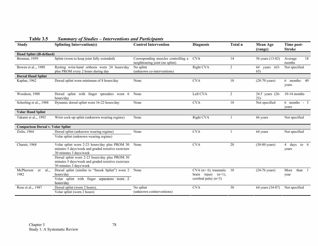
Chapter 3 78 Study 1: A Systematic Review
Table 3.5 Summary of Studies – Interventions and Participants Study Splinting Intervention(s) Control Intervention Diagnosis Total n Mean Age
(range)
Time post-
Stroke
Hand Splint (ill-defined)
Brennan, 1959 Splint (worn to keep joint fully extended) Corresponding muscles controlling a neighbouring joint (no splint)
CVA 14 56 years (13-82) Average 18 months
Bowen et al.,, 1988 Resting wrist-hand orthosis worn 24 hours/day plus PROM every 2 hours during day
No splint (unknown co-interventions)
Right CVA 2 64 years (63- 65)
Not specified
Dorsal Hand Splint
Kaplan, 1962 Dorsal splint worn minimum of 8 hours/day None CVA 10 (28-70 years) 6 months- 40 years
Woodson, 1988 Dorsal splint with finger spreaders worn 6 hours/day
None Left CVA 2 24.5 years (26-23)
10-14 months
Scherling et al.,, 1988 Dynamic dorsal splint worn 16-22 hours/day None CVA 18 Not specified 6 months - 3 years
Volar Hand Splint
Takami et al.,, 1992 Wrist cock-up splint (unknown wearing regime) None Right CVA 1 66 years Not specified
Comparison Dorsal v. Volar Splint
Dorsal splint (unknown wearing regime) Zislis, 1964
Volar splint (unknown wearing regime)
None CVA 1 64 years Not specified
Volar splint worn 2-23 hours/day plus PROM 30 minutes 5 days/week and graded resistive exercises 30 minutes 3 days/week
Charait, 1968
Dorsal splint worn 2-23 hours/day plus PROM 30 minutes 5 days/week and graded resistive exercises 30 minutes 3 days/week
None CVA 20 (30-80 years) 4 days to 6 years
Dorsal splint (similar to “Snook Splint”) worn 2 hours/day
McPherson et al.,, 1982
Volar splint with finger separators worn 2 hours/day
None CVA (n= 6), traumatic brain injury (n=1), cerebral palsy (n=3)
10 (24-76 years) More than 1 year
Dorsal splint (worn 2 hours), Rose et al.,, 1987
Volar splint (worn 2 hours)
No splint (unknown cointerventions)
CVA 30 64 years (34-87) Not specified

Chapter 3 79 Study 1: A Systematic Review
Table 3.5 continued… Study Splinting Intervention(s) Control Intervention Diagnosis Total n Mean Age
(range)
Time post-
Stroke
Dorsal-Volar Hand Splint (Snook Splint)
Snook, 1979 Dorsal-volar splint (“Snook Splint”) worn 6 hours on, 6 hours off.
None Right CVA (n=1), hypoxic brain injury (n=1)
2 38 years (27-50) 14 years
Finger Spreader/Abduction Splint
Doubilet et al.,, 1977 Finger abduction splint worn all day (removed for therapy and meals) plus “standard treatment in therapy sessions”
None CVA 15 Not specified Not specified
Langlois et al.,, 1991 Finger spreader splint (worn 6, 12, 22 hours/day) None CVA 13 (4 drop-outs)
64 years Over 12 months
Cone Splint
Dayhoff, 1975 Firm cone splint worn all day, removed for therapy.
None CVA 3 81 years (75-84) 5 years to 7 years
Jamison et al.,, 1980 Cone splint worn 24 hours/day None CVA 15 (4 drop-outs)
76 years (58 - 96)
36 months-11 years
Volar resting splint (immediate wearing effect recorded only)
Finger spreader splint (immediate wearing effect recorded only)
Mathiowetz et al.,, 1983
Cone splint (immediate wearing effect recorded only)
No splint (unknown cointerventions) CVA 4 44 years (19-96) 4-9 years
Inflatable Pressure Splint
Poole et al.,, 1990 Inflatable arm splint worn for 30 minutes 5 days/week plus “traditional occupational therapy” 5 days/week.
No splint plus “traditional occupational therapy” 5 days/week
CVA 18 70 years (55-82) Not specified
Dynamic Splint
McPherson et al.,, 1985
Dynamic splint (“Becker splint”) worn 1 hour 3 days/week
Passive range of motion 3 days/week CVA 8 77 years (67-86) Not specified
Lycra Splint
Gracies et al.,, 2000 Custom-made lycra arm and glove splint worn 3 hours
None CVA 16 65 years (36–85)
11 weeks (3-36)

Chapter 3 80 Study 1: A Systematic Review
Table 3.6 Summary of Studies – Research Methods Study Research Design Level of
Evidence
Internal Validity External
Validity
Treatment Duration Splinting n Control (no splint)
n
Hand Splint (ill-defined)
Brennan, 1959 Case series 4 Low *†‡§†† Moderate*** 19.8 weeks (7-36 weeks range)
14 (13 upper limb, 1 lower limb)
Subject’s joint proximal to that splinted served as own control
Bowen et al.,, 1988 Case series 4 Low *†‡§** High 8 weeks 1 1
Dorsal Hand Splint
Kaplan, 1962 Case series 4 Low *‡§**††‡‡ Moderate §§ 20 weeks (12-24 week range)
10 No control group
Woodson, 1988 Case series 4 Low *†‡§** Moderate *** 6 weeks 2 No control group
Scherling et al.,, 1988 Case series 4 Low *†‡§** Moderate *** Varied (2-7 months) 2 No control group
Volar Hand Splint
Takami et al.,, 1992 Single case study 5 Low *‡§**†† Moderate *** Unknown 1 No control group
Comparison Dorsal v. Volar Splint
Zislis, 1964 Single case study (crossover design)
4 Low *†‡§**††‡‡ Moderate §§ Unknown 1 Subject served as own control
Charait, 1968 Cohort study 3 Low *†‡§**††‡‡ Moderate §§ Varied (2 months to 3 years)
n(dorsal)=10 n(volar)= 10
No control group
McPherson et al.,, 1982 Randomised controlled trial
2b Low *‡** High 5 weeks n(dorsal)= 5 n(volar)= 5
No control group
Rose et al.,, 1987 Randomised controlled trial
2b Low *‡**†† Moderate §§ 2 hours n(dorsal)=10 n(volar)= 10
10
Dorsal-Volar Hand Splint (Snook Splint)
Snook, 1979 Case series 4 Low *†‡§** Moderate §§ Varied (4 days to 1 week) 2 No control group
Finger Spreader/Abduction Splint
Doubilet et al.,, 1977 Case series 4 Low *†‡§**††‡‡ Moderate *** 1 week 15 No control group
Langlois et al.,, 1991 Randomised controlled trial
2b Low *‡** High 2 weeks (+ 2 weeks follow-up)
n(6hrs/day)= 3 n(12hrs/day)=3 n(22hrs/day)=3
No control group

Chapter 3 81 Study 1: A Systematic Review
Table 3.6 continued… Study Research Design Level of
Evidence
Internal Validity External
Validity
Treatment Duration Splinting n Control (no splint)
n
Cone Splint
Dayhoff, 1975 Case series 4 Low *†‡§** Moderate §§ 6 weeks (follow-up to 1 year)
3 No control group
Jamison et al.,, 1980
Case series 4 Low *‡§** High 4 weeks 15 No control group
Mathiowetz et al.,, 1983
Case series (using non-CVA controls)
4 Moderate *§ Low §§*** Immediate effects measured (all conditions provided in one session, with rest in between)
4 10
Inflatable Pressure Splint
Poole et al.,, 1990 Randomised controlled trial
2b Moderate **†† High 3 weeks 9 9
Dynamic Splint
McPherson et al.,, 1985
Cohort study (matched controls)
3 Low *‡§** Moderate *** 6 weeks 4 4
Lycra Splint
Gracies et al.,, 2000
Case series (cross-over) 4 Moderate §** Moderate §§ 3 hours 16 Subjects served as own controls
Note: * Internal Validity Concern: threat to statistical conclusion validity; † Internal Validity Concern: threat of maturation present; ‡ Internal Validity Concern: threat of measurement/instrumentation bias present; § Internal Validity Concern: threat of selection bias present; ** Internal Validity Concern: threat of co-intervention bias present; †† Internal Validity Concern: threat of intervention bias present; ‡‡ Internal Validity Concern: threat of contamination present; §§ External Validity Concern: threat that treatment may not represent current practice; *** External Validity Concern: threat that participants may not be representative of population; ††† External Validity Concern: research was not conducted in clinical (natural) setting.

Chapter 4 82 Study 2: A Randomised Controlled Trial of Splinting plus Stretch
CHAPTER FOUR
Study 2: A Randomised Controlled Trial Comparing Splinting
plus Stretch to Stretch Alone in Adults with Hemiplegia
following Acquired Brain Impairment
[This study was published as: Lannin, N.A., Horsley, S., Herbert, R., McCluskey, A., & Cusick, A. (2003).
Splinting the hand in the functional position after brain impairment: a Randomized controlled trial. Archives of
Physical Medicine and Rehabilitation, 84, 297-302]

Chapter 4 83 Study 2: A Randomised Controlled Trial of Splinting plus Stretch
Chapter Four: Randomised Controlled Trial of Splinting plus Stretch
4.1 Chapter Overview
Management of contractures of the wrist and finger flexor muscles following acquired brain
impairment is a challenge for clinicians. There is still insufficient evidence on how to
effectively prevent or manage contractures. This chapter presents the findings from a
randomised controlled trial that investigated the effectiveness of hand splints with an
existing inpatient rehabilitation population of people post stroke, who were also receiving
wrist extension stretches. This study was designed to determine whether or not prescribing
a hand splint in acute rehabilitation (where active training and stretches are commonly
practiced) benefits adults with hemiplegia following acquired brain impairment.
4.2 Background
Reduction of disability due to stroke and other acquired brain impairments is a recognised
national health priority in Australia (National Health Priority Action Council, 2002).
Hemiplegia is one of the most prevalent types of disability in this population (Ing, Tesio,
Arnould, Zancan, & Thonnard, 2001; Sato, Kaji, Tsuru, & Oizumi, 1999). As described in
Chapter One, one of the most common and preventable complications of hemiplegia is the
development of contractures (Farmer & James, 2001; O’Dwyer & Ada, 1996; Yarkony &
Sahgal, 1987). Contractures limit the available movement at the joints affected, preventing
full use of the hand and arm, and are associated with poor functional recovery (Carr &
Shepherd, 1987). Occupational therapists and other health care professionals have the
opportunity during rehabilitation to minimise contracture following brain impairment.
A number of treatment modalities are used to address such contracture, including passive
stretching, splinting (Farmer & James, 2001), casting and movement training (Ada &

Chapter 4 84 Study 2: A Randomised Controlled Trial of Splinting plus Stretch
Canning, 1990). In most rehabilitation units, patients who experience hemiplegia are
routinely provided with a hand splint by their occupational therapist (Kogler, 2002), whilst
also receiving prolonged stretching and movement training by their occupational therapist
and/or physiotherapist. There are, however, still discrepancies in occupational therapy
splinting practices, and use of hand splints with this population is controversial (Edwards &
Charlton, 2002; Langlois, Pederson & Mackinnon, 1991; McPherson, Kreimeyer, Aalderks
& Gallagher, 1982; Milazzo & Gillen, 1998; Wilton, 1997). As outlined in-depth in Chapter
2, the most common aim of hand splinting in the neurological population, and the one used
as a basis for the present study, is the prevention of contracture (Neuhaus et al., 1981) based
on the biomechanical rationale. While some therapists reportedly aim to reduce muscle
spasticity (Langlois, Mackinnon, & Pederson, 1989; Neuhaus et al., 1981), the clinical
relevance of spasticity as an outcome is questionable, since published research suggests that
spasticity is unrelated, or only weakly related, to functional ability following stroke
(O’Dwyer, Ada, & Neilson, 1996).
4.3 Description and Rationale for Study Design
The efficacy of splinting was investigated using a randomised controlled study. As adjunct
to splinting, rehabilitation interventions such as passive stretching and movement training
are commonly used in Australia. The conduct of the study in a typical clinical setting
(which includes passive stretching and movement training) thus increases the applicability
of the results so that the very real concerns of clinicians working in the Australian health
care setting are addressed.
Recommendations from Pocock (1983) and criticism of previous research methodologies
(Ashburn, Cornall, Melville, Simpson, & Wright, 1998) were used to assist in the
development of the study protocol. Specifically, those researchers advocate making sure
there are sufficient numbers of participants to provide statistical power, accounting for

Chapter 4 85 Study 2: A Randomised Controlled Trial of Splinting plus Stretch
participant demographics and baseline performance, use of blinding (masking), random,
concealed allocation, and using validated and well-established outcome measures.
4.4 Aim
The primary aim of this study was to determine the effect of four weeks of splint wearing
on the length of the wrist and finger flexor muscles, in patients who were in the early stages
of rehabilitation following acquired brain impairment. Secondary aims were to determine
whether splint wearing affected recovery of hand function and pain in the upper limb.
4.5 Research Question
In adults who were receiving daily stretching and movement training for treatment of
hemiplegia following acquired brain impairment, does splinting in the traditional,
‘functional’ position prevent contracture of the wrist and finger flexor muscles, improve
function and/or decrease pain, compared to those who do not wear a splint?
4.6 Method
4.6.1 Ethics approval
Ethics approval was gained through the University of Western Sydney Ethics Review
Committee (Human Subjects) – Protocol Number: 2000/054, and Townsville General
Hospital Ethics Committee-Protocol Number: Q02/128 (Appendix B).
4.6.2 Participants
4.6.2.1 Inclusion Criteria:
Participants were required to meet the following criteria for inclusion in the study:
a) History of a single acquired brain impairment resulting in upper limb hemiplegia
of no greater than six months duration;

Chapter 4 86 Study 2: A Randomised Controlled Trial of Splinting plus Stretch
b) Be unable to actively extend the affected wrist; and
c) Be aged over 18 years.
As it is not customary in clinical practice to provide a hand splint to patients with
active movement, participants with active wrist extension were excluded from taking
part in the study. Age limitations were imposed to ensure the study met Australian
guidelines4 as outlined by the National Statement of Ethical Conduct in Research
Involving Humans (2001).
4.6.2.2 Exclusion criteria
Participants were excluded from the study if they met any of the following criteria:
a. Previous stroke which had resulted in upper limb hemiplegia
b. Previous upper limb trauma causing structural imbalance or reduced range of
movement at the wrist or fingers (for example, due to colles fracture)
c. Osseous abnormality in the wrist or fingers
d. Arthritic condition of the wrist or fingers.
Participants were ineligible if they had language comprehension, perceptual or
cognitive deficits that would prevent written informed consent or participation in the
program.
4.6.3 Power Analysis and Sample size
The required sample size was calculated using the pooled estimate of within-group standard
deviations (5.1°) obtained from pilot data (Pocock, 1983). A 5° change in measured muscle
extensibility at the wrist was selected as the minimum clinically important change which
would be considered worthwhile following the extensive period of splint wearing. Power
calculations indicated that a sample of 28 subjects would provide an 80% probability of
4 People under 18 years of age are required by legislation to have the consent of their parent/guardian to participate in research.

Chapter 4 87 Study 2: A Randomised Controlled Trial of Splinting plus Stretch
detecting a 5° effect on wrist and finger flexor length, with non-compliance and loss to
follow-up of 20% and alpha set at .05.
The minimum clinically important change (5° effect on the primary outcome, wrist and
finger flexor muscle lengths) was determined after consideration of the anticipated financial
cost of making a hand splinting versus the burden of wearing a hand splint each night for
four weeks from the patient perspective, and the amount of change in wrist extensibility that
would make a measureable difference to hand function.
4.6.4 Randomisation
Participants were randomly allocated to control and experimental groups using a simple
randomisation process (Pocock, 1983); participants were allocated to groups according to a
random number sequence generated using a random number table (Altman & Bland,
1999a). Group allocation was concealed from the investigator, with the random numbers
generated by a third person not involved in the study and located at an external site.
Random allocation followed baseline measurement (Moher, Shulz, & Altman, 2001). The
investigator contacted an independent, third person via telephone to obtain group allocation
for each subject, thereby ensuring concealed randomisation.
4.6.5 Blinding
This trial used single blinding where the assessors (those obtaining outcome measures) were
unaware of group allocation and whether participants had been allocated a hand splint or
not. Due to difficulty blinding individuals to a physical therapy such as hand splints,
participants were not blinded. Some but not all treating therapists were blind to group
allocation (the therapist providing the daily stretching program was blind to group
allocation). Although lack of participant and therapist blinding was a potential source of
bias, the main outcome measure (the extensibility measure of range of movement) and one

Chapter 4 88 Study 2: A Randomised Controlled Trial of Splinting plus Stretch
of the secondary outcome measures (hand function) were rated by the blinded assessor. The
data from these measures were quantitative and there was no subjective interpretation by the
investigator or the participants who were unblinded. Therefore, bias was minimised as much
as possible given the physical constraints of researching this form of therapy.
4.6.6 Intervention
This study was designed to reflect current Australian rehabilitation practice and to ensure
applicability of results. In Australia, and elsewhere, hand splinting is typically provided in
combination with routine upper limb therapy. The usual alternative to hand splinting (and
therefore the most appropriate ‘control’ comparison) is routine therapy without a splint.
Therefore, the control group received only routine therapy and the experimental group wore
a hand splint for up to 12 hours a night, for four weeks in addition to routine therapy.
The routine therapy of individual motor training and upper limb stretches provided five
days per week to all study participants was:
Upper limb motor training: An individually designed motor training program was provided
(Carr & Shepherd, 1987, 1998; Dean & Shepherd, 1997). The program aimed to improve
upper limb task performance. Each participant received approximately 30 minutes of
training a day, five days a week. The training typically involved attempts to elicit muscle
contraction. No limits were placed on the amount of finger or wrist extension and
participants were encouraged to practice outside of therapy.
Stretching: The second component of routine therapy received by both the control and
experimental groups was daily upper limb stretching. Two 30-minute stretches were applied
to the upper limb five-days a week for the duration of the study and follow-up period (five
weeks in total). These stretches provided a prolonged low-load stretch to muscles at risk of

Chapter 4 89 Study 2: A Randomised Controlled Trial of Splinting plus Stretch
developing contracture (Carr & Shepherd, 1995; Ada & Canning, 1990), including the long
finger and wrist, elbow flexors, forearm pronator and thumb abductor muscles. The first
stretch was a seated weight-bearing stretch, with (i) the shoulder joint positioned in external
rotation, abduction, and slight extension, (ii) the elbow in extension, (iii) the forearm in
supination, and (iv) the wrist and fingers in extension (Figure 4.1). The second stretch was a
seated stretch of involving use of an inflatable long-arm air splint5 with (i) the shoulder
joint positioned in external rotation and abduction, (ii) the elbow positioned in extension,
(iii) the wrist positioned in extension and (iv) the thumb positioned in abduction (Figure
4.2). Participants were helped to move into and maintain stretch positions by their treating
physiotherapist who was blind to treatment allocation. If necessary, the therapist remained
with the participant throughout the duration of the stretch.
Figure 4.1 A seated weight-bearing stretch of the upper limb
5 URIAS Pressure Splint; Svend Andersen Plastic Industri, Maersk Medical A/S, Engmosen 1, 3540 Lynge, Denmark.

Chapter 4 90 Study 2: A Randomised Controlled Trial of Splinting plus Stretch
Figure 4.2 A seated stretch of the upper limb demonstrating the use of an air splint
4.6.6.1 Control Program
Control participants did not wear a hand splint at any time during the study period.
They continued to receive routine therapy as outlined above.
4.6.6.2 Splinting Program
In addition to the routine motor training and stretching described above, participants
in the experimental group wore a hand splint on a daily basis, for a maximum of 12-
hours each night for the four-week intervention period. The chief investigator and
nursing staff on the hospital ward were responsible for ensuring that the splints were
applied and fitted correctly for the duration of the study.

Chapter 4 91 Study 2: A Randomised Controlled Trial of Splinting plus Stretch
Splint design: A static palmar-mitt splint (Copley & Kuipers, 1999) was custom
made by the chief investigator using Ezeform®6 thermoplastic. The splint held the
hand in the ‘functional’ resting position: wrist positioned in 10º extension, thumb in
opposition and abduction, and semiflexion of the finger joints (Figure 4.3) (Hill,
1988; Lowe, 1989; Wilton, 1997). Splints were checked daily by the chief
investigator for fit and comfort. No adjustments to the splints were required during
the study period to maintain the ‘functional’ resting position.
Figure 4.3
Static palmar-mitt splint with the wrist and fingers positioned in the ‘functional position’
Wearing Regime: In this study, the hand splints were worn overnight for a maximum
of 12-hours. This splint wearing regime was chosen on the basis of the best available
literature as discussed in Chapter 2.
Compliance with the splinting program: Compliance with the splint wearing regime
was important for the overall success of the trial. As non-compliance with the
splinting program could influence results, the study protocol addressed compliance
6 Ezeform® Splinting Material; Smith & Nephew plc, 15 Adam Street, London WC2N 6LA United Kingdom.

Chapter 4 92 Study 2: A Randomised Controlled Trial of Splinting plus Stretch
through participant and family education (Bond & Burt, 2002). Participants were all
resident in a single inpatient rehabilitation unit which allowed the chief investigator
to put the splints on each evening and remove them each morning to improve and
monitor compliance.
4.6.6.3 Potential Contaminants
The effect of practicing wrist extension (motor training) and of stretches may have
affected the primary outcome, muscle extensibility of the wrist and long-finger
flexor muscles, and as such could contaminate the results of the study. The amount
of stretching and time spent in therapy was consistent across all participants
involved in the trial. This was made possible as all participants were located in a
single rehabilitation unit and treating therapists remained constant during the study
period.
4.6.7 Data Collection
Age, gender, ethnicity, hand dominance, hemiplegic hand, location and type of acquired
brain impairment, and time post-impairment were recorded to provide demographic and
background information on the participants. Outcome measures were obtained on three
occasions: before random allocation (baseline), at the end of the 4-week experimental
period on the 30th day (post-intervention), and one-week after the experimental intervention
was ceased on the 38th day (follow-up). Measurements on the 30th day were taken at least 3
hours after the splint had been removed. Measurements were conducted by trained
physiotherapists who were blind to group allocation (blinded assessors).

Chapter 4 93 Study 2: A Randomised Controlled Trial of Splinting plus Stretch
4.6.7.1 Outcome Measures
Three outcomes were recorded. These were length of wrist and finger flexor muscles
(primary outcome), hand and arm function, and pain levels (secondary outcomes).
Primary Outcome: The length of the wrist and finger flexor muscles was obtained using a
standard procedure which provided a torque-controlled measurement of wrist extension
with the fingers extended (Harvey, King, & Herbert, 1994). The measurement procedure
and its psychometric properties were discussed in Chapter 2 (2.7.1 Measurement of Passive
Range of Movement). To ensure that muscle length was being measured (as opposed to
range of joint motion) the following guidelines were followed:
a. Muscles to be measured (long finger flexors and wrist flexors) were placed
in the fully elongated position by holding both the fingers and the wrist in
extension;
b. The muscle should be isolated across one or possibly two joints; and
c. The bony landmarks to be used to align the measurement tool should be
palpable and in proper alignment.
The extensibility device used to measure the primary outcome ensured that all of these
guidelines could be met. The extensibility of the wrist flexor muscles (flexor digitorum
superficialis and profundus, flexor carpi radialis and ulnaris, palmaris longus and flexor
pollicus longus) was reflected by the angle of wrist extension that corresponded with a
standardised torque. Torque was standardised for a particular participant but not across all
participants. Angle of wrist extension was measured using skin surface markers and a
goniometer attached to the side of the instrument (see Figure 4.3). Three consecutive
measurements were taken on each occasion, with the mean reading of the three used for
analysis.

Chapter 4 94 Study 2: A Randomised Controlled Trial of Splinting plus Stretch
Secondary Outcomes: Secondary outcomes included the upper limb domains (items six,
seven and eight) of the Motor Assessment Scale at four and five weeks; and self-reported
pain level in the wrist at four and five weeks.
Motor Assessment Scale:
Upper limb function was measured using items 6 (upper limb function), 7 (hand
movements) and 8 (advanced hand activities) of the Motor Assessment Scale (MAS)
(see Table 4.1) (Carr, Shepherd, Nordholm, & Lynne, 1985; Dean & Mackey, 1992;
Poole & Whitney, 1988). Psychometric properties of this scale were discussed in
Chapter 2 (2.7.3 Measurement of Motor Function). As required by the standardised
procedure, three consecutive measurements were taken on each occasion and the
best performance recorded for analysis. Summed scores from items 6, 7, and 8 were
used to provide an overall indicator of upper limb function.
Upper Limb Pain:
Upper limb pain was measured using a 10-centimeter vertical visual analogue scale
(Chapman, Casey, Dubnar, Foley, Gracely & Reading, 1985; Tiplady, Jackson,
Maskrey, & Swift, 1998), which is shown in the literature to be a sensitive (Jensen,
Karole & Braver, 1994; Ohnhaus & Adler, 1975), valid (Jensen et al., 1994;
Ohnhaus & Adler, 1975) and reliable method of collecting subjective pain intensity
(Jensen et al., 1994). Pain intensity was recorded prior to measuring the wrist and
finger flexor muscle length to avoid contamination between outcome measures.

Chapter 4 95 Study 2: A Randomised Controlled Trial of Splinting plus Stretch
Table 4.1 Upper Limb Subscale of the Motor Assessment Scale (UL-MAS): Items 6, 7
and 8
6. Upper arm
function
1. lying protract shoulder girdle with arm in elevation
2. lying hold extended arm in elevation for 2 seconds
3. flexion & extension of elbow to take palm to forehead with arm as in 2
4. sitting hold extended arm in forward flexion at 90º to body for 2 seconds
5. sitting patient lifts arm to above position and holds it there for 10 seconds, then lowers it
6. standing, hand against wall maintain arm position while turning body towards wall
7. Hand movement
1. sitting extension of wrist
2. sitting radial deviation of wrist
3. sitting elbow into side pronation and supination
4. reach forward, pick up large ball of 14 cm diameter with both hands & put it down
5. pick up a polystyrene cup from table & put it down on table across other side of body
6. continuous opposition of thumb and each finger more than 14 times in 10 seconds
8. Advanced hand
activities 1. picking up the top of a pen and putting it down again
2. picking up one jelly bean from a cup and placing it in another cup
3. drawing horizontal lines to stop at a vertical line ten times in 20 seconds
4. holding a pencil, making rapid consecutive dots on a sheet of paper
5. taking a desert spoon of liquid to the mouth
6. holding a comb and combing hair at back of head
Splinting Program Compliance: In addition to these clinical outcome measures,
compliance with the splint wearing schedule was logged daily using ‘time-on/time-
off’ recording charts. Data were recorded by nursing staff on the hospital ward, and
monitored by the chief investigator. The splint wearing recording sheets were based
on the concept of a time-use diary, one of the most accurate methods of measuring
time-use (Harvey, 1993; Pentland, Harvey, & Walker, 1998; Stanley, 1995).

Chapter 4 96 Study 2: A Randomised Controlled Trial of Splinting plus Stretch
4.6.8 Data Analysis
Change in the length of wrist and finger flexor muscles, upper limb function scores, and
pain scores in control and experimental groups were compared using two-tailed independent
sample t tests. Parametric data were reported for all outcomes, including the UL-MAS
which is considered a long ordinal scale with parametric data being presented in previous
studies (for example, Baskett, Broad, Reekie, Hocking & Green, 1999). Parametric tests are
also robust to violations of their assumptions (Box, Leonard, & Wu, 1983; Armitage &
Berry, 1994) and the results of parametric tests are generally more useful to clinicians; for
example, it is easier to generate confidence intervals for parametric tests, and parametric
test results can be included in meta-analyses. To avoid controversy about the most
appropriate statistical tests to use for rank-ordinal data, a secondary analysis of the
differences between the two groups on the upper limb function (UL-MAS) scores was
performed using the non-parametric Mann-Whitney test.
Significance levels were pre-established at the p≤.05 level. Size of treatment effect was
estimated by differences in group means and their 95% confidence intervals. Where
possible, outcome measures were obtained for all participants allocated to the trial. Each
participant’s data were analysed in the group to which the participant was allocated, in
accordance with the principle of intention-to-treat (Lachin, 2000; Hollis & Campbell, 1999).
Missing data were imputed using the last observation carried forward method (Pocock,
1983).
4.7 Results
Results will be presented in the following order. First, the baseline characteristics of
participants are presented (Table 4.2). These data are followed by a participant flow
diagram demonstrating the progression of participants through the trial. After this,

Chapter 4 97 Study 2: A Randomised Controlled Trial of Splinting plus Stretch
comparisons of effectiveness will be made between the two groups: the primary outcome is
presented first, followed by the secondary outcomes.
4.7.1 Baseline Characteristics
Following baseline measurement, 11 participants were randomly assigned to the control
group, and 17 were assigned to the two splinting intervention. Subjects in the two groups
were similar, on average, at the time of recruitment to the study (Table 4.2). Mean age of
the sample was 66 years. Just over half of the sample were women (54%) and 89% were
Caucasian. Twenty seven participants had experienced a stroke or subarachnoid
haemhorrage and one participant had experienced a traumatic brain injury.
Table 4.2 Baseline Characteristics Variable Control Group (n=11)
stretch only
Intervention Group (n=17)
stretch plus splint
Age in years 68 years, 1 month
(7.4)
65 years
(16.4)
Gender: number of women (%) 6 (54.5%) 9 (52.9%)
Time post-impairment in days 57 (63.6) 47 (21.4)
Dominance: number of right-hand dominant (%)
11 (100%) 17 (100%)
Number of right-sided hemiplegia
7 (63.63%) 10 (58.8%)
Note: Values are means (± standard deviations) unless otherwise stated.

Chapter 4 98 Study 2: A Randomised Controlled Trial of Splinting plus Stretch
4.7.2 Participant Flow Details
Fifty three adults underwent eligibility screening of which 30 were eligible for participation
(eligibility fraction7 for study was therefore 56.6%). Reasons for exclusion included: active
wrist extension (n=19), concurrent fracture of the hemiplegic arm (n=2), and decreased
ability to provide informed consent (n=2). Two eligible participants elected not to
participate in the study. Twenty eight adults who met entry criteria and agreed to take part
were enrolled in the study and completed baseline measurements (enrolment fraction8 was
therefore 93.3% and recruitment fraction9 52.8%). Baseline data was not collected from
non-participants.
Of the 28 participants randomised, 25 (11 from the control group and 14 from the
experimental group) participated in all interventions and assessments as allocated (Figure
4.4). Outcome measures were obtained from 26 participants post-intervention, and from 27
participants at follow-up. One participant in the experimental group withdrew from the
study during the intervention period as a result of self-discharge from the rehabilitation unit.
Two additional participants in the splint group refused post-intervention measurement but
participated in follow-up measurements (one participant continued to receive interventions
as allocated). One participant in the control group reported a pain rating of 0 at baseline, of
0 at post-intervention and increased her rating by 100% (maximum score of 10) at the
follow-up measure one week later. As this follow-up value for pain differed so greatly that
it was omitted from all analyses.
7 The proportion of participants deemed eligible for the study of all potential participants screened is termed the eligibility fraction. 8 The proportion of eligible people who enroll in the study of those who were eligible for participation is termed the enrolment fraction. 9 The product of the eligibility and the enrolment fractions represents the proportion of potential participants who actually enrolled in the study and is termed the recruitment fraction.

Chapter 4 99 Study 2: A Randomised Controlled Trial of Splinting plus Stretch
Numbers of participants in each group who were non-compliant with the protocol are
shown below in Table 4.3. Non-compliance with protocol included participants: refusing
part of the outcome assessment protocol, not wearing their assigned hand splint for a
minimum of 5 hours in a 24 hour period, or using an alternative hand splint (not assigned).
Table 4.3
Numbers of participants in each group who did not adhere to the protocol
Time Control Group (n=11)
stretch only
Intervention Group (n=14)
stretch plus splint
4 Weeks 0 3
5 Weeks 0 1
At study commencement, differences between the two groups for key outcomes
(contracture, function, and pain) were small and insignificant (see Table 4.4). Data are mean
(SD) baseline values of wrist extensibility, and subjective pain rating, and median
(frequency) baseline values of function for participants in the two groups. Although there
were no systematic differences between groups, parameters did vary between subjects.
Some participants appeared to have normal, or near-normal, wrist muscle extensibility at
baseline while others had signs of clinical contracture when compared to their non-affected
hand.
Table 4.4 Baseline performance of groups on dependent variables
Variable Control Group (n=11)
Routine therapy only
Intervention Group (n=17)
routine therapy plus splint
Non-affected wrist extensibility
in degrees 80.8 (9.5) 83.4 (7.8)
Hemiplegic wrist extensibility
in degrees 79 (10.5) 76 (13.9)
Function on UL-MAS 0.8 (1.8) 1.8 (3.0)
Pain (%)
1.6 (2.7) 2 (2.9)
Note: Data are mean (SD) baseline values of wrist extensibility, and subjective pain rating, and median (frequency) baseline values of function and spasticity rating for participants in both groups.

Chapter 4 100 Study 2: A Randomised Controlled Trial of Splinting plus Stretch
Patients screened at initialappointment (N=53)
Reasons behind patients who were approached not
taking part were: able to extend the wrist (19),
refused- no reason given (2), medically unfit-
non-brain impairment related medical condition (2),
unable to provide informed consent (2).
Registered Participants (N=28)
Randomised to CONTROLgroup: Stretch only (n=11)
Randomised to SPLINT group:Splint plus Stretch (n=17)
Provided FUNCTIONAL WRISTSPLINT for 4 weeks (n=17)
1 month review(n=11)
dropout (n=0)noncompliant (n=0)
1 month review(n=14)
dropout (n=1)noncompliant (n=2)
6 week review(n=11)
dropout (n=0)noncompliant (n=0)
6 week review(n=16)
dropout (n=1)noncompliant (n=0)
Analysed(n=11)
Analysed(n=16)
Analysed(n=14)
Analysed(n=11)
Baseline Assessment (N=28)
Figure 4.4 Participant flow diagram
The effects of splinting (the difference between the experimental and control groups) were
not clinically important or statistically significant.

Chapter 4 101 Study 2: A Randomised Controlled Trial of Splinting plus Stretch
4.7.3 Primary Outcome: Effect of splinting on contracture
Splinting increased wrist extension by a mean of 1° after the intervention (95% CI -3.7º to
6.1º), and reduced wrist extension by a mean of 2° at follow-up (95% CI -7.2º to 3.2º).
Neither result was large enough to be of clinical importance (see Figure 4.5).
Figure 4.5 Mean (95% confidence intervals) of differences between torque-controlled
wrist extension range of motion of the splint and control groups at the
commencement of the study (initial), then after four weeks of splint
intervention (post-intervention), then one week after the removal of the
splint (follow-up).
A negative effect indicates that the splint treatment did not have an advantageous effect on
the extensibility of the wrist extensor muscles in relation to the control group.
90 80 -
70 -
60 - 50 -
40 - 30 -
20 -
10 -
0
Wri
st
Ex
ten
sio
n (
de
gre
es
)
Initial Post-intervention
Follow-up
▲ Control
● Splint

Chapter 4 102 Study 2: A Randomised Controlled Trial of Splinting plus Stretch
4.7.4 Secondary Outcome: Effect of splinting on function
Splinting decreased upper limb function (MAS component 6) by a mean of 0.3 points on a
6-point scale after the intervention (95% CI -1.5 to 0.9) and decreased upper limb function
by 0.8 points at follow-up (95% CI -2.0 to 0.3). Splinting decreased performance of hand
movements (MAS component 7) by 0.4 points after the intervention (95% CI -1.4 to 0.7)
and by 0.5 points at follow-up (95% CI -1.5 to 0.6). Splinting decreased performance of
advanced hand activities (MAS component 8) after the intervention by 0.03 points after the
intervention (95% CI -0.5 to 0.4), and by 0.1 point at follow-up (95% CI -0.8 to 0.5). None
of these differences were statistically significant or clinically important.
Splinting decreased overall upper limb function, measured by the summed scores of UL-
MAS, by 0.06 points on an 18-point scale after the intervention (95% CI -2.5 to 2.7) and by
0.2 points at follow-up (95% CI -2.7 to 2.3). Non-parametric analyses yielded zero effects
(median difference of all paired differences between groups) for all comparisons of MAS
scores at both final measurement and follow up, and confidence intervals for differences
between medians (Armitage & Berry, 1994) were similar, or tighter, to those obtained with
parametric analyses.
4.7.5 Secondary Outcome: effect of splinting on pain
Splinting increased the reported intensity of upper limb pain by a mean of 0.2 cm on a 10
cm visual analogue scale after the intervention (95% CI -2.3 to 2.7) and reduced the
reported pain intensity by 1 cm at follow-up (95% CI -4.6 to 2.2). These differences were
not considered statistically or clinically significant.

Chapter 4 103 Study 2: A Randomised Controlled Trial of Splinting plus Stretch
4.7.6 Splint compliance and participant satisfaction
All participants in the experimental group wore hand splints, but as could be expected in a
clinical setting, there was some daily variability compliance to the splinting protocol.
Compliance with the splint wearing protocol was, however, very high (see Table 4.5).
Participants appeared motivated to wear the splints with no additional encouragement. No
participant in the experimental group demonstrated evidence of skin breakdown or adverse
reactions following splint wearing. On the contrary, the typical subjective response was that
the splinting procedure was comfortable. At the end of the study, participants in the
experimental group were surveyed about the level of ‘annoyance’ related to wearing a
splint. Fifteen participants rated their level of annoyance at wearing the splint, on a 10 cm
visual analogue scale. The mean level of reported ‘annoyance’ at the completion of the
study was 3.9 cm, which is considered to be low.
Table 4.5 Compliance with Splinting Program
Variable Intervention Group (n=17)
Average total wear time (sd) in hours 259.5 (79.3)
Average weekly wear time (sd) in hours 74.3 (13.8)
Average nightly wear time (sd) in hours 10.8 (3.3)
4.8 Discussion
4.8.1 Key Findings
The key finding from this study was that wearing a hand splint in addition to participating in
upper limb stretches and motor training five days a week did not improve wrist extensibility
or prevent contracture development. Hand splinting was therefore ineffective as an

Chapter 4 104 Study 2: A Randomised Controlled Trial of Splinting plus Stretch
intervention for maintaining muscle extensibility, improving functional use, and decreasing
pain in people with recently acquired brain impairment.
4.8.2 Findings in Detail
Primary Outcome
Participants with acquired brain impairment who were already receiving routine motor
training and prolonged, upper limb stretches did not show detectable or important changes
in wrist and finger flexor extensibility after wearing a splint daily for four weeks during this
study. Contrary to expectations, participants in the control group did not lose wrist and
finger flexor extensibility (i.e. acquire a contracture) in this four week period if they did not
wear a hand splint.
The literature recommends that splints for this population should be worn for between two
and six hours per day (Wilton, 1997; Milazzo & Gillen, 1998). In this study, splints were
worn for up to 12 hours a day (daily mean 11 hours, range 0–13 hours, SD 3 hours). In spite
of this intensive regime of splinting, no significant differences in muscle extensibility and
contracture rate were detected between groups.
Secondary Outcomes
Findings related to the study’s secondary outcomes also indicate no effect. First, there was
no evidence of clinically significant effects of splinting on upper limb function, as measured
by individual item or summed UL-MAS scores. Second, the 4-week splint wearing program
did not significantly reduce upper limb pain levels. It is acknowledged that the pain scores
were low at baseline which may have made reduction in pain difficult to detect. These two
findings do suggest, however, that the practice of splinting to improve function (Wilton,
1997; Hill, 1988; Neuhaus et al., 1981; Milazzo & Gillen, 1998; Wilson & Caldwell, 1978)

Chapter 4 105 Study 2: A Randomised Controlled Trial of Splinting plus Stretch
and reduce pain levels (Perry, 1980; Snook, 1979; Wilson & Caldwell, 1978) following
acquired brain impairment should be questioned if patients are already receiving motor
training and upper limb stretches as part of their rehabilitation.
Sample Characteristics
The mean age of participants was 66 years, similar to that of inpatients with acquired brain
impairments in other Australian rehabilitation units (67.4 years) (Eagar et al., 1997). This
variation is also similar to that reported in other studies of hand splinting, which include
participants with diagnoses of either stroke or traumatic brain injury (for example,
McPherson et al., 1982). As outlined above, only one participant in the study had
experienced a traumatic brain injury.However, irrespective of the underlying cause, upper
motor neuron damage leads to hemiplegia and the motor impairments affecting both groups
are similar (Eng, Rows, & McLaren, 2002). The conclusions from this study are therefore
likely to be applicable to the majority of adults undergoing inpatient rehabilitation in
Australia with hemiplegia from acquired brain impairment, irrespective of the cause.
Drop-outs and non-compliance
One participant self-discharged from the rehabilitation unit, and returned to live in a rural
area where follow-up was not possible. No other participants dropped out of the trial. The
splint group had two participants who refused to participate in the primary outcome
measurement at the end of the treatment arm (i.e. at 4 weeks). All remaining participants
adhered to the protocol. Loss to follow-up was therefore low (10.7% at four weeks, and
3.6% at five weeks).

Chapter 4 106 Study 2: A Randomised Controlled Trial of Splinting plus Stretch
Strengths and limitations of this study
The study’s strengths include its prospective design, the standardised protocol for the
experimental and control groups, and the blinded, torque-controlled assessment of contracture.
The randomised controlled trial methodology provides the strongest form of evidence when
determining the effectiveness of treatments (Roberts & Dicenso, 1999; Sackkett & Wennberg,
1997). In addition, this trial employed design features which help to minimise bias
(randomisation, allocation concealment, and blinded assessment of outcomes).
This trial used rigorously validated outcome measures, including a torque-controlled measure
of wrist range of movement as the primary outcome measure. Only one previous randomised
controlled trial of hand splinting measured contracture (Rose & Shah, 1987). The measure of
contracture used in that study was passive range of movement using a goniometer without
standardising the torque force used to produce the range of movement (Rose & Shah, 1987). If
torque is not standardised then changes in a person’s ability to tolerate uncomfortable stretch
torques may result in corresponding increases in joint angle (Folpp, Deall, Harvey, & Gwin,
2005). The previous study by Rose and Shah (1987) is likely to have measured tolerance to
stretch rather than the effect of a hand splint on contracture.
There are limitations of this study that need to be considered. Although this trial was
randomised it was not double blind (i.e. the participants were not blinded to the treatment
received) because it is difficult to blind the participants when the therapy – the hand splint-
is visible. To minimise observer bias, primary and secondary outcomes were measured by a
blinded assessor and were not self-reported by the participant who was aware of group
allocation.

Chapter 4 107 Study 2: A Randomised Controlled Trial of Splinting plus Stretch
The present trial was conducted in Australia. Consequently, care is needed when
generalising the results to participants outside of Australia. When interpreting the findings,
the routine therapy provided needs to be considered. Participants in both the treatment and
control groups received upper-limb motor training and upper-limb stretches, which may
have been enough to negate or mask the effect of the additional stretch applied by the splint.
Literature indicates that the routine therapy received in this study does not commonly occur
in countries outside of Australia, for instance in the United Kingdom (“Journal review”,
2003; Walker, 2003).
4.8.3 Implications for Future Research
The results of this study suggest that splinting the hand in the functional resting position
does not produce clinically useful effects in adults with acquired brain impairment.
However, it could be that hand splinting in other positions, which administer greater torque
at the wrist, may be beneficial. Further research studies should continue to use simple,
accurate and reliable assessment measures of torque-controlled range of motion, motor
function, and pain. Results do not suggest post-acquired brain impairment sub-groups
which may benefit from splinting. Analysis of future studies could use predetermined
covariates for use in analysis, which may provide guidance relating to the effect of
participant age, time post-lesion and lesion type.
The absence of an effect following splint wearing on wrist and finger flexor extensibility
may have been because the routine motor training and upper limb stretches were already
maintaining the length of wrist and finger flexor muscles. These therapies may have
rendered the additional ‘stretch’ (the stretch provided by the hand splints) redundant. Future
research should examine the effect of splinting in the absence of prolonged stretches in
adults following acquired brain impairment.

Chapter 4 108 Study 2: A Randomised Controlled Trial of Splinting plus Stretch
There is, however, still no clear evidence that prolonged stretches do in fact reverse
contracture of the wrist and finger flexor muscles following acquired brain impairment.
While a number of animal studies have found that short periods of daily stretching can
prevent the development of muscle contracture in animal muscle (Goldspink, 1977; Herbert
& Balnave, 1993; Williams, 1988, 1990; Williams, Cantonese, Luey & Goldspink, 1988;
Williams & Goldspink, 1978), clinical studies on human muscles have yet to demonstrate
the same benefits (Farmer & James, 2001). Further research is still needed to determine
whether or not prolonged stretching at maximum wrist extension prevents or reduces
contracture at the wrist in adults post-brain impairment.
Finally, the importance of replication of research is acknowledged, particularly given the
statistically non-significant results (Ottenbacher, 1995). It is, however, noted that results do
indicate a precise treatment effect of 0° with a narrow confidence interval. The narrow
confidence interval, coupled with the a priori sample size calculation providing greater than
80% power to detect a difference, does suggest that no true difference between group’s
performance exists.

Chapter 4 109 Study 2: A Randomised Controlled Trial of Splinting plus Stretch
4.8.4 Implications for Clinical Practice
One of the research questions was: Did the hand splint prevent or manage contractures for
people with hemiplegia following acquired brain impairment who participated in an active
rehabilitation program? The answer is no; there was no difference between groups.
Participants who wore the splint at night for up to 12 hours did not show greater wrist and
finger flexor muscle extensibility, nor did they demonstrate greater functional return or less
pain after four weeks than those participants who did not wear a splint. Therefore, a splint,
when applied during rehabilitation which included prolonged stretching, had no effect on
the intervention group. The findings of this study are of great clinical relevance for
occupational therapists (Walker, 2003).
4.9 Conclusion from the Study
This randomised controlled trial demonstrated that in people with hemiplegia from an
acquired brain impairment, splinting the hand in the traditional ‘functional’ position does
not prevent contracture of the wrist and finger flexor muscles, improve function or decrease
pain, in comparison to the group who do not wear a splint.
4.10 Synopsis
The study was designed to evaluate the effect of hand splinting post-brain impairment.
Specifically, the study aimed to determine the effect of splinting when prescribed in acute
rehabilitation, a setting where people with acquired brain impairment are typically receiving
active training and prolonged stretching of the upper limb. The effect of hand splinting was
examined over a four week period using a randomised, controlled trial design with a one
week follow-up period.

Chapter 4 110 Study 2: A Randomised Controlled Trial of Splinting plus Stretch
The splint wearing regime produced no statistically significant or clinically important
benefits. The study makes an important contribution to the evidence-base in relation to
splinting. The results of the study provide compelling evidence that the extensibility of the
muscles in the wrist did not respond to the application of a hand splint. However, patients
in both the treatment and control groups received intensive upper-limb motor training and
upper-limb stretches, and this may have been enough to negate or mask the effect of the
additional stretch applied by the splint. Inclusion of a third group (no additional training or
stretches) may have resulted in statistical or clinical benefits from the splint-wearing
regime. The following chapter will therefore present the findings of a study specifically
designed to examine the effect of hand splinting on wrist muscle extensibility in a group
who did not receive stretching.

Chapter 5 Study 3: Randomised controlled trial of splinting in two positions.
111
CHAPTER FIVE
Study 3: Effectiveness of two types of hand splints for preventing
contractures in adults who are not receiving hand stretches post-
stroke: a single blind, randomised controlled trial.
[This study has been submitted for publication: Lannin, N.A., Cusick, A., Herbert, R.D., & McCluskey, A. A
(2006) randomised trial of the effectiveness of two hand splint positions to apply stretch to the wrist of adults
with hemiplegia following stroke.]

Chapter 5 Study 3: Randomised controlled trial of splinting in two positions.
112
Chapter Five: Randomised Controlled Trial of Splinting in Two Positions
5.1 Chapter Overview
This chapter presents a second randomised controlled trial (Study 3) that evaluated the
effectiveness of two different hand splints for preventing wrist and finger flexor muscle
contractures after stroke.
5.2 Background
The randomised controlled trial presented in this chapter was informed by findings from
Chapters 2, 3 and 4. The literature reviewed in Chapter 2 indicated that several types of
hand splints were commonly used post-stroke, and that the primary aims of therapists who
make such splints is to prevent or reduce contractures. The systematic review conducted in
Chapter 3 (Study 1) demonstrated a lack of high quality research on the effect of splinting
for contracture prevention post-stroke. No clinical trials were found which evaluated the
effect of the commonly used functional splint position, compared to no splint.
Chapter 4 (Study 2) demonstrated no difference in contracture prevention between
participants who received splinting in the functional position plus routine motor training and
hand stretching, compared to controls who received training and stretching only. In order
to test whether it was the position of wrist and finger stretch which influenced contracture
development, and not simply the presence or absence of a splint, the following randomised
controlled trial was conducted.

Chapter 5 Study 3: Randomised controlled trial of splinting in two positions.
113
5.3 Aim
The aim of this study was to determine whether providing a hand splint within eight weeks
of stroke reduced the development of wrist and long-finger flexor muscle contracture in
adults with dense hemiplegia compared to wearing no splint. The study also aimed to
determine if there was a difference in contracture development when the wrist was
positioned in extension or in neutral in the splint.
5.4 Research Questions
1. Does splinting in the traditional, ‘functional’ position prevent contracture of the
wrist and finger flexor muscles, improve function and/or self-reported quality of life
post-stroke, in comparison to no-splint, in the absence of routine hand stretching?
2. Does splinting with the wrist and fingers positioned in extension, prevent contracture
of the wrist and finger flexor muscles, improve function and/or self-reported quality
of life, in comparison to no-splint, in the absence of routine hand stretching?
3. Which of these treatment options (no splint, functional splint, or extension splint) is
more effective in preventing contracture?
5.5 Description and Rationale for Study Design
The present study builds on the findings of Study 2. As a result of the previous randomised
controlled trial (Lannin et al., 2003) there was a need: to compare the efficacy of two
different wrist positions used for splinting after stroke; to include an appropriate control

Chapter 5 Study 3: Randomised controlled trial of splinting in two positions.
114
condition which does not receive routine hand stretches; to attain an adequate sample size,
necessitating involvement of a number of clinical sites rather than one; and to employ
change-score statistics to conserve statistical power. The second randomised controlled trial
was an assessor-blind, controlled, randomised, three-group study that evaluated the efficacy
of overnight wrist splinting in the extended wrist position versus neutral (‘functional’) wrist
splinting involving adults with hemiplegia post-stroke.
5.6 Method
5.6.1 Ethics approval
Ethics approval was obtained from seven ethical review committees: (1) the University of
Western Sydney Ethics Review Committee (Human Subjects) – Protocol Number:01/166,
(2) Sydney Adventist Hospital Ethics Committee (letter of approval 12 July 2002), (3) St
Joseph’s Hospital Quality Care Committee (letter of approval- 13 June 2002), (4) South
Eastern Sydney Area Health Service Human Research Ethics Committee Eastern Section –
Protocol Number:02/152, (5) South Western Sydney Area Health Service Research Ethics
Committee – Protocol Number: 02/113, (6) St Vincent’s Hospital Sydney Human Research
Ethics Committee – Protocol Number:Q02/128, and (7) Western Sydney Area Health
Service Human Research Ethics Committee – Protocol Number: HREC2003/12/4.18(1737)
(Appendix C).
5.6.2 Study Participants
5.6.2.1 Inclusion Criteria:
Participants were required to meet the following criteria for inclusion in the study:
a. Medical diagnosis of stroke, which had occurred within the previous eight weeks
b. Aged 18 years or older

Chapter 5 Study 3: Randomised controlled trial of splinting in two positions.
115
c. UL-MAS (Item 6) score of zero, indicating no active wrist extension
d. Sufficient cognitive and hearing function to be able to provide informed consent
and fully participate in the trial
e. Resident of Sydney metropolitan area to be seen for follow-up clinical
examinations.
The Mini Mental State Examination (MMSE) (Folstein, Folstein, & McHugh, 1975)
was used to determine a participant’s competency to provide informed consent. The
MMSE is a widely used, standardised method for assessing cognitive mental status.
The assessment tests orientation, attention, immediate and short-term recall, language,
and the ability to follow simple verbal and written commands. Potential participants
who attained a MMSE score of ≥ 18 provided independent consent. Those who
scored between 10 and 17 on the MMSE provided consent together with a proxy
(typically a relative), in line with research recommendations (Pucci, Belardinelli,
Borsetti, Rodriguez, & Signorino, 2001). Potential participants who scored less than
10 on the MMSE were excluded, unless the relevant hospital ethics committee
clearance permitted next of kin to give consent.
5.6.2.2 Exclusion criteria
Participants were excluded from the study if they met any of the following criteria
a. Previous stroke resulting in upper limb hemiplegia
b. Previous upper limb trauma causing structural imbalance, or reduced range of
movement, at the wrist or fingers (for example, a colles fracture)
c. Osseous abnormality in the wrist or fingers
d. Arthritic condition of the wrist or fingers.

Chapter 5 Study 3: Randomised controlled trial of splinting in two positions.
116
5.6.3 Power Analysis and Sample size
A prospective sample size calculation was performed (Pocock, 1983). Consistent with
Study 2, a difference of 5° in wrist muscle extensibility measure was chosen as the
minimum clinically important difference. A 5° difference also reflects the minimum
measurable objective change in range of movement about the wrist; any less is likely to be
affected by measurement error. The standard deviation of 4.1° was derived from the
previous randomised controlled trial and used in this sample size calculation. A sample size
of 63 participants (21 per group) provided 80% power to detect a 5° difference at an alpha
level of 0.05. A dropout and non-compliance rate of 20% was included in the sample size
calculation.
5.6.3.1 Selection of Data Collection Sites and Recruitment
To ensure adequate recruitment, the study used a multi-centre design. Occupational
therapists from nine rehabilitation or stroke units in the greater Sydney metropolitan
area were enlisted to assist with recruitment. Strategies based on best evidence about
recruitment to trials were used (Lannin & Cusick, 2006). These strategies included
presentations to staff, regular face-to-face, telephone and email contact before and
during the trial, study and recruitment reminders, newsletters and information about
recruitment progress (Cavalieri, 2003; Visanji, & Oldham, 2001). Hospitals were
selected partly on the volume of people with stroke admitted in the previous year, to
help maximise recruitment. Recruiting therapists identified potential eligible
participants from their in-patient caseload, explained the goals of the study and
treatment options to potential participants, then introduced them to the investigator
who then confirmed eligibility and obtained consent.

Chapter 5 Study 3: Randomised controlled trial of splinting in two positions.
117
5.6.4 Randomisation
Participants were allocated to groups using computer-generated random numbers generated
using Microsoft Excel. Simple randomisation was performed (Altman & Bland, 1999b;
Pocock, 1983).
Group allocation was concealed from the investigator. Random numbers were generated by
a third person not involved in the study and located at an external site. Opaque envelopes
with participant numbers in sequence were used to maintain concealed allocation. After
recruiting a new participant, and completing their baseline assessments, the envelope
containing group allocation was opened by the investigator.
5.6.5 Blinding
This trial used single blinding, where the assessors (those obtaining outcome measures)
were unaware of the group allocation and whether or not participants had been allocated a
hand splint. Assessors were local therapists not involved in the participants’ rehabilitation
program who were paid to collect measurement data. Due to the difficulty of keeping
participants and treating therapists unaware group allocation, treating therapists and
participants were not blinded.
5.6.6 Intervention
The study period was six weeks in total, consisting of a four-week intervention and two-
week follow-up period. Hand splints were withdrawn during the follow-up period. There
were three study groups: Group 1 (control, no splint); Group 2 (experimental, functional
splint); and Group 3 (experimental, extension splint).

Chapter 5 Study 3: Randomised controlled trial of splinting in two positions.
118
5.6.6.1 Control Program (Group 1)
Participants did not wear a hand-splint for the six week study period and continued
to receive their usual rehabilitation. Usual inpatient rehabilitation in Sydney
rehabilitation centres included physiotherapy and occupational therapy five days per
week (Canning, Ada, Adams, & O’Dwyer, 2004) involving upper limb motor
training based on either a neurophysiological or biomechanical frame of reference.
Training may or may not have included passive range of movement exercises (high-
load, brief stretches of less than 10 seconds duration). Participants in Group 1 were
offered a splinting program after completion of the six-week intervention period (i.e.
after all measures had been obtained and the study concluded).
5.6.6.2 Splinting Programs
Functional splinting program (Group 2): Participants wore a hand-splint which
positioned the wrist in less than 10º extension (Wilton, 1997; Gillen & Burkhardt,
1998) for the four weeks of intervention. Participants did not wear a hand splint
during the final two weeks of the study period. Splints were made by the primary
investigator (NL) (Figure 5.1).

Chapter 5 Study 3: Randomised controlled trial of splinting in two positions.
119
Figure 5.1 Static palmar-mitt splint which positioned the wrist and fingers in the
‘functional position’
Extension splinting program (Group 3): Participants wore a hand-splint which
positioned their wrist in comfortable end-of-range position (greater than 45º wrist
extension) for four weeks of intervention. This position of stretch is commonly
achieved through other means within rehabilitation programs, for example
positioning or casting. Stretching the wrist in an end-of-range position is based on
recommendations of clinical experts who propose that unwanted effects of muscle
length changes (i.e. contracture) can be minimised if a joint is positioned at end-of-
range (Gracies, 2001). Participants did not wear a hand splint during the final two
weeks of the study period. Splints were made by the primary investigator (NL)
(Figure 5.2).

Chapter 5 Study 3: Randomised controlled trial of splinting in two positions.
120
Figure 5.2 Static palmar-mitt splint with the wrist and fingers positioned in extension
Splint design (Groups 2 and 3): A resting pan-mitt splint (Copley & Kuipers, 1999)
was custom made for each participant allocated to a splinting program. Only the
angle of wrist extension in the splint differed between groups. Custom-made splints
were used to accommodate changes in the degree of extension at the wrist
(Swedberg, 1997) allowing the investigator to choose between neutral to 10° wrist
extension or greater than 45° extension when fabricating the splint. All splints were
fabricated from low-temperature thermoplastic material (Synergy® splinting
material10) and used soft-foam strapping (Alpha Strap). The splints had three
strapping points: the forearm, wrist and fingers. To prevent wrist flexion within the
extension splint (group 3), a cross-over strapping method was used to provide a band
of pressure across the dorsum of the wrist (See Figure 5.2). To prevent a loss of
wrist extension angle in the splint over time, all extension splints (Group 3) were
10 Synergy® Splinting Material; Smith & Nephew plc, 15 Adam Street, London WC2N 6LA United Kingdom.

Chapter 5 Study 3: Randomised controlled trial of splinting in two positions.
121
reinforced with a thermoplastic bar on the outside of the splint running from the
palm across the wrist.
Wearing Regime (Groups 2 and 3): In this study, the hand splints were worn
overnight, for a minimum of five hours and a maximum of 12-hours. Splints were
typically applied at 7:00pm and removed at 7:00am although there was variance
depending on the evening routine of the rehabilitation units. This wearing regime
was based on clinical experience and research findings as outlined in Chapter 2.
Compliance with the splinting program: Compliance with the splint wearing
regimen was important. Whilst non-compliance may arise due to participant-related
factors, such as forgetfulness or lack of motivation (Meichenbaum & Turk, 1987),
the patient–therapist relationship and the clinical environment may also affect the
degree to which participants comply with a protocol regime (Gleeson, Chant,
Cusick, Dickson, & Hodgers, 1991). As non-compliance with the splint wearing
regime would influence results, the following evidence-based procedures were
followed:
1. Participant and family education (Bond & Burt, 2002): Participants and their
families were informed about the theory behind their splint, the wearing regime,
risk factors and were shown how to correctly apply and remove the splint.
2. Support staff training (Levin et al., 2002): Nurses, nurse assistants and carers in
each centre received education which covered the theory behind the splint, the
wearing regime and risk factors. They were shown how to correctly apply and
remove the splint by the primary investigator. This education included written
information and photographs which remained on the wards following the

Chapter 5 Study 3: Randomised controlled trial of splinting in two positions.
122
sessions. For those participants who were discharged to the community during
the trial, this training was also provided to care facilities and/or families
dependent on the discharge destination.
3. Reminder system (Levin et al., 2002): A telephone reminder system was used
each evening to remind staff or family members to assist each participant to put
on their splint. All calls were made by the primary investigator every evening
(seven days a week).
4. Participant choice (Ickovics & Meisler, 1997): Participants were informed of the
risks, benefits and the level of evidence related to hand splinting.
5.6.6.3 Potential Contaminants
Prolonged upper limb stretches and the effect of practicing wrist extension may
have provided an effect on the primary outcome measure, muscle extensibility, and
as such could contaminate the results of the study. Potential contaminants were
addressed within the study protocol in the following ways:
Prolonged stretching: Stretches to wrist or long-finger flexor muscles were not
performed during the six week study period. If participants had been receiving
stretches prior to involvement in the study, there was a two week ‘wash-out’ period
to prevent contamination effects. Participants could still receive shoulder (Ada et
al., 2005) or elbow stretches, if these were part of usual care at their treating centre.
Time spent in therapy: Therapy time dedicated to eliciting and training wrist and
finger extensor muscle activity was limited to a maximum of 10 minutes practice
each day. Ten minutes was chosen based on research which suggested that this
amount of practice is greater than that usually received by patients in rehabilitation

Chapter 5 Study 3: Randomised controlled trial of splinting in two positions.
123
units both in Australia (Ada, Mackey, Heard & Adams, 1999; Mackey, Ada, Heard,
& Adams, 1996) and overseas (Lincoln, Willis, Philips, Juby, & Berman, 1996;
Tinson, 1989). Ten minutes is also less than the critical 30-minute minimum
reported to be necessary to increase muscle length (Williams, 1988; Williams,
1990).
Passive range of movement (‘passive ranging’) exercises were not controlled,
because of the lack evidence of effect from short-duration stretches (Halbertsma &
Goeken, 1994; Nafziger, Lee, & Huang, 1992).
Compliance with these study procedures was monitored at least weekly by the
investigator using site visits, telephone and email contact.
5.6.7 Data Collection
Age, gender, hand dominance, side of hemiplegia, location and type of stroke, time
post-stroke, and years of education were recorded from the medical notes by the
treating occupational therapist. Outcome data were collected at three time points
during the study: at baseline (prior to concealed, random allocation), at the end of
the intervention period (end of week four), and at the end of the follow-up period
(end of week six). At each time point, both primary and secondary outcome
measures were administered and recorded by one of the blinded assessors. Blinded
assessors were trained occupational therapists and physiotherapists with experience
working with people after stroke; all assessors had undergone training and were
accredited to use the UL-MAS instrument.

Chapter 5 Study 3: Randomised controlled trial of splinting in two positions.
124
5.6.7.1 Outcome Measures
Primary Outcome: The primary outcome was wrist extensibility, recorded in
degrees and obtained using a torque-controlled measurement device. The primary
outcome was measured at four and six weeks.
The validated procedure for measuring the extensibility of the wrist and finger flexor
muscles has been discussed in detail previously, in Chapters Two and Four. In
addition to following this standard procedure, the angle of torque controlled wrist
extension was measured from a photograph taken at the time of measurement.
Recording the joint angles from a photograph was used in preference to reading
directly from the goniometer on the side of the measurement apparatus for two
reasons. First, this method was more efficient for one assessor and necessitated
fewer blinded research personnel at each measurement occasion than in Study 2 (one
assessor rather than two). Second, photographic procedures have been shown to be
more reliable than goniometry for measurements of elbow joint position (Fish &
Wingate, 1985), and the reliability and accuracy is comparable with goniometry for
measurements of finger (Georgeu, Mayfield, & Logan, 2002) and shoulder joint
position (Hayes, Walton, Szomor, & Murrell, 2001).
When recording the wrist joint angle for use in analysis, each photograph was
randomly presented by the primary investigator to three blinded raters who then
applied a goniometer to the photograph. The goniometry method described by
Trombly (1989) was followed, with one exception. Rather than aligning the
moveable arms of the goniometer with the body, lines were drawn by the blinded
raters on the photograph and the wrist extension angle between these lines was
measured. To assist this process, markers were placed on the 5th digit MCP head,

Chapter 5 Study 3: Randomised controlled trial of splinting in two positions.
125
ulnar styloid process, mid-ulnar, and proximal ulnar head by the blinded assessor,
prior to taking the photographs during the measurement session.
Secondary Outcomes: Secondary outcomes included:
a. The upper limb domains (Items 6, 7 and 8) of the UL-MAS at baseline,
four and six weeks.11
b. The Tardieu scale for spasticity at four and six weeks.
c. Disabilities of the Arm, Shoulder and Hand (DASH) subjective rating of
arm and hand symptoms (which included self-reported pain).
b. The Tardieu Scale: Many therapists advocate measuring upper limb
spasticity after stroke, and the effect of treatments on spasticity (Haas, 1994; Morris,
2002; Schmit, 2001; Worley et al., 1996). Spasticity was therefore chosen and
measured as a secondary dependent variable in this study. Although the Modified
Ashworth Scale is the most widely used measure of spasticity (Pomeroy et al.,
2000), this scale was not chosen as the measure for spasticity in this study because
that scale does not distinguish between components of hypertonia associated with
contracture and those caused by spasticity (Bakheit et al., 2003; Schmit, 2001). The
modified Ashworth Scale also only has marginal intrarater and interrater reliability
(Allison, Abraham, & Petersen, 1996; Haas, Bergstrom, Jamous, & Bennie, 1996).
Therefore, spasticity was measured using a method developed by Tardieu (1954).
The Tardieu method is based on the difference between the angle of ‘catch and
release’ at fast speed of stretch and the angle of ‘arrest’ at a very slow speed, the
11 The Motor Assessment Scale was discussed in detail previously in 4.6.6.1 “Secondary Outcomes”. The same principles,
description and rationale apply here (pages 99 to 100).

Chapter 5 Study 3: Randomised controlled trial of splinting in two positions.
126
latter measuring exactly the passive range of motion angle (Gracies et al., 2000;
Gracies, Weisz, Yang, Flanagan, & Simpson, 2002). The difference between these
angles is known as the ‘spasticity angle’. This spasticity angle is unique to the
Tardieu Scale. While the scale also includes an ordinal clinical rating of tone (Held
& Pierrot-Deseilligny, 1969), it is the spasticity angle which provides an estimate of
the relative contribution of spasticity and mechanical restraint of soft tissues
(Morris, 2002).
c. Patient-Rating of Impairment, Disability and Handicap: Measuring client-
centred outcomes post-stroke, particularly at the level of activity and participant has
been emphasised by a number of authors in recent years (for example, Gold,
Burchett, Shipp, Pieper, & Lyles, 1999; Kramer, 1997; Long, McQueen, Bangalore,
& Schurman, 2005; Stewart & Ware, 1992). However, there are no post-stroke
patient-rated wrist outcome measurement tools that capture features of the World
Health Organisation International Classification of Functioning, Disability and
Health (ICF) (World Health Organisation, 2002). In the current study, this
information was captured using the Disabilities of the Arm, Hand and Shoulder
(DASH) questionnaire (Marx et al., 1999), an instrument designed for a broader
population.
The DASH is an upper limb scale that measures function, symptoms and quality of
life related to upper limb disorders (MacDermid, Richards, Donner, Bellamy &
Roth, 2000). The 30-item questionnaire includes 21 physical function items, 6
symptom items and 3 social/role function items. The DASH symptom score12 is out
of a maximum of 100, with a higher score indicating greater disability. Question 24
12 A transformed score.

Chapter 5 Study 3: Randomised controlled trial of splinting in two positions.
127
(“Please rate the severity of the following symptoms in the last week: Arm, shoulder
or hand pain”) was also used as a secondary outcome measure to capture self-
reported pain, with rating from 1 to 5, with a higher score indicating greater pain.
The DASH questionnaire has been shown to be valid, reliable and responsive for
measuring the wrist and hand (Beaton et al., 2001; MacDermid et al., 2000;
MacDermid & Tottenham, 2004). Studies of the validity (against constructs of
function and pain) and reliability (alpha, 0.96; test-retest reliability, ICC 0.96) of the
DASH have been performed by both test-developers (Beaton, et al., 2001; Hudak,
Amadio, Bombardier, & Upper Extremity Collaborative Group, 1996; McConnell,
Beaton, & Bombardier, 1999) and others (MacDermid et al., 2000; MacDermid &
Tottenham, 2004). The DASH has concurrent validity with objective clinical
measures of function, disability and pain (r≥0.70 (Pearsons) (Beaton, et al., 2001).
Test developers have additionally validated the use of the pain item (question 24) of
the DASH as an independent scale (Hudak, Amadio, Bombardier, & Upper
Extremity Collaborative Group, 1996). Psychometric validation with the stroke
population, or any neurological population, has not been conducted.
Splinting Program Compliance: Compliance with the splinting program was
recorded using a time-use diary as in Study 2. Diaries were completed by nursing
staff responsible for participants at each shift. The use of a time diary is reported to
be the most accurate method of measuring time use (Harvey, 1993; Pentland et al.,
1998; Stanley, 1995). To monitor recording, the investigator telephoned nursing
staff each evening to ensure splints were correctly donned and the time recorded
accurately. In addition, treating occupational therapists checked that splints were
removed the following morning, and the time recorded.

Chapter 5 Study 3: Randomised controlled trial of splinting in two positions.
128
5.6.8 Data Analysis
To compare outcome data, a linear regression approach to analysis of covariance
(ANCOVA) was used since all outcome measures – including the MAS – provided a
continuous, ordinal measure of the latent variable (Davison & Sharma, 1990). The baseline
measure and time post-stroke were used as covariates in the analysis of each variable.
Statistical significance was set at the conventional level of p<0.05. The size of treatment
effect was estimated by comparing differences in group means and their 95% confidence
intervals.
Where possible, outcome measures were obtained for all participants recruited to the trial,
irrespective of compliance. Each participant’s data were analysed in the group to which they
had been allocated, in accordance with the principle of intention-to-treat (Lachin, 2000).
The conventional last-observation-carried-forward (LOCF) approach was used to impute
missing data (Pocock, 1983). Resource costs were derived from records kept during the
trial, which allowed calculation of the average cost of splinting materials and staff time.
5.7 Results
Results are presented in the following order: (a) baseline characteristics of participants
(Table 5.1); (b) participant flow diagram (Figure 5.4); (c) comparisons of effectiveness
across the three groups (primary outcome followed by secondary outcomes); and (d)
resource costs.

Chapter 5 Study 3: Randomised controlled trial of splinting in two positions.
129
5.7.1 Baseline Characteristics
Following baseline measurement, 21 participants were randomly assigned to the control
group, and to each of the two splinting intervention groups (21 participants each). Baseline
characteristics of participants in the three treatment arms were nearly identical; there were
no significant differences between groups at baseline (p>0.05). Mean age was 71 years; just
over half were women (51%); and 59% were Caucasian. Participant characteristics at
baseline are presented in Table 5.1.
5.7.2 Participant Flow Details
Ninety five adults underwent eligibility screening of which 82 were eligible for
participation (eligibility fraction13 for study was therefore 86.3%). Sixty three adults met
eligibility criteria and agreed to take part. They were enrolled in the study and completed
baseline measurements (enrolment fraction14 was therefore 76.8% and recruitment fraction15
66.2%). Baseline data were not collected from non-participants.
Four weeks after the baseline assessment and again at six weeks, 63 participants remained
in the study. This represents a 100% retention rate, with nil loss to follow-up and reduces
susceptibility to bias. One participant from each group was non-compliant with the primary
outcome measure at six-week follow-up (n=3).
13 The proportion of participants deemed eligible for the study of all potential participants screened is termed the eligibility fraction. 14 The proportion of eligible people who enroll in the study of those who were eligible for participation is termed the enrolment fraction. 15 The product of the eligibility and the enrolment fractions represents the proportion of potential participants who actually enrolled in the study and is termed the recruitment fraction.

Chapter 5 Study 3: Randomised controlled trial of splinting in two positions.
130
Table 5.1 Baseline Characteristics of Groups
Variable Control Group Functional Splint
Group
Extended Splint
Group
Age in years 75 years, 5 months
(11.0)
70 years, 3 months
(12.6)
68 years, 8 months
(12.1)
Gender: number of women (%) 12 (57%) 11 (52.3%) 9 (42.8%)
Time post-stroke in days 30 (1.3) 27.8 (14.5) 25 (11.6)
Mini-Mental State Exam Score16
22 (5.9) 21.9 (5.4) 24.5 (3.6)
Canadian Neurological Scale
Score17
4.6 (1.3) 4.4 (1.5) 4.9 (1.1)
Dominance: number of right-
hand dominant (%)
20 (95.2%) 19 (90.5%) 21 (100%)
Number of right-sided
hemiplegia
11 (52.3%) 10 (47.6%) 10 (47.6%)
Years of education, including
school
10.3 (3.2) 8.1 (4.7) 10.5 (3.6)
Ethnicity: number from a non-
English speaking background
(%)
7 (33.3%) 11 (52.4%) 9 (42.9%)
Note. Values are means (± standard deviations) unless otherwise stated.
There were no differences in key baseline characteristics such as age, time post-stroke, or
level of education between those who provided data at both end-points and those who did
not. One participant was randomised to the ‘functional splinting program’ group and
completed the study protocol as planned but was not included in analysis because the initial
diagnosis of stroke was revised by an independent physician unaware of group allocation.
Non-inclusion of this participant’s data is in line with recommendations by Fergusson and
colleagues (2002). Figure 5.1 presents the participant flow diagram.
16 Maximum possible score on Mini-Mental State Exam is 30. 17 Maximum possible score on Canadian Neurological Scale is 11.5

Chapter 5 Study 3: Randomised controlled trial of splinting in two positions.
131
Baseline comparability on key outcomes:
At study commencement, differences between the three groups were small and insignificant
for all outcome variables (contracture, function, spasticity, pain and the participants’ rating
of performance) (Table 5.2). Data compared were mean and standard deviation (SD)
baseline values for maximum passive wrist extensibility, the angle at which spasticity was
detected, subjective pain rating and disability scores, and median baseline values for hand
function and spasticity rating across all three groups. There were no significant differences
between groups at the commencement of the study for any of these variables. Performance
did vary between participants for muscle extensibility. Some participants had normal, or
near-normal, wrist muscle extensibility while others exhibited clinical contracture when
compared to their unaffected hand.

Chapter 5 Study 3: Randomised controlled trial of splinting in two positions.
132
Patients screened at initialappointment (N=95)
Reasons behind patients who were approached not
taking part were: able to extend the wrist (8), outside
the 8-weeks post-stroke (3), refused randomisation-
wanted a splint (3), refused randomisation- did not
want a splint (3), refused- no reason given (6),
medically unfit- stroke related medical condition (2),
medically unfit- non-stroke related medical condition
(3), unable to provide informed consent (2), lived
outside Sydney metropolitan area (2).
Participants initially randomised(N=63)
Randomised to CONTROLgroup (n=21)
Randomised to FUNCTIONALWRIST SPLINT group (n=21)
Randomised to EXTENDEDWRIST SPLINT group (n=21)
Baseline Assessment(n=21)
Baseline Assessment(n=21)
Baseline Assessment(n=21)
Provided FUNCTIONAL WRISTSPLINT for 4 weeks (n=21)
Provided EXTENDED WRISTSPLINT for 4 weeks (n=21)
1 month review(n=21)
dropout (n=0)
1 month review(n=21)
dropout (n=0)
1 month review(n=21)
dropout (n=0)
6 week review(n=21)
dropout (n=0)noncompliant (n=1)
6 week review(n=21)
dropout (n=0)noncompliant (n=1)
6 week review(n=21)
dropout (n=1)noncompliant (n=2)
Analysed(n=21)
One participant was randomised to the
'functional splinting program' group but not
included in the primary analysis of data following
revision of their initial diagnosis of stroke by an
independent physician blinded to treatment and
outcome after the participant's completion of trial
participation.
Analysed(n=20)
Analysed(n=21)
Analysed(n= 20)
Analysed(n=21)
Analysed(n=21)
Figure 5.3 Participant flow diagram

Chapter 5 Study 3: Randomised controlled trial of splinting in two positions.
133
Table 5.2 Baseline Performance of Groups on Dependent Variables
Variable Control Group
(n=21)
Functional Splint
Group
(n=21)
Extended Splint
Group
(n=20)
Unaffected wrist passive range of
movement
(Degrees)
64.5 (10.1) 65.8 (12.3) 65.2 (15.0)
Wrist extensibility
(Degrees) 56.2 (15.0) 62.1 (16.4) 56.8 (12.4)
Function on UL-MAS (0 to 18) 0 (19) 0 (16) 0 (15)
Spasticity Rating on Tardieu Scale
(0 to 4)
2 (9) 1.5 (8) 1 (9)
Spasticity Angle (Degrees) -1.2 (1.1) -0.9 (1.5) -0.8 (1)
Patient Rated Wrist Evaluation (%) 56.6 (16.4) 61.4 (14.9) 57.1 (23.1)
Pain (%) 23.9 (26.9) 36.0 (26.3) 34.3 (26.1)
Disabilities of the Arm, Shoulder
and Hand questionnaire (%) 60.8 (21.7) 57.6 (24.0) 62.8 (24.4)
The numbers of participants in each group that were non-compliant with their respective
protocols are presented in Table 5.3. Non-compliance with protocol could include any one
of the following reasons: refusing part of the outcome assessment protocol (n=3), wearing
the assigned hand splint for less than five hours in a 24 hour period (n=1), using an
alternative hand splint (not assigned) (n=1), or receiving prolonged wrist extension stretches
(n=1).

Chapter 5 Study 3: Randomised controlled trial of splinting in two positions.
134
Table 5.3 Numbers of participants in each group who did not adhere to the protocol
Time Control Group
(n=21)
Functional Splint Group
(n=21) Extended Splint Group
(n=20)
4 Weeks 0 0 1
6 Weeks 1 1 2
5.7.3 Primary Outcome: Effect of splinting on contracture
There were no statistically significant or clinically important differences in contracture rate
between the three study groups (Figure 5.4).
Wrist extensibility at the end of treatment (four weeks): Between group comparisons.
After four weeks, splinting the wrist in a functional position increased wrist extension by a
mean of 1.5° (95% CI -5.7 to 8.7º) when compared to the control group. Splinting the wrist
in maximum extension reduced wrist extension by a mean of 2.5° (95% CI -9.6 to 4.6º)
compared to the control group.
Wrist extensibility at follow-up (six weeks): Between-group comparisons
After follow-up (six weeks), splinting the wrist in a functional position increased wrist
extension by a mean of 4.3° (95% CI -3.2 to 11.8º) when compared to the control group.
Splinting the wrist in maximum extension increased wrist extension by a mean of 1.6° (95%
CI -5.7 to 8.9º) when compared to the control group.

Chapter 5 Study 3: Randomised controlled trial of splinting in two positions.
135
Figure 5.4 Mean (95% Confidence Interval) of differences between torque-controlled
wrist extension range of motion of the control, functional splint and extension splint
groups at the commencement of the study (initial), then after four weeks of splint
intervention (post-intervention), then two weeks after the removal of the splint (follow-
up).
A negative effect indicates that the splint treatment did not have an advantageous effect on the extensibility of the wrist extensor muscles in relation to the extensibility of the muscles in the control group.
Wrist extensibility: within-group comparisons.
Within-group analysis indicated that participants in all three groups lost wrist extension
range during the trial. The control group lost 8.6° and 15.8° of wrist extension range at four
and six weeks respectively (p=0.001, p=0.0001). The functional splint group lost 9.0° and
14.0° at four and six weeks respectively (p=0.003, p=0.00001). Finally, the extension splint
group lost 11.3° and 14.7° of wrist extension range at four and six weeks respectively
(p=0.0008, p=0.00003).
Post-intervention
90 80 -
70 -
60 - 50 -
40 - 30 -
20 -
10 -
0
Wri
st
Ex
ten
sio
n (
de
gre
es
)
Initial Follow-up
▲ Control
■ Neutral Splint
● Extension Splint

Chapter 5 Study 3: Randomised controlled trial of splinting in two positions.
136
5.7.4 Secondary Outcome: Effect of splinting on upper limb function
After four weeks (end of treatment), splinting the wrist in a functional position had not
improved function (95% CI -0.7 to 0.7) and splinting the wrist in maximum extension
reduced function by a mean of 0.1 points on the UL-MAS (95% CI -0.8 to 0.6) when
compared to the control group. After six weeks (follow-up), splinting the wrist in a
functional position increased function by a mean of 0.1 points (95% CI -1.0 to 1.2) and
splinting the wrist in maximum extension reduced function by a mean of 0.2 points (95% CI
-1.3 to 0.9) on the UL-MAS when compared to the control group. These results ostensibly
indicate no difference and demonstrate that splinting in either ‘functional’ position or a
position of ‘maximum extension’ stretch had no clinically meaningful effect on hand
function when compared to a control group, as measured by the UL-MAS.
5.7.5 Secondary Outcome: Effect of splinting on spasticity
Spasticity was measured using the spasticity angle and rating (Tardieu scale).
Spasticity Angle: After four weeks, splinting the wrist in a functional position increased the
available wrist extension range before spasticity was elicited by 2.4° (95% CI -5.1° to 9.9°) in comparison to the control group. Splinting the wrist in maximum extension reduced the
available range by 1° (95% CI -8.4° to 6.3°) in comparison to the control group. After six
weeks, splinting the wrist in a functional position increased available wrist extension range
before spasticity was elicited by 8.3° (95% CI -0.2° to 16.9°) in comparison to the control
group. Splinting the wrist in maximum extension increased the available range before
spasticity was elicited by 7.0° (95% CI -1.5° to 15.5°) in comparison to the control group.
Results at four and six weeks for the splinted groups did not differ statistically significantly
from those of the control group.

Chapter 5 Study 3: Randomised controlled trial of splinting in two positions.
137
Spasticity Rating: After four weeks (end of treatment), splinting the wrist in a functional
position increased the spasticity rating by a mean of 0.2 points on the 5 point Tardieu scale
(95% CI -0.3 to 0.7) and splinting the wrist in maximum extension increased the spasticity
rating by a mean of 0.3 points on the Tardieu scale (95% CI -0.2 to 0.8) in comparison to
the control group. After six weeks (follow-up), splinting the wrist in a functional position
decreased the spasticity rating by a mean of 0.3 points on the Tardieu Scale (95% CI -0.8 to
0.2) and splinting the wrist in maximum extension decreased the spasticity rating by a mean
of 0.3 points on the Tardieu Scale (95% CI -0.8 to 0.2) in comparison to the control group.
Results at four and six weeks for the splinted groups did not differ statistically significantly
from those of the control group.
5.7.6 Secondary Outcome: Effect of splinting on participants’ perception of their upper
limb disability and pain
Using the DASH, results at four and six weeks for perceived upper limb disability in either
splinted group did not statistically differ significantly from those of the control group. After
four weeks (end of treatment), those participants who had their wrist splinted in a functional
position perceived less upper limb disability, with a mean reduction of 8.3 points on the 100
point DASH scale (95% CI -20.4 to 3.8), compared to the control group. Those who had
their wrist splinted in maximum extension perceived less upper limb disability, with a mean
reduction of 7.0 points on the DASH scale (95% CI -18.8 to 4.7) compared to the control
group. After six weeks (follow-up), those who had their wrist splinted in a functional
position perceived less upper limb disability, with a mean of 5.4 points on the DASH scale
(95% CI -18.3 to 7.5) and those who had their wrist splinted in maximum extension

Chapter 5 Study 3: Randomised controlled trial of splinting in two positions.
138
perceived less upper limb disability with a mean reduction of 10.0 points on the DASH
scale (95% CI -22.6 to 2.5) compared to the control group.
Pain
Using the DASH, results at four weeks indicated that splinting the wrist in a functional
position decreased perceived pain by a mean of 17.4% (95% CI -38.6% to 9.9%) and
splinting the wrist in maximum extension decreased perceived pain by a mean of 12.3%
(95% CI -34.5% to 9.9%) compared to the control group. After six weeks (follow-up) those
who had their wrist splinted in a functional position perceived less pain, with a mean of
27.2% (95% CI -46.3% to -8.1%), and those who had their wrist splinted in maximum
extension perceived less pain, with a mean of 20.3% (95% CI -40.2% to -0.3%) compared
to the control group. Results were statistically significant at the six week end-point
(p=0.006 and p=0.046 for functional and extension splint groups respectively).
Upper-limb related self-efficacy:
In response to the DASH question “I feel less capable, less confident or less useful because
of my arm, shoulder or hand problem” results at four weeks indicate that those who had
their wrist splinted in a functional position perceived greater capability, confidence and/or
usefulness with a mean of 0.4 points (95% CI -1.0 to 0.3) on a five point DASH scale, and
those who had their wrist splinted in maximum extension perceived less capability,
confidence and/or usefulness, with a mean of 0.2 points (95% CI -0.4 to 0.8) in comparison
to the control group. Neither result was statistically significant. At follow-up, those
participants who had worn a splint with their wrist in a functional position perceived greater
capability, confidence and/or usefulness, with a mean of 1.1 points (95% CI -1.7 to -1.2) on
a five point DASH scale (p=0.0019) when compared to controls. Those who had their wrist

Chapter 5 Study 3: Randomised controlled trial of splinting in two positions.
139
splinted maximum extension perceived less capability, confidence and/or usefulness by a
mean of 0.3 points (95% CI -0.3 to 0.9) when compared to controls.
5.7.7 Splint compliance
Overall, there was high compliance with the overnight splinting regimes prescribed in the
study. Participants wore their hand splints for an average of 10 hours 11 minutes per night,
for four consecutive weeks (SD 2 hours). Individual and group differences were observed
(Table 5.4). There was a significant difference in compliance between groups. The
functional splint group wore their splint for an average of 1.67 hours more each night than
the extended splint group (95% CI 2.8 to 0.5 hours, p=0.0054).
Table 5.4 Compliance with Splinting Protocol
Variable
Functional Splint Group
(n=21)
Extended Splint Group
(n=20)
Total wear time in hours 297.52 (44.61) 238.25 (84.50)
Average weekly wear time in hours 74.66 (11.19) 59.77 (20.67)
Average nightly wear time in hours 11.02 (1.16) 9.35 (2.33)
Note: Data are mean (SD) values of total wear time, average weekly wear time, and average nightly wear time for participants in the two experimental groups.
5.7.8 Average cost of making a splint: therapist time and cost of materials
Data indicated that each functional splint required the time of a single therapist for 17 to 80
minutes (mean 37 minutes, SD 13.8), and each extension splint required the time of either
one or two therapists (median 1) for 25 to 70 minutes (mean 44 minutes, SD 13.1).
Materials for fabricating each splint cost between $11.20 and $19.23 (mean $15.41). Added
to this should be the purchase of a small heat pan (approximately AUS$150.00), a heat gun
(approximately AUS$124.00), a scratch awl (approximately AUS$24.00) and splinting

Chapter 5 Study 3: Randomised controlled trial of splinting in two positions.
140
scissors (approximately AUS$80.00). Finally, costs associated with staff education time
should also be considered, calculated within the study to be a mean of 10 minutes per splint
(SD 9.6).
To calculate the average cost of splinting programs, personnel was costed at New South
Wales Public Hospital (Physiotherapists, Occupational Therapists and Speech Pathologists)
Award rate for Grade Two therapists (AUS$32.20 per hour).The cost of
resources/equipment such as the heat pan, was divided among those participants who
received splints (n=42). The cost of a simple functional hand splint was therefore
AUS$49.79 and the cost of an extension splint was AUS$53.57, excluding costs associated
with putting on and removing the splint nightly by nursing staff.
5.8 Discussion
5.8.1 Key Findings
Muscle extensibility of wrist and long finger flexors of all participants decreased during the
study. That is, all participants developed a contracture. Differences between the groups
were neither statistically nor clinically significant at either four or six weeks. Hand splinting
was therefore no more effective than the control intervention (no splint, no stretch) with
regard to maintaining muscle extensibility, improving functional use, and decreasing
spasticity and pain.
5.8.2 Findings in Detail
Baseline Characteristics
The study sample well represented the general stroke population in terms of age and stroke
nature. The mean age of participants was 71.7 years (±12.4). The sample mean age was
similar to other trials which have evaluated the effectiveness of rehabilitation following

Chapter 5 Study 3: Randomised controlled trial of splinting in two positions.
141
stroke (for example, Duncan et al., 2003). The sample mean age is also in line with national
statistics which indicate that around two-thirds of stroke hospitalisations occur among
Australians aged 70 years and over (Australian Institute of Health and Welfare, 2004). The
mean number of days post-stroke on recruitment was 27.8 (±13.1) and all participants had
strokes that were moderate to severe. The conclusions from this study are therefore likely to
be applicable to the majority of stroke patients undergoing rehabilitation.
Drop-outs and non-compliance
One participant had the diagnosis of stroke revised and this participant’s data was
subsequently omitted from analyses. Otherwise, no participants dropped-out of the trial.
Each group had one person refuse to participate in the primary outcome measure (4.8%) at
follow-up (i.e. at 6 weeks), and one participant in the extension splint group deviated from
the study protocol by not wearing the splint for a minimum of 5 hours in a 24-hour period
(1.6%). Although all other participants adhered to the protocol, there were differences
between the two splint groups with respect to length of time wearing the splint.
Interestingly, those in the extension splint group wore their splints on average of 1.6 hours
less than those in functional splint group. This difference in wearing times may be due to
the extension splints being less comfortable to wear, however this is speculative.
Primary Outcome
Results from analysis of the primary outcome showed no difference between the control
group, the functional hand splint group and the extended hand splint group for the
development of contracture. Differences between groups were neither statistically
significant, nor large enough to be clinically important. After four weeks, splinting the wrist
in a functional position increased wrist extension by a mean of only 1.5° (95% CI -5.7° to
8.7°) and splinting the wrist in maximum extension reduced wrist extension by a mean of

Chapter 5 Study 3: Randomised controlled trial of splinting in two positions.
142
2.5° (95% CI -9.6° to 4.6°), in comparison to the control group. When assessing the upper
confidence limits, the effect would still not be considered clinically important even if
measurement error (5º) was considered.
On average, participants in the trial lost 10° muscle extensibility in their wrist during the
first 4 weeks and had lost closer to 17° by the end of the study. These decreases represent a
loss of approximately 17% and 29% of baseline available range respectively. This finding
of progressive loss of muscle extensibility in adults following stroke who participated in a
contracture management study is not new. In a recent trial evaluating stretch positioning in
the upper limb (Turton & Britton, 2005) a loss of approximately 40% of wrist extension
range was reported after four weeks, regardless of whether or not participants received
prolonged stretching. Further, Ada and colleagues (2005) reported a progressive loss of
range in their trial which investigated the effect of stretch on shoulder musculature
contracture. Unlike the current trial and that of Turton and Britton (2005), the trial by Ada
and colleagues (2005) study found a statistically significant reduction in contracture
development in the shoulder internal rotator muscles but not in shoulder flexors.
Animal muscle physiology studies have also shaped current thinking on muscle contracture
and stretch (Herbert, 1988; Williams, 1988). Growth is considered a response to the
average position that a muscle is held in (Wren, 2003). However results from the current
trial are contrary to findings that could be expected based on animal models. In the current
study, participants’ wrists were held in positions of maximum stretch for an average of nine
hours overnight (extension splint group) but this did not result in an increase in torque-
controlled range of movement when compared with the control group who received no
stretching. In fact, all three groups of participants in this study lost range at a similar rate.

Chapter 5 Study 3: Randomised controlled trial of splinting in two positions.
143
Secondary Outcomes
Secondary outcome analyses were conducted using data from the UL-MAS, the Tardieu
scale, and the DASH questionnaire. Before discussing findings from the secondary
outcomes, it must be noted that the primary outcome analysis provides the most important
and statistically rigorous results of this randomised controlled trial. The probability of Type
1 error increases as the number of tests increases (Bauer, 1991). Twelve inferential tests
were conducted in analysis of secondary outcomes. Using p<0.05 as the critical
significance level, there is thus a probability of 46% of finding one or more significant
differences from the 12 analyses, due to chance alone. The power of these secondary
outcome analyses, and their ability to detect true differences, is therefore diminished and
caution is warranted when interpreting these results. Nevertheless, secondary analyses do
provide further information which largely supports the analysis of the primary outcome.
Analyses of the secondary outcome variables of function, spasticity and pain provide a
similar picture to those data from the primary outcome of contracture. The splint groups did
not differ from the control (non-splinted) group in functional hand use, spasticity
management, or reported pain levels at either four or six weeks when compared to the
control group. There was no ostensible difference. Findings demonstrate that splinting,
with the wrist in a position of either neutral or extended stretch, did not have a clinically
meaningful effect on these secondary outcomes. Not only were the mean differences
between groups not statistically significantly different from zero (LeFort, 1993), but
confidence intervals around the results were relatively narrow. Even with greater power,
(i.e. a larger sample size), the results are unlikely to become clinically significant. Similar to
the analyses of primary outcomes, groups demonstrated little difference at four or six
weeks. As with previous analyses, these results add support to the finding that there is no

Chapter 5 Study 3: Randomised controlled trial of splinting in two positions.
144
benefit from splinting the hand in either a ‘functional’ resting position or an ‘extended’
position of stretch following stroke.
Strengths and limitations of this study
There are a number of strengths in this study. The ‘gold-standard’ methodology – a
randomised controlled trial – was used to evaluate the effectiveness of two different hand
splints to manage contracture following stroke. The randomised controlled trial
methodology provides the strongest form of evidence when comparing treatments (Roberts
& Dicenso, 1999; Sackett & Wennberg, 1997). In addition, the current trial employed
appropriate randomisation, allocation concealment, and blinded assessment of outcomes to
further minimise bias. The research protocol also used rigorously validated outcome
measures, including a torque-controlled range of movement of the wrist as the primary
outcome measure. And finally, the research protocol was carefully designed to be similar to
clinical practice (for example, the hand splints used were representative of the splints most
commonly used in clinical practice). The results should, therefore, have good
generalisability.
There are limitations of this study that need to be taken into account. Firstly, although this
trial was randomised it was not double blind (i.e. the participants were not blinded to the
treatment the participants received). It is widely recognised that to reduce observer bias it is
best to blind the assessors (Day & Altman, 2000; Delgado-Rodríguez, Llorca, 2004; Forder,
Gebski, Keech, 2005; Kaptchuk, 1998; Schulz, Chalmers & Altman, 2002). Nevertheless,
when dealing with physical therapies such as hand splints, it is difficult to blind the
participants because the therapy can be seen. That said, there are randomised trials of lower-
limb orthoses in different clinical populations who have achieved participant blinding (for
example, Conrad, et al., 1996, and Wu, Ng, & Mak, 2001). To minimise observer bias as

Chapter 5 Study 3: Randomised controlled trial of splinting in two positions.
145
much as possible in this present trial, the primary outcome and the majority of secondary
outcomes were measured by a blinded assessor and were not self-reported by the participant
who was aware of group allocation.
The present trial and the study that preceded it (Study 2) were conducted in Australia.
Consequently, care is needed when interpreting the results to participants outside of
Australia as routine therapy may differ. Baseline characteristics of the participants in this
trial are similar to previous rehabilitation trials of people following stroke in the United
States and the United Kingdom, so it is reasonable to assume that the results would be
reproducible in these countries.
5.8.3 Implications for Future Research
There are a number of research projects that could be performed which would build upon
the findings of this study. This randomised trial did not set out to investigate the effect of
hand splinting for adults who were not receiving rehabilitation. It is uncertain whether the
finding of no effect in this study would be generalisable to the population of adults who are
living in the community and not receiving upper limb training; therefore trials that
specifically investigate the effect of hand splinting with a community-based sample of
adults not receiving rehabilitation (e.g. nursing home residents or community-dwelling
adults with chronic stroke) would be worthwhile.
This trial did not measure the long-term effect of hand splinting. Although best available
research indicates that muscle contracture would be likely to occur within the study time
frame (4 weeks), it is not possible to comment on the long-term efficacy of hand splinting
for adults following stroke; therefore trials that investigate the effect of hand splinting over

Chapter 5 Study 3: Randomised controlled trial of splinting in two positions.
146
a longer period of time are likely to provide useful information for long term rehabilitation
management of contracture. If future randomised trials are conducted, the results could be
made as applicable to the clinical setting as possible by using a similar protocol to that
employed by this study. Double-blinding would also be an advantage, however the
complexity of achieving this makes double-blinding a costly exercise.
5.8.3 Implications for Clinical Practice
Evidence to support the effectiveness of hand splinting is lacking. The findings of this
study present new challenges to clinicians making decisions on how to manage contracture
for people following stroke. The trial findings suggest an abandonment of splinting
intervention post-stroke. Study limitations and other issues previously discussed make the
following question a difficult one for therapists: Should clinical practice be changed on the
basis of this clinical trial data, or do we require data on the long-term implications of not
providing hand splints to people following stroke? Potentially even more important, and in
light of the accumulating evidence that stretch is unable to prevent contracture of the hand
following stroke (for example, Turton & Britton, 2005), what are the implications for
deciding whether a patient is a candidate for stretch intervention at all? The implications
for clinical practice are complex, but we currently have few data from clinical trials for
guidance when making such complex choices. Results from this study provide the highest
level of evidence currently available to guide clinicians in making these choices, and, taken
together with the results of Study 2, suggest that clinicians should begin to rethink current
clinical practice which involves the provision of a hand splint early in rehabilitation
following stroke.

Chapter 5 Study 3: Randomised controlled trial of splinting in two positions.
147
5.9 Conclusion
At the beginning of Chapter 3, three research questions were presented. Each question is
answered below, with a brief synopsis of the findings. In people with hemiplegia following
stroke:
a. Splinting the hand in the traditional, ‘functional’ position does not prevent
contracture of the wrist and finger flexor muscles, improve function and/or self-
reported quality of life, in comparison to the group who do not wear a splint;
b. Splinting with the wrist and fingers positioned in extension does not prevent
contracture of the wrist and finger flexor muscles, improve function and/or self-
reported quality of life, in comparison to the group who do not wear a splint;
and
c. There is no clinically or statistically important difference between choosing not
to provide a splint, or providing a functional splint, or providing an extension
splint in an acute clinical setting. People in all three groups lost, on average,
similar amounts of wrist extensibility at the end of the trial.
5.10 Synopsis
Results show that early post-stroke splints which hold the wrist in a functional (neutral) or
extended position have no greater therapeutic effect than providing no splint. Over the
duration of the study, there were no differences in therapeutic effect for any outcomes. All
participants lost passive range of movement at the wrist, and at similar rates. Chapter 6
draws together issues and findings from the literature review and study series.

Chapter 6 148 Discussion
CHAPTER SIX
Discussion
Chapter 6 149 Discussion
Chapter Six: Discussion
6.1 Introduction
Contracture is a common consequence of acquired brain impairment which can have
negative effects on upper limb function. Contracture affects the ability of individuals to
participate fully in daily activities. Although the problem of contracture is well recognised,
there is limited evidence to inform clinical decisions regarding intervention. In the absence
of evidence, therapists continue a practice a tradition of passive stretching and hand
splinting to prevent or reduce contracture.
This thesis explored the effects of splinting on upper limb contractures after acquired brain
impairment. The program of research contributes new knowledge about the effect of hand
splinting in preventing contracture of the wrist and finger flexor muscles early after
acquired brain impairment. The literature review established the clinical importance of the
program of research, demonstrating that acquired brain impairment is prevalent in
Australian society and that upper limb contracture post-acquired brain impairment is
common. Hand splinting is frequently used after acquired brain impairment to manage
such contractures despite the paucity of evidence to inform splinting decisions.
Further, the literature review in Chapter 2 highlighted the considerable controversy that
exists about treatment choices, theories and practice traditions regarding the use of splints to
reduce or prevent contracture. The systematic review presented in Chapter 3 revealed a lack
of research concerning hand splinting for contracture prevention (Lannin & Herbert, 2003).
While animal studies had been conducted into effect of stretch on contracture, and four
human trials have been conducted into the effect of hand splinting (Langlois et al., 1991;

Chapter 6 150 Discussion
McPherson et al., 1982; Poole, Whitney, Hangeland, & Baker, 1990; Rose & Shah, 1987),
this collective evidence has not moved the clinical debate forward.
Two randomised controlled trials were therefore conducted. First, the commonly used
‘functional’ wrist position splint was compared to those not wearing a splint after recent
acquired brain impairment (Chapter 4, and also published in Lannin, Horsley, Herbert,
McCluskey, & Cusick, 2003). Next, the impact of a maximum attainable ‘extended’
position splint was investigated in comparison to the ‘functional’ splint and no splint
(Chapter 5, and also submitted for publication as Lannin, Cusick, Herbert & McCluskey,
2006). The long finger flexor and wrist flexor muscles were chosen for splinting because
these muscles have a large impact on hand function and are the common targets of
contracture intervention following acquired brain impairment. A four-week study period
was chosen as it is consistent with most in-patient admissions for acquired brain impairment
rehabilitation and is considerably longer than the time period used in previous hand
splinting trials, which examined only short-term impact (for example 12 hours in the trial by
Rose and Shah, 1987)). The two studies reported in this thesis contribute new knowledge
for therapists, people post-brain impairment and their families regarding the effectiveness of
splinting for contracture prevention.
This discussion chapter reviews the key findings and explores these further in the context of
existing literature. The contribution of the three studies to the existing body of knowledge is
discussed. Limitations of the studies are outlined and implications for practice, education,
and research discussed.

Chapter 6 151 Discussion
6.2 Key Findings
Splinting the wrist for four weeks did not prevent or reverse contracture in the wrist or long
finger flexors, after either four or six weeks in adults post-acquired brain impairment.
Splinting the wrist in a neutral (or functional) position for four weeks was no better than no
splint. Further, an extension wrist splint was no better than no splint for the purpose of
contracture prevention at four or six weeks.
6.2.1 Major findings and their significance
At the beginning of this program of research, five questions were presented. Three studies
were conducted and with five major findings, answering the questions posed in Chapter 1.
First, the systematic review revealed that there was a paucity of evidence to support
common splinting practices. Second, splinting in neutral or in maximum attainable
extension had no effect on contracture after four or six weeks. Third, splinting did not have
any clinically meaningful effect on function. Fourth, neither splint position had any effect
on spasticity. Finally, those who wore a splint during the study reported an increase in
perceived pain when the splint was removed, but compared to perceived pain in the control
group there was no difference between groups. These major findings are now discussed, and
implications for practice, research and policy proposed.
The literature reviewed in Chapter 2 identified a belief that stretch applied to muscles
through splints may prevent or reduce contracture. The review also identified that there was
little evidence to support hand splinting post-brain impairment. Previous trials did not
provide convincing evidence when critically evaluated, because outcome measures were not
blinded in these studies (Rose & Shah, 1987; McPherson et al., 1982). Absence of blinded
assessment is likely to produce over-estimates of treatment efficacy (Bandolier, 2000). The
systematic review (Chapter 3) confirmed a paucity of evidence to support use of splints for

Chapter 6 152 Discussion
contracture prevention. These findings were brought to the attention of therapists through
international literature (Lannin & Herbert, 2003) and conference presentations. The
dissemination of findings led to a healthy questioning of splinting practices (Intercollegiate
Stroke Working Party, 2004).
The second study (Chapter 4), a randomised controlled trial, investigated the application of
neutral position wrist splints on contracture development at four weeks in conjunction with
low-load long-duration stretches. The study showed no significant effect (Lannin et al.,
2003). One important clinical issue arising from these findings is that how patients spend
the rest of their day may have an effect on contracture development rates. Given earlier
animal studies on the impact of stretch on muscles, Study 2 findings suggested one of two
possibilities: either that the high-load weightbearing stretch may have been enough to
prevent or reduce contracture across both groups on its own; or alternatively, that the degree
of stretch provided by the splint was not clinically sufficient when the wrist was positioned
in a ‘neutral’ position. Prior to the first randomised controlled trial, splints could be
provided in good faith on the basis of both practice tradition and theoretical assumptions
relating to the effect on muscles of neutral splints. Following dissemination of the results of
study 2, this practice is no longer tenable (‘Journal review’, 2003; Walker, 2003). Study 2
was the first to rigorously demonstrate the effect of splinting in a neutral position.
The third study (Chapter 5), a randomised controlled trial, investigated the question arising
from Study 2 regarding intensity of stretch by comparing the effects of no splint, with a
functional or neutral splint position and an extension splint position. Findings of this study
also revealed no significant effect from either splint condition at four or six weeks on
degree of contracture compared to no splint (Lannin et al., 2005). Given previous findings,

Chapter 6 153 Discussion
it suggests that where contracture prevention is the short-term goal (a four to six week time
frame), splinting is not an effective intervention.
Study 3 thus has three clinical implications. First, the intensity or duration of stretch may
not the critical factor in contracture prevention, as the rate of contracture in Study 3 did not
differ between groups. This finding is counter-intuitive given previous animal studies and
trials and suggests that either stretch is indeed not a factor of importance, or that other
factors impacted on the finding. If this latter proposition is correct, the second clinical issue
is that although stretch may be important, splints may not be able to provide the intensity or
duration of stretch that is required to achieve significant differences in contracture
prevention or reduction. Therefore other intervention methods to achieve the necessary
stretch intensity and duration should be explored. One possibility is that low-load long-
duration stretches (as used in Study 2) may be capable of providing the necessary stretch
intensity. The third clinical issue arising from Study 3 findings is that there is no benefit
gained from having any splint for the purposes of increasing function, decreasing pain, or
reducing spasticity.
In summary, the two randomised controlled trials (Chapters 4 and 5) confirm that splinting
does not prevent or reduce contracture rate in the short-term after brain impairment. The
findings lead to provocative questions regarding the role of stretch in contracture
prevention, and the role that other factors play in relation to stretching. The findings
highlight areas of need for further research on contracture prevention, including: (1)
mechanisms of stretch and contracture, (2) alternative forms of stretch intervention, and (3)
other factors that may potentially affect stretch conditions when wearing splints.

Chapter 6 154 Discussion
Both studies found that splinting does not decrease perceived pain. This finding is
surprising, given that upper limb pain is believed to be due to immobilisation, muscle
shortening and stiffness (Atwood, 1999; Waters, 1978), and current practice aims to manage
pain with splinting (e.g. Milazzo & Gillen, 1998). The finding suggests that other
mechanisms may be contributing to/producing the pain experienced by people post-
acquired brain impairment, particularly as rating of self-perceived pain were similar across
all groups at all data points.
6.3 Strengths and Limitations
6.3.1 Study Design
The randomised controlled trial is considered the best study design for measuring the
efficacy of an intervention because of its ability to minimise bias (Barton, 2000; Jadad,
1989). The randomised controlled trial design includes a control group to account for rival
hypotheses that could explain changes in the dependent variable (i.e. the presence of
contracture) other than those related to the intervention (hand splinting). Random
assignment of individuals to the different groups in both trials ensured a balance between
groups for the known and unknown factors that might have influenced outcomes. The use of
a randomised controlled trial design in both studies 2 and 3 was a strength.
6.3.2 Recruitment and selection biases
Random allocation reduced the threat of selection bias (Pocock, 1983) in both trials
presented in this program of research. Randomisation alone is not sufficient, however, to
protect against selection bias. The effects of an intervention can be exaggerated if the
randomisation sequence is not concealed from the investigators during trial consent
(Chalmers, Celano, Sacks, & Smith, 1983). Thus, concealed allocation is a hallmark of a
high quality randomised controlled trial. To guard against inflation of effect sizes, both

Chapter 6 155 Discussion
trials reported in this program of research used concealed allocation during randomisation
(Jadad, 1998). In other words, when participants were randomised to groups, the
investigator had no knowledge of what the next treatment allocation was going to be (Lewis
& Warlow, 2004). This process reduces the likelihood that participants will be selected in a
biased way.
Selection bias can also be introduced with convenience sampling of the population. That is,
sites may be selected based on convenience rather than random selection. Further, people
who give informed consent to enter trials may differ from those who are eligible but decline
or are not invited. In Studies 2 and 3, study sites were chosen based on convenience. Such
selection biases may have allowed more highly motivated sites to be overrepresented in the
studies. The extent of selection bias remains unknown in both Studies 2 and 3 as
randomised controlled trials as no baseline data were collected from non-participants.
Recruitment fractions for both studies two and three, which represent the proportion of
potential participants who actually enrolled in each study (53% and 66% respectively) were
equivalent or higher than the average rate reported in the literature (Gross, Mallory, Heiat,
& Krumholz, 2002). These recruitment fractions indicate that participants in the trials were
less ‘highly selected’ than participants in other medical clinical trials. Inclusion and
exclusion criteria deliberately made studies as ‘inclusive’ as possible to allow for
comparisons between groups to be representative of a broad population. Taken together the
unrestrictive participation criteria and high participation fraction suggest that participants
are unlikely to be highly selected.

Chapter 6 156 Discussion
6.3.3 Attrition
A limitation of many randomised controlled trials conducted in rehabilitation is an
inadequate sample size. The potential for bias in underpowered clinical trials is commonly
accepted (Halpern, Karlawish, & Berlin, 2002; Pocock, 1983). The sample size calculations
for both randomised trials presented in this thesis were deliberately inflated to account for
potential non-compliance and attrition of participants in line with recommendations by
Kirby and colleagues (2002). The low attrition rates for both studies 2 and 3 (11% and 4%
respectively) are less than the average loss to follow-up of 19% found in trials with a similar
population (Dickinson, Bunn, Wentz, Edwards, & Roberts, 2000). These low attrition rates
suggest that it was unlikely Studies 2 and 3 were biased by differential loss to follow-up.
To handle missing data in both Studies 2 and 3, the intention-to-treat strategy (Lachin,
2000; Hollis & Campbell, 1999) was used during data analysis. Using the intention-to-treat
strategy, participants in a randomised controlled trial were analysed as part of the treatment
group to which they were assigned regardless of whether they received the intended
treatment. Thus, all participants who enrol in a study with the intention of receiving the
treatment are analysed as if they received the full treatment. The intention-to-treat approach
preserves randomisation and has a robust pragmatic interpretation of results of a study even
where there are high levels of non-compliance. However one caution is that because some
participants in the experimental group may never fully receive the planned treatment the
intention-to-treat strategy can minimise the reported effects of a treatment (Fogg & Gross,
2000).

Chapter 6 157 Discussion
6.3.4 Outcome Measures
Another potential threat to the validity of results in randomised controlled trials is the
assessment of meaningful change. Meaningful change is defined as the difference that is
observable and relevant to study participants (Hollon & Flick, 1988). Selecting outcome
measures that are sensitive to difference between experimental and control groups is
essential for estimating intervention efficacy (Wade, 2003; Whyte, 2003a). In both RCTs, a
strength was that measures used had strong reliability, validity and evidence of sensitivity to
change
A further potential limitation of studies is the presence of measurement bias. Bias can occur
when the therapist measuring outcomes influences the results to favour one group
(intentionally or otherwise) to confirm the hypothesis. Measurement bias can be minimised
by blinding the assessors and/or the participants, so that they are unaware of group
assignment. Although it was not possible in either Study 2 or 3 to blind participants for
practical reasons (i.e. all participants were aware of the study question and whether or not
they were wearing a splint), assessors were blinded. Unblinded participants may introduce
bias through a number of mechanisms, including placebo effects, the differential use of
other effective interventions (contamination), differential reporting of symptoms (Altman et
al., 2001), or differential loss to follow-up. Placebo effects are unlikely to have occurred in
the current studies because we did not observe effects of splinting on most variables. (It is,
however, possible that the observed effect of splinting on pain at six weeks was a placebo
effect.) Use of inpatient populations in both Studies 2 and 3 provided the opportunity to
monitor and control co-interventions to avoid contamination. Attrition rates between groups
in studies 2 and 3 did not vary, suggesting that the threat of this bias was minimised. While
it is not possible to influence differential reporting of symptoms, the person who assesses
the primary outcome can be blinded to treatment allocation, thereby helping to reduce bias

Chapter 6 158 Discussion
(Lewis & Warlow, 2004). In both studies the primary outcome, presence of contracture, was
assessed by a clinician who was unaware of group assignment; participant self-report was
not used as a primary outcome measure in either trial.
6.3.5 Intervention Biases
Contamination and study limitations also arise when members of the control group
unintentionally receive the intervention. This occurred once only in Study 2, with the
incorrect random allocation of one participant to the control group. Contamination was
managed using intention-to-treat analysis. Nonetheless, the potential for bias is
acknowledged.
A methodological strength of the studies presented in this thesis is that co-interventions
were anticipated and managed proactively. Inpatient rehabilitation is multidisciplinary by
nature, therefore participants and therapists were asked to refrain from using other
interventions that might result in stretch of the target muscles and to record other
interventions received (for example acupuncture).
Study 3 was conducted at multiple sites leading to a greater threat of contamination and co-
intervention. If different therapists were involved in the provision of splints to the different
groups of clients, results may have been influenced. To avoid bias induced by different
levels of education and skill among therapists, the primary investigator fabricated hand
splints in Studies 2 and 3. Additionally, sites were monitored for adherence to study
protocol and to prevent unintentional contamination.
Even when participants are highly motivated, clinical practice and anecdotal report indicates
that it is unlikely all will adhere precisely to the intervention protocol (Fogg & Gross,

Chapter 6 159 Discussion
2000). The variability in frequency and amount of splinting was monitored in both Studies 2
and 3 using time on-time off log sheets completed by nursing staff. To guard against
possible threats that non-adherence might present to power, sample size calculations were
inflated to allow for 20% non-compliance/los to follow-up by participants.
6.4 Recommendations and Implications
A number of recommendations and implications flow from the program of research
presented in this thesis. First recommendations for clinical practice are presented. Second,
policy initiatives that have the potential to enhance the connection between the current
research and clinical practice are addressed. Third, a broad perspective of future directions
for contracture research is proposed. Finally, suggestions are provided for clinicians and
policy makers keep up with important contracture research findings related to contracute
prevention and management.
6.4.1 Implications for clinical practice and policy
There is a need for allied health staff to be informed about the effectiveness of commonly
used interventions in rehabilitation. Tate and Douglas (2002) encourage clinicians to move
away from their reliance on consensus opinion (the lowest level of evidence) and move
towards integrating high level evidence into treatment decisions. Therapists can use results
presented in this thesis to inform their treatment decisions with regard to splinting. In order
to further advance evidence-based practice which has so far lagged behind research
practice, therapists may benefit from exploring their own beliefs about the usefulness of
hand splinting. The value therapists place on stretch as an intervention is worthy of
consideration in light of the new evidence the present series of studies provides.

Chapter 6 160 Discussion
Therapists can access and apply this evidence using clinical guidelines. Recent guidelines
have incorporated results from the presented program of research (Intercollegiate Stroke
Working Party, 2004; Teasell et al., 2003). Clinical practice guidelines aim to promote the
delivery of evidence-based health care by appraising and summarising best evidence
(Thomas et al., 1999). The impact of studies presented in this thesis can be seen by
comparing guidelines published prior to, and following dissemination of results. In contrast
to clinical guidelines published prior to 2003, hand splinting is now no longer recommended
for people following stroke. For example, in 1995, the post-stroke clinical practice guideline
recommended that “spasticity and contracture should be treated/prevented by splinting”
(Gresham, Duncan, Stason, Adams, Adelman, Alexander et al., 1995) whereas a recent set
of guidelines concludes that “Hand splinting does not improve motor function or reduce
contracture in the upper extremity” (Teasell et al., 2003, p. 31). None of the recent
guidelines recommend hand splinting for contracture management (National Stroke
Foundation, 2005; Teasell et al., 2003). In this day of computers, electronic searches and
databases, therapists can now access clinical guidelines online. Relevant sites include the
National Guideline Clearinghouse18 and the Physiotherapy Evidence Database.19 Both
websites enable therapists with the ability to search across a number of guidelines, and
provide links to each guideline.
There are many challenges to disseminating this new knowledge on splinting to therapists,
educators and researchers. There are, however, even more challenges to changing splinting
practice traditions. Integrating evidence into practice is one of the challenges of evidence-
based practice. A recent discussion paper addressed this issue (Dean-Baar & Pakieser-Reed,
2004), and suggested ways of supporting rehabilitation staff who wish to change their
practice behaviour. The first step in overcoming resistance to change is to acknowledge that
18 http://www.guideline.gov 19 http://www.pedro.fhs.usyd.edu.au

Chapter 6 161 Discussion
change takes time and energy. The use of clinical practice guidelines reduces the demand on
time, since research evidence has already been integrated into recommendations for
clinicians. The approach required to change practice behaviours will differ according to the
research, practice area and disciplines involved. In line with recommendations for using a
multi-targeted approach to facilitate change (Clifford & Clark, 2004), the candidate has
disseminated findings using scientific publications as well as conference presentations to
local and international colleagues, with a view to lobbying for change in practice.
A key finding of the thesis, that splinting in the neutral or maximum-tolerated extension
position was not clinically worthwhile, suggests that splinting should no longer be used
when the goal of treatment is contracture prevention or reduction over 4 to 6 weeks during
early rehabilitation. Results from Studies 2 and 3 demonstrate the lack of effect of splinting
under these conditions, both when used in conjunction with prolonged low load stretch and
when alone as the sole treatment.
Prolonged low load stretching may have some effect on contracture prevention. The
absence of a control group in Study 2 that did not receive splinting or stretching meant that
this thesis was unable to determine the effect of positioning ‘stretches’. However the
provision of a prolonged, low-load stretch by means other than a hand splint needs more
investigation, especially given the conflicting results on the effects of stretches in the upper
limb post-stroke. A recent randomised controlled trial (Ada et al., 2005) reported that
positioning in a prolonged low-load stretch for 30 minutes prevented contracture of
shoulder external rotation muscles. These results are in contrast with the findings to those
reported by Turton and Britton (2005). In that randomised controlled study of stretches to
the upper limb post-stroke, no significant effects were reported following stretch positioning
for contracture prevention following stroke. Until more definitive research provides

Chapter 6 162 Discussion
guidance to therapists, providing this type of stretch using different modalities other than
splinting may be worthwhile continuing as this intervention is low-cost and does not appear
to cause any harm.
With particular reference to the wrist extension splint used in Study 3, results were contrary
to expectations based on animal models. Results showed that positioning the wrist in
maximum achievable extension in a hand splint for up to 12 hours did not produce greater
wrist range of movement than a control group that received no positioning from a stretch or
splinting. Such results bring into question mechanisms of contracture development, and
point to other factors that may affect the ability of the splint to provide adequate stretch to
the appropriate muscles. Results indicate that if therapists aim to prevent contracture in the
wrist and fingers of a person in early rehabilitation, clinical practice should no longer be to
provide a hand splint.
With regard to health policy and health funding, resources should be allocated to those
interventions which are effective, and withdrawn from those which are ineffective (Black,
2004; Klein, 2000; Wooldridge, 1998). Policy makers should therefore refer to the evidence
generated by the two randomised controlled studies presented in this thesis when making
decisions about the funding of treatments for adults following acquired brain impairment.
Results have already been incorporated into national clinical guidelines in Australia and
overseas (National Stroke Foundation, 2005; Teasell et al., 2003; Intercollegiate Stroke
Working Party, 2004). Evidence from this thesis will have benefits for policy beyond
allocation of resources, including reduction of risk of legal liability due to negligent care.
Despite the potential benefits of evidence-based policy, the translation of policy into
practice continues to be a challenge.

Chapter 6 163 Discussion
Therapists should use suitable standard measures as part of their practice to evaluate clinical
outcomes and make decisions about whether an intervention is of value for a patient. It is
widely accepted that outcome measures should be convenient for use by clinicians, and
acceptable to the patient, in addition to being reliable, valid and sensitive to change (Cole,
Finch, Gowland, & Mayo, 1994). The extensibility measurement procedure, upper limb
subscales of the Motor Assessment Scale, and rating scales used in this study were practical,
efficient and psychometrically sound. They are therefore ideal for implementation in
clinical settings.
In summary, practice and policy changes emerging from this study include amendments to
clinical practice guidelines and what has previously been considered evidence-based
practice, specific client decisions regarding splint use, and appropriate use of evaluation
measures.
6.4.2 Implications for education
Education of undergraduate and graduate therapists should include research presented in
this thesis as best evidence. Course materials should reflect the finding that the application
of stretch using hand splints is not effective for adults with recently acquired brain
impairment. The testing of a long-held consensus opinion, and the finding that data do not
support clinical practice, is also important for therapists undergoing professional
preparation. Informed, critical discussion of evidence-based practice will provide an
opportunity to demonstrate to students the importance of life-long learning with respect to
clinical skills.

Chapter 6 164 Discussion
Professional development/Continuing education
Evidence-based practice does not come without some cost. Current therapy practices in
neurological rehabilitation are not, in many cases, evidence-based. Ongoing evaluation of
an individual’s own clinical practice is an essential component of being an evidence-based
practitioner, yet it can be professionally demanding for some clinicians. Previous authors
have already highlighted the difficulties health professionals experience when trying to alter
the way they have always practiced (Buchan, Sewell, & Sweet, 2004; Cockburn, 2004; Grol
& Wensing, 2004). In this way, recommendations for clinical practice and for education are
closely related, since use of best research evidence and the ensuing change in clinical
practice will require substantial education within the clinical setting.
The goal of disseminating the findings of this thesis not only includes wide dissemination
but also understanding and action by clinicians. In general, results have been, and continue
to be, disseminated as broadly as possible, but in ways that respect the concerns and
sensitivities of therapists, patients and other key stakeholders. Target audiences include not
only occupational therapists and physiotherapists, but also other rehabilitation stakeholders
such as consultant physicians, nursing staff, and more importantly, support groups and
advocates for people with acquired brain impairment. Internet technology gives people with
acquired brain impairment and their advocates access to a wealth of knowledge and
information (Eysenbach & Jadad, 2001), including best-practice guidelines and results of
the candidate’s collective research. Informed advocates are able to participate more actively
in healthcare decisions (Fox, Ward, & O’Rourke, 2005; Hittner, 1997). Online information
shows that results from the candidate’s systematic review and first randomised controlled
trial are freely available to consumers through PEDro, the Physiotherapy Evidence
Database, and OTseeker, a database of trials and systematic reviews relevant to
occupational therapy, as well as via clinical guidelines in various countries. Publication of
the results of Study 3 and its appraisal and inclusion on evidence websites and in clinical

Chapter 6 165 Discussion
guidelines will inform not only the practice of clinicians but also assist with goal setting and
treatment planning by family members.
While improved information sharing among therapists will help to disseminate research
results, professional associations must play a role in facilitating this. For example, the
mission statement of OT-Australia (the peak representative body in Australia) states that
they are committed to enabling members to become evidence-based practitioners. It is
hoped that such a commitment will extend beyond providing education in the skills of
evidence-based practice, and also develop a mechanism to examine assumptions in
professional practices, and mechanisms for dissemination of research results to all
members.
6.4.3 Implications for future research
This thesis has provided answers to important clinical questions regarding the treatment of
adults following acquired brain impairment who are at risk of developing contractures.
However, many important contracture-related issues still need to be researched.
No statistical difference in passive range of movement to standardised force was found
between the splinted and control groups in either clinical trial. Considering the excellent
compliance to the splinting regimes and the adequate statistical power of both Studies 2 and
3, the results are conclusive. Research which has measured the effects of four weeks of
wrist stretches in acute stroke patients (Turton & Britton, 2005), eight weeks of thumb web-
space stretches in acute neurology patients (Harvey, Simpson, Pironello, Glinsky, Baillie, &
Ritchie, 2006), and four weeks of lower limb stretches of spinal cord injured patients

Chapter 6 166 Discussion
(Harvey, Batty, Crosbie, Poulter, & Herbert, 2000; Harvey et al., 2003) also found no
significant effects on muscle extensibility. Given the amount of research funding and
resources allocated to this topic already, further investigation of the effect of hand splinting
on wrist and finger flexor muscle extensibility in patients with an acute acquired brain
impairment is not warranted.
Studies 2 and 3 did show that patients who cannot move their hand and wrist after acquired
brain impairment lose considerable range of movement. This finding is consistent with
those of a study of the time course of development of contractures at the wrist (Pandyan,
Cameron, Powell, Stott, & Greanat, 2003) and with the results from a study of the effect of
stretch positioning to prevent contractures post stroke (Turton & Britton, 2005). Therefore,
it remains important that an effective treatment to prevent contractures is found and
empirically tested through future research studies.
Further research investigating the effectiveness of other modalities that can provide stretch
to the wrist and finger flexor muscles following acquired brain impairment may be
worthwhile. Modalities which remain inconclusive include positioning a muscle on stretch
(Ada et al., 2005; Turton & Britton, 2005) and casting a joint so that muscles are placed on
stretch (Gossman et al., 1982). Positioning a muscle on stretch using casting provides the
opportunity to deliver a low-load long duration stretch with greater intensity (that is, greater
range of movement) than can be applied using a hand splint. Casting a joint at its end of
range provides the opportunity for clinicians to deliver a low-load long duration stretch with
greater compliance (since the cast is not easily removed) and with longer durations. With
evident differences between these modalities and the hand splint, further research
investigating their effectiveness is may be worthwhile.

Chapter 6 167 Discussion
The mechanism of contracture development and prevention following acquired brain
impairment remains unknown. There is a developing body of evidence across a number of
populations (including populations with acquired brain impairment) that stretching as it is
usually applied in the clinical setting is not effective. This represents a clear disparity
between animal model behaviour and that measured in this thesis. The influence of other
factors with relation to contracture formation and prevention in human muscles therefore
warrants investigation. To this end, subgroup analyses from Study 3 may have provided
information about factors which influenced the time-course development of contractures.
Subgroup analyses are, however, prone to serious bias, particularly when subgroup
hypotheses are not specified, a priori, and when the study is not adequately powered to
investigate effect modifiers (Herbert, Jamtvedt, Mead & Hagen, 2005). For this reason
subgroup analyses were not performed. The importance of clear differentiation between
prevention and reduction of contracture is acknowledged. Future studies should determine
whether the duration of stretch differs for prevention of contracture versus reduction of
contracture, since studies to date have investigated the issue of prevention and treatment as
a single concept.
Further development and evaluation of the self-report style of upper limb assessment for use
with people following acquired brain impairment would also be a valuable contribution to
both rehabilitation and research. Examination of sensitivity data from the Disabilities of
Arm Shoulder and Hand Questionnaire (Solway, Beaton, McConnell, & Bombardier, 2002)
used in study 3 may provide insight into its relevance and usefulness for clinicians.
Consumer perspectives gained from self-report assessments would fill important gaps in the
information needed to make evidence-based decisions and allow healthcare decisions to
incorporate consumer priorities.

Chapter 6 168 Discussion
In addition to areas of research evident from the findings of these studies, much can also be
learnt from the challenges faced during the conduct of the randomised controlled trials.
During rehabilitation many individuals receive a combination of therapies and often
treatments are highly individualised and poorly documented (Whyte, 2003a; Whyte & Hart,
2003b). Rehabilitation as a setting for research requires an understanding of the range of
co-interventions, and substantial monitoring to facilitate compliance. Moreover, because of
the interest in outcomes at various levels of analysis, rehabilitation research is at risk of
statistical problems associated with multiple testing (Fuhrer, 2003; Pocock, 1983; Whyte,
2003a). Finally, randomised controlled trials require sufficient numbers of suitable
participants for statistical analyses. In Australian settings the required sample size often
exceeds the capacity of a single setting and multicentre clinical trials become a necessity (as
in Study 3). Recruiting for multicentre clinical trials present their own challenges (Lannin &
Cusick, 2006), however this approach is unavoidable in many instances to ensure sufficient
participant enrolment (Fuhrer, 2003).
Research carried out in this thesis makes a substantial contribution to the development of
evidence-based rehabilitation practice. It is acknowledged, however, that the translation of
findings of research into clinical practice is poor (McCluskey & Cusick, 2002; Tse, Lloyd,
Penman, King, & Bassett, 2004). The opportunity to implement research-based practice is
great, but requires attention, resources, education and support. A clear next-step in this
project is the application of evidence-based practice – focussing on knowledge transfer and
supporting behaviour change in clinicians. Arising from this logical next step is an
opportunity to research how well recommendations from this research and clinical
guidelines are adopted by clinicians for the benefit of people following acquired brain
impairment.

Chapter 6 169 Discussion
6.5 Conclusion
Investigating the effectiveness of hand splinting in the prevention of wrist and finger flexor
muscle contracture following acquired brain impairment involved conducting a systematic
review and two randomised controlled trials. Overall, the findings indicate that neither the
traditional position for hand splinting, nor one of greater wrist extension, prevented or
reversed contracture following acquired brain impairment. With increasing numbers of
people experiencing acquired brain impairment each year, these findings represent an
important contribution to rehabilitation literature and practice.
“Rehabilitation stakeholders are in accord in demanding scientifically credible
evidence that interventions are safe, effective, and worthwhile.”
(Fuhrer, 2003:S10).
The results of this thesis provide evidence that overnight splinting of the hand and wrist for
periods of up to 12 hours is neither effective nor worthwhile for adults who have acquired
brain impairment.
References 170
References
Ada, L., & Canning, C. (1990). Anticipating and avoiding muscle shortening. In L. Ada &
C.Caning (Eds.), Key issues in neurological physiotherapy (pp. 219–236). Oxford:
Butterworth-Heinemann.
Ada, L., & O’Dwyer, N. (2001). Do associated reactions in the upper limb after stroke
contribute to contracture formation? Clinical Rehabilitation, 15, 186–194.
Ada, L., Canning, C., & Dwyer, T. (2000). Effect of muscle length on strength and dexterity
after stroke. Clinical Rehabilitation, 14, 55–61.
Ada, L., Canning, C., & Low, S.L. (2003). Stroke patients have selected muscle weakness
in shortened range. Brain, 126, 724–731.
Ada, L., Goddard, E., McCully, J., Stavrinos, T., & Bampton, J. (2005). Thirty minutes of
positioning reduces the development of shoulder external rotation contracture after
stroke: A randomized controlled trial. Archives of Physical Medicine and
Rehabilitation, 8, 230–234.
Ada, L., Mackey, F., Heard, R. & Adams, R. (1999). Stroke rehabilitation: Does the therapy
area provide a physical challenge? Australian Journal of Physiotherapy, 45, 33–38.
Ada, L., Vattanaslip, W., O’Dwyer, N. J., & Crosbie, J. (1998). Does spasticity contribute
to walking dysfunction after stroke? Journal of Neurology, Neurosurgery and
Psychiatry, 64, 628–635.
Akeson, W. H., Amiel, D., Abel, M. F., Garfin, S. R., & Woo, S. L. Y. (1987). Effects of
immobilization on joints. Clinical Orthopaedics and Related Research, 219, 28–37.
Allison, S. C., Abraham, L. D., & Pertersen, C. L. (1996). Reliability of the modified
Ashworth scale in the assessment of plantarflexor muscle spasticity in patients with
traumatic brain injury. International Journal of Rehabilitation Research, 19, 67–78.

References 171
Altman, D. G., & Bland, J. M. (1999a). How to randomize. BMJ, 319, 703–704.
Altman, D. G., & Bland, J. M. (1999b). Treatment allocation in controlled trials: Why
randomise. BMJ, 318, 1209. (Statistics Note).
Altman, D.G., Schulz, K.F., Moher, D., Egger, M., Davidoff, F., Elbourne, D., et al.,
(2001). The revised CONSORT statement for reporting randomized trials:
Explanation and elaboration. Annals of Internal Medicine, 134, 663–694.
American Academy of Orthopedic Surgeons (A.A.O.S.). (1965). Joint motion: Method of
measuring and recording. Chicago: Author.
American Heart Association. (2005). Heart disease and stroke statistics — 2005 Update.
Dallas: Author.
Anderson, J. P., Snow, B., Dorey, F. J., & Kabo, J. M. (1988). Efficacy of soft splints in
reducing severe knee-flexion contractures. Developmental Medicine and Child
Neurology, 30, 502–508.
Armitage, P., & Berry, G. (1994). Statistical methods in medical research (3rd ed.). Oxford:
Blackwell.
Ashburn, A., Cornall, C., Melville, N., Simpson, M., & Wright, R. (1998). Clinical practice
guidelines on splinting adults with neurological dysfunction. Final report. London:
Association of Chartered Physiotherapists Interested in Neurology (ACPIN), March
1998.
Ashworth B (1964). Preliminary trial of carisoprodal in multiple sclerosis. Practitioner,
192, 540–542.
Atwood, R. M. (1999). Managing contractures in a long-term-care setting. OT Practice,
May, 20–25.

References 172
Australian Bureau of Statistics. (1993). Disability and disabling conditions. ABS Catalogue
Number 4430.0.55.002. Canberra: Author.
Australian Institute of Health and Welfare (2003). Disability prevalence and trends. (AIHW
Catalogue No. DIS 34). Canberra: Author.
Australian Institute of Health and Welfare (2004). Australian hospital statistics. 2002–03.
(AIHW Catalogue No. HSE 32). Canberra: Author.
Bakheit, A. M. O., Maynard, V. A., Curnow, J., Hudson, N., & Kodapala, S. (2003). The
relation between Ashworth scale scores and the excitability of the α motor neurones
in patients with post-stroke muscle spasticity. Journal of Neurology, Neurosurgery
and Psychiatry, 74, 646–648.
Bandolier. (2000, October). Bias, 80–82. Retrieved August 29, 2002, from
http://www.jr2.ox.ac.uk/bandolier/band80/b80–2.html
Barnard, P., Dill, H., Eldridge, P., Held, J.M., Judd, D.L., & Nalette, E. (1984). Reduction
of hypertoncity by early casting in a comatose head-injured individual: A case
report. Physical Therapy, 64, 1540–1542.
Barnes, M. P. (1998). Management of spasticity. Age and Ageing, 2, 239–245.
Barton, S. (2000). Which clinical studies provide the best evidence? BMJ, 321, 255–256.
Baskett, J. J., Broad, J. B., Reekie, G. R., Hocking, C., & Green, G. (1999). Shared
responsibility for ongoing rehabilitation: A new approach to home-based therapy
after stroke. Clinical Rehabilitation, 13, 23–33.
Bauer, P. (1991). Multiple testing in clinical trials. Statistics in Medicine, 10, 871–889.
Bawa, P., Chalmers, G. R., Jones, K. E., Segaard, K., & Walsh, M. L. (2000). Control of the
wrist in humans. European Journal of Applied Physiology, 83, 116–127.

References 173
Beaton, D.E., Katz, J.N., Fossel, A.H., Wright, J.G., Tarasuk, V., & Bombardier, C. (2001).
Measuring the whole or the parts? Validity, reliability and responsiveness of the
disabilities of the arm, shoulder, and hand outcome measure in different regions of
the upper extremity. Journal of Hand Therapy, 14, 128–146.
Becher, J. G., Harlaar, J., Lankhorst, G. J., & Vogelaar, T. W. (1998). Measurement of
impaired muscle function of the gastrocnemius, soleus, and tibialis anterior muscles
in spastic hemiplegia: A preliminary study. Journal of Rehabilitation Research and
Development, 35, 314–326.
Bennett, S. & Bennett, J.W. (2000). The process of evidence-based practice in occupational
therapy: Informing clinical decisions. Australian Occupational Therapy Journal, 47,
171–180.
Berger, W., Quintern, J., & Dietz, V. (1982). Pathophysiology of gait in children with
cerebral palsy. Electroencephalography and Clinical Neurophysiology, 53, 538–
548.
Bernhardt, J., Ellis, P., Denisenko, S., & Hill, K. (1998). Changes in balance and
locomotion measures during rehabilitation following stroke. Physiotherapy
Research International 3, 109–122.
Berryman Reese, N., & Bandy, W. D. (2002). Joint range of motion and muscle length
testing. Philadelphia: WB Saunders.
Bhogal, S. K., Teasell, R. W., Foley, N. C., & Speechley, M. R. (2003). Quality of the
stroke rehabilitation research. Topics in Stroke Rehabilitation, 10, 21–28.
Bierman, S. N., & Atchison, B. (2000). Cerebrovascular accident. In: R. A. Hansen, & B.
Atchison (Eds.). Conditions in occupational therapy: Effect on occupational
performance (2nd ed., pp. 121–146). Philadelphia: Lippincott Williams & Wilkins.

References 174
Black, C. M. (2004). A medical student’s review of the British National Health Service.
Commentary. Pharos Alpha Omega Alpha Honor Medical Society, 67, 28–29.
Bobath, B. (1979). The application of physiological principles to stroke rehabilitation.
Practitioner, 223, 793–794.
Bobath, B. (1990). Adult hemiplegia: Evaluation and treatment (3rd ed.). Oxford:
Bohannon, R.W. & Smith, M.B. (1987). Interrater reliability of a modified Ashworth scale
of muscle spasticity. Physical Therapy, 67, 206–207.
Bond, J.H. & Burt, R.W. (2002). How to increase colorectal screening rates. Patient Care,
3, 32–43.
Booth, B. J., Doyle, M., & Montgomery, J. (1983). Serial casting for the management of
spasticity in the head-injured adult. Physical Therapy, 63, 1960–1966.
Booth, F. W. (1982). Effect of limb immobilization on skeletal muscle. Journal of Applied
Physiology, 52, 1113.
Bowen, M. & Germanos, L. (1988). Orthotic management of the flaccid upper limb in
hemiplegia: two case studies. Australian Occupational Therapy Journal, 35, 130–
138.
Box, G. E. P., Leonard, T., & Wu, C.F. (1983). Scientific inference, data analysis, and
robustness. San Diego: Academic Press.
Boyd, R. N., & Ada, L. (2001). Physiotherapy management of spasticity. In M. P. Barnes &
G. R. Johnson (Eds.), Upper motor neurone syndrome and spasticity (pp. 96–103).
Cambridge: Cambridge University Press.
Boyd, R. N., & Hays, R. M. (1997). Current evidence for the use of botulinum toxin type A
in the management of children with cerebral palsy: A systematic review. European
Journal of Neurology, 8, S1–S20.

References 175
Boyd, R. N., Barwood, S. A., Baillieu, C. E. & Graham, H.K. (1998). Validity of a clinical
measure of spasticity in children with cerebral palsy in a randomized clinical trial
[abstract]. Developmental Medicine and Child Neurology, 40, S7.
Breger-Lee, D., Bell-Krotoski, J., & Brandsma, J. W. (1990). Torque range of motion in the
hand clinic. Journal of Hand Therapy, 3, 7–13.
Breger-Lee, D., Voelker, E. T., Giurintano, D., Novick, A., & Browder, L. (1993).
Reliability of torque range of motion: A preliminary study. Journal of Hand
Therapy, 6, 29–34.
Brennan, B. J. (1959). Response to stretch of hypertonic muscle groups in hemiplegia. BMJ,
1, 1504–1507.
Brock, K. A., Goldie, P. A, & Greenwood, K. M. (2002). Evaluating the effectiveness of
stroke rehabilitation: choosing a discriminative measure. Archives of Physical
Medicine and Rehabilitation, 83, 92–99.
Brown, G. T., & Rodger, S. (1999). Research use models: frameworks for implementing
evidence-based occupational therapy practice. Occupational Therapy International,
b, 1–23.
Brumfield, R.H. & Champoux, J.A. (1984). Biomechanical study of normal functional wrist
motion. Clinical Orthopedics, 187, 23–25.
Brunnstrom, S. (1970). Movement therapy in hemiplegia: A neurophysiological approach.
Hagerstown: Harper and Row.
Buchan, H., Sewell, J. R., & Sweet, M. (2004).Translating evidence into practice. Medical
Journal of Australia, 180, S43.
Bukowski, E. L. (2000). Muscular analysis of everyday activities. Thorofare: SLACK.

References 176
Bulthaup, S., Cipriani, D. J., & Thomas, J. J. (1999). An electromyography study of wrist
extension orthoses and upper-extremity function. American Journal of Occupational
Therapy, 5, 434–440.
Burgess, M. K. (1989). Motor control and the role of occupational therapy: Past, present,
and future. American Journal of Occupational Therapy, 43, 345–348.
Burke, D. (1988). Spasticity as an adaptation to pyramidal tract injury. Advances in
Neurology, 47, 401–423.
Canning, C., Ada, L., Adams, R., & O’Dwyer, N. (2004). Loss of strength contributes more
to physical disability after stroke than loss of dexterity. Clinical Rehabilitation, 18,
300–308.
Carey, J. R. (1990). Manual stretch: Effect on finger movement control and force control in
stroke subjects with spastic extrinsic finger flexor muscles. Archives of Physical
Medicine and Rehabilitation, 71, 888–894.
Carlson, S. J. (1984). A neurophysiological analysis of inhibitive casting. Physical and
Occupational Therapy in Pediatrics, 4, 31–42.
Caronna, J. (1979). Diagnosis, prognosis, and treatment of hypoxic coma. Advances in
Neurology, 26, 1–15.
Carr, J. H., & Shepherd, R. B. (1987). A Motor Relearning Programme for stroke (2nd ed.).
London: William Heinemann.
Carr, J. H., & Shepherd, R. B. (1998). Reaching and manipulation. In J.H. Carr & R.B.
Shepherd (Eds.), Neurological rehabilitation: Optimising motor performance (pp.
126–153). Oxford: Butterworth-Heinemann.
Carr, J. H., Shepherd, R. B., & Ada, L. (1995). Spasticity: Research findings and
implications for intervention. Physiotherapy, 81, 421–429.

References 177
Carr, J. H., Shepherd, R. B., Nordholm, L., & Lynne, D. (1985). Investigation of a new
motor assessment scale for stroke patients. Physical Therapy, 55, 175–180.
Cavalieri, R.J. (2003, January 1). Recruiting strategies for clinical trials. Applied Clinical
Trials. Retrieved December 1, 2005, from
http://www.actamagazine.com/appliedclinicaltrials/article/articleDetail.jsp?id=8810
8
Chalmers, T.C., Celano, P., Sacks, H.S., & Smith, H. (1983). Bias in treatment assignment
in controlled clinical trials. New England Journal of Medicine, 309, 1358–1361.
Chalmers, T. C., Levin, H., Sacks, H. S., Reitman, D., Berrier, J., & Nagalingam, R. (1987).
Meta-analysis of clinical trials as a scientific discipline – 1. Control of bias and
comparison with large co-operative trials. Statistics in Medicine, 6, 315–325.
Chapman, C. R., Casey, K. L., Dubnar R., Foley, K. M., Gracely, R. H., & Reading A. E.
(1985). Pain measurement: An overview. Pain, 22, 1–31.
Charait, S.E. (1968). A comparison of volar and dorsal splinting of the hemiplegic hand.
American Journal of Occupational Therapy, 22, 319–321.
Cherry, D. B. (1980). Review of physical therapy alternatives for reducing muscle
contracture. Physical Therapy, 60, 877–881.
Cirstea, M. C, & Levin, M. F. (2000). Compensatory strategies for reaching in stroke.
Brain, 123, 940–953.
Clifford, C. & Clark, J. E. (2004). Getting research into practice. Edinburgh: Churchill
Livingstone.
Cockburn, J. (2004). Adoption of evidence into practice: Can change be sustainable?
Medical Journal of Australia, 180 (6 Suppl.), S66–S67.

References 178
Cole, B., Finch, E., Gowland, C., & Mayo, N. (1994). Physical rehabilitation outcome
measures. Toronto: Canadian Physiotherapy Association.
Collins, C., & Dean, J. (2002). Acquired brain injury. In A. Turner, M. Foster & S. E.
Johnson (Eds.), Occupational therapy and physical dysfunction: Principles, skills
and practice (5th ed., pp. 395–415). Edinburgh: Churchill Livingstone.
Collins, L. (1999). Helping patients help themselves: Improving orthotic use. OT Practice,
March, 30–34.
Conrad, K.J., Budiman-Mak, E., Roach, K.E., Hedeker, D., Caraballada, R., Burks, D., &
Moore, H. (1996). Impacts of foot orthoses on pain and disability in rheumatoid
arthritis. Journal of Clinical Epidemiology, 49, 1–7.
Cook, D. J., Mulrow, C. D., & Haynes, R.B. (1997). Systematic reviews: Synthesis of best
evidence for clinical decisions. Annals of Internal Medicine, 126, 376–380.
Cook, D.J., Sackett, D.L., & Spitzer, W.O. (1995). Methodologic guidelines for systematic
reviews of randomized control trials in health care from the Potsdam consultation on
meta analysis. Journal of Clinical Epidemiology, 48, 167–171.
Cooper, J. E., Shwedyk, E., Quanbury, A., Miller, J., & Hildebrand, D. (1993). Elbow joint
restriction: Effect on functional upper limb motion performance of three feeding
activities. Archives of Physical Medicine and Rehabilitation, 74, 805–809.
Copley, J., & Kuipers, K. (1999). Management of upper limb hypertonicity. San Antonio:
Therapy Skill Builders.
Copley, J., Watson-Will, A., & Dent, K. (1996). Upper limb casting for clients with cerebral
palsy: a clinical report. Australian Occupational Therapy Journal, 43, 39–50.

References 179
Cox, V. M., Williams P. E., Wright, H., James, R.S., Gillott, K.L., Young, I.S., &
Goldspink, D.F. (2000). Growth induced by incremental static stretch in adult rabbit
latissimus dorsi muscle. Experimental Physiology, 85, 193–202.
Craig, J.C., Irwig, L.M., & Stockler, M.R. (2001). Evidence-based medicine: useful tools
for decision making. Medical Journal of Australia, 174, 248–253.
Cusick, A. (2001). OZ OT EBP 21C. Australian occupational therapy, evidence based
practice and the 21st century. Australian Occupational Therapy Journal, 48, 102–
117.
Dalyan, M., Sherman, A., & Cardenas, D. D. (1998). Factors associated with contractures in
acute spinal cord injury. Spinal Cord, 36, 405–408.
Davies, P.M. (1985). Steps to follow: A guide to the treatment of adult hemiplegia. Berlin:
Springer-Verlag.
Davies, J.M., Mayston, M.J., & Newham, D.J. (1996). Electrical and mechanical output of
the knee muscles during isometric and isokinetic activity in stroke and healthy
adults. Disability and Rehabilitation, 18, 83–90.
Davison, M. L., & Sharma, A. R. (1990). Parametric statistics and levels of measurement:
factorial designs and multiple regression. Psychological Bulletin, 107, 394–400.
Day, S. J., & Altman D. G. (2000). Blinding in clinical trials and other studies. BMJ, 321,
504.
Dayoff, N. (1975). Rethinking stroke: Soft or hard devices to position hands. American
Journal of Nursing, 75, 1142–1144.
Dean, C.M., & Mackay, F. (1992). Motor assessment scale scores as a measure of
rehabilitation outcome following stroke. Australian Journal of Physiotherapy, 38,
31–35.

References 180
Dean, C.M., Mackey, F.H., & Katrak, P. (2000). Examination of shoulder after stroke – A
randomised controlled pilot trial. Australian Journal of Physiotherapy, 46, 35–40.
Dean, C. M., & Shepherd, R. B. (1997). Task related training improves performance of
seated reaching tasks after stroke: A randomised controlled trial. Stroke, 28, 722–
728.
Dean-Baar, S. & Pakieser-Reed, K (2004). Closing the gap between research and clinical
practice. Topics in Stroke Rehabilitaiton, 11, 60–68.
DeDeyne, P. G. (2001). Application of passive stretch and its implications for muscle fibres.
Physical Therapy, 81, 819–827.
Delgado-Rodríguez, M., & Llorca J. (2004). Bias. Journal of Epidemiology and Community
Health, 58, 635–641.
Department of Human Services and Health. (1994). National policy on services for people
with acquired brain injury. Canberra: Author.
Dewey, H.M., Thrift, A.G., Mihalopoulos, C., Carter, R., Macdonell, R.A.L., McNeil, J.J.,
& Donnan, G.A. (2003). Lifetime costs of stroke subtypes in Australia. Stroke, 34,
2502.
Dickinson, K., Bunn, F., Wentz, R., Edwards, P., & Roberts, I. (2000). Size and quality of
randomised controlled trials in head injury: Review of published studies. BMJ, 320,
1308–1311.
Dietz, V., Quintern, J., & Berger, W. (1981). Electrophysiological studies of gait in
spasticity and rigidity: evidence that altered mechanical properties of muscle
contribute to hypertonia. Brain, 10, 431–449.
Dietz, V., Trippel, M., & Berger, W. (1991). Reflex activity and muscle tone during elbow
movements in patients with spastic paresis. Annals of Neurology, 30, 767–778.

References 181
Ding, Y., Yao, B., Lai, Q., & McAllister, J. P. (2001). Impaired motor learning and diffuse
axonal damage in motor and visual systems of the rat following traumatic brain
injury. Neurological Research, 23, 193–202.
Dobkin, B. H. (1989). Focused stroke rehabilitation programs do not improve outcome.
Archives of Neurology, 41, 701–703.
Dobkin, B. H. (2005). Rehabilitation after stroke. The New England Journal of Medicine,
352, 1677–1684.
Doubilet, L., & Polkow, L. W. (1977). Theory and design of a finger abduction splint for
the spastic hand. American Journal of Occupational Therapy, 31, 320–322.
Dromerick, A. W. (2003). Evidence-based rehabilitation: The case for and against
constraint-induced movement therapy [editorial]. Journal of Rehabilitation Research
and Development, 40, vii–ix.
Dubouloz, C., Egan, M., Vallerand, K., & von Zweck, C. (1999). Occupational therapists’
perceptions of evidence-based practice. American Journal of Occupational Therapy,
53, 445–453.
Duncan R. M. (1989). Basic principles of splinting the hand. Physical Therapy, 69, 1104–
1116.
Duncan, P. W., Goldstein, L. B., Matcharr, D., Divine, G. W. & Fussner, J. (1992).
Measurement of motor recovery after stroke: outcome assessment and sample size
requirements. Stroke, 23, 1084–1089.
Duncan, P., Studenski, S., Richards, L., Gollub, S., Min Lai, S., Reker, D., et al., (2003).
Randomized clinical trial of therapeutic exercise in subacute stroke. Stroke, 34,
2173–2180

References 182
Eagar, K., Gordon, R., Hodkinson, A., Green, J., Eagat, L., Erven, J., et al., (1997). The
Australian national sub-acute and non-acute patient classification (AN-SNAP):
Report of the national sub-Acute and non-acute casemix classification study.
Wollongong: Centre for Health Service Development, University of Wollongong.
Eakin, P. (1997). Shifting the balance: Evidence based practice. British Journal of
Occupational Therapy, 60, 290–294.
Edwards, S., & Charlton, P. T. (2002). Splinting and the use of orthoses in the management
of patients with neurological disorders. In S. Edwards (Ed.), Neurological
physiotherapy (2nd ed., pp. 219–253). Edinburgh: Churchill Livingstone.
Ellis B & Bruton A. (1998) Clinical assessment of the hand - A review of joint angle
measures. British Journal of Hand Therapy, 3, 5–8.
Eng, J. J., Rows, S. J., & McLaren, L. (2002). Mobility status during inpatient
rehabilitation: A comparison of patients with stroke and traumatic brain injury.
Archives of Physical Medicine and Rehabilitation, 83, 483–90.
Engfer, D. (2004, October 1). Retaining subjects is harder than recruiting them. Applied
Clinical Trials, Retrieved January 19, 2005, from
http://www.actmagazine.com/appliedclinicaltrials/article/articleDetail.jsp?id=12789
8
Eysenbach, G., & Jadad, A. R. (2001). Evidence-based patient choice and consumer health
informatics in the Internet age. Journal of Medical Internet Research, 3, article e19.
Retrieved January 10, 2005, from http://www.jmir.org/2001/2/e19/
Farmer, S. E. & James, M. (2001). Contractures in orthopaedic and neurological conditions:
A review of causes and treatment. Disability and Rehabilitation, 23, 549–558.

References 183
Feldman, P. A. (1990). Upper extremity casting and splinting. In M. B. Glenn & J. Whyte
(Eds.), The practical management of spasticity in children and adults (pp. 149–166).
Phuladelphia: Lea & Febiger.
Fellows, S. J., & Thilmann, A. F. (1994). Voluntary movement at the elbow in spastic
hemiparesis. Annals of Neurology, 36, 397–407.
Fergusson, D., Aaron, S. D., Guyatt, G., & Hébert, P. (2002). Post-randomisation
exclusions: the intention to treat principle and excluding patients from analysis.
BMJ, 325, 652–654.
Fess, E. (2002). A history of splinting. Journal of Hand Therapy, 15, 97–132.
Fess, E. E. (1995). Splints: Mechanics versus convention. Journal of Hand Therapy, April–
June, 124–130.
Fess, E. E., & Kiel, J. H. (1998). Neuromuscular treatment: Upper extremity splinting. In
M.E. Neistadt & E.B. Crepeau (Eds.), Willard and Spackman’s occupational
therapy (9th ed., pp. 406–421). Philadelphia: Lippincott.
Fess, E.E., & Philips, C.A. (1987). Hand splinting: Principles and methods (2nd ed.). St.
Louis: C.V. Mosby.
Filiatrault, J., Arsenault, A. B, Dutil, E., & Bourbonnais, D. (1992). Motor function and
activities of daily living assessments: A study of three tests for persons with
hemiplegia. American Journal of Occupational Therapy, 45, 806–810.
Fish, D. R. & Wingate, L. (1985). Sources of goniometric error at the elbow. Physical
Therapy, 65, 1666–1670.
Flowers, K. R., & LaStayo, P. (1994). Effect of total end range time on improving passive
range of motion. Journal of Hand Therapy, 7, 150–157.

References 184
Fogg, L., & Gross, D. (2000). Threats to validity in randomized controlled trials. Research
in Nursing and Health, 23, 79–87.
Folpp, H., Deall, S., Harvey, L., & Gwin, T. (2005). Can apparent increases in muscle
extensibility with regular stretch be explained by tolerance to stretch? Australian
Journal of Physiotherapy.
Folstein, M. F., Folstein, S. E., & McHugh, P. R. (1975). “Mini-Mental State”: A practical
method for grading the cognitive state of patients for the clinician. Journal of
Psychiatric Research, 12, 189–98.
Forder, P. M., Gebski, V. J., Keech, A. C. (2005). Allocation concealment and blinding:
when ignorance is bliss. Medical Journal of Australia, 182, 87–89.
Fortune, N. & Wen, X. (1999). The definition, incidence and prevalence of acquired brain
injury in Australia. Australian Institute of Health and Welfare No. DIS15. Canberra:
Australian Institute of Health and Welfare.
Fosang, A. L, Galea, M. P, McCoy, A. T., Reddihough, D. S., & Story, I. (2003). Measures
of muscle and joint performance in the lower limb of children with cerebral palsy.
Developmental Medicine and Child Neurology, 45, 664–670.
Foucault, M. (1989). Foucault Live: Interviews, 1966–84. New York: Semiotext(e).
Fox, N. J., Ward, K. J., & O'Rourke, A. J. (2005). The ‘expert patient’: Empowerment or
medical dominance? The case of weight loss, pharmaceutical drugs and the Internet.
Social Science & Medicine, 60, 1299–1309.
Frantseva, M.V., Kokarovtseva, L., Naus, C.C.G., Carlen, D., MacFabe, J.L., & Velazquez,
J.L.P. (2002). Specific gap junctions enhance the neuronal vulnerability to brain
traumatic injury. Journal of Neuroscience, 22, 644–653.

References 185
Freehafer, A.A. (1977). Flexion and supination deformities of the elbow in tetraplegia.
Paraplegia, 3, 221–225.
Fuhrer, M. (2003). Overview of clinical trials in medical rehabilitation: impetuses,
challenges, and needed future directions. American Journal of Physical Medicine
and Rehabilitation, 82, S8–S15.
Gans, B. M. & Glenn, M. B. (1990). Introduction. In M.B. Glenn & J. Whyte (Eds.), The
practical management of spasticity in children and adults (pp. 1–7). Philadelphia:
Lea & Febiger.
Gardiner, R. (1996). The pathophysiology and clinical implications of neuromuscular
changes following cerebrovascular accident. Australian Journal of Physiotherapy,
42, 139–147.
Garfield, E. (1987). Reviewing review literature. Part 1. Definitions and uses of reviews.
Current Contents, 18, 3–6.
Garland, D. E., Thompson, R., & Waters, R. L. (1980). Musculocutaneous neurectomy for
spastic elbow flexion in non-functional upper extremities in adults. Journal of Bone
and Joint Surgery, 62A, 108–112.
Georgeu, G. A., Mayfield, S., & Logan, A. M. (2002). Lateral digital photography with
computer-aided goniometry versus standard goniometry for recording finger joint
angles. Journal of Hand Surgery, 27B, 184–186.
Gillen, G., & Burkhardt, A. (1998). Stroke rehabilitation: A function-based approach. St.
Louis: Mosby.
Gleeson, R., Chant, A., Cusick, A., Dickson, N., & Hodgers, E. (1991). Noncompliance
with occupational therapy outpatient attendance: a quality assurance study.
Australian Occupational Therapy Journal, 38, 55–61.

References 186
Gold, D. T., Burchett, B. M., Shipp, K. M., Pieper, C. F., & Lyles, K. W. (1999). Factors
associated with self-rated health in patients with Paget’s disease of bone. Journal of
Bone Mineral Research, 14 (Suppl. 2), 99–102.
Goldspink, D. F. (1977). The influence of immobilization and stretch on protein turnover of
rat skeletal muscle. Journal of Physiology, 264, 267–282.
Goldspink, G. (1999). Changes in muscle mass and phenotype and the expression of
autocrine and systemic growth factors by muscle in response to stretch and overload.
Journal of Anatomy, 194, 323–334.
Goldspink, G., & Williams, P. E. (1990). Muscle fibre and connective tissue changes
associated with use and disuse. In L. Ada & C. Canning (Eds.), Key issues in
neurological physiotherapy (pp.197–218). Oxford: Butterworth-Heinmann.
Gordon, J. (1990). Disorders of motor control. In: L. Ada & C. Canning (Eds.). Key issues
in neurological physiotherapy (pp. 25–50). Oxford: Butterworth-Heinemann
Medical.
Gordon, J. (2000). Assumptions underlying physical therapy intervention. In: J. Carr & R.
Shepherd (Eds.). Movement science: foundations for physical therapy rehabilitation
(pp. 1–27). Gaithersburg: Aspen.
Gossman, M. R., Sahrmann, S. A., & Rose, S. J. (1982). Review of length associated
changes in muscle. Physical Therapy, 62, 1799–1808.
Gowland, C. (1982). Recovery of motor function following stroke: profile and predictors.
Physiotherapy Canada, 34, 77–84.
Gracies, J. M. (2001). Pathophysiology of impairment in patients with spasticity and use of
stretch as a treatment of spastic hypertonia. Physical Medicine and Rehabilitation
Clinics of North America, 12, 747–768.

References 187
Gracies, J. M., Elovic, E., McGuire, J., & Simpson, D. M. (1997). Traditional
pharmacological treatments for spasticity part 1: Local treatments. Muscle & Nerve,
20, S21–S91.
Gracies, J. M., Marosszeky, J. E., Renton, R., Sandanam, J., Gandevia, S. C., & Burke, D.
(2000). Short-term effects of dynamic lycra splints on upper limb in hemiplegic
patients. Archives of Physical Medicine and Rehabilitation, 81, 1547–1555.
Gracies, J. M., Weisz, D. J., Yang, B. Y., Flanagan, S., & Simpson, D. (2002). Impact of
botulinum toxin type A (BTX-A) dilution and endplate targeting technique in upper
limb spasticity. Annals of Neurology, 52 (Suppl.), S87.
Gracies, J.M., Marosszeky, J.E., Renton, R., Sananam, J., Gandevia, S.C., & Burke, D.
(2000). Short-term effects of dynamic lycra splints on upper limb in hemiplegic
patients. Archives of Physical Medicine and Rehabilitation, 81, 1547–1555.
Greener J., & Langhorne, P. (2002). Systematic reviews in rehabilitation for stroke: issues
and approaches to addressing them. Clinical Rehabilitation, 16, 69–74.
Gresham, G.E., Duncan, P.W., Stason, W.B., Adams, H.P., Adelman, A.M., Alexander,
D.N., et al., (1995). Post-stroke rehabilitation clinical practice guideline, no. 16,
Rockville, MD: U.S. Department of Health and Human Services, Public Health
Service, Agency for Health Care Policy and Research & AHCPR Publication N. 95–
0662.
Griffin, S. D., & McConnell, D. (2001). Australian occupational therapy practice in acute
care settings. Occupational Therapy International, 8, 184–197.
Grol, R. & Wensing, M. (2004). Adopting best evidence in practice: What drives change?
Barriers to and incentives for achieving evidence-based practice. Medical Journal of
Australia, 180 (6 Suppl.), S57–S60.

References 188
Gross, C. P., Mallory, R., Heiat, A., & Krumholz, H. M. (2002). Reporting the recruitment
process in clinical trials: Who are these patients and how did they get there? Annals
of Internal Medicine, 137, 10–16.
Gossman, M. R., Sahrmann, S. A., & Rose, S. J. (1982). Review of length-associated
changes in muscle: experimental evidence and clinical implications. Physical
Therapy, 62, 1801–1808.
Groswasser, Z., Cohen, M., & Costeff, H. (1989). Rehabilitation outcome after anoxic brain
damage. Archives of Physical Medicine & Rehabilitation, 70, 186–188.
Grover, J., Gellman, H., & Waters, R. L. (1996). The effect of a flexion contracture of the
elbow on the ability to transfer in patients who have quadriplegia at the sixth
cervical level. Journal of Bone and Joint Surgery, A78, 1397–1400.
Gutman S. (2001). Traumatic brain injury. In L.W. Pedretti & M.B. Early (Eds.),
Occupational therapy practice skills for physical dysfunction (5th ed., pp. 671–702).
St Louis: Mosby.
Haas, B. M. (1994). Measuring spasticity: A survey of current practice among health-care
professionals. British Journal of Therapy and Rehabilitation, 1, 90–95.
Haas, B. M., Bergstrom, E., Jamous, A., & Bennie, A. (1996). The interrater reliability of
the original and of the modified Ashworth scale for the assessment of spasticity in
patients with spinal cord injury. Spinal Cord, 34, 560–564.
Halbertsma, J.P.K., & Goeken, L.N.H. (1994). Stretching exercises: Effect on passive
extensibility and stiffness in short hamstrings of healthy subjects. Archives of
Physical Medicine and Rehabilitation, 75, 976–981.
Halpern, S.D., Karlawish, J.H., & Berlin, J.A. (2002). The continuing unethical conduct of
underpowered clinical trials. Journal of the American Medical Association, 288,

References 189
358–362.
Hamill, J., & Knutzen, K. M. (1995). Biomechanical basis of human movement. Baltimore:
Williams & Wilkins.
Hardy, M. & Woodall, W. (1998). Therapeutic effects of heat, cold and stretch on
connective tissue. Journal of Hand Therapy, 11, 148–156.
Harvey, A.S. (1993). Quality of life and the use of time theory and measurement. Journal of
Occupational Science, 1(2), 27–30.
Harvey, L., Herbert, R., & Crosbie, J. (2002). Does stretching induce lasting increases in
joint ROM? A systematic review. Physiotherapy Research International, 7, 1–13.
Harvey, L., Batty, J., Crosbie, J., Poulter, S., & Herbert, R. (2000). A randomised trial
assessing the effects of four weeks of daily stretching on ankle mobility in patients
with spinal cord injuries. Archives of Physical Medicine and Rehabilitation, 81,
1340–1347.
Harvey, L., Byak, A., Ostrovskaya, M., Glinsky, J., Katte, L., & Herbert, R. (2003).
Randomized trial of the effects of four weeks of daily stretch on extensibility of
hamstring muscles of people with spinal cord injuries. Australian Journal of
Physiotherapy, 49, 176–181.
Harvey, L. A., & Herbert, R. D. (2002). Muscle stretching for treatment and prevention of
contracture in people with spinal cord injury. Spinal Cord, 40, 1–9.
Harvey, L., King, M., & Herbert, R. (1994). Test-retest reliability of a procedure for
measuring extensibility of the extrinsic finger flexor muscles. Journal of Hand
Therapy, 7, 251–4.
Harvey, L., Simpson, D., Pironello, D., Glinsky, J., Baillie, R., Ritchie, B. (under review,
2006). Does three months of nightly splinting reduce the extensibility of the flexor

References 190
pollicus longus muscle in people with tetraplegia?
Hayes, K., Walton, J.R., Szomor, Z.L., & Murrell, G.A.C. (2001). Reliability of five
methods for assessing shoulder range of motion. Australian Journal of
Physiotherapy, 47, 289–294.
Hazelton, F. T., Smidt, G. L., Flatt, A. E., & Stephens, R. I. (1975). The influence of wrist
position on the force produced by the finger flexors. Journal of Biomechanics, 8,
301–306.
Heart and Stroke Foundation of Ontario. (2000). Stroke rehabilitation consensus panel
report May 2000, Toronto. Retrieved October 21, 2002, from
http://66.40.210/css/strokestrag/rehad-consensus.htm
Heitzner, J. D., & Teasell, R. W. (1998). Clinical consequences of stroke. Physical
Medicine and Rehabilitation: State of the Art Reviews, 12, 387–404.
Held, J. & Peirrot-Deseilligny, AQ sp as per p 132E. (1969). Reeducation motrice des
affections neurologiques. Paris: Bailliere.
Heller, A., Wade, D. T., Wood, V. A., Sunderland, A., Langton Hewer, R., & Ward, E.
(1987). Arm function after stroke: Measurement and recovery over the first three
months. Journal of Neurology, Neurosurgery and Psychiatry, 50, 714–719.
Hepburn, G. R. (1987). Case studies: Contracture and stiff joint management with
dynasplint. Journal of Orthopaedic and Sports Physical Therapy, 8, 498–504.
Herbert, R. (1988). The passive and mechanical properties of muscle and their adaptations
to altered patterns of use. Australian Journal of Physiotherapy, 34, 141–149.
Herbert, R., & Balnave, R. J. (1993). The effect of position of immobilisation on resting
length, resting stiffness and weight of the soleus muscle of the rabbit. Journal of
Orthopaedic Research, 11, 358–366.

References 191
Herbert, R.D. (1993). Preventing and treating stiff joints. In: J.Crosbie & J.McConnell
(Eds.). Key issues in musculoskeletal physiotherapy (pp. 114–141). London:
Butterworth-Heinemann.
Herbert, R.D., & Crosbie, J. (1997). Rest length and compliance of non-immobilised and
immobilized rabbit soleus muscle and tendon. European Journal of Applied
Physiology and Occupational Physiology, 76, 472–479.
Herbert RD, Jamtvedt G, Mead J, Hagen KB (2005). Practical Evidence-Based
Physiotherapy. Oxford: Elsevier.
Herbert, R. D., Sherrington, C., Maher, C., & Moseley, A. M. (2001). Evidence-based
practice – imperfect but necessary. Physiotherapy Theory and Practice, 17, 201–
211.
Herring, S.W., Grimm, A.F., & Grimm, B.R. (1984). Regulation of sarcomere number in
skeletal muscle: a comparison of hypotheses. Muscle and Nerve, 7, 161–173.
Hill, J. (1988). Management of abnormal tone through casting and orthotics. In K. M.
Kovich, & D. E. Bermann (Eds.), Head injury: A guide to functional outcomes in
occupational therapy (pp. 107–124). Rockville: Aspen Publishers.
Hiroka, K. (2001). Rehabilitation effort to improve upper extremity function in post-stroke
patients: A meta-analysis. Journal of Physical Therapy Science, 13, 5–9.
Hittner, P. (1997, November). Could cyberspace save your life? Health information on the
Internet. Better Homes and Gardens. Retrieved January 5, 2005, from
http://www.findarticles.com/p/articles/mi_m1041/is_n11_v75/ai_19921373
Hollis, S., & Campbell, F. (1999). What is meant by intention to treat analysis? Survey of
published randomised controlled trials. British Medical Journal, 319, 670–674.
Hollon, S., & Flick, S. (1988). On the meaning and methods of clinical significance.

References 192
Behavioral Assessment, 10, 197–206.
Horger, M.M. (1990). The reliability of goniometric measurements of active and passive
wrist motion. American Journal of Occupational Therapy, 44, 342–348.
Horin, A. & Reardon, D. (2000, August 2). Aged residents suffer bedsores, dehydration.
The Sydney Morning Herald, p. 5.
Hudak, P. L., Amadio, P. C., Bombardier, C., & the Upper Extremity Collaborative Group
(UECG). (1996). Development of an upper extremity outcome measure: The DASH
(Disabilities of Arm, Shoulder, and Hand). American Journal of Industrial
Medicine, 29, 602–608.
Hufschmidt, A., & Mauritz, K. H. (1985). Chronic transformation of muscle in spasticity:
Peripheral contribution to increased tone. Journal of Neurology, Neurosurgery and
Psychiatry, 48, 676–685.
Ickovics, J. R., & Meisler, A. W. (1997). Adherence in AIDS clinical trials: A framework
for clinical research and clinical care. Journal of Clinical Epidemiology, 50, 385–
391.
Ing, M. P., Tesio, L., Arnould, C., Zancan, A., & Thonnard, J. L. (2001). The ABILHAND
Questionnaire as a measure of manual ability in chronic stroke patients: Rasch-based
validation and relationship to upper limb impairment. Stroke, 32, 1627–1634.
Intercollegiate Stroke Working Party. (2004). National clinical guidelines for stroke (2nd
ed.). London: Clinical Effectiveness and Evaluation Unit, Royal College of
Physicians.
Ivanhoe, C. B., & Reistetter, T. A. (2004). Spasticity: The misunderstood part of the upper
motor neuron syndrome. American Journal of Physical Medicine and
Rehabilitation, 83 (Suppl.), S3–S9.

References 193
Jadad, A. R. (1998). Randomised controlled trials: A user’s guide. London: BMJ Books.
Jamison, S.L. & Dayoff, N.E. (1980). A hard hand-positioning device to decrease wrist and
finger hypertonicity: a sensorimotor approach for the patient with nonprogressive
brain damage. Journal of Research Nursing, 29, 285–289.
Jensen, M. P., Karole, P., & Braver, S. (1994). The measurement of clinical pain intensity:
A comparison of six methods. Pain, 27, 117–26.
Jutai, J. W., & Teasell, R. W. (2003). The necessity and limitations of evidence-based
practice in stroke rehabilitation. Topics in Stroke Rehabilitation, 10, 71–78.
Kalra, L., Smith, D.H., & Crome, P. (1993). Stroke in patients aged over 75 years: outcome
and predictors. Postgraduate Medical Journal, 69, 33–36.
Kaplan, N. (1962). Effect of splinting on reflex inhibition and sensorimotor stimulation
treatment of spasticity. Archives of Physical Medicine and Rehabilitation, 43, 565–
569.
Kaptchuk, T. J. (1998). Intentional ignorance: A history of blind assessment and placebo
controls in medicine. Bulletin of the History of Medicine, 72, 389–433.
Katz, H. (1999, September 23). Stroke victims lack services. McGill Reporter, Retrieved
July 14, 2004, from http://ww2.mcgill.ca/
Katz R. T., & Rymer W. Z. (1989). Spasticity hypertonia: mechanisms and measurement.
Archives of Physical Medicine and Rehabilitation, 70, 144–155.
Kelley, G. A. (1999). Aerobic exercise and resting blood pressure among women: A meta-
analysis. Preventative Medicine, 28, 264–275.
Kendall, F. P., McCreary, E. K., & Provance, P. G. (1993). Muscles testing and function (4th
ed). Baltimore: Williams & Wilkins.

References 194
Khan, K. S., Ter Riet, G., Glanville, J., Sowden, A. J., Kleijnen, J. (Eds.) NHS Centre for
Reviews and Dissemination (CRD). (2000). Undertaking systematic reviews of
research on effectiveness. CRD’s guidance for carrying out or commissioning
reviews, (2nd ed., pp. 319–321). CRD Report No. 4. York: NHS Centre for Reviews
and Dissemination, University of York.
King, T. (1982). Plaster splinting as a means of reducing elbow flexor spasticity: a case
study. American Journal of Occupational Therapy, 36, 671–673.
Kirby, A., Gebski, V., & Keech, A. C. (2002). Determining the sample size in a clinical
trial. Medical Journal of Australia, 177, 256–257.
Kjendahl, A., Sallstrom, S., Osten, P. E., Stanghelle, J. K., & Borchgrevink, C. F. (1997). A
one year follow-up study on the effects of acupuncture in the treatment of stroke
patients in the subacute stage: A randomized, controlled study. Clinical
Rehabilitation, 11, 192–200.
Klein, R. (2000). From evidence-based medicine to evidence-based policy? Journal of
Health Services Research Policy, 5, 65–66.
Knott, M., & Voss, D. E. (1968). Proprioceptive neuromuscular facilitation. New York:
Harper & Row.
Koes, B. W. & Hoving, J. L. (1998). The value of the randomised clinical trial in the field
of physiotherapy. Manual Therapy, 3, 179–186.
Kogler, G. F. (2002). Orthotic management. In D. A Gelber, & D. R. Jeffery (Eds.), Clinical
evaluation and management of spasticity (pp. 67–91). Totowa, N.J: Humana.
Kotila, M., Waltimo, O., Niemi, M.L, Laaksonen, R., & Lempinen, M. (1986). The profile
of recovery from stroke and factors influencing outcome. Stroke, 15, 1039–1044.

References 195
Kramer, A. M. (1997). Rehabilitation care and outcomes from the patient’s perspective.
Medical Care, 35, JS48–57.
Kwakkel, G., Wagenaar, R. C., Koelman, T.W., Lankhorst, G. J., & Koetsier, J. C. (1997).
Effects of intensity of rehabilitation after stroke: A research synthesis. Stroke, 28,
1550–1556.
Lachin, J. M. (2000). Statistical considerations in the intent-to-treat principle. Control
Clinical Trials, 21, 67–89.
Laird, N.M. & Mosteller, F. (1990). Some statistical methods for combining experimental
results. International Journal of Technology Assessment in Health Care, 6, 5–30.
Lance, J.W. (1980). The control of muscle tone, reflexes, and movement: Robert
Wartenberg Lecture. Neurology, 30, 1303–1313.
Lance, J. W. (1990). What is spasticity? The Lancet, 335, 606.
Landau, W. M. (1980). Spasticity: What is it? What is it not? In R. G. Feldman, R. R.
Young & W. P. Koella (Eds.), Spasticity: Disordered motor control. Chicago: Year
Book.
Landau, W.M. (2004). Spasticity after stroke: Why bother? Stroke, 35, 1787.
Landau, W. M., & Hunt, C. C. (1990). Dorsal rhizotomy, a treatment of unproven efficacy.
Journal of Child Neurology, 5, 174–178.
Langhammer, B., & Stanghelle, J. K. (2000). Bobath or motor relearning programme? A
comparison of two different approaches of physiotherapy in stroke rehabilitation. A
randomised controlled trial. Clinical Rehabilitation, 14, 361–369.
Langhorne, P., Legg, L., Pollock, A., & Sellars, C. (2002). Evidence-based stroke
rehabilitation. Age and Ageing, 31, 17–20.

References 196
Langlois, S., MacKinnon, J., & Pederson, S. (1989). Hand splints and cerebral spasticity: A
review of the literature. Canadian Journal of Occupational Therapy, 58, 17–25.
Langlois, S., Pederson, L., & Mackinnon, J. R. (1991). The effects of splinting on the
spastic hemiplegic hand: Report of a feasibility study. Canadian Journal of
Occupational Therapy, 58, 17–25.
Lannin, N.A. (2004). Reliability, validity and factor structure of the upper limb subscale of
the Motor Assessment Scale (UL-MAS) in adults following stroke. Disability and
Rehabilitation, 26, 109–116.
Lannin, N. & Cusick, A. (2006). Factors affecting patient recruitment in an acute
rehabilitation randomised controlled trial. American Journal of Occupational
Therapy, 60, 177–181.
Lannin, N.A., & Herbert, R.D. (2003). Is hand splinting effective for adults following
stroke? A systematic review and methodological critique of published research.
Clinical Rehabilitation, 17, 807–816.
Lannin, N.A., Cusick, A., Herbert, R.D., & McCluskey, A. (2005). A randomized trial of
the effectiveness of two hand splint positions to apply stretch to the wrist of adults
with hemiplegia following stroke. Manuscript submitted for publication.
Lannin, N.A., Horsley, S.A., Herbert, R., McCluskey, A., & Cusick, A. (2003). Splinting
the hand in the functional position after brain impairment: a randomized, controlled
trial. Archives of Physical Medicine and Rehabilitation, 84, 297–302.
LaStayo, P. C., Wheeler, D. L., & Flowers, K. R. (1994). Reliability of passive wrist flexion
and extension goniometric measurements: a multicentre study. Physical Therapy,
74, 162–174.

References 197
Laurer, H.L., Lenzlinger, P.M., & McIntosh, T.K. (2000). Models of traumatic brain injury.
European Journal of Trauma, 26, 95–100.
Law, M., & Baum, C. (1998). Evidence-based occupational therapy. Canadian Journal of
Occupational Therapy, 65, 131–135.
LeFort, S. (1993). The statistical versus clinical significance debate. Journal of Nursing
Scholarship, 25, 57–62.
Leong, B. (2002). Critical review of passive muscle stretch: implications for the treatment
of children in vegetative and minimally conscious states. Brain Injury, 16, 169–183.
Levin, B., Smith, R.A., Feldman, G.E., Colditz, G.A., Fletcher, R.H., Nadel, M., et al.,
(2002). Promoting early detection tests for colorectal carcinoma and adenomatous
polyps: A framework for action: The strategic plan of the National Colorectal
Cancer Roundtable. Cancer, 95, 1618–1628.
Lewis, S.C., & Warlow, C.P. (2004). How to spot bias and other potential problems in
randomised controlled trials. Journal of Neurology, Neurosurgery, and Psychiatry,
75, 181–187.
Light, K.E., Nuzik, S., Personius, W., & Barstrom, A. (1984). Low-load prolonged stretch
vs. high-load brief stretch in treating knee contractures. Physical Therapy, 64, 330–
333.
Lincoln, N.B., Parry, R. H., & Vass, C. D. (1999). Randomized, controlled trial to evaluate
increased intensity of physiotherapy treatment of arm function after stroke. Stroke,
30, 573–579.
Lincoln, N.B., Willis, D., Philips, S.A., Juby, L.C., Berman, P. (1996). Comparison of
rehabilitation practice on hospital wards for stroke patients. Stroke, 27, 18–23.

References 198
Linden, C. A., & Trombly, C. A. (1995). Orthoses: Kinds and purposes. In C.A. Trombly
(Ed.), Occupational therapy for physical dysfunction (5th ed., pp. 551–581).
Baltimore: Williams & Wilkins.
Lloyd, C., King, R., & Bassett, H. (2002). Evidence-based practice in occupational therapy-
why the jury is still out. New Zealand Journal of Occupational Therapy, 49, 10–14.
Lloyd-Smith, W. (1997). Evidence based practice and occupational therapy. British Journal
of Occupational Therapy, 60, 474–478.
Loewen, S.C., & Anderson, B.A. (1988). Reliability of the modified motor assessment scale
and the Barthel Index. Physical Therapy, 8, 1077–1081.
Long, M. J., McQueen, D.A., Bangalore, V.G., & Schurman, J.R. (2005). Using self-
assessed health to predict patient outcomes after total knee replacement. Clinical
Orthopedics and Related Research, 434, 189–192.
Lowe, C.T. (1989). Construction of hand splints. In C.A. Trombly (Ed.), Occupational
therapy for physical dysfunction (4th ed., pp. 583–597). Baltimore: Williams &
Wilkins.
Luke, C., Dodd, K.J., Brock, K. (2004). Outcomes of the Bobath concept on upper limb
recovery following stroke. Clinical Rehabilitation, 18, 1–11.
Ma, H., & Trombly, C. A. (2002). A synthesis of the effects of occupational therapy for
persons with stroke, Part II: Remediation of impairments. American Journal of
Occupational Therapy, 56, 260–274.
MacDermid, J.C., & Tottenham, V. (2004). Responsiveness of the disability of the arm,
shoulder, and hand (DASH) and patient-rated wrist/hand evaluation (PRWE) in
evaluating change after hand therapy. Journal of Hand Therapy, 17, 18–23.

References 199
MacDermid, J. C., Richards, R. S., Donner, A., Bellamy, N., & Roth, J. H. (2000).
Responsiveness of the Short Form-36, Disability of the Arm, Shoulder and Hand
Questionnaire, patient-rated wrist evaluation and physical impairment measurements
in evaluating recovery after a distal radius fracture. Journal of Hand Surgery, 25A,
330–40.
MacKay-Lyons, M. (1989). Low-load, prolonged stretch in treatment of elbow flexion
contractures secondary to head trauma: a case report. Physical Therapy, 69, 292–
296.
Mackey, F., Ada, L., Heard, R., & Adams, R. (1996). Stroke rehabilitation: Are highly
structured units more conducive to physical activity than less structured units?
Archives of Physical Medicine and Rehabilitation, 77, 1066–1070.
Magee, D. J., Oborn-Barrett, E., Turner, S., & Fenning, N. (2000). A systematic overview
of the effectiveness of physical therapy intervention on soft tissue neck injury
following trauma. Physiotherapy Canada, Spring, 111–130.
Magnusson, S. P., Simonsen E. B., Dhyre-Poulsen, A. P., Aagard, P., Mohr, T., & Kyaer,
M. (1996). Viscoelastic stress relaxation during static stretch in human skeletal
muscle in the absence of EMG activity. Scandinavian Journal of Medicine and
Science in Sports, 6, 323–328.
Maher, C. G., Sherrington, C., Herbert, R. D., Moseley, A., & Elkins, M. (2003). Reliability
of the PEDro scale for rating methodological quality of randomized controlled trials.
Physical Therapy, 83, 713–721.
Malouin, F., Pichard, L., Bonneau, C., Durand, A., & Corriveau, D. (1994). Evaluating
motor recovery early after stroke: Comparison of the Fugl-Meyer assessment and
the motor assessment Scale. Archives of Physical Medicine & Rehabilitation, 75,
1206–1212.

References 200
Marshall, M. M., Mozrall, J. R., & Shealy, J. E. (1999). The effects of complex wrist and
forearm posture on wrist range of motion. Human Factors, 41, 205–209.
Marx, R.G., Bombardier, C., & Wright, J.G. (1999). What do we know about the reliability
and validity of physical examination of the upper extremity? Journal of Hand
Surgery, 22A, 185–193.
Mathiowetz, V., Bolding, D.J., & Trombly, C.A. (1983). Immediate effects of positioning
devices on the normal and spastic hand measured by electromyography. American
Journal of Occupational Therapy, 37, 247–253.
Mayo, N.E., Wood-Dauphinee, S., Ahmed, S., Gordon, C., Higgins, J., McEwen, S., &
Salbach, N. (1999). Disablement following stroke. Disability and Rehabilitation, 21,
258–268.
McClure, P. W., Blackburn, L. G., & Dusold, C. (1994). The use of splints in the treatment
of joint stiffness: Biologic rationale and an algorithm for making clinical decisions.
Physical Therapy, 74, 1101–1107.
McCluskey, A., & Cusick, A., (2002). Strategies for introducing evidence-based practice
and changing clinician behaviour: A manager’s toolbox. Australian Occupational
Therapy Journal 49, 63–70.
McConnell, S., Beaton, D. E., & Bombardier, C. (1999). The DASH outcome measure: A
user’s manual. Toronto, ON: Institute for Work and Health.
McKenna, K., Bennett, S., Dierselhuis, Z., Hoffmann, T., Tooth, L. & McCluskey, A.
(2005). Australian occupational therapists’ use of an online evidence-based practice
database. Health Information & Libraries Journal, 22, 205–214.

References 201
McPherson, J.J., Becker, H., & Franszczak, N. (1985). Dynamic splint to reduce the passive
component of hypertonicity. Archives of Physical Medicine & Rehabilitation, 66,
249–252.
McPherson, J.J., Kreimeyer, D., Aalderks, M., & Gallagher, T. (1982). A comparison of
dorsal and volar resting hand splints in the reduction of hypertonus. American
Journal of Occupational Therapy, 36, 664–670.
Meichenbaum, D.C., & Turk, D. (1987). Facilitating treatment adherence: a practitioner’s
guidebook. New York: Plenum Press.
Milazzo, S., & Gillen, G. (1998). Splinting applications. In G. Gillen & A. Burkhardt
(Eds.), Stroke rehabilitation: A function-based approach (pp. 161–184). St Louis:
Mosby.
Moher, D., Shulz, K. F., & Altman, D. (2001). The CONSORT statement: Revised
recommendations for improving the quality of reports of parallel-group randomised
trials. Lancet, 357,1191–1994.
Moojen, T.M., Snel, J.G., Ritt, M.J.P.F., Venema, H.W., Kauer, J.M.G., & Bos, K.E.
(2003). In vivo analysis of carpal kinematics and comparative review of the
literature. The Journal of Hand Surgery, 28A, 81–87.
Morgans, L. & Gething, S. (2002). Cerebrovascular accident. In A. Turner, M. Foster & S.
E. Johnson (Eds.), Occupational therapy and physical dysfunction: principles, skills
and practice (5th ed., pp. 477–488). Edinburgh: Churchill Livingstone.
Morris, S. (2002). Ashworth and Tardieu scales: Their clinical relevance for measuring
spasticity in adult and paediatric neurological populations. Physical Therapy
Reviews, 7, 53–62.

References 202
Mortenson, P.A., & Eng, J.J. (2003). The use of casts in the management of joint mobility
and spasticity in individuals with acquired brain injury. Physical Therapy, 83, 648–
658.
Moseley, A. (2002). Physical management and rehabilitation of patients with traumatic
brain injury. In C. Partridge (Ed.), Neurological physiotherapy: Bases of evidence
for practice (pp. 92–106). London: Whurr Publishers.
Moseley, A. M. (1997). The effect of casting combined with stretching on passive ankle
dorsiflexion in adults with traumatic head injuries. Physical Therapy, 77, 240–247.
Muir, I. W., Chesworth, B. M., & Vandervoort, A. A. (1999). Effect of a static calf
stretching exercise on the resistive torque during passive ankle dorsiflexion in
healthy subjects. Journal of Orthopedic Sports Physical Therapy, 29, 106–113.
Mulrow, C.D. (1987). The medical review article: State of science. Annals of Internal
Medicine, 106, 485–488.
Mulrow, C.D. (1994). Systematic reviews: Rationale for systematic reviews. BMJ, 309,
597–599.
Mulrow, C.D., & Lohr, K.N. (2001). Proof and policy from medical research evidence.
Journal of Health Politics, Policy and Law, 26, 2.
Nafziger, N.A., Lee, S.B., & Huang, S.Q. (1992). Passive exercise system: Effect on muscle
activity, strength and lean body mass. Archives of Physical Medicine and
Rehabilitation, 73, 184–189.
Nakayama, H., Jorgensen, H. S., Raaschou, H. A. & Olsen, T. M. (1994). Recovery of
upper extremity function in stroke patients: The Copenhagen stroke study. Archives
of Physical Medicine and Rehabilitation, 75, 394–398.

References 203
National Health and Medical Research Council. (2000). How to use the evidence:
Assessment and application of scientific evidence. Canberra: Biotext.
National Health Priority Action Council. (2002, September 30). National health priority
areas initiative. Retrieved December 1, 2005, from
http://www.health.gov.au/internet/wcms/publishing.nsf/Content/health-pq-nhpa-
index.htm#nhpa
National Stroke Foundation (2005). Clinical Guidelines for Stroke Rehabilitation and
Recovery. Melbourne: Author.
Neuhaus, B. E., Ascher, E. R., Coullon, B. A., Donohue, M. V., Einbond, A., Glover, J. M.,
Goldberg, S. R., & Takai, V. L. (1981). A survey of rationales for and against hand
splinting in hemiplegia. American Journal of Occupational Therapy, 35, 83–90.
Neumann, D. A. (2002). Wrist. In D.A. Neumann (Ed.), Kinesiology of the musculoskeletal
system: Foundations for physical rehabilitation (pp. 172–193). St Louis: Mosby.
Norkin, C. C., & White, D. J. (1995). Measurement of joint motion: A guide to goniometry
(2nd ed). Philadelphia: FA Davis.
O’Driscoll, S. W., Horii, E., Ness, R., Cahalan, T. D., Richards, R. R., & An, K. N. (1992).
The relationship between wrist position, grasp size and grip strength. Journal of
Hand Surgery, 17A, 169–177.
O’Dwyer, N. J., & Ada, L. (1996). Reflex hyperexcitability and muscle contracture in
relation to spastic hypertonia. Current Opinion in Neurology, 9, 451–455.
O’Dwyer, N. J., Ada, L., & Neilson, P.D. (1996). Spasticity and muscle contracture
following stroke. Brain, 119, 1737–1749.
Ohnhaus, E. E., & Adler, R. (1975). Methodological problems in the measurement of pain:
A comparison between the verbal rating scale and the visual analogue scale. Pain, 1,

References 204
379–84.
Okkema, K. A., & Culler, K. H. (1998). Functional evaluation of upper limb use following
stroke: A literature review. Topics in Stroke Rehabilitation, 4, 54–75.
Olsen, T. S. (1990). Arm and leg paresis as outcome predictors in stroke rehabilitation.
Stroke, 21, 247–251.
Ottenbacher, K. J. (1995). Why rehabilitation research does not work (as well as we think it
should). Archives of Physical Medicine and Rehabilitation, 76, 123–129.
Oxman, A. D., & Guyatt, G. H. (1988). Guidelines for reading literature reviews. Canadian
Medical Association Journal, 138, 697–703.
Pandyan, A. D. (1999). A review of the properties and limitations of the Ashworth and
modified Ashworth scale. Clinical Rehabilitation, 5, 373–383.
Pandyan, A. D., & Granat, M. H. (1997). Effects of electrical stimulation on flexion
contractures in the hemiplegic wrist. Clinical Rehabilitation, 11, 123–130.
Pandyan, A.D., Price, C.I.M, Barnes, M.P., Johnson, G.R. (2003). A biomechanical
investigation into the validity of the modified Ashworth scale as a measure of elbow
spasticity. Clinical Rehabilitation, 17, 290–293.
Pandyan, A. D., Cameron, M., Powell, J., Stott, D. J., & Granat, M. H. (2003). Contractures
in the post-stroke wrist: A pilot study of its time course of development and its
association with upper limb recovery. Clinical Rehabilitation, 17(1), 88–95.
Paternostro-Sluga, T., & Stieger, M. (2004). Hand splints in rehabilitation. Critical Reviews
in Physical and Rehabilitation Medicine, 16, 233–256.
Pearce, J.M.S. (2004). Positive and negative cerebral symptoms: the roles of Russell
Reynolds and Hughlings Jackson. Journal of Neurology, Neurosurgery and
Psychiatry, 75, 1148.

References 205
Pearson, K.C. (1998). Role of evidence based medicine and clinical practice guidelines in
treatment decisions. Clinical Therapy, 20:C80–85.
Pedretti, L. W., Smith, J. A., & McHugh-Pendleton, H. (1996). Cerebral vascular accident.
In L. W. Pedretti & B. Zoltan (Eds.), Occupational therapy: Practice skills for
physical dysfunction (4th ed., pp. 785–805). St Louis: Mosby.
Pentland, W., Harvey, A., & Walker, J. (1998). The relationship between time use and
health and wellbeing in men with spinal cord injury. Journal of Occupational
Science, 5(1), 14–25.
Perry, C. E. (1967). Principles and techniques of the Brunnstrom Approach to the treatment
of hemiplegia. American Journal of Physical Medicine, 46, 789–815.
Perry, J. (1980). Rehabilitation of spasticity. In R. G. Feldman, R. R. Young & K. P.
Werner (Eds.), Spasticity: Disordered motor control (pp. 87–99). Chicago: Year
Book.
Phillips, B., Ball, C., Sackett, D., Badenoch, D., Straus, S., Haynes, B., & Dawes, M.
(1988). Levels of evidence and grades of recommendations. Retrieved July, 6, 2001,
from http://cebm.jr2.ox.ac.uk/docs/levels.html
Pierson, S. H. (2002). Physical and occupational approaches. In D. A. Gelber & D. R.
Jeffery (Eds.), Clinical evaluation and management of spasticity (pp. 47–66).
Totowa, NJ: Humana Press.
Pocock, S. J. (1983). Clinical trials: A practical approach. Chichester: John Wiley & Sons.
Pomeroy, V., & Tallis, R. (2003). Neurological rehabilitation: A science struggling to come
of age. Physiotherapy Research International, 7, 76–89.
References 206
Pomeroy, V., Dean, D., Sykes, L., Faragher, E. B., Yates, M., Tyrrell, P. J., Moss, S., &
Tallis, R. C. (2000). The unreliability of clinical measures of muscle tone:
Implications for stroke therapy. Age and Ageing, 29, 229–233.
Poole, J. L., & Whitney, S. L. (1992). Inflatable pressure splints (airsplints) as adjunct
treatment for individuals with strokes. Physical and Occupational Therapy in
Geriatrics, 11, 17–27.
Poole, J., & Whitney, S. (1988). Motor assessment scale for stroke patients: Concurrent
validity and interrater reliability. Archives of Physical Medicine and Rehabilitation,
69, 195–197.
Poole, J. L., Whitney, S. L., Hangeland, N., & Baker C. (1990). The effectiveness of
inflatable pressure splints on motor function in stroke patients. Occupational
Therapy Journal of Research, 10, 360–366.
Pucci, E., Belardinelli, N., Borsetti, G., Rodriguez, D., & Signorino, M. (2001). Information
and competency for consent to pharmacologic clinical trials in alzheimer disease:
An empirical analysis in patients and family caregivers. Alzheimer Disease &
Associated Disorders, 15, 146–154.
Rijken, P.M., & Dekker, J. (1998). Clinical experience of rehabilitation therapists with
chronic diseases: A quantitative approach. Clinical Rehabilitation, 12, 143–150.
Roberson, L., & Giurintano, D. J. (1995). Objective measures of joint stiffness. Journal of
Hand Therapy, 8, 163–166.
Roberts, J., & Dicenso, A. (1999). Identifying the best research design to fit the question.
Part 1: Quantitative designs. Evidence-Based Nursing 2, 4–6.
Robson, C. (1993). Real world research. Oxford: Blackwell Publishers.

References 207
Rodwin, M. A. (2001). The politics of evidence-based medicine. Journal of Health Politics,
Policy and Law, 26, 2.
Rood, M.S. (1954). Neurophysiological reactions as a basis for physical therapy. Physical
Therapy Review, 34, 444–449.
Rose, G. K. (1986). Orthotics: Principles and practice. London: William Heinemann
Medical Books.
Rose, V., & Shah, S. (1987). A comparative study on the immediate effects of hand
orthoses on reduction of hypertonus. Australian Occupational Therapy Journal, 34,
59–64.
Rothstein, J. M., Miller, P. J., & Roettger, R. E. (1983). Goniometric reliability in a clinical
setting. Physical Therapy, 63, 1611–1615.
Ryerson, S. D. (2001). Hemiplegia. In D.A. Umphred (Ed.), Neurological rehabilitation (4th
ed., pp. 741–789). St Louis: Mosby.
Ryu, J.Y., Cooney, W.P., Askew, L.J., An, K.N., & Chao, E.Y. (1991). Functional ranges of
motion of the wrist joint. Journal of Hand Surgery, 16A, 409–419.
Sackett, D.L. & Rosenberg, W.M. (1995). Need for evidence-based medicine. Journal of
the Royal Society of Medicine, 88, 620–624.
Sackett, D. L., & Wennberg, J. E. (1997). Choosing the best research design for each
question. British Medical Journal, 315, 1636.
Sackett, D. L., Richardson W., Rosenberg, W., & Haynes, R. (1997). Evidence-based
medicine: How to practice and teach EBM. New York: Churchill Livingston.
Sackett, D.L., Rosenberg, W.M.C., Muir Gray, J.A., Haynes, R.B., & Richardson, W.S.
(1996). Evidence based medicine: What it is and what it isn’t. BMJ, 312, 71–72.

References 208
Safaee-Rad, R., Shwedyk, E., Quanbury, A. O., & Cooper, J. E. (1990). Normal functional
range of motion of upper limb joints during performance of three feeding activities.
Archives of Physical Medicine and Rehabilitation, 71(7), 505–509.
Sahgal, V. (1998). Medical aspects of head injury. In K. M. Kovich & D. E. Bermann
(Eds.), Head injury: A guide to functional outcomes in occupational therapy (pp. 3–
9). Rockville: Aspen Publishers.
Sahrmann, S. A., & Norton, B. J. (1977). The relationship of voluntary movements to
spasticity in the upper motor neuron syndrome. Annals of Neurology, 2, 460–465.
Sanders, B. (1990). Sports physical therapy. Norwalk: Appleton & Lange.
Sanford, J., Moreland, J., Swanson, L. R., Stratford, P. W., & Gowland, C. (1993).
Reliability of the Fugl-Meyer assessment for testing motor performance in patients
following stroke. Physical Therapy, 73, 447–454.
Sarrafian, S. K., Melamed, J. L., & Goshgarian, G. M. (1977). Study of wrist motion in
flexion and extension. Clinical Orthopedics, 126, 153–159.
Sato, Y., Kaji, M., Tsuru, T., & Oizumi, K. (1999). Carpal tunnel syndrome involving
unaffected limbs of stroke patients. Stroke, 30, 414–418.
Scherling, E. & Johnson, H. (1988). A tone-reducing wrist-hand orthosis. American Journal
of Occupational Therapy, 43, 609–611.
Schmit, B. D. (2001). Mechanical measures of spasticity in stroke. Topics in Stroke
Rehabilitation, 8(1), 13–26.
Schultz-Johnson, K. (1996). Splinting the wrist: Mobilization and protection. Journal of
Hand Therapy, April–June, 165–177.
Schulz, K. F., Chalmers, I., & Altman, D. G. (2002). The landscape and lexicon of blinding
in randomized trials. Annals of Internal Medicine, 136, 254–259.

References 209
Scott, A., & Dow, P. (1995). Traumatic brain injury. In C. Trombly (Ed.), Occupational
therapy for physical dysfunction (4th ed., pp. 705–734). Baltimore: Williams and
Wilkins.
Scott, J.A. & Donovan, W. H. (1981). The prevention of shoulder pain and contracture in
acute tetraplegia patient. Paraplegia, 19, 313–319.
Scottish Intercollegiate Guidelines Network. (2002, November). Management of patients
with stroke. Rehabilitation, prevention and management of complications, and
discharge planning. Retrieved July 15, 2004, from
http://www.sign.ac.uk/pdf/sign64.pdf
Sennwald, G. R., Zdravkovic, V., Kern, H. P., & Jacob, H. A. C. (1993). Kinematics of the
wrist and its ligaments. Journal of Hand Surgery, 18A, 805–814.
Sheean, G. (2001). Neurophysiology of spasticity. In M. P. Barnes & G. R. Johnson (Eds.),
Upper motor neurone syndrome and spasticity: Clinical management and
neurophysiology (pp. 12–78). Cambridge: Cambridge University Press.
Shelton, F. N., & Reding, M. J. (2001). Effect of lesion location on upper limb motor
recovery after stroke. Stroke, 32, 107–112.
Singer, B., Dunne, J., & Allison, G. (2001). Clinical evaluation of hypertonia in the triceps
surae muscles. Physical Therapy Reviews, 6, 71–80.
Sivenius, J. (1982). Studies on the rehabilitation, epidemiology, and clinical features of
stroke in East Central Finland. Kuopio: University of Kuopio.
Smith, G. D., & Egger, M. (1998). Meta-analysis: unresolved issues and future
developments. BMJ, 316, 221–225.
Snook, J. H. (1979). Spasticity reduction splint. American Journal of Occupational
Therapy, 33, 648–651.

References 210
Solway, S., Beaton, D. E., McConnell, S., & Bombardier, C. (2002). The DASH outcome
measure user’s manual (2nd ed.). Toronto: Institute for Work & Health.
Sommerfeld, D.K., Eek, E.U., Svensson, A.K., Holmqvist, L.W., von Arbin, M.H. (2004).
Spasticity after stroke: its occurrence and association with motor impairments and
activity limitations. Stroke, 35, 134–139.
Stanley, M. (1995). An investigation into the relationship between engagement in valued
occupations and life satisfaction for elderly. Journal of Occupational Science, 2(3),
100–114.
Stanton, J., Jessop, M. & Henstridge, J. (1994). Acquired brain injury accommodation and
support needs. Perth: Bureau for Disability Services.
Steer, V. (1989). Upper limb serial casting of individuals with cerebral palsy: A preliminary
report. Australian Journal of Occupational Therapy, 36, 69–77.
Steffen, T. M., & Mollinger, L. A. (1995). Low-load prolonged stretch in the treatment of
knee flexion contractures in nursing home residents. Physical Therapy, 75, 886–895.
Stein, D. G. (1998). Brain injury and theories of recovery. In L. B. Goldstein (Ed.),
Restorative neurology (pp. 1–34). New York: Futura Publishing.
Steultjens, E. M. J., Dekker, J., Bouter, L. M., van de Nes, J. C. M., Cup, E. H. C., & van
den Ende, C. H. M. (2003). Occupational therapy for stroke patients: A systematic
review. Stroke, 34, 676–687.
Stewart, A. L., & Ware, J. E. (1992). Measuring functioning and well-being. Durham, NC:
Duke University Press.
Stockmeyer, S. A. (1967). An interpretation of the approach of Rood to the treatment of
neuromuscular dysfunction. American Journal of Physical Medicine, 46, 900–956.

References 211
Stroke Australia Taskforce. (1996). National stroke strategy. Melbourne: National Stroke
Foundation.
Swedberg, L. (1997). Splinting the difficult hand. World Federation Occupational Therapy
Bulletin, 35, 15–20.
Tabary, J. C., Tabary, C., Tardieu, C., Tardieu, G., & Goldspink G. (1972). Physiological
and structural changes in the cat’s soleus muscle due to immobilization at different
lengths by plaster casts. Journal of Physiology, 224, 231–244.
Takami, M., Fukui, K., Saitou, S., Sugiyama, I., & Terayama, K. (1992). Application of a
shape memory alloy to hand splinting. Prosthetics & Orthotics International, 16,
57–63.
Tardieu, C., Huet de la Tour, E., Bret, M. D., & Tardieu, G. (1982). Muscle
hypoextensibility in children with cerebral palsy: I. Clinical and experimental
observations. Archives of Physical Medicine and Rehabilitation, 63, 97–102.
Tardieu, C., Lespargot, A., Tabary, C., & Bret, M. D. (1988). For how long must the soleus
muscle be stretched each day to prevent contracture? Developmental Medicine and
Child Neurology, 30, 3–10.
Tardieu, C., Tabary, J. C., Tabary, C., & Tardieu, G. (1982). Adaptation of connective
tissue length to immobilization in the lengthened and shortened positions in cat
soleus muscle. Journal of Physiology, 78, 214–220.
Tardieu, C., Tardieu, G., Colbeau-Justin, P., & Lespargot, A. (1982). Muscle
hypoextensibility in children with cerebral palsy: II. Therapeutic implications.
Archives of Physical Medicine and Rehabilitation, 63, 103–107.
Tardieu, G. A. (1954). La recherche d'une technique de mesure de la spasticité `imprime'
avec le periodique. Revue Neurologique, 91, 143–144.

References 212
Tardieu, G., Shentoub, S., & Delarue, R. (1954). A la recherché d’une technique de mesure
de la spasticite. Revue Neurologique, 91, 143–144.
Tate, R., & Douglas, J. (2002). Evidence-based clinical practice in rehabilitation (Editorial).
Brain Impairment, 3, ii–iv.
Taylor, M. C. (1997). What is evidence based practice? British Journal of Occupational
Therapy, 60, 470–473.
Teasell, R.W., Doherty, T., Speechley, M., Foley, N.C., & Bhogal, S. K. (2003, September).
Evidence based review of stroke rehabilitation: Upper extremity interventions (3rd
update). Retrieved February 29, 2004, from
http://www.ebrsr.com/modules/module10.pdf
Teasell, R.W., Foley, N., Bhogal, S., & Speechley, M. (2003). An evidence-based review of
stroke rehabilitation. Topics in Stroke Rehabilitation, 10, 29–58.
Teasell, R. W., Jutai, J. W., Bhogal, S. K., & Foley, N. C. (2003). Research gaps in stroke
rehabilitation. Topics in Stroke Rehabilitation, 10, 59–71.
Tenney, C.G., & Lisak, J.M. (1986). Atlas of hand splinting. Boston: Little Brown and
Company.
Teplicky, R., Law, M., & Russell, D. (2002). The effectiveness of casts, orthoses, and
splints for children with neurological disorders. Infants and Young Children, 15, 42–
50.
The Stroke Association. (n.d.). Facts and figures about stroke. Retrieved September 6,
2005, from http://www.stroke.org.uk/media_centre/facts_and_figures/
Thomas, L., Cullum, N., McColl, E., Rousseau, N., Soutter, J., & Steen, N. (1999).
Guidelines in professions allied to medicine. [Cochrane Review]. In Cochrane
Database of Systematic Reviews, 1999 (Issue 1). Retrieved January 31, 2006, from

References 213
The Cochrane Library, Wiley Interscience.
Tinson, D. J. (1989). How stroke patients spend their days. International Disability Studies,
11, 45–49.
Tiplady, B., Jackson, S. H. D., Maskrey, V. M., & Swift, C. G. (1998). Validity and
sensitivity of visual analogue scales in young and older healthy subjects. Age and
Aging, 27, 63–66.
Trombly, C. A. (1989). Treatment planning process. In C. A. Trombly (Ed.), Occupational
therapy for physical dysfunction (3rd ed., pp. 3–12). Baltimore: Williams and
Wilkins.
Trombly, C.A. (1995). Occupational therapy for physical dysfunction. (4th ed). Baltimore:
Williams & Wilkins.
Trombly, C. A., & Ma, H. (2002). A synthesis of the effects of occupational therapy for
persons with stroke, part 1: Restoration of roles, tasks, and activities. American
Journal of Occupational Therapy, 56, 250–59.
Trudel, G., Himori, K., Goudreau, L., & Uhthoff, H.K. (2003). Measurement of articular
cartilage surface irregularity in rat knee contracture. Journal of Rheumatology, 30,
2218–2225.
Tse, S., Lloyd, C., Penman, M., King, R., & Bassett, H. (2004). Evidence-based practice
and rehabilitation: Occupational therapy in Australia and New Zealand experiences.
International Journal of Rehabilitation Medicine, 27, 269–274.
Turton, A. J., & Britton, E. (2005). A pilot randomised controlled trial of daily muscle
stretch regime to prevent contractures in the arm after stroke. Clinical
Rehabilitation, 19, 600–612.

References 214
Twitchell, T. E. (1951). The restoration of motor function following hemiplegia in man.
Brain, 74, 443–80.
Van Der Weyden, M.B. & Armstrong, R.M. (2004). Evidence and Australian policy.
Medical Journal of Australia, 180, 607–608.
van Vliet, P.M., Lincoln, N.B., & Foxall, A. (2005). Comparison of Bobath based and
movement science based treatment for stroke: A randomised controlled trial. Journal
of Neurology, Neurosurgery and Psychiatry, 76, 503–508.
Vannucci, R. C., Connor, J. R., Mauger, D. T., Palmer, C., Smith, M. B. J., Towfighi, J., &
Vannucci, S. J. (1999). Rat model of perinatal hypoxic-ischemic brain damage.
Journal of Neuroscience Research, 55, 158–163.
Vattanasilp, W., Ada, L., & Crosbie, J. (2000). Contribution of thixotropy, spasticity, and
contracture to ankle stiffness after stroke. Journal of Neurology, Neurosurgery and
Psychiatry, 69, 34–39.
Visanji, E.C., & Oldham, J.A. (2001). Patient recruitment in clinical trials: A review of the
literature. Physical Therapy Reviews, 6, 141–150.
Wade, D. T. (2003). Outcome measures for clinical rehabilitation trials: Impairment,
function, quality of life, or value? American Journal of Physical Medicine and
Rehabilitation, 82 (Suppl.), S26–S31.
Walker, M. (2003). Critically appraised paper: Splinting the hand in the functional position
after acquired brain injury did not influence muscle length, hand function or pain
[Commentary]. Australian Occupational Therapy Journal, 50(4), 262.
Walker, M.F., Drummond, A.E.R., Gatt, J., & Sackley, C.M. (2000). Occupational therapy
for stroke patients: A survey of current practice. British Journal of Occupational
Therapy, 63, 367–372.

References 215
Waters, R. L. (1978). Upper extremity surgery in stroke patients. Clinical Orthopedics, 131,
30–37.
Weber, R. P. (1990). Basic Content Analysis (2nd ed.). Newbury Park: Sage.
Weiss, A., Suzuki, T., Bean, J., & Fielding, R. A. (2000). High intensity strength training
improves strength and functional performance after stroke. American Journal of
Physical Medicine and Rehabilitation, 79, 369–376.
Whyte, J. (2003a). Clinical trials in rehabilitation: what are the obstacles? American Journal
of Physical Medicine and Rehabilitation, 82, S16–S21.
Whyte, J. & Hart, T. (2003b). It’s more than a black box; It’s a Russian doll: Defining
rehabilitation treatments. American Journal of Physical Medicine and
Rehabilitation, 82, 639–652.
Williams, B. K., Galea, M. P., & Winter, A. T. (2001). What is the functional outcome for
the upper limb after stroke? Australian Journal of Physiotherapy, 47, 19–27.
Williams, P. E. (1988). Effect of intermittent stretch on immobilised muscle. Annals of
Rheumatology Disease, 47, 1014–1016.
Williams, P. E. (1990). Use of intermittent stretch in the prevention of serial sarcomere loss
in immobilised muscle. Annals of Rheumatology Disease, 49, 316–317.
Williams, P. E., & Goldspink, G. (1978). Changes in sarcomere length and physiological
properties in immobilized muscle. Journal of Anatomy, 127, 459–468.
Williams, P. E., & Goldspink, G. (1984). Connective tissue changes in immobilised muscle.
Journal of Anatomy, 138, 343–350.
Williams, P. E., Cantonese, T., Luey, E. G., & Goldspink, G. (1988). The importance of
stretch and contractile activity in the prevention of connective tissue accumulation in
muscle. Journal of Anatomy, 159, 109–114.

References 216
Wilson, D., & Caldwell, C.B. (1978). Central control insufficiency III. Disturbed motor
control and sensation: A treatment approach emphasizing upper extremity orthoses.
Physical Therapy, 58, 313–320.
Wilton, J.C. (1997). Hand splinting: Principles of design and fabrication. London: WB
Saunders.
Winters, J.M. & Crago, P.E. (2000). Biomechanics and neural control of posture and
movement. New York: Springer-Verlag.
Woldag, H., & Hummelsheim, H. (2002). Evidence-based physiotherapeutic concepts for
improving arm and hand function in stroke patients: A review. Journal of
Neurology, 249, 518–528.
Woodson, A.M. (1988). Proposal for splinting the adult hemiplegic hand to promote
function. Occupational Therapy in Health Care, 4, 85–95.
Woodson, A.M. (2002). Stroke. In C. A. Trombly & M. V. Radomski (Eds.), Occupational
therapy for physical dysfunction (5th ed., pp. 817–853). Philadelphia: Lippincott
Williams & Wilkins.
Woolcott, L. E. (1966). Orthotic management of the spastic hand. Southern Medical
Journal, August, 971–974.
Wooldridge, M. (1998). Australia first in world to adopt evidence based medicine. Media
release MW 77/98. 6 Apr 1998. Retrieved November 16, 2004, from
www.health.gov.au/archive/mediarel/1998/mw7798.htm
World Health Organisation. (1982). Global strategy for health by the year 2000. Geneva:
WHO.

References 217
World Health Organisation. (2002). International classification of functioning, disability
and health. Retrieved September 5, 2005, from
http://www3.who.int/icf/icftemplate.cfm
Worley, J. S., Harmon, N., Miller, G. J., Hollis, S. B., Harlow, S., & Briggs, V. (1996).
Reliability of potential clinical measures of muscle tone in the elbows of patients
after stroke. American Journal of Occupational Therapy, 50, 554–560.
Wren, T. A. (2003). A computational model for the adaptation of muscle and tendon length
to average muscle length and minimum tendon strain. Journal of Biomechanics, 36,
1117–1124.
Wu, G.K., Ng, G.Y., & Mak, A.F. (2001). Effects of knee bracing on the functional
performance of patients with anterior cruciate ligament reconstruction.
Archives of Physical Medicine & Rehabilitation, 82, 282–285.
Yarkony, G. M., & Sahgal, V. (1987). Contractures: A major complication of craniocerebral
trauma. Clinical Orthopaedics and Related Research, 219, 93–96.
Yelnick, A., Albert, T., Bonan, I., & Laffont, I. (1999). A clinical guide to assess the role of
lower limb extensor overactivity in hemiplegic gait disorders. Stroke, 30, 580–585.
Zhang, H., Kelly, G., Zerillo, C., Jaworski, D.M., & Hockfield, S. (1998). Expression of a
cleaved brain-specific extracellular matrix protein meditates glioma cell invasion in-
vivo. Journal of Neuroscience, 18, 230–236.
Zislis, J.M. (1964). Splinting of the hand in a spastic hemiplegic patient. Archives of
Physical Medicine & Rehabilitation, 45, 41–43.

Appendices 218
List of Appendices
Appendix A Publications ...........................................................................................222
Appendix B Study 2 ethics approval .........................................................................253
Appendix C Study 3 ethics approval .........................................................................256

Appendix A 219
Appendix A
Publications by the candidate relevant to the thesis.

Appendix A 220
Candidate’s statement of contribution to jointly published work Three studies forming part of this thesis have been published (or are in press) in peer-
reviewed journals and were jointly authored. A further two related publications are also
included in the Appendices and one of these was jointly authored. For each publication, the
first-listed author was principally responsible for the concept, design, conduct and write-up
and acted as guarantor, with decreasing contributions by subsequent authors in order of
listing. Therefore the candidate has provided the lead for the four jointly authored
publications and was sole author for the remaining publication. A copy of all publications
and a manuscript under review are presented in Appendix A.
Publications relevant to the thesis with details of contributions by the candidate
1. Lannin N.A. & Herbert, R. (2003). Is hand splinting effective for adults following
stroke? A systematic review and methodological critique of published research. Clinical
Rehabilitation, 17, 807–816.
Lannin initiated and coordinated this review, was jointly involved in the systematic review and analysis,
and was responsible for the study design, comparison of searches and content, majority of the write up
and responses to reviewers.
2. Lannin, N.A., Horsley, S., Herbert, R., McCluskey, A., & Cusick, A. (2003). Splinting
the hand in the functional position after brain impairment: a randomised controlled trial.
Archives of Physical Medicine and Rehabilitation, 84, 297–302.
Lannin initiated and coordinated this trial, participated in design and statistical analysis, and was
responsible for the hand splinting intervention, majority of the write up and responses to reviewers.

Appendix A 221
3. Lannin, N.A., Cusick, A., Herbert, R., & McCluskey, A. (2005). A randomized trial of
the effectiveness of two hand splint positions to apply stretch to the wrist of adults with
hemiplegia following stroke. Manuscript submitted for publication.
Lannin coordinated this trial, substantially contributed to design and statistical analysis, and was
responsible for the majority of the write up.
Publications in the appendices only
4. Lannin, N.A. (2004). Reliability, validity and factor structure of the upper limb subscale
of the Motor Assessment Scale (UL-MAS) in adults following stroke. Disability and
Rehabilitation, 26, 109–116.
5. Lannin, N. & Cusick, A. (2006). Factors affecting patient recruitment in an acute
rehabilitation randomised controlled trial. American Journal of Occupational
Therapy, 60, 177–181.

Appendix A 222

Appendix A 223

Appendix A 224

Appendix A 225

Appendix A 226

Appendix A 227

Appendix A 228

Appendix A 229

Appendix A 230

Appendix A 231

Appendix A 232

Appendix A 233

Appendix A 234

Appendix A 235

Appendix A 236

Appendix A 237

Appendix A 238

Appendix A 239

Appendix A 240

Appendix A 241

Appendix A 242

Appendix A 243

Appendix A 244

Appendix A 245

Appendix A 246

Appendix A 247

Appendix A 248

Appendix A 249

Appendix B 250
Appendix B
Study 2 Ethics Approval
Copies of letters from:
- Townsville General Hospital Ethics Committee for Protocol Number: 64/98
University of Western Sydney Ethics Review Committee (Human Subjects) for
Protocol Number: 2000/054.

Appendix B 251

Appendix B 252
Appendix C 253
Appendix C
Study 3 Ethics Approval
Copies of letters from:
- University of Western Sydney Ethics Review Committee (Human Subjects) –
Protocol Number: 01/166,
- Sydney Adventist Hospital Ethics Committee (letter of approval 12 July 2002),
- St Joseph’s Hospital Quality Care Committee (letter of approval 13 June 2002),
- South Eastern Sydney Area Health Service Human Research Ethics Committee
Eastern Section – Protocol Number: 02/152,
- South Western Sydney Area Health Service Research Ethics Committee – Protocol
Number: 02/113,
- St Vincent’s Hospital Sydney Human Research Ethics Committee – Protocol Number:
Q02/128, and
- Western Sydney Area Health Service Human Research Ethics Committee – Protocol
Number: HREC2003/12/4.18(1737).

Appendix C 254

Appendix C 255

Appendix C 256

Appendix C 257

Appendix C 258

Appendix C 259

Appendix C 260