Services withdrawal: Declaration of plan to discontinue lease ...
Upload
khangminh22Category
view
1download
0
HAL Id: inserm-02968341https://www.hal.inserm.fr/inserm-02968341
Submitted on 15 Oct 2020
HAL is a multi-disciplinary open accessarchive for the deposit and dissemination of sci-entific research documents, whether they are pub-lished or not. The documents may come fromteaching and research institutions in France orabroad, or from public or private research centers.
L’archive ouverte pluridisciplinaire HAL, estdestinée au dépôt et à la diffusion de documentsscientifiques de niveau recherche, publiés ou non,émanant des établissements d’enseignement et derecherche français ou étrangers, des laboratoirespublics ou privés.
Distributed under a Creative Commons Attribution - NonCommercial| 4.0 InternationalLicense
The effect of Alcohol Withdrawal Syndrome severity onsleep, brain and cognition Running title: The harmful
effects of drinking cessationAlice Laniepce, Nicolas Cabé, Claire André, Françoise Bertran, Céline
Boudehent, Najlaa Lahbairi, Angéline Maillard, Alison Mary, ShailendraSegobin, François Vabret, et al.
To cite this version:Alice Laniepce, Nicolas Cabé, Claire André, Françoise Bertran, Céline Boudehent, et al.. The effectof Alcohol Withdrawal Syndrome severity on sleep, brain and cognition Running title: The harm-ful effects of drinking cessation. Brain Communications, Oxford University Press on behalf of theGuarantors of Brain, 2020, Online ahead of print. �10.1093/braincomms/fcaa123�. �inserm-02968341�

1
Title: The effect of Alcohol Withdrawal Syndrome severity on sleep, brain and cognition
Running title: The harmful effects of drinking cessation
Alice Laniepce1, Nicolas Cabé
1,2, Claire André
1, Françoise Bertran
1,3, Céline Boudehent
1,2,
Najlaa Lahbairi1, Angéline Maillard
1, Alison Mary
1, Shailendra Segobin
1, François Vabret
1,2,
Géraldine Rauchs1*, Anne-Lise Pitel
1*
(1) Normandie Univ, UNICAEN, PSL Université, EPHE, INSERM, U1077, CHU de
Caen, GIP Cyceron, NIMH, 14000 Caen, France
(2) Service d’Addictologie, CHU Caen Normandie, 14000 Caen, France.
(3) Unité d’exploration et de traitement des troubles du sommeil, CHU Caen Normandie,
14000 Caen, France
*: equally contributed to this work.
Corresponding author:
Pitel Anne-Lise
Centre Cyceron, Campus Jules Horowitz
Boulevard Henri Becquerel, BP 5229
14074 Caen Cedex 5, FRANCE
+33 (0)2 31 47 01 25

2
Abstract:
Background: In Alcohol Use Disorder (AUD), drinking cessation is frequently associated
with an alcohol withdrawal syndrome (AWS). Early in abstinence (within the first two
months after drinking cessation), when patients do not exhibit physical signs of AWS
anymore (such as nausea, tremor or anxiety), studies report various brain, sleep and cognitive
alterations, highly heterogeneous from one patient to another. While the acute neurotoxicity
of AWS is well known, the contribution of AWS severity to structural brain alterations, sleep
disturbances and neuropsychological deficits observed early in abstinence has never been
investigated.
Objective: This study aimed at elucidating the association between AWS severity and brain
volume, sleep and cognitive functioning in recently detoxified AUD patients.
Methods: We included 54 AUD patients early in abstinence (from 4 to 21 days of sobriety)
and 50 healthy controls (HC) matched for age, sex and education. When acute physical signs
of AWS were no more present, patients performed a detailed neuropsychological assessment,
a T1-weighted MRI, and a polysomnography for a subgroup of patients. According to the
severity of the clinical symptoms collected during the acute withdrawal period, patients were
subsequently classified as Mild-AWS patients (Cushman score 4, no benzodiazepine
prescription, N=17) or Moderate-AWS patients (Cushman score > 4, benzodiazepine
prescription, N=37). Patients with severe withdrawal complications (delirium tremens or
seizures) were not included.
Results: Mild-AWS patients presented similar gray matter (GM) volume and sleep quality as
HC, but lower processing speed and episodic memory performance. Compared to HC,
moderate-AWS patients presented Non-Rapid Eye Movement (NREM) sleep alterations,
widespread GM shrinkage and lower performance for all the cognitive domains assessed
(processing speed, short-term memory, executive functions and episodic memory). Moderate-
AWS patients presented a lower percentage of slow wave sleep, GM atrophy in fronto-insular
and thalamus/hypothalamus regions, and lower short-term memory and executive
performance than mild-AWS patients. Mediation analyses revealed both direct and fronto-
insular and thalamus/hypothalamus atrophy-mediated relationships between poor sleep
quality and cognitive performance.

3
Conclusion: AWS severity, which reflects neurotoxic hyperglutamatergic activity, should be
considered as a critical factor for the development of NREM sleep alterations, fronto-insular
atrophy and executive impairments in recently detoxified AUD patients. The glutamatergic
activity is involved in sleep-wake circuits and may thus contribute to molecular mechanisms
underlying alcohol-related brain damage, resulting in cognitive deficits. AWS severity and
sleep quality deserve special attention for a better understanding and treatment of brain and
cognitive alterations observed early in abstinence, and ultimately for more efficient relapse
prevention strategies.
Keywords: Alcohol Use Disorder; Alcohol Withdrawal Syndrome; Sleep; Brain Structure;
Cognition; Executive Functions.

4
Abbreviated summary
Even in absence of severe alcohol withdrawal syndrome (AWS), moderate-AWS was
associated with lower percentage of slow-wave sleep, which related to cognitive deficits both
directly and indirectly via gray matter shrinkage. AWS severity seems to contribute to the
pathophysiology of brain, sleep and cognitive damage in Alcohol Use Disorder patients.

5
Abbreviations:
AHI: Apnea-Hypopnea Index
AUD: Alcohol Use Disorder
AUDIT: Alcohol Use Disorder Identification Test
AWS: Alcohol Withdrawal Syndrome
BDI: Beck Depression Inventory
ESS: Epworth Sleepiness Scale
FWE: Family wise error
GM: Gray Matter
HC: Healthy Controls
MNI: Montreal Neurological Institute
NREM: Non Rapid Eye Movement
PSG: polysomnography
PSQI: Pittsburg Sleep Quality Index
REM: Rapid Eye Movement
SE: sleep efficiency
SPM: Statistical Parametric Mapping,
TST: Total sleep time

6
1. Background
In Alcohol Use Disorder (AUD) patients, cessation of alcohol consumption is
frequently associated with several clinical symptoms (tremor, nausea, anxiety, insomnia, etc.)
that constitutes the alcohol withdrawal syndrome (AWS; American Psychiatric Association,
2013). The severity of AWS is variable in AUD patients, ranging from a mild clinical form to
severe neurological complications such as seizures and delirium tremens, potentially leading
to death (Jesse et al., 2017). From a neurobiological perspective, AWS results from a brain
hyperexcitability due to increased glutamate transmission combined with decreased GABA
transmission (De Witte et al., 2003). This excessive brain glutamate release is neurotoxic and
has important consequences on brain functioning, mainly underlying the acute
symptomatology of AWS (Tsai and Coyle, 1998; Lukoyanov et al., 1999; Kühn et al., 2014;
Frischknecht et al., 2017).
This cerebral hyperexcitability is also known to be related to sleep abnormalities,
which can persist several months after alcohol cessation and increase the risk of relapse
(Begleiter and Porjesz, 1979; Chakravorty et al., 2016). Early in abstinence (from 2 to 8
weeks after detoxification, when acute physical symptoms of alcohol withdrawal are no more
present), sleep abnormalities can be observed and consist of increased sleep latency and
fragmentation, decreased sleep duration and sleep efficiency (defined as the ratio between
time spent asleep and time in bed) as well as a decreased percentage of stage 3 (N3) of Non
Rapid Eye Movement (NREM) sleep, also named slow-wave sleep (Heilig et al., 2010;
Angarita et al., 2016). The potential alteration of Rapid Eye Movement (REM) sleep is still
debated (Gillin et al., 1990; Chakravorty et al., 2016).
Structural brain alterations and cognitive deficits have been well-described (Zahr et
al., 2017) in recently sober AUD patients. Neuroimaging studies reported gray matter (GM)
alterations mainly affecting two brain networks: the fronto-cerebellar (Kelly and Strick, 2003)
and Papez circuits (Aggleton, 2012), which are involved in motor and executive abilities, and
episodic memory respectively. As a result, a large number of recently detoxified AUD
patients present neuropsychological deficits including executive, working memory and
episodic memory impairments (Stavro et al., 2013; Le Berre et al., 2017). The extent of these
cognitive alterations is extremely variable. Some patients have preserved abilities, others
exhibit mild-to-moderate deficits and others present severe impairments. This heterogeneity
observed in the severity of AUD-related brain and cognitive deficits could be explained by
several factors, such as demographical variables (Bates et al., 2002; Nolen-Hoeksema, 2004;

7
Oscar-Berman et al., 2004), alcohol-related history (Zahr and Sullivan, 2008; Ritz et al.,
2016), malnutrition and thiamine deficiency (Zahr and Sullivan, 2008; Pitel et al., 2011), liver
disease (Junghanns et al., 2004; Ritz et al., 2016), and repeated alcohol withdrawals
(Kouimtsidis et al., 2019).
Several studies reported that AUD patients who experienced multiple detoxifications
(two or more) have more severe executive and decision making deficits (Duka et al., 2003,
2011; Loeber et al., 2009), emotional impairments (Townshend and Duka, 2003; O’Daly et
al., 2012), increased craving and anxiety-levels (Loeber et al., 2010), brain functional
connectivity abnormalities (O’Daly et al., 2012) and altered cognitive recovery (Loeber et al.,
2010) compared to patients with none or only one previous withdrawal. Beyond the frequency
of alcohol-withdrawal experiences, AWS severity may contribute to the heterogeneity of
altered brain structure and function observed in AUD patients. To date, studies mainly
focused on the identification of clinical predictors to prevent the development of severe AWS
(Goodson et al., 2014; Kim et al., 2015; Silczuk and Habrat, 2020). These investigations
highlighted the role of the alcohol consumption level and the number of previous
detoxifications (Duka et al., 2004) in the development of seizures and/or delirium tremens. To
our knowledge, little is known regarding the effect of mild to moderate AWS on cognitive
performance, GM volume and sleep quality in recently detoxified AUD patients.
The aim of the present study was thus to explore whether, even in absence of delirium
tremens and/or seizures, AWS severity contributes to the heterogeneity of cognitive deficits,
brain alterations and sleep changes observed in recently detoxified AUD patients.
2. Materials & Methods
2.1.Participants
One hundred and four participants were included in this study: fifty-four AUD
inpatients and fifty healthy controls (HC). None of them had a history of neurological,
endocrinal, infectious diseases, depression (assessed using both the Beck Depression
Inventory (BDI (Beck et al., 1961) and a psychiatric assessment) nor other forms of substance
use disorder (except tobacco). All participants were informed about the study approved by the
local ethics committee of Caen University Hospital (CPP Nord Ouest III, no. IDRCB: 2011-
A00495-36) prior to their inclusion and provided their written informed consent. The study
was conducted in France from 2016 to 2019.
8
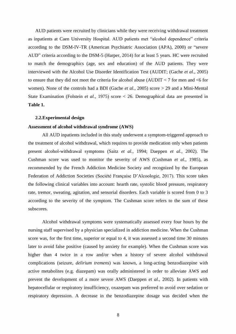
AUD patients were recruited by clinicians while they were receiving withdrawal treatment
as inpatients at Caen University Hospital. AUD patients met “alcohol dependence” criteria
according to the DSM-IV-TR (American Psychiatric Association (APA), 2000) or “severe
AUD” criteria according to the DSM-5 (Harper, 2014) for at least 5 years. HC were recruited
to match the demographics (age, sex and education) of the AUD patients. They were
interviewed with the Alcohol Use Disorder Identification Test (AUDIT; (Gache et al., 2005)
to ensure that they did not meet the criteria for alcohol abuse (AUDIT < 7 for men and <6 for
women). None of the controls had a BDI (Gache et al., 2005) score > 29 and a Mini-Mental
State Examination (Folstein et al., 1975) score < 26. Demographical data are presented in
Table 1.
2.2.Experimental design
Assessment of alcohol withdrawal syndrome (AWS)
All AUD inpatients included in this study underwent a symptom-triggered approach to
the treatment of alcohol withdrawal, which requires to provide medication only when patients
present alcohol-withdrawal symptoms (Saitz et al., 1994; Daeppen et al., 2002). The
Cushman score was used to monitor the severity of AWS (Cushman et al., 1985), as
recommended by the French Addiction Medicine Society and recognized by the European
Federation of Addiction Societies (Société Française D’Alcoologie, 2017). This score takes
the following clinical variables into account: hearth rate, systolic blood pressure, respiratory
rate, tremor, sweating, agitation, and sensorial disorders. Each variable is scored from 0 to 3
according to the severity of the symptom. The Cushman score refers to the sum of these
subscores.
Alcohol withdrawal symptoms were systematically assessed every four hours by the
nursing staff supervised by a physician specialized in addiction medicine. When the Cushman
score was, for the first time, superior or equal to 4, it was assessed a second time 30 minutes
later to avoid false positive (caused by anxiety for example). When the Cushman score was
higher than 4 twice in a row and/or when a history of severe alcohol withdrawal
complications (seizure, delirium tremens) was known, a long-acting benzodiazepine with
active metabolites (e.g. diazepam) was orally administered in order to alleviate AWS and
prevent the development of a more severe AWS (Daeppen et al., 2002). In patients with
hepatocellular or respiratory insufficiency, oxazepam was preferred to avoid over sedation or
respiratory depression. A decrease in the benzodiazepine dosage was decided when the

9
Cushman score was lower or equal to 2 for at least 24 hours, at a rate of 15 to 30% of the total
dose every 24 to 48 hours.
AUD patients were classified into two groups according to the severity of AWS:
- mild-AWS: a maximum Cushman score 4 and no benzodiazepine treatment required
during the acute alcohol withdrawal period according to the symptom-triggered
approach.
- moderate-AWS: a maximum Cushman score > 4 and/or the presence of a history of
severe alcohol withdrawal complications resulting in a benzodiazepine administration
during the acute alcohol withdrawal period.
None of the patients had undergone severe AWS defined by neurological
complications such as delirium tremens or seizures during the acute withdrawal period
examined in the course of the present study.
For all AUD patients, the maximum Cushman score and the number of previous
detoxifications were collected. For patients whom benzodiazepines were prescribed, we also
recorded the number of days and the total amount of benzodiazepines received during alcohol
withdrawal. Alcohol-related variables and withdrawal history are summarized in Table 1.
Neuropsychological assessment
All participants underwent a detailed neuropsychological examination focusing on
processing speed, short-term memory, executive functioning, and episodic memory.
Processing speed was measured with a composite score including the part A (time in seconds)
of the Trail Making Test (Reitan and Wolfson, 1985) and the denomination condition (time in
seconds) of the Stroop Test (Stroop, 1935). Short-term memory was assessed using verbal
spans of the WAIS-III (Wechsler, 1997). For executive functions, a composite score was
computed including performance on 3 tests assessing manipulation of information (verbal
backward spans of the WAIS-III; Wechsler, 1997), inhibition (Stroop Test; Stroop, 1935)
with the time in seconds needed to complete the interference condition minus time needed for
the denomination condition, and mental flexibility (Modified Card Sorting Test, Cianchetti et
al., 2005) with the number of perseverative errors. Two patients had missing data for one

10
measure; their composite score assessing executive functions was therefore computed using
remaining scores. Episodic memory was examined through the sum of the 3 free recalls of the
French version of the Free and Cued Selective Reminding Test (Van der Linden et al., 2004).
Neuropsychological data were transformed into z-scores using the mean and standard
deviation obtained from the HC. The sign of all variables for which high scores were in the
impaired direction (such as completion time or number of errors) was reversed so that all the
z-scores had the same direction: higher z-scores reflecting better performance.
Magnetic Resonance Imaging Data Acquisition and preprocessing
Brain imaging examinations were conducted in 31 HC and 43 AUD patients (13 mild-
AWS and 30 moderate-AWS) within the same week as the neuropsychological assessment.
HC and the two subgroups of AUD patients were matched for age, sex and education level.
All neuroimaging examinations were performed at Cyceron center (Caen, France).
A high-resolution T1-weighted anatomical image was acquired for each subject on a
Philips Achieva 3T scanner (Philips Health-care/Philips Medical Systems International B.V.,
Eindhoven, the Netherlands) using a 3-dimensional fast-field echo sequence (sagittal;
repetition time, 20 ms; echo time, 4.6 ms; flip angle 10°; 180 slices; slice thickness: 1 mm;
field of view 256 x 256 mm2; matrix, 256 x 256). The volumetric magnetic resonance
imaging (MRI) data were analyzed using the Statistical Parametric Mapping software
(SPM12; Welcome Department of Cognitive Neurology, Institute of Neurology, London,
UK). Preprocessing steps included segmentation of the MRI data into gray matter (GM) and
spatial normalization to the Montreal Neurological Institute (MNI) template (voxel size = 1.5
mm3, matrix = 121 x 145 x 121). The normalized GM images were modulated by the
Jacobian determinants to correct for nonlinear warping so that the resulting brain volumes
were corrected for brain size. The resulting images were smoothed by a Gaussian kernel of 8
mm full width at half maximum (FWHM). A GM mask was obtained taking the unmodulated
GM images of HC normalized to the MNI space, averaging them, and thresholding the
resultant mean image at 0.5. The resulting GM mask was applied to GM data analyses.
Sleep
A subgroup of 21 AUD (8 mild-AWS and 13 moderate-AWS) and 15 HC, matched for
age, sex and education, underwent one night of polysomnography (PSG) using a portable
recording device (Siesta®, Compumedics, Australia), allowing AUD patients to sleep at the

11
addiction department and HC at their home. The PSG was conducted within the same week as
the neuropsychological and MRI examinations. The PSG acquisition consisted of twenty EEG
electrodes (Fp1, Fp2, F3, F4, F7, F8, T3, T4, C3, C4, P3, P4, O1, O2, FZ, CZ, PZ, vertex
ground and a bi-mastoid reference) placed over the scalp according to the international 10-20
system, with impedances kept below 5 kΩ. We also recorded the electrooculogram (EOG),
chin EMG, ECG, respiratory movements using abdominal and thoracic belts, respiratory
airflow using nasal and oral thermistor, and oxygen saturation with a finger pulse oximeter.
The EEG signal was digitalized at a sampling rate of 256 Hz. High-pass and low-pass filters
were applied, respectively at 0.3 Hz and 35 Hz. PSG recordings were scored in 30-s epochs
according to the American Association of Sleep Medicine (AASM, 2017) standard criteria.
The following parameters were obtained: total sleep time (TST; in minutes), sleep efficiency
(SE (%), corresponding to time spent asleep/ time in bed), sleep onset latency (in minutes,
referring to the time from lights-off to the first three epochs of any stage of sleep), wake after
sleep onset (in minutes), time spent in each sleep stage (N1, N2, N3, and REM sleep,
expressed as percentages of TST), arousal index (number of arousals/ TST), stage shifts index
(number of sleep stage transitions to N1/TST) and the Apnea-Hypopnea Index (AHI,
corresponding to the number of respiratory events per hour of sleep). We also calculated a
composite score to assess sleep fragmentation including the micro-arousal index, the arousal
index and the number of stage transitions to N1.
All participants underwent a self-assessment of their sleep quality using the Pittsburg
Sleep Quality Index (PSQI; Buysse et al., 1989) in its initial version (previous month) for HC
and with an adapted version (previous week, to better reflect the different stages of alcohol
treatment) for AUD patients. The Epworth Sleepiness Scale (ESS; Johns et al., 1991) was
proposed to assess signs of daytime sleepiness in all participants.
2.3.Statistical analysis
To test the differences between HC and the two subgroups of AUD patients (mild-
AWS and moderate-AWS), non-parametric Kruskall-Wallis’s tests were conducted on
demographical variables (age and education) and a Chi² test was performed to compare the
sex ratio in each group. Mann-Whitney’s tests were performed between mild-AWS and
moderate-AWS for alcohol- and withdrawal-related variables. Non-parametric Kruskall-
Wallis’s tests were conducted on neuropsychological data and sleep measures followed by
post-hoc comparisons (Mann-Whitney’s tests) when appropriate. The statistical threshold was
set to p <0.05.

12
Neuroimaging data were analyzed using SPM12 (Statistical Parametric Mapping,
www.fil.ion.ucl.ac.uk/spm). More precisely, we conducted voxel-based ANCOVAs to
compare GM volume between HC, mild-AWS and moderate-AWS patients, controlling for
the intracranial volume. We corrected for multiple comparisons (family wise error (FWE),
p<0.05) with a minimal cluster size (k) of 60 voxels (200 mm3). Then, comparisons between
the two-subgroups of AUD patients were reported at p<0.001 with a minimal cluster size (k)
of 60 voxels (200 mm3). Only results surviving a cluster-level correction are reported.
Finally, the relationships between GM volume, sleep variables and cognitive
performance were examined in the entire group of AUD patients with Spearman’s
correlations. Only the variables for which we found a significant difference between mild-
AWS and moderate-AWS were entered in the analyses. When we observed significant
relationships between a sleep variable on the one hand and both a GM volume and a cognitive
variable on the other hand, we performed causal mediation analyses to assess the
directionality of the relationships. Mediations analyses allow to test whether the causal effect
of an independent variable (X) on a dependent variable (Y) is explained by a mediating
variable (M). In other words, X exerts its effects on Y because X affects M, which in turn,
affects Y (Goldstone et al., 2018). Applied to our study, two models were tested to determine
i) whether GM volume mediates the relationships between sleep and cognitive performance or
ii) whether sleep mediates the relationships between GM volume and cognition. These
analyses were performed using the “mediation” R package (Tingley et al., 2014). We reported
the average direct effects (ADE) and average causal mediation effect (ACME) estimated
using nonparametric bootstrapping (5000 simulations, p<.05) for each model.
2.4.Data availability
All data and materials used within this study will be made available, upon reasonable
request, to research groups wishing to reproduce/confirm our results.
3. Results
3.1.Comparisons between HC, mild-AWS and moderate-AWS patients
Demographical variables and anxiety-depression levels: Kruskall-Wallis tests with Group as
a between-subject factor (HC, mild, moderate-AWS) did not reveal any significant effect of
group for age (H(2,104) = 2.48, p = 0.29) and education (H(2,104) = 4.24, p = 0.12). A Chi2-test

13
showed that sex ratio was similar between groups (χ² = 2.85, p = 0.24). Kruskall-Wallis tests
showed a significant effect of group for depression (H(2,103) = 42, p<0.0001), anxiety-state
(H(2,102) = 8.55, p=0.01) and anxiety-trait (H(2,102) = 32.83, p<0.0001). Mild-AWS and
moderate-AWS were both more depressed than HC (all p-values < 0.0001) but did not differ
from each other (p = 0.85). Only moderate-AWS differed from HC on the anxiety-state
questionnaire (p = 0.004). Mild-AWS and moderate-AWS exhibited higher levels of anxiety-
trait compared to HC (all p-values < 0.0001), but did not differ from each other (p = 0.14).
Results are presented in Table 1.
Alcohol-related variables: A Kruskall-Wallis test showed a significant effect of group for the
AUDIT score (H(2,103) = 76.97, p < 0.0001). Post-hoc comparisons revealed, as expected, that
mild-AWS and moderate-AWS patients presented higher AUDIT scores than HC (all p-
values = 0.0001) but did not differ from each other (p = 0.41). Mann-Whitney U tests did not
show any significant difference between mild-AWS and moderate-AWS for the duration of
alcohol misuse (U = 279,5, p = 0.73) and dependence (U = 265, p = 0.44), but moderate-AWS
reported a higher daily alcohol consumption than mild-AWS patients (U = 191,5, p = 0.05;
Table 1).
Alcohol withdrawal variables: Mild-AWS and moderate-AWS patients had an equivalent
number of previous detoxifications (U = 265,5, p = 0.44). As expected, compared to mild-
AWS patients, moderate-AWS patients had experienced a higher maximum Cushman score
(U = 35, 5, p < 0.0001), had received more diazepam (U = 0, p < 0.0001) and during a longer
period (U = 0, p < 0.0001 ; Table 1).
Pattern of cognitive alterations Kruskall-Wallis tests revealed a significant effect of group for
processing speed (H(2, 104) = 29.7, p < 0.0001), short-term memory (H(2, 103) = 12.68, p =
0.002), executive functions (H(2, 104) = 23.38, p < 0.0001) and episodic memory (H(2, 104) =
22.32, p < 0.0001). Post-hoc comparisons indicated that mild-AWS patients presented lower
performance than HC for episodic memory (U = 148, p < 0.0001) and processing speed (U =
177, p = 0.0004), but not for short-term memory (U = 422, p = 0.97) and executive
performance (U = 384.5 , p = 0.56). Compared to HC, moderate-AWS patients presented
lower performance for all cognitive measures (all p-values < 0.001). Compared to mild-AWS
patients, moderate-AWS patients had lower executive (U = 154, p = 0.003) and short-term
memory performance (U = 178. 5, p = 0.01), but did not differ for processing speed and

14
episodic memory (all p values > 0.05; Table 2 and Fig. 1).
Pattern of GM alterations: Mild-AWS patients did not differ from HC regarding GM volume
(p(FWE) < 0.05; Fig. 2A). Compared to HC, moderate-AWS patients had significantly lower
GM volume in frontal and prefrontal areas, insula, lateral and medial temporal cortices
(including the hippocampus and parahippocampal gyrus), cingulate and occipital cortices,
cerebellum, and in subcortical regions including the thalamus, putamen, and caudate nuclei
(p(FWE) < 0.05; Fig. 2B). Compared to mild-AWS patients, moderate-AWS patients had
significantly lower GM volume in the right inferior frontal cortex (Broadman area 44), the
bilateral insula, a cluster encompassing the anterior cingulate cortex and the medial superior
frontal cortex, occipito-parietal regions and limbic structures including the anterior part of the
thalamus and the hypothalamus. All regions listed above are significant at p < 0.001
(uncorrected) threshold and survived the cluster-level correction p < 0.05; Fig. 2C). The
reverse comparison (mild-AWS patients < moderate-AWS patients) did not reveal any
significant result.
Pattern of sleep alterations: Non-parametric Kruskall-Wallis tests revealed a significant effect
of group for both percentage of sleep stages N1 (H (2, N=36) = 11.81 p = 0.003) and N3 (H (2,
N=36) = 15.87 p = 0.0004). Post-hoc comparisons (Mann-Whitney U tests) showed that
compared to HC, mild-AWS patients had similar percentage of N1 (p = 0.24) and N3 sleep (p
= 0.52). Compared to HC, moderate-AWS patients showed higher percentage of N1 (p =
0.0006) and lower percentage of N3 sleep (p = 0.0002). Compared to mild-AWS patients,
moderate-AWS patients showed lower percentage of N3 sleep (p = 0.004; Table 2 and Fig.
3).
A significant effect of group was observed for the apnea-hypopnea index (AHI) (H (2, N=36) =
7.99 p = 0.02). Post-hoc comparisons showed that mild-AWS patients and moderate-AWS
patients exhibited a higher AHI compared to controls (p = 0.03 and p = 0.01 respectively) but
did not differ from each other (p > 0.05). No group differences were observed for sleep
latency, duration, efficiency and wake after sleep onset (all p-values > 0.05). Results
remained unchanged when controlling for the AHI (data not shown).
A significant effect of group was observed for the PSQI score (H(2, N=30) = 11.19, p = 0.004).
Post-hoc comparisons showed that compared to HC, moderate-AWS patients had a higher
score (p = 0.001). Mild-AWS patients did not differ from HC and moderate-AWS patients (all

15
p-values > 0.05). No group difference was observed on the ESS score (p-value > 0.05; Table
2).
3.2.Relationships between GM volume, sleep and cognitive performance in the entire
group of AUD patients.
Signal values within the significant clusters obtained from the comparison mild-AWS >
moderate-AWS were extracted. Then, we conducted Spearman’s correlations between GM
volumes, sleep variables and cognitive performance in the entire group of patients. Only
variables for which we found a significant difference between mild-AWS and moderate-AWS
patients were entered in these analyses (i.e. short-term memory performance, executive
abilities and N3 sleep). Short-term memory performance positively correlated with GM
volume in the insula (rho = 0.51, p = 0.01), occipito-parietal cortex (rho = 0.62, p = 0.003),
inferior frontal gyrus (rho = 0.45, p = 0.04) and thalamus/hypothalamus (rho = 0.45, p =
0.04), but not in the anterior cingulate cortex (p > 0.05). Executive performance positively
correlated with all significant clusters of GM volume (insula: rho = 0.7, p = 0.0004; occipito-
parietal cortex: rho = 0.66, p = 0.001; anterior cingulate cortex: rho = 0.57, p = 0.007; inferior
frontal gyrus: rho = 0.53, p = 0.01; thalamus/hypothalamus: rho = 0.64, p = 0.002). GM
volume in all significant clusters positively correlated with the percentage of N3 sleep (insula:
rho = 0.52, p = 0.01; occipito-parietal cortex: rho = 0.55, p = 0.009; anterior cingulate cortex:
rho = 0.43, p = 0.05; inferior frontal gyrus: rho = 0.56, p = 0.008; thalamus/hypothalamus: rho
= 0.51, p = 0.02). Moreover, the percentage of N3 sleep positively correlated with short-term
memory (rho = 0.48, p = 0.03) and executive performance (rho = 0.44, p = 0.04).
To better understand the directionality of the relationships between GM volume, N3 sleep and
cognitive performance, we conducted mediation analyses and tested two models for each
cluster. In the first one (Model 1), N3 sleep was the independent variable (X) and GM volume
in the 5 significant clusters reported in the previous analysis was the mediator (M). In the
second model (Model 2), GM volume was the independent variable (X) and N3 sleep was the
mediator (M). In both models, cognitive variables (short-term memory or executive scores)
were entered as the dependent variable (Y). First, considering short-term memory
performance as the dependent variable, mediation analyses referring to both Model 1 and 2
were not significant (p > 0.05; Supplementary Table 1). Second, considering executive
functions performance as the dependent variable, mediation analyses revealed that the volume

16
of the bilateral insula (p=0.006), right inferior frontal cortex (p=0.02), occipito-parietal cortex
(p=0.01) and the cluster including the anterior thalamus and the hypothalamus (p=0.02)
significantly mediated the relationships between the percentage of N3 sleep and executive
performance. None of the analyses referring to Model 2 was significant. Results of these
mediation analyses are detailed in Table 3 and Fig. 4.
4. Discussion
The present study aimed at determining whether AWS severity contributes to the
heterogeneity of sleep changes, brain alterations and cognitive deficits observed in AUD
patients early in abstinence. We showed that AWS severity contributes to the
pathophysiology of NREM sleep abnormalities, decrease GM volumes in fronto-insular and
thalamus/hypothalamus regions as well as short-term memory and executive deficits in AUD
patients early in abstinence. We also found that lower percentage of N3 sleep related to
cognitive deficits (short-term memory and executive functions) both directly and indirectly
via GM shrinkage in AUD patients.
We showed that 68% of AUD patients exhibited a moderate AWS, which is a higher
prevalence than the one reported in a previous study (Mirijello et al., 2015). In the present
study, all patients were recruited in a special unit for alcohol detoxification. They were thus
potentially at risk for AWS complications. Contrary to a previous study (Duka et al., 2004) in
which only patients with severe alcohol withdrawal complications were included, we did not
observe any association between multiple detoxifications and the severity of the current
AWS. We also found that AUD patients with moderate AWS reported higher alcohol
consumption during the month preceding withdrawal than those with mild AWS. This result
suggests that during withdrawal, the brain hyperexcitability resulting from increased
glutamate transmission combined to decreased GABA transmission may be related to the
quantity of recent alcohol drinking (De Witte et al., 2003; Jesse et al., 2017).
Our study first confirms the pattern of sleep alterations observed in recently detoxified
AUD patients, consisting of increased light sleep (N1), a lower percentage of N3 sleep (Gillin
et al., 1990; Junghanns et al., 2009; de Zambotti et al., 2014, Irwin et al., 2016a; Singh et al.,
2018) and the presence of a sleep complaint (Laniepce et al., 2019). It also specifies that only

17
patients with moderate AWS presented these objective sleep alterations, as well as self
reported sleep difficulties. The difference observed between moderate- and mild-AWS
patients for N3 sleep is in accordance with a previous study showing that the number of
alcohol withdrawal symptoms was negatively related to the percentage of N3 sleep (Gillin et
al., 1990). Sleep changes may be directly related to AWS severity since N3 sleep has been
associated with lower glutamate levels (Dash et al., 2009). Even when the clinical symptoms
of AWS have faded, a hyper-glutamate activity may persist and alter brain regions involved in
the generation and maintenance of sleep rhythms, resulting in lower percentage of N3 sleep
(Dang-Vu, 2012). Sleep abnormalities observed in our group of moderate-AWS patients may
thus be interpreted as a persistent subacute alcohol withdrawal symptom (Brower, 2001;
Feige et al., 2007).
AWS severity was also related to structural brain alterations in the right inferior
frontal cortex, the bilateral insula, the anterior cingulate cortex and the
thalamus/hypothalamus, which are brain regions known to be affected in AUD patients (Pitel
et al., 2012; Yang et al., 2016). Regarding the effect of AWS severity on the anterior
cingulate cortex, studies using magnetic resonance spectroscopy reported increased glutamate
levels in this brain region during alcohol withdrawal in both humans and rats (Lee et al.,
2007; Hermann et al., 2012). Several studies suggested that the extent of structural brain
abnormalities in AUD patients may be partially explained by alcohol withdrawal-related
toxicity (De Witte et al., 2003; Duka et al., 2011; O’Daly et al., 2012; Trick et al., 2014;
Frischknecht et al., 2017). During alcohol withdrawal, the glutamate-mediated excitotoxicity
induces neuronal death, which may explain structural brain alterations observed early in
abstinence in AUD patients (Tsai and Coyle, 1998). The frontal lobes being particularly rich
in glutamatergic pathways (Kril et al., 1997), they are likely to be especially vulnerable to the
severity of AWS. However, we do not exclude that these brain alterations may have been
present before alcohol cessation because of the effects of chronic and heavy alcohol
consumption or a family history of AUD or comorbidities such as liver disease or thiamine
deficiency (Harper, 2009; Chen et al., 2012; Filippi et al., 2019). In this case, altered brain
structure would constitute a vulnerability factor for exhibiting more severe AWS. Further
studies including longitudinal measures of glutamate levels combined with structural MRI at
different stages of the disease (active drinking, withdrawal period and abstinence) are now
required.
Regarding cognitive abilities, our findings indicate that AWS severity may have
deleterious effects on short-term memory and executive performance. While short-term

18
memory deficits have previously been reported in AUD patients (Pitel et al., 2007), we
suggest here that AWS may contribute to this alteration. Executive dysfunctions are frequent
in AUD patients early in abstinence (Stavro et al., 2013) and could notably be influenced by
alcohol history (Sullivan et al., 2000; Zinn et al., 2004; Maurage et al., 2014), associated liver
disease (Ritz et al., 2016) and multiple detoxifications (Duka et al., 2004; Loeber et al.,
2009). The present study suggests that beyond the repetition of alcohol withdrawals, the
severity of AWS itself may represent another factor influencing the heterogeneity of
executive impairments in AUD patients. By contrast, AWS severity does not appear to
modulate processing speed and episodic memory deficits. Indeed, episodic memory was
affected to the same extent in the two subgroups of patients. Previous studies suggested that
episodic memory abilities may rather be influenced by thiamine metabolism (Pitel et al.,
2011; Ritz et al., 2016).
Mediation analyses enabled us to deepen our understanding of the relationships
between sleep, brain and cognition considering the severity of AWS. For the first time, we
showed that poor restorative sleep (reflected by a lower percentage of N3 sleep) is related to
cognitive deficits both directly and indirectly through GM shrinkage. In line with a previous
study in sleep-deprived individuals (Chengyang et al., 2017), we demonstrated that poor
restorative sleep is associated with short-term memory deficits in AUD patients. This
relationship was not mediated by GM alterations, suggesting the potential contribution of
other mechanisms such as altered functional activity of hippocampal-cortical circuits
(Chengyang et al., 2017). Interestingly, we found that poor restorative sleep contributes to
fronto-insular shrinkage that in turn results in executive deficits. Our results suggest a
potential role of sleep in the pathophysiological mechanisms of alcohol-related GM
alterations in AUD patients. Poor sleep quality may result from alcohol-related brain damage
such as increased glutamate levels (Cortese et al., 2010), neuroinflammation (Irwin et al.,
2016b) and oxidative stress (Villafuerte et al., 2015). These mechanisms may contribute to
fronto-insular and thalamic/hypothalamic abnormalities in AUD patients recently detoxified.
Fronto-insular regions, such as the right inferior frontal gyrus, have been related to executive
deficits, notably during inhibition tasks (Levy and Wagner, 2011; Aron et al., 2014; Wiers et
al., 2015). The insula promotes the integration and representation of interoceptive information
into conscious feelings and viscerosensory signals leading to decision making (Craig, 2009).
The insula being also a neuronal substrate of craving, insular shrinkage may underpin the
relationship between sleep alterations and relapse (Brower, 2003). Thus, these analyses seem

19
to explain, at least partially, the potential associations existing between sleep, brain and
cognition in AUD patients, by showing that N3 sleep contributes to GM shrinkage in fronto-
insular and thalamus/hypothalamus regions resulting in executive deficits. We do not exclude
that, on the contrary, alterations in other brain regions may induce sleep abnormalities that, in
turn, underlie cognitive functioning.
The present study has several strengths, including a meticulous collection of
information about AWS severity associated with a detailed cognitive assessment, an MRI
scan and an objective sleep assessment in AUD patients examined early in abstinence.
However, several limitations should be mentioned. First, the results of the present study were
obtained in AUD patients without comorbidities nor other forms of substance use disorder
(except tobacco). They cannot therefore be generalized to all AUD patients in Addiction
departments given the frequency of comorbidities and multiple drug use. Second, all
participants performed only one night of PSG-recording, which did not allow to control for
“the first-night effect”. Third, although none of the AUD patients included in the present
study fulfilled diagnostic criteria for current anxiety disorder and/or depression, higher self-
report anxiety and depressive levels were reported by AUD patients compared to HC. Psycho-
affective factors may influence sleep quality and cognitive performance in AUD patients as
these factors are known to have a negative impact on neuropsychological abilities (Gualtieri
and Morgan, 2008) and sleep quality (Baglioni et al., 2016). Nevertheless, the two groups of
AUD patients did not differ from each other on these variables and results remained
unchanged when psycho-affective factors (BDI, STAI-A, STAI-B scores) were added as
covariates (data not shown). Finally, the current clinical measure of AWS severity makes it
difficult to disentangle the direct effect of pathophysiological mechanisms underlying AWS
(hyperglutamatergy) from the potential effect of AWS-related benzodiazepine treatment.
AWS severity was treated with benzodiazepines, corresponding to the gold-standard for AWS
treatment (Amato et al., 2010). Benzodiazepines are known to alter objective sleep quality
and cognitive functions after an acute administration (Huron et al., 2001; Deakin et al., 2004;
Roux and Kryger, 2010) and/or after a chronic consumption (Barker, 2004; Doghramji and
Jangro, 2016; Fond et al., 2018). In the present study, AUD patients with benzodiazepine
dependence prior the hospitalization were not included and patients who needed
benzodiazepines during withdrawal were prescribed for only a few days (4-17 days). In
addition, patients were included at least 48 hours after the last benzodiazepine prescription
(according to the half-life of the benzodiazepine used), after a progressive decrease of the

20
benzodiazepine dosage starting when the Cushman score was lower or equal to 2 for at least
24 hours, at a rate of 15 to 30% of the total dose every 24 to 48 hours. Since prolonged
diazepam intake may increase its terminal elimination time, we conducted supplementary
analyses that did not show any correlation between the number of days since the last
benzodiazepine prescription and our main results (Supplementary Table 2). This absence of
relationship suggests that the differences observed between mild- and moderate-AWS patients
do not result from the residual effects of benzodiazepines. However, further studies are
required to specify the effects of short-term prescription of benzodiazepines on sleep quality,
GM volumes and cognition in AUD patients.
Taken together, our results bring new insights on the pathophysiological mechanisms
of sleep, structural brain alterations and cognitive deficits observed in recently detoxified
AUD patients, showing the contribution of AWS severity. Moreover, we added novel
evidence that poor sleep quality may contribute to cognitive deficits directly or indirectly
through increased GM shrinkage in AUD patients early in abstinence. Further studies aiming
at exploring brain, sleep or cognition in AUD patients should consider AWS severity to limit
the heterogeneity of the AUD sample. For clinicians, these results suggest that a careful
monitoring of AWS is not only useful to prevent the development of severe AWS
complications, but also to predict sleep alterations, brain damage and cognitive deficits
associated with a poor treatment outcome.
Acknowledgments:
The authors are grateful to the Cyceron MRI staff members for their help with patients and
imaging examination acquisition, and Coralie Lannuzel, Stéphane Rehel, Ludivine Ritz and
Hélène Beaunieux for their help at various stages of the study. We would also like to thank all
the participants.
Funding:
This work was supported by the French National Institute for Health and Medical Research
(INSERM), the French National Agency for Research (ANR), and Conseil Regional de
Normandie. Alice Laniepce’s doctoral fellowship was co-funded by European Union in the
framework of the ERDF-ESF Operational Programme 2014-2020 and Lundbeck.
Competing interests:

21
The authors report no competing interests.
5. References
Aggleton JP. Multiple anatomical systems embedded within the primate medial temporal
lobe: Implications for hippocampal function. Neurosci Biobehav Rev 2012; 36: 1579–1596.
Amato L, Minozzi S, Vecchi S, Davoli M. Benzodiazepines for alcohol withdrawal. Cochrane
Database Syst Rev 2010
American Psychiatric Association. DSM-5 Diagnostic Classification. In: Diagnostic and
Statistical Manual of Mental Disorders. American Psychiatric Association; 2013
American Psychiatric Association (APA). Diagnostic and Statistical Manual of Mental
Disorders, Fourth Edition, Text Revision (DSM-IV-TR). Arlington, VA: American
Psychiatric Association; 2000
Angarita GA, Emadi N, Hodges S, Morgan PT. Sleep abnormalities associated with alcohol,
cannabis, cocaine, and opiate use: a comprehensive review. Addict Sci Clin Pract 2016; 11: 9.
Aron AR, Robbins TW, Poldrack RA. Inhibition and the right inferior frontal cortex: One
decade on. Trends Cogn Sci 2014; 18: 177–185.
Baglioni C, Nanovska S, Regen W, Spiegelhalder K, Feige B, Nissen C, et al. Sleep and
mental disorders: A meta-analysis of polysomnographic research. Psychol Bull 2016; 142:
969–990.
Barker M. Persistence of cognitive effects after withdrawal from long-term benzodiazepine
use: a meta-analysis. Arch Clin Neuropsychol 2004; 19: 437–454.
Bates ME, Bowden SC, Barry D. Neurocognitive impairment associated with alcohol use
disorders: implications for treatment. Exp Clin Psychopharmacol 2002; 10: 193–212.
Beck AT, Ward CH, Mendelson M, Mock J, Erbaugh J. An inventory for measuring
depression. Arch Gen Psychiatry 1961; 4: 561–571.
Begleiter H, Porjesz B. Persistence of a “subacute withdrawal syndrome” following chronic
ethanol intake. Drug Alcohol Depend 1979; 4: 353–357.
Le Berre AP, Fama R, Sullivan E V. Executive Functions, Memory, and Social Cognitive
Deficits and Recovery in Chronic Alcoholism: A Critical Review to Inform Future Research.
Alcohol Clin Exp Res 2017; 41: 1432–1443.
Brower KJ. Alcohol’s effects on sleep in alcoholics. Alcohol Res Health 2001; 25: 110–25.
Brower KJ. Insomnia, alcoholism and relapse. Sleep Med Rev 2003; 7: 523–539.
Chakravorty S, Chaudhary NS, Brower KJ. Alcohol Dependence and Its Relationship With

22
Insomnia and Other Sleep Disorders. Alcohol Clin Exp Res 2016; 40: 2271–2282.
Chen CH, Walker J, Momenan R, Rawlings R, Heilig M, Hommer DW. Relationship
Between Liver Function and Brain Shrinkage in Patients with Alcohol Dependence. Alcohol
Clin Exp Res 2012; 36: 625–632.
Chengyang L, Daqing H, Jianlin Q, Haisheng C, Qingqing M, Jin W, et al. Short-term
memory deficits correlate with hippocampal-thalamic functional connectivity alterations
following acute sleep restriction. Brain Imaging Behav 2017; 11: 954–963.
Cianchetti C, Corona S, Foscoliano M, Scalas F, Sannio-Fancello G. Modified Wisconsin
Card Sorting Test: proposal of a supplementary scoring method. Arch Clin Neuropsychol
2005; 20: 555–8.
Cortese BM, Mitchell TR, Galloway MP, Prevost KE, Fang J, Moore GJ, et al. Region-
specific alteration in brain glutamate: Possible relationship to risk-taking behavior. Physiol
Behav 2010; 99: 445–450.
Craig AD. How do you feel - now? The anterior insula and human awareness. Nat Rev
Neurosci 2009; 10: 59–70.
Cushman P, Forbes R, Lemer W, Stewart M. Alcohol Withdrawal Syndromes: Clinical
Management with Lofexidine. Alcohol Clin Exp Res 1985; 9: 103–108.
Daeppen JB, Gache P, Landry U, Sekera E, Schweizer V, Gloor S, et al. Symptom-triggered
vs fixed-schedule doses of benzodiazepine for alcohol withdrawal: A randomized treatment
trial. Arch Intern Med 2002; 162: 1117–1121.
Dang-Vu TT. Neuronal oscillations in sleep: Insights from functional neuroimaging.
NeuroMolecular Med 2012; 14: 154–167.
Dash MB, Douglas CL, Vyazovskiy V V., Cirelli C, Tononi G. Long-Term Homeostasis of
Extracellular Glutamate in the Rat Cerebral Cortex across Sleep and Waking States. J
Neurosci 2009; 29: 620–629.
Deakin JB, Aitken MRF, Dowson JH, Robbins TW, Sahakian BJ. Diazepam produces
disinhibitory cognitive effects in male volunteers. Psychopharmacology (Berl) 2004; 173: 88–
97.
Doghramji K, Jangro WC. Adverse Effects of Psychotropic Medications on Sleep. Sleep Med
Clin 2016; 11: 503–514.
Duka T, Gentry J, Malcolm R, Ripley TL, Borlikova G, Stephens DN, et al. Consequences of
multiple withdrawals from alcohol. Alcohol Clin Exp Res 2004; 28: 233–46.
Duka T, Townshend JM, Collier K, Stephens DN. Impairment in Cognitive Functions After
Multiple Detoxifications in Alcoholic Inpatients. Alcohol Clin Exp Res 2003; 27: 1563–1572.

23
Duka T, Trick L, Nikolaou K, Gray MA, Kempton MJ, Williams H, et al. Unique brain areas
associated with abstinence control are damaged in multiply detoxified alcoholics. Biol
Psychiatry 2011; 70: 545–552.
Feige B, Scaal S, Hornyak M, Gann H, Riemann D. Sleep Electroencephalographic Spectral
Power After Withdrawal from Alcohol in Alcohol-Dependent Patients. Alcohol Clin Exp Res
2007; 31: 19–27.
Filippi I, Hoertel N, Artiges E, Airagnes G, Guérin-Langlois C, Seigneurie A-S, et al. Family
history of alcohol use disorder is associated with brain structural and functional changes in
healthy first-degree relatives. Eur Psychiatry 2019; 62: 107–115.
Folstein MF, Folstein SE, McHugh PR. “Mini-mental state”. J Psychiatr Res 1975; 12: 189–
198.
Fond G, Berna F, Boyer L, Godin O, Brunel L, Andrianarisoa M, et al. Benzodiazepine long-
term administration is associated with impaired attention/working memory in schizophrenia:
results from the national multicentre FACE-SZ data set. Eur Arch Psychiatry Clin Neurosci
2018; 268: 17–26.
Frischknecht U, Hermann D, Tunc-Skarka N, Wang G-Y, Sack M, van Eijk J, et al. Negative
Association Between MR-Spectroscopic Glutamate Markers and Gray Matter Volume After
Alcohol Withdrawal in the Hippocampus: A Translational Study in Humans and Rats.
Alcohol Clin Exp Res 2017; 41: 323–333.
Gache P, Michaud P, Landry U, Accietto C, Arfaoui S, Wenger O, et al. The Alcohol Use
Disorders Identification Test (AUDIT) as a Screening Tool for Excessive Drinking in Primary
Care: Reliability and Validity of a French Version. Alcohol Clin Exp Res 2005; 29: 2001–
2007.
Gillin JC, Smith TL, Irwin M, Kripke DF, Schuckit M. EEG sleep studies in ‘pure’ primary
alcoholism during subacute withdrawal: Relationships to normal controls, age, and other
clinical variables. Biol Psychiatry 1990; 27: 477–488.
Goldstone A, Willoughby AR, de Zambotti M, Franzen PL, Kwon D, Pohl KM, et al. The
mediating role of cortical thickness and gray matter volume on sleep slow-wave activity
during adolescence. Brain Struct Funct 2018; 223: 669–685.
Goodson CM, Clark BJ, Douglas IS. Predictors of Severe Alcohol Withdrawal Syndrome: A
Systematic Review and Meta-Analysis. Alcohol Clin Exp Res 2014; 38: 2664–2677.
Gualtieri CT, Morgan DW. The frequency of cognitive impairment in patients with anxiety,
depression, and bipolar disorder: An unaccounted source of variance ie clinical trials. J Clin
Psychiatry 2008; 69: 1122–1130.

24
Harper C. The neuropathology of alcohol-related brain damage. Alcohol Alcohol 2009; 44:
136–140.
Harper C. Diagnostic and statistical manual of mental disorders. In: A Companion to Criminal
Justice, Mental Health & Risk. Arlington; 2014
Heilig M, Egli M, Crabbe JC, Becker HC. Acute withdrawal, protracted abstinence and
negative affect in alcoholism: are they linked? Addict Biol 2010; 15: 169–184.
Hermann D, Weber-Fahr W, Sartorius A, Hoerst M, Frischknecht U, Tunc-Skarka N, et al.
Translational magnetic resonance spectroscopy reveals excessive central glutamate levels
during alcohol withdrawal in humans and rats. Biol Psychiatry 2012; 71: 1015–21.
Huron C, Servais C, Danion JM. Lorazepam and diazepam impair true, but not false,
recognition in healthy volunteers. Psychopharmacology (Berl) 2001; 155: 204–209.
Irwin MR, Bjurstrom MF, Olmstead R. Polysomnographic measures of sleep in cocaine
dependence and alcohol dependence: Implications for age-related loss of slow wave, stage 3
sleep. Addiction 2016; 111: 1084–1092.
Irwin MR, Olmstead R, Carroll JE. Sleep disturbance, sleep duration, and inflammation: A
systematic review and meta-analysis of cohort studies and experimental sleep deprivation.
Biol Psychiatry 2016; 80: 40–52.
Jesse S, Bråthen G, Ferrara M, Keindl M, Ben-Menachem E, Tanasescu R, et al. Alcohol
withdrawal syndrome: mechanisms, manifestations, and management. Acta Neurol Scand
2017; 135: 4–16.
Junghanns K, Backhaus J, Veltrup C, Dageförde J, Brückmann H, Wetterling T. Mildly
disturbed hepatic and pancreatic function during early abstention from alcohol is associated
with brain atrophy and with disturbed psychometric performance. Alcohol Alcohol 2004; 39:
113–118.
Junghanns K, Horbach R, Ehrenthal D, Blank S, Backhaus J. Chronic and high alcohol
consumption has a negative impact on sleep and sleep-associated consolidation of declarative
memory. Alcohol Clin Exp Res 2009; 33: 893–897.
Kelly RM, Strick PL. Cerebellar loops with motor cortex and prefrontal cortex of a nonhuman
primate. J Neurosci 2003; 23: 8432–44.
Kim DW, Kim HK, Bae EK, Park SH, Kim KK. Clinical predictors for delirium tremens in
patients with alcohol withdrawal seizures. Am J Emerg Med 2015; 33: 701–704.
Kouimtsidis C, Duka T, Palmer E, Lingford-Hughes A. Prehabilitation in alcohol dependence
as a treatment model for sustainable outcomes. A narrative review of literature on the risks
associated with detoxification, from animal models to human translational research. Front

25
Psychiatry 2019; 10: 339.
Kril JJ, Halliday GM, Svoboda MD, Cartwright H. The cerebral cortex is damaged in chronic
alcoholics. Neuroscience 1997; 79: 983–98.
Kühn S, Charlet K, Schubert F, Kiefer F, Zimmermann P, Heinz A, et al. Plasticity of
hippocampal subfield volume cornu ammonis 2+3 over the course of withdrawal in patients
with alcohol dependence. JAMA Psychiatry 2014
Laniepce A, Segobin S, Lannuzel C, Boudehent C, Ritz L, Urso L, et al. Neuropsychological
and Neuroimaging Examinations of Self‐Reported Sleep Quality in Alcohol Use Disorder
With and Without Korsakoff’s Syndrome. Alcohol Clin Exp Res 2019; 43: 952–964.
Lee E, Jang DP, Kim JJ, An SK, Park S, Kim IY, et al. Alteration of brain metabolites in
young alcoholics without structural changes. Neuroreport 2007; 18: 1511–1514.
Levy BJ, Wagner AD. Cognitive control and right ventrolateral prefrontal cortex: Reflexive
reorienting, motor inhibition, and action updating. Ann N Y Acad Sci 2011; 1224: 40–62.
Van der Linden M, Coyette F, Poitrenaud J, Kalafat M, Calicis F, Wyns C, et al. L’épreuve de
rappel libre / rappel indicé à 16 items (RL/RI-16). In: L’évaluation des troubles de la
mémoire : présentation de quatre tests de mémoire épisodique avec leur étalonnage. Marseille;
2004. p. 25–42
Loeber S, Duka T, Márquez HW, Nakovics H, Heinz A, Mann K, et al. Effects of repeated
withdrawal from alcohol on recovery of cognitive impairment under abstinence and rate of
relapse. Alcohol Alcohol 2010; 45: 541–547.
Loeber S, Duka TT, Welzel H, Nakovics H, Heinz A, Flor H, et al. Impairment of cognitive
abilities and decision making after chronic use of alcohol: The impact of multiple
detoxifications. Alcohol Alcohol 2009; 44: 372–381.
Lukoyanov N V., Madeira MD, Paula-Barbosa MM. Behavioral and neuroanatomical
consequences of chronic ethanol intake and withdrawal. Physiol Behav 1999
Maurage P, de Timary P, Billieux J, Collignon M, Heeren A. Attentional Alterations in
Alcohol Dependence Are Underpinned by Specific Executive Control Deficits. Alcohol Clin
Exp Res 2014; 38: 2105–2112.
Mirijello A, D’Angelo C, Ferrulli A, Vassallo G, Antonelli M, Caputo F, et al. Identification
and Management of Alcohol Withdrawal Syndrome. Drugs 2015; 75: 353–365.
Nolen-Hoeksema S. Gender differences in risk factors and consequences for alcohol use and
problems. Clin Psychol Rev 2004; 24: 981–1010.
O’Daly OG, Trick L, Scaife J, Marshall J, Ball D, Phillips ML, et al. Withdrawal-Associated
Increases and Decreases in Functional Neural Connectivity Associated with Altered

26
Emotional Regulation in Alcoholism. Neuropsychopharmacology 2012; 37: 2267–2276.
Oscar-Berman M, Kirkley SM, Gansler DA, Couture A. Comparisons of Korsakoff and non-
Korsakoff alcoholics on neuropsychological tests of prefrontal brain functioning. Alcohol
Clin Exp Res 2004; 28: 667–75.
Pitel A-L, Chételat G, Le Berre AP, Desgranges B, Eustache F, Beaunieux H. Macrostructural
abnormalities in Korsakoff syndrome compared with uncomplicated alcoholism. Neurology
2012; 78: 1330–1333.
Pitel AL, Beaunieux H, Witkowski T, Vabret F, Guillery-Girard B, Quinette P, et al. Genuine
episodic memory deficits and executive dysfunctions in alcoholic subjects early in abstinence.
Alcohol Clin Exp Res 2007; 31: 1169–1178.
Pitel AL, Zahr NM, Jackson K, Sassoon SA, Rosenbloom MJ, Pfefferbaum A, et al. Signs of
preclinical wernicke’s encephalopathy and thiamine levels as predictors of
neuropsychological deficits in alcoholism without korsakoff’s syndrome.
Neuropsychopharmacology 2011; 36: 580–588.
Reitan RM, Wolfson D. The Halstead–Reitan Neuropsycholgical Test Battery: Therapy and
clinical interpretation. In: Comprehensive Handbook of Psychological Assessment. 1985
Ritz L, Coulbault L, Lannuzel C, Boudehent C, Segobin S, Eustache F, et al. Clinical and
Biological Risk Factors for Neuropsychological Impairment in Alcohol Use Disorder. PLoS
One 2016; 11: e0159616.
Roux FJ, Kryger MH. Medication Effects on Sleep. Clin Chest Med 2010; 31: 397–405.
Saitz R, Mayo-Smith MF, Roberts MS, Redmond HA, Bernard DR, Calkins DR.
Individualized treatment for alcohol withdrawal. A randomized double-blind controlled trial.
JAMA 1994; 272: 519–23.
Silczuk A, Habrat B. Alcohol-induced thrombocytopenia: Current review. Alcohol 2020; 86:
9–16.
Singh LK, Nizamie SH, Tikka SK. Sleep architecture and EEG power spectra in recently
detoxified alcohol dependent patients. Asian J Psychiatr 2018; 32: 126–136.
Société Française D’Alcoologie. Mésusage de l’alcool, dépistage, diagnostic, et traitement.
Recommandation de bonne pratique. Princeps éditions; 2017
Stavro K, Pelletier J, Potvin S. Widespread and sustained cognitive deficits in alcoholism: a
meta-analysis. Addict Biol 2013; 18: 203–213.
Stroop JR. Studies of interference in serial verbal reactions. J Exp Psychol 1935; 18: 643–
662.
Sullivan E V, Rosenbloom MJ, Pfefferbaum A. Pattern of motor and cognitive deficits in

27
detoxified alcoholic men. Alcohol Clin Exp Res 2000; 24: 611–21.
Tingley D, Yamamoto T, Hirose K, Keele L, Imai K. mediation : R Package for Causal
Mediation Analysis. J Stat Softw 2014; 59: 1–38.
Townshend JM, Duka T. Mixed emotions: alcoholics’ impairments in the recognition of
specific emotional facial expressions. Neuropsychologia 2003; 41: 773–82.
Trick L, Kempton MJ, Williams SCR, Duka T. Impaired fear recognition and attentional set-
shifting is associated with brain structural changes in alcoholic patients. Addict Biol 2014; 19:
1041–1054.
Tsai G, Coyle JT. THE ROLE OF GLUTAMATERGIC NEUROTRANSMISSION IN THE
PATHOPHYSIOLOGY OF ALCOHOLISM. Annu Rev Med 1998; 49: 173–184.
Villafuerte G, Miguel-Puga A, Murillo Rodríguez E, Machado S, Manjarrez E, Arias-Carrión
O. Sleep deprivation and oxidative stress in animal models: A systematic review. Oxid Med
Cell Longev 2015
Wechsler D. WAIS‐III administration and scoring manual. 1997
Wiers CE, Gawron CK, Gröpper S, Spengler S, Stuke H, Lindenmeyer J, et al. Decreased
gray matter volume in inferior frontal gyrus is related to stop-signal task performance in
alcohol-dependent patients. Psychiatry Res Neuroimaging 2015; 233: 125–130.
De Witte P, Pinto E, Ansseau M, Verbanck P. Alcohol and withdrawal: from animal research
to clinical issues. Neurosci Biobehav Rev 2003; 27: 189–197.
Yang X, Tian F, Zhang H, Zeng J, Chen T, Wang S, et al. Cortical and subcortical gray matter
shrinkage in alcohol-use disorders: a voxel-based meta-analysis. Neurosci Biobehav Rev
2016; 66: 92–103.
Zahr NM, Pfefferbaum A, Sullivan E V. Perspectives on fronto-fugal circuitry from human
imaging of alcohol use disorders. Neuropharmacology 2017; 122: 189–200.
Zahr NM, Sullivan E V. Translational studies of alcoholism: bridging the gap. Alcohol Res
Health 2008; 31: 215–30.
de Zambotti M, Baker FC, Sugarbaker DS, Nicholas CL, Trinder J, Colrain IM. Poor
autonomic nervous system functioning during sleep in recently detoxified alcohol-dependent
men and women. Alcohol Clin Exp Res 2014; 38: 1373–1380.
Zinn S, Stein R, Swartzwelder HS. Executive functioning early in abstinence from alcohol.
Alcohol Clin Exp Res 2004; 28: 1338–46.

28
Figures legends:
Figure 1: Neuropsychological performance according to the severity of the alcohol
withdrawal syndrome.
Z-scores were computed based of the mean and standard deviation of the HC (mean=0;
standard deviation=1).
*: significant difference compared to HC.
†: significant difference compared to mild-AWS patients.
Figure 2: Structural brain abnormalities in AUD patients with mild and moderate alcohol
withdrawal syndrome (AWS) compared to controls.
A: Absence of gray matter (GM) atrophy in mild-AWS patients compared to healthy controls
(HC). B: Pattern of GM atrophy in moderate-AWS patients compared to HC. Results are
presented at p < 0.05 corrected for family-wise-error (FWE). C: Brain areas showing lower
GM volume in AUD-moderate patients compared to AUD-mild patients. Results are
presented at p<0.001 (uncorrected) but only results surviving a cluster-level correction are
reported. Minimum cluster size: > 60 voxels.
Figure 3: Time spent in each sleep stage expressed as a percentage of total sleep time
according to the severity of the alcohol withdrawal syndrome
*: significant difference compared to HC.
†: significant difference compared to mild-AWS patients.
Figure 4: Results of mediation analyses showing that brain volume mediates the relationships
between sleep and executive functions in recently detoxified AUD patients.
Direct effects in filled arrows and indirect effects were represented in dotted arrows (when the
effect of brain volume is partially out).
*p<0.05; **p<0.01; ***p<0.001. n.s, non-significant.

29
Figure 1

30
Figure 2

31
Figure 3

32
Figure 4

33
Table 1: Demographic, clinical and alcohol-related data in healthy controls and AUD patients.
0
Abbreviations: HC, Healthy Controls; AUD, Alcohol Use Disorder patients; mild-AWS (Cushman ≤ 4), moderate-AWS (Cushman > 4 or the presence of a 1
history of severe alcohol withdrawal history); BDI, Beck Depression scale 2
Data were analyzed using non-parametric tests for demographic, alcohol- and withdrawal related variables and cognitive functions. Groups effects were tested 3
with Kruskall-Wallis tests and post-hoc comparisons were performed using Mann-Whitney U tests. We used a Chi² test to compare the sex ratio in each group. 4 a: correlations between daily alcohol consumption on the one hand, and sleep quality, brain volume, and cognitive abilities on the other hand were not 5
significant neither in the entire group of AUD patients, nor in the two subgroups (data not shown). 6
*: Missing value for one patient 7
Healthy controls
(HC)
Alcohol Use Disorder patients
(AUD) Between-group comparisons
HC
(N=50)
mild-AWS
(n=17)
moderate-AWS
(n=37)
Demographics
Age (years) 44.02 ± 7.79 45.53 ± 11.36 46.86 ± 8.12 NS
Education (years) 12.30 ± 2.07 11.5 ± 2.18 11.69 ± 2.13 NS
Sex ratio (M/F) 34/16 13/4 31/6 NS
Anxiety and depression factors
BDI score* 3.52 ± 3.99 12.29 ± 8.15 13.28 ± 8.96 HC < mild-AWS†††; HC < moderate-AWS††† ; mild-AWS = moderate-AWS
STAI A (state anxiety)** 26.52 ± 5.72 31.82 ± 11.97 32.05 ± 10.59 HC = mild-AWS; HC < moderate-AWS†† ; mild-AWS = moderate-AWS
STAI B (trait anxiety)** 32.22 ± 6.84 49.18 ± 12.21 43.59 ± 11.28 HC < mild-AWS†††; HC < moderate-AWS†††; mild-AWS = moderate-AWS
Alcohol history
Abstinence before inclusion (days)* - 8.76 ± 3.73 11.22 ± 3.48 mild-AWS < moderate-AWS†
AUDIT* 2.60 ± 1.81 28.06 ± 5 28.89 ± 6.24 HC < mild-AWS††† ; HC < moderate-AWS††† ; mild-AWS = moderate-AWS
Daily alcohol consumption (units) - 15.63 ± 7.67 20.34 ± 8.39 mild-AWS < moderate-AWS†, a
Alcohol misuse (years) - 21.37 ± 10.16 20.62 ± 10.17 NS
Alcohol dependancy (years) - 13 ± 10.03 11.94 ± 11.47 NS
Alcohol withdrawal history
Number of days between last benzodiazepines administration and
inclusion - - 2.78 ± 1.24
-
Number of previous detoxifications - 1.94 ± 0.82 2.53 ± 1.70 NS
Hightest Cushman score - 2.88 ± 0.92 5.86 ± 1.81 mild-AWS < moderate-AWS†††
Total amount of benzodiazepine (equivalent diazepam) received (mg) - 0 ± 0 294.83 ± 221.82 mild-AWS < moderate-AWS†††
Number of days of benzodiazepine prescription - 0 ± 0 9.05 ± 3.26 mild-AWS < moderate-AWS†††
34
**: Missing value for two patients 8
NS: non-significant ; † : p < 0.05;
†† : p < 0.01 ;
††† : p < 0.001 9

35
Table 2: Cognitive 10
performance and 11
sleep variables in in 12
healthy controls and 13
AUD patients. 14
15
16
17
18
19
Healthy controls (HC) Alcohol Use Disorder patients (AUD)
Between-group comparisons
HC
(N=50)
mild-AWS
(n=17)
moderate-AWS
(n=37)
Cognitive functions (z-score)
Processing speed 0 ± 1 -0.62 ±1.13 -0.97 ± 1.49 HC > mild-AWS†††; HC > moderate-AWS†††; mild-AWS = moderate-AWS
Short term memory 0 ± 1 -0.007 ± 0.93 -0.82 ± 1.09 HC = mild-AWS; HC > moderate-AWS†††; mild-AWS > moderate-AWS††
Executive functions 0 ± 0.64 -0.13 ± 0.62 -1.15 ± 1.40 HC = mild-AWS; HC > moderate-AWS†††; mild-AWS > moderate-AWS†
Episodic memory* 0 ± 1 -1.12 ± 0.80 -0.95 ±1.25 HC > mild-AWS†††; HC > moderate-AWS†††; mild-AWS = moderate-AWS
Sleep architecturea
Sleep latency (min) 29.07 ± 17.71 14.06 ± 14.18 24.81 ± 15.11 NS
Total sleep time (min) 384.57 ± 53.75 376.87 ± 61.48 381.73 ± 69.30 NS
Sleep efficiency. % 80.95 ± 6.87 88.54 ± 8.72 80.78 ± 7.54 NS
Wake after sleep onset (min) 61.03 ± 31.53 37.19 ± 34.14 64.23 ± 40.65 NS

36
20
21
22
23
24
25
26
27
28
29
30
31
32
33
34
35
36
N1% 11.15 ± 5.22 14.31 ± 5.72 20.91 ± 8.19 HC = mild-AWS ; HC > moderate-AWS††† ; mild-AWS = moderate-AWS
N2% 46.49 ± 8.53 41.7 ± 5.78 45.63 ± 9.39 NS
N3% 20.56 ± 8.15 20.08 ± 5.55 10.71 ± 4.60 HC = mild-AWS; HC > moderate-AWS†††; mild-AWS > moderate-AWS††
REM% 21.79 ± 5.50 23.85 ± 6.41 22.75 ± 4.19 NS
Apnea-hypopnea Index (AHI) 13.59 ± 8.16 24.82 ± 13.57 26.77 ± 16.71 HC < mild-AWS† ; HC < moderate-AWS† ; mild-AWS = moderate-AWS
Composite sleep fragmentation 0 ± 0.85 -0.33 ± 0.96 -0.85 ± 1.18 NS
Subjective sleep assessmenta
PSQI total score** 2.13 ± 1.35 7 ± 4.9 5.9 ± 2.64 HC = mild-AWS ; HC < moderate-AWS†† ; mild-AWS = moderate-AWS
ESS total score 4.78 ± 2 6.37 ± 2.33 3.92 ± 2.56 NS

37
37
38
39
40
41
42
43
Abbreviations: see legend of Table 1. PSQI: Pittsburg Sleep Quality Index; ESS: Epworth Severity Scale. 44
Data were analyzed using non-parametric tests: groups effects were tested with Kruskall-Wallis tests and post-hoc comparisons were performed 45
using Mann-Whitney U test. 46
a: For sleep analyses, subgroups consisted of 15 HC, 8 mild-AWS and 13 moderate-AWS patients. 47
*Missing data for one patient; **Missing data for six AUD patients; NS: non-significant; † : p < 0.05;
†† : p < 0.01 ;
††† ; p < 0.001 48
38
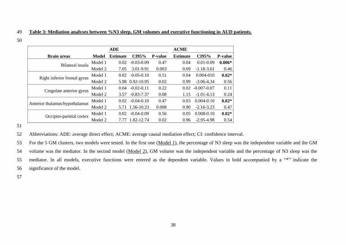
Table 3: Mediation analyses between %N3 sleep, GM volumes and executive functioning in AUD patients. 49
50
ADE ACME
Brain areas Model Estimate CI95% P-value Estimate CI95% P-value
Bilateral insula Model 1 0.02 -0.03-0.09 0.47
0.04 0.01-0.09 0.006*
Model 2 7.05 3.01-9.91 0.003 0.69 -1.18-3.61 0.46
Right inferior frontal gyrus Model 1 0.02 -0.05-0.10 0.51
0.04 0.004-010 0.02*
Model 2 5.98 0.92-10.95 0.02 0.99 -3.06-4.34 0.56
Cingulate anterior gyrus Model 1 0.04 -0.02-0.11 0.22
0.02 -0.007-0.07 0.11
Model 2 3.57 -0.83-7.37 0.08 1.15 -1.01-4.13 0.24
Anterior thalamus/hypothalamus Model 1 0.02 -0.04-0.10 0.47
0.03 0.004-0.10 0.02*
Model 2 5.71 1.56-10.23 0.008 0.90 -2.16-3.23 0.47
Occipito-parietal cortex Model 1 0.02 -0.04-0.09 0.56 0.05 0.008-0.10 0.02*
Model 2 7.77 1.82-12.74 0.02 0.96 -2.95-4.98 0.54
51
Abbreviations: ADE: average direct effect; ACME: average causal mediation effect; CI: confidence interval. 52
For the 5 GM clusters, two models were tested. In the first one (Model 1), the percentage of N3 sleep was the independent variable and the GM 53
volume was the mediator. In the second model (Model 2), GM volume was the independent variable and the percentage of N3 sleep was the 54
mediator. In all models, executive functions were entered as the dependent variable. Values in bold accompanied by a “*” indicate the 55
significance of the model. 56
57

39
Supplementary Table 1: Mediation analyses between %N3 sleep, GM volumes and short-term memory in AUD patients. 58
ADE ACME
Brain areas Model Estimate CI95% P-value Estimate CI95% P-value
Bilateral insula Model 1 0.06 -0.01-0.13 0.11
0.02 -0.01-0.07 0.23
Model 2 3.09 -2.88-8.76 0.28 1.93 -1.15-5.15 0.2
Right inferior frontal gyrus Model 1 0.05 -0.02-0.13 0.15
0.02 -0.01-0.07 0. 24
Model 2 3.01 -1.81-7.97 0.21 1.93 -0.48-5.45 0.13
Cingulate anterior gyrus Model 1 0.06 -0.03-0.14 0.20
0.02 -0.02-0.08 0.30
Model 2 3.09 -2.86-8.72 0.08 1.93 -1.13-5.09 0.2
Anterior thalamus/hypothalamus Model 1 0.06 -0.01-0.12 0.10
0.02 -0.01-0.07 0.25
Model 2 3.10 -1.79-7.91 0.21 1.92 -0.45-5.43 0.13
Occipito-parietal cortex Model 1 0.06 -0.02-0.13 0.13 0.02 -0.01-0.07 0.25
Model 2 3.17 -1.99-8.15 0.19 1.92 -0.17-5.5 0.1
59
Abbreviations: ADE: average direct effect; ACME: average causal mediation effect; CI: confidence interval. 60
For the 5 GM clusters, two models were tested. In the first one (Model 1), the percentage of N3 sleep was the independent variable and the GM 61
volume was the mediator. In the second model (Model 2), GM volume was the independent variable and the percentage of N3 sleep was the 62
mediator. In all models, short-term memory performance was entered as the dependent variable. All models were not significant. 63
64
65

40
Supplementary Table 2 : Relationships between the number of days since the last benzodiazepine prescription and %N3 sleep, GM 66
volumes and cognition. 67
68
Number of days since the last benzodiazepine prescription
Sleep quality %N3 sleep rho = -0.15 ; p = 0.61
Cognitive functioning Short-term memory rho = -0.26 ; p = 0.39
Executive functions rho = -0.29 ; p = 0.33
GM volumes
Right inferior frontal rho = -0.13 ; p = 0.67
Bilateral Insula rho = -0.23 ; p = 0.44
Anterior cingulate rho = -0.38 ; p = 0.20
Occipito-parietal rho = -0.17 ; p = 0.56
Thalamus/hypothalamus rho = -0.42 ; p = 0.15
69
Spearman’s correlations were conducted between the main results of the present study and the number of days since the last benzodiazepine 70
prescription and %N3 sleep, GM volumes and cognition. The number of days since the last benzodiazepine prescription was different between 71
the PSG-recording and the neuropsychological assessment because examinations were not performed during the same day. No correlation was 72
significant. 73
74
75
76
77

41
Graphical abstract 78
79
80
81
Copyright © 2022 FDOKUMEN