Energy fluxes and driving forces for photosynthesis in Lemna minor exposed to herbicides
Upload
khangminh22Category
view
3download
0
water
Article
The Duckweed, Lemna minor Modulates HeavyMetal-Induced Oxidative Stress in the Nile Tilapia,Oreochromis niloticus
Fagr Kh. Abdel-Gawad 1,*,†, Wagdy K. B. Khalil 2,†, Samah M. Bassem 1, Vikas Kumar 3,Costantino Parisi 4,5 , Sara Inglese 4, Tarek A. Temraz 6, Hossam F. Nassar 7,† andGiulia Guerriero 4,8,*,†
1 Water Pollution Research Department, Centre of Excellence for Advanced Sciences (CEAS),National Research Centre, Dokki, Giza 12622, Egypt; [email protected]
2 Department of Cell Biology, Centre of Excellence for Advanced Sciences (CEAS), National Research Centre,Dokki, Giza 12622, Egypt; [email protected]
3 Aquaculture Research Institute, Department of Animal and Veterinary Science, University of Idaho,Moscow, ID 83844, USA; [email protected]
4 Comparative Endocrinology Lab, Department of Biology, University of Naples Federico II,80126 Naples, Italy; [email protected] (C.P.); [email protected] (S.I.)
5 Laboratory of Zebrafish Developmental Genomics, International Institute of Molecular and Cell Biology,02-109 Warsaw, Poland
6 Marine Science Department, Canal Suez University, Ismailia 41522, Egypt; [email protected] Environmental Sciences and Industrial Development Department, Faculty of Postgraduate Studies for
Advanced Sciences (PSAS), Beni-Suef University, Beni-Suef 62511, Egypt; [email protected] Interdepartmental Research Centre for Environment, University of Naples Federico II, 80134 Naples, Italy* Correspondence: [email protected]; (F.K.A.-G.); [email protected] (G.G.)† Authors contributed equally.
Received: 18 September 2020; Accepted: 21 October 2020; Published: 24 October 2020�����������������
Abstract: A two-fold integrated research study was conducted; firstly, to understand the effects ofcopper (Cu) and zinc (Zn) on the growth and oxidative stress in Nile tilapia, Oreochromis niloticus;secondly, to study the beneficial effects of the duckweed Lemna minor L. as a heavy metal removerin wastewater. Experiments were conducted in mesocosms with and without duckweed. Tilapiafingerlings were exposed to Cu (0.004 and 0.02 mg L−1) and Zn (0.5 and 1.5 mg L−1) and fish fedfor four weeks. We evaluated the fish growth performance, the hepatic DNA structure using cometassay, the expression of antioxidative genes (superoxide dismutase, SOD; catalase, CAT; glutathioneperoxidase, GPx and glutathione-S-transferase, GST) and GPx and GST enzymatic activity. The resultsshowed that Zn exhibited more pronounced toxic effects than Cu. A low dose of Cu did not influencethe growth whereas higher doses of Cu and Zn significantly reduced the growth rate of tilapiacompared to the control, but the addition of duckweed prevented weight loss. Furthermore, in thepresence of a high dose of Cu and Zn, DNA damage decreased, antioxidant gene expressions andenzymatic activities increased. In conclusion, the results suggest that duckweed and Nile tilapia canbe suitable candidates in metal remediation wastewater assessment programs.
Keywords: Nile tilapia; Oreochromis niloticus; liver; duckweed; Lemna minor; Cu; Zn; glutathioneperoxidase; GPx; glutathione-S-transferase; GST; superoxide dismutase; SOD; catalase; CAT;remediation assessment
Water 2020, 12, 2983; doi:10.3390/w12112983 www.mdpi.com/journal/water

Water 2020, 12, 2983 2 of 14
1. Introduction
Among the major health concerns worldwide is the massive release of toxic compounds into thenatural environment including soil and water [1,2]. Many of these compounds are defined as metalsand are dangerous even at minimal concentrations and which may be cytotoxic, carcinogenic andmutagenic in nature [3,4]. They occur in the environment from natural and anthropogenic sources [5,6].Dietary contamination by these chemical elements gives rise to numerous adverse effects on human andanimal physiology [1,2,7,8]. These compounds may seriously affect cellular processes [7]. Their toxicityinvolves the generation of reactive oxygen and nitrogen species, which disturb redox systems [9],and antioxidants [10–12]. An overexpression of free radical production or a downregulation ofradicals-scavenging activity alters cellular functions through the direct modification of biomoleculesand by the alteration of signaling pathways [7].
The most effective antioxidative physiological defense systems are comprised of enzymessuch as superoxide dismutase (SOD), catalase (CAT), glutathione peroxidase (GPx) andglutathione-S-transferase (GST), which are known as biomarkers of oxidative stress [9,13]. The SODconverts superoxides (O2−) generated in peroxisomes and mitochondria to hydrogen peroxide detoxifiedby the CAT enzyme. The SOD and CAT systems provide the first mechanism for combating oxygentoxicity. The GPx catalyzes the reduction of hydrogen peroxide and lipid peroxides; GST acts asa catalytic agent in the biotransformation process by the conjugation of metabolites as xenobioticmetabolites. Antioxidant enzymes have been shown to have different responses and significantlylower activities in the polluted sites [14].
Thus, to make the environment safer and healthier for humans with regards to food consumption,and to ensure adequate fish growth performance, contaminated waters and lands need to bedecontaminated to lower levels of heavy metals and trace compounds [15–17]. Several techniques arecurrently used to remove heavy metals. Most of them, in particular physico-chemical methods, becomeineffective when heavy metal concentrations are under 100 mg L−1 [18]. In fact, metal salts are presentin water in a dissolved form and cannot be separated using physical approaches [19]. The introductionof aquatic phytoremediation plant species and adsorbents should be performed in land managementplans in order to reduce risks due to their contamination [20]. Therefore, plants represent an alternativeremediation approach which has escalated in recent decades [21,22].
The eco-friendly macrophyte Lemna minor (family Lemnaceae, genera Lemna), commonly knownas duckweed, is present worldwide [23], and is used as a standard ecotoxicological test species [24].As macrophytes are more sensitive than equivalent indicators which lack a vascular system, they aremore environmentally protective, thus confirming this plant as an acceptable species for toxic metalremediation [25,26]. Lemna minor is also used for the elimination of organic matter, nutrients suchas phosphorus and nitrogen, soluble salts, as well as the reduction of fecal coliform densities andsuspended solids [27,28]. It is also well known to be able to accumulate Cu and Zn from contaminatedwastewater [29–32]. Specifically, Lemna minor can accumulate a wide range of pollutants in its roottissue [32,33], and is able to keep the hyperaccumulated metal out of the cytoplasm as nontoxic or lesstoxic complexes and to sequester the complexes’ metal ions in the vacuoles by chelation and suchmechanistic evidence provides the best example of bioremediation programs [5,26,34].
Fish, as aquatic organisms, are subject to a vast array of water pollutants and as such, may serve asindicators for contamination assessment. Therefore, a series of biomarkers, including oxidative stressbiomarkers, can be successfully applied for the detection of biological impacts and for environmentalquality assessment [35]. These biomarkers provide a clear and useful link between pollution exposure,tissue contamination, and early adverse effects in organisms [9,36–38].
The Nile tilapia, Oreochromis niloticus, is a domesticated fish species extensively used inenvironmental studies because it is easily handled and maintained in the laboratory; it readilyadapts to confinement; it is susceptible to various pollutants; it has economic importance but at sametime it is very invasive. Moreover, the Nile tilapia and its primary tissue for detoxification, the liver,has been widely used for the toxicity evaluation of several contaminants in aquatic ecosystems [39,40].

Water 2020, 12, 2983 3 of 14
In our toxicological studies, we utilized zinc and copper which are essential trace minerals forteleost fish and all vertebrates, present in all organs, tissues, and fluids. These metals have structuraland catalytic functions and also play a regulatory role in multiple metalloenzymes as a specific cofactorand catalyst. Their toxicity is often linked to the physiological processes’ disruption. Zn, in fact, is oneof the most important essential trace elements involved in animal growth and the most widely usedmetal cofactor in many enzymes. Cu acts as a catalyst in many enzyme systems, mainly for cytochromeoxidase and the electron carrier plastocyanin and is actively taken up by liver mitochondria via anenergy-dependent system [35,41,42]. Nonetheless, it is also known that under normal conditions,these elements are essential micronutrients.
The chief aim of this research was to detect the low and high concentrations of zinc and coppereffects on hepatic antioxidative biomarkers in tilapia and to examine the efficacy of the duckweedLemna minor for their bioremediation in the environment.
2. Materials and Methods
2.1. Fish and Mesocosm
Tilapia fish, Oreochromis niloticus (n = 810, monosex type of body weight 36± 3.2 g), were transferredfrom the National Research Centre farm in Nubaria, Egypt. The tilapia fish were treated with lidocaine,CHNO (5 mg L−1), during the transportation, for stress reduction. Over an approximate two-hourtransport period, the fish were transferred to an outdoor experimental system (mesocosms) at thelaboratory at National Research Centre in a fiberglass container (1 m3 water capacity) supplied withbattery-powered aerators for oxygen supply. The fish underwent 40 days of acclimatization in a40 L glass mesocosm under natural light (45 × 60 × 30 cm, N = 9 mesocosms with ten fish each),un-chlorinated, well aerated and tap water (27.2 ± 1.8 ◦C and pH 7–8, dissolved oxygen 7–8 mgL−1). A pelleted diet (32% protein ration, 6.1% crude lipid, 4.5% pure crude fiber, and total energy4080 Kcal/Kg, Zoo-Control Co., Giza, Egypt) was provided daily at a rate of 3% of fish body weightand the water was removed daily. This experiment followed the Egyptian ethical guidance for animalresearch of the Institutional Animal Care and Use Committee (IACUC), 2013.
2.2. Experimental Design
The fish were distributed into nine experimental groups and were exposed to water with copperand zinc for four weeks, as follows: the first fish group was exposed to regular, uncontaminated wateras a control. The second and third groups were exposed to water contaminated with low and highdoses of copper sulfate of 0.004 mg L−1 (CuL) and 0.02 (CuH) mg L−1 respectively. The fourth andfifth groups were exposed to water with the same doses of copper as in the previous groups plusone layer of duckweed, Lemna minor, covering the water surface. The sixth and seventh groups werecontaminated with low (ZnL) and high (ZnH) doses of zinc acetate of 0.5 and 1.5 mg L−1, respectively.The eighth and ninth groups were exposed to water with the same concentrations of zinc as in theprevious groups, plus one layer of duckweed covering the water surface.
Copper was added in the form of copper sulfate pentahydrate, CuSO4·5H2O at 25% Cu, and zincfrom the zinc acetate dehydrate, Zn(CH3COO)2·2H2O at ≥98% Zn. Cu and Zn ions were determinedby inductively coupled plasma mass spectrometry in each mesocosm. The applied doses of copperand zinc were based on the permissible concentrations in natural water [43] and the estimated levels inpolluted areas in Egypt [44]. The amount of around 2000 g of Lemna minor was used in each mesocosmwith duckness treatment. At the end of the four weeks, the fish were euthanized in 2-phenoxyethanoland dissected. Growth performance was evaluated in the mesocosms treated and untreated withmetals, and with and without duckweed.
Each liver (1 ± 0.3 g weight for the control and treated animals) was divided into 4 aliquots: two of50 mg and two of 250 mg. One of the two aliquots of 50 mg was used directly to perform a comet assay,the other to study the gene expression by qRT-PCR. Both aliquots of 250 g were used to constitute a

Water 2020, 12, 2983 4 of 14
pool of 500 mg with one other liver aliquot of tilapia of the same experimental group to perform thebiochemical measurement in triplicate. Aliquots (n = 8 for each experimental group) and pools (n = 8for each experimental group) and the additional tissue were stored at −80 ◦C until the analyses.
2.3. Growth Performance
Growth performance was measured as follows: weight gain (g) = W2 −W1; where W2 is the finalweight after the experimental periods (four weeks), and W1 is the initial weight.
2.4. Analysis of DNA
The Comet Assay
Comet assay followed the protocol established by Blasiak et al. (2004) [45]. Images from100 randomly selected cells (fifty counts on each duplicate slide) were analyzed for each sample byDNA damage analysis software (Comet Score, Tri Tek Corp., Sumerduck, VA, USA). In each cometclass were calculated the mean score and standard deviation. Different classes were distinguished asfollows: class 0 (no visible tail), class 1 (low fluorescence, round head and low damage—tail lengthno more than 30 µm), class 2 (equally brightly fluorescent for head and tail, medium damage taillength between 30 and 50 µm), and class 3 (bright and head small and weakly fluorescent and highdamage—tail length between 50 and 70 µm). Comets with a completely disintegrated head and onlyvisible tails were considered apoptotic and were not included in the analysis.
2.5. Gene Expression Analysis
2.5.1. RNA Extraction
RNA was extracted from each tilapia liver tissue (n = 8 of each treatment group) using TRIzolReagent (Invitrogen, Darmstadt, Germany). Then, 1 mL of TRIzol reagent buffer was used tohomogenize 50 mg of liver at room temperature for 15 min. Subsequently, 0.2 mL of chloroformwas added. The samples were vortexed for 15 s, incubated for 3 min and then centrifuged at 4 ◦Cat 12,000× g. for 15 min. The upper aqueous layer was transferred to a fresh tube and mixed to0.5 mL isopropyl alcohol for RNA precipitation. Samples were first incubated at 30 ◦C for 10 minand then centrifuged at 4 ◦C, 12,000× g for 10 min. The RNA pellet obtained was washed with 1 mLof 75% ethanol, centrifuged at 4 ◦C, 7500× g for 5 min, air-dried for 10 min, dissolved in 100 µL ofdiethylpyrocarbonate (DEPC)-treated water and stored at −80 ◦C.
2.5.2. Reverse Transcription (RT) Reaction
The RNA from the tilapia liver was transcribed in 20 µL of cDNA using RevertAidTM First StrandcDNA Synthesis Kit (MBI Fermentas, St. Leon-Roth, Germany). The RNA (5 µg) was mixed to 50 UM-MuLV reverse transcriptase, 20 U ribonuclease inhibitor (50 kDa recombinant enzyme to inhibit RNaseactivity), 50 µM oligo-dT primer, 10 mM of each dNTP, 50 mM MgCl2 and 5× reverse transcription(RT) buffer. The RT thermal reaction program was 25 ◦C for 10 min followed by 1 h at 42 ◦C with afinal heating at 99 ◦C for 5 min. The final reaction was cooled in ice and then used for quantitative realtime-polymerase chain reaction (qRT-PCR).
2.5.3. Quantitative Real Time-Polymerase Chain Reaction (qRT-PCR)
Reaction mixtures (35 µL) of qRT-PCR consisted of 5 µL of cDNA template, 5 µL 0.2 µM of eachprimer, 12.5 µL of 1× SYBR® Premix Ex Taq TM (TaKaRa, Biotech. Co. Ltd., Dalian, China) and 7.5 µLdH2O was used. The PCR was performed as follows: 95.0 ◦C for 3 min, then 28 cycles of 95 ◦C, 1 min;60 ◦C, 1 min; 72 ◦C, 1 min then, 71 cycles at 60 ◦C and then changed every 10 s at about 0.5 ◦C untilreaching 95 ◦C. By the end of each qRT-PCR, a melting curve analysis was carried out at 95 ◦C to checkthe the quality of primers used in the reaction [46]. All reactions were performed using the Step One

Water 2020, 12, 2983 5 of 14
Real-Time PCR system (Applied Biosystems, Forster City, CA, USA), and each run contained distilledwater as a control. The expression level of the following antioxidant enzyme genes was quantified inthe liver tissues of tilapia fish: SOD, CAT, GPx and GST. The primers were designed using Primer3software (http://bioinfo.ut.ee/primer3/) Table 1. The quantitative values of the RT-PCR were normalizedusing housekeeping genes β-actin [47].
Table 1. Primer sequences for the Nile tilapia Oreochromis niloticus genes encoding antioxidant enzymes.
Gene Forward Primer Sequence (5′-3′) Reverse Primer Sequence (5′-3′)
SOD GGTGCCCTGGAGCCCTA ATGCGAAGTCTTCCACTGTCCAT TCCTGAATGAGGAGGAGCGA ATCTTAGATGAGGCGGTGATGGPx CCAAGAGAACTGCAAGAACGA CAGGACACGTCATTCCTACACGST TAATGGGAGAGGGAAGATGG CTCTGCGATGTAATTCAGGA
β-actin CAATGAGAGGTTCCGTTGC AGGATTCCATACCAAGGAAGG
Superoxide dismutase, SOD; catalase, CAT; glutathione peroxidase, GPx and glutathione-S-transferase, GST.
2.6. Biochemical Measurements
2.6.1. Glutathione-S-Transferase (GST) Activity
GST activity was estimated in tilapia liver pools (n = 4 of each treatment group) according tomethods described by Habig et al. (1974) [48]. The GST was evaluated with a spectrophotometer for5 min at 25 ◦C due to the conjugation of reduced glutathione with 1- chloro-2,4-dinitrobenzene (CDNB)at 1 mM final concentration, 1 mM 1-chloro-2,4-dinitrobenzene, and 100 mM potassium phosphatebuffer (pH 6.5) considering the blank values. A Bradford protein assay was used to determinethe protein concentration, using bovine serum albumin (Sigma, St. Louis, MO, USA) as standard.GST activity was expressed as µM/min/mg protein.
2.6.2. Glutathione Peroxidase (GPx) Activity
GPx activity was measured in tilapia liver pools (n = 4 of each treatment group) group accordingto methods described by Mannervik (1985) [49]. The enzymatic reaction was estimated usingthe consecutive glutathione reductase reaction, the oxidation of NADPH (nicotinamide adeninedinucleotide phosphate oxidase) and the substrate t-butyl hydroperoxide. A Bradford protein assaywas used to determine the protein concentration, using bovine serum albumin (Sigma) as a standard.In accordance with Flohé and Gunzler (1984) [50], a unit of GPx activity was defined as the amountof GPx needed to reduce the initial glutathione concentration. The GPx activity was expressed asµM/min/mg protein.
2.7. Statistical Analysis
One-way ANOVA, and when appropriate, the Scheffé post-hoc test, were used to analyze multiplegroup data. Data are shown as the mean ± standard error of the mean (SEM). The level of statisticalsignificance was set at p < 0.05.
3. Results
3.1. Effect of Duckweed on Growth Performance
The results for the fish weight gain reported in Table 2 show that the tilapia fish exposed to a lowdose of Cu did not have a significantly reduced final body weight compared to the control fish.

Water 2020, 12, 2983 6 of 14
Table 2. Growth performance of Nile tilapia, Oreochromis niloticus exposed to metals: low and high doseCu (CuL, CuH), and low and high dose Zn (ZnL, ZnH) in mesocosm with or without the duckweed,Lemna minor.
Treatment Initial Weight (g) Final Weight (g)
Control 36.2 ± 2.4 99.3 ± 3.2 a
CuL 37.1 ± 3.2 88.1 ± 4.1 ab
CuH 35.4 ± 1.9 77.6 ± 2.9 bc
CuL + L. minor 38.2 ± 2.7 93.2 ± 4.8 ab
CuH + L. minor 36.4 ± 1.6 84.4 ± 5.2 b
ZnL 36.2 ± 1.5 81.5 ± 3.7 b
ZnH 37.5 ± 2.2 71.2 ± 2.4 c
ZnL + L. minor 38.2 ± 3.3 89.1 ± 3.8 ab
ZnH + L. minor 36.6 ± 2.1 80.3 ± 3.1 b
Data are presented as the mean ± SEM. a,b,c Mean values within tissue with unlike superscript letters weresignificantly different (p < 0.05, Scheffé Test).
However, a high dose of Cu resulted in significantly reduced final body weights of tilapiacompared to the control fish. Likewise, low and high doses of Zn reduced significantly the final bodyweight of tilapia compared with the control fish.
3.2. Effect of Duckweed against Heavy Metals Induced DNA Damage
The results for the percentage of DNA-damaged cells reported in Table 3 revealed that the fishexposed to Zn exhibited rates of DNA damage more significant than those exposed to Cu comparedto the control group. Furthermore, the fish exposed to a low dose of Cu and Zn revealed relativelysimilar rates of DNA damage compared to those in control fish. However, the high dose of Cu andZn induced higher frequencies of DNA damage with percentages of 17.4% and 19.6% for Cu and Zn,respectively, compared to the control group. Results for the percentage of DNA damaged cells assessedin Oreochromis niloticus liver indicated less damage when Lemna minor was added with respect to thetreatment with both metals. Specifically, DNA damage reduction was 1.6% for CuL concentration and6.2% for CuH concentration and 2.0% for ZnL and 7.2% for ZnH concentration.
Table 3. Total comets, class of comet and % DNA damaged liver cells in Nile tilapia, Oreochromisniloticus exposed to metals: low and high dose Cu (CuL, CuH), and low and high dose Zn (ZnL, ZnH)in mesocosm with or without the duckweed, Lemna minor, using the comet assay.
Treatment Total CometsComet Class DNA %
Damaged Cells0 1 2 3
Control 33 467 22 11 0 6.6 ± 1.1 c
CuL 46 454 17 16 13 9.2 ± 1.6 bc
CuH 87 413 26 32 29 17.4 ± 2.4 a
CuL + L. minor 38 462 12 15 11 7.6 ± 1.2 c
CuH + L. minor 56 444 16 21 19 11.2 ± 1.6 b
ZnL 49 451 18 15 16 9.8 ± 1.5 bc
ZnH 98 402 28 37 33 19.6 ± 2.2 a
ZnL + L. minor 39 461 21 11 7 7.8 ± 1.3 c
ZnH + L. minor 62 438 18 22 21 12.4 ± 1.8 b
Data are presented as the mean ± SEM. a,b,c Mean values within tissue with unlike superscript letters weresignificantly different (p < 0.05, Scheffé Test) (n = 5).
3.3. Effect of Duckweed on Antioxidants Gene Expression
The quantitative expression of antioxidant enzyme-related genes including glutathione-s-transferase (GST), catalase (CAT), superoxide dismutase (SOD) and glutathione peroxidase (GPx)genes in the liver tissues of Nile tilapia is summarized in Figure 1A–C. GST, SOD, CAT and GPx

Water 2020, 12, 2983 7 of 14
genes were significantly downregulated in the liver tissues of tilapia exposed to a high dose of Zn(1.5 mg L−1) and Cu (0.02 mg L−1) compared to the control group. In particular, even at a low Zndose (0.02 mg L−1), the GST, SOD and CAT (p < 0.01) were affected in comparison to control fish.Interestingly, the SOD, CAT and GPx expression, which were reduced with high doses of Cu, were notaffected in the presence of Lemna minor. While for the low Zn treatment, a decreased expression ofSOD and CAT was not observed for the same treatment with the Lemna minor addition. Surprisingly,the reduced CAT expression (p < 0.01) observed at high Zn exposure remained at control levels inexperiments where Lemna minor was added.
Water 2020, 12, x FOR PEER REVIEW 7 of 14
3.3. Effect of Duckweed on Antioxidants Gene Expression
The quantitative expression of antioxidant enzyme-related genes including glutathione-s-transferase (GST), catalase (CAT), superoxide dismutase (SOD) and glutathione peroxidase (GPx) genes in the liver tissues of Nile tilapia is summarized in Figure 1A–C. GST, SOD, CAT and GPx genes were significantly downregulated in the liver tissues of tilapia exposed to a high dose of Zn (1.5 mg L−1) and Cu (0.02 mg L−1) compared to the control group. In particular, even at a low Zn dose (0.02 mg L−1), the GST, SOD and CAT (p < 0.01) were affected in comparison to control fish. Interestingly, the SOD, CAT and GPx expression, which were reduced with high doses of Cu, were not affected in the presence of Lemna minor. While for the low Zn treatment, a decreased expression of SOD and CAT was not observed for the same treatment with the Lemna minor addition. Surprisingly, the reduced CAT expression (p < 0.01) observed at high Zn exposure remained at control levels in experiments where Lemna minor was added.
Figure 1. RTqPCR expression analysis of liver antioxidant enzyme genes (A: GST; B: CAT; C: SOD; D: GPx) of Nile tilapia, Oreochromis niloticus. The relative expression indicated in arbitrary units defines the expression change in comparison to that of the reference housekeeping β-actina rRNA gene in samples exposed to metals, Cu and Zn in mesocosm with or without duckweed, Lemna minor (Cu L: 0.004 and H: 0.02 mg L−1); (Zn, L: 0.5 and H: 1.5 mg L−1) with respect to samples without treatment used as control. * p < 0.05 and ** p < 0.01 for the treated groups compared with the control group.
3.4. Effect of Duckweed on the GST and GPx Activities
Results show damaged liver cells of Nile tilapia Oreochromis niloticus exposed to different concentrations (low, L and high, H) of heavy metals, Cu and Zn alone or combined with duckweed, Lemna minor. The applied doses of Cu and Zn were chosen based on the estimated levels in polluted areas in Egyptian river water in the last [43,44] and recent assessment [51].
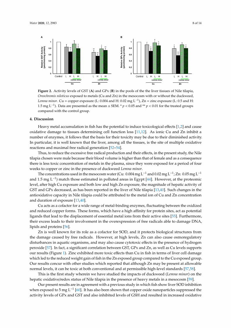
Biochemical measurements were performed to examine the hepatic GST and GPx activities in Oreochromis niloticus (Figure 2). The results show that a high dose of Cu (0.02 mg L−1) and Zn (1.5 mg L−1) induced significantly lower activity levels of GST and GPx. In particular, for both enzymes, Zn doses induced the lowest activity levels of the enzymes even at low concentrations (0.05 mg L−1). Moreover, the significant decrease in GPx activity subjected to low Zn concentration (0.5 mg L−1) was not affected in the presence of Lemna minor (Figure 1B).
Figure 1. RTqPCR expression analysis of liver antioxidant enzyme genes (A: GST; B: CAT; C: SOD; D:GPx) of Nile tilapia, Oreochromis niloticus. The relative expression indicated in arbitrary units definesthe expression change in comparison to that of the reference housekeeping β-actina rRNA gene insamples exposed to metals, Cu and Zn in mesocosm with or without duckweed, Lemna minor (Cu L:0.004 and H: 0.02 mg L−1); (Zn, L: 0.5 and H: 1.5 mg L−1) with respect to samples without treatmentused as control. * p < 0.05 and ** p < 0.01 for the treated groups compared with the control group.
3.4. Effect of Duckweed on the GST and GPx Activities
Results show damaged liver cells of Nile tilapia Oreochromis niloticus exposed to differentconcentrations (low, L and high, H) of heavy metals, Cu and Zn alone or combined with duckweed,Lemna minor. The applied doses of Cu and Zn were chosen based on the estimated levels in pollutedareas in Egyptian river water in the last [43,44] and recent assessment [51].
Biochemical measurements were performed to examine the hepatic GST and GPx activitiesin Oreochromis niloticus (Figure 2). The results show that a high dose of Cu (0.02 mg L−1) and Zn(1.5 mg L−1) induced significantly lower activity levels of GST and GPx. In particular, for both enzymes,Zn doses induced the lowest activity levels of the enzymes even at low concentrations (0.05 mg L−1).Moreover, the significant decrease in GPx activity subjected to low Zn concentration (0.5 mg L−1) wasnot affected in the presence of Lemna minor (Figure 1B).
Water 2020, 12, 2983 8 of 14
Water 2020, 12, x FOR PEER REVIEW 8 of 14
Figure 2. Activity levels of GST (A) and GPx (B) in the pools of the the liver tissues of Nile tilapia, Oreochromis niloticus exposed to metals (Cu and Zn) in the mesocosm with or without the duckweed, Lemna minor. Cu = copper exposure (L: 0.004 and H: 0.02 mg L−1), Zn = zinc exposure (L: 0.5 and H: 1.5 mg L−1). Data are presented as the mean ± SEM. * p < 0.05 and ** p < 0.01 for the treated groups compared with the control group.
4. Discussion
Heavy metal accumulation in fish has the potential to induce toxicological effects [1,2] and cause oxidative damage to tissues determining cell function loss [11,12]. As ionic Cu and Zn inhibit a number of enzymes, it follows that the basis for their toxicity may be due to their diminished activity. In particular, it is well known that the liver, among all the tissues, is the site of multiple oxidative reactions and maximal free radical generation [52–54].
Thus, to reduce the excessive free radical production and their effects, in the present study, the Nile tilapia chosen were male because their blood volume is higher than that of female and as a consequence there is less toxic concentration of metals in the plasma, since they were exposed for a period of four weeks to copper or zinc in the presence of duckweed Lemna minor.
The concentrations used in the mesocosm water (Cu: 0.004 mg L−1 and 0.02 mg L−1; Zn: 05 mg L−1 and 1.5 mg L−1) match those estimated in polluted areas in Egypt [44]. However, at the proteomic level, after high Cu exposure and both low and high Zn exposure, the magnitude of hepatic activity of GST and GPx decreased, as has been reported in the liver of Nile tilapia [13,40]. Such changes in the antioxidative capacity in Nile tilapia could be attributed to the metal ion of Cu and Zn concentration and duration of exposure [13,40].
Cu acts as a cofactor for a wide range of metal-binding enzymes, fluctuating between the oxidized and reduced copper forms. These forms, which have a high affinity for protein sites, act as potential ligands that lead to the displacement of essential metal ions from their active sites [55]. Furthermore, their excess leads to their involvement in the overexpression of free radicals able to damage DNA, lipids and proteins [56].
Zn is well known for its role as a cofactor for SOD, and it protects biological structures from the damage caused by free radicals. However, at high levels, Zn can also cause osmoregulatory disturbances in aquatic organisms, and may also cause cytotoxic effects in the presence of hydrogen peroxide [57]. In fact, a significant correlation between GST, GPx and Zn, as well as Cu levels supports our results (Figure 1). Zinc exhibited more toxic effects than Cu in fish in terms of liver cell damage which led to the reduced weight gain of fish in the Zn exposed group compared to the Cu exposed group. Our results concur with other studies which reported that although Zn may be present at allowable normal levels, it can be toxic at both conventional and at permissible high-level standards [57,58].
This is the first study wherein we have studied the impacts of duckweed (Lemna minor) on the hepatic oxidative/redox status of Nile tilapia in the presence of heavy metals in a mesocosm [59].
Our present results are in agreement with a previous study in which fish show liver SOD inhibition when exposed to 5 mg L−1 [60]. It has also been shown that copper oxide nanoparticles suppressed the activity levels of GPx and GST and also inhibited levels of GSH and resulted in increased oxidative stress in the digestive gland of the freshwater snail [61]. Moreover, the treatment
Figure 2. Activity levels of GST (A) and GPx (B) in the pools of the the liver tissues of Nile tilapia,Oreochromis niloticus exposed to metals (Cu and Zn) in the mesocosm with or without the duckweed,Lemna minor. Cu = copper exposure (L: 0.004 and H: 0.02 mg L−1), Zn = zinc exposure (L: 0.5 and H:1.5 mg L−1). Data are presented as the mean ± SEM. * p < 0.05 and ** p < 0.01 for the treated groupscompared with the control group.
4. Discussion
Heavy metal accumulation in fish has the potential to induce toxicological effects [1,2] and causeoxidative damage to tissues determining cell function loss [11,12]. As ionic Cu and Zn inhibit anumber of enzymes, it follows that the basis for their toxicity may be due to their diminished activity.In particular, it is well known that the liver, among all the tissues, is the site of multiple oxidativereactions and maximal free radical generation [52–54].
Thus, to reduce the excessive free radical production and their effects, in the present study, the Niletilapia chosen were male because their blood volume is higher than that of female and as a consequencethere is less toxic concentration of metals in the plasma, since they were exposed for a period of fourweeks to copper or zinc in the presence of duckweed Lemna minor.
The concentrations used in the mesocosm water (Cu: 0.004 mg L−1 and 0.02 mg L−1; Zn: 0.05 mg L−1
and 1.5 mg L−1) match those estimated in polluted areas in Egypt [44]. However, at the proteomiclevel, after high Cu exposure and both low and high Zn exposure, the magnitude of hepatic activity ofGST and GPx decreased, as has been reported in the liver of Nile tilapia [13,40]. Such changes in theantioxidative capacity in Nile tilapia could be attributed to the metal ion of Cu and Zn concentrationand duration of exposure [13,40].
Cu acts as a cofactor for a wide range of metal-binding enzymes, fluctuating between the oxidizedand reduced copper forms. These forms, which have a high affinity for protein sites, act as potentialligands that lead to the displacement of essential metal ions from their active sites [55]. Furthermore,their excess leads to their involvement in the overexpression of free radicals able to damage DNA,lipids and proteins [56].
Zn is well known for its role as a cofactor for SOD, and it protects biological structures fromthe damage caused by free radicals. However, at high levels, Zn can also cause osmoregulatorydisturbances in aquatic organisms, and may also cause cytotoxic effects in the presence of hydrogenperoxide [57]. In fact, a significant correlation between GST, GPx and Zn, as well as Cu levels supportsour results (Figure 1). Zinc exhibited more toxic effects than Cu in fish in terms of liver cell damagewhich led to the reduced weight gain of fish in the Zn exposed group compared to the Cu exposed group.Our results concur with other studies which reported that although Zn may be present at allowablenormal levels, it can be toxic at both conventional and at permissible high-level standards [57,58].
This is the first study wherein we have studied the impacts of duckweed (Lemna minor) on thehepatic oxidative/redox status of Nile tilapia in the presence of heavy metals in a mesocosm [59].
Our present results are in agreement with a previous study in which fish show liver SOD inhibitionwhen exposed to 5 mg L−1 [60]. It has also been shown that copper oxide nanoparticles suppressed theactivity levels of GPx and GST and also inhibited levels of GSH and resulted in increased oxidative
Water 2020, 12, 2983 9 of 14
stress in the digestive gland of the freshwater snail [61]. Moreover, the treatment of Nile tilapia with 1and 2 mg L−1 ZnONPs resulted in the suppression of antioxidant activity. ZnONPs also decreased thegene expression of SOD and CAT in the liver and gills of Nile tilapia [62].
At transcriptional levels, the SOD, CAT, GPx and GST gene expression patterns have beenvalidated as biomarkers of exposure to oxidative stress-inducing chemical pollutants and also to abioticfactors such as hyperthermia [12].
In our study, exposure to Cu and Zn caused the greatest reduction in SOD, CAT and GPx andGST transcription and an increase in DNA damage. However, Zn may have the more deleteriouseffect by notably decreasing enzymatic activity even at low concentrations (Figure 2). These results arein accordance with other studies on antioxidative mRNA expression, in which the hepatopancreas,gills and kidney were shown to be downregulated by the exposure to Cd, Cu and Zn [13,63,64].In contrast, much research has shown an increase in hepatic gene expression in relation to toxic metalsexposure [65–67]. Thus, it has been suggested that the expression of antioxidant biomarkers canbe enhanced or reduced depending on many factors as the chemical stress intensity and duration,as well as the investigated species sensitivity [12,41,65]. These studies of antioxidative expressions attranscriptional and translational levels can answer fundamental questions linked to the xenobiotictype, exposure times, data on seasonal time of sampling, and the sex and sexual maturity of fish [39].
Our results on fish growth performance and DNA structure together with our analysis of genesexpression and biochemical measurements highlight the potential use of Lemna minor for reducingoxidative stress and enhancing the capacity for heavy metal tolerance in Nile tilapia. This is importantbecause when the antioxidative capacity is lowered, protection against cell damage is also impacteddue to a reduction in the scavenging ability for free radicals leading to increased oxidative stress.
Our DNA damage analysis provides high concentrations of metals, either individually or incombination [68–70], which induced both sub-lethal and lethal effects in fish. The parameters mostmarkedly affected include: tissue genotoxicity, immunity suppression, endocrine disruption, enzymeand vitamin degradation and morphological alteration in cells [5,71,72].
Interestingly, the expression levels of all examined genes were significantly increased, and the rateof DNA damage decreased in the fish treated with duckweed Lemna minor, highlighting the inhibitionof the deleterious effect posed by Cu and Zn exposure in water. Thus, the consistency between thechange of enzyme activities and gene mRNA abundance exposed to toxic substances underscoreshow activities of antioxidant enzymes could be regulated. This strengthens our data showing thatthe decrease in antioxidant activity reflects the reduction in the gene expression, and the addition ofduckweed Lemna minor prevents the alteration of enzymatic activity and gene expression previouslydiminished by metal exposure [65].
Finally, it was demonstrated that Lemna minor prevented the decrease in final body weight offish exposed to low doses of Cu and Zn compared to control fish. This result confirms the duckweedLemna minor as a successful treatment for preventing the deteriorating effects of water-borne metals,copper and zinc, on the growth performance and health of Nile tilapia. These effects resulting froma series of events as well as cell surface biosorption/precipitation of metals, the exclusion of metalchelates into the extracellular space and enzymatic redox reaction through the conversion of metal ionsinto a non-toxic or less toxic state, afforded protection and nourishment [15,16,34].
5. Conclusions
In summary, Lemna minor is a potential remediator for the protection of one of the most importantaquaculture species in Egypt and worldwide, the Nile tilapia Oreochromis niloticus. This remediationmay be achieved by reducing oxidative stress and enhancing the heavy metal tolerance of these fish.In this regard, tilapia can be introduced as an in vivo model through the utilization of liver antioxidantsas biomarkers for remediation screening in the countries with scarce or absent native species, whereasin countries rich in biodiversity, their use must be very restricted as this species is very invasive.

Water 2020, 12, 2983 10 of 14
Understanding the relationships between stressors, stress responses, and the recovery processcontribute to the effective management and restoration of aquatic ecosystems.
Author Contributions: Conceptualization, F.K.A.-G. and G.G.; methodology, F.K.A.-G., S.M.B., W.K.B.K., G.G.;software, S.M.B., T.A.T.; validation, F.K.A.-G., S.M.B., V.K., C.P. and G.G.; formal analysis and investigation,S.M.B., T.A.T., H.F.N.; resources, F.K.A.-G., W.K.B.K., H.F.N. and G.G.; data curation, W.K.B.K., C.P., V.K.,S.I.; writing—original draft preparation, F.K.A.-G. and G.G.; writing—review and editing, F.K.A.-G. and G.G.;visualization, all; supervision, all; project administration, F.K.A.-G., W.K.B.K., H.F.N. and G.G.; funding acquisition,F.K.A.-G., W.K.B.K., H.F.N. and G.G. All authors have read and agreed to the published version of the manuscript.
Funding: This research received no external funding.
Acknowledgments: This work was performed within the framework of the Memorandum of Understandingbetween the National Research Centre of Giza (Egypt) and Federico II University. The authors acknowledgeItalian and Egyptian students in mobility for the logistic technical support, the International Office of Federico IIUniversity for the publication fee and Emidio Sivieri, Biomedical Engineer at Children’s Hospital of Philadelphia,Philadelphia (USA), for the English revision.
Conflicts of Interest: The authors declare no conflict of interest.
References
1. Guerriero, G.; Parisi, C.; Abdel-Gawad, F.K.; Hentati, O.; D’Errico, G. Seasonal and pharmaceutical-inducedchanges in selenoprotein glutathione peroxidase 4 activity in the reproductive dynamics of the soil biosentinelPodarcis sicula (Chordata: Reptilia). Mol. Reprod. Dev. 2019, 86, 1378–1387. [CrossRef] [PubMed]
2. Lecomte, S.; Habauzit, D.; Charlier, T.; Pakdel, F. Emerging estrogenic pollutants in the aquatic environmentand breast cancer. Genes 2017, 8, 229. [CrossRef] [PubMed]
3. Fasulo, S.; Guerriero, G.; Cappello, S.; Colasanti, M.; Schettino, T.; Leonzio, C.; Mancini, G.; Gornati, R.The “SYSTEMS BIOLOGY” in the study of xenobiotic effects on marine organisms for evaluation of theenvironmental health status: Biotechnological applications for potential recovery strategies. Rev. Environ.Sci. Bio/Technol. 2015, 14, 339–345. [CrossRef]
4. Diaconu, M.; Pavel, L.V.; Hlihor, R.-M.; Rosca, M.; Fertu, D.I.; Lenz, M.; Corvini, P.X.; Gavrilescu, M.Characterization of heavy metal toxicity in some plants and microorganisms—A preliminary approach forenvironmental bioremediation. New Biotechnol. 2020, 56, 130–139. [CrossRef] [PubMed]
5. Conte, B.; Sorbo, S.; Piscopo, M.; Rabbito, D.; De Ruberto, F.; Guerriero, G.; Basile, A. Antioxidant activityand ultrastructural alterations in the biosensor Lemna minor l. exposed in bags in sarno river (South Italy).Fresenius Environ. Bull. 2017, 26, 225–236.
6. Sabiha, J.; Mehmood, T.; Chaudhry, M.M.; Tufail, M.; Irfan, N. Heavy metal pollution from phosphate rockused for the production of fertilizer in Pakistan. Microchem. J. 2009, 91, 94–99. [CrossRef]
7. Jan, A.T.; Azam, M.; Siddiqui, K.; Ali, A.; Choi, I.; Haq, Q.M. Heavy metals and human health: Mechanisticinsight into toxicity and counter defense system of antioxidants. Int. J. Mol. Sci. 2015, 16, 29592–29630.[CrossRef]
8. Strungaru, S.-A.; Nicoara, M.; Teodosiu, C.; Baltag, E.; Ciobanu, C.; Plavan, G. Patterns of toxic metalsbioaccumulation in a cross-border freshwater reservoir. Chemosphere 2018, 207, 192–202. [CrossRef]
9. Abdel-Gawad, F.K.; Nassar, H.F.; Bassem, S.M.; Guerriero, G.; Khalil, W.K.B. Effect of polycyclic aromatichydrocarbons (PAHs) on modulate genes encoding stress-related proteins and antioxidant enzymes indifferent marine fish species of Red Sea Water. World Appl. Sci. J. 2014, 32, 2337–2346. [CrossRef]
10. Arena, C.; Vitale, L.; Bianchi, A.R.; Mistretta, C.; Vitale, E.; Parisi, C.; Guerriero, G.; Magliulo, V.; De Maio, A.The Ageing Process Affects the Antioxidant Defences and the Poly (ADPribosyl) ation Activity in Cistus IncanusL. Leaves. Antioxidants 2019, 8, 528. [CrossRef]
11. Guerriero, G.; Trocchia, S.; Abdel-Gawad, F.K.; Ciarcia, G. Roles of reactive oxygen species in thespermatogenesis regulation. Front. Endocrinol. 2014, 5, 56. [CrossRef] [PubMed]
12. Parisi, C.; Guerriero, G. Antioxidative Defense and Fertility Rate in the Assessment of Reprotoxicity RiskPosed by Global Warming. Antioxidants 2019, 8, 622. [CrossRef] [PubMed]
Water 2020, 12, 2983 11 of 14
13. Mohamed, A.A.-R.; El-Houseiny, W.; Abd Elhakeem, E.-M.; Ebraheim, L.L.; Ahmed, A.I.; Abd El-Hakim, Y.M.Effect of hexavalent chromium exposure on the liver and kidney tissues related to the expression of CYP450and GST genes of Oreochromis niloticus fish: Role of curcumin supplemented diet. Ecotoxicol. Environ. Saf.2020, 188, 109890. [CrossRef]
14. Nofal, M.I. Effects of heavy metal pollution on nile tilapia in manzala farm: Oxidative stress biomarkers andhistopathological findings. Int. J. Fish Aquat. Stud. 2019, 7, 315–328.
15. Abdel-Tawwab, M.; El-Sayed, G.O.; Shady, S.H. Growth, biochemical variables, and zinc bioaccumulationin Nile tilapia, Oreochromis niloticus (L.) as affected by water-born zinc toxicity and exposure period.Int. Aquat. Res. 2016, 8, 197–206. [CrossRef]
16. Ali, A.; Al-Ogaily, S.M.; Al-Asgah, N.A.; Gropp, J. Effect of sublethal concentrations of copper on the growthperformance of Oreochromis niloticus. J. Appl. Ichthyol. 2003, 19, 183–188. [CrossRef]
17. Plavan, G.; Jitar, O.; Teodosiu, C.; Nicoara, M.; Micu, D.; Strungaru, S.-A. Toxic metals in tissues of fishesfrom the Black Sea and associated human health risk exposure. Environ. Sci. Pollut. Res. 2017, 24, 7776–7787.[CrossRef]
18. Ahluwalia, S.S.; Goyal, D. Microbial and plant derived biomass for removal of heavy metals from wastewater.Bioresour. Technol. 2007, 98, 2243–2257. [CrossRef]
19. Hussein, H.; Farag, S.; Moawad, H. Isolation and characterization of Pseudomonas resistant to heavy metalscontaminants. Arab J. Biotechnol. 2003, 7, 13–22.
20. Kumar, V.; Parihar, R.D.; Sharma, A.; Bakshi, P.; Sidhu, G.P.S.; Bali, A.S.; Karaouzas, I.; Bhardwaj, R.;Thukral, A.K.; Gyasi-Agyei, Y. Global evaluation of heavy metal content in surface water bodies:A meta-analysis using heavy metal pollution indices and multivariate statistical analyses. Chemosphere 2019,236, 124364. [CrossRef]
21. Suhag, A.; Gupta, R.; Tiwari, A. Biosorptive removal of heavy metals from waste water using duckweed.Int. J. Biomed. Adv. Res. 2011, 2, 281–290. [CrossRef]
22. Danner, R.I.; Mankasingh, U.; Anamthawat-Jonsson, K.; Thorarinsdottir, R.I. Designing aquaponic productionsystems towards integration into greenhouse farming. Water 2019, 11, 2123. [CrossRef]
23. Zimmo, O.R.; Van Der Steen, N.P.; Gijzen, H.J. Effect of organic surface load on process performance ofpilot-scale algae and duckweed-based waste stabilization ponds. J. Environ. Eng. 2005, 131, 587–594.[CrossRef]
24. Organization for Economic Cooperation and Development. 221: Lemna sp. growth inhibition test. In OECDGuidelines for the Testing of Chemicals; Organization for Economic Cooperation and Development: Paris,France, 2006.
25. Liu, C.; Gu, W.; Dai, Z.; Li, J.; Jiang, H.; Zhang, Q. Boron accumulation by Lemna minor L. under salt stress.Sci. Rep. 2018, 8, 1–6. [CrossRef]
26. Beheary, M.; M Sheta, B.; Hussein, M.; Nawareg, M.; A El-Matary, F.; Hyder, A. Environmental Remediationof Tilapia Aquaculture Wastewater Using Ceratophyllum demersum and Lemna minor. Egypt. J. Aquat.Biol. Fish. 2019, 23, 379–396. [CrossRef]
27. Nassar, H.; Shaban, M.; Bassem, S.; Abdel-Gawad, F. Utilization of duckweed (DW) in nutrient removalfrom agricultural waste water and producing alternative economic animal fodder. Der Pharma Chem. 2015, 7,280–285.
28. Nieder, W.C.; Barnaba, E.; Findlay, S.E.; Hoskins, S.; Holochuck, N.; Blair, E.A. Distribution and abundanceof submerged aquatic vegetation and Trapa natans in the Hudson River estuary. J. Coast. Res. 2004, 150–161.[CrossRef]
29. Dirilgen, N.; Inel, Y. Effects of zinc and copper on growth and metal accumulation in duckweed, Lemnaminor. Bull. Environ. Contam. Toxicol. 1994, 53. [CrossRef]
30. Zayed, A.; Gowthaman, S.; Terry, N. Phytoaccumulation of trace elements by wetland plants: I. Duckweed.J. Environ. Qual. 1998, 27, 715–721. [CrossRef]
31. Megateli, S.; Semsari, S.; Couderchet, M. Toxicity and removal of heavy metals (cadmium, copper, and zinc)by Lemna gibba. Ecotoxicol. Environ. Saf. 2009, 72, 1774–1780. [CrossRef]
32. Kastratovic, V.; Jacimovic, Ž.; DJurovic, D.; Bigovic, M.; Krivokapic, S. Lemna minor L.: As bioindicator ofheavy metal pollution in Skadar lake: Montenegro. Kragujev. J. Sci. 2015, 123–134. [CrossRef]
Water 2020, 12, 2983 12 of 14
33. Appenroth, K.-J.; Krech, K.; Keresztes, Á.; Fischer, W.; Koloczek, H. Effects of nickel on the chloroplastsof the duckweeds Spirodela polyrhiza and Lemna minor and their possible use in biomonitoring andphytoremediation. Chemosphere 2010, 78, 216–223. [CrossRef] [PubMed]
34. Ma, Y.; Oliveira, R.S.; Freitas, H.; Zhang, C. Biochemical and Molecular Mechanisms of Plant-Microbe-MetalInteractions: Relevance for Phytoremediation. Front. Plant Sci. 2016, 7. [CrossRef] [PubMed]
35. Guerriero, G.; Ciarcia, G. Stress biomarkers and reproduction in fish. Fish Endocrinol. 2006, 2, 665–692.[CrossRef]
36. Kroon, F.; Streten, C.; Harries, S. A protocol for identifying suitable biomarkers to assess fish health:A systematic review. PLoS ONE 2017, 12, e0174762. [CrossRef] [PubMed]
37. Abdel-Gawad, F.K.; Guerriero, G.; Khalil, W.K.; Abbas, H.H. Evaluation of oxidative stress, genotoxicity andgene expression alterations as oil pollution markers in Solea vulgaris, from Suez canal. Quantum Matter 2016,5, 291–296. [CrossRef]
38. Jitar, O.; Teodosiu, C.; Nicoara, M.; Plavan, G. Study of heavy metal pollution and bioaccumulation in theBlack Sea living environment. Environ. Eng. Manag. J. 2013, 12, 271–276.
39. Wang, J.; Xiao, J.; Zhang, J.; Chen, H.; Li, D.; Li, L.; Cao, J.; Xie, L.; Luo, Y. Effects of dietary Cu and Zn onthe accumulation, oxidative stress and the expressions of immune-related genes in the livers of Nile tilapia(Oreochromis niloticus). Fish Shellfish Immunol. 2020. [CrossRef]
40. Abd-Allah, M.M.; Ramadan, A.A.; Said, N.M.; Ibrahim, I.H.; Abdel-karim, E.A. Effects of Cadmium Chlorideand Glyphosate on Antioxidants as Biochemical Biomarkers in Nile Tilapia. J. Aquac. Res. Dev. 2019, 10, 2.[CrossRef]
41. Guerriero, G.; Di Finizio, A.; Ciarcia, G. Oxidative Defenses in the Sea Bass, Dicentrarchus labrax. In OxygenTransport to Tissue XXIV; Dunn, J.F., Swartz, H.M., Eds.; Springer: Boston, MA, USA, 2003; pp. 681–688,ISBN 978-1-4613-4912-9.
42. Yuan, S.-S.; Lv, Z.-M.; Zhu, A.-Y.; Zheng, J.-L.; Wu, C.-W. Negative effect of chronic cadmium exposureon growth, histology, ultrastructure, antioxidant and innate immune responses in the liver of zebrafish:Preventive role of blue light emitting diodes. Ecotoxicol. Environ. Saf. 2017, 139, 18–26. [CrossRef]
43. Mohamed, A.; El Safty, M. Current situation of water pollution and its effect on aquatic life in Egypt. Egypt. J.Occup. Med. 2013, 37, 95–115. [CrossRef]
44. Alawy, A.E.; El-Tras, W.F.; El Raiy, H.R. Impact of industrial wastewater on water and fish quality of NileRiver in Kafr El-Zayat, Egypt. Benha Vet. Med. J. 2015, 28, 78–87. [CrossRef]
45. Blasiak, J.; Arabski, M.; Krupa, R.; Wozniak, K.; Zadrozny, M.; Kasznicki, J.; Zurawska, M.; Drzewoski, J.DNA damage and repair in type 2 diabetes mellitus. Mutat. Res./Fundam. Mol. Mech. Mutagenesis 2004, 554,297–304. [CrossRef] [PubMed]
46. Kumar, V.; Khalil, W.K.B.; Weiler, U.; Becker, K. Influences of incorporating detoxified Jatropha curcas kernelmeal in common carp (Cyprinus carpio L.) diet on the expression of growth hormone-and insulin-like growthfactor-1-encoding genes. J. Anim. Physiol. Anim. Nutr. 2013, 97, 97–108. [CrossRef]
47. AbdEl-Rahim, W.M.; Khalil, W.K.; Eshak, M.G. Evaluation of the gene expression changes in Nile tilapia(Oreochromis niloticus) as affected by the bio-removal of toxic textile dyes from aqueous solution insmall-scale bioreactor. Environmentalist 2010, 30, 242–253. [CrossRef]
48. Habig, W.H.; Pabst, M.J.; Jakoby, W.B. Glutathione S-transferases the first enzymatic step in mercapturic acidformation. J. Biol. Chem. 1974, 249, 7130–7139.
49. Mannervik, B. Glutathione peroxidase. In Methods in Enzymology; Academic Press: New York, NY, USA,1985; pp. 490–495, ISSN 0076-6879.
50. Flohé, L.; Günzler, W.A. [12] Assays of glutathione peroxidase. In Methods in Enzymology; Academic Press:New York, NY, USA, 1984; pp. 114–120, ISSN 0076-6879.
51. Shalaby, B.; Samy, Y.M.; Mashaly, A.O.; El Hefnawy, M.A.A. Comparative Geochemical Assessment of HeavyMetal Pollutants among the Mediterranean Deltaic Lakes Sediments (Edku, Burullus and Manzala), Egypt.Egypt. J. Chem. 2017, 60, 361–378. [CrossRef]
52. Heath, A.G. Behavior and nervous system function. In Water Pollution and Fish Physiology; CRC Press: BocaRaton, FL, USA, 1987; pp. 181–196.
53. Atli, G.; Alptekin, Ö.; Tükel, S.; Canli, M. Response of catalase activity to Ag+, Cd2+, Cr6+, Cu2+ and Zn2+
in five tissues of freshwater fish Oreochromis niloticus. Comp. Biochem. Physiol. Part C Toxicol. Pharmacol.2006, 143, 218–224. [CrossRef] [PubMed]

Water 2020, 12, 2983 13 of 14
54. Saglam, D.; Atli, G.; Dogan, Z.; Baysoy, E.; Gurler, C.; Eroglu, A.; Canli, M. Response of the antioxidantsystem of freshwater fish (Oreochromis niloticus) exposed to metals (Cd, Cu) in differing hardness. Turk. J.Fish. Aquat. Sci. 2014, 14, 43–52. [CrossRef]
55. Rae, T.D.; Schmidt, P.J.; Pufahl, R.A.; Culotta, V.C.; O’Halloran, T.V. Undetectable intracellular free copper:The requirement of a copper chaperone for superoxide dismutase. Science 1999, 284, 805–808. [CrossRef][PubMed]
56. Halliwell, B.; Gutteridge, J.M.C. Role of free radicals and catalytic metal ions in human disease: An overview.In Oxygen Radicals in Biological Systems Part B: Oxygen Radicals and Antioxidants; Methods in Enzymology;Academic Press: New York, NY, USA, 1990; pp. 1–85.
57. McGeer, J.C.; Szebedinszky, C.; McDonald, D.G.; Wood, C.M. Effects of chronic sublethal exposure towaterborne Cu, Cd or Zn in rainbow trout. 1: Iono-regulatory disturbance and metabolic costs. Aquat. Toxicol.2000, 50, 231–243. [CrossRef]
58. Trevisan, R.; Flesch, S.; Mattos, J.J.; Milani, M.R.; Bainy, A.C.D.; Dafre, A.L. Zinc causes acute impairmentof glutathione metabolism followed by coordinated antioxidant defenses amplification in gills of brownmussels Perna perna. Comp. Biochem. Physiol. Part C Toxicol. Pharmacol. 2014, 159, 22–30. [CrossRef]
59. Omar, W.A.; Mikhail, W.Z.; Abdo, H.M.; Abou El Defan, T.A.; Poraas, M.M. Ecological risk assessment ofmetal pollution along greater Cairo sector of the river Nile, Egypt, using nile tilapia, Oreochromis niloticus,as Bioindicator. J. Toxicol. 2015, 2015. [CrossRef]
60. Xiong, D.; Fang, T.; Yu, L.; Sima, X.; Zhu, W. Effects of nano-scale TiO2, ZnO and their bulk counterparts onzebrafish: Acute toxicity, oxidative stress and oxidative damage. Sci. Total Environ. 2011, 409, 1444–1452.[CrossRef] [PubMed]
61. Ali, D.; Ali, H. Susceptibility of the freshwater pulmonate snail Lymnea luteola L. to copper oxide nanoparticle.Toxicol. Environ. Chem. 2015, 97, 576–587. [CrossRef]
62. Alkaladi, A.; Afifi, M.; Mosleh, Y.; AbuZinada, O. Histopathological effects of zinc oxide nanoparticles on theliver and gills of Oreochromis niloticus, protective effect of vitamins C and E. Microbiol 2014, 8, 4549–4558.
63. Hansen, B.H.; Rømma, S.; Garmo, Ø.A.; Pedersen, S.A.; Olsvik, P.A.; Andersen, R.A. Induction and activityof oxidative stress-related proteins during waterborne Cd/Zn-exposure in brown trout (Salmo trutta).Chemosphere 2007, 67, 2241–2249. [CrossRef] [PubMed]
64. Kim, K.-Y.; Lee, S.Y.; Cho, Y.S.; Bang, I.C.; Kim, K.H.; Kim, D.S.; Nam, Y.K. Molecular characterization andmRNA expression during metal exposure and thermal stress of copper/zinc- and manganese-superoxidedismutases in disk abalone, Haliotis discus discus. Fish Shellfish Immunol. 2007, 23, 1043–1059. [CrossRef][PubMed]
65. Craig, P.M.; Wood, C.M.; McClelland, G.B. Oxidative stress response and gene expression with acute copperexposure in zebrafish (Danio rerio). Am. J. Physiol. Regul. Integr. Comp. Physiol. 2007, 293, R1882–R1892.[CrossRef]
66. Varela-Valencia, R.; Gómez-Ortiz, N.; Oskam, G.; de Coss, R.; Rubio-Piña, J.; del Río-García, M.;Albores-Medina, A.; Zapata-Perez, O. The effect of titanium dioxide nanoparticles on antioxidant geneexpression in tilapia (Oreochromis niloticus). J. Nanoparticle Res. 2014, 16, 2369. [CrossRef]
67. El-Sayed, Y.S.; El-Gazzar, A.M.; El-Nahas, A.F.; Ashry, K.M. Vitamin C modulates cadmium-induced hepaticantioxidants’ gene transcripts and toxicopathic changes in Nile tilapia, Oreochromis niloticus. Environ. Sci.Pollut. Res. 2016, 23, 1664–1670. [CrossRef] [PubMed]
68. Obiakor, M.O.; Okonkwo, J.C.; Ezeonyejiaku, C.D.; Ezenwelu, C.O. Genotoxicology: Single and joint action ofcopper and zinc to Synodontis clarias and Tilapia nilotica. J. Appl. Sci. Environ. Manag. 2010, 14. [CrossRef]
69. Shahzad, K.; Khan, M.N.; Jabeen, F.; Kosour, N.; Chaudhry, A.S.; Sohail, M.; Ahmad, N. Toxicity of zincoxide nanoparticles (ZnO-NPs) in tilapia (Oreochromis mossambicus): Tissue accumulation, oxidative stress,histopathology and genotoxicity. Int. J. Environ. Sci. Technol. 2019, 16, 1973–1984. [CrossRef]
70. Shahzad, K.; Khan, M.N.; Jabeen, F.; Kosour, N.; Chaudhry, A.S.; Sohail, M. Evaluating toxicity of copper(II) oxide nanoparticles (CuO-NPs) through waterborne exposure to tilapia (Oreochromis mossambicus) bytissue accumulation, oxidative stress, histopathology, and genotoxicity. Environ. Sci. Pollut. Res. 2018, 25,15943–15953. [CrossRef] [PubMed]
71. De Maio, A.; Trocchia, S.; Guerriero, G. The amphibian Pelophylax bergeri (Günther, 1986) testis poly(ADP-ribose) polymerases: Relationship to endocrine disruptors during spermatogenesis. Ital. J. Zool. 2014,81, 256–263. [CrossRef]

Water 2020, 12, 2983 14 of 14
72. Scalici, M.; Traversetti, L.; Spani, F.; Malafoglia, V.; Colamartino, M.; Persichini, T.; Cappello, S.; Mancini, G.;Guerriero, G.; Colasanti, M. Shell fluctuating asymmetry in the sea-dwelling benthic bivalve Mytilusgalloprovincialis (Lamarck, 1819) as morphological markers to detect environmental chemical contamination.Ecotoxicology 2017, 26, 396–404. [CrossRef]
Publisher’s Note: MDPI stays neutral with regard to jurisdictional claims in published maps and institutionalaffiliations.
© 2020 by the authors. Licensee MDPI, Basel, Switzerland. This article is an open accessarticle distributed under the terms and conditions of the Creative Commons Attribution(CC BY) license (http://creativecommons.org/licenses/by/4.0/).
Copyright © 2022 FDOKUMEN