The Drosophila wing differentiation factor Vestigial–Scalloped is required for cell proliferation...
13
The Drosophila wing differentiation factor Vestigial– Scalloped is required for cell proliferation and cell survival at the dorso-ventral boundary of the wing imaginal disc R Delanoue 1,2 , K Legent 1,2 , N Godefroy 1 , D Flagiello 1 , A Dutriaux 1 , P Vaudin 1 , JL Becker 1 and J Silber* ,1 1 Institut Jacques Monod, Tour 43, 2, Place Jussieu, 75251 Paris, France 2 These authors contributed equally to this work. * Corresponding author: J Silber. Fax: þ 33-0-144273660; E-mail: [email protected] Received 29.1.03; revised 14.7.03; accepted 24.7.03; published online 19.9.03 Abstract Links between genes involved in development, proliferation and apoptosis have been difficult to establish. In the Drosophila wing disc, the vestigial (vg) and the scalloped (sd) gene products dimerize to form a functional transcription factor. Ectopic expression of vg in other imaginal discs induces outgrowth and wing tissue specification. We investigated the role of the VG–SD dimer in proliferation and showed that vg antagonizes the effect of dacapo, the cyclin-cdk inhibitor. Moreover, ectopic vg drives cell cycle progression and in HeLa cultured cells, the VG–SD dimer induces cell proliferation per se. In Drosophila, ectopic vg induces expression of dE2F1 and its targets dRNR2 and string. In addition vg, but not dE2F1, interacts with and induces expression of dihydrofolate reductase (DHFR). Moreover, a decrease in VG or addition of aminopterin, a specific DHFR inhibitor, shift the dorso-ventral boundary cells of the disc to a cell death sensitive state that is correlated with reaper induction and DIAP1 downregulation. This indicates that vg in interaction with dE2F1 and DHFR is a critical player for both cell proliferation and cell survival in the presumptive wing margin area. Cell Death and Differentiation (2004) 11, 110–122. doi:10.1038/ sj.cdd.4401321 Published online 19 September 2003 Keywords: vestigial; scalloped; dE2F; DHFR; cell proliferation; cell survival; Drosophila Abbreviations: vg, vestigial; sd, scalloped; DHFR, dihydrofo- late reductase; vgBE, boundary enhancer; Su(H), suppressor of hairless; MAD, mothers against dpp; TEA, transcription enhancer activator Introduction Cell fate specification during development relies largely on positional information. This specification is controlled by cell cycle regulators that allow arrest of proliferation at defined times during development. Conversely, little is known about how developmental cues can act on genes driving the cell cycle. The developing wing of Drosophila melanogaster provides a relevant model to identify possible links between developmental pathways and gene products that allow cell survival and cell cycle progression. The wing originates during embryogenesis from a primor- dium which proliferates rapidly and homogeneously until the beginning of the third instar. 1 Wing growth is patterned by the establishment of compartments of specialized cells, marked by boundaries. The antero-posterior (A/P) compartments are established early during embryogenesis under the control of the engrailed (en) selector gene that regulates the decapen- taplegic (dpp) morphogen. The dorso-ventral (D/V) compart- ments, established at the second instar, are defined by cells respectively expressing or not apterous (ap). 2 This leads to the activation of the Notch receptor by its ligands at the D/V compartment boundary. 3 Several studies point to the vestigial (vg) gene as the main target for the patterning systems defined by the two axes of the wing. 4–6 The vg gene encodes a nuclear protein expressed during embryogenesis in the presumptive regions of wing and halter imaginal discs in the central nervous system and in a subset of thoracic muscles. 7 All vg mutants are characterized by a reduced wing phenotype, indicating a requirement for VG in wing development. During larval development vg expression in the wing disc is dependent on the sequential activation of two intronic enhancers. The boundary enhancer (vgBE) mediates transcription of vg under the control of the Notch pathway via the binding of the suppressor of hairless (Su(H)) transcription factor. 4,8 The quadrant enhancer (vgQE) directs vg expression under control of the dpp pathway via the binding of the mothers against dpp (MAD) protein. 4,5 In this way, vg integrates inputs from the two axes that pattern wing development. 6,9 It has been shown that vg interacts genetically and molecularly with the scalloped (sd) gene product. 10–12 vg and sd are coexpressed in the wing imaginal disc and dimerization of VG and SD seems to be a prerequisite for translocation of the dimer to the nucleus, where it acts as a functional transcription factor. 12–14 In fact, in the heterodimer, SD contains the transcription enhancer activator (TEA) DNA binding domain, while VG provides the transcriptional activa- tion domain. 10,12,14 It has been shown that ectopic expression of vg in other imaginal discs induces wing outgrowths, this being dependent on the presence of SD. 4,11 Therefore, vg seems to be required for both wing identity and cell proliferation, suggesting that the VG–SD dimer could be one of the links between signalling genes and genes involved in cell proliferation. 6 We have previously shown a strong interaction between specific antiproliferative drugs, such as methotrexate, Cell Death and Differentiation (2004) 11, 110–122 & 2004 Nature Publishing Group All rights reserved 1350-9047/04 $25.00 www.nature.com/cdd
Transcript of The Drosophila wing differentiation factor Vestigial–Scalloped is required for cell proliferation...
The Drosophila wing differentiation factor Vestigial–Scalloped is required for cell proliferation and cellsurvival at the dorso-ventral boundary of the wingimaginal disc
R Delanoue1,2, K Legent1,2, N Godefroy1, D Flagiello1,A Dutriaux1, P Vaudin1, JL Becker1 and J Silber*,1
1 Institut Jacques Monod, Tour 43, 2, Place Jussieu, 75251 Paris, France2 These authors contributed equally to this work.* Corresponding author: J Silber. Fax: ! 33-0-144273660;E-mail: [email protected]
Received 29.1.03; revised 14.7.03; accepted 24.7.03; published online 19.9.03
AbstractLinks between genes involved in development, proliferationand apoptosis have been difficult to establish. In theDrosophila wing disc, the vestigial (vg) and the scalloped(sd) gene products dimerize to form a functional transcriptionfactor. Ectopic expression of vg in other imaginal discsinduces outgrowth and wing tissue specification. Weinvestigated the role of the VG–SD dimer in proliferationand showed that vg antagonizes the effect of dacapo, thecyclin-cdk inhibitor. Moreover, ectopic vg drives cell cycleprogression and in HeLa cultured cells, the VG–SD dimerinduces cell proliferation per se. In Drosophila, ectopic vginduces expression of dE2F1 and its targets dRNR2 andstring. In addition vg, but not dE2F1, interacts with andinduces expression of dihydrofolate reductase (DHFR).Moreover, a decrease in VG or addition of aminopterin, aspecific DHFR inhibitor, shift the dorso-ventral boundary cellsof the disc to a cell death sensitive state that is correlated withreaper induction and DIAP1 downregulation. This indicatesthat vg in interaction with dE2F1 and DHFR is a critical playerfor both cell proliferation and cell survival in the presumptivewing margin area.Cell Death and Differentiation (2004) 11, 110–122. doi:10.1038/sj.cdd.4401321Published online 19 September 2003
Keywords: vestigial; scalloped; dE2F; DHFR; cell proliferation;cell survival; Drosophila
Abbreviations: vg, vestigial; sd, scalloped; DHFR, dihydrofo-late reductase; vgBE, boundary enhancer; Su(H), suppressor ofhairless; MAD, mothers against dpp; TEA, transcription enhanceractivator
Introduction
Cell fate specification during development relies largely onpositional information. This specification is controlled by cellcycle regulators that allow arrest of proliferation at defined
times during development. Conversely, little is known abouthow developmental cues can act on genes driving the cellcycle. The developing wing of Drosophila melanogasterprovides a relevant model to identify possible links betweendevelopmental pathways and gene products that allow cellsurvival and cell cycle progression.The wing originates during embryogenesis from a primor-
dium which proliferates rapidly and homogeneously until thebeginning of the third instar.1 Wing growth is patterned by theestablishment of compartments of specialized cells, markedby boundaries. The antero-posterior (A/P) compartments areestablished early during embryogenesis under the control ofthe engrailed (en) selector gene that regulates the decapen-taplegic (dpp) morphogen. The dorso-ventral (D/V) compart-ments, established at the second instar, are defined by cellsrespectively expressing or not apterous (ap).2 This leads tothe activation of the Notch receptor by its ligands at the D/Vcompartment boundary.3
Several studies point to the vestigial (vg) gene as the maintarget for the patterning systems defined by the two axes ofthe wing.4–6 The vg gene encodes a nuclear proteinexpressed during embryogenesis in the presumptive regionsof wing and halter imaginal discs in the central nervous systemand in a subset of thoracic muscles.7 All vg mutants arecharacterized by a reduced wing phenotype, indicating arequirement for VG in wing development. During larvaldevelopment vg expression in the wing disc is dependent onthe sequential activation of two intronic enhancers. Theboundary enhancer (vgBE) mediates transcription of vg underthe control of the Notch pathway via the binding of thesuppressor of hairless (Su(H)) transcription factor.4,8 Thequadrant enhancer (vgQE) directs vg expression undercontrol of the dpp pathway via the binding of the mothersagainst dpp (MAD) protein.4,5 In this way, vg integrates inputsfrom the two axes that pattern wing development.6,9
It has been shown that vg interacts genetically andmolecularly with the scalloped (sd) gene product.10–12 vgand sd are coexpressed in the wing imaginal disc anddimerization of VG and SD seems to be a prerequisite fortranslocation of the dimer to the nucleus, where it acts as afunctional transcription factor.12–14 In fact, in the heterodimer,SD contains the transcription enhancer activator (TEA) DNAbinding domain, while VG provides the transcriptional activa-tion domain.10,12,14
It has been shown that ectopic expression of vg in otherimaginal discs induces wing outgrowths, this being dependenton the presence of SD.4,11 Therefore, vg seems to be requiredfor both wing identity and cell proliferation, suggesting that theVG–SD dimer could be one of the links between signallinggenes and genes involved in cell proliferation.6
We have previously shown a strong interaction betweenspecific antiproliferative drugs, such as methotrexate,
Cell Death and Differentiation (2004) 11, 110–122& 2004 Nature Publishing Group All rights reserved 1350-9047/04 $25.00
www.nature.com/cdd

aminopterin, fluorodeoxyuridine, and the phenotype of vgmutants.15 These inhibitors of the dihydrofolate reductase(DHFR) and thymidylate synthetase enzymes block DNAreplication by depleting the pool of thymidylate (dTMP). Thiseffect can be rescued by the addition of thymidine.16 Inaddition, several enzymatic activities of the dTMP biosynth-esis pathway, such as DHFR and thymidine kinase (TK), areperturbed in vg mutants.15 Therefore, we conclude that vgcould be a regulator of dTMP synthesis in the tissue where it isnormally expressed.15,17
In mammals, genes involved in dTMP biosynthesis aredirectly regulated by the E2F transcription factor.18 E2F formsa heterodimer with a subunit encoded by the DP gene familyand its expression is required for S-phase transition. Inquiescent or differentiated cells, the E2F-DP dimer is bound tothe hypophosphorylated form of the retinoblastoma familyprotein pRB. When cells are stimulated to enter the S phase,the pRb protein is phosphorylated by cyclin-CDK complexesand free E2F-DP factor accumulates, activating genesinvolved in DNA replication as well as cell cycle progres-sion.19,20 In Drosophila, it has been shown that dE2F1 (calledhere dE2F) regulates cell cycle progression from G1 to Sand its overexpression can drive cells from G2 to M byactivating the expression of string (stg), the homolog of yeastcdc25.21–25 Thus, in Drosophila as in mammals, dE2F-1 is akey regulator of cell cycle progression.Here, we present genetic and molecular data that demon-
strate a critical role for vg in cell proliferation and cell cycleprogression in the wing imaginal disc. Moreover, vg and sd areable to induce proliferation of HeLa cultured cells. In addition,this indicates that VG–SD targets responsible for inducingproliferation are conserved between humans and Drosophila.Our results indicate that the effects of vg on proliferation aremediated via activation of the expression of dE2F transcrip-tion factor and its target genes, RNR2 and stg. However,ectopic vg, but not dE2F, is able to activate DHFR expressionin the wing disc, raising the possibility that vg could controlgenes involved in DNA replication independently of dE2F.Interestingly, we found that VG and DHFR are, in addition,
specifically required for cell survival at the D/V boundary of thewing disc. Using aminopterin, a specific inhibitor of DHFR, wefound that a decrease in VG and its target DHFR leads tomisregulation of apoptosis regulators such as reaper (rpr) andDIAP1, and subsequent caspase-mediated cell death activa-tion. These results demonstrate that vg is a critical componentfor both proliferation and cell survival in the wing disc.
Results
vg and sd induce proliferation
vg antagonizes the effect of dacapoEctopic expression of vg in different imaginal discs induceswing outgrowth and a role for vg in normal proliferation in thewing disc has been postulated based on the phenotype of vgmutants and vg mutant clonal analysis.4,26 However, otherinvestigators have argued that vg does not induce proliferationbut only transdetermination.27,28
To clearly establish the role of vg and sd in proliferation, weoverexpressed the Drosophila Dacapo (DAP) protein which
shares strong homology with human p21.29 Human p21 hasbeen shown to inhibit cyclin-CDK complexes, Rb phosphor-ylation and therefore E2F1 factor activation of its target genes.In most cases examined, p21 is essential for G1 arrest beforedifferentiation of cells. In the wing disc, vg is highly expressedalong the D/V boundary that will form the future wing margin.We overexpressed dap in this region using the vg-GAL4driver.30 This led to nicks at the wing tips due to G1 arrestwithout apoptosis (Figure 1a, b).31 To visualize the effect onproliferation in the wing disc, we analyzed the distribution of Mphase cells using the antiphospho-histone H3 mitosis marker.The overexpression of dap led to a significant decrease inmitotic cells along the D/V boundary (Figure 1d, e).Next, we expressed both dap and vg using the vg-GAL4
driver and observed a complete rescue of the wing phenotypeinduced by overexpression of dap (Figure 1c). Consistent withthe rescue of the wing phenotype, a significant increase in Mphase cells at the level of the D/V boundary of wing imaginaldiscs was observed when both transgenes were over-expressed compared to when only dap was overexpressed(Figure 1f). Since the role of dap as a general inhibitor ofproliferation has been well established, these results stronglyargue in favor of a role for vg as a positive regulator of cellproliferation in the zone of the future wing margin.
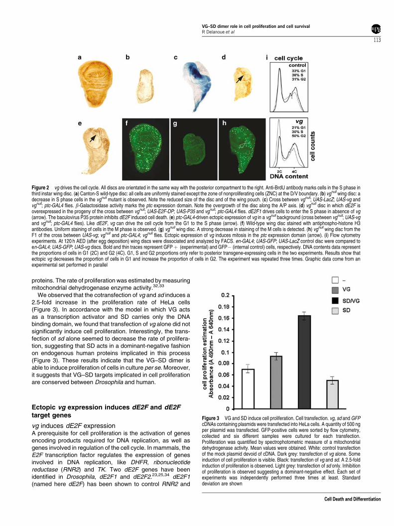
vg induces G1/S and G2/M phase transitionsNext, we tested whether vg was able to induce proliferationand thus, cell cycle progression (i.e., S and M phases) in otherparts of the wing disc using a different driver. In bromodeox-yuridine (BrdU) pulse experiments, we observed significantlyfewer cells in the S phase in most of the wing disc in the vgnull
mutant than in the wild-type strain (Figure 2a, b). Thisargument, together with the reduced vgnull disc size, is in favorof a role for vg in cell proliferation. Therefore, the homozygousvgnull genetic context was used in the following experiments,as it emphasizes the effects of ectopic vg on cell cycletransition. As it has already been shown, the ectopicexpression of dE2F in the wing disc, using the patched(ptc)-GAL4 driver, is sufficient to drive cells into the S phase(Figure 2d).21 We observed that ectopic expression of vgaccording to the ptc expression domain in a vgnull background(Figure 2c) also increases the number of cells in the S phase(Figure 2e). Moreover, ectopic vg also led to overgrowth of thedisc along the A/P axis (Figure 2c, e). Compared to the wild-type strain, a significant decrease in cells in the M phase wasobserved in vgnull discs using antiphospho-histone H3antibodies (Figure 2f, g). Moreover, ectopic expression of vg,driven by ptc-GAL4 in a vgnull background, induced transitionto the M phase (Figure 2h). These data indicate that vg is ableto regulate proliferation, increasing the number of cells in boththe S and M phases. However, we cannot exclude thepossibility that the effect of vg on the number of the M phasecells is merely the consequence of induction of the S phase.
Ectopic expression of vg changes the repartitionof cells in the cycle
To quantify the effect of ectopic expression of vg on the cellcycle, we performed flow cytometry analysis on en-GAL4,
VG–SD dimer role in cell proliferation and cell survivalR Delanoue et al
111
Cell Death and Differentiation

UAS-GFP, UAS-vg discs that expresses the green fluores-cent protein (GFP) and vg in posterior compartment cells ofwing imaginal discs. GFP expression thus allows us tosegregate anterior wild-type cells and posterior transgene-expressing cells.24 We compared G1, S and G2/M phasesrepartition in dissociated wing disc cells, overexpressing vgaccording to en (GFP! posterior compartment cells) or not(GFP" anterior internal control compartment). Control en-GAL4, UAS-GFP, UAS-LacZ discs displayed no significantdifference between the two compartments (Figure 2i, com-pare bold and thin lines).24 Conversely, ectopic vg expressiontruncated the G1 phase, as we observed a 15.15% meandecrease in posterior cells in G1 compared to anterior ones(Figure 2i). These results indicate that ectopic vg causes ashift from theG1 phase to theG2/M phase of the cell cycle andso is an activator of cell proliferation. Furthermore, theanalysis of the Forward Scatter data, which is a measure ofcell size, reveals no difference in average cell size between
the two compartments when vg is overexpressed in theposterior compartment (data not shown). This is surprising asoverproliferation is generally accompanied by a reduction incell size. However, this could be explained if VG not onlyactivates cell proliferation, but also promotes cell growth.These data and those of the former paragraph confirm that vgis able to drive the cell cycle and induce proliferation in thewing disc.
VG and SD induce proliferation of HeLa cellsOur data show that ectopically expressed vg inducesproliferation in vivo. In order to evaluate if the capacity of theVG–SD dimer, per se, to induce proliferation is dependent onother components expressed in the wing disc, we testedwhether the dimer can induce proliferation in cultured cells. Inaddition, we chose to transfect vg and sd constructs intoheterologous HeLa cells that are devoid of Drosophila
Figure 1 vg antagonises the effect of dap. The vg-GAL4 driver reflects the expression of the vgBE-enhancer. (a) vg-GAL4; vgnull/! flies display a wild-type wingphenotype. (b) vg-GAL4; UAS-dap; vgnull/! wing. A scalloped wing phenotype is observed. The same phenotype is observed using the two transgenes: UAS-dap;UAS-LacZ. (c) vg-GAL4; UAS-dap; UAS-vg; vgnull/! wing. A wild-type phenotype is observed showing that vg antagonises the effect of dap. We assume that this resultis not due to titration of the GAL4 protein. M phase staining is correlated with wing phenotypes. Third instar wing discs were stained with antiphospho-histone H3 and anti-b-galactosidase antibodies. All discs are orientated in the same way with the posterior compartment to the right. The genotypes in (d), (e), and (f) correspond togenotypes in (A), (B) and (C) respectively, with an additional vgBE-lacZ insertion that reflects the vg-GAL4 driver at the D/V boundary. A diminution of M cells(arrowheads) is observed at the D/V boundary in (e), while an increase (arrowheads) in staining is detected in (f) explaining the rescue of the wing phenotype observed in(c). The vg-GAL4 driver expression at the D/V boundary is monitored with the vgBE-lacZ staining in (d0), (e0) and (f0), respectively; and merge pictures are presented in(d00), (e00) and (f00)
VG–SD dimer role in cell proliferation and cell survivalR Delanoue et al
112
Cell Death and Differentiation
proteins. The rate of proliferation was estimated bymeasuringmitochondrial dehydrogenase enzyme activity.32,33
We observed that the cotransfection of vg and sd induces a2.5-fold increase in the proliferation rate of HeLa cells(Figure 3). In accordance with the model in which VG actsas a transcription activator and SD carries only the DNAbinding domain, we found that transfection of vg alone did notsignificantly induce cell proliferation. Interestingly, the trans-fection of sd alone seemed to decrease the rate of prolifera-tion, suggesting that SD acts in a dominant-negative fashionon endogenous human proteins implicated in this process(Figure 3). These results indicate that the VG–SD dimer isable to induce proliferation of cells in culture per se. Moreover,it suggests that VG–SD targets implicated in cell proliferationare conserved between Drosophila and human.
Ectopic vg expression induces dE2F and dE2Ftarget genes
vg induces dE2F expressionA prerequisite for cell proliferation is the activation of genesencoding products required for DNA replication, as well asgenes involved in regulation of the cell cycle. In mammals, theE2F transcription factor regulates the expression of genesinvolved in DNA replication, like DHFR, ribonucleotidereductase (RNR2) and TK. Two dE2F genes have beenidentified in Drosophila, dE2F1 and dE2F2.23,25,34 dE2F1(named here dE2F) has been shown to control RNR2 and
Figure 3 VG and SD induce cell proliferation. Cell transfection. vg, sd and GFPcDNAs containing plasmids were transfected into HeLa cells. A quantity of 500 ngper plasmid was transfected. GFP-positive cells were sorted by flow cytometry,collected and six different samples were cultured for each transfection.Proliferation was quantified by spectrophotometric measure of a mitochondrialdehydrogenase activity. Mean values were obtained. White: control transfectionof the mock plasmid devoid of cDNA. Dark grey: transfection of vg alone. Someinduction of cell proliferation is visible. Black: transfection of vg and sd. A 2.5-foldinduction of proliferation is observed. Light grey: transfection of sd only. Inhibitionof proliferation is observed suggesting a dominant-negative effect. Each set ofexperiments was independently performed three times at least. Standarddeviation are shown
Figure 2 vg drives the cell cycle. All discs are orientated in the same way with the posterior compartment to the right. Anti-BrdU antibody marks cells in the S phase inthird instar wing disc. (a) Canton-S wild-type disc: all cells are uniformly stained except the zone of nonproliferating cells (ZNC) at the D/V boundary. (b) vgnull wing disc: adecrease in S phase cells in the vgnull mutant is observed. Note the reduced size of the disc and of the wing pouch. (c) Cross between vgnull; UAS-LacZ; UAS-vg andvgnull; ptc-GAL4 flies. b-Galactosidase activity marks the ptc expression domain. Note the overgrowth of the disc along the A/P axis. (d) vgnull disc in which dE2F isoverexpressed in the progeny of the cross between vgnull; UAS-E2F-DP; UAS-P35 and vgnull; ptc-GAL4 flies. dE2F1 drives cells to enter the S phase in absence of vg(arrow). The baculovirus P35 protein inhibits dE2F induced cell death. (e) ptc-GAL4-driven ectopic expression of vg in a vgnull background (cross between vgnull; UAS-vgand vgnull; ptc-GAL4 flies). Like dE2F, vg can drive the cell cycle from the G1 to the S phase (arrow). (f) Wild-type wing disc stained with antiphospho-histone H3antibodies. Uniform staining of cells in the M phase is observed. (g) vgnull wing disc. A strong decrease in staining of the M cells is detected. (h) vgnull wing disc from theF1 of the cross between UAS-vg; vgnull and ptc-GAL4; vgnull flies. Ectopic expression of vg induces mitosis in the ptc expression domain (arrow). (i) Flow cytometryexperiments. At 120 h AED (after egg deposition) wing discs were dissociated and analyzed by FACS. en-GAL4; UAS-GFP; UAS-LacZ control disc were compared toen-GAL4; UAS-GFP; UAS-vg discs. Bold and thin traces represent GFP! (experimental) and GFP" (internal control) cells, respectively. DNA contents data representthe proportions of cells in G1 (2C) and G2 (4C). G1, S and G2 proportions only refer to posterior transgene-expressing cells in the two experiments. Results show thatectopic vg decreases the proportion of cells in G1 and increase the proportion of cells in G2. The experiment was repeated three times. Graphic data come from anexperimental set performed in parallel
VG–SD dimer role in cell proliferation and cell survivalR Delanoue et al
113
Cell Death and Differentiation

proliferating cell nuclear antigen (PCNA) gene expression,while a role for dE2F in DHFR and TK activation has not beeninvestigated yet.22,35
A possible link between vg and dE2F was investigated firstby testing for genetic interactions in double heterozygousmutant combinations of a dE2F null allele (dE2F91),22 andeither vg or sdmutations. No interaction was observed in vgnull
and dE2F91 double heterozygotes (data not shown). How-ever, vg is strongly haplo-sufficient and double heterozygoussd58/! ; vgnull/! females displayed a wild-type wing pheno-type although sd is a well-established partner of vg. OnlysdETX4/Y males displayed an enhanced wing phenotype in avgnull/! heterozygous background (Figure 4a, b).11 Similarly,sdETX4/Y, dE2F91/! males displayed a more severe mutantwing phenotype compared to sdETX4/Y males (Figure 4a, c),suggesting a genetic interaction between dE2F and sd.We therefore examined the hypothesis that VG–SD
induces cell proliferation through regulation of the dE2Ftranscription factor. Since it is not possible to obtain vgnull
clones and since the wing pouch is severely reduced in thevgnullmutant, we used the overexpressionUAS-GAL4 systemto address this issue.4,30,36 We first tested if vg is able toinduce dE2F ectopically, using in situ hybridization with RNAprobes. In the wild-type disc, dE2F is expressed at relativelylow levels in the notum and the wing pouch (Figure 4d).However, it has already been shown that dE2F, thoughexpressed at the D/V boundary, is lately inactive.22,37
We overexpressed vg in the entire posterior compartmentof the wing disc using the en-GAL4 driver. Under theseconditions, an increase in dE2F RNA expression wasobserved in the posterior part of the wing disc (Figure 4e).These results were confirmed at the protein level by
immunostaining using anti-dE2F antibodies when we over-expressed vg in the ptc domain (Figure 4g). The E2Frm729
mutant is a P[LacZ] element inserted in the dE2F gene.22 Inptc-GAL4, UAS-vg, E2F-LacZ flies, b-galactosidase activitywas observed in the ptc expression domain, suggesting thatthe VG–SD dimer acts on the dE2F promoter (Figure 4f).However, we cannot exclude the possibility that VG–SDinduces expression of other targets that would in turn activatethe dE2F promoter.
vg induces dRNR2 and stgIn mammals, E2F1 activates a large variety of genes involvedin DNA replication, cell cycle progression and oncogenesis. Inmammals, the gene encoding RNR2 is regulated by E2F1,and in Drosophila by dE2F1.22 Moreover, it has been shownthat, in Drosophila, overexpression of dE2F directs not onlycell cycle progression from G1 to S, but also from G2 to M byactivating the expression of the stg gene, a homolog of yeastcdc25.24Wewanted to determine ifDmRNR2 and stg are alsovg targets. In situ hybridization with a DmRNR2 proberevealed expression of this gene in the wing disc but not atthe level of the wingmargin (Figure 5a). Johnston and Edgar37
hypothesized that the absence of DmRNR2 expression at thewing margin is due to the fact that dE2F1 is probably inactivesince these cells are arrested in G1.We ectopically expressed vg in the posterior compartment
of the wing disc using the en-GAL4 driver. Strong DmRNR2expression was induced in this posterior compartmentcompared to the control (Figure 5a, b), indicating that vg caninduce this identified target of dE2F. In the same way, stgexpression was detected in wild-type wing discs as previously
Figure 4 sd and dE2F interact genetically. (a) Wing of a sdETX4/Ymale. (b) wing of a sdETX4/Y; vgnull/! mutant. The increase in the mutant wing phenotype in a vgnull
heterozygous background reflects the known protein–protein interaction between the two products. (c) Wing of a sdETX4/Y; dE2F91/! mutant. As in (b), an increase inthe mutant phenotype is also observed. Ectopic expression of vg induces dE2F expression. All discs are orientated in the same way with the posterior compartment to theright. (d) In situ hybridization with a dE2F antisense RNA probe on a wild-type third instar wing disc. (e) Third instar wing disc of progeny of the cross between UAS-vgand en-GAL4 strains. vg is overexpressed in the posterior compartment. An induction of dE2F is observed. (f) Third instar wing disc of progeny of the cross betweenUAS-vg and ptc-GAL4; E2F-LacZ strains. A faint expression of dE2F is normally observed at the level of the D/V boundary. The arrow shows ectopic expression ofb-galactosidase according to ptc expresssion pattern confirming the in situ experiment observed in (e). (g) vgnull wing disc in which vg is overexpressed in the progeny ofthe cross between vgnull; UAS-vg and vgnull; ptc-GAL4 flies. Staining with anti-dE2F antibodies reveals that vg induces the expression of the dE2F protein
VG–SD dimer role in cell proliferation and cell survivalR Delanoue et al
114
Cell Death and Differentiation

described,1 and was induced by ectopic vg (Figure 5c, d).Taken together our results show that ectopic vg is sufficient toinduce expression of a functional dE2F transcription factorand two of its target genes dmRNR2 and stg.
vg, but not dE2F, induces DHFR
Another well-identified target of E2F1 in mammals is theDHFR gene. DHFR is involved in a critical step of dTMPsynthesis and, thus, is necessary for DNA replication. In situhybridization with a Drosophila DHFR probe showed almostuniform staining in the wild-type wing disc (Figure 5e). It hadnot been established whether dE2F is able to induce DHFRexpression in Drosophila. Surprisingly, we found that ectopicexpression of dE2F using the en-GAL4 driver had no effect onDHFR expression in the wing disc (data not shown). Sinceectopic expression of dE2F induces apoptosis, we coex-pressed dE2F and the baculovirus P35 protein which inhibitscell death.21 No induction of DHFR either was observed evenunder these conditions (data not shown).We next tested by in situ hybridization whether over-
expression of vg in the en expression domain of the wing discwas able to induce DHFR expression. Indeed vg inducedstrong DHFR expression in the posterior part of the wingdisc, the effect being particularly striking in the wing pouch(Figure 5e, f).
Genetic interactions between vg, but not dE2F, anddef(DHFR)To test, at a genetic level, the interaction between vg andDHFR, we used two different DHFR deficiencies since noDHFR mutants were available (see Materials and Methods).As for vg and dE2F, no interaction was observed between vgand def(DHFR) in double heterozygotes (vgnull/! ;def(DHFR)/! ) (data not shown). However, the wing pheno-type of sdETX4/Y mutants is severely aggravated in adef(DHFR)/! context (Figure 6a, b), indicating a geneticinteraction between DHFR and sd. We cannot exclude at thispoint the possibility that this interaction is due to other genesincluded in the deficiency even if no likely candidate genescould be identified in the database.In order to try to reveal possible genetic interactions
between vg, dE2F and DHFR, we used very low doses ofaminopterin (0.5mg/kg) to reduce the strong haplo-sufficiencyassociated with vg mutations (see the section ‘vg inducesdE2F expression’). Indeed aminopterin is a specific inhibitor ofDHFR activity and it has been shown to lead to a decrease invg expression in wild-type strains.17 When flies were rearedon a low dose of aminopterin (0.5mg/kg), the slight inhibitionof DHFR activity led to nicks in wings in 2% of def(DHFR)/!heterozygous flies (data not shown). Potential geneticinteractions between dE2F and def(DHFR) or vg anddef(DHFR) should lead to increased wing phenotypes indouble heterozygous flies reared on the drug, compared todef(DHFR)/! controls. Wild type or dE2F91/! heterozygousflies exhibit no wing abnormalities when raised at this lowconcentration of aminopterin (Figure 6f). Nicks at wing tipswere observed in vgnull/! heterozygous flies reared onaminopterin (Figure 6c), while a much stronger notched wing
Figure 5 Ectopic vg induce DmRNR2, stg and DHFR. In situ hybridization withantisense RNA probes on third instar wing discs. All discs are orientated in thesame way with the posterior compartment to the right. Wild-type discs (a, c, e) arecompared to third instar wing discs of progeny from the cross between UAS-vgand en-GAL4 strains (b, d, f) in which vg is overexpressed in the posteriorcompartment. The probes used were: DmRNR2 (a, b), stg (c, d) and DHFR (e, f).All three genes are overexpressed in the posterior compartment in en expressiondomain, in response to ectopic vg
VG–SD dimer role in cell proliferation and cell survivalR Delanoue et al
115
Cell Death and Differentiation

phenotype was obtained in vg/! ; def(DHFR)/! doubleheterozygotes (Figure 6d). No differences in wing phenotypeswere detected in vgnull/! ; E2F91/! (Figure 6e) or in theE2F91/! ; def(DHFR)/! double heterozygotes flies reared onthe inhibitor (Figure 6f and data not shown) compared to thecontrols.
These results demonstrate a strong genetic interactionbetween vg and def(DHFR) leading to a wing marginphenotype. In contrast, no interaction was detected betweendE2F and vg or dE2F and def(DHFR).
Figure 6 Genetic interaction between sd and DHFR. (a) Wing of a sdETX4/Ymale. (b) Wing of a sdETX4/Y; def(DHFR)/! male. A strong increase of themutant phenotype is observed in a def(DHFR) heterozygous background,suggesting a genetic interaction between the two genes. Genetic interactionbetween vg and DHFR in the presence of a low dose of aminopterin (0.5 mg/kg).Each picture shows the most representative phenotype of the cross. (c) vgnull/!wing. A scalloped phenotype is observed. (d) vgnull/! ; def(DHFR)/! wing. Astrong increase in the mutant phenotype is observed compared to (c). (e)dE2F91/! ; vgnull/! wing. Small notches at the wing tips are observed. (f) Wingof the wild-type strain which also corresponds to what is observed for def(DHFR)/! or dE2F91/! ; def(DHFR)/! flies
Figure 7 vg together with DHFR are required for cell survival at the D/Vboundary of the wing imaginal disc. Effect of aminopterin, the inhibitor of DHFR.All discs are orientated in the same way with the posterior compartment to theright. X-gal staining was performed rigorously, with the same experimentalprocedure for all genotypes. Staining times were precisely the same andexperiments were repeated four times. We could not find any difference instaining between the wild-type Canton S and w1118 strain, which is the geneticbackground for the different mutants. The cut-LacZ transgene marks a stripe ofcells precisely at the D/V boundary and allows to monitor their fate (a–e). (a) Wild-type wing disc heterozygous for the cut-LacZ transgene. Similar LacZ expressionis observed in a vgnull/! heterozygous disc (data not shown). (b) Wild-type wingdisc of flies reared on medium containing a concentration of 2 mg/kg ofaminopterin. cut-LacZ pattern is normal suggesting absence of D/V boundaryspecific apoptosis in LacZ-expressing cells. (c) In vgnull/! heterozygotes, LacZstaining at the D/V boundary is interrupted, probably due to diminution of DHFRactivity induced by aminopterin (arrows). (d) No visible effect is observed inE2F91/! wing discs. (e) Conversely, in a def(DHFR)/! background, gaps atthe level of the D/V boundary are observed (arrows). The expression of theapoptosis inducer gene reaper (rpr) was monitored using the rpr-LacZ transgene.Flies were reared on medium containing aminopterin (0.5 mg/kg) (f–i).Aminopterin has no effect on rpr expression in the canton S wild-type strain (fand data not shown), which is normally detected in the wing pouch, particularly atthe D/V boundary. (g) Conversely, in vgnull/! heterozygous wing discs, the rpr-LacZ reporter construct shows an increased activity especially at the D/Vboundary. (h) E2F91/! wing discs do not display D/V boundary specific rpr-lacZactivation (a slight decrease can even sometimes be observed). (i) def(DHFR)/! discs resemble the vgnull/! context. This specific rpr-lacZ inductioncorrelates with the disappearance of D/V boundary cut-LacZ-expressing cellreported (c, e). Cell death was monitored with acridine orange staining. (j)Aminopterin (0.5 mg/kg) induces apoptosis quite homogeneously in wild-typewing discs. (k) Conversely, a specific increase in apoptosis at the D/V boundary isvisualized in vgnull/! heterozygous discs
VG–SD dimer role in cell proliferation and cell survivalR Delanoue et al
116
Cell Death and Differentiation

These results suggest that in the Drosophila wing disc,unlike what has been described in mammals, dE2F does notcontrol DHFR expression, this role possibly being allotted tothe VG–SD dimer. This raises the possibility that in addition toDHFR, other identified targets of dE2F may also be regulatedin Drosophila by transcription factors involved in proliferation,others than dE2F.
Diminution of DHFR activity leads to D/Vboundary defects
In order to distinguish the respective roles of DHFR and dE2Fat the D/V boundary of the wing disc, we used the cut-LacZstrain, which precisely marks a stripe of D/V boundary cellsand allowed to monitor their fate. To reduce DHFR activity wealso used aminopterin at a concentration of 2mg/kg tocorrelate the wing phenotype and D/V boundary cell fate indifferent genotypes. No difference in the cut-LacZ expressionpattern was detected between wild-type flies and vgnull/!heterozygous flies reared on normal medium (Figure 7a).Nevertheless, in flies grown on medium containing aminop-terin, although the wild-type strain exhibited normal cut-LacZexpression (Figure 7b), vgnull/! heterozygotes displayedsignificant gaps in the staining (Figure 7c). Moreover, thesediscs also displayed a high level of cell death specifically at thelevel of D/V boundary that could be visualized by staining withacridine orange (Figure 7k). No other part of this disc wasexposed to such a specific increase in cell death. This was notobserved in the wild-type strain where quite homogenous celldeath in the wing disc was detected (Figure 7j). Asaminopterin is a specific inhibitor of DHFR activity, theseresults suggest that a decrease in DHFR in a vgnull/!heterozygous mutant background induces specific cell deathat the D/V boundary and thus loss of cut-LacZ-expressingcells.When reared on aminopterin, def(DHFR)/! discs clearly
displayed abnormal cut-LacZ staining (Figure 7e) like that invgnull/! heterozygotes (Figure 7c). This is in agreement withthe hypothesis that the abnormal cut-LacZ phenotype is dueto a decrease in DHFR activity.If the DHFR gene was to be regulated by dE2F at the D/V
boundary, then reduced DHFR expression in dE2F91/!mutant wing discs and consequent abnormal cut-LacZexpression in larvae reared on the inhibitor should beexpected. This was not the case since cut-LacZ expressionwas normal in this genetic background (Figure 7d). This is inaccordance with the hypothesis that vg, but not dE2F, is ableto regulate DHFR expression at the D/V boundary in wingdiscs of the third instar larvae. Moreover, these resultssuggest that VG is also required for cell survival in the wingdisc.
vg and DHFR are specifically required forcell survival at the D/V boundary
We next tested whether the effects observed on wing margincell fates and cell death induction, when flies of differentgenotypes were reared on aminopterin (0.5mg/kg), could becorrelated with misregulation of apoptosis regulators. Droso-
phila inhibitor of apotosis protein 1 (DIAP1) is a negativeregulator of apoptosis that binds to and inhibits caspaseactivity.38 DIAP1 has recently been reported to be detectedthroughout the wing disc with an increased expression patternalong the pouch D/V boundary.39 The authors also demon-strated that the proapoptotic Reaper (Rpr) protein is able tobind to and trigger ubiquitin-mediated DIAP1 degradationleading to subsequent caspase activation and apoptosis.39
But more concerning is the fact that rpr is also expressed inthe wing pouch, particularly at the D/V boundary.40,41 Thisboundary, therefore, seems to be a quite sensitive area undertight regulation of cell death and cell survival signals. Tomonitor rpr expression in the different vg, dE2F and DHFRheterozygous contexts, we used a rpr-LacZ reporter trans-genic line.41,42 No difference in LacZ expression wasobserved between the different genotypes when flies werereared on normal medium (data not shown). On aminopterin-containing medium (0.5mg/kg), specific induction of rpr wasnot observed at the level of the wing margin in the wild-typestrain (Figure 7f and data not shown) nor in dE2F91
heterozygotes (Figure 7h) where a slight decrease in rpr-LacZ expression at the D/V boundary can even sometimes beobserved. Conversely, rpr was specifically activated at theD/V boundary in vgnull/! (Figure 7g) and def(DHFR)/!(Figure 7i) heterozygotes, indicating a complete correlationwith the loss of cut-LacZ-expressing cells and cell deathinduction.These results led us to investigate the effect of rpr induction
on DIAP1, upon reduction of VG and DHFR levels. Strikingly,DIAP1 protein was specifically and severely downregulated atthe D/V boundary of vgnull/! mutants compared to wild-typediscs (Figure 8a, b). Nevertheless, no effect could beobserved on DIAP1 transcription, using a P[LacZ] insertionat the thread (th) locus encoding DIAP1 (Figure 8d and datanot shown). These results are consistent with the rpr-inducedpost-translational degradation of DIAP-1 reported so far.Conversely, vg overexpression along the D/V boundary (vg-GAL4 driver) completely rescued the loss of DIAP1 in vgnull/!disc (Figure 8c), as expected if DIAP1 is regulated by VG. Weconcluded that vg requirement for cell survival at the D/Vboundary is, at least, mediated through DIAP1.Interestingly, specific inhibition of DHFR (aminopterin
0.5mg/kg) in wild-type flies also induced DIAP1 downregula-tion (Figure 8e) that was rescued upon vg overexpression(Figure 8f). These results confirm that a decrease in the VGproduct shifts the D/V boundary cells to a cell death sensitivestate that seems to be correlated with a reduced DHFRactivity.Finally, we monitored the effects of aminopterin on cell
death effectors, using the CM1 antiactivated caspases anti-body. Only few caspase-positive cells were detected in wild-type or vgnull/! discs grown on normal medium (Figure 8g, h).These cells displayed pyknotic nuclei, indicative of apoptosis(Figure 8h, i).43 However, we observed that vgnull/! discs onaminopterin displayed a high level of activated caspases,which was especially restricted to the D/V boundary(Figure 8k) compared to wild-type ones (Figure 8i). Manyvgnull/! cells along the D/V boundary displayed pyknoticnuclei (Figure 8k, l). This result accounts for acridine stainedcell death that was previously observed (Figure 7k), and is
VG–SD dimer role in cell proliferation and cell survivalR Delanoue et al
117
Cell Death and Differentiation
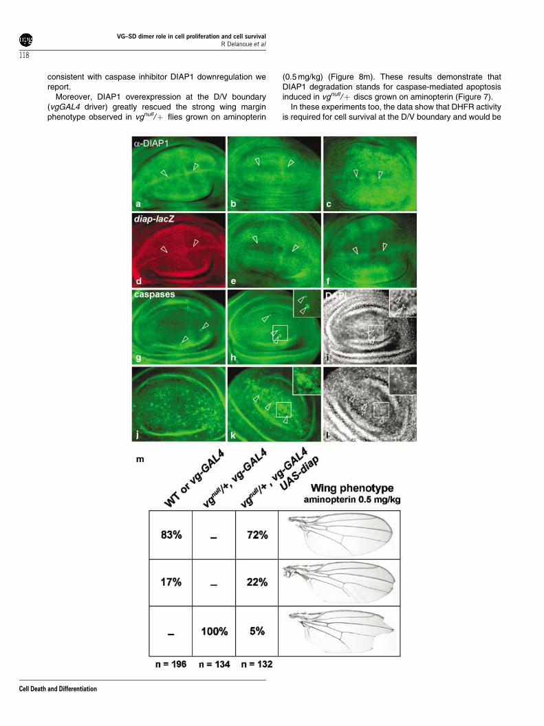
consistent with caspase inhibitor DIAP1 downregulation wereport.Moreover, DIAP1 overexpression at the D/V boundary
(vgGAL4 driver) greatly rescued the strong wing marginphenotype observed in vgnull/! flies grown on aminopterin
(0.5mg/kg) (Figure 8m). These results demonstrate thatDIAP1 degradation stands for caspase-mediated apoptosisinduced in vgnull/! discs grown on aminopterin (Figure 7).In these experiments too, the data show that DHFR activity
is required for cell survival at the D/V boundary and would be
VG–SD dimer role in cell proliferation and cell survivalR Delanoue et al
118
Cell Death and Differentiation

under the control of vg. A decrease in VG or DHFR activitywould lead to specific rpr expression, DIAP1 degradation,caspase-mediated cell death and, consequently, to wingmargin defects.
Discussion
The normal development of multicellular organisms requiresproliferation of precursor cells followed by cell cycle arrest andcommitment to specialized fates. In mammals, E2F, P21 andthe pRb pocket proteins have been shown to be involved indifferentiation.20 However, little is known about how develop-mental pathways regulate the cell cycle. Regulation of p21 byHOX genes has been reported in a human myelomonocyticcell line; this constitutes one of the few examples described sofar.44 Recently, the transcription factor Cubitus interruptus(Ci) was shown to mediate the effects of Hedgehog (Hh)signalling on cell growth and proliferation in the wing disc bydirect binding to the Cyclin E promoter.45 In addition, theCaudal (CAD) homeodomain protein was shown to bind thedE2F promoter in vitro, and CAD binding sites in the dE2Fpromoter are required for expression of dE2F in living flies.46
These results most clearly demonstrate that developmentalgenes like Cad, Hh and Hox can directly regulate cell growthand proliferation.There are several indications that the vg gene can induce
cell proliferation when expressed ectopically,4,47 and maymediate the cross-talk between developmental and cell cyclegenes in wing development. Interestingly, Notch and dppsignalling pathways have been shown to both activate vg inthe wing disc via specific enhancers, and induce proliferation.In a recent report, cell proliferation induced by ectopicexpression of an activated form of Thick vein (TKV), theDPP receptor, was shown to require the presence of VG.However, TKV was unable to induce vg expression, indicatingonly a synergetic effect between the two genes in thisprocess.48
The results presented here clearly demonstrate that vg isrequired and sufficient for cell proliferation at the D/Vboundary of the wing imaginal disc. Not only can theoverexpression of the gene counteract the effect of dap, butit also induces cell cycle progression. The latter wasconfirmed by: (i) overexpression of vg in the wing disc andconsequent induction of G1/S and G2/M cell progression inthe region where vgwas overexpressed and (ii) fluorescence-activated cell sorter (FACS) experiments showing that over-
expression of vg in the posterior compartment of the wing discshifts the cells toward theG2/M phase. VG therefore seems tobe an active regulator of G1/S phase transition.Moreover, in a heterologous system (HeLa cells) the
Drosophila VG–SD dimer, per se, induces cell proliferation.This suggests that the dimer is able to act on humanproliferation genes and, thus, that target sites of the SD TEADNA binding domain are likely conserved betweenDrosophilaand humans. In addition, our results in human HeLa cells arein favor of a dominant-negative effect of an excessive amountof SD, which probably saturates the target sites involved in theprocess and results in inhibition of cell proliferation. On thewhole, data are in favor of the hypothesis that the target sitesof the SD TEA DNA binding domain are conserved inDrosophila and human and are necessary for cell proliferation.The prerequisite for cell proliferation is the activation of
specific transcription factors, like E2F, that trigger DNAreplication and cell cycle progression. Our results show thatvg induces cell proliferation in the wing disc by activatingdE2F, but also dE2F target genes like dmRNR2 and stg.Although DHFR is a well-established target of dE2F in
mammals, we found that in contrast to dE2F, vg inducesDHFR expression in the Drosophila wing imaginal disc,suggesting the possibility that vg exerts some of its effectsdirectly on proliferation genes in the wing pouch and notmerely through dE2F. In addition, on medium containing lowconcentration of aminopterin that inhibits DHFR activity, agenetic interaction was observed between vg and def(DHFR),and not between the latter and dE2F, with respect to wingphenotype.Moreover, the genetic interaction between vg and
def(DHFR) leading to wing margin phenotypes was corrobo-rated with the loss of D/V boundary cut-LacZ-expressing cellsin vgnull/! and def(DHFR)/! heterozygotes grown onaminopterin. No such effect could be visualized in dE2F 91/! mutant, confirming that the regulation of DHFR by vg wasnot mediated by dE2F.We further showed that vgnull/! mutants on aminopterin
displayed a clear increase in cell death that was restricted tothe D/V boundary. These results led us to monitor a potentialadditional role for vg in cell survival. Interestingly, the strongexpressions of both apoptosis regulators rpr and DIAP1,specifically at the D/V boundary of the wing disc, are in favor ofa precise regulation of cell survival in this area.40,41 Inaddition, the fact that vg is highly expressed in the same areaand is absolutely required for wing pouch development, led us
Figure 8 Apotosis inhibitor DIAP1 is required for regulation of cell survival at the D/V boundary of the wing disc by vg. All discs are orientated in the same way with theposterior compartment to the right and ventral part on the top. (a) anti-DIAP1 staining. DIAP1 protein is observed throughout the whole wing disc of wild-type strain with astriking increase in expression at the D/V boundary (arrowheads), as already described.39 (b) This D/V specific DIAP1 protein is lost in vgnull/! heterozygous mutants(arrowheads). (c) vg overexpression along the D/V boundary (vgGAL4 driver) of vgnull/! disc completely rescues DIAP1 protein level (arrowheads). (d) anti-b-galactosidase staining. P[LacZ] insertion at the th locus encoding DIAP1 reflects diap1 transcription with a marked increase at the D/V boundary of wild-type disc(arrowheads).39 No difference can be monitored in a vgnull/! context (data not shown). (e) Addition of aminopterin (0.5 mg/kg) to flies medium leads to DIAP1downregulation similar to (b) (arrowheads). (f) Moreover, vg overexpression along the D/V boundary rescues DIAP1 protein, as well (arrowheads). (g) CM1 antiactivatedcaspases staining of wild-type disc. Positive cells are quite rare in the wing pouch (arrowheads). (h) Only few caspase-positive cells are observed in vgnull/! disc(arrowheads and inset). (i) Same disc stained with DAPI. Caspase-positive cells display pyknotic nuclei (arrowheads and inset). (j) Upon addition of aminopterin (0.5 mg/kg), caspase-positive cells are observed in a quite homogeneous pattern in the wing disc. (k) Strikingly, activation of caspases specifically increases at the D/V boundaryof vgnull/! discs gown on aminopterin (arrowheads and inset). (l) Same disc staining with DAPI. Pyknotic nuclei are particularly observed along the D/V boundary(arrowheads and inset). (m) DIAP1 overexpression along the D/V boundary (vgGAL4 driver) rescues to some extent the wing phenotype of vgnull/! flies grown onaminopterin (0.5 mg/kg)
VG–SD dimer role in cell proliferation and cell survivalR Delanoue et al
119
Cell Death and Differentiation

to monitor cell death regulators expression in both wild-typeand vgnull/! discs.7 Using aminopterin, we showed that VGand DHFR are required for the integrity of wingmargin cells, inaddition to cell proliferation. A decrease in both products,induces specific rpr expression, DIAP1 downregulation andcaspase-mediated cell death at the D/V boundary of the wingdisc. However, even if DIAP1 was specifically lost in vgnull/!cells at the D/V boundary, we could not detect any clearinduction of apoptosis without addition of aminopterin. Thisindicates that VG reduction only promotes a cell deathsensitive state. Apoptosis was further triggered upon con-comitant DHFR inhibition. However, it has been shown that vgdisplays incomplete penetrance depending on genetic back-grounds.49 This suggests that cell death eventually occurs inheterozygotes on normal medium.Interestingly, our results clearly show that vg and its target
DHFR are required for both cell survival and proliferation.Therefore, the complete loss of wing tissue in vgnull homo-zygous mutant7,11 and the impossibility to recover vgnull
clones4,36 are probably due to both proliferation impairmentand apoptosis triggering. The requirement for DHFR duringboth processes raises the possibility of mutual cross-talkbetween cell survival and proliferation that would rely on VGfunction and DNA synthesis. Moreover, the wing phenotype ofvgnull/! flies grown on aminopterin was greatly rescued uponectopic expression of DIAP1 along the D/V boundary. Thisdemonstrates that wing defects observed upon DHFRinhibition and VG reduction are significantly due to cell deathtriggering.It has been proposed that vg can be viewed as a wing
selector gene that would impose the wing developmentalpathway.27,50,51 Interactions between vg and genes likeNotch, wg and dpp would influence gene expression patternsto promote wing identity.28 However, some authors recentlyconcluded that vg is not a ‘master gene’ but a component ofthe genetic combination that drives wing blade development.Thus, only an indirect role of vg in cell proliferation washypothesized by these authors.52 In this study, we demon-strate that vg not only promotes wing differentiation, but is alsorequired for cell survival and proliferation.In addition, several counter arguments can be used to
uncouple a role for vg in cell proliferation from its role in cellidentity. Indeed, TONDU (TDU) a human vg homolog,dimerizes with SD when expressed in flies.14 Ectopicexpression of TDU in the eye disc induces cell proliferationwithout promoting wing cell differentiation.14 Another VGpartner in the wing disc has been identified, Strawberry notch(SNO), which is also a Notch target.53,54 The possibility existsthat wing specification requires more than just the VG–SDtranscription factor, while the VG–SD dimer alone would besufficient to induce proliferation, as was suggested by ourexperiments of heterologous expression of vg and sd in HeLacells. Indeed, it has been shown that SDDNA target selectivitycan be modified when SD is bound to VG.55 In vitro studies onthe binding of the TEA domain of SD on well-establishedtargets show that the presence of a cofactor, in thisoccurrence VG, dramatically shifts the affinity of SD for thesetargets. Therefore, we can hypothesize that induction of SDtarget genes depends on different components bound to theSD protein.
Taken together our results point to the existence of a tissue-specific activator of dE2F and DHFR, namely vg, which isrequired for cell proliferation and cell survival. On the whole,our results demonstrate that vg drives cell cycle progression inthe wing disc, acting on the expression of key genes thatcontrol the pathway, and that the VG–SD dimer retains itspotentialities as an inductor of cell proliferation in a hetero-logous system. Moreover, we show that vg is also required fornormal expression of apoptosis regulators and cell survival inthe presumptive wing margin area.
Materials and Methods
Drosophila strains and mediumThe vgnull and UAS-vg strains were generated in our laboratory.11 The vg-GAL4 strain that drives GAL4 expression according to part of the vgBE-enhancer was provided by M Hoffmann and S Morimura. The vgBE-lacZstrain that drives b-galactosidase expression according to the vgBEenhancer was described in Williams et al.8 The sdETX4 strain is a sd-LacZenhancer trap described in Campbell et al.13 The E2Frm729 line, whichcontains an enhancer trap inserted in the dE2F1 gene, is called E2F-lacZin this study.22 The E2F91 mutation is described as a null allele for thegene.22 The en-GAL4; UAS-GFP (GFP) strain was used for FACSexperiments. The DHFR gene is located at 89D2. The first deficiency usedis Df (3R) p10 (Bloomington no. 3483), (breaks at 89C1–89E2). UAS-dapis a gift from C Lehner and UAS-E2F-DP; UAS-P35 a gift from B Edgar.The latter strain allows expression of dE2F without induction of cell death.The rpr-LacZ strain that carries the lacZ sequence under the control of theentire promoter of the rpr gene was provided by JM Abrams.41,42 The thj5c8
strain that carry a P[lacZ] reporter in the 50 untranslated region of the diap1transcription unit and the UAS-diap1 strain were provided by K White andH Steller.39 All other stocks come from the Bloomington DrosophilaCenter. Aminopterin (Sigma) was added to the medium at a concentrationof 0.5 or 2mg/kg depending on the experiment.
Histology
A Leica DMR microscope was used. X-Gal staining was performed asdescribed31 and viewed using Nomarski optics. Digoxigenin (DIG)-labeledantisense or sense RNA probes were generated with T3 or T7 RNApolymerase (Promega) and DIG-UTP (Roche) from cloned cDNAs ofdE2F1, DHFR, stg and DmRNR2. These probes were used for whole-mount in situ hybridization of fixed larval imaginal discs. The DIG-labeledRNA probes were detected with the aid of an anti-DIG antibody coupled toalkaline phosphatase (Roche) and NBT/BCIP as substrate. BrdU andDAPI labeling of imaginal discs was performed as described.1 Stainingwith rabbit antiphospho-histone H3 antibody (Upstate Biotechnology),anti-VG (gift from S Carroll), anti-dE2F1 (gift from P Leopold) anti-DIAP1(gift from HD Ryoo and H Steller), CM1 antiactivated caspases antibodies(BD Pharmingen) and mouse anti-b-galactosidase antibody (JacksonImmuno Labs) were performed according to standard protocols.
FACS experiments
Staged larvae (120 h after egg deposition) derived from 2 to 3 h eggcollections and raised at 251C were dissected in PBS for each genotype.In all, 20 wing discs were washed twice in PBS and incubated with gentleagitation in trypsin (Invitrogen) and Hoechst 33342 (Sigma) for 150min
VG–SD dimer role in cell proliferation and cell survivalR Delanoue et al
120
Cell Death and Differentiation

according to Nuefeld et al.24 About 20 000 GFP! and GFP" cells wereanalyzed for each genotype. An Elite Beckman Coulter FACS was usedand data were analyzed using the Multicycle Software. P-values werecalculated using a two-tailed Student’s t-test.
Cell transfection
vg and sd cDNAs were subcloned in the pXJ40 vector (gift from IDavidson) in order to allow expression of these cDNAs under the control ofa CMV promoter. HeLa cells were transfected using the FuGENE6treagent (Roche), with pXJ40 plasmids containing vg, sd cDNAs andpEGFP (GFP) (Clontech) according to the supplier’s instructions.
Cell proliferation
Transfected HeLa cells were incubated for 48 h at 371C in an atmospherecontaining 5% CO2. GFP-positive cells were sorted by flow cytometry(Fluorescent Activated Cell Sorter FACS), collected, and then placed in96-well plates (2000 cells per well) and cultured for 48 h at 371C in 100 mlof medium. In all, 50 ml of coloring solution XTT (0.3 mg/ml) (Cellproliferation kit II, Roche), which allows quantification of cell proliferationby measuring mitochondrial dehydrogenase activity, was added to eachwell.32,33 Light Absorption in each well was measured at 490 and 640 nm 4and 48 h later with an ELISA reader (MRX II Thermosystem).
Acknowledgements
We are grateful to BA Edgar for helpful comments on the manuscript. Wethank SB Carroll, C Lehner, JM Abrams, P Leopold, HD Ryoo, K Whiteand H Steller for fly strains and reagents, and MC Gendron for help withFACS experiments. B Legois and A Skov provided technical assistance,and AM Pret for comments and corrections with the language. This workwas supported by the Association pour la Recherche contre le Cancer(ARC), and an ‘Action Thematique concertee (ATC) vieillissement’ grantfrom the Institut National pour la Sante Et la Recherche Medicale(INSERM).
References
1. Milan M, Campuzano S and Garcia-Bellido A (1996) Cell cycling and patternedcell proliferation in the wing primordium of Drosophila. Proc. Natl. Acad. Sci.USA 93: 640–645
2. Blair SS (1995) Compartments and appendage development in Drosophila.Bioessays 17: 299–309
3. de Celis JF (1999) The function of vestigial in Drosophila wing development:how are tissue-specific responses to signalling pathways specified? Bioessays21: 542–545
4. Kim J, Sebring A, Esch JJ, Kraus ME, Vorwerk K, Magee J and Carroll SB(1996) Integration of positional signals and regulation of wing formation andidentity by Drosophila vestigial gene. Nature 382: 133–138
5. Kim J, Magee J and Carroll SB (1997) Intercompartmental signaling and theregulation of vestigial expression at the dorsoventral boundary ofthe developing Drosophila wing. Cold Spring Harb. Symp. Quant. Biol. 62:283–291
6. Cohen SM (1996) Controlling growth of the wing: vestigial integrates signalsfrom the compartment boundaries. Bioessays 18: 855–858
7. Williams JA, Bell JB and Carroll SB (1991) Control of Drosophila wing andhaltere development by the nuclear vestigial gene product. Genes Dev. 5:2481–2495
8. Williams JA, Paddock SW, Vorwerk K and Carroll SB (1994) Organization ofwing formation and induction of a wing-patterning gene at the dorsal/ventralcompartment boundary. Nature 368: 299–305
9. Neumann CJ and Cohen SM (1996) A hierarchy of cross-regulation involvingNotch, wingless, vestigial and cut organizes the dorsal/ventral axis of theDrosophila wing. Development 22: 3477–3485
10. Simmonds AJ, Liu X, Soanes KH, Krause HM, Irvine KD and Bell JB (1998)Molecular interactions between Vestigial and Scalloped promote wingformation in Drosophila. Genes Dev. 12: 3815–3820
11. Paumard-Rigal S, Zider A, Vaudin P and Silber J (1998) Specificinteractions between vestigial and scalloped are required to promotewing tissue proliferation in Drosophila melanogaster. Dev. Genes Evol. 208:440–446
12. Halder G, Polaczyk P, Kraus ME, Hudson A, Kim J, Laughon A andCarroll S (1998) The Vestigial and Scalloped proteins act together todirectly regulate wing-specific gene expression in Drosophila. Genes Dev. 12:3900–3909
13. Campbell S, Inamdar M, Rodrigues V, Raghavan V, Palazzolo M and ChovnickA (1992) The scalloped gene encodes a novel, evolutionarily conservedtranscription factor required for sensory organ differentiation in Drosophila.Genes Dev. 6: 367–379
14. Vaudin P, Delanoue R, Davidson I, Silber J and Zider A (1999) TONDU (TDU),a novel human protein related to the product of vestigial (vg) gene of Drosophilamelanogaster interacts with vertebrate TEF factors and substitutes for Vgfunction in wing formation. Development 126: 4807–4816
15. Silber J, Bazin C and Le Menn A (1989) Vestigial mutants of Drosophilamelanogaster live better in the presence of aminopterin: increased level ofdihydrofolate reductase in a mutant. Mol. Gen. Genet. 218: 475–480
16. Silber J, Le Menn A, Chevillard S, Zider A and Paumard S (1993) The vestigiallocus of Drosophila melanogaster is involved in resistance to inhibitors of dTMPsynthesis. Mol. Gen. Genet. 241: 42–48
17. Zider A, Flagiello D, Frouin I and Silber J (1996) Vestigial gene expression inDrosophila melanogaster is modulated by the dTMP pool. Mol. Gen. Genet.251: 91–98
18. Johnson DG, Schwarz JK, Cress WD and Nevins JR (1993) Expression oftranscription factor E2F1 induces quiescent cells to enter S phase. Nature 365:349–352
19. Harbour JW and Dean DC (2000) The Rb/E2F pathway: expanding roles andemerging paradigms. Genes Dev. 14: 2393–2409
20. Dyson N (1998) The regulation of E2F by pRB-family proteins. Genes Dev. 12:2245–2262
21. Asano M, Nevins JR and Wharton RP (1996) Ectopic E2F expression inducesS phase and apoptosis in Drosophila imaginal discs. Genes Dev. 10: 1422–1432
22. Duronio RJ, O’Farrell PH, Xie JE, Brook A and Dyson N (1995) Thetranscription factor E2F is required for S phase during Drosophilaembryogenesis. Genes Dev. 9: 1445–1455
23. Dynlacht BD, Brook A, Dembski M, Yenush L and Dyson N (1994) DNA-bindingand trans-activation properties of Drosophila E2F and DP proteins. Proc. Natl.Acad. Sci. USA 91: 6359–6363
24. Neufeld TP, de la Cruz AF, Johnston LA and Edgar BA (1998) Coordination ofgrowth and cell division in the Drosophila wing. Cell 93: 1183–1193
25. Ohtani K and Nevins JR (1994) Functional properties of a Drosophila homologof the E2F1 gene. Mol. Cell. Biol. 14: 1603–1612
26. Go MJ, Eastman DS and Artavanis-Tsakonas S (1998) Cell proliferationcontrol by Notch signaling in Drosophila development. Development 125:2031–2040
27. Klein T and Arias AM (1999) The vestigial gene product provides a molecularcontext for the interpretation of signals during the development of the wing inDrosophila. Development 126: 913–925
28. Maves L and Schubiger G (1998) A molecular basis for transdetermination inDrosophila imaginal discs: interactions between wingless and decapentaplegicsignaling. Development 125: 115–124
29. de Nooij JC, Letendre MA and Hariharan IK (1996) A cyclin-dependent kinaseinhibitor, Dacapo, is necessary for timely exit from the cell cycle duringDrosophila embryogenesis. Cell 87: 1237–1247
30. Brand AH and Perrimon N (1993) Targeted gene expression as a means ofaltering cell fates and generating dominant phenotypes. Development 118:401–415
VG–SD dimer role in cell proliferation and cell survivalR Delanoue et al
121
Cell Death and Differentiation

31. Van de Bor V, Delanoue R, Cossard R and Silber J (1999) Truncated products ofthe vestigial proliferation gene induce apoptosis. Cell Death Differ. 6: 557–564
32. Roehm NW, Rodgers GH, Hatfield SM and Glasebrook AL (1991) An improvedcolorimetric assay for cell proliferation and viability utilizing the tetrazolium saltXTT. J. Immunol. Methods 142: 257–265
33. Nishimura M, Abiko Y, Mitamura J and Kaku T (1999) Effect of cell platingdensity and extracellular matrix protein on cell growth of epithelial rests ofMalassez in vitro. Med. Electron. Microsc. 32: 127–132
34. Frolov MV, Huen DS, Stevaux O, Dimova D, Balczarek-Strang K, Elsdon M andDyson NJ (2001) Functional antagonism between E2F family members. GenesDev. 15: 2146–2160
35. Yamaguchi M, Hayashi Y and Matsukage A (1995) Essential role of E2Frecognition sites in regulation of the proliferating cell nuclear antigen genepromoter during Drosophila development. J. Biol. Chem. 270: 25159–25165
36. Liu X, Grammont M and Irvine KD (2000) Roles for scalloped and vestigial inregulating cell affinity and interactions between the wing blade and the winghinge. Dev. Biol. 228: 287–303
37. Johnston LA and Edgar BA (1998) Wingless and Notch regulate cell-cyclearrest in the developing Drosophila wing. Nature 394: 82–84
38. Goyal L, McCall K, Agapite J, Hartwieg E and Steller H (2000) Induction ofapoptosis by Drosophila reaper, hid and grim through inhibition of IAP function.EMBO J. 19: 589–597
39. Ryoo HD, Bergmann A, Gonen H, Ciechanover A and Steller H (2002)Regulation of Drosophila IAP1 degradation and apoptosis by reaper andubcD1. Nat. Cell Biol. 4: 432–438
40. Gullaud M, Delanoue R and Silber J (2003) A Drosophila model to study thefunctions of TWIST orthologs in apoptosis and proliferation. Cell Death Differ.10: 641–651
41. NordstromW, Chen P, Steller H and Abrams JM (1996) Activation of the reapergene during ectopic cell killing in Drosophila. Dev. Biol. 180: 213–226
42. Brodsky MH, Nordstrom W, Tsang G, Kwan E, Rubin GM and Abrams JM(2000) Drosophila p53 binds a damage response element at the reaper locus.Cell 101: 103–113
43. Brennecke J, Hipfner DR, Stark A, Russell RB and Cohen SM (2003)Bantam encodes a developmentally regulated microRNA that controls cell
proliferation and regulates the proapoptotic gene hid in Drosophila. Cell 113:25–36
44. Bromleigh VC and Freedman LP (2000) p21 is a transcriptional target ofHOXA10 in differentiating myelomonocytic cells. Genes Dev. 14: 2581–2586
45. Duman-Scheel M, Weng L, Xin S and Du W (2002) Hedgehog regulatescell growth and proliferation by inducing Cyclin D and Cyclin E. Nature 417:299–304
46. Hwang MS, Kim YS, Choi NH, Park JH, Oh EJ, Kwon EJ, Yamaguchi M andYoo MA (2002) The caudal homeodomain protein activates Drosophila E2Fgene expression. Nucleic Acids Res. 30: 5029–5035
47. Kim J, Johnson K, Chen HJ, Carroll S and Laughon A (1997) Drosophila Madbinds to DNA and directly mediates activation of vestigial by Decapentaplegic.Nature 388: 304–308
48. Martin-Castellanos C and Edgar BA (2002) A characterization of the effects ofDpp signaling on cell growth and proliferation in the Drosophila wing.Development 129: 1003–1013
49. Goux JM and Paillard M (1976) Incomplete penetrance at the vestigal locus ofDrosophila. C. R. Hebd. Seances Acad. Sci. D 283: 667–669
50. Guss KA, Nelson CE, Hudson A, Kraus ME and Carroll SB (2001) Control of agenetic regulatory network by a selector gene. Science 292: 1164–1167
51. Affolter M and Mann R (2001) Development. Legs, eyes, or wings – selectorsand signals make the difference. Science 292: 1080–1081
52. Baena-Lopez LA and Garcia-Bellido A (2003) Genetic requirements ofvestigial in the regulation of Drosophila wing development. Development 130:197–208
53. Nagel AC, Wech I and Preiss A (2001) Scalloped and strawberry notch aretarget genes of Notch signaling in the context of wing margin formation inDrosophila. Mech. Dev. 109: 241–251
54. Majumdar A, Nagaraj R and Banerjee U (1997) strawberry notch encodes aconserved nuclear protein that functions downstream of Notch and regulatesgene expression along the developing wing margin of Drosophila. Genes Dev.11: 1341–1353
55. Halder G and Carroll SB (2001) Binding of the Vestigial co-factor switches theDNA-target selectivity of the Scalloped selector protein. Development 128:3295–3305
VG–SD dimer role in cell proliferation and cell survivalR Delanoue et al
122
Cell Death and Differentiation