The crystal structure of Escherichia coli TdcF, a member of the highly conserved YjgF/YER057c/UK114...
14
BioMed Central Page 1 of 14 (page number not for citation purposes) BMC Structural Biology Open Access Research article The crystal structure of Escherichia coli TdcF, a member of the highly conserved YjgF/YER057c/UK114 family Julia D Burman 1,2,3 , Clare EM Stevenson 1 , R Gary Sawers 2,4 and David M Lawson* 1 Address: 1 Department of Biological Chemistry, John Innes Centre, Norwich NR4 7UH, UK, 2 Department of Molecular Microbiology, John Innes Centre, Norwich NR4 7UH, UK, 3 Department of Biology and Biochemistry, University of Bath, Bath BA2 7AY, UK and 4 Max-Planck-Institut für Terrestrische Mikrobiologie, Karl-von-Frisch Strasse, D-35043 Marburg, Germany Email: Julia D Burman - [email protected]; Clare EM Stevenson - [email protected]; R Gary Sawers - [email protected] marburg.de; David M Lawson* - [email protected] * Corresponding author Abstract Background: The YjgF/YER057c/UK114 family of proteins is widespread in nature, but has as yet no clearly defined biological role. Members of the family exist as homotrimers and are characterised by intersubunit clefts that are delineated by well-conserved residues; these sites are likely to be of functional significance, yet catalytic activity has never been detected for any member of this family. The gene encoding the TdcF protein of E. coli, a YjgF/YER057c/UK114 family member, resides in an operon that strongly suggests a role in the metabolism of 2-ketobutyrate for this protein. Results: We have determined the crystal structure of E. coli TdcF by molecular replacement to a maximum resolution of 1.6 Å. Structures are also presented of TdcF complexed with a variety of ligands. Conclusion: The TdcF structure closely resembles those of all YjgF/YER057c/UK114 family members determined thus far. It has the trimeric quaternary structure and intersubunit cavities characteristic of this family of proteins. We show that TdcF is capable of binding several low molecular weight metabolites bearing a carboxylate group, although the interaction with 2- ketobutyrate appears to be the most well defined. These observations may be indicative of a role for TdcF in sensing this potentially toxic metabolite. Background The YjgF/YER057c/UK114 family of proteins is highly conserved and is found in bacteria, archaea and eukarya, and has recently been discovered in plants [1]. Despite their ubiquity, no function for any member of the class has been clearly defined, although evidence has been obtained which suggests that, at least in certain microbes, there is a link with isoleucine metabolism [2-5]. All mem- bers of the class studied in detail are homotrimeric with subunits of 120–130 amino acids in length. The structures of several YjgF family members have been determined, the first two of which were published in 1999, namely YjgF from Escherichia coli [6] and YabJ from Bacillus subtilis [7]. These structures revealed a cavity, located at the subunit interfaces and decorated by seven totally conserved amino acids within the family [8]. This finding immediately sug- Published: 16 May 2007 BMC Structural Biology 2007, 7:30 doi:10.1186/1472-6807-7-30 Received: 24 January 2007 Accepted: 16 May 2007 This article is available from: http://www.biomedcentral.com/1472-6807/7/30 © 2007 Burman et al; licensee BioMed Central Ltd. This is an Open Access article distributed under the terms of the Creative Commons Attribution License (http://creativecommons.org/licenses/by/2.0 ), which permits unrestricted use, distribution, and reproduction in any medium, provided the original work is properly cited.
Transcript of The crystal structure of Escherichia coli TdcF, a member of the highly conserved YjgF/YER057c/UK114...
BioMed CentralBMC Structural Biology
ss
Open AcceResearch articleThe crystal structure of Escherichia coli TdcF, a member of the highly conserved YjgF/YER057c/UK114 familyJulia D Burman1,2,3, Clare EM Stevenson1, R Gary Sawers2,4 and David M Lawson*1Address: 1Department of Biological Chemistry, John Innes Centre, Norwich NR4 7UH, UK, 2Department of Molecular Microbiology, John Innes Centre, Norwich NR4 7UH, UK, 3Department of Biology and Biochemistry, University of Bath, Bath BA2 7AY, UK and 4Max-Planck-Institut für Terrestrische Mikrobiologie, Karl-von-Frisch Strasse, D-35043 Marburg, Germany
Email: Julia D Burman - [email protected]; Clare EM Stevenson - [email protected]; R Gary Sawers - [email protected]; David M Lawson* - [email protected]
* Corresponding author
AbstractBackground: The YjgF/YER057c/UK114 family of proteins is widespread in nature, but has as yetno clearly defined biological role. Members of the family exist as homotrimers and arecharacterised by intersubunit clefts that are delineated by well-conserved residues; these sites arelikely to be of functional significance, yet catalytic activity has never been detected for any memberof this family. The gene encoding the TdcF protein of E. coli, a YjgF/YER057c/UK114 family member,resides in an operon that strongly suggests a role in the metabolism of 2-ketobutyrate for thisprotein.
Results: We have determined the crystal structure of E. coli TdcF by molecular replacement to amaximum resolution of 1.6 Å. Structures are also presented of TdcF complexed with a variety ofligands.
Conclusion: The TdcF structure closely resembles those of all YjgF/YER057c/UK114 familymembers determined thus far. It has the trimeric quaternary structure and intersubunit cavitiescharacteristic of this family of proteins. We show that TdcF is capable of binding several lowmolecular weight metabolites bearing a carboxylate group, although the interaction with 2-ketobutyrate appears to be the most well defined. These observations may be indicative of a rolefor TdcF in sensing this potentially toxic metabolite.
BackgroundThe YjgF/YER057c/UK114 family of proteins is highlyconserved and is found in bacteria, archaea and eukarya,and has recently been discovered in plants [1]. Despitetheir ubiquity, no function for any member of the classhas been clearly defined, although evidence has beenobtained which suggests that, at least in certain microbes,there is a link with isoleucine metabolism [2-5]. All mem-
bers of the class studied in detail are homotrimeric withsubunits of 120–130 amino acids in length. The structuresof several YjgF family members have been determined, thefirst two of which were published in 1999, namely YjgFfrom Escherichia coli [6] and YabJ from Bacillus subtilis [7].These structures revealed a cavity, located at the subunitinterfaces and decorated by seven totally conserved aminoacids within the family [8]. This finding immediately sug-
Published: 16 May 2007
BMC Structural Biology 2007, 7:30 doi:10.1186/1472-6807-7-30
Received: 24 January 2007Accepted: 16 May 2007
This article is available from: http://www.biomedcentral.com/1472-6807/7/30
© 2007 Burman et al; licensee BioMed Central Ltd. This is an Open Access article distributed under the terms of the Creative Commons Attribution License (http://creativecommons.org/licenses/by/2.0), which permits unrestricted use, distribution, and reproduction in any medium, provided the original work is properly cited.
Page 1 of 14(page number not for citation purposes)
BMC Structural Biology 2007, 7:30 http://www.biomedcentral.com/1472-6807/7/30
gested that this cavity could represent a binding site for asubstrate or ligand. Indeed, the site of substitution for themercury derivative used to solve the structure of YabJ wasCys-104, a reasonably well-conserved residue that linesthis pocket. In total, the structures of 14 homologues havenow been deposited in the Protein Data Bank (PDB) andthese are summarised in Table 1, only one of which showsa biologically significant ligand bound in this site. This isthe structure of the human protein hp14.5 [9], which con-tains at least one benzoic acid molecule per site formingbi-dentate interactions between its carboxylate moietyand the guanidinium group of the strictly conserved Arg-107. This structure contains three trimers per asymmetricunit and four of the nine possible sites also show an addi-tional, weakly bound, benzoic acid molecule adjacent tothe first, oriented in one of two possible conformations. Itis notable that these crystals were obtained in the presenceof 0.3 M sodium benzoate, and thus the biological rele-vance of this interaction may be questionable.
Recently, nuclear magnetic resonance spectroscopy of theHI0719 protein from H. influenzae [8] revealed that 2-ketobutyrate and analogues of its cognate enamine, inter-acted with this cavity, suggesting that at least some ofthese proteins might bind keto acids. This finding sup-ports a number of in vivo observations, which havepointed towards a role for some family members in L-iso-leucine metabolism [2-5].
During isoleucine biosynthesis L-threonine is deaminatedto 2-ketobutyrate by the IlvA protein. In yeast a mutationin one of the YjgF/YER057c/UK114 family paraloguesresults in isoleucine auxotrophy and impaired mitochon-drial maintenance [4,10,11]. In Salmonella enterica strongevidence has been provided [3] that shows when the yjgFgene is mutated, the specific activity of the final enzyme(IlvE) on the biosynthetic pathway to L-isoleucine is sig-nificantly reduced, suggesting that YjgF acts at a post-translational level in controlling IlvE activity. More recentdata [12] reaffirm the conclusion that YjgF interacts witha specific metabolite. Taken together, these data suggestthat at least some members of this large family of proteinsmight act as sensors of cellular 2-ketobutyrate levels.Accumulation of 2-ketobutyrate results in toxicity towardscells and this has been proposed to result from competi-tion with 2-ketoisovalerate, which is a precursor in coen-zyme A biosynthesis [13]. Consequently, cells may wellhave evolved a mechanism for sensing 2-ketobutyrate lev-els and altering metabolism to degrade this potentiallytoxic intermediate.
Degradation of 2-ketobutyrate to propionate occurs viapropionyl-CoA and propionyl-phosphate intermediateswith the generation of ATP [14,15] (Figure 1). This path-way is analogous to the fermentative metabolism of
acetyl-CoA. In enteric bacteria, coenzyme A-dependentcleavage of 2-ketobutyrate can occur both aerobically viapyruvate dehydrogenase and anaerobically via the glycylradical enzyme pyruvate formate-lyase. Escherichia colialso has a dedicated pathway for the anaerobic degrada-tion of L-threonine or L-serine that is encoded by the tdcoperon [15,16] (Figure 1). The sixth gene of the tdcoperon encodes a protein, TdcF, that is a member of theYjgF/YER057c/UK114 family. The location of this genewithin the tdc operon is strongly suggestive of a role in thedegradation of 2-ketobutyrate. However, all enzymic stepson this pathway can be accounted for by other enzymesencoded within the tdc operon, together with phospho-transacetylase [15], suggesting that TdcF might have a dif-ferent function related to 2-ketobutyrate metabolism. Inthis study, we present crystal structures of TdcF in complexwith a variety of ligands, most notably 2-ketobutyrate.
ResultsStructure determination of TdcFThe first crystal structure of TdcF was determined at 2.35Å resolution by molecular replacement using the structureof the paralogue YjgF [6] as a template, as described previ-ously [17]. There is one homotrimer per asymmetric unit,with the subunits tightly packed together around a non-crystallographic three-fold axis, as a result burying around2000 Å2 of accessible surface per monomer, as estimatedby the Protein Interfaces, Surfaces and Assemblies (PISA)server [18]. The trimer resembles an oblate spheroid witha pole-to-pole distance of approximately 45 Å and anequatorial diameter of approximately 55 Å (Figure 2). Asexpected, the TdcF structure is closely similar to those ofthe other YjgF/YER057c/UK114 family members thathave been determined previously, with root-mean-square-displacements (rmsd) in Cα positions not exceed-ing 2.0 Å after superposition (see Table 1). As the fold hasbeen described extensively elsewhere [6-10,19,20] onlybrief details will be given here. Each 14 kDa subunit com-prises a single domain that contains a predominantlyanti-parallel, six-stranded β-sheet against which arepacked two short α-helices. The connecting loops on theouter surface of the trimer are long in comparison to theloops positioned at the centre of the protein, which aremuch shorter and tighter. The core of the trimer containsa triangular barrel-like structure, which is formed fromtwelve β-strands, with four donated from each monomer(Figure 2). This central cavity is filled with some 20ordered water molecules. The six α-helices, two per subu-nit, decorate the periphery of the molecule.
The most significant structural features are the three sym-metry-related solvent-accessible clefts, located at the inter-faces between pair of subunits, close to the "equator" ofthe trimer. The clustering of well-conserved amino acidsin and around these sites strongly suggests that they are
Page 2 of 14(page number not for citation purposes)
BMC Structural Biology 2007, 7:30 http://www.biomedcentral.com/1472-6807/7/30
Page 3 of 14(page number not for citation purposes)
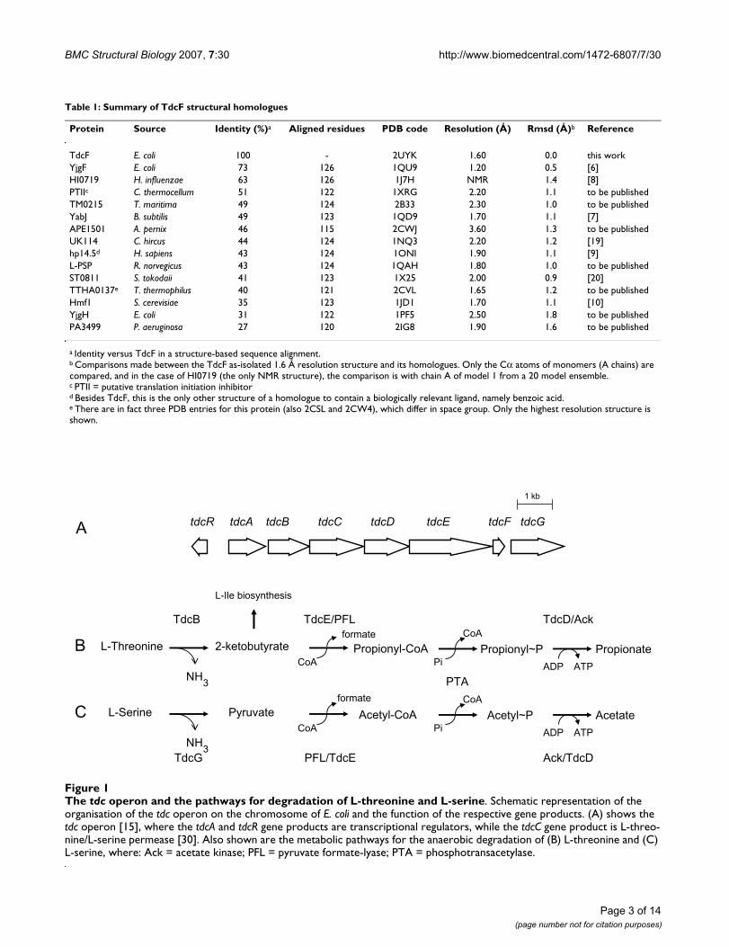
Table 1: Summary of TdcF structural homologues
Protein Source Identity (%)a Aligned residues PDB code Resolution (Å) Rmsd (Å)b Reference
TdcF E. coli 100 - 2UYK 1.60 0.0 this workYjgF E. coli 73 126 1QU9 1.20 0.5 [6]HI0719 H. influenzae 63 126 1J7H NMR 1.4 [8]PTIIc C. thermocellum 51 122 1XRG 2.20 1.1 to be publishedTM0215 T. maritima 49 124 2B33 2.30 1.0 to be publishedYabJ B. subtilis 49 123 1QD9 1.70 1.1 [7]APE1501 A. pernix 46 115 2CWJ 3.60 1.3 to be publishedUK114 C. hircus 44 124 1NQ3 2.20 1.2 [19]hp14.5d H. sapiens 43 124 1ONI 1.90 1.1 [9]L-PSP R. norvegicus 43 124 1QAH 1.80 1.0 to be publishedST0811 S. tokodaii 41 123 1X25 2.00 0.9 [20]TTHA0137e T. thermophilus 40 121 2CVL 1.65 1.2 to be publishedHmf1 S. cerevisiae 35 123 1JD1 1.70 1.1 [10]YjgH E. coli 31 122 1PF5 2.50 1.8 to be publishedPA3499 P. aeruginosa 27 120 2IG8 1.90 1.6 to be published
a Identity versus TdcF in a structure-based sequence alignment.b Comparisons made between the TdcF as-isolated 1.6 Å resolution structure and its homologues. Only the Cα atoms of monomers (A chains) are compared, and in the case of HI0719 (the only NMR structure), the comparison is with chain A of model 1 from a 20 model ensemble.c PTII = putative translation initiation inhibitord Besides TdcF, this is the only other structure of a homologue to contain a biologically relevant ligand, namely benzoic acid.e There are in fact three PDB entries for this protein (also 2CSL and 2CW4), which differ in space group. Only the highest resolution structure is shown.
The tdc operon and the pathways for degradation of L-threonine and L-serineFigure 1The tdc operon and the pathways for degradation of L-threonine and L-serine. Schematic representation of the organisation of the tdc operon on the chromosome of E. coli and the function of the respective gene products. (A) shows the tdc operon [15], where the tdcA and tdcR gene products are transcriptional regulators, while the tdcC gene product is L-threo-nine/L-serine permease [30]. Also shown are the metabolic pathways for the anaerobic degradation of (B) L-threonine and (C) L-serine, where: Ack = acetate kinase; PFL = pyruvate formate-lyase; PTA = phosphotransacetylase.
tdcR tdcA tdcB tdcC tdcD tdcE tdcF tdcG
L-Threonine 2-ketobutyrate
NH3
TdcB TdcE/PFL
Propionyl-CoACoA
Propionyl~PPi
CoA
Propionate
ADP ATP
TdcD/Ack
PTA
formate
L-Serine Pyruvate
NH3
TdcG PFL/TdcE
Acetyl-CoACoA
Acetyl~PPi
CoA
Acetate
ADP ATP
Ack/TdcD
formate
L-Ile biosynthesis
A
B
C
1 kb

BMC Structural Biology 2007, 7:30 http://www.biomedcentral.com/1472-6807/7/30
functionally important. The three sites, which we shallrefer to as sites A, B and C, are not crystallographicallyequivalent, and are thus subject to different crystal pack-ing environments, with site A being the most occluded byneighbouring trimers, and site C being the most solventexposed.
The binding of ethylene glycolThe original 2.35 Å resolution as-isolated X-ray data setwas collected at 100 K using a cryoprotectant solutioncontaining 20% (v/v) ethylene glycol. During modelbuilding and refinement, it became apparent that whilst
the electron density in site A was consistent with orderedwater molecules, a more substantial region of elongateddensity was present in both sites B and C. This could bemodelled convincingly as a single ethylene glycol mole-cule in each of the two sites making a single hydrogenbonding interaction with the side chain of Arg-105 (Fig-ure 3B). It was noted that the central part of the loop con-necting β1 and β2, which delineates one side of the cleftand bears the conserved residue Tyr-17, had elevated tem-perature factors, with Ile-14 being poorly defined in theelectron density maps. This was especially true for theloop adjacent to site A, which adopted a slightly more
The overall structure of E. coli TdcFFigure 2The overall structure of E. coli TdcF. Ribbon representation of the E. coli TdcF structure. Shown is the 2-ketobutyrate-bound form with the individual subunits coloured green, blue and grey; the 2-ketobutyrate ligands are shown in space-filling representation in yellow. Part of the loop between β1 and β2 that moves depending on the status of the ligand-binding pocket is shown in red for each subunit. The inset shows the extent of this movement in the various states, where: grey = empty site; blue = ethylene glycol-bound; yellow = serine-bound; green = propionate-bound; red = 2-ketobutyrate-bound. The small spheres indicate the variation in the position of the Cα atom of Ile-14, being the residue that is displaced the most (maximum displacement 4.2Å). It is notable that the loop is poorly ordered in the empty site and best resolved in the in the 2-ketobu-tyrate-bound site, where Ile-14 makes non-bonding interactions with the ligand. Figure generated using PyMOL [31].
90º
Page 4 of 14(page number not for citation purposes)
BMC Structural Biology 2007, 7:30 http://www.biomedcentral.com/1472-6807/7/30
open conformation with respect to the loops in sites B andC that were partially closed over the ligand (see Figure 2).Only one other structure of a YjgF/YER057c/UK114homologue, that of TM0215 (PDB accession code 2B33)from Thermotoga maritima, reports the use of ethylene gly-col; although three molecules were resolved in this struc-ture, they were all bound at surface sites away from theconserved ligand-binding pocket. Since ethylene glycolmakes only a single hydrogen bond with TdcF, it is likelythat it binds with low affinity, and it is only seen in thestructure because of its high concentration in the cryopro-tectant.
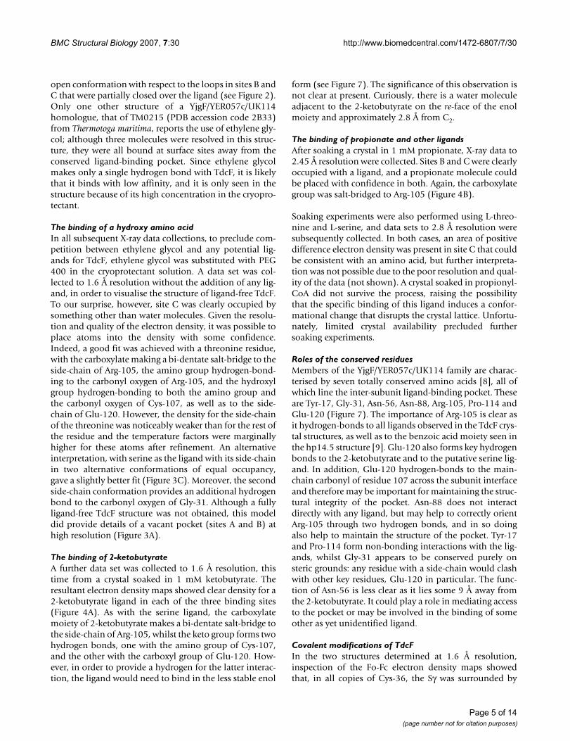
The binding of a hydroxy amino acidIn all subsequent X-ray data collections, to preclude com-petition between ethylene glycol and any potential lig-ands for TdcF, ethylene glycol was substituted with PEG400 in the cryoprotectant solution. A data set was col-lected to 1.6 Å resolution without the addition of any lig-and, in order to visualise the structure of ligand-free TdcF.To our surprise, however, site C was clearly occupied bysomething other than water molecules. Given the resolu-tion and quality of the electron density, it was possible toplace atoms into the density with some confidence.Indeed, a good fit was achieved with a threonine residue,with the carboxylate making a bi-dentate salt-bridge to theside-chain of Arg-105, the amino group hydrogen-bond-ing to the carbonyl oxygen of Arg-105, and the hydroxylgroup hydrogen-bonding to both the amino group andthe carbonyl oxygen of Cys-107, as well as to the side-chain of Glu-120. However, the density for the side-chainof the threonine was noticeably weaker than for the rest ofthe residue and the temperature factors were marginallyhigher for these atoms after refinement. An alternativeinterpretation, with serine as the ligand with its side-chainin two alternative conformations of equal occupancy,gave a slightly better fit (Figure 3C). Moreover, the secondside-chain conformation provides an additional hydrogenbond to the carbonyl oxygen of Gly-31. Although a fullyligand-free TdcF structure was not obtained, this modeldid provide details of a vacant pocket (sites A and B) athigh resolution (Figure 3A).
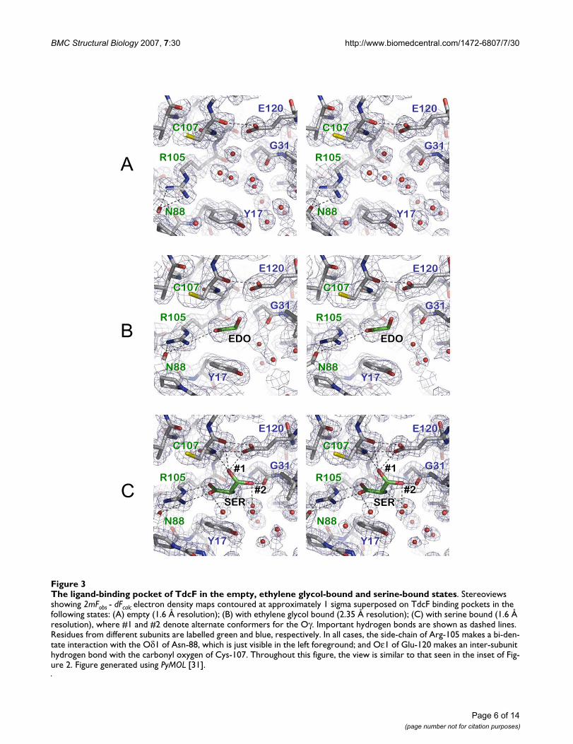
The binding of 2-ketobutyrateA further data set was collected to 1.6 Å resolution, thistime from a crystal soaked in 1 mM ketobutyrate. Theresultant electron density maps showed clear density for a2-ketobutyrate ligand in each of the three binding sites(Figure 4A). As with the serine ligand, the carboxylatemoiety of 2-ketobutyrate makes a bi-dentate salt-bridge tothe side-chain of Arg-105, whilst the keto group forms twohydrogen bonds, one with the amino group of Cys-107,and the other with the carboxyl group of Glu-120. How-ever, in order to provide a hydrogen for the latter interac-tion, the ligand would need to bind in the less stable enol
form (see Figure 7). The significance of this observation isnot clear at present. Curiously, there is a water moleculeadjacent to the 2-ketobutyrate on the re-face of the enolmoiety and approximately 2.8 Å from C2.
The binding of propionate and other ligandsAfter soaking a crystal in 1 mM propionate, X-ray data to2.45 Å resolution were collected. Sites B and C were clearlyoccupied with a ligand, and a propionate molecule couldbe placed with confidence in both. Again, the carboxylategroup was salt-bridged to Arg-105 (Figure 4B).
Soaking experiments were also performed using L-threo-nine and L-serine, and data sets to 2.8 Å resolution weresubsequently collected. In both cases, an area of positivedifference electron density was present in site C that couldbe consistent with an amino acid, but further interpreta-tion was not possible due to the poor resolution and qual-ity of the data (not shown). A crystal soaked in propionyl-CoA did not survive the process, raising the possibilitythat the specific binding of this ligand induces a confor-mational change that disrupts the crystal lattice. Unfortu-nately, limited crystal availability precluded furthersoaking experiments.
Roles of the conserved residuesMembers of the YjgF/YER057c/UK114 family are charac-terised by seven totally conserved amino acids [8], all ofwhich line the inter-subunit ligand-binding pocket. Theseare Tyr-17, Gly-31, Asn-56, Asn-88, Arg-105, Pro-114 andGlu-120 (Figure 7). The importance of Arg-105 is clear asit hydrogen-bonds to all ligands observed in the TdcF crys-tal structures, as well as to the benzoic acid moiety seen inthe hp14.5 structure [9]. Glu-120 also forms key hydrogenbonds to the 2-ketobutyrate and to the putative serine lig-and. In addition, Glu-120 hydrogen-bonds to the main-chain carbonyl of residue 107 across the subunit interfaceand therefore may be important for maintaining the struc-tural integrity of the pocket. Asn-88 does not interactdirectly with any ligand, but may help to correctly orientArg-105 through two hydrogen bonds, and in so doingalso help to maintain the structure of the pocket. Tyr-17and Pro-114 form non-bonding interactions with the lig-ands, whilst Gly-31 appears to be conserved purely onsteric grounds: any residue with a side-chain would clashwith other key residues, Glu-120 in particular. The func-tion of Asn-56 is less clear as it lies some 9 Å away fromthe 2-ketobutyrate. It could play a role in mediating accessto the pocket or may be involved in the binding of someother as yet unidentified ligand.
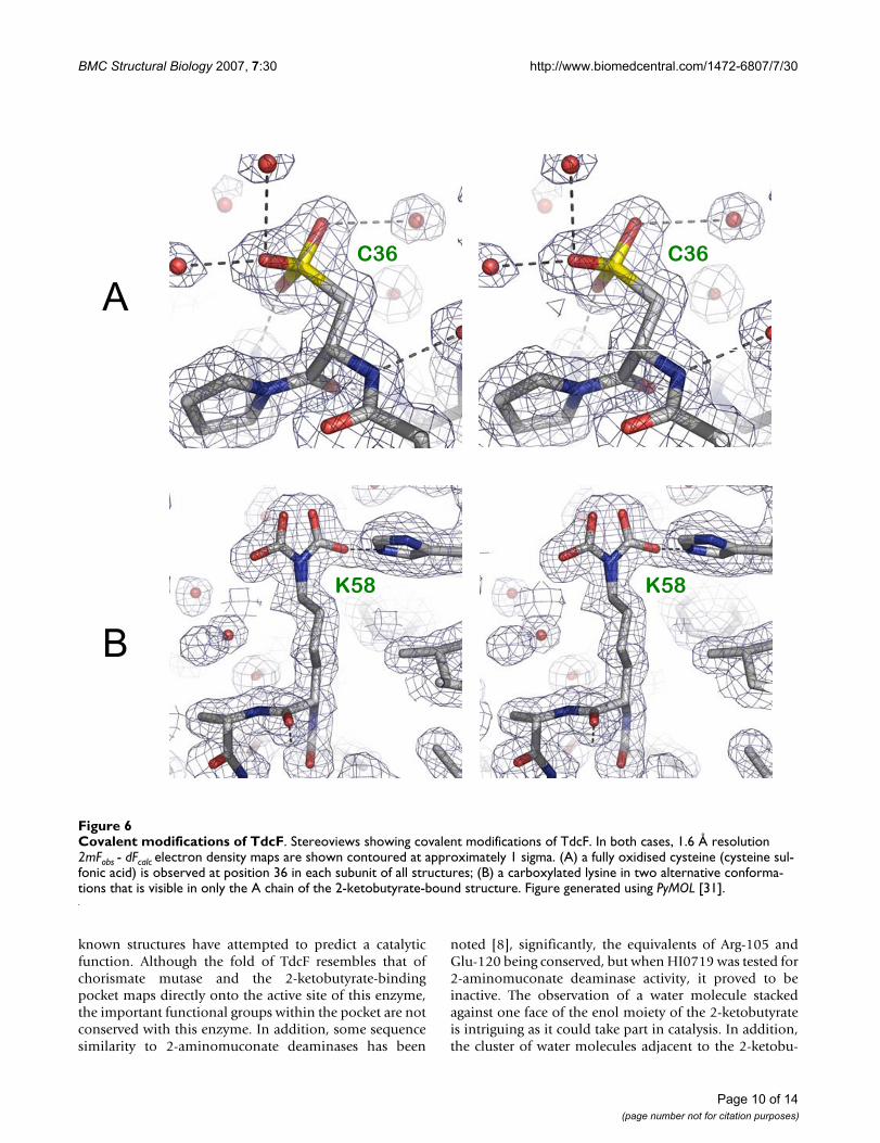
Covalent modifications of TdcFIn the two structures determined at 1.6 Å resolution,inspection of the Fo-Fc electron density maps showedthat, in all copies of Cys-36, the Sγ was surrounded by
Page 5 of 14(page number not for citation purposes)
BMC Structural Biology 2007, 7:30 http://www.biomedcentral.com/1472-6807/7/30
Page 6 of 14(page number not for citation purposes)
The ligand-binding pocket of TdcF in the empty, ethylene glycol-bound and serine-bound statesFigure 3The ligand-binding pocket of TdcF in the empty, ethylene glycol-bound and serine-bound states. Stereoviews showing 2mFobs - dFcalc electron density maps contoured at approximately 1 sigma superposed on TdcF binding pockets in the following states: (A) empty (1.6 Å resolution); (B) with ethylene glycol bound (2.35 Å resolution); (C) with serine bound (1.6 Å resolution), where #1 and #2 denote alternate conformers for the Oγ. Important hydrogen bonds are shown as dashed lines. Residues from different subunits are labelled green and blue, respectively. In all cases, the side-chain of Arg-105 makes a bi-den-tate interaction with the Oδ1 of Asn-88, which is just visible in the left foreground; and Oε1 of Glu-120 makes an inter-subunit hydrogen bond with the carbonyl oxygen of Cys-107. Throughout this figure, the view is similar to that seen in the inset of Fig-ure 2. Figure generated using PyMOL [31].
R105R105
C107C107
E120E120
Y17Y17
G31G31
R105R105
C107C107
E120E120
Y17Y17
G31G31
N88N88 N88N88
R105R105
C107C107
E120E120
Y17Y17
G31G31
R105R105
C107C107
E120E120
Y17Y17
G31G31
N88N88 N88N88
SERSER SERSER
#1#1
#2#2
#1#1
#2#2
A
R105R105
C107C107
E120E120
Y17Y17
G31G31
R105R105
C107C107
E120E120
Y17Y17
G31G31
N88N88 N88N88
EDOEDO EDOEDOB
C
BMC Structural Biology 2007, 7:30 http://www.biomedcentral.com/1472-6807/7/30
three peaks of positive density, that were too close to theatom to represent water molecules. This was modelled asa fully oxidised cysteine ie. cysteine sulfonic acid (Cys-SO3H), which gave a good fit to the electron density afterrefinement (Figure 6A). It was subsequently shown thatcysteine sulfonic acid refined well at these positions in thetwo lower resolution structures as well. This modificationis currently present in some 38 entries in the Protein DataBank but is likely to be present, though undetected, in
other structures determined at medium to low resolution.The fact that the residue is fully oxidised suggests that it isunusually reactive, but since this modification is generallyirreversible, it is most likely of no biological significance.Furthermore, this residue is highly variable in the YjgF/YER057c/UK114 family, and it occurs in a surface loop ofthe protein, the Sγ being approximately 15 Å away fromthe nearest 2-ketobutyrate.
The ligand-binding pocket of TdcF in the ketobutyrate-bound and propionate-bound statesFigure 4The ligand-binding pocket of TdcF in the ketobutyrate-bound and propionate-bound states. Stereoviews, pre-pared as for Figure 3, showing TdcF binding pockets in the following states: (A) with 2-ketobutyrate bound (1.6 Å resolution); (B) with propionate bound (2.45 Å resolution).
A
B
R105R105
C107C107
E120E120
Y17Y17
G31G31
R105R105
C107C107
E120E120
Y17Y17
G31G31
N88N88 N88N88
2KT2KT 2KT2KT
R105R105
C107C107
E120E120
Y17Y17
G31G31
R105R105
C107C107
E120E120
Y17Y17
G31G31
N88N88 N88N88
PPIPPI PPIPPI
Page 7 of 14(page number not for citation purposes)
BMC Structural Biology 2007, 7:30 http://www.biomedcentral.com/1472-6807/7/30
A further modification was apparent at Lys-58 in just theA chain of the 2-ketobutyrate-bound structure. This wasmodelled as a carboxylated lysine in two alternative con-formations (Figure 6B). This modification occurs in overone hundred PDB entries, and in some cases has a well-defined functional role, e.g. in coordinating a metal ion inthe active centre of dihydropyrimidinase (eg. PDB acces-sion code 2FTW) [21]. Coincidentally, one occurrence ofthis modification is seen in the active site of transcarbox-ylase 5S subunit adjacent to a bound 2-ketobutyrate mol-ecule (PDB accession code 1RR2) [22]. Although betterconserved than Cys-36, this residue is located on the sur-face in helix 1 with its Cα approximately 15 Å away fromthe nearest 2-ketobutyrate, and thus is unlikely to be func-tionally relevant. Moreover, no evidence was seen for thismodification in any of the other TdcF structures, althoughit is possible that this was not seen elsewhere due to dis-order. Neither the Cys-36 nor the Lys-58 modificationscould be detected by mass spectroscopy on freshly pre-pared sample or dissolved crystals (data not shown),which suggests that they may have arisen as artefacts afterharvesting the crystals, and further supports the conclu-sion that they are not biologically important.
DiscussionAlignment of YjgF/YER057c/UK114 family membersfrom different biological sources has identified seventotally conserved amino acid residues [2,6-8,20]. Theseconserved amino acids are found in a pocket located at thesubunit interfaces of the trimer (Figure 7), and it has beensuggested that they may form a substrate- or ligand-bind-ing site. Recently, using NMR spectroscopy and a ligand-screening approach Parsons et al. [8] identified six com-pounds that interacted with the HI0719 protein from H.influenzae at, or near, this putative binding site. The ligandthat showed the strongest interaction was 2-ketobutyrate(KD~2 mM), which is an intermediate in the biosyntheticpathway of L-isoleucine. Some 16 residues were perturbedin the 15N-HSQC spectrum upon binding 2-ketobutyrate,but curiously these did not include the equivalents of res-idues Arg-105 and Glu-120. In this study, we have demon-strated structurally that 2-ketobutyrate binds specificallyto a new family member, TdcF from E. coli. All three lig-and-binding sites were occupied in the crystal structureand the key residues identified to make hydrogen-bond-ing interactions with 2-ketobutyrate were Arg-105 andGlu-120, both of which are highly conserved throughoutthe family. Although the 2mFobs - dFcalc electron densitymaps for the side-chain of Cys-107 were always clearlydefined, showing a conformation directed away from thebinding pocket (see Figures 3 and 4), in most instances,there was some evidence in the difference electron densitymaps for one, or occasionally two, very minor alternateconformations directed either towards Arg-105 or towardsthe site of ligand binding. In the latter cases, this would
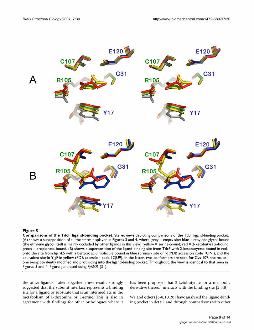
preclude ligand binding and thus indicate less than unitoccupancy for the ligand. Nevertheless, even in the vacantligand-binding sites the predominant conformation wasas shown in Figure 3A. None of these alternate conformerswas modelled in the structures. Whether this residue hassome role in mediating access to the pocket is not clear,but it is notable that the equivalent residue could not beclearly defined in the solution structure of HI0719 fromH. influenza, suggesting that it might also be able to adoptdifferent conformations [8]. The corresponding Cys-107residue in the E. coli YjgF protein was observed to have acovalent modification that was proposed to be either thi-osulphate or thiophosphate, perhaps also suggesting arole in ligand interaction [6]. Indeed, the modified Cysprojects into the ligand-binding pocket of YjgF and hydro-gen-bonds to Arg-105. In so doing, it overlaps the posi-tions occupied by the ligands seen in the TdcF structuresand thus could actually prevent ligand binding here (Fig-ure 5B). This position is occupied by a cysteine in onlyabout half of the TdcF orthologues currently identified.Notably, all of the orthologues predicted to bind 2-keto-butyrate or metabolites on the pathway to isoleucine bio-synthesis (Hmf1 and Mmf1 in yeast [4,10,11], HI0719, inH. influenzae [8], YjgF in E. coli and S. enterica [2,6] have acysteine residue at position 107.
A comparison of the four TdcF structures presented hereshows that the largest changes occur between the 1.6 Åresolution structures of the as-isolated and the 2-ketobu-tyrate-bound forms. The rmsd value calculated for thewhole trimer is 0.341 Å based on common Cα atoms. Inpairwise comparisons between corresponding monomers,the largest rmsd value was for the B subunits at 0.501 Å(next largest value 0.226 Å). This was to be expected, sincein the 1.6 Å resolution as-isolated structure, both the sitesassociated with the B monomer are empty, whilst both arefull in the 2-ketobutyrate-bound structure. The largest Cαdisplacement of approximately 4 Å was for Ile-14 in theβ1 and β2 loop (Figure 2). Recalculating the rmsd formonomer B with the exclusion of residues 11 – 17 inclu-sive, gave a much lower value of 0.102 Å, indicating thatthe conformational changes are essentially restricted tothe β1 – β2 loop. In general, this loop is poorly definedand more open for the ligand-free sites, whilst in the lig-and-bound sites, it closes over the ligand and becomesmore ordered.
In our initial structure of TdcF, the cryoprotectant ethyl-ene glycol was shown to occupy two of the three bindingsites, but this is unlikely to be physiologically relevant.Subsequent experiments showed that propionate, L-serineand/or L-threonine could also occupy the binding sites.However, again not all of the binding sites were occupied.By contrast, full site occupancy was achieved using 2-keto-butyrate, indicating that it binds with higher affinity than
Page 8 of 14(page number not for citation purposes)
BMC Structural Biology 2007, 7:30 http://www.biomedcentral.com/1472-6807/7/30
the other ligands. Taken together, these results stronglysuggested that the subunit interface represents a bindingsite for a ligand or substrate that is an intermediate in themetabolism of L-threonine or L-serine. This is also inagreement with findings for other orthologues where it
has been proposed that 2-ketobutyrate, or a metabolicderivative thereof, interacts with the binding site [2,3,8].
We and others [6-8,10,20] have analysed the ligand-bind-ing pocket in detail, and through comparisons with other
Comparisons of the TdcF ligand-binding pocketFigure 5Comparisons of the TdcF ligand-binding pocket. Stereoviews depicting comparisons of the TdcF ligand-binding pocket. (A) shows a superposition of all the states displayed in Figures 3 and 4, where: grey = empty site; blue = ethylene glycol-bound (the ethylene glycol itself is mainly occluded by other ligands in this view); yellow = serine-bound; red = 2-ketobutyrate-bound; green = propionate-bound. (B) shows a superposition of the ligand-binding site from TdcF with 2-ketobutyrate bound in red, onto the site from hp14.5 with a benzoic acid molecule bound in blue (primary site only)(PDB accession code 1ONI), and the equivalent site in YjgF in yellow (PDB accession code 1QU9). In the latter, two conformers are seen for Cys-107, the major one being covalently modified and protruding into the ligand-binding pocket. Throughout, the view is identical to that seen in Figures 3 and 4. Figure generated using PyMOL [31].
A
B
R105R105
C107C107
E120E120
Y17Y17
G31G31
R105R105
C107C107
E120E120
Y17Y17
G31G31
R105R105
C107C107
E120E120
Y17Y17
G31G31R105R105
C107C107
E120E120
Y17Y17
G31G31
Page 9 of 14(page number not for citation purposes)
BMC Structural Biology 2007, 7:30 http://www.biomedcentral.com/1472-6807/7/30
known structures have attempted to predict a catalyticfunction. Although the fold of TdcF resembles that ofchorismate mutase and the 2-ketobutyrate-bindingpocket maps directly onto the active site of this enzyme,the important functional groups within the pocket are notconserved with this enzyme. In addition, some sequencesimilarity to 2-aminomuconate deaminases has been
noted [8], significantly, the equivalents of Arg-105 andGlu-120 being conserved, but when HI0719 was tested for2-aminomuconate deaminase activity, it proved to beinactive. The observation of a water molecule stackedagainst one face of the enol moiety of the 2-ketobutyrateis intriguing as it could take part in catalysis. In addition,the cluster of water molecules adjacent to the 2-ketobu-
Covalent modifications of TdcFFigure 6Covalent modifications of TdcF. Stereoviews showing covalent modifications of TdcF. In both cases, 1.6 Å resolution 2mFobs - dFcalc electron density maps are shown contoured at approximately 1 sigma. (A) a fully oxidised cysteine (cysteine sul-fonic acid) is observed at position 36 in each subunit of all structures; (B) a carboxylated lysine in two alternative conforma-tions that is visible in only the A chain of the 2-ketobutyrate-bound structure. Figure generated using PyMOL [31].
K58K58 K58K58
C36C36 C36C36
A
B
Page 10 of 14(page number not for citation purposes)

BMC Structural Biology 2007, 7:30 http://www.biomedcentral.com/1472-6807/7/30
tyrate (Figure 4A and Figure 7) could indicate a bindingsite for another substrate, or perhaps together with thespace occupied by the 2-ketobutyrate, may represent thebinding site for a much larger ligand. Another possibilityis that a cofactor binds adjacent to the 2-ketobutyrate.Crude docking experiments with our TdcF structures sug-gest that a pyridoxal phosphate molecule could be accom-modated, whilst thiamine pyrophosphate or coenzyme A(CoA) could not. Nevertheless, we cannot rule out thepossibility that the latter two ligands induce large confor-mational changes that allow them to bind. However,
investigations with HI0719 showed that none of thesecofactors induced chemical shifts in the 15N-HSQC spec-trum of this protein [8].
ConclusionThe findings presented in this study strongly support thecontention that 2-ketobutyrate is a physiological ligandrecognised by TdcF. The tdcF gene is located in a multi-cis-tronic operon whose gene products have a role in theanaerobic degradation of L-threonine and L-serine. Thefirst intermediate in the degradation of L-threonine or L-
The binding of 2-ketobutyrate to TdcFFigure 7The binding of 2-ketobutyrate to TdcF. Schematic representation of the binding of 2-ketobutyrate to TdcF. 2-ketobu-tyrate is shown in red, whilst residues from one subunit are in green and those from the other subunit are in blue; darker col-ours indicate residues in the foreground and paler colours are used for those residues in the background. Underlining indicates strictly conserved residues in the YjgF/YER057c/UK114 family [8]. Important hydrogen bonds are shown as dashed lines and the curved magenta lines indicate non-bonding interactions with the ligand. The numbers in red give average hydrogen bond lengths in Å. Water molecules are indicated by the letter "W". The cluster of water molecules to the right of the ligand is in direct contact with bulk solvent. The view is similar to that seen in Figures 3–5.
Arg105
Asn88
Phe84
Ile14
Cys107
Glu120
Gly31
Pro114
Ile33
Tyr17
W
W
W
WW
W
2.98
2.72
2.79
2.79
2.662.852.80
-
Asn56
OH
ONH
SH
O
NH
NH2
NH
NH
O
O
NH2
OC1
C2
O
C3
OH
C4
OH
O
NH
O
N
O
NH2
Page 11 of 14(page number not for citation purposes)
BMC Structural Biology 2007, 7:30 http://www.biomedcentral.com/1472-6807/7/30
serine is the ketoacid, 2-ketobutyrate or pyruvate, respec-tively. Pyruvate can be further metabolised via pyruvateformate-lyase or pyruvate dehydrogenase to acetyl-CoA.2-ketobutyrate can either be used as a substrate on thepathway to L-isoleucine or, under anaerobic conditions,can be metabolised to propionate via propionyl-CoA andpropionyl-P intermediates with concomitant generationof 1 ATP (Figure 1). All the prerequisite enzymes for thisfermentative route, with the exception of phospho-transacetylase are encoded by the tdc operon [15]. Theonly gene product of the operon whose function couldnot yet be assigned is TdcF. Since to date no enzyme activ-ity has been detected for any member of this protein fam-ily, we suggest, based on the current findings, that TdcFmay be a post-translational regulator that controls themetabolic fate of L-threonine or the potentially toxicintermediate 2-ketobutyrate. Depending on whether L-isoleucine is limiting or not for growth, TdcF, by sensingthe levels of 2-ketobutyrate and forming a ternary com-plex with one or more of the enzymes of isoleucine bio-synthesis or 2-ketobutyrate degradation, ensures that 2-ketobutyrate does not accumulate in the cell. Experimentsto determine putative protein-protein interaction partnersof TdcF are in hand. A similar proposal for the function ofthe members of this protein family has been made previ-ously [2,3]. This is consistent with the recent demonstra-tion that YjgF appears to function at the post-translationallevel in controlling the activity of IlvE, which catalyses thefinal transamination step on the L-isoleucine pathway inSalmonella enterica [3]. The accumulation of 2-ketobu-tyrate has been proposed [23], and genetically demon-strated [13], to compete with 2-ketoisovalerate, theprecursor of pantothenate synthesis, resulting in starva-tion for coenzyme A. Metabolic poisoning by 2-ketobu-tyrate is prevented by its degradation aerobically viapyruvate dehydrogenase [14] and anaerobically via pyru-vate formate-lyase or TdcE [15]; these reactions are CoA-dependent. This role of a sensor of 2-ketobutyrate wouldnot only afford protection to cells from the toxic effects of2-ketobutyrate accumulation and provide an additionalmeans of energy generation, but also would ensure thatsufficient 2-ketobutyrate was available for L-isoleucinebiosynthesis.
MethodsPurification and crystallisationOverproduction, purification and crystallisation of therecombinant, N-terminally His-tagged, TdcF protein wasperformed exactly as described previously [17]. The His-tag was not cleaved prior to crystallisation. Briefly, crystalswere obtained using the hanging drop vapour diffusionmethod with a 1:1 mixture of protein (10 mg/ml in 20mM Tris-HCl, pH 8) and well solution (12.5% w/v PEG1500). Needle-like crystals appeared after a week,although in some drops, more substantial, rectangular,
crystals formed over a period of up to 2 months, having amaximum size of approximately 150 × 60 × 60 μm. Crys-tals were routinely transferred from one solution toanother, and ultimately mounted for X-ray data collec-tion, using cryo-loops (Hampton research). For the firstdata set, crystals were cryoprotected by soaking for a max-imum of 5 min in crystallisation solution supplementedwith 20% (w/v) ethylene glycol. However, after discover-ing ethylene glycol bound to the protein, 20% (w/v) PEG400 was substituted as the cryoprotectant.
Crystal soakingFor ligand soaking experiments, crystals were transferredfrom the mother liquor to a soaking solution comprising12.5% (w/v) PEG 1500 in 20 mM Tris-HCl pH8 and con-taining 1 mM of the potential ligand molecule. The latterwere chosen on the basis of being substrates, products ormetabolic intermediates on the L-serine/L-threonine deg-radation pathways. These included L-threonine, L-serine,pyruvate, propionate, 2-ketobutyrate and propionyl-CoA.Crystals were soaked for 2 h before being cryoprotected infresh soaking solution containing the ligand and 20% (v/v) PEG 400.
X-ray data collection and processingFor in-house data collection, crystals were flash-cooledand maintained at 100 K using an X-Stream cryocooler(Rigaku-MSC) and X-ray data were recorded on a Mar 345image plate detector (X-ray Research) mounted on aRigaku RU-H3RHB rotating anode X-ray generator (oper-ated at 50 kV and 100 mA) fitted with Osmic confocaloptics and a copper target (Cu Kα ; λ = 1.542 Å).
For synchrotron data collection, crystals were flash-cooledby plunging into liquid nitrogen and stored prior to trans-port to the synchrotron. Crystals were transferred to thegoniostat on station ID14-2 (λ = 0.933 Å) at the EuropeanSynchrotron Radiation Source (ESRF) in Grenoble usingHampton Research cryotools and maintained at 100 Kwith a Cryostream cryocooler (Oxford Instruments). Dif-fraction data were recorded on an ADSC Quantum 4CCD.
All X-ray data were processed using version 1.97 of theHKL software package [24], and downstream processingand statistical analysis was effected using programs fromthe CCP4 suite [25]. Data collection statistics are summa-rised in Table 2.
Structure solution and refinementThe space group of the TdcF crystals was P21212, withapproximate cell parameters of a = 72, b = 86 and c = 63Å. Solvent-content estimation based on a TdcF trimer inthe asymmetric unit gave a VM value of 2.02 Å3 Da-1, cor-responding to a solvent content of 39%.
Page 12 of 14(page number not for citation purposes)

BMC Structural Biology 2007, 7:30 http://www.biomedcentral.com/1472-6807/7/30
Molecular replacement was performed using the programAMoRe [26] with the structure of the closely-related YjgF[6] as a template; this was successful in placing a trimer inthe asymmetric unit [17]. Model building was performedby interactive computer graphics using the program O[27] by inspection of 2mFobs- dFcalc and mFobs- dFcalc Fourierelectron density maps. Ligands were docked with refer-ence to unbiased mFobs- dFcalc difference maps. All data setswere essentially isomorphous and an equivalent subset ofthe data comprising 5% of the reflections was set aside forthe calculation of 'free' (Rfree) crystallographic R-factors[28] during model refinement. Throughout refinement,neither low resolution nor amplitude cut-offs wereapplied. Both positional and thermal parameters of themodels were subsequently refined using REFMAC5 [29].Anisotropic thermal parameters were refined for the twostructures at 1.6 Å resolution. A summary of the modelcontents and geometrical parameters of the final struc-tures are given in Table 2. The coordinates and structure
factor data for these structures have been deposited in theProtein Data Bank.
Authors' contributionsRGS and DML conceived the study and together with JDB,drafted the manuscript. All experimental work was per-formed by JDB, with assistance from RGS for the expres-sion and purification; and with assistance from CEMS andDML for the X-ray crystallographic analysis. All authorshave approved the final manuscript.
AcknowledgementsThis work was funded by the BBSRC as part of the competitive strategic grant to the John Innes Centre. JDB was supported by a John Innes Foun-dation studentship. We are grateful for support and access to the ESRF in Grenoble and would particularly like to thank Andrew Hemmings for assist-ance with X-ray data collection. We are also indebted to Stephen Bornemann and Robert Field for helpful discussions, and for critically read-ing this manuscript.
Table 2: Summary of X-ray data and model parameters for TdcF
Data set As-isolated (ethylene glycol) As-isolated (PEG 400) 2-Ketobutyrate soak Propionate soak
Data collectionRadiation source RU-H3RHB 1D14-2 ESRF 1D14-2 ESRF RU-H3RHBWavelength (Å) 1.542 0.933 0.933 1.542Cell parameters (Å) a = 72.7
b = 86.2c = 62.6
a = 72.7b = 86.4c = 62.6
a = 72.6b = 85.9c = 62.7
a = 72.4b = 85.7c = 62.5
Resolution rangea (Å) 23.7 – 2.35 (2.43 – 2.35) 35.9 – 1.60 (1.63 – 1.60) 35.4 -1.60 (1.63 – 1.60) 25.5 – 2.45 (2.54 – 2.45)Unique reflections 16863 50526 49876 14841Completenessa (%) 99.3 (95.2) 95.2 (66.4) 94.9 (71.2) 98.9 (94.0)Redundancy 4.4 2.9 3.3 3.4Rmerge
a,b 0.103 (0.368) 0.071 (0.116) 0.060 (0.243) 0.077 (0.303)<I/σ (I)>a 12.1 (2.7) 19.1 (3.8) 20.5 (2.1) 14.6 (3.2)Wilson B value (Å2) 29.5 15.8 17.1 35.9RefinementRcryst
c (based on 95% of data; %) 17.2 15.5 16.2 16.6Rfree
c (based on 5% of data; %) 24.4 19.9 20.4 25.3DPId (based on Rfree; Å) 0.257 0.087 0.089 0.295Ramachandran plote (%) 94.0 95.8 95.5 94.6Rmsd bond distances (Å) 0.020 0.017 0.015 0.021Rmsd bond angles (°) 1.800 1.555 1.441 1.843Contents of modelProtein (residues/atoms) 383/2876 381/2886 381/2895 382/2860Waters (molecules) 205 457 400 195Ligands (molecules/atoms) 2/8 1/7 3/21 2/10Ligands (identityf/sites occupied) EDO/B,C SER/C 2KT/A,B,C PPI/B,CAverage temperature factors (Å2)Main-chain atoms 17.7 13.0 14.9 21.0Side-chain atoms 18.5 15.6 17.4 21.6Waters 23.1 29.8 30.8 26.8Ligands 29.4 19.2 24.7 33.8Overall 18.4 16.4 17.9 27.7PDB accession code 2UYJ 2UYK 2UYN 2UYP
a The figures in brackets indicate the values for outer resolution shell.b Rmerge = ΣhΣl|Ihl - <Ih>|/ΣhΣl <Ih>, where Il is the lth observation of reflection h and <Ih> is the weighted average intensity for all observations l of reflection h.c The R-factors Rcryst and Rfree are calculated as follows: R = Σ (|Fobs - Fcalc|)/Σ |Fobs| × 100, where Fobs and Fcalc are the observed and calculated structure factor amplitudes, respectively.d Diffraction-component precision index [32] – an estimate of the overall coordinate errors calculated in REFMAC5 [29].e Residues with most favoured Φ/Ψ angles as calculated using PROCHECK [33].f EDO = ethylene glycol (1,2-ethanediol); 2KT = 2-ketobutyrate; PPI = propionate.
Page 13 of 14(page number not for citation purposes)

BMC Structural Biology 2007, 7:30 http://www.biomedcentral.com/1472-6807/7/30
Publish with BioMed Central and every scientist can read your work free of charge
"BioMed Central will be the most significant development for disseminating the results of biomedical research in our lifetime."
Sir Paul Nurse, Cancer Research UK
Your research papers will be:
available free of charge to the entire biomedical community
peer reviewed and published immediately upon acceptance
cited in PubMed and archived on PubMed Central
yours — you keep the copyright
Submit your manuscript here:http://www.biomedcentral.com/info/publishing_adv.asp
BioMedcentral
References1. Leitner-Dagan Y, Ovadis M, Zuker A, Shklarman E, Ohad I, Tzfira T,
Vainstein A: CHRD, a plant member of the evolutionarily con-served YjgF family, influences photosynthesis and chromo-plastogenesis. Planta 2006, 225:89-102.
2. Enos-Berlage JL, Langendorf MJ, Downs DM: Complex metabolicphenotypes caused by a mutation in yjgF, encoding a mem-ber of the highly conserved YER057c/YjgF family of proteins.J Bacteriol 1998, 180:6519-6528.
3. Schmitz G, Downs DM: Reduced transaminase B (IlvE) activitycaused by the lack of yjgF is dependent on the status of thre-onine deaminase (IlvA) in Salmonella enterica serovar Typh-imurium. J Bacteriol 2004, 186:803-810.
4. Kim JM, Yoshikawa H, Shirahige K: A member of the YER057c/yjgf/Uk114 family links isoleucine biosynthesis and intactmitochondria maintenance in Saccharomyces cerevisiae.Genes Cells 2001, 6:507-517.
5. Goupil-Feuillerat N, Cocaign-Bousquet M, Godon JJ, Ehrlich SD,Renault P: Dual role of alpha-acetolactate decarboxylase inLactococcus lactis subsp. lactis. J Bacteriol 1997, 179:6285-6293.
6. Volz K: A test case for structure-based functional assignment:the 1.2 A crystal structure of the yjgF gene product fromEscherichia coli. Protein Sci 1999, 8:2428-2437.
7. Sinha S, Rappu P, Lange SC, Mantsala P, Zalkin H, Smith JL: Crystalstructure of Bacillus subtilis YabJ, a purine regulatory pro-tein and member of the highly conserved YjgF family. ProcNatl Acad Sci USA 1999, 96:13074-13079.
8. Parsons L, Bonander N, Eisenstein E, Gilson M, Kairys V, Orban J:Solution structure and functional ligand screening of HI0719,a highly conserved protein from bacteria to humans in theYjgF/YER057c/UK114 family. Biochemistry 2003, 42:80-89.
9. Manjasetty BA, Delbruck H, Pham DT, Mueller U, Fieber-Erdmann M,Scheich C, Sievert V, Bussow K, Neisen FH, Weihofen W, Loll B,Saenger W, Heinemann U: Crystal structure of Homo sapiensprotein hp14.5. Proteins 2004, 54:797-800.
10. Deaconescu AM, Roll-Mecak A, Bonanno JB, Gerchman SE, Kycia H,Studier FW, Burley SK: X-ray structure of Saccharomyces cer-evisiae homologous mitochondrial matrix factor 1 (Hmf1).Proteins 2002, 48:431-436.
11. Oxelmark E, Marchini A, Malanchi I, Magherini F, Jaquet L, HajibagheriMA, Blight KJ, Jauniaux JC, Tommasino M: Mmf1p, a novel yeastmitochondrial protein conserved throughout evolution andinvolved in maintenance of the mitochondrial genome. MolCell Biol 2000, 20:7784-7797.
12. Browne BA, Ramos AI, Downs DM: PurF-independent phos-phoribosyl amine formation in yjgF mutants of Salmonellaenterica utilizes the tryptophan biosynthetic enzyme com-plex anthranilate synthase-phosphoribosyltransferase. J Bac-teriol 2006, 188:6786-6792.
13. LaRossa RA, Van Dyk TK: Leaky pantothenate and thiaminmutations of Salmonella typhimurium conferring supho-meturon methyl sensitivity. J Gen Microbiol 1989, 135:2209-2222.
14. Van Dyk TK, LaRossa RA: Involvement of ack-pta operon prod-ucts in alpha-ketobutyrate metabolism by Salmonella typh-imurium. Mol Gen Genet 1987, 207:435-440.
15. Heßlinger C, Fairhurst SA, Sawers G: Novel keto acid formate-lyase and propionate kinase enzymes are components of ananaerobic pathway in Escherichia coli that degrades L- thre-onine to propionate. Mol Microbiol 1998, 27:477-492.
16. Goss TJ, Schweizer HP, Datta P: Molecular characterization ofthe tdc operon of Escherichia coli K-12. J Bacteriol 1988,170:5352-5359.
17. Burman JD, Stevenson CE, Hauton KA, Sawers G, Lawson DM: Crys-tallisation and preliminary X-ray analysis of the E. coli hypo-thetical protein TdcF. Acta Crystallogr D Biol Crystallogr 2003,59:1076-1078.
18. Protein Interfaces, Surfaces and Assemblies (PISA) server[http://www.ebi.ac.uk/msd-srv/prot_int/pistart.html]
19. Deriu D, Briand C, Mistiniene E, Naktinis V, Grutter MG: Structureand oligomeric state of the mammalian tumour-associatedantigen UK114. Acta Crystallogr D Biol Crystallogr 2003,59:1676-1678.
20. Miyakawa T, Lee WC, Hatano K, Kato Y, Sawano Y, Miyazono K,Nagata K, Tanokura M: Crystal structure of the YjgF/YER057c/UK114 family protein from the hyperthermophilic archaeonSulfolobus tokodaii strain 7. Proteins 2006, 62:557-561.
21. Lohkamp B, Andersen B, Piskur J, Dobritzsch D: The crystal struc-tures of dihydropyrimidinases reaffirm the close relationshipbetween cyclic amidohydrolases and explain their substratespecificity. J Biol Chem 2006, 281:13762-13776.
22. Hall PR, Zheng R, Antony L, Pusztai-Carey M, Carey PR, Yee VC:Transcarboxylase 5S structures: assembly and catalyticmechanism of a multienzyme complex subunit. EMBO J 2004,23:3621-3631.
23. Primerano DA, Burns RO: Metabolic basis for the isoleucine,pantothenate or methionine requirement of ilvG strains ofSalmonella typhimurium. J Bacteriol 1982, 150:1202-1211.
24. Otwinowski Z, Minor W: Processing of X-ray diffraction datacollected in oscillation mode. Methods in Enzymology 1997,276:307-326.
25. Collaborative Computational Project, Number 4: The CCP4 suite:programs for protein crystallography. Acta Crystallogr D BiolCrystallogr 1994, 50:760-763.
26. Navaza J: AMoRe: an automated package for molecularreplacement. Acta Crystallographica 1994, A50:157-163.
27. Jones TA, Zou JY, Cowan SW, Kjeldgaard M: Improved methodsfor building protein models in electron density maps and thelocation of errors in these models. Acta Crystallogr A 1991,47:110-119.
28. Brünger AT: Assessment of phase accuracy by cross valida-tion: the free R-value. Methods and applications. Acta Crystal-logr D Biol Crystallogr 1993, 49:24-36.
29. Murshudov GN, Vagin AA, Dodson EJ: Refinement of macromo-lecular structures by the maximum-likelihood method. ActaCrystallogr D Biol Crystallogr 1997, 53:240-255.
30. Sumantran VN, Schweizer HP, Datta P: A novel membrane-asso-ciated threonine permease encoded by the tdcC gene ofEscherichia coli. J Bacteriol 1990, 172:4288-4294.
31. PyMOL Home Page [http://pymol.sourceforge.net/]32. Cruickshank DW: Remarks about protein structure precision.
Acta Crystallogr D Biol Crystallogr 1999, 55:583-601.33. Laskowski RA, Macarthur MW, Moss DS, Thornton JM: PRO-
CHECK: a program to check the stereochemical quality ofprotein structures. Journal of Applied Crystallography 1993,26:283-291.
Page 14 of 14(page number not for citation purposes)
http://www.ncbi.nlm.nih.gov/entrez/query.fcgi?cmd=Retrieve&db=PubMed&dopt=Abstract&list_uids=9851994
http://www.ncbi.nlm.nih.gov/entrez/query.fcgi?cmd=Retrieve&db=PubMed&dopt=Abstract&list_uids=9851994
http://www.ncbi.nlm.nih.gov/entrez/query.fcgi?cmd=Retrieve&db=PubMed&dopt=Abstract&list_uids=9335274
http://www.ncbi.nlm.nih.gov/entrez/query.fcgi?cmd=Retrieve&db=PubMed&dopt=Abstract&list_uids=9335274
http://www.ncbi.nlm.nih.gov/entrez/query.fcgi?cmd=Retrieve&db=PubMed&dopt=Abstract&list_uids=2699328
http://www.ncbi.nlm.nih.gov/entrez/query.fcgi?cmd=Retrieve&db=PubMed&dopt=Abstract&list_uids=2699328
http://www.ncbi.nlm.nih.gov/entrez/query.fcgi?cmd=Retrieve&db=PubMed&dopt=Abstract&list_uids=2699328
http://www.ncbi.nlm.nih.gov/entrez/query.fcgi?cmd=Retrieve&db=PubMed&dopt=Abstract&list_uids=3039301
http://www.ncbi.nlm.nih.gov/entrez/query.fcgi?cmd=Retrieve&db=PubMed&dopt=Abstract&list_uids=3039301
http://www.ncbi.nlm.nih.gov/entrez/query.fcgi?cmd=Retrieve&db=PubMed&dopt=Abstract&list_uids=3039301
http://www.ncbi.nlm.nih.gov/entrez/query.fcgi?cmd=Retrieve&db=PubMed&dopt=Abstract&list_uids=9484901
http://www.ncbi.nlm.nih.gov/entrez/query.fcgi?cmd=Retrieve&db=PubMed&dopt=Abstract&list_uids=9484901
http://www.ncbi.nlm.nih.gov/entrez/query.fcgi?cmd=Retrieve&db=PubMed&dopt=Abstract&list_uids=9484901
http://www.ncbi.nlm.nih.gov/entrez/query.fcgi?cmd=Retrieve&db=PubMed&dopt=Abstract&list_uids=3053659
http://www.ncbi.nlm.nih.gov/entrez/query.fcgi?cmd=Retrieve&db=PubMed&dopt=Abstract&list_uids=3053659
http://www.ncbi.nlm.nih.gov/entrez/query.fcgi?cmd=Retrieve&db=PubMed&dopt=Abstract&list_uids=7042686
http://www.ncbi.nlm.nih.gov/entrez/query.fcgi?cmd=Retrieve&db=PubMed&dopt=Abstract&list_uids=7042686
http://www.ncbi.nlm.nih.gov/entrez/query.fcgi?cmd=Retrieve&db=PubMed&dopt=Abstract&list_uids=7042686
http://www.ncbi.nlm.nih.gov/entrez/query.fcgi?cmd=Retrieve&db=PubMed&dopt=Abstract&list_uids=2025413
http://www.ncbi.nlm.nih.gov/entrez/query.fcgi?cmd=Retrieve&db=PubMed&dopt=Abstract&list_uids=2025413
http://www.ncbi.nlm.nih.gov/entrez/query.fcgi?cmd=Retrieve&db=PubMed&dopt=Abstract&list_uids=2025413
http://www.ncbi.nlm.nih.gov/entrez/query.fcgi?cmd=Retrieve&db=PubMed&dopt=Abstract&list_uids=2115866
http://www.ncbi.nlm.nih.gov/entrez/query.fcgi?cmd=Retrieve&db=PubMed&dopt=Abstract&list_uids=2115866