The crystal structure of endoglucanase CelA, a family 8 glycosyl hydrolase from Clostridium...
11
The crystal structure of endoglucanase CelA, a family 8 glycosyl hydrolase from Clostridium thermocellum Pedro M Alzari*, Hélène Souchon and Roberto Dominguez Background: Cellulases, which catalyze the hydrolysis of glycosidic bonds in cellulose, can be classified into several different protein families. Endoglucanase CelA is a member of glycosyl hydrolase family 8, a family for which no structural information was previously available. Results: The crystal structure of CelA was determined by multiple isomorphous replacement and refined to 1.65 Å resolution. The protein folds into a regular (a/a) 6 barrel formed by six inner and six outer a helices. Cello-oligosaccharides bind to an acidic cleft containing at least five D-glucosyl-binding subsites (A–E) such that the scissile glycosidic linkage lies between subsites C and D. The strictly conserved residue Glu95, which occupies the center of the substrate- binding cleft and is hydrogen bonded to the glycosidic oxygen, has been assigned the catalytic role of proton donor. Conclusions: The present analysis provides a basis for modeling homologous family 8 cellulases. The architecture of the active-site cleft, presenting at least five glucosyl-binding subsites, explains why family 8 cellulases cleave cello- oligosaccharide polymers that are at least five D-glucosyl subunits long. Furthermore, the structure of CelA allows comparison with (a/a) 6 barrel glycosidases that are not related in sequence, suggesting a possible, albeit distant, evolutionary relationship between different families of glycosyl hydrolases. Introduction Cellulolytic microorganisms produce a wide range of enzymes that hydrolyze the glycosidic bond of cellulose [1,2]. Despite the chemical regularity of the cellulose sub- strate (linear chains of b-1,4-linked D-glucosyl residues), the catalytic domains of cellulases vary widely, belonging to at least 11 distinct protein families [3,4]. Cellulases differ in protein architecture, endo/exo specificity, and inverting/retaining reaction mechanism, but all hydrolyze the glycosidic linkage via general acid catalysis. They therefore require two critical residues, a proton donor and a nucleophile or general base [5]. The three-dimensional (3D) structures of several cellulases have been determined by X-ray crystallography. Family 5 endoglucanases CelC, from Clostridium thermocellum [6] and CelCCA, from Clostridium cellulolyticum [7], are regular (a/b) 8 proteins, or TIM barrels, whereas cellobiohydrolase CBHII (an exoglucanase) from Trichoderma reesei [8] and endoglucanase E2 from Thermomonospora fusca [9] (both from family 6) fold into a related, albeit more irregular, a/b barrel structure. Other cellulases have entirely different structures: the catalytic domain of cellobiohydrolase CBHI from T. reesei (family 7) is arranged in two antiparallel b sheets, with the topology of plant lectins [10]; endoglu- canase CelD from C. thermocellum (family 9) folds into a helical barrel tightly bound to an immunoglobulin-like domain [11]; and endoglucanase V from Humicola insolens (family 45) is a small six-stranded b barrel protein [12]. We report here a crystallographic study of family 8 endo- glucanase CelA, which shows no sequence similarity to other cellulases with known structures. CelA was one of the first cellulases to be purified from the culture supernatant of the thermophilic anaerobe C. thermocellum [13]. This bacterium secretes an extracellular multiprotein complex, the cellulosome (Mw>2 MDa), that is very efficient in degrading crystalline cellulose (recently reviewed in [2]). The cellulosome contains several enzymes with distinct carbohydrate specificities, as well as various non-catalytic components [2,14,15]. Most cellulo- somal enzymes share a highly conserved ‘dockerin’ domain of about 70 amino acid residues that anchors the individual polypeptides to a scaffolding protein devoid of catalytic activity [16], thus providing a modular mecha- nism of macromolecular assembly. The CelA gene (CelA) was characterized [17,18] and found to encode a polypeptide of 488 amino acid residues, con- sisting of a signal-peptide-like segment, a catalytic domain, and a C-terminal dockerin domain. CelA hydrolyzes car- boxymethylcellulose and, to a lesser extent, lichenan, a b-glucan containing b-1,4 and b-1,3 linkages, but shows very low or no activity for Avicel (crystalline cellulose) or Address: Unité d’Immunologie Structurale and URA 1961 CNRS, Institut Pasteur, 25 rue du Dr. Roux, 75724 Paris Cedex 15, France. *Corresponding author. Key words: (a/a) 6 barrel, enzyme mechanism, glycosidase family 8, substrate binding, X-ray structure Received: 4 Dec 1995 Revisions requested: 22 Dec 1995 Revisions received: 19 Jan 1996 Accepted: 24 Jan 1996 Structure 15 March 1996, 4:265–275 © Current Biology Ltd ISSN 0969-2126 Research Article 265
Transcript of The crystal structure of endoglucanase CelA, a family 8 glycosyl hydrolase from Clostridium...

The crystal structure of endoglucanase CelA, a family 8 glycosylhydrolase from Clostridium thermocellumPedro M Alzari*, Hélène Souchon and Roberto Dominguez
Background: Cellulases, which catalyze the hydrolysis of glycosidic bonds incellulose, can be classified into several different protein families. EndoglucanaseCelA is a member of glycosyl hydrolase family 8, a family for which no structuralinformation was previously available.
Results: The crystal structure of CelA was determined by multiple isomorphousreplacement and refined to 1.65 Å resolution. The protein folds into a regular(a/a)6 barrel formed by six inner and six outer a helices. Cello-oligosaccharidesbind to an acidic cleft containing at least five D-glucosyl-binding subsites (A–E)such that the scissile glycosidic linkage lies between subsites C and D. Thestrictly conserved residue Glu95, which occupies the center of the substrate-binding cleft and is hydrogen bonded to the glycosidic oxygen, has beenassigned the catalytic role of proton donor.
Conclusions: The present analysis provides a basis for modeling homologousfamily 8 cellulases. The architecture of the active-site cleft, presenting at least fiveglucosyl-binding subsites, explains why family 8 cellulases cleave cello-oligosaccharide polymers that are at least five D-glucosyl subunits long.Furthermore, the structure of CelA allows comparison with (a/a)6 barrelglycosidases that are not related in sequence, suggesting a possible, albeitdistant, evolutionary relationship between different families of glycosyl hydrolases.
IntroductionCellulolytic microorganisms produce a wide range ofenzymes that hydrolyze the glycosidic bond of cellulose[1,2]. Despite the chemical regularity of the cellulose sub-strate (linear chains of b-1,4-linked D-glucosyl residues),the catalytic domains of cellulases vary widely, belongingto at least 11 distinct protein families [3,4]. Cellulasesdiffer in protein architecture, endo/exo specificity, andinverting/retaining reaction mechanism, but all hydrolyzethe glycosidic linkage via general acid catalysis. Theytherefore require two critical residues, a proton donor anda nucleophile or general base [5].
The three-dimensional (3D) structures of several cellulaseshave been determined by X-ray crystallography. Family 5endoglucanases CelC, from Clostridium thermocellum [6] andCelCCA, from Clostridium cellulolyticum [7], are regular(a/b)8 proteins, or TIM barrels, whereas cellobiohydrolaseCBHII (an exoglucanase) from Trichoderma reesei [8] andendoglucanase E2 from Thermomonospora fusca [9] (bothfrom family 6) fold into a related, albeit more irregular, a/bbarrel structure. Other cellulases have entirely differentstructures: the catalytic domain of cellobiohydrolase CBHIfrom T. reesei (family 7) is arranged in two antiparallelb sheets, with the topology of plant lectins [10]; endoglu-canase CelD from C. thermocellum (family 9) folds into ahelical barrel tightly bound to an immunoglobulin-like
domain [11]; and endoglucanase V from Humicola insolens(family 45) is a small six-stranded b barrel protein [12]. Wereport here a crystallographic study of family 8 endo-glucanase CelA, which shows no sequence similarity toother cellulases with known structures.
CelA was one of the first cellulases to be purified from the culture supernatant of the thermophilic anaerobe C. thermocellum [13]. This bacterium secretes an extracellularmultiprotein complex, the cellulosome (Mw>2 MDa), thatis very efficient in degrading crystalline cellulose (recentlyreviewed in [2]). The cellulosome contains severalenzymes with distinct carbohydrate specificities, as well asvarious non-catalytic components [2,14,15]. Most cellulo-somal enzymes share a highly conserved ‘dockerin’domain of about 70 amino acid residues that anchors theindividual polypeptides to a scaffolding protein devoid ofcatalytic activity [16], thus providing a modular mecha-nism of macromolecular assembly.
The CelA gene (CelA) was characterized [17,18] and foundto encode a polypeptide of 488 amino acid residues, con-sisting of a signal-peptide-like segment, a catalytic domain,and a C-terminal dockerin domain. CelA hydrolyzes car-boxymethylcellulose and, to a lesser extent, lichenan, ab-glucan containing b-1,4 and b-1,3 linkages, but showsvery low or no activity for Avicel (crystalline cellulose) or
Address: Unité d’Immunologie Structurale andURA 1961 CNRS, Institut Pasteur, 25 rue du Dr. Roux, 75724 Paris Cedex 15, France.
*Corresponding author.
Key words: (a/a)6 barrel, enzyme mechanism,glycosidase family 8, substrate binding, X-raystructure
Received: 4 Dec 1995Revisions requested: 22 Dec 1995Revisions received: 19 Jan 1996Accepted: 24 Jan 1996
Structure 15 March 1996, 4:265–275
© Current Biology Ltd ISSN 0969-2126
Research Article 265

xylan. In order to elucidate the folding topology and reac-tion mechanism of family 8 cellulases, we describe herethe 3D structure of the catalytic domain of CelA in bothits free and substrate-bound forms.
Results and discussionThe structure of the catalytic core of endoglucanase CelAand of its complexes with the inhibitor o-iodobenzyl-1-thio-b-cellobioside (IBTC) and various cello-oligosaccha-rides have been determined using multiple isomorphousreplacement (MIR) techniques and refined to crystallo-graphic R-factors of 14.9–16.2% (Table 1). The atomicmodel of the catalytic domain contains 363 amino acidresidues and has good overall stereochemistry. The elec-tron density is continuous for the entire polypeptide back-bone, with most amino acid side chains showingwell-defined density (Fig. 1).
Overall structureThe catalytic domain of endoglucanase CelA folds into an(a/a)6 barrel consisting of six internal, mutually parallel,a helices interconnected by six external helices (Fig. 2).The globular core has an overall spherical form which isabout 50 Å in diameter with a long acidic cleft runningacross the molecular surface at the N-terminal end of thecentral helices. The (a/a)6 barrel has a circular cross-section, with the central volume between the inner heliceslargely occupied by aliphatic (Val93, Leu102, Ala150,Ala153, Leu159, Ala160, Ala217, Val223, Val288, Ala342,Leu376 and Leu379) and aromatic (Tyr99, Phe163,Trp220, Trp284, Trp292, Tyr383 and Phe388) sidechains. CelA contains five cysteine residues, none ofwhich are involved in disulfide bridges. Most ionic inter-actions between charged groups (Table 2) are partially ortotally hydrated at the molecular surface, with the singleexception of a salt bridge between the internal residues
Asp156 and Arg285. This salt bridge occurs within thecentral hydrophobic core, adjacent to the catalytic center,and may be important for stability of the active-site archi-tecture. No metal-binding sites were observed within thecatalytic core of CelA, although a few water moleculesoccupy internal positions and display low temperature-factor values.
The N- and C-terminal residues of the (a/a)6 barrel are inclose proximity to each other and on the opposite side ofthe structure to the active site (Fig. 2a). In agreement withfunctional studies of other C. thermocellum cellulases [19],the structure of CelA suggests that the dockerin domain(which is separated by a short linker from the catalyticsubunit and serves to anchor CelA to the cellulosome)folds as a separate domain, independent of the catalyticcore, and plays no role in substrate binding or enzymeactivity. The interdomain flexibility of cellulosomalenzymes does not exist in most other macromolecularcomplexes, such as the pyruvate kinase complex or theribosome, in which specific protein–protein interactionsdetermine a functionally critical quaternary structure.Therefore these results may indicate that the bacterial cel-lulosome provides a structural mechanism to increase thesynergistic action of carbohydrases by assembling theminto flexible multiprotein aggregates [2,14,15].
Protein–substrate interactionsThe structures of CelA complexed with IBTC and variouscello-oligosaccharides define at least five D-glucosyl-binding subsites within the acidic cleft (denoted A to E
266 Structure 1996, Vol 4 No 3
Table 1
Refinement statistics.
Model CelA
Native Cellobiose IBTC
Resolution range (Å) 10–1.65 10–1.65 10–1.90No. of reflections 39 653 38 884 26 786R-factor (%) 16.2 14.9 16.1R-free (%) 19.1 18.4 19.0No. of protein atoms (Å2) 2850 2850 2850No. of water molecules (B<50 Å2) 266 272 262Mean B (protein atoms; Å2) 12.8 12.8 11.8Mean B (solvent atoms; Å2) 29.9 29.3 27.9Mean B (sugar atoms; Å2) – 49.5 49.8Rms bond lengths (Å) 0.010 0.007 0.008Rms bond angles (°) 1.40 1.34 1.34Rms dihedral angles (°) 22.3 21.9 22.1Rms improper angles (°) 1.38 1.22 1.18Rms B bond lengths (Å2) 2.3 2.9 3.2
Figure 1
Electron-density map of the catalytic center of uncomplexed CelAshowing the environment of the two putative catalytic carboxylates. Themap, contoured at 1.1s, was calculated with observed amplitudes andDM-modified MIR phases at 1.9 Å resolution. The refined model ofCelA is superimposed.

from the non-reducing end of the substrate). The cellobio-side moiety of IBTC occupies subsites D and E, whereastwo cellobiose molecules bind, such that one occupiessubsites A–B and the other binds to D–E, in the CelA–cel-lobiose complex structure (Fig. 3). Glucose also binds tothe same four subsites, indicating that each of these indi-vidual subsites has high substrate affinity. Although thecenter of the binding cleft (subsite C) is not occupied bysubstrate in complexed CelA, the distance between thesugar atoms O1 from subsite B and O4 from subsite D(5.6 Å) is compatible with the separation required to posi-tion an intermediate glucopyranoside ring in the chairconformation. The presence of five D-glucosyl-bindingsubsites accounts for the cellodextrinase activity of CelA,which hydrolyzes cellopentaose and longer cello-oligosac-charides but shows very low activity for cellotriose andcellotetraose [13].
Beyond subsite E, the molecular surface of CelA forms apocket that could accommodate an additional sugarresidue. Although not occupied by substrate in the com-plexes of CelA with cello-oligosaccharides, this pocketbinds the aromatic iodobenzyl group of the inhibitor in the CelA–IBTC structure and could form a low-affinityglucosyl-binding subsite F.
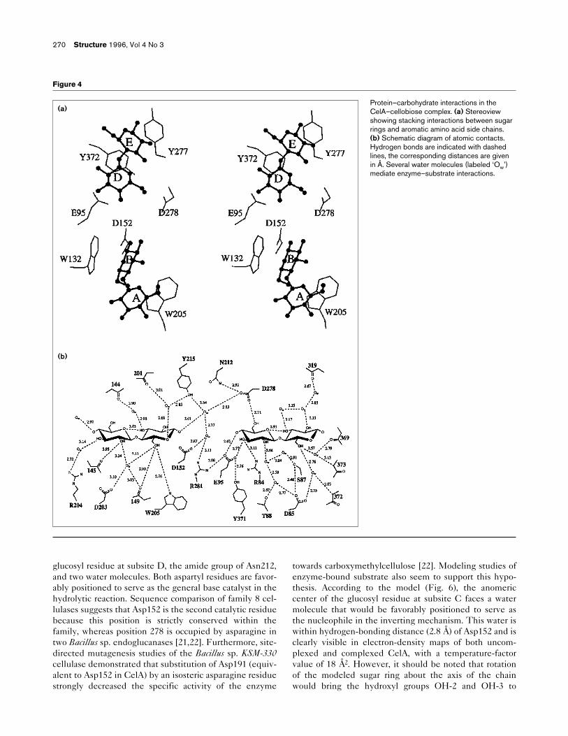
The active-site cleft of CelA conforms to the typicalconfiguration for sugar-binding sites of proteins [20], witharomatic residues stacking against the hydrophobic facesof sugar rings and hydrogen bonds conferring bindingaffinity and specificity. The four glucopyranose rings makestacking interactions with aromatic residues that are con-served in family 8 cellulases: Trp205 in subsite A, Trp132in subsite B, Tyr372 in subsite D, and Tyr277 in subsite E(Fig. 4a). In addition, Tyr369 can make van der Waals
Research Article Endoglucanase CelA Alzari, Souchon and Dominguez 267
Figure 2
Overall view of the (a/a)6 barrel ofendoglucanase CelA. (a) Side view of CelAshowing the active-site cleft at the N-terminalend of the inner helices. The 12 a helicesforming the barrel involve residuesGln52–Arg70, Ser94–Cys106,Gln110–Lys121, Thr151–Trp168,Tyr176–Cys191, Pro218–Thr228,Arg232–Val247, Tyr282–Phe293,Gln296–Ala310, Ala334–Ala343,Leu350–Ala362 and Tyr372–Ile384 (asdefined by PROCHECK [35]). (b) Stereo Catrace of CelA, viewed along the barrel axis.Amino acid positions are labeled every 20residues.
(b)

contacts with substrate bound at subsite F, as it does withthe inhibitor. The hydroxyl groups of glucosyl residuesparticipate in several hydrogen-bonding interactions withprotein and water atoms (Fig. 4b). Direct protein–carbohy-drate hydrogen bonds occur at the non-reducing end ofthe substrate. They include the interaction of hydroxylOH-2 at subsite A with the main-chain carbonyl group ofGly145, and a bifurcated hydrogen bond of hydroxyl OH-6at subsite B with the indole ring of Trp205 and themain-chain carbonyl of Ala149. On the other side of thecleft, the glucopyranoside ring at subsite D is withinhydrogen-bonding distance of three amino acid sidechains (Glu95, Arg84, and Asp278; see Fig. 4b). In addi-tion to these direct contacts, several water moleculesmediate protein–carbohydrate interactions through anextensive network of hydrogen bonds.
Substrate conformationSubstrate binding promotes no significant conformationalrearrangements in the active-site architecture of CelA; theoverall root mean square (rms) deviation between theuncomplexed and complexed forms of the catalyticdomain is 0.2 Å. In contrast, the cellulose chain is clearlybent within the enzyme cleft upon binding. As estimated
from the structure of the CelA–cellobiose complex(Fig. 3a), the linear cellulose chains emerging from bothends of the active-site cleft form an angle of approxi-mately 110°. Chain bending probably occurs at the glyco-sidic linkage between subsites C and D (which is thepresumed scissile bond), and can be accounted for by dis-ruption of the alternative orientation that adjacent gluco-syl rings present in a linear cellulose chain (Fig. 5a). Theintermediate glucosyl residue at subsite C can be posi-tioned by linearly extending the cellulose chain from thenon-reducing end of the substrate. The model that resultssuggests that the sugar rings on both sides of the glyco-sidic linkage (subsites C–D) are similarly oriented, thuspromoting the observed chain bending (Fig. 5b).
Preliminary modeling of bound substrate at subsite C(Fig. 6) was carried out assuming a rigid polypeptidebackbone, preserving both the position of glucosyl ringsobserved in the electron density (subsites A–B and D–E)and hydrogen bonds involving protein atoms. Underthese constraints, in order to position the sugar ringwithin the binding cleft steric clashes with protein atomswere unavoidable, suggesting that local strain in the sub-strate (either bending or torsion) could explain the lack ofglucosyl binding to subsite C in the crystal structures. Itis tempting to speculate that both overall chain bendingand local steric strain within the active-site cleft couldhelp to stabilize the transition state of the substrate withrespect to the ground state. However, further experimen-tal evidence is required to validate this hypothesis, aswell as to determine the actual nature of structuralchanges induced in both protein and substrate uponformation of the complex.
Catalytic mechanismFamily 8 endoglucanase CelCCC from C. cellulolyticum,which has 55% sequence identity with CelA, hydrolyzesthe glycosidic bond via a single displacement mechanismwith inversion of the anomeric configuration [21]. Thecatalytic residues necessary for an inverting reaction areapparent in the CelA structure. Three carboxylate groupsat the N-terminal end of inner helices 2, 4, and 8 (Glu95,Asp152 and Asp278, respectively) are exposed to solventat the center of the substrate-binding cleft, close to theb-1,4-linkage between subsites C and D (Fig. 6). BothGlu95 and Asp278 form hydrogen bonds to hydroxylgroups of the glucosyl unit bound at subsite D, whereasthe carboxyl group of Asp152 occupies the bottom of thecleft at subsite C and is not involved in direct interac-tions with substrate. The proximity of Glu95, a residuestrictly conserved in family 8 cellulases, to the glycosidicoxygen between subsites C and D suggests that the glu-tamate side chain may act as a general acid catalyst orproton donor. One carboxyl oxygen atom of Glu95 formshydrogen bonds with the OH-4 atom of the glucosylresidue at subsite D (equivalent to the b-1,4-linking
268 Structure 1996, Vol 4 No 3
Table 2
Intramolecular salt bridges (donor–acceptor distances <3.3 Å)in the catalytic core of CelA.
Donor Acceptor Distance (Å)
Asp50 Od2 Arg358 Ne 2.91Glu62 Oe1 Lys364 Nz 2.82Glu62 Oe1 Arg377 Nh1 2.74Glu64 Oe2 Lys67 Nz 3.29Asp65 Od2 Lys69 Nz 2.94Asp65 Od2 Lys364 Nz 2.93Asp91 Od1/Od2 Arg84 Ne/Nh2 2.84/2.89Asp91 Od2 His133 Ne2 2.74Glu95 Oe1 Arg281 Nh1 3.23Glu109 Oe2 Lys67 Nz 2.92Asp115 Od1 Arg118 Nh1 3.26Asp144 Od2 His133 Nd1 2.70Asp147 Od2 Arg204 Nh1 3.20Asp152 Od2 Arg281 Nh1 3.30Asp156 Od1/Od2 Arg285 Nh1/Nh2 2.76/2.95Glu179 Oe2 Lys121 Nz 2.89Asp230 Od2/Od1 Arg232 Nh2/Ne 2.85/2.95Asp238 Od1 Arg297 Nh1 2.95Asp238 Od2 Lys307 Nz 2.65Glu245 Oe1 Lys248 Nz 2.81Glu246 Oe1 Lys249 Nz 3.04Asp289 Od1/Od2 Lys222 Nz 2.75/2.98Asp295 Od2 Lys222 Nz 2.80Asp295 Od2/Od1 Arg297 Nh2/Ne 2.88/3.04Asp303 Od2 Lys299 Nz 2.77Asp312 Od2 Lys327 Nz 2.82Glu359 Oe1 Lys315 Nz 3.18Glu359 Oe2 His332 Ne2 2.65Glu367 Oe2 Arg70 Nh1 3.06

oxygen) and with the buried d-guanido group of Arg281(Fig. 6). The other carboxyl oxygen of Glu95 formshydrogen bonds to the hydroxyl group of Tyr371 and tothe OH-3 atom of the same glucosyl residue. The tworesidues contacting the catalytic glutamate (Arg281 andTyr371) are also conserved in family 8 cellulases, sug-gesting that these hydrogen-bonding interactions play animportant role in determining the protonation state of thecarboxyl group.
The acidic residues Asp152 and Asp278 are positioned at asimilar distance from the scissile bond, although the corre-sponding side chains approach the substrate in differentorientations (Fig. 6). In the CelA–cellobiose complexstructure, the side chain of Asp152 (at the bottom of thecleft) interacts with four water molecules and forms aweak hydrogen bond with Arg281 (Table 2), whereas thecarboxyl group of Asp278 (on one side of the binding cleft)makes hydrogen bonds with the OH-6 group of the
Research Article Endoglucanase CelA Alzari, Souchon and Dominguez 269
Figure 3
Stereoview of substrate binding to CelA. Thedifference maps, contoured at 3s, werecalculated at 1.9 Å resolution with observedamplitudes (Fobsprotein+ligand–Fobsprotein) andDM-modified MIR phases. The refined modelof bound ligand is shown in thick lines. Theside chains of aromatic and acidic residuesclose to the sugar rings are shown as thinlines. (a) Complex of CelA with cellobiose(other cello-oligosaccharides produce similardifference maps). (b) Complex of CelA withIBTC.
(a)
(b)
glucosyl residue at subsite D, the amide group of Asn212,and two water molecules. Both aspartyl residues are favor-ably positioned to serve as the general base catalyst in thehydrolytic reaction. Sequence comparison of family 8 cel-lulases suggests that Asp152 is the second catalytic residuebecause this position is strictly conserved within thefamily, whereas position 278 is occupied by asparagine intwo Bacillus sp. endoglucanases [21,22]. Furthermore, site-directed mutagenesis studies of the Bacillus sp. KSM-330cellulase demonstrated that substitution of Asp191 (equiv-alent to Asp152 in CelA) by an isosteric asparagine residuestrongly decreased the specific activity of the enzyme
towards carboxymethylcellulose [22]. Modeling studies ofenzyme-bound substrate also seem to support this hypo-thesis. According to the model (Fig. 6), the anomericcenter of the glucosyl residue at subsite C faces a watermolecule that would be favorably positioned to serve asthe nucleophile in the inverting mechanism. This water iswithin hydrogen-bonding distance (2.8 Å) of Asp152 and isclearly visible in electron-density maps of both uncom-plexed and complexed CelA, with a temperature-factorvalue of 18 Å2. However, it should be noted that rotationof the modeled sugar ring about the axis of the chainwould bring the hydroxyl groups OH-2 and OH-3 to
270 Structure 1996, Vol 4 No 3
Figure 4
Protein–carbohydrate interactions in theCelA–cellobiose complex. (a) Stereoviewshowing stacking interactions between sugarrings and aromatic amino acid side chains.(b) Schematic diagram of atomic contacts.Hydrogen bonds are indicated with dashedlines, the corresponding distances are givenin Å. Several water molecules (labeled ‘Ow’)mediate enzyme–substrate interactions.
(a)
(b)

within hydrogen-bonding distance of the two carboxyloxygens of Asp152, leaving Asp278 (which approaches theglycosidic bond from the opposite side of the substratewith respect to the proton donor, Glu95) facing theanomeric carbon C1. Therefore, although the presentstudy unambiguously identifies Glu95 as the proton donorin the catalytic mechanism, a definitive assignment of thegeneral base catalyst (Asp152 or Asp278) requires further
structural evidence of the conformation of the glucosylresidue at subsite C.
It was previously noted [5] that the average separationbetween the catalytic carboxylates of a- and b-glycosi-dases was significantly different for retaining (4.8–5.3 Å)and inverting (9.0–9.5 Å) enzymes; the greater distance ininverting enzymes is presumably required to accommo-date the nucleophilic water between the two carboxylates.Endoglucanase CelA appears to deviate from this pattern,as the average separation between the carboxyl groups ofGlu95 and Asp152 is 5.8 Å (7.5 Å between Glu95 andAsp278), a value significantly lower than that expected foran inverting enzyme. Indeed, retaining and invertingmechanisms impose different stereochemical constraintson the catalytic residues. In retaining enzymes the twocatalytic residues must both be close to the scissile bond,thus setting an upper limit for the separation between thecorresponding carboxyl groups. However, although similardistance constraints apply to the water nucleophile andthe proton donor in inverting enzymes, the position of thegeneral base is less restricted. The possible separationbetween the two catalytic residues can thus present awider range of values.
Structural relationship with other glycosyl hydrolasesIn addition to CelA, the (a/a)6 barrel topology has alsobeen observed in two other glycosidases that have com-pletely unrelated amino acid sequences: family 9 endoglu-canase CelD from C. thermocellum [11] and family 15glucoamylase-I from Aspergillus awamori [23]. Theseenzymes all have an acidic active-site cleft at the N-termi-nal end of the inner a helices. They hydrolyze the glyco-sidic linkage via a single displacement mechanism leadingto inversion of configuration at the anomeric carbon.However, structural comparisons between these three
Research Article Endoglucanase CelA Alzari, Souchon and Dominguez 271
Figure 5
Substrate conformation. (a) Alternative orientation of adjacent b-1,4-linked glucosyl residues promotes the formation of a linear chain incellulose. (b) Substrate binding to CelA imposes a similar orientationto consecutive glucopyranoside rings at subsites C and D, inducing abend of the cellulose chain when within the active-site cleft. In theconformation shown, both the anomeric carbon C1 and the glycosidicoxygen O4 (indicated with arrows) can be approached from the sameside of the substrate.
Figure 6
View of the catalytic center of CelA showingthe relative disposition of three carboxylategroups (Glu95, Asp152, and Asp278) closeto the scissile glycosidic linkage and the twoglucosyl residues bound at subsites B and D.Hydrogen bonds are represented by thindashed lines. A possible orientation of theglucosyl ring at subsite C is shown in thickdashed lines. (The figure was drawn withMOLSCRIPT [38].)
(a)
(b)

enzymes reveal important differences. As a result of dis-similar packing of a helices within the barrel, the cross-section is nearly circular in CelA and glucoamylase-I, butmore elliptical in CelD (Fig. 7). The architecture and ori-entation of the corresponding active-site clefts also differconsiderably. When the structures are superimposed withthe a helices in the same topological order (as shown inFig. 7), the long substrate-binding grooves in CelA andCelD run in approximately perpendicular directions. Fur-thermore, glucoamylase-I has no groove, but has a centralactive pocket with an approximate diameter of 15 Å and adepth of 10 Å [23]. In addition, the carboxyl groupsassigned as the proton donor and the general base catalystsoccupy unrelated positions in both the amino acidsequence and the 3D structure. In CelA, these residues,Glu95 and Asp152 (or Asp278), at the N-terminal end ofhelices 2 and 4 (or 8), lie close to each other and near themiddle of the cleft. The corresponding residues in CelD(Glu555 and Asp201) occur in the loops connecting helices11 and 12 and helices 1 and 2, at the reducing end of thesubstrate-binding groove [11]. Those of glucoamylase-I,Glu179 (between helices 5 and 6) and Glu400 (between
helices 11 and 12), approach the substrate from oppositesides of the central binding pocket [24]. These compar-isons reflect the intrinsic stability of the (a/a)6 barrel as atertiary structure motif, and suggest a possible, albeitdistant, evolutionary connection between distinct classesof glycosyl hydrolases.
Biological implicationsCellulose is the most abundant polysaccharide of theplant cell wall. In order to hydrolyze natural cellulose,cellulolytic microorganisms have evolved a widevariety of enzymes with different carbohydrate speci-ficities and three-dimensional (3D) structures. Indeed,cellulases and hemicellulases account for over onethird of all the known families of glycosidic enzymes.The thermophilic anaerobe Clostridium thermocellum,one of the most thoroughly studied cellulolyticmicroorganisms, produces a high molecular weightmultienzyme complex, the cellulosome, which is veryactive against crystalline cellulose. Here we report thecrystal structure of the catalytic core of C. thermocel-lum endoglucanase CelA, a cellulosomal enzyme that
272 Structure 1996, Vol 4 No 3
Figure 7
Structural comparison of (a/a)6 glycosyl hydrolases. (a) Family 8endoglucanase CelA. (b) Family 9 endoglucanase CelD fromClostridium thermocellum. (c) Family 15 glucoamylase-I fromAspergillus awamori. The figure shows a schematic view witha helices represented as cylinders (top) and the molecular surface of
the active-site clefts colored according to charge (bottom). Thecoordinates of endoglucanase CelD (code 1CLC) and glucoamylase-I(code 1GLY) were taken from the Brookhaven Protein Data Bank [37].(The figure was drawn with programs QUANTA [MolecularSimulations, Inc.] and GRASP [39].)

belongs to glycosidase family 8, a family for which nostructural information was previously available.
The catalytic core of CelA folds into an (a/a)6 barrelformed by six inner and six outer a helices, with anacidic active-site cleft running across the molecularsurface at the N-terminal end of the inner helices.The structures of CelA complexed with various cello-oligosaccharides reveal five D-glucosyl binding sub-sites (A–E) which are lined with tryptophan andtyrosine residues that are involved in stacking inter-actions with the substrate. The carboxylate group ofGlu95 is hydrogen bonded to the b-1,4-linking oxygenbetween subsites C and D, and has been assigned therole of proton donor. The carboxylate groups ofAsp152 or Asp278, close to the scissile glycosidicbond, are the likely candidates for the general basecatalyst in the hydrolytic reaction.
The protein folding pattern and active-site architec-ture of CelA can be extended to other members offamily 8 cellulases. Furthermore, the comparison ofCelA with family 9 cellulases and family 15 glu-coamylases, which present a similar protein fold butdifferent active-site architecture, reflects the stabilityof the (a/a)6 barrel as a protein folding motif and sug-gests a possible evolutionary relationship between dif-ferent families of glycosyl hydrolases.
Materials and methodsCrystallization and data collectionCellulase CelA, expressed in E. coli, was purified and crystallized asdescribed in [25]. The protein crystallizes in the orthorhombic spacegroup P212121 with cell dimensions a=50.12 Å, b=63.52 Å, andc=104.97 Å, and with one molecule in the asymmetric unit(Vm=1.94 Å3 Da–1).
All diffraction intensities were collected at room temperature using aMARresearch Image Plate scanner mounted on a Rigaku RU-200 rotat-ing anode generator, and reduced to structure-factor amplitudes with
DENZO and SCALEPACK [26] and other programs from the CCP4suite [27]. A first diffraction data set was measured at 1.65 Å resolu-tion from a single crystal of native CelA (Table 3). For heavy-atom datacollection, the same crystal was soaked in 1mM HgCl2 and a data setcollected to 1.9 Å resolution. Subsequently, the crystal was soaked in5 mM trimethyl lead acetate (derivative Hg+Pb) and a data set was col-lected to 1.9 Å. After obtaining new crystals of CelA by macroseedingtechniques, data from a second mercurial derivative were collectedafter soaking a fresh crystal in 0.2 mM HgCl2. This mercurial and the(Hg+Pb) derivative were used for structure determination (Table 3).
In order to characterize protein–carbohydrate interactions, CelA wasco-crystallized in the presence of the inhibitor IBTC and various cello-oligosaccharides (glucose, cellobiose, cellotriose, cellotetraose, cel-lopentaose). Isomorphous crystals of the enzyme–ligand complexeswere grown as described for uncomplexed CelA [25], using anenzyme:ligand molar ratio of 1:10 in all cases. Data collection statisticsof the various CelA complexes are shown in Table 3.
Structure determination and refinement of uncomplexed CelAHeavy-atom parameters for the Hg derivative (4 sites, phasing powerfor acentric reflections of 2.3, and R-Cullis of 0.48) and the (Hg+Pb)derivative (6 sites, phasing power of 2.2 and R-Cullis of 0.57) wererefined at 1.9 Å resolution with the CCP4 program MLPHARE [28]. Asshown in Table 4, the major heavy-atom binding sites in the (Hg+Pb)derivative were essentially occupied by Hg atoms bound to the five freecysteine residues of CelA. The initial MIR phases calculated with theprogram MLPHARE (figure of merit of 0.64 at 1.9 Å) were furtherrefined using the program DM [29] from CCP4. Starting with an R-freeof 53%, 50 cycles of solvent flattening (assuming a solvent content of35%) and histogram matching refinement converged to an overallfigure of merit of 0.82 and an R-free of 32% in the resolution range10–1.9 Å.
The electron-density map calculated with DM-modified MIR phases(Fig. 1) allowed unambiguous tracing of the entire polypeptide chain(363 residues from positions 33–395). Chain tracing and model build-ing was carried out with the program ‘O’ [30]. The model was sub-jected to a first round of simulated annealing refinement at 1.9 Åresolution with X-PLOR [31] using potential parameters proposed byEngh and Huber [32] and restrained individual isotropic thermal para-meters (sB=1 Å2 for bonded atoms). This initial cycle reduced the crys-tallographic R-factor from 35% to 26%, maintaining good modelstereochemistry. Subsequent refinement was carried out by successiverounds of positional and temperature factor refinement with X-PLORfollowed by automatic insertion and deletion of water molecules withthe program ARP [33]. All 39653 independent reflections between
Research Article Endoglucanase CelA Alzari, Souchon and Dominguez 273
Table 3
Data collection statistics.
Crystal Measured Unique Resolution Completeness (%) R-merge* R-iso†
reflections reflections (Å) overall last shell overall last shell
Native CelA 249 508 39 868 1.65 96.9 94.0 0.050 0.155 –(Hg) derivative‡ 281 017 26 347 1.90 97.8 95.3 0.044 0.077 0.208(Hg+Pb) derivative§ 182 750 31 806 1.80 99.1 98.2 0.059 0.123 0.311CelA–IBTC# 215 160 30 650 1.80 96.7 93.5 0.042 0.118 0.051CelA–(glc)1** 276 484 39 183 1.65 95.5 91.7 0.054 0.214 0.052CelA–(glc)2 288 790 39 112 1.65 95.2 90.8 0.051 0.192 0.046CelA–(glc)3 273 755 39 521 1.65 96.2 92.5 0.037 0.111 0.046CelA–(glc)4 268 329 38 613 1.65 94.1 89.1 0.057 0.254 0.055CelA–(glc)5 287 675 38 774 1.65 94.4 90.0 0.059 0.128 0.057
*R-merge=Shkl|I–⟨I⟩|/Shkl(I). †R-iso=Shkl|Fpl–Fp|/Shkl Fp , where Fpl isthe observed amplitude for the protein–ligand complex and Fp is theobserved amplitude for the uncomplexed protein. ‡(Hg) derivative:
0.2 mM HgCl2. §(Hg+Pb) derivative: 1 mM HgCl2+5 mM(CH3)3PbOOCCH3. #IBTC: o-iodobenzyl-1-thio-b-D-cellobioside. **glc: D-glucosyl.

10 Å and 1.65 Å resolution were used for refinement, 5% of whichwere set aside for R-free calculation [34]. A first round of 50 combinedX-PLOR–ARP cycles converged to an R-factor of 15.5%(R-free=18.7%) for a model containing 401 water molecules, with rmsdeviations from ideal bond lengths and bond angles of 0.011 Å and1.4°, respectively. After visual inspection of electron-density maps,minor modifications were introduced to the protein model, someresidues with side chains they were not visible in density were con-verted to alanine, and 101 water molecules were removed based onstereochemical and temperature-factor criteria. A second round of com-bined X-PLOR–ARP refinement converged to an R-factor of 15.6% (R-free=18.9%) for a model including 370 water molecules. At this stage,inspection of (3Fo–2Fc) electron-density maps allowed positioning ofthe missing side chains. The final model retained 266 water moleculeswith temperature-factor values B<50 Å2. The parameters after a finalpositional refinement round with X-PLOR are reported in Table 1.
Quality of the modelThe atomic model of CelA includes 363 amino acid residues corre-sponding to positions 33–395 of the CelA gene [18], and accounts fora catalytic core of 40.2 kDa, in good agreement with the molecularweight of papain-treated CelA (42 kDa) determined by SDS/PAGE[25]. The final (3Fo–2Fc) electron-density map shows continuousdensity for the entire polypeptide chain. Most amino acid side chains(in particular all those forming the active-site cleft) are well defined indensity. The model of CelA has good overall stereochemistry (Table 1).All main-chain dihedral angles fall within allowed (90.4%) or additionalallowed (9.6%) regions [35] of the Ramachandran plot (Fig. 8). Fromthe resolution dependence of the R-factor, the mean positional error ofthe final model coordinates is estimated to lie within 0.15–0.20 Å [36].
Refinement of CelA complexesThe initial models for IBTC and the various cello-oligosaccharideswere built from Fourier difference maps calculated with observedamplitudes and MIR phases (Fig. 3). Crystallographic refinement wascarried out as described above for uncomplexed CelA, assuming fulloccupancy of bound sugar atoms. The final refinement parameters forthe CelA–IBTC and the CelA–cellobiose complexes are given in Table1. Complexes of CelA with other cello-oligosaccharides (glucose, cel-lotriose, cellotetraose, cellopentaose) were also refined (data notshown), leading to structural models essentially identical to that of theCelA–cellobiose complex, with a glucosyl residue bound to each ofthe subsites A, B, D, and E.
Bound glucosyl residues are well defined in the electron-density mapscalculated with experimental phases (Fig. 3), but display high tempera-ture-factor values in the refined models of the complexes (Table 1).These values probably reflect partial substrate occupancy, sinceglucose (which cannot simultaneously occupy adjacent glucosyl-binding subsites) presents similar high temperature factors in therefined structure of the CelA–glucose complex (data not shown).
The atomic coordinates of the refined protein model (for the uncom-plexed CelA) have been deposited with the Brookhaven Protein DataBank [37], code 1CEM.
AcknowledgementsWe thank P Béguin for providing the plasmid encoding endoglucanase CelA,and F Saul for helpful discussions. This work was supported by the InstitutPasteur and a grant from the European Union, contract AIR1-CT92-0321.
References1. Gilkes, N.R., Henrissat, B., Kilburn, D.G., Miller, R.C., Jr. & Warren, R.A.J.
(1991). Domains in microbial b-1,4-glycanases: sequence conservation,function, and enzyme families. Microbiol. Rev. 55, 303–315.
2. Béguin, P. & Aubert, J.-P. (1994). The biological degradation ofcellulose. FEMS Microbiol. Rev. 13, 25–58.
3. Henrissat, B. (1991). A classification of glycosyl hydrolases based onamino acid sequence similarities. Biochem. J. 280, 309–316.
4. Henrissat, B. & Bairoch, A. (1993). New families in the classification ofglycosyl hydrolases based on amino acid sequence similarities.Biochem. J. 293, 781–788.
5. McCarter, J.D. & Withers, S.G. (1994). Mechanisms of enzymaticglycoside hydrolysis. Curr. Opin. Struct. Biol. 4, 885–892.
6. Dominguez, R., et al., & Alzari, P.M. (1995). A common protein foldand similar active site in two distinct families of b-glycanases. Nat.Struct. Biol. 2, 569–576.
7. Ducros, V., et al., & Haser, R. (1995). Crystal structure of the catalyticdomain of a bacterial cellulase belonging to family 5. Structure 3,939–949.
8. Rouvinen, T., Bergfors, T., Teeri, T.T., Knowles, J.K.C. & Jones, T.A.(1990). Three-dimensional structure of cellobiohydrolase II fromTrichoderma reesei. Science 249, 380–385.
274 Structure 1996, Vol 4 No 3
Figure 8
Ramachandran plot of CelA. All main-chain dihedral angles occur inenergetically allowed regions. Glycine residues are represented asfilled triangles. (Produced with the program PROCHECK [35].)
Table 4
Heavy-atom binding sites.
Site Relative Close protein Distanceoccupancy* atom (Å)
(Hg) derivative1 0.50 Sg–Cys106 1.92 0.31 Sg–Cys191 1.53 0.17 Sg–Cys106 1.94† 0.09 Site 1 2.3
(Hg+Pb) derivative1 1.01 Sg–Cys302 2.42 0.83 Sg–Cys261 1.93‡ 0.71 Sg–Cys191 1.64‡ 0.69 Sg–Cys106 1.95 0.53 Sg–Cys240 1.96 0.19 Nd–His143 2.0
*All heavy-atom sites were refined assuming Hg atomic scatteringfactors and a fixed B value of 30 Å2. †Possible chloride atom. ‡Bindingsites 3 and 4 in the second derivative are identical to sites 2 and 3 inthe first derivative.

9. Spezio, M., Wilson, D.B. & Karplus, P.A. (1993). Crystal structure ofthe catalytic domain of a thermophilic endoglucanase. Biochemistry32, 9906–9916.
10. Divne, C., et al., & Jones, T.A. (1994). The three-dimensional crystalstructure of the catalytic core of cellobiohydrolase I from Trichodermareesei. Science 265, 524–528.
11. Juy, M., et al., & Aubert, J.-P. (1992). Three-dimensional structure of athermostable bacterial cellulase. Nature 357, 89–91.
12. Davies, G.J., et al., & Schülein, M. (1993). Structure and function ofendoglucanase V. Nature 365, 362–364.
13. Pétré, J., Longin, R. & Millet, J. (1981). Purification and properties ofan endo-b-1,4-glucanase from Clostridium thermocellum. Biochimie63, 629–639.
14. Wu, J.H.D. (1993). Clostridium thermocellum cellulosome. Newmechanistic concept for cellulase degradation. In Biocatalyst Designfor Stability and Specificity. (Himmel, M.E. & Georgiu, G., eds), vol.516, pp. 251–264, American Chemical Society, Washington DC.
15. Bayer, E.A., Morag, E. & Lamed, R. (1994). The cellulosome — atreasure-trove for biotechnology. Trends Biotechnol. 12, 379–386.
16. Tokatlidis, K., Dhurjati, P. & Béguin, P. (1993). Properties conferred onClostridium thermocellum endoglucanase CelC by grafting theduplicated segment of endoglucanase CelD. Protein Eng. 6,947–952.
17. Cornet, P., Millet, J., Béguin, P. & Aubert, J.-P. (1983).Characterization of two cel (cellulose degradation) genes ofClostridium thermocellum coding for endoglucanases. Biotechnol. 1,589–594.
18. Béguin, P., Cornet, P. & Aubert, J.-P. (1985). Sequence of a cellulasegene of the thermophilic bacterium Clostridium thermocellum.J. Bacteriol. 162, 102–105.
19. Hall, J., Hazlewood, G.P., Barker, P.J. & Gilbert, H.J. (1988).Conserved reiterated domains in Clostridium thermocellumendoglucanases are not essential for catalytic activity. Gene 69,29–38.
20. Vyas, N.K. (1991). Atomic features of protein–carbohydrateinteractions. Curr. Opin. Struct. Biol. 1, 732–740.
21. Fierobe, H.P., et al., & Belaich, J.-P. (1993). Purification andcharacterization of endoglucanase C from Clostridium cellulolyticum.Catalytic comparison with endoglucanase A. Eur. J. Biochem. 217,557–565.
22. Ozaki, K., Sumitomo, N., Hayashi, Y., Kawai, S. & Ito, S. (1994). Site-directed mutagenesis of the putative active site of endoglucanase Kfrom Bacillus sp. KSM-330. Biochim. Biophys. Acta 1207, 159–164.
23. Aleshin, A., Golubev, A., Firsov, L.M. & Honzatko, R.B. (1992). Crystalstructure of glucoamylase from Aspergillus awamori var. X100 to2.2 Å resolution. J. Biol. Chem. 267, 19291–19298.
24. Harris, E.M.S., Aleshin, A.E., Firsov, L.M. & Honzatko, R.B. (1993).Refined structure of 1-deoxynojirimycin with glucoamylase fromAspergillus awamori var. X100 to 2.4 Å resolution. Biochemistry 32,1618–1626.
25. Souchon, H., Béguin, P. & Alzari, P.M. (1996). Crystallization of afamily 8 cellulase from Clostridium thermocellum. Proteins, in press.
26. Otwinowski, Z. (1993). Oscillation data reduction program. InProceedings of the CCP4 Study Weekend: Data Collection andProcessing. (Sawyer, L., Isaacs, N. & Bailey, S., eds), pp. 56–62,SERC Daresbury Laboratory, Warrington, UK.
27. Collaborative Computational Project Number 4 (1994). The CCP4suite: programs for protein crystallography. Acta Cryst. D 50,760–763.
28. Otwinowski, Z. (1991). Maximum likelihood refinement of heavy atomparameters. In Isomorphous Replacement and Anomalous Scattering:Proceedings of the CCP4 Study Weekend. (Wolf, W., Evans, P.R. &Leslie, A.G.W., eds), pp. 80–86, SERC Daresbury Laboratory,Warrington, UK.
29. Cowtan, K. (1994). DM, an automated procedure for phaseimprovement by density modification. Joint CCP4 and ESF-EACBMNewsletter on Protein Crystallography 31, 24–28.
30. Jones, T.A., Zou, J.-Y., Cowan, S.W. & Kjeldgaard, M. (1991).Improved methods for building protein models in electron densitymaps and the location of errors in these models. Acta Cryst. A 47,110–119.
31. Brünger, A.T., Kuriyan, J. & Karplus, M. (1987). CrystallographicR-factor refinement by molecular dynamics. Science 235, 458–460.
32. Engh, R.A. & Huber, R. (1991). Accurate bond and angle parametersfor X-ray protein structure refinement. Acta Cryst. A 47, 392–400.
33. Lamzin, V.S. & Wilson, K.S. (1993). Automated refinement of proteinmodels. Acta Cryst. D 49, 129–147.
34. Brünger, A.T. (1992). The free R value: a novel statistical quantity forassessing the accuracy of crystal structures. Nature 335, 472–474.
35. Laskowski, R.A., MacArthur, M.W., Moss, D.S. & Thornton, J.M.(1993). PROCHECK: a program to check the stereochemical qualityof protein structures. J. Appl. Cryst. 26, 283–291.
36. Luzzati, V. (1952). Traitement statistique des erreurs dans ladetermination des structures cristallines. Acta Cryst. 5, 802–810.
37. Bernstein, F.C., et al., & Tasumi, M. (1977). The protein data bank: acomputer-based archival file for macromolecular structures J. Mol.Biol. 112, 535–542.
38. Kraulis, P.J. (1991). MOLSCRIPT: a program to produce both detailedand schematic plots of protein structures. J. Appl. Cryst. 24,946–950.
39. Nicholls, A. & Honig, B. (1993). GRASP — graphical representationand analysis of surface properties. Columbia University, NY.
Research Article Endoglucanase CelA Alzari, Souchon and Dominguez 275