The Clostridium thermocellum Cellulosome -the Paradigm of a Multienzyme Complex
11
1 The Clostridium thermocellum Cellulosome - the Paradigm of a Multienzyme Complex Vladimir V. Zverlov 1,2 , and Wolfgang H. Schwarz 2* 1 ) Institute of Molecular Genetics, Russian Academy of Science, Kurchatov Sq., 123182 Moscow, Russia. 2 ) Research Group Microbial Biotechnology, Technische Universitaet Muenchen, Am Hochanger 4, D-85350 Freising-Weihenstephan, Germany The cellulosome is the large extracellular enzyme-complex of the anaerobic bacteria which is responsible for the highly efficient degradation of insoluble cellulose in lignocellulosic biomass. A great number of Clostridium thermocellum genes code for putative components of the cellulosome: the unfinished genomic sequence reveals more than 60 genes containing dockerin modules. However, the scaffoldin protein CipA has only nine dockerin binding sites (cohesins), which limits the number of components present in each individual cellulosome particle. Not all genes may be expressed: the components are not distributed equally in the multienzyme complex. Moreover: the C. thermocellum cellulosomes are not only composed of cellulases, but also of xylanases, pectinases, glycosidases and structural proteins. By estimating the quantitative distribution of the proteins in a 2D-electrophoresis gel from a cellulosome preparation, the major components from cellulose-grown cells were identified. Major enzymatic components contain not only the already known endo-, processive endo- and exo-glucanases of the reducing- and non-reducing-end specificity type, but also hitherto unknown ß-glucanases, xylanases, xyloglucanases, and probably also at least one newly detected non-catalytic protein which are now under investigation. The quantitative distribution of the protein pattern present in cellulosomes is described and the major spots in the 2D-gel are identified. ABUNDANCE OF LIGNOCELLULOSIC BIOMASS AND ENZYMATIC HYDROLYSIS A huge amount of plant biomass is available in waste or crops: alone in Germany about 76 Mio metric tons per year of lignocellulosic biomass were collected but not useful otherwise in 2001. Much more cold be purposefully collected without additional planting efforts (37, 18). About 30 to 40 % of dry biomass is cellulose which can be hydrolyzed to glucose, the universal substrate for microbial fermentations. Complete hydrolysis can easily be performed at large industrial scale by treatment with sulfuric acid, or with a combination of heat and high pressure (TDH). However, a certain percentage of the sugars are lost due to unwanted chemical reactions. In contrast, enzymatic hydrolysis has a much higher yield of sugars per digested biomass, but is slow and expensive pretreatment is unavoidable. The cost effectiveness of the enzymatic process at a large scale is still to be shown (22). The search for more effective enzymes or enzyme systems goes on. Two methods to reach this goal are used: new genetically engineered enzymes are under development which promise a cost effective enzymatic hydrolysis in the near future. Alternatively new and more effective enzyme systems are screened from natural environments (30). Native cellulose is a difficult substrate for enzymatic hydrolysis: it is partially crystalline and it is enwrapped with extremely heterogeneous hemicellulose (Fig. 1). This means that a great number of different enzymes is needed for digestion. It was observed that enzymes may support each other in a synergistic action. This synergism is not exerted by successive action: enzymes have to be present simultaneously; the greater the proximity of different enzyme components the greater the synergism (32).
Transcript of The Clostridium thermocellum Cellulosome -the Paradigm of a Multienzyme Complex

1
The Clostridium thermocellum Cellulosome - the Paradigm of aMultienzyme Complex
Vladimir V. Zverlov1,2, and Wolfgang H. Schwarz2*
1) Institute of Molecular Genetics, Russian Academy of Science, Kurchatov Sq., 123182Moscow, Russia. 2) Research Group Microbial Biotechnology, Technische Universitaet
Muenchen, Am Hochanger 4, D-85350 Freising-Weihenstephan, Germany
The cellulosome is the large extracellular enzyme-complex of the anaerobicbacteria which is responsible for the highly efficient degradation of insoluble cellulose inlignocellulosic biomass. A great number of Clostridium thermocellum genes code forputative components of the cellulosome: the unfinished genomic sequence reveals morethan 60 genes containing dockerin modules. However, the scaffoldin protein CipA hasonly nine dockerin binding sites (cohesins), which limits the number of componentspresent in each individual cellulosome particle. Not all genes may be expressed: thecomponents are not distributed equally in the multienzyme complex. Moreover: the C.thermocellum cellulosomes are not only composed of cellulases, but also of xylanases,pectinases, glycosidases and structural proteins.
By estimating the quantitative distribution of the proteins in a 2D-electrophoresisgel from a cellulosome preparation, the major components from cellulose-grown cellswere identified. Major enzymatic components contain not only the already known endo-,processive endo- and exo-glucanases of the reducing- and non-reducing-end specificitytype, but also hitherto unknown ß-glucanases, xylanases, xyloglucanases, and probablyalso at least one newly detected non-catalytic protein which are now under investigation.The quantitative distribution of the protein pattern present in cellulosomes is describedand the major spots in the 2D-gel are identified.
ABUNDANCE OF LIGNOCELLULOSIC BIOMASS AND ENZYMATIC HYDROLYSIS
A huge amount of plant biomass is available in waste or crops: alone in Germanyabout 76 Mio metric tons per year of lignocellulosic biomass were collected but not usefulotherwise in 2001. Much more cold be purposefully collected without additional plantingefforts (37, 18). About 30 to 40 % of dry biomass is cellulose which can be hydrolyzed toglucose, the universal substrate for microbial fermentations. Complete hydrolysis can easilybe performed at large industrial scale by treatment with sulfuric acid, or with a combination ofheat and high pressure (TDH). However, a certain percentage of the sugars are lost due tounwanted chemical reactions. In contrast, enzymatic hydrolysis has a much higher yield ofsugars per digested biomass, but is slow and expensive pretreatment is unavoidable. The costeffectiveness of the enzymatic process at a large scale is still to be shown (22). The search formore effective enzymes or enzyme systems goes on. Two methods to reach this goal are used:new genetically engineered enzymes are under development which promise a cost effectiveenzymatic hydrolysis in the near future. Alternatively new and more effective enzymesystems are screened from natural environments (30).
Native cellulose is a difficult substrate for enzymatic hydrolysis: it is partiallycrystalline and it is enwrapped with extremely heterogeneous hemicellulose (Fig. 1). Thismeans that a great number of different enzymes is needed for digestion. It was observed thatenzymes may support each other in a synergistic action. This synergism is not exerted bysuccessive action: enzymes have to be present simultaneously; the greater the proximity ofdifferent enzyme components the greater the synergism (32).

2
Fig. 1: Hypothetical structure of a cellulose fiber. The squares represent the cross-section of cellulose microfibrils, a number of which is glued together by hemicellulose andlignin (crooked lines). The approximate size of single cellulase enzymes is indicated by thecircles.
C. THERMOCELLUM IS A SUPERIOR CELLULOSE DEGRADER
Increased reaction temperature is favorable for biotechnological processes. It wasintended to demonstrate the occurrence of thermophilic cellulolytic bacteria in variousenvironments. Soil from grass lands and woods, or self heated compost from cellulosic plantor algae waste from southern Germany and northern France was incubated with various mediaat 60 and 65 °C. Anaerobic as well as aerobic conditions were used for enrichment withWhatman filter paper as sole carbohydrate. Invariably the anaerobic cultures degraded thefilter paper completely with no visible fibers or pieces, whereas in the aerobic cultures thepaper was only disintegrated into fibers (Fig. 2). Isolated from one of the enriched anaerobiccultures at 65 °C, the sequence of 10 from 10 PCR amplificates of the small subunitribosomal DNA (16S rDNA) could be identified as closely related to Clostridiumthermocellum (> 96 % sequence identity over 1450 bp). Similarly a complete degradation offilter paper was effected by C. thermocellum strains DSM 1237 (the type strain) and F7 inanaerobic flasks.
Surprisingly, none of the aerobic cultures did show efficient cellulose degradation atthe high temperatures, although the operator of one of the composting plants has detectedtemperatures up to 70 °C in the aerated compost from which samples were taken.Consequently, the anaerobic bacterium C. thermocellum (and close relatives) is an ubiquitousand easily isolated anaerobic thermophilic bacterium which seems to play a major role inanaerobic cellulose degradation in nature.
Fig. 2: Digestion of Whatman fiter paper No. 1 in anaerobic cultures. The rubberstoppered bottles containing reduced medium in N2 atmosphere were inoculated with1/100 vol. of a diluted soil or compost probe and incubated 0, 2 or 4 days at 60 °C.The insert shows the beginning of paper dissolution.
3 nm
cellulases
0d 2d 4d
3
WHY IS BIOMASS SO STABLE?
Cellulose and hemicellulose are difficult to hydrolyze: although cellulose ischemically homogeneous, its physical structure is at least partially crystalline. This poses anumber of problems for enzymes: only a small portion of the substrate is hydratized andexposed to the surface of the crystal in a way, the enzymes have access to it; the number ofsites which can be attacked is very limited and different sites may need a different way ofattack (Fig. 1). I.e. some enzymes degrade other parts of the substrate than others. Thus, incellulose a very homogeneous chemistry is combined with a heterogeneous topography and agreat number of enzymes is necessary to degrade them – but all enzymes cleave the samechemical bond. In hemicellulose the resistance to enzymatic degradation may rather comefrom the chemical heterogeneity: different sugars are linked with different chemical bonds.These polymers are often also derivatized with phenolic esters like feruylic acid. Here also agreat number of different enzymes is necessary for hydrolysis, this time with activity fordifferent chemical bonds.
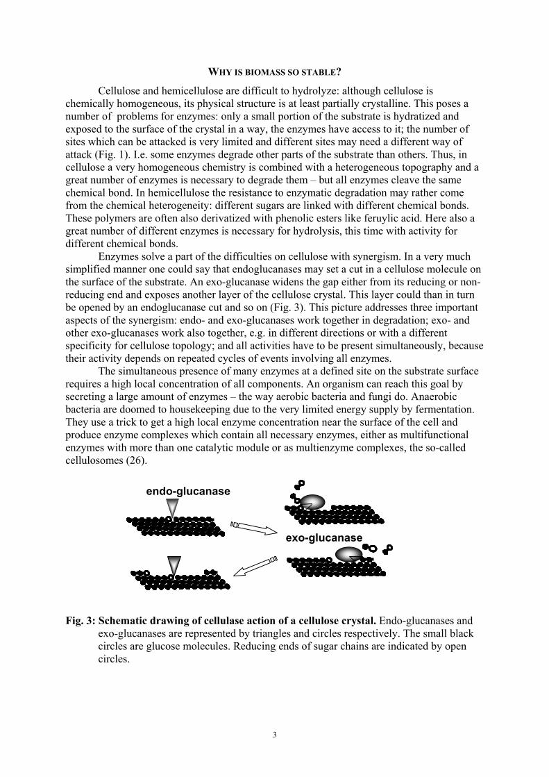
Enzymes solve a part of the difficulties on cellulose with synergism. In a very muchsimplified manner one could say that endoglucanases may set a cut in a cellulose molecule onthe surface of the substrate. An exo-glucanase widens the gap either from its reducing or non-reducing end and exposes another layer of the cellulose crystal. This layer could than in turnbe opened by an endoglucanase cut and so on (Fig. 3). This picture addresses three importantaspects of the synergism: endo- and exo-glucanases work together in degradation; exo- andother exo-glucanases work also together, e.g. in different directions or with a differentspecificity for cellulose topology; and all activities have to be present simultaneously, becausetheir activity depends on repeated cycles of events involving all enzymes.
The simultaneous presence of many enzymes at a defined site on the substrate surfacerequires a high local concentration of all components. An organism can reach this goal bysecreting a large amount of enzymes – the way aerobic bacteria and fungi do. Anaerobicbacteria are doomed to housekeeping due to the very limited energy supply by fermentation.They use a trick to get a high local enzyme concentration near the surface of the cell andproduce enzyme complexes which contain all necessary enzymes, either as multifunctionalenzymes with more than one catalytic module or as multienzyme complexes, the so-calledcellulosomes (26).
Fig. 3: Schematic drawing of cellulase action of a cellulose crystal. Endo-glucanases andexo-glucanases are represented by triangles and circles respectively. The small blackcircles are glucose molecules. Reducing ends of sugar chains are indicated by opencircles.
endo-glucanase
exo-glucanase

4
THE CELLULOSOME, A MOST EFFICIENT DEPOLYMERIZATION MACHINERY
Evidence accumulated that the enzymatic cellulose degradation machinery of C.thermocellum is 50 to 100 times more efficient per gram of protein than the fungal enzymes,which are commercially exploited for cellulose hydrolysis (29). Even if the fungal enzymesystem would be improved by a factor of ten, the C. thermocellum cellulosomes would bemore efficient – and they were not optimized yet. However the production of the cellulosomesby the thermophilic host is presently very low. It could probably be improved by mutantselection or metabolic engineering. The high efficiency of the cellulase systemcounterbalances well the low energy efficiency of the anaerobic metabolism, which needs ahigh amount of glucose for little energy spent for the production of extracellular enzymes. Inaddition, these enzymes are located on the cell surface, minimizing diffusion of enzymes andhydrolysis products, and thus ensuring a high percentage of products taken up by the cellstransport mechanism. This on the other hand is a disadvantage for technical enzymeproduction.
The Israeli group around Lamed and Bayer were the first to characterize the enzymesystem of C. thermocellum as a huge extracellular enzyme complex which turned out to bearranged along the scaffoldin protein CipA (25,12). It can reach a mass of 1,6 MDa in somestrains. This cellulosome is hold together by protein-protein interactions between the cohesinmodules located on the scaffoldin and corresponding dockerin modules located mostly at theC-termini of the cellulosome components. The scaffoldin itself is docking to a cell wallanchoring protein (28). It also binds to the substrate with a very tightly binding carbohydratebinding module (CBM) and thus diminishes the necessity of the single enzymatic componentsto have a CBM by its own. The structure of the cellulosomes from C. thermocellum and otherbacteria and the interaction between its components is described in recent reviews (4, 33, 35).
It is conspicuous that the production of cellulosomes is restricted to the bacterialfamily Clostridiaceae and the closely related Lachnospiraceae (anaerobic rumen bacteria)(34). All other reports of large extracellular enzyme complexes have so far no genetical datafor support. The common gene structure, the similarity of the binding modules which hold thecomplexes together and the major composition of distinct enzyme families suggestheterologous gene transfer between the bacterial species. The very large enzyme complexes ofthe cellulosomes are an interesting model for the in vitro construction of bioengineeredprotein complexes (9).
GENOMIC LIBRARIES AND STRUCTURE OF CELLULOSOMAL PROTEINS
Libraries of genomic C. thermocellum DNA were screened for enzymatic activitiesknown to be involved in biomass hydrolysis, such as ß-1,4-glucanase, cellobiohydrolase, andxylanase. A number of genes were identified with high redundancy (15). 25 of them containeda tandem repeat of a 24 amino acid peptide, called the dockerin module. It could be shown tobind strongly to the cohesin modules of the large scaffoldin CipA, which was identified byimmunological screening (11). The C. thermocellum scaffoldin contains 9 cohesins type I, butcarries itself a dockerin of type II with a different binding specificity to the cohesin of the cellwall anchoring protein OlpB, which has three repeats of a surface layer homologous module(SLH, reviewed in 3, 35). Substrate binding is mediated by a cellulose binding module offamily CBM3a near the C-terminus of CipA. Only few of the enzymatic cellulosomecomponents contain an own CBM which is commonly found in non-cellulosomalextracellular cellulases (table 1; Fig. 4).
All enzyme components of the cellulosome are composed of a catalytic and a dockerinmodule. Some have in addition a carbohydrate binding module or other non-catalytic modules(table 1). The CBM sometimes functions in addition to substrate binding inthermostabilization of the catalytic module. The non-catalytic modules also may modify the

5
action mode of the catalytic module (reviewed in 35). For cellulases only a limited number ofglycosyl hydrolase families is known: GH5, GH8 and GH9 for endo-, processive endo- andexo-glucanases, and GH48 for exo-glucanases (7). Examples for their structure are given inFig. 4. Although family 48 enzymes are the most important exo-glucanases in cellulosomalsystems, family 5 and family 9 enzymes are found as well to be exo- as endoglucanases. Thiswas shown e.g. for the structural pairs CbhA/CelD or CelO/CelB (6, 13, 41, 42, 44).
In addition to cellulases (ß-1,4-glucanases) a number of other enzyme specificitieshave been identified: ß-1,3-1,4-glucanses, xylanases, mannanases and chitinases – not only inthe cellulosomes of C. thermocellum, but also in that of mesophilic clostridia (table 1)(8).
Fig. 4: Structure of selected enzymatic cellulosome components. Typical and repeatedlyoccurring module architectures are shown. The structure of the proteins is drawnschematically, beginning left with the N-terminus of the polypeptide. The bars areonly approximately drawn to scale. GHF, glycosyl hydrolase family; DD, dockerinmodule; Ig, immunoglobulin-like fold; CBM, carbohydrate binding module.
IDENTIFICATION OF THE PROTEINS IN THE CELLULOSOME
The great number of genes and the limited number of docking sites on the scaffoldinrises the question, if on the average all gene products are expressed and present in thecellulosome. Earlier investigations made clear that there are a few major components whichseem to dominate: the scaffoldin CipA, which must be present in each individual cellulosomeparticle, and the exo-glucanase Cel48S, of which more than 1 copy seems to be incorporatedper particle. On the other hand, a chitinase ChiA protein is only present in each 20th
cellulosomal particle (43).
GHF48 DDCELS
CBM4 Ig GHF9 DD
CelK
CelO
CelB/CelGGHF5 DD
GHF5 DD
CelN/CelFGHF9 CBM3 DD
CelDIg GHF9 DD
CBM3
CipA
CbhA
CelK
CelS
Fig. 5: SDS-PAGE of cellulosome preparations. A fresh (left lane) and a partially degradedcellulosome preparation (right lane) are shown. Bands identified by N-terminal sequencingin the partially degraded preparation (including multiple bands of CipA) and identical bandsin the left lane are indicated.

6
To identify the major components, purified cellulosomes were denaturated by boilingin SDS/ß-mercaptoethanol and separated in denaturing SDS-PAGE. 14 protein bands,designated S1 to S14, were detected and the genes coding for most of them could beidentified by several methods (Fig. 5)(25, 33). One method was the immunoblotting withspecific antibodies against a recombinant protein devoid of the dockerin module which wouldgive a reaction with all cellulosomal components (e.g. CbhA in 42; CipA in 11). Crossreactions between proteins having a highly homologous sequence can occur (e.g. CbhA/CelK;42). Another problem was the instability of the cellulosomal components for proteasedegradation: the proteins have a modular structure and the modules are connected withflexible peptide linkers which are prone to protease attack. An example is shown in Figure 5:CipA fragments are frequently obtained if distinct protein bands were N-terminallysequenced. Particularly many CelA fragments were common in the low molecular weightregion of the gel (unpublished data). Thus the number of proteins present in the cellulosomecannot be determined alone by counting protein bands in a gel.
CELLULOSOMAL PROTEINS SEPARATED BY 2D-GEL ELECTROPHORESIS
To reach a higher resolution of the proteins and to resolve common proteins besidesrare ones, a 2D gel electrophoresis gel technique was developed, which involved denaturationof the proteins with urea and separation by pI (isolelectric focusing) followed by denaturingSDS-gel PAGE (46). The proteins separated well, most of them between pH 5 and 3 (data notshown). Protein smears due to overload could not be completely avoided in an attempt toidentify also minor components.
Single spots were identified by MALDI-TOF and MALDI-TOF-TOF to avoidmisinterpretation of spots due to protease action (method according to 36). However, esp.some of the minor spots could not be tested so far. Many of the major spots were identifiedagainst the library of sequenced genes. The most prominent proteins were CipA, CelS, CelA,CelK, XynC and XynZ. The locations for CbhA, CelG, CelN and the chitinase ChiA werealso determined (46). However, the mass pattern of 3 major spots did not fit to the collectionof known sequences of cellulosomal components. Only when the partial genomic sequence ofC. thermocellum was included in the search (URLhttp://genome.ornl.gov/microbial/cthe/25jun02/cthe_contig9-15.html), three hitherto un-cloned open reading frames were identified.
The ORFs were amplified by PCR from strain C. thermocellum F7 genomic DNA.The resulting recombinant proteins were enzymatically active and could be purified.Preliminary biochemical data on the newly detected enzymes suggest that they code for anendo-glucanase (CelR), an endo-xyloglucanase (XghA) and an endo-xylanase (XynD)belonging to the glycosyl hydrolase families GH9, GH74 and GH10 respectively (Acc.no.AJ585348, AJ585344, AJ585345 resp.). They were among the most prominent spots in thegel when the cells were grown on cellulose.
The 2D-gels show a reproducible map of identified major protein spots and can nowbe used to look for differential expression of genes on various substrates or culture conditions.Preliminary data show differences in the expression of the cellulosomal genes (see also 16).
A GENOMIC ANALYSIS: MORE THAN 60 CELLULOSOME COMPONENTS?
Motivated by the detection of three new cellulosomal genes, the entire genomicsequence contigs were screened by a BLAST-search for reading frames containing dockerinsequences. This search resulted in a list of potential genes for cellulosome components whichare shown in table 1. However, the genomic sequence is unfinished and not closed yet (10th ofOctober, 2003); it contains a number of gaps, incompletely sequenced DNA stretches andoverlaps. The screening revealed some reading frames connecting obviously unrelated

7
modules and some sequences of already cloned genes were not found. Nevertheless the greatnumber of 62 reading frames with dockerin modules is overwhelming and some of thepresumed enzyme activities would never have been suspected and therefore have not beenlooked for in the genomic library screenings.
Besides the known genes containing catalytic modules of GH5, GH8, GH9 and GH48,few additional reading frames were found in the families GH5 and GH9. This may be due tothe ease of screening for these enzymatic activities by conventional methods, and manyresearch groups have undertaken extended screening programs for these ß-glucanases.However, it is evident that GH8 and GH48 are represented by only 1 gene each among thecellulosomal genes. Both are major components (Cel8A and Cel48S). ß-1,4-Glucanases ofother families were not identified.
Surprising is the large number of hemicellulase components potentially present in thecellulosome (table 1). With XynD a 6th xylanase has been identified, 5 of which have beenshown to be present in the isolated cellulosomes, 3 as major components. For completehydrolysis of xylan, the presence of additional glycosidases has to be postulated, none ofwhich was found so far by biochemical analysis or random cloning. But genes for ß-glucuronidase (GH2), and ß-xylosidase / α-arabinofuranosidase (GH39, GH43, GH54) arepresent and can now be investigated. In addition, genes for carbohydrate-esterases may alsobe expressed as components of the cellulosome which may split feruloyl residues and otheresters of the hemicelluloses. Other hemicelluloses may be degraded by ß-1,3- and ß-1,6-glucanases (GH30, GH81), mannanase (GH26) or endo-ß-1,4-galactanase (GH53). Acomplete hydrolysis system for pectin including 4 families of pectate lyases may also bepresent (GH28, PL1, PL9, PL10, PL11).
Some cellulosome components contain more than one functionally related catalyticmodule like ß-xylosidase and α-arabinofuranosidase (GH54 + GH43), xylanase andcarbohydrate-esterase (GH10 + CE1), ß-1,4-glucanase and mannanase (GH5 + GH26), or twodifferent ß-1,4-glucanases (GH9 + GH44). A number of reading frames contains besides thedockerin module unknown modules with no obvious sequence homology in BLAST searches.They may be structural components or silent genes without function. However, one putativestructural component has homology to the previously described CseP protein (#55 in table 1)for which a structural role has been expected (45).
Predicted cellulosomal genes with interesting architecture or hitherto not detectedprobable activities are presently under investigation. Not all of these genes will be expressedor even incorporated into the cellulosome. And some will be expressed possibly only undercertain induction conditions, i.e. by growth on certain substrates. Some genes will be poorlyexpressed and not identified with the 2D gel-electrophoresis. But the number of cellulosomalcomponents seems to be by far greater than estimated so far. However, only a few enzymecomponents will play a major role in the degradation of crystalline cellulose or of definedbiomass substrates. These components have to be identified by biochemistry and genetechnology.
The data presented here are a good basis for doing functional genomics with C.thermocellum. The methods will be applied for identifying the protein components present inthe cellulosome under different conditions of growth on a range of carbohydrate substrate, thenumber of which is limited due to growth restrictions of the Clostridium thermocellum strain,e.g. on xylan or glucose. Probably not all reading frames identified will produce cellulosomecomponents in vivo. But the major components produced on crystalline cellulose as substrateare already identified.

8
REFERENCES
1) Ahsan, M. M. et al., J. Bacteriol. 178, 5732-5740 (1996).2) Arai, T. et al., Appl. Microbiol. Biotechnol. 57, 660-666 (2001).3) Bayer, E. A. et al., in M. Claeyssens, et al. (eds.), Carbohydratases from Trichoderma
reesei and other microorganisms, p. 39-65. The Royal Society of Chemistry, London(1998).
4) Bayer, E. A., et al., in Dworkin, M. et al. (eds.), “The Prokaryotes: An EvolvingElectronic Resource for the Microbiological Community”, 3rd edition. Springer-Verlag,New York. (2000).
5) Béguin, P. et al., J. Bacteriol. 162, 102-105 (1985).6) Chauvaux, S. et al., J. Biol. Chem. 267, 4472-4478 (1992).7) Coutinho, P.M. & Henrissat, B., URL: http://afmb.cnrs-mrs.fr/~cazy/CAZY/index.html
(1999).8) Doi, R. H. et al., J. Bacteriol. 185, 5907-14 (2003).9) Fierobe, H. P. et al., J. Biol. Chem. 276, 21257-21261 (2001).10) Fontes, C. M. G. A. et al., Biochem. J. 307, 151-158 (1995).11) Fujino, T. et al., FEMS Microbiol. Let. 94, 165-170 (1992).12) Gerngross, U. T. et al., Molec. Microbiol. 8, 325-334 (1993).13) Grépinet, O., & Béguin, P., Nuc. Ac. Res. 14, 1791-1799 (1986).14) Grepinet, O. et al., J. Bacteriol. 170, 4582-4588 (1988).15) Guglielmi, G., & Béguin, P., FEMS Microbiol. Let. 161, 209-215 (1998).16) Halliwell, G. et al., Process Biochem. 30, 243-250 (1995).17) Halstead, J. R. et al., Microbiol. 145, 3101-3108 (1999).18) Hartmann, H. & Kaltschmitt, M. (Eds.): Biomasse als erneuerbarer Energieträger.
Schriftenreihe „Nachwachsende Rohstoffe“, Vol. 3. Landwirtschaftsverlag, Münster(2002).
19) Hayashi, H. et al., J. Bacteriol. 179, 4246-4253 (1997).20) Hayashi, H. et al., Appl. Microbiol. Biotechnol. 51, 348-357 (1999).21) Hazlewood, G. P. et al., Enz. Microb. Technol. 12, 656-662 (1990).22) Himmel, M. E. et al., Curr. Opin. Biotechnol. 10, 358-364 (1999).23) Joliff, G. et al., Nuc. Acids Res. 14, 8605-8613 (1986).24) Kurokawa, J. et al., Appl. Microbiol. Biotechnol. 59, 455-461 (2002).25) Lamed , R. et al., J. Bacteriol. 156, 828-836 (1983).26) Lamed, R. et al., J. Bacteriol. 169, 3792-3800 (1987).27) Lemaire, M., & Béguin, P., J. Bacteriol. 175, 3353-3360 (1992)28) Lemaire, M. et al., J. Bacteriol. 177, 2451-2459 (1995).29) Lynd, L. et al., Microbiol. Molec. Biol. Rev. 66, 506-577 (2002).30) Montoya, D. et al., J. Ind. Microbiol. Biotechnol. 27, 329-335 (2001).31) Navarro, A. et al., Res. Microbiol. 142, 927-936 (1991).32) Riedel, K., & Bronnenmeier, K., Molec. Microbiol. 28, 767-775 (1998).33) Schwarz, W. H., Appl. Microbiol. Biotechnol. 56, 634-649 (2001).34) Schwarz, W. H., URL http://www.wzw.tum.de/mbiotec/cellmo.htm (2003).35) Schwarz, W. H. et al., Adv. Appl. Microbiol. (in press).36) Shevchenko, A. et al., Anal. Chem. 68, 850-858 (1996).37) van Wyk, J. P. H., Trends Biotechnol. 19, 172-177 (2001).38) Wang, W. K. et al., J. Bacteriol. 175, 1293-1302 (1993).39) Yaguee, E. et al., Gene 89, 61-67 (1990).40) Zverlov, V. V. et al., Biotechnol. Let. 16, 29-34 (1994).41) Zverlov, V. V. et al., J. Bacteriol. 180, 3091-3099 (1998).42) Zverlov, V. V. et al., Appl. Microbiol. Biotechnol. 51, 852-859 (1999).

9
43) Zverlov, V. V. et al., Appl. Environ. Microbiol. 68, 3176-3179 (2002).44) Zverlov, V. V. et al., Microbiol. 148, 247-255 (2002).45) Zverlov, V. V. et al., Microbiol. 149, 515-524 (2003).46) Zverlov, V. V., in preparation.
ACKNOWLEDGMENTS
This work was supported by grants from the Deutsche Forschungsgemeinschaft DFGand the Leonhard-Lorenz-Foundation to WHS, and from the A.-v.-Humboldt Foundation toVVZ. We are very grateful to W. L. Staudenbauer for countless stimulating discussions. D.Minde provided the data on paper hydrolysis under elevated temperature conditions. C.Arevalo is thanked for valuable support with the 2D-gel electrophoresis and J. Kellermann(MPI for Biochemistry, Martinsried) for the performing all the MALDI-TOF determinations.
TABLE 1: List of cellulosomal components in the genome of C.thermocellum
Hypothetical reading frames and cloned genes in the unfinished genome sequence. Onlyreading frames containing dockerin modules, a Shine-Dalgarno sequence and stop codon, anda recognizable module composition are listed. The protein (gene) designation and enzymaticactivity or function (if known), and the ORF number from http://genome.ornl.gov/microbial/cthe/ (Cthe) are given (as of October 2003). If no Cthe number is indicated, the gene wascloned but is missing in the genomic sequence. The components are sorted according to theirGH family or their putative function, as obvious from the catalytic module family.Components with more than one catalytic module or unknown modules are listed at the end.
Gene* Reading frame /function
Structure ** Referencelocalisation
Structural component1. CipA + scaffoldin, Cthe1933-1930 2(Coh1)-CBM3a-7(Coh1)-X2-Doc2 11, 42
GH22. Cthe1580 GH2-CBM6-Doc1
GH53. CelO cellobiohydrolase, Cthe1674 CBM3b-GH5-Doc1 444. Cthe1575 GH5-CBM6-Fn3–Doc15. CelB endoglucanase, Cthe0374 GH5-Doc1 136. CelG + endoglucanase, Cthe0885 GH5-Doc1 277. Cthe0444 GH5-Doc1
GH88. CelA + endoglucanase, Cthe0722 GH8-Doc1 5; 46
GH99. CbhA + cellobiohydrolase CBM4-Ig-GH9-2(Fn3)-CBD3b-Doc1 4110. CelK + cellobiohydrolase, Cthe2598 CBM4-Ig-GH9-Doc1 4111. CelD endoglucanase, Cthe0968 Ig-GH9-Doc1 2312. Cthe1953 GH9-CBM3c-CBM3b-Doc113. Cthe0850 GH9-CBM3c-CBM3b- Doc1

10
14. CelN + endoglucanase, Cthe1222 GH9-CBM3c-Doc1 4615. CelR + endoglucanase, Cthe1837 GH9-CBM3c–Doc1 4616. CelQ + endoglucanase, Cthe0300 GH9-CBM3c-Doc1 217. CelF endoglucanase, Cthe0382 GH9-CBM3c-Doc1 3118. Cthe1308 GH9-CBM3c–Doc119. Cthe0727 GH9-Doc120. CelT + endoglucanase GH9-Doc1 24
Xylanases21. XynD + xylanase, Cthe0688 CBM22-GH10–Doc1 4622. XynC + xylanase, Cthe0626 CBM22-GH10-Doc1 1923. XynA,
XynU +xylanase, Cthe1161 GH11-CBM4-Doc1-NodB 20
24. XynB,XynV +
xylanase GH11-CBM4-Doc1 19
Other hemicellulases25. LicB + lichenase GH16-Doc1 4026. ChiA + chitinase GH18-Doc1 4327. ManA + mannanase, Cthe0533 CBM-GH26-Doc1 1728. Cthe2142 GH26-Doc129. Cthe1127 GH30-CBM6-Doc130. Cthe2333 GH53-Doc131. Cthe0269 GH81-Doc1
Putative glycosidases32. Cthe1665 GH39-2(CBM6)-Doc133. Cthe1579 GH43-CBM6-Doc134. Cthe0268 GH43-CBM13-Doc135. Cthe0484 GH43-2(CBM6)-Doc1
GH4836. CelS + exoglucanase, Cthe0939 GH48-Doc1 38
Xyloglucanhydrolase37. XghA + xyloglucanase, Cthe2335 GH74-CBM2-Doc1 46
Putative carbohydrateesterases
38. Cthe0066 Fn3-CE12-Doc1-CBM6-CE1239. Cthe1577 CE1-CBM6-Doc1
Putative pectinases40. Cthe2008 GH28-Doc141. Cthe2236 PL1-Doc1-CBM642. Cthe1810 <-Doc1-CBM6-PL943. Cthe2234 PL10-UN-Doc144. Cthe0702 Doc1-CBM6-PL11
Multifunctional components45. CelJ + cellulase, Cthe0301 X-Ig-GH9-GH44-Doc1-X 146. CelH endoglucanase, Cthe0837 GH26-GH5-CBD9-Doc1 3947. Cthe1667 GH30-GH54-GH43-Doc148. Cthe1211 GH54-Doc1-GH4349. Cthe1666 GH54-GH43-Doc150. XynZ + xylanase, Cthe1691 CE1-CBM6-Doc1-GH10 14

11
51. XynY xylanase, Cthe2036 CBM22-GH10-CBM22-Doc1-CE1 1052. CelE + endoglucanase, Cthe0940,
Cthe2702, Cthe2514GH5-Doc1-CE2 21
Putative protease inhibitors53. Cthe1412 Fn3-Doc1-serpin54. Cthe1413 DOC1-SERPIN
Components with unknownfunction
55. Cthe0694 2(UN)-UN-UN(CelP 550-870)-Doc156. Cthe1578 UN-CBM6-Doc157. CseP + Cthe1223 UN-Doc1 4558. Cthe1474 Doc1-UN59. Cthe0287 UN1-UN2-Doc160. Cthe0416 Doc1-UN61. Cthe0073 UN-Doc162. Cthe0649 UN-Doc1
*A “+” in the reference column indicates, that the component was shown to be present in thecellulosome.** Module classification according to (7), URL:
http://afmb.cnrs-mrs.fr/CAZY/: Coh, cohesin module; Doc, dockerin module; CBM,carbohydrate binding module; X, hydrophobic module; GH, glycosyl hydrolase family; Fn3,fibronectin III module; Ig, immunoglobulin like fold; NodB, acetylxylan-esterase NodB type;CE, carbohydrate esterase; PL, pectin lyase; UN, unknown module; serpin, serine-proteaseinhibitor homologue.