The coding polymorphism T263I in TGF- 1 is associated with otosclerosis in two independent...
10
The coding polymorphism T263I in TGF-b1 is associated with otosclerosis in two independent populations Melissa Thys 1 , Isabelle Schrauwen 1 , Kathleen Vanderstraeten 1 , Katrien Janssens 1,{ , Nele Dieltjens 1 , Kris Van Den Bogaert 1,{ , Erik Fransen 1 , Wenjie Chen 2 , Megan Ealy 2 , Mireille Claustres 3 , Cor R.W.J. Cremers 4 , Ingeborg Dhooge 5 , Frank Declau 6 , Jos Claes 6 , Paul Van de Heyning 6 , Robert Vincent 7 , Thomas Somers 8 , Erwin Offeciers 8 , Richard J.H. Smith 2 and Guy Van Camp 1, * 1 Department of Medical Genetics, University of Antwerp, Universiteitsplein 1, 2610 Wilrijk (Antwerp), Belgium, 2 Molecular Otolaryngology Research Laboratories, Department of Otolaryngology, University of Iowa, 200 Hawkins Drive, Iowa City IA 52242, USA, 3 Laboratoire de Genetique Moleculaire et chromosomique CHU, IURC Montpellier, 641 avenue du Doyen Gaston Giraud, 34093 Montpellier Cedex 5, France, 4 Department of Otorhinolaryngology, University Medical Centre St Radboud, Philips van Leydenlaan 15, 6500 HB Nijmegen, The Netherlands, 5 Department of Otorhinolaryngology, University Hospital of Ghent, De Pintelaan 185, 9000 Ghent, Belgium, 6 Department of ORL, University Hospital of Antwerp, Wilrijkstraat 10, 2650 Edegem, Belgium, 7 Jean Causse Ear Clinic, Traverse de Be ´ ziers, 34440 Colombiers, France and 8 University Department of Otolaryngology, St-Augustinus Hospital Antwerp, Oosterveldlaan 24, 2610 Antwerp, Belgium Received May 8, 2007; Revised and Accepted June 8, 2007 Otosclerosis is a progressive hearing loss characterized by an abnormal bone homeostasis of the otic capsule that leads to stapes fixation. Although its etiology remains unknown, otosclerosis can be considered a complex disease. Transforming growth factor-beta 1 (TGF-b1) was chosen for a case–control association study, because of several non-genetic indications of involvement in otosclerosis. Single nucleotide poly- morphism (SNP) analysis in a large Belgian–Dutch sample set gave significant results (P 5 0.0044) for an amino acid changing SNP, T263I. Analysis of an independent French population replicated this association with SNP T263I (P 5 0.00019). The results remained significant after multiple testing correction in both popu- lations. Haplotype analysis and the results of an independent effect test using the weighted haplotype (WHAP) computer program in both populations were both compatible with SNP T263I being the only causal variant. The variant I263 is under-represented in otosclerosis patients and hence protective against the disease. Combining the data of both case – control groups for SNP T263I with a Mantel – Haenszel estimate of common odds ratios gave a very significant result (P 5 9.2 3 10 26 ). Functional analysis of SNP T263I with a luciferase reporter assay showed that the protective variant I263 of TGF-b1 is more active than the WT variant T263 (P 5 1.6 3 10 26 ). On the basis of very low P-values, replication in an independent population and a func- tional effect of the protective variant, we conclude that TGF-b1 influences the susceptibility for otosclerosis, and that the I263 variant is protective against the disease. # The Author 2007. Published by Oxford University Press. All rights reserved. For Permissions, please email: [email protected] { Present address: European Molecular Biology Laboratory (EMBL), Meyerhofstrasse 1, 69117 Heidelberg, Germany. ‡ Present address: Stem Cell Institute Leuven (SCIL), Catholic University Leuven, Herestraat 49, 3000 Leuven, Belgium. *To whom correspondence should be addressed. Tel: þ32 38202491; Fax: þ32 38202566; Email: [email protected] Human Molecular Genetics, 2007, Vol. 16, No. 17 2021–2030 doi:10.1093/hmg/ddm150 Advance Access published on June 22, 2007 by guest on June 6, 2013 http://hmg.oxfordjournals.org/ Downloaded from
-
Upload
independent -
Category
Documents
-
view
0 -
download
0
Transcript of The coding polymorphism T263I in TGF- 1 is associated with otosclerosis in two independent...
The coding polymorphism T263I in TGF-b1 isassociated with otosclerosis in two independentpopulations
Melissa Thys1, Isabelle Schrauwen1, Kathleen Vanderstraeten1, Katrien Janssens1,{,
Nele Dieltjens1, Kris Van Den Bogaert1,{, Erik Fransen1, Wenjie Chen2, Megan Ealy2,
Mireille Claustres3, Cor R.W.J. Cremers4, Ingeborg Dhooge5, Frank Declau6, Jos Claes6,
Paul Van de Heyning6, Robert Vincent7, Thomas Somers8, Erwin Offeciers8, Richard J.H. Smith2
and Guy Van Camp1,*
1Department of Medical Genetics, University of Antwerp, Universiteitsplein 1, 2610 Wilrijk (Antwerp), Belgium,2Molecular Otolaryngology Research Laboratories, Department of Otolaryngology, University of Iowa, 200 Hawkins
Drive, Iowa City IA 52242, USA, 3Laboratoire de Genetique Moleculaire et chromosomique CHU, IURC Montpellier,
641 avenue du Doyen Gaston Giraud, 34093 Montpellier Cedex 5, France, 4Department of Otorhinolaryngology,
University Medical Centre St Radboud, Philips van Leydenlaan 15, 6500 HB Nijmegen, The Netherlands, 5Department
of Otorhinolaryngology, University Hospital of Ghent, De Pintelaan 185, 9000 Ghent, Belgium, 6Department of ORL,
University Hospital of Antwerp, Wilrijkstraat 10, 2650 Edegem, Belgium, 7Jean Causse Ear Clinic, Traverse de
Beziers, 34440 Colombiers, France and 8University Department of Otolaryngology, St-Augustinus Hospital Antwerp,
Oosterveldlaan 24, 2610 Antwerp, Belgium
Received May 8, 2007; Revised and Accepted June 8, 2007
Otosclerosis is a progressive hearing loss characterized by an abnormal bone homeostasis of the oticcapsule that leads to stapes fixation. Although its etiology remains unknown, otosclerosis can be considereda complex disease. Transforming growth factor-beta 1 (TGF-b1) was chosen for a case–control associationstudy, because of several non-genetic indications of involvement in otosclerosis. Single nucleotide poly-morphism (SNP) analysis in a large Belgian–Dutch sample set gave significant results (P 5 0.0044) for anamino acid changing SNP, T263I. Analysis of an independent French population replicated this associationwith SNP T263I (P 5 0.00019). The results remained significant after multiple testing correction in both popu-lations. Haplotype analysis and the results of an independent effect test using the weighted haplotype(WHAP) computer program in both populations were both compatible with SNP T263I being the onlycausal variant. The variant I263 is under-represented in otosclerosis patients and hence protective againstthe disease. Combining the data of both case–control groups for SNP T263I with a Mantel–Haenszel estimateof common odds ratios gave a very significant result (P 5 9.2 3 1026). Functional analysis of SNP T263I with aluciferase reporter assay showed that the protective variant I263 of TGF-b1 is more active than the WT variantT263 (P 5 1.6 3 1026). On the basis of very low P-values, replication in an independent population and a func-tional effect of the protective variant, we conclude that TGF-b1 influences the susceptibility for otosclerosis,and that the I263 variant is protective against the disease.
# The Author 2007. Published by Oxford University Press. All rights reserved.For Permissions, please email: [email protected]
{Present address: European Molecular Biology Laboratory (EMBL), Meyerhofstrasse 1, 69117 Heidelberg, Germany.‡Present address: Stem Cell Institute Leuven (SCIL), Catholic University Leuven, Herestraat 49, 3000 Leuven, Belgium.
*To whom correspondence should be addressed. Tel: þ32 38202491; Fax: þ32 38202566; Email: [email protected]
Human Molecular Genetics, 2007, Vol. 16, No. 17 2021–2030doi:10.1093/hmg/ddm150Advance Access published on June 22, 2007
by guest on June 6, 2013http://hm
g.oxfordjournals.org/D
ownloaded from
INTRODUCTION
Otosclerosis is a common bone dysplasia that is unique to theendochondral layer of the otic capsule. Otosclerosis is adisease particularly frequent among the white population,whereas it is very rare among Blacks, Asians and NativeAmericans (1). Schuknecht and Barber (2) classified oto-sclerosis as clinical and histological. Histological otosclerosishas an estimated prevalence of 2.5%, and denotes a diseaseprocess in which clinical symptoms are absent, and can onlybe discovered by CT-scanning or sectioning of the temporalbone at autopsy. Clinical otosclerosis, in contrast, is causedby otosclerotic lesions that impinge on stapes mobility,thereby causing a progressive conductive hearing loss. Theprevalence of clinical otosclerosis is estimated at 0.3–0.4%in the Caucasian population and is twice as frequent infemales as in males (3).
The otic capsule may be ‘singled out’ as a disease site inotosclerosis because it is unique among bone. The oticcapsule undergoes very little remodeling of bone (only0.13% per year) after development (4,5). Recently, researchersfound that this specific inhibition at the otic capsule is due tointrinsic factors produced by the cochlea (6–8). However, inotosclerosis there is an abnormal balance between boneresorption and bone deposition, and bone remodeling withinthe otic capsule occurs more rapidly (4,5). The biologicalmechanisms that control this special bone metabolism in theotic capsule remain largely unknown at present.
Many theories about the etiology of otosclerosis have beenpostulated. However, the true nature about the origin ofotosclerosis still remains unclear. From a genetic viewpoint,otosclerosis is a multi-factorial disease with rare autosomaldominant forms. About 50% of patients report no otheraffected family members, and in instances in which thefamily history is positive, pedigrees are rarely large enoughfor genetic linkage analysis. Factors like reduced penetranceand phenocopies often complicate genetic analysis ofotosclerosis. To date six autosomal dominant loci have beenpublished (9–14).
In contrast to the scarcity of large monogenic otosclerosisfamilies, sporadic cases of otosclerosis are very common. Acase–control association study is a very suitable method tosearch for genes involved in multi-factorial diseases. Theonly association studies performed thus far for otosclerosishave analyzed the COL1A1, COL1A2 and COL2A1 genesand have generated conflicting results (15–18). Possibleenvironmental factors that can contribute to the etiology ofotosclerosis include viruses like measles, and hormonalfactors like estrogens, but corresponding studies have gener-ated inconsistent results (19–23). Some researchers suggestthat the trigger of the disease process is an autoimmune reac-tion of the endochondral otic capsule to the ‘globuli interossei’(19,24,25).
Transforming growth factor-b1 (TGF-b1) is the prototypeof the TGF-b superfamily that includes TGF-bs, the bonemorphogenetic proteins (BMPs) and activins. TGF-b1 is amulti-functional growth factor that regulates a broad rangeof biological processes in many cell types, including cell pro-liferation, differentiation, migration and production of extra-cellular matrix (26). It is synthesized as a precursor structure
(pre-pro-TGF-b1) consisting of three distinct parts: thesignal peptide (29 amino acids), the latency-associatedpeptide (LAP; 249 amino acids) and the mature peptide (112amino acids).
Pre-pro-TGF-b1 monomer is extensively processed beforeits secretion. Initially, the signal peptide is cleaved-off andpro-TGF-b1 dimers are formed (27). The LAP and themature peptide are non-covalently bound to each other,conferring latency to the mature peptide. This latentcomplex is stored in the extracellular matrix and is an import-ant way of controlling biological activity since expression isubiquitous (28,29). For full biological activity, TGF-b1needs to be activated. Once activated, TGF-b1 can bind toits receptors and regulate gene transcription through theSmad pathway (30,31).
Bone is a dynamic tissue that is continuously remodeledby the balanced process of bone resorption and formation.TGF-b1 is stored in a latent state in the bone matrix. Ithas been hypothesized that resorbing osteoclasts can activatethe latent form of TGF-b1 in the acidic microenvironment oftheir ruffled border (31). Active TGF-b1 will in turn inhibitfurther bone resorption by impairing osteoclast differen-tiation and activation, and promote bone formation bystimulating osteoblast chemotaxis, proliferation and differen-tiation (32). Although the details are still not clarified, itcan be concluded that TGF-b1 plays a pivotal role in bone-remodeling, coupling bone resorption to new boneformation.
TGF-b1 also plays a role in embryonic development of theear. During embryonic formation of the otic capsule, TGF-b1plays a role as a regulatory molecule that is very important inthe epithelial–mesenchymal interactions and the epithelialinduction of the otic capsule chondrogenesis. In an earlystage of inner ear development, the otic epithelium producesTGF-b1 that stimulates the chondrogenesis of the mesen-chyme to promote otic capsule growth. In a later stage,TGF-b1 selectively inhibits this process to permit perilym-phatic space formation and capsular modeling (33–35).TGF-b1 expression was also observed in specimens of adulthuman otosclerotic and normal bone consisting of the stape-dial footplate, suggesting a role for this signal moleculein the bone turnover and remodeling of the otic capsule(33–35). Consistent with this possibility, another studyreported a difference in phenotypic expression of glycosami-noglycans, fibronectin and collagen in the extracellularmatrix between adult normal and otosclerotic bone cells,and these differences can be modified by TGF-b1 treatment(36,37). Many studies can be found in the literature thatsuggest that TGF-b1 is implicated in the pathogenesis of oto-sclerosis (33–40), but proof is still lacking.
In this study, we have completed a case–control associationstudy between the otosclerosis phenotype and TGF-b1 var-iants in two independent populations. TGF-b1 was chosenas a primary candidate gene because of its importance in thebone metabolism of the otic capsule, and because of the sug-gestions in the literature of its involvement in otosclerosis. Forone TGF-b1 variant (T263I) we present a significant differ-ence in genotype frequencies between otosclerosis patientsand controls as well as functional evidence that TGF-b1confers susceptibility for otosclerosis.
2022 Human Molecular Genetics, 2007, Vol. 16, No. 17
by guest on June 6, 2013http://hm
g.oxfordjournals.org/D
ownloaded from
RESULTS
Genetic analyses
Association study in the Belgian–Dutch case–control group.A total of seven SNPs in TGF-b1 were analyzed in theBelgian–Dutch (B–D) case–control group, including theoptimal set of tagSNPs retrieved from the InternationalHapMap Project database (www.hapmap.org; SNPs 1, 4 and6), supplemented with possible functional SNPs based uponinformation in the literature (SNPs 2, 3, 5 and 7; Fig. 1;Table 1). The tagSNPs covered more than 90% of the allelesin the region with r2 value of 0.8 or greater (41). The SNPswere genotyped in 632 otosclerosis patients from Belgiumor the Netherlands and 632 control individuals at these SNPloci (Table 2). All SNPs were in Hardy–Weinberg equilibrium(HWE). To test the association between the candidate SNPsand otosclerosis, we performed logistic regression, wherebythe genotype was coded linearly, testing for significanceusing the likelihood ratio test (LRT). Since otosclerosis hasa higher prevalence in women compared with men (42), wealso tested the possibility that some SNPs have a differenteffect for males and females.
We found two SNPs significantly associated with otosclero-sis. The first SNP (SNP 5) was an amino acid changing SNP,T263I, located in exon 5 in the LAP of TGF-b1. This SNP hasa P-value of 0.0044 and an odds ratio (OR) of 0.423 with a95% confidence interval (CI) of 0.228–0.784. The rareallele of the associated SNP T263I (allele leading to I at pos-ition 263) was more frequent in the control population (5.9%in controls and 2.4% in patients). Homozygotes for the I allelewere never seen. The second associated SNP in the B–Dpopulation (SNP 7) is located just 20 bases before exon 6and has a P-value of 0.019 and an OR of 0.804 (0.670–0.965). Logistic regression analysis showed no evidence foreffect modification of sex on the SNP effect in any of theSNPs tested (data not shown).
With the weighted haplotype (WHAP) program (43), haplo-types were inferred (SNP 2, 3, 5, 7) and tested for association.The overall haplotype analysis gave a P-value of 0.0296.Testing the haplotype CGTC against all other haplotypes,revealed a P-value of 0.0085 (Table 3). This haplotypeCGTC is also the only haplotype that contains the associatedI263 variant of SNP 5.
Although correcting for multiple testing is still a contro-versial issue in association studies, we performed both aBonferroni correction and a permutation test that is implemen-ted in the Haploview program to assess statistical significanceof the results. Only SNP T263I survived both multiple testingcorrections (Table 2) The Bonferroni corrected cut-off P-valueis 0.007 (0.05/7 SNPs). The haplotype CGTC was still signifi-cant after the permutation test (Table 3). SNP 7 was no longersignificantly associated with otosclerosis.
With the WHAP program we performed the independenteffect test, which is a conditional test that can determinewhich variant (SNP) is most likely to be causal, as opposedto showing only indirect association due to linkage disequili-brium (LD) with the causal variant. This test investigates ifthe SNP is associated with the disease conditional on theother SNPs. The test showed that the significance of SNPT263I remains when controlling for the genotype of the sur-rounding SNPs, whereas the significance of SNP 7 dropped.This is compatible with T263I being the only causal variant(Table 4).
Confirmation of the association in a French case–controlgroup. To confirm this association, we sought to replicateour association in an independent population. Therefore, wecollected an independent sample of 457 French otosclerosispatients and 497 French controls and only analyzed SNPs 2,3, 5 and 7. Significant results (Table 5) were found in thispopulation for SNP L10P with a P-value of 0.012 and anOR of 1.260 (1.051–1.510), and SNP T263I with a P-valueof 0.00019 and an OR of 0.334 (0.180–0.620). The overallP-value for the haplotype analysis was 0.00144 (Table 6).We found a significant result for the same haplotype as inthe B–D case–control group (CGTC) with a P-value of0.0003.
Moreover, the association with SNP T263I, but notSNP L10P, survived both corrections for multiple testing(Bonferroni cut-off P-value 0.013; Table 5). The haplotypeCGTC also stays significant after permutation (Table 6). Inaddition, the independent effect test was compatible, withSNP T263I being the only causal variant (Table 4). This analy-sis therefore confirmed the association we found in the B–Dpopulation.
In aggregate, these data are consistent across two indepen-dent populations, with the same allele in the same SNP(T263I) and the same haplotype (CGTC) showing an associ-ation with otosclerosis in both populations. This replication inour independent population strongly supports the true natureof the association. These analyses suggest that this associationmay be present in other populations of European descent.
Combined analysis of Belgian–Dutch and French case–control group. For the associated SNP (T263I), results fromboth B–D and French case–control samples were combined
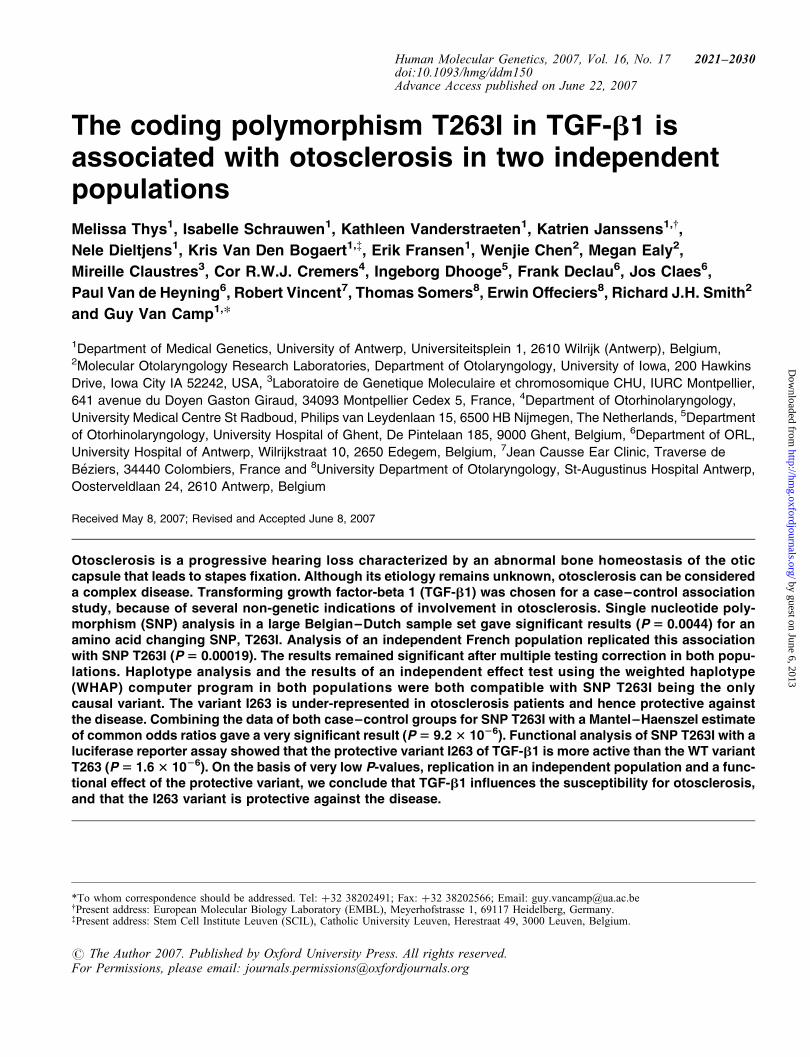
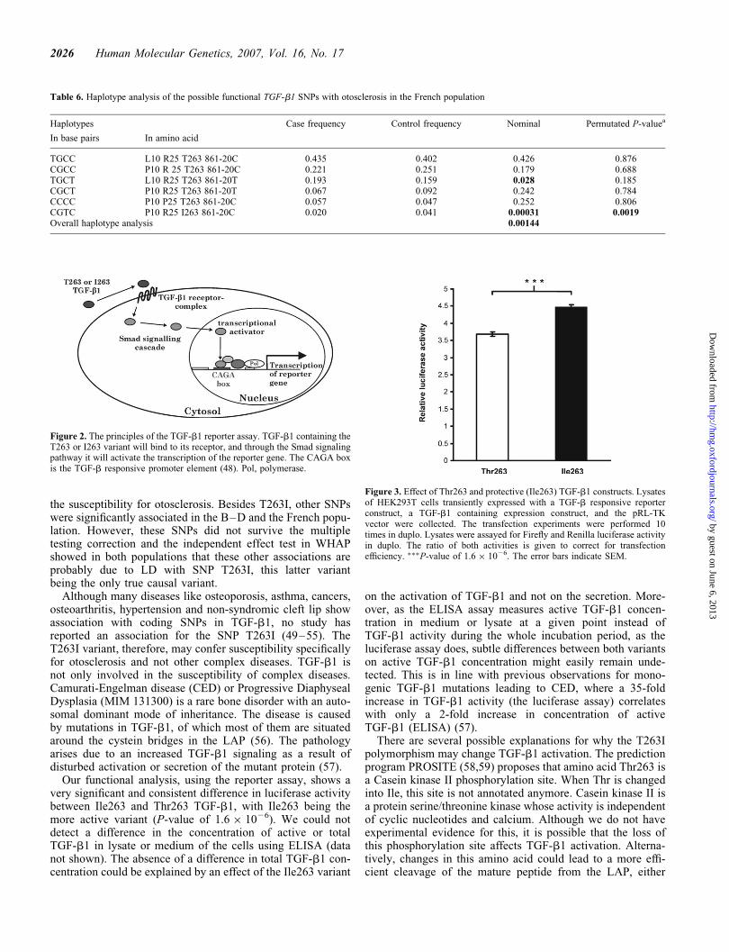
Figure 1. Schematic representation of the TGF-b1 gene (A) and thepre-pro-TGF-b1 (B). (A) The boxes represent the seven numbered exonsand the 50- and 30-untranslated region (UTR) are marked in gray. AnalyzedSNPs are marked with an arrow; the first line gives the SNP number, thesecond line the SNP position and the base substitution starting from thestart codon, and the third line is the SNP name (rs-number) from NCBI. (B)The numbers above indicate the amino acid position of the cleavage sites ofthe pre-pro-TGF-b1. The amino acid changing SNPs are shown: the firstline is the rs-number, the second line indicates the base substitution and thethird line the amino acid substitution both starting from the start codon.
Human Molecular Genetics, 2007, Vol. 16, No. 17 2023
by guest on June 6, 2013http://hm
g.oxfordjournals.org/D
ownloaded from
into a Mantel–Haenszel estimate of common allelic OR, anda Cochran–Mantel–Haenszel test for conditional indepen-dence (44,45). This revealed a very robust and consistentassociation with a highly significant P-value of 9.2 � 1026
and an OR of 0.377 (0.243–0.583).
Assessment of population stratification. To assess populationstratification that could lead to spurious associations, theLCT (lactase gene) SNP-13910C! T, which has a wide vari-ation in allele frequency among closely related populations(46) was analyzed (Supplementary Material, Table S1).According to Campbell et al. (47), genotyping this markercan be very informative in detecting subtle, but importantlevels of population stratification in apparently homogeneousstudy samples. We found no differences for this SNP in thetotal B–D population or between sub-populations, separatedby origin (Antwerp, Ghent and Nijmegen). Analysis of theLCT SNP in our French sample set also did not show a signi-ficant result. Therefore, it is unlikely that the results weobtained are an artifact caused by population stratification.
Functional analyses
The very low P-values for association and the replication in anindependent population, strongly suggest that TGF-b1 plays a
role in the otosclerosis susceptibility in the Western-Europeanpopulation. As the most significant and replicated SNP, T263Ichanges an amino acid in the LAP of TGF-b1, and the inde-pendent effect test suggests that this SNP T263I may becausal, we hypothesized that T263I has a direct causativeeffect. Our strategy to confirm the causality of the associatedSNP T263I was to investigate this variant Ile263 functionally.Using a luciferase reporter assay we sought to compare theactivity of the two variants, respectively, Thr and Ile atposition 263.
Reporter assay. A pcDNA3 vector containing the TGF-b1gene product with either the Thr263 variant or the Ile263variant was co-transfected in HEK293T cells with a pRL-Tkcalibration vector and a TGF-b-responsive reporter vector.The TGF-b1 responsive reporter vector contains a constructthat consists of 12 CAGA-elements, a minimal promoter andthe Firefly luciferase gene (48). Only active TGF-b1, andnot latent TGF-b1 that is linked to the LAP, will lead to thetranscription of Firefly luciferase by way of auto- or paracrineactivation of the Smad signaling pathway (Fig. 2). Firefly andRenilla luciferase activity were measured in the lysates of thetransfected cells (Thr263 and Ile263 variant). Two-way Anovaanalysis, controlling for differences between the differenttransfection experiments, was carried out. Despite differences,
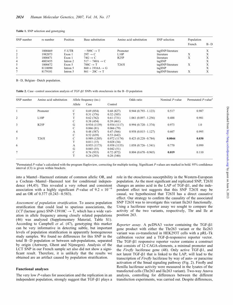
Table 2. Case–control association analysis of TGF-b1 SNPs with otosclerosis in the B–D population
SNP number Amino acid substitution Allele frequency (no.) Odds ratio Nominal P-value Permutated P-valuea
Allele Case Control
1 Promoter C 0.69 (854) 0.68 (827) 0.944 (0.793–1.123) 0.517 0.987T 0.31 (376) 0.32 (385)
2 L10P T 0.62 (762) 0.61 (731) 1.061 (0.897–1.256) 0.488 0.981C 0.38 (454) 0.39 (461)
3 R25P G 0.934 (1139) 0.934 (1117) 0.994 (0.720–1.374) 0.973 1.0C 0.066 (81) 0.066 (79)
4 A 0.48 (587) 0.47 (566) 0.958 (0.815–1.127) 0.607 0.995C 0.52 (639) 0.53 (642)
5 T263I C 0.989 (1205) 0.972 (1174) 0.423 (0.228–0.784) 0.0044 0.030T 0.011 (15) 0.028 (34)
6 A 0.955 (1173) 0.958 (1155) 1.058 (0.726–1.541) 0.770 0.999G 0.045 (55) 0.042 (51)
7 C 0.76 (933) 0.72 (872) 0.804 (0.670–0.965) 0.019 0.110T 0.24 (293) 0.28 (340)
aPermutated P-value’s calculated with the program Haploview, correcting for multiple testing. Significant P-values are marked in bold. 95% confidenceinterval (CI) is given within brackets.
Table 1. SNP selection and genotyping
SNP number rs number Position Base substitution Amino acid substitution SNP selection Population
French B–D
1 1800469 50-UTR 2509C! T Promoter tagSNP/literature X2 1982073 Exon 1 29T! C L10P literature X X3 1800471 Exon 1 74G! C R25P literature X X4 4803455 Intron 2 517 2 740A! C tagSNP X5 1800472 Exon 5 788C! T T263I tagSNP/literature X X6 8110090 Intron 5 860þ 1916A! G tagSNP X7 8179181 Intron 5 861 2 20C! T tagSNP/literature X X
B–D, Belgian–Dutch population.
2024 Human Molecular Genetics, 2007, Vol. 16, No. 17
by guest on June 6, 2013http://hm
g.oxfordjournals.org/D
ownloaded from
the activity of the Ile263 variant was consistently higher thanthe Thr263 variant in each individual transfection experiment.
The analysis showed a significant difference between theThr263 variant and the Ile263 variant with a P-value of1.6 � 1026 (Fig. 3). The average relative luciferase activityof the Ile263 variant was 0.78 units or 21.2% higher whencompared with the average luciferase activity of the Thr263variant after correcting for the random variation between indi-vidual transfection experiments using a two-way Anova. Thisresult supports a protective nature for the Ile263 variant.
DISCUSSION
We have completed a case–control association study ofTGF-b1 variants with otosclerosis in two independent popu-lations. TGF-b1 is a multi-functional growth factor thatregulates a broad range of biological processes. Related to
otosclerosis, its most important function is its role in the chon-drogenesis of the otic capsule. Moreover, the expression ofTGF-b1 in adult human otosclerotic stapedial bone suggestsa role in the bone turnover of the otic capsule (36). Therefore,we and other researchers believe that TGF-b1 is a goodcandidate for involvement in the pathogenesis of otosclerosis(33–40).
Consistent with this hypothesis, our SNP analysis in theB–D population and the French population revealed that therare genotype (allele coding for Ile) of the associated SNPT263I is more frequent in the control population. This pointsto a protective role of I263 in the development of otosclerosis.The results of the association testing being replicated inan independent population, and in addition surviving twodifferent multiple testing corrections (Bonferroni and permu-tation test), strongly suggest that the association of otosclero-sis with TGF-b1 is real and that TGF-b1 has an influence on
Table 4. Independent effect test in the Belgian–Dutch and the French population
SNP number Base substitution Amino acid substitution P-value
B–D population French population
2 29T! C L10P 0.840 0.0923 74G! C R25P 0.689 0.1715 788C! T T263I 0.011 0.00267 861-20C! T 0.082 0.406
Significant P-values are marked in bold, and P-values between 0.1 and 0.05 are in italics.
Table 5. Case–control association analysis of TGF-b1 SNPs with otosclerosis in the French population
SNP number Amino-acid substitution Allele frequency (no.) Odds ratio Nominal P-value Permutated P-valuea
Allele Case Control
2 L10P T 0.63 (561) 0.56 (557) 1.260 (1.051–1.510) 0.012 0.052C 0.37 (347) 0.44 (437)
3 R25P G 0.938 (842) 0.947 (941) 0.818 (0.558–1.198) 0.301 0.740C 0.062 (58) 0.053 (53)
5 T263I C 0.985 (900) 0.957 (951) 0.334 (0.180–0.620) 0.00019 0.0014T 0.015 (14) 0.043 (43)
7 C 0.73 (659) 0.74 (736) 1.101 (0.897–1.351) 0.359 0.859T 0.27 (253) 0.26 (258)
aPermutated P-value calculated with the Haploview program, correcting for multiple testing. Significant P-values are marked in bold, and P-valuesbetween 0.1 and 0.05 are in italics. 95% CI is given within brackets.
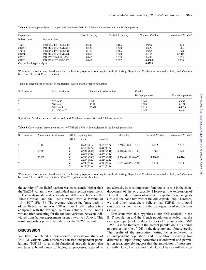
Table 3. Haplotype analysis of the possible functional TGF-b1 SNPs with otosclerosis in the B–D population
Haplotypes Case frequency Control frequency Nominal P-value Permutated P-valuea
In base pairs In amino acid
TGCC L10 R25 T263 861-20C 0.447 0.406 0.051 0.149CGCC P10 R25 T263 861-20C 0.237 0.228 0.629 0.986TGCT L10 R25 T263 861-20T 0.180 0.206 0.089 0.369CGCT P10 R25 T263 861-20T 0.057 0.068 0.156 0.7412CCCC P10 P25 T263 861-20C 0.065 0.059 0.490 0.976CGTC P10 R25 I263 861-20C 0.012 0.027 0.0085 0.036Overall haplotype analysis 0.0296
aPermutated P-value calculated with the Haploview program, correcting for multiple testing. Significant P-values are marked in bold, and P-valuesbetween 0.1 and 0.05 are in italics.
Human Molecular Genetics, 2007, Vol. 16, No. 17 2025
by guest on June 6, 2013http://hm
g.oxfordjournals.org/D
ownloaded from
the susceptibility for otosclerosis. Besides T263I, other SNPswere significantly associated in the B–D and the French popu-lation. However, these SNPs did not survive the multipletesting correction and the independent effect test in WHAPshowed in both populations that these other associations areprobably due to LD with SNP T263I, this latter variantbeing the only true causal variant.
Although many diseases like osteoporosis, asthma, cancers,osteoarthritis, hypertension and non-syndromic cleft lip showassociation with coding SNPs in TGF-b1, no study hasreported an association for the SNP T263I (49–55). TheT263I variant, therefore, may confer susceptibility specificallyfor otosclerosis and not other complex diseases. TGF-b1 isnot only involved in the susceptibility of complex diseases.Camurati-Engelman disease (CED) or Progressive DiaphysealDysplasia (MIM 131300) is a rare bone disorder with an auto-somal dominant mode of inheritance. The disease is causedby mutations in TGF-b1, of which most of them are situatedaround the cystein bridges in the LAP (56). The pathologyarises due to an increased TGF-b1 signaling as a result ofdisturbed activation or secretion of the mutant protein (57).
Our functional analysis, using the reporter assay, shows avery significant and consistent difference in luciferase activitybetween Ile263 and Thr263 TGF-b1, with Ile263 being themore active variant (P-value of 1.6 � 1026). We could notdetect a difference in the concentration of active or totalTGF-b1 in lysate or medium of the cells using ELISA (datanot shown). The absence of a difference in total TGF-b1 con-centration could be explained by an effect of the Ile263 variant
on the activation of TGF-b1 and not on the secretion. More-over, as the ELISA assay measures active TGF-b1 concen-tration in medium or lysate at a given point instead ofTGF-b1 activity during the whole incubation period, as theluciferase assay does, subtle differences between both variantson active TGF-b1 concentration might easily remain unde-tected. This is in line with previous observations for mono-genic TGF-b1 mutations leading to CED, where a 35-foldincrease in TGF-b1 activity (the luciferase assay) correlateswith only a 2-fold increase in concentration of activeTGF-b1 (ELISA) (57).
There are several possible explanations for why the T263Ipolymorphism may change TGF-b1 activation. The predictionprogram PROSITE (58,59) proposes that amino acid Thr263 isa Casein kinase II phosphorylation site. When Thr is changedinto Ile, this site is not annotated anymore. Casein kinase II isa protein serine/threonine kinase whose activity is independentof cyclic nucleotides and calcium. Although we do not haveexperimental evidence for this, it is possible that the loss ofthis phosphorylation site affects TGF-b1 activation. Alterna-tively, changes in this amino acid could lead to a more effi-cient cleavage of the mature peptide from the LAP, either
Table 6. Haplotype analysis of the possible functional TGF-b1 SNPs with otosclerosis in the French population
Haplotypes Case frequency Control frequency Nominal Permutated P-valuea
In base pairs In amino acid
TGCC L10 R25 T263 861-20C 0.435 0.402 0.426 0.876CGCC P10 R 25 T263 861-20C 0.221 0.251 0.179 0.688TGCT L10 R25 T263 861-20T 0.193 0.159 0.028 0.185CGCT P10 R25 T263 861-20T 0.067 0.092 0.242 0.784CCCC P10 P25 T263 861-20C 0.057 0.047 0.252 0.806CGTC P10 R25 I263 861-20C 0.020 0.041 0.00031 0.0019Overall haplotype analysis 0.00144
Figure 2. The principles of the TGF-b1 reporter assay. TGF-b1 containing theT263 or I263 variant will bind to its receptor, and through the Smad signalingpathway it will activate the transcription of the reporter gene. The CAGA boxis the TGF-b responsive promoter element (48). Pol, polymerase.
Figure 3. Effect of Thr263 and protective (Ile263) TGF-b1 constructs. Lysatesof HEK293T cells transiently expressed with a TGF-b responsive reporterconstruct, a TGF-b1 containing expression construct, and the pRL-TKvector were collected. The transfection experiments were performed 10times in duplo. Lysates were assayed for Firefly and Renilla luciferase activityin duplo. The ratio of both activities is given to correct for transfectionefficiency. ���P-value of 1.6 � 1026. The error bars indicate SEM.
2026 Human Molecular Genetics, 2007, Vol. 16, No. 17
by guest on June 6, 2013http://hm
g.oxfordjournals.org/D
ownloaded from
through a direct effect, this site is only 15 amino acids awayfrom the cleavage site, or an indirect effect (through a confor-mation change of the LAP).
The reason why a more active form of TGF-b1 could beprotective against otosclerosis is at present unclear, but wepropose the following hypothesis: Active TGF-b1 inhibitsosteoclast differentiation and activation through the osteo-blasts and stimulates osteoblast chemotaxis, proliferation anddifferentiation (60–63). The more active protective variant(Ile263) results in the production of more active TGF-b1;this is expected to inhibit osteoclast differentiation and acti-vation. Inhibiting osteoclast activity may be important in pre-venting the first otospongiotic phase of otosclerosis (64)during which bone is resorbed by special osteoclasts thatpresent CD51/61 antigens on their cell surface (65). Conse-quently, this would be a means to keep bone remodeling ofthe otic capsule under control.
In summary, in this study we have shown that TGF-b1influences otosclerosis susceptibility in a B–D and a Frenchpopulation and that the SNP T263I is most likely the causalvariant. Collection of additional patient sets is needed to deter-mine whether this association is true for other otosclerosispopulations. As TGF-b1 is involved in numerous pathways,it will be difficult to completely understand the effect ofTGF-b1 on the development of otosclerosis only by studyingTGF-b1, and additional association studies analyzing othercandidate genes involved in the same pathway as TGF-b1are required. Alternatively, whole genome associationstudies using high-density micro arrays would be veryhelpful in identifying pathways that can explain not only theeffect of TGF-b1, but also the development and pathogenesisof otosclerosis in a more general way. With that knowledge,the development of prevention strategies or therapies may bepossible. While we recognize that stapes surgery is very effec-tive and in most cases restores hearing, treatment based onbiological insights may have an important impact as acheaper, safer and more easily offered option to prevent thedevelopment of hearing loss. When provided as post-surgicaltherapy, medical treatment may prevent disease recurrenceand the need for a revision surgery or even prevent pro-gression to inoperable sensorineural hearing loss, whichultimately affects approximately 10% of persons with oto-sclerosis (66,67).
MATERIALS AND METHODS
Clinical diagnosis
The selection of the otosclerosis patients was performed byaudiologists who are expert in otosclerosis at participatingcenters in Antwerp, Ghent, Nijmegen and Colombiers. Weonly included patients for which otosclerosis was surgicallyconfirmed or for which stapes surgery was recommended bythe participating center.
Pure-tone audiometry was performed on all patients with airconduction at 125, 250, 500, 1000, 2000, 4000 and 8000 Hz,and bone conduction at 250, 500, 1000, 2000 and 4000 Hz.Otoscopy and tympanoscopy were performed to rule outouter or middle ear pathology, respectively, and stapedialreflex decay was measured to assess the mobility of the stapes.
Belgian–Dutch case–control study group
A total of 632 unrelated otosclerosis patients of Belgian orDutch origin were ascertained through the Department ofOtolaryngology of four different hospitals: University Hospitalof Antwerp, St Augustinus Hospital in Antwerp, UniversityHospital of Ghent for the Belgian patients and the UniversityMedical Center St Radboud in Nijmegen for the Dutchpatients.
From a DNA repository at the Department of MedicalGenetics in Antwerp, 632 controls were selected. Everypatient was matched one to one with a control sample forethnicity, age (within +1 year) and sex.
French case–control study group
In total, 457 French otosclerosis patients were collectedthrough the Jean Causse Ear Clinic in Colombiers, France.The Laboratoire de Genetique Moleculaire et chromosomiqueCHU, IURC Montpellier in France provided 497 controlsamples. Patients and controls were matched for ethnicity.
Single nucleotide polymorphism selection
SNPs were selected in TGF-b1 based on their presence in anumber of association studies (49,50), and their possible func-tional effect (Table 1). To fully cover the gene, additionalSNPs were selected based on the HAPMAP phase II (release20). TagSNPs were selected using the aggressive taggingmethod implemented in the program Tagger (41) with theHaploview software (68) (minor allele frequency .0.05 andr2 . 0.8).
Genotyping
Blood samples from study participants were obtained afterinformed consent. Genomic DNA was isolated by standardtechniques.
Assay designs and SNP analyses were performed byK-Biosciences (Cambridge, UK; www.kbioscience.co.uk)using their system of competitive allele-specific PCR system(KASPar). Hidden duplicates were added as a qualitycontrol of the analysis.
Association analysis
We tested all SNPs for deviation from the HWE in the controlsusing Transposer V1-0 software (69). None of the SNPsshowed a significant deviation from HWE in any of the twocontrol populations. A cut-off P-value of 0.001 was taken.
Association between our markers and the disease status wastested using stepwise backward logistic regression, allowingfor sex effects and sex–genotype interaction. Effect modifi-cation or sex–SNP interaction was tested in a full factoriallogistic regression model including the main effects of theSNP genotype, sex and the interaction term between them.The significance of the interaction term indicates whetherthe effect of the SNP is different between males andfemales. When this was significant, we tested for associationbetween the SNP and affection status in males and females
Human Molecular Genetics, 2007, Vol. 16, No. 17 2027
by guest on June 6, 2013http://hm
g.oxfordjournals.org/D
ownloaded from
separately. Otherwise, we tested for significance of the SNPin a univariate logistic regression, using the LRT (70) whichis asymptotically equivalent to the Cochran–Armitage trendtest (ATT). Allelic frequencies and allele-specific ORs werecalculated assuming an additive model.
The common OR was estimated using the Mantel–Haenszelestimator. Conditional independence was tested using theCochran–Mantel–Haenszel test. Homogeneity of the ORwas tested by the Breslow-Day test. All analyses were con-ducted with the statistical programs R 2.3.1 and SPSS 12.0.1for Windows.
Correcting for multiple testing was performed with both theBonferroni correction and the permutations test. For the permu-tation test, genotypes or haplotypes were randomly permutedfor 106 iterations to generate the distribution of P-valuesunder the null hypothesis, given the LD pattern of the SNPs.This corrects for multiple hypothesis tests on SNPs in LD.This test is implemented in the Haploview program (68).
For the haplotype analysis the WHAP program (43) wasused. WHAP directly calculates likelihood estimates, likeli-hood ratios and P-values taking into account the loss of infor-mation due to haplotype phase uncertainty and missinggenotypes. Association between the inferred haplotypes andotosclerosis was tested using an omnibus test.
To test which of the associated SNPs was causal and whichwere significantly associated by LD, the independent effecttest was used. Hereby, the significance of each SNP istested, fixing the genotype of the surrounding SNPs.
Cell culture condition
HEK293T, human embryonic kidney cells, were grown inDulbecco’s modified Eagle’s medium with 4500 mg/lglucose, supplemented with fetal calf serum (FCS) (10% v/v),penicillin (100 units/ml), streptomycin (100 mg/ml) andL-glutamine (2 mM). Medium and supplements were fromInvitrogen (Paisley, UK).
Construction of expression vectors and transfection
A pcDNA3 plasmid (Invitrogen) containing the full-lengthTGF-b1 (Thr263) (57) was used. The variant Ile263 was intro-duced in this TGF-b1 construct by site-directed mutagenesis(Quickchange site-directed mutagenesis kit, Stratagene, LaJolla, USA). The complete sequence of the TGF-b1 insertwas validated by sequencing analysis for both the Thr263and the Ile263 TGF-b1 producing constructs. For transientexpression of the respective gene products, the constructswere transfected into HEK293T cells, using the transfectionreagent Fugene6 (Roche Diagnostics Belgium, Vilvoorde,Belgium). Each transfection was performed in duplicate andrepeated 10 times. HEK293T cells were transfected at60–80% confluency with 9 ml of Fugene 6, 10 ng ofpRL-TK (Promega, Madison, USA), 1.5 mg of the pcDNA3construct and 1.5 mg of the TGF-b responsive reporterconstruct (48) in a total volume of 2 ml for subsequent useof media and/or lysates in reporter assays and ELISAs. After24 h of incubation, the medium was replaced with 2 ml ofserum-free Opti-MEM I (Invitrogen) to avoid the influenceof TGF-b1 contained in the FCS on our measurements.
After another 24 h incubation of the transfected cells, thesupernatants were collected. The cells were lysed with500 ml of passive lysis buffer (Promega, Madison, USA).Media and lysates were aliquoted and stored at 2808C.
Reporter assay
A TGF-b responsive reporter construct was provided byDr Dennler (48). Cotransfection of the pRL-TK vector, con-taining the herpes simplex virus-thymidine kinase (HSV-TK)promoter region upstream of the Renilla luciferase gene,allowed calibration of the Firefly luciferase activity againstthe Renilla luciferase activity, providing normalization oftransfection efficiency. Cell lysates were assayed for Fireflyand Renilla luciferase activity using the dual-luciferase repor-ter assay system (Promega), following the manufacturer’sinstructions. Briefly, 20 ml of the lysate was transferred to a1.5 ml microfuge tube. Fifty microlitres of luciferase assayreagent II was added to the lysate, and Firefly luciferaseactivity was measured for a period of 10 s. After adding50 ml of Stop&Glo reagent, Renilla luciferase activity wasmeasured, again for a period of 10 s. This process was auto-matically done by the luminometer GloMax 20/20n(Promega). The ratios of both activities were determined.
Ten transfection experiments of both Thr263 and Ile263were performed in duplo. The significance of the Ile263effect was tested with a two-way Anova.
During every transfection experiment, a positive controlexperiment was performed to validate the assay. The controlvectors were TGF-b1 producing pcDNA3 vectors containinga Camurati-Engelman mutation (either R218C or LLL12-13insertion). The reporter assay results obtained with thesevectors were consistent with the published results (data notshown) (57).
SUPPLEMENTARY MATERIAL
Supplementary Material is available at HMG Online.
ACKNOWLEDGEMENTS
We thank all the otosclerosis patients and the participantswhose contribution made this work possible. This work wassupported by the European Commission (FP6 Integratedproject EuroHear LSHG-CT-20054-512063), the NIH (grantR01DC05218), the FWO (grant G.0138.07) and the Universityof Antwerp (TOP grant). M.T. holds a pre-doctoral researchposition with the Institute for the Promotion of Innovationthrough Science and Technology in Flanders (IWT-Vlaanderen). I.S. holds a predoctoral research position andK.J., K.V.D.B. holds a post-doctoral position with the ‘Fondsvoor Wetenschappelijk Onderzoek Vlaanderen’ (FWO).
Conflict of Interest statement. None declared.
REFERENCES
1. Altmann, F., Glasgold, A. and Macduff, J.P. (1967) The incidence ofotosclerosis as related to race and sex. Ann. Otol. Rhinol. Laryngol., 76,377–392.
2028 Human Molecular Genetics, 2007, Vol. 16, No. 17
by guest on June 6, 2013http://hm
g.oxfordjournals.org/D
ownloaded from
2. Schuknecht, H.F. and Barber, W. (1985) Histologic variants inotosclerosis. Laryngoscope, 95, 1307–1317.
3. Declau, F., Van Spaendonck, M., Timmermans, J.P., Michaels, L., Liang,J., Qiu, J.P. and Van de Heyning, P. (2001) Prevalence of otosclerosis inan unselected series of temporal bones. Otol. Neurotol., 22, 596–602.
4. Frisch, T., Sorensen, M.S., Overgaard, S. and Bretlau, P. (2000)Estimation of volume referent bone turnover in the otic capsule aftersequential point labeling. Ann. Otol. Rhinol. Laryngol., 109, 33–39.
5. Frisch, T., Sorensen, M.S., Overgaard, S. and Bretlau, P. (2000)Predilection of otosclerotic foci related to the bone turnover in the oticcapsule. Acta Otolaryngol. Suppl., 543, 111–113.
6. Zehnder, A.F., Kristiansen, A.G., Adams, J.C., Kujawa, S.G., Merchant,S.N. and McKenna, M.J. (2006) Osteoprotegerin knockout micedemonstrate abnormal remodeling of the otic capsule and progressivehearing loss. Laryngoscope, 116, 201–206.
7. Zehnder, A.F., Kristiansen, A.G., Adams, J.C., Merchant, S.N. andMcKenna, M.J. (2005) Osteoprotegerin in the inner ear may inhibit boneremodeling in the otic capsule. Laryngoscope, 115, 172–177.
8. Karosi, T., Jokay, I., Konya, J., Szabo, L.Z., Pytel, J., Jori, J., Szalmas,A. and Sziklai, I. (2006) Detection of osteoprotegerin and TNF-alphamRNA in ankylotic Stapes footplates in connection with measles viruspositivity. Laryngoscope, 116, 1427–1433.
9. Tomek, M.S., Brown, M.R., Mani, S.R., Ramesh, A., Srisailapathy, C.R.,Coucke, P., Zbar, R.I., Bell, A.M., McGuirt, W.T., Fukushima, K.et al. (1998) Localization of a gene for otosclerosis to chromosome15q25-q26. Hum. Mol. Genet., 7, 285–290.
10. Van Den Bogaert, K., Govaerts, P.J., Schatteman, I., Brown, M.R.,Caethoven, G., Offeciers, F.E., Somers, T., Declau, F., Coucke, P., Van deHeyning, P. et al. (2001) A second gene for otosclerosis, OTSC2, mapsto chromosome 7q34-36. Am. J. Hum. Genet., 68, 495–500.
11. Chen, W., Campbell, C.A., Green, G.E., Van Den Bogaert, K.,Komodikis, C., Manolidis, L.S., Aconomou, E., Kyamides, Y.,Christodoulou, K., Faghel, C. et al. (2002) Linkage of otosclerosis to athird locus (OTSC3) on human chromosome 6p21.3-22.3. J. Med. Genet.,39, 473–477.
12. Brownstein, Z., Goldfarb, A., Levi, H., Frydman, M. and Avraham, K.B.(2006) Chromosomal mapping and phenotypic characterization ofhereditary otosclerosis linked to the OTSC4 locus. Arch. Otolaryngol.
Head Neck Surg., 132, 416–424.13. Van Den Bogaert, K., De Leenheer, E.M., Chen, W., Lee, Y., Nurnberg,
P., Pennings, R.J., Vanderstraeten, K., Thys, M., Cremers, C.W.,Smith, R.J. et al. (2004) A fifth locus for otosclerosis, OTSC5, maps tochromosome 3q22-24. J. Med. Genet., 41, 450–453.
14. Thys, M., Van Den Bogaert, K., Iliadou, V., Vanderstraeten, K., Dieltjens,N., Schrauwen, I., Chen, W., Eleftheriades, N., Grigoriadou, M., Pauw,R.J. et al. (2007) A seventh locus for otosclerosis, OTSC7, maps tochromosome 6q13-16.1. Eur. J. Hum. Genet., 15, 362–368.
15. McKenna, M.J., Kristiansen, A.G., Bartley, M.L., Rogus, J.J. and Haines,J.L. (1998) Association of COL1A1 and otosclerosis: evidence for ashared genetic etiology with mild osteogenesis imperfecta. Am. J. Otol.,19, 604–610.
16. McKenna, M.J., Nguyen-Huynh, A.T. and Kristiansen, A.G. (2004)Association of otosclerosis with Sp1 binding site polymorphism inCOL1A1 gene: evidence for a shared genetic etiology with osteoporosis.Otol. Neurotol., 25, 447–450.
17. Rodriguez, L., Rodriguez, S., Hermida, J., Frade, C., Sande, E., Visedo,G., Martin, C. and Zapata, C. (2004) Proposed association between theCOL1A1 and COL1A2 genes and otosclerosis is not supported by acase–control study in Spain. Am. J. Med. Genet., 128A, 19–22.
18. Chen, W., Meyer, N.C., McKenna, M.J., Pfister, M., McBride, D.J., Jr,Fukushima, K., Thys, M., Camp, G.V. and Smith, R.J. (2007)Single-nucleotide polymorphisms in the COL1A1 regulatory regions areassociated with otosclerosis. Clin. Genet., 71, 406–414.
19. Menger, D.J. and Tange, R.A. (2003) The aetiology of otosclerosis: areview of the literature. Clin. Otolaryngol., 28, 112–120.
20. Lippy, W.H., Berenholz, L.P., Schuring, A.G. and Burkey, J.M.(2005) Does pregnancy affect otosclerosis? Laryngoscope, 115,1833–1836.
21. Grayeli, A.B., Palmer, P., Tran Ba Huy, P., Soudant, J., Sterkers, O.,Lebon, P. and Ferrary, E. (2000) No evidence of measles virus in stapessamples from patients with otosclerosis. J. Clin. Microbiol., 38,2655–2660.
22. Karosi, T., Konya, J., Szabo, L.Z., Pytel, J., Jori, J., Szalmas, A. andSziklai, I. (2005) Codetection of measles virus and tumor necrosisfactor-alpha mRNA in otosclerotic stapes footplates. Laryngoscope, 115,1291–1297.
23. Niedermeyer, H.P., Arnold, W., Neubert, W.J. and Sedlmeier, R. (2000)Persistent measles virus infection as a possible cause of otosclerosis: stateof the art. Ear Nose Throat J., 79, 552–554.
24. Causse, J.R., Causse, J.B., Bretlau, P., Uriel, J., Berges, J., Chevance,L.G., Shambaugh, G.E. and Bastide, J.M. (1989) Etiology ofotospongiotic sensorineural losses. Am. J. Otol., 10, 99–107.
25. Chevance, L.G., Causse, J., Berges, J., Manach, Y. and Adrian, M. (1976)A biochemical and cytological explanation of cochlear otospongiosis.Ann. Otolaryngol. Chir. Cervicofac., 93, 275–287.
26. Janssens, K., ten Dijke, P., Janssens, S. and Van Hul, W. (2005)Transforming growth factor-beta1 to the bone. Endocr. Rev., 26,743–774.
27. Gentry, L.E., Lioubin, M.N., Purchio, A.F. and Marquardt, H. (1988)Molecular events in the processing of recombinant type-1pre-pro-transforming growth factor-beta to the mature polypeptide. Mol.
Cell. Biol., 8, 4162–4168.28. Miyazono, K., Hellman, U., Wernstedt, C. and Heldin, C.H. (1988) Latent
high molecular-weight complex of transforming growth factor-beta-1:purification from human-platelets and structural characterization. J. Biol.
Chem., 263, 6407–6415.29. Wakefield, L.M., Smith, D.M., Flanders, K.C. and Sporn, M.B. (1988)
Latent transforming growth factor-beta from human-platelets: a highmolecular-weight complex containing precursor sequences. J. Biol.
Chem., 263, 7646–7654.
30. Wrana, J.L., Attisano, L., Wieser, R., Ventura, F. and Massague, J. (1994)Mechanism of activation of the TGF-beta receptor. Nature, 370, 341–347.
31. Derynck, R. and Zhang, Y.E. (2003) Smad-dependent andSmad-independent pathways in TGF-beta family signalling. Nature, 425,577–584.
32. Oreffo, R.O.C., Mundy, G.R., Seyedin, S.M. and Bonewald, L.F. (1989)Activation of the bone-derived latent Tgf beta-complex by isolatedosteoclasts. Biochem. Biophys. Res. Commun., 158, 817–823.
33. Quinn, J.M.W., Itoh, K., Udagawa, N., Hausler, K., Yasuda, H., Shima,N., Mizuno, A., Higashio, K., Takahashi, N., Suda, T. et al. (2001)Transforming growth factor beta affects osteoclast differentiation viadirect and indirect actions. J. Bone Miner. Res., 16, 1787–1794.
34. Frenz, D.A. (2001) Growth factor control of otic capsule chondrogenesis.Einstein Quart. J. Biol. Med., 18, 7–14.
35. Frenz, D.A., Galinovic-Schwartz, V., Liu, W., Flanders, K.C. and Van deWater, T.R. (1992) Transforming growth factor beta 1 is anepithelial-derived signal peptide that influences otic capsule formation.Dev. Biol., 153, 324–336.
36. Frenz, D.A., Van de Water, T.R. and Galinovic-Schwartz, V. (1991)Transforming growth factor beta: does it direct otic capsule formation?Ann. Otol. Rhinol. Laryngol., 100, 301–307.
37. Bodo, M., Venti, G., Baroni, T., Bellucci, C., Giammarioli, M., Donti, E.,Paludetti, G., Stabellini, G. and Carinci, P. (1995) Phenotype ofin vitro human otosclerotic cells and its modulation by TGF beta.Cell. Mol. Biol. (Noisy-le-grand), 41, 1039–1049.
38. Kanaan, R.A. and Kanaan, L.A. (2006) Transforming growth factor beta1,bone connection. Med. Sci. Monit., 12, 164–169.
39. Butts, S.C., Liu, W., Li, G. and Frenz, D.A. (2005) Transforming growthfactor-beta1 signaling participates in the physiological and pathologicalregulation of mouse inner ear development by all-trans retinoic acid.Birth Defects Res. A Clin. Mol. Teratol., 73, 218–228.
40. Frenz, D.A., Liu, W., Williams, J.D., Hatcher, V., Galinovic-Schwartz,V., Flanders, K.C. and Van de Water, T.R. (1994) Induction ofchondrogenesis: requirement for synergistic interaction of basic fibroblastgrowth factor and transforming growth factor-beta. Development, 120,415–424.
41. Frenz, D.A. and Liu, W. (2000) Treatment with all-trans-retinoic aciddecreases levels of endogenous TGF-beta(1) in the mesenchyme of thedeveloping mouse inner ear. Teratology, 61, 297–304.
42. de Bakker, P.I., Yelensky, R., Pe’er, I., Gabriel, S.B., Daly, M.J. andAltshuler, D. (2005) Efficiency and power in genetic association studies.Nat. Genet., 37, 1217–1223.
43. Guild, S. (1944) Histologic otosclerosis. Ann. Otol. Rhinol. Laryngol., 53,246–267.
Human Molecular Genetics, 2007, Vol. 16, No. 17 2029
by guest on June 6, 2013http://hm
g.oxfordjournals.org/D
ownloaded from
44. Purcell, S., Daly, M.J. and Sham, P.C. (2007) WHAP: haplotype-basedassociation analysis. Bioinformatics, 23, 255–256.
45. Lohmueller, K.E., Pearce, C.L., Pike, M., Lander, E.S. and Hirschhorn,J.N. (2003) Meta-analysis of genetic association studies supports acontribution of common variants to susceptibility to common disease. Nat.Genet., 33, 177–182.
46. Woolson, R.F. and Bean, J.A. (1982) Mantel–Haenszel statistics anddirect standardization. Stat. Med., 1, 37–39.
47. Bersaglieri, T., Sabeti, P.C., Patterson, N., Vanderploeg, T., Schaffner,S.F., Drake, J.A., Rhodes, M., Reich, D.E. and Hirschhorn, J.N. (2004)Genetic signatures of strong recent positive selection at the lactase gene.Am. J. Hum. Genet., 74, 1111–1120.
48. Campbell, C.D., Ogburn, E.L., Lunetta, K.L., Lyon, H.N., Freedman,M.L., Groop, L.C., Altshuler, D., Ardlie, K.G. and Hirschhorn, J.N. (2005)Demonstrating stratification in a European American population. Nat.Genet., 37, 868–872.
49. Dennler, S., Itoh, S., Vivien, D., ten Dijke, P., Huet, S. and Gauthier, J.M.(1998) Direct binding of Smad3 and Smad4 to critical TGF beta-inducibleelements in the promoter of human plasminogen activator inhibitor-type 1gene. EMBO J., 17, 3091–3100.
50. Langdahl, B.L., Carstens, M., Stenkjaer, L. and Eriksen, E.F. (2003)Polymorphisms in the transforming growth factor beta 1 gene andosteoporosis. Bone, 32, 297–310.
51. Tzakas, P., Wong, B.Y., Logan, A.G., Rubin, L.A. and Cole, D.E. (2005)Transforming growth factor beta-1 (TGFB1) and peak bone mass:association between intragenic polymorphisms and quantitativeultrasound of the heel. BMC Musculoskelet. Disord., 6, 29.
52. Stoll, C., Mengsteab, S., Stoll, D., Riediger, D., Gressner, A.M. andWeiskirchen, R. (2004) Analysis of polymorphic TGFB1 codons 10, 25and 263 in a German patient group with non-syndromic cleft lip, alveolus,and palate compared with healthy adults. BMC Med. Genet., 5, 15.
53. Silverman, E.S., Palmer, L.J., Subramaniam, V., Hallock, A., Mathew, S.,Vallone, J., Faffe, D.S., Shikanai, T., Raby, B.A., Weiss, S.T. et al. (2004)Transforming growth factor-beta1 promoter polymorphism C-509T isassociated with asthma. Am. J. Respir. Crit. Care Med., 169, 214–219.
54. Dunning, A.M., Ellis, P.D., McBride, S., Kirschenlohr, H.L., Healey, C.S.,Kemp, P.R., Luben, R.N., Chang-Claude, J., Mannermaa, A., Kataja,V. et al. (2003) A transforming growth factorbeta1 signal peptide variantincreases secretion in vitro and is associated with increased incidence ofinvasive breast cancer. Cancer Res., 63, 2610–2615.
55. Yamada, Y. (2000) Association of a Leu(10)! Pro polymorphism of thetransforming growth factor-beta1 with genetic susceptibility toosteoporosis and spinal osteoarthritis. Mech. Ageing Dev., 116, 113–123.
56. Cambien, F., Ricard, S., Troesch, A., Mallet, C., Generenaz, L., Evans, A.,Arveiler, D., Luc, G., Ruidavets, J.B. and Poirier, O. (1996)Polymorphisms of the transforming growth factor-beta 1 gene in relationto myocardial infarction and blood pressure. The Etude Cas-Temoin del’Infarctus du Myocarde (ECTIM) Study. Hypertension, 28, 881–887.
57. Janssens, K., Gershoni-Baruch, R., Guanabens, N., Migone, N., Ralston,S., Bonduelle, M., Lissens, W., Van Maldergem, L., Vanhoenacker, F.,
Verbruggen, L. et al. (2000) Mutations in the gene encoding thelatency-associated peptide of TGF-beta 1 cause Camurati-Engelmanndisease. Nat. Genet., 26, 273–275.
58. Janssens, K., ten Dijke, P., Ralston, S.H., Bergmann, C. and Van Hul,W. (2003) Transforming growth factor-beta 1 mutations inCamurati-Engelmann disease lead to increased signaling by altering eitheractivation or secretion of the mutant protein. J. Biol. Chem., 278,7718–7724.
59. Hulo, N., Bairoch, A., Bulliard, V., Cerutti, L., De Castro, E.,Langendijk-Genevaux, P.S., Pagni, M. and Sigrist, C.J. (2006) ThePROSITE database. Nucleic Acids Res., 34, D227–D230.
60. Sigrist, C.J., Cerutti, L., Hulo, N., Gattiker, A., Falquet, L., Pagni, M.,Bairoch, A. and Bucher, P. (2002) PROSITE: a documented databaseusing patterns and profiles as motif descriptors. Brief Bioinform., 3,265–274.
61. Murakami, T., Yamamoto, M., Ono, K., Nishikawa, M., Nagata, N.,Motoyoshi, K. and Akatsu, T. (1998) Transforming growth factor-beta1increases mRNA levels of osteoclastogenesis inhibitory factor inosteoblastic/stromal cells and inhibits the survival of murineosteoclast-like cells. Biochem. Biophys. Res. Commun., 252, 747–752.
62. Takai, H., Kanematsu, M., Yano, K., Tsuda, E., Higashio, K., Ikeda, K.,Watanabe, K. and Yamada, Y. (1998) Transforming growth factor-betastimulates the production of osteoprotegerin/osteoclastogenesisinhibitory factor by bone marrow stromal cells. J. Biol. Chem., 273,27091–27096.
63. Noda, M. and Camilliere, J.J. (1989) In vivo stimulation of bone formationby transforming growth factor-beta. Endocrinology, 124, 2991–2994.
64. Beck, L.S., Ammann, A.J., Aufdemorte, T.B., Deguzman, L., Xu, Y., Lee,W.P., McFatridge, L.A. and Chen, T.L. (1991) In vivo induction of boneby recombinant human transforming growth factor beta 1. J. Bone Miner.
Res., 6, 961–968.
65. Niedermeyer, H.P. and Arnold, W. (2002) Etiopathogenesis ofotosclerosis. ORL J. Otorhinolaryngol. Relat. Spec., 64, 114–119.
66. Karosi, T., Jokay, I., Konya, J., Petko, M., Szabo, L.Z., Pytel, J., Jori,J. and Sziklai, I. (2006) Activated osteoclasts with CD51/61 expression inotosclerosis. Laryngoscope, 116, 1478–1484.
67. Hueb, M.M., Goycoolea, M.V., Paparella, M.M. and Oliveira, J.A. (1991)Otosclerosis: the University of Minnesota temporal bone collection.Otolaryngol. Head Neck Surg., 105, 396–405.
68. Browning, G.G. and Gatehouse, S. (1984) Sensorineural hearing loss instapedial otosclerosis. Ann. Otol. Rhinol. Laryngol., 93, 13–16.
69. Barrett, J.C., Fry, B., Maller, J. and Daly, M.J. (2005) Haploview: analysisand visualization of LD and haplotype maps. Bioinformatics, 21,263–265.
70. Cox, D.G. and Canzian, F. (2001) Genotype transposer: automatedgenotype manipulation for linkage disequilibrium analysis.Bioinformatics, 17, 738–739.
71. Sasieni, P.D. (1997) From genotypes to genes: doubling the sample size.Biometrics, 53, 1253–1261.
2030 Human Molecular Genetics, 2007, Vol. 16, No. 17
by guest on June 6, 2013http://hm
g.oxfordjournals.org/D
ownloaded from