The ciliary proteins Meckelin and Jouberin are required for retinoic acid-dependent neural...
13
The ciliary proteins Meckelin and Jouberin are required for retinoic acid-dependent neural differentiation of mouse embryonic stem cells Sveva Romani a,b,1 , Barbara Illi a,n,1,2 , Roberta De Mori a , Mauro Savino c , Joseph G. Gleeson d , Enza Maria Valente a,e a Neurogenetics Unit, Mendel Laboratory, IRCCS Casa Sollievo della Sofferenza, San Giovanni Rotondo, FG, Italy b Department of Molecular Medicine,“Sapienza” University, Rome, Italy c Nucleic Acids Laboratory, Institute of Molecular Biology and Pathology, National Council of Research (IBPM-CNR), Rome, Italy d Department of Neuroscience and Pediatrics, Neurogenetics Laboratory, Howard Hughes Medical Institute, University of California, La Jolla, San Diego, CA, USA e Department of Medicine and Surgery, University of Salerno, Salerno, Italy article info Article history: Received 2 September 2013 Received in revised form 23 January 2014 Accepted 17 February 2014 Keywords: Embryonic stem cells Neural differentiation Primary cilium abstract The dysfunction of the primary cilium, a complex, evolutionarily conserved, organelle playing an important role in sensing and transducing cell signals, is the unifying pathogenetic mechanism of a growing number of diseases collectively termed “ciliopathies”, typically characterized by multiorgan involvement. Developmental defects of the central nervous system (CNS) characterize a subset of ciliopathies showing clinical and genetic overlap, such as Joubert syndrome (JS) and Meckel syndrome (MS). Although several knock-out mice lacking a variety of ciliary proteins have shown the importance of primary cilia in the development of the brain and CNS-derived structures, developmental in vitro studies, extremely useful to unravel the role of primary cilia along the course of neural differentiation, are still missing. Mouse embryonic stem cells (mESCs) have been recently proven to mimic brain development, giving the unique opportunity to dissect the CNS differentiation process along its sequential steps. In the present study we show that mESCs express the ciliary proteins Meckelin and Jouberin in a developmentally-regulated manner, and that these proteins co-localize with acetylated tubulin labeled cilia located at the outer embryonic layer. Further, mESCs differentiating along the neuronal lineage activate the cilia-dependent sonic hedgehog signaling machinery, which is impaired in Meckelin knock- out cells but results unaffected in Jouberin-deficient mESCs. However, both lose the ability to acquire a neuronal phenotype. Altogether, these results demonstrate a pivotal role of Meckelin and Jouberin during embryonic neural specification and indicate mESCs as a suitable tool to investigate the developmental impact of ciliary proteins dysfunction. & 2014 International Society of Differentiation. Published by Elsevier B.V. All rights reserved. 1. Introduction Primary cilia are non-motile microtubule-based, dynamic, elon- gated structures extending from the membrane of non-proliferating and G1 phase cycling cells, that play a central role in sensing and transducing cell signals, in regulating developmental pathways and in maintaining tissue homeostasis (Marshall and Nonaka, 2006). A variety of signal transduction pathways depend on the primary cilium, especially during the embryonic development. In particular, the sonic hedgehog (shh), the non-canonical wnt/Planar Cell Polarity (PCP), and the Platelet Derived Growth Factor Receptor (PDGFR) pathways are all known to be regulated by primary cilia (Lancaster and Gleeson, 2009). Both positive and negative effects on the canonical wnt pathway have been reported (Lancaster et al., 2009; Ocbina et al., 2009) and it has recently been shown that primary cilia down-regulate the wnt/β-catenin signaling through a spatial mechanism involving compartimentalization of specific down- stream signaling components (Lancaster et al., 2011a). Contents lists available at ScienceDirect journal homepage: www.elsevier.com/locate/diff Differentiation http://dx.doi.org/10.1016/j.diff.2014.02.005 Join the International Society for Differentiation (www.isdifferentiation.org) 0301-4681 & 2014 International Society of Differentiation. Published by Elsevier B.V. All rights reserved. Abbreviations: JS, Joubert syndrome; MS, Meckel syndrome; Jbn, Jouberin; NB, Neurobasal medium n Correspondence to: Neurogenetics Unit, CSS-Mendel Laboratory, Viale Regina Margherita 261, 00198 Rome, Italy. Phone: þ39 0644160537; fax: þ39 0644160548. E-mail address: [email protected] (B. Illi). 1 These two authors equally contributed to this work. 2 Permanent address: Institute of Molecular Biology and Pathology, National Council of Research (IBPM-CNR), P.le Aldo Moro 5, 00185 Rome, Italy. Phone.: þ39 0649912227; fax: þ39 0649912500. Please cite this article as: Romani, S., et al., The ciliary proteins Meckelin and Jouberin are required for retinoic acid-dependent neural differentiation of mouse embryonic stem cells. Differentiation (2014), http://dx.doi.org/10.1016/j.diff.2014.02.005i Differentiation ∎ (∎∎∎∎) ∎∎∎–∎∎∎
Transcript of The ciliary proteins Meckelin and Jouberin are required for retinoic acid-dependent neural...

The ciliary proteins Meckelin and Jouberin are required for retinoicacid-dependent neural differentiation of mouse embryonic stem cells
Sveva Romani a,b,1, Barbara Illi a,n,1,2, Roberta De Mori a, Mauro Savino c,Joseph G. Gleeson d, Enza Maria Valente a,e
a Neurogenetics Unit, Mendel Laboratory, IRCCS Casa Sollievo della Sofferenza, San Giovanni Rotondo, FG, Italyb Department of Molecular Medicine,“Sapienza” University, Rome, Italyc Nucleic Acids Laboratory, Institute of Molecular Biology and Pathology, National Council of Research (IBPM-CNR), Rome, Italyd Department of Neuroscience and Pediatrics, Neurogenetics Laboratory, Howard Hughes Medical Institute, University of California, La Jolla, San Diego,CA, USAe Department of Medicine and Surgery, University of Salerno, Salerno, Italy
a r t i c l e i n f o
Article history:Received 2 September 2013Received in revised form23 January 2014Accepted 17 February 2014
Keywords:Embryonic stem cellsNeural differentiationPrimary cilium
a b s t r a c t
The dysfunction of the primary cilium, a complex, evolutionarily conserved, organelle playing animportant role in sensing and transducing cell signals, is the unifying pathogenetic mechanism of agrowing number of diseases collectively termed “ciliopathies”, typically characterized by multiorganinvolvement. Developmental defects of the central nervous system (CNS) characterize a subset ofciliopathies showing clinical and genetic overlap, such as Joubert syndrome (JS) and Meckel syndrome(MS). Although several knock-out mice lacking a variety of ciliary proteins have shown the importance ofprimary cilia in the development of the brain and CNS-derived structures, developmental in vitro studies,extremely useful to unravel the role of primary cilia along the course of neural differentiation, are stillmissing.
Mouse embryonic stem cells (mESCs) have been recently proven to mimic brain development, givingthe unique opportunity to dissect the CNS differentiation process along its sequential steps. In thepresent study we show that mESCs express the ciliary proteins Meckelin and Jouberin in adevelopmentally-regulated manner, and that these proteins co-localize with acetylated tubulin labeledcilia located at the outer embryonic layer. Further, mESCs differentiating along the neuronal lineageactivate the cilia-dependent sonic hedgehog signaling machinery, which is impaired in Meckelin knock-out cells but results unaffected in Jouberin-deficient mESCs. However, both lose the ability to acquire aneuronal phenotype. Altogether, these results demonstrate a pivotal role of Meckelin and Jouberin duringembryonic neural specification and indicate mESCs as a suitable tool to investigate the developmentalimpact of ciliary proteins dysfunction.
& 2014 International Society of Differentiation. Published by Elsevier B.V. All rights reserved.
1. Introduction
Primary cilia are non-motile microtubule-based, dynamic, elon-gated structures extending from the membrane of non-proliferating
and G1 phase cycling cells, that play a central role in sensing andtransducing cell signals, in regulating developmental pathways andin maintaining tissue homeostasis (Marshall and Nonaka, 2006). Avariety of signal transduction pathways depend on the primarycilium, especially during the embryonic development. In particular,the sonic hedgehog (shh), the non-canonical wnt/Planar Cell Polarity(PCP), and the Platelet Derived Growth Factor Receptor (PDGFR)pathways are all known to be regulated by primary cilia (Lancasterand Gleeson, 2009). Both positive and negative effects on thecanonical wnt pathway have been reported (Lancaster et al., 2009;Ocbina et al., 2009) and it has recently been shown that primary ciliadown-regulate the wnt/β-catenin signaling through a spatialmechanism involving compartimentalization of specific down-stream signaling components (Lancaster et al., 2011a).
Contents lists available at ScienceDirect
journal homepage: www.elsevier.com/locate/diff
Differentiation
http://dx.doi.org/10.1016/j.diff.2014.02.005Join the International Society for Differentiation (www.isdifferentiation.org)0301-4681 & 2014 International Society of Differentiation. Published by Elsevier B.V. All rights reserved.
Abbreviations: JS, Joubert syndrome; MS, Meckel syndrome; Jbn, Jouberin; NB,Neurobasal medium
n Correspondence to: Neurogenetics Unit, CSS-Mendel Laboratory, Viale ReginaMargherita 261, 00198 Rome, Italy. Phone: þ39 0644160537; fax: þ39 0644160548.
E-mail address: [email protected] (B. Illi).1 These two authors equally contributed to this work.2 Permanent address: Institute of Molecular Biology and Pathology,
National Council of Research (IBPM-CNR), P.le Aldo Moro 5, 00185 Rome, Italy.Phone.: þ39 0649912227; fax: þ39 0649912500.
Please cite this article as: Romani, S., et al., The ciliary proteins Meckelin and Jouberin are required for retinoic acid-dependent neuraldifferentiation of mouse embryonic stem cells. Differentiation (2014), http://dx.doi.org/10.1016/j.diff.2014.02.005i
Differentiation ∎ (∎∎∎∎) ∎∎∎–∎∎∎
Mutations in several ciliary genes have been found to causean expanding number of human disorders now grouped underthe term “ciliopathies”. The wide genetic heterogeneity of thesediseases is the consequence of the high complexity of the primarycilium, whose central structure is constituted by more than 1000polypeptides (Gherman et al., 2006 and http://www.ciliaproteome.org); on the other hand, the broad phenotypic spectrumof ciliopathies, often involving distinct organs and tissues, wellreflects the extensive distribution of cilia in all sorts of cell types,including renal podocytes, endothelial and smooth muscle cells,fibroblasts, retinal photoreceptors and neurons.
The involvement of the central nervous system (CNS) is foundin a subset of ciliopathies, including Joubert syndrome (JS; MIM213300) and Meckel syndrome (MS; MIM 249000), and is mainlycharacterized by an abnormal development of the mid-hindbrainstructures, leading to cerebellar vermis hypoplasia and otherposterior fossa abnormalities. The pathogenetic mechanismunderlying these neurological phenotypes has not been fullyelucidated. A perturbed shh signaling has been evoked as a majorcause of primary cilia-dependent CNS developmental defects (Hanet al., 2008; Louie and Gleeson, 2005); more recently, decreasedwnt activity was found in the developing cerebellum of miceknock out for Ahi1, the first gene found mutated in JS patients,which encodes for the ciliary protein Jouberin (Jbn) (Lancasteret al., 2011b). The vast majority of these studies are based on theuse of knock out animals, and related functional analyses havebeen largely performed in recipient cells overexpressing the ciliaryprotein(s) of interest, beyond any developmental context.
Mouse embryonic stem cells (mESCs) represent the prototypeof pluripotent stem cells and are a useful tool to investigatedevelopmental pathways in vitro. They maintain their self-renewal properties when cultured in the presence of the LeukemiaInhibitory Factor (LIF), ensuring the constant expression of atranscriptional network, whose members (mainly Oct4, Nanogand Sox2) are responsible for the epigenetic maintenance of mESCstemness (Boheler, 2009). Upon LIF deprivation, the expression ofthese factors begins to “oscillate” to decline when differentiationoccurs Boheler, 2009; Spallotta et al., 2010). Under proper cultureconditions and addition of specific morphogens, mESCs may giveorigin to virtually any cell type. A great advantage is representedby the opportunity to culture these cells both as adherent culturesand three-dimensional embryoid bodies (EBs), which mimic at thebest the early stages of in vivo embryonic differentiation, as soonas they form the three embryonic germ layers (ectoderm, meso-derm and endoderm) (Kurosawa, 2007). Currently, at least 200somatic cell types have been obtained by using mESCs, includingthose belonging to the neuronal lineage. Indeed, using differentia-tion protocols based on the administration of defined cocktails ofgrowth factors and/or morphogens within precise temporal win-dows, neuronal population from forebrain, mid-hindbrain andspinal cord have been obtained together with glial cells, followingsequential differentiation steps that closely resemble those occur-ring in vivo (Gaspard and Vanderhaeghen, 2010). These resultshave given the unique opportunity to study complex develop-mental pathways in a relatively simple in vitro cell model.
Intriguingly, human ESCs possess primary cilia and cilia-dependent signaling machinery, and it has been recently demon-strated that also mESCs are provided with primary cilia(Hunkapiller et al., 2011; Kiprilov et al., 2008). Further, primarycilia are required for the formation of neural progenitors (Spasskyet al., 2008). In the present study, we demonstrate that two ciliaryproteins implicated in the pathogenesis of MS and JS, Meckelin(Baala et al., 2007; Brancati et al., 2010; Smith et al., 2006) andJouberin (Louie et al., 2010), influence mESCs neuronal differentia-tion. Specifically, we found that Meckelin and Jouberin localized tocilia found at the outer ectodermal embryonic layer when mESCs
were cultured as EBs, in a neural differentiation medium allowingthe generation of mESC-derived neurons and, to a lesser extent,astrocytes. Moreover, Meckelin and Jouberin silencing (siMeckelinand siJbn) severely impaired the onset of specific early neuralmarkers in mESC, but showed a differential effect on shh and wntligands production. In fact, siMeckelin blocked retinoic acid-dependent expression of shh (Chang et al., 1997), and consistently,the production of wnt3a was enhanced in siMeckelin mESCs withrespect to control cells (Corbit et al., 2008). On the contrary, siJbncells showed a marked decrease in wnt3a protein, while shhexpression levels remained comparable in siJbn and scrambledtransfected mESCs.
Altogether, these results provide new insights about the role ofprimary cilia and ciliary proteins during embryonic neurogenesisand, more importantly, suggest mESCs as a suitable model to studyprimary cilia-related molecular mechanisms, that may be impli-cated in embryonic development and in the pathogenesis ofhuman ciliopathies.
2. Materials and methods
2.1. Cell culture, treatments and embryoid bodies formation
mESCs (ESD3, LGC Promochem, London, UK) were cultured asdescribed (Illi et al., 2005). Briefly, mESCs were adapted in culturewithout feeder layer and grown in Dulbecco's modified eaglemedium (DMEM, Life Technologies, Carlsbad, CA, USA) supple-mented with 20 ng/ml Leukemia Inhibitory Factor (LIF) (Euroclone,Milan, Italy), 0.1 mM β-mercaptoethanol, 10% ES-tested FetalBovine Serum (FBS, Euroclone, Milan, Italy), 20 mM glutamineand penicillin/streptomycin (Life Technologies, Carlsbad, CA, USA).EBs were obtained in hanging drops as described (Kurosawa,2007). mESCs were dissociated with trypsin and diluted to aconcentration of 2.5�104 cells/ml. 20 μl of the cell suspensionwere used to obtained drops on the cover plate of a Petri dish(about 20 drops/dish for at least 80–100 total drops to be analyzedlater) previously filled with sterile PBS. Cells were allowed to formEBs for 48 h at 37 1C/5% CO2. Thereafter, only well formed EBs wereused for experimental procedures. For neural induction, mESCs,either as EBs or as adherent cells, were plated in culture mediumwithout LIF onto 0.1% gelatin-coated dishes and treated, immedi-ately after plating, with 10 nM RA or control DMSO for 7 days.Fresh RA was added every 2 days. After 1 week, mESCs wereshifted to Neurobasal medium supplemented with N2 cocktail(100 μg/ml human transferrin, 5 μg/ml insulin recombinant fullchain, 6 ng/ml progesterone, 16 μg/ml putrescine, 5.2 ng/ml sele-nite) (Life Technologies, Carlsbad, CA, USA), glutamine and peni-cillin/streptomycin (hereafter named NBþN2) for another week.Cell density at plating was as follow: for earlier time points (i.e.5 and 7 days) cells were at 60% confluence; for the latest timepoint (i.e. 14 days) cells were at 30% confluence. Control cells weremaintained in mESCs culture medium (Illi et al., 2005) deprived ofLIF. HEK293T cells were cultured in DMEM (Life Technologies,Carlsbad, CA, USA) supplemented with 10% Fetal Bovine Serum(FBS, Life Technologies, Carlsbad, CA, USA), 20 mM glutamine andpenicillin/streptomycin (Life Technologies, Carlsbad, CA, USA).
2.2. Reverse transcription and real time PCR
Total RNA was extracted with TRIZOL reagent (Life Technolo-gies, Carlsbad, CA, USA) according to the manufacturer's instruc-tions. cDNA was produced by using SuperScript III (LifeTechnologies, Carlsbad, CA, USA) according to the manufacturer'sinstructions. mRNA levels were analyzed using the QuantiTect
S. Romani et al. / Differentiation ∎ (∎∎∎∎) ∎∎∎–∎∎∎2
Please cite this article as: Romani, S., et al., The ciliary proteins Meckelin and Jouberin are required for retinoic acid-dependent neuraldifferentiation of mouse embryonic stem cells. Differentiation (2014), http://dx.doi.org/10.1016/j.diff.2014.02.005i
SYBR Green PCR Kit (QIAGEN, Hilden, Germany) and quantified bythe ABI Prism 7000 SDS analyzer (Life Technologies, Carlsbad, CA,USA). The RPL13 gene was used for normalization. The foldchanges of each target mRNA expression relative to RPL13 underexperimental and control conditions were calculated based on thethreshold cycle (CT) as r¼2�Δ (ΔCT), where ΔCT¼CT (target)�CT (RPL13) and Δ (ΔCT)¼ΔCT (experimental)�ΔCT (control).Primers (PAGE purified) were:
Meckelin Fw: 5'-ggagccccatgattgattta-3'; Meckelin Rev: 5'-acattggccagatcaccag-3'Jouberin Fw: 5'-ggattatggacctgcggata-3'; Jouberin Rev: 5'-ctcacggtaatttgctgcac-3'Math1 Fw: 5'-tgcgatctccgagtgagag-3'; Math1 Rev: 5'-ctcttctgcaaggtctgattttt-3'Ptf1a Fw: 5'-gggacgagcaagcagaagta-3'; Ptf1a Rev: 5'-cgcggtag-cagtattcgtg-3'Gbx2 Fw: 5'-gctgctcgctttctctgc-3'; Gbx2 Rev: 5'-gctgtaatcca-catcgctctc-3'En2 Fw: 5'-gaccggccttcttcaggt-3'; En2 Rev: 5'-cctgttg-gtctgaaactcagc-3'RPL13 Fw: 5'-ctcggccgttctcctgtat-3'; RPL13 Rev: 5'-gtgga-gtggggcttcagta-3'
2.3. Cell extracts, nuclear extracts and western blot
Cells were lysed in a buffer containing 100 mM Tris (pH 6.8), 20%glycerol, and 4% sodium dodecyl sulfate, phosphatase and proteaseinhibitors cocktails (Life Technologies, Carlsbad, CA, USA). Nuclearextracts were performed as described (Illi et al., 2008). Proteinamount was determined by BCA assay kit (Thermo Fisher Scientific,Waltham, MA, USA). Lysates were boiled for 5 min after the additionof 200 mM DTT (Magenta et al., 2008) and loaded onto NuPAGENovex Bis-Tris 4–12% polyacrylamide gels (Life Technologies, Carls-bad, CA, USA). Western blots were performed according to standardprocedures. Briefly, protein were transferred to a nitrocellulosemembrane (Bio-Rad, Hercules, CA, USA) O/N at þ4 1C at 80 mA.The day after, membranes were blocked with 5% milk in PBS/0.1%Tween-20 (SIGMA, St. Louis, MO, USA) (PBST), 1 h at room tempera-ture (RT). Primary antibodies were incubated for 2 h at RT. After3 washes in PBST (5' each), secondary antibodies were incubated for1 h at RT. Membranes were washed 3 times with PBST (10' eachwash) and proteins revealed with SuperSignal WestPico Chemilumi-nescent Substrate (Thermo Fisher Scientific, Waltham, MA, USA),according to the manufacturer's instruction. Red Ponceau staining orH3 histone were used to normalize protein loading. Meckelin (code:ab76786), Jouberin (code: ab93386), Math1 (code: ab137534), Gli2(code: ab26056), Gbx2 (code: ab58576), and H3 (code: ab1791)antibodies were from Abcam (Cambridge, UK); En2 (code:sc66878), Ptf1a, (code: sc98612) shh (code: sc9024), β-catenin (code:sc7199), wnt3a (code: sc28824), Vangl2 (code: sc67136) antibodieswere from Santa Cruz Biotechnology (Santa Cruz, CA, USA). Allprimary antibodies were used at a dilution of 1:1000 in PBST 2.5%milk. Secondary antibodies were from Millipore (Darmstadt, Ger-many) and used at a dilution of 1:5000 (anti-rabbitHRP) and 1:2000(anti-mouseHRP) in PBST 2.5% milk.
2.4. Densitometric analysis
Densitometric analyses were performed by using the ImageJsoftware (http://rsbweb.nih.gov/ij/index.html). Normalized areas'values were reported as Relative Area Density (R.A.D.).
2.5. Plasmids and transfections
pEGFP-Jbn and pCMV-HA-Meckelin expression vectors werecourtesy of Prof. J.G. Gleeson and C.A. Johnson, respectively. 4 μg ofplasmid DNA (or control empty pEGFP and pcDNA3 plasmids)were transfected in HEK293T cells, plated the day before at 80%confluence onto 60 mm culture plates, by using Lipofectamine2000 reagent (Life Technologies, Carlsbad, CA, USA), according tothe manufacturer's instructions. After 48 h, cells were harvested,lysed and analyzed for Jouberin and Meckelin expression bywestern blot, as described (see previous section).
2.6. Immunofluorescence and confocal analyses
Immunofluorescence experiments were performed asdescribed (Illi et al., 2005). Briefly, whole EBs were fixed with 4%paraformaldheyde 10', at RT, washed 3 times with PBS and blockedwith 10% Bovine Serum Albumin (BSA, SIGMA, St. Louis, MO, USA)in PBS for 1 h, at RT. Primary antibodies (1:100 dilution in PBS/1%BSA) were allowed to incubate O/N at 4 1C. The day after, threewashes in PBS were performed and secondary antibodies ( anti-rabbit AlexaFluor555 and anti-mouse AlexaFluor488, MolecularProbes, Life technologies, Carlsbad, CA, USA; 1:100 dilution in PBS/1%BSA), were incubated 1 h, at RT, in dark. After three washes inPBS, nuclei were stained with Hoechst (1 μg/ml, Life Technologies,Carlsbad, CA, USA), 15', at RT. After additional three washes, a totalof 20 μl 70% glycerol buffered in PBS were used to mount cover-slips. Confocal analysis for the detection of Jouberin and Tmem67 co-localization with acetylated-tubulin labeled cilia was performed asdescribed (Illi et al., 2008). Lasers' power, beam splitters, filter settings,pinhole diameters, and scan mode were the same for all examinedfield of each sample. Fields reported in the figures are representativeof all examined fields. Both immunofluorescence and confocalimages were captured with a PCM Eclipse TE300 (Nikon Instru-ments, Tokyo, Japan). Merged images were obtained with the NISElement software. Meckelin (ab76786), Jouberin (ab93386), β-tubulin III (ab18207) antibodies were from Abcam; GFAP (sc9065)and Nestin (sc21248) were from Santa Cruz Biotechnology (SantaCruz, CA, USA); O4 (O7139), acetylated-α-tubulin (T7451) and γ-tubulin (T5326) antibodies were from SIGMA (St. Louis, MO, USA).All the antibodies were used according to the manufacturer'sinstructions.
2.7. Immunoprecipitation
Immunoprecipitation experiments were performed as described(Spallotta et al., 2010). Briefly, proteins were extracted in a lysis buffercontaining 150 mM NaCl, 50 mM Tris pH 7.4, 0.5% Nonidet P-40,protease and phosphatase inhibitors (Eley et al., 2008), centrifuged at10,000g for 20' at 4 1C and the supernatant was precleared withprotein A/G agarose (Santa Cruz Biotechnology, Santa Cruz, CA, USA),1 h at RT. At the same time, 4 μg of either Jouberin or Tmem67specific antibody or control rabbit and mouse IgG were conjugatedwith 50 μl of protein A/G agarose in 500 μl of PBS at 4 1C. Preclearedextracts (about 0.5 mg) were allowed to incubate with protein A/Gagarose-conjugated antibodies O/N, at 4 1C. The day after, sampleswere washed three times with lysis buffer and once with PBS. Afteraddition of Laemmli sample buffer, samples were boiled and resolvedonto a NuPAGE Novex Bis-Tris 4–12% polyacrylamide gel (LifeTechnologies, Carlsbad, CA, USA) and processed in western blotanalysis.
2.8. Count of cilia
EBs, about 150 μm in diameter the smallest and 500 μm thelargest, were observed at the immunofluorescence microscope
S. Romani et al. / Differentiation ∎ (∎∎∎∎) ∎∎∎–∎∎∎ 3
Please cite this article as: Romani, S., et al., The ciliary proteins Meckelin and Jouberin are required for retinoic acid-dependent neuraldifferentiation of mouse embryonic stem cells. Differentiation (2014), http://dx.doi.org/10.1016/j.diff.2014.02.005i
at 40� magnification on a gridded area of four 0.04 mm2 and0.16 mm2 squares respectively and acetylated-α-tubulin andγ-tubulin positive cilia were manually counted within. The averageof the four squares was considered as the number of cilia for each EB.
2.9. Small interfering RNA-mediated gene silencing
Small interfering RNA-mediated gene silencing was per-formed as described (Spallotta et al., 2010). Briefly, cells wereplated in Pen-Strep free medium; the next day were transfectedusing siRNA Transfection Reagent (sc29528, Santa Cruz Biotech-nology, Santa Cruz, CA, USA) at 40% confluence (750,000 in60 mm), in serum-free medium. siRNAs final concentration was0.2 μM. After 12 h, cells were re-fed with fresh medium The dayafter transfection, cells were treated with RA or control solventand experiments were performed 5 days later. Jouberin(sc72466), Meckelin (sc149460) and control (FITC-conjugate)-A(sc36869) siRNAs were from Santa Cruz Biotechnology (SantaCruz, CA, USA).
2.10. Statistical analysis
Variables were analyzed by both Student's t test and one-wayanalysis of variance, and a value of po0.05 was deemed statisti-cally significant. Values are expressed as standard error (s.e.).
3. Results
3.1. mESCs express mid-hindbrain and cerebellar markers whencultured in specific conditions
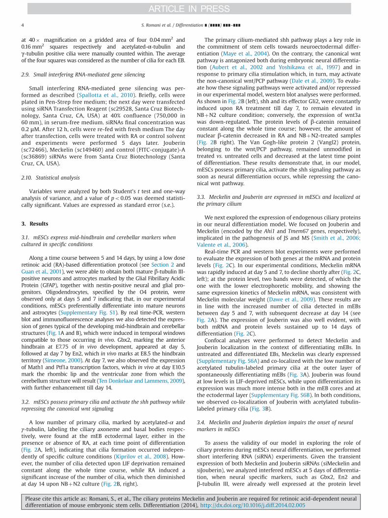
Along a time course between 5 and 14 days, by using a low doseretinoic acid (RA)-based differentiation protocol (see Section 2 andGuan et al., 2001), we were able to obtain both mature β-tubulin III-positive neurons and astrocytes marked by the Glial Fibrillary AcidicProtein (GFAP), together with nestin-positive neural and glial pro-genitors. Oligodendrocytes, specified by the O4 protein, wereobserved only at days 5 and 7 indicating that, in our experimentalconditions, mESCs preferentially differentiate into mature neuronsand astrocytes (Supplementary Fig. S1). By real time-PCR, westernblot and immunofluorescence analyses we also detected the expres-sion of genes typical of the developing mid-hindbrain and cerebellarstructures (Fig. 1A and B), which were induced in temporal windowscompatible to those occurring in vivo. Gbx2, marking the anteriorhindbrain at E7.75 of in vivo development, appeared at day 5,followed at day 7 by En2, which in vivo marks at E8.5 the hindbrainterritory (Simeone, 2000). At day 7, we also observed the expressionof Math1 and Ptf1a transcription factors, which in vivo at day E10.5mark the rhombic lip and the ventricular zone from which thecerebellum structure will result (Ten Donkelaar and Lammens, 2009),with further enhancement till day 14.
3.2. mESCs possess primary cilia and activate the shh pathway whilerepressing the canonical wnt signaling
A low number of primary cilia, marked by acetylated-α andγ-tubulin, labeling the ciliary axoneme and basal bodies respec-tively, were found at the mEB ectodermal layer, either in thepresence or absence of RA, at each time point of differentiation(Fig. 2A, left), indicating that cilia formation occurred indepen-dently of specific culture conditions (Kiprilov et al., 2008). How-ever, the number of cilia detected upon LIF deprivation remainedconstant along the whole time course, while RA induced asignificant increase of the number of cilia, which then diminishedat day 14 upon NBþN2 culture (Fig. 2B, right).
The primary cilium-mediated shh pathway plays a key role inthe commitment of stem cells towards neuroectodermal differ-entiation (Maye et al., 2004). On the contrary, the canonical wntpathway is antagonized both during embryonic neural differentia-tion (Aubert et al., 2002 and Yoshikawa et al., 1997) and inresponse to primary cilia stimulation which, in turn, may activatethe non-canonical wnt/PCP pathway (Dale et al., 2009). To evalu-ate how these signaling pathways were activated and/or repressedin our experimental model, western blot analyses were performed.As shown in Fig. 2B (left), shh and its effector Gli2, were constantlyinduced upon RA treatment till day 7, to remain elevated inNBþN2 culture condition; conversely, the expression of wnt3awas down-regulated. The protein levels of β-catenin remainedconstant along the whole time course; however, the amount ofnuclear β-catenin decreased in RA and NBþN2-treated samples(Fig. 2B right). The Van Gogh-like protein 2 (Vangl2) protein,belonging to the wnt/PCP pathway, remained unmodified intreated vs. untreated cells and decreased at the latest time pointof differentiation. These results demonstrate that, in our model,mESCs possess primary cilia, activate the shh signaling pathway assoon as neural differentiation occurs, while repressing the cano-nical wnt pathway.
3.3. Meckelin and Jouberin are expressed in mESCs and localized atthe primary cilium
We next explored the expression of endogenous ciliary proteinsin our neural differentiation model. We focused on Jouberin andMeckelin (encoded by the Ahi1 and Tmem67 genes, respectively),implicated in the pathogenesis of JS and MS (Smith et al., 2006;Valente et al., 2006).
Real-time PCR and western blot experiments were performedto evaluate the expression of both genes at the mRNA and proteinlevels (Fig. 2C). In our experimental conditions, Meckelin mRNAwas rapidly induced at day 5 and 7, to decline shortly after (Fig. 2C,left); at the protein level, two bands were detected, of which theone with the lower electrophoretic mobility, and showing thesame expression kinetics of Meckelin mRNA, was consistent withMeckelin molecular weight (Dawe et al., 2009). These results arein line with the increased number of cilia detected in mEBsbetween day 5 and 7, with subsequent decrease at day 14 (seeFig. 2A). The expression of Jouberin was also well evident, withboth mRNA and protein levels sustained up to 14 days ofdifferentiation (Fig. 2C).
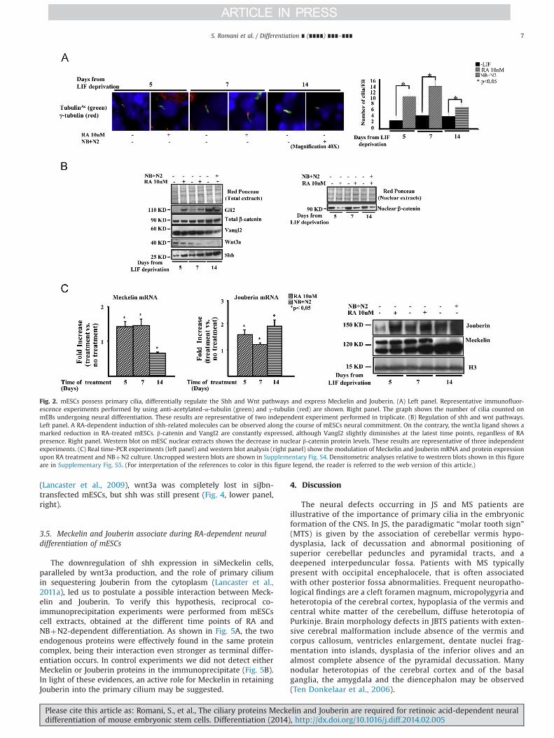
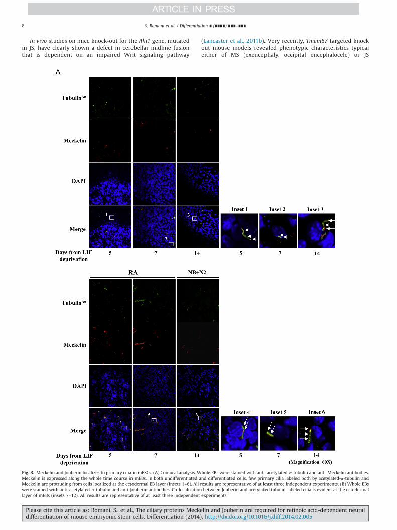
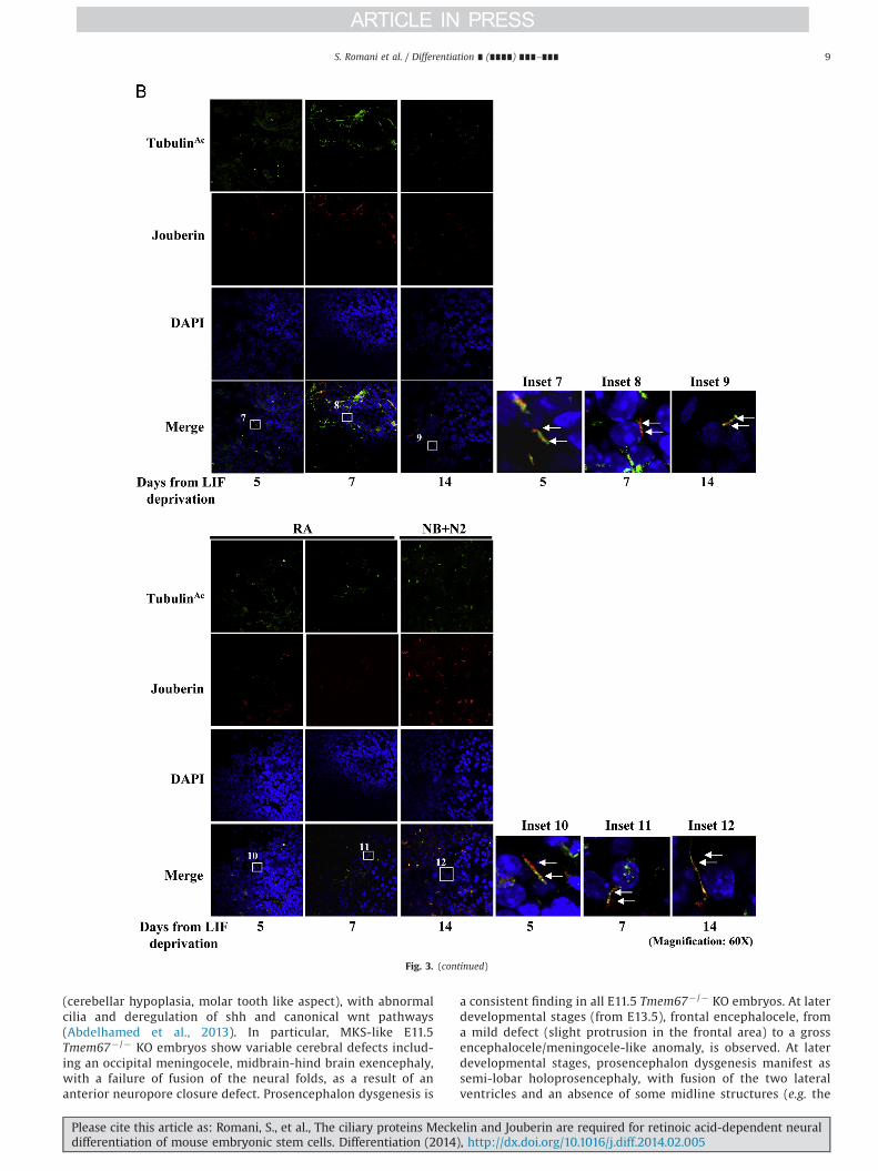
Confocal analyses were performed to detect Meckelin andJouberin localization in the context of differentiating mEBs. Inuntreated and differentiated EBs, Meckelin was clearly expressed(Supplementary Fig. S6A) and co-localized with the low number ofacetylated tubulin-labeled primary cilia at the outer layer ofspontaneously differentiating mEBs (Fig. 3A). Jouberin was foundat low levels in LIF-deprived mESCs, while upon differentiation itsexpression was much more intense both in the mEB cores and atthe ectodermal layer (Supplementary Fig. S6B). In both conditions,we observed co-localization of Jouberin with acetylated tubulin-labeled primary cilia (Fig. 3B).
3.4. Meckelin and Jouberin depletion impairs the onset of neuralmarkers in mESCs
To assess the validity of our model in exploring the role ofciliary proteins during mESCs neural differentiation, we performedshort interfering RNA (siRNA) experiments. Given the transientexpression of both Meckelin and Jouberin siRNAs (siMeckelin andsiJouberin), we analyzed interfered mESCs at 5 days of differentia-tion, when neural specific markers, such as Gbx2, En2 andβ-tubulin III, were already well expressed at the protein level
S. Romani et al. / Differentiation ∎ (∎∎∎∎) ∎∎∎–∎∎∎4
Please cite this article as: Romani, S., et al., The ciliary proteins Meckelin and Jouberin are required for retinoic acid-dependent neuraldifferentiation of mouse embryonic stem cells. Differentiation (2014), http://dx.doi.org/10.1016/j.diff.2014.02.005i

(see Fig. 1A and B and Supplementary Fig. S1), consistently withshh activation (see Fig. 2B). As a control we used a FITC-conjugatecontrol scrambled siRNA which allowed us to monitor siRNAsexpression till day 6 (Supplementary Fig. S7C). Transfection ofsiMeckelin and siJouberin produced a significant decrease of bothendogenous proteins at day 6 from transfection, as revealed byimmunofluorescence (Fig. 4A, top). In the presence of RA, lowlevels of β-tubulin III were observed in both silenced cells
compared to controls (Fig. 4A, bottom). Similarly, western blotanalyses showed a severe impairment of Gbx2 and En2 RA-dependent expression, when compared to scramble-transfectedmESCs (Fig. 4B), suggesting a failure of mESCs to differentiatetowards the neural lineage.
Accordingly, siMeckelin-transfected cells failed to activate shhin response to RA, while inducing wnt3a (Fig. 4, lower panel, left);on the contrary, and consistently with previously published data
Fig. 1. mESCs express mid-hindbrain and cerebellar markers when cultured in RA-enriched and neurobasal plus N2 culture medium. (A) Picture depicts the kinetics of expression ofGbx2, En2, Math1 and Ptf1a. Left panels show real time-PCR experiments; middle panels show representative western blot analyses and right panels show the densitometric analysesrelative to western blots. These results are representative of three independent experiments. (B) Immunofluorescence experiments showing the expression of mid-hindbrain (Gbx2and En2) and cerebellar markers (Math1 and Ptf1a) in differentiating mEBs and their co-localizationwith β-tubulin III. Consistently with real-time PCR and western blot data, Gbx2 andEn2 expression anticipates Math1 and Ptf1a onset. Nuclei are shown in blue. These results are representative of three independent experiments. Uncropped western blots are shown inSupplementary Fig. 2. (For interpretation of the references to color in this figure legend, the reader is referred to the web version of this article.)
S. Romani et al. / Differentiation ∎ (∎∎∎∎) ∎∎∎–∎∎∎ 5
Please cite this article as: Romani, S., et al., The ciliary proteins Meckelin and Jouberin are required for retinoic acid-dependent neuraldifferentiation of mouse embryonic stem cells. Differentiation (2014), http://dx.doi.org/10.1016/j.diff.2014.02.005i
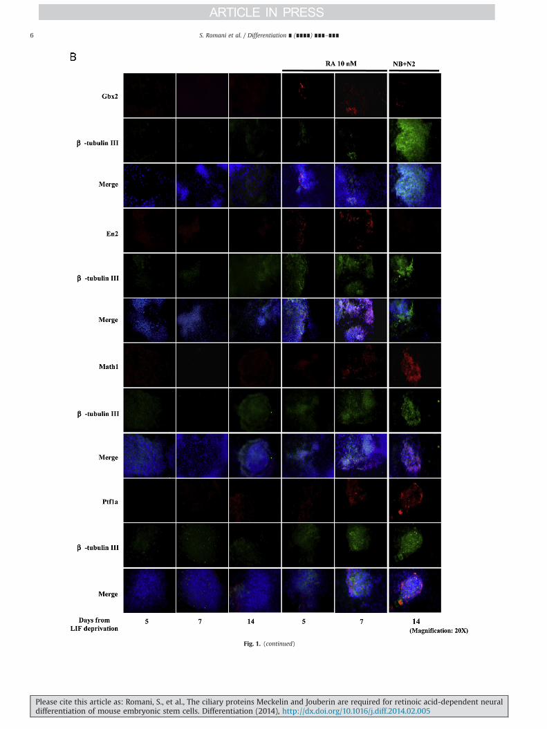
Fig. 1. (continued)
S. Romani et al. / Differentiation ∎ (∎∎∎∎) ∎∎∎–∎∎∎6
Please cite this article as: Romani, S., et al., The ciliary proteins Meckelin and Jouberin are required for retinoic acid-dependent neuraldifferentiation of mouse embryonic stem cells. Differentiation (2014), http://dx.doi.org/10.1016/j.diff.2014.02.005i
(Lancaster et al., 2009), wnt3a was completely lost in siJbn-transfected mESCs, but shh was still present (Fig. 4, lower panel,right).
3.5. Meckelin and Jouberin associate during RA-dependent neuraldifferentiation of mESCs
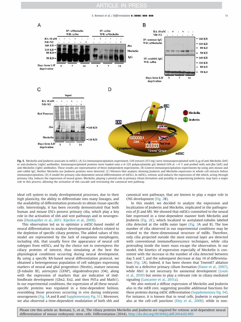
The downregulation of shh expression in siMeckelin cells,paralleled by wnt3a production, and the role of primary ciliumin sequestering Jouberin from the cytoplasm (Lancaster et al.,2011a), led us to postulate a possible interaction between Meck-elin and Jouberin. To verify this hypothesis, reciprocal co-immunoprecipitation experiments were performed from mESCscell extracts, obtained at the different time points of RA andNBþN2-dependent differentiation. As shown in Fig. 5A, the twoendogenous proteins were effectively found in the same proteincomplex, being their interaction even stronger as terminal differ-entiation occurs. In control experiments we did not detect eitherMeckelin or Jouberin proteins in the immunoprecipitate (Fig. 5B).In light of these evidences, an active role for Meckelin in retainingJouberin into the primary cilium may be suggested.
4. Discussion
The neural defects occurring in JS and MS patients areillustrative of the importance of primary cilia in the embryonicformation of the CNS. In JS, the paradigmatic “molar tooth sign”(MTS) is given by the association of cerebellar vermis hypo-dysplasia, lack of decussation and abnormal positioning ofsuperior cerebellar peduncles and pyramidal tracts, and adeepened interpeduncular fossa. Patients with MS typicallypresent with occipital encephalocele, that is often associatedwith other posterior fossa abnormalities. Frequent neuropatho-logical findings are a cleft foramen magnum, micropolygyria andheterotopia of the cerebral cortex, hypoplasia of the vermis andcentral white matter of the cerebellum, diffuse heterotopia ofPurkinje. Brain morphology defects in JBTS patients with exten-sive cerebral malformation include absence of the vermis andcorpus callosum, ventricles enlargement, dentate nuclei frag-mentation into islands, dysplasia of the inferior olives and analmost complete absence of the pyramidal decussation. Manynodular heterotopias of the cerebral cortex and of the basalganglia, the amygdala and the diencephalon may be observed(Ten Donkelaar et al., 2006).
Fig. 2. mESCs possess primary cilia, differentially regulate the Shh and Wnt pathways and express Meckelin and Jouberin. (A) Left panel. Representative immunofluor-escence experiments performed by using anti-acetylated-α-tubulin (green) and γ-tubulin (red) are shown. Right panel. The graph shows the number of cilia counted onmEBs undergoing neural differentiation. These results are representative of two independent experiment performed in triplicate. (B) Regulation of shh and wnt pathways.Left panel. A RA-dependent induction of shh-related molecules can be observed along the course of mESCs neural commitment. On the contrary, the wnt3a ligand shows amarked reduction in RA-treated mESCs. β‐catenin and Vangl2 are constantly expressed, although Vangl2 slightly diminishes at the latest time points, regardless of RApresence. Right panel. Western blot on mESC nuclear extracts shows the decrease in nuclear β-catenin protein levels. These results are representative of three independentexperiments. (C) Real time-PCR experiments (left panel) and western blot analysis (right panel) show the modulation of Meckelin and Jouberin mRNA and protein expressionupon RA treatment and NBþN2 culture. Uncropped western blots are shown in Supplementary Fig. S4. Densitometric analyses relative to westrern blots shown in this figureare in Supplementary Fig. S5. (For interpretation of the references to color in this figure legend, the reader is referred to the web version of this article.)
S. Romani et al. / Differentiation ∎ (∎∎∎∎) ∎∎∎–∎∎∎ 7
Please cite this article as: Romani, S., et al., The ciliary proteins Meckelin and Jouberin are required for retinoic acid-dependent neuraldifferentiation of mouse embryonic stem cells. Differentiation (2014), http://dx.doi.org/10.1016/j.diff.2014.02.005i
In vivo studies on mice knock-out for the Ahi1 gene, mutatedin JS, have clearly shown a defect in cerebellar midline fusionthat is dependent on an impaired Wnt signaling pathway
(Lancaster et al., 2011b). Very recently, Tmem67 targeted knockout mouse models revealed phenotypic characteristics typicaleither of MS (exencephaly, occipital encephalocele) or JS
Fig. 3. Meckelin and Jouberin localizes to primary cilia in mESCs. (A) Confocal analysis. Whole EBs were stained with anti-acetylated-α-tubulin and anti-Meckelin antibodies.Meckelin is expressed along the whole time course in mEBs. In both undifferentiated and differentiated cells, few primary cilia labeled both by acetylated-α-tubulin andMeckelin are protruding from cells localized at the ectodermal EB layer (insets 1–6). All results are representative of at least three independent experiments. (B) Whole EBswere stained with anti-acetylated-α-tubulin and anti-Jouberin antibodies. Co-localization between Jouberin and acetylated tubulin-labeled cilia is evident at the ectodermallayer of mEBs (insets 7–12). All results are representative of at least three independent experiments.
S. Romani et al. / Differentiation ∎ (∎∎∎∎) ∎∎∎–∎∎∎8
Please cite this article as: Romani, S., et al., The ciliary proteins Meckelin and Jouberin are required for retinoic acid-dependent neuraldifferentiation of mouse embryonic stem cells. Differentiation (2014), http://dx.doi.org/10.1016/j.diff.2014.02.005i
(cerebellar hypoplasia, molar tooth like aspect), with abnormalcilia and deregulation of shh and canonical wnt pathways(Abdelhamed et al., 2013). In particular, MKS-like E11.5Tmem67�/� KO embryos show variable cerebral defects includ-ing an occipital meningocele, midbrain-hind brain exencephaly,with a failure of fusion of the neural folds, as a result of ananterior neuropore closure defect. Prosencephalon dysgenesis is
a consistent finding in all E11.5 Tmem67� /� KO embryos. At laterdevelopmental stages (from E13.5), frontal encephalocele, froma mild defect (slight protrusion in the frontal area) to a grossencephalocele/meningocele-like anomaly, is observed. At laterdevelopmental stages, prosencephalon dysgenesis manifest assemi-lobar holoprosencephaly, with fusion of the two lateralventricles and an absence of some midline structures (e.g. the
Fig. 3. (continued)
S. Romani et al. / Differentiation ∎ (∎∎∎∎) ∎∎∎–∎∎∎ 9
Please cite this article as: Romani, S., et al., The ciliary proteins Meckelin and Jouberin are required for retinoic acid-dependent neuraldifferentiation of mouse embryonic stem cells. Differentiation (2014), http://dx.doi.org/10.1016/j.diff.2014.02.005i

anterior commissar and variable degrees of corpus callosumdysgenesis). The ‘MKS-like’ group of animals shows also anenlargement of the hippocampus and basal ganglia. All JBTS-like Tmem67�/� embryos have a reduced anteroposterior axis ofthe developing forebrain, a small hindbrain region, microce-phaly or other overt facial dysmorphologies. Some of them havea deep interpenduncular fossa with a reduced anterioposterioraxis of the midbrain tegmentum at the level of the isthmus,associated with cerebellar vermis hypoplasia or aplasia. Thesephenotypic manifestation comprise the MTS that is pathogno-monic for JBTS in humans. Complex posterior fossa defects and
features compatible with the Dandy–Walker malformation arealso observed (Abdelhamed et al., 2013).
Primary cilia are required for cerebellar development in theembryo (Spassky et al., 2008) and for the generation of adultneural stem cells (Han et al., 2008). To date, a major limitation instudying primary cilia developmental function in vitro remains thelack of suitable cellular models that could recapitulate as accu-rately as possible the developmental steps observed in vivo. Suchmodels would represent a key tool to rapidly analyze the impactof distinct ciliary proteins on the wide variety of differentiationmodules regulated by primary cilia. To this end, ESCs represent an
Fig. 4. The absence of Meckelin and Jouberin impairs mESCs response to retinoic acid. mESCs were transfected with siMeckelin and siJbn in the absence of serum andantibiotics and in the presence of LIF. After 12 h, mESCs were re-fed with fresh LIF-containing medium. After additional 24 h, cells were shifted to LIF-deprived medium andtreated with RA or control solvent for 5 days. (A) Upper panel. Immunofluorescence showing the reduction in Jouberin and Meckelin expression. Lower panel.Immunofluorescence showing the dramatic decrement of β-tubulin III protein in siMeckelin and siJbn mESCs. (B) Western blot analyses showing both the down-regulation ofMeckelin and Jbn in siRNA-transfected cells, the modulation of shh and wnt pathways and the effect on the expression of the transcription factors Gbx2 and En2. Theseresults are representative of three independent experiments. Uncropped western blots are shown in Supplementary Fig. S7A and B.
S. Romani et al. / Differentiation ∎ (∎∎∎∎) ∎∎∎–∎∎∎10
Please cite this article as: Romani, S., et al., The ciliary proteins Meckelin and Jouberin are required for retinoic acid-dependent neuraldifferentiation of mouse embryonic stem cells. Differentiation (2014), http://dx.doi.org/10.1016/j.diff.2014.02.005i
ideal cell system to study developmental processes, due to theirhigh plasticity, the ability to differentiate into many lineages, andthe availability of differentiation protocols to obtain tissue-specificcells. Interestingly, it has been recently demonstrated that bothhuman and mouse ESCs possess primary cilia, which play a keyrole in the activation of shh and wnt pathways and in neurogen-esis (Hunkapiller et al., 2011; Kiprilov et al., 2008).
This observation led us to optimize a mESC-based model ofneural differentiation to analyze developmental defects related tothe depletion of specific ciliary proteins. The added values of thismodel are represented by the lack of exogenous morphogens,including shh, that usually force the appearance of neural cellsubtypes from mESCs, and by the choice not to overexpress theciliary proteins of interest, thus simulating at the best thephysiological conditions occurring during neural development.By using a specific RA-based neural differentiation protocol, weobtained a heterogeneous population of cell subtypes expressingmarkers of neural and glial progenitors (Nestin), mature neurons(β-tubulin III), astrocytes (GFAP), oligodendrocytes (O4), alongwith the expression of markers that are indicative of mid-hindbrain development (Gbx2, En2, and then Ptf1a and Math1).In our experimental conditions, the expression of all these neural-specific proteins was regulated in a time-dependent fashion,resembling those processes occurring in vivo during embryonicneurogenesis (Fig. 1A and B and Supplementary Fig. S1). Moreover,we also observed a time-dependent modulation of both shh and
canonical wnt pathways, that are known to play a major role inCNS development (Fig. 2B).
In this model, we decided to analyze the expression andlocalization of Jouberin and Meckelin, implicated in the pathogen-esis of JS and MS. We showed that mESCs committed to the neuralfate expressed in a time-dependent manner both Meckelin andJouberin (Fig. 2C), which localized to acetylated-tubulin labeledcilia detected at the mEBs outer layer (Fig. 3A and B). The lownumber of cilia observed in our experimental conditions may berelated to the three-dimensional structure of mEBs. Therefore,only cilia projected outside the most external layer are detectedwith conventional immunofluorescence techniques, while ciliaprotruding inside the inner mass escape the observation. In ourmodel, the kinetics of expression especially of Meckelin is con-sistent with the increase in the number of cilia detected betweenday 5 and 7, and the subsequent decrease at day 14 of differentia-tion (Fig. 2A). Indeed, it has been shown that Tmem67 ablationleads to a defective primary cilium formation (Dawe et al., 2007),while Ahi1 is not necessary for axonemal development (Louieet al., 2010) but seems to play a relevant role in ciliary-mediatedsignaling (Lancaster et al., 2011a).
We also noticed a diffuse expression of Meckelin and Jouberinalso in the mEB core, suggesting possible additional functions forthese proteins during mESC differentiation (Supplementary Fig. S4).For instance, it is known that in renal cells, Jouberin is expressedalso at the cell–cell junctions (Eley et al., 2008); while in non-
Fig. 5. Meckelin and Jouberin associate in mESCs. (A) Co-immunoprecipitation experiment. Cell extracts (0.5 mg) were immunoprecipitated with 4 μg of anti-Meckelin (left)or anti-Jouberin (right) antibodies. Immunoprecipitated proteins were loaded onto a 4–12% polyacrylamide gel, blotted O/N at þ4 1C and probed with anti-Jbn (left) andanti-Meckelin (right) antibodies. These results are representative of three independent experiments. (B) Control immunoprecipitation experiments by using anti-mouse andanti-rabbit IgG. Neither Meckelin nor Jouberin proteins were detected. (C) Western blot analysis showing Jouberin and Meckelin expression in whole cell extracts beforeimmunoprecipitation. (D) A model for primary cilia-dependent neural differentiation of mESCs. In mESCs, retinoic acid induces the expression of shh which, acting throughprimary cilia, induces the expression of neural genes. Meckelin, playing a pivotal role in primary cilium formation and possibly in sequestering Jouberin, may have a majorrole in this process, allowing the activation of shh cascade and restraining the canonical wnt pathway.
S. Romani et al. / Differentiation ∎ (∎∎∎∎) ∎∎∎–∎∎∎ 11
Please cite this article as: Romani, S., et al., The ciliary proteins Meckelin and Jouberin are required for retinoic acid-dependent neuraldifferentiation of mouse embryonic stem cells. Differentiation (2014), http://dx.doi.org/10.1016/j.diff.2014.02.005i
ciliated cells it is found in the nuclear compartment (Lancaster et al.,2011b) and it acts as an oncogene in cutaneous T-cell lymphomasand Sezary cells (Kennah et al., 2009).
The observed time-dependent production of shh, which is indis-pensable for the differentiation of cells belonging to the neuroecto-dermal lineage (Maye et al., 2004), supports the hypothesis that shhdeficiency represents a major determinant of the neurological defectsobserved in patients with CNS-related ciliopathies (Cardenas-Rodriguez and Badano, 2009). In parallel with shh activation, wealso observed a progressive reduction of wnt3a and a decrease of thenuclear quota of β-catenin in RA-treated mESCs. These decrementsperfectly paralleled the increase of Jouberin and the appearance ofcilia in mEB, in line with the known role of the primary cilium inlimiting β-catenin nuclear entry once Jouberin has been sequestered(Lancaster et al., 2011b). Therefore, our in vitro model well recapitu-lates the cilia-dependent activation of shh pathway and constraint ofwnt canonical signaling occurring during embryonic development(Corbit et al., 2008). To assess whether this model could represent auseful tool to explore the function of specific ciliary proteins in neuraldifferentiation of mESCs, we knocked down Jouberin and Meckelinexpression by use of siRNAs (Fig. 4). Silenced cells failed to induceearly neural genes in response to RA, as shown by the low expressionlevels of the shh-responsive Gbx2 and En2 genes (Kobayashi et al.,2002; Szabo et al., 2009), as well as to terminally differentiate intoneurons, as illustrated by the markedly lower number of β-tubulin IIIpositive cells compared to controls. Thus, both proteins appear to berequired for early neural differentiation of mESCs. These in vitrofindings are in agreement with the results obtained in mouseembryos, either wild type or knock out for Ahi1 and Tmem67 genes.The Ahi1� /� mouse features cerebellar hypoplasia and defects in thevermis-midline fusion (Lancaster et al., 2011b), while targetedTmem67 KO mice show a variable range of early (E11.5–E12.5) neuraltube and mid-hindbrain defects (Abdelhamed et al., 2013; Loganet al., 2011). Further, we observed that Meckelin silencing impairedRA-dependent shh production in mESCs, while enhancing wnt3asignaling. Interestingly, Jouberin silencing had an opposite effect, asdemonstrated by the complete loss of wnt3a in RA-treated mESCs,while shh expression was not affected at all. Till now, the loss ofciliary proteins has been mostly related to both the reduction orabsence of shh receptors and dysregulation of β-catenin expressionlevels and/or subcellular localization (Mahjoub and Stearns, 2012;Abdelhamed et al., 2013; Lancaster et al., 2011a). However, veryrecently it has been demonstrated that Tmem67� /� KO embryoshave reduced levels of shh protein at the floor plate and notochord(Abdelhamed et al., 2013), supporting our RA-based in vitro model asa reliable tool to study primary cilia-related neurodevelopmentaldefects. Further, our results fit perfectly with the known role ofJouberin in stimulating wnt signaling (Lancaster et al., 2009) andwith the recent finding that another ciliary protein mutated in MS,MKS1, is linked to hedgehog signaling, while Jouberin does not (Sanget al., 2011). The results obtained in siMeckelin and siJbn mESCs andthe recent finding that Jouberin is sequestered as soon as the ciliumis formed, to constrain wnt signaling, led us to postulate a possibleinteraction between Meckelin and Jouberin, suggesting a potentialadditional role for the transmembrane protein Meckelin, in retainingJouberin into the cilium. To answer this question, we performedimmunoprecipitation experiments in mESCs directed to the neurallineage, in which the two endogenous proteins were effectivelydetected in the same protein complex (Fig. 5A). Therefore, in thepresence of Meckelin, shh signaling may prevail, while in its absence,Jouberin may be released from the cilium and wnt signaling may beactivated. In light of these evidences we suggest a model mESCs inwhich RA-dependent neural commitment rely on shh productionwhich, through primary cilia, activates a transcriptional programnecessary for the acquisition of the neuronal phenotype (Fig. 5C). Theciliary protein Meckelin, which is required for primary cilium
formation (Dawe et al., 2007) and possibly sequestering Jouberin toprimary cilia, seems to have a major role in this process.
Overall, this study enlightens the great potential of mESCs tostudy ciliary-related developmental disorders. In fact, the mole-cular mechanisms underlying the multiorgan defects in humanciliopathies may be investigated in a wide variety of differentiationmodules by simply culturing mESCs in the presence of appropriatemorphogens. Further, to our knowledge, our data represent thefirst evidence of a developmental role of Meckelin and Jouberin inregulating the onset of neuronal lineages from mESCs.
Acknowledgments
We thank prof. Joseph G. Gleeson and prof. Colin A. Johnson forpEGFP-Jbn and pCMV-HA-Meckelin expression vectors, respec-tively and for helpuful criticisms and discussion. This work hasbeen partially supported by: Italian Telethon Foundation (grantGGP13146 to EMV), Italian Ministry of Health (Ricerca Corrente2012-2013, Ricerca Finalizzata 2009 Malattie Rare to EMV),European Research Council (Starting Grant nr. 260888 to EMV),National Institute of Health (grant R01NS048453 to JGG).
Appendix A. Supplementary materials
Supplementary data associated with this article can be found inthe online version at http://dx.doi.org/10.1016/j.diff.2014.02.005.
References
Abdelhamed, Z.A., Wheway, G., Szymanska, K., Natarajan, S., Toomes, C., Inglehearn,C., Johnson, C.A., 2013. Variable expressivity of ciliopathy neurological pheno-types that encompass Meckel–Gruber syndrome and Joubert syndrome iscaused by complex de-regulated ciliogenesis, Shh and Wnt signalling defects.Hum. Mol. Genet. 22, 1358–1372.
Aubert, J., Dunstan, H., Chambers, I., Smith, A., 2002. Functional gene screening inembryonic stem cells implicates Wnt antagonism in neural differentiation. Nat.Biotechnol. 20, 1240–1245.
Baala, L., Romano, S., Khaddour, R., Saunier, S., Smith, U.M., Audollent, S., Ozilou, C.,Faivre, L., Laurent, N., Foliguet, B., et al., 2007. The Meckel–Gruber syndromegene, MKS3, is mutated in Joubert syndrome. Am. J. Hum. Genet. 80, 186–194.
Boheler, K.R., 2009. Stem cell pluripotency: a cellular trait that depends ontranscription factors, chromatin state and a checkpoint deficient cell cycle.J. Cell. Physiol. 221, 10–17.
Brancati, F., Dallapiccola, B., Valente, E.M., 2010. Joubert Syndrome and relateddisorders. Orphanet J. Rare Dis. 5, 20.
Cardenas-Rodriguez, M., Badano, J.L., 2009. Ciliary biology: understanding thecellular and genetic basis of human ciliopathies. Am. J. Med. Genet. C: Semin.Med. Genet. 151C, 263–280.
Chang, B.E., Blader, P., Fischer, N., Ingham, P.W., Strähle, U., 1997. Axial (HNF3beta)and retinoic acid receptors are regulators of the zebrafish sonic hedgehogpromoter. EMBO J. 16, 3955–3964.
Corbit, K.C., Shyer, A.E., Dowdle, W.E., Gaulden, J., Singla, V., Chen, M.H., Chuang, P.T.,Reiter, J.F., 2008. Kif3a constrains beta-catenin-dependent Wnt signallingthrough dual ciliary and non-ciliary mechanisms. Nat. Cell Biol. 10, 70–76.
Dale, R.M., Sisson, B.E., Topczewski, J., 2009. The emerging role of Wnt/PCPsignaling in organ formation. Zebrafish 6, 9–14.
Dawe, H.R., Smith, U.M., Cullinane, A.R., Gerrelli, D., Cox, P., Badano, J.L., Blair-Reid,S., Sriram, N., Katsanis, N., Attie-Bitach, T., et al., 2007. The Meckel–GruberSyndrome proteins MKS1 and meckelin interact and are required for primarycilium formation. Hum. Mol. Genet. 16, 173–186.
Dawe, H.R., Adams, M., Wheway, G., Szymanska, K., Logan, C.V., Noegel, A.A., Gull,K., Johnson, C.A., 2009. Nesprin-2 interacts with meckelin and mediatesciliogenesis via remodelling of the actin cytoskeleton. J. Cell Sci. 122,2716–2726.
Eley, L., Gabrielides, C., Adams, M., Johnson, C.A., Hildebrandt, F., Sayer, J., 2008.Jouberin localizes to collecting ducts and interacts with nephrocystin-1. KidneyInt. 74, 1139–1149.
Gaspard, N., Vanderhaeghen, P., 2010. Mechanisms of neural specification fromembryonic stem cells. Curr. Opin. Neurobiol. 20, 37–43.
Gherman, A., Davis, E.E., Katsanis, N., 2006. The ciliary proteome database: anintegrated community resource for genetic and fucntional dissection of cilia.Nat. Genet. 38, 961–962.
Guan, K., Chang, H., Rolletschek, A., Wobus, A.M., 2001. Embryonic stem cell-derived neurogenesis. Retinoic acid induction and lineage selection of neuronalcells. Cell Tissue Res. 305, 171–176.
S. Romani et al. / Differentiation ∎ (∎∎∎∎) ∎∎∎–∎∎∎12
Please cite this article as: Romani, S., et al., The ciliary proteins Meckelin and Jouberin are required for retinoic acid-dependent neuraldifferentiation of mouse embryonic stem cells. Differentiation (2014), http://dx.doi.org/10.1016/j.diff.2014.02.005i
Han, Y.G., Spassky, N., Romaguera-Ros, M., Garcia-Verdugo, J.M., Aguilar, A.,Schneider-Maunoury, S., Alvarez-Buylla, A., 2008. Hedgehog signaling andprimary cilia are required for the formation of adult neural stem cells. Nat.Neurosci. 11, 277–284.
Hunkapiller, J., Singla, V., Seol, A., Reiter, J.F., 2011. The ciliogenic protein Oral–Facial–Digital 1 regulates the neuronal differentiation of embryonic stem cells.Stem Cells Dev. 20, 831–841.
Illi, B., Scopece, A., Nanni, S., Farsetti, A., Morgante, L., Biglioli, P., Capogrossi, M.C.,Gaetano, C., 2005. Epigenetic histone modification and cardiovascular lineageprogramming in mouse embryonic stem cells exposed to laminar shear stress.Circ. Res. 96, 501–508.
Illi, B., Dello Russo, C., Colussi, C., Rosati, J., Pallaoro, M., Spallotta, F., Rotili, D.,Valente, S., Ragone, G., Martelli, F., et al., 2008. Nitric oxide modulateschromatin folding in human endothelial cells via protein phosphatase 2Aactivation and class IIa histone deacetylases nuclear shuttling. Circ. Res. 102,51–58.
Kennah, E., Ringrose, A., Zhou, L.L., Esmailzadeh, S., Qian, H., Su, M.W., Zhou, Y.,Jiang, X., 2009. Identification of tyrosine kinase, HCK, and tumor suppressor,BIN1, as potential mediators of AHI-1 oncogene in primary and transformedCTCL cells. Blood 113, 4646–4655.
Kiprilov, E.N., Awan, A., Desprat, R., Velho, M., Clement, C.A., Byskov, A.G., Andersen,C.Y., Satir, P., Bouhassira, E.E., Christensen, S.T., Hirsch, R.E., 2008. Humanembryonic stem cells in culture possess primary cilia with hedgehog signalingmachinery. J. Cell Biol. 180, 897–904.
Kobayashi, D., Kobayashi, M., Matsumoto, K., Ogura, T., Nakafuku, M., Shimamura,K., 2002. Early subdivisions in the neural plate define distinct competence forinductive signals. Development 129, 83–93.
Kurosawa, H., 2007. Methods for inducing embryoid body formation: in vitrodifferentiation system of embryonic stem cells. J. Biosci. Bioeng. 103, 389–398.
Lancaster, M.A., Gleeson, J.G., 2009. The primary cilium as a cellular signalingcenter: lessons from disease. Curr. Opin. Genet. Dev. 19, 220–229.
Lancaster, M.A., Louie, C.M., Silhavy, J.L., Sintasath, L., Decambre, M., Nigam, S.K.,Willert, K., Gleeson, J.G., 2009. Impaired Wnt-beta-catenin signaling disruptsadult renal homeostasis and leads to cystic kidney ciliopathy. Nat. Med. 15,1046–1054.
Lancaster, M.A., Schroth, J., Gleeson, J.G., 2011a. Subcellular spatial regulation ofcanonical Wnt signalling at the primary cilium. Nat. Cell Biol. 13, 700–707.
Lancaster, M.A., Gopal, D.J., Kim, J., Saleem, S.N., Silhavy, J.L., Louie, C.M., Thacker, B.E.,Williams, Y., Zaki, M.S., Gleeson, J.G., 2011b. Defective Wnt-dependent cerebellarmidline fusion in a mouse model of Joubert syndrome. Nat. Med. 17, 726–731.
Logan, C.V., Abdel-Hamed, Z., Johnson, C.A., 2011. Molecular genetics and patho-genic mechanisms for the severe ciliopathies: insights into neurodevelopmentand pathogenesis of neural tube defects. Mol. Neurobiol. 43, 12–26.
Louie, C.M., Gleeson, J.G., 2005. Genetic basis of Joubert syndrome and relateddisorders of cerebellar development. Hum. Mol. Genet. 14, R235–242.
Louie, C.M., Caridi, G., Lopes, V.S., Brancati, F., Kispert, A., Lancaster, M.A., Schloss-man, A.M., Otto, E.A., Leitges, M., Gröne, H.J., et al., 2010. AHI1 is required for
photoreceptor outer segment development and is a modifier for retinaldegeneration in nephronophthisis. Nat. Genet. 42, 175–180.
Magenta, A., Fasanaro, P., Romani, S., Di Stefano, V., Capogrossi, M.C., Martelli, F.,2008. Protein phosphatase 2A subunit PR70 interacts with pRb and mediates itsdephosphorylation. Mol. Cell. Biol. 28, 873–882.
Mahjoub, M.R., Stearns, T., 2012. Supernumerary centrosomes nucleate extra ciliaand compromise primary cilium signaling. Curr. Biol. 22, 1628–1634.
Marshall, W.F., Nonaka, S., 2006. Cilia: tuning in to the cell's antenna. Curr. Biol. 16,R604–614.
Maye, P., Becker, S., Siemen, H., Thorne, J., Byrd, N., Carpentino, J., Grabel, L., 2004.Hedgehog signaling is required for the differentiation of ES cells into neur-ectoderm. Dev. Biol. 265, 276–290.
Ocbina, P.J., Tuson, M., Anderson, K.V., 2009. Primary cilia are not required fornormal canonical Wnt signaling in the mouse embryo. PLoS One 4, e6839.
Sang, L., Miller, J.J., Corbit, K.C., Giles, R.H., Brauer, M.J., Otto, E.A., Baye, L.M., Wen, X.,Scales, S.J., Kwong, M., et al., 2011. Mapping the NPHP–JBTS–MKS proteinnetwork reveals ciliopathy disease genes and pathways. Cell 145, 513–528.
Simeone, A., 2000. Positioning the isthmic organizer: where Otx2 and Gbx2 meet.Trends Genet. 16, 237–240.
Smith, U.M., Consugar, M., Tee, L.J., McKee, B.M., Maina, E.N., Whelan, S., Morgan, N.V., Goranson, E., Gissen, P., Lilliquist, S., et al., 2006. The transmembrane proteinmeckelin (MKS3) is mutated in Meckel–Gruber syndrome and the wpk rat. Nat.Genet. 38, 191–196.
Spallotta, F., Rosati, J., Straino, S., Nanni, S., Grasselli, A., Ambrosino, V., Rotili, D.,Valente, S., Farsetti, A., Mai, A., et al., 2010. Nitric oxide determines mesodermicdifferentiation of mouse embryonic stem cells by activating class IIa histonedeacetylases: potential therapeutic implications in a mouse model of hindlimbischemia. Stem Cells 28, 431–442.
Spassky, N., Han, Y.G., Aguilar, A., Strehl, L., Besse, L., Laclef, C., Ros, M.R., Garcia-Verdugo, J.M., Alvarez-Buylla, A., 2008. Primary cilia are required for cerebellardevelopment and Shh-dependent expansion of progenitor pool. Dev. Biol. 317,246–259.
Szabo, N.E., Zhao, T., Zhou, X., Alvarez-Bolado, G., 2009. The role of Sonic hedgehogof neural origin in thalamic differentiation in the mouse. J. Neurosci. 29,2453–2466.
Ten Donkelaar, H.J., Lammens, M., Hori, A., 2006. Clinical Neuroembriology.Springer-Verlag, Berlin Heidelberg
Ten Donkelaar, H.J., Lammens, M., 2009. Development of the human cerebellumand its disorders. Clin. Perinatol. 36, 513–530.
Valente, E.M., Brancati, F., Silhavy, J.L., Castori, M., Marsh, S.E., Barrano, G., Bestini, E.,Boltshauser, E., Zaki, M.S., Abdel-Aleem, A., et al., 2006. AHI1 gene mutationscause specific forms of Joubert syndrome-related disorders. Ann. Neurol. 59,527–534.
Yoshikawa, Y., Fujimori, T., McMahon, A.P., Takada, S., 1997. Evidence that absenceof Wnt-3a signaling promotes neuralization instead of paraxial mesodermdevelopment in the mouse. Dev. Biol. 183, 234–242.
S. Romani et al. / Differentiation ∎ (∎∎∎∎) ∎∎∎–∎∎∎ 13
Please cite this article as: Romani, S., et al., The ciliary proteins Meckelin and Jouberin are required for retinoic acid-dependent neuraldifferentiation of mouse embryonic stem cells. Differentiation (2014), http://dx.doi.org/10.1016/j.diff.2014.02.005i