THE BRITISH JOURNAL OF - NCBI
8
Vol. XLVIII, No. 1 (February, 1967) wa.s issued on 7.3.67. THE BRITISH JOURNAL OF EXPERIMENTAL PATHOLOGY V VOL. LVill APRIL. 1967 NO. I9- VAGOTOMY IN THE RAT A STUDY OF ITS EFFECTS ON STOMACH AND SMALL INTESTINE H. ELLIS AND J. PRYSE-DAVIES From the Surgical Unit, Westminster Hospital and Bernhard Baron Memorial Research Laboratories, Queen Charlotte's Maternity Hospital, London Received for publication August 9, 1966 VAGOTOMY combined with some type of drainage procedure has become established as a standard operation for duodenal ulcer, yet much remains to be learned about the effects of division of the vagus nerve. Studies in man are hindered because the operation cannot be performed alone but must be complicated by pyloroplasty or gastrojejunostomy in order to drain the atonic stomach; moreover the procedure is only carried out in abnormal subjects who possess a peptic ulcer. To date, most investigations of pure vagotomy have been confined to the dog, (e.g. Baldwin, Albo, Jaffe and Silen, 1965, Golding, Mendoza, Aiello, Fierst, Solomon and Enquist, 1965; Wastell 1966). Comparative studies in other species are obviously of interest in order to extend our range of knowledge of the physiological and pathological effects of this operation. In this paper we describe a study of vagotomy in the rat with special reference to the ulceration which occurs in the resulting atonic and dilated stomach, the histochemistry of stomach and small intestine, the appearances of the villous pattern of the small gut, and body weight changes in both adult and immature rats. MATERIAL AND METHODS Albino male -Wistar rats were maintained on Oxoid breeder diet and water ad libitumi. Anaesthesia was induced with intraperitoneal Nembutal and maintained by ether. Vago- tomy was performed through an upper abdominal mid-line incision. The oesophagus was mobilised and the vagi identified on the anterior and posterior aspects of the abdominal part of the oesophagus; an approximately 1 cm. segment of each was excised and the abdominal iincision then closed with two layers of thread. The left gastric vessels were sacrificed in performing the posterior vagal resection. Controls comprised litter mates who underwent a laparotomy only. 11
-
Upload
khangminh22 -
Category
Documents
-
view
3 -
download
0
Transcript of THE BRITISH JOURNAL OF - NCBI
Vol. XLVIII, No. 1 (February, 1967) wa.s issued on 7.3.67.
THE BRITISH JOURNAL OF
EXPERIMENTAL PATHOLOGY
VVOL. LVill APRIL. 1967 NO. I9-
VAGOTOMY IN THE RATA STUDY OF ITS EFFECTS ON STOMACH AND SMALL INTESTINE
H. ELLIS AND J. PRYSE-DAVIES
From the Surgical Unit, Westminster Hospital and Bernhard Baron MemorialResearch Laboratories, Queen Charlotte's Maternity Hospital, London
Received for publication August 9, 1966
VAGOTOMY combined with some type of drainage procedure has becomeestablished as a standard operation for duodenal ulcer, yet much remains to belearned about the effects of division of the vagus nerve. Studies in man arehindered because the operation cannot be performed alone but must be complicatedby pyloroplasty or gastrojejunostomy in order to drain the atonic stomach;moreover the procedure is only carried out in abnormal subjects who possess apeptic ulcer. To date, most investigations of pure vagotomy have been confinedto the dog, (e.g. Baldwin, Albo, Jaffe and Silen, 1965, Golding, Mendoza, Aiello,Fierst, Solomon and Enquist, 1965; Wastell 1966). Comparative studies in otherspecies are obviously of interest in order to extend our range of knowledge of thephysiological and pathological effects of this operation.
In this paper we describe a study of vagotomy in the rat with special referenceto the ulceration which occurs in the resulting atonic and dilated stomach, thehistochemistry of stomach and small intestine, the appearances of the villouspattern of the small gut, and body weight changes in both adult and immaturerats.
MATERIAL AND METHODS
Albino male -Wistar rats were maintained on Oxoid breeder diet and water ad libitumi.Anaesthesia was induced with intraperitoneal Nembutal and maintained by ether. Vago-tomy was performed through an upper abdominal mid-line incision. The oesophagus wasmobilised and the vagi identified on the anterior and posterior aspects of the abdominal partof the oesophagus; an approximately 1 cm. segment of each was excised and the abdominaliincision then closed with two layers of thread. The left gastric vessels were sacrificed inperforming the posterior vagal resection. Controls comprised litter mates who underwenta laparotomy only.
11
H. ELLIS AND J. PRYSE-DAVIES
Group I: Gross and histological appearances and weight changeFifty adult rats were subjected to vagotomy and ten to laparotomy only. The animals
were weighed weekly and were killed 12-32 weeks after operation. The control laparotomieswere killed at 32 weeks. In each case a full autopsy was performed, the stomach was weighed,opened along its greater curvature? pinned out on a cork and fixed as rapidly as possible informol salinie. Routine histological examination was performed on representative portionsof the gastro-intestinal tract, and specifically upon any gastric erosion or ulcer after this hadbeen photographed.
A further group of 12 immature rats (average weight 90 g.) were vagotomised, weighedweekly and killed at 19 weeks. They were compared with 11 litter mate normal controls.
Group II: Histochemistry of stomach and small intestine, villous pattern of small bowelFourteen adult rats were vagotomised and killed by a blow on the head between 1-21
weeks post-operatively together with both unoperated controls and sham laparotomies.Seven were prepared for gastric enzyme histochemical studies.In these animals the stomach was opened along the greater curvature, cleared of food
debris by gentle rinsing in warm (37°) normal saline and pinned out on a cork. Dilatationwas assessed by measuring the maximum circumference. Blocks were taken to includepyloric, fundic and squamous regions of the mucosa and included any abnormal zones seennaked eye. Enzyme histochemical studies were made on freshly frozen tissue sectioned ina cryostat: the tissue was made into a roll and frozen by means of solid carbon dioxide onto a microtome chuck. The remaining stomach was fixed in 10 per cent neutral formalin andsimilar blocks were taken later for paraffin embedding.
Seven vagotomised rats were prepared for small intestinal enzyme histochemical studies.In these, the second quarter and fourth quarter of the small intestine were taken for jejlnllaland ileal studies respectively. Frozen tissue for enzyme histochemistry was prepared froman unopened coil of gut, washed of debris by gentle syringing with warm saline and frozeinrapidly on to a chuck with solid carbon dioxide for cryostat sectioning.
The enzymes investigated in 15,u cryostat sections were succinate dehydrogenase, leucinieaminopeptidase and glucose-6-phosphate dehydrogenase in the stomach and succinlatedehydrogenase, leucine aminopeptidase and alkaline phosphatase in the small intestine.The MTT tetrazolium method was used for demonstrating succinate dehydrogenase activity(Pearse, 1957). Leucine amino peptidase was demonstrated by the method of Nachlas,Crawford and Seligman (1957) using L-leucyl-4-methoxy-naphthylamide as substrate. Forglucose-6-phosphate dehydrogenase the MTT tetrazolium method of Hess, Scarpelli anldPearse (1958) was employed. An azo dye coupling was used to demonstrate alkaline phos-phatase with Fast Red LTR as the stabilized diazotate (Pryse-Davies, Dawson and,Snape.1961).
An attempt to demonstrate carbonic anhydrase activity in the rat stomach and kidneywas unsuccessful using the method of Hausler quoted by Pearse (1960), negative controlstaining in the absence of substrate and in the presence of " Diamox " could not be obtained.
In all 14 animals the dissecting microscope appearances of jejunal villi and the histologyof the jejunum were also studied, and compared with both the normal controls and animalssubmitted to sham operations.
For dissecting microscope examination, segments of jejunum were opened longitudinally,flattened on to filter paper and immersed in both normal saline and neutral formalin; mucuswas removed by gently stoking the mucosa with a camel hair brush. Similar segments offlattened jejunum and ileum were fixed in formalin for paraffin wax embedding; sectionswere cut longitudinally and transversely to obtain a better picture of the leaf-shaped villi.
RESULTS
Gross and histological changesin the vagotomised stomachThe fifty adult rats subjected to vagotomy 12-32 weeks previously were
specifically studied for morphological and histological changes in the stomach.Thirteen of these rats had stomachs of normal size, mean weight with contents
8-8 g. (range 3-13 g.).
136

VAGOTOMY IN THE RAT 137
Twenty rats had dilated stomachs but with normal mucosal appearances bothnaked eye and histologically with a mean weight of 25 g. (range 13-41 g.).
Seventeen rats had dilated stomachs showing gastric ulceration with a meanweight of 26-5 g. (range 12-54 g.).
The non-dilated stomachs probably represented incomplete vagotomy. Thiswas supported by cine-radiographic studies on a small number of these animalsbefore sacrifice by Dr. Louis Kreel at the Royal Free Hospital which showed normalgastric peristalsis in contrast to the atonic bag appearance of the dilated stomachs.In addition, careful dissection under the magnifying lens in some of these casesat autopsy revealed minute undivided strands of nerve.
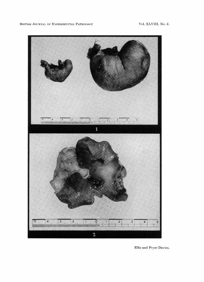
The dilated stomachs (Fig. 1) varied considerably in weight depending onwhether they were distended with food debris or partially with gas at the timeof sacrifice.
The gastric ulceration (Fig. 2) was confined to the body of the stomach,occurred either on the lesser curvature itself or within one cm. of it on either side,and was never seen either in the antrum or rumen. The ulcers were usually solitarybut in 2 animals 2 ulcers occurred and in one, 3 small erosions were found. Ofthe total of 21 ulcers, 2 were 1 mm. in diameter, six 2 mm., seven 3 mm., two 4 mm.,one 6 mm., 2 measured 7 x 3 mm., and one was a long erosion measuring10 mm. x 1 mm. On naked eye inspection, 8 of the ulcers appeared as super-ficial erosions and 13 as typical penetrating ulcers. Although not occurring inthis series, perforations were encountered in other experiments, either plugged withomentum or else free into the peritoneal cavity.
Histologically, all grades of ulceration were seen from superficial erosions,through sub-acute ulcers which penetrated down to the muscle layer, to typicalchronic ulceration with the muscle wall completely replaced by fibrous andinflammatory tissue.
No relationship was found between the extent of the ulceration histologicallyand the time from vagotomy, which ranged from 1-32 weeks.
Ten control rats submitted to laparotomy only and killed at 32 weeks hadundilated stomachs of mean weight 8-1 g. (range 6 to 11 g.). The differencesbetween the stomach weights of the controls and incompletely vagotomisedanimals on the one hand and those with dilated stomachs on the other werestatistically significant (Table I)
TABLE-Stomach Weights (g.)No. of animals Mean S.D.
Laparotomy . . 10 . 81 1-44Non-dilated
" Failed Vagotomy" . 13 8-8 . 2-41DilatedNot ulcerated . 20 25 10-34Dilated
Ulcerated . 17 26- 5 9 95
Of the 12 immature vagotomised rats killed after 19 weeks, 3 were consideredto have been incompletely vagotomised, with non-dilated stomachs. Nineanimals had dilated stomachs and 3 of these demonstrated gastric ulceration.
Among the 14 adult rats vagotomised specifically for histochemical studies,4 showed gastric ulcers.

H. ELLIS AND J. PRYSE-DAVIES
Thus in a total of 60 rats in whom vagotomy appeared to be complete, 24examples of gastric ulcer were encountered (40 per cent).
Gastric ulceration was not seen in any of the animals submitted to laparotomy-only, nor in many dozens of autopsies carried out on rats of the same strain livingunder the same laboratory conditions who had been subjected to a wide range ofother experimental procedures in this department.
OesophagusNone of the animals with a non-dilated stomach showed any abnormality of
the oesophagus. One animal with a dilated stomach but without ulcerationshowed dilatation of the oesophagus with a diameter of between 3-4 mm. and 2of the animals showing ulceration of the stomach showed more marked distentionof the oesophagus, in one to 5 mm. and the other to 6 mm. in diameter. In all3 cases the dilatation was confined to the lower part of the oesophagus, whichcontained food debris.
Weight and general nutritionAll the immature and adult vagotomised animals progressively gained weight
during the period of the experiment, even those which developed gastric ulceration.There was no significant difference in weight gain from controlled laparotomiesor from the growth weight of normal animals in this laboratory. In those rats withdilated stomachs, the excess weight of the stomach was taken into considerationin estimating the weight gain. There were no obvious changes in the generalappearance of the vagotomised animals compared with controls apart from, inmany cases, the obvious bulge of the distended stomach which could easily bepalpated through the abdominal wall.
Enzyme histochemistry of stomnachSuccinate dehydrogenase activity was present in all epithelial cells of the
fundic and pyloric regions but showed selectively heavier staining of the oxynticcells. Glucose-6-phosphate dehydrogenase showed a similar distribution. Neitherof the enzyme stains showed any significant loss of activity in the vagotomisedrats, apart from local loss of staining in the immediate vicinity of the erosions andulcers. Leucine aminopeptidase activity was absent from the glandular gastricmucosa apart from some staining of mast cells in the lamina propria in all speci-mens studied, both in the vagotomised and normal stomachs. No abnormalepithelial activity was seen in the region of the mucosal lesions to suggest theoccurrence of small intestinal metaplasia (Planteydt and Willighagen, 1960).Some increased activity was noted at the base of a subacute ulcer where there wasgranulation tissue and underlying fibrosis: such findings have been reported inwound healing (Monis, 1963).
Enzyme histochemistry of small intestineSuccinate dehydrogenase activity was present in both the superficial and
deeper glandular epithelium below the crypts of Lieberkuhn. Alkaline phos-phatase and leucine aminopeptidase were confined to the superficial villous epi-thelium apart from vascular endothelial staining by the phosphatase method.No significant difference in enzyme pattern could be seen between the control andvagotomised rats.
138

VAGOTOMY IN THE RAT
Mlorphology of small intestineThe rat small gut mucosa has a leaf-like villous pattern with the flat surfaces
arranged in a regular transverse manner. No significant difference was detectedin the small bowel of vagotomised rats from the appearances in the controlsl)ecimens. No abnormality of jejunal nor ileal mucosa was found in paraffinnor cryostat sections of any rat. Villi, best assessed in longitudinal sections.showed no evidence of stunted development nor of inflammatory change. Thevilli of the ileum were slightly smaller than those of the jejunum in all animals.
DISCUSSION
Gross appearances in the alimentary tract following vagotomyShay, Komarov, Fels, Meranze, Gruenstein and Siplet (1945) described the
production of single or multiple ulcers in the stratified squamous lined rumen ofthe rat's stomach within 17-19 hr of ligation of the pylorus. Ulceration was seenin 80 out of 85 animals submitted to this procedure. Pauls, Wick and MacKay(1946) confirmed these findings and indeed found that ulceration had taken placewithin 7-9 hr of pyloric ligation. They suggested the use of this preparationin the evaluation of anti-secretory and anti-ulcer agents. Harkins and hiscolleagues (Harkins, 1947; Harkins and Elliot, 1947 and Harkins and Hooker, 1947)described the protective effect of abdominal vagotomy on such animals; noulceration occurred in the vagotomised series at 24 hr compared with the uniformulceration in the control group of Shay ligations.
Shay, Komarov and Gruenstein (1949) gave a detailed account of their studyof rats vagotomised by the abdominal route in which at least one cm of eachnierve was resected, the proximal cut end then retracting 7-10 mm. above thediaphragm. They too noted the protective effect of this procedure in the preven-tion of the development of multiple ruminal ulcers in rats killed 17-19 hr afterpyloric ligation. In further experiments, in which vagotomised rats were allowedto survive, they described gross dilatation of the stomach and of the oesophagus,the latter reaching 5-10 mm. in diameter, with death occurring within a week.By performing repeated gastrotomies to remove food, debris and hair balls fromthe stomach, they were able to keep their animals alive for up to 120 days butnoted weight loss in most animals in these instances. Acid studies by the Shaytechnique showed considerable reduction in acid secretion in the vagotomisedrats compared with controls. No mention was made by these workers on themucosal appearances of the stomach or small intestine.
Girsh and Friedman (1951) found that both section and crushing of the vagiin the rat were equally effective in decreasing rumen ulceration and gastric secre-tion after pyloric ligation and noted, in chronic experiments, that gastric dilatationto a similar degree occurred in both groups. No comment was made about theoccurrence of gastric ulceration.
Dorchester (1959) also found an increase in the amount of food residue in thestomachs of rats vagotomised 1-9 weeks previously, noted a marked increase instomach tissue weight but did not remark on the mucosal appearances of thealimentary tract.
In the present study, ulceration of the lesser curvature of the body of thestomach was found in 24 out of 60 rats in whom we believe complete vagotomylhad been effected (40 per cent). These lesions showed histological appearances
139
H. ELLIS AND J. PRYSE-DAVIES
varying from acute, through sub-acute to the chronic type of peptic ulceration.The findings fall in line with the lesser curve gastric ulceration seen in man aftervagotomy without drainage (Dragstedt, 1956a; Slaney, Bevan and Brooke, 1956),and also with the interesting finding of Linares, de la Rosa, Woodward andDragstedt (1964) who carried out vagotomy in 10 rabbits and after 90 daysfound that 3 had developed active small gastric ulcers, 2 had healed ulcers and 1a gastric diverticulum devoid of mucosa. Twenty-four rabbits with vagotomycombined either with gastro-jejunostomy or pyloroplasty showed no such ulcera-tion.
The mechanism of this gastric ulceration in vagotomised rats, rabbits andman, when this procedure is performed without gastric drainage, would appear tobe a hyper-secretion of gastric juice of hormonal origin dependent upon prolongedor excessive liberation of gastrin from the antrum (Dragstedt, 1956b). Thisconcept agrees with the known occurrence of gastric ulceration in pyloric stenosisin man (Kreel and Ellis, 1965) and in the dog where pyloric obstruction has beenproduced by cellophane wrapping of the gastric outlet (Rosa, Linares, Woodwardand Dragstedt, 1964).
Unlike the gross dilatation of the oesophagus noted by Shay, Komarov andGruenstein (1949) dilatation of a mild degree was seen in only 3 of our animals.This may be accounted for by the fact that in the vagotomy performed by theseauthors a considerable length of the lower oesophagus was deprived of its vagalinnervation. In our own procedure the vagotomy was confined to the oesophago-cardiac zone.
Histology and histochemistry of stomach and small intestineBallinger, Jida, Aponte, Wirts and Goldstein (1964) reported mucosal atrophy
in the dog's small intestine following vagotomy, the effect being maximal at 3weeks after operation. Recovery then slowly took place and was completebetween the 12-21st week. These changes were somewhat preceded by reductionin succinic dehydrogenase and acid phosphatase and, to a lesser extent, alkalinephosphatase and non-specific esterase activity. Gradual improvement occurredby the 7th week and normality was restored by the 11th week. This work canbe criticised first in that it was complicated by an accompanying pyloroplastyand second in the absence of controls.
Sander (1965) was unable to detect any change in succinic dehydrogenase,lactic dehydrogenase or glucose-6-phosphate dehydrogenase in the gastric mucosaof a vagotomised rat compared with controls and with rats subjected to shamoperations.
We were unable to demonstrate changes in the glucose-6-phosphate dehydro-genase in the vagotomised stomach, alkaline phosphatase in the small intestine,or succinate dehydrogenase and leucine amino-peptidase in both stomach andsmall bowel. Nor could we confirm any alteration of villous morphology eitherat routine histological examination or surface study using the dissecting micro-scope.
EXPLANATION OF PLATEFIG. 1. Typical distended stomach in the vagotomised rat compared with normal control.FIG. 2. Chronic gastric ulcers in the body of the vagotomised stomach. The stomach has been
opened along the greater curve and shows one ulcer directly on the lesser curve and another1 cm. away from it.
140
BRITISH JOURNAL OF EXPERIMENTAL PATHOLOGY
1l -Dl - lul!s S:; _ i3
_I s s 2| . _ I | II _ I I I| - -|| | s|| X l . . . s 4 ss 5 4 3 2 t 0 21 3 4 S_| * = -s h A Su t--- .2 i_ * ^ . ......
Ellis and Pryse-Davies.
F-F17473 2 1 9 2 3! 41 5lltiSllli{fillllii Mi I I 1z _1'1__~~~~~~~~11c
Vol. XL171II, -No. 2.

VAGOTOMY IN THE RAT 141
Apart from the histological changes of the gastric ulcers already described, noother microscopic abnormalities were noted in the rest of the vagotomised stomach.
Faecal fat studies in the vagotomised rats are at present under investigationin our laboratory. Preliminary investigations indicate that the procedure hasno effect on the faecal fat excretion of these animals.
SUMMARY
Abdominal vagotomy, uncomplicated by gastric drainage, has been studied inboth adult and immature rats.
The vagotomised stomach becomes dilated and, in 40 per cent of cases,develops one or more gastric ulcers along the lesser curve of its body.
There are no histological or histochemical changes elsewhere in the alimentarycanal, nor does the procedure affect the growth rate of these animals.
We would like to thank Mr. K. Napier, Miss J. Sheerman-Chase and Mr.J. Haynes for their skilled technical assistance, and Dr. Peter Hansell and theDepartment of Medical Photography, Westminster Medical School for the illus-trations.
REFERENCESBALDWIN, J. N., ALBO, R., JAFFE, B. AND SILEN, W.-(1965) Surgery Gynec. Obstet.,
120, 777.BALLINGER, W. F., IIDA, J., APONTE, G. E., WIRTS, C. W. AND GOLDSTEIN, F.-(1964)
Surgery Gynec. Obstet., 118, 1305.DORCHESTER, J. E. C. (1959) Proc. Soc. exp. Biol. Med., 102, 49.DRAGSTEDT, L. R. (1956a) Am. J. Roentgenol., 75, 219-(1956b) Gastroenterology,
30, 208.GIRSH, L. S. AND FRIEDMAN, M. H.-(1951) Am. J. Physiol., 167, 787.GOLDING, M. R., MENDOZA, M., AIELLO, R. G., FIERST, S. M., SOLOMON, N. A. AND
ENQUIST, I. F.-(1965) Am. J. Surg., 109, 21.HARKINS, H. N.-(1947) Bull. Johns Hopk. Hosp., 80, 1-176.HARKINS, H. N. AND ELLIOTT, S. R.-(1947) Fed. Proc., 6, 123.HARKINS, H. N. AND HOOKER, D. H. (1947) Surgery, 22, 239.HESS, R., SCARPELLI, D. G. AND PEARSE, A. G. E.-(1958) Nature, Lond., 181, 1531.KREEL, L. AND ELLIS, H.-(1965) Gut, 6, 253.LINARES, C. A., DE LA ROSA, C., WOODWARD, E. R. AND DRAGSTEDT, L. R.- (1964)
Arch. Surg., 88, 932-938.MONIS, B.-(1963) Am. J. Path., 42, 310.NACHLAS, M. M., CRAWFORD, D. T. AND SELIGMAN, A. M. (1957) J. Histocheln. Cyto-
chem., 5, 264.PAULS, F., WICK, A. N. AND MACKAY, C. A. (1946) Science, 103, 673.PEARSE, A. G. E. (1957) J. Histochem. Cytochem., 5, 515.-(1960) 'Histochemistry:
Theoretical and applied', London, (Churchill.) p. 914.PLANTEYDT, H. T. AND WILLIGHAGEN, R. G.-(1960) J. Path. Bact., 80, 317.PRYSE-DAVIES, J., DAWSON, I. M. P. AND SNAPE, I. M.-(1961) J. Path. Bact., 81, 197.DE LA ROSA, C., LINARES, C. A., WOODWARD, E. R. AND DRAGSTEDT, L. R.-(1964)
Arch. Surg., 88, 927.SANDER, S. (1965) Acta chir. scand., 129, 81.SHAY, H., KOMAROV, S. A., FELS, S. S., MERAN-ZE, D., GRUENSTEIN, M. AND SIPLET, H.-
(1945) Gastroenterology, 5, 43.SHAY, H., KOMAROV, S. A. AND GRUENSTEIN, M.-(1949) Arch. Surg., 59, 210.SLANEY, G., BEVAN, P. G. AND BROOKE, B. N.-(1956) Lancet, ii, 221.WASTELL, C.-(1966) Br. med. J. 1 1198.