Bioavailability of nanoparticulate hematite to Arabidopsis thaliana
Upload
independentCategory
view
0download
0
The Arabidopsis thaliana SOMATIC EMBRYOGENESISRECEPTOR-LIKE KINASES1 and 2 Control Male Sporogenesis
Catherine Albrecht,a Eugenia Russinova,a Valerie Hecht,b Erik Baaijens,a and Sacco de Vriesa,1
a Laboratory of Biochemistry, Department of Agrotechnology and Food Sciences, Wageningen University and
Research Centre, 6703 HA Wageningen, The Netherlandsb School of Plant Science, University of Tasmania, Hobart, Tasmania 7001, Australia
The Arabidopsis thaliana SOMATIC EMBRYOGENESIS RECEPTOR-LIKE KINASE (SERK) family of plasma membrane
receptors consists of five closely related members. The SERK1 and SERK2 genes show a complex expression pattern
throughout development.Bothare expressed inanther primordia up to thesecondparietal division. After thispoint, expression
ceases in the sporocytes and is continued in the tapetum andmiddle layer precursors. Single knockoutmutants ofSERK1 and
SERK2 show no obvious phenotypes. Double mutants of SERK1 and SERK2 are completely male sterile due to a failure in
tapetum specification. Fertility can be restored by a single copy of either gene. The SERK1 and SERK2 proteins can form
homodimers or heterodimers in vivo, suggesting they are interchangeable in the SERK1/SERK2 signaling complex.
INTRODUCTION
In plants, embryogenesis can be recapitulated starting from
single somatic cells in culture (Reinert, 1959). This remarkable
observation implies that plant cells are able to achieve totipo-
tency long after completion of embryogenesis. Several genes
have been described that alter the totipotency status of somatic
cells, either upon mutation or after ectopic expression (reviewed
in Mordhorst et al., 1997, 2005). The natural occurrence of par-
thenogenesis and apomixis and the ease by which embryogen-
esis can be initiated from microspores suggest that female and
male gametophytic cells already contain the property to develop
embryos prior to the occurrence of fertilization (Bicknell and
Koltunow, 2004).
The Daucus carota Somatic Embryogenesis Receptor-like
Kinase (DcSERK) gene was found to be a marker for single
embryogenic cells in culture (Schmidt et al., 1997). The predicted
protein structure of the SERKproteins start at theN terminuswith
a signal peptide followed by a Leu zipper domain, five Leu-rich
repeats (LRRs), a Pro-rich domain called the Ser-Pro-Pro motif,
a single transmembrane domain, the 11 conserved subdomains
of a Ser-Thr kinase, and a C-terminal Leu-rich domain (Hanks
et al., 1988). The hallmark of the SERKproteins is the presence of
the extracellular Ser-Pro-Pro motif in combination with precisely
five LRRs.
According to the most recent annotation, the Arabidopsis
thaliana (SERK) family of receptor kinases now consists of five
closely related members (Hecht et al., 2001). They are annotated
as SERK1 to SERK5 and all belong to subclass LRR II of the
Arabidopsis receptor-like kinase (RLK) family (Shiu andBleecker,
2001). The genomic organization of these genes is highly con-
served (i.e., the intron–exon boundaries are in the same position
in all five genes).
Overexpression of the Arabidopsis ortholog SERK1, closest to
carrot DcSERK in predicted protein sequence, resulted in en-
hanced formation of embryogenic cells in response to the growth
regulator 2,4-D (Hecht et al., 2001). This supports the notion that
SERK1-mediated signaling is involved in embryogenic cell for-
mation. In planta, the SERK1 protein is located in the plasma
membrane of both the male and female gametophytes as well as
in surrounding sporophytic primordial cell layers. Later, the pro-
tein is found in all cells of the embryo and in seedlings predom-
inantly in the vascular tissue of roots, shoots, and leaves. Lower
levels of the protein are seen in epidermal cells, while formation
of lateral roots appears to be accompanied with a higher level of
SERK1 protein (Hecht et al., 2001; Kwaaitaal et al., 2005). Ap-
parently, the signaling mediated by SERK1 is required in cells
that retain or regain their embryogenic potential but is also re-
quired in cells that participate in organogenesis.
The SERK2 gene is most closely related to SERK1, with an
overall amino acid identity of 90%. Phylogenic analysis suggests
that SERK1 and SERK2 have evolved by recent duplication and
are closest homologues (Hecht et al., 2001).
The SERK3 gene is identical to the recently described BRI1-
Associated Receptor Kinase1 (BAK1) gene, of which a knockout
mutation results in a semidwarf phenotype, reminescent of weak
alleles of the brassinosteroid-insensitive1 (BRI1) receptor (Li
et al., 2002; Nam and Li, 2002). SERK3 may be involved in
relocalizing the BRI1 receptor via accelerated endocytosis
(Russinova et al., 2004). BRI1 binds brassinosteroids directly, and
this binding was not affected in a bak1 background (Kinoshita
et al., 2005), suggesting that BAK1 is a non-ligand-binding
coreceptor of BRI1. In general, bri1mutants exhibit a muchmore
severe phenotype than bak1 (serk3) alleles, suggesting that BRI1
1 To whom correspondence should be addressed. E-mail [email protected]; fax 31-317-484801.The author responsible for distribution of materials integral to thefindings presented in this article in accordance with the policy describedin the Instructions for Authors (www.plantcell.org) is: Sacco de Vries([email protected]).Article, publication date, and citation information can be found atwww.plantcell.org/cgi/doi/10.1105/tpc.105.036814.
This article is published in The Plant Cell Online, The Plant Cell Preview Section, which publishes manuscripts accepted for publication after they
have been edited and the authors have corrected proofs, but before the final, complete issue is published online. Early posting of articles reduces
normal time to publication by several weeks.
The Plant Cell Preview, www.aspb.orgª 2005 American Society of Plant Biologists 1 of 13

is able to carry out the entire brassinolide signaling in the ab-
sence of BAK1 or that the BAK1 function is taken over by a close
homologue. An alternative explanation would be that SERK3
controls only a subset of the BRI1-mediated effects resulting in
only part of the complete brassinosteroid phenotypic defect.
Candidates for genes carrying out other elements of the highly
complex brassinosteroid defect would be other members of the
BRI family (Cano-Delgado et al., 2004) and perhaps other mem-
bers of the SERK family.
For the last members of the family, SERK4 and SERK5, no
additional evidence is currently available other then being pre-
dicted to be the closest paralogs of SERK3 (BAK1).
In this work, we show that SERK1 and SERK2 genes are ex-
pressed in the same cells throughout development. SERK1 and
SERK2 proteins can form either homodimers or heterodimers
in vivo.We also present genetic evidence that SERK1andSERK2
are functionally redundant and together control the formation of
functional microspores.
RESULTS
Characterization of SERK1 and SERK2 Null Mutant Alleles
The serk1-1 T-DNA–tagged allele was identified in the SIGnAL
TDNA-Express collection as nr 044330. serk1-1 has an insertion
at nucleotide 3109 starting from the translation initiation codon.
This corresponds to exon 11, encoding the last kinase subdo-
main XI and the C-terminal region of the protein (Hanks et al.,
1988). The insertion leads to a truncated protein lacking the last
103 amino acids. DNA gel blot mapping confirms that serk1-1
carries a T-DNA insertion in the expected genomic DNA frag-
ments. No additional T-DNA insertions are detected in this line.
serk1-1 was backcrossed to Columbia wild type, and F1 and F2
progenies were analyzed for segregation of the kanamycin-
resistant phenotype and the T-DNA insertion in SERK1. The F2
progeny segregated 3:1 (190 kanamycin-resistant plants:45
kanamycin-sensitive plants). Of 129 plants, PCR analysis showed
that 30were homozygouswild-type, 70 were heterozygotes, and
29 were homozygous T-DNA–tagged alleles. As such, the seg-
regation ratio of the F2 progeny was approximately 1:2:1 (wild
type:heterozygous:mutant), indicating a Mendelian segration for
a single locus serk1-1. These data together with the DNA gel blot
mapping are consistent with the fact that this T-DNA insertion
line is a single insertion line in the SERK1 gene.
The second allele, serk1-2 (nr 053021), was characterized as
described for serk1-1 and has an insertion in exon 11, 87 bp
downstream of the serk1-1 insertion site leading to a truncated
protein lacking the last 74 amino acids. The locations of the
serk1-1 and serk1-2 T-DNA insertions are presented in Figure 1A.
The serk2-2 T-DNA–tagged allele was identified in the Syn-
genta Arabidopsis Insertion Library (SAIL) lines (nr 119-G03).
serk2-2 has an insertion at nucleotide 1655 from the translation
Figure 1. Characterization of SERK1 and SERK2 Loci.
(A) Characterization of the SERK1 locus. Arrowheads 1 and 2 indicate the positions of the T-DNA insertions in serk1-1 and serk1-2 mutant alleles,
respectively. The position of the primers used for RT-PCR and genotyping analysis is indicated. Bar ¼ 1 kb.
(B) Characterization of the SERK2 locus. The arrowhead indicates the position of the T-DNA insertion in the serk2-2 mutant allele. The position of the
primers used for RT-PCR and genotyping analysis is indicated. Bar ¼ 1 kb.
2 of 13 The Plant Cell

initiation codon and is disrupting the 59 donor splicing site of the
6th intron. No additional T-DNAs are detected by DNA gel blot
analysis. serk2-2 was backcrossed with Columbia and the F2
progeny analyzed by segregation of the BASTA-resistant phe-
notype and by PCR-assisted genotyping. The results indicate
that this T-DNA insertion line also represents a single insertion in
the SERK2 gene. The location of the serk2-2 T-DNA insertion is
presented in Figure 1B.
To determine whether the alleles identified are null mutants,
RT-PCR was used to detect SERK1 and SERK2 transcripts in
plants shown to be homozygous for the tagged alleles. SERK1
transcripts could be detected in serk1-1 (Figure 2A). The serk1-1
mutant allele may therefore not represent a full knockout. A
SERK2 transcript containing sequences upstream of the T-DNA
insertion can be detected (data not shown), but no SERK2
transcripts downstream of the T-DNA insertion were observed in
serk2-2mutant plants (Figure 2B). Because the truncatedSERK2
transcripts lack the entireSERK2 kinase domain, we assume that
the serk2-2 allele is a null mutant.
In order to investigate whether the truncated serk1-1 encodes
a functional protein, we engineered a modified SERK1 kinase
missing the last 90 amino acids corresponding to kinase sub-
domain XI and the C-terminal region. The SERK1kin and deletion
mutant SERK1kinDD536-R626 constructs were tested for kinase
activity in vitro. Figure 2D shows that the SERK1kinDD536-R626
mutant protein no longer autophosphorylates, indicating that
subdomain XI and the C-terminal region of the SERK1 protein are
essential for kinase activity. We conclude that the serk1-1 allele
encodes a nonfunctional kinase.
The serk1-1 and serk2-2 T-DNA insertion alleles contain a sin-
gle T-DNA insertion in the SERK1 and SERK2 genes. The homo-
zygous serk1-1, serk1-2, and serk2-2 single mutant plants are
morphologically indistinguishable from wild-type plants, display
normal growth and development, and are fully fertile (data not
shown).
SERK1 and SERK2 Expression Patterns Are Overlapping
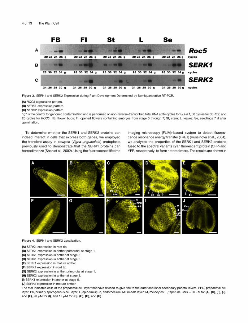
Semiquantitative RT-PCR analysis was used to compare SERK1
and SERK2 expression in wild-type plants. Transcripts of SERK1
and SERK2 were most abundant in floral buds but were also
present at a lower level in the different vegetative tissues (Figures
3 and 4A to 4F).
C-terminal fusions between SERK1 and yellow fluorescent
protein (YFP) (Kwaaitaal et al., 2005) as well as SERK2 and YFP
were expressed in Arabidopsis under control of their respective
promoters. Fluorescent fusion protein localization was then fol-
lowed inmore detail during root and anther development by con-
focal microscopy. In the wild type, both the tapetum and the
developing microspores up to and including the differentiation of
the descendents of the second parietal division contain the
SERK1 (Kwaaitaal et al., 2005; Figures 4B to 4E) and SERK2
proteins (Figures 4G to 4J). After that stage, both proteins are
found to be specifically present in the membranes of the de-
veloping tapetal andmiddle cell layers. By the end of stage 5, the
receptors are no longer found in meiocytes (Figures 4E and 4J).
This pattern of fusion protein localization is in accordance with
the mRNA in situ hybridization data presented by Colcombet
et al. (2006), who report that the expression pattern of both genes
is initially uniform and at stage 5 of anther development is
restricted to the middle layer and tapetum tissues. However, we
also observe SERK1-green fluorescent protein fusions at a low
level in the endothecium and epidermal cell layers of the anther.
These differences may be caused either by technical limitations
or differences in the stability of the SERK1 and SERK2 mRNA
compared with the respective proteins. Our results also confirm
that SERK2 protein localization largely overlaps with that of
SERK1. However, differences are noted: SERK2 proteins are
more abundant in the root vasculature, while SERK1 proteins are
more abundant in the epidermal cell layers of the root (Figures 4A
and 4F). It remains to be determinedwhether these local changes
in abundance have functional importance
Figure 2. Molecular Analysis of serk1-1 and serk2-2 Mutant Alleles.
(A) Semiquantitative RT-PCR analysis of SERK1 transcripts in the wild
type and the serk1-1 mutant.
(B) Semiquantitative RT-PCR analysis of SERK2 transcripts in the wild
type and the serk2-2 mutant. The ROC5 gene was amplified simulta-
neously as a control in both experiments.
(C) Bacterially produced SERK1kin and SERK1kinDD536-R626 proteins were
affinity purified and incubated with [g-32P]ATP. After separation on 10%
SDS-PAGE, the resulting gels were stained with Coomassie blue.
(D) Same gels after autoradiography using a PhosphorImager.
SERK1 and SERK2 Control Male Sporogenesis in Arabidopsis 3 of 13
To determine whether the SERK1 and SERK2 proteins can
indeed interact in cells that express both genes, we employed
the transient assay in cowpea (Vigna unguiculata) protoplasts
previously used to demonstrate that the SERK1 proteins can
homodimerize (Shah et al., 2002). Using the fluorescence lifetime
imaging microscopy (FLIM)–based system to detect fluores-
cence resonance energy transfer (FRET) (Russinova et al., 2004),
we analyzed the properties of the SERK1 and SERK2 proteins
fused to the spectral variants cyan fluorescent protein (CFP) and
YFP, respectively, to form heterodimers. The results are shown in
Figure 3. SERK1 and SERK2 Expression during Plant Development Determined by Semiquantitative RT-PCR.
(A) ROC5 expression pattern.
(B) SERK1 expression pattern.
(C) SERK2 expression pattern.
‘‘g’’ is the control for genomic contamination and is performed on non-reverse-transcribed total RNA at 34 cycles for SERK1, 30 cycles for SERK2, and
26 cycles for ROC5. FB, flower buds; FI, opened flowers containing embryos from stage 0 through 7; St, stem; L, leaves; Se, seedlings 7 d after
germination.
Figure 4. SERK1 and SERK2 Localization.
(A) SERK1 expression in root tip.
(B) SERK1 expression in anther primordial at stage 1.
(C) SERK1 expression in anther at stage 3.
(D) SERK1 expression in anther at stage 5.
(E) SERK1 expression in mature anther.
(F) SERK2 expression in root tip.
(G) SERK2 expression in anther primordial at stage 1.
(H) SERK2 expression in anther at stage 3.
(I) SERK1 expression in anther at stage 5.
(J) SERK2 expression in mature anther.
The star indicates cells of the preparietal cell layer that have divided to give rise to the outer and inner secondary parietal layers. PPC, preparietal cell
layer; PS, primary sporogenous cell layer; E, epidermis; En, endothecium; Ml, middle layer; M, meiocytes; T, tapetum. Bars ¼ 50 mM for (A), (D), (F), (J),
and (E), 20 mM for (I), and 10 mM for (B), (C), (G), and (H).
4 of 13 The Plant Cell
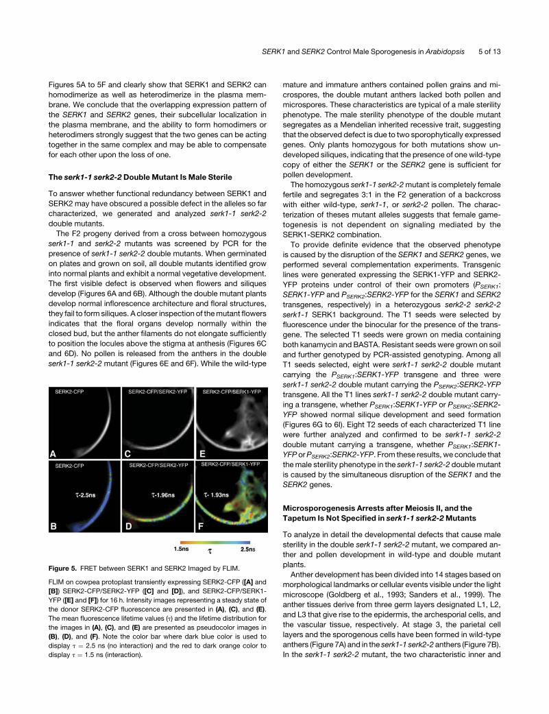
Figures 5A to 5F and clearly show that SERK1 and SERK2 can
homodimerize as well as heterodimerize in the plasma mem-
brane. We conclude that the overlapping expression pattern of
the SERK1 and SERK2 genes, their subcellular localization in
the plasma membrane, and the ability to form homodimers or
heterodimers strongly suggest that the two genes can be acting
together in the same complex and may be able to compensate
for each other upon the loss of one.
The serk1-1 serk2-2 Double Mutant Is Male Sterile
To answer whether functional redundancy between SERK1 and
SERK2 may have obscured a possible defect in the alleles so far
characterized, we generated and analyzed serk1-1 serk2-2
double mutants.
The F2 progeny derived from a cross between homozygous
serk1-1 and serk2-2 mutants was screened by PCR for the
presence of serk1-1 serk2-2 double mutants. When germinated
on plates and grown on soil, all double mutants identified grow
into normal plants and exhibit a normal vegetative development.
The first visible defect is observed when flowers and siliques
develop (Figures 6A and 6B). Although the double mutant plants
develop normal inflorescence architecture and floral structures,
they fail to form siliques. A closer inspection of themutant flowers
indicates that the floral organs develop normally within the
closed bud, but the anther filaments do not elongate sufficiently
to position the locules above the stigma at anthesis (Figures 6C
and 6D). No pollen is released from the anthers in the double
serk1-1 serk2-2 mutant (Figures 6E and 6F). While the wild-type
mature and immature anthers contained pollen grains and mi-
crospores, the double mutant anthers lacked both pollen and
microspores. These characteristics are typical of a male sterility
phenotype. The male sterility phenotype of the double mutant
segregates as a Mendelian inherited recessive trait, suggesting
that the observed defect is due to two sporophytically expressed
genes. Only plants homozygous for both mutations show un-
developed siliques, indicating that the presence of one wild-type
copy of either the SERK1 or the SERK2 gene is sufficient for
pollen development.
The homozygous serk1-1 serk2-2mutant is completely female
fertile and segregates 3:1 in the F2 generation of a backcross
with either wild-type, serk1-1, or serk2-2 pollen. The charac-
terization of theses mutant alleles suggests that female game-
togenesis is not dependent on signaling mediated by the
SERK1-SERK2 combination.
To provide definite evidence that the observed phenotype
is caused by the disruption of the SERK1 and SERK2 genes, we
performed several complementation experiments. Transgenic
lines were generated expressing the SERK1-YFP and SERK2-
YFP proteins under control of their own promoters (PSERK1:
SERK1-YFP and PSERK2:SERK2-YFP for the SERK1 and SERK2
transgenes, respectively) in a heterozygous serk2-2 serk2-2
serk1-1 SERK1 background. The T1 seeds were selected by
fluorescence under the binocular for the presence of the trans-
gene. The selected T1 seeds were grown on media containing
both kanamycin and BASTA. Resistant seeds were grown on soil
and further genotyped by PCR-assisted genotyping. Among all
T1 seeds selected, eight were serk1-1 serk2-2 double mutant
carrying the PSERK1:SERK1-YFP transgene and three were
serk1-1 serk2-2 double mutant carrying the PSERK2:SERK2-YFP
transgene. All the T1 lines serk1-1 serk2-2 double mutant carry-
ing a transgene, whether PSERK1:SERK1-YFP or PSERK2:SERK2-
YFP showed normal silique development and seed formation
(Figures 6G to 6I). Eight T2 seeds of each characterized T1 line
were further analyzed and confirmed to be serk1-1 serk2-2
double mutant carrying a transgene, whether PSERK1:SERK1-
YFP orPSERK2:SERK2-YFP. From these results, we conclude that
themale sterility phenotype in the serk1-1 serk2-2 doublemutant
is caused by the simultaneous disruption of the SERK1 and the
SERK2 genes.
Microsporogenesis Arrests after Meiosis II, and the
Tapetum Is Not Specified in serk1-1 serk2-2Mutants
To analyze in detail the developmental defects that cause male
sterility in the double serk1-1 serk2-2 mutant, we compared an-
ther and pollen development in wild-type and double mutant
plants.
Anther development has been divided into 14 stages based on
morphological landmarks or cellular events visible under the light
microscope (Goldberg et al., 1993; Sanders et al., 1999). The
anther tissues derive from three germ layers designated L1, L2,
and L3 that give rise to the epidermis, the archesporial cells, and
the vascular tissue, respectively. At stage 3, the parietal cell
layers and the sporogenous cells have been formed in wild-type
anthers (Figure 7A) and in the serk1-1 serk2-2 anthers (Figure 7B).
In the serk1-1 serk2-2 mutant, the two characteristic inner and
Figure 5. FRET between SERK1 and SERK2 Imaged by FLIM.
FLIM on cowpea protoplast transiently expressing SERK2-CFP ([A] and
[B]) SERK2-CFP/SERK2-YFP ([C] and [D]), and SERK2-CFP/SERK1-
YFP ([E] and [F]) for 16 h. Intensity images representing a steady state of
the donor SERK2-CFP fluorescence are presented in (A), (C), and (E).
The mean fluorescence lifetime values (t) and the lifetime distribution for
the images in (A), (C), and (E) are presented as pseudocolor images in
(B), (D), and (F). Note the color bar where dark blue color is used to
display t ¼ 2.5 ns (no interaction) and the red to dark orange color to
display t ¼ 1.5 ns (interaction).
SERK1 and SERK2 Control Male Sporogenesis in Arabidopsis 5 of 13
outer cortical cell layers surrounding the sporogenous cells are
clearly visible, and no deviation from the wild-type development
was observed up to stage 4 (Figures 7B and 7E). At stage 5,
a wild-type anther is typically formed of five layers that are, from
the outside to the inside, the epidermis, endothecium, middle
layer, tapetum, and microsporocytes. The microsporocytes are
located at the center of the locules and are larger than the tapetal
cells and more rectangular in shape (Figure 7C). In the serk1-1
serk2-2 mutant anther at stage 5, no tapetal layer can be
identified. Instead, an increased number of microsporocytes is
observed compared with thewild type (Figure 7F). At stage 6, the
wild-type microsporocytes are isolated and detached from the
tapetum layer and the middle layer degenerates (Figure 7G). In
the doublemutant anther, enlarged andundetachedmicrosporo-
cytes are observed (Figure 7J). At stage 7, cytokinesis in the
wild-type anther is taking place, leading to the formation of
tetrads (Figure 7H). In the double mutant anther, degenerating
tetrads can be observed (Figure 7K). At stage 11, individual
spores are released from the tetrads, and the tapetum layer
degenerates in the wild-type anther (Figure 7I). In the double
mutant, the cell debris of the collapsed microspores occupies
the center of the locule and a highly vacuolated middle layer
persists (Figure 7L).
To confirm that the anther of the serk1-1 serk2-2 double
mutant lacks a tapetal cell layer, we tested the expression of
the ARABIDOPSIS THALIANA ANTHER7 (ATA7) gene, an early
tapetum marker (Rubinelli et al., 1998). ATA7 is not expressed in
the double serk1-1 serk2-2 mutant, providing molecular evi-
dence for the complete absence of tapetal layer specification in
the double mutant (Figure 8).
We observed that in the double serk1-1 serk2-2mutant anther
the tapetal cell layer is absent and an increased number of
microsporocytes developed. One possibility is that the additional
microsporocytes formed at stage 5 are derived from the inner
secondary parietal layer. To test that hypothesis, we examined
51 and 61 locules of wild-type and mutant anthers, respectively,
originating from three individual wild-type and double mutant
plants. In wild-type anthers, the average number of microsporo-
cytes and tapetal cells is 3.36 0.7 and 10.26 1.1, respectively.
In the double mutant anther, the average number of micro-
sporocytes is 9.7 6 1.2. This number is close to the sum of
microsporocytes and tapetal cells per wild-type locule. These
results indicate that, in the double serk1-1 serk2-2 mutant, cells
that normally develop into tapetal cells may have acquired
microsporocytic fate.
The transverse sections of the serk1-1 serk2-2 double mutant
anthers indicate that the differentiation of the meiocytes is ab-
normal starting from stage 6 onwards. To confirm that serk1-1
serk2-2 meiocytes enter and proceed into meiosis, we followed
the progression of meiosis in pollen mother cells derived from
wild-type and double mutant plants. Wild-type and double
mutant anther were therefore stained with propidium iodide
and visualized using confocal microscopy. In wild-type male
meiocytes, individual chromosomes are first detected at lepto-
tene, when they start to condense and form granular and thread-
like structures. Fully synapsed chromosomes can be visualized
as looped ribbon-like structures also called pachytene chromo-
somes (Figure 9A). The five paired chromosomes align along the
equator and can be detected as five bivalents at metaphase I.
The homologues separate at anaphase I (Figure 9C), and five
homologues migrate towards each opposite pole, leading to the
formation of two groups of five partially decondensed chromo-
somes at telophase I. At metaphase II, 10 condensed chromo-
somes are clearly visible and are distributed equally to the two
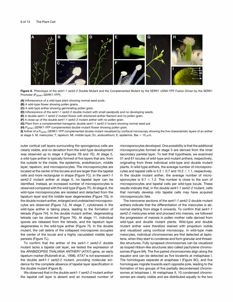
Figure 6. Phenotype of the serk1-1 serk2-2 Double Mutant and the Complemented Mutant by the SERK1 cDNA-YFP Fusion Driven by the SERK1
Promoter (PSERK1:SERK1-YFP).
(A) Inflorescence of a wild-type plant showing normal seed pods.
(B) A wild-type flower showing pollen grains.
(C) A wild-type anther showing germinating pollen grain.
(D) Inflorescence of the serk1-1 serk2-2 double mutant with small seedpods and no developing seeds.
(E) A double serk1-1 serk2-2 mutant flower with shortened anther filament and no pollen grain.
(F) A close-up of the double serk1-1 serk2-2 mutant anther with no pollen grain.
(G) Plant from a complemented transgenic double serk1-1 serk2-2 mutant showing normal seed pot.
(H) PSERK1:SERK1-YFP complemented double mutant flower showing pollen grain.
(I) Anther of a PSERK1:SERK1-YFP complemented double mutant visualized by confocal microscopy showing the five characteristic layers of an anther
at stage 5. M, meiocytes; T, tapetum; Ml, middle layer; En, endocethium; E, epidermis. Bar ¼ 10 mm.
6 of 13 The Plant Cell

sides of the organelle band into two groups of five each. At
anaphase II, the individual chromatids separate and move to-
ward the spindle poles (Figure 9E). Finally, at telophase II, four
groups of five chromatids are formed (Figure 9G). At the end of
meiosis, the external and intersporal walls of the tetrad are
dissolved to release individual microsporocytes by a mixture of
enzymes collectively referred to as callase (Frankel et al., 1969).
The individual microspores of the tetrad then initiate develop-
ment of the pollen wall (Figure 9I).
We examined microsporocytes derived from the double mu-
tant and found various stages of nuclear division, fromprophase I
to telophase II (Figures 9B, 9D, 9F, and 9H), suggesting that the
meiotic nuclear divisions in the double serk1-1 serk2-2 mutant
was essentially normal. However, in serk1-1 serk2-2microsporo-
cytes, the formed tetrads rapidly degenerate and disintegrate
completely. Finally, there is a complete absence of pollen in the
mature serk1-1 serk2-2 flowers (Figures 9J and 9L).
The phenotypic data of the serk1-1 serk2-2 mutant were all
confirmed with the second independent allele serk1-2 that we
used to create the serk1-2 serk2-2 double mutant (data not
shown).
DISCUSSION
SERK1 and SERK2 Are Required for Tapetal Cell Fate and
Microspore Formation
We report here that two members of the Arabidopsis LRR-RLK
family, SERK1 and SERK2, are essential for male sporogenesis.
In the serk1-1 serk2-2 and serk1-2 serk2-2 double mutants, the
tapetal cells are not specified. By contrast, additional sporocytes
are observed, suggesting that tapetal precursor cells can acquire
a meiocyte fate in the absence of SERK1/SERK2-mediated
signaling. The supernumerary mutant sporocytes enter and
proceed into meiosis until the tetrad stage, after which the
meiocytes degenerate, resulting in completemale sterility. These
results indicate that SERK1 and SERK2 are required for normal
Figure 7. Anther Development in the Double serk1-1 serk2-2 Mutant.
Micrographs of one of the four lobes of the wild type and double mutant
stained with Toluidine blue. PPC, primary parietal cells; PSC, primary
sporogenous cells; ISP, inner secondary parietal layer; OSP, outer
secondary parietal layer; Sp, sporocytes; E, epidermis; En, endothecium;
Ml, middle layer; Ms, microsporocytes; Msp, microspores; T, tapetal
layer; Tds, tetrads. Bars ¼ 10 mm.
(A) Wild-type anther at stage 3 showing the inner primary sporogenous
layer and the outer primary parietal layer.
(B) Wild-type anther at early stage 4 showing the inner and outer
secondary parietal cell layer.
(C) Wild-type anther at late stage 5 showing the well organized four
anther layers, the epidermis, the endothecium, the middle layer, and the
tapetum and the sporocytes developing in the middle of the locule.
(D) serk1-1 serk2-2 double mutant at stage 3 showing the inner primary
sporogenous layer and the outer primary parietal layer.
(E) serk1-1 serk2-2 double mutant anther at late stage 4 showing the two
characteristic inner and outer secondary parietal cell layers.
(F) serk1-1 serk2-2 double mutant anther at late stage 5 showing the
absence of the tapetal layer and an aberrant number of microsporocytes.
(G)Wild-type anther at stage 6 showing isolated microsporocytes, highly
vacuolated tapetum cells, a collapsing middle layer, the endocethium,
and the epidermis.
(H) Wild-type anther at stage 7 with tetrads.
(I) Wild-type anther at stage 11 with pollen grains and degenerating
tapetum.
(J) serk1-1 serk2-2 double mutant anther at stage 6 showing enlarged
and undetached microspores. The middle layer has not degenerated.
(K) serk1-1 serk2-2 double mutant anther at stage 7 with degenerating
tetrads.
(L) serk1-1 serk2-2 double mutant anther at stage 11 showing the cell
debris of the collapsed meiocytes and a persistent and highly vacuolated
middle layer.
Figure 8. Expression of ATA7 in the serk1-1 serk2-2 Double Mutant.
ATA7, a tapetum-specific marker, is not expressed in the serk1-1 serk2-2
double mutant. PCR products were collected after 28, 30, 32, and
34 cycles for ATA7 and after 20, 22, 24, and 26 cycles for ROC5. ‘‘g’’
stands for the control on genomic contamination and is performed on
non-reverse-transcribed total RNA at 34 cycles for ATA7 and 26 cycles
for ROC5.
(A) ROC5 expression pattern.
(B) ATA7 expression pattern.
SERK1 and SERK2 Control Male Sporogenesis in Arabidopsis 7 of 13

meiotic cytokinesis and subsequent spore formation but have no
apparent function in meiosis. Neither SERK1 nor SERK2 is ex-
pressed in the meiocytes at the stage of development in which
the defect in the double mutant is observed. Instead, both pro-
teins are present in tapetal cells in the wild type at the compa-
rable stage. Hence, the observed failure to complete meiocyte
cytokinesis in the double mutant is apparently the result of a
previous defect in signaling between the sporocytes and the
tapetal precursor cells.
In the serk1-1 serk2-2 and serk1-2 serk2-2 doublemutants, the
appearance of the developing meiocytes is different from their
wild-type counterparts. Therefore, SERK1 andSERK2might also
be involved in meiocyte differentiation at the time of cytokinesis.
We consider this unlikely due to the absence of expression of
both genes in the wild-type meiocytes at this stage. The ob-
served arrest in meiocyte cytokinesis in the double mutants is
therefore more likely to be a consequence of the absence of the
tapetal cell layer. In wild-type anthers, SERK1 and SERK2
proteins are highly expressed in the tapetal layer at the time of
meiocyte cytokinesis.
The essential role of tapetal cells for pollen formation was
demonstrated by ablation studies and genetic analysis. Ablation
studies made use of genetic lasers in transgenic tobacco (Nico-
tiana tabacum) plants. A tapetum-specific promoter (TA29) was
fused to several cytotoxic genes to selectivelydestroy the tapetum
during anther development. These studies show that destruction
of the tapetumby the cytotoxic gene led to the production ofmale-
sterile plants (Koltunow et al., 1990; Mariani et al., 1990). These
studies are further supported by the identification of mutants that
are defective in tapetumdevelopment. For example,ms1 andms3
mutations that cause abnormal vacuolation and enlargement of
the tapetal cells also lead to defects in microspores (Chaudhury
et al., 1994; Wilson et al., 2001; Ito and Shinozaki, 2002). These
studies indicate that destruction of the tapetal layer or defects of
tapetal cell formation result in the failure of pollen development.
Tapetal cells are important for pollen formation because they
provide callase and other proteins important for the release of
microspores from the tetrad aswell as chemicals important for the
formation of pollen outer walls (Mepham, 1970; Izhar and Frankel,
1971; Stieglitz, 1977). Based on the phenotypic deviations ob-
served in the double mutants, we therefore propose that SERK1
and SERK2 perform a fully interchangeable signaling role re-
quired formale sporogenesis. ExpressionofSERK1was observed
in the ovule, female gametophyte, early embryos, and vascular
cells of seedling roots, hypocotyls, and leaves (Hecht et al., 2001;
Kwaaitaal et al., 2005). The SERK2 protein has the same ex-
pression pattern (C. Albrecht, unpublished data), suggesting that
SERK1 andSERK2 are playing other roles in these tissues besides
their role in male sporogenesis.
SERK1, SERK2, and Other Tapetum Genes
Two genes with a phenotype very similar to the one reported for
the serk1-1 serk2-2 and serk1-2 serk2-2 double mutants have
been identified in Arabidopsis. The EXS/EMS gene encodes
a different putative Ser/Thr LRR-RLK (Canales et al., 2002; Zhao
Figure 9. Male Meiosis in the serk1-1 serk2-2 Double Mutant.
Micrographs of wild-type and serk1-1 serk2-2 double mutant anthers stained with propidium iodide and viewed by confocal microscopy. Bars¼ 10 mm
for (A) to (J) and 20 mm for (K) and (L).
(A) Wild type pachytene chromosomes.
(B) serk1-1 serk2-2 pachytene chromosomes.
(C) Wild-type anaphase I.
(D) serk1-1 serk2-2 anaphase I.
(E) Wild-type anaphase II.
(F) serk1-1 serk2-2 anaphase II.
(G) Wild-type telophase II.
(H) serk1-1 serk2-2 telophase II.
(I) Wild-type pollen grain after mitosis I.
(J) serk1-1 serk2-2 degenerated tetrads.
(K) Wild-type mature anther showing pollen grain.
(L) serk1-1 serk2-2 collapsed anther.
8 of 13 The Plant Cell

et al., 2002). TPD1 encodes a putative secreted protein and is
believed to be involved in the same signaling pathway (Yang
et al., 2003). These two genes affect microspore formation as
a result of the absence of a functional tapetal cell layer. In both
the exs/ems and tpd1 mutants, as well as in the serk1-1 serk2-2
and serk1-2 serk2-2 double mutants, the absence of the tapetal
cell layer results in an increase of the number of meiocytes
developed. The reciprocal effect was recently observed in
mutants of the MYB33 and MYB65 genes, where the tapetum
cells are seen to expand at the expense of the meiocytes, finally
resulting in complete male sterility (Millar and Gubler, 2005).
Apparently, reciprocal control between the gametophytic and
the sporophytic tissues is required to maintain the appropriate
balance between meiocyte and tapetum fate in order to finally
develop fertile pollen. Recently, it was shown that theAGAMOUS
transcriptional activator controls microspore development by
activation of the SPOROCYTELESS (SPL) gene (Ito et al., 2004).
The SPL/NOZZLE (NZZ) gene, a nuclear protein related toMADS
box transcription factors, is required for archesporial specifica-
tion (Schiefthaler et al., 1999; Yang et al., 1999). The SPL gene is
expressed in the sporogenous cells and their descendants, the
sporocytes. Yang et al. (1999) suggested that SPL/NZZ regulates
the expression of genes necessary for sporocyte cell fate de-
termination. They further suggested that a signal from develop-
ing sporocytes is necessary for proper differentiation and growth
of the adjacent parietal cell layer. SERK1-SERK2 as well as EXS/
EMS and TPD1 genes clearly act downstream of SPL/NZZ since
meiocytes are being properly specified in the former mutant
backgrounds. During anther development, the SERK1, SERK2,
andEXS/EMS genes are expressed in the same parietal cells and
their precursors. Thus, SERK1 and SERK2 proteins, perhaps
together with EXS/EMS, may be involved in the perception of
a signal from developing microsporocytes to immature tapetal
cells, as proposed by Yang et al. (1999) for EXS/EMS. The TPD1
gene encodes a putative secreted protein, is predominantly ex-
pressed in microsporocytes, and may represent the signal from
developing microsporocytes to surrounding tapetal cells. There
is molecular-genetic evidence that the TDP1 and EXS/EMS genes
are involved in the same pathway (Yang et al., 2005). While the
serk1-1 serk2-2 double mutant clearly phenocopies the tpd1 and
exs/ems mutants, it remains to be determined whether the
SERK1 andSERK2proteins also act in the TPD1-EXS/EMSpath-
way or are part of a separate signal transduction pathway.
Different explanations have been proposed for the phenotype
of the exs/ems mutant. The observed absence of tapetum cells
and overproliferation of meiocytes can be the result of a failure to
assume the tapetum cell fate of the descendants of the inner
secondary parietal cells and the adoption of a meiocyte fate
(Zhao et al., 2002). Alternatively, the formation of extrameiocytes
is proposed to result froma failure to restrict archesporial cell fate
in exs/ems mutant anthers (Canales et al., 2002). Our data sug-
gest amodel that is close to that of Zhao et al. (2002). In wild-type
Figure 10. A Model for the SERK1-SERK2 Signaling Pathway in the Anther.
TDP1 and/or brassinosteroids produced in the developing meiocytes (Sp) signal the surrounding immature tapetal cells (ISP) through SERK1-SERK2/
EMS-EXS or SERK1-SERK2/BRI1, respectively. Perception of the TPD1 and/or brassinosteroid signals ensures specification and maintenance of the
tapetal and middle cell fate. One proposed function of the tapetum is to inhibit further proliferation of the meiocytes. In the serk1-1 serk2-2 double
mutant, specification of the tapetal cells does not occur. As a result, tapetal cells either adopt a meiocyte fate or the inner secondary parietal layer cells
only form the middle layer. In both situations, formation of extra meiocytes occurs. Red-colored cells are expressing the SERK1 and SERK2 genes
moderately (light red) or highly (dark red). Only anther stages 2 to 5 are represented. AC, archesporial cell; PPC, preparietal cell; PS, primary
sporogenous cell layer; ISP, inner secondary parietal layer; OSP, outer secondary parietal layer; Sp, sporocyte; BLs, brassinosteroids.
SERK1 and SERK2 Control Male Sporogenesis in Arabidopsis 9 of 13

anthers, SERK1/SERK2-mediated signaling can be employed to
provide the tapetum fate to the inner descendants of the inner
secondary parietal cells and could also be required for mainte-
nance of the tapetum fate in stage 5. The tapetum cells then
provide a second unknown signal to arrest multiplication of the
stage 5 meiocytes. In serk1-1/2 serk2-2 anthers, the observed
extra meiocytes can then arise as proposed by Zhao et al. (2002)
or from a series of extra cell divisions of themeiocytes. Presently,
we have no evidence to support one or the other model for extra
meiocyte formation. However, one can easily envisage the
requirement of multiple, parallel signaling pathways involved in
fate determination, maintenance, and proliferation in the de-
veloping anther (Figure 10).
SERK Genes Can Be Coreceptors in Multiple Receptor
Kinase Complexes
While the expression of the SERK1 and SERK2 genes is fairly
broad (Kwaaitaal et al., 2005), suggesting that both genes play
a general role in plants, the phenotype of the serk1-1 serk2-2
mutant is restricted to tapetal cells in the anther.
Previously, we reported that overexpression of the SERK1
gene results in enhanced formation of embryogenic cells in
tissue culture (Hecht et al., 2001). Comparing the formation of
somatic embryos in culture of the serk1-1 and serk2-2 mutants
using previously published protocols (Mordhorst et al., 1998)
revealed no deviation from the wild type (C. Albrecht and S.C. de
Vries, unpublished data). This suggested that there is redun-
dancy in the proposed function of SERK1 in embryogenic cell
formation in culture. We cannot exclude that this is due to the
presence of the other member of the family due to the difficulty of
molecularly characterizing homozygous double serk1-1 serk2-2
immature mutant embryos and seedlings simultaneously with
testing their embryogenic potential in culture. serk1-1 serk2-2
mutants show no visible alteration in embryo development,
suggesting functional redundancy of other SERK receptors
during embryo development.
As shown for the ERECTA family RLKs (Shpak et al., 2003),
tissue-specificand redundant expression of functionally equivalent
SERK receptors might play a regulatory role in different aspects of
plant development. The SERK3/BAK1 gene has been identified as
the coreceptor of BRI1 (Li et al., 2002; Nam and Li, 2002). The
serk3/bak1mutant phenotype is weaker than bri1, suggesting that
SERK3 controls only a subset of the BRI1-mediated effects. Other
SERK genes are therefore good candidates to mediate other
elements of BRI1 signaling. Among the many phenotypic defects
observed in strong bri1mutant alleles is male sterility, although the
defects have not been investigated in detail (Clouse et al., 1996).
Brassinosteroids were actually first isolated from pollen extracts,
where theyare produced inmassiveamounts (Groveet al., 1979). It
is therefore tempting to speculate that SERK1 and SERK2, being
close homologs of SERK3, canmediate BRI1 aswell as EXS/EMS-
mediated signaling during anther development. BRI1 and BRL1
proteins have significant similarity to the EXS/EMS receptor (Zhou
et al., 2004). An attractive scenario could therefore be that the
SERK1 and SERK2 proteins act as coreceptors of the EXS/EMS
receptor kinase in the anther and perhaps as coreceptors of other
receptors, such as BRI1, in other tissues as well as in anthers
(Figure 10). This may represent an analogous situation to, for
example, the TGFb complexes in animal cells where the presence
of particular main receptors in combination with coreceptors
defines the ability of the cell to respond to various ligands (Di
Guglielmo et al., 2003; Gonzales-Gaitan, 2003).
METHODS
Plant Materials and Growth Conditions
TheArabidopsis thalianaColumbia ecotypewas used aswild-type parent
throughout. Seeds were surface sterilized, chilled at 48C for 2 d, and then
germinated and grown on plant growth Murashige and Skoog medium
(Murashige and Skoog, 1962) supplemented with 1% sucrose at 228C
under a 16-h-light/8-h-dark photoperiod. One or two weeks after germi-
nation, seedlings were transferred to soil and grown to maturity in the
same temperature and light conditions. The antibiotic plate assays were
performed by supplementing the Murashige and Skoog medium with
25 mg�L�1 kanamycin or 15 mg�L�1 BASTA.
RT-PCR Analysis and Primers Used
RNA isolation, cDNA synthesis, and RT-PCR were performed as de-
scribed by Hecht et al. (2001). For the detection of the SERK1 and SERK2
transcripts in their respective mutant backgrounds, cDNA synthesis was
performed using random hexamer primers (Amersham Biosciences)
instead of oligo(dT) primers. PCR products were collected after various
cycles depending on the transcript amplified; the number of cycles used
for each transcript is indicated in the figure. For the SERK1 and SERK2
expression patterns, the primer combinations used were V3 and S1utr39
for SERK1, GSP4 and GSP1 for SERK2, and ATA7f and ATA7r for ATA7.
The cyclophilin ROC5 gene constitutively expressedwas used as control,
and the primers specific for this gene were ROC5-5 and ROC5-3. For the
detection of the SERK1 and SERK2 transcripts in their respective mutant
backgrounds, the primers usedwere V2 andS3 forSERK1 andS2-K2 and
GSP3 for SERK2. The primers used in this study are as follows: P2F
(59-TTTTGTTGAATTCACAATCTCTGCACC-39); P2-NcoR (59-CCCCAT-
GGACCAAAAAAAAGCAAATTTCTCCTCCC-39); P1F (59-CTATGTGGC-
GAAAACTCACTTAATCTG-39); P1-NcoR (59-CGACTCCATGGCAAACA-
ACAATGCTAAATTTCG-39); GSP4 (59-GATCTGGGAGGAGAAATTTGC-39);
Tag1R (59-TGTTGCCGGTCTTGCGATGATTAT-39); GSP1 (59-CGG-
CTAGTAACTGGGCCGCATAGATCC-39); S1utr39 (59-CCCTTTTAATCG-
AACCATAGCAC-39); LB1 (59-GCCTTTTCAGAAATGGATAAATAGCCTT-
GCTTCC-39); S1-NcoF (59-CGACTCCATGGAGTCGAGTTATGTGGTG-39);
S1-NcoR (59-CGACTCCATGGCCTTGGACCAGATAACTCAACGGCG-39);
S2-NcoF (59-CGACTCCATGGGGAGAAAAAAGTTTGAAGC-39); S2-NcoR
(59-CGACTCCATGGTCTTGGACCAGACAACTCCATAGC-39); V3 (59-CGT-
GACAACAGCAGTCCGTGGCACCATCGG-39); ROC5-5 (59-TCTCTCTT-
CCAAATCTCC-39); ROC5-3 (59-AAGTCTCTCACTTTCTCACT-39); YFPr
(59-CGATGGGGGTGTTCTGCTGGTAGT-39); S2-K2K1 (59-AGACAG-
CAAATCGCGCTAGG-39); ATA7f (59-AGACAGCAAATCGCGCTAGG-39);
ATA7r (59-ATTTCTCAACGTCGGGATTCT-39); SmaI1000 (59-TCCCCCG-
GGTATTTTCTTCGATGTCCCTG-39); BE-NotIR1785R (59-AAGGAAAAA-
AGCGGCCGCCCACTAACATCTCTAGCTTC-39); NotI2068 (59-ATAAGAAT-
GCGGCCGCCCTTGGACCAGATA-39); V2 (59-GCTGCTCCTGCAATAG-
CCTTTGCTTGGTGG-39); S3 (59-AGAGATATTCTGGAGCGATGTGACC-
GATGG-39); S2-K2 (59-ACCCTGAGGTTCACTTGGG-39); GSP3 (59-GTT-
AGGCAAATTACAATGTCG-39).
Molecular Analysis of Single and Double Mutants
The serk1 insertion lines (nr 044330 for serk1-1 and nr 053021 for serk1-2)
were obtained from the SIGnAL TDNA-Express collection (kanamycin
10 of 13 The Plant Cell

resistance) (Alonso et al., 2003) and the serk2-2 insertion line (nr 119-G03)
from the SAIL lines (BASTA or DL-phosphinothricin resistance) (Sessions
et al., 2002). The PCR primers used for screening were V3 and S1utr39 for
SERK1, GSP4 and GSP1 for SERK2, and Tag1R and LB1 for the primers
designed on the T-DNA insertion in the SERK1 and SERK2 genes,
respectively. PCR screening employed a three primer PCR strategy that
identifiedwild-type, heterozygous, andhomozygous individuals in asingle
step using two gene-specific primers and one primer designed on the
T-DNA. T-DNA insertion sites were determined by sequencing the PCR
fragments. serk1-1, serk1-2, and serk2-2 were backcrossed once with
Columbia wild-type plants. The segregation of the F2 progeny was
followed both by PCR-based genotyping and by the antibiotic plate
assay. The serk1-1 and serk1-2 alleles were crossed with the serk2-2
insertion line to generate the double serk1-1 serk2-2 and serk1-2 serk2-2
mutants. The F1 were allowed to self-fertilize, and candidate double
mutant F2 plants were genotyped by a combination of PCR genotyping
and antibiotic plate assays.
Genetic Complementation and Plasmid Generation
The entire open reading frame of SERK1 and SERK2 cDNAs was
amplified by PCR from SERK1 and SERK2 full-length cDNAs (accession
numbers A67827 and AF384969, respectively). The forward and reverse
primers were engineered with an NcoI site to replace the SERK1 and
SERK2 stop codons and allow an in-frame fusion with YFP. The primers
usedwere S1-NcoF and S1-NcoR for theSERK1 cDNA and S2-NcoF and
S2-NcoR for the SERK2 cDNA.
To prepare the SERK1 and SERK2 promoter constructs, a 2-kb region
upstream of the start codons of the SERK1 and SERK2 genes was
amplified from Columbia genomic DNA and cloned in the PGEM-T vector
(Promega). The primers used were P1F and P1-NcoR for the SERK1
promoter and P2F and P2-NcoR for the SERK2 promoter. The PGEM-T
cloned promoters were inserted via SalI-NcoI in a modified pBluescript
SKþ vector containing theYFP gene inserted asNcoI-BamH1 fragment in
front of the Tnos terminator. The entire open reading frames of SERK1
and SERK2 as described above were then inserted as NcoI fragments.
The resulting full cassettes were then subcloned into a modified pFluar
vector via SalI-SmaI (Stuitje et al., 2003). These constructs will be further
referred as PSERK1:SERK1-YFP and PSERK2:SERK2-YFP for the SERK1
and SERK2 transgenes, respectively.
These constructs were verified by sequencing and were electroporated
in Agrobacterium tumefaciens strain C58C1 containing a disarmed C58 Ti
plasmid (Koncz et al., 1989).Arabidopsisplants homozygotemutant for the
serk2-2 locus and heterozygote for the serk1-1 locus were transformed by
the floral dip method as described by Clough and Bent (1998). Fluorescent
T1 seeds were selected with a stereomicroscope using a DsRed filter. The
selected T1 seedswere selected on plates containing both kanamycin and
BASTA. The resistant seedlings were transferred to soil and analyzed by
PCR-assisted genotyping to identify the serk1-1 serk2-2 double mutant
harboring the transgene. The primers used for the identification of the
double serk1-1 serk2-2mutant were as previously described. The primers
used for the detection of the transgene were V3 and YFPr for the SERK1
transgene and S2-K2K1 and YFPr for the SERK2 transgene.
SERK1 Kinase Constructs
The cDNA sequence encoding the SERK1 kinase catalytic domain,
corresponding to nucleotides 1000 to 2068, was amplified by PCR using
SERK1 cDNA clone (accession number A67827; Hecht et al., 2001) as
template. The primers used had tailored restriction sites and were
SmaI1000 and NotI2068. The PCR fragment was digested by SmaI and
NotI, purified, and cloned into pGEX-4T1 (Pharmacia), resulting in
SERK1kin construct. The SERK1 deletion mutant was obtained by PCR
amplification using primers SmaI1000 and BE-NotIR1785R, located
40 bp downstream of the T-DNA insertion in the serk1-1 allele. PCR
fragments were digested by SmaI and NotI and cloned into pGEX-4T1,
resulting in SERK1kinDD536-R626 construct. These constructs were verified
by sequencing.
Kinase Assays
Glutathione S-transferase fusion proteins were purified according to the
manufacturer’s instructions as previously described (Shah et al., 2001).
The kinase activity of the purified proteins was demonstrated by in-
cubation for 30 min at 308C of 500 ng of protein in 30-mL reaction mixture
(20mM Tris, pH 7.5, 50mMNaCl, 0.01% Triton X-100, 1mMDTT, 10mM
MgCl2, 50 mM unlabelled ATP, and 10 mCi [g-32P]ATP). The reaction was
stopped by adding Laemmli SDS-PAGE sample buffer, boiled at 958C for
5 min, and separated by 10% SDS-PAGE. The gel was stained with
Coomassie Brilliant Blue to verify equal loading and dried. The radioac-
tivity was quantified with a PhosphorImager using the ImageQuant pro-
gram (Molecular Dynamics).
Microscopy and Histological Staining
For the anther structure study, inflorescences of the wild type and the
serk1-1 serk2-2 double mutant were fixed in 5% glutaraldehyde in 25 mM
sodium phosphate, pH 7.4, dehydrated in ethanol series to 95%, and
embedded in Technovit 7100 according to the recommendations of the
manufacturer (Heraeus Kulzer). Sections (7 mm) were prepared and
stained with 0.25% of Toluidine blue.
Meiosis in wild-type and double serk1-1 serk2-2 mutant plants was
analyzed by confocal microscopy, as essentially described by Peirson
et al. (1997). Inflorescences of wild-type and double mutant plants were
fixed overnight in acetic acid:ethanol (1:3) solution. The fixedmaterial was
then incubated in 10 mM Tris, pH 8, containing 10 mM NaCl, 1% Triton
X-100 (v/v), and 0.2mg/mLpropidium iodide forDNAstaining.Afterwashing
in Tris buffer, the flower buds were dissected under the binocular before
observation. Samples were viewed with a Zeiss confocal microscope
(Axiovert 100Mequippedwith an LSM510 argon laser with a 543-nm laser
line). Propidium iodide fluorescence was imaged using the following
settings: 543-nm laser / HFT488/543 / sample / HFT488/543 /
mirror / BP560-615 / detector.
Fluorescence Microscopy
Anthers and root apices from transgenic plants harboring PSERK1:SERK1-
YFP or PSERK2:SERK2-YFP transgenes were used for confocal analyses.
Transgenic roots and anthers were analyzed using a Zeiss confocal
microscope (Axiovert 100M equipped with an LSM510 argon laser with
a 514-nm laser line). YFP fluorescence was imaged using the following
settings: 514-nm laser / HFT458/514 / sample / HFT458/514 /
NFT635vis/ BP535-590/ detector. Bright field was imaged using the
following settings: 514-nm laser / HFT458/514 / sample / bright
field. Autofluorescence spectral bleed-through was assessed by imaging
at the same time with the YFP channel a channel that detects red
fluorescence: 514-nm laser/ HFT458/514/ sample/ NFT635vis/
LP650 / detector. The pinhole was adjusted for each channel in such
away that Z-resolution is equal (typically 2mm). Amplifier gain for YFP and
autofluorescence/spectral bleed-through channels are always set equal.
Transient Expression in Protoplasts
Cowpea (Vigna unguiculata) mesophyll protoplasts were prepared and
transfected as previously described by Russinova et al. (2004).
FLIM
FLIM was performed using a Bio-Rad Radiance 2100 MP system in
combination with a Nikon TE 300 inverted microscope. Two-photon
SERK1 and SERK2 Control Male Sporogenesis in Arabidopsis 11 of 13

excitation pulses were generated by a Ti:sapphire laser (Coherent Mira)
that was pumped by a 5-W Coherent Verdi laser. Pulse trains of 76 MHz
(150-fs pulse duration; 860-nm center wavelength) were produced. The
excitation light was directly coupled into themicroscope and focused into
the sample using a CFI Plan Apochromat360 water immersion objective
lens (numerical aperture 1.2). Fluorescent light was detected using the
nondescanned single photon counting detection, which is the most
sensitive solution for two-photon imaging. For the FLIM experiment, the
Hamamatsu R3809U multichannel plate photomultiplier tube was used,
which has a typical time resolution of;50 ps. CFP emissionwas selected
using a 480DF 30-nm band-pass filter. Images with a frame size of 64 3
64 pixels were acquired, and the average count rate was 2.104 photons/s,
for an acquisition time of 90 s (Borst et al., 2003; Chen et al., 2003; Becker
et al., 2004; Chen and Periasamy, 2004). From the intensity images
obtained, complete fluorescence lifetime decays were calculated per
pixel and fitted using a double exponential decay model. The fluores-
cence lifetime of one component was fixed to the value found for SERK1-
CFP (2.5 ns). The FRET efficiency (E) was determined by E ¼ 1 � tDA/tD,
where tD is the fluorescence lifetime of the donor in the absence of
acceptor and tDA that of the donor in the presence of acceptor at
a distance (R). The distance between the donor and the acceptor was
determined from the relation tDA¼ tD/(1þ (R0/R)6), where R0 is the Forster
radius, the distance between the donor and acceptor at which 50%
energy transfer takes place (Elangovan et al., 2002).
Accession Numbers
Sequence data and seed stocks from this article can be found in the
GenBank/EMBL data libraries under the following accession numbers:
SERK1 cDNA clone (A67827, At1g71830) and SERK2 cDNA clone
(AF384969, At1g34210). The serk1-1 insertion lines were obtained from
the SIGnAL TDNA-Express collection, with the accession numbers
044330 and 053021. The serk2-2 insertion line was obtained from the
SAIL lines, with the accession number 119-G03.
ACKNOWLEDGMENTS
We thank Jan-Willem Borst (Wageningen University) for help with FLIM,
Olga Kulikova (Wageningen University) for help with microscopical
analysis, Boudewijn van Veen (Wageningen University) for help in editing
and formatting the images shown in this article, and the Nottingham
Arabidopsis Stock Centre for supplying the Salk insertion seed and SAIL
lines. Thisworkwas supportedby theAgrotechnology andFoodSciences
Group of Wageningen University (C.A., E.R., and S.d.V.), by Grant
ERBIO4-CT96-0689 from the European Union Biotechnology Program
(V.H.), and by Grant QLG2-2000-00602 from the European Union Quality
of Life and Management of Living Resources Program (E.R.).
Received August 4, 2005; revisedOctober 5, 2005; accepted October 18,
2005; published November 11, 2005.
REFERENCES
Alonso, J.M., et al. (2003). Genome-wide insertional mutagenesis of
Arabidopsis thaliana. Science 301, 653–657.
Becker, W., Bergmann, A., Hink, M.A., Konig, K., Benndorf, K., and
Biscup, C. (2004). Fluorescence lifetime imaging by time-correlated
single-photon counting. Microsc. Res. Tech. 63, 58–66.
Bicknell, R.A., and Koltunow, A.M. (2004). Understanding apomixis:
Recent advances and remaining conundrums. Plant Cell 16 (suppl.),
S228–S245.
Borst, J.W., Hink, M., van Hoek, A., and Visser, A.J.W.G. (2003).
Multiphoton microspectroscopy in living plant cells. In Multiphoton
Microscopy in the Biomedical Sciences III, A. Periasamy and P.T. So,
eds (Bellingham, WA: SPIE), pp. 231–238.
Canales, C., Bhatt, A.M., Scott, R., and Dickinson, H. (2002). EXS,
a putative LRR receptor kinase, regulates male germline cell number
and tapetal identity and promotes seed development in Arabidopsis.
Curr. Biol. 12, 1718–1727.
Cano-Delgado, A., Yin, Y., Yu, C., Vafeados, D., Mora-Garcia, S.,
Cheng, J.-C., Nam, K.H., and Chory, J. (2004). BRL1 and BRL3 are
novel brassinosteroid receptors that function in vascular differentia-
tion in Arabidopsis. Development 131, 5341–5351.
Chaudhury, A.M., Lavithis, M., Taylor, P.E., Craig, S., Singh, M.B.,
Signer, E.R., Knox, R.B., and Dennis, E.S. (1994). Genetic control of
male fertility in Arabidopsis thaliana: Structural analysis of premeiotic
developmental mutants. Sex. Plant Reprod. 7, 17–28.
Chen, Y., Mills, J.D., and Periasamy, A. (2003). Protein localization
in living cells and tissues using FRET and FLIM. Differentiation 71,
528–541.
Chen, Y., and Periasamy, A. (2004). Characterization of two-photon
excitation fluorescence lifetime imaging microscopy for protein local-
ization. Microsc. Res. Tech. 63, 72–80.
Clough, S.J., and Bent, A.F. (1998). Floral dip: A simplified method for
Agrobacterium-mediated transformation in Arabidopsis thaliana. Plant
J. 16, 735–743.
Clouse, S.D., Langford, M., and McMorris, T.C. (1996). A brassino-
steroid-insensitive mutant in Arabidopsis thaliana exhibits multiple
defects in growth and development. Plant Physiol. 111, 671–678.
Colcombet, J., Boisson-Dernier, A., Ros-Palau, R., Vera, C.E., and
Schroeder, J.I. (2005). Arabidopsis SOMATIC EMBRYOGENESIS
RECEPTOR KINASES1 and 2 are essential for tapetum development
and microspore maturation. Plant Cell 17, nnn.
Di Guglielmo, G.M., Le Roy, C., Goodfellow, A.F., and Wrana, J.L.
(2003). Distinct endocytic pathways regulate TGF-b receptor signal-
ling and turnover. Nat. Cell Biol. 5, 410–421.
Elangovan, M., Day, R.N., and Periasamy, A. (2002). Nanosecond
fluorescence resonance energy transfer-fluorescence lifetime imaging
microscopy to localize the protein interactions in a single living cell.
J. Microsc. 205, 3–14.
Frankel, R., Izhar, S., and Nitsan, J. (1969). Timing of callase activity
and cytoplasmic male sterility in Petunia. Biochem. Genet. 3,
451–455.
Goldberg, R.B., Beals, T.P., and Sanders, P.M. (1993). Anther de-
velopment: Basic principles and practical applications. Plant Cell 5,
1217–1229.
Gonzales-Gaitan, M. (2003). Signal dispersal and transduction through
the endocytic pathway. Nat. Rev. Mol. Cell Biol. 4, 213–224.
Grove, M.D., Spencer, G.F., and Rehvedder, W.K. (1979). Brassino-
lide, a plant growth-promoting steroid isolated from Brassica napus
pollen. Nature 281, 216–217.
Hanks, S.K., Quinn, A.M., and Hunter, T. (1988). The protein kinase
family: Conserved features and deduced phylogeny of the catalytic
domains. Science 241, 42–52.
Hecht, V., Vielle-Calzada, J.P., von Recklinghausen, I., Hartog, M.,
Zwartjes, C., Schmidt, E., Boutilier, K., Grossniklaus, U., and de
Vries, S. (2001). The Arabidopsis SOMATIC EMBRYOGENESIS
RECEPTOR KINASE 1 gene is expressed in developing ovules and
embryos and enhances embryogenic competence in culture.
Plant Physiol. 127, 803–816.
Ito, T., and Shinozaki, K. (2002). The MALE STERILITY 1 gene of
Arabidopsis, encoding a protein with a PHD-finger motif, is expressed
in the tapetal cells and is required for pollen maturation. Plant Cell
Physiol. 43, 1285–1292.
12 of 13 The Plant Cell

Ito, T., Wellmer, F., Yu, H., Das, P., Ito, N., Alves-Ferreira, M.,
Riechmann, J.L., and Meyerowitz, E.M. (2004). The homeotic
protein AGAMOUS controls microsporogenesis by regulation of
SPOROCYTELESS. Nature 430, 356–360.
Izhar, S., and Frankel, R. (1971). Mechanism of male sterility in Petunia:
The relationship between pH, callase activity in the anthers, and the
breakdown of the microsporogenesis. Theor. Appl. Genet. 44, 104–108.
Kinoshita, T., Cano-Delgado, A., Seto, H., Hiranuma, S., Fujioka, S.,
Yoshida, S., and Chory, J. (2005). Binding of brassinosteroids to
the extracellular domain of plant receptor kinase BRI1. Nature 433,
167–171.
Koltunow, A.M., Truettner, J., Cox, K.H., Wallroth, M., and Goldberg,
R.B. (1990). Different temporal and spatial gene expression patterns
occur during anther development. Plant Cell 2, 1201–1224.
Koncz, C., Martini, N., Mayerhofer, R., Koncz-Kalman, Z., Korber, H.,
Redei, J.P., and Schell, J. (1989). High-frequency T-DNA-mediated
gene tagging in plants. Proc. Natl. Acad. Sci. USA 86, 8467–8471.
Kwaaitaal, M., de Vries, S.C., and Russinova, E. (2005). The Arabi-
dopsis Somatic Embryogenesis Receptor Kinase 1 protein undergoes
endocytosis and is present in sporophytic and gametophytic cells.
Protoplasma 226, 55–65.
Li, J., Wen, J.Q., Lease, K.A., Doke, J.T., Tax, F.E., and Walker, J.C.
(2002). BAK1, an Arabidopsis LRR receptor-like protein kinase,
interacts with BRI1 and modulates brassinosteroid signaling. Cell
110, 213–222.
Mariani, C., Beuckeleer, M.D., Truettner, J., Leemans, J., and
Goldberg, R.B. (1990). Induction of male sterility in plants by
a chimaeric ribonuclease gene. Nature 347, 384–387.
Mepham, R.M. (1970). Development of pollen grain wall: Further work
with Tradescantia bracteata. Protoplasma 68, 39–54.
Millar, A.A., and Gubler, F. (2005). The Arabidopsis GAMYB-like genes,
MYB33 and MYB65, are microRNA-regulated genes that redundantly
facilitate anther development. Plant Cell 17, 705–721.
Mordhorst, A., Charbit, E., and de Vries, S.C. (2005). Somatic
embryogenesis. In Plant Development and Biotechnology, R. Trigiano
and R. Gray, eds (Boca Raton, FL: CRC Press), pp. 201–209.
Mordhorst, A.P., Toonen, M.A.J., and de Vries, S.C. (1997). Plant
embryogenesis. CRC Crit. Rev. Plant Sci. 16, 535–576.
Mordhorst, A.P., Voerman, K.J., Hartog, M.V., Meijer, E.A., van
Went, J., Koornneef, M., and de Vries, S.C. (1998). Somatic
embryogenesis in Arabidopsis thaliana is facilitated by mutations in
genes repressing excess meristematic cell divisions. Genetics 149,
549–563.
Murashige, T., and Skoog, F. (1962). A revised medium for rapid
growth and bioassays with tobacco tissue culture. Plant Physiol. 15,
473–497.
Nam, K.H., and Li, J. (2002). BRI1/BAK1, a receptor kinase pair
mediating brassinosteroid signaling. Cell 110, 203–212.
Peirson, B.N., Bowling, S.E., and Makaroff, C.A. (1997). A defect in
synapsis causes male sterility in a T-DNA-tagged Arabidopsis thaliana
mutant. Plant J. 11, 659–669.
Reinert, J. (1959). Uber die kontrolle der morphogenese und die
induktion von adventivembryonen in gewebekulturen aus karotten.
Planta 53, 318–333.
Rubinelli, P., Hu, Y., and Ma, H. (1998). Identification, sequence
analysis and expression studies of novel anther-specific genes of
Arabidopsis thaliana. Plant Mol. Biol. 37, 607–619.
Russinova, E., Borst, J.W., Kwaaitaal, M., Yanhai Yin, Y., Cano-
Delgado, A., Chory, J., and de Vries, S.C. (2004). Heterodimerization
and endocytosis of Arabidopsis brassinosteroid receptors BRI1 and
SERK3 (BAK1). Plant Cell 16, 3216–3229.
Sanders, P.M., Anhthu, Q.B., Weterings, K., McIntire, K.N., Hsu, Y.,
Lee, P.Y., Troung, M.T., Beals, T.P., and Goldberg, R.B. (1999).
Anther developmental defects in Arabidopsis thaliana male sterile
mutants. Sex. Plant Reprod. 11, 297–322.
Schiefthaler, U., Balasubramatan, S., Sieber, P., Chevalier, D.,
Wisman, E., and Schneitz, K. (1999). Molecular analysis of NOZZLE,
a gene involved in pattern formation and early sporogenesis during
sex organ development in Arabidopsis thaliana. Proc. Natl. Acad. Sci.
USA 96, 11664–11669.
Schmidt, E.D.L., Guzzo, F., Toonen, M.A.J., and de Vries, S.C. (1997).
A leucine-rich receptor-like kinase marks somatic plant cells compe-
tent to form embryos. Development 124, 2049–2062.
Sessions, A., et al. (2002). A high-throughput Arabidopsis reverse
genetics system. Plant Cell 14, 2985–2994.
Shah, K., Russinova, E., Gadella, T.W. Jr, Willemse, J., and de Vries,
S.C. (2002). The Arabidopsis kinase-associated protein phosphatase
controls internalization of the somatic embryogenesis receptor kinase 1.
Genes Dev 16, 1707–1720.
Shah, K., Vervoort, J., and de Vries, S.C. (2001). Autophosphorylation
properties of AtSERK1, a receptor-like kinase from Arabidopsis
thaliana. J. Biol. Chem. 276, 41263–41269.
Shiu, S.H., and Bleecker, A.B. (2001). Receptor-like kinases from
Arabidopsis form a monophyletic gene family related to animal
receptor kinases. Proc. Natl. Acad. Sci. USA 98, 10763–10768.
Shpak, E.D., Berthiaume, C.T., Hill, E.J., and Torii, K.U. (2003).
Synergistic interaction of three ERECTA-family receptor-like kinases
controls Arabidopsis organ growth and flower development by pro-
moting cell proliferation. Development 131, 1491–1501.
Stieglitz, H. (1977). Role of b-1,3-glucan in postmeiotic microspore
release. Dev. Biol. 57, 87–97.
Stuitje, A.R., Verbree, E.C., van der Linden, K.H., Mietkiewska, E.M.,
Nap, J.-P., and Kneppers, T.J.A. (2003). Seed-expressed fluores-
cent proteins as versatile tools for easy (co)transformation and high-
throughput functional genomics in Arabidopsis. Plant Biotechnol. J. 1,
301–309.
Wilson, Z.A., Morroll, S.M., Dawson, J., Swarup, R., and Tighe, P.J.
(2001). The Arabidopsis MALE STERILITY 1 (MS1) gene is a tran-
scriptionnal regulator of male gametogenesis, with homology
to the PHD-finger family of transcription factors. Plant J. 28,
27–39.
Yang, S.L., Jiang, L., Puah, C.S., Xie, L.F., Zhang, X.Q., Chen, L.Q.,
Yang, W.C., and Ye, D. (2005). Overexpression of TAPETUM DE-
TERMINANT 1 alters the cell fate in the Arabidopsis carpel and
tapetum via genetic interaction with EXCESS MICROSPOROCYTES1/
EXTRA SPOROGENOUS CELLS. Plant Physiol. 139, 186–191.
Yang, S.Y., Xie, L.F., Mao, H.Z., Puah, C.P., Yang, W.C., Jiang, L.,
Sundaresan, V., and Ye, D. (2003). TAPETUM DERTERMINANT1 is
required for cell specialization in the Arabidopsis anther. Plant Cell 15,
2792–2804.
Yang, W.C., Ye, D., Xu, J., and Sundaresan, V. (1999). The SPORO-
CYTELESS gene of Arabidopsis is required for initiation of sporo-
genesis and encodes a novel nuclear protein. Genes Dev. 13,
2108–2117.
Zhao, D.Z., Wang, G.W., Speal, B., and Ma, H. (2002). The EXCESS
MICROSPOROCYTES1 gene encodes a putative leucine-rich repeat
receptor protein kinase that controls somatic and reproductive cell
fate in the Arabidopsis anther. Genes Dev. 16, 2021–2031.
Zhou, A., Wang, H., Walker, J.-C., and Li, J. (2004). BRL1, a leucine-
rich repeat receptor-like protein kinase, is functionally redundant with
BRI1 in regulating Arabidopsis brassinosteroid signaling. Plant J. 40,
399–409.
SERK1 and SERK2 Control Male Sporogenesis in Arabidopsis 13 of 13

DOI 10.1105/tpc.105.036814; originally published online November 11, 2005;Plant Cell
Catherine Albrecht, Eugenia Russinova, Valerie Hecht, Erik Baaijens and Sacco de VriesControl Male Sporogenesis
SOMATIC EMBRYOGENESIS RECEPTOR-LIKE KINASES1 and 2Arabidopsis thalianaThe
This information is current as of November 2, 2014
Permissions https://www.copyright.com/ccc/openurl.do?sid=pd_hw1532298X&issn=1532298X&WT.mc_id=pd_hw1532298X
eTOCs http://www.plantcell.org/cgi/alerts/ctmain
Sign up for eTOCs at:
CiteTrack Alerts http://www.plantcell.org/cgi/alerts/ctmain
Sign up for CiteTrack Alerts at:
Subscription Information http://www.aspb.org/publications/subscriptions.cfm
is available at:Plant Physiology and The Plant CellSubscription Information for
ADVANCING THE SCIENCE OF PLANT BIOLOGY © American Society of Plant Biologists
Copyright © 2022 FDOKUMEN