The Anti-Sigma Factor TcdC Modulates Hypervirulence in an Epidemic BI/NAP1/027 Clinical Isolate of...
11
The Anti-Sigma Factor TcdC Modulates Hypervirulence in an Epidemic BI/NAP1/027 Clinical Isolate of Clostridium difficile Glen P. Carter 1 , Gillian R. Douce 2 , Revathi Govind 3 , Pauline M. Howarth 1 , Kate E. Mackin 1 , Janice Spencer 2 , Anthony M. Buckley 2 , Ana Antunes 4 , Despina Kotsanas 5 , Grant A. Jenkin 5 , Bruno Dupuy 4 , Julian I. Rood 1 , Dena Lyras 1 * 1 Department of Microbiology, Monash University, Clayton, Victoria, Australia, 2 Division of Infection and Immunity, FBLS Glasgow Biomedical Research Centre, University of Glasgow, Glasgow, United Kingdom, 3 Division of Biology, Kansas State University, Manhattan, Kansas, United States of America, 4 Laboratoire Pathogene `se des Bacte ´ries Anae ´robies, Institut Pasteur, Paris, France, 5 Department of Infectious Diseases, Southern Health, Monash Medical Centre, Clayton, Victoria, Australia Abstract Nosocomial infections are increasingly being recognised as a major patient safety issue. The modern hospital environment and associated health care practices have provided a niche for the rapid evolution of microbial pathogens that are well adapted to surviving and proliferating in this setting, after which they can infect susceptible patients. This is clearly the case for bacterial pathogens such as Methicillin Resistant Staphylococcus aureus (MRSA) and Vancomycin Resistant Enterococcus (VRE) species, both of which have acquired resistance to antimicrobial agents as well as enhanced survival and virulence properties that present serious therapeutic dilemmas for treating physicians. It has recently become apparent that the spore-forming bacterium Clostridium difficile also falls within this category. Since 2000, there has been a striking increase in C. difficile nosocomial infections worldwide, predominantly due to the emergence of epidemic or hypervirulent isolates that appear to possess extended antibiotic resistance and virulence properties. Various hypotheses have been proposed for the emergence of these strains, and for their persistence and increased virulence, but supportive experimental data are lacking. Here we describe a genetic approach using isogenic strains to identify a factor linked to the development of hypervirulence in C. difficile. This study provides evidence that a naturally occurring mutation in a negative regulator of toxin production, the anti-sigma factor TcdC, is an important factor in the development of hypervirulence in epidemic C. difficile isolates, presumably because the mutation leads to significantly increased toxin production, a contentious hypothesis until now. These results have important implications for C. difficile pathogenesis and virulence since they suggest that strains carrying a similar mutation have the inherent potential to develop a hypervirulent phenotype. Citation: Carter GP, Douce GR, Govind R, Howarth PM, Mackin KE, et al. (2011) The Anti-Sigma Factor TcdC Modulates Hypervirulence in an Epidemic BI/NAP1/ 027 Clinical Isolate of Clostridium difficile. PLoS Pathog 7(10): e1002317. doi:10.1371/journal.ppat.1002317 Editor: Theresa M. Koehler, The University of Texas-Houston Medical School, United States of America Received May 10, 2011; Accepted August 30, 2011; Published October 13, 2011 Copyright: ß 2011 Carter et al. This is an open-access article distributed under the terms of the Creative Commons Attribution License, which permits unrestricted use, distribution, and reproduction in any medium, provided the original author and source are credited. Funding: This work was supported by Project Grants from the Australian National Health and Medical Research Council and the Australian Research Council (Monash University), Grant AI057637 from the United States National Institute of Allergy and Infectious Diseases (Monash University and Institut Pasteur) and by Project and Programme Grants from Institut Pasteur, The Wellcome Trust and a personal fellowship for GRD from the Royal Society of Edinburgh (Glasgow University). The funders had no role in study design, data collection and analysis, decision to publish, or preparation of the manuscript. Competing Interests: The authors have declared that no competing interests exist. * E-mail: [email protected] Introduction C. difficile is the causative agent of a spectrum of gastrointestinal diseases, collectively known as C. difficile infections, or CDI, that are induced by treatment with antibiotics that disrupt the normal gastrointestinal microbiota. CDI can range from mild diarrhoea, through moderately serious disease, to severe life-threatening pseudomembranous colitis, a chronic, often fatal, gastrointestinal disease [1]. During the past decade, there has been an astonishing increase in the rate and prevalence of C. difficile infections in many parts of the world, including the UK, USA, Canada and Europe, largely due to the emergence of a ‘‘hypervirulent’’ or epidemic group of isolates belonging to the BI/NAP1/027 category [2,3]. These strains are highly resistant to fluoroquinolones [3] and are associated with more severe disease and higher mortality rates [4– 7]. C. difficile now also causes disease in those previously not at risk, such as children and pregnant women, with community-associated C. difficile disease being increasingly common [8–10]. The reasons for the emergence of these strains, and for their increased virulence, remain largely speculative. The use of fluoroquinolones, and the emergence of fluoroquinolone resistant strains, are undoubtedly driving factors in these new epidemics [11], however, the reasons for the heightened virulence and persistence of these strains are unknown. Genotypic and phenotypic comparison of the hypervirulent BI/NAP1/027 isolates to historical strains has identified numerous differences that may contribute to hypervirulence. Phenotypically, these differences may include the production of a toxin known as binary toxin, or CDT [3], and a higher sporulation rate [12]. Whole genome comparisons have identified numerous genetic differences with BI/NAP1/027 strains having an additional 234 genes compared to the well characterised strain 630 [13], PLoS Pathogens | www.plospathogens.org 1 October 2011 | Volume 7 | Issue 10 | e1002317
Transcript of The Anti-Sigma Factor TcdC Modulates Hypervirulence in an Epidemic BI/NAP1/027 Clinical Isolate of...
The Anti-Sigma Factor TcdC Modulates Hypervirulence inan Epidemic BI/NAP1/027 Clinical Isolate of ClostridiumdifficileGlen P. Carter1, Gillian R. Douce2, Revathi Govind3, Pauline M. Howarth1, Kate E. Mackin1, Janice
Spencer2, Anthony M. Buckley2, Ana Antunes4, Despina Kotsanas5, Grant A. Jenkin5, Bruno Dupuy4,
Julian I. Rood1, Dena Lyras1*
1 Department of Microbiology, Monash University, Clayton, Victoria, Australia, 2 Division of Infection and Immunity, FBLS Glasgow Biomedical Research Centre, University
of Glasgow, Glasgow, United Kingdom, 3 Division of Biology, Kansas State University, Manhattan, Kansas, United States of America, 4 Laboratoire Pathogenese des
Bacteries Anaerobies, Institut Pasteur, Paris, France, 5 Department of Infectious Diseases, Southern Health, Monash Medical Centre, Clayton, Victoria, Australia
Abstract
Nosocomial infections are increasingly being recognised as a major patient safety issue. The modern hospital environmentand associated health care practices have provided a niche for the rapid evolution of microbial pathogens that are welladapted to surviving and proliferating in this setting, after which they can infect susceptible patients. This is clearly the casefor bacterial pathogens such as Methicillin Resistant Staphylococcus aureus (MRSA) and Vancomycin Resistant Enterococcus(VRE) species, both of which have acquired resistance to antimicrobial agents as well as enhanced survival and virulenceproperties that present serious therapeutic dilemmas for treating physicians. It has recently become apparent that thespore-forming bacterium Clostridium difficile also falls within this category. Since 2000, there has been a striking increase inC. difficile nosocomial infections worldwide, predominantly due to the emergence of epidemic or hypervirulent isolates thatappear to possess extended antibiotic resistance and virulence properties. Various hypotheses have been proposed for theemergence of these strains, and for their persistence and increased virulence, but supportive experimental data are lacking.Here we describe a genetic approach using isogenic strains to identify a factor linked to the development of hypervirulencein C. difficile. This study provides evidence that a naturally occurring mutation in a negative regulator of toxin production,the anti-sigma factor TcdC, is an important factor in the development of hypervirulence in epidemic C. difficile isolates,presumably because the mutation leads to significantly increased toxin production, a contentious hypothesis until now.These results have important implications for C. difficile pathogenesis and virulence since they suggest that strains carrying asimilar mutation have the inherent potential to develop a hypervirulent phenotype.
Citation: Carter GP, Douce GR, Govind R, Howarth PM, Mackin KE, et al. (2011) The Anti-Sigma Factor TcdC Modulates Hypervirulence in an Epidemic BI/NAP1/027 Clinical Isolate of Clostridium difficile. PLoS Pathog 7(10): e1002317. doi:10.1371/journal.ppat.1002317
Editor: Theresa M. Koehler, The University of Texas-Houston Medical School, United States of America
Received May 10, 2011; Accepted August 30, 2011; Published October 13, 2011
Copyright: � 2011 Carter et al. This is an open-access article distributed under the terms of the Creative Commons Attribution License, which permitsunrestricted use, distribution, and reproduction in any medium, provided the original author and source are credited.
Funding: This work was supported by Project Grants from the Australian National Health and Medical Research Council and the Australian Research Council(Monash University), Grant AI057637 from the United States National Institute of Allergy and Infectious Diseases (Monash University and Institut Pasteur) and byProject and Programme Grants from Institut Pasteur, The Wellcome Trust and a personal fellowship for GRD from the Royal Society of Edinburgh (GlasgowUniversity). The funders had no role in study design, data collection and analysis, decision to publish, or preparation of the manuscript.
Competing Interests: The authors have declared that no competing interests exist.
* E-mail: [email protected]
Introduction
C. difficile is the causative agent of a spectrum of gastrointestinal
diseases, collectively known as C. difficile infections, or CDI, that
are induced by treatment with antibiotics that disrupt the normal
gastrointestinal microbiota. CDI can range from mild diarrhoea,
through moderately serious disease, to severe life-threatening
pseudomembranous colitis, a chronic, often fatal, gastrointestinal
disease [1]. During the past decade, there has been an astonishing
increase in the rate and prevalence of C. difficile infections in many
parts of the world, including the UK, USA, Canada and Europe,
largely due to the emergence of a ‘‘hypervirulent’’ or epidemic
group of isolates belonging to the BI/NAP1/027 category [2,3].
These strains are highly resistant to fluoroquinolones [3] and are
associated with more severe disease and higher mortality rates [4–
7]. C. difficile now also causes disease in those previously not at risk,
such as children and pregnant women, with community-associated
C. difficile disease being increasingly common [8–10].
The reasons for the emergence of these strains, and for their
increased virulence, remain largely speculative. The use of
fluoroquinolones, and the emergence of fluoroquinolone resistant
strains, are undoubtedly driving factors in these new epidemics
[11], however, the reasons for the heightened virulence and
persistence of these strains are unknown. Genotypic and
phenotypic comparison of the hypervirulent BI/NAP1/027
isolates to historical strains has identified numerous differences
that may contribute to hypervirulence. Phenotypically, these
differences may include the production of a toxin known as
binary toxin, or CDT [3], and a higher sporulation rate [12].
Whole genome comparisons have identified numerous genetic
differences with BI/NAP1/027 strains having an additional 234
genes compared to the well characterised strain 630 [13],
PLoS Pathogens | www.plospathogens.org 1 October 2011 | Volume 7 | Issue 10 | e1002317

including five unique genetic regions that are absent from both
strain 630 and non-epidemic 027 strains [4]. Fundamentally,
however, the factors directly resulting in the development of
hypervirulence by these strains remain unknown.
The major virulence factors of C. difficile are two members of the
large clostridial cytotoxin family, toxin A and toxin B, encoded by
the tcdA and tcdB genes, respectively, which are potent mono-
glucosyltransferases that irreversibly modify members of the Rho
family of host regulatory proteins [14]. Two recent studies
definitively showed that toxin B plays a major role in the virulence
of C. difficile [15,16]. The role of toxin A in disease was less clear
however, with conflicting data concerning toxin A reported
[15,16].
Epidemic strains are reported to produce significantly more
toxin A and toxin B than other strains [2]. The tcdA and tcdB genes
are located on the chromosome within a region known as the
pathogenicity locus or PaLoc [17]. In addition to tcdA and tcdB, the
PaLoc encodes three additional genes designated tcdR, tcdE and
tcdC, which encode an alternative sigma factor, TcdR [18], a
putative holin, TcdE [19], and an anti-sigma factor, TcdC [20],
respectively. The expression of toxins A and B is controlled in a
complex manner by several factors, including TcdR and TcdC.
TcdC is thought to negatively regulate toxin production by
interacting with TcdR or with TcdR-containing RNA polymerase
holoenzyme or both [20], TcdR is essential for toxin production
[18]. BI/NAP1/027 C. difficile strains have a nonsense mutation in
tcdC, which results in the production of a truncated protein that no
longer negatively regulates TcdR. This mutation is postulated to
be responsible for the increased toxin production observed in vitro
in these strains [2]. Accordingly, this observation has prompted
debate over the importance of the tcdC mutation in the
hypervirulent phenotype. However, there is currently a lack of
experimental evidence to support this hypothesis, with inconsistent
reports in the published literature [20–22].
Despite their important impact worldwide on public health little
is known about the virulence factors of BI/NAP1/027 strains and
many important questions about the pathogenesis of disease
caused by these strains remain to be answered, especially the role
played by TcdC. BI/NAP1/027 isolates have proven difficult to
genetically manipulate, which has hampered our ability to study
these strains at the molecular level. To address these questions,
here we use a novel Tn916-based plasmid conjugation system to
facilitate the efficient transfer of plasmids into BI/NAP1/027
strains of C. difficile. Using this system, we have demonstrated
conclusively the role of TcdC as a negative regulator of toxin
production in C. difficile. Furthermore, using the hamster model of
infection, we provide evidence to show that the tcdC mutation
found in BI/NAP1/027 strains is an important factor in the
development of hypervirulence by these strains. This study is the
first to use isogenic strains to identify a factor involved in the
development of a hypervirulent phenotype in C. difficile, and also
represents the first in vivo demonstration of the role of TcdC in the
pathogenesis of C. difficile disease.
Results
Complementation of the tcdC mutation in a BI/NAP1/027epidemic isolate in trans
To determine if mutation of the tcdC gene in C. difficile BI/
NAP1/027 isolates leads to the development of a hypervirulent
phenotype it was necessary to construct isogenic BI/NAP1/027
strains that only differed in their ability to produce a functional
TcdC protein. To construct the isogenic strains required for this
analysis, genetic manipulation of BI/NAP1/027 isolates was
required. The genetic manipulation of these strains has proved
difficult and attempts to transfer plasmids into BI/NAP1/027
strains using published methods, which rely on RP4-mediated
conjugation from Escherichia coli [23–27], were not successful, even
though transfer of plasmids into the genetically amenable strains
JIR8094, an erythromycin sensitive derivative of strain 630 [24],
and CD37 was readily achieved (Table S1). To overcome this
barrier and to facilitate DNA transfer into the strains of interest,
we developed a novel plasmid transfer system that exploits the
conjugation apparatus encoded by the broad-host range transpo-
son Tn916.
The oriT region of Tn916 (oriTTn916) [28] was cloned into the
catP-containing C. difficile shuttle plasmid pMTL9361Cm [29],
generating pDLL4. This plasmid was introduced into C. perfringens
strain JIR4225, which contains five copies of Tn916 [30] and plate
matings were performed between this donor strain and several C.
difficile strains, including a BI/NAP1/027 strain, M7404, which is
a Canadian epidemic isolate [29]. Transconjugants from these
matings were isolated on medium supplemented with thiamphe-
nicol and cefoxitin. The efficiency of plasmid transfer into strain
M7404 was 1.26102–46104 transconjugants/ml of plated culture.
Analysis of transconjugants using PCR specific for the catP gene
together with restriction analysis confirmed that all putative
colonies carried pDLL4 (data not shown), verifying successful
plasmid transfer into the BI/NAP1/027 strain M7404. Similar
plasmid transfer efficiencies were obtained for numerous other C.
difficile strains (Table S1), highlighting the utility of this
methodology for the genetic manipulation of clinically relevant
strains.
To complement the tcdC mutation in a BI/NAP1/027 strain the
intact tcdC gene from strain VPI10463, together with 300 bp of its
upstream region, was cloned into the shuttle plasmid pDLL4,
generating pDLL17. This plasmid was transferred by Tn916-
mediated conjugation from C. perfringens strain JIR4225 to C.
difficile strain M7404 as before. PCR was subsequently used to
confirm the presence of plasmid pDLL17 in representative
transconjugants (data not shown).
Author Summary
Hospital infections are increasingly being recognised as amajor patient safety issue with the hospital environmentproviding a niche for the rapid evolution of microbialpathogens that are well adapted to infecting susceptiblepatients. The spore-forming Clostridium difficile is one suchbacterium, which causes disease in patients undergoingantibiotic therapy. Since 2000, there has been a strikingincrease in C. difficile infections due to the emergence ofhypervirulent isolates that appear to possess extendedantibiotic resistance and virulence properties. Here we usea genetic approach to identify a factor linked to thedevelopment of hypervirulence in C. difficile. This studyshows that a naturally occurring mutation in a negativeregulator of toxin production, the anti-sigma factor TcdC,is an important factor contributing to the development ofhypervirulence in epidemic isolates, presumably because itleads to significantly increased toxin production. Theseresults have important implications for C. difficile patho-genesis since they suggest that strains carrying a similarmutation have the inherent potential to develop ahypervirulent phenotype. This study has increased ourunderstanding of how these new variant strains causedisease and why they are more harmful, which is critical forthe development of improved strategies for preventingand treating these infections.
TcdC Modulates Hypervirulence in C. difficile
PLoS Pathogens | www.plospathogens.org 2 October 2011 | Volume 7 | Issue 10 | e1002317

To determine whether the presence of pDLL17 complemented
the TcdC deficiency of M7404, Western immunoblots using
TcdC-specific antibodies were performed. Lysates were collected
from the wild-type M7404, the pDLL4-carrying vector control
strain M7404(VC) and the pDLL17-tcdC+ strain M7404(tcdC+), as
well as strains VPI10463 and the PaLoc-deficient strain
VPI11186, which served as positive and negative controls,
respectively. An additional control strain, M7404(cured), was
generated by serially passaging strain M7404(tcdC+) on non-
selective growth medium and curing the plasmid from this strain.
Loss of the plasmid was confirmed by sensitivity of the strain to
thiamphenicol followed by PCR analysis to verify the absence of
several plasmid encoded genes (data not shown). As Figure 1A
shows, whilst no TcdC could be detected in the lysates of the
negative control strain, the wild-type M7404, M7404(VC) and the
plasmid-cured strain M7404(cured), a 34-kDa protein that reacted
with TcdC-specific antibodies was detected in lysates from the
tcdC+-complemented strain M7404(tcdC+). This band was the same
size as the immunoreactive TcdC protein produced by the positive
control strain VPI10463, confirming that the tcdC mutation in the
BI/NAP1/027 epidemic isolate M7404 was efficiently comple-
mented in trans. Since complementation was performed using a
multicopy plasmid, we also quantified TcdC production levels
from strain M7404(tcdC+) in comparison to strain VPI10463 using
a time-course assay. Previous studies involving transcriptional
analysis of PaLoc genes during different growth phases showed
that tcdC is expressed in early exponential phase but not in
stationary phase, whereas the other PaLoc genes show the opposite
expression pattern [31]. VPI10463 (Figure 1B) and M7404(tcdC+)
(Figure 1C) exhibited similar TcdC expression patterns, with
higher levels of TcdC observed in early exponential phase and
negligible amounts detected beyond 16 hours, suggesting that the
regulatory regions governing tcdC expression have been retained
on the tcdC-carrying fragment used to construct pDLL17. In
addition to the kinetics of TcdC expression in strain M7404(tcdC+)
mirroring that of VPI10463, a similar amount of protein was also
detected at each time point with VPI10463 producing 1.3- to 1.6-
fold more protein (Figure 1B) than M7404(tcdC+) (Figure 1C).
Therefore, although tcdC complementation was achieved using a
multicopy plasmid vector, a physiologically relevant amount of
TcdC protein was expressed during the appropriate growth phases
in strain M7404(tcdC+).
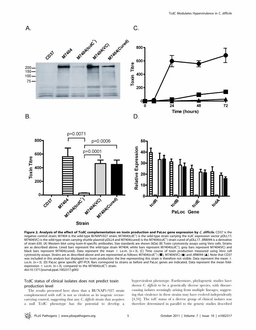
TcdC-mediated repression of toxin production in C.difficile
To determine the effect of TcdC on toxin production in strain
M7404, a combination of Western immunoblots and cytotoxicity
assays were performed using supernatants collected from strain
M7404 and isogenic M7404 derivatives carrying the vector
pDLL4, pDLL17, the cured strain M7404(cured) and the
PaLoc-negative control strain CD37. To assess toxin A produc-
tion, Western immunoblotting was performed using TcdA-specific
antibodies (Figure 2A). The results showed that the presence of the
tcdC+ plasmid pDLL17 resulted in a dramatic decrease in the
amount of toxin A produced by M7404(tcdC+) when compared to
the wild-type strain. By contrast, M7404(cured) produced
qualitatively similar levels of toxin A to wild-type, as did M7404
carrying the vector plasmid, whereas the PaLoc-negative strain
CD37 produced no detectable toxin A, as expected.
Vero cell cytotoxicity assays, which predominantly measure
toxin B activity [15], were then performed to quantitatively
determine the effect of TcdC on toxin production from these
strains. As previously observed with toxin A, the amount of toxin
produced from strain M7404 was significantly reduced when
functional TcdC was restored (Figure 2B). The amount of toxin
produced in the TcdC-complemented strain was approximately
16–32-fold less, and therefore significantly lower (p = 0.0001;
unpaired t-test, 95% confidence interval), than in the vector
control-carrying M7404 derivative. There was, however, no
significant difference in toxin activity levels between strains
M7404, the vector control strain M7404(VC) or M7404(cured)
(Figure 2B). A kinetic analysis of toxin production also clearly
showed that the presence of TcdC delayed the onset of toxin
production in M7404(tcdC+) in comparison to M7404 carrying the
vector plasmid, mirroring the delayed toxin production observed
from the tcdC+ 630-strain derivative JIR8094 (Figure 2C).
We also determined if TcdC-mediated repression of toxin
production was at the transcriptional level and evaluated the effect
of tcdC complementation on the expression of the other PaLoc-
encoded genes, tcdR and tcdE. Quantitative real-time PCR (qRT-
PCR) analysis using RNA extracted from the wild-type strain and
its isogenic derivatives was performed to ascertain the relative
transcription levels of the tcdA, tcdB, tcdR and tcdE genes. As shown
in Figure 2D, an approximate 13- and 23-fold reduction in tcdA-
and tcdB-specific mRNA levels, respectively, in strain M7404(tcdC+)
was observed compared to M7404. Similar observations were
made for tcdR and tcdE expression levels, with 33-fold and 21-fold
less tcdR and tcdE mRNA, respectively, in the tcdC-complemented
strain compared to the wild-type. No significant differences in the
expression levels of these four genes were detected when M7404,
the vector-carrying derivative or the cured strain were compared.
These data conclusively demonstrate that TcdC negatively
regulates toxin production in C. difficile and show that repression
occurs at the transcriptional level.
Reduction of the virulence of a BI/NAP1/027 isolate viacomplementation with TcdC
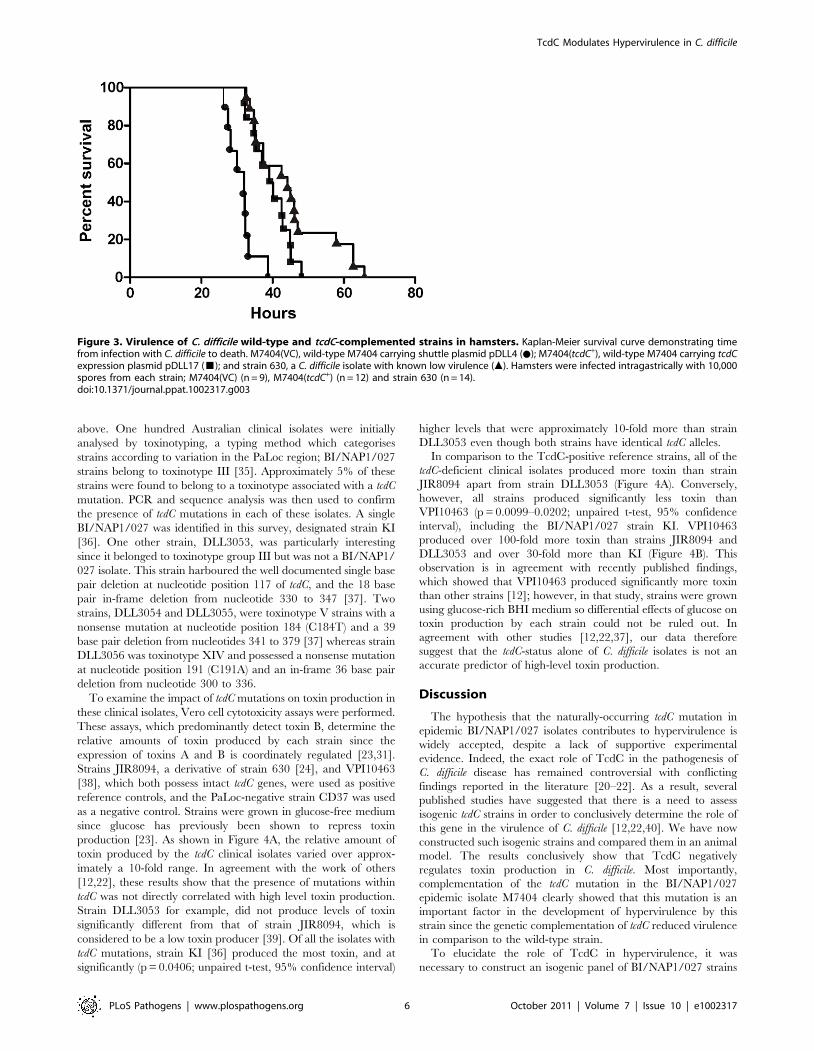
To define the role of TcdC in the virulence of a BI/NAP1/027
C. difficile isolate, female Golden Syrian hamsters were infected
with spores of strain M7404 carrying either the vector control or
the tcdC+ plasmid (n = 10 and n = 12, respectively). For compar-
ative purposes, a group of hamsters (n = 14) was also infected with
strain 630, a strain previously characterised as being less virulent
than other clinical isolates [32]. Following infection, all C. difficile
strains were found to be equally efficient at colonising the hamsters
(data not shown). Infection of colonised hamsters was allowed to
proceed and animals were monitored by telemetry. The end point
of infection was achieved when the core body temperature of the
hamsters dropped to 35uC. This parameter has previously been
shown to be a reliable indicator of non-recoverable disease [33].
At this point, the animals were immediately culled for animal
ethics reasons. Bacteria were then isolated from the culled
hamsters, the bacterial load quantified and isolates subjected to
MVLA analysis [33] to confirm that these isolates were the same
strain as originally used for infection.
Hamsters infected with the M7404(tcdC+) derivative showed a
significant delay (p = 0.0003; Logrank (Mantel-Cox) test; 95%
confidence interval) in the mean time taken to reach non-
recoverable disease (2370 minutes or 39.5 hours) in comparison to
the vector-carrying M7404 group (M7404(VC)), with a mean time
of 1869 minutes or 31.15 hours (Figure 3). In one of the hamsters
colonised with the M7404(VC) strain, the time taken to reach the
end point of infection was substantially longer than the other
hamsters in this group (2814 minutes or 46.9 hours). This hamster
was shown by statistical analysis (p = 0.0405; Grubbs test; 95%
confidence interval) to be an outlier and was therefore excluded
from the experimental analysis. Note that statistical significance
would be retained upon inclusion of this outlier. Interestingly,
TcdC Modulates Hypervirulence in C. difficile
PLoS Pathogens | www.plospathogens.org 3 October 2011 | Volume 7 | Issue 10 | e1002317

whilst the mean time to the end point of infection in the strain 630
group of hamsters (2701 minutes or 45.02 hours) was significantly
longer than that of hamsters infected with M7404(VC)
(p = 0.0001; Logrank (Mantel-Cox) test; 95% confidence interval),
there was no significant difference in the mean time taken to
achieve non-recoverable disease in the 630 group compared to the
M7404(tcdC+) derivative, indicating that the virulence of the TcdC-
complemented strain was equivalent to that of strain 630.
It is apparent from these virulence experiments that the
expression of TcdC in a BI/NAP1/027 isolate has an important
effect on virulence, resulting in a significant delay in the time
needed to reach non-recoverable disease. These data therefore
provide compelling evidence that the naturally occurring
mutation of tcdC in BI/NAP1/027 isolates is an important
factor in the development of a hypervirulent phenotype by these
strains.
Figure 1. Western blot analysis of TcdC production by wild-type and complemented C. difficile strains. (A) Qualitative analysis of TcdCproduction. VPI10463 is the positive control strain; VPI11186 is the negative control strain; M7404 is a wild-type Canadian BI/NAP1/027 strain;M7404(VC) is the wild-type strain carrying the shuttle plasmid pDLL4; M7404(tcdC+) is the wild-type strain carrying the tcdC expression plasmidpDLL17 and M7404(cured) is the M7404(tcdC+) strain cured of pDLL17. (B) Time course analysis of TcdC production by the positive control strain C.difficile VPI10463. Samples were taken at the indicated times shown in hours. 60 ng of purified recombinant his-tagged TcdC protein (rTcdC) wasused as the positive reference sample. (C) Time course analysis of TcdC production by C. difficile strain M7404(tcdC+). Samples were taken at theindicated times shown in hours. 300 ng of purified recombinant his-tagged TcdC protein (rTcdC) was used as the positive reference sample. Westernblots were performed with rabbit TcdC-specific antibodies. Size standards are shown (kDa).doi:10.1371/journal.ppat.1002317.g001
TcdC Modulates Hypervirulence in C. difficile
PLoS Pathogens | www.plospathogens.org 4 October 2011 | Volume 7 | Issue 10 | e1002317
TcdC status of clinical isolates does not predict toxinproduction level
The results presented here show that a BI/NAP1/027 strain
complemented with tcdC is not as virulent as its isogenic vector-
carrying control, suggesting that any C. difficile strain that acquires
a null TcdC phenotype has the potential to develop a
hypervirulent phenotype. Furthermore, phylogenetic studies have
shown C. difficile to be a genetically diverse species, with disease-
causing isolates seemingly arising from multiple lineages, suggest-
ing that virulence in these strains may have evolved independently
[4,34]. The tcdC status of a diverse group of clinical isolates was
therefore determined in parallel to the genetic studies described
Figure 2. Analysis of the effect of TcdC complementation on toxin production and PaLoc gene expression by C. difficile. CD37 is thenegative control strain; M7404 is the wild-type BI/NAPI/027 strain; M7404(tcdC+) is the wild-type strain carrying the tcdC expression vector pDLL17;M7404(VC) is the wild-type strain carrying shuttle plasmid pDLL4 and M7404(cured) is the M7404(tcdC+) strain cured of pDLL17. JIR8094 is a derivativeof strain 630. (A) Western blot using toxin-A-specific antibodies. Size standards are shown (kDa) (B) Toxin cytotoxicity assays using Vero cells. Strainsare as described above. Lined bars represent the wild-type strain M7404; white bars represent M7404(tcdC+); grey bars represent M7404(VC) andblack bars represent M7404(cured). Data represent the mean 6 s.e.m. (n = 3). (C) Time course of toxin production measured using Vero cellcytotoxicity assays. Strains are as described above and are represented as follows: M7404(tcdC+) (&), M7404(VC) (N) and JIR8094 (m). Note that CD37was included in this analysis but displayed no toxin production; the line representing this strain is therefore not visible. Data represent the mean 6s.e.m. (n = 3). (D) PaLoc gene specific qRT-PCR. Bars correspond to strains as before and PaLoc genes are indicated. Data represent the mean fold-expression 6 s.e.m. (n = 3), compared to the M7404(tcdC+) strain.doi:10.1371/journal.ppat.1002317.g002
TcdC Modulates Hypervirulence in C. difficile
PLoS Pathogens | www.plospathogens.org 5 October 2011 | Volume 7 | Issue 10 | e1002317
above. One hundred Australian clinical isolates were initially
analysed by toxinotyping, a typing method which categorises
strains according to variation in the PaLoc region; BI/NAP1/027
strains belong to toxinotype III [35]. Approximately 5% of these
strains were found to belong to a toxinotype associated with a tcdC
mutation. PCR and sequence analysis was then used to confirm
the presence of tcdC mutations in each of these isolates. A single
BI/NAP1/027 was identified in this survey, designated strain KI
[36]. One other strain, DLL3053, was particularly interesting
since it belonged to toxinotype group III but was not a BI/NAP1/
027 isolate. This strain harboured the well documented single base
pair deletion at nucleotide position 117 of tcdC, and the 18 base
pair in-frame deletion from nucleotide 330 to 347 [37]. Two
strains, DLL3054 and DLL3055, were toxinotype V strains with a
nonsense mutation at nucleotide position 184 (C184T) and a 39
base pair deletion from nucleotides 341 to 379 [37] whereas strain
DLL3056 was toxinotype XIV and possessed a nonsense mutation
at nucleotide position 191 (C191A) and an in-frame 36 base pair
deletion from nucleotide 300 to 336.
To examine the impact of tcdC mutations on toxin production in
these clinical isolates, Vero cell cytotoxicity assays were performed.
These assays, which predominantly detect toxin B, determine the
relative amounts of toxin produced by each strain since the
expression of toxins A and B is coordinately regulated [23,31].
Strains JIR8094, a derivative of strain 630 [24], and VPI10463
[38], which both possess intact tcdC genes, were used as positive
reference controls, and the PaLoc-negative strain CD37 was used
as a negative control. Strains were grown in glucose-free medium
since glucose has previously been shown to repress toxin
production [23]. As shown in Figure 4A, the relative amount of
toxin produced by the tcdC clinical isolates varied over approx-
imately a 10-fold range. In agreement with the work of others
[12,22], these results show that the presence of mutations within
tcdC was not directly correlated with high level toxin production.
Strain DLL3053 for example, did not produce levels of toxin
significantly different from that of strain JIR8094, which is
considered to be a low toxin producer [39]. Of all the isolates with
tcdC mutations, strain KI [36] produced the most toxin, and at
significantly (p = 0.0406; unpaired t-test, 95% confidence interval)
higher levels that were approximately 10-fold more than strain
DLL3053 even though both strains have identical tcdC alleles.
In comparison to the TcdC-positive reference strains, all of the
tcdC-deficient clinical isolates produced more toxin than strain
JIR8094 apart from strain DLL3053 (Figure 4A). Conversely,
however, all strains produced significantly less toxin than
VPI10463 (p = 0.0099–0.0202; unpaired t-test, 95% confidence
interval), including the BI/NAP1/027 strain KI. VPI10463
produced over 100-fold more toxin than strains JIR8094 and
DLL3053 and over 30-fold more than KI (Figure 4B). This
observation is in agreement with recently published findings,
which showed that VPI10463 produced significantly more toxin
than other strains [12]; however, in that study, strains were grown
using glucose-rich BHI medium so differential effects of glucose on
toxin production by each strain could not be ruled out. In
agreement with other studies [12,22,37], our data therefore
suggest that the tcdC-status alone of C. difficile isolates is not an
accurate predictor of high-level toxin production.
Discussion
The hypothesis that the naturally-occurring tcdC mutation in
epidemic BI/NAP1/027 isolates contributes to hypervirulence is
widely accepted, despite a lack of supportive experimental
evidence. Indeed, the exact role of TcdC in the pathogenesis of
C. difficile disease has remained controversial with conflicting
findings reported in the literature [20–22]. As a result, several
published studies have suggested that there is a need to assess
isogenic tcdC strains in order to conclusively determine the role of
this gene in the virulence of C. difficile [12,22,40]. We have now
constructed such isogenic strains and compared them in an animal
model. The results conclusively show that TcdC negatively
regulates toxin production in C. difficile. Most importantly,
complementation of the tcdC mutation in the BI/NAP1/027
epidemic isolate M7404 clearly showed that this mutation is an
important factor in the development of hypervirulence by this
strain since the genetic complementation of tcdC reduced virulence
in comparison to the wild-type strain.
To elucidate the role of TcdC in hypervirulence, it was
necessary to construct an isogenic panel of BI/NAP1/027 strains
Figure 3. Virulence of C. difficile wild-type and tcdC-complemented strains in hamsters. Kaplan-Meier survival curve demonstrating timefrom infection with C. difficile to death. M7404(VC), wild-type M7404 carrying shuttle plasmid pDLL4 (N); M7404(tcdC+), wild-type M7404 carrying tcdCexpression plasmid pDLL17 (&); and strain 630, a C. difficile isolate with known low virulence (m). Hamsters were infected intragastrically with 10,000spores from each strain; M7404(VC) (n = 9), M7404(tcdC+) (n = 12) and strain 630 (n = 14).doi:10.1371/journal.ppat.1002317.g003
TcdC Modulates Hypervirulence in C. difficile
PLoS Pathogens | www.plospathogens.org 6 October 2011 | Volume 7 | Issue 10 | e1002317

that were identical except for the presence or absence of the wild-
type tcdC gene. Despite the publication of studies describing the
successful transfer of plasmids into the BI/NAP1/027 isolate
R20291 [25,26,41], this group of strains has remained difficult to
work with at the molecular genetic level. As such, a new system
that utilised the conjugation apparatus of Tn916 was developed in
this study and used successfully to genetically manipulate a
number of clinically relevant isolates, including a BI/NAP1/027
strain of C. difficile. Tn916 is a broad host-range conjugative
transposon that was recently used to transfer plasmids into
genetically intractable strains of Enterococcus faecium [28] and has
been shown to transfer into C. difficile [42,43]. C. perfringens was
chosen for use as a donor strain in anticipation that it may be more
proficient for the transfer of plasmids into C. difficile in comparison
to the more distantly related E. coli. The addition of oriTTn916 onto
the shuttle vector pMTL9361Cm facilitated the efficient transfer
of this plasmid into strain M7404 from a Tn916-carrying C.
perfringens strain. Furthermore, this system has been successfully
used to transfer shuttle plasmids into every C. difficile isolate tested
so far (Table S1). Most importantly, this new technology facilitated
the complementation of the tcdC mutation in strain M7404
enabling the role of TcdC in the virulence of BI/NAP1/027
strains of C. difficile to be investigated.
Previous in vitro studies have shown that TcdC is able to
sequester the TcdR sigma factor, preventing its association with
core RNA polymerase and blocking toxin gene expression [20].
These experiments suggested that TcdC was important in the
regulation of toxin production by C. difficile, but the in vivo role of
this protein was not determined. Conversely, several studies on C.
difficile clinical isolates [22,37] showed that the absence of a
functional tcdC gene was not an accurate predictor of high level
toxin production or increased disease severity, indicating that
TcdC may not play an important role in virulence in these strains
[22,37]. The analysis of Australian clinical isolates in the present
study is in accordance with these latter studies in that isolates with
naturally occurring tcdC mutations were found to produce toxin at
a range of different levels that were not necessarily high. However,
since these strains, and those in the other studies [22,37], are not
isogenic it is not possible to draw conclusions about the
importance of tcdC in the context of toxin yield or virulence. By
contrast, the isogenic tcdC strains studied here clearly show that
TcdC is a negative regulator of toxin production since the tcdC
complemented BI/NAP1/027 C. difficile strain produced signifi-
cantly less toxin A and B than the non-complemented control
strains. The finding that TcdC-status is not correlated with toxin
production in clinical isolates highlights the limitation of accurately
assigning gene function by studying non-isogenic strains, partic-
ularly in a highly heterogeneous species such as C. difficile. In this
context, it might be of interest to study the function of tcdC in
isogenic strains generated in a different genetic background such
as a ribotype 078 isolate.
Analysis of PaLoc gene expression by qRT-PCR demonstrated
that TcdC exerts regulatory control of toxin production at the
transcriptional level, and this is in keeping with its proposed role as
an anti-sigma factor [20]. The observation that the expression of
tcdR and tcdE is reduced in the tcdC-complemented strain, together
with tcdA and tcdB, is probably because of autoregulation of tcdR
since TcdR upregulates its own expression and that of the other
PaLoc genes [23].
The virulence of strain M7404 was reduced upon complemen-
tation of tcdC, clearly demonstrating that the tcdC mutation in BI/
NAP1/027 strains has a significant impact on virulence and is
likely to be an important factor in the development of
hypervirulence by these strains. Surprisingly, the virulence of
M7404(tcdC+) was found to be equivalent to that of strain 630,
which has been shown in other studies to be reduced in virulence
in comparison to other isolates, including three other BI-type
strains [32]. These findings have important implications for C.
Figure 4. Comparative analysis of toxin production by naturally occurring tcdC clinical isolates. Vero cell cytotoxicity assays were used todetermine toxin production levels. (A) Strain JIR8094 is a tcdC+ control strain, CD37 is a PaLoc-negative control strain and KI is an Australian BI/NAPI/027 isolate [36]. All other strains (DLL3053-DLL3056) are clinical isolates carrying naturally occurring tcdC mutations, collected from Australianhospitals. (B) Toxin production by the tcdC+ control strain VPI10463 is shown on a separate bar chart due to the much higher levels of toxin produced.For comparative purposes strain KI is represented on both bar charts. Data represent the mean6s.e.m. (n = 3).doi:10.1371/journal.ppat.1002317.g004
TcdC Modulates Hypervirulence in C. difficile
PLoS Pathogens | www.plospathogens.org 7 October 2011 | Volume 7 | Issue 10 | e1002317

difficile virulence since they suggest that strains carrying tcdC
mutations have the inherent potential to develop hypervirulence.
The recent emergence of a new class of hypervirulent strains,
ribotype 078 [44], may be one such example. These isolates
encode a non-functional TcdC protein [37], produce significantly
more toxin than non-epidemic strains, are associated with more
severe disease as well as higher rates of mortality and are
increasingly being identified as the causative agent of CDI [44,45].
Although these experiments show that TcdC-status alone can
modulate virulence it is probable that multiple factors working
synergistically are necessary for the development of hypervirulence
in the BI/NAP1/027 strains. It is likely that the accumulation of
multiple genetic changes in addition to the tcdC mutation has
enabled BI/NAP1/027 strains to become the predominant
disease-causing isolates in numerous countries. Of particular
importance might be variations in the functional activity of the
encoded toxins since these isolates were recently shown to produce
a toxin B that shows variation across the C-terminal receptor
binding domain of the protein [46], resulting in more potent
activity across a wider range of cell lines in comparison to toxin B
from the historical, non-epidemic strain 630 [4]. Furthermore,
using the zebrafish embryo model of intoxication, the BI/NAP1/
027 toxin B was recently shown to have pronounced in vivo
cytotoxic activity in comparison to toxin B from VPI10463,
another historical non-epidemic isolate, with greater tissue tropism
and more extensive tissue destruction observed [47]. Since toxin B
is thought to be one of the major virulence factors of C. difficile
[15,16] these observations suggest that TcdB variations might play
an important role in the hypervirulent phenotype.
There are other factors that may influence C. difficile
hypervirulence. The BI/NAP1/027 strains encode an additional
toxin known as binary toxin or CDT [2]. The role of this toxin in
CDI remains to be elucidated but a recent study showed that CDT
induces the formation of microtubule-based protrusions on the
host cell surface thereby increasing C. difficile adherence to
epithelial cells. Moreover, intestinal colonisation of gnotobiotic
mice with a BI/NAP1/027 C. difficile strain was significantly
reduced in mice treated with CDT-neutralising antibodies in
comparison to control mice [48]. These findings suggest that CDT
may be an important colonisation factor, enhancing the ability of
BI/NAP1/027 strains to initiate infection as well as causing
adjunctive tissue damage during later stages of infection,
potentially leading to more severe disease. Many BI/NAP1/027
strains are also more proficient at sporulation than non-epidemic
C. difficile strains [12,49]. C. difficile spores are highly infectious [50]
and play a critical role in the transmission of CDI and perhaps in
disease relapse, which is a serious problem in patients with CDI
[51]. In this context, enhanced sporulation is ostensibly an
important adaptation by BI/NAP1/027 isolates, which would
result in larger numbers of spores being shed from infected
patients and an increased environmental spore load, ultimately
leading to higher transmission rates. Finally, the development of
fluoroquinolone resistance, in particular to moxifloxacin and
gatifloxacin, is unquestionably a major factor in epidemics caused
by BI/NAP1/027 strains [3,11]. In this regard, the hypothetical
co-evolution of enhanced virulence traits and antibiotic resistance
in C. difficile mirrors trends seen with other significant nosocomial
pathogens such as Methicillin Resistant Staphylococcus aureus
(MRSA) [52] and Vancomycin Resistant Enterococcus (VRE) species
[53]. In summary, it is clear that our findings represent an
important breakthrough in our understanding of the development
of hypervirulence in prevailing C. difficile isolates and will provide a
significant reference point for future studies on epidemic strains
and their control.
Materials and Methods
Ethics statementThis study was carried out in strict accordance with the
recommendations in the United Kingdoms Home Office Animals
(Scientific Procedures) Act of 1986 which outlines the regulation of
the use of laboratory animals for the use of animals in scientific
procedures. The experiments described were subject to approval
by the University of Glasgow Ethics Committee and by a
designated Home Office Inspector (Project Number 60/4218).
All experiments were subject to the 3 R consideration (refine,
reduce and replace) and all efforts were made to minimize
suffering.
Bacterial strains and growth conditionsThe characteristics and origins of all recombinant strains and
plasmids are shown in Table 1 and Table S2, respectively. All
bacteriological culture media were obtained from Oxoid. C. difficile
strains were cultured in BHIS [54] or TY medium [15], unless
otherwise stated, in an atmosphere of 10% H2, 10% CO2, and
80% N2 at 37uC in a Coy anaerobic chamber. Escherichia coli was
cultured in 26YT medium aerobically at 37uC, with shaking for
broth cultures. All antibiotics were purchased from Sigma-Aldrich
and were used at the following concentrations: cycloserine (Cs,
250 mg/ml), cefoxitin (Cf, 8 mg/ml), thiamphenicol (Tm, 10 mg/
ml) or tetracycline (Tc, 10 mg/ml), chloramphenicol (Cm, 25 mg/
ml).
Molecular biology and PCR techniquesPlasmid DNA was isolated using a QIAprep spin miniprep kit
(Qiagen). Genomic DNA was prepared using a DNeasy tissue kit
(Qiagen). Standard methods for the digestion, modification,
ligation, and analysis of plasmid and genomic DNA were used
[55]. Nucleotide sequence analysis was carried out using a PRISM
BigDye Terminator cycle sequencing kit (Applied Biosystems) and
detection was performed by Micromon at Monash University.
Oligonucleotide primer sequences are listed below. Unless
otherwise stated, all PCR experiments were carried out with
Phusion DNA polymerase (New England Biolabs) and the 26Failsafe PCR buffer E (Epicentre) according to the manufacturer’s
instructions.
Construction of recombinant plasmidsFor construction of the Tn916 transferrable clostridial shuttle
vector, PCR was performed using primers DLP33 (59-G-
AATTCGCCCTTTTTTATACTCCCCTTG-39) and DLP34
(59-GAATTCGCCCTCAAAGGACGAATATGTCGC-39) and
chromosomal DNA extracted from Clostridium perfringens strain
JIR4225 [30]. The resulting 700 bp DNA fragment, which
contained the oriT region of Tn916, was TOPO-cloned into
pCR-Blunt II-TOPO according to the manufacturer’s instructions
(Invitrogen). The fragment was then excised from pCR-Blunt II-
TOPO using EcoRI and cloned into the equivalent sites of plasmid
pMTL9361Cm [29], resulting in plasmid pDLL4.
For construction of the tcdC-carrying plasmid, PCR was
performed using primers DLP35 (59-CTGCAGCCACCTCTA-
AATCACTGAGTCACTTAATTAC-39) and DLP36 (59-CTGC-
AGAGCCTTGTAACTGTTTATTTGC-39) and C. difficile strain
VPI10463 genomic DNA in order to amplify a 1085 bp fragment
encompassing the tcdC gene and upstream region. This fragment
was then TOPO-cloned into pCR-Blunt II-TOPO, before being
excised with PstI and subcloned into the equivalent site of plasmid
pDLL4, resulting in the final construct pDLL17.
TcdC Modulates Hypervirulence in C. difficile
PLoS Pathogens | www.plospathogens.org 8 October 2011 | Volume 7 | Issue 10 | e1002317

Transfer of plasmid DNA into C. difficile by conjugationThe conjugation procedure utilising E. coli HB101(pVS520) as
the conjugative donor was carried out as previously described [29].
Recombinant plasmids were introduced into C. perfringens strain
JIR4225 as before [56]. Conjugations utilising C. perfringens
JIR4225 were then performed as follows: separate 90 ml BHIS
broth cultures were inoculated with 1 ml aliquots from an
overnight C. difficile recipient strain or C. perfringens donor strain
starter culture and grown to mid-exponential phase. Approxi-
mately 1 ml was removed from each culture, mixed and
centrifuged. The cell pellet was then resuspended in phosphate-
buffered saline (PBS), spread onto a BHIS agar plate and
incubated overnight at 37uC. Bacterial growth was harvested in
sterile PBS before being spread onto BHIS agar supplemented
with thiamphenicol and incubated overnight as before. Bacterial
growth was again harvested with PBS and dilutions spread onto
BHIS agar supplemented with cefoxitin and thiamphenicol or
tetracycline, and the plates incubated under anaerobic conditions
for 24 to 72 h.
Toxin A-specific Western blotsThe toxins were partially purified by ammonium sulphate
precipitation from culture supernatants harvested after growth for
72 hours and toxin A was then detected by Western blotting as
described previously [15].
TcdC-specific Western blotsFor non-quantitative TcdC-specific Western Blots, crude
extracts of C. difficile were prepared by sonication of samples
taken from cultures that had been grown for 12 hours under
anaerobic conditions. The crude extracts from each strain were
then subjected to electrophoresis in a 15% SDS-PAGE gel and
transferred to a nitrocellulose membrane using standard methods
[55]. Membranes were treated with anti-TcdC antibody [57] and
detected following treatment with goat anti-mouse IgG-alkaline
phosphatase conjugated secondary antibody using standard
procedures. For quantitative TcdC-specific Western blots, cultures
of C. difficile VPI10463 and C. difficile M7404(tcdC+) were grown
under anaerobic conditions and samples were removed every
4 hours for 24 or 28 hours respectively. Each sample was
normalized to an optical density (600 nm) of 0.9 prior to lysis, to
ensure that the same number of cells was present. Lysates were
then prepared by sonication prior to SDS-PAGE gel electropho-
resis, transfer and detection, as described above. Following
detection, the amount of TcdC in each lysate was quantified by
densitometric analysis, using purified recombinant TcdC protein
(rTcdC) as the standard and the ImageJ software package,
according to published methods [58].
Vero cell cytotoxicity assaysToxin B was detected in C. difficile culture supernatants
harvested after growth for 72 hours by Vero cell cytotoxicity
assays as described previously [15], except that each well was
seeded with 16105 cells.
RNA extraction and reverse transcriptionTotal RNA was extracted from C. difficile cultures grown for
12 h in TY media. Reverse transcription was performed using
Table 1. Bacterial strains.
Strain Characteristics Source/Reference
E. coli
DH5a F-w80D lacZdM15D(lacZYA-argF)U169 endA1recA1 hsdr17(tk2mk
+)deoR thi-1 supE44 gyrA96 relA1 Life Technologies
TOP10 F- mcrA D(mrr-hsdRMS-mcrBC) w80lacZDM15 DlacX74 nupG recA1 araD139 D(ara-leu)7697galU galK rpsL(StrR) endA1 l2
Invitrogen
HB101 thi-1 hsdS20(rB2 mB
2) supE44 recAB ara-14 leuB5 proA2 lacY1 galK rpsL20 (Smr) xyl-5 mtl-1 [60]
C. perfringens
JIR4225 C. perfringens strain JIR325 with 5 chromosomal copies of Tn916 [30]
C. difficile
M7404 Canadian BI/NAP1/027 isolate [29]
630 Wild-type C. difficile strain; first C. difficile genome available [61]
JIR8094 Erythromycin sensitive derivative of strain 630 [24]
VPI10463 PaLoc-positive C. difficile isolate [38]
VPI11186 PaLoc-negative C. difficile isolate [38]
CD37 PaLoc-negative C. difficile isolate [62]
KI Australian BI/NAP1/027 isolate [36]
DLL3053 Australian toxinotype III clinical isolate This study
DLL3054 Australian toxinotype V clinical isolate This study
DLL3055 Australian toxinotype V clinical isolate This study
DLL3056 Australian toxinotype XIV clinical isolate This study
M7404(VC) DLL3001 (M7404 carrying shuttle plasmid pDLL4) This study
M7404(tcdC+) DLL3002 (M7404 carrying tcdC expression plasmid pDLL17) This study
M7404(cured) DLL3003 (M7404(tcdC+) cured of plasmid pDLL17) This study
doi:10.1371/journal.ppat.1002317.t001
TcdC Modulates Hypervirulence in C. difficile
PLoS Pathogens | www.plospathogens.org 9 October 2011 | Volume 7 | Issue 10 | e1002317
AMV Reverse Transcriptase (Promega) using random hexamer
oligonucleotides primers and 2 mg template RNA. The cDNA
samples were then purified using Qiaquick Columns (Qiagen).
qRT-PCR assay design and real time PCRPaLoc gene specific primers were designed using Primer 3
software (Geneious Software). qRT-PCR was performed using an
AB7300 real-time PCR instrument (Applied Biosystems). Reac-
tions were carried out using the FastStart Universal SYBR Green
Master Mix (Roche) with 40 ng of cDNA as template. Standard
curves were generated for each primer pair using C. difficile
genomic DNA, and melt curve analysis was performed following
each qRT-PCR reaction to verify amplification specificity.
Samples were normalised using the C. difficile rrnA gene.
Preparation of spores for animal infectionSpores were prepared from C. difficile cultures grown in 500 ml
of BHI broth. Cultures were pelleted by centrifugation for 10 mins
and re-suspended in 50% ethanol. The material was then vortexed
every 10 min for 1 h before centrifugation for 10 mins. The pellet
was then treated with 1% Sarkosyl in PBS for 1 h at room
temperature and again pelleted by centrifugation, followed by
incubation overnight at 37uC with lysozyme (10 mg/ml) in
125 mM Tris-HCl buffer (pH 8.0). The sample was treated in a
sonicating water bath (3 pulses of 3 min each; 1510 Branson)
before centrifugation through a 50% sucrose gradient for 20 mins.
The pellet was incubated in 2 ml of PBS containing 200 mM
EDTA, 300 ng/ml proteinase K and 1% Sarkosyl for 30 mins at
37uC before centrifugation through a 50% sucrose gradient for
20 mins. The final pellet was then washed twice in sterile distilled
water before finally being resuspended in 1 ml of sterile water.
Spore preparations were stored at 280uC prior to use.
Hamster experimentsFemale Golden Syrian hamsters purchased from Harlan Olac
UK were used for all animal experiments. Telemetry chips
(Vitalview Emitter) were inserted by laparotomy into the body
cavity of the animals at least 3 weeks before infection with C.
difficile. Animal experiments were then carried out as described
previously [33], except that animals received 16104 spores of C.
difficile. Animals were culled when core body temperature dropped
below 35uC. This study was carried out in strict accordance with
the recommendations in the United Kingdoms Home Office
Animals (Scientific Procedures) Act of 1986 which outlines the
regulation of the use of laboratory animals for the use of animals in
scientific procedures. The experiments described were subject to
approval by the University of Glasgow Ethics Committee and by a
designated Home Office Inspector (Project Number 60/4218). All
experiments were subject to the 3 R consideration (refine, reduce
and replace) and all efforts were made to minimize suffering.
Quantification of bacterial loadTo estimate colonisation, hamsters were sacrificed and the gut
region from the caecum to the anus removed. The tissues were
homogenised in PBS using a Stomacher and viable counts were
performed on the homogenate as described previously [33].
Confirmation of infecting strainsTo confirm that the bacteria isolated from the hamster were the
same strain as originally used for infection, genomic DNA was
isolated and subjected to MVLA as described previously [59].
Plasmid rescue was performed as previously described [29]
followed by restriction digest analysis to confirm plasmid integrity.
Supporting Information
Table S1 Efficiency of RP4 or Tn916-mediated plasmidtransfer to C. difficile strains. The efficiency of RP4 or
Tn916-mediated plasmid transfer from E. coli or C. perfringens
donors, respectively, to C. difficile recipient strains is shown,
calculated as described in Materials and Methods and expressed as
transconjugants per ml.
(DOC)
Table S2 Bacterial plasmids. Bacterial plasmids used in this
study, together with the genetic features relevant to this work, are
shown.
(DOC)
Acknowledgments
We thank Christoph von-Eichel Streiber for kindly providing toxin A-
specific antibodies and Rachael Poon and Tongted Phumoonna-Das for
assistance with Vero cell cytotoxicity assays.
Author Contributions
Conceived and designed the experiments: GPC BD GAJ JIR GRD DL.
Performed the experiments: GPC PMH KEM DK RG GRD JS AMB AA.
Analyzed the data: GPC BD GAJ JIR PMH KEM DK RG AA GRD JS
AMB DL. Contributed reagents/materials/analysis tools: GPC BD GAJ
JIR DK RG GRD DL. Wrote the paper: GPC DL.
References
1. Borriello SP (1998) Pathogenesis of Clostridium difficile infection. J Antimicrob
Chemother 41: 13–19.
2. Warny M, Pepin J, Fang A, Killgore G, Thompson A, et al. (2005) Toxinproduction by an emerging strain of Clostridium difficile associated with outbreaks
of severe disease in North America and Europe. Lancet 366: 1079–1084.
3. McDonald LC, Killgore GE, Thompson A, Owens RC, Kazakova SV, et al.(2005) An epidemic, toxin gene–variant strain of Clostridium difficile. N Engl J Med
353: 2433–2441.
4. Stabler RA, He M, Dawson L, Martin M, Valiente E, et al. (2009) Comparativegenome and phenotypic analysis of Clostridium difficile 027 strains provides insight
into the evolution of a hypervirulent bacterium. Genome Biol 10: R102.
5. Loo VG, Poirier L, Miller MA, Oughton M, Libman MD, et al. (2005) A
predominantly clonal multi-institutional outbreak of Clostridium difficile-associateddiarrhea with high morbidity and mortality. N Engl J Med 353: 2442–2449.
6. Muto CA, Pokrywka M, Shutt K, Mendelsohn AB, Nouri K, et al. (2005) A large
outbreak of Clostridium difficile-associated disease with an unexpected proportionof deaths and colectomies at a teaching hospital following increased
fluoroquinolone use. Infect Control Hosp Epidemiol 26: 273–280.
7. Kuijper EJ, Coignard B, Tull P (2006) Emergence of Clostridium difficile-associated
disease in North America and Europe. Clin Microbiol Infect 12 Suppl 6: 2–18.
8. Rouphael NG, O’Donnell JA, Bhatnagar J, Lewis F, Polgreen PM, et al. (2008)
Clostridium difficile-associated diarrhea: an emerging threat to pregnant women.Am J Obstet Gynecol 198: 635 e631–636.
9. Zilberberg MD, Tillotson GS, McDonald C (2010) Clostridium difficile infections
among hospitalized children, United States, 1997–2006. Emerg Infect Dis 16:604–609.
10. Baker SS, Faden H, Sayej W, Patel R, Baker RD (2010) Increasing Incidence of
Community-Associated Atypical Clostridium difficile Disease in Children. ClinPediatr (Phila) 49: 644–647.
11. Riley TV (2009) Is Clostridium difficile a threat to Australia’s biosecurity?
Med J Aust 190: 661–662.
12. Merrigan M, Venugopal A, Mallozzi M, Roxas B, Viswanathan VK, et al.
(2010) Human Hypervirulent Clostridium difficile Strains Exhibit IncreasedSporulation as Well as Robust Toxin Production. J Bacteriol 192: 4904–4911.
13. Sebaihia M, Wren BW, Mullany P, Fairweather NF, Minton N, et al. (2006) The
multidrug-resistant human pathogen Clostridium difficile has a highly mobile,mosaic genome. Nat Genet 38: 779–786.
14. Just I, Selzer J, Wilm M, von Eichel-Streiber C, Mann M, et al. (1995)
Glucosylation of Rho proteins by Clostridium difficile toxin B. Nature 375:
500–503.
TcdC Modulates Hypervirulence in C. difficile
PLoS Pathogens | www.plospathogens.org 10 October 2011 | Volume 7 | Issue 10 | e1002317
15. Lyras D, O’Connor JR, Howarth PK, Sambol SP, Carter GP, et al. (2009)
Toxin B is essential for virulence of Clostridium difficile. Nature 458: 1176–1179.16. Kuehne SA, Cartman ST, Heap JT, Kelly ML, Cockayne A, et al. (2010) The
role of toxin A and toxin B in Clostridium difficile infection. Nature 467: 711–713.
17. Braun V, Hundsberger T, Leukel P, Sauerborn M, Eichel-Streiber Cv (1996)Definition of the single integration site of the pathogenicity locus in Clostridium
difficile. Gene 181: 29–38.18. Mani N, Dupuy B (2001) Regulation of toxin synthesis in Clostridium difficile by an
alternative RNA polymerase sigma factor. Proc Natl Acad Sci U S A 98:
5844–5849.19. Tan KS, Wee BY, Song KP (2001) Evidence for holin function of tcdE gene in
the pathogenicity of Clostridium difficile. J Med Microbiol 50: 613–619.20. Matamouros S, England P, Dupuy B (2007) Clostridium difficile toxin expression is
inhibited by the novel regulator TcdC. Mol Microbiol 64: 1274–1288.21. Dupuy B, Govind R, Antunes A, Matamouros S (2008) Clostridium difficile toxin
synthesis is negatively regulated by TcdC. J Med Microbiol 57: 685–689.
22. Murray R, Boyd D, Levett PN, Mulvey MR, Alfa MJ (2009) Truncation in thetcdC region of the Clostridium difficile PathLoc of clinical isolates does not predict
increased biological activity of Toxin B or Toxin A. BMC Infect Dis 9: 103.23. Mani N, Lyras D, Barroso L, Howarth P, Wilkins T, et al. (2002) Environmental
response and autoregulation of Clostridium difficile TxeR, a sigma factor for toxin
gene expression. J Bacteriol 184: 5971–5978.24. O’Connor JR, Lyras D, Farrow KA, Adams V, Powell DR, et al. (2006)
Construction and analysis of chromosomal Clostridium difficile mutants. MolMicrobiol 61: 1335–1351.
25. Burns DA, Heap JT, Minton NP (2010) SleC is essential for germination ofClostridium difficile spores in nutrient-rich medium supplemented with the bile salt
taurocholate. J Bacteriol 192: 657–664.
26. Cartman ST, Minton NP (2010) A mariner-based transposon system for in vivo
random mutagenesis of Clostridium difficile. Appl Environ Microbiol 76:
1103–1109.27. Heap JT, Kuehne SA, Ehsaan M, Cartman ST, Cooksley CM, et al. (2010) The
ClosTron: Mutagenesis in Clostridium refined and streamlined. J Microbiol
Methods 80: 49–55.28. Nallapareddy SR, Singh KV, Murray BE (2006) Construction of improved
temperature-sensitive and mobilizable vectors and their use for constructingmutations in the adhesin-encoding acm gene of poorly transformable clinical
Enterococcus faecium strains. Appl Environ Microbiol 72: 334–345.29. Carter GP, Lyras D, Allen DL, Mackin KE, Howarth PM, et al. (2007) Binary
toxin production in Clostridium difficile is regulated by CdtR, a LytTR family
response regulator. J Bacteriol 189: 7290–7301.30. Awad MM, Rood JI (1997) Isolation of a-toxin, q-toxin and k-toxin mutants of
Clostridium perfringens by Tn916 mutagenesis. Microb Pathogen 22: 275–284.31. Hundsberger T, Braun V, Weidmann M, Leukel P, Sauerborn M, et al. (1997)
Transcription analysis of the genes tcdA-E of the pathogenicity locus of Clostridium
difficile. European J Bioch 244: 735–742.32. Razaq N, Sambol S, Nagaro K, Zukowski W, Cheknis A, et al. (2007) Infection
of hamsters with historical and epidemic BI types of Clostridium difficile. J InfectDis 196: 1813–1819.
33. Goulding D, Thompson H, Emerson J, Fairweather NF, Dougan G, et al. (2009)Distinctive profiles of infection and pathology in hamsters infected with
Clostridium difficile strains 630 and B1. Infect Immun 77: 5478–5485.
34. He M, Sebaihia M, Lawley TD, Stabler RA, Dawson LF, et al. (2010)Evolutionary dynamics of Clostridium difficile over short and long time scales. Proc
Natl Acad Sci U S A 107: 7527–7532.35. Rupnik M, Avesani V, Janc M, von Eichel-Streiber C, Delmee M (1998) A novel
toxinotyping scheme and correlation of toxinotypes with serogroups of
Clostridium difficile isolates. J Clin Microbiol 36: 2240–2247.36. Richards M, Knox J, Elliott B, Mackin K, Lyras D, et al. (2011) Severe infection
with Clostridium difficile PCR ribotype 027 acquired in Melbourne, Australia.Med J Australia 194: 369–371.
37. Curry SR, Marsh JW, Muto CA, O’Leary MM, Pasculle AW, et al. (2007) tcdC
genotypes associated with severe TcdC truncation in an epidemic clone andother strains of Clostridium difficile. J Clin Microbiol 45: 215–221.
38. Lyerly DM, Barroso LA, Wilkins TD, Depitre C, Corthier G (1992)Characterization of a toxin A-negative, toxin B-positive strain of Clostridium
difficile. Infect Immun 60: 4633–4639.
39. Carter GP, Rood JI, Lyras D (2010) The role of toxin A and toxin B in
Clostridium difficile-associated disease: Past and present perspectives. Gut Microbes
1: 58–64.
40. O’Connor JR, Johnson S, Gerding DN (2009) Clostridium difficile infection caused
by the epidemic BI/NAP1/027 strain. Gastroenterology 136: 1913–1924.
41. Heap JT, Pennington OJ, Cartman ST, Carter GP, NP. M (2007) The
ClosTron: a universal gene knock-out system for the genus Clostridium.
J Microbiol Meth 70: 452–464.
42. Mullany P, Wilks M, Tabaqchali S (1991) Transfer of Tn916 and Tn916DE into
Clostridium difficile: demonstration of a hot-spot for these elemnets in the C. difficile
genome. FEMS Microbiol Lett 79: 191–194.
43. Minton N, Carter G, Herbert M, O’Keeffe T, Purdy D, et al. (2004) The
development of Clostridium difficile genetic systems. Anaerobe 10: 75–84.
44. Goorhuis A, Debast SB, van Leengoed LA, Harmanus C, Notermans DW, et al.
(2008) Clostridium difficile PCR ribotype 078: an emerging strain in humans and in
pigs? J Clin Microbiol 46: 1157; author reply 1158.
45. Dawson LF, Valiente E, Wren BW (2009) Clostridium difficile–a continually
evolving and problematic pathogen. Infect Genet Evol 9: 1410–1417.
46. Stabler RA, Dawson LF, Phua LT, Wren BW (2008) Comparative analysis of
BI/NAP1/027 hypervirulent strains reveals novel toxin B-encoding gene (tcdB)
sequences. J Med Microbiol 57: 771–775.
47. Lanis JM, Barua S, Ballard JD (2010) Variations in TcdB activity and the
hypervirulence of emerging strains of Clostridium difficile. PLoS Pathog 6:
e1001061.
48. Schwan C, Stecher B, Tzivelekidis T, van Ham M, Rohde M, et al. (2009)
Clostridium difficile toxin CDT induces formation of microtubule-based protru-
sions and increases adherence of bacteria. PLoS Pathog 5: e1000626.
49. Akerlund T, Persson I, Unemo M, Noren T, Svenungsson B, et al. (2008)
Increased sporulation rate of epidemic Clostridium difficile Type 027/NAP1. J Clin
Microbiol 46: 1530–1533.
50. Lawley TD, Clare S, Walker AW, Goulding D, Stabler RA, et al. (2009)
Antibiotic treatment of Clostridium difficile carrier mice triggers a supershedder
state, spore-mediated transmission, and severe disease in immunocompromised
hosts. Infect Immun 77: 3661–3669.
51. Johnson S (2009) Recurrent Clostridium difficile infection: a review of risk factors,
treatments, and outcomes. J Infect 58: 403–410.
52. Lindsay JA (2010) Genomic variation and evolution of Staphylococcus aureus.
Int J Med Microbiol 300: 98–103.
53. Bonten MJ, Willems R, Weinstein RA (2001) Vancomycin-resistant enterococci:
why are they here, and where do they come from? Lancet Infect Dis 1: 314–325.
54. Smith CJ, Markowitz SM, Macrina FL (1981) Transferable tetracycline
resistance in Clostridium difficile. Antimicrob Agents and Chemother 19:
997–1003.
55. Sambrook J, Russell DW (2001) Molecular Cloning: A Laboratory Manual.
Cold Spring Harbor, NY: Cold Spring Harbor Laboratory Press.
56. Awad MM, Bryant AE, Stevens DL, Rood JI (1995) Virulence studies on
chromosomal a-toxin and q-toxin mutants constructed by allelic exchange
provide genetic evidence for the essential role of a-toxin in Clostridium perfringens-
mediated gas gangrene. Mol Microbiol 15: 191–202.
57. Govind R, Vediyappan G, Rolfe RD, Fralick JA (2006) Evidence that Clostridium
difficile TcdC is a membrane-associated protein. J Bacteriol 188: 3716–3720.
58. Abramoff MD, Magelhaes PJ, Ram SJ (2004) Image Processing with ImageJ.
Biophotonics Internatl 11: 36–42.
59. Marsh JW, O’Leary MM, Shutt KA, Pasculle AW, Johnson S, et al. (2006)
Multilocus variable-number tandem-repeat analysis for investigation of Clostrid-
ium difficile transmission in Hospitals. J Clin Microbiol 44: 2558–2566.
60. Boyer HW, Roulland-Dussoix D (1969) A complementation analysis of the
restriction and modification of DNA in Escherichia coli. J Mol Biol 41: 459–472.
61. Wust J, Sullivan NM, Hardegger U, Wilkins TD (1982) Investigation of an
outbreak of antibiotic-associated colitis by various typing methods. J Clin
Microbiol 16: 1096–1101.
62. Mullany P, Wilks M, Lamb I, Clayton C, Wren B, et al. (1990) Genetic analysis
of a tetracycline resistance element from Clostridium difficile and its conjugal
transfer to and from Bacillus subtilis. J Gen Microbiol 136: 1343–1349.
TcdC Modulates Hypervirulence in C. difficile
PLoS Pathogens | www.plospathogens.org 11 October 2011 | Volume 7 | Issue 10 | e1002317