The Nutritional Status of Patients with Clostridium Difficile ...
220
Dublin Institute of Technology ARROW@DIT Masters Science 2012-7 e Nutritional Status of Patients with Clostridium Difficile Associeated Disease and Dietetic Practices Concerning the Management of ese Patients Yvonne Hickey [esis] Dublin Institute of Technology Follow this and additional works at: hp://arrow.dit.ie/scienmas Part of the Dietetics and Clinical Nutrition Commons is eses, Masters is brought to you for free and open access by the Science at ARROW@DIT. It has been accepted for inclusion in Masters by an authorized administrator of ARROW@DIT. For more information, please contact [email protected], [email protected]. is work is licensed under a Creative Commons Aribution- Noncommercial-Share Alike 3.0 License Recommended Citation Hickey, Y.: e Nutritional Status of Patients with Clostridium Difficile Associeated Disease and Dietetic Practices Concerning the Management of ese Patients. Masters esis. Dublin Institute of Technology, 2012
-
Upload
khangminh22 -
Category
Documents
-
view
1 -
download
0
Transcript of The Nutritional Status of Patients with Clostridium Difficile ...

Dublin Institute of TechnologyARROW@DIT
Masters Science
2012-7
The Nutritional Status of Patients with ClostridiumDifficile Associeated Disease and Dietetic PracticesConcerning the Management of These PatientsYvonne Hickey [Thesis]Dublin Institute of Technology
Follow this and additional works at: http://arrow.dit.ie/scienmas
Part of the Dietetics and Clinical Nutrition Commons
This Theses, Masters is brought to you for free and open access by theScience at ARROW@DIT. It has been accepted for inclusion in Masters byan authorized administrator of ARROW@DIT. For more information,please contact [email protected], [email protected].
This work is licensed under a Creative Commons Attribution-Noncommercial-Share Alike 3.0 License
Recommended CitationHickey, Y.: The Nutritional Status of Patients with Clostridium Difficile Associeated Disease and Dietetic Practices Concerning theManagement of These Patients. Masters Thesis. Dublin Institute of Technology, 2012

The nutritional status of patients with Clostridium
difficile associated disease and dietetic practices
concerning the management of these patients
Yvonne Hickey
BSc Human Nutrition and Dietetics (TCD); Graduate Diploma Human
Nutrition and Dietetics (DIT)
A thesis submitted for the degree of Master of Philosophy
Dublin Institute of Technology
Supervisors:
Dr Clare Corish
Dr Denise Drudy
School of Biological Sciences
June 2012

ii
Abstract
Background: Clostridium difficile is a leading cause of noscomial infection and is
responsible for increased morbidity and mortality (Hookman & Barkin, 2009). There
are limited data available on the nutritional status and dietetic management of these
patients.
Aims:
1. To carry out an observational study to assess the prevalence of the risk of
malnutrition in patients with Clostridium difficile associated disease (CDAD) and
compare it to a group of patients in the same hospital.
2. To investigate dietitians’ beliefs and recommendations of probiotics in CDAD and
determine the probiotic products and strains being used in this patient group. To
assess the current enteral feeding practices of dietitians in patients with CDAD.
Methods: The Malnutrition Screening Tool (MUST) was used to assess the
prevalence of risk of malnutrition in the patients with CDAD and a hospital
comparison group. A questionnaire was sent to members of the Irish Nutrition and
Dietetic Institute to gather information on dietitians’ opinions and use of probiotics
and their enteral feeding practices in patients with CDAD.
Results: There was no significant difference in the prevalence of malnutrition risk
(MUST of 1 or more) between the CDAD group (75.5%) compared to the hospital
comparison group (66.7%). The questionnaire response rate was 41% with 215
questionnaires undergoing analysis. One-third (34.5%) of dietitians considered
probiotics to have a role in the prevention of CDAD yet only 11% used them in

iii
practice. Almost two-thirds (65.4%) believed that they have a role in the treatment of
CDAD but only 40% regularly used them in practice. A yogurt drink containing
Lactobacillus rhamnosus GG was mostly commonly available probiotic product for
use by dietitians. When enterally feeding patients with CDAD, the majority of
dietitians use a polymeric feed (79.6%).
Conclusion: Both patient groups studied were at similar nutritional risk. There are
mixed beliefs among Irish dietitians on the role of probiotics in CDAD. Probiotics
are frequently recommended by dietitians in their clinical practice with little
standardisation in practice. The probiotic strain that is most commonly being used in
patients with CDAD does not have strong evidence to support its use.

iv
Declaration of Work
I certify that this thesis which I now submit for examination for the award of Master of
Philosophy (MPhil), is entirely my own work and has not been taken from the work of
others, save and to the extent that such work has been cited and acknowledged within the
text of my work.
This thesis was prepared according to the regulations for postgraduate study by research of
the Dublin Institute of Technology and has not been submitted in whole or in part for another
award in any Institute.
The work reported on in this thesis conforms to the principles and requirements of the
Institute’s guidelines for ethics in research.
The Institute has permission to keep, lend or copy this thesis in whole or in part, on condition
that any such use of the material of the thesis be duly acknowledged.
Signature_____________________________ Date___________________________
Candidate

v
Acknowledgements
Sincere thanks are due to my supervisors Dr Clare Corish and Dr Denis Drudy. A
special thanks to my primary supervisor Dr Clare Corish, for all her help, supervision
and encouragement in undertaking post graduate study.
I would like to express my gratitude to Dr Linda Fenlon and Dr Lorraine Kyne for
allowing me to participate in their initial research project and gain insight into the
work that is being conducted by their research group in the area of CDAD in Ireland.
My thanks also, to the microbiology department in St Vincent’s University Hospital
for their assistance with patient recruitment.
My appreciation is due to members of the INDI council and INDI members for their
time and participation in the questionnaire. I would like to acknowledge the support
of the dietetic department in St Vincent’s University Hospital for all their assistance
and encouragement.
Finally I thank my family and friends for their constant support and interest.

vi
Abbreviations
AAD Antibiotic associated diarrhoea
ASPEN American Society for Parenteral and Enteral Nutrition
BAPEN British Society of Parenteral and Enteral Nutrition
BDA British Dietetic Association
BMI Body Mass Index
C.difficile Clostridium difficile
CDAD Clostridium difficile associated disease
CFU Colony forming units
CVC Central venous catheter
DIT Dublin Institute of Technology
ECDC European Centre for Disease Prevention and Control
E. coli Escherichia. coli
EFT Enteral feeding tube
ESCMID European Society of Clinical Microbiology and Infectious
Diseases
ESPEN European Society for Parenteral and Enteral Nutrition
FF Functional Food
FOS Fructo-oligosaccharides
FSAI Food Safety Authority Ireland

vii
g Grammes
GIT Gastrointestinal tract
GOS Galacto-oligosaccharides
HPSC Health Protection Surveillance Centre
HSE Health Service Executive
IBD Inflammatory bowel disease
IBS Irritable bowel syndrome
ICU Intensive care unit
IL Interleukin
INDI Irish Nutrition and Dietetic Institute
kg Kilogrammes
L.rhamnosus GG Lactobacillus rhamnosus GG
LOS Length of stay
mg Milligrammes
MNA Mini Nutritional Assessment
Mphil Master of Philosophy
MUST Malnutrition Universal Screening Tool
n Sample size
NGT Nasogastric tube
NICE National Institute for Clinical Excellence

viii
NJT Nasojejunal tube
NK Natural killer
NRS Nutrition risk score
NSW Nutrition screening week
ONS Oral nutrition supplements
PEG Percutaneous endoscopic gastrostomy
PMC Pseudomembraneous colitis
PN Parenteral nutrition
PPI’s Proton pump inhibitors
RCT Randomised control trial
ROI Republic of Ireland
SCFA Short chain fatty acid
SGA Subjective global assessment
SPSS Statistical Package for Social Sciences
SUVH St Vincent’s University Hospital
UK United Kingdom
UNICEF United Nations International Children Emergency Fund
US United States
WHO World Health Organisation

Table of Contents
Abstract………………………………………………………………………………………………………...ii
Declaration of work……………………………………………………………………………………...iv
Acknowledgments………………………………………………………………………………………….v
Abbreviations ………………………………………………………………………………………………vi
List of Tables…………………………………………………………………………………………………4
List of Figures …………………………………………………………………………………………….....4
CHAPTER 1 - Literature Review ......................................................................... 5
Part 1 - Clostridium difficile associated disease ..................................................... 6
1.1 Clostridium difficile..................................................................................... 6
1.2 Definition of Clostridium difficile associated disease (CDAD) .................... 7
1.3 Clinical manifestations of CDAD ................................................................ 9
1.4 Immune response to CDAD....................................................................... 10
1.5 Changing epidemiology of CDAD............................................................. 10
1.6 CDAD in Ireland ....................................................................................... 11
1.7 Risk factors for the development of CDAD ............................................... 12
1.8 Treatment for CDAD................................................................................. 17
Part 2 – The prevalence of malnutrition in patients who develop Clostridium
difficile associated disease .....................................................................................19
1.9 Malnutrition .............................................................................................. 19
1.10 Malnutrition in the clinical setting ............................................................. 20
1.11 The prevalence of malnutrition and CDAD ................................................ 21
1.12 CDAD and risk factors for malnutrition ..................................................... 21
1.13 Impact of malnutrition ............................................................................... 24
1.14 Nutritional screening ................................................................................. 25
Part 3 - Probiotics and Clostridium difficile associated disease ...........................29
1.15 Functional Foods ....................................................................................... 29
1.16 Mechanism of action of probiotics ............................................................. 31
1.17 Probiotic availability ................................................................................. 31
1.18 Commonly used probiotics ........................................................................ 32
1.19 Dosage of probiotics.................................................................................. 36

2
1.20 Regulation of probiotics ............................................................................ 37
1.21 The variation of probiotics products .......................................................... 38
1.22 Safety of probiotics ................................................................................... 39
1.23 Consumer attitudes towards probiotics ...................................................... 41
1.24 Dietitians’ attitudes to probiotics ............................................................... 42
1.25 Dietitians’ knowledge of probiotics and professional practice .................... 44
1.26 CDAD and probiotics ................................................................................ 44
1.27 Conclusion .....................................................................................................53
CHAPTER 2 - Prevalence of malnutrition in patients who develop CDAD .......55
2.1 Introduction.....................................................................................................56
2.2 Aims .................................................................................................................57
2.3 Methods and Materials ...................................................................................58
2.3.1 Study Design and setting ............................................................................ 58
2.3.2 Ethical approval ......................................................................................... 59
2.3.3 Participants................................................................................................. 59
2.3.4 Exclusion criteria........................................................................................ 60
2.3.5 Consent ...................................................................................................... 61
2.3.6 Data collection ........................................................................................... 61
2.3.7 Hygiene ...................................................................................................... 64
2.3.8 Statistical analysis ...................................................................................... 65
2.4 Results .............................................................................................................67
2.4.1 Subjects characteristics ............................................................................... 67
2.4.2 Risk of malnutrition.................................................................................... 69
2.4.3 Nutritional intake in CDAD group .............................................................. 71
2.4.4 Dietetic referral of CDAD patients ............................................................. 72
2.4.5 Direct logistic regression ............................................................................ 72
2.5 Discussion ........................................................................................................74
2.5.1 Nutritional status of CDAD group and hospital comparison groups ............ 74
2.5.2 Nutritional status of patients 65 years and older in CDAD and comparison groups ................................................................................................................. 76
2.5.3 BMI ........................................................................................................... 77
2.5.4 Identification of malnutrition in the patients with CDAD ............................ 80
2.5.5 Provision of probiotics to the CDAD group ................................................ 81
2.5.6 Enteral nutrition and CDAD ....................................................................... 82
2.5.7 Future considerations for patients with CDAD............................................ 82
2.5.8 Limitations ................................................................................................. 85

3
2.6 Conclusion .......................................................................................................86
CHAPTER 3 - Dietitians’ opinions and use of probiotics in CDAD and enteral feeding practices in CDAD ...................................................................................87
3.1 Introduction.....................................................................................................88
3.2 Aims .................................................................................................................89
3.3 Methodology ....................................................................................................90
3.3.1 Participants................................................................................................. 90
3.3.2 Study questionnaire .................................................................................... 90
3.3.3 Data collection ........................................................................................... 92
3.3.4 Statistical analysis ...................................................................................... 93
3.4 Results .............................................................................................................95
3.4.1 Response rate ............................................................................................. 95
3.4.2 Participant characteristics ........................................................................... 95
3.4.3 Dietitians’ opinions about probiotics in the prevention and treatment of CDAD ................................................................................................................ 98
3.4.4 Recommendations for probiotics in the prevention and treatment of CDAD99
3.4.5 Dietitians’ recommendations for the use of probiotics in other clinical conditions ......................................................................................................... 100
3.4.6 Probiotic provision and guidance in healthcare facilities ........................... 102
3.4.7 Enteral nutrition and CDAD ..................................................................... 104
3.4.8 Vitamin and mineral supplementation and CDAD .................................... 105
3.5 Discussion ...................................................................................................... 106
3.5.1 Response rate ........................................................................................... 106
3.5.2 Participants............................................................................................... 107
3.5.3 Dietitians’ use and opinions of probiotics and CDAD ............................... 107
3.5.4 Dietitians’ recommendations for probiotic use .......................................... 109
3.5.5 Use of probiotics in other clinical conditions ............................................ 111
3.5.6 Probiotics supplied in hospitals/ healthcare facilities and guidance around probiotic use ..................................................................................................... 112
3.5.7 Type of enteral feed used in CDAD .......................................................... 114
3.5.8 The provision of probiotics via an enteral tube.......................................... 116
3.5.9 CDAD and mineral and vitamin supplementation ..................................... 119
3.5.10 Limitations and future considerations ..................................................... 121
3.6 Conclusion ..................................................................................................... 123
4.1 Conclusions and Recommendations ............................................................. 126
References ........................................................................................................... 129
Appendices .......................................................................................................... 174

4
Publications and Presentations ........................................................................... 210
List of Tables
Table 1.1 Physical and psychosocial effects of malnutrition..............................25
Table 2.1 Subject characteristics of CDAD and hospital comparison group......68
Table 2.2 Nutritional status of CDAD and hospital comparison group..............70
Table 2.3 Nutritional status of CDAD and hospital comparison group of subjects
aged 65 years and older.......................................................................71
Table 2.4 Direct logistic regression predicting likelihood of reporting CDAD..73
Table 2.5 The nutritional status of the CDAD group compared to other Irish
nutritional status data..........................................................................76
Table 3.1 Demographic and Professional Characteristics of dietitians..............97
Table 3.2 Dietitians’ opinions on the role of probiotics in the prevention and
treatment of CDAD.............................................................................99
Table 3.3 Dietitians’ use of probiotics in the prevention and treatment of
CDAD...............................................................................................100
List of Figures
Figure 3.1 Clinical conditions in which probiotics are recommended by dietitians
...........................................................................................................101
Figure 3.2 Dietitians' opinions on the healthcare professional to recommend the
commencement of probiotics............................................................102
Figure 3.3 Dietitians’ use of probiotics in patients receiving enteral nutrition..105

5
CHAPTER 1 - Literature Review

6
Part 1 - Clostridium difficile associated disease 1.1 Clostridium difficile The human gastrointestinal tract (GIT) contains up to 1014 microorganisms comprised
of around one thousand different bacterial species (Backhed et al., 2005). Some of
these microorganisms live permanently in the GIT while others are introduced with
food or other contaminants and pass through the tract. The stomach contains very
few resident bacteria due to its low pH. The upper intestine also contains small
numbers of microorganisms due to the presence of bile salts and the action of
peristalsis. A dramatic increase in the number and variety of microorganisms, in
particular, anaerobes is found in the colon. One such anaerobe, estimated to form
part of the faecal flora in approximately 3% of healthy adults, is the gram positive,
spore-forming Clostridium difficile (C.difficile) (Viscidi et al., 1981). The bacterium
was first described in 1935 by Hall and O’Toole as part of the normal intestinal flora
of newborn infants (Hall & O'Toole, 1935). The bacterium is present in over 20% of
hospitalised patients (Starr, 2005) and has been identified in 4-20% of patients in long
term care facilities (Simor et al., 2002). It is a major cause of nosocomial infection,
and has been associated with an increased incidence of morbidity and mortality, in
hospitals and long term care facilities (Loo et al., 2005; McDonald et al., 2005; Pepin
et al., 2005a). In 1978, C. difficile was identified as the pathogen responsible for
pseudomembranous colitis (PMC) (Bartlett et al., 1978).
Acquisition of C. difficile occurs by oral ingestion of spores. These spores are
resistant to the acidity of the stomach and germinate into the vegetative form in the
alkaline environment of the small intestine. Disruption of the commensal flora of the
colon, typically through exposure to antimicrobial medication, allows C. difficile to
flourish and produce toxins. The two main exotoxins that are produced are toxin A

7
and toxin B, both of which are cytotoxic and enteropathic in the human intestine.
Both toxins appear to have cytotoxic effects through disruption of the actin
cytoskeleton within cells (Rupnik et al., 2005). The toxins loosen the junctions of
the epithelial cells that line the colon allowing for penetration between epithelial
cells (Starr, 2005). This begins the cascade of a tissue damaging inflammatory
process that involves releasing destructive leukotrienes and cytokines, with the
subsequent death of epithelial cells by apoptosis. The cytokines identified as being
involved are interleukins (IL) 6, IL8, IL 1β, leukotriene B4 and interferon γ.
Epithelial cells and monocytes have been found to be more sensitive to C. difficle
toxin A induced death than lymphocytes (Mahida et al., 1996). More recently, a
third toxin (binary toxin) encoded by the cdtA and cdtB genes has been found in 10%
of strains (Goncalves et al., 2004). The role of this third toxin in disease development
is still unclear (Pituch, 2009). It has been suggested that it has a clinically relevant
role (Barbut et al., 2005). A correlation between the amount of toxin produced and
the extent of clinical disease is not thought to exist (Akerlund et al., 2006).
1.2 Definition of Clostridium difficile associated disease (CDAD) The Health Protection Surveillance Centre (HPSC) of Ireland has adopted the
definition of CDAD recommended by the European Society of Clinical Microbiology
and Infectious Diseases (ESCMID) study group for C. difficile and the European
Centre for Disease Prevention and Control (ECDC) (Kuijper et al., 2006). In Ireland,
Clostridium difficile associated disease (CDAD) is diagnosed when patients have one
or more of the following criteria: diarrhoeal stools or toxic megacolon with either a
positive laboratory assay of C. difficile toxin A and/or toxin B in stools or a toxin
producing C. difficile organism detected in stool culture or by other laboratory
methods. PMC can only be diagnosed by endoscopy and/or colonic histopathology.

8
In all cases, diarrhoea is defined as three or more loose or watery bowel movements
in a 24-hour period. Similar criteria are used to diagnose recurrent CDAD (Cohen et
al., 2010). The infection is classified as severe when there is admission to an
intensive care unit (ICU) for treatment of CDAD or its complications, surgery for
toxic megacolon, perforation or refractory colitis or death within 30 days of diagnosis
if CDAD is either the primary or a contributory cause. Admission to a healthcare
facility for treatment of community associated CDAD is also classified as severe
infection. According to the HPSC, CDAD can also be classified according to its
origin as follows: “Health care associated” CDAD or “Community associated”
CDAD. The HPSC classifies acquisition as “unknown” when CDAD presents in a
patient who was discharged from a healthcare facility 4-12 weeks before the onset of
symptoms (HPSC, 2008).
CDAD has been classified as recurrent if an episode of CDAD occurs within eight
weeks following the onset of a previous episode, provided that the CDAD symptoms
from the earlier episode resolved with or without therapy. Older age, low quality of
life score and being female are known risk factors for disease recurrence (McFarland
et al., 1999). Following a first episode of CDAD, 24% of patients relapse within two
months (Sunenshine & McDonald, 2006). Having one recurrence of CDAD
increases the risk of subsequent recurrences (McFarland et al., 1999). Re-infection
with different strains of C. difficile has been found in a large proportion of
recurrences (Barbut et al., 2000). The risk of recurring infection increases to 50-65%
of patients with two or more previous episodes (Rohde et al., 2009).

9
1.3 Clinical manifestations of CDAD The spectrum of C. difficile human disease ranges from asymptomatic colonisation to
potentially fatal colitis. Fulminant, life threatening PMC is the extreme end of the
spectrum of disease and occurs in 3-5% of those affected (Jobe et al., 1995; Rubin et
al., 1995). The symptoms of CDAD usually begin soon after colonisation, with
median time to onset of 2-3 days (McFarland et al., 1989; Johnson et al., 1990;
Samore et al., 1994; Kyne et al., 2000). Typically, CDAD presents as diarrhoea,
abdominal cramps, fever and leukocytosis occurring from several days to 10 weeks
after antibiotic therapy (Kelly et al., 1994; Kyne et al., 1999; Bartlett et al., 2002).
Histologically, the disease is characterized by focal epithelial ulceration associated
with inflammatory exudates (Price & Davies, 1977). Severely ill patients may have
no diarrhoea due to dilation of the colon (toxic megacolon) and paralytic ileus that
may result from loss of colonic muscular tone (Kelly et al., 1994; Kyne et al., 1999;
Bartlett et al., 2002). In severely ill patients, with no response to treatment, a
colectomy may be required (Bartlett, 2002). In general, complications of diarrhoea
threaten the already compromised health and well being of the hospitalised patient.
Severe diarrhoea can result in haemodynamic and metabolic instability due to fluid
and electrolyte imbalance. Incontinence of diarrhoeal stool is a major cause of
perianal skin damage. An unusual manifestation of CDAD was described by
Dansinger et al., (1996) who reported that up to half of patients with indolent C.
difficile infection develop manifestations of protein losing enteropathy involving
ascites, peripheral oedema and hypoalbuminaemia. Inflammation of the bowel may
result in leakage of albumin across the lumen causing colonic loss of albumin with
inadequate compensatory hepatic synthesis (Hookman & Barkin, 2009).
Psychological problems are often overlooked, as diarrhoea can be exhausting and

10
embarrassing for the patient (Thorson et al., 2008). Complications result in extended
hospital stay (on average by 3-7 days), an increase in unrelated infections and a two
to three times increased risk of mortality (McFarland, 2006). Kuijper et al., (2006)
have estimated the cost of CDAD to the healthcare system as between €5-15,000 per
case in the United Kingdom. CDAD results in a total expenditure of $1.1 billion per
year in the United States (Kuijper et al., 2006).
1.4 Immune response to CDAD The host immune response to C. difficile colonisation and toxin production is crucial
in influencing disease severity, but not all patients have a similar immunological
response (Jiang et al., 2007). The earliest research in this area reported an antibody
response in 64% of adults to toxin A and in 66% of adults to toxin B. Antibody
levels to toxin B were higher in the serum of convalescent CDAD patients when
compared to those of a control population (Viscidi et al., 1983). Kyne et al., (2000)
reported that asymptomatic carriage of C. difficile was strongly associated with an
immune response to C. difficile toxins, and this was demonstrated by high serum
levels of IgG antibody against toxin A.
1.5 Changing epidemiology of CDAD In recent years, there has been an unexpected increase in CDAD infection with large
outbreaks in North America and Europe (Kuijper et al., 2006). In many of the
locations where the increase has been identified, the disease has become more serious
and refractory to standard therapy. These recent outbreaks have been associated with
the unique strain of C. difficile that has been identified as B1/NAP1/027 or “ribotype
027”. This strain is now affecting relatively healthy adults, including some who have
not been exposed to the hospital setting or recent antibiotics. B1/NAP1/027 strain is
characterized as toxinotype III and has a 18-bp deletion within the tcdC gene

11
(putative negative regulator for the production of toxins A and B) as well as a
deletion at position 117. It produces 23 and 16 times more toxin B and A respectively
in vitro than previously described (Warny et al., 2005). This strain has a high
resistance to fluoroquinolones (Pepin et al., 2005a) and the mortality associated with
the new strain has been estimated to be between 6-12% (Pepin et al., 2005b; Smith,
2005; Paltansing et al., 2007).
An increased incidence of the disease has also been identified in the community
setting; again affecting people who would traditionally have been at low risk (Wilcox
et al., 2008). Mc Farland et al., (2007), observed patients with community-acquired
CDAD were younger, had less severe disease and that most had no prior exposure to
antibiotics. It has been suggested that this may be because CDAD is an emerging
problem, or that possibly that the disease was overlooked in the past (Weese, 2010).
A variety of hypotheses has been proposed to explain the current outbreaks of CDAD
in the community. Levels of exposure to the organism and its spores may have
increased due to the possible colonisation of recently discharged patients from
hospitals, increased asymptomatic carriage and increased contact with asymptomatic
carriers (Bauer et al., 2008a). Weese (2010) commented that animals and
contaminated food may be a source of C. difficile, especially in community-
associated CDAD.
1.6 CDAD in Ireland Until recently there was limited information from the Republic of Ireland on CDAD.
However, in May 2008, it became a notifiable disease. In 2009, there were 1897 new
cases of CDAD reported (HPSC, 2009) and this decreased to 1696 new cases in 2010
(HPSC, 2010). In both years, the disease was more prevalent in females (59% in

12
2010) and in those over 65 years of age (HPSC, 2009; HPSC, 2010). Further
information about the disease in Ireland was captured during a one month national
enhanced surveillance in March 2009. Typing and antimicrobial susceptibility of 211
cases of C. difficile infection from 33 healthcare facilities was conducted. The results
found that the majority of patients had been treated with antibiotics in the previous
eight weeks prior to developing CDAD. 10% of the cases were community-associated
and 83% were health-care associated. The most common ribotypes identified were
027, 106, 078, 044,014 and 001 (Fenelon et al., 2009).
1.7 Risk factors for the development of CDAD There are a number of identifiable risk factors known to increase the likelihood of
colonisation and development of CDAD. The most common risk factors are
exposure to antibiotics, advanced age and hospitalisation. Other factors which are
attributed to increased risk are; recent gastrointestinal surgery or procedures,
immunosuppressive therapy, severe underlying disease, use of enteral tube feeding
(ETF) and proton pump inhibitors (PPIs) (Bignardi, 1998).
1.7.1 Antibiotics
There are multifactorial mechanisms by which antibiotics lead to increased
susceptibility to the acquisition of C. difficile. Antibiotics destabilise the GIT
microflora and can alter the integrity of epithelial surfaces (Levy, 2000). In addition,
they increase peristalsis by acting as colonic irritants. The extent to which they affect
colonic microflora depends on the composition of the original microflora, the
spectrum of activity and dosage of the antibiotic, the route and duration of
administration and concentration of the active drug in the GIT. The most commonly
reported antibiotics implicated in the development of CDAD are clindamycin,
cephalosporins and fluoroquinolones (Bartlett, 1992). CDAD usually begins 4-9 days

13
after the antibiotic therapy has stopped but it can occur up to eight weeks post
treatment (Rohde et al., 2009). Approximately 5-30% of patients receiving
antibiotics, particularly broad spectrum antibiotics, develop diarrhoea either during
the course of treatment or up to two months after the course finished (Hogenauer et
al., 1998).
1.7.2 Hospitalisation and length of stay
The risk of developing CDAD is highly correlated with the length of hospital stay
(Clabots et al., 1992). This is most likely due to the increased risk of exposure to
spores while in hospital (Imhoff & Karpa, 2009). The hands of health care workers
contaminated with C. difficile spores are the most probable vehicle for the spread of
spores during non-outbreak periods (Fekety et al., 1981; McFarland et al., 1989).
Spores of C. difficile are resistant to alcohol and standard hospital germicides (Wult
et al., 2003a). To limit the spread of CDAD a number of infection control measure
are required, these include; staff education, use of patient isolation, protective
clothing, hand hygiene, environmental clean cleaning of medical equipment and good
antibiotic stewardship (Vonberg et al., 2008).
1.7.3 Increasing age
Older age is considered a risk factor for the acquisition of C. difficile. A Swedish
group reported that the rate of CDAD per 100,000 persons older than 65 years was 20
times higher than that in persons younger than 20 years of age (Karlstrom et al.,
1998). Factors which are thought to be involved in the increased risk include age-
related changes in faecal flora, immune system dysfunction or more severe
underlying disease.

14
Studies have shown a decrease in the level of beneficial and protective bacteria in the
GIT in later life, and an increase in species diversity (Woodmansey, 2007). There is
a decrease in the number of bifidobacteria with increasing age (O’Connell, 2009).
Bifidobacteria are capable of inhibiting the growth of many pathogenic bacterial
species; and any reduction in the number of beneficial bacteria increases the risk of
growth of pathogenic organisms. Such alterations in GIT microflora with age could
result in a reduced functionality and immune responsiveness in the gut and increase
susceptibility to infections (Woodmansey, 2007). Weakening of the immune system
also occurs with increasing age, and this can increase susceptibility to infection.
There is a reduction in the production of antibodies and T-cells. Natural killer (NK)
cells may also be reduced in number and activity (Takeda & Okumura, 2007).
1.7.4 Intensive care setting
Being a patient in ICU is acknowledged to be an independent risk factor for the
development of CDAD (Bignardi, 1998). Patients admitted to ICU are bed-bound,
have more severe underlying disease and decreased bowel motility. They are
generally mechanically ventilated, receiving narcotics and at risk of developing
bowel ischemia. A decrease in bowel motility may allow those colonised with C.
difficile to develop colitis by increasing the exposure time of the toxin to its receptor
(Modena et al., 2005).
1.7.5 Proton pump inhibitors (PPIs)
Dial et al., (2007) estimated that the increased risk of developing CDAD with current
use of PPIs is 2.9 (95% CI 1.6-3.4). Gastric acid production is a key host defence
mechanism. PPIs inhibit an individual’s defence against ingested bacteria by
reducing the acidity of the gastric contents. C. difficile spores are resistant to acid,

15
but vegetative cells are easily killed by gastric acid, reducing inocula by 99%. A
study by Jump et al., (2007) found forms of C. difficile survived exposure to gastric
contents if the pH was greater than, or equal to, five. There is also risk of recurrent
CDAD with the use of PPIs therapy. Patients receiving PPIs have been found to be
4.17 times more likely to have recurrence when compared to their counterparts not
receiving PPIs (Cadle et al., 2007).
1.7.6 Pre existing illness
The suggestion that pre-existing mucosal injury pre-disposes to C. difficile
colonization is supported by the fact that patients with inflammatory bowel diseases
(IBD) are at greater risk of acquiring CDAD than the general population (Issa et al.,
2007). Two recent retrospective studies have reported a two - to threefold increase in
the frequency of C. difficile infection in patients with IBD over a five to six year
period (Issa et al., 2007; Rodemann et al., 2007). These patient groups are exposed
to many of the risk factors for CDAD. They are often prescribed antibiotics for
treatment of other gastrointestinal pathogens and require frequent hospitalization for
management of exacerbations of their IBD. In addition, many take
immunosuppressive medications. CDAD in patients with IBD carries a higher risk of
mortality than in patients without underlying IBD (Hookman & Barkin, 2009). A
recent multivariate analysis reported that patients with IBD and C. difficile had a
fourfold higher mortality than patients admitted to hospital with IBD or C. difficle
alone. Higher mortality, more frequent endoscopy and surgery were found in patients
with ulcerative colitis compared to Crohn’s disease (p < 0.05) who had associated C.
difficile (Ananthakrishnan et al., 2008).

16
1.7.7 Enteral tube feeding (ETF)
C. difficile can be a cause of diarrhoea during enteral tube feeding. One prospective
cohort study of residents in a long term care facility demonstrated that ETF was an
independent risk factor for C. difficile colonisation (Odds Ratio 6.5, p = 0.006)
(Simor et al., 1993). A prospective cohort study to determine the incidence of C.
difficile acquisition and CDAD in tube fed and non-tube fed patients was conducted
by Bliss et al., (1998) Seventy-six patients receiving enteral tube feeding were
matched for age, ward and disease severity with 76 patients not on enteral tube
feeding. Acquisition of C. difficile was significantly greater in tube fed patients (p =
0.03) and more tube fed patients developed CDAD (p = 0.03). In this study, both
patient groups were on high levels of antibiotics.
There are a number of proposed reasons for the association of CDAD with ETF.
Firstly, enteral feeding provides a high frequency portal for inoculation of C. difficile
spores deep into the gut (O'Keefe, 2010). Environmental factors have been proposed,
as spores may be passed from the contaminated hands of healthcare workers during
manipulation of the tube feeding system and following inadequate hygiene measures.
There may also be contamination if decanting of formula is required, in the absence
of adequate quality control protocols. The method of administration of enteral
feeding may also play a role, with post-pyloric feeding increasing the risk, as feed is
delivered below the gastric acid barrier and this may facilitate the introduction and
survival of C. difficile organisms. Bliss et al., (1998) reported that the incidence of
CDAD was greater in tube fed patients who received post-pyloric tube feeding at
some time during the study when compared to those who received pre-pyloric feeding
continually or those without a feeding tube. It should also be noted that patients who

17
require enteral feeding, in particular those requiring post-pyloric feeding are usually
sicker patients with a higher rate of complications and are more often on antibiotics.
1.7.8 Other risk factors
Cancer chemotherapy is another risk factor for the development of CDAD. The
antimicrobial activity of several chemotherapeutic agents being thought to be the
causative factor (Anand & Glatt, 1993). Increased risk is also thought to be linked to
the immunosuppressive effects of neutropenia (Gorschluter et al., 2001). CDAD
mortality rates are higher for Caucasian than other ethnic/racial groups. This has
been attributed to the increased access to healthcare by elderly white people and to
the fact that they are more likely to receive treatment with antibiotics and, as a result
are at increased risk of developing C. difficile (Redelings et al., 2007).
1.8 Treatment for CDAD There is no treatment required for asymptomatic carriers of CDAD (Johnson et al.,
1992). For those who are symptomatic, the first approach in their treatment should
be, if possible, discontinuation of the precipitating antibiotic(s). It has been shown
that 15-23% of CDAD will resolve with the discontinuation of antibiotics (Teasley et
al., 1983; Olson et al., 1994) . Supportive therapy with replacement fluids and
electrolytes may be required depending on disease severity. Antiperistaltic agents
should be avoided as, theoretically, they can increase the risk of precipitating toxic
megacolon by slowing the clearance of C. difficile toxin from the intestine (Aslam et
al., 2005; Bouza et al., 2005). Metronidazole, vancomycin, teicoplanin and
bacitracin have been used to treat CDAD. These antibiotics are known to inhibit C
difficile growth and toxin production (Bricker et al., 2005). Most cases will respond
to metronidazole or vancomycin and will show improvement of symptoms within one

18
to two days of starting therapy (Gerding, 2005). Treatment for ten days is indicated
for mild CDAD (HPSC, 2008). Approximately 78-97% of patients treated for an
initial infection will respond positively (Sunenshine & McDonald, 2006). However,
in some cases, the C. difficile spores can remain in the gut even after aggressive
treatment. Spores that remain hidden within the colonic diverticula can avoid gut
peristalsis and antibiotic treatment (Tedesco et al., 1985).
With the emergence of the B1/NAP1/027 strain and its associated increased
virulence, interest in the disease has increased. Investigation into methods of
preventing and treating the disease has intensified. A number of emerging
therapeutic options for CDAD treatment are being developed. Medications used to
treat other infections are being studied as alternatives to metronidazole and
vancomycin. Due to the evidence that increased risk of recurrent CDAD is associated
with poor host humoral response (Wilcox, 2004), the use of intravenous
immunoglobulins to treat severe or refractory CDAD has had some promising results.
For prevention of the spread of the bacterium, sporicidal cleaning agents have been
developed. Probiotics are another of the options that are being studied for their role
in both prevention and treatment of CDAD.

19
Part 2 – The prevalence of malnutrition in patients who develop Clostridium difficile associated disease 1.9 Malnutrition Malnutrition is commonly defined as, ‘a state in which deficiency, excess or
imbalance of energy, protein and other nutrients causes adverse effects on body
form, function and clinical outcome’ (Stratton et al., 2003). In recent years, there has
been a greater understanding of the pathophysiology of malnutrition associated with
disease or injury which is commonly observed in the clinical setting. The presence
of inflammation results in an alteration of nutrient requirements, with the acute phase
response resulting in an increase in energy expenditure and nitrogen excretion.
Anorexia often accompanies inflammation and promotes further loss of lean tissue if
nutritional intake is inadequate. In critical illness or injury, an acute inflammatory
response occurs which has a rapid catabolic effect on lean body mass (Hill et al.,
1997). In the chronic disease state, the loss of muscle mass and function occurs at a
slower rate over a longer period of time.
Representatives from the international clinical nutrition support community formed
an International Guideline Consensus Committee and developed three aetiologically-
based terms for the diagnosis of malnutrition in adults in the clinical setting. In
chronic starvation without inflammation, the term ‘starvation-related malnutrition’
should be used. ‘Chronic disease-related malnutrition’ is the term recommended
when chronic disease and mild to moderate inflammation are present. ‘Acute disease
or injury-related malnutrition’ should be used when inflammation is acute and of a
severe degree (Jensen et al., 2010).
20
1.10 Malnutrition in the clinical setting In health care facilities, there is evidence that malnutrition is present in 15-60% of
patients (Stratton et al., 2003; Sorensen et al., 2008). Variation in prevalence data
occurs because of different methods of nutritional assessment and different patient
groups studied (Elia et al., 2005a). Irish data show that 11% of a mixed group of
surgical and medical patients were malnourished (Corish et al., 2000). The most
recent information on the prevalence of malnutrition in patients in Irish hospitals
comes from the Nutrition Week Survey (NSW), conducted in January 2010. This
data collection was part of a wider BAPEN survey which has an ongoing aim to
obtain prevalence data on malnutrition. The Malnutrition Universal Screening Tool
(MUST) was carried out on all new admissions complying with specified inclusion
criteria 1,2 ,over a specific three day period. The nutritional status of 1602 patients
was recorded in 29 hospitals and 17 care homes. One-third of patients were at risk of
malnutrition (33%); 25% were at high risk and 8% at medium risk. These Irish
results are very similar to those reported from the United Kingdom (UK) data (34%
in UK hospitals found to be at risk of malnutrition) (Russell & Elia, 2011).
Malnutrition was common in all types of hospitals, all types of wards, diagnostic
groups and at all ages. Eighty-six percent of patients identified as at risk of
malnutrition were admitted directly from home suggesting that the risk of
malnutrition originates in the community (Russell et al., 2011).
1 Inclusion criteria for Nutrition Screening Week (NSW) BAPEN: all adult patients admitted to medical, surgical, orthopaedic/trauma, care of the elderly, stroke and oncology wards between 00.01 hours on the 12th of January to 23.59 hours on the 14th of January 2010. 2 Exclusion criteria for NSW BAPEN: patient under 18 years of age, already established on nutrition support (oral nutritional supplements, Percutaneous Gastrostomy (PEG) feeding or Parenteral Nutrition)

21
1.11 The prevalence of malnutrition and CDAD A number of the risk factors associated with the acquisition of C. difficile are also the
risk factors associated with malnutrition. There is limited information in the
literature on the prevalence of malnutrition in patients with CDAD. One study
assessed prevalence of malnutrition using MUST in 76 patients at time of diagnosis
of CDAD. In this group, 57% of the patients were malnourished (MUST score of 2
or more) (Wong et al., 2008). No other studies have focused on the prevalence of
malnutrition in this patient group and there are no Irish data available, although it is
thought that prevalence is likely to be similar to that observed in the UK.
1.12 CDAD and risk factors for malnutrition
1.12.1 Increased age
As previously discussed, one of the risk factors for the acquisition of C. difficile is
increased age. Although not observed in the recent Irish study (Russell & Elia,
2011), some studies have shown that elderly patients are more at risk of malnutrition
than younger patients (McWhirter & Pennington, 1994). A number of reasons have
been identified that contribute to the changes in body weight and composition of the
elderly and which can result to the development of malnutrition. Physiological
changes include age-related changes to the GIT. Selective neurodegeneration of the
ageing enteric nervous system, resulting in symptoms of dysphagia, reflux and
constipation, often occurs (Saffrey, 2004). Changes are also seen in the pancreas,
liver and small intestine with increasing age (Popper, 1986; Dreiling et al., 1991).
There is a reduction in overall energy intake and this can occur due to a decrease in
appetite and early satiety. Changes in satiety are a result of alterations to the GIT
sensory function. There is also an impairment of the receptive relaxation of the
gastric fundus, with rapid antral filling and distension (Morley, 2001). Daily food

22
intake has been reported to decrease by up to 30% between the ages of 20 to 80 years
(Wurtman et al., 1988). The sense of smell is known to decline with age; this results
in a reduced intake and interest in food. Hormonal alterations and the increased
activity of cytokines are also linked to the anorexia that occurs with ageing.
Social factors such poverty, isolation and reduced ability to prepare and cook food
can all have an impact on the nutritional status of older people living in the
community setting (Ahmed & Haboubi, 2010). Dentition is often impaired with
increasing age. There can be loss of teeth and, in cases where dentures are worn, ill-
fitting dentures will impact on oral intake (Nakanishi et al., 1999).
Changes occur to body composition over time; body fat increases with a decrease in
fat-free mass due to a loss of skeletal muscle (Prentice & Jebb, 2001). The increase
in body fat with increasing age is related to reduced physical activity, reduced
growth hormone secretion, diminished sex hormones and reduced resting metabolic
rate observed in older people (Ahmed & Haboubi, 2010).
1.12.2 Length of hospital stay
A further risk factor to the development of malnutrition is length of hospital stay.
Studies have shown that during a hospital admission, a patient’s nutritional status
deteriorates and, as a result, those with a longer hospital stay are at increased risk of
malnutrition. Several studies have found evidence to suggest that hospitalized
patients receive less than optimal nutritional care due to lack of training and
awareness of malnutrition among hospital staff (Dupertuis et al., 2003). During a
hospital stay, patients are frequently placed nil per os (NPO) without being
artificially fed, and this can occur on multiple occasions. Patients may undergo

23
procedures prior to, or during, meal service resulting in interruptions and/or missed
meals, and meals can often be considered unpalatable.
Mc Whirter and Pennington (1994) investigated changes in nutritional status in
general surgical and medical patients that occurred from hospital admission to
discharge. Their study showed that 40% of patients were undernourished on
admission. A further assessment on discharge indicated that a mean weight loss of
5.4% occurred as did a gradual decline in nutritional status in 14% of overweight,
26% of mildly malnourished and 37% of moderately malnourished patients.
Braunschweig et al., (2000) observed a 31% decline in nutritional status from
admission to discharge. Naber et al., (2004) investigated nutritional status on
admission to, and discharge from, hospital using three methods of nutritional
assessment including the Subjective Global Assessment (SGA). Using this
classification, 45% of patients were classified as malnourished on admission and
51% were classified as malnourished on discharge. In a study carried out in patients
admitted to two Dublin teaching hospitals, weight loss during the hospital stay
occurred in 65% of the overweight and obese, 66% of normal weight and 43% of the
underweight patients (Corish et al., 2000). It is difficult to compare these studies
adequately due to the different methods of nutritional assessment used but overall,
they indicate that the nutritional status of patients in hospital deteriorates during their
period of hospital admission.
1.12.3 Enteral tube feeding (ETF)
As with older age and length of stay, the use of ETF is also associated with patients
who are malnourished or at risk of malnutrition. ETF is used to feed patients who
cannot attain adequate oral intake from food and/or oral nutritional supplements or

24
who cannot eat/drink safely. The National Institute of Clinical Excellence (NICE)
guidelines (2006) state that healthcare professionals should consider ETF in people
who are malnourished or at risk of malnutrition and have inadequate or unsafe oral
intake and a functional, accessible gastrointestinal tract. Nutrition support should be
considered in people who are malnourished as defined by any of the following:
A Body Mass Index (BMI) less than 18.5 (kg/m2);
Unintentional weight loss greater than 10% in the last 3-6 months;
BMI less than 20 (kg/m2) and unintentional weight loss greater than 10%
within the last 3-6 months.
NICE also provide guidelines for the introduction of nutrition support in people at
risk of malnutrition, defined as follows:
Have eaten little or nothing for more than 5 days and/or are likely to eat
little or nothing for the next 5 days or longer;
Have poor absorptive capacity and/or have high nutrient losses and/or
have increased nutritional needs from causes such as catabolism.
In summary, ETF is used in those patients who are sicker and are at risk of
malnutrition or who are malnourished.
1.13 Impact of malnutrition Malnutrition is known to predispose to disease and adversely affect outcome. It has
detrimental effects on both physical and psychological health (Stratton et al., 2003a).
Malnutrition has been associated with higher rates of complications, and this has
been shown in a number of studies as summerised in Table 1.1. These complications
are associated with higher hospital costs (Messner et al., 1991; Correia & Waitzberg
2003) higher mortality (Coats et al., 1993; Correia & Waitzberg, 2003) and longer

25
hospital stay (Coats et al., 1993; Correia & Waitzberg, 2003). In the UK, the cost of
malnutrition was calculated to be responsible for over 10% of the total health care
budget. This is twice the amount that is spent on obesity and its related co-
morbidities (Elia & Stratton, 2009). In Ireland, the cost of malnutrition on the health
budget was calculated to be 10% of the €13.8 billion budget (Rice, 2010).
Table 1.1 Physical and psychosocial effects of malnutrition
Adverse effect of malnutrition Consequence
Impaired immune response Predisposes to infection
Reduced muscle strength and increased fatigue
Inactivity, inability to work effectively and poor self care
Reduced respiratory muscle strength Poor cough pressure
Inactivity, especially in bed bound individuals
Predisposes to pressure ulcers and thrombo embolisms
Impaired thermoregulation Hypothermia
Impaired wound healing Increased wound infection, prolonged recovery from illness and increased length of hospital stay
Impaired psycho social function Apathy, depression, self neglect
Source: Adapted from The “MUST” Report (Elia, 2003)
1.14 Nutritional screening Nutritional screening is an assessment to identify those at risk of malnutrition and
who require further nutritional assessment. All nutritional organisations and
healthcare accrediting groups agree that nutrition screening is essential. It identifies
those patients who are at nutritional risk so that appropriate nutritional assessment

26
and intervention can occur. Nutrition screening tools try to incorporate objective and
subjective parameters that are sensitive to changes in nutritional status. It is accepted
that no single marker is adequate and numerous factors have to be taken into account.
There is no universal agreement on the best method of performing nutritional
screening. Currently, many different measures are used, either alone or in
combination, to gather information about an individual’s nutritional status. Green
and Watson (2006) identified over 70 tests and tools to detect malnutrition. It is also
believed that many unpublished screening tools are being used in the clinical setting.
A number of the published screening tools have been critically reviewed and their
advantages and disadvantages identified. Those commonly used are the SGA
(Detsky et al., 1987), the Nutritional Risk Screening Tool (2002) – NRS 2002
(Kondrup et al., 2003a), the Mini Nutritional Assessment (MNA) (Guigoz et al.,
1994) and MUST (Elia, 2003).
The European Society of Parenteral and Enteral Nutrition (ESPEN) guidelines for
nutritional screening recommend MUST, NRS-2002 and MNA (Kondrup et al.,
2003b). Of the 29 Irish hospitals that participated in NSW (2010), 28% were using
MUST and a further 6% were using MUST with the MNA or a local tool. Only 3%
were using the MNA alone (Russell et al., 2011).
1.14.1 Malnutrition Universal Screening Tool (MUST)
MUST is the screening tool with the strongest support in the UK and Ireland. It is
recommended as a screening tool by NICE, BAPEN and the British Dietetic
Association (BDA). In 2008, the Irish Department of Health and Children strongly
supported the use of MUST in Irish hospitals in the guideline document “Food and

27
nutritional care in hospitals, guidelines for preventing under-nutrition in acute
hospitals” (Department of Health and Children, 2008).
MUST has been developed to detect protein-energy malnutrition and the risk of
developing malnutrition using evidence based criteria (Elia, 2003). The three
independent criteria used are; BMI, unintentional weight loss and acute disease effect
producing or likely to produce no nutritional intake for greater than five days.
Together, the three components are better predictors of outcome than the individual
component (Elia, 2003). MUST has content validity (comprehensiveness of the
tool), face validity (issues that are relevant to the purpose of the test) and internal
consistency (Wood et al., 2004; Stratton et al. 2006). It has been shown to have
predictor validity in the hospital setting for length of hospital stay, mortality and
discharge destination. In the community setting, it has predictive validity for general
practitioner visits and hospital admission in free living individuals (Stratton et al.,
2002, Elia, 2003). It has excellent reproducibility between users in different
healthcare settings across the UK (Elia, 2003) and has been shown to have excellent
agreement with a dietitian’s assessment of malnutrition (Elia, 2003). In comparison
with a number of other screening tools, MUST has been found to have “fair to good”
to “excellent” concurrent validity (Elia et al., 2005a).
1.14.2 Mini Nutritional Assessment (MNA)
The original MNA was developed to provide a simple and reliable way to screen the
nutritional status of persons over 65 years and to add a nutritional component to a
comprehensive geriatric assessment. MNA screening is likely to identify risk of
developing undernutrition at an early stage since it includes questions about physical
and mental health which frequently affect the nutritional status of elderly. It is a

28
combination of a screening and an assessment tool. The predictive validity of MNA
has been evaluated by demonstrating its association with adverse health outcomes
(Ek et al., 1996), social functioning, mortality and higher rates of visits to the GP
(Anderson et al., 1984; Berner, 2003). The tool has been validated specifically for
the elderly (Guigoz et al., 1996) and has been found to be sensitive and reliable
(Gazzotti et al., 2000; Bleda et al., 2002; Holm & Soderhamn, 2003). However
MNA has a low efficacy with regard to predicting future malnutrition or adverse
health outcomes for older people when screened at baseline (Rasmussen et al., 2010).
In 2001, the short form MNA (MNA –SF) was developed based on the original
MNA. It was developed as a standalone screening tool and identifies malnutrition
using 6 questions. The MNA – SF has been found to have 98% sensitivity, 100%
specificity and 99% diagnostic accuracy for predicting malnutrition (Kaiser et al.,
2009).

29
Part 3 - Probiotics and Clostridium difficile associated disease 1.15 Functional Foods Probiotics, prebiotics and synbiotics are classified as a group of foods called
‘Functional Food’ (FF).
“A food can be regarded as ‘functional’ if it satisfactorily demonstrates to
affect beneficially one or more target functions in the body, beyond adequate
nutritional effects, in a way that is either an improved state of health and
wellbeing and/or reduction of risk of disease” (Diplock et al., 1999).
FF must remain a food, and it must demonstrate its effects in amounts that can be
normally consumed in the diet (Diplock et al., 1999).
1.15.1 Probiotics
Probiotics are live microorganisms that when administered in adequate amounts
confer a beneficial effect on their host, by improving microbial balance and exerting
health benefits beyond inherent general nutrition (FAO/WHO, 2001). The concept
of probiotics emerged from observations early in the 19th century by the Russian
immunologist, Elie Metchnikoff. He postulated that lactic acid bacteria offered
health benefits and promoted longevity. In recent years, knowledge of the role of
probiotics in health has increased. There is a growing body of scientific literature and
a rise in the number of commercially available probiotic products. Numerous health
effects are now associated with probiotic use. While some of these are well
supported by evidence, probiotics are often used to treat conditions for which data
regarding their efficacy is lacking or conflicting (Williams, 2010).
30
1.15.2 Prebiotics
Prebiotics are “selectively fermented ingredients that allow specific changes in both
the composition and/or activity in the GIT microflora that confers benefit upon a
host’s wellbeing and health” (Gibson et al., 2004). Fermentation of the prebiotics by
endogenous anaerobic microorganisms occurs in the colon. Short chain carboxylic
acids and lactic acid are produced and, these in turn, provide metabolic substrates for
the colonocytes.
Prebiotics can occur naturally in foods or be added to food products. The common
prebiotics include inulin, fructo-oligosaccharides (FOS), galacto-oligosaccharides
(GOS), soya-oligosaccharides, xylo-oligosacchardies, pyrodextrins, isomalto-
oligosaccharides and lactulose. The majority of studies have used FOS, GOS and
inulin which have a long history of safe use. The reported benefits of prebiotics are
still in the early stage of investigation but their efficacy has been examined in
conditions such as constipation, diarrhoea, osteoporosis, obesity and type II diabetes
mellitus (Roberfroid, 2000).
1.15.3 Synbiotics
Synbiotics are a mixture of prebiotics and probiotics. They beneficially affect the
host by improving the survival and implantation of the live microbial dietary
supplements in the GIT (Gibson et al., 2004). It is thought such products can
potentially offer the properties of both probiotics and prebiotics. Some researchers
feel that synbiotics should be more than a mixture of the two ingredients; that a
synergy must exist between the ingredients (Jardine, 2009). To date most of the
research has been on probiotics and prebiotics individually and there is less
information about synbiotics.

31
1.16 Mechanism of action of probiotics The mechanisms of action of probiotics remain an area of research. Many of the
effects have been studied and established in animal models or in vitro assays. In
humans, the direct demonstration of the effects of probiotics on relevant biomarkers
is still incomplete (Rijkers et al., 2010). There are a number of proposed
mechanisms through which probiotics could have an effect. Many have been shown
to inhibit the growth and development of pathogenic bacteria, alter the composition
of the GIT microflora and modulate the innate immune system. The suggested
mechanisms for their effects on human health are proposed to be:
Competitive inhibition of other organisms through competition for
fermentable substrates and adhesion sites within the gut;
Lowering gut pH through production of organic acids;
Production of bacteriocins;
Stimulation of the immune system;
Regulation of gut motility;
Strengthening gut barrier function;
Binding and metabolism of toxic substances;
Release of gut protective metabolites.
(O’Connell, 2009)
1.17 Probiotic availability A probiotic is defined by its genus (e.g. Lactobacillus), its species (e.g. rhamnosus)
and strain designation (often a letter or a number e.g. GG). Frequently, product
manufacturers will generate a consumer friendly name; these names may be
trademarked (™) or be a registered trademark (®) and are not the scientific name.

32
The FAO/WHO has a number of criteria that must be met for an organism to be
classified as a probiotic. The organism must be characterised to strain level using
phenotypic and genotypic techniques. There must be proof that the probiotic strain is
safe for human consumption and is not contaminated in the form in which it is
delivered. There should be evidence of its functional efficacy from in-vivo studies
using animals and humans (FAO/WHO, 2002).
Probiotics are available in a wide range of commercial forms; capsules of freeze dried
or lyophilized cultures, heat dried culture supernatants and mixed into dairy and other
foods. They are available as a combination of organisms of varying quality and
viability. The majority of commercial cultures for the food industry contain either
Lactobacillus species or Bifidobacterium species. Some Escherichia. coli (E. Coli)
species, Bacillus species and yeasts are also used. In the last few years, the range of
food products containing probiotics has increased considerably. Products are
available in supermarkets, in health foods stores and on the internet. Europe
traditionally has a strong position in the probiotic food market. In 1997, probiotic
yogurts and milk accounted for 65% of the European FF market, with a market value
of $889 million (Young, 1998). Daily dose dairy probiotic drinks are the largest
growing probiotic product type in the European market (Saxelin, 2008). In Europe,
Lactobacillus rhamnosus GG (L. rhamnosus GG) appears to be the most widely used
probiotic on the food market. L. rhamnosus GG containing foods are available in at
least 15 European countries under various brand names (Saarela, 2009).
1.18 Commonly used probiotics The most common probiotic genii used in food and in clinical trials are the bacteria
Bifidobacteria and Lactobacilli and the yeast Sacharomyces boulardii. Yeast

33
probiotics differ from bacterial probiotics in physiological structure, size and their
impact on antibiotics, with yeast not being affected by antibiotics (Czerucka et al.,
2007).
1.18.1 Bifidobacteria
Bifidobacteria are gram positive polymorphic rods. They are non-spore forming,
non-motile and catalase-negative bacteria (O’Connell, 2009). Bifidobacteria have
been widely used in food products for the past twenty years. There are upwards of 30
species of bifidobacteria identified (Leahy et al., 2005). They have been isolated
from many sources such as sewage, human and animal faeces, and the rumen of
cattle, dental caries and honey bees (Felis & Dellaglio, 2007). Bifidobacteria account
for 3-6% of microflora in the GIT and are considered as key commensal bacteria in
human microbial interactions, playing a pivotal role in a healthy GIT (Benno et al.,
1984; Satokari et al., 2003). The proportion represented by bifidobacteria species
varies between individuals and depends on lifestyle factors such as diet, exercise and
stage of life. The dosage of bifidobacteria in food or other products can depend on the
method of administration. The scientific literature appears to support a minimum
probiotic dosage of 109 colony forming units (CFU) per day if a measurable benefit to
the host is to be observed (O’Connell, 2009).
1.18.2 Lactobacilli
Lactobacilli are gram positive, non-spore forming, catalyse-negative, usually non-
motile and typically nitrate-reducing, rod-shaped bacteria. They utilise glucose
fermentatively and may be homofermentative, thereby producing lactic acid from
glucose or they may also be heterofermentative i.e. they can produce lactic acid,
carbon dioxide, ethanol and/or acetic acid (Saarela, 2009). Their production of

34
organic acids and resultant lowering of pH gives them an advantage over other
microbes (Saarela, 2009). In 2008, the number of lactobacilli species identified was
around 120 (Saarela, 2009). However, this number is continually changing as new
species are identified or some species are merged to form subspecies or transferred
into another genus. Some species and strains are broadly used as starters and adjunct
cultures to drive food and feed fermentation and have been used for thousands of
years for this purpose. The notable products are dairy products (cheese and yogurts),
fermented vegetables (olives, pickles and sauerkraut), fermented meats (salami and
sausages), sourdough breads and other cereal-based commodities.
L. rhamnosus GG has received the most clinical attention to date. L. rhamnosus GG
was discovered in 1985 by natural selection of healthy human microflora using the
ideal characteristics of a probiotic (Silva et al., 1987). L. rhamnosus GG has been
shown to secrete a low molecular weight compound that inhibits a broad spectrum of
gram positive and negative bacteria and anaerobic bacteria (Silva et al., 1987). It has
been shown to inhibit the attachment of E. coli, Klebsiella pneumonia and
Pseudomonas aeruginosa to uroepithelial cells and intestinal epithelial cells (Chan et
al., 1985; Mack et al., 1999). It has also been found to suppress bacterial enzyme
activity (Gorbach, 2000).
1.18.3 Saccharomyces boulardii (S. boulardii)
S. boulardii is the most common yeast used as a probiotic. Since its discovery in the
1920s by a French microbiologist, Henri Boulard, it has been used as a probiotic in
Europe and has been investigated in clinical trials worldwide. S. boulardii is not
endogenously found in the GIT of humans (Imhoff & Karpa, 2009). It has an
optimum growth temperature of 370C, is resistant to low pH and is tolerant to bile

35
acids (Fietto et al., 2004; Graff et al., 2008a; Pardo et al., 2009). Survival at this
temperature makes it one of the few types of yeast able to live at human body
temperature. S. boulardii is resistant to antibacterial agents and survives gastric
acidity (Graff et al., 2008a; Buts, 2009).
S. boulardii is administered in capsules of lyophilized or heat dried product. When S.
boulardii is given orally, it achieves a steady state concentration within three days
and is cleared within three to five days following discontinuation (Blehaut et al.,
1989; Elmer et al., 1999; Graff et al., 2008b). Lyophilized S. boulardii is distinct
from dietary probiotic products and could be considered an example of a “probiotic
drug”(Czerucka et al., 2007).
Studies in animals and cell models indicate S. boulardii has beneficial effects against
a number of pathogenic toxins, preserves cellular physiology, interferes with
pathogen attachment or assists in re-establishing short chain fatty acid (SCFA)
levels. It may also act as an immune regulator both within the GIT lumen and
systemically (McFarland, 2010). Castagliuolo et al., (1999) reported that 54 kDa
serine protease produced by S. boulardii directly degrades C. difficile toxins A and
B. It also produces proteases capable of degrading C. difficile toxin A and B receptor
sites on the cell surfaces of enterocytes unlike any other strains of Saccharomyces
(Pothoulakis et al., 1993; Castagliuolo et al., 1999). It has been proposed that once
the toxin receptors have been acted upon by the protease, C. difficile is unable to
produce disease as the bacterial toxins cannot attach to the inactivated receptor sites.
This yeast may also stimulate chloride absorption and modulate C. difficile toxin B
levels thereby reducing the symptoms of CDAD and permitting the re-establishment
of normal GIT microflora. It is proposed that there has to be clearance of C. difficile

36
before there can be inactivation of toxin receptor sites by S. boulardii (McFarland et
al., 1994; Imhoff & Karpa, 2009). Two studies have shown S. boulardii has an
effect on the immune system resulting in up-regulation of total and specific anti-
toxin A secretory IgA expression in animal models of CDAD (Buts et al., 1994;
Qamar et al., 2001). These actions of S. boulardii have lead to a number of trials
investigating the role of the yeast in CDAD.
1.19 Dosage of probiotics Commercially available products typically have at least 106 CFU but may have as
many as 1012 CFU (Verna & Lucak, 2010). Probiotics have a limited lifespan and, as
a result, repeated dosing is needed to achieve constant concentrations within the GIT.
One week after the discontinuation of oral probiotics, there are no longer detectable
amounts in the stool (Spiller et al., 2008).
Confusion occurs among the general public and health professionals as to which
probiotics are best for good health. The wide range of probiotic strains in
commercially available products and the quality control of probiotics are some of the
reasons why confusion arises. Also, there is no consensus on the effective dosage of
probiotics. In clinical trials, doses have varied from 107 to 1011 per day. In most
studies using the bacteria lactobacilli, the dose typically used ranged from 1-20
billion colony forming units per day (Williams, 2010). In the clinical trials with the
yeast S. boulardii, the dose ranged from 250 to 300 mg per day for durations of
between three days and six months (McFarland, 2010). In addition, different
probiotic species and genii may have different immunological and physiological
effects in various disease states (Verna & Lucak, 2010). In products providing a

37
combination of probiotics, the probiotics may interact and have a different impact on
host intestinal microflora than if given as a single probiotic preparation.
As probiotics contain live organisms, the administration of antibiotics together with
probiotics could kill a large number of the organisms and, as a result, reduce the
efficacy of the probiotics (Williams, 2010). It has been recommended that patients
consume bacteria derived probiotics separately from antibiotics, leaving at least a
two hour gap between consumption. Anti-fungal medications may also interact with
yeast probiotic products and reduce their efficacy (Williams, 2010). It is
recommended by one commercial manufacturer that S. boulardii should not be taken
with any oral systemic anti-fungal medications (Williams, 2010).
1.20 Regulation of probiotics The regulation of probiotics depends on how they are to be consumed, for example
whether they are included in a food, used as a dietary supplement or sold as an over–
the-counter or prescribed medication. Within Europe, there are no defined lists of
compounds that are accepted for use as prebiotics or probiotics. Quality assurance
can also vary with the type of product, indication for use and country in which the
product is sold. If the product makes a claim promising a health or nutritional
benefit, this is regulated by European legislation. The legislation 2000/13/EC
prohibits a company from stating that a foodstuff has the properties to prevent, treat
or cure human disease or from making reference to any such properties. The
regulation 2006/1924/EC on Nutrition and Health claims allowed on food products
permits only standardised nutritional claims or specifically authorised health claims
to be made. This regulation provides consumer assurance that misleading claims
cannot be made. When a health claim is made, the format of the nutritional label is

38
specified in accordance with article 4(1) of Directive 90/496/EC. In Ireland, the
Food Safety Authority of Ireland (FSAI) is responsible for the enforcement of all
aspects of food legislation, including food labelling.
1.21 The variation of probiotics products For optimal probiotic functionality, the probiotic product should contain sufficient
quantities of the specific probiotic strain throughout the storage period until
consumed. The pH, storage temperature and exposure to other microbes all impact
on the viability and stability of the probiotic strain in a food product. How the
products are manufactured may significantly affect the potency of the probiotic over
time. Lyophilized products are stable at room temperature for one year provided
they are protected from moisture (Graff et al., 2008a). Products containing heat
dried probiotics must be refrigerated. All probiotic products should be stored
according to manufacturers’ guidelines and all have a limited shelf life. Probiotic
supplements produced by different manufacturers differ widely. Even the same
product produced by the same manufacturer may vary due to the combination of
probiotics they contain and bioactivity at the time of production.
Concerns have been expressed regarding the accuracy, quality and quantity of
probiotic products available on the market. A number of studies have been
conducted that illustrate the difficulties with product stability and labelling despite
the legislation that exists to protect consumers. A gradual reduction in viable cell
count occurs in probiotic products regardless of the producer’s expiry date, calling
into question the accuracy of expiry dates on probiotic containers (Jayamanne &
Adams, 2006). A study examined 58 probiotic products from Europe, the UK,
Canada, Asia and Japan. Only 38% contained the dose described on the label and

39
29% did not contain strains listed on the label (Masco et al., 2005). A further study
using polymerase chain reaction reported that two of five products tested contained
no bifidobacteria and two of the products contained Lactobacilli that were not
included on the product label (Drisko et al., 2005). A review of brewer’s yeast found
the tablets contained no viable yeast at all (Sargent & Wickens, 2004). Marcobel et
al., (2008) tested 14 commercial probiotic products and many of the probiotic
products contained unadvertisied additional lactobacilli and bifidobacteria, whereas,
others were missing species listed on the product label. These findings would
indicate that the manufacturers of probiotic products should be more tightly regulated
and consumers must be wary of the product information on the products they are
consuming.
1.22 Safety of probiotics The safety of probiotics generally relates to the fact that they contain living
organisms. Potential risks include transfer of antibiotic resistant genes, translocation
of living organisms from the intestine to other areas of the body, persistence of the
probiotic in the small or large bowel or the development of adverse reactions relating
to interactions with the host microflora. The incidence of bacteraemia attributable to
probiotic strains remains extremely low (Salminen et al., 2002),the estimated risk of
developing bacteraemia from ingested Lactobacilli being less than 1 per million
individuals (Borriello et al., 2003). The typical adverse reactions reported in clinical
trials are mild to moderate nausea, vomiting, abdominal pain, cramps, rash, diarrhoea
and constipation (Kligler & Cohrssen, 2008). Constipation and increased thirst have
also been related to S. boulardii (Karpa, 2007) while the risk of developing
fungaemia from S. boulardii is estimated at 1 per 5.6 million users (Karpa, 2007). A
population based study in Finland found no increase in the number of cases of

40
Lactobacillus bacteraemia despite a massive increase in consumption of a product
containing L. rhamnosus GG. The incidence of Lactobacillus bacteraemia was
0.3/100,000 inhabitants per year over a 5 year period (Salminen et al., 2002;
Salminen et al., 2004). Williams (2010) commented that the overall risk of
developing an infection from ingested probiotics is low, particularly in a generally
healthy population.
Boyle et al., (2006) suggest probiotics should be used cautiously in immune
compromised individuals including those in a debilitated state or with malignancies.
Caution should also be exercised in those with artificial heart valves, in patients with
a history of rheumatic heart disease and those with central venous catheters. Any
administration of probiotics to patients who are severely ill, in the intensive care
setting or who have central line catheters should be monitored closely for episodes of
unexplained fever (Buts, 2009). Concern has been expressed about the use of
probiotics in pre-term infants (Boyle et al., 2006). An increase in infectious
complications could also be anticipated should probiotics be administered by
jejunostomy, as this mode of delivery allows for an increase in the number of viable
bacteria in the small intestine, as by passing of the acidic contents of the stomach
occurs. Lactobacillus has been reported to cause bacteraemia in patients with short
bowel syndrome, possibly due to altered gut integrity (Kligler & Cohrssen, 2008).
Lactobacillus preparations are contra-indicated in patients with hypersensitivity to
lactose or milk (Kligler & Cohrssen, 2008) while S. boulardii is contraindicated in
patients with yeast allergy (Williams, 2010). Neither should it be taken with any
systemic oral antifungal products. If patients are on antifungal medications, a

41
staggered dosing regimen is suggested if a decision is made to use probiotics (Elmer
et al., 1995).
1.23 Consumer attitudes towards probiotics Probiotics can be a very attractive option for patients for whom traditional
pharmacological therapies can be marginally effective and their disease/illness is
impacting on their quality of life. The current marketing of probiotic containing
foods, together with the widespread availability of probiotics, reinforces the
perception that probiotics are a ‘natural’ product. Individuals can commence this
alternative therapy without any advice from a medical professional. However,
studies would indicate consumer knowledge about these products is variable.
A focus group of 100 consumers was used to assess attitudes towards the probiotic
cultures Lactobacillus and Bifidobacterium (Bruhn et al., 2002). Responses varied
from those who were knowledgeable to those who were “repulsed” and unaware.
Some individuals associated “culture” and “bacteria” with harmful bacteria and,
therefore, expressed concern with their use. A number of consumers believed that
probiotic cultures were unnecessary and a well balanced diet should provide the
necessary bacteria. More detailed information on labelling and directions for
consumption and the possible side effects from over consumption of probiotics was
also raised as concerns by the group. In a Brazilian study, 34% of consumers defined
probiotics as foods that contributed to weight reduction only, 29% defined them as
foods that contributed to the intestinal environment while 21.5% of participants were
unaware of the benefits of probiotics. Overall, the authors considered that consumers
were confused about probiotic foods and their benefits (Vianna et al., 2008). A study
of a New Zealand population found 25.4% of a random sample of consumers had

42
previously used probiotic products. The majority of the users had been recommended
probiotics by the media but other common sources who recommended the products
were friends and doctors (Schultz et al., 2011). A number of studies have found that
probiotic users have similar profiles. There are more likely to be female (Block et al.,
2007), educated (Block et al., 2007; Landstrom et al., 2007a), users of other non
prescription supplements (Landstrom et al., 2007a) and also of European origin
(Schultz & Lindstrom, 2008). From the perspective of the Irish population, a survey
of Irish consumers was conducted in 2000 by Bogue and Ryan. 90% of respondents
were aware of FF or the concept of FF. There was a high awareness of the benefits of
FF ingredients such as calcium, vitamin C and iron but consumers were less familiar
with the benefits of phytoestrogens, oligosaccharides and probiotics. Respondents
would be more likely to purchase FF if there was more detailed information about
the FF concept on the product label. As with previous studies of consumers, the
authors concluded that the consumers did not fully understand the concept of FF and
the associated benefits. Further qualitative research may help understand more about
consumers’ perceptions of health enhancing foods and the various health messages
generated by public health bodies (Bogue & Ryan, 2000).
1.24 Dietitians’ attitudes to probiotics Dietitians are health professionals that assess, diagnose and treat, diet and nutrition
related problems at an individual and wider public health level. Dietitians use the
most up to date public health and scientific research on food, health and disease,
which they then translate into practical guidance to enable people make appropriate
lifestyle and food choices (BDA, 2010). Dietitians are the healthcare professional
most likely to educate consumers and assist in scientific research into the benefits
and effects of FF use (de Jong et al., 2004). They are in a key position to influence

43
consumers to use or avoid FF. There is no study examining dietitians’ beliefs or
attitudes towards probiotics alone. There are a number of studies investigating their
attitude towards FF, the group of foods into which probiotics are classified. The
results from these studies have been mixed. An American study reported that in a
group of 162 dietitians there was a positive view of FF. More than 80% of dietitians
were confident about the effectiveness of FF in the prevention of illness and
treatment of chronic illness, and at least 89% were confident of their safety (Lee et
al., 2000). In a study of dietitians’, nurses’ and physicians’ trust in FF and their
willingness to recommend them, dietitians were more willing to recommend FF than
the other groups of health professionals studied. There was also greater trust and
interest in the FF products among the dietitians when compared to the other two
groups (Landstrom et al., 2007b). The need for scientific evidence for particular FF
before recommending them to clients was required by the dietitians in this study
however (Landstrom et al., 2007b).
In a Dutch study of dietitians, FF were considered useful only in specific cases with
defined health problems. However, the authors concluded that confusion existed
among dietitians about claims, safety, efficacy and the product handling aspects of
FF (de Jong et al., 2004). McConnon et al., (2004) studied the differences in
perception of FF between the public and nutritionists. There were some evident
differences with the consumer being more worried about the benefits and the
nutritionists being concerned over the control and responsibility of the products. An
Irish study found consumers and dietitians had a similar perception of FF, but
knowledge about FF was greater in the dietitians (Morrissey, 2007).

44
1.25 Dietitians’ knowledge of probiotics and professional practice In Dutch dietitians 57% had a limited knowledge of FF (de Jong et al., 2004). Two
thirds of a group of American dietitians were confident of the role of FF in the
maintenance of good health and prevention of chronic diseases, but fewer dietitians
were knowledgeable about the role in acute disease (Lee et al., 2000). The Dutch
dietitians got most of their information from journals, magazine articles or the
internet. Swedish dietitians got their information from scientific statements and the
food industry (Landstrom et al., 2007b). There were varying findings among the
groups on the practice among dietitians. Swedish dietitians were willing to
recommend FF to patients (Landstrom et al., 2007b). American dietitians
recommend FF for prevention rather than treatment (Lee et al., 2000). Nutritionists
in the UK were hesitant to recommend FF possibly due to the lack of scientific
evidence supporting some products (McConnon et al., 2004).
1.26 CDAD and probiotics From a review of the mechanistic actions of probiotics, it can be seen that there are a
number of ways that both bacterial and yeast probiotics could be of benefit in the
prevention and treatment of CDAD. A large number of clinical trials and case studies
have used different probiotic species, at varying doses and for differing lengths of
time to assess their impact on the disease.
1.26.1 Probiotics in primary prevention of CDAD
The proposal that probiotics could replace the missing elements of the normal GIT in
the prevention of CDAD has been investigated for nearly twenty years. There is
relatively little evidence for a preventative role because of a lack of published data.
The necessity to have large exclusion criteria, difficulty in recruiting adequate patient
numbers to provide definitive evidence are the main issues in prevention studies

45
(Imhoff & Karpa, 2009). Of the studies that have been published, many have had too
few participants to show statistical significance between groups while many others
have been poorly designed.
One study investigated the ability of 2 x 10¹⁰ CFU per day of Lactobacillus
acidophillus and bifidobacterium to prevent CDAD. Of the 138 patients recruited,
2.95% of the probiotic group were C. difficile positive compared to 7.25% of the
placebo group. Even though the results look favorable, the study was inadequately
powered to show statistically significant differences between the two groups.
Another flaw identified in the study was that it may not have captured a true at risk
patient group as patients requiring more than 20 days of antibiotic therapy were
withdrawn from the study after day 20. It has been established that longer duration
of antibiotic use is associated with greater risk of CDAD; therefore, in this study
patients who developed CDAD past the 20 day period in both the treatment and
control group were missed (Plummer et al., 2004). Another observational analysis
did suggest a benefit from a combination of lactobacilli/bifidobacteria in CDAD
prevention. There was an estimated reduction in CDAD of 66%. There were
limitations to the study results, as the data were gathered retrospectively and no
placebo group was included in the study design. The fact that historical controls
were used for comparison and that antibiotic usage and C. difficile strains may have
changed over time, meant the groups could not be regarded as truly comparable
(Graul et al., 2009).
1.26.2 Probiotics in the prevention of antibiotic-associated diarrhoea (AAD) as a primary outcome, with CDAD as a secondary outcome
Several studies have investigated the capability of probiotics and yeasts to prevent
antibiotic-associated diarrhoea (AAD) and, as a secondary outcome, have assessed

46
their effectiveness in the prevention of CDAD. The studies by Arvola et al., (1999),
Thomas et al., (2001) and Plummer et al., (2004) had insufficient numbers to truly
identify the benefits of probiotics in preventing CDAD. The yeast S. boulardii has
been studied in four clinical trials for the prevention of AAD with positive outcomes
(Surawicz et al., 1989a; Mc Farland et al.,1994; Kotowska et al., 2005; Can et al.,
2006). The dose of S. boulardii used in these studies ranged from 500 mg to 1 g
daily during antibiotic therapy or continued for up to two weeks after antibiotic
therapy. When these studies were analysed to assess the role of S. boulardii in the
prevention of CDAD as a secondary outcome, statistical significance was not
achieved. In three of the studies, a trend towards significance was reported (Surawicz
et al., 1989a; McFarland et al., 1994; Kotowska et al., 2005).
One randomized double-blind placebo-controlled trial did demonstrate a positive
outcome from probiotics administered in the yogurt drink, Actimel™. Actimel™,
contains a unique Lactobacillus casei strain, Lactobacillus casei imunitass, and two
yoghurt associated bacteria Streptococcus thermophilus and Lactobacillus bulgarius.
The drink was taken twice daily during antibiotic therapy and continued for one
week after antibiotic therapy was discontinued. The subjects were over 50 years of
age and were receiving either oral or intravenous antibiotics. A significant reduction
in the incidence of AAD was observed in the probiotic group (p=0.007) and there
was a significant reduction in the incidence of CDAD (p=0.001). No patient in the
probiotic group and nine patients in the control group developed CDAD. The
absolute risk reduction ratio was 17% (Hickson et al., 2007). Since publication of
this study, it has been criticised. Wilcox & Sandoe (2007) state, that many patients
were excluded if they were “too high risk” and had more than two recent courses of

47
antibiotics. Overall, only 7% of the patients screened were deemed eligible to
participate in the trial. The control group received milk products which may have led
to more episodes of diarrhoea as the lactose contained in the product could have
triggered secondary lactose intolerance. Healthcare staff were not sufficiently
blinded to treatment allocation as products were in different containers. It is also not
clear that both groups were equally tested for C. difficile toxins when diarrhoea
occurred. Another issue that has been raised is that Streptococcus thermophilus and
Lactobacillus bulgarius are essential to the yogurt making processes and their
sensitivity to bile makes survival and colonic colonization unlikely. Despite the
initial promising results portrayed by this study, it does not appear to have the
strength it was first thought to have.
1.26.3 Probiotics in prevention of recurrence of CDAD
There are some case reports and clinical trials investigating Lactobacillus and S.
boulardii in the prevention of recurrent episodes of CDAD. Gorbach, in 1987,
reported the use of L. rhamnosus GG (1010 CFU) administered to five patients.
These patients had suffered two to five relapses of CDAD over a two to five month
period, despite antibiotic treatment. The treatment period with L. rhamnosus GG
was seven to ten days. Of the patients treated, four had no further relapses and one
patient improved initially but required a further dose of metronidazole. In another
study, L. rhamnosus GG (5 x 10⁹) at a dose of 125 mg was given twice daily for two
weeks to four children, who had experienced three to five relapses of CDAD. All
four children had a reduction in stool frequency. However, two children suffered
relapses one to two months later requiring additional treatment (Biller et al., 1995).
Even though these are positive findings, limitations are inherent in these studies as
48
with all case reports and, caution should be exercised before drawing definitive
conclusions.
Following the positive results from the case reports, two double-blinded,
randomized, placebo-controlled trials were conducted using the Lactobacillus
species. Wullt et al., (2003b) investigated the use of Lactobacillus plantarum 299v (5
x 1010 CFU/day) in patients who had one previous episode of CDAD in the previous
two months. Patients were randomized to receive 400 mg of metronidazole orally
three times daily for ten days and a fruit drink containing either the probiotic or
placebo. Eleven of the twelve from the treatment group and all those in the placebo
group achieved a clinical cure by days eleven to thirteen. In the follow up period,
four of eleven patients (36.4%) who received the probiotics and six of nine patients
(66.7%) in the placebo group experienced recurrence of CDAD symptoms; however,
this result was not significant due to the study being inadequately powered. A
further study by Lawrence et al., (2005) investigated the role of L. rhamnosus GG in
the prevention of recurrence of CDAD. Adults with recurrent CDAD were
randomized to receive either 320 mg of inulin and L. rhamnosus GG (2.8 x 10 11
CFU) or a placebo or 360 mg of inulin orally twice daily while on antibiotic therapy.
The supplement was continued for an additional 21 days after the antibiotic therapy
was concluded. In three of the eight lactobacillus patients, and one of the seven
control patients, recurrence occurred. The C. difficile therapy also differed between
the groups and additional systemic antibiotics and gastric suppressive therapies were
used in individual subjects. The prebiotic, inulin, was used in both groups and may
have prevented differences being observed between the groups (Imhoff & Karpa,
2009). Although these two studies were randomized and placebo-controlled, they

49
did not show positive outcomes for probiotics, perhaps due to the number of
limitations previously described.
S. boulardii has been shown to have a more influential role in managing CDAD
recurrence and there are some case reports and two positive double blind randomised
control trials (RCT) using this yeast. In an open trial, there was successful
eradication of CDAD in eleven of thirteen patients when S. boulardii was
administered in combination with vancomycin (Surawicz et al., 1989a). Kimmey et
al., (1990) reported on the case of 6 patients with recurrent CDAD; S. boulardii was
commenced in the patients prior to discontinuing vancomycin therapy and continued
for a further 3 months. There were no recurrences of diarrhoea or colitis while taking
S. boulardii and this continued for a following 18 months. Following from this, two
randomized double blind control trials were conducted. Mc Farland et al., (1994)
recruited 60 patients with recurrent CDAD. S. boulardii was found to provide
significant benefits, with nine of twenty-six patients (34.6%) experiencing recurrence
in the probiotic group compared to twenty-two of thirty-four (64.7%) in the placebo
group (p=0.04), a reduction in occurance of 46.5%. There was a similar positive
result in the trial conducted by Surawicz et al., (2002) during an eight week study.
However, only when high dose vancomycin (high dose = 2 g vancomycin, low dose
= 500 mg vancomycin) was administered in conjunction with S. boulardii, did the
yeast significantly reduce the recurrence of CDAD. Three of the eighteen S.
boulardii patients (16.7%) experienced recurrence compared to seven of fourteen
(50%) of the patients receiving high dose vancomycin and placebo (p=0.05). In a 5-
month follow up period, of those who received the probiotic and high dose
vancomycin, none of the 16 followed up experienced recurrence, while 3/13 (23%)
50
of those given the placebo and high dose vancomycin experienced recurrence. These
two studies do show positive results and promise for the role of S. boulardii in the
prevention of CDAD recurrence. However, in the literature a number of valid flaws
have been identified in these two studies and are discussed in a review paper by
Miller (2009). In the study by Mc Farland et al., (1994) the study design did not
control for the dose or the duration of antibiotic. There was no benefit of the
probiotic in patients who had developed CDAD for the first time. The benefit
occurred only in the patients with recurrent CDAD. Furthermore, this study was
conducted before the NAP1/027/BI strain had been identified, so its potential benefit
on the current outbreaks of CDAD may not be as large. In the second study
conducted by Surawicz et al., (2000) the antibiotic therapy was not randomized in
the probiotic and placebo groups. The benefits were only seen in a sub-group of
patients in a study that already had a small number of patients. Safety concerns about
the use of S. boulardii in certain groups of patients are often quoted as reasons why
the routine use of this yeast in the prevention of CDAD recurrence is not yet fully
supported (Bauer et al., 2009).
1.26.4 Meta-analyses of probiotics and CDAD
The studies discussed previously have been included in a number of meta-analyses
examining the efficacy of probiotics in CDAD. Dendukuri et al., (2005) conducted a
systematic review, four studies were included. The benefit of probiotic therapy seen
in two studies was restricted to subgroups characterized by severe CDAD and higher
doses of vancomycin (McFarland et al., 1994; Plummer et al., 2004). The authors
considered that the two other studies were methodologically flawed (Surawicz et al.,
2000; Wullt et al., 2003b). Of the studies conducted in which the prevention of
CDAD was a secondary outcome, four studies were identified (Surawicz et al.,

51
1989a; McFarland et al., 1995; Lewis et al., 1998; Thomas et al., 2001). These
studies had too few cases of CDAD and provided no evidence for probiotic
prophylaxis. The conclusion from the review was that there was insufficient
evidence to support the use of probiotics in the prevention and treatment of CDAD
and further better designed and larger studies were required.
A meta analysis was conducted by Mc Farland (2006), again looking at the role of
probiotics for the prevention of AAD and the treatment of CDAD. The review
reported on the analysis of six RCT’s, the probiotics had a significant efficacy in the
treatment of CDAD (RR=0.59, 95% CI 0.41, 0.85 p=0.005). Only S. boulardii was
effective for treating CDAD. The author concluded that a number of different
probiotics strains showed promise as effective therapy for AAD and CDAD.
However, this analysis was criticized in letters to the editor by (Lewis, 2007) and
Dendurkuri (2007) who stated that both prevention and treatment studies were
combined, and the “pooled” odds ratio was driven by a single positive study.
Dendurkuri (2007) further commented that uncritical reading of Mc Farland’s meta-
analysis was “misleading and risked the use of possibly ineffective, potentially
hazardous therapies”.
In 2008, a Cochrane review was conducted on the role of probiotics for the treatment
of C. difficile associated colitis. Only four studies met the inclusion criteria and the
data could not be pooled because of variations in patient recruitment, the types of
probiotics used, high dropout rates and variations in concommitant antibiotic
therapy. From the data analysed, the two conclusions reached were that firstly;
“probiotics are an unproven alternative or addition to antibiotics to cure infection

52
with C. difficile” and “the studies used did not provide enough evidence to support
the use of probiotics for treating C. difficile infection” (Pillai & Nelson, 2008).

53
1.27 Conclusion CDAD is the most common cause of nosocomial infection, typically resulting in
symptoms of diarrhoea, abdominal cramps and fever, all of which compromise the
health of the vulnerable hospitalized patient. In recent years, with the emergence of
new virulent strains, the disease has become more severe and resistant to treatment.
Research into all aspects of the disease remains ongoing.
To have accurate information on the nutritional status of this patient group is
important. Although the prevalence of malnutrition in many disease states has been
well documented in the literature, yet there are little data available on the nutritional
status of patients with CDAD. Nonetheless, it is logical to assume that malnutrition
occurs frequently in this patient group, particularly when the risk factors of
prolonged hospital stay; ETF and increasing age are associated with both the
acquisition of CDAD and with the development of malnutrition. The aim of this
research was, therefore, to investigate the nutritional status of Irish patients who
develop CDAD and to compare these data with those of medical and surgical patients
within the same hospital and to published Irish data on the prevalence of
malnutrition.
The use of probiotics for the prevention and treatment of CDAD is relevant to the
professional practice of dietitians. To date, the scientific evidence for their efficacy
shows promise but conclusive evidence remains unavailable. Some studies surveyed
dietitians’ opinions and use of FF, however none have specifically looked at their use
of probiotics. With a large range of commercial products available and an emerging
body of research in probiotics, it is important to establish that the practices of

54
dietitians are evidence-based, safe and consistent. This study will investigate Irish
dietitians’ opinions and recommendations for the use of probiotics in patients with
CDAD and other clinical conditions. Information about probiotic products being
recommended and used within Irish healthcare facilities and the factors that influence
probiotic selection will be obtained.

55
CHAPTER 2 - Prevalence of malnutrition in patients who develop CDAD

56
2.1 Introduction Clostridium difficile is the most common cause of diarrhoea resulting from
nosocomial infections (Johnson & Gerding, 1998). There are a number of
identifiable risk factors for its acquisition; of these, advanced age and length of
hospital stay also predispose to malnutrition. It has been well established that the
presence of malnutrition is detrimental physiologically and clinically, impairing
quality of life and delaying recovery from illness (Stratton et al., 2003). Data
suggest that disease-related malnutrition doubles the risk of mortality in hospital
patients and triples mortality in older patients (over 65 years) both in hospital and
following discharge (Stratton et al., 2006a; Stratton et al., 2006b). One UK study
reported that 57% of patients (n = 76) were malnourished at time of diagnosis of
CDAD (as per MUST classification) (Wong et al., 2008). Although it is generally
assumed that patients who develop CDAD are at increased risk of malnutrition, there
is little information in the literature to substantiate this. There are no data available
on the nutritional status of Irish patients who develop this condition and how this
compares to other patients admitted to hospital.
Nutritional screening is an assessment to identify those at risk of malnutrition and
who require further nutritional assessment. MUST has been developed to detect
protein-energy malnutrition and the risk of developing malnutrition using evidence-
based criteria (Elia, 2003). MUST takes into consideration three factors: BMI,
weight change and food intake and assigns equal weight to these three components,
as they have been shown to have statistically significant effects when considered
individually or together. BMI indicates chronic protein-energy malnutrition;
however, this alone will not always detect patients at risk of malnutrition. Change in
weight reflects an acute change in protein energy status and unintentional weight loss

57
from outside the normal intra-individual range suggests the presence of underlying
disease processes or psychological problems that predispose to malnutrition. Recent
weight loss in ill patients is correlated with undernutrition-related complications and
these, in turn, are linked to poorer healthcare outcomes. Diminished food intake also
predicts deterioration in nutritional status.
2.2 Aims The aim of this study was to assess the prevalence of malnutrition using the MUST
screening tool in patients at St Vincent’s University Hospital on diagnosis of C.
difficile associated disease (CDAD group) and to compare this to the prevalence of
malnutrition observed in a comparison group of patients in the same hospital.

58
2.3 Methods and Materials
2.3.1 Study Design and setting This was a prospective study in which all data were collected in a major academic
teaching hospital in Dublin, St Vincent’s University Hospital (SVUH). The hospital
has 479 inpatient beds and admits both medical and surgical patients.
Initially, the data collection for this study was one component of a collaborative
research project between the microbiology department of SVUH and the care of the
elderly department of the Mater Misericordiae Hospital, Dublin. This research
project was titled “Immune Response and Molecular Epidemiology of Clostridium
difficile-associated Diarrhoea (CDAD)”: A Prospective Study. The collaborative
study set out to establish:
1. The epidemiology of prevalent C. difficile strains using molecular typing methods
and, from this information, the outcomes of infection and the rates of re-infection
versus relapse in recurrent CDAD;
2. The immune response to C. difficile surface layer proteins and their importance in
predicting recurrent diarrhoea and/or disease severity;
3. The genetic determinants of the innate and acquired immune response to C.
difficile.
Nutritional information (weight, height, BMI, Mini Nutritional Assessment
(Appendix 1)) and method of nutritional intake) was one aspect of the data collected
for the collaborative research project. These data were collected by the research
dietitian (YH) for 16 cases on the SUVH site. Data gathered for completion of the

59
screening tool MUST (Appendix 2) were also collected. As the desired number of
patients for the collaborative research study had been achieved by March 2009,
further subject recruitment (n = 18) in SVUH continued independent of the
SVUH/Mater collaborative research project.
Data on the comparison group were gathered during a one day MUST compliance
audit that was conducted by the SVUH multidisciplinary Nutrition Committee in
November 2010. The data were collected by a multidisciplinary group that included
the research dietitian.
2.3.2 Ethical approval Ethical approval to carry out this study was granted by the ethics committee of the
Dublin Institute of Technology [DIT reference 74/08] and the ethical and medical
research committee for St Vincent’s Healthcare Group. The audit committee of St
Vincent’s Healthcare Group and the SVUH Nutrition Committee granted permission
to use the data collected in the MUST compliance audit for the purpose of providing
a comparison group for this study.
2.3.3 Participants Patients in the CDAD group were in-patients in SVUH who had a stool sample
positive for C. difficile toxin A or B. Initially, the subjects were identified by the
researchers working on the collaborative SVUH/Mater project. Once the subject had
given consent to participate, the dietitian was informed and the nutritional data were
collected. For the remaining subjects, the dietitian identified patients by regular
contact with the microbiology laboratory. Subjects were recruited between August
2008 and May 2010. Not all patients diagnosed with CDAD during this time were
recruited. This was due to re-submission to the ethics committee in SVUH for

60
further permission for patient recruitment and also occasional difficulties in the
system of identifying patients with CDAD.
The comparison group patients were a group of patients from five wards (two
surgical and three medical) who were hospital inpatients on the audit date (total of 94
beds). The length of stay of these patients prior to audit varied. The data were
collected on the 24th November, 2010. Only patients for whom the date of admission
was available were included in the hospital comparison group (n = 59 patients were
audited on the day). Due to time limitations matching for the age and sex of the
comparison group to the CDAD was not possible. The other hospital patient group
was hoped to reflect a general hospital patient, with varying age, clinical conditions
and length of stay.
In the CDAD group 34 subjects were recruited and there were 36 subjects recruited
for the hospital comparison group.
2.3.4 Exclusion criteria
2.3.4.1 CDAD group
Patients were excluded if they were under 18 years of age or outpatients. Patients
were not included if they were already enrolled in another investigational study or
clinical trial and if, at time of diagnosis, were patients in ICU.
2.3.4.2 Hospital comparison group
Patients who were deemed too unwell or were not at their bedside at the time of audit
were excluded. Patient suitability was based on the discretion of those collecting the
data in consultation with ward nursing staff.
61
2.3.5 Consent
2.3.5.1 CDAD group
In the CDAD patient group, a consent form was signed by both the participant and
the research dietitian and was filed in the patient’s medical notes (Appendix 3). In
cases where patients were unable to give written consent, the next of kin was
contacted and verbal consent was obtained and documented on the consent form and
filed in the medical notes.
2.3.5.2 Hospital comparison group
As agreed with the Audit and Nutrition Committees in SVUH, verbal consent for the
data collected for the MUST audit to be used as comparison group data for this study
was deemed sufficient. This was provided by all patients who participated in the
MUST audit.
2.3.6 Data collection Detailed clinical data were obtained in the CDAD group within 72 hours of diagnosis
of CDAD. All data for the comparison group was collected on the 24th of November
2010. For each case in both groups, baseline demographic details of age, gender and
date of admission were collected. Information was determined by patient interview,
medical chart review and review of nursing records. The data collection, patient
interview and anthropometric measurements were conducted by the research dietitian
for the CDAD group. For the comparison group, the data were collected by a
member of the multidisciplinary audit team that included the research dietitian.
Patients were allocated into diagnostic categories in accordance with BAPEN NSW
recommendations for diagnostic categories. The diagnostic categories are systems
based, and in patients with infection or cancer they are included within the relevant

62
diagnostic category. The eight categories are central nervous system, gastrointestinal,
respiratory, cardiovascular, gentio/renal, musculoskeletal, others and not known.
Information was recorded on a standardised form for both groups (CDAD group,
Appendix 4; Hospital comparison group, Appendix 5). All patient identifiers were
removed and each patient was allocated a unique study number.
2.3.6.1 Nutritional Assessment
The main outcome measure of this study was the assessment of nutritional risk in
patients with CDAD. Although there is no universally accepted definition of
malnutrition nor is any one screening tool accepted as a gold standard, the MUST
screening tool was selected to determine nutritional risk in this study. This screening
tool is supported by the UK and Irish Departments of Health and expert bodies in
both countries (BAPEN, ISPEN, INDI, and Nutrition Society). Within the study
hospital itself, MUST has been shown to perform well against the previously used
screening tool and other commonly advocated tools (Keaskin et al., 2010). MUST
takes into consideration three independent criteria and has been shown to have
excellent agreement with a dietitian’s assessment of malnutrition. Additionally
advantages to its use include surrogate measures for current and previous weight and
height. This is particularly useful in patients with CDAD, as this group is older and
frequently frail or bedbound.
2.3.6.2 Anthropometry
The weighing of subjects was conducted where possible by the research dietitian or
member of the audit team. Weight was measured to the nearest 0.01 kilogramme
using ward scales (CDAD, n = 28; hospital comparison group, n = 32). Scales used
in SVUH are Seca 710 electronic column scales and Seca 955 electronic chair scales
63
(Seca Hamburg, Germany). All hospital scales are calibrated annually. In cases
where patients could not be measured accurately, recalled weight (if reliable and
realistic) was used (CDAD, n = 6; hospital comparison group, n = 4) (Elia 2003,
Stratton et al., 2003).
To obtain height in patients who were mobile and able to stand, height was measured
using a wall mounted stadiometer (Seca 220, Hamburg, Germany or Seca 222,
Hamburg, Germany) according to standard methodology (Elia, 2003) (CDAD, n = 2;
hospital comparison group, n = 9). The subject’s height was measured with the
patient standing straight and stretched, heels against the wall and the measurement
taken with the head in the Frankfort Plane position. In patients that were unable to
stand, ulna length (CDAD, n = 23; hospital comparison group, n = 18) was used to
calculate height. To calculate the patient’s ulna length, the patient’s arm (left) was
bent diagonally across the chest, with the palm is facing inwards and fingers pointing
towards the shoulders. The measurement is taken using a measuring tape (Seca 201,
Hamburg, Germany) between the central and most prominent parts on the styloid
process and the centre of the olecranon at the elbow. If ulna length was not able to
be conducted, recalled height was asked and used if deemed to be reliable and
realistic (CDAD, n = 9, hospital comparison group n = 9) (Elia, 2003; Stratton et al.,
2003).
2.3.6.3 MUST classification
For both patient groups, MUST was completed in accordance with the guidance
document (Elia, 2003). For step one, the weight and height obtained for the patient
was used to determine the patient’s BMI (weight (kg)/ height (m2)) and was scored
according to MUST classification. For step two of MUST, three methods were used

64
to obtain the percentage of unplanned weight loss in the previous 3-6 months. Where
previous weights were documented in patients’ notes the difference from current to
previous weight was calculated (CDAD, n = 15; hospital comparison group, n = 8).
If a weight history was not documented, the patient was asked about their weight
history for the previous 3-6 months (CDAD, n = 15; hospital comparison group, n =
22). In subjects where reliable records or reports could not be obtained, subjective
criteria (the presence of loose fitting clothes or jewellery) was used (CDAD, n = 4;
hospital comparison group, n = 6). To determine a score for step three of MUST (the
acute disease affect on inability for nutritional intake for greater than 5 days) in both
patient groups, a combination of patient interview, review of the medical and nursing
notes and information from the nursing staff was used.
2.3.6. 4 MNA – CDAD group
For completion of the MNA screening tool the anthropometric details required for
current BMI (questions b) and weight loss (question f) were gathered in accordance
with the methods described above. Details required for the assessment questions on
current oral intake (question a), mobility (question c), psychological problems
(question d) and neuropsychological problems (question e) were obtained from
patient interview, nursing and medical notes and nursing staff.
2.3.7 Hygiene All hygiene guidelines for dealing with patients and equipment were adhered to as
per HPSC C. difficile document (HPSC, 2008) (Appendix 6) and the SVUH infection
control manual (Appendix 7).

65
2.3.8 Statistical analysis Data from the standardised forms from both groups were entered and stored on a
Microsoft Excel (version 2007) database (Microsoft Corporation, Redmond,
Washington). For statistical analysis, a codebook was prepared. Each variable was
given a unique variable name and a numerical code was assigned to all applicable
variables (hospital comparison / CDAD group, gender and ward classification). The
data set was transferred to the Statistical Package for Social Sciences (SPSS) for
Windows Version 19 (IMB, SPSS Statistics, United States) for analysis. All data
were checked prior to analysis being performed. For categorical variables (sex,
CDAD/ hospital comparison group, ward and diagnostic criteria), frequencies were
analysed, maximum and minimum values were reviewed and it was established if
any cases were missing. For the continuous variables (age, length of stay (LOS),
MUST, BMI, weight and height), descriptive statistics were reviewed for out of
range values indicating incorrect figures or errors in the database. The characteristics
of both the CDAD and hospital comparison patient groups were described using
descriptive statistics. The normality of distribution of continuous variables in both
datasets was assessed using the result of the Kolmogorov-Smirnov test in
conjunction with an assessment of histograms, skewness and kurtosis values. As the
distribution of all continuous variables was skewed, the Mann-Whitney U-test was
used to test for differences in continuous variables (age, weight, LOS, MUST and
BMI) between the CDAD and comparison groups. Chi-squared (or Fishers Exact
Probability) tests were used to investigate differences between categorical variables
(sex, ward and diagnostic criteria) in both groups. Direct logistic regression was
performed to assess the impact of four variables (age, sex, BMI and MUST) on the

66
likelihood of having CDAD. Statistical significance was set at a probability of less
than 5% (p < 0.05).

67
2.4 Results
2.4.1 Subjects characteristics All details for age, gender and date of admission were collected on 34 patients in the
CDAD group and 36 patients in the hospital comparison group. Of patients in the
CDAD group, 14.7 % (n = 5) were diagnosed with CDAD on admission to hospital,
the remaining patients in this group acquired C. difficile during their hospital
admission. Table 2.1 describes the patient characteristics. There was a statistical
difference in the age of the patients in each group (p = 0.002) with 85% (n =29) of
patients in the CDAD group aged sixty five years or older compared to 50% (n = 18)
in the hospital comparison group. There was no difference in LOS or primary
diagnosis between the two groups. There was a trend towards significance between
the groups in gender (p = 0.082) and if medical or surgical patients in the groups (p =
0.057).

68
Table 2.1 Subject characteristics of CDAD and hospital comparison group
CDAD group
n = 34
n (%)
Hospital Comparison
group
n = 36
n (%)
p value*
Gender
Female
Male
24 (70.6)
10 (29.4)
17 (47.2)
19 (52.8)
0.0821
Age (years)
Median (interquartile range)
82 ( 72 -86.3)
64.5 (53.5 -77)
0.0022
Ward Location
Medical
Surgical
21(61.8)
13 (38.2)
13 (36.1)
23 (63.9)
0.0571
Length of Stay (days)
Median (interquartile range)
12.5 (3.8 -29.8)
15 (10 -28.5)
0.3472
Primary Diagnosis:
Central Nervous System
Gastrointestinal disease
Respiratory
Cardiovascular disease
Genitourinary/renal
Musculoskeletal
Other
1 (2.9)
9 (26.5)
4 (11.8)
10 (29.4)
3 (8.8)
3 (8.8)
4 (11.8)
4 (11.1)
11 (30.6)
3 (8.3)
4 (11.1)
0 (0)
8 (22.2)
6 (16.7)
0.1111
kg (kilogramme) *p < 0.05 = statistically significant 1Chi-squared test used to determine difference between groups; 2Mann-Whitney U test used to determine difference between groups

69
2.4.2 Risk of malnutrition Malnutrition or risk of malnutrition was observed in 75.5% of the CDAD group and
66.7% of the hospital comparison group as classified by a MUST score of 1 or more.
This classification includes those at medium and high risk of malnutrition. There
was no statistical difference between the CDAD and hospital comparison group.
Table 2.2 describes the nutritional status of patients in each group. In the CDAD
group, underweight (BMI < 20 kg/m2) contributed to 38% of patients being classified
as “malnourished” (medium to high risk). In the hospital comparison group, only
22.2 % were underweight using BMI <20 kg/m2. Of patients in the CDAD group,
26.5% had a BMI less than 18.5 kg/m2 while in the comparison group, 13.9% had a
BMI less than 18.5 kg/m2. Although the MNA identified a greater number of CDAD
patients as malnourished or at risk of malnutrition (85.3% compared to 75.5%
identified using MUST), this was not statistically significant (p = 0.207). Table 2.3
describes a subgroup of those aged 65 years in the CDAD and hospital comparison
group. No significant difference between the weight, BMI and MUST scores in this
sub group.

70
Table 2.2 Nutritional status of CDAD and hospital comparison group
CDAD group
n = 34
n (%)
Hospital Comparison
group
n = 36
n (%)
p value*
Weight (kg)
Median (inter quartile range)
57.2 (47.3-68.0)
70.1 (56.9 -83.4)
0.0041
BMI (kg/m2)
Median (inter quartile range)
<20
20-24.9
>25
21.0 (18.2-25.1)
14 (41.2)
11 (32.4)
9 (26.5)
24.15 (20.5-29.6)
8 (22.2)
11 (30.6)
17 (47.2)
0.0151
0.8111
0.8291
0.1371
MUST
Score 0 = low risk
Score1 = medium risk
Score 2 = malnourished or high risk
8 (23.5)
6 (16.7)
20 (58.8)
12 (33.3)
6 (16.7)
18 (50)
0.2671
MNA
Score 0-7 (malnourished)
Score 8-11 (at risk of malnutrition )
Score 12-14 (normal nutritional status)
18 (52.9)
11 (32.4)
5 (14.7)
*p < 0.05 = significant 1Mann-Whitney U test used to determine difference between groups

71
Table 2.3 Nutritional status of CDAD and hospital comparison group of subjects aged 65 years and older CDAD group
n = 29
n (%)
Hospital Comparison
group
n = 18
n (%)
p value*
Weight (kg)
Median (inter quartile range)
57.0 (47.1- 68.5)
58.8 (54.0 -74.1)
0.2601
BMI (kg/m2)
Median (inter quartile range)
<20
20-24.9
>25
21.2 (18.2-25.3)
12 (41.4)
10 (34.5)
7 (24.1)
22.2 (19.1-28.9)
5 (27.8)
8 (44.4)
5 (27.8)
0.3641
0.9161
0.8941
0.0601
MUST
Score 0 = low risk
Score1 = medium risk
Score 2 = malnourished or high risk
0 (0)
7 (24.1)
22 (75.9)
0 (0)
5 (27.8)
13 (72.2)
0.6811
*p < 0.05 = significant 1Mann-Whitney U test used to determine difference between groups
2.4.3 Nutritional intake in CDAD group Five patients were receiving ETF at the time of the study assessment. Two of these
patients were NPO and the remaining three were consuming some oral diet
supplemented with oral nutrition supplements (ONS). All five patients were
receiving a polymeric enteral feed and only one patient was not being provided with
a source of fibre from the feed. Of those patients in the CDAD group, 47% were

72
prescribed ONS in addition to their hospital diet. In each case, the ONS prescribed
were polymeric.
Two patients were prescribed a multivitamin and mineral supplement, and nine
patients were prescribed probiotics, the dose ranging from once per day to three
times per day. Either “probiotics” (n =3) or “Everybody” (n = 6) was written in the
medication kardex. There was no reference made to the probiotic strain.
2.4.4 Dietetic referral of CDAD patients At the time of the study assessment, 55.5% (20 patients) with CDAD had been
referred to a dietitian. Of those referred, 65% (13 patients) had been prescribed
ONS. All five tube-fed patients had been referred; two were on nasogastric (NG)
feeds and three were on NG feeds to supplement their oral intake and had also been
prescribed ONS. 30 % (6 of the 20 patients) in the CDAD group that were classified
as high risk of malnutrition (i.e. with a MUST score of 2) had not been referred a
dietitian.
2.4.5 Direct logistic regression Direct logistic regression was performed to assess the impact of a number of factors
on the likelihood of having CDAD. The model contained four variables (sex, age,
BMI and MUST), the full model containing all predictors was statistically significant
X2 (4 n = 70) = 15.11, p =0.04 indicating that the model was able to distinguish
between respondents who had CDAD and were the comparison group. The model as
a whole explained between 19.4% (Cox and Snell R square) and 25.9% (Nagelkerke
R squared) of the variance in CDAD status, and correctly classified 51.4% of cases.
As shown in table 2.4 only age made a unique statistical significant contribution to
the model (p = 0.043).

73
Table 2.4 Direct logistic regression predicting likelihood of reporting CDAD
B S.E. Wald Df p Odds ratio
95.0 % C.I for Odds Ration
Lower Upper
Age 0.036 0.018 4.092 1 0.043 1.037 1.001 1.074
BMI -0.095 0.063 2.285 1 0.131 0.909 0.803 1.029
MUST 0.062 0.205 0.092 1 0.762 1.064 0.712 1.590
Sex -0.598 0.573 1.088 1 0.297 0.550 0.179 1.692
Constant -0.287 2.255 0.016 1 0.899 0.750
,

74
2.5 Discussion
2.5.1 Nutritional status of CDAD group and hospital comparison groups There was no statistical difference in MUST score between the CDAD group and the
hospital comparison group indicating that both groups were at equal nutritional risk.
However, a major limitation of this finding was that the groups were not matched for
age and sex. The CDAD group were older females on medical wards, whereas the
comparison group were younger and a greater percentage on surgical wards. Despite
these factors the findings of this study indicate that the CDAD group have a high rate
of malnutrition as defined by BMI. This is highlighted when compared to other data
on malnutrition in one other group of CDAD patients and published Irish data of
nutritional status in different hospital patient groups (table 2.5).
The high rate of malnutrition found in this CDAD patient group is supported when
compared to the UK study of Wong et al., (2008). A comparable percentage of
patients with CDAD in these studies were observed to be at risk of malnutrition. The
UK study reported that 57% of their CDAD patients were at risk of malnutrition, a
similar percentage to that observed in this Irish group of CDAD patients (58.8%).
Further comparison of these two groups of CDAD patients with the BAPEN data on
nutritional status of the general hospital population highlights that the prevalence of
nutritional risk is nearly double in CDAD patients. Using MUST to determine
malnutrition risk in the hospital setting, data from the 2010 NSW indicates that
malnutrition risk was identified in 33% of patients admitted to hospital most of
whom were at high risk of malnutrition (25%) with 8% at medium risk. Using these
data, the authors estimated a ward prevalence of malnutrition as 39% (assuming there
was no mortality and no “malnutrition” develops during the hospital stay) (Russell &
Elia, 2011). Previous data from an Irish setting found a prevalence of malnutrition in

75
11% of medical and surgical patients on admission to two teaching hospitals (Corish
et al., 2000). Both BMI and upper arm anthropometry were used to define
undernutrition.
The use is of the MNA again highlighted that CDAD patients are a group at high risk
of malnutrition as a similar number was classified at high risk by each screening tool.
The MNA identified more patients in the CDAD group as at risk of malnutrition
compared to MUST but this was difference was not statistically significant. There
were discrepancies between the two screening tools in patients identified as being at
risk; six patients classified as high risk using MUST were classified by the MNA as
only being “at risk” (n = 4) or “normal” (n =2). The MNA has been validated
specifically for the elderly (Guigoz et al., 1996) and it may not be valid to use with
these patients as both the CDAD and comparison patient groups included patients
under 65 years of age. The usefulness of the MNA as a malnutrition screening tool
in the acute setting could be questioned as it was originally validated for use in
relatively healthy older people (Rasmussen et al., 2004) and, therefore, it may not be
suitable for frailer older populations (Bauer et al., 2008b). Communication and
comprehension deficits in some older people limit its usefulness with such patient
groups and this factor was encountered in the study patient group. The MNA is
thought to be most effective for nutritionally screening those aged over 65 living in
the community, older residents in sub-acute care and those living in nursing homes
(Bauer et al., 2008b). Despite its limitations in using the MNA observed in this
study, the MNA is advocated as a nutritional screening tool particularly for use in the
elderly. In this study similar findings for nutritional risk were obtained using both

76
methods. This supports the use of use of MUST as a screening tool able to identify
nutritional risk in this patient group.
Table 2.5 The nutritional status of the CDAD groups compared to other Irish nutritional status data.
CDAD group Wong et al., 2008
Corish et al., 2000
BAPEN
2011
CDAD patients Medical and surgical
admissions
Nutrition Screening Week
2010
n 34 76 569 1527
BMI ( kg/m2)
median
21.0
--
--
26.3
< 20 (%)
20-25 (%)
> 25 (%)
41.2
32.4
26.5
26
--
--
13.5
9
46
8
32
60
MUST
Score 0 (%)
Score 1 (%)
Score 2 or more (%)
23.5
16.7
58.8
--
--
57
--
--
--
67
8
25
-- data not provided/available
2.5.2 Nutritional status of patients 65 years and older in CDAD and comparison groups As the two study groups were not controlled the CDAD group were an older group of
hospitalised patients compared to the comparison group. It would be expected that
those with CDAD would be older as older age is considered a risk factor for the
acquisition of C. difficile. This was also supported in the regression analysis

77
performed on the data. The variable age, was found to be the statistically significant
variable in predicating the likelihood of having CDAD. In Ireland, the incidence of
CDAD increases with age with those aged over 65 years at highest risk of infection
(Health Protection Surveillance Centre, 2011). When those over 65 years of age in
both groups were compared there was no statistical difference seen between weight,
BMI and MUST score of these two groups. However, when the subjects over 65
years in the CDAD group are compared to Irish data of elderly populations, the high
nutritional risk of the CDAD group is evident. True comparisons are very difficult to
make as the patient groups were free living individuals, yet the vast contrast in BMI
emphasises that those with CDAD are a very malnourished group. One Irish study
reported that only 3% of healthy older people had a BMI lower than 20 kg/m2
(Corish & Kennedy, 2003). More recent data on the Irish free-living population
found that 1% of men and 2% of women over 65 years had a BMI less than 20 kg/m2
(Morgan et al., 2008). In those aged between 18-64 years, only 0.3% of men and 1%
of women had a BMI less than 18.5 kg/m2; the mean BMI for men over 65 years was
28.1 kg/m2 and women over 65 years was 27.3 kg/m2 (Irish University Nutrition
Alliance, 2011). The Irish Longitudinal Study on Aging (TILDA) published results
of a national study of 8000 older people aged 50 years and older in May 2011. Three
quarters of the subjects were overweight (44%) or obese (34%), only 1% of the
population were underweight (BMI < 18.5 kg/m2) (Barrett et al., 2011).
2.5.3 BMI BMI is frequently quoted as an indicator of nutritional status and reflects chronic
protein energy malnutrition. MUST and the Malnutrition Advisory Group of
BAPEN use the lower boundary BMI value of 20 kg/m2 to indicate the clinical risk
of underweight; the risk becomes even greater below a BMI of 18.5 kg/m2. These

78
cut-off values are based on physiological and clinical observations which show loss
of body function as BMI decreases and the apparent normal body function in many
older subjects with a BMI above 20 kg/m2. RCTs of nutritional supplementation
suggest benefits are more likely in older subjects with a BMI below 20 kg/m2 with
less benefit seen in those with a BMI over 20 kg/m2 (Stratton et al., 2003). There is
some debate over the appropriate level of BMI to define undernutrition, for example,
WHO classifies normal or ideal BMI as 18.5 – 24.9 kg/m2 (WHO, 1998) while
BAPEN and the NICE guidelines consider that a BMI of between 18.5 and 20 kg/m2
indicates possible risk of protein-energy malnutrition. If a BMI of <20 kg/m2 was
used as the sole determinant of malnutrition, a prevalence of malnutrition of 41.2 %
in the CDAD group compared to 22.2% in the hospital comparison group would be
observed. The rate of 41.2% is high when compared to other Irish data on the BMI
of Irish hospitalised patients that was previously reported in a study carried out in
two Dublin hospitals. This study found only 13.5% of a mixed group of medical and
surgical patients had a BMI below 20 kg/m2 (Corish et al., 2000) (Table 2.5). A true
comparison is difficult, as the data in the older study was taken within 48 hours of
the patient’s admission to hospital, whereas in this study population, the data were
collected at various stages during hospital admission. In addition the percentage of
patients with BMI <20 kg/m2 in the study CDAD group is higher than the 26% with
a BMI <20 kg/m2 reported in the study by Wong et al., (2008). The high percentage
of patients with low BMI’s perhaps indicates that the presence of chronic
malnutrition is more common in patients with CDAD.
The differences observed in the BMI between the CDAD and comparison group
could question the use of the MUST as the indicator of nutritional risk in this patient
79
group. There are a number of limitation of MUST, firstly MUST allows for the use
of surrogate and self-reported measurements for weight, weight loss and height. The
reliability and accuracy of these measurements can be questioned at the individual
level and in some cases are relying on subjective criteria. In the elderly population in
the presence of cognitive impairment it can be difficult and unreliable. In the CDAD
patient group the majority of the data for recent weight loss was based on self-
reported or estimated data rather than accurate measurements which could impact on
the reliability of this information. In addition MUST assign equal weight to these
three components of the tool (BMI, weight loss and oral intake). It could be
questioned does a low BMI indicating chronic protein-energy malnutrition have a
greater impact on outcome compared to recent weight loss or short term reduced oral
intake in the CDAD patient group. However, it has been found BMI alone will not
always detect patients at risk of malnutrition. As MUST take into consideration a
BMI less than 20 kg/m2 it will captured that chronically malnourished group as being
at nutritional risk. As the majority of CDAD patients are the older population the
surrogate measures for weight and height are often the only way to obtain data due to
issues with mobility. In the CDAD patients there are a number of limitations when
interpreting BMI. CDAD can result in manifestations of protein losing enteropathy
involving ascites and peripheral oedema (Dansinger et al., 1996). Alterations in fluid
balance, such as the presence of oedema, can make it difficult to accurately quantify
the amount of excess extracellular fluid present. Although there are guidelines
available to estimate body weight for the presence of excess fluid (Wicks & Madden,
1994) these only provide and approximation of oedema free body weight and lack
precision. Future studies should consider if a low BMI is associated with in increased

80
risk of C. difficile acquisition and disease outcome. It would be useful measurement
for the HPSC to collect in their surveillance data.
2.5.4 Identification of malnutrition in the patients with CDAD The failure of nursing and medical staff to identify some patients in the CDAD group
who were at nutritional risk highlights the concern that malnutrition is frequently not
identified in healthcare facilities. This ongoing problem has been highlighted in the
declaration released at the 2011 international conference “Fight Againist
Malnutrition – Two Decades On”. One of the four aims of the conference
declaration is to improve the identification of malnutrition. The declaration is
endorsed by the EU, Polish Society for Enteral and Parenteral Nutrition, ESPEN,
European Nutrition for Health Alliance and the medical nutrition industry. The wide
support for this declaration indicates that malnutrition and its identification is a major
problem. In SVUH, there is a mandatory requirement to carry out MUST nutritional
screening on each patient admitted. However, compliance with this requirement
remains an issue. A hospital audit was carried out on 63 patient records on one day
in February, March and April 2011. Completion of the screening tool ranged from
44-59% over the three day audit resulting in a number of medium and high risk
patients who had not been identified on admission (Nutrition Committee SUVH,
2011). Despite the well-known medical and economic impact of malnutrition, it still
remains unidentified in hospitalised patients. Previous Irish data showed that 40% of
malnourished patients were referred for nutritional intervention (Corish et al., 2000).
Another study reported that of 40% patients at nutritional risk, only a small number
had been identified and no nutritional plan or monitoring systems were in place
(Rasmussen et al., 2004). More recently, the presence of nutrition problems were
observed in up to half the patients studied but these were only partly documented by

81
health care professionals. Clinical judgement was used to identify malnutrition in the
majority of patients, BMI was not calculated and nutritional problems were not
documented in many cases (Volkert et al., 2010). The previously discussed NSW
2010 data also highlights the difficulty that remains with nutritional screening and
identification of malnutrition in Irish healthcare facilities. 72% of the hospitals did
not have a nutritional screening policy and only 38% of the hospital audited
nutritional screening (Russell & Elia, 2011). From the perspective of the study
hospital the documented failure to identify high risk patients highlights the
importance of the nutritional screening all patients. A comprehensive nutrition
screening protocol will detect all patients at high nutritional risk and if implemented
would have detected the six patients. Within any care setting there will always be
groups of patients who are at greater nutritional risk than others. A universal policy
of nutritional screening allows for all such patients to be detected and not only those
thought to be “at risk”. The finding that nutritional supplements were being
prescribed to patients that were not under the care of the dietetic service would
warrant further investigation at a hospital level. The introduction of education about
nutritional supplements to the relevant health care staff would be advisable to ensure
appropriate supplements are being prescribed. Another option that may be the use of
particular nutritional supplements being incorporated into care plans related to the
nutritional screening tool.
2.5.5 Provision of probiotics to the CDAD group Although this study had only a small patient sample, it does highlight that probiotics
are being used in this patient group. However, the use of probiotics in patients with
CDAD in SVUH was inconsistent in this study. Only 25% were prescribed
probiotics and, among these, the dose of probiotics varied. No hospital-wide

82
guidance on the use of probiotics in CDAD or other conditions exists in SVUH. In
addition the probiotic product supplied to patients contained L. rhamnosus GG for
which there is no conclusive evidence to support its use as an adjunct to antibiotic
therapy (Hickson, 2011).
2.5.6 Enteral nutrition and CDAD In this study, only a small number of patients with CDAD were on ETF at the time
they became infected with CDAD. It should be noted that the total sample size was
small and that no patient had a gastrostomy in situ. It has previously been
demonstrated that ETF is an independent risk factor for C. difficile colonisation
(Simor et al., 1993). Additionally post pyloric tube feeding significantly increases
the risk for the development of CDAD in hospitalized patients (Bliss et al., 1998).
One study in a Dublin teaching hospital followed 73 subjects who developed CDAD
through their hospital admission. Of these, 4.1% of patients were on PN and 30.1%
were receiving nutrition via a NGT or PEG. NGT insertion/feeding were associated
with more severe disease. However, this association was not statistically significant
(Kyne et al., 1999).
2.5.7 Future considerations for patients with CDAD The purpose of this study design was to assess the nutritional risk as the time of
CDAD diagnosis. From extrapolation of the literature describing the effects of
infection it is commonly believed that CDAD is both a cause and effect of
malnutrition. However no literature specific to CDAD is available to support this
hypothesis. As this was an observational study whose primary outcome was the
MUST score of patients only limited data on the other factors associated with CDAD
development were collected. Accurate testing of the hypothesis that malnutrition
contributed to the likelihood of CDAD acquisition was not the primary outcome of

83
the study and cannot be determined retrospectively as it would require the
availability of data on all possible contributors to malnutrition before a causal
relationship could be determined. Future work should look at above hypothesis as
well as the impact of CDAD on a patient’s nutritional status.
It is well established that nutritional status declines during hospital admission
(Braunschweig et al., 2000; Corish et al., 2000; Rasmussen et al., 2004). The Dublin
study of 73 subjects who developed CDAD during their hospital admission
mentioned previously classified 12.3% of their subjects as malnourished, defining
malnutrition as weight loss of greater than 10% of body weight. However, no
anthropometric data were provided at initial diagnosis of the disease (Kyne et al.,
1999). Another significant factor for malnutrition risk in this patient group is
diarrhoea. Diarrhoea is most commonly defined as a minimum of three liquid stools
per day (WHO, 2005). It is characterized as a symptom of a disease in which the
large or small intestine becomes irritated and this results in rapid movement of faecal
matter through the large intestine. Diarrhoea leads to poor absorption of nutrients
due to the rapid transit in the gut, deterioration of the absorptive mucosa and loss of
specific transporters. It may produce a secretory state in the small intestine,
preventing or reducing net absorption. Many studies have shown that there is
evidence of carbohydrate malabsorption with diarrhoea with several focussing on the
disaccharide lactose as the enzyme lactase, responsible for lactose digestion, can be
reduced in enteric infection (Duggan & Nurko, 1997). Undigested carbohydrate may
draw fluid from the intestinal lumen due to osmosis (Duggan & Nurko, 1997). Fat
malabsorption may increase severity of the diarrhoea due to fatty acid induced
colonic secretions (MacLean et al., 1978). A study of faecal output and nitrogen

84
content over a 24-hour period in eleven patients with CDAD reported a mean faecal
nitrogen loss was 38 mg/kg/body weight/day. The authors recommended that faecal
nitrogen loss should be considered in nitrogen balance studies in these patients
(Tayek et al., 1987). Loss of nutrients may also increase during diarrhoeal disease of
bacterial origin. Cellular debris increases, intestinal flora proliferates and undigested
solids may adsorb minerals including zinc and reduce their bioavailability.
If diarrhoea is persistent (usually classified as lasting more than 14 days), this will
have severe medical and nutritional implications (WHO, 1988). A close association
between diarrhoea and malnutrition is documented in the literature (Checkley et al.,
2008) with general acceptance that poor nutritional status, as assessed by
anthropometric measurements, leads to greater risk of diarrhoea (Chowdhury et al.,
1990; Checkley et al., 2002) and that pre-existing malnutrition is associated with
increased severity of diarrhoeal disease. If left untreated, diarrhoea-induced
malnutrition can increase morbidity.
A further aspect of the pathophysiology of malnutrition is the disease process itself.
In recent years, there is a greater understanding that a combination of under - or
overnutrition associated with acute or chronic inflammation from the disease state
leads to altered body function and will worsen nutritional status (Jensen et al., 2010).
The clinical manifestations of CDAD result in systemic symptoms such as fever,
anorexia, nausea and malaise. Leucocytosis, raised C-reactive protein and low
albumin levels are frequently seen. Infection impairs nutritional status and body
composition in a number of ways. Firstly, infection is characterised by anorexia and
can range from as little as 5% to almost complete loss of appetite (Yaqoob & Calder,
2003). This will promote loss of lean tissue if nutritional intake is inadequate.

85
Infection is also associated with increased basal metabolic rate during fever. Each
one degree in body temperature is associated with a 13% increase in metabolic rate
which in turn increases energy requirements. The infection itself can result in
nutrient loss and malabsorption. In CDAD, as previously discussed, the symptoms
of the disease can result in malabsorption and nutrient losses. A further consequence
of inflammation as a result of the disease is altered metabolism and redistribution of
nutrients. This is a result of the production of proinflammatory cytokines and
immunoglobulins by leucocytes. The synthesis of these acute phase protein results in
the loss of amino acids from skeletal and muscle tissues (Yaqoob & Calder, 2003).
Each of these factors impact on a patient’s nutritional status, including those
identified as being at low nutritional risk at initial nutritional screening. This
highlights the need for nutritional screening to be carried out at regular intervals.
2.5.8 Limitations There are two main limitations of this study. Firstly is the small sample size so,
interpretation of the results must be with caution. An additional criticism which was
previously discussed was that the hospital comparison group was not matched to the
CDAD group. As the hospital comparison group patients selected in this study came
from patients on both medical and surgical wards who were at different time points
in their admission, it was anticipated that the patient group would reasonably
accurately reflect the nutritional status and malnutrition risk of the general hospital
patient. Many advantages and disadvantages of matching are quoted in the literature.
Matching controls for confounding variables can be relevant when there is a
substantial difference in the occurrence of possible confounders between cases and
controls. Matching can increase a study’s effectiveness by forcing case and controls

86
to have a similar distribution across confounding variables (Rose & van der Lann,
2009). However, matching may introduce bias, as when trying to match the cases
with controls, some of the cases may be excluded as they cannot be matched. This
will therefore create a sample of cases that is not a true representative of the study
population. The variables chosen to match are very important. If the controls are
matched to cases based on a variable that is not a true confounder, this can impact on
the study’s accuracy (Rose & van der Lann, 2009). However, the failure to control
for age and sex in this study may be the reason why no difference was seen between
the MUST score in the two groups. There was a trend towards more females and
more medical patients in the CDAD group.
2.6 Conclusion Despite the lack of a significant difference in the prevalence of nutritional risk as
determined by MUST between patients with CDAD and the other hospital group, this
study highlights that those with CDAD are at high risk of malnutrition. The data also
indicate that these patients are older, female and have a lower BMI perhaps implying
that they are more likely to be chronically malnourished. Both the groups studied had
a similar prevalence of nutritional risk, however it is clear irrespective of the method
used to ascertain nutrition risk that those patients with CDAD are at higher risk of
malnutrition.
The study assessed malnutrition only at time of diagnosis and further study as to the
effect of the disease on the nutritional status of patients with CDAD is warranted.

87
CHAPTER 3 - Dietitians’ opinions and use of probiotics in CDAD and enteral feeding practices in CDAD

88
3.1 Introduction As CDAD is a disease associated with altered GIT microflora, the idea that
probiotics may play some role in preventing the development or recurrence of the
disease is very appealing. The mechanisms of action of probiotics imply that there
are a number of ways that both bacterial and yeast probiotics could be of benefit in
CDAD. Over the past few decades, a number of probiotics have been investigated
for their effectiveness in the primary prevention of CDAD, the prevention of AAD
and subsequent development of CDAD as a secondary outcome, and prevention of
recurrence of CDAD. Unfortunately, the results of these studies remain
inconclusive.
Dietitians are one of the healthcare professionals involved in the care of patients with
CDAD in both hospital and community settings. As a result of increased availability,
consumer awareness and use of probiotic products, dietitans are often asked for
advice on these products. It is vital that dietitians understand fully the scientific basis
of FF claims, including probiotics, as they are the healthcare professional with
primary responsibility for nutrition education and medical nutrition therapy
(Anderson et al., 1998). Information on dietitians’ opinions about probiotics and the
recommendations they currently make on their use in dietetic professional practice
has not been previously reported. To gather this information, a questionnaire was
designed to assess Irish dietitians’ opinions about probiotics and the
recommendations they currently make on the use of these products in a number of
clinical conditions, but with a specific focus on CDAD.

89
3.2 Aims
The aims of this study were to:
Investigate Irish dietitians’ beliefs and recommendations regarding probiotics
in patients with CDAD and other clinical conditions;
Obtain data on the probiotic products and strains currently in use in Irish
healthcare facilities and assess the factors determining use of these products
and if guidance on their use exists;
Assess the current enteral feeding practices of dietitians in patients with
CDAD.

90
3.3 Methodology
3.3.1 Participants A self-completed questionnaire was chosen as the most appropriate research tool to
facilitate access to dietitians working in a range of professional settings and varied
geographical locations within the Republic of Ireland (ROI). When this study was
undertaken, there were 550 members of the the Irish Nutrition and Dietetic Institute
(INDI) with ROI addresses; each was sent a postal questionnaire.
The study design and methodology was approved by the ethics committee of the
Dublin Institute of Technology (DIT). Permission to access its members was
approved by the executive council of the Irish professional body for dietitians, the
INDI.
3.3.2 Study questionnaire The study questionnaire consisted of five pages and included an introductory page
explaining the background and rationale to the research (Appendix 8). The
questionnaire was divided into four sections and consisted of a combination of 26
closed and open questions. Advantages to closed questions are that they are specific,
provide the same frame of reference for all respondents and allow for quantative
analysis (Wall et al., 2002). Disadvantages to closed questions are they can limit the
depth of participant response and consequently data collection (Bowling, 2009).
Open questions are often difficult to define, neither being strictly qualitative nor
quantitative, but can capture and identify issues that were not taken into
consideration in the closed questions (O'Cathain & Thomas, 2004).

91
3.3.2.1 Demographic and professional data
Data on gender, place of employment, size of dietetic department in place of work,
number of years qualified, work setting and patient specialty were collected.
3.3.2.2 Dietitians’ opinions
Participants were asked if they believed probiotics had a role in the prevention and
treatment of CDAD. They were also asked if they recommend probiotics in their
clinical practice. A list of six clinical areas in which some studies have shown
positive results for probiotic use was included in the questionnaire. Participants were
asked if they recommend probiotics in these conditions. The names of other clinical
conditions in which they recommend probiotics were requested.
3.3.2.3 Provision of probiotics in the clinical setting
This section of the study questionnaire asked if the healthcare facilities in which the
participating dietitians worked provided probiotics to patients/clients and, if so, were
policies or guidelines on the use of probiotics available for some or all conditions in
which they were used. Participants were also asked to identify the form, strain and
brand of probiotic used in the healthcare facility in which they worked, and what
factor(s) determine the probiotic(s) products used. Current practice in recommending
the use of probiotics in the prevention or treatment of CDAD was ascertained,
specifically dietitians’ recommendations for probiotic use in patients prescribed
antibiotics, and those with CDAD or non-CDAD diarrhoea. Dietetic
recommendations for vitamin and mineral supplementation in patients with CDAD
were also elicited.
92
3.3.2.4 Enteral feeding, CDAD and probiotics
The fourth section requested information about dietitians’ practices using probiotics
in patients fed via enteral tubes and the enteral feeds most commonly recommended
for patients with CDAD.
3.3.2.5 Pilot questionnaire
The survey questionnaire was piloted among dietitians (n = 11) working in one
nutrition and dietetics department in an acute academic teaching hospital to ensure
that the questions asked could be clearly understood and that the directions for
answering the questions were unambiguous. Piloting of questionnaires ensures that
questions are worded clearly and they are appropriate for the target population (Wall
et al., 2002). Following the pilot study, changes were made to some of the questions.
The option of “do not know” as an answer in the questions about dietitians’ opinions
was included [questions: 2 (a) and 2 (b)]. The wording of the question was changed
to reflect that dietitians recommend probiotics rather than the dietitian administering
probiotics to patients [questions: 3 (l), 3 (m), 4 (a) and 4 (b)].
3.3.3 Data collection In order to maximise the response rate to the postal questionnaire, the
recommendations in the systematic review on the topic by Edwards et al., (2009)
were followed. A stamped addressed envelope was included with each questionnaire
to make responding easier. A prize was offered to participants as an incentive to
respond. A further call for participation offering an online method of response was
also offered.
In order to identify the winner of the prize, the questionnaires were coded. These
identification codes were removed once the data were collected. A request for

93
further participation in the study was made using the INDI newsletter. This offered
an opportunity to respond to the questionnaire online up to four weeks after the
initial closing date with the aim of increasing the response rate.
3.3.4 Statistical analysis A codebook was prepared. Each question in the questionnaire was given a unique
variable name, and numbers were assigned to each of the possible responses to each
question. For the three open ended questions, the responses were reviewed and
common themes identified. Six themes were identified and assigned a numerical
code and a further numerical code was assigned to those responses that did not fall
into the six themes. A data file was created following the format of the codebook
using Microsoft Excel (version 2007) database (Microsoft Corporation, Redmond,
Washington). The data set was then transferred to the Statistical Package for Social
Sciences (SPSS) for Windows Version 19 (IMB, SPSS Statistics, United States) for
analysis. Prior to analysis being performed, the data were checked for errors. All the
variables in the data set were categorical variables and checked by reviewing the
descriptive statistics. The minimum, maximum, number of valid and missing cases
were reviewed to ensure that all values were within the possible values for that
variable (Pallant, 2007). Once the data file contained no errors, descriptive statistics
were used to describe the dataset. As all the variables were categorical, frequency of
occurrence was used to describe the study variables. In the analysis of the questions
regarding use of probiotics in practice, the responses from dietitians working in the
“other category” were excluded. Many of these were not working directly with
patients/clients and therefore probiotic use was not applicable. Chi- squared tests
were performed to investigate differences between the opinions and the practices of
dietitians who worked in the hospital setting compared to the community setting and

94
for differences between more and less experienced dietitians. For comparison of the
use of probiotics among dietitians, dietitians who responded that the use of probiotics
was not applicable to their practice were excluded from analysis. Due to the small
number of dietitians with greater than 25 years or less than one year professional
experience, age was grouped into three categories (less than one year to five years
experience, five to fifteen years experience and greater than sixteen years
experience). In cases where the lowest frequency in a cell was less than five Fisher’s
Exact Probability Test was used instead. Statistical significance was set at a
probability of less than 5% (p < 0.05).

95
3.4 Results
3.4.1 Response rate There was a 40.9% (225/550) response rate to the questionnaire. All responses were
returned via the postal method, there were no responses via the additional online
method that had been offered. Eleven responses were excluded from statistical
analysis as they were returned by student or transition members of the INDI (n = 2),
retired or non-practicing members (n = 4), questionnaire was not applicable to
current work (n = 3) or contained incomplete information (n = 2). Therefore, of the
225 questionnaires returned, 214 were included in the analysis.
3.4.2 Participant characteristics Forty-three hospitals were represented by the questionnaire responses. These
included responses from 10 academic teaching hospitals and from hospitals within
each of the four Health Service Executive (HSE) areas. Responses were received
from six private hospitals and from one hospital in the United Kingdom (Appendix
9).
Each of the four HSE areas was represented by responses from the community
setting, with the largest response from the Dublin Mid-Leinster region. Of those
employed in the community, 75.6% (n = 31) worked in primary care, 22% (n = 9)
had a mixed case load and worked in both health promotion and primary care and
2.4% (n = 1) worked only in health promotion.
The employment in the ‘other’ category was private practice (n = 8), consultancy (n
= 1), industry (n = 2), charity (n = 1), education (n = 2), research (n = 1) and other
HSE organisations (n = 3).

96
In each category, the majority (47.7%; n = 102) of dietitians had six to 15 years
experience and worked with adults (79.3%; n = 158). Many treated both medical and
surgical patients. There was a subgroup of hospital dietitians who identified part or
all of their case load as working in critical care (n = 18). The demographic and
professional characteristics of the respondents are described in Table 3.1.

97
Table 3.1 Demographic and professional characteristics of dietitians (n =214)
Gender
Female
Male
n (%)
212(99)
2(1)
Place of employment
Hospital
Community
Other
155(72.4)
41(19.2)
18(8.4)
Years qualified as a dietitian
Less than 1 year
1-5 years
6 to 15 years
16 -25 years
Greater than 25 years
4(1.9)
67(31.3)
103(48.8)
28(13.1)
12(5.6)
Patients worked with
Adults
Paediatrics
Both adults and paediatrics
Not applicable
158(79.3)
31(14.5)
23(10.7)
2(0.9)

98
3.4.3 Dietitians’ opinions about probiotics in the prevention and treatment of CDAD There was no consensus among dietitians about the role of probiotics in both the
treatment and prevention of CDAD (Table 3.2). More than half (52.8%) responded
that they did not know if there was a role for probiotics in the prevention of CDAD
whereas almost two-thirds believed that they could play a role in the treatment of
CDAD (Table 3.2). There was no significant difference in the opinions of dietitians
working in community and hospital settings on the role of probiotics in the
prevention (p = 0.285) or treatment of CDAD (p = 0.680). There was also no
relationship between dietitians’ opinions about probiotics in the prevention (p =
0.119) or treatment of CDAD (p = 0.107) and the number of years qualified.
Additional comments were made by respondents about the role of probiotics in the
prevention of CDAD (n = 116) and their role in the treatment of CDAD (n = 124).
In the prevention of CDAD, a similar proportion commented on the positive
scientific evidence (19.8%; 23/116) as those who thought the scientific evidence was
lacking or inconclusive (18.1%; 21/116). One individual had positive experiences
with probiotics in preventing CDAD; however; 20.3% (24/116) commented on their
lack of experience or knowledge in the area. Of those who supported the use of
probiotics in the prevention of CDAD, the proposed mechanisms of action of
probiotics were mentioned by 29.3% (34/116).
Similar themes emerged in response to the use of probiotics for the treatment of
CDAD. The positive evidence for a role for probiotics in treatment was mentioned
by 29% (36/124) and the mechanism of action of probiotics in the treatment of
CDAD by 29% (36/124). For those who did not know or who did not believe that
probiotics had a role in the treatment of CDAD, the negative scientific evidence
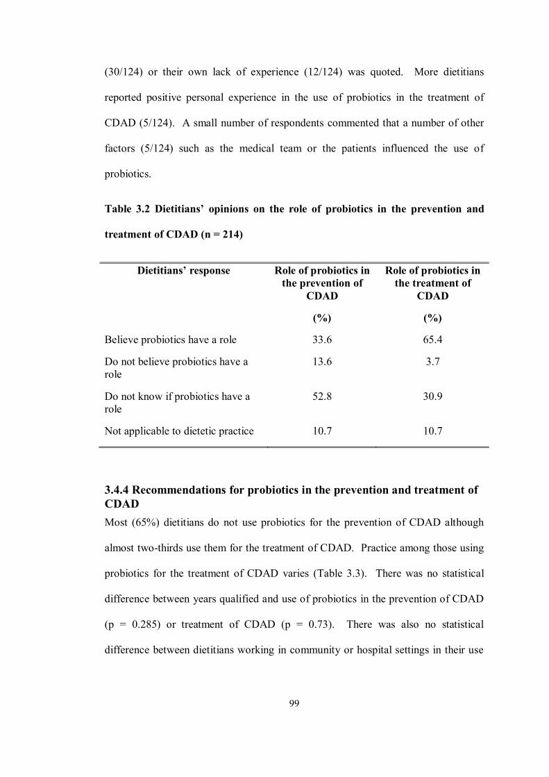
99
(30/124) or their own lack of experience (12/124) was quoted. More dietitians
reported positive personal experience in the use of probiotics in the treatment of
CDAD (5/124). A small number of respondents commented that a number of other
factors (5/124) such as the medical team or the patients influenced the use of
probiotics.
Table 3.2 Dietitians’ opinions on the role of probiotics in the prevention and
treatment of CDAD (n = 214)
Dietitians’ response Role of probiotics in the prevention of
CDAD
(%)
Role of probiotics in the treatment of
CDAD
(%)
Believe probiotics have a role 33.6 65.4
Do not believe probiotics have a role
13.6 3.7
Do not know if probiotics have a role
52.8 30.9
Not applicable to dietetic practice 10.7 10.7
3.4.4 Recommendations for probiotics in the prevention and treatment of CDAD Most (65%) dietitians do not use probiotics for the prevention of CDAD although
almost two-thirds use them for the treatment of CDAD. Practice among those using
probiotics for the treatment of CDAD varies (Table 3.3). There was no statistical
difference between years qualified and use of probiotics in the prevention of CDAD
(p = 0.285) or treatment of CDAD (p = 0.73). There was also no statistical
difference between dietitians working in community or hospital settings in their use

100
of probiotics for the prevention of CDAD (p = 0.581) and their use of probiotics for
the treatment of CDAD (p = 0.45).
Table 3.3 Dietitians’ use of probiotics in the prevention and treatment of CDAD
(n = 196)
Dietitians’ response Use of probiotics in the
prevention of CDAD
(%)
Use of probiotics in the
treatment of CDAD
(%)
Always use probiotics 9.8 36.4
Never use probiotics 65.0 27.6
Occasionally use
probiotics
14.5 25.2
Not applicable to practice 10.7 10.7
3.4.5 Dietitians’ recommendations for the use of probiotics in other clinical conditions
Thirty-three (15.4%) respondents would never recommend probiotics in their clinical
practice. There was no association between those who recommend probiotics and
years qualified (p = 0.390). Of those who recommend probiotics (84.6%), this was
most frequently for the management of irritable bowel syndrome (IBS) (82.3%, n =
149). The use of probiotics in the other five clinical conditions that were provided as
options for the dietitians is illustrated in Figure 3.1. Respondents also provided other
conditions for which they would recommend probiotics. Antibiotic treatment and

101
antibiotic-associated diarrhoea were conditions in which probiotics were
recommended most often. Dietitians also reported recommending probiotics for the
treatment of thrush, diarrhoea, diverticular disease, constipation, radiation enteritis,
cystic fibrosis, pouchitis, autism, sports nutrition, on an individual basis,
gastrointestinal distension, malnutrition and acute diarrhoea in children.
3.4.5.1 Probiotic use by dietitians with non-Clostridium difficile diarrhoea
There is variation in the use of probiotics in patients with diarrhoea that is not due to
C. difficile. Forty two percent (n= 82) of dietitians working in hospital and
community settings stated that they would occasionally use probiotics when a patient
develops diarrhoea while on antibiotics in the absence of C. difficile toxins. When
diarrhoea occurs, C. difficile is not isolated and the patient is not on antibiotics,

102
40.8% of dietitians would recommend the use of probiotics whereas 34.7% would
never recommend them in this situation.
3.4.6 Probiotic provision and guidance in healthcare facilities
3.4.6.1 Commencement of probiotics
In the hospital and community setting, respondents indentified “dietitians” as the
healthcare professional most likely to recommend the commencement of probiotics
in patients with CDAD. They also indicated that medical staff are the healthcare
professional group next most likely to recommend the administration of probiotics to
patients (Figure 3.2).

103
3.4.6.2 Probiotic products supplied by healthcare facilities
To establish the probiotic products supplied by healthcare facilities, only the
responses from hospital dietitians were analysed (n = 155) as probiotics are not
supplied to patients within the community. Of the 43 hospitals, 93% (n = 40) buy
probiotic products for provision to patients. Two hospitals no longer provide
probiotics for financial reasons and the dietitian in one hospital (maternity) reported
that probiotics are never used.
Eleven different products were reported as available for patients (Appendix 10).
There were some inconsistencies between dietitians working in individual hospitals
as to the products available. Overall, it was evident that the yogurt drink
“Everybody” (Yoplait franchise produced by Glanbia plc Consumer Foods Division,
Dublin) was the most commonly available commercial product. This product
contains the probiotic strain L. rhamnosus GG. The factors influencing the probiotic
supplied within hospitals were catering department choice (40.7%; n = 63) and
clinical evidence for the product supplied (16.8%; n = 26). Twenty dietitians
(12.9%) reported that a combination of both factors mentioned above determined the
product supplied. Despite over half the dietitians surveyed reporting that clinical
evidence for the product influenced the product supplied, almost half (48.4%; n = 75)
did not know the strain present in the products used.
3.4.6.3 Guidelines /policies on the use of probiotics
To establish if there is guidance available to dietitians on their use of probiotics, the
study questionnaire investigated the existence of policies or guidelines in this area.
Responses from dietitians working in hospital and community settings only were
analysed (n = 196). Of these, 4.1% (n = 8) responded they had a policy or guideline

104
in their place of work for the use of probiotics while 4.6 % (n = 9) responded they
had a policy or guideline in their place of work for the use of probiotics in CDAD.
The response to this question varied within individual hospital and community
settings, indicating that in those settings where a policy exists, not all staff were
aware of its existence. Among the additional comments provided by respondents (n
=55) at the end of the questionnaire, 21.8% (n =12) identified the need for policies or
guidance on probiotic use.
3.4.7 Enteral nutrition and CDAD No standardardisation of practice was observed among dietitians in the use of
probiotics with enteral tube feeding (Figure 3.3). Approximately one-third of
dietitians (30.6%) reported recommending the use of probiotics occasionally in
patients with CDAD who were being enterally fed and a similar proportion (36.2%)
recommended their use in other clinical conditions when enteral nutrition was
required. There was no statistical association between the place of work (p = 0.229)
or years practicing (p = 0.340) and the use of probiotics in ETF patients with CDAD.
Neither was an association seen between the use of probiotics in ETF in other
conditions and dietitians’ place of work or years of experience. The enteral feed
most commonly used in patients with CDAD by the majority of dietitians was a
polymeric feed (79.6%); few dietitians recommended the use of semi-elemental
(7.1%) or elemental (0.5%) feeds. There was no consensus on the provision of
dietary fibre to patients with CDAD; 31.6% provide no fibre to patients with CDAD,
27% use a mixture of a non-fibre containing feed with a fibre containing feed, while
only 17.9% stated that they would always recommend a fibre-containing feed.

105
3.4.8 Vitamin and mineral supplementation and CDAD The majority of dietitians (57.9%) reported recommending vitamin and mineral
supplementation during an episode of CDAD if warranted by the patient’s nutritional
status, while 17.3% consider both diagnosis and nutritional status when deciding
whether to recommend additional vitamin and minerals. Only one dietitian reported
routinely recommending a vitamin and mineral supplement in patients who develop
CDAD.

106
3.5 Discussion In recent years, there has been a greater awareness of the potential role of probiotics
with the emerging evidence of their efficacy in some human diseases, and the
increased availability of probiotic products to consumers. Dietitians are key
healthcare professionals who can influence the use of probiotics both among
consumers and other healthcare professionals. There are a number of studies
investigating dietitians’ attitudes towards FF (Lee et al., 2000; de Jong et al., 2004;
McConnon et al., 2004; Landstrom et al., 2007b). In the studies by Lee et al., (2000)
and Landstrom et al., (2007b) dietitians’ opinions of FF were investigated. The
authors did not differentiate between dietitians’ opinions about different FF. This is
the first study that has examined dietitians’ beliefs or attitudes towards probiotics
alone, specifically in relation to their professional practice. It also highlights some
aspects of dietetic professional practice currently taking place in the ROI.
3.5.1 Response rate The response rate of 40.9% was lower than anticipated and has the potential to
introduce some bias to the results, as non-responders can undermine study validity
and thus generalisability to a wider population (Cook et al., 2009). A response rate
of 75% is considered ideal (Bowling, 2009); however; response rates to all types of
surveys have been declining (Bowling, 2005), especially among healthcare
professionals. This has been attributed to an increased demand to participate in
research activities (Cook et al., 2009). Cook et al., (2009) showed that the average
response rate to postal surveys among healthcare professionals was 56% (in studies
published from 1996-2005). Only 16% of the studies analysed achieved a response
rate of 75% or over. In the literature, response rates for postal surveys of dietitians

107
have been between 46.8 and 53% (Cashman et al., 2003; Mackel et al., 2003; de
Jong et al., 2004). An online web survey conducted by another Irish dietitian on the
topic of professional development at the same time and to the same group of
dietitians had a similar response rate (43.7%) (Fitzpatrick, 2010).
3.5.2 Participants The employment profile of the study participants was similar to that obtained from
the INDI workforce planning survey of clinical nutritionist/dietitians undertaken in
May 2010 (INDI, 2010). This showed that 72% of dietitians were employed in
public and private hospitals and 28% in the community setting. It is, possible that
dietitians with greater experience of managing CDAD were more likely to complete
the questionnaire. As 73% of CDAD cases in Ireland occur in hospitals (HPSC,
2010); this may have resulted in the higher response rate from dietitians working in
that setting. Additionally, CDAD is more commonly seen in adults and older
persons which may account for the lower response from dietitians working in the
paediatric setting.
3.5.3 Dietitians’ use and opinions of probiotics and CDAD There is no consensus among Irish dietitians on the role and use of probiotics in the
prevention and treatment of CDAD with more than half of all dietitians surveyed
(52.8%) reporting that they did not know if there is a role for probiotics in the
prevention of CDAD. There was greater support (65.4%) for a role for probiotics in
the treatment of CDAD. As might be expected, given the number of dietitians who
did not know if there was a role for probiotics in the prevention of CDAD, the
majority do not recommend probiotics for its prevention. In the treatment of CDAD,
no consensus on the practice of recommending probiotics was evident. Some
dietitians recommended probiotics occasionally, and some in all cases, to prevent

108
and/or treat CDAD. This is despite the lack of published evidence to support the use
of probiotics in the treatment of CDAD. Although there appears to be a possible role
for use of the probiotic S. boulardii in conjunction with high dose vancomycin in the
prevention of CDAD recurrence (McFarland et al., 1994; Surawicz et al.,2000),
randomised double-blind placebo trials of sufficient size to detect significant
differences are still lacking. Currently, this yeast is not being used as a probiotic in
Irish hospitals.
The treatment guidance document for CDAD from the ESCMID (Bauer et al., 2009)
states that there is insufficient evidence to recommend probiotics in addition to
antibiotics for the treatment of CDAD. The guidelines make reference to safety
issues that have been reported with S. boulardii (Bassetti et al., 1998; Munoz et al.,
2005) and the increased mortality observed in a study when probiotics were used in a
RCT in patients with acute pancreatitis (Besselink et al., 2004). The UK guidelines
from the Health Protection Agency & Department of Health (2008) also comment on
the use of probiotics in CDAD. They currently do not recommend probiotics to
prevent CDAD due to the lack of statistical significance in two meta-analyses by
Dendukuri et al., (2005) and Pillai & Nelson (2008). US clinical practice guidelines
also do not recommend the administration of probiotics to prevent primary CDAD.
This is due to the lack of evidence and the potential risk of blood stream infection
from probiotic use (Cohen et al., 2010). There is also no support for the routine use
of probiotics from the Australasian Society for Infectious Diseases; again this is
based on the lack of efficacy and the potential for adverse events reported in the
scientific literature (Cheng et al., 2011).
Irish dietetic practice should follow the guidance provided by the review document
‘Surveillance, Diagnosis and Management of Clostridium difficile in Ireland (HPSC,

109
2008). Following their review of the evidence on probiotics, the HPSC concluded
that the routine use of probiotics cannot be recommended. Probiotics should not be
used either in the prevention or treatment of CDAD until there is more conclusive
evidence for their use. Moreover, the specific probiotics currently used to prevent or
treat CDAD in Irish health care facilities have even less evidence for their use than
those referred to in the studies above.
3.5.4 Dietitians’ recommendations for probiotic use Only 15.4% of dietitians never recommend the use of probiotics in their practice. In
those recommending probiotics, IBS was the most common condition for which they
were recommended (by 81.4%). These findings are comparable to those of US
gastroenterologists; the majority (98%) of gastroenterologists supported the use of
probiotics in patients with IBS (Williams et al., 2010). The symptoms of IBS are
thought to be due to bacterial overgrowth in the small intestine causing increased
fermentation and gas production (Wilhelm et al., 2008). The most commonly
studied probiotics in IBS are Lactobacillus, Bifidobacterium and the mixture
probiotic product VSL#3 (Williams, 2010). The NICE guidelines on the
management of IBS, published in 2008, reviewed thirteen studies that met their
inclusion criteria and concluded that probiotics are effective in some people with IBS
but not in all. When products were from reputable sources, no harm was identified
from the ingestion of probiotics. The NICE guidelines conclude with the
recommendation that ‘people with IBS who choose to try probiotics should be
advised to take the product for at least four weeks while monitoring the effect.
Probiotics should be taken at the dose recommended by the manufacturer’ (NICE,
2008). A systematic review on the use of probiotics in IBS conducted by Moayyedi
et al., (2010) which included 18 papers with 1650 participants concluded that

110
probiotics had a statistical effect in reducing IBS symptoms, but that there was no
difference in the different types of probiotics used, all of them showing a trend
towards benefit. The main limitation acknowledged in this review was the number
of different probiotic strains and doses used in the studies reviewed and therefore no
recommendation on the best probiotic and dose for the treatment of IBS were made.
This limitation has been reiterated by other authors investigating this topic
(Williams, 2010).
For more specific evidence and guidance for the dietetic profession, the British
Dietetic Association (BDA) gastroenterology specialist group developed UK
evidenced-based practice guidelines for the dietetic management of IBS in adults.
Their inclusion criteria were studies that used probiotic products that are available to
consumers in the UK. Of the five studies identified, the results were too weak to
support the therapeutic use of probiotics in IBS. They advised that interventions
using probiotics to further improve IBS symptoms can be considered within second
line dietary treatment. As with other reviews and guidelines, there was insufficient
evidence to recommend a specific probiotic. The dietetic practice guideline
recommended that patients should be advised that some products in probiotics may
increase IBS symptoms, that the long term effects of probiotics are unknown and that
if there is no improvement with one probiotic product, another one may need to be
tried (Mc Kenzie et al., 2010). It would seem reasonable for Irish dietitians to
recommend probiotics for IBS patients in a manner similar to that of UK dietitians.
However, specific guidance on probiotic products available on the Irish market
would be useful for practicing dietitians.

111
The lack of support for probiotics in the management of the paediatric condition,
necrotitsing enterocolitis (NEC), may be explained by the low response rate from
dietitians who work with children and, that even within paediatrics, the management
of NEC is a specialist area.
3.5.5 Use of probiotics in other clinical conditions A number of other clinical conditions in which this group of dietitians would
recommend probiotics were cited. Antibiotic treatment and antibiotic-associated
diarrhoea were the most common situations in which probiotics would be suggested
(by 12% and 8.7% of dietitians respectively). In comparison probiotics were
recommended by 21% Dutch dietitians, for those with recent diarrhoea and after
antibiotic treatment (de Jong et al., 2004). This study investigated FF in general
rather than probiotics specifically, and so contained limited detail on products used
and doses recommended. In a study of New Zealand GPs (n = 45), probiotics were
mostly commonly recommended for diarrhoea (44.4%) (Schultz et al., 2011).
However, there was no information given on the cause(s) of diarrhoea in this study.
AAD is a common problem, occurring in 5-25% of patients receiving antibiotics,
although the rates reported depend on the antibiotics used and the population studied
(Bartlett, 2002). Probiotics given in conjunction with antibiotics for the prevention of
AAD have been extensively studied in both children and adults. Several systematic
reviews document that most of the probiotics tested have been shown to be effective
in reducing risk of AAD in the general population (Cremonin et al., 2002; D’Souza
et al., 2002; Hawrelak et al., 2005; Szajewska & Mrukowicz, 2005). L. rhamnosus
GG and S.boulardii are documented as being most efficacious. These are the strains
which have been most commonly used in the studies. From a review of all studies, it
can be concluded that the results of studies using probiotics to reduce AAD are

112
promising but that conclusive evidence is still lacking, additionally it cannot be
stated that all probiotic strains are effective (McFarland, 2009) Again the findings
from this study show that this group of dietitian are using probiotics in an area where
the evidence does not fully support their use.
3.5.6 Probiotics supplied in hospitals/ healthcare facilities and guidance around probiotic use From this questionnaire it is evident that probiotics are being provided to patients in
the majority of hospitals and other health care facilities in Ireland. The dietitians
have identified themselves as the primary healthcare professional to recommend
probiotics in CDAD. Similarly, in a randomly selected group of Canadian dietitians,
81% (n = 122) they too believed that dietitians were the most appropriate
professional to recommend FF (Sheeshka & Lacroix, 2008). Such beliefs mean that
dietitians should have a good knowledge of FF, the probiotic products provided in
healthcare facilities and also the evidence for their use. The dietitians surveyed in
this study had poor knowledge of the probiotic strains in the products they supplied
or recommended. Around half (47.7%) could not identify even one strain present in
the product supplied. The catering departments of healthcare facilities were also
reported to have the biggest influence on the probiotic supplied. One issue that is
very clear from the studies carried out with probiotics in the treatment of various
clinical conditions is that the positive effect is dose and strain dependent. The
administration of a probiotic product that may only contain one strain to a diverse
hospital population could be described as futile. The limited knowledge of dietitians
of the strains being used, despite them taking responsibility for recommending
probiotics, identifies a deficit in knowledge and professional practice.

113
The presence of policies or guidelines on the administration of probiotics within
clinical settings appeared to be unclear, with disagreement within departments as to
the existence of guidelines. Analysis of the practices of dietitians working in specific
areas was not possible due to the mixed caseloads of many dietitians. However, a
sub-group of 18 (8.4%) dietitians working in critical care was apparent, although it
should be noted that some dietitians did not work solely in the ICU setting. ICU is
the one area where guidance exists for dietitians on the use of probiotics. The recent
Canadian Critical Care Guidelines (2009) state that there is insufficient data to make
a recommendation on the use of probiotics, prebiotics and synbiotics in the critically
ill (Heyland et al., 2009). The Committee considered that the studies available are
inconsistent in their method of reporting outcomes such as septic mortality,
complications and diarrhoea. The variability in the probiotics used in studies and the
choice of control group made the published studies difficult to assess. The probiotic
prophylaxis in predicted severe acute pancreatitis (PROPTRIA) trial (Besselink et
al., 2004) and concerns about risk associated with S. boulardii were also a cause for
concern for the Committee. The American Society for Parenteral and Enteral
Nutrition (ASPEN) Critical Care Guidelines of 2009 reiterate that recommendations
cannot be made about the use of probiotics in the general ICU population, again due
to the lack of consistent outcomes (McClave et al., 2009).
The safety issues highlighted in the literature would appear to be an aspect needing
consideration. Although dietitians’ opinions about the safety of probiotics were not
specifically investigated in this study, some respondents highlighted that they would
not use them in specific patient groups (i.e. infants and immunocompromised, ICU

114
and paediatric oncology patients). Safety concerns arising from the use of probiotics
were not apparent in the questionnaire responses.
The reasons quoted for not using probiotics by Dutch dietitians related to safety and
functionality of the products and to products containing bacteria or “foreign”
invaders (de Jong et al., 2004). In another study, nutritionists (Mc Connon et al.,
2004) expressed concern about the risks associated with probiotics; however, this
study focused on FF as a group of foods and did not seek specific detail on dietitians’
opinions on the safety aspects of probiotics.
3.5.7 Type of enteral feed used in CDAD Although the majority of dietitians used a polymeric feed, there was no consistent
practice in the provision of dietary fibre to patients with CDAD. A small number of
studies have investigated the impact of diet on the acquisition and growth of C.
difficile. The results of this study showed that few dietitians used elemental enteral
feeds in their clinical management of CDAD patients. Elemental diet is thought to
favour the growth and proliferation of C. difficile. An in-vitro culture study found
that elemental diet enhanced the number of C. difficile and induced the production of
C. difficile toxin (Iizuka et al., 2004). It has been reported that C. difficile toxin was
frequently detected in patients undergoing elemental diet therapy who were not
taking antibiotics. Faecal C. difficile toxin disappeared soon after the elemental diet
therapy stopped (Itou et al., 1999; Itou et al., 2000). Elemental diet contains no
complex carbohydrate; the absence of dietary fibre and resistant starch disturbs
microbial balance in the GIT and deprives the colonic epithelium of its chief energy
source and proliferation regulator. This reduces fermentation and the production of
short chain fatty acids (SCFA), especially the SCFA butyrate. It has been shown that

115
butyrate deficiency in the colon potentiates the growth and toxin production of C.
difficile organism (May et al., 1994). It has also been suggested the relatively large
amounts of amino acids in elemental diet may to provide a favourable environment
for C. difficile. A recent review by O’Keefe (2010) stated that elemental feeding as a
risk factor for C. difficile infection is frequently overlooked.
To provide guidance for dietitians on the provision of fibre to this patient group, the
mechanisms of action of fibre in the GIT should firstly be examined. Studies that
investigate the impact of fibre on the management of diarrhoea should also be
reviewed, given that diarrhoea is the major symptom of CDAD. Fibre intake
influences nutrient absorption, carbohydrate, fat and sterol metabolism, stool bulk
and weight, colonic fermentation and gastrointestinal transit time (Kapadia et al.,
1995; Alam et al., 1998). Fibre also improves gut barrier function, helping to
prevent translocation of bacteria and toxins from the gut into the systemic circulation
(Spaeth et al., 1995). Fibre also promotes increased turnover or regeneration of
epithelial cells (Rehman et al., 2003).
The effects of supplementing enteral feeds with dietary fibre in both healthy
volunteers and patients in hospital and community settings was investigated in a
systematic review and meta-analysis of 51 studies (Elia et al., 2008). The authors
concluded that fibre supplementation was generally well tolerated. In the hospital
setting, incidence of diarrhoea was reduced as the result of fibre administration (OR
0.68; 95% CI 0.48-0.96). Results showed a strong effect of fibre in reducing the
incidence of diarrhoea in patient groups with a high incidence of diarrhoea and a
smaller or absent effect in those with a low incidence of diarrhoea. A likely
mechanism underlying the effect of fibre in reducing diarrhoea is the stimulation of

116
colonic water and electrolyte absorption by SCFA that are produced as a result of
anaerobic metabolism of fibre. SCFA have been shown to reverse fluid secretions in
the ascending colon during enteral feeding (Bowling et al., 1993). SCFA production
depends on the number of types of bacteria species present in the colon, substrate
source(s) and gut transit time (Wong et al., 2006) , all of which have a number of
properties that may be of benefit in maintaining normal bowel structure and function,
and preventing or alleviating colonic based diarrhoea. SCFAs and acidic pH have
been shown to contribute to resistance to C. difficile colonisation and proliferation in
vitro (Rolfe & Iaconis, 1983; Rolfe, 1984). Different types of dietary fibre affect the
type and amount of SCFA produced. Nakao et al., (2002) reported an increase in the
ratio of anaerobic bacteria to aerobic bacteria with fibre supplementation. This may
be beneficial as anaerobic bacteria are thought to protect against the overgrowth of
potential pathogens, some of which may cause diarrhoea (Bourlioux, 1997). It is
thought that lack of dietary fibre could produce an intestinal environment conducive
to the growth of C. difficile. The recent review by O’Keefe (2010), speculates that
the prolonged use of low fibre tube feeds will enhance C. difficile colonization and
subsequent C. difficile associated colitis by reducing the resistance to pathogen
adherence and subsequent cytotoxic injury.
3.5.8 The provision of probiotics via an enteral tube A limitation of the present study was that the questions asked of dietitians about their
use of probiotics with enteral tube feeding did not distinguish between the positions
of the enteral tube. Such questions would have provided more detailed information
on dietetic practice. It appears that approximately half of all dietitians either
regularly or occasionally recommend probiotics via enteral feeding tubes in both
CDAD and other clinical conditions. There is very little guidance from the scientific

117
literature or the probiotic or enteral feeding tube industries in this area. Danone
Ireland Limited, the company that produce “Actimel” does not recommend the use of
probiotics via enteral feeding tubes; the reason provided by the company is that
clinical evidence is lacking to support its safety if used in this way, as the product is
intended for oral consumption and has only been tested for oral use (Cox, M. 2010
pers. comm., 15 May 2010). Communication with representatives from Yakult,
Ireland (Yakult) and Glanbia (“Everybody”) indicated that neither company had
guidelines for the administration of their products via enteral feeding tubes (Jordan,
D. 2011 pers. comm., 19 January; McGartland, C. 2011 pers. comm., 19 April;).
Three companies that manufacture enteral feeding tubes that are commonly used in
Irish healthcare facilities were contacted to seek their position on the administration
of probiotics via enteral feeding tubes. No company had recommendations; some of
their comments compared the administration of probiotics to the administration of
medications via enteral feeding tubes (Nolan, O. 2010, pers. comm., 4 May; Coakley,
D. 2010, pers. comm., 14 May; Smith, R. 2010, pers. comm., 14 May).
The safety issues that arise with probiotics and nutrition support were addressed in a
recent review by Whelan and Myers (2010). Patients receiving nutrition support are
often high risk patients who are frequently critically ill and at risk of bacterial
translocation or are immunocompromised and, so, have additional risk factors for
probiotic infection. They are, also, regularly on medications that increase gastric pH.
The method of administering nutrition support can increase patients’ risk; post-
pyloric feeding enteral nutrition by-passes the gastric acid barrier completely and this
can result in a greater innoculum of the probiotic surviving in the small intestine. A
10-fold increase in the dose reaching the small intestine has been reported (Whelan

118
& Myers, 2010). Central venous catheters (CVC) are used in the administration of
parenteral nutrition (PN) and these have been identified as a potential risk factor for
probiotic infection (Boyle et al., 2006). The review by Whelan and Myers (2010)
included 20 case reports and 52 papers with 53 trials (41 RCT’s and 12 non-
randomized trials). In the case reports examined, 32 patients received various
methods of nutrition support and probiotics of varying strains and doses.
Bacteraemia occurred in five cases and fungaemia in 27 cases. Two patients also
developed endocardititis (Land et al., 2005; Munoz et al., 2005). The risk factors
were variable, but the majority of patients who developed infections had received
antibiotics and had intravenous access. In eight patients, the adverse events resulted
in death. The cases in which infection developed had used L. rhamnosus GG or S.
boulardii. The authors commented this is probably due to these strains being used
more frequently rather than them having greater virulence. The presence of a CVC
also carried a greater risk; many of these patients were in ICU and required a CVC
for their medical management although it was highlighted that the CVC tips were not
examined for contamination. The authors of this review concluded that the risk of
environmental contamination may also occur and be a greater risk factor than
administration of probiotics. In the case reports of S. boulardii fungaemia, the
patients themselves were not receiving the yeast but patients located beside them
were (Cassone et al., 2003; Graf & Gavazzi, 2007).
In the 53 trials examined by Whelan and Myers (2010), the probiotics used varied
and were administered in varying doses and using a range of administration methods.
Subjects in the studies were from a variety of locations with a range of disorders.
Many of the studies showed a reduction in mortality, sepsis or infection; in general

119
the studies showed either no effect or a positive effect of probiotic use on the overall
outcome. Only three studies, (one post liver transplant, another in acute pancreatitis
and a further study in NEC) found significant increases in negative clinical sequelae
which were largely non-infectious in nature (Besselink et al., 2004; Rayes et al.,
2005; Lin et al., 2008). In two of the RCT’s, there was increased negative outcome
in patients in which the probiotics were administered via naso jejunal tube (NJT)
(Besselink et al., 2004; Rayes et al., 2005). Whelan and Myers (2010) remark, in the
three trials with negative outcomes, there are some issues with the study designs.
One of the trials had low numbers which may result in statistical error (Rayes et al.,
2005) and in the second trial a novel probiotic was used and the study itself has been
heavily criticized (Besselink et al., 2004). In many of the studies the probiotic
strains were not characterized, inconsistent doses were reported, probiotic infection
occurred at different doses of probiotics, adverse events were either unreported or
there was a lack of information about the adverse events. In the cases where patients
died, it was difficult to attribute the patient’s death to probiotic infection.
3.5.9 CDAD and mineral and vitamin supplementation The symptom of diarrhoea which can often be persistent and severe in CDAD can
impact on the individual’s nutritional status, especially micronutrient status. There is
no guidance in the literature on the provision of additional micronutrients during
CDAD. From the results of this study, it can be seen that a dietitian’s decision to
supplement with vitamins or minerals appears to be generally based on his/her
clinical judgement. A more structured approach should be considered as mineral
balance is always altered when diarrhoea persists for more than a few hours (Wiesen
et al., 2006). A range of micronutrient deficiencies during diarrhoeal disease has
been described; some of the micronutrient deficits identified are copper, iron,

120
magnesium, selenium, zinc, folate and vitamins A, B12 and D (Tomkins et al., 1993).
Micronutrient deficiency impairs immune function and increases susceptibility to
infection (Yaqoob & Calder, 2003).
Zinc is a micronutrient that has been extensively studied for its role in diarrhoea. In
the GIT, it restores mucosal barrier integrity and enterocytes, promotes brush border
enzyme activity and the production of antibodies and circulating lymphocytes against
intestinal pathogens (Shankar & Prasad, 1998; Hess et al., 2009; Prasad, 2009). The
association between zinc and diarrhoeal associated morbidity was first noted in early
observational studies that documented increased faecal zinc loss, a negative zinc
balance and low tissues zinc concentration among children with diarrhoea (Anon,
Zinc and copper wastage during acute diarrhoea, 1990). There have been a number
of studies investigating prophylactic zinc supplementation and its role in diarrhoea.
A recently published Cochrane review also supports the role of zinc in treating
diarrhoea in children. In the 22 trials reviewed, zinc was seen to shorten the duration
of an episode of diarrhoea. However, there was no benefit demonstrated in children
under 6 months (Lazzerini & Ronfani, 2008). Due to the positive results of studies
on zinc supplementation in children, the WHO/UNICEF now recommend zinc
supplementation for the treatment of all episodes of childhood diarrhoea
(WHO/UNICEF, 2004). Although there is strong evidence to support the
supplementation of zinc in children, the majority of the studies on zinc
supplementation have been conducted in children in developing countries and,
therefore, may not be applicable to the adult population in developed countries.
Nonetheless, dietitians should be more aware of zinc losses that can occur as a result
121
of CDAD diarrhoea. In particular, if diarrhoea is persistent, zinc supplementation
should be considered, especially if nutritional intake is inadequate.
3.5.10 Limitations and future considerations An aspect not considered in the design of this study was dietitians’ personal
consumption of probiotic products. In previous studies, this has been reported to
influence dietitians’ professional practice. In a study of Korean dietitians, the
dietitian’s own frequency of consumption of FF had a significantly positive effect on
their recommendation of such foods and those who ate FF demonstrated a more
positive attitude towards the efficacy of such foods. The authors of this study
expressed concern that these findings may result in some dietitians providing
misleading advice to consumers, because the advice provided was not necessarily
based on scientific knowledge (Cha et al., 2010). An additional factor not explored
as part of this study was methods dietitians use to obtain information about
probiotics. The study of Dutch dietitians showed that they got most of their
information from journals, magazine articles or the internet, but would have
preferred if information had been provided by independent scientists instead of
through product promotions. Specific information requested was about the safety
and functionality aspects of the probiotics (de Jong et al., 2004).
An important issue to be addressed from these study results is that dietetic practice in
the use of probiotics is not underpinned by scientific research. Similar findings on
the opinions and use of probiotics have been reported among gastroenterologists in
the US. Williams et al., (2010) reported that gastroenterologists believe that there is a
role for probiotics in patient care but actual practices did not concur with the
published expert panel based recommendations for evidence based probiotic use.

122
Cha et al., (2010) commented on the need for undergraduate students of nutrition and
dietetics to learn about FF from a scientific, evidence-based approach and that
education about FF should be provided as continual professional development for
practicing dietitians.
Given the vast amount of research into the efficacy of probiotics in different clinical
conditions, the need for guidance for healthcare professionals, perhaps in the form of
clinical practice guidelines should be considered. One of the most consistent
findings in health service research is the gap between best practice as identified by
scientific research and the actual clinical care provided (Grol & Grimshaw, 2003;
Brown et al., 2004). Several studies have suggested that 10-40% of patients do not
receive care that is based on current scientific evidence and that this deficit in
evidence-based practice results in care that is either unnecessary or is potentially
harmful in ≥20% of cases (Avezum et al., 2000; Grol & Grimshaw, 2003; Leape et
al., 2003; Mc Glynn et al., 2003). A cross-sectional survey in a Swedish University
Hospital explored dietitians’, occupational therapists’, and physical therapists’
attitudes towards evidence-based practice. All professions were reported to have
positive attitudes towards evidence-based practice and support the use of evidence in
decision making about patient care. Although the groups had good knowledge about
research terminology, they lacked understanding of the specific terminology relating
to evidence-based medicine. Lack of time was given as a barrier to the use of
evidence-based care (Heiwe et al., 2011).

123
3.6 Conclusion This is the first study to describe Irish dietitians’ opinions on, and use of, probiotics,
and their current practice in the management of CDAD. The role and efficacy of
probiotics in the prevention and management of CDAD is an area of emerging
research and understanding. We need a greater understanding of the complex
interaction between the human gastrointestinal microflora, pathogens, probiotics and
antibiotics and to increase our knowledge about the role of probiotics in CDAD. The
low cost, good safety profile and low risk of infection make probiotics a favourable
option in the prevention and treatment of CDAD, provided sufficient evidence
becomes available to support their use. Studies of appropriate statistical power and
adequate follow up are necessary to provide insight into the efficacy of particular
bacterial and yeast strains and the doses of these strains that are most likely to
prevent CDAD and its recurrence, and treat CDAD should it develop.
The findings of this study indicate that Irish dietitians have mixed views about the
role of probiotics in CDAD. Despite this, they frequently recommend probiotics in
their clinical practice either orally or via enteral feeding tubes, with little
standardisation of practice apparent. The probiotic strain recommended or available
to prevent or treat CDAD frequently does not contain the strain for which evidence
exists. Guidelines or polices in Irish healthcare facilities for probiotic use are lacking
or not consistently referred to by Irish dietitians. Guidance on the use of probiotics
in CDAD should be sought from current scientific literature and the development of
consensus practice guidelines for dietitians managing patients with CDAD is
essential.

124
Although the majority of Irish dietitians use a polymeric feed when enterally feeding
patients with CDAD, practice on the provision of dietary fibre to this patient group is
variable. The provision of additional vitamin and mineral supplementation is also
dependent on the dietitian’s clinical judgement. The enteral feeding practices of Irish
dietitians in this patient group need to be compliant with scientific evidence. From
the review of the literature there are a number of considerations for dietetic practice.
Those at high risk of developing CDAD elemental and semi elemental feeds should
be avoided. A source of fibre should be provided when enterally feeding a high risk
patient. In cases of CDAD where diarrhoea occurs the provision of additional
vitamins and minerals should be considered to account for increased losses.
Irish dietitians in this study identifed themselves as the main healthcare professional
to recommend probiotics. However the survey found that there was underutilization
of current scientific evidence and guidelines to conform with evidence-based
practice. Health professionals need to remain competent in their knowledge of this
developing area of research and be able to translate research findings into their
professional practice (Schultz et al., 2011). To achieve this, it is vital that
information on FF is correctly communicated to those involved in nutrition education
and health promotion (Blades, 2000). Given the increasingly widespread availability
and use of probiotics in both community and hospital healthcare settings, Irish
dietitians need to acquire a thorough understanding of these products, including the
risks and benefits of probiotic treatment and the effectiveness of probiotics using
different modes of delivery. If probiotics are recommended to prevent or treat
disease in patients, consideration must be given to quality control and documentation

125
of the efficacy of such treatment. It is imperative that dietitians develop their
knowledge about probiotics so that they are able to convey science based information
to their clients and healthcare professionals and their credibility as nutrition experts
is strengthened (Hetherwick et al., 2006).

126
4.1 Conclusions and Recommendations This study looked at a number of aspects involved in the dietetic management in
patients with CDAD in Ireland. The first study’s aim was to assess the nutritional
status of patients with CDAD and compare this to a group of hospital patients. The
study found there was no significant difference in MUST scores between a group of
patients with CDAD and a comparative hospital patient group. However the CDAD
group did have a significantly lower BMI and were a significantly older. In addition,
when the data from the CDAD group were compared to malnutrition data from other
Irish studies, there was a higher prevalence of malnutrition (as determined by BMI)
and of malnutrition risk (as determined by MUST) in the CDAD group in this study.
These findings would indicate that hospitalised patients with CDAD are at high risk
of malnutrition.
The second study investigated dietitians’ opinions and uses of probiotics in CDAD
and other clinical conditions. It also looked at the enteral feeds being provided to
these patients should tube feeding be required. This study showed that Irish dietitians
have inconsistent views on the role and use of probiotics in different clinical
conditions. What has emerged from the work is that probiotics are being
recommended by dietitians in clinical conditions for which insufficient evidence
exists, in particular patients with CDAD. It is also clear that a variety of probiotic
products are being supplied within Irish health care facilities, that knowledge of the
specific benefits and limitations for particular strains is poor and that very little
guidance for their use exists. The study also showed that general enteral feeding
practices varies among dietitians, specifically in the provision of feeds containing
dietary fibre.

127
From the findings of this study and reviewing the literature a number of conclusions
and recommendations are made:
The dietetic profession should continue to support and be involved in the
promotion, training and implementation of nutritional screening for all patient
groups in both the hospital and community setting.
Dietitians should be aware of the risk factors for the development of CDAD;
in those patients at high risk who require enteral feeding the provision of
feeds containing dietary fibre and the avoidance of elemental and semi
elemental feeds should be considered. Supplementation of micronutrient
intake including zinc in patients who develop CDAD may be beneficial.
Medical and nursing staff should be aware that patients with CDAD are a
patient group at high risk of malnutrition and instigate care plans that address
this risk.
Probiotics should not be recommended for use in either the prevention or
treatment of CDAD, until there are further supportive trials in the area. The
author is unwilling to make a recommendation endorsing any product for any
patient group for the treatment or prevention of CDAD as this would
contravene current evidence. The probiotic S. bouldarii for which there is
some limited evidence is not available in Ireland. However, in countries
where this product is available international guidelines do not recommend it
use due to safety concerns.
There is a need for education for practicing dietitians on the role of probiotics
in clinical practice. This education should be continuous to capture the
emerging evidence in the area. The information provided should be

128
independent of commercial influence and provide a balanced view of the
scientific evidence.

129
References
Ahmed T & Haboubi N (2010) Assessment and management of nutrition in older
people and its importance to health. Clin Interv Aging, 5, 207-16.
Akerlund T, Svenungsson B, Lagergren A & Burman LG (2006) Correlation of
disease severity with fecal toxin levels in patients with Clostridium difficile-
associated diarrhea and distribution of PCR ribotypes and toxin yields in vitro
of corresponding isolates. J Clin Microbiol 44, 353-8.
Alam NH, Meier R, Rausch T, Meyer-Wyss B, Hilderbrand P, Schneider H,
Bachmann C, Minder E, Fowler B & Gyr K (1998) Effects of a partially
hydrolyzed guar gum on intestinal absorption of carbohydrate, protein and
fat: a double-blind controlled study in volunteers. Clin Nutr 17, 125-9.
American Society for Parenteral and Enteral Nutrition (A.S.P.E.N) Board of
Directors (2009) Clinical Guidelines for the Use of Parenteral and Enteral
Nutrition in Adult and Pediatric Patients. J Parenter Enteral Nutr, 33, 255-
259.
Anand A & Glatt AE (1993) Clostridium difficile infection associated with
antineoplastic chemotherapy: a review. Clin Infect Dis 17, 109-13.
Ananthakrishnan AN, McGinley EL & Binion DG (2008) Excess hospitalisation
burden associated with Clostridium difficile in patients with inflammatory
bowel disease. Gut 57, 205-10.

130
Anderson CF, Moxness K, Meister J & Burritt MF (1984) The sensitivity and
specificity of nutrition-related variables in relationship to the duration of
hospital stay and the rate of complications. Mayo Clin Proc 59, 477-83.
Anderson JV, Palombo RD & Earl R (1998) Position of the American Dietetic
Association: the role of nutrition in health promotion and disease prevention
programs. J Am Diet Assoc 98, 205-8.
Arvola T, Laiho K, Torkkeli S, Mykkanen H, Salminen S, Manula L & Isolauri E
(1999) Prophylactic Lactobacillus GG reduces antibiotic-associated diarrhea
in children with respiratory infections: a randomized
study. Pediatrics, 104, 64.
Aslam S, Hamill RJ & Musher DM (2005) Treatment of Clostridium difficile-
associated disease: old therapies and new strategies. Lancet Infect Dis 5, 549-
57.
Avezum A, Cavlcanti A, Sousa A, Farsky P & Knobel M (2000) Adjuvant therapy in
acute myocardial infarction:evidence based recommendations. Rev Assoc
Med Bras 46, 363-368.
Backhed F, Ley RE, Sonnenburg JL, Peterson DA & Gordon JI (2005) Host-bacterial
mutualism in the human intestine. Science 307, 1915-20.
Barbut F, Decre D, Lalande V, Burghoffer B, Noussair L, Gigandon A, Espinasse F,
Askine L, Robert J, Mangeol A, Branger C & Petit J C (2005) Clinical
features of Clostridium difficile-associated diarrhoea due to binary toxin

131
(actin-specific ADP-ribosyltransferase)-producing strains. J Med
Microbiol 54, 181-5.
Barbut F, Richard A, Hamadi K, Chomette V, Burghoffer B & Petit JC (2000)
Epidemiology of recurrences or reinfections of Clostridium difficile-
associated diarrhea. J Clin Microbiol 38, 2386-8.
Barrett A, Savva G, Timonen V & Kenny RA (2011) Fifty plus in Ireland – first
results from the Irish Longitudinal Study on Aging (TILDA). Retrived from
http://www.tcd.ie/tilda/events/first%20wave%20results/Tilda_Master_First_F
indings_Report.pdf 12/05/2012.
Bartlett JG (1992) Antibiotic-associated diarrhea. Clin Infect Dis 15, 573-81.
Bartlett JG (2002) Clinical practice. Antibiotic-associated diarrhea. N Engl J
Med 346, 334-9.
Bartlett JG, Moon N, Chang TW, Taylor N & Onderdonk AB (1978) Role of
Clostridium difficile in antibiotic-associated pseudomembranous
colitis. Gastroenterology, 75,778-82.
Bassetti S, Frei R & Zimmerli W (1998) Fungemia with Saccharomyces cerevisiae
after treatment with Saccharomyces boulardii. Am J Med, 105, 71-2.
Bauer JM, Kaiser MJ, Anthony P, Guigoz Y & Sieber CC (2008b) The Mini
Nutritional Assessment - its history, today's practice, and future
perspectives. Nutr Clin Pract, 23, 388-96.

132
Bauer MP, Goohuis A, Koster T, Numan-Ruberg SC, Hagen EC, Debast SB, Kuijper
EJ & Van Dissel JT (2008a) Community-onset Clostridium difficile-
associated diarrhoea not associated with antibiotic usage--two case reports
with review of the changing epidemiology of Clostridium difficile-associated
diarrhoea. Neth J Med, 66, 207-11.
Bauer MP, Kuijper EJ & Van Dissel JT (2009) European Society of Clinical
Microbiology and Infectious Diseases (ESCMID): treatment guidance
document for Clostridium difficile infection (CDI). Clin Microbiol
Infect, 15, 1067-79.
Benno Y, Sawada K & Mitsuoka T (1984) The intestinal microflora of infants:
composition of fecal flora in breast-fed and bottle-fed infants. Microbiol
Immunol, 28, 975-86.
Berner YN (2003) Assessment tools for nutritional status in the elderly. Isr Med
Assoc J, 5, 365-7.
Besselink MG, Timmerman HM, Buskens E, Nieuwenhuijs VB, Akkermans LM &
Gooszen, HG (2004) Probiotic prophylaxis in patients with predicted severe
acute pancreatitis (PROPATRIA): design and rationale of a double-blind,
placebo-controlled randomised multicenter trial [ISRCTN38327949]. BMC
Surg, 4, 12.
Bignardi GE (1998) Risk factors for Clostridium difficile infection. J Hosp
Infect, 40, 1-15.
133
Biller JA, Katz AJ, Flores AF, Buie TM & Gorbach SL (1995) Treatment of
recurrent Clostridium difficile colitis with Lactobacillus GG. J Pediatr
Gastroenterol Nutr, 21, 224-6.
Bleda MJ, Bolibar I, Pares R & Salva A (2002) Reliability of the mini nutritional
assessment (MNA) in institutionalized elderly people. J Nutr Health
Aging, 6, 134-7.
Blehaut H, Massot J, Elmer GW & Levy RH (1989) Disposition kinetics of
Saccharomyces boulardii in man and rat. Biopharm Drug Dispos, 10, 353-64.
Bliss DZ, Johnson S, Savik K, Clabots CR, Willard K & Gerding DN (1998)
Acquisition of Clostridium difficile and Clostridium difficile-associated
diarrhea in hospitalized patients receiving tube feeding. Ann Intern
Med, 129, 1012-9.
Block G, Jensen CD, Norkus EP, Dalvi TB, Wong LG, McManus JF & Hudes ML
(2007) Usage patterns, health, and nutritional status of long-term multiple
dietary supplement users: a cross-sectional study. Nutr J, 6, 30.
Bogue J & Ryan M (2000) Market-orientated New Product Development. Functional
Foods and their Irish Consumers. Discussion Paper Series. Department of
Food Economics: University College Cork, Ireland.
Borriello SP, Hammes WP, Holzafel W, Marteau P, Schrezenmeir J, Vaara M &
Valtonen V (2003) Safety of probiotics that contain lactobacilli or
bifidobacteria.Clin Infect Dis, 36, 775-80.

134
Bourlioux P (1997) What is currently known about the molecular mechanisms of
colonisation resistance. Anaerobe, 3, 179-84.
Bouza E, Munoz P & Alonso R (2005) Clinical manifestations, treatment and control
of infections caused by Clostridium difficile. Clin Microbiol Infect, 11 Suppl
4 , 57S-64S.
Bowling A (2005) Mode of questionnaire administration can have serious effects on
data quality. J Public Health (Oxf), 27, 281-91.
Bowling A (2009) Research methods in health investigating, health and health
services. 3rd ed. Maidenhead, England, Open University Press.
Bowling TE, Raimundo AH, Grimble GK & Silk DB (1993) Reversal by short-chain
fatty acids of colonic fluid secretion induced by enteral
feeding. Lancet, 342, 1266-8.
Boyle RJ, Robins-Browne ER M & Tang ML (2006) Probiotic use in clinical
practice: what are the risks? Am J Clin Nutr, 83, 1256-64.
Braunschweig C, Gomez S & Sheehan PM (2000) Impact of declines in nutritional
status on outcomes in adult patients hospitalized for more than 7 days. J Am
Diet Assoc, 100, 1316-22.
Bricker E, Garg R, Nelson R, Loza A, Novak T & Hansen J (20050 Antibiotic
treatment for Clostridium difficile-associated diarrhea in adults. Cochrane
Database Syst Rev,CD004610.

135
Bristish Dietetic Assocaition. Dietitian ? nutritionist?, nutritional therapist ? diet
expert ?. Retrived from http://www.bda.uk.com/publications/dietitian-
nutritionist2010.pdf on 17/11/2011
Brown L, Johnson J, Majumdar S, Tsuyuki R & McAlister F (2004) Evidence of
suboptimal management of cardiovascular risk in patients with type 2
diabetes mellitus and symptomatic atherosclerosis. CMAJ, 171, 1189-1192.
Bruhn C, Bruhn J, Cotter A, Garrett C, Klenk M, Powell C, Stanford G, Steinberg Y
& West E (2002) Consumer Attitude Towards Use of Probiotic
Cultures.Journal of Food Science, 67, 1969- 1972.
Buts JP (2009) The probiotic Saccharomyces boulardii upgrades intestinal digestive
functions by several mechanisms. Acta Gastroenterol Belg, 72, 274-6.
Buts JP, De Keyser N & De Raedemaeker L (1994) Saccharomyces boulardii
enhances rat intestinal enzyme expression by endoluminal release of
polyamines. Pediatr Res, 36, 522-7.
Cadle RM, Mansouri MD, Logan N, Kudva DR & Musher DM (2007) Association
of proton-pump inhibitors with outcomes in Clostridium difficile colitis. Am J
Health Syst Pharm, 64, 2359-63.
Can M, Besirbellioglu BA, Avci IY, Beker CM & Pahsa A (2006) Prophylactic
Saccharomyces boulardii in the prevention of antibiotic-associated diarrhea:
a prospective study.Med Sci Monit, 12, 19-22.

136
Cashman, LS, Burns JT, Otieno IM & Fung T (2003) Massachusetts registered
dietitians' knowledge, attitudes, opinions, personal use, and recommendations
to clients about herbal supplements. J Altern Complement Med, 9, 735-46.
Cassone M, Serra P, Mondello F, Girolamo A, Scafetti S, Pistella E & Venditti M
(2003) Outbreak of Saccharomyces cerevisiae subtype boulardii fungemia in
patients neighboring those treated with a probiotic preparation of the
organism. J Clin Microbiol, 41, 5340-3.
Castagliulo I, Riegler MF, Valenick L, Lamont JT & Pothoulakis C (1999)
Saccharomyces boulardii protease inhibits the effects of Clostridium difficile
toxins A and B in human colonic mucosa. Infect Immun, 67, 302-7.
Cha MH, Lee J & Song MJ (2010) Dieticians' intentions to recommend functional
foods: The mediating role of consumption frequency of functional
foods. Nutr Res Pract, 4, 75-81.
Chan RC, Reid G, Irvin RT, Bruce AW & Costerton JW (1985) Competitive
exclusion of uropathogens from human uroepithelial cells by Lactobacillus
whole cells and cell wall fragments. Infect Immun, 47, 84-9.
Checkley W, Buckley G, Gilman RH, Assis AM, Guerrant RL, Morris SS, Molbak
K, Valentiner-Branth P, Lanata CF & Black RE (2008) Multi-country
analysis of the effects of diarrhoea on childhood stunting. Int J
Epidemiol, 37, 816-30.

137
Checkley W, Gilman RH, Black RE, Lescano AG, Cabrera L, Taylor DN & Moulton
LH (2002) Effects of nutritional status on diarrhea in Peruvian children. J
Pediatr, 140, 210-8.
Cheng AC, Ferguson JK, Richards MJ, Robson JM, Gilbert GL, McGregor A,
Roberts S, Korman TM & Riley TV (2011) Australasian Society for
Infectious Diseases guidelines for the diagnosis and treatment of Clostridium
difficile infection. Med J Aust, 194, 353-8.
Chowdhury MK, Gupta VM, Bairagi R & Bhattacharya BN (1990) Does
malnutrition predispose to diarrhoea during childhood? Evidence from a
longitudinal study in Matlab, Bangladesh. Eur J Clin Nutr, 44, 515-25.
Clabots CR, Johnson S, Olson MM, Peterson LR & Gerding DN (1992) Acquisition
of Clostridium difficile by hospitalized patients: evidence for colonized new
admissions as a source of infection. J Infect Dis, 166, 561-7.
Coats KG, Morgan SL, Bartolucci AA & Weinsier RL (1993) Hospital-associated
malnutrition: a reevaluation 12 years later. J Am Diet Assoc, 93, 27-33.
Cohen SH, Gerding DN, Johson S, Kelly CP, Loo VG, McDonald LC, Pepin J &
Wilcox M H (2010) Clinical practice guidelines for Clostridium difficile
infection in adults: 2010 update by the society for healthcare epidemiology of
America (SHEA) and the infectious diseases society of America
(IDSA). Infect Control Hosp Epidemiol, 31, 431-55.

138
Cook JV, Dickinson HO & Eccles MP (2009) Response rates in postal surveys of
healthcare professionals between 1996 and 2005: an observational
study. BMC Health Serv Res, 9, 160.
Corish CA, Flood P, Mulligan S & Kennedy NP (2000) Apparent low frequency of
undernutrition in Dublin hospital in-patients: should we review the
anthropometric thresholds for clinical practice? Br J Nutr, 84, 325-35.
Corish CA & Kennedy NP (2003) Anthropometric measurements from a cross-
sectional survey of Irish free-living elderly subjects with smoothed centile
curves. Br J Nutr, 89, 137-45.
Correia MI & Waitzberg DL (2003) The impact of malnutrition on morbidity,
mortality, length of hospital stay and costs evaluated through a multivariate
model analysis. Clin Nutr, 22, 235-9.
Cremoini F, Di Caro S, Nista EC, Bartolozzi F, Capelli G, Gasbarrini G & Gasbarrini
A (2002) Meta-analysis: the effect of probiotic administration on antibiotic-
associated diarrhoea. Aliment Pharmacol Ther, 16, 1461-7.
Czerucka D, Piche T & Rampal P (2007) Review article: yeast as probiotics --
Saccharomyces boulardii. Aliment Pharmacol Ther, 26, 767-78.
D'Souza AL, Rajkumar C, Cooke J & Bulpitt CJ (2002) Probiotics in prevention of
antibiotic associated diarrhoea: meta-analysis. BMJ, 324, 1361.
Dansinger ML, Johnson S, Jansen PC, Opstad NL, Bettin KM & Gerding DN (1996)
Protein-losing enteropathy is associated with Clostridium difficile diarrhea

139
but not with asymptomatic colonization: a prospective, case-control
study. Clin Infect Dis, 22, 932-7.
de Jong N, Hoendervangers CT, Bleeker JK & Ocke MC (2004) The opinion of
Dutch dietitians about functional foods. J Hum Nutr Diet, 17, 55-62.
Dendukuri N & Brophy J (2007) Inappropriate use of meta-analysis to estimate
efficacy of probiotics. Am J Gastroenterol, 102, 201; author reply 202-4.
Dendukuri N, Costa V, McGregor M & Brophy JM (2005) Probiotic therapy for the
prevention and treatment of Clostridium difficile-associated diarrhea: a
systematic review. CMAJ, 173, 167-70.
Department of Health and Children (2008) Food and Nutrition Care in Hospitals.
Guidelines for preventing under nutrition in acute hospitals. Dublin:
Department of Health and Children.
Detsky AS, McLaughlin JR, Baker JP, Johnston N, Whittaker S, Mendelson RA &
Jeejeebhoy KN (1987). What is subjective global assessment of nutritional
status? J Parenter Enteral Nutr, 11, 8-13.
Dial S, Delaney JA, Barkun AN & Suissa S (2005) Use of gastric acid-suppressive
agents and the risk of community-acquired Clostridium difficile-associated
disease. JAMA, 294, 2989-95.
Diplock A, Agget P, Ashwell M, Bornet F, Fern E & Roberfroid M (1999) Scientific
Concepts of Functional Foods in Europe:Consensus Document. British
Journal of Nutrition, 81, 1S-28S.

140
Dreiling DA, Triebling AT & Koller M (1991) Human exocrine pancreatic secretion
changes associated with age. Mater Med Pol, 23, 17-9.
Drisko J, Bischoff B, Giles C, Adelson M, Rao RV & McCallum R (2005)
Evaluation of five probiotic products for label claims by DNA extraction and
polymerase chain reaction analysis. Dig Dis Sci, 50, 1113-7.
Duggan C & Nurko S (1997) "Feeding the gut": the scientific basis for continued
enteral nutrition during acute diarrhea. J Pediatr, 131, 801-8.
Dupertuis YM, Kossovsky MP, Kyle UG, Raguso CA, Genton L & Pichard C (2003)
Food intake in 1707 hospitalised patients: a prospective comprehensive
hospital survey. Clin Nutr, 22, 115-23.
Edwards PJ, Roberts I, Clarke MJ, Diguiseppi C, Wentz R, Kwan I, Cooper R, Felix
LM & Pratap S (2009) Methods to increase response to postal and electronic
questionnaires. Cochrane Database Syst Rev, MR000008.
Ek AC, Unosson M, Larsson J, Ganowiak W & Bjurulf P (1996) Interrater variability
and validity in subjective nutritional assessment of elderly patients. Scand J
Caring Sci, 10,163-8.
Elia M (2003) Screening for malnutrition a multidisciplinary responsibility.
Development and use of the Malnutrition Universal Screening Tool (MUST)
for adults. Redditch: BAPEN.

141
Elia M, Engfer MB, Green CJ & Silk DB (2008) Systematic review and meta-
analysis: the clinical and physiological effects of fibre-containing enteral
formulae. Aliment Pharmacol Ther,27, 120-45.
Elia M & Stratton R (2009) Calculating the cost of disease related malnutrition in the
UK in 2007.In: Combating malnutrition recommendations for action. A
report from the Advisory Group on Malnutrition.: BAPEN.
Elia M, Stratton R, Russell C, Green C & Pang F (2005) The cost of disease related
malnutrition in the UK and economic considerations for the use of oral
nutritional supplements (ONS) in adults. Health economic group of the
British Association of Parenteral and Enteral Nutrition (BAPEN).
Elia M, Zellipour L & Stratton RJ (2005) To screen or not to screen for adult
malnutrition? Clin Nutr, 24, 867-84.
Elmer GW, McFarland LV, Surawicz CM, Danko L & Greenberg RN (1999)
Behaviour of Saccharomyces boulardii in recurrent Clostridium difficile
disease patients. Aliment Pharmacol Ther, 13, 1663-8.
FAO/WHO (2002) Guidelines for the evaluation of probiotics in food. London
Ontario, Canada. Retrived from
http://www.who.int/foodsafety/fs_management/en/probiotic_guidelines.pdf,
accessed 19 August 2010.
FAO/WHO (2001) Health and nutritional properties of probiotics in food including
powder milk with live lactic acid bacteria. Cordoba. Retrived from

142
http://www.who.int/foodsafety/publications/fs_management/en/probiotics.pdf
accessed on 19 August 2010.
Fekety R, Kim KH, Brown D, Batts DH, Cudmore M & Silva J JR (1981)
Epidemiology of antibiotic-associated colitis; isolation of Clostridium
difficile from the hospital environment. Am J Med, 70, 906-8.
Felis GE & Dellaglio F (2007) Taxonomy of Lactobacilli and Bifidobacteria. Curr
Issues Intest Microbiol, 8, 44-61.
Fenelon L, Kyne L & Fitzpatrick F (2009) One month national surveillance, typing
and antimicrobial susceptibility of Clostridium difficile infection in Ireland.
National Report. Retrived from http://www.hpsc.ie/hpsc/A-
Z/Gastroenteric/Clostridiumdifficile/CdifficileSurveillance/NationalSurveillanceProje
ctMarch2009/File,3870,en.pdf on 7 August 2011
Fietto JL, Araujo RS, Valadao FN, Fietto LG, Brandao RL, Neves MJ, Gomes FC,
Nicoli J R & Castro IM (2004) Molecular and physiological comparisons
between Saccharomyces cerevisiae and Saccharomyces boulardii. Can J
Microbiol, 50, 615-21.
Fitzpatrick S (2010) Exploring continuing professional development (CPD) on the
profession of clinical nutrition and dietetic in Ireland 2010; A summary of
findings of an online survey of INDI members. Thesis for MA Health Care
Managment, National University of Ireland.

143
Gazzotti C, Albert A, Pepinster A & Petermans J (2000) Clinical usefulness of the
mini nutritional assessment (MNA) scale in geriatric medicine. J Nutr Health
Aging, 4, 176-81.
Gerding DN (2005) Metronidazole for Clostridium difficile-associated disease: is it
okay for Mom? Clin Infect Dis, 40, 1598-600.
Gibson GR, Probert HM, Loo JV, Rastall RA & Roberfroid MB (2004) Dietary
modulation of the human colonic microbiota: updating the concept of
prebiotics. Nutr Res Rev,17, 259-75.
Goncalves C, Decre D, Barbut F, Burghoffer B & Petit JC (2004) Prevalence and
characterization of a binary toxin (actin-specific ADP-ribosyltransferase)
from Clostridium difficile. J Clin Microbiol, 42, 1933-9.
Gorbach SL (2000) Probiotics and gastrointestinal health. Am J
Gastroenterol, 95, 2S-4S.
Gorbach SL, Chang TW & Goldin B (1987) Successful treatment of relapsing
Clostridium difficile colitis with Lactobacillus GG. Lancet, 2, 1519.
Gorschluter M, Glasmacher A, Hahn C, Schakowski F, Ziske C, Molitor E, Marklein
G, Sauerbruch T & Schmidt-Wolf IG (2001) Clostridium difficile infection in
patients with neutropenia. Clin Infect Dis, 33, 786-91.
Graf C & Gavazzi G (2007) Saccharomyces cerevisiae fungemia in an
immunocompromised patient not treated with Saccharomyces boulardii
preparation. J Infect, 54, 310-1.

144
Graff S, Chaumeil JC, Boy P, Lai-kuen R & Charrueau C (2008a) Formulations for
protecting the probiotic Saccharomyces boulardii from degradation in acidic
condition. Biol Pharm Bull, 31, 266-72.
Graff S, Chaumeil JC, Boy P, Lai-kuen R & Charrueau C (2008b) Influence of pH
conditions on the viability of Saccharomyces boulardii yeast. J Gen Appl
Microbiol, 54, 221-7.
Graul T, Cain AM & Karpa KD (2009) Lactobacillus and bifidobacteria
combinations: a strategy to reduce hospital-acquired Clostridium difficile
diarrhea incidence and mortality. Med Hypotheses, 73, 194-8.
Green SM & Watson R (2006) Nutritional screening and assessment tools for older
adults: literature review. J Adv Nurs, 54, 477-90.
Grol R & Grimshaw J (2003). From best evidence to best practice: effective
implementation of change in patients' care. Lancet. 632,1225-30.
Guigoz Y, Vellas B & Garry P (1994) Mini Nutritional Assessment: a practical
assessment tool for grading the nutritional state of elderly patients. Facts and
Research in Gerentology, 4 Suppl 2, 15S-59S.
Guigoz Y, Vellas B & Garry P (1996). Assessing the nutritional status of the elderly:
The Mini Nutritional Assessment as part of the geriatric evaluation. Nutr
Rev, 54, 59S-65S.

145
Hall I & O'Toole E (1935) Intestinal flora in newborn infants with a decription of a
new pathogenic anerobe, Bacillus difficilis,. American Journal of Disease of
Children, 49, 390 - 402.
Hawrelak JA, Whitten DL & Myers SP (2005) Is Lactobacillus rhamnosus GG
effective in preventing the onset of antibiotic-associated diarrhoea: a
systematic review. Digestion, 72, 51-6.
Health Protection Agency and Department of Health (2008) Clostridium difficile
infection: How to deal with the problem. In: Health care associated infection
and antimicrobial resistance. London: Department of Health. Retrived from
http://www.hpa.org.uk/webc/HPAwebFile/HPAweb_C/1232006607827 on
15 January 2011.
Health Protection Surveillance Centre (2008) “Surveillance, Diagnosis and
Management of Clostridium difficile associated disease in Ireland” Dublin:
Health Protection Surveillance Centre.
Health Protection Surveillance Centre (2010) Clostridium difficile – associated
disease. A National Overview of Notifiable Cases in Ireland 2010. Retrived
fromhttp://www.hpsc.ie/hpsc/A-
Z/Gastroenteric/Clostridiumdifficile/Presentations/File,12518,en.pdf on 7
August 2011
Health Protection Surveillance Centre (2009) Clostridium difficile asscoaited disease
in Ireland. Retrived from http://www.hpsc.ie/hpsc/A-

146
Z/Gastroenteric/Clostridiumdifficile/CdifficileSurveillance/NotifiableCdifficileSurveill
ance/File,12322,en.pdf on 7 August 2011.
Heiwe, S., Kajermo, K. N., Tyni-Lenne, R., Guidetti, S., Sammuelsson, M.,
Andersson, I. L. & Wengstrom, Y. (2011) Evidence-based practice: attitudes,
knowledge and behaviour among allied health care professionals. Int J Qual
Health Care.23, 198-209.
Hess SY, Lonnerdal B, Hotz C, Rivera JA & Brown KH (2009) Recent advances in
knowledge of zinc nutrition and human health. Food Nutr Bull, 30, 5S-11S.
Hetherwick C, Morris MN & Siltiman K (2006) Perceived knowledge, attitudes and
practices of California registered dietitians regarding dietary supplements.J
Am Diet Assoc, 106, 438-2.
Heyland DK, Dhaliwal R, Drover JW, Gramlich L & Dodek P (2003) Canadian
clinical practice guidelines for nutrition support in mechanically ventilated,
critically ill adult patients. J Parenter Enteral Nutr, 27, 355-73.
Hickson M (2011) Probiotics in the prevention of antibiotic-associated diarrhoea and
Clostridium difficile infection. Therap Adv Gastroenterol, 4, 185-97.
Hickson M, D'Souza AL, Muthu N, Rogers TR, Want S, Rajkumar C & Bulpitt CJ
(2007) Use of probiotic Lactobacillus preparation to prevent diarrhoea
associated with antibiotics: randomised double blind placebo controlled
trial. BMJ, 335, 80.

147
Hill AA, Plank LD, Finn PJ, Whalley GA, Sharpe N, Clark MA & Hill GL (1997)
Massive nitrogen loss in critical surgical illness: effect on cardiac mass and
function. Ann Surg, 226, 191-7.
Hogenauer C, Hammer HF, Krejes GJ & Reisinger EC (1998) Mechanisms and
management of antibiotic-associated diarrhea. Clin Infect Dis, 27, 702-10.
Holm B & Soderhamn O (2003) Factors associated with nutritional status in a group
of people in an early stage of dementia. Clin Nutr, 22, 385-9.
Hookman P & Barkin JS (2009) Clostridium difficile associated infection, diarrhea
and colitis. World J Gastroenterol, 15, 1554-80.
Iizuka M, Iyou H, Konno S, Chihara J, Tobita M, Oyamada H, Toyoshima I, Sasaki
K, Sato A, Horie Y & Watanabe S (2004) Elemental diet modulates the
growth of Clostridium difficile in the gut flora. Aliment Pharmacol Ther, 20
Suppl 1 , 151S-7S.
Imhoff A & Karpa K (2009) Is there a future for probiotics in preventing Clostridium
difficile-associated disease and treatment of recurrent episodes? Nutr Clin
Pract, 24, 15-32.
Irish Nutrition and Dietetic Institute (2010) Dietitian/Clinical Nutritionist workforce
planning data May 2010. Retrived from
http://www.indi.ie/privatedocs/1365_staffing_survey_Aug_2010pdf.pdf 12
October 2010.

148
Irish Univeristy Nutrition Alliance (2010) National Adult Nutrition Survey.
Summary Report 2011. Retrived from http://www.iuna.net/wp-
content/uploads/2010/12/National-Adult-Nutrition-Survey-Summary-
Report-March-2011.pdf on 27 November 2011.
Issa M, Vijaypal A, Graham MB, Beaulieu DB, Otterson MF, Lundeen S, Skaros S,
Weber L R, Komorowski RA, Knox JF, Emmons J, Bajaj JS & Binion DG
(2007) Impact of Clostridium difficile on inflammatory bowel disease. Clin
Gastroenterol Hepatol, 5, 345-51.
Itou H, Iizuka M, Chihara J, Tobita M & Watanabe S (2000) Clostridium difficile,
disinfectant, and elemental diet. Lancet, 356, 2098-9.
Itou, H, Iizuka M, Yukawa M, Horie Y, Chiba M, Watanabe S & Masamune O
(1999) Five cases of detected fecal Clostridium difficile toxin during
elemental diet. Nihon Shokakibyo Gakkai Zasshi, 96, 834-9.
Jardine (2009) Synbiotics. In Jardine S (ed). Prebiotics and Probiotics, Wiley-
Blackwell and Leatherhead Publishing, Surrey.
Jayamanne VS & Adams MR (2006) Determination of survival, identity and stress
resistance of probiotic bifidobacteria in bio-yoghurts. Lett Appl
Microbiol, 42, 189-94.
Jensen GL, Mirtallo J, Compher C, Dhaliwal R, Forbes A, Grijalba RF, Hardy G,
Kondrup J, Labadrios D, Nyulasi I, Castillo Pineda JC & Waitzberg D (2010)
Adult starvation and disease-related malnutrition: a proposal for etiology-
149
based diagnosis in the clinical practice setting from the International
Consensus Guideline Committee. J Parenter Enteral Nutr, 34, 156-9.
Jiang ZD, Garey KW, Price M, Graham G, Okhuysen P, Dao-Tran T, Larocco M &
Dupont HL (2007) Association of interleukin-8 polymorphism and
immunoglobulin G anti-toxin A in patients with Clostridium difficile-
associated diarrhea. Clin Gastroenterol Hepatol, 5, 964-8.
Jobe BA, Grasley A, Deveney KE, Deveney CW & Sheppard BC (1995) Clostridium
difficile colitis: an increasing hospital-acquired illness. Am J Surg, 169, 480-
3.
Johnson S, Clabots CR, Linn FV, Olson MM, Peterson LR & Gerding DN (1990).
Nosocomial Clostridium difficile colonisation and disease. Lancet, 336, 97-
100.
Johnson S & Gerding N (1998) Clostridium difficile – associated diarrhoea. Clin
Infect Dis, 26, 1027-34.
Johnson S, Homann SR, Bettin KM, Quick JN, Clabots CR, Peterson LR & Gerding
DN (1992) Treatment of asymptomatic Clostridium difficile carriers (fecal
excretors) with vancomycin or metronidazole. A randomized, placebo-
controlled trial. Ann Intern Med, 117, 297-302.
Jump RL, Pultz MJ & Donskey CJ (2007) Vegetative Clostridium difficile survives
in room air on moist surfaces and in gastric contents with reduced acidity: a
potential mechanism to explain the association between proton pump

150
inhibitors and C. difficile-associated diarrhea? Antimicrob Agents
Chemother, 51, 2883-7.
Kaiser MJ, Bauer JM, Ramsch C, Uter W, Guigoz Y, Cederholm T, Thomas DR,
Anthony P, Charlton KE, Maggio M, Tsai AC, Grathwohl D, Vellas B &
Sieber CC (2009) Validation of the Mini Nutritional Assessment short-form
(MNA-SF): a practical tool for identification of nutritional status. J Nutr
Health Aging, 13, 782-8.
Kapadia SA, Raimundo AH & Silk DB (1993) The effect of a fibre free and fibre
supplemented polymeric enteral diet on normal human bowel function. Clin
Nutr, 12, 272-6.
Karlstrom O, Fryklund B, Tullus K & Burman LG (1998) A prospective nationwide
study of Clostridium difficile-associated diarrhea in Sweden. The Swedish C.
difficile Study Group. Clin Infect Dis, 26, 141-5.
Karpa KD (2007) Probiotics for Clostridium difficile diarrhea: putting it into
perspective. Ann Pharmacother, 41, 1284-7.
Keaskin L, Corish C & Barnes E (2010) An investigation into the efficacy of four
nutrition risk screening tools in respiratory patients. Proc Nutr Soc, 69, E154.
Kelly CP, Pothoulakis C & Lamont JT (1994) Clostridium difficile colitis. N Engl J
Med, 330, 257-62.

151
Kimmey MB, Elmer GW, Surawicz CM & McFarland LV (1990) Prevention of
further recurrences of Clostridium difficile colitis with Saccharomyces
boulardii. Dig Dis Sci, 35, 897-901.
Kligler B & Cohrssen A (2008) Probiotics. Am Fam Physician, 78, 1073-8.
Kondrup J, Allison SP, Elia M, Vellas B & Plauth M (2003a) ESPEN guidelines for
nutrition screening 2002. Clin Nutr, 22, 415-21.
Kondrup J, Rasmussen HH, Hamberg O & Stanga Z (2003b) Nutritional risk
screening (NRS 2002): a new method based on an analysis of controlled
clinical trials. Clin Nutr, 22, 321-36.
Kotowska M, Albrecht P & Szajewska H (2005) Saccharomyces boulardii in the
prevention of antibiotic-associated diarrhoea in children: a randomized
double-blind placebo-controlled trial.Aliment Pharmacol Ther, 21, 583-90.
Kreymann KG, Berger MM, Deutz NE, Hiesmayr M, Jolliet P, Kazandjiev G,
Nitenberg G, Van Den Berghe G, Wernerman J, Ebner C, Hartl W, Heymann
C & Spies C (2006) ESPEN Guidelines on Enteral Nutrition: Intensive
care. Clin Nutr, 25, 210-23.
Kuijper EJ, Coignard B & Tull P (2006) Emergence of Clostridium difficile-
associated disease in North America and Europe. Clin Microbiol Infect, 12
Suppl 6 , 2S-18S.

152
Kyne L, Merry C, O'Connell B, Kelly A, Keane C & O'Neill D (1999) Factors
associated with prolonged symptoms and severe disease due to Clostridium
difficile. Age Ageing, 28,107-13.
Kyne L, Warny M, Qamar A & Kelly CP (2000) Asymptomatic carriage of
Clostridium difficile and serum levels of IgG antibody against toxin A. N
Engl J Med, 342, 390-7.
Land MH, Rouster-Stevens K, Woods CR, Cannon ML, Cnota J & Shetty AK (2005)
Lactobacillus sepsis associated with probiotic therapy. Pediatrics, 115, 178-
81.
Landstrom E, Hursti UK, Becker W & Magnusson M (2007a) Use of functional
foods among Swedish consumers is related to health-consciousness and
perceived effect. Br J Nutr,98, 1058-69.
Landstrom E, Sidenvall B, Koivisto Hursti UK & Magnusson M (2007b) Health-care
professionals' perceived trust in and willingness to recommend functional
foods: a qualitative study. Appetite, 48, 241-7.
Lawrence SJ, Korzenik JR & Mundy LM (2005) Probiotics for recurrent Clostridium
difficile disease. J Med Microbiol, 54, 905-6.
Lazzerini M & Ronfani L (2008) Oral zinc for treating diarrhoea in
children. Cochrane Database Syst Rev, CD005436.
Leahy SC, Higgins DG, Fitzgerald GF & Van Sinderen D (2005) Getting better with
bifidobacteria. J Appl Microbiol, 98, 1303-15.

153
Leape L, Weissman J, Schneider E, Piana R, Gatsonis C & Epstein A (2003)
Adherence to practice guidelines: the role of speciality society guidelines. Am
Heart J, 145, 19-26.
Lee YK, Georgiou C & Raab C (2000) The knowledge, attitudes, and practices of
dietitians licensed in Oregon regarding functional foods, nutrient
supplements, and herbs as complementary medicine. J Am Diet
Assoc, 100, 543-8.
Levy J (2000) The effects of antibiotic use on gastrointestinal function. Am J
Gastroenterol, 95, S8-10.
Lewis S (2007) Response to the article: McFarland LV. Meta-analysis of probiotics
for the prevention of antibiotic-associated diarrhea and the treatment of
Clostridium difficile disease. Am J Gastroenterol 2006; 101:812-22. Am J
Gastroenterol, 102, 201-2.
Lewis SJ, Potts LF & Barry RE (1998) The lack of therapeutic effect of
Saccharomyces boulardii in the prevention of antibiotic-related diarrhoea in
elderly patients. J Infect, 36, 171-4.
Lin HC, Hsu CH, Chen HL, Chung MY, Hsu JF, Lien RI, Tsao LY, Chen CH & Su
BH (2008) Oral probiotics prevent necrotizing enterocolitis in very low birth
weight preterm infants: a multicenter, randomized, controlled
trial. Pediatrics, 122, 693-700.
Loo VG, Poirier L, Miller MA, Oughton M, Libman MD, Michaud S, Bourgault
AM, Nguyen T, Frenette C, Kelly M, Vibien A, Brassard P, Fenn S, Dewar

154
K, Hudson TJ, Horn R, Rene P, Monczak Y & Dascal A( 2005) A
predominantly clonal multi-institutional outbreak of Clostridium difficile-
associated diarrhea with high morbidity and mortality. N Engl J
Med, 353, 2442-9.
Mc Kenzie Y, Alder A, Anderson W, Brian A, Goddard L, Gulia P, Jakkovich E,
Mutch P, Reeves L, Singer A & Lomer M (2010) UK evidence - based
practice guidelines for the dietetic managment of irritable bowel syndrome
(IBS) in adults: British Dietetic Association.
Mack DR, Michail S, Wei S, McDougall L & Hollingsworth MA (1999) Probiotics
inhibit enteropathogenic E. coli adherence in vitro by inducing intestinal
mucin gene expression.Am J Physiol, 276, G941-50.
Mackle TJ, Touger-Decker R, O'Sullivan Maillet J & Holland BK (2003) Registered
dietitians' use of physical assessment parameters in professional practice. J
Am Diet Assoc,103, 1632-8.
MacLean WC JR, Klein GL, Lopez De Romana G, Massa E & Graham GG (1978)
Transient steatorrhea following episodes of mild diarrhea in early infancy. J
Pediatr, 92, 562-5.
Madden & Wicks (1994) A practical guide to nutrition in Liver Disease - Liver
group of the BDA. British Dietetics Association.
Mahida YR, Makh S, Hyde S, Gray T & Borriello SP (1996) Effect of Clostridium
difficile toxin A on human intestinal epithelial cells: induction of interleukin 8
production and apoptosis after cell detachment. Gut, 38, 337-47.

155
Marcobal A, Uunderwood MA & Mills DA (2008) Rapid determination of the
bacterial composition of commercial probiotic products by terminal
restriction fragment length polymorphism analysis. J Pediatr Gastroenterol
Nutr, 46, 608-11.
Masco L, Huys G, De Brandt E, Temmerman R & Swings J (2005) Culture-
dependent and culture-independent qualitative analysis of probiotic products
claimed to contain bifidobacteria. Int J Food Microbiol, 102, 221-30.
May T, Mackie RI, Fahey GC JR, Cremin JC & Garleb KA (1994) Effect of fiber
source on short-chain fatty acid production and on the growth and toxin
production by Clostridium difficile. Scand J Gastroenterol, 29, 916-22.
McGlynn E, Asch S, Adams J, Keesey J, Hicks J, Decristofaro A & Kerr E (2003)
The quality of health care delivered to adults in the United States. New
England Journal of Medicine, 348, 2635-2645.
McClave SA, Martindale RG, Vanek VW, McCarthy M, Roberts P, Taylor B, Ochoa
JB, Napolitano L & Cresci G (2009) Guidelines for the Provision and
Assessment of Nutrition Support Therapy in the Adult Critically Ill Patient:
Society of Critical Care Medicine (SCCM) and American Society for
Parenteral and Enteral Nutrition (A.S.P.E.N.). J Parenter Enteral
Nutr, 33, 277-316.
McConnon A, Fletcher PL, Cade JE, Greenwood DC & Pearman AD (2004)
Differences in perceptions of functional foods: UK public vs.
nutritionists. Nutrition Bulletin, 29,11-18.

156
McDonald LC, Killgore GE, Thompson A, Owens RC JR, Kazakova SV, Sambol
SP, Johnson S & Gerding DN (2005) An epidemic, toxin gene-variant strain
of Clostridium difficile. N Engl J Med, 353, 2433-41.
McFarland LV (2006) Meta-analysis of probiotics for the prevention of antibiotic
associated diarrhea and the treatment of Clostridium difficile disease. Am J
Gastroenterol, 101, 812-22.
McFarland LV (2009) Evidence-based review of probiotics for antibiotic-associated
diarrhea and Clostridium difficile infections. Anaerobe, 15, 274-80.
McFarland LV (2010) Systematic review and meta-analysis of Saccharomyces
boulardii in adult patients. World J Gastroenterol, 16, 2202-22.
McFarland LV, Clarridge JE, Beneda HW & Raugi GJ (2007) Fluoroquinolone use
and risk factors for Clostridium difficile-associated disease within a Veterans
Administration health care system. Clin Infect Dis, 45, 1141-51.
McFarland LV, Mulligan ME, Kwok RY & Stamm WE (1989). Nosocomial
acquisition of Clostridium difficile infection. N Engl J Med, 320, 204-10.
McFarland LV, Surawicz CM, Greenberg RN, Fekety R, Elmer GW, Moyer KA,
Melcher S A, Bowen KE, Cox JL, Noorani Z & et al., (1994) A randomized
placebo-controlled trial of Saccharomyces boulardii in combination with
standard antibiotics for Clostridium difficile disease. JAMA, 271, 1913-8.

157
McFarland LV, Surwicz CM, Rubin M, Fekety R, Elmer GW & Greenberg RN
(1999) Recurrent Clostridium difficile disease: epidemiology and clinical
characteristics.Infect Control Hosp Epidemiol, 20, 43-50.
McWhirter JP & Pennington CR (1994) Incidence and recognition of malnutrition in
hospital. BMJ, 308, 945-8.
Mendenhall CL (1992) Protein-calorie malnutrition in alcoholic liver disease. In:
Watson RR & Watzl E (eds.) Nutrition and Alcohol. CRC Press, pp 363-384.
Messner RL, Stephens N, Wheeler WE & Hawes MC (1991) Effect of admission
nutritional status on length of hospital stay. Gastroenterol Nurs, 13, 202-5.
Miller M (2009) The fascination with probiotics for Clostridium difficile infection:
lack of evidence for prophylactic or therapeutic efficacy. Anaerobe, 15, 281-
4.
Moayyedi P, Ford AC, Talley NJ, Cremonini F, Foxx-Orenstein AE, Brandt LJ &
Quigley E M (2010) The efficacy of probiotics in the treatment of irritable
bowel syndrome: a systematic review. Gut, 59, 325-32.
Modena S, Bearelly D, Swartz K & Friedenberg FK (2005) Clostridium difficile
among hospitalized patients receiving antibiotics: a case-control study. Infect
Control Hosp Epidemiol, 26, 685-90.
Morgan K, McGee H, Watson D, Perry I, Barry M, ShellyYE, Harrington J, Malcho
M, Layte R, Tully N, Van Lente E, Ward M, Lutomski J, Conroy R & Brugha

158
R (2008) SLAN 2007:Survey of Lifestyle, Attitudes,and Nutrition in Ireland.
Main Report. Dublin: Department of Health and Children.
Morley JE (2001) Decreased food intake with aging. J Gerontol A Biol Sci Med
Sci, 56 Spec No 2, 81S-8S.
Morrissey J (2007) An Investigation of Consumers' and Dietitians' Knowledge,
Attitudes and Practice in relation of Functional Foods. Thesis Master of
Science European Food Regulatory Affairs, University of Ulster, Coleraine.
Munoz P, Bouza E, Cuenca-Estrella M, Eiros JM, Perez MJ, Sanchez-Somolinos M,
Rincon C, Hortal J & Pelaez T (2005) Saccharomyces cerevisiae fungemia:
an emerging infectious disease. Clin Infect Dis, 40, 1625-34.
Naber TH (2004) Lean body mass depletion is associated with an increased length of
hospital stay. Am J Clin Nutr, 79, 527-8.
Nakanishi N, Hino Y, Ida O, Fukuda H, Shinsho F & Tatara K (1999) Associations
between self-assessed masticatory disability and health of community-
residing elderly people.Community Dent Oral Epidemiol, 27, 366-71.
Nakao M, Ogura Y, Satake S, Ito I, Iguchi A, Takagi K & Nabeshima T(2002)
Usefulness of soluble dietary fiber for the treatment of diarrhea during enteral
nutrition in elderly patients. Nutrition, 18, 35-9.
NICE (2008) Diagnosis and managment of irritable bowel syndrome in adults.
Diagnosis and managment of irritable bowel syndrome in primary
care. National Institute for Health and Clinical Excellence,London. Retrived

159
from http://www.nice.org.uk/nicemedia/live/11927/39746/39746.pdf on 24
August 2010.
NICE (2006) Nutrition Support for Adults Oral Nutrition Suppport, Enteral Tube
Feeding and Parenteral Nutrition. National Institution for Clinical Excellence.
London.Retrived from
http://www.nice.org.uk/nicemedia/live/10978/29981/29981.pdf on 24 August
2010.
Nutrition Committee (2011) MUST Audit Report May 2011. St Vincents Univeristy
Hospital Elm Park Dublin: Nutrition Committee.
O'Cathain A & Thomas KJ (2004) "Any other comments?" Open questions on
questionnaires a bane or a bonus to research? BMC Med Res Methodol, 4, 25.
O'Connell M (2009) Bifidobacteria. In Jardine S (ed). Prebiotics and Probiotics,
Wiley- Blackwell and Leatherhead Publishing, Surrey.
O'Keefe SJ (2010) Tube feeding, the microbiota, and Clostridium difficile
infection. World J Gastroenterol, 16, 139-42.
Olson MM, Shanholtzer CJ, Lee JT JR & Gerding DN (1994) Ten years of
prospective Clostridium difficile-associated disease surveillance and
treatment at the Minneapolis VA Medical Center, 1982-1991. Infect Control
Hosp Epidemiol, 15, 371-81.
Pallent J (2007) SPSS Survival Manual. A step by step guide to data analysis using
SPSS for Windows, 3rd ed. England, Open University Press.

160
Paltansing S, Van Den Berg RJ, Guseinova RA, Visser CE, Van Der Vorm ER &
Kuijper E J (2007) Characteristics and incidence of Clostridium difficile-
associated disease in The Netherlands, 2005. Clin Microbiol Infect, 13, 1058-
64.
Pardo S, Galvagno MA & Cerrutti P (2009) Studies of viability and vitality after
freezing of the probiotic yeast Saccharomyces boulardii: physiological
preconditioning effect. Rev Iberoam Micol, 26, 155-60.
Pepin J, Saheb N, Coulombe MA, Alary ME, Corriveau MP, Authier S, LeBlanc M,
Rivard G, Bettez M, Primeau V, Nguyen M, Jacob CE & Lanthier L (2005a)
Emergence of fluoroquinolones as the predominant risk factor for
Clostridium difficile-associated diarrhea: a cohort study during an epidemic in
Quebec. Clin Infect Dis, 41, 1254-60.
Pepin J, Valiquette L & Cossette B (2005b) Mortality attributable to nosocomial
Clostridium difficile-associated disease during an epidemic caused by a
hypervirulent strain in Quebec. CMAJ, 173, 1037-42.
Pillai A & Nelson R (2008) Probiotics for treatment of Clostridium difficile-
associated colitis in adults. Cochrane Database Syst Rev, CD004611.
Pituch H (2009) Clostridium difficile is no longer just a nosocomial infection or an
infection of adults. Int J Antimicrob Agents, 33, S42-5S.
Plummer S, Weaver MA, Harris JC, Dee P & Hunter J (2004) Clostridium difficile
pilot study: effects of probiotic supplementation on the incidence of C.
difficile diarrhoea. Int Microbiol, 7, 59-62.

161
Popper H (1986) Aging and the liver. Prog Liver Dis, 8, 659-83.
Pothoulakis C, Kelly CP, Joshi MA, Gao N, O'Keane CJ, Castagliuolo I & Lamont
JT (1993) Saccharomyces boulardii inhibits Clostridium difficile toxin A
binding and enterotoxicity in rat ileum. Gastroenterology, 104, 1108-15.
Prasad AS (2009) Impact of the discovery of human zinc deficiency on health. J Am
Coll Nutr. 28, 257- 65.
Prentice AM & Jebb SA (2001) Beyond body mass index. Obes Rev, 2, 141-7.
Price AB & Davies DR (1977) Pseudomembranous colitis. J Clin Pathol, 30, 1-12.
Qamar A, Aboudola S, Warny M, Michetti P, Pothoulakis C, Lamont JT & Kelly CP
(2001) Saccharomyces boulardii stimulates intestinal immunoglobulin A
immune response to Clostridium difficile toxin A in mice. Infect
Immun, 69, 2762-5.
Rasmussen HH, Holst M & Kondrup J (2010) Measuring nutritional risk in
hospitals. Clin Epidemiol, 2, 209-16.
Rasmussen HH, Kondrup J, Staun M, Ladefoged K, Kristensen H & Wengler A
(2004) Prevalence of patients at nutritional risk in Danish hospitals. Clin
Nutr, 23, 1009-15.
Rayes N, Seehofer D, Theruvath T, Schillier RA, Langrehr JM, Jonas S, Bengmark S
& Neuhaus P (2005) Supply of pre- and probiotics reduces bacterial infection
rates after liver transplantation - a randomized, double-blind trial. Am J
Transplant, 5, 125-30.

162
Redlings MD, Sorillo F & Mascola L (2007) Increase in Clostridium difficile-related
mortality rates, United States, 1999-2004. Emerg Infect Dis, 13, 1417-9.
Rehman KU, Wingertzahn MA, Teichberg S, Harper RG & Wapnir RA (2003) Gum
arabic (GA) modifies paracellular water and electrolyte transport in the small
intestine. Dig Dis Sci, 48, 755-60.
Rice N (2010) Unpublished data. Cost of disease related malnutrition, Retrived from
http://www.ispen.ie/wp-content/uploads/2011/01/Malnutrition-in-context-
2011-01-07.pdf on 24 October 2011.
Rijkers GT, Bengmark S, Enck P, Haller D, Herz U, Kalliomaki M, Kudo S, Lenoir-
Wijnkoop I, Mercenier A, Myllyluoma E, Rabot S, Rafter J, Szajewska H,
Watzl B, Wells J, Wolvers D & Antoine JM (2010) Guidance for
substantiating the evidence for beneficial effects of probiotics: current status
and recommendations for future research. J Nutr, 140, 671S-6S.
Roberfroid MB (2000) Prebiotics and probiotics: are they functional foods? Am J
Clin Nutr, 71, 1682S-7S.
Rodemann JF, Dubberke ER, Reske KA, Seo Da H & Stone CD (2007) Incidence of
Clostridium difficile infection in inflammatory bowel disease. Clin
Gastroenterol Hepatol, 5, 339-44.
Rohde CL, Bartolini V & Jones N (2009) The use of probiotics in the prevention and
treatment of antibiotic-associated diarrhea with special interest in Clostridium
difficile-associated diarrhea.Nutr Clin Pract, 24, 33-40.

163
Rolfe RD (1984) Role of volatile fatty acids in colonization resistance to Clostridium
Difficile. Infect Immun, 45, 185-91.
Rolfe RD (2000) The role of probiotic cultures in the control of gastrointestinal
health. J Nutr, 130, 396S-402S.
Rolfe RD & Iaconis JP (1983) Intestinal colonization of infant hamsters with
Clostridium difficile. Infect Immun, 42, 480-6.
Rose S & van der Lann MJ (2009) Why Match? Investigating matched case control
study design with casual effect estimation. Int J Biostat, 1, article 1.
Rubin MS, Bodenstein LE & Kent KC (1995) Severe Clostridium difficile
colitis. Dis Colon Rectum, 38, 350-4.
Rupnik M, Dupuy B, Fairweather NF, Gerding DN, Johnson S, Just I, Lyerly DM,
Popoff M R, Rood JI, Sonenshein AL, Thelestam M, Wren BW, Wilkins TD
& Von Eichel-Streiber C (2005) Revised nomenclature of Clostridium
difficile toxins and associated genes. J Med Microbiol, 54, 113-7.
Russell C & Elia M (2011) Report Nutrition Screening Survey in the UK and
Republic of Ireland in 2010. Redditch: BAPEN.
Saarela M (2009) Lactobacilli. In Jardine S (ed). Prebiotics and Probiotics, Wiley-
Blackwell and Leatherhead Publishing, Surrey.
Saffrey MJ (2004) Ageing of the enteric nervous system. Mech Ageing
Dev, 125, 899-906.

164
Salminen MK, Rautelin H, Tynkkynen S, Poussa T, Saxelin M, Valtonen V &
Jarvinen A (2004) Lactobacillus bacteremia, clinical significance, and patient
outcome, with special focus on probiotic L. rhamnosus GG. Clin Infect
Dis, 38, 62-9.
Salminen MK, Tynkkynen S, Rautelin H, Saxelin M, Vaara M, Ruutu P, Sarna S,
Valtonen V & Jarvinen A (2002) Lactobacillus bacteremia during a rapid
increase in probiotic use of Lactobacillus rhamnosus GG in Finland. Clin
Infect Dis, 35, 1155-60.
Samore MH, Degirolami PC, Tlucko A, Lichtenberg DA, Melvin ZA & Karchmer
AW (1994) Clostridium difficile colonization and diarrhea at a tertiary care
hospital.Clin Infect Dis, 18, 181-7.
Sargent G & Wickens H (2004) Brewer’s yeast in C.difficile infection: probiotic or
B-group vitamin ? Pharm J, 273, 230-1.
Satokari RM, Vaughan EE, Smidt H, Saarela M, Matto J & De Vos WM (2003)
Molecular approaches for the detection and identification of bifidobacteria
and lactobacilli in the human gastrointestinal tract. Syst Appl
Microbiol, 26, 572-84.
Saxelin M (2008) Probiotic formulations and applications, the current probiotics
market, and changes in the marketplace: a European perspective. Clin Infect
Dis, 46 Suppl 2, 76S-9S.
Schultz M, Baranchi A, Thurston L, Yu YC, Wang L, Chen J, Sapsford M, Chung J,
Binsadiq M, Craig L, Wilkins B, McBride D & Herbison P (2011) Consumer

165
demographics and expectations of probiotic therapy in New Zealand: results
of a large telephone survey. N Z Med J, 124, 36-43.
Schultz M & Lindstrom AL (2008) Rationale for probiotic treatment strategies in
inflammatory bowel disease. Expert Rev Gastroenterol Hepatol, 2, 337-55.
Seres D, Compher C, Seidner D, Byham-Gray L, Gervasio J, McClave S &
A.S.P.E.N Standards and Guidelines Committees (2006) 2005 American
Society for Parenteral and Enteral Nutrition (A.S.P.E.N.) Standards and
Guidelines Survey. Nutrition in Clinical Practice, 21, 529-532.
Shankar AH & Prasad AS (1998) Zinc and immune function: the biological basis of
altered resistance to infection. Am J Clin Nutr, 68, 447S-463S.
Sheeshka J & Lacroix BJ (2008) Canadian dietitians' attitudes toward functional
foods and nutraceuticals. Can J Diet Pract Res, 69, 119-25.
Silva M, Jacobus NV, Deneke C & Gorbach SL (1987) Antimicrobial substance
from a human Lactobacillus strain. Antimicrob Agents Chemother, 31, 1231-
3.
Simor AE, Bradley SF, Strausbaugh LJ, Crossley K & Nicolle LE (2002)
Clostridium difficile in long-term-care facilities for the elderly. Infect Control
Hosp Epidemiol, 23,696-703.
Simor AE, Yake SL & Tsimidis K (1993) Infection due to Clostridium difficile
among elderly residents of a long-term-care facility. Clin Infect Dis, 17, 672-
8.

166
Smith A (2005) Outbreak of Clostridium difficile infection in an English hospital
linked to hypertoxin-producing strains in Canada and the US. Euro
Surveill, 10, E050630 2.
Sorensen J, Kondrup J, Prokopowicz J, Schiesser M, Krahenbuhl L, Meier R &
Liberda M (2008) EuroOOPS: an international, multicentre study to
implement nutritional risk screening and evaluate clinical outcome. Clin
Nutr, 27, 340-9.
Spaeth G, Gottwald T & Hirner A (1995) Fibre is an essential ingredient of enteral
diets to limit bacterial translocation in rats. Eur J Surg, 161, 513-8.
Spiller R (2008) Review article: probiotics and prebiotics on irritable bowel
syndrome. Aliment Pharmacol Ther, 28, 385-96.
Starr J (2005) Clostridium difficile associated diarrhoea: diagnosis and
treatment. BMJ, 331, 498-501.
Stratton R, Green CJ & Elia M (2003b) Disease related malnutrition: an evidence
based approach to treatment, Oxon, CABI.
Stratton R, Thompson R, Margetts B, Stroud M, Jackson A & Elia M (2002) Health
care utilisation according to malnutrition risk in the elderly an analysis of
data from the National Diet and Nutrition Survey. Proc Nutr Soc,, 61, 20A.
Stratton R, & Elia M (2006) Deprivation linked to malnutrition risk and mortality in
hospital. Br J Nutr, 96, 870-6.

167
Stratton R, King CL, Stroud MA, Jackson AA & Elia M (2006) 'Malnutrition
Universal Screening Tool' predicts mortality and length of hospital stay in
acutely ill elderly. Br J Nutr, 95, 325-30.
Sunenshine RH & McDonald LC (2006) Clostridium difficile-associated disease:
new challenges from an established pathogen. Cleve Clin J Med, 73, 187-97.
Surawicz CM, Elmer GW, Speelman P, McFarland LV, Chinn J & Van Belle G
(1989a) Prevention of antibiotic-associated diarrhea by Saccharomyces
boulardii: a prospective study. Gastroenterology, 96, 981-8.
Surawicz CM, McFarland LV, Elmer G & Chinn J (1989b) Treatment of recurrent
Clostridium difficile colitis with vancomycin and Saccharomyces
boulardii. Am J Gastroenterol, 84, 1285-7.
Surawicz CM, McFarland LV, Greenberg RN, Rubin M, Fekety R, Mulligan ME,
Garcia R J, Brandmarker S, Bowen K, Borjal D & Elmer GW (2000) The
search for a better treatment for recurrent Clostridium difficile disease: use of
high-dose vancomycin combined with Saccharomyces boulardii. Clin Infect
Dis, 31, 1012-7.
Szajewska H & Mrukowicz J (2005) Meta-analysis: non-pathogenic yeast
Saccharomyces boulardii in the prevention of antibiotic-associated
diarrhoea. Aliment Pharmacol Ther, 22, 365-72.
Takeda K & Okumura K (2007) Effects of a fermented milk drink containing
Lactobacillus casei strain Shirota on the human NK-cell activity. J
Nutr, 137, 791S-3S.

168
Tayek JA, Bistrian BR, Blackburn GL (1987) The effects of acute Clostrdium
difficile diarrhoea on fecal nitrogen content in adult hospitalized patients. J
Am Coll Nutr, 6, 255-9.
Teasley DG, Gerding DN, Olson MM, Peterson LR, Gebhard RL, Schwartz MJ &
Lee JT JR (1983) Prospective randomised trial of metronidazole versus
vancomycin for Clostridium-difficile-associated diarrhoea and
colitis. Lancet, 2, 1043-6.
Tedesco FJ, Gordon D & Fortson WC (1985) Approach to patients with multiple
relapses of antibiotic-associated pseudomembranous colitis. Am J
Gastroenterol, 80, 867-8.
The Veterans Affairs Total Parenteral Nutrition Cooperative Study Group (1991)
Perioperative total parenteral nutrition in surgical patients. N Engl J
Med, 325, 525-32.
Thomas MR, Litin SC, Osmon DR, Corr AP, Weaver AL & Lohse CM (2001) Lack
of effect of Lactobacillus GG on antibiotic-associated diarrhea: a
randomized, placebo-controlled trial. Mayo Clin Proc, 76, 883-9.
Thorson MA, Bliss DZ & Savik K (2008) Re-examination of risk factors for non-
Clostridium difficile-associated diarrhoea in hospitalized patients. J Adv
Nurs, 62, 354-64.
Tomkins A, Behrens R & Roy S (1993) The role of zinc and vitamin A deficiency in
diarrhoeal syndromes in developing countries. Proc Nutr Soc, 52, 131-42.

169
Verna EC & Lucak S (2010) Use of probiotics in gastrointestinal disorders: what to
recommend? Therap Adv Gastroenterol, 3, 307-19.
Vianna JV, da Cruz AG, Zoellner SS, Silva R & Batista ALD (2008) Probiotic foods:
consumer perception and attitudes. Int J F Sci Tech, 43, 1577-1580.
Viscidi R, Laughon BE, Yolken R, Bo-Linn P, Moench T, Ryder RW & Bartlett JG
(1983) Serum antibody response to toxins A and B of Clostridium difficile. J
Infect Dis, 148, 93-100.
Viscidi R, Willey S & Bartlett JG (1981) Isolation rates and toxigenic potential of
Clostridium difficile isolates from various patient
populations. Gastroenterology, 81, 5-9.
Volkert TD, Saeglitz C, Gueldenzoph H, Sieber CC & Stehle P (2010) Undiagnosed
malnutrition and nutrition-related problems in geriatric patients. J Nutr
Health Aging, 14, 387-92.
Vonberg RP,Kuijper EJ, Wilcox MH, Barbut F, Tüll P, Gastmeier P, European C
Difficile Infection Control Group et al., (2008) Infection control measurers to
limit the spread of Clostridium difficile, Clin Microbiol Infect, 2S- 20S.
Wall CR, Dehaven MJ & Oeffinger KC (2002) Survey methodology for the
uninitiated. J Fam Pract, 51, 573.
Warny M, Pepin J, Fang A, Killgore G, Thompson A, Brazier J, Frost E &
McDonald LC (2005) Toxin production by an emerging strain of Clostridium

170
difficile associated with outbreaks of severe disease in North America and
Europe. Lancet, 366, 1079-84.
Weese JS (2010) Clostridium difficile in food innocent bystander or serious
threat? Clin Microbiol Infect, 16, 3-10.
Whelan K & Myers CE (2010) Safety of probiotics in patients receiving nutritional
support: a systematic review of case reports, randomized controlled trials, and
nonrandomized trials. Am J Clin Nutr, 91, 687-703.
Wicks C & Madden A (1994) A practical guide to nutrition in liver disease. The
Liver Interest Group of the British Dietetic Assocaition.
Wiesen P, Van Gossum A & Preiser JC (2006) Diarrhoea in the critically ill. Curr
Opin Crit Care, 12, 149-54.
Wilcox MH (2004) Descriptive study of intravenous immunoglobulin for the
treatment of recurrent Clostridium difficile diarrhoea. J Antimicrob
Chemother, 53, 882-4.
Wilcox MH, Mooney L, Bendall R, Settle CD & Fawley WN (2008) A case-control
study of community-associated Clostridium difficile infection. J Antimicrob
Chemother, 62,388-96.
Wilcox MH & Sandoe JA (2007) Probiotics and diarrhea: Data are not widely
applicable. BMJ, 335, 171.

171
Wilhelm SM, Brubaker CM, Varcak EA & Kale-Pradhan PB (2008) Effectiveness of
probiotics in the treatment of irritable bowel
syndrome. Pharmacotherapy, 28, 496-505.
Williams MD, Ha CY & Ciorba MA (2010). Probiotics as therapy in
gastroenterology: a study of physician opinions and recommendations. J Clin
Gastroenterol, 44, 631-6.
Williams NT (2010) Probiotics. Am J Health Syst Pharm, 67, 449-58.
Wong JM, De Souza R, Kendall CW, Emam A & Jenkins DJ (2006) Colonic
health: fermentation and short chain fatty acids. J Clin
Gastroenterol, 40, 235-43.
Wong S, O'Driscoll J, Weldon M & Yau C (2008) A piolt study to evaluate the
prevalence of malnutrition in patients with Clostridium difficile (C.diff)
infection. The British Association for Parenteral and Enteral nutrition and
The Nutrition Society conference. Harrogate, England.
Wood C, Stubbs S, Warwick H, Dunnachie A, Elia M & Straton R (2004)
Malnutrition risk and health care utilisation in orthopaedic patients. The Proc
Nutr Soc, 63, 20A.
Wooddmasey EJ (2007) Intestinal bacteria and ageing. J Appl Microbiol, 102, 1178-
86.
World Health Organisation & United Nations International Children Emergency
Fund (2004) Clinical managment of acute diarrhoea. Geneva: World Health

172
Organisation. Department of Child and Adolescent Health and development
/UNICEF.
World Health Organisation (1988) Persistent diarrhoea in children in developing
countries: memorandum from a WHO meeting. Bull World Health
Organ, 66, 709-17.
World Health Organisation (2005) The treatment of diarrhoea: a manual for
physicians and other senior health workers 4th rev. WHO/CDD/SER/80.2.
Geneva. Retrived from
http://whqlibdoc.who.int/publications/2005/9241593180.pdf 24 November
2011
World Health Organisation International Obesity Task Force (1998) Obesity:
Preventing and managing the global epidemic. Geneva: WHO.
Wullt M, Hagslatt ML & Odenholt I (2003b) Lactobacillus plantarum 299v for the
treatment of recurrent Clostridium difficile-associated diarrhoea: a double-
blind, placebo-controlled trial.Scand J Infect Dis, 35, 365-7.
Wullt M, Odenholt I & Walder M (2003a) Activity of three disinfectants and
acidified nitrite against Clostridium difficile spores. Infect Control Hosp
Epidemiol, 24, 765-8.
Wurtman JJ, Lieberman H, Tsay R, Nader T & Chew B (1988) Calorie and nutrient
intakes of elderly and young subjects measured under identical conditions. J
Gerontol, 43, B174-80.

173
Yaqoob P & Calder P (2003) The Immune and Inflammatory System. In Gibney MJ,
Macdonald IA & Roche HM (eds). Nutrition and Metabolism, Blackwell
Publishing, Oxford.
Young J (1998) European market developments in prebiotic and probiotic-containing
foodstuffs. Br J Nutr, 80, S231-3.
1990. Zinc and copper wastage during acute diarrhea. Nutr Rev, 48, 19-22.

174
Appendices

175
Appendix 1 – The Mini Nutritional Assessment form
1M in il N utri!ional Asse5srne,nt IMNA"'
...
~ D¥tIl put ~ rraTtu lUI Ieku E#~ lI;;.tnp .. ..Q;Im .. dIH'hi Dr
..... ~:~ ~
r:; • ....,. ...... fltG:lHUI , -l.T'II:IdIrDi~'" ta:l1riIb .... 1m HII'J:a 0
ID 'lII1IWri "=0 41m; itiI-=iI ~ C - WICf IDMQ=-- 0I!l JIa IfUb.: , • rial rill irIiwI l - w.:g IDM~ I .. ~ »,; (J".2 m U t.: l - ~_ 0 .-, - ..:;;., tE. glltcU cf~fc.t.t tu:"'rdFoU
l - rPH a.x
, - ,.. C· n::II
iii T"'a.cn.it.., J~ch.a'PIO'l'"
I • '" ,IDIH an u
i!Io_ ......... :nr. z, . ... I"'
o
o DO
0
0
a IibIII rrM'l)'tJ l Z"IMlI ,:!IM 'N ~ ..t'~"'~
D - , rIIIiIII I . :Z r..a. :z - 3 rlIIiIIIo
~I"U ~~
,Mj Iuti~ ~~,:tirt~
(rriII;,~'I"lItu'fj ~~jI'
Tw:I cr~~. tI~
tI"~·fM-.-:t III&.. iWI crpt:drt WIrY __
CLI) • ' D.z 1 rt'H D..!i . ". .....
o
o
1 . -n,.. DO
o sg'll! ... .:,mirl~ IiriJI D . ........ u~~ I • IIIl1'DriaI'l rI rU<ib'allIIItaiI
o
:z · ......... u ""!'ira Fft:t:!Im 0 P I n ~.n..:e,.. - pg.pk all ..... ur.H ICtI. MIt ZH.n ~oxlliHf lU, 'h-. 12b7 CLD - 1'd ... ;cct!I D..S - =c.. ret bM' 1J.t - u ;m:l
B.!i - IiLIL 21 iE'.ll t D- IiLIL:Z;:.z ;
R t:a t:&iU&iI1ii I!fidj ., r.lII II - OOa,hnl l I -ooJ. ClpDr
N;.::;e"~~ .. ---"""" TIII:!II ,"=",..-t "'"","J
o o o -..-
00
0 00.0 00 .0
00 .0

176
Appendix 2 – The Malnutrition Universal Screening Tool and supporting documents
y ... ... " ....... c-.._ ...
'Mal'nutrUion Unlv,ersal Screening Tool' -... ..-----~
'MUST' usr Is a i'tJI!"SIi!O SCf~ I0OI to klenU -. Wf10 _ maloourlSl1l!d, at ~ 01 m:1mub1Dan
(Ul1Il!!mutJtlonl, !If OIJE5e. n aI9J _If~ m~ S'll ilfelnes. Which can be used to dl!Yl!IOD
3 C3ll! 111311.
Ills lOr use ~ ho5o'ftal>, o:rnmurlt)' an:! attIer cae s~ and can be us£(!
rJf all C3'e wor1lE'lE..
__ cmboInI:
• A. now chzt ~n,g U:Q 5 ~ fD L"5' ror mDU1t"-t imd mOlm.gmra"' • BMI (hiort
• 'Wt:f,&hL kra .. bt:5 ... Ar..o:rr. ~ lTCiiiI:SUli:m;nls wtl!;n ENI (iI".1N:i. biz «t;Ingf ~ nw.iii2:r11l1 'Mtlt iilM tJU.&hl
The 5 'MUST' Steps
Step 1 MHsure ~ _ WeIJ!hI to ~ a 11M] !Com U!t:1g clUri prlJll1l11!od . . If _ to • . aIII
hefBManI ~ ~H!IIe afte .... _IDcailre5 _bllIIl!I~.
Step 2 !lUte ~ "pI_ nlgM Ios.s _ SCOle ~ _ pm'ItIed.
Step 3 Est3lllsh, xllle ~1sI! __ and score.
Step 4 Add !'cores 110m, steps 1. 2 and a tIIgIMIIer t o a_n 011_ risk 01 m3brtrltlon.
Step 5 use ~_ ~s and/or Iccal tJQ!IC~ to ~'INliP care !fan.
~!aI rakr lD iIba 'K1S1~ .E:IIUJ..oIl!7y Bo~t!'Dr Tn:«I lrtamiiJon ..,.b;;n '~ ;r..d h:WJII cOImo1 ~ m::-;uDd,..rId
Ji:Qn SD't'C!nl1J, 1Jijtkr.i IJIU-lK In 'fl'hk I31r.I mUliI ~.iJn 15 ~ fR..i. lJm4iI ¥i ud dstlJbiloo;:-s"
~f c~ .rmutZ.bfls., Il"Itk;II II1'K55 IiOnd ~j, or 10d7~ W!ITKf".). TbIZ 000tId ~ also tt;. uSDd b' ~rt"i
Saa iDKl ."S1" Jilo,JEIlti'tI' 5UQ;:D" n,g ~ ~!aI n:tR lh~ IWST" hOl5 n:t tam ~ I.D artact.
IkIkkl"..l:k511' ~siIIiI lntam. DI' .. n1nsliOnd ./ills D ts III I"" 0fi0 II1II adIttI.

177
step 1 - 8MI score (& 8MI)
100
" "
~1 1fee.t ond .. lIKl
t;' "4 ., .., 1II II . ... 4.40 11,. II • Cj <t ., 0(1 ~l II .. ., «t . , til '.w ::.1 •• ~::: : : : U~ ~ : ! ~ : : ~] :~ E ~ : · .... ... at » .. " "11"
11 .. 1I ill l! .. ,. 71 12 71 y: · ., .. fI
i .. j :
CI CI .. CI II .. I'
" " ,. II liz I. III "l .. ., .. ., .. tJ II ' " ., " .. I>
• " ~
IT • • J2 • :a U II .. ..
• 16 ~
" $I .. " •
i o
I I .. ~
I

178
Ste:p 1 + Step 2 + Step 3 ~ BMI scarD weight 1011 StOOl Acuto [lsease effect scarD
II~ r 'I IIMI kg .. ' Selle ~Ioso ' W PIIIiont i. ""~ l and ~2.0 r~:!O~l .0 : t ' IRIan:' Ihore I!.>. b..n CfI is ikofJ iBh.20 ,.1 " S~ to be no rolrilirolll
~S .0 irwi .... f<II ~S G¥ da.S .2 o.lll ·1 St ... 2
..J >10 .2 ..J
1I.'.'II/.1R Iomtrllllll''''1) ... "'~ .... ,1< ... Io_~_lJuarr 10 _~ SrI ;111m ..... ".,"' ........ !b iJDII[ 'llt<rIl>JZitil s...1IIl~r
Step 4 Overall risk of malnutrition
£ld;r.'b,_1 SrI ~ .. 1IIr 1>_-Add SCOres ~r 10 catl!J1a1e MfSll llsk Of IT18lnubttlan
Scae 0 lnIf Risk "Soare ll'i!Mlum Risk SOlre 2 or film> HIgII Rls
o Low Rlslk
Ilollline elinieal care + ~5CIDilrI~
H""~I --" a... H:m<5 - I1Drlh~ (lmruyjfr - .m""~
_Ill .. Il,i e.:.o > S I!'
AI dIt. CZ-"",
Step 5 Managgm.t gJlllI9Ill1!9I
1 Medium Risk,
Obser~e,
• DmmaI1 dlltz:l nID ftr 3d~
• ~ .... "'III - IWO ",,,,,m :rid IlZm;I! ",rout". e Homta _ rM.'fttJ • C'"" HmII - ;t 1«151 ~ • CI:mItu1i:! - ;t _ Mf1
2-3 rmrtIt.
• Ir I~"'III - dill:.;ll 00I:<I'II - lllowlx;jJc:dty, .. Ii"'i' , imlJtR iIl:l lrmzZi cm31 _",I illD, rmrI\:< on:! _ "''' ~n qJ~/IJ
0""",,
2 o'r more High Risk
Treat' • RII~r to ~ U:rI, iltnU",,,,
511:mrl am .. _mort IXlII D<ty
• SIll "'*" nJ:11MI 'n:!I>:= ......IIlJIIIJMIII 1'-
• r.t:rfllr :rid ....... ' .,... tlon - --III Clo. t-IJrno - rnc.tljy Can.'IIJl'ItJ - rrarlhr
• I:"nIIu. dIIthaUI ali rEo t.- til lI:l .... tIn ......... ~ ..,--
• ..., 1J1~ coo:ltiln ~ .. <oila IDa :rid _ oo lOOd drt .. , al1f! ~ allKile:whon 11I:<"';rJ'.
; R","'" _UlrtIJ r8I< t~
• Ro<Ir~ azm:. or IIK!s , Fft om. ~ ","'Um .. _. "",,"'~
a:n'l";jlod -. • _It of dK5Iy.
• R","'" """ ilr .. ""III dloI. ~ rtjllw 1"",1 IDle:/.
AHS1.855 AlbjeoCtS ldelltlfieod 81 rI5t; as 1I1ey move " 00811 cam 5t!11r\g5 s. r ... "MJR'" ~'lU7~ D1Irt. mlIb m r.'II ·M.In'·~b DlI:Il't'tI:M:1

179
Step 2 - Weight loss score
.... ~ II .. .. .. ... • .. • .. " .! -.i ..
.>= .. ... .. 'i ~ ;I
! !
" l ii • .,jj .. ~
.. ~
,",
~ ...
• i I: I:
. ... N

180
Alternative measurem.entsand eonsldeliltlons
ittlp 1: IIMI (1mdJ .,.11 Dlul II heiBJII cl ~ Im9SllJ<ll • ~ ""1!<IIiJ d:xun..,te:! Cf seI~OjIDI'lod hoiett (llelioble md ,.00 i!ilia~
• rlfl • ..q.,:1 h. ntt kn"" III i. InIblo to lepcrl theil heiIfrt. U~ """ of It.. .It.rn.m. ""' •• III<rn.11t5 to ..trnot.lzighl [tm., ~ lzighl III ~n~
y GIi P I:! N =
~ he¢!..~ 01 BMI comer! • cbt.illi!d, the iolknIYIg oritori • ..m:h rebt.to them ,an "" "., r>J1
pra!B";,,no/ ;""'..,1 o/tho ..q..rt". ruIJi1:i.:nol Ii ... c egorr. Pl •••• n~, lhes. c " ..... hwd be
u~ "*'tm!y IIClI "'POI .IT.' ""'0ItIIIl~.,. to ~ 1 oro 2 01' llST .rot .'" .." de~ to .~n. """~ . Mil UllIl<I lIm ,i,umleen;:. ~MUAC) 1M) ~ u~lo .otiltllllell,~ oot~y in llldel lo ,u~ :IIJIII 0 .... 11 irrp"";"n of u-.. .ubjeol"s IlIIIritiIlMl rislt.
1.
• Clin'" i"",,,, .. ion -IN ... ~~ ""~ ""'nei#rL ~ •• Mt<1g [""'1 IN .nd o~ (>~ """weiIrt) oon ak> b. note:!.
2. .....,nned ,...;g!I1 1oH • Cltth .. oro/Ol je.we Y fIooe beoo"", I:!ose FlHi"l te~Jrt Iml. • 1Iist"IJ of d ... ......, food irt.or .. , ledJ:ed .ppe1~. III .... lknIYIg ~oblems ..... a.8 .... ~. oro <n:I~ d;"' ... <I ~iol/phfsi::. 1 ~. Ii.e; to .""~ 'ftiIrt 00.
:I.. Ai:1IIe 1i1O .. 0 ,!!If .. 1
• AIM.IT iI .nd no riionol irt ..... Cf kelflllod of no m.k. DI .... ,. lhon 5 <lop..
FtJnt... detms on 1.1:qj .Its..""" m ...... ...-., ..,..... .... nTlSt"" ... nd .ubj~ aitorio .on be fomd in iii. "M!JSJ' &donMD1Y llool<let. A oOllr oon be d",1lI:>aded III ..... ~..,..DI,,'* III p...n.~ ftt<n 1fI. IlAPEN offc • . The filj l "';de!ll!..&.~ fill 'MIIBT is I!Qntoi>«I in II>e "J.«Jllf R·....,r .1Td i. 01"" ..ml!bl. fur p!>IIb ... 1ran lho I!APEN off ....
IW!£.'I IJtb,. ~ Ii*! a.h:mllnn, Sb!ftq! Jb£, AafI::h,. Wm::a., TLG. tit w:n 451 EO.~: a~1 "!JII1~1.
a4*'1 azQ;cal m,.. 1W\1\l ........... Iid;t,....,WnnT. ~
e IMPt!t :moo I5mt L -.zJ4G'I'!Xl 41 rtt::..o.oo .u rVD....... ~ n:IIIIIt. ~ .... dIun.nID:I n:I ....... ~ _ bt._Ihl.arcII lI.~nI"""
C'lWIUIII-~ b IIw ~ rI JI.i*Ir n:I ~ Wrtim ~ I'7Dt '-~ fmllWOIl ~ ar 1tilI!;:td::Ia ... ~ HlHlltr ~"'I'I . bra"my!ll~
+ IilUQ 5ftI ........ :m1 ElrMlll .. ~AtalryIDA 1I!bdqc.zu.aI;.AIi.
"~A:i~.1iUW dII.'r,..u:rdJl:roIMI~.Im 'IIBl' .uaridflll_ DbtJDIiIIt~ flllI;JII~IIt\aq.-d" ~"'-'I"".a.a...
• ",",N

181
Altematlve measureme;lIIts: Instructloos alild tables If h.;y.t • .nod: bo oomined, _ I~ ~f too ...... f"'!» to •• IttJ~ he~ u~ Ioblos bellM". (Se-. 11Ie HlST" ~mlfnr yBOOl<1e! 'ill ,*,1Iril. of ~Ifrer ~} ... nllli •• m.".UT""""'!' (m eo ire ~fr! . 'lId ,*,mi:;panJ Iii II! mn also be ""eQ ID e.llmlrlZo JieiPrrjl.
.... ~«rQ'
~-4
.... ~["n]
:wi'
1M 1M
UI 115
2s..o
_re --.en ~. point CI!f ttie .b:<i<
(<>,«"""" ......... 1_ me m!'l!poInt III ttie 1IfOOIb:no Dr ttie ' .. rl.t (~ pnx!=) ~ I.rt ~030 " poo~~~
1M 1M UI Ul 115 1M UI 1M lJII 1.. 1M 1M 1M UI lJII 1.]11 1.]1 IlIi lJII 1.11 us 8:t!.Q »'15 30.0 19.5 i!QJI' 26 D.II 2.U 21..0
1M UI UI lJII 1.n 1.]11 us 1.11 111 1.. 1M UI 1.. 1.]11 1.. IlIi l.JI l.JI lJII 1M
Ul UI 1M UI UI 18 1Iil 115 18 18 1M 1M UI 18 1Iil 18 1M UI UI I.e
:!:l .1S 21.0 22:.5 i!2..o 21.5 21.D' 211.5 iiOJI'
IlIi 1.11 1.]11 UI
211 .5 iiU
1M 1M 115
I.e I.e I.e I.e
T~ .ul:j"" '~ left ... m .hocld be bent.t !fr. ~ ~ a 90 degree ~e.
~i!fr the UPIIC!' "' .. held ","""elID u.. . ide af I~ ooq.. M ... ,,,,,,, u.. dista""" boIY.""" Ifro barf ",lltru,"",1Xl ifl • .n:..kI", facromiDfl) .nd the pa<rt 01 u.. .Ibco. fol.,.,lnln 1000=). Mrrrk Ihe mid;roint.
tr.llfro ... t to lot ..... ho~ moo 00:1 me •• ure ",.un! the upper ..... at 1fle ~ m.kil\& ..... lizIIlhe 1_
""".u~ i. ~ bllt .." IiIfrt.
If MUAC is <:23.5 orn. &-'11 ;.. Ii:r:\' to t:.. .ao kg/ .. '. If MUAC is .32.0 .rn. ~ld l ;.. Ii:r:\' to t:.. > 30 kg/ .. '.
1.11 1.Il
2&.5
1M
I.e UI u .s
The """ 01 U.w:: Pf<P'ides • general Od~z:Don of 11M and i. ndI ~ to gene<dI • .., """",I """''' for _ wih ~rusT'. Fcr Iinher ri:>rmlllian 00 uoo of MUAC ~."" ,eI ... to Tho :M16T" &plonatory ,gooI\H.

182
Appendix 3 – Consent form for patients with Clostridium difficile associated disease
PARTICIPANT INFORMATION AND CONSENT FORM
STUDY TITLE:
Nutritional status of patients with Clostridium difficile-associated Diarrhoea (CDAD)
This is part of a larger study that is examining:
Immune Response and Molecular Epidemiology of Clostridium difficile-associated
Diarrhoea (CDAD): A Prospective Study.
The principle investigator is: Dr Lorraine Kyne
Researcher: Yvonne Hickey
You are being invited to participate in a research study. Thank you for taking time to read
this.
WHAT IS THE PURPOSE OF THIS STUDY?
Clostridium difficile is a bacterium, which causes diarrhoea in some patients who have
taken antibiotics. Different strains of C. difficile diarrhoea have been responsible for
major outbreaks of diarrhoeal illnesses in the UK and Canada. As part of this study we
want to look at the nutritional status of patients diagnosed with C. difficile diarrhoea.
Also to examine how C. difficile diarrhoea affects nutritional status of patients over a
defined time period.
WHY HAVE I BEEN CHOSEN?
You have been invited to participate in this study because you are suffering from diarrhoea,
caused by the C. difficile bacterium. We will be asking all patients with C. difficile diarrhoea
to consider participation in the study. We hope to enrol approximately 150 patients over
the next 18 months
WHAT WILL HAPPEN IF I VOLUNTEER?
Your participation is entirely voluntary. If you initially decide to take part you can
subsequently change your mind without difficulty. This will not affect your future
treatment in any way. Furthermore your doctor may decide to withdraw you from this
study if he or she feels it is in your best interest. If you agree to participate, you will be

183
requested to complete an interview. We will record information from your medical records
on treatment, tests and general health. In addition, information will be gathered regarding
your recent weight history and dietary intake. We will also take measurement of you
weight, height, tricep skinfold thickness, upper arm and calf circumference and hand grip
strength. While you are in the hospital a researcher will contact you to re assess your
dietary intake and nutritional status, and will repeat the measurements that were initially
taken.
ARE THERE ANY BENEFITS FROM MY PARTICIPATION?
By participating in this study you will help researchers identify the nutritional status of
patient who develop C. difficile diarrhoea and how it is affected after people develop C.
difficile diarrhoea .
ARE THERE ANY RISKS INVOLVED IN PARTICIPATING?
We do not anticipate any discomfort or potential risks associated with participation in this
study.
WHAT HAPPENS IF I DO NOT AGREE TO PARTICIPATE?
If you decide not to participate in this study your treatment will not be affected in any way.
CONFIDENTIALITY
Your identity will remain confidential. A study number will identify you. Your name will
not be published or disclosed to anyone.
COMPENSATION
The researchers are adequately insured by virtue of their participation in the clinical
indemnity scheme.
IS THIS STUDY SAFE AND BENEFICIAL?
The St. Vincent’s Healthcare Group, Ethics and Medical Research Committee have reviewed
and approved this study.
WHO IS ORGANISING AND FUNDING THIS RESEARCH?
The main study is funded by a grant from the Health Research Board and is organised by Dr
Lorraine Kyne, Consultant Geriatrician, Mater Hospital.
Research Team CONTACT PHONE: 01 716 6345
Principal investigator: Dr. Lorraine Kyne, Phone: 01 803 4242 or 01 716 6301
Consultant Physician in Medicine for the Older Person, Medicine for the Older Person Day
Ward, Mater Misercordiae University Hospital.

184
Researcher:
Yvonne Hickey, Dietitian, St Vincent’s University Hospital
Dr. Lynda Fenelon, Consultant Microbiologist, St Vincent’s University
PLEASE TICK YOUR RESPONSE IN THE APPROPRIATE BOX
I have read and understood the Participant
Information YES NO
I have had the opportunity to ask questions and discuss
the study YES NO
I have received satisfactory answers to all my questions YES NO
I have received enough information about this study YES NO
I understand that I am free to withdraw from the study
at any time without giving a reason and without this
affecting my future medical care YES NO
I agree to take part in the study YES NO
Participant’s Signature: ____________________________ Date: _________
Participant’s Name in print: __________________________
Witness Signature: _______________________________ Date: _________
Witness Name in print: ____________________________

185
Investigator’s Signature: ___________________________ Date: _________
Investigator’s Name in print: ________________________

186
Appendix 4 – Data collection record for patients with CDAD
Study ID_______ Date:___________
Name: Initials
Location / Ward
Sex: Male Female Race :
MRN
Date of Birth / age
Date of C Diff positiv
Admission date
Admitte from: Home Nursing Home Rehab Hospital
Other ____________________
Previous sta on his
admission in:
ICU HDU
Hospitalised in previous 30
days
yes no
Dietetic intervention Yes no

187
Weight
Method
Odema present Yes No
Mild moderate severe
Yes No
Mild moderate severe
Usual weight
Reported weight
% Weight loss
Weighthistory
Height
Reported
stadiometer
Ulna length
Ulna length
Mid upper arm circumference

188
Nutritional Intake
Oral intake in past five days Normal
Less than normal
Very little / none
Diet Consistency
Fluid Consistency
Coeliac Disease yes no
Oral Nutritional Supplements
Yes No
Enteral feeding
NG NJ PEG RIG
Date PEG/RIG inserted _______________
Date feeding Commenced
Feed type
Feed Rate
TPN Regimen details
Total volume
Date Commenced
Probiotic charted on drug kardex
Previous probiotic se
Vitamin and mineral supplement

189

Appendix 5- Data collection sheet for Control group
Patient Number Age Sex
Type of
ward* Diagnostic Category**
Oedema Present?
Has MUST been
completed?
If MUST was done,
is their evidence of
an appropriate care plan?
Weight (Kg)
Height (m) - if not available,
calculate from Ulna Length
BMI
Step 1-
Score
Yrs M/F 1-5,*8 1-7** Y/N
Y/N - if no, complete
tool. If yes - give score,
e.g Y-2. Y/N Actual Recalled N/A*** Actual Recalled Calculated
Kg/m2
1
2
3
4
5
6
7
8

191
Patient Number Unintentional weight loss (past 3-6 months) Step 2-Score
Food intake over past 5 days
Likely food intake over next 5 days
Step 3 - Score
MUST Score
Kg Normal Less than normal
V. little or none Normal
Less than normal
V. little or none
1 2 3 4 5 6 7 8

180
Appendix 6 – the H
ealth Protection Surveillance Centres infection guidelines for patients
with C
lostridium difficile associated disease (H
PSC, 2008).
z
PATIENT PLACEMENT
PATIENT MOVEMENT AND TRANSFER
App~ to all pat ients , residents and dients irnspective of thei' perceived infection ri sk
Indude the potential for transmission of infectious agents in patient placement decisions.
'M1ere possble, place patients.....ho contam nate the environment or (Klnot mantain appropriate hygiene in sng le rooms,
When the patie nt has had at least 48 hours withovt diarrhoea and has had a form ed/normal stool for that patient. Contact Precautions can b e discontinued , HOWEVER STANDARD PRECAUTIONS MUST BE CONTINUED
Place all patients with suspected or known CDAD in a single room with cl inical hand wasting sink and ensloite facili ties. If ensuite facilities are not available. dedicate toilet or commode for patients' sole use. Place a notice on t he isolation room door advising those entering to report to staff-in-charge before entering. In an outbreak situation, if the numbel" of patients with CDAD exceeds the availability of single rooms, alternative placement options include:
Cohort ward or bay with a dedicated nursing staff for t he area.
Isolation / dedicated ward in the event of a large outbreak.
Limit the moveme nt and transport o f the patient to essentia l purposes only,
Priorto patient transfer
nform transport personnel (e.g, porters, emergency medical technicioll'l) and the receiving departmentlhealthcare facility of the need for Contact Precautions.
Remove contaminated aprons/gowns and gloves and dispose and perform hand hygiene prior to transporting patients.
Don apron/gown and glCNes prior to handling the patient at the transport destination .
Prior to acceptng a patient with CDAD, it is the responsibility of the receiving facility to ensure compl iance with single room, clinical hand washing sir*, ensuite facilities and Contact Precautions. The receiving ward/ department, bed manager must be notified .
Transport equipment (stretdler, bed, wheeldlair) used for the transfer must be cleaned and disinfected before use with another patient/resident.
;;;n ~ » e 8g !tl "'C ~ ~!ii Q "'C ::0 ~ c I'D !? ~"O ~' :::J -o;; g a... 'i! ;::l. Q VI _ . c~ s.~ _>< -st- g' g -.0 <1) .... n " § I III 11.1 -
~ g- 5' ~ ()'E. to ? U'I<1) _ em c c... 0 =I: 9"=:- ...... ~ () 0 ""'0 ... o 3 IU g
§if g· ~ :=i' c.o :::J 3: :g .~ v: ~ o ~ :E :I ::l III _ . ""
<~ - '" !D lS ::r "5,' o' n '" S~ O ~'::l l> ~ ~ O 0' a... m=1' . . 8 Q 5~
~'
!L .g [ ~ 57 "-
15
" ~.
!r f I
ill
I i 5
f
~

193
S<
HAND H YGIENE
Patients should wash their hands after toi!eting and before meals. Hew should provide assistance with hand washing for those patients who are unable to perform hand washing independently
GLOVES
Should be worn as single use items
Should conform to European Community Standard s.
EYE , NASAL AND MOUTH PROTECTION (e.g., goggles, visors and face masks)
Hand Hygie ne is recommended:
a ~and .a..fulr. each episode of patie nt contact
• Betvveen individual patient contacts.
Aftm: cont act with b lood, body fluids . secretions or excretions, whether or not gloves are worn.
Aft.m handling soiled/contaminated equipment, materials or the environment.
Immediately after removing gloves or o t her protective clothing
Hand may be decontaminated using both plain soap and warm water o r if hands are physically dean, an alcohol based hand ru b.
Gloves are recommen d ed:
For all act;...ities that carry a risk of exposure to blood, body fluids, secretions o r excre tions, sharp s orcontaminated instruments
When touching mu cous membranes and n on-intact skin.
When handling contaminated equipment, e.g. commodes o r bedpans.
Gloves should be:
Put on immediately before an episode of patient con tact, and removed as soon as the activity is completed
Changed between caring for different patients and between different care activities on the same patient.
Disposed of as health care ris k waste if contaminated with blood, body fluids
Facemasks and eye pro tection are recommended where there is a risk of b lood, body fluid s, secret ions or excretions sp lashing into th e face or eyes.
Masks should be single use a nd fl u id resistant.
In addition to carry ing out ha nd hygiene as required in Standard Precautions
Hand s should be washed before and after each contact with patient equ ipment
Hands should be washed with soap (anti microbial or non-a ntimicrobial) and water. I'll
None of the agents (including alco hols, chlorhexidine, iodophor'S or triclosan) used in a nt iseptic ha nd-w ash o r a nt iseptic hand-rub preparations are reliably sporicidal against C. diffici le. Th e physical action of rubbi ng and rinsing is the only way to re move spores from hands .
In addition t o wearing g loves as required for Standard Precautions, wear gloves wh en entering a room for all interactions that may invowe contact with the patient or potentially contaminated areas in the patients environment.
Remove gloves Immediately afte r contact with any infective material
Before touching non-contaminated items a nd environmental surfaces
Before leaving the patients environmen t
Wash hands as above immediately after glove removal.
~
~ ~
"
§
t [
" ~ ~

194
Ii:
APRONS
PATIENT CA.RE
EQUIPMENT
Disposable plastic aprons should b ewom where there is a risk. that clothing or skin may become exposed to blood, body fluids, excret ions or secretions.
Fluid repellent gowns may be required if there is a risk of extensive eJq)osure to the above.
Handle equipment soiled with b lood, body fluids. secretions and excret ions in a man ner that preve nts skin and mucous membranes, contamination of clothing, and tran sfer of micro-organisms to other patients and environments.
Ensure that reusable equipment is not used by another patie nt until it has been cleaned and rep rocessed appropriately.
In addition to w earing apron/gowns as required (or Standard Precautions, wear aprons/gowns when entering a roo m for all interactions that may involve contact with the patient or potentially contaminated areas in the patient s' e nvironmen t.
Remove apron/gown
Immediately after contact with any infective material
Before leaving the patients environment
Wash hands as above immediately after apron/ gOlNn removal.
Dedicate medical devices (e.g., thermometers, s phyg moman ome ters, stethoscopes, glucose metres) to single patient use and d isposa b le materials used wh e never possible.
On ly take essential eq uipment and supplies into the room. Do not stockpile as unused s tock will have to be discarded on cessation of Isolation Contac t Precaut ions.
Patient charts/records should not be tak e n into the room.
~ • ~
j
il.
I i • -~
~ r s ;; 8.
~

195
HP5C
z ~ I- Q 00:{ Z b Z... ~
w Z ~ ~ ~ z S ~ () 0 Z If: W 0
~ ~ ~ W " C
• c '0 , e .2
" • " c
~ E o u ~
" c
~ .~ c c ~ . u •
,$! v .S "jij
~~ -. E .!:! c E e • 'S 6.
SUl'V( 11l~ ,I:
" • t
'" c
~ " c • 1: c ~ V
• "' " "3 o .,

196
g:
LAUNDRY CARE:
DECON TAMINATION OF MEDICAL DEVIC ES
Laundry shou ld be handled and transported in a manner that prevents transmission of micro-organisms to othe r patie nts, HeWs or the environm ent.
Laundry shou ld be categorised and segregated according t o recommended guidel in es
Staff handling soiled linen sh ould wear gloves and a disposable plastic apron.
Soiled and infectious linen should be carefully placed in an a lginate s titc hed o r water sol u ble bag with a tie. Then place bag into a colour-c:oded laundry bag which should be secu rely closed prior to transport to an approved laundry capable o f dealing with potentially contaminated linen
Staff shou ld not manually sluice or soak so~ed or infected linen Idatning because o f the risk of cross in fection~26
Soiled linen should be tra nsported and stored safely.
Linen should be h eat disinfected during th e wash p rocess by raising the temperature to e ither 6S"C for not less then 1 0 minutes o r preferably 71 "C for not less then 3 minutes.
Disinfection o f heat labile materials (according to manufacturer instructions) can be achieved at low temp eratures by
150 ppm of chlorine into t he penultimate rinse.
Medical d evices designated as "Singl e Use Only" must not be re processed or reused under any circumstances (MDA DB 2000), (M[x) 93/42/EEC
2 This symbol means "Single Use Only " (BS EN 980:1997).
Reusable medical devices should be cleaned and reprocessed according to the manufacturer's in structions and local po li cy.
In add ition to hand ling a nd tra ns porta t ion of laundry as required for Standard Precautions:
All laundry should be carefully pl aced in an alginate stitched o r water soluble bag and then placed into a laundry bag clearly identified with label s, co lourcoding or o t he r m ethods prior to transp o rt to an approved laundry capab le of dealing with contaminated line n .
~ f f [
i a
i t I r " (
~

197
.8'
MANAG EMENT OF HEALTH CARE RI SK WASTE :
Dispose of healthcare risk waste in accordance wit h the Department of Health & Children's National Guidelines for Waste Disposal,:m whic h outlines the categorisation and segregation of health care waste.
DISPOSAL OF SHARPS:
Syringes and needles should be disposed o f as a single unit.
Used sharps shou ld be carefully discarded into designated sharps cont ainers at, the poin t o f use.
Need les should not be re-capped, bent , broke n or disassembled .
Sharps shou ld not be passed from person to person by hand.
Guidel ines should be availab le at local IeYe I on the management of needle stick a nd sharps injuries.
Waste contaminated with diarrh oea from a suspected or known CDAD patient snould be disposed as healthcare risk waste with in a healthcare faci lity
No additional precautions are needed for non-healthcare waste that is being removed from rooms of patients on Contact Precautions
~
~ f [
I ~
J ~
" I~~
~ .. r , (

198
~
SPILLA G ES Spillages of blood, urine, faeces or vomit s hould be dealt with immediately wea ring protective clot hing (i.e. disposab le gloves and apron).
Fo r spilla g es o f b ody fluid (e.g ., urine, faeces o r vomit),
Soakup as much afthe visible mate rial as possible with disposab le pape r towels.
Dispose of the soiled paper towels according to national guidelin es.
Clean the area usi ng warm wa ter and general purpose neutral detergent.
Disinfect using a chlori ne-releas ing solution o f 1000ppm, o r equivalent according to manufacturers' instructions, rinse and d",_
Discard gloves and apron according to n a ti onal guidelines.
Wash and dry hands thoroughly.
Do not a p ply ch lorine-based disinfectants direct ly onto spillages of urine as it may result in t he release of ch lorine vapou r.
Fo r b lood s p ill ages:
Decontaminate a ll blood spills wit h a c hlorine based disi nfectant (e.g .. powder. g ran ules o r liq uid con taining 10.000ppm available chlorine) or suitab le al t erna tive. in li n e with t he man u fac tu rer's instructions and local pol icy.
Wipe up the spillage with disposable paper towels a n d discard into a yellow plastic bag . Wash the area with a general purpose neu tral d etergent and water.
Discard gloves and apron according to national g u ideli nes.
Wash a nd dry hands thoroughly
For a ll suriaces/items cont aminated with blood or body fluids. following deanin g d isinfect using a chlorine-releasing solution of 1COOppm . o r equivalen t according to manufacturers instru ctions. rinse and dry.
5 -~ i 8.
f a I • ~
I l 5 ~
~
~

199
Appendix 7 – St Vincent’s University Hospital Infection Control guidelines for Clostridium difficile associated disease.
SI Vincent 's Heallhca re Grou p
10. INFECTIOUS DISEASES
10.1. Ove n ' jew of Infection Control Management of Infectious Diseases
The infec tion preve ntion and control (lPe) team should be in formed promptly if any patient known or suspected to have any of the following in fections is admitted (ext 4948/4088. bleep 122).
Duration o f isolation varies accordi ng to the infection with w hich the patient had been diagnosed. Some infections require that the patient be iso la ted for the entirely of their hospital stay ; wi th other infections. the patient may be removed from isolatio n o nce they have been sy mptom-free for a specified time period, or have had appropriate treatment for a spec ified time. Further information o n some infec tio ns may be found furthe r on in this section; otherwise. p)easc d iscuss with the IPC team.
Recomme ndatjons may be subject to change following assessment by the IPC team
For thc llI:1uagcmcnl or pallcnls In Isolalloll , plcase re rcr 10 Guldcllncs on numagcmcnt or paticnts in Isolation (Section 30).
Notm"blc InrccUvc nl<.ltcrl411 AddilIOll<.11 Isolutlon rC(lulrcd Dlsc~,sc Precautions
Campylobacter Yo< Faeces Contact Single room
Chickenpox No Respiratory secretions Airborne Single room (Varicella zoster Lesion secretions Contact virus)
Cholera Yo< Faeces Contact Not required
Clostridium No Faeces Contact Single room dijficUe Faccally contaminated
objects and surfaces Creutzfeldt- Yo< Brdin . spinal, cranial and Seek advice from Not required Jakob disease dura mater Infection control (CJD) Optic nerve and retina
Varian! CJD Yo< As above and lymphoid (vCJD) tissue
Diphtheria Yes Oral & nasal discharges Droplet Single room
Ecoli0157 Yo< Faeces Contact Single room
Haemophilll.!' Ye, Respiratory secretions Droplet Not required illj114e'lzae B {invasive)

200
St Vincent's Hea lthcare Group
10.5. ClostridiulII di/Jicile
This is a NOTIFIABLE DISEASE
AGENT: ClostridiulIl cliffici/e
INCUBA T ION PERIOD: Variable
TRANSMISSION: Environmental contamination Hands of hcalthcarc workers Faeca l-oral.
PREDISPOSING FACTORS:
INFECTIVE MATERIAL:
DURATION OF INFECTIVITY:
PRECAUTIONS:
T REATMENT:
PERIOD OF ISOLATION:
Antibiotic therapy. especially ccphlusporins, clindamycin and quinolones (e.g., ciprofloxucin) Exposure to infected patients
Faeces FaecaUy contaminated objects/surfaces
While symptomatic, and until free of symptoms for 72 hours
CONTA CT PRECAUTIONS Isolation of the patient in a single room until free of symptoms for 72 hours. In the event of non-availability of a single room, cohorting of patients with C. difficile may have to be considered A commode should be secured for the sole use of the infected patient. Environmental cleaning with I,OOOppm hypochlorite.
Discontinue anljbiotic therapy if c1jnically feasible. Do not use anti-diarrhoeal agents Metronidazole ill vancomycin orally, depending on clinical severity
From onset of symptoms, until symptom free for 72 hours

201
Appendix 8 – Cover letter and survey to members of the INDI School of Biological Sciences,
Kevin Street,
Dublin 8
9th of March 2010
Questionnaire - Dietetic practice in Ireland - The use of probiotics and enteral feeding in
patients with Clostridium difficile associated diarrhoea
Dear Member
I am a dietitian working in St Vincent’s University Hospital and am currently completing a
Masters degree with the School of Biological Sciences, Dublin Institute of Technology under
the supervision of Dr. Clare Corish.
My primary area of research is an investigation of the nutritional status of patients who
develop Clostridium difficile infection in hospital. However, in recent years, a number of
studies have been published on the use of probiotics as an emerging therapy in both the
prevention of Clostridium difficile (Hickson et al. 2007) and the treatment of patients who
develop this infection (Mc Farland, 2006). I now wish to investigate the current use of
probiotics in dietetic practice focusing on patients with Clostridium difficile. In addition I
would also like to ascertain Dietitians’/ INDI members’ opinions regarding the use of
probiotics in the management of Clostridium difficile and other medical conditions.
To obtain this information, I am circulating this questionnaire to all members of the INDI.
The questionnaire will collect some demographic data on respondents (e.g. place and area
of work, years practicing), and information on the use and administration of probiotics in
patients with Clostridium difficile associated diarrhoea. The questionnaire will also include
questions relating to the dietetic management of enterally fed patients who develop
Clostridium difficile associated diarrhoea.
The questionnaire is divided into 4 sections and will take approximately 5 minutes to
complete. Please complete each section providing me with your opinions and current
practices and return in the included stamped addressed envelope by the 31st of March
2010.
All completed questionnaires will be entered into a draw to win a € 75 One 4all gift
voucher in appreciation of your help and time with this
If you have any queries, please do not hesitate to contact me.
___________________________________
Yvonne Hickey [email protected]

202
Section 1 - Baseline Details (a) Gender
Male Female
(b) Place of employment
Hospital please name hospital: ____________________________
Community dietetics please name HSE area: _____________________________
Private practice
Student
Other please state:_____________________________________
(c) How many whole time equivalent (WTE) dietitians work in your department?
_________________________________________
(d) How many years have you been working as a qualified dietitian?
Less than one year 1-5 years
6-15 years 16-25 years + 25 years
(e) Which best describes the patients you work with?
Paediatrics
Adults
(f)What areas of dietetic practice do you currently work in ( e.g. care of the elderly , surgery,
gastroenterology, etc……)
___________________________________________________________________________
Section 2 – Your Opinions
(a)Do you think probiotics have a place in dietetic practice in the prevention of
Clostridium difficile?
Yes No Don’t know
Please give a reason for your
answer:____________________________________________________________________
________________________________________________________________________

203
(b) Do you think probiotics have a place in dietetic practice in the treatment of
Clostridium difficile?
Yes No Don’t know
Please give a reason for your
answer:____________________________________________________________________
___________________________________________________________________________
(c) Would you recommend the use of probiotics in your clinical practice in any of the
following clinical conditions?
I do not recommend the use of probiotics
OR
Liver disease
Inflammatory bowel disease
Irritable bowel syndrome
Small bowel bacterial overgrowth
Acute gastroenteritis
Necrotising enterocolitis
Other conditions:
__________________________________________________________________
___________________________________________________________________
Section 3 - Probiotics in practice
(a)Does your hospital/place of work have a policy/guidelines for the use of probiotics?
Yes No (b) Does your hospital/place of work have a policy/guidelines for the use of probiotics specifically in patients with Clostridium difficile associated diarrhoea?
Yes No
(c) Does your hospital/place of work use/supply probiotics to patients? Yes No

204
(d) Do you use probiotics in the prevention of Clostridium difficile associated diarrhoea in at risk patients ? Yes No Occasionally (e) Do you use probiotics used in the treatment of Clostridium difficile associated
diarrhoea?
Yes No Occasionally
(f) If probiotics are commenced in patients with Clostridium difficile associated diarrhoea,
who generally recommends their commencement? (Choose more than one option if
appropriate)
Not applicable Microbiologist
Dietitian/clinical nutritionist Nursing Staff
Medical staff Pharmacist
Other ____________________
(g) If probiotics are commenced, who recommends their commencement most often?
(Please classify 1-4, 1 being the most often, 4 not as often)
Not applicable
1. ____________________2.____________________
3.____________________ 4.____________________
(h) In what form are the probiotics given?
Yogurts containing probiotics Tablet / Capsule
Yogurt drinks containing probiotics
(i) What different brand(s) of products are used (please specify product and type eg
yogurt and brand
___________________________________________________________________________
___________________________________________________________________________
(j) If probiotics are used, what strain(s) of probiotic is used?
Unknown
Strain(s):___________________________________________________________________
___________________________________________________________________________

205
(k) If probiotics are used, what determines the strain of probiotic or brand of products
used in patients with Clostridium difficile associated diarrhoea? (More than one option
can be chosen)
The clinical evidence
The product supplied by catering
The product supplied by pharmacy
Hospital guidelines
Medical/ Microbiological opinions
Other:_____________________________________
(l) Do you recommend administration of probiotics to patients who develop diarrhoea? If
the diarrhoea is not caused by Clostridium difficile and who are on antibiotics?
Yes No Occasionally
(m) Do you recommend administration of probiotics to patients who develop diarrhoea?
If the diarrhoea is not caused by Clostridium difficile and is of unknown origin?
Yes No Occasionally
(n) Would you recommend the commencement of a multivitamin or mineral supplement
in a patient who is Clostridium difficile positive? (more than one option can be chosen)
Yes, always Would depend on patient’s diagnosis
Would depend on patient’s nutritional status
Other reason: ________________________________________ _____________________
Section 4– Enteral feeding
(a) Do you recommend administration of probiotics via enteral tubes if a patient being
tube fed gets Clostridium difficile associated diarrhoea?
Yes No Occasionally Not applicable
(b) Do you recommend administration of probiotics via enteral tubes to patients with
other clinical conditions ?
Yes No Occasionally Not applicable

206
(c)If a tube fed patient develops Clostridium difficile what type of feed would you use
most often?
Fibre containing Semi elemental
Non fibre containing Elemental
A mixture of fibre and non fibre containing Feeding is withheld
Parenteral nutrition is commenced
Please provide any further comments you may have on the use of probiotics and/or
nutritional management in patients with Clostridium difficile associated diarrhoea?
___________________________________________________________________________
___________________________________________________________________________
___________________________________________________________________________
___________________________________________________________________________
___________________________________________________________________________
___________________________________________________________________________
___________________________________________________________________________
___________________________________________________________________________
___________________________________________________________________________
_______________________________________________________________
Thank you for taking the time to complete this questionnaire.
Your response and time is greatly appreciated.
Please return in included envelope
Yvonne Hickey ,Dietitian

207
Appendix 9 – Irish Hospitals represented by responses from dietitians.
Hospital
Adelaide and Meath Hospital Incorporating The National Childrens Hospital Beaumont Hospital Bons Secours Cork Bons Secours Dublin Cavan General Hospital Connolly Hospital Blanchardstown Cork University Hospital Kerry General Hospital Letterkenny General Hospital Mary’s Hospital Phoneix Park Mater Misericordiae University Hospital Mater Private Hospital Mayo General Hospital Mercy Univeristy Hospital Cork Merlin Park University Hospital Galway Mid Western Regional Hospital Dooradoyle Midland Regional Hospital Mullingar Midland Regional Hospital Portlaoise Midland Regional Hospital Tullamore Monaghan General Hospital Mount Carmel Hospital Mount Carmel Hospital Naas General Hospital Our Lady’s of Lourdes Hospital Drogheda Our Ladys Children Hospital Crumlin Portiuncula Hospital Roscommon County Hospital Sligo General Hospital South Infirmary Victoria Univeristy Hospital Cork South Tipperary General Hospital St Columcille’s Hospital St Finbarrs Hospital, Cork St James Hospital St Lukes Hospital Rathgar St Lukes Kilkenny St Michaels Hospital Dun Laoghaire St Vincent’s Private Hospital St Vincent’s University Hospital Temple Street Childrens Univeristy Hospital The National Maternity Hospital University Hospital Galway UPMC Beacon Hospital Waterford Regional Hospital

208
Appendix 10 - Probiotic products supplied in hospitals
Product name and
manufacturer
Probiotic Genus , species, strain(commercial strain designation) in the product
Form
Everybody Yoplait
Lactobacillus rhamnosus GG Yogurt drink
Actimel Danone
Lactobacillus bulgaricus Streptococcus thermophilus Lactobacillus caesi Imunittas ( DN114001)
Yogurt drink
Yakult Yakult
Lactobacillus caesi Shirota Yogurt drink
Udos Super 8 Lactobacillus acidophilus Lactobacillus rhamnosus Streptococcus thermophilius Lactobacillus plantarum Bifidobactermium bifidum Bifidobacterium longum Lactobacillus bulgaricus Lactobacillus salivarius
Capsules
Acidophilus Complete Sona
Lactobacillus Lactis Lactobacillus Acidophilus Lactobacillus Para Casei
Capsules
VSL # 3 Living Shield
Streptococcus thermophilus Bifibobacterium breve Bifibobacterium longum Bifibobacterium infantis Lactobacillus acidophilus Lactobacillus plantus Lactobacillus paracasei Lactobacillus bulgaricus
Sachet To be added to water
Old Mac Donnells Farm Natural Yoghurt Old Mac Donnells Farm
Lactobacillus bulgaricus, Streptococcus thermophillus, Bifidobacterium longum , Lactobacillus acidophilus
Yogurt
Activa Danone
Lactobacillus bulgaricus Streptococcus thermophilus Bifidobacterium Acti Regularis
Yogurt
Mixed seeds yogurt Yoplait
Bifidobacterium BB12 r Yogurt

209
Product name and
manufacturer
Probiotic Genus , species, strain(commercial strain designation) in the product
Form
Yogurt Glenisk
Adults products Active cultures –Streptococcus thermophilus, Lactobacillus bulgaricus Probiotic cultures – Lactobacillus casei, Bifidus Childrens products Lactobacillus casei
Yogurt
Restore Protexin
Lactobacilus casei Lactobacillus rhamnosus Streptococcus thermophilus Bifibobacterium breve Lactobacillus acidophilus Bifidobacterium infantis Lactobacillus bulgaricus
Sachet added to fluid

210
Publications and Presentations
Publication
Hickey Y & Corish C (2011) Irish dietitians' opinions and use of probiotics in
patients with Clostridium difficile-associated disease. J Clin Gastroenterol,
45, 568-9.
Poster Presentations
Irish Dietitians' opinions and use of probiotics in Clostridium difficile - associated
disease. Dublin Institute of Technology Annual Graduate Research
Symposium, Institute of Technology Kevin Street, Dublin, 28 January
2011.
Irish Dietitians' opinions and use of probiotics in Clostridium difficile - associated
disease.British Dietetic Association Conference, Hammersmith London, 9
-11 May 2011.
Enteral feeding practices of Irish dietitians in patients with Clostridium difficile -
associated disease. British Dietetic Association Conference, Hammersmith
London, 9 -11 May 2011.
Irish Dietitians' opinions and use of probiotics in Clostridium difficile - associated
disease.Irish Nutrition and Dietetic Institute Research Study Afternoon,
Dublin , 8 October 2011.

211
The prevalence of malnutrition in patients who develop Clostridium difficile -
associated disease. Dublin Institute of Technology Annual Graduate
Research Symposium, Dublin Institute of Technology Aungier Street,
Dublin, 2 November 2011.
Irish Dietitians' opinions and use of probiotics in Clostridium difficile - associated
disease.Irish Society for Parenteral and Enteral Nutrition, Dublin, 9
November 2011.