The Abdomen - HealthManagement.org
68
Point-of-Care Test Devices in the ED Redesigning Ambulatory Emergency Care with Point-of-Care Testing Candida Spp. in the Respiratory Tract Vasoactive Drugs in Sepsis Controversies in VAP Diagnosis Monitoring Peripheral Circulation Touch Creates a Healing Bond in Healthcare Women in Leadership in Intensive Care Medicine Intensive Care Syndrome: Promoting Independence and Return to Employment Burden Caused by Administrators and Managers Interview: Prof. Gernot Marx, University Hospital Aachen Country Focus: Brazil Plus The Abdomen icu-management.org ICU Management & Practice - part of HealthManagement.org @ICU_Management Visit us @ ESICM #LIVES2016 #113 Cover Story ICU MANAGEMENT & PRACTICE THE OFFICIAL MANAGEMENT JOURNAL OF ISICEM VOLUME 16 - ISSUE 3 - AUTUMN 2016 ©For personal and private use only. Reproduction must be permitted by the copyright holder. Email to [email protected].
-
Upload
khangminh22 -
Category
Documents
-
view
0 -
download
0
Transcript of The Abdomen - HealthManagement.org
Point-of-Care Test Devices in the ED
Redesigning Ambulatory Emergency Care with Point-of-Care Testing
Candida Spp. in the Respiratory Tract
Vasoactive Drugs in Sepsis
Controversies in VAP Diagnosis
Monitoring Peripheral Circulation
Touch Creates a Healing Bond in Healthcare
Women in Leadership in Intensive Care Medicine
Intensive Care Syndrome: Promoting Independence and Return to Employment
Burden Caused by Administrators and Managers
Interview: Prof. Gernot Marx, University Hospital Aachen
Country Focus: Brazil
Plus
The Abdomen
icu-management.org ICU Management & Practice - part of HealthManagement.org @ICU_Management
Visit us @ ESICM
#LIVES2016#113
Cover Story
ICUMANAGEMENT & PRACTICETHE OFFICIAL MANAGEMENT JOURNAL OF ISICEM VOLUME 16 - ISSUE 3 - AUTUMN 2016
©Fo
r pe
rson
al a
nd p
riva
te u
se o
nly.
Rep
rodu
ctio
n m
ust
be p
erm
itte
d by
the
cop
yrig
ht h
olde
r. E
mai
l to
copy
right
@m
indb
yte.
eu.
You are Cordially Invited to Our Scientific Symposium at ESICM LIVES 2016 in Milano
The Future of Noninvasive Monitoring: Optimizing Fluids, Blood and Oxygen
Location: Room Athens, MiCo Milano Congressi, Milano Date and Time: Monday October 3rd • 12:30pm - 2:00pm Lunch will be provided Chairperson: Prof. Azriel Perel Please register at www.masimo.com/ICUFuture
Presenters
Interactive Session, please ask any questions to our Faculty now! For more information, please stop by Masimo, Stand #103. Register and ask your questions at www.masimo.com/ICUFuture
Understanding the Benefits and Harms of Oxygen TherapyPeter Radermacher, Prof. Dr. med. Dr. med. hc.Deputy Medical Director Universitätsklinikum Ulm, Klinik für Anästhesiologie Department of Clinical Anaesthesiology Director, Institute for Anaesthesiological Pathophysiology and Process Engineering Ulm, Germany
Oxygen Reserve Index (ORI™): Validation and Application of a New VariableThomas W.L. Scheeren, MD, PhDProfessor of Anaesthesiology, Head Cardiothoracic Anaesthesia Department of Anaesthesiology, University Medical Center Groningen Groningen, The Netherlands
Oxygen Delivery (DO2): An Oversimplified Concept?Azriel Perel, MDProfessor of Anesthesiology and Intensive Care Sheba Medical Center, Tel Aviv University Tel Aviv, Israel
Space is Limited
RSVP
Required
PLCO
-000
412/
PLLT
-102
07A-
0916
ICU_MANAGEMENT_ISSUE_AUTUMN_2016_210x276_EN.indd 1 08/09/16 17:29
©Fo
r pe
rson
al a
nd p
riva
te u
se o
nly.
Rep
rodu
ctio
n m
ust
be p
erm
itte
d by
the
cop
yrig
ht h
olde
r. E
mai
l to
copy
right
@m
indb
yte.
eu.
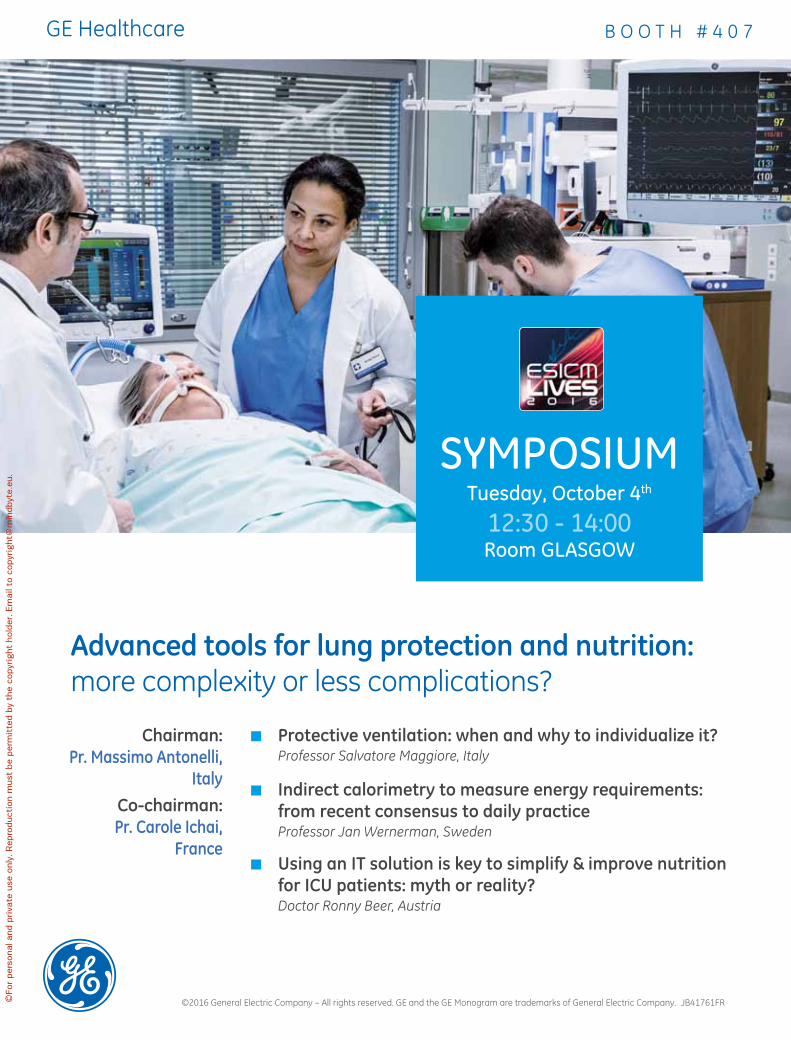
Protective ventilation: when and why to individualize it? Professor Salvatore Maggiore, Italy
Indirect calorimetry to measure energy requirements: from recent consensus to daily practice Professor Jan Wernerman, Sweden
Using an IT solution is key to simplify & improve nutrition for ICU patients: myth or reality? Doctor Ronny Beer, Austria
Tuesday, October 4th
12:30 - 14:00Room GLASGOW
SYMPOSIUM
©2016 General Electric Company – All rights reserved. GE and the GE Monogram are trademarks of General Electric Company. JB41761FR
Advanced tools for lung protection and nutrition: more complexity or less complications?
Chairman: Pr. Massimo Antonelli,
ItalyCo-chairman: Pr. Carole Ichai,
France
GE Healthcare B O O T H # 4 0 7©
For
pers
onal
and
pri
vate
use
onl
y. R
epro
duct
ion
mus
t be
per
mit
ted
by t
he c
opyr
ight
hol
der. E
mai
l to
copy
right
@m
indb
yte.
eu.
Jean-Louis Vincent
Editor-in-ChiefICU Management & Practice
ProfessorDepartment of Intensive Care
Erasme Hospital / Free University of Brussels
Brussels, Belgium
the abdomen
edItoRIaL 129
ICU Management & Practice 3 - 2016
Protective ventilation: when and why to individualize it? Professor Salvatore Maggiore, Italy
Indirect calorimetry to measure energy requirements: from recent consensus to daily practice Professor Jan Wernerman, Sweden
Using an IT solution is key to simplify & improve nutrition for ICU patients: myth or reality? Doctor Ronny Beer, Austria
Tuesday, October 4th
12:30 - 14:00Room GLASGOW
SYMPOSIUM
©2016 General Electric Company – All rights reserved. GE and the GE Monogram are trademarks of General Electric Company. JB41761FR
Advanced tools for lung protection and nutrition: more complexity or less complications?
Chairman: Pr. Massimo Antonelli,
ItalyCo-chairman: Pr. Carole Ichai,
France
GE Healthcare B O O T H # 4 0 7
Managing the abdomen and its complications in the intensive care unit is the subject of our Cover Story. First, Jan de Waele considers the
data on new antibiotics for complicated intra-abdominal infections. While these, singly and in combination, show promise, he cautions that recent studies have certain short-comings from a critical care perspective, and recommends that local antibiotic stewardship programmes guide treat-ment decisions.
Next, Annika Reintam Blaser and colleagues explain when enteral feeding is feasible for critically ill patients with abdominal conditions: emergency gastrointestinal surgery, open abdomen, abdominal aortic surgery, trauma, abdominal compartment syndrome, severe gastrointestinal bleeding, bowel ischaemia, obstruction and paralysis as well as acute colitis with toxic megacolon. They recom-mend that enteral nutrition is considered early for most patients following initial management of abdominal crisis.
Acute-on-chronic liver failure does not preclude admis-sion to the intensive care unit (ICU), according to Alex-ander Wilmer and Philippe Meersseman, who review the latest insights on this serious condition and their potential repercussions on the way intensivists should understand and manage patients with ACLF. Lastly, Manu Malbrain provides some answers to the unanswered questions about intra-abdominal hypertension and abdominal compart-ment syndrome.
In our Matrix section Shashank Patil advises how point-of-care test devices can be implemented effectively in an emergency department, with great benefit for patient management. Next, Silvia Terraneo and colleagues explore the role of Candida spp. in the respiratory tract, asking wheth-er there is a real causality between Candida spp. and worse outcomes, or whether it is simply a marker of severity.
Simon Bocher and colleagues review recent data on the type of vasopressors to use in sepsis, the timing of infusion, the mean arterial pressure target and alternative approaches. Pieter Depuydt and Lisbeth De Bos discuss controversiesregarding ventilator-associated pneumonia diagnosis: whether invasively obtained microbiology improves diag-nosis and outcome, ventilator-associated tracheobronchitis as a separate condition and the concept of ventilator-asso-ciated events. Alexandre Lima and Michel van Genderen review the latest developments in noninvasive monitor-
ing of peripheral circulation, which they suggest should be central to intensive care clinical practice. Last, Richard Gunderman and LeLand considers the power of touch in healthcare.
In our Management section, Lucy Modra and colleagues from the Women in Intensive Care Network in Australia spell out the startling gender disparity in intensive care medicine leadership and provide suggestions for improve-ment, so that the sustainability and quality of intensive care leadership is assured. Next, a cri de coeur from Armand Girbes and colleagues, who contend that a jumble of rules, proto-cols, checklists on both sides of the Atlantic has emerged, which jeopardises not only the pivotal relationship between doctor and patient, but also the quality and costs of care, and the quality of future healthcare workers.
Intensive care units naturally put all their efforts into treating patients when they are in the ICU. However, reha-bilitation after leaving the ICU is a somewhat neglected area and post-intensive care syndrome is a burgeoning area of research. Tara Quasim and Joanne McPeake share their experiences of setting up a peer-supported, self-management programme aimed at empowering patients and relatives after leaving the ICU.
Our interview is with Gernot Marx, Director of the Department of Intensive Care Medicine and Intermedi-ate Care, University Hospital Aachen, and Professor of Anaesthesiology and Operative Intensive Care Medicine at RWTH Aachen University, Aachen, Germany. Univer-sity Hospital Aachen has pioneered tele-ICUs, and Marx shares his thoughts on this and on his research into sepsis and fluids.
Our Country Focus is Brazil. Jorge Salluh and Thiago Lisboa describe the challenges and opportunities for criti-cal care in this vast country, where there are active critical care research networks and quality of care initiatives.
The ICU Management & Practice team will be at the Euro-pean Society of Intensive Care Medicine Congress in Milan this month. If you will be attending, make sure to drop by to say hullo and pick up your copy of the journal.
As always, if you would like to get in touch, please email [email protected]
Jean-Louis Vincent
©Fo
r pe
rson
al a
nd p
riva
te u
se o
nly.
Rep
rodu
ctio
n m
ust
be p
erm
itte
d by
the
cop
yrig
ht h
olde
r. E
mai
l to
copy
right
@m
indb
yte.
eu.

130tabLe oF ContentS 130
COVER STORYNew Antibiotics for Abdominal Infections: What Can We Expect? (Jan De Waele)Reviews new antibiotics and combinations of these and how they can be used in
patients with complicated intra-abdominal infections
Is Enteral Feeding Feasible Early After Abdominal Crisis? (Annika Reintam Blaser, Heleen M. Oudemans-van Straaten, Joel Starkopf)Reviews different abdominal conditions in critically ill patients where safety
and feasibility of enteral nutrition might be questioned.
Evolving Concepts in Acute-on-Chronic Liver Failure (Alexander Wilmer, Philippe Meersseman)Outlines recent insights and their potential repercussions on the way intensiv-
ists should understand and manage patients with ACLF.
Update on Intra-Abdominal Hypertension (Manu LNG Malbrain)Provides some answers to the unanswered questions that still cloud the under-
standing of the pathophysiology of intra-abdominal hypertension and abdomi-
nal compartment syndrome.
MATRIXPOCTED: Use of Point-of-Care Test Devices in Emergency Department (Shashank Patil)Use of point-of-care test devices in the emergency department has shown
significant benefits in patient management. A proper governance policy will
ensure credible, effective and safe practice.
Candida Spp. in the Respiratory Tract: A Real Causality With Worse Outcomes or Just a Marker of Severity? (Silvia Terraneo, Miquel Ferrer, Antoni Torres)Is there a real causality between Candida spp. and worse outcomes, or is it simply
a marker of severity?
Vasoactive Drugs in Sepsis (Simon Bocher, François Beloncle, Pierre Asfar)Reviews recent data on the type of vasopressors that should be used, the timing
of infusion, the mean arterial pressure target and alternative approaches.
Controversies in Ventilator-Associated Pneumonia Diagnosis (Pieter Depuydt, Liesbet De Bus) Discussion of four controversies regarding ventilator-associated pneumonia
diagnosis.
136
140
144
148
154
158
161
164
IN EVERY ISSUe
129EDITORIALTHE ABDOMEN(Jean-Louis Vincent)
134NEWS
Three Criteria Can Identify Out-of-Hospital Cardiac
Patients for Potential Organ Donation
Study: Vasopressin vs. Norepinephrine in Septic
Shock
192AGENDA
Upcoming Events/ Congresses
VOLUME 16 - ISSUE 3 - AUTUMN 2016
ICU Management & Practice 3 - 2016
POINT OF VIEWRedesigning Ambulatory Emergency Care with Point-of-Care Testing: Reduced Costs, Length of Stay (Phil Weihser, Josip Stosic, Dominic Giles)
152
©Fo
r pe
rson
al a
nd p
riva
te u
se o
nly.
Rep
rodu
ctio
n m
ust
be p
erm
itte
d by
the
cop
yrig
ht h
olde
r. E
mai
l to
copy
right
@m
indb
yte.
eu.

C
M
J
CM
MJ
CJ
CMJ
N
27th ICISEM 21x27,6.pdf 1 23/02/16 17:55
©Fo
r pe
rson
al a
nd p
riva
te u
se o
nly.
Rep
rodu
ctio
n m
ust
be p
erm
itte
d by
the
cop
yrig
ht h
olde
r. E
mai
l to
copy
right
@m
indb
yte.
eu.
Monitoring Peripheral Circulation (Alexandre Lima, Michel van Genderen)Reviews recent developments in noninvasive monitoring of the peripheral cir-
culation, which helps to early identify patients at high risk for tissue hypoper-
fusion, organ failure and poor outcome.
Touch Creates a Healing Bond in Healthcare (Richard Gunderman, Brian LeLand)Touch is a powerful tool in medicine.
MANAGEMENTWomen in Leadership in Intensive Care Medicine (Lucy J. Modra, Sarah A. Yong, Danielle E Austin)To ensure the sustainability and quality of intensive care leadership in the
future, the gender imbalance should be redressed through cultural change,
workplace structural reforms, advocacy and mentoring.
Intensive Care Syndrome: Promoting Independence and Return to Employment (InS:PIRE): a New Model for ICU Rehabilitation (Tara Quasim, Joanne McPeake)A peer-supported, self-management programme aimed at empowering patients
and relatives to take control of their own health and wellbeing by finding com-
munity resources to help them.
The Burden Caused by Administrators and Managers: a Euro-American Jumble (Armand R.J. Girbes, Jan G. Zijlstra, Paul E. Marik)A jumble of rules, protocols and checklists jeopardises not only the pivotal rela-
tionship between doctor and patient, but also the quality and costs of care, and
the quality of future healthcare workers.
INTERVIEWTelemedicine is the Future (Gernot Marx) Interview with Gernot Marx, Director of the Department of Intensive Care
Medicine and Intermediate Care, University Hospital Aaachen, and Professor
of Anaesthesiology and Operative Intensive Care Medicine at RWTH Aachen
University, Aachen, Germany.
COUNTRY FOCUSCritical Care in Brazil (Jorge Salluh, Thiago Lisboa)The challenges and opportunities for critical care in the fifth most populous
country in the world.
168
172
174
178
182
186
189
ICU management & Practice
is the official management Journal of the International Symposium on Intensive Care and emergency medicine.
editor-in-Chief
Prof. Jean-Louis Vincent Belgium
editorial board
Prof. antonio artigas SpainProf. Jan bakker NetherlandsProf. Richard bealeUnited KingdomProf. Rinaldo bellomo AustraliaProf. todd dormanUnited StatesProf. Jan de Waele Belgiumdr. bin du ChinaProf. hans Kristian Flaatten NorwayProf. Luciano Gattinoni ItalyProf. armand Girbes NetherlandsProf. edgar Jimenez United StatesProf. John a. Kellum United StatesProf. Jeff Lipman AustraliaProf. John mariniUnited StatesProf. John marshall CanadaProf. Paul e. Pepe United StatesProf. Paolo PelosiItalydr. Shirish Prayag IndiaProf. Peter PronovostUnited StatesProf. Konrad Reinhart GermanyProf. Gordon RubenfeldCanadaProf. Jukka takalaSwitzerland
Correspondents
Prof. dr. dominique Vandijck Belgium
132tabLe oF ContentS 132
VOLUME 16 - ISSUE 3 - AUTUMN 2016
ICU Management & Practice 3 - 2016
Reaching forward.
For over 60 years, Mindray has been dedicated to improving the way you monitor patients. This time, we are bringing
the entire family of BeneVision N Series - the eye-opening innovation that redefines patient monitoring. It leverages
your care to the patient in improved efficiency and safety by powerful clinical IT capability. The alternative models
enable you to pursue clinical excellence in all kinds of medical settings.
See more, with ease
bringing the entire family of
BeneVision N Series Patient Monitors
C
M
Y
CM
MY
CY
CMY
K
BeneVison N final(210X276+3mm bleed).pdf 1 2016-9-19 18:32:28
©Fo
r pe
rson
al a
nd p
riva
te u
se o
nly.
Rep
rodu
ctio
n m
ust
be p
erm
itte
d by
the
cop
yrig
ht h
olde
r. E
mai
l to
copy
right
@m
indb
yte.
eu.

neWS134
ICU Management & Practice 3 - 2016
©Fo
r pe
rson
al a
nd p
riva
te u
se o
nly.
Rep
rodu
ctio
n m
ust
be p
erm
itte
d by
the
cop
yrig
ht h
olde
r. E
mai
l to
copy
right
@m
indb
yte.
eu.
Three objective criteria could identify out-of-hospital cardiac arrest (OHCA) patients with
zero chance of survival, who can be considered for organ donation. Prof. Xavier Jouven, Georg-es Pompidou European Hospital, Paris, and colleagues, analysed data from two registries and a clinical trial, and found that there is essentially no chance of survival in patients whose OHCA is not witnessed by emergency medical services personnel, who have nonshockable initial cardiac rhythm, and in whom spontaneous circulation does not return before receipt of a third 1-mg
dose of epinephrine (Jabre et al. 2016). Prof. Jouven told ICU Management & Practice that he
would like these results to contribute to elaborat-ing new recommendations about out-of-hospital cardiac arrest patients. "We are aware there is a psychological barrier, but the possibility to collect organs from a proportion of those cardiac arrests represents an important opportunity to fill the gap of organ shortage,” he said.
Existing termination-of-resuscitation rules help to identify cases where further resuscitation is futile, but do not take into consideration the potential utility of transporting dead patients to the hospital for organ donation. Applying the three criteria to the validation cohorts, the research-ers found that between 8 and 12% of patients with no chance of survival might have had organs that were potentially suitable for transplantation. They applied the eligibility criteria used in France for kidney retrieval from uncontrolled donation after cardiac death (UDCD). Jouven said that they considered this new approach first for kidney donation. In Europe kidney allograft represents
60% of all allografts, with 15,000 new grafts per year. In future this early identification may be applied for other organ donation (liver, cornea), he added.
The researchers acknowledge that their results may be overestimates, as some UDCD eligibility criteria may be difficult to verify at the OHCA scene. They recommend that emergency medical services develop protocols and implementation plans with their organ donation programmes to optimise donation after OHCA, and implement these three objective criteria in their protocols for cardiac arrest. “Rapid referral to an organ dona-tion institution under mechanical ventilation and continuous automated external cardiac massage should be considered for patients with no chance of survival,” they write.
Three CriTeria Can idenTify OuT-Of-hOspiTal CardiaC paTienTs fOr pOTenTial Organ dOnaTiOn
ReferenceJabre P, Bougouin W, Dumas F et al. (2016) Early identification of patients with out-of-hospital cardiac arrest with no chance of survival and consideration for organ donation. Annals Intern Med, 13 Sept. doi: 10.7326/M16-0402.
Xavier Jouven
sTudy: VasOpressin Vs. nOrepinephrine in sepTiC shOCk
A multicentre trial investigating early use of vasopressin compared to norepineph-
rine to treat septic shock found no reduction in the number of kidney failure-free days. The results of the VAsopressin vs. Noradrenaline as Initial therapy in Septic sHock (VANISH) trial are published in JAMA (Gordon et al. 2016).
Patients who had septic shock requiring vasopressors despite fluid resuscitation within a maximum of 6 hours after the onset of shock were randomised to vasopressin and hydro-cortisone (n=101), vasopressin and placebo (n=104), norepinephrine and hydrocortisone (n=101), or norepinephrine and placebo
(n=103). The primary outcome was kidney failure-free days during the 28-day period after randomisation, namely the proportion of patients who never developed kidney failure and the median number of days alive and free of kidney failure for patients who did not survive, who experienced kidney failure, or both.
Early use of vasopressin compared with norepinephrine did not improve the number of kidney failure-free days. However, the confi-dence interval included a potential clinically important benefit for vasopressin, and larger trials may be warranted to assess this further, the authors write. Prof. Anthony Gordon, Impe-rial College London, the trial’s Chief Investigator, told ICU Management and Practice that this potential benefit was related to the secondary outcomes measured in the trial, which related to kidney function: fewer patients in the vasopressin group needed renal replacement therapy, and they had greater urine output and lower creatinine levels over the first week. Gordon said that the results will probably not change routine first-line pressors for septic shock, i.e. norepinephrine.
However, clinicians may consider starting vaso-pressin early in patients whose kidney function is deteriorating.
ReferenceGordon AC, Mason AJ, Thirunavukkarasu N, Perkins GD, Cecconi M, Cepkova M, Pogson DG, Hollman DA, Anjum A, Frazier GJ, Santhakumaran S, Ashby D, Brett SJ (2016) Effect of early vaso-pressin vs norepinephrine on kidney failure in patients with septic shock. JAMA, 316(5): 509-18. doi:10.1001/jama.2016.10485
Results409 patients (median age, 66 years)Median time to study drug administration: 3.5 hours after diagnosis of shock
Vasopressin groupSurvivors who never developed kidney failure: 94/165 (57%)Median number of kidney failure-free days for pa-tients who did not survive, who experienced kidney failure: 9 days
Norepinephrine groupSurvivors who never developed kidney failure: 93/157 patients (59%)Median number of kidney failure-free days for pa-tients who did not survive, who experienced kidney failure: 13 days
Anthony Gordon
Imag
e cr
edit:
Hel
le A
sbjo
rn S
oren
sen
BREATHE MORENATURALLYThe new Puritan Bennett™ 980 ventilator helps follow your patient’s unique pattern of breathing.
For patients in the ICU, time on a ventilator can be a disturbing experience. Being forced to breathe too quickly, too shallowly or too deeply is unsatisfying,extremely tiring, and can lead to distress.1
At Medtronic, we believe mechanical ventilation can and should be more natural. That’s the idea behind the all-new Puritan Bennett™ 980 ventilator with PAV™*+ software. It helps clinicians better manage work of breathing and improve synchrony† by allowing patients to help direct the fl ow and timing of ventilator support based on their realtime demands.2, 3 In other words, it can help them breathe more naturally.†
IMPORTANT: Please refer to the package insert for complete instructions, contraindications, warnings and precautions.
© 2016 Medtronic. All rights reserved. Medtronic, Medtronic logo and Further, Together are trademarks of Medtronic.™* Third party brands are trademarks of their respective owners. All other brands are trademarks of a Medtronic company.. All rights16-emea-resize-of-pb-980-ad-for-icu-1286333
† Compared to conventional volume control mechanical ventilation
1. Epstein, Scott K. Optimizing Patient-Ventilator Synchrony. Seminars in Resp and Crit Care Med., 2001; 22: 137-152.
2. Xirouchaki N, Kondili E, Vapoidi K, et al. Proportional assist ventilation with load-adjustable gain factors in critically ill patients: comparison with pressure support. Int Care Med. 2008;34:2026-2034.
3. Younes, Magdy. Proportional Assist Ventilation, a New Approach to Ventilatory Support. Am Rev Respir Dis. 1992; 145:114-120.
16-emea-resize-of-pb-980-ad-for-icu-1286333.indd 1 16/09/2016 11:53
©Fo
r pe
rson
al a
nd p
riva
te u
se o
nly.
Rep
rodu
ctio
n m
ust
be p
erm
itte
d by
the
cop
yrig
ht h
olde
r. E
mai
l to
copy
right
@m
indb
yte.
eu.

ICU Management & Practice 3 - 2016
136CoVeR StoRY: THE ABDOMEN
©Fo
r pe
rson
al a
nd p
riva
te u
se o
nly.
Rep
rodu
ctio
n m
ust
be p
erm
itte
d by
the
cop
yrig
ht h
olde
r. E
mai
l to
copy
right
@m
indb
yte.
eu.
neW anTiBiOTiCs fOr aBdOMinal infeCTiOns WHAT CAN WE EXPECT?
Complicated intra-abdominal infections (cIAI) remain one of the most chal-lenging infections in the intensive care
unit (ICU). Compared to patients with other infections, patients with cIAI typically will develop multiple organ dysfunction syndrome (MODS) more often and have a higher risk of mortality; often they have a protracted stay in the ICU and in the hospital (De Waele et al. 2014). The management of these patients can be challenging. This includes evaluating the need for source control as well as effectively getting the source of infection controlled, but also selecting the appropriate antibiotic in times of changing susceptibility patterns and the rise of antimicrobial resistance (AMR).
The role of source control is more relevant in cIAI than in most other commonly encountered infections in the ICU. At times difficult choices have to be made (Leppäniemi et al. 2015). The role of surgery in this context is changing, new techniques are being introduced, and, increas-ingly, percutaneous drainage is being used as a primary strategy. Despite the prominent role of source control, administering appropriate antibiotics is equally important. Although there are fewer limitations in correctly diagnosing abdominal infections compared to e.g. respira-tory tract infections, both timing and spectrum of empirical antibiotic therapy are critical. Antibiotics should be administered when the diagnosis is made and not postponed until intraoperative cultures are obtained.
Antibiotic resistance is also increasingly described in cIAI. In particular the spread of extended spectrum beta-lactamase (ESBL)-producing Enterobacteriaceae in community-acquired cIAI is striking, and may limit the use of many currently available antibiotics. This in turn may put an inappropriate strain on the carbapenem
antibiotics with the risk of increasing resistance to this class of antibiotics. The need for new antibiotics in this context is urgent.
Options for appropriate empirical therapy are becoming limited in some situations, and every attempt should be made to choose the correct antibiotic for the patient with cIAI. It should also be remembered that cIAI are typi-cally polymicrobial infections with both aerobic and anaerobic bacteria present in most situa-tions, and will typically require antibiotics that cover both Gram-positive and Gram-negative pathogens.
Rise of Multidrug Resistance in cIAIAs in other types of infections, AMR is a press-ing issue in cIAI. Patients with cIAI may be at increased risk of AMR as they are often exposed to antibiotics for prolonged periods of time, and source control plays a crucial role. Particularly when source control is inadequate or even impossible, the inoculum persists. As bacteria are exposed to antibiotics during that time, AMR is bound to develop. This has been documented in severe abdominal infections including peritonitis and pancreatitis (De Waele 2016; Montravers et al. 2016).
As typically more than one pathogen is involved, the risk of encountering antibiotic
resistance is also increased. For the same reason the extensive coverage needed to cover all pathogens (often with multiple antibiotics) may fuel AMR, as bacteria are exposed to more than one antibiotic at the same time. Whereas AMR was only relevant in nosocomial infections until recently, it is now also posing problems in community-acquired disease.
Overall, AMR is a concern mostly with Gram-negative pathogens. ESBL-producing bacteria are a primary worry worldwide (Sartelli et al. 2015), even more so in some areas, e.g. in Asia. Even then important regional differences are present.
The prevalence of ESBL in E. Coli, K. pneumonia, K. oxytoca and P. Mirabilis has increased dramati-cally from 2002 to 2011 in cIAI in Asia and the Middle East, where up to 40% of these pathogens isolated from cIAI produce ESBLs (Morrissey et al. 2013). It is unclear if this trend has changed in more recent years as epidemiological studies on AMR after 2013-2014 are lacking. Regional differences are important, and extrapolating data from other parts of the world to develop local empirical therapy guidelines should be avoided.
Carbapenemase-producing Klebsiella pneumonia (KPC) has been posing particular problems in nosocomial infections in some parts of the world. cIAI have not been exempt from KPC involvement, but this appears to be a regional problem mostly at this point.
Although the problem of AMR in cIAI is most relevant for Gram-negative pathogens, trends in Gram-positive infections should not be ignored. Enterococci are considered to be more pathogenic in nosocomial cIAI, and typically are involved in patients who have been exposed to antibiotics that do not cover enterococci, e.g. cephalosporins or fluoroquinolones. Apart from their different appreciation in nosocomial cIAI,
Jan de WaeleIntensivistDepartment of Critical Care MedicineGhent University Hospital
Associate ProfessorGhent University
Ghent, Belgium
ICU Management & Practice Editorial Board Member
@CriticCaredoc
Recently a number of new antibiotics or combinations for compli-cated intra-abdominal infections have been introduced. Here we review the currently available data of these new drugs and discuss how they can be used in critically ill patients with complicated intra-abdominal infections.
new agents should be used only where they
have a clearly added value
ICU Management & Practice 3 - 2016
137CoVeR StoRY: THE ABDOMEN
©Fo
r pe
rson
al a
nd p
riva
te u
se o
nly.
Rep
rodu
ctio
n m
ust
be p
erm
itte
d by
the
cop
yrig
ht h
olde
r. E
mai
l to
copy
right
@m
indb
yte.
eu.
resistance in enterococci is increasing as well; E. faecium is typically non-susceptible to penicillin antibiotics, but in E. faecalis ampicillin resistance is also rising. Infection with vancomycin-resistant enterococci is also increasingly described.
New Antibiotics for cIAIRecently a number of new antibiotics or antibi-otic combinations have been studied in patients with cIAI. Antibiotics recently introduced or coming soon for the treatment of cIAI include ceftolozane/tazobactam, ceftazidime/avibactam and eravacycline. Although several other new antibiotics may have activity against pathogens typically associated with cIAI, none of them is currently under investigation for this indication, and will not be discussed.
Ceftolozane Plus Tazobactam Ceftolozane is a new fifth-generation cepha-losporin antibiotic that has been marketed in combination with a well-known beta-lactamase inhibitor (BLI), tazobactam, in a fixed 2:1 ratio. It is active against a wide range of Gram-negative bacteria, including Pseudomonas aeruginosa and many ESBL-producing Enterobacteriaceae. It has been approved by the United States Food and Drug Administration for the treatment of complicated urinary tract infections and cIAI (combined with metronidazole for the latter). Dosing for patients with normal renal function is 1000mg ceftolozane plus 500mg tazobactam TID.
Three clinical trials have been performed in patients with cIAI. In a phase 2 study, 121 patients with cIAI requiring surgery were randomised to receive either meropenem or ceftolozane/tazobactam with metronidazole (Lucasti et al. 2014). Clinical cure rates were 83.6% and 96% for ceftolozane and meropenem respectively, on the basis of which the noninferiority of the drug was concluded. The Assessment of the Safety Profile and Efficacy of Ceftolozane/Tazobactam in Complicated Intra-abdominal Infections (ASPECT-cIAI) programme, reporting on two identical phase 3 studies with a similar setup to the phase 2 study, and using the same comparator, included 993 patients, 806 of which were analysed in the modified intention to treat (MITT) group (Solomkin et al. 2015). For the primary endpoint clinical cure rates were 83% with ceftolozane/tazobactam plus metronidazole vs. 87.3% with meropenem in the MITT population. In both studies the incidence of adverse effects reported was similar in both groups. Based on these studies, ceftolozane/tazobactam was approved
for the indication of cIAI at the end of 2014.In a recent substudy investigating the
outcomes of patients with Pseudomonas aeruginosa, the strong in vitro activity of ceftolozane against these pathogens was confirmed, with high clinical cure rates in the subgroup of patients with Pseudomonas infections (Miller et al. 2016).
Ceftazidime Plus AvibactamAvibactam is a novel BLI that restores the activity of beta-lactam antibiotics such as ceftazidime against ESBL-producing pathogens.
In a phase 2 study the combination of ceftazi-dime/avibactam (2000mg/500mg TID) with metronidazole 500mg TID was compared with meropenem in 204 patients with cIAI (Lucasti et al. 2013). Clinical cure was 91.2% and 93.4% for ceftazidime/avibactam co-administered with metronidazole and meropenem respectively. Adverse events were comparable in both groups.
In two large phase 3 studies with an identical setup 1066 patients with cIAI requiring surgery of percutaneous drainage were randomised to receive ceftazidime/tazobactam plus metro-nidazole and the combination was found to be noninferior to meropenem (Mazuski et al. 2016). In the microbiologically MITT group, clinical cure at test of cure was statistically not different in the ceftazidime/tazobactam plus metronidazole group (81.6% vs. 85.1% respectively), and at other time points outcome was comparable. Safety evaluation did not demonstrate any differences between the groups.
EravacyclineEravacycline is a novel antibiotic in the tetracycline class, structurally comparable with tigecycline. It inhibits bacterial protein synthesis through binding to the 30S ribosomal subunit and has broad-spectrum antimicrobial activity against Gram-positive, Gram-negative and anaerobic bacteria with the exception of Pseudomonas aeru-ginosa, but including MDR pathogens such as methicillin-resistant Staphylococcus aureus (MRSA) and some carbapenem-resistant Gram-negative bacteria. In a phase 2 study the efficacy and safety of two dose regimens of eravacycline was compared with ertapenem in adult hospi-talised patients with cIAI requiring surgical or percutaneous intervention: 1.5 mg/kg of body weight every 24 hours (q24h), or 1.0 mg/kg every 12 h (q12h) (Mazuski et al. 2016). In the microbiologically evaluable population the clinical cure was 92.9% and 100% in the groups receiving eravacycline at 1.5 and 1.0 mg/kg respectively, and 92.3% in the ertapenem
group. Another large phase 3 study comparing eravacycline with ertapenem has been final-ised but not yet published (IGNITE 1)—the manufacturer has reported that noninferiority was demonstrated but full analysis is not yet available (Tetraphase Pharmaceuticals 2014).
Caveats for Critical CareShortcomings of Recent cIAI Studies From a Critical Care PerspectiveAlthough these antibiotics represent new thera-peutic options in the management of cIAI, there are some things to consider from a critical care perspective. This is primarily related to the type of patients in the studies with these new antibiotics, and with the type of patients not included due to an often long list with exclusion criteria. Overall the patients in these studies are mild to moderately ill only, with a high prevalence of infections that are typically not encountered in the ICU, such as appendicitis.
In the studies investigating ceftolozane, it was not reported how many patients were diagnosed with severe sepsis or septic shock, or were admitted to an ICU. In the first study more than half of the patients were treated because of appendicitis, and median Acute Physiology and Chronic Health Evaluation (APACHE)-II score was 6 and 7 respectively (Lucasti et al. 2014). Similarly, in the ASPECT-cIAI programme, APACHE-II scores were 6 and 6.2 in the study groups and degree of organ dysfunction was not reported (Solomkin et al. 2015). Both studies excluded patients with thrombocytopenia or abnormal renal function.
The studies investigating avibactam in combi-nation with metronidazole excluded severely ill patients; exclusion criteria in the phase 2 study included APACHE-II score of 26 or higher, abnormal renal function and fluid-unresponsive septic shock (Lucasti et al. 2013). Only 1 out of 6 patients had an APACHE-II score between 10 and 25, and the appendix and stomach were the most frequent sites of the primary infection. The phase 3 study included mainly patients with low to moderate disease sever-ity as exemplified by the APACHE-II score that was 10 or lower in about 85% of the patients (Mazuski et al. 2016). That study also excluded patients with septic shock or who were receiv-ing haemodialysis. The fact that patients could not be treated with an antifungal agent may have precluded including patients with more severe disease in the study.
One particular finding in the phase 3 study was the worse outcome in patients with moder-
ICU Management & Practice 3 - 2016
138CoVeR StoRY: THE ABDOMEN
©Fo
r pe
rson
al a
nd p
riva
te u
se o
nly.
Rep
rodu
ctio
n m
ust
be p
erm
itte
d by
the
cop
yrig
ht h
olde
r. E
mai
l to
copy
right
@m
indb
yte.
eu.
ReferencesDe Waele J, Lipman J, Sakr Yet al. (2014) Abdominal infections in the intensive care unit: characteristics, treatment and determinants of outcome. BMC Infect Dis, 14: 420.
De Waele JJ (2016) Abdominal sepsis. Curr Infect Dis Rep, 18(8); 23.
Leppäniemi A, Kimball EJ, De Laet I et al. (2015) Management of abdominal sepsis-a paradigm shift? Anaesthesiol Intensive Ther, 47(4): 400-8.
Lucasti C, Hershberger E, Miller B et al. (2014) Multicenter, double-blind, randomized, phase II trial to assess the safety and efficacy of ceftolozane-tazobactam plus metronidazole compared with meropenem in adult patients with complicated intra-abdominal infections. Antimicrob Agents Chemother, 58(9): 5350-7.
Lucasti C, Popescu I, Ramesh MK et al. (2013) Comparative study of the efficacy and safety of ceftazidime/avibactam plus metronidazole versus meropenem in the treatment of complicated intra-abdominal infections in hospitalized adults: results of a randomized, double-blind, Phase II trial. J Antimicrob Chemother, 68(5): 1183-92.
Mazuski JE, Gasink LB, Armstrong J et al. (2016) Efficacy and safety of ceftazidime-avibactam plus metronidazole versus meropenem in the treatment of complicated intra-abdominal infec-tion - results from a randomized, controlled, double-blind, phase 3 program. Clin Infect Dis, 62(11): 1380-9.
Miller B, Popejoy MW, Hershberger E et al. (2016) Characteristics and outcomes of complicated intra-abdominal infections involving pseudo-monas aeruginosa from a randomized, double-blind, phase 3 ceftolozane/tazobactam study.
Antimicrob Agents Chemother, 60(7): 4387-90.
Montravers P, Blot S, Dimopoulos G et al. (2016) Therapeutic management of perito-nitis: a comprehensive guide for intensivists. Intensive Care Med, 42(8): 1234-47.
Morrissey I, Hackel M, Badal R et al. (2013) A review of ten years of the study for monitoring antimicrobial resistance trends (SMART) from 2002 to 2011. Pharmaceuticals (Basel), 6(11): 1335-46.
Roberts JA, Abdul-Aziz MH, Lipman J et al. (2014) Individualised antibiotic dosing for patients who are critically ill: challenges and potential solutions. Lancet Infect Dis, 14(6): 498-509.
Sartelli M, Catena F, di Saverio S et al. (2015) The challenge of antimicrobial resistance in managing intra-abdominal infections. Surg Infect (Larchmt), 16(3): 213-20.
Solomkin J, Hershberger E, Miller B et al. (2015) Ceftolozane/tazobactam plus metronidazole for complicated intra-abdominal infections in an era of multidrug resistance: results from a randomized, double-blind, phase 3 trial (ASPECT-cIAI). Clin Infect Dis, 60(10): 1462-71.
Solomkin JS, Mazuski JE, Bradley JS et al. (2010) Diagnosis and management of compli-cated intra-abdominal infection in adults and children: guidelines by the Surgical Infection Society and the Infectious Diseases Society of America. Clin Infect Dis, 50(2): 133-64.
Tetraphase Pharmaceuticals (2014) Tetrap-hase announces positive top-line results from phase 3 IGNITE 1 clinical trial of eravacyclin in complicated intra-abdominal infections. [press release] 17 December. [Accessed: 14 July 2016] Available from ir.tphase.com/releasedetail.cfm?releaseid=888162
ate renal impairment, defined as a creatinine clearance of 30-50ml/min. This may have been caused by the rapid changes in renal function in the subsequent days when patients still received renal function adjusted doses of the drug, although the effect should be present in both the interventional and comparator group (Mazuski et al. 2016).
The study investigating eravacycline excluded more critically ill patients such as patients with septic shock or an APACHE-II score of 25 or higher. Effectively, APACHE-II score was 6 and 8.2 in the study groups, and appendicitis was the source of infection in more than 50% of the patients. The use of ertapenem as a comparator can also limit the number of critically ill patients included, as this drug is not recommended for the treatment of severe cIAIs (Solomkin et al. 2010).
Implications for Critically Ill Patients With cIAISo how does this translate to the use of these new agents in the critically ill? Although it is clear that the in vitro activity of these drugs against a wide range of pathogens is similar or better than many of the antibiotics that we are using now, the changes in physiology of the critically ill may be profound and lead to lower concentrations than expected. This phenomenon has been demonstrated for many antibiotics (Roberts et al. 2014) and is now an integral part of most drug development programmes.
In this context it is remarkable that an ongo-ing study comparing ceftolozane/tazobactam to
meropenem for hospital-acquired pneumonia (Safety and efficacy study of ceftolozane/tazobactam to treat ventilated nosocomial pneumonia (MK-7625A-008) (ASPECT-NP), NCT02070757) uses a dose that is double what was used in the cIAI study (clinicaltrials.gov/ct2/show/NCT02070757). It is unclear if this is solely because of the different infection focus. Future pharmacokinetic studies of these new antibiotics in more severely ill patients should answer these concerns.
The exact place of these new agents in our current armamentarium will need to be discussed primarily considering the local ecol-ogy. This is where antibiotic stewardship teams should jointly define the indications as well as consider restriction in the use of these power-ful agents. Apart from treating the infections adequately, new agents should be cherished and used only where they have a clearly added value – whether this is in empirical therapy in one country or directed therapy for highly resistant pathogens in another.
ConclusionsAntibiotic therapy of cIAI is becoming increas-ingly challenging due to the changes in suscep-tibility of pathogens involved. Although our current armamentarium may be effective in the treatment of many patients, new therapeutic options are highly desirable. The development of ceftolozane/tazobactam, ceftazidime/avibac-tam and eravacycline offers an opportunity to effectively treat MDR pathogens and avoid more toxic regimens. The exact place of these agents
in the treatment of cIAI should be defined by local antibiotic stewardship teams, consider-ing local ecology and other available options.
Conflict of InterestJan De Waele declares Consultancy for AtoxBio, Bayer Healthcare, Cubist, Fresenius, Merck. He is Infection section Chair at the European Society of Intensive Care Medicine, President of the Belgian Society of Intensive Care Medicine, Past President of WSACS - the Abdominal Compart-ment Society and Senior Clinical Investigator at the Flanders Research Foundation.
AbbreviationsAMR antimicrobial resistanceAPACHE Acute Physiology and Chronic Health Evalu-ation ASPECT-cIAI Assessment of the Safety Profile and Efficacy of Ceftolozane/Tazobactam in Complicated Intra-abdominal Infections BLI Beta-lactamase inhibitorcIAI complicated intra-abdominal infectionsESBL extended spectrum beta-lactamaseICU intensive care unitKPC Klebsiella pneumoniaMDR multi-drug resistanceMITT modified intention to treatMODS multiple organ dysfunction syndrome

From admission to dischargeIntensive Care Units (ICU) are the locations where the most difficult
to treat patients are often staying for days or even weeks. For this
critical and costly environment, Getinge always focus on developing
user-friendly and reliable solutions that help caregivers to achieve
tangible and cost-effective patient outcomes.
Getinge Group. Passion for life.
www.getingegroup.comThis document is intended to provide information to an international audience outside the US.
Getinge ICU Solutions
ICU
EarlyMobility
HealingEnvironment
ARDSManagement
©Fo
r pe
rson
al a
nd p
riva
te u
se o
nly.
Rep
rodu
ctio
n m
ust
be p
erm
itte
d by
the
cop
yrig
ht h
olde
r. E
mai
l to
copy
right
@m
indb
yte.
eu.
ICU Management & Practice 3 - 2016
140CoVeR StoRY: THE ABDOMEN
©Fo
r pe
rson
al a
nd p
riva
te u
se o
nly.
Rep
rodu
ctio
n m
ust
be p
erm
itte
d by
the
cop
yrig
ht h
olde
r. E
mai
l to
copy
right
@m
indb
yte.
eu.
Enteral nutrition (EN) prevents loss of physical and immunological barrier function (Kudsk 2002; McClave 2009).
Early EN reduces infections and is recommend-ed in critically ill patients with stable haemody-namics and functional gastrointestinal (GI) tract (Taylor et al. 2016).
Feeding in the Early Phase of Critical IllnessEven if feeding is started early, a negative energy balance in the first acute phase of critical illness is generally unavoidable. New insights show that early hypocaloric nutrition may even be preferred (Casaer and Van den Berghe 2014) because of an inflammation-induced endoge-nous energy production and nutrition-induced inhibition of autophagy. Therefore early EN should be started at a low rate in the acute
phase and be slowly increased towards target. This is especially true in patients with, or after, abdominal crisis, with continuing vulnerability of GI tract.
Based on common sense, EN is considered harmful in the case of the clinical syndrome called “acute abdomen”, in case of obvious gut ischaemia, mechanical obstruction or perfora-tion, and in cases with no continuity of GI tract. In most other abdominal pathologies initiation of EN remains a matter of “try and see”, e.g. starting low dose EN and evaluating feeding tolerance/intolerance.
Feeding intolerance (FI) is not uniformly defined; gastric residual volumes (GRV) have been mainly used for assessment of FI (Reintam Blaser et al. 2014). Some authors suggest abandoning GRV measurements all together (Reignier et al. 2013). We suggest that GRVs may still be useful to avoid gastric overfilling in the initial phase of EN or in the presence of abdominal symptoms (e.g. abdominal disten-sion or pain). Evaluation of gastric filling with ultrasound may offer a good alternative to GRV (Gilja et al. 1999).
Enteral Nutrition in Specific Abdomi-nal Conditions In critically ill patients with severe abdomi-nal pathology, both abdominal pathology and systemic disease may contribute to GI dysfunc-tion (Table 1). GI function will usually recover
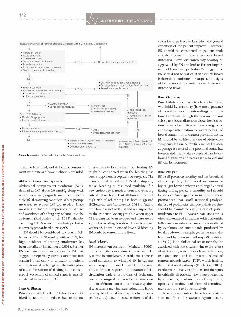
if haemodynamics and gut perfusion improve, fluid resuscitation-induced gut oedema resolves and analgo-sedation can be reduced. On the contrary, a patient with persisting severe general condition is prone to complications. Thus EN should be initiated at a low rate and slowly increased under careful monitoring of abdominal symptoms to avoid dilatation of the stomach, bowel distension and increasing intra-abdominal pressure (IAP) (Figure 1).
Emergency Gastrointestinal SurgeryDirect injury of the GI tract due to trauma or surgery and/or infection/inflammation leads to gut oedema and dysmotility. Denerva-tion, discontinuation of spinal reflexes and resection of enterochromaffin cells producing motilin may add to gut paresis. In emergency GI surgery, gut hypoperfusion due to shock, bowel oedema and intra-abdominal hyperten-sion, exacerbated by inflammation and massive fluid resuscitation, is often evident. Therefore, major factors to consider for recovery after emergency GI surgery (if bowel continuity is restored) are: bowel perfusion, bowel oedema and bowel distension. The intraoperative evalua-tion of bowel viability is important; therefore good communication with surgeons is crucial. If a stoma is created and bowel cranial to stoma has normal appearance, low dose EN can usually be started within 24 hours. In elective surgery, performed anastomoses will likely heal better
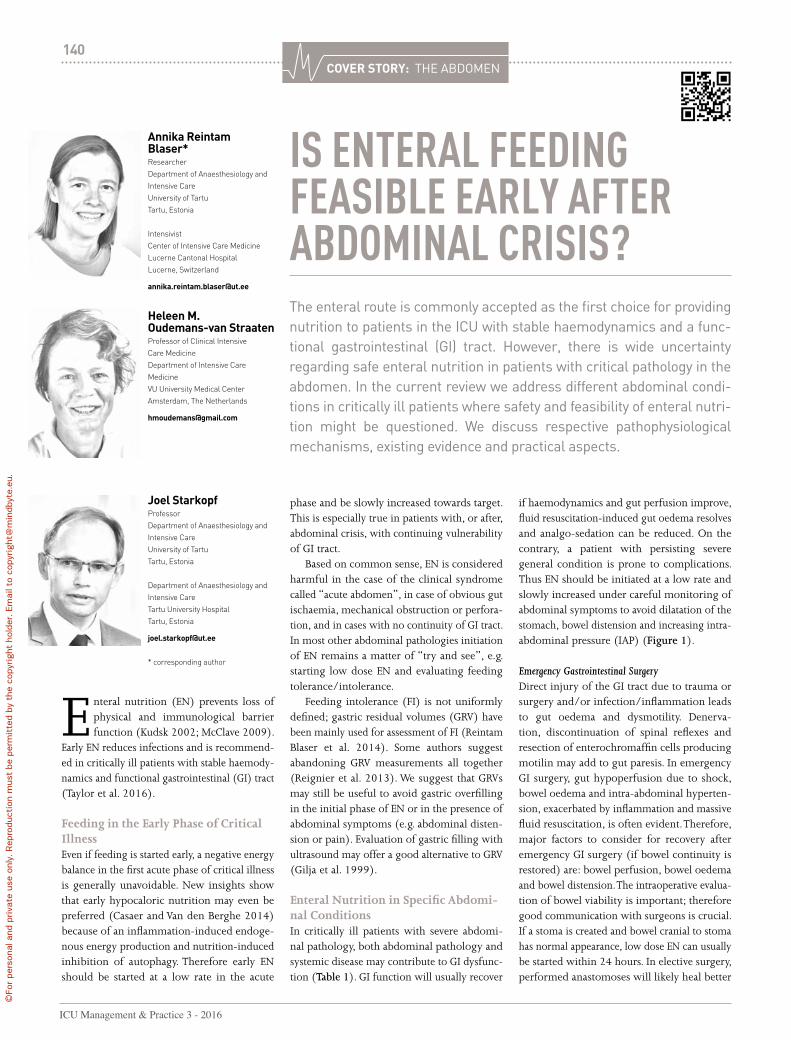
is enTeral feeding feasiBle early afTer aBdOMinal Crisis?The enteral route is commonly accepted as the first choice for providing nutrition to patients in the ICU with stable haemodynamics and a func-tional gastrointestinal (GI) tract. However, there is wide uncertainty regarding safe enteral nutrition in patients with critical pathology in the abdomen. In the current review we address different abdominal condi-tions in critically ill patients where safety and feasibility of enteral nutri-tion might be questioned. We discuss respective pathophysiological mechanisms, existing evidence and practical aspects.
annika Reintam blaser*ResearcherDepartment of Anaesthesiology and Intensive CareUniversity of TartuTartu, Estonia
IntensivistCenter of Intensive Care MedicineLucerne Cantonal Hospital Lucerne, Switzerland
heleen m. oudemans-van StraatenProfessor of Clinical Intensive Care MedicineDepartment of Intensive Care MedicineVU University Medical CenterAmsterdam, The Netherlands
Joel StarkopfProfessor Department of Anaesthesiology and Intensive CareUniversity of TartuTartu, Estonia
Department of Anaesthesiology and Intensive CareTartu University HospitalTartu, Estonia
* corresponding author

ICU Management & Practice 3 - 2016
141CoVeR StoRY: THE ABDOMEN
©Fo
r pe
rson
al a
nd p
riva
te u
se o
nly.
Rep
rodu
ctio
n m
ust
be p
erm
itte
d by
the
cop
yrig
ht h
olde
r. E
mai
l to
copy
right
@m
indb
yte.
eu.
with EN than without (Boelens et al. 2014). The risk of anastomotic leak is much higher if an anastomosis is performed during emergency surgery, but there is no evidence on harmfulness of early EN in this situation. A positive effect of early EN regarding infections after emergent GI surgery has been shown in one randomised controlled study (Singh et al. 1998).
Damage control surgery enables postpone-ment of restoration of bowel continuity until hypoperfusion, oedema and distension are resolved. Still, trophic EN might already be considered if a diverting stoma is present and the next surgery is not planned within the next 24 hours.
In patients with prolonged abdominal sepsis requiring multiple interventions and clearly not reaching their energy and protein targets with EN, supplemental PN should be considered after a couple of days, while avoiding overfeeding. Supplemental PN should also be considered if such patients have severe diarrhoea with impaired absorption of nutrients.
Open AbdomenPatients with open abdomen often require multiple surgeries and have increased risk for fistula formation. A few studies have shown that EN is feasible in patients with open abdomen and is associated with a higher rate of abdomi-nal closure and a lower incidence of ventilator-associated pneumonia (Collier 2007; Byrnes et al. 2010; Dissanaike 2008).
EN should be applied early, as soon as bowel continuity is confirmed or restored and haemodynamic and tissue perfusion goals can be reached with or without vasopressors/inotropes. Continuing need for fluid resuscita-tion may refer to unsolved abdominal pathol-ogy, whereas losses due to the open abdomen need to be taken into account.
Abdominal Aortic SurgeryRupture of the abdominal aorta and associ-ated surgery carry a risk of massive bleeding
and transfusion, retroperitoneal haematoma formation and impaired gut perfusion, which might be an argument for delaying EN in these patients. The major adverse event after abdomi-nal aortic surgery is colonic ischaemia (CI), which occurs in about 2% of patients after elective surgery for aneurysm, and 10% in case of rupture (Björck et al. 1996; Van Damme et al. 2000), somewhat less in endovascular repair (Becquemin et al. 2008). Presumed causes of CI are ligation or obstruction of supply arteries (inferior mesenteric artery, hypogastric arteries, meandering mesenteric arteries), non-occlusive ischaemia due to shock or vasopressor drugs, and (micro) embolisation (Steele 2007).
Length of operation, aneurysm rupture and renal insufficiency are independent risk factors of CI (Becquemin et al. 2008). Surgical details (reimplantation of inferior mesenteric artery, intraoperative assessment of blood flow by Doppler flowmetry, large bowel viability, etc.) should be carefully recognised. The main clinical symptoms of CI are early diarrhoea, haematoschisis (Björck et al. 1996) and ileus (Valentine et al. 1998). Colonoscopy remains the method of choice to detect ischaemic lesions of colonic mucosa, but its routine application is not supported (Steele 2007). Whether and how the endoscopic findings can guide EN is not clear. Circulating biochemical
markers such as intestinal fatty acid-binding protein may facilitate the recognition of CI (Vermeulen Windsant et al. 2012), but whether this information can be used for feeding decisions remains unknown.
Taking the relatively low incidence of CI, it is not rational to delay EN in all patients routinely for several days after abdominal aortic surgery. Instead, EN should be initiated with low dose under careful monitoring of abdominal symptoms, IAP and signs of CI, and increased gradually (van Zanten 2013). In overt bowel ischaemia, EN should be withheld.
Abdominal Trauma Abdominal trauma is a complex injury, where a multidisciplinary approach has made non-operative management increasingly feasible and effective (Prachalias and Kontis 2014). Early EN may be well integrated in this approach. However, obstacles such as GI tract discontinuity, compromised gut perfusion and/or abdominal compartment syndrome may necessitate delay of EN. At the same time, some older RCTs using needle catheter jejunostomy have shown benefit of early EN over early PN (Kudsk 1992) and over delayed EN (Moore 1986) regarding infectious compli-cations. We suggest starting EN early after abdominal trauma if continuity of GI tract is
In most patients EN should be considered early after initial management of
abdominal crisis
Pathophysiological mechanisms Condition/diagnosis
Local/gastrointestinal
1. direct injury in GI tract 2. inflammation/infection 3. bowel distension4. ischaemia5. dysmotility6. gut oedema7. reduction of bowel length
GI perforationGI surgeryGI bleedingBowel ischaemiaFistulaColitisIleus
Abdominal/peritoneal/retroperitoneal
1. inflammation/infection 2. intra-abdominal hypertension3. intra-abdominal bleeding
Abdominal traumaAbdominal surgeryAbdominal bleedingRetroperitoneal bleedingPeritonitisPancreatitis
Systemic 1. hypoperfusion2. tissue oedema3. splanchnic vasoconstriction4. inflammation/infection5. dysmotility caused by drugs or
electrolyte disturbances
ShockCapillary leak syndromeMassive fluid resuscitationVasoconstrictorsDrugs causing hypomotility: e.g. vasoactives, opiates, sedatives Drugs causing hypermotility (diarrhoea): e.g. antibioticsElectrolyte disturbances
Table 1. Main Pathophysiological Mechanisms Contributing to GI Dysfunction and Possibly Conflicting With EN In Different Clinical Conditions
ICU Management & Practice 3 - 2016
142CoVeR StoRY: THE ABDOMEN
©Fo
r pe
rson
al a
nd p
riva
te u
se o
nly.
Rep
rodu
ctio
n m
ust
be p
erm
itte
d by
the
cop
yrig
ht h
olde
r. E
mai
l to
copy
right
@m
indb
yte.
eu.
confirmed/restored, and abdominal compart-ment syndrome and bowel ischaemia excluded.
Abdominal Compartment SyndromeAbdominal compartment syndrome (ACS), defined as IAP above 20 mmHg along with new or worsening organ failure, is an immedi-ately life-threatening condition, where prompt measures to reduce IAP are needed. These measures include decompression of GI tract and avoidance of adding any volume into the abdomen (Kirkpatrick et al. 2013), thereby excluding EN. Moreover, splanchnic perfusion is severely jeopardised during ACS.
EN should be considered at elevated IAPs between 12 and 20 mmHg without ACS, but high incidence of feeding intolerance has been described (Reintam et al 2008). Further, EN itself may cause an increase in IAP. We suggest incorporating IAP measurements into standard monitoring of critically ill patients with abdominal pathologies in the initial phase of EN, and cessation of feeding to be consid-ered if worsening of clinical status is possibly attributed to increasing IAP.
Severe GI BleedingPatients admitted to the ICU due to acute GI bleeding require immediate diagnostics and
intervention to localise and stop bleeding. EN might be considered when the bleeding has been stopped endoscopically or surgically. The main rationale to withhold EN after stopping active bleeding is disturbed visibility if a new endoscopy is needed; therefore delaying enteral intake for at least 48 hours in case of high risk of rebleeding has been suggested (Hébuterne and Vanbiervliet 2011). Such a time frame is not well justified nor supported by the evidence. We suggest that when upper GI bleeding has been stopped and there are no signs of rebleeding, low dose EN can be started within 48 hours. In case of lower GI bleeding EN could be started immediately.
Bowel IschaemiaEN increases gut perfusion (Matheson 2000), but only if the vasculature is intact and the systemic haemodynamics sufficient. There is broad consensus to withhold EN in patients with suspected small bowel ischaemia. This condition requires optimisation of the circulation and, if symptoms of ischaemia persist, a surgical or radiological interven-tion. In addition, continuous thoracic epidur-al anaesthesia may increase splanchnic blood flow by blocking afferent sympathic reflexes (Holte 2000). Local mucosal ischaemia of the
colon has a tendency to heal when the general condition of the patient improves. Therefore EN should be considered in patients with colonic mucosal ischaemia without bowel distension. Bowel distension may possibly be aggravated by EN and lead to further impair-ment of bowel wall perfusion. We suggest that EN should not be started if transmural bowel ischaemia is confirmed or suspected or signs of local mucosal ischaemia are seen in severely distended bowel.
Bowel ObstructionBowel obstruction leads to obstructive ileus, with initial hypermotility (be warned: presence of bowel sounds is misleading) to force bowel contents through the obstruction and subsequent bowel distension above the obstruc-tion. Bowel obstruction requires a surgical or endoscopic intervention to restore passage of bowel contents or to create a proximal stoma. EN should be withheld in case of obstructive symptoms, but can be carefully initiated as soon as passage is restored or a proximal stoma has been created. It may take a couple of days before bowel distension and paresis are resolved and EN can be increased.
Bowel ParalysisEN itself promotes motility and has beneficial effects regarding the physical and immuno-logical gut barrier, whereas prolonged enteral fasting will aggravate dysmotility and should be avoided. Since gastroparesis is often more pronounced than small intestinal paralysis, the use of prokinetics and postpyloric feeding should be considered early in case of gastric intolerance to EN. However, paralytic ileus is often encountered in patients with peritonitis. Inflammation-induced dysmotility is mediated by cytokines and nitric oxide produced by locally activated macrophages in the muscular layer, and by neuronal pathways (Schmidt et al. 2012). Non-abdominal sepsis may also be associated with bowel paresis, due to the release of nitric oxide, which causes bowel relaxation, oxidative stress and the systemic release of tumour necrosis factor (TNF), which inhibits the central vagal pathways (Emch et al. 2000). Furthermore, many conditions and therapies in critically ill patients (e.g. hyperglycaemia, hypokalaemia, acidosis, use of dopamine, opioids, clonidine and dexmedetomidine) may contribute to bowel paralysis.
In rare cases, isolated large bowel disten-sion mainly in the caecum region occurs,
Evaluate systemic, abdominal and local GI factors within 24h after ICU admission
• Persistent shock• Acute abdomen• GI tract not intact• Overt mesenteric ischaemia• Bowel obstruction• Abdominal compartment syndrome • Overt active upper GI bleeding
• Bowel distension • Intraoperative or endoscopic finding of • Impaired gut perfusion • Severe gut oedema
YES
YES
YES
YES
Respective management, delay EN
NO
Start EN 10-20 ml/h• Monitor GI symptoms• Consider enteral laxative
• Bowel distension• Intra-abdominal pressure increasing
• Gastric dilatation• Large gastric residualsNO
NO
• Delay EN or consider trophic feeding• Consider further investigations/interventions• Reevaluate after 24 hours
1. Prokinetics2. Monitor GI symptoms3. Postpyloric feeding
• Increase EN slowly until target if tolerated• Reevaluate frequently• Consider enteral laxative
Consider parenteral nutrition if short term improvement is not expected
• Stop EN • Investigate/treat condition
General condition improving?GI symptoms stable or improving?
NO
Figure 1. Algorithm for Using EN Early after Abdominal Crisis

143CoVeR StoRY: THE ABDOMEN
AbbreviationsACS abdominal compartment syndromeCI colonic ischaemiaEN enteral nutritionFI feeding intoleranceGRV gastric residual volumeIAP intra-abdominal pressureTNF tumour necrosis factor
called Ogilvie’s syndrome seu colonic pseudo-obstruction. This condition carries high risk of bowel ischaemia and perforation due to disten-sion, and should be promptly recognised and managed (Oudemans-van Straaten 2011; De Giorgio and Knowles 2009) with intravenous neostigmine (van der Spoel et al. 2001; Valle and Godoy 2014), endoscopic decompression or temporary coecostomy. Early start of lactulose or polyethylene glycol (van der Spoel et al. 2007) and neostigmine, if defaecation does not occur, may help to prevent Ogilvie’s syndrome. In less severe cases of bowel paralysis, there are no confirmed contraindications to start a trial of low dose EN under careful monitoring of symptoms and promotion of defaecation with laxatives and neostigmine.
Acute Colitis with Toxic MegacolonAcute colitis as a cause of diarrhoea in intensive care is a rare condition that is mostly caused by a Clostridium difficile infection. Sometimes severe
enterocolitis is caused by chemotherapy for haematological disorders. In most severe cases toxic megacolon—a severe and life-threatening condition associated with systemic toxicity—may develop (Oudemans-van Straaten 2011). Colitis requires specific therapy, includ-ing antibiotics, discontinuation of motility impairing drugs, replacement of intravenous fluids, electrolytes, trace elements and vitamins (Dickinson 2014; Oudemans-van Straaten 2011). In rare cases of toxic megacolon, total colectomy becomes necessary for the patient’s survival. In most patients with colitis, there is no contraindication for EN, because the small intestine is intact. However, EN should probably not be applied to patients with toxic megacolon.
ConclusionsIn most patients EN should be considered early after initial management of abdominal crisis, when continuity of GI tract is confirmed or restored, and bowel ischaemia and abdominal
compartment syndrome are excluded. However, EN should be started at a slow rate under careful monitoring of GI symptoms and IAP.
Conflict of interestAnnika Reintam Blaser declares that she has no conflict of interest. Heleen M. Oudemans-van Straaten declares that she has no conflict of interest. Joel Starkopf declares that he has no conflict of interest.
For full references, please email editorial@icu management.org, visit icu-management.org or use the article QR code.
XENIOS console - one integrated lung & heart therapy platform
available with ¼" oder ⅜" pump headcustomized Patient Kits & Patient Kits ECMO Sets
Approval for novalung Kits in process.
www.xenios-ag.com
the unique platform for lung & heart assistadapt the system to your needsincreased safety through integrated emergency functions
ONE CONSOLE FOR ALL THERAPIES
JOIN - LEARN - CARE - THE ONLINE PLATFORM FOR CLINICAL SUPPORT, INFORMATION AND CONTINUING TRAINING.
PATIENT KITS
LU
NG
A N D H E A R T T H E R A PI E
S
THE PERFECT MATCH FOR YOUR NEEDS
A FULL RANGE OF PATIENT KITS: FROM CUSTOMIZED ECMO SETS UP TO LONGTERM APPROVED PATIENT KITS
©Fo
r pe
rson
al a
nd p
riva
te u
se o
nly.
Rep
rodu
ctio
n m
ust
be p
erm
itte
d by
the
cop
yrig
ht h
olde
r. E
mai
l to
copy
right
@m
indb
yte.
eu.
ICU Management & Practice 3 - 2016
144CoVeR StoRY: THE ABDOMEN
©Fo
r pe
rson
al a
nd p
riva
te u
se o
nly.
Rep
rodu
ctio
n m
ust
be p
erm
itte
d by
the
cop
yrig
ht h
olde
r. E
mai
l to
copy
right
@m
indb
yte.
eu.
Most patients with liver cirrhosis remain in a compensated stage for more than 10 years, regardless of
the aetiology of the liver disease. The progres-sion to decompensated cirrhosis is defined by the occurrence of a major complication such as ascites, variceal bleeding and/or hepatic encephalopathy. From here on most patients will not die because of a progressive, irrevers-ible decrease in liver function, but because of a relatively sudden event that precipitates an acute deterioration in their clinical condi-tion, a syndrome termed acute-on-chronic liver failure (ACLF). For many intensive care specialists, ACLF stands for a critically ill patient who is suffering from an intra- or extrahe-patic acute insult with serious repercussions on both an existing chronic liver disease and on other organ functions. It also means that, as compared to the average intensive care unit (ICU) patient, the patient has an unusually high risk of death.
Concepts about cirrhosis have evolved significantly in recent years, and major advances have been made in defining the natural history of ACLF (for general reviews see Arroyo et al. 2016; Bernal et al. 2015; Sarin and Choudhury 2016). The syndrome is highly challenging for intensivists and poses difficult questions related to the recognition of precipi-tating factors, pathogenesis of extrahepatic organ failures, accurate prognosis, medical management, evaluation for urgent liver
transplantation and finally the identification of those situations that may render intensive care futile. The present appraisal will focus on recent insights and their potential repercussions on the way intensivists should understand and manage patients with ACLF.
Definition and Natural History of Acute-on-Chronic Liver Failure There is no uncontested universal definition for ACLF and the two most widely used definitions depend on the origin of the hepatologists—West versus East (Arroyo et al 2015; Sarin et al. 2014). For the purpose of this text we will use the definition of the European Association for the Study of the Liver – Chronic Liver Failure (EASL-CLIF) Consortium, because extrahepatic organ failure(s) and short-term mortality are central to the definition and therefore more closely mimic circumstances in the ICU. This
definition is based on a prospective, multi-centre, observational study (CANONIC study) of 1343 patients who were hospitalised for acute decompensation of cirrhosis (Moreau et al 2013). ACLF is thus defined as a specific syndrome comprising acute decompensation of cirrhosis (development of ascites, variceal bleeding, hepatic encephalopathy and/or bacterial infections), organ failure and high
short-term mortality (by definition 28-day mortality rate ≥15%) (Arroyo et al 2015).
Based on the chronic liver failure (CLIF) Acute-on-Chronic Liver Failure in Cirrhosis (CANONIC) study a new grading system for severity of ACLF (grade 0 to 4) has been intro-duced built on a modified Sequential Organ Failure Assessment (SOFA) score (Tables 1 and 2). This new grading system is proving useful to diagnose the condition, to study the natural history of ACLF, to stratify patients in interven-tional trials and for prognostication (Gustot et al 2015; Silva et al. 2015; Shi et al. 2016).
In the CANONIC study the prevalence of ACLF in patients presenting to the hospital with acute decompensation of cirrhosis was 31%. Twenty-three percent had ACLF at the time of admission and another 11% developed ACLF during hospitalisation. Twenty-four percent of the patients required care in the ICU with one in three not fulfilling criteria for ACLF at the time of admission to the ICU. A similar prevalence ranging from 24 to 34% has been reported in other large studies from China, North America and Scandinavia (Li et al 2016; Bajaj et al. 2014a; Sargenti et al. 2015). Almost half of the patients with ACLF did not have a prior history of acute decompensation, or had developed the first decompensating event within the three months prior to the diag-nosis of ACLF. This observation is relevant to the extent that ACLF is not necessarily the final event in a progressive course of decompen-sating liver disease, but may occur at any point in time after diagnosis of cirrhotic liver disease.
The clinical course of the condition is very dynamic. One study observed resolution of ACLF in 42.5% of patients across all grades of ACLF, 53.5% in ACLF-1, 34.6% in ACLF-2 and 16% in ACLF-3 (Table 1) (Gustot et al. 2015). In the CANONIC study the overall 28-day and 90-day mortality rates for patients with ACLF, who did not undergo liver transplantation, were 32.8% and 51.2%. Similar rates have
Philippe meerssemanAdjunct Head of ClinicMedical Intensive CareUniversity Hospital LeuvenLeuven, Belgium
alexander Wilmer Head of ClinicAssociate Professor of MedicineMedical Intensive CareUniversity Hospital LeuvenLeuven, Belgium
eVOlVing COnCepTs in aCuTe-On-ChrOniC liVer failure
ACLF is not necessarily the final event
in a progressive course of decompensating liver disease, but may occur
at any point in time after diagnosis of cirrhotic liver
disease

ICU Management & Practice 3 - 2016
145CoVeR StoRY: THE ABDOMEN
©Fo
r pe
rson
al a
nd p
riva
te u
se o
nly.
Rep
rodu
ctio
n m
ust
be p
erm
itte
d by
the
cop
yrig
ht h
olde
r. E
mai
l to
copy
right
@m
indb
yte.
eu.
been reported in other studies (Li et al. 2016). These mortality rates are clearly different from those in patients with acute decompensation of liver cirrhosis but not fulfilling criteria for ACLF (1.9% and 9.3%, respectively). The most frequent cause of death in patients with ACLF was multiple organ failure without septic or hypovolaemic shock (40%), followed by septic shock in approximately 25% of cases. The aeti-ology of cirrhosis does not seem to be determi-nant of outcome, but patients with gastrointes-tinal haemorrhage as a precipitating factor do better than patients who were not bleeding at admission (McPhail et al. 2014).
It is often assumed that acute decompensa-tion of liver function is triggered by a clinically identifiable, precipitating event. The trigger may have a hepatic origin, such as drug-induced liver injury, viral or ischaemic hepatitis, liver surgery or undue alcohol consumption. It can also have an extrahepatic origin such as acute bacterial infection, major surgery or paracentesis. Interest-ingly, in the CANONIC study, in 43.6% of the patients with ACLF, no precipitating event could be identified (Moreau et al. 2013). This obser-vation underscores the fact that in the majority of patients we are not yet able to diagnose the pathogenetic mechanism leading to acute decompensation. Acute bacterial infection was the most frequent precipitating event in 33% of the patients (Moreau et al. 2013).
Prevalence and Pathogenesis of Organ Dysfunctions Associated With ACLFOrgan dysfunction or failure is highly prevalent in ACLF. Hepatic, renal, cerebral, coagulation and circulatory dysfunctions are well known, but important derangements in the function of the heart, immune system, adrenal glands and muscle have been also well documented. In ACLF patients in the CANONIC study kidney failure (56%) was the most frequent organ failure followed by liver failure (44%), coag-ulation (28%), cerebral (24%), circulation (17%) and lung failure with 9%. The number of failing organs correlates with increasing white cell count and C-reactive protein (CRP) levels (Jalan and Williams 2002).
Two pathogenetic mechanisms seem to be important drivers of both intra- and extrahe-patic organ dysfunction: systemic inflamma-tion and dysbiosis of the microbiome (Bernardi M. et al. 2015). Systemic inflammation may be induced by bacterial pathogen-associated molecular patterns (PAMPs) or by virulence factors produced by bacteria. Patients with cirrhosis have increased permeability of the gut related to portal hypertension, inflamma-tion-mediated damage to the gut barrier and altered gut flora. The result is increased trans-location of particularly Gram-negative bacteria, PAMPs or virulence factors from the intestinal lumen to the systemic circulation. The preva-
lence of translocation of enteric organisms to mesenteric lymph nodes in cirrhotic patients is significantly increased according to the Child-Pugh classification: 3.4% in Child A, 8.1% in Child B and 30.8% in Child C patients (Cirera et al. 2001). Systemic inflammation may also be induced by ongoing necrosis of hepatocytes or damage to the extracellular matrix caused by alcohol, viral disease or any other aetio-pathogenetic mechanisms of cirrhosis. In this case the molecules inducing inflammation are called damage-associated molecular patterns (DAMPs). How inflammation contributes to organ dysfunction in ACLF has not yet been fully elucidated. Besides the well-described severe immune dysfunction associated with cirrhosis with increased susceptibility to infec-tion, the following concepts are likely to be important (Verbeke et al. 2011):1. The effects caused by immunopathology, a
term that describes the potential negative impact of an excessive immune response (Iwasaki and Medzhitov 2015). Either PAMPs or DAMPs can cause immuno-pathology that in turn may cause organ dysfunction. In this case defence mecha-nisms directed at controlling infection or immunopathology are insufficient. This is the likely mechanism in ACLF precipitated by acute bacterial infection or severe alco-holic hepatitis.
2. Failed tolerance, a concept that describes the incapacity to develop tolerance mech-anisms to persistent infection–mediated inflammation (Medzhitov et al 2012). In this case persistent ‘low-grade’ systemic exposure to PAMPs or DAMPs may be the reason for ongoing ‘sterile’ inflammation for which no tolerance can be developed. This concept provides an array of potential new therapeutic targets aimed at increasing tolerance.
Recent evidence points to gut dysbiosis as a second important pathogenetic driver of organ dysfunction in ACLF (Bajaj et al. 2014b; Chen et al. 2015; Rai et al. 2014). Several factors contribute to altered microbiota in cirrhosis, including increased intestinal permeability, abnormal small intestinal motility, impaired antimicrobial defence, small intestinal bacterial overgrowth, decreased bile acid production and compromised enterohepatic circulation (Rai et al. 2014). In stable cirrhosis there is a clear change in diversity and composition of gut microbiota with progressive dysbiosis in the setting of decompensation. Similar changes
Organ/system 0 1 2 3 4
Liver (Bilirubin, mg/dl) <1.2 ≥1.2 - ≤2.0 ≥2.0 - <6.0 ≥6.0 - <12.0 ≥12.0
Kidney(Creatinine, mg/dl) <1.2 ≥1.2 - <2.0 ≥2.0 - <3.5 ≥3.5 - <5.0 ≥5.0
or use of renal replacement therapy
Cerebral (HE grade) No HE I II III IV
Coagulation (INR) <1.1 ≥1.1 – <1.25 ≥1.25 - <1.5 ≥1.5 – <2.5 ≥2.5 or Platelets≤20x109/l
Circulation (MAP mm Hg)
≥70 <70
Dopamine ≤5 or
Dobutamine or Terlipressin
Dopamine >5 or
E ≤ 0.1 orNE ≤ 0.1
Dopamine >15 orE > 0.1 orNE > 0.1
Lungs PaO/FiO2: or SpO2/FiO2
>400
>512
>300 - ≤400
>357 - ≤512
>200 - ≤300
>214 - ≤357
>100 - ≤200or
>8 - ≤214
≤100
≤89
HE hepatic encephalopathy INR international normalised ratio MAP mean arterial pressure E epinephrine NE nor-epinephrine PaO2 partial pressure of arterial oxygen; FIO2 fraction of inspired oxygen SpO2 pulse oximetric saturation. The highlighted areas in grey show the diagnostic criteria for organ failures.
Table 1. Chronic Liver Failure (CLIF)-Sequential Organ Failure Assessment (CLIF-SOFA) Score
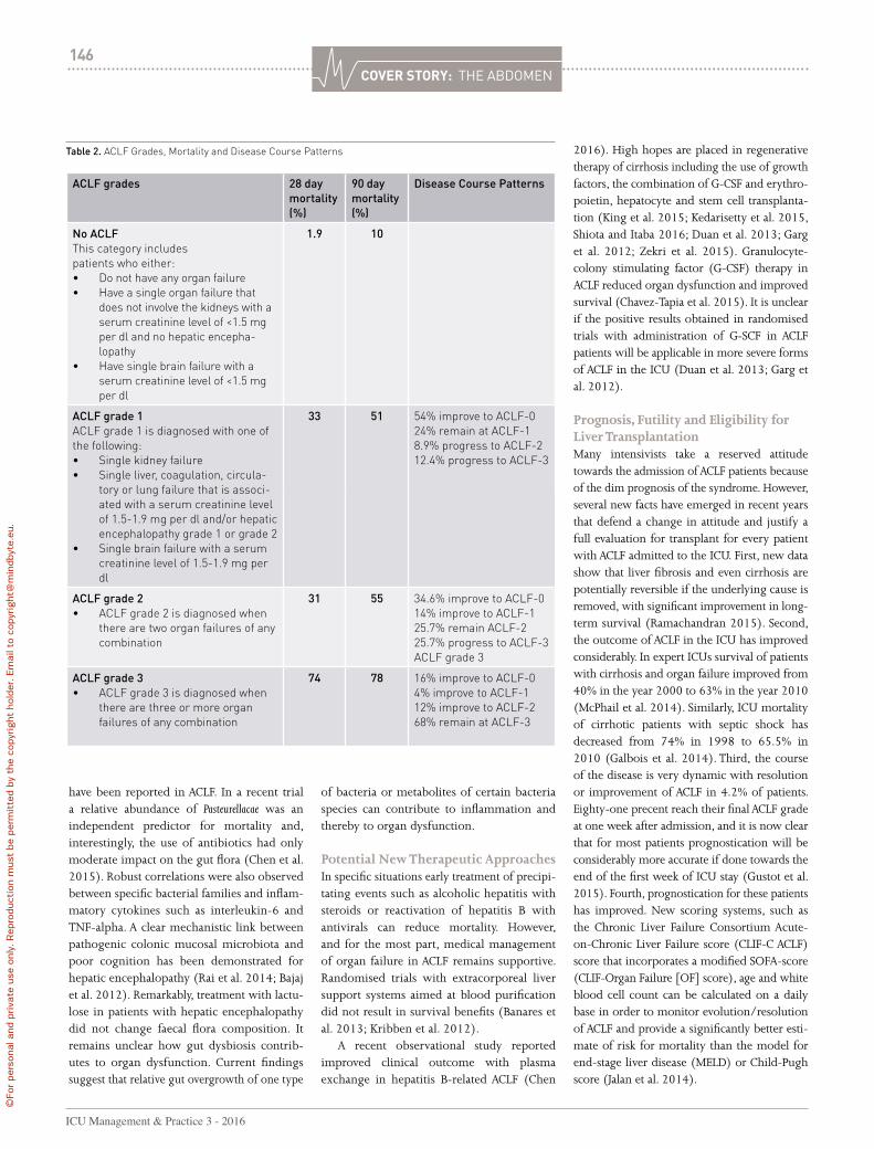
ICU Management & Practice 3 - 2016
146CoVeR StoRY: THE ABDOMEN
©Fo
r pe
rson
al a
nd p
riva
te u
se o
nly.
Rep
rodu
ctio
n m
ust
be p
erm
itte
d by
the
cop
yrig
ht h
olde
r. E
mai
l to
copy
right
@m
indb
yte.
eu.
have been reported in ACLF. In a recent trial a relative abundance of Pasteurellacae was an independent predictor for mortality and, interestingly, the use of antibiotics had only moderate impact on the gut flora (Chen et al. 2015). Robust correlations were also observed between specific bacterial families and inflam-matory cytokines such as interleukin-6 and TNF-alpha. A clear mechanistic link between pathogenic colonic mucosal microbiota and poor cognition has been demonstrated for hepatic encephalopathy (Rai et al. 2014; Bajaj et al. 2012). Remarkably, treatment with lactu-lose in patients with hepatic encephalopathy did not change faecal flora composition. It remains unclear how gut dysbiosis contrib-utes to organ dysfunction. Current findings suggest that relative gut overgrowth of one type
of bacteria or metabolites of certain bacteria species can contribute to inflammation and thereby to organ dysfunction.
Potential New Therapeutic ApproachesIn specific situations early treatment of precipi-tating events such as alcoholic hepatitis with steroids or reactivation of hepatitis B with antivirals can reduce mortality. However, and for the most part, medical management of organ failure in ACLF remains supportive. Randomised trials with extracorporeal liver support systems aimed at blood purification did not result in survival benefits (Banares et al. 2013; Kribben et al. 2012).
A recent observational study reported improved clinical outcome with plasma exchange in hepatitis B-related ACLF (Chen
2016). High hopes are placed in regenerative therapy of cirrhosis including the use of growth factors, the combination of G-CSF and erythro-poietin, hepatocyte and stem cell transplanta-tion (King et al. 2015; Kedarisetty et al. 2015, Shiota and Itaba 2016; Duan et al. 2013; Garg et al. 2012; Zekri et al. 2015). Granulocyte-colony stimulating factor (G-CSF) therapy in ACLF reduced organ dysfunction and improved survival (Chavez-Tapia et al. 2015). It is unclear if the positive results obtained in randomised trials with administration of G-SCF in ACLF patients will be applicable in more severe forms of ACLF in the ICU (Duan et al. 2013; Garg et al. 2012).
Prognosis, Futility and Eligibility for Liver TransplantationMany intensivists take a reserved attitude towards the admission of ACLF patients because of the dim prognosis of the syndrome. However, several new facts have emerged in recent years that defend a change in attitude and justify a full evaluation for transplant for every patient with ACLF admitted to the ICU. First, new data show that liver fibrosis and even cirrhosis are potentially reversible if the underlying cause is removed, with significant improvement in long-term survival (Ramachandran 2015). Second, the outcome of ACLF in the ICU has improved considerably. In expert ICUs survival of patients with cirrhosis and organ failure improved from 40% in the year 2000 to 63% in the year 2010 (McPhail et al. 2014). Similarly, ICU mortality of cirrhotic patients with septic shock has decreased from 74% in 1998 to 65.5% in 2010 (Galbois et al. 2014). Third, the course of the disease is very dynamic with resolution or improvement of ACLF in 4.2% of patients. Eighty-one precent reach their final ACLF grade at one week after admission, and it is now clear that for most patients prognostication will be considerably more accurate if done towards the end of the first week of ICU stay (Gustot et al. 2015). Fourth, prognostication for these patients has improved. New scoring systems, such as the Chronic Liver Failure Consortium Acute-on-Chronic Liver Failure score (CLIF-C ACLF) score that incorporates a modified SOFA-score (CLIF-Organ Failure [OF] score), age and white blood cell count can be calculated on a daily base in order to monitor evolution/resolution of ACLF and provide a significantly better esti-mate of risk for mortality than the model for end-stage liver disease (MELD) or Child-Pugh score (Jalan et al. 2014).
Table 2. ACLF Grades, Mortality and Disease Course Patterns
ACLF grades 28 day mortality (%)
90 day mortality (%)
Disease Course Patterns
No ACLFThis category includes patients who either:• Do not have any organ failure• Have a single organ failure that
does not involve the kidneys with a serum creatinine level of <1.5 mg per dl and no hepatic encepha-lopathy
• Have single brain failure with a serum creatinine level of <1.5 mg per dl
1.9 10
ACLF grade 1ACLF grade 1 is diagnosed with one of the following:• Single kidney failure• Single liver, coagulation, circula-
tory or lung failure that is associ-ated with a serum creatinine level of 1.5-1.9 mg per dl and/or hepatic encephalopathy grade 1 or grade 2
• Single brain failure with a serum creatinine level of 1.5-1.9 mg per dl
33 51 54% improve to ACLF-024% remain at ACLF-1 8.9% progress to ACLF-2 12.4% progress to ACLF-3
ACLF grade 2• ACLF grade 2 is diagnosed when
there are two organ failures of any combination
31 55 34.6% improve to ACLF-014% improve to ACLF-125.7% remain ACLF-225.7% progress to ACLF-3ACLF grade 3
ACLF grade 3• ACLF grade 3 is diagnosed when
there are three or more organ failures of any combination
74 78 16% improve to ACLF-04% improve to ACLF-112% improve to ACLF-268% remain at ACLF-3
AbbreviationsACLF acute-on-chronic liver failureCANONIC CLIF Acute-on-Chronic Liver Failure in Cirrhosis CLIF chronic liver failureCLIF-C Chronic Liver Failure ConsortiumCRP c-reactive proteinDAMP damage-associated molecular patternsG-CSF Granulocyte-colony stimulating factor ICU intensive care unitMELD model for end-stage liver diseaseOF organ failurePAMP pathogen-associated molecular patternsSOFA sequential organ failure assessment
Considering the above, indiscriminate refusal of ICU admis-sion of ACLF patients is not acceptable any more, since no specific group of patients can be identified at the time of diagnosis for which medical ICU treatment may be considered futile. However, intensivists also need to acknowledge that patients with four or more organ failures or a CLIF-C ACLF score > 64 after one week of ICU care have 28-day mortality rates in the range of 90 to 100%. If ineligible for transplantation withdrawal of care is a reasonable option for these patients.
Liver transplantation in ACLF is controversial and fraught with uncertainties regarding case selection and timing (Pamecha et al. 2015; Reddy et al. 2015). Only 15-25 % of patients are actually transplanted (Gustot et al. 2015, Finkenstedt et al. 2013). Recent series have reported encouraging results with 1- and 5-year survival of 80-90% (Finkenstedt et al. 2013; Chan et al. 2009). Even patients with ACLF-3 may expect a 1-year survival probability of 78% (Gustot et al. 2015).
SummaryMajor progress has been made in defining the natural history and prognosis of ACLF. Regenerative therapies and liver transplantation in selected cases hold promise for the future.
Conflict of InterestPhilippe Meersseman and Alexander Wilmer declare that they have no conflict of interest.
ReferencesArroyo V, Moreau R, Jalan R et al. (2015) Acute-on-chronic liver failure: a new syndrome that will re-classify cirrhosis. J Hepatol, 62(1 Suppl): S131-43.
Arroyo V, Moreau R, Kamath PS et al. (2016) Acute-on-chronic liver failure in cirrhosis. Nat Rev Dis Primers, 2: 16041.
Bajaj JS, Hylemon PB, Ridlon JM et al. (2012) Colonic mucosal microbiome differs from stool microbiome in cirrhosis and hepatic encephalopathy and is linked to cognition and inflammation. Am J Physiol Gastrontest Liver Physiol, 303 (6): G675-85.
Bajaj JS, O'Leary JG, Reddy KR et al. (2014a) Survival in infection-related acute-on-chronic liver failure is defined by extrahepatic organ failures. Hepatology, 60(1): 250-6.
Bajaj JS, Heuman DM, Hylemon PB et al. (2014b) Altered profile of human gut microbiome is associated with cirrhosis and
its complications. J Hepatol, 60(5): 940-7.
Banares R, Nevens F, Larsen FS et al. (2013) Extracorporeal albumin dialysis with the molecular adsorbent recirculating system in acute-on-chronic liver failure: the RELEF trial. Hepatology, 57(3): 1153-62.
Bernal W, Jalan R, Quaglia A et al. (2015) Acute-on-chronic liver failure. Lancet, 386(10003): 1576-87.
Bernardi M, Moreau R, Angeli P et al. (2015) Mechanisms of decompensation and organ failure in cirrhosis: from peripheral arterial vasodilation to systemic inflamma-tion hypothesis. J Hepatol, 63(5): 1272-84.
Chan AC, Fan ST, Lo CM et al. (2009) Liver transplantation for acute-on-chronic liver failure. Hepatol Int, 3(4): 571-81.
For full references, please email editorial@icu management.org, visit icu-management.org or use the article QR code.
• More information: thermoscientifi c.com/procalcitonin
Reduce AntibioticsAntibiotic overuse contributes to increasing antimicrobial
resistance, increased risk of drug-related adverse events and
higher medical costs. Antibiotic guidance with B·R·A·H·M·S PCT™
limits antibiotic prescription to patients with true bacterial
infection, allows to tailor the duration of antibiotic therapy
for individual patients, and reduces signifi cantly the antibiotic
exposure with no adverse impact on patient outcome.
B·R·A·H·M·S PCTassays
1. Bouadma L et al. Lancet 2010, 375 (9713): 463-474 2. Schuetz P et al. Expert Rev Anti Infect Ther 2010, 8 (5): 575-87. © 2016 Thermo Fisher Scientifi c Inc. All rights reserved. B·R·A·H·M·S PCT™ and all other trademarks are the property of Thermo Fisher Scientifi c and its subsidiaries unless otherwise specifi ed. Thermo Fisher Scientifi c products are distributed worldwide; not all intended uses and applications mentioned in this printing are registered in every country.
AB days without Procalcitonin
-35% AB days in LRTI 2
-23% AB days in Sepsis1
Visit us at EuSEM, Vienna, booth n° 42Lunch Symposium: Oct 4th, 2016, 12:55 -1:55 pm, Rittersaal“Improving cardiac care in the emergency department”
©Fo
r pe
rson
al a
nd p
riva
te u
se o
nly.
Rep
rodu
ctio
n m
ust
be p
erm
itte
d by
the
cop
yrig
ht h
olde
r. E
mai
l to
copy
right
@m
indb
yte.
eu.

ICU Management & Practice 3 - 2016
148CoVeR StoRY: THE ABDOMEN
©Fo
r pe
rson
al a
nd p
riva
te u
se o
nly.
Rep
rodu
ctio
n m
ust
be p
erm
itte
d by
the
cop
yrig
ht h
olde
r. E
mai
l to
copy
right
@m
indb
yte.
eu.
Understanding Intra-Abdominal Hypertension: What to Worry About?The abdomen can be considered as a closed anatomical space with the abdominal contents being primarily fluid in character, following Pascal’s Law: any change in pressure applied at any given point is transmitted undiminished throughout the abdomen (Malbrain 2004). This means that intra-abdominal pressure (IAP) can be measured by way of different direct and indirect routes via the stomach, bladder, uterus or rectum (Malbrain 2004). The intra-abdominal volume (IAV) will exert a certain force on the abdominal compartment walls, resulting in a baseline IAP that will be mainly determined by the abdominal compli-ance (Cab) (Malbrain 2016). The relationship between IAV and IAP is curvilinear, with an initial linear part followed by an exponen-tial increase once a critical volume is reached (Malbrain et al. 2014a; Malbrain et al. 2014b). IAP is an important physiological parameter and the recent updated consensus definitions must be used (Table 1) (Kirkpatrick 2013). IAH is defined as a sustained increase in IAP ≥ 12 mmHg and ACS is an IAP > 20 mmHg with new-onset organ failure (Kirkpatrick et al. 2013). While IAH is a graded continuum, ACS is an all-or-nothing phenomenon (Table 2) (Kirkpatrick et al. 2013). IAP should be measured at end-expiration, with the patient in the supine position and ensuring that there
is no abdominal muscle activity. Intravesicular IAP measurement is convenient, most widely used and considered the gold standard tech-nique (Kirkpatrick et al. 2013; Malbrain 2004). Where the mid-axillary line crosses the iliac crest is the recommended reference level for transvesicular IAP measurement and marking this level on the patient increases reproduc-ibility of IAP measurement (Kirkpatrick et al. 2013; De Waele et al. 2008). Instillation volume (maximal 25 ml) and temperature (above room temperature) may affect IAP readings, and the head of the bed elevation above 30° increases IAP while PEEP only minimally affects IAP (Cheatham et al. 2009; Verzilli et al. 2010). Protocols for IAP measurement should be developed for each intensive care unit (ICU) based on the locally available tools and equip-ment, and the ICU physician should pick the technique that the nurses are going to use. Pitfalls in IAP measurement are multiple, and thorough knowledge is essential, e.g. absence of abdominal muscle activity should be checked, particularly in awake patients.
Underlying Predisposing Conditions: When to Worry?1. Decreased CabClinicians should worry about patients in whom Cab is decreased. The major problem is that Cab is not routinely measured in clinical practice (Malbrain 2014a). However,
some indirect measures of Cab are available in mechanically ventilated patients: the ∆IAP (= IAP at end-inspiration minus IAP at end-expiration) and the abdominal pressure varia-tion (APV = mean IAP divided by ∆IAP) are such parameters and they are inversely corre-lated with Cab, i.e. the higher the ∆IAP or APV, the lower the Cab (Malbrain 2014a). True Cab can only be measured in case of addition or removal of a known abdominal volume (e.g. laparoscopic insufflation, paracenthesis etc.) with simultaneous measurement of the change in IAP. Cab is defined as the ease with which abdominal expansion can occur, and is deter-mined by the elasticity of the abdominal wall and diaphragm (Malbrain 2014a). It should be expressed as the change in IAV per change in IAP (ml/mmHg). Cab helps to understand the pathophysiological mechanisms and possible therapeutic targets (Malbrain et al. 2014a). Increased compliance indicates a loss of elastic recoil of the abdominal wall. Decreased compli-ance (e.g. in obesity, fluid overload, burn eschars, young age etc.) means that the same change in IAV will result in a greater change in IAP, and this can be a major contributor to secondary IAH.
2. Increased IAVClinicians should also worry when IAV is increased: this can be either related to free abdominal fluids or increased intraluminal
manu LnG malbrainHospital DirectorICU and High Care Burn UnitDirectorZiekenhuis Netwerk AntwerpenZNA StuivenbergAntwerp, Belgium
@manu_malbrain
Knowledge of intra-abdominal hypertension (IAH) and abdominal compartment syndrome (ACS) is crucial for successful treatment of critically ill patients, whether medical or surgical, young or old (Kirkpatrick et al. 2013). Today we understand that IAH and ACS are frequent causes of increased morbidity and mortality (De Waele et al. 2016). More importantly, we now also know that IAH and ACS have correctable causes, can easily be diagnosed and effectively treated, but only if the clinician is aware of these conditions and pursues their recognition (Wise et al. 2015). A monograph has recently been published on this topic, as despite the increasing interest unanswered questions still cloud the understanding of the pathophysiology of IAH and ACS (Malbrain and De Waele 2013). In this article we will try to provide at least some answers.
updaTe On inTra-aBdOMinal hyperTensiOn

ICU Management & Practice 3 - 2016
149CoVeR StoRY: THE ABDOMEN
©Fo
r pe
rson
al a
nd p
riva
te u
se o
nly.
Rep
rodu
ctio
n m
ust
be p
erm
itte
d by
the
cop
yrig
ht h
olde
r. E
mai
l to
copy
right
@m
indb
yte.
eu.
Def 2006 definitions (Malbrain et al. 2006) Def 2013 definitions (Kirkpatrick et al. 2013)1 IAP is the steady-state pressure concealed within the
abdominal cavity.1 IAP is the steady-state pressure concealed within the abdominal cavity.
2 APP = MAP – IAP 2 APP = MAP – IAP
3 FG = GFP – PTP = MAP – 2 * IAP REJECTED
4 IAP should be expressed in mmHg and measured at end-expiration in the complete supine position after ensuring that abdominal muscle contractions are absent and with the transducer zeroed at the level of the mid-axillary line.
3 IAP should be expressed in mmHg and measured at end-expiration in the complete supine position after ensuring that abdominal muscle contractions are absent and with the transducer zeroed at the level of the mid-axillary line.
5 The reference standard for intermittent IAP measurement is via the bladder with a maximal instillation volume of 25 mL of sterile saline.
4 The reference standard for intermittent IAP measurements is via the bladder with a maximal instillation volume of 25 mL of sterile saline.
6 Normal IAP is approximately 5-7 mmHg in critically ill adults.
5 IAP is approximately 5-7 mmHg and around 10 mmHg in critically ill adults.
7 IAH is defined by a sustained or repeated pathologic elevation of IAP ≥ 12 mmHg.
6 IAH is defined by a sustained or repeated pathologic elevation of IAP ≥ 12 mmHg.
8 IAH is graded as follows:•GradeI:IAP12-15mmHg•GradeII:IAP16-20mmHg•GradeIII:IAP21-25mmHg•GradeIV:IAP>25mmHg
7 IAH is graded as follows:•GradeI:IAP12-15mmHg•GradeII:IAP16-20mmHg•GradeIII:IAP21-25mmHg•GradeIV:IAP>25mmHg
9 ACS is defined as a sustained IAP ≥ 20 mmHg (with or without an APP < 60 mmHg) that is associated with new organ dysfunction/failure.
8 ACS is defined as a sustained IAP ≥ 20 mmHg (with or without an APP < 60 mmHg) that is associated with new organ dysfunction/failure.
10 Primary ACS is a condition associated with injury or disease in the abdomino-pelvic region that frequently requires early surgical or interventional radiological intervention.
9 Primary ACS is a condition associated with injury or disease in the abdomino-pelvic region that frequently requires early surgical or interventional radiological intervention.
11 Secondary ACS refers to conditions that do not originate from the abdomino-pelvic region.
10 Secondary ACS refers to conditions that do not originate from the abdomino-pelvic region.
12 Recurrent ACS refers to the condition in which ACS redevelops following previous surgical or medical treatment of primary or secondary ACS.
11 Recurrent ACS refers to the condition in which ACS redevelops following previous surgical or medical treatment of primary or secondary ACS.
12 NEW: A poly-compartment syndrome is a condition where two or more anatomical compartments have elevated compartmental pressures.
13 NEW: Abdominal compliance quantifies the ease of abdominal expansion, is determined by the elasticity of the abdominal wall and diaphragm, and is expressed as the change in intra-abdominal volume per change in intra-abdominal pressure in L/mmHg.
14 NEW: An open abdomen (OA) is any abdomen requiring a temporary abdominal closure due to the skin and fascia not being closed after laparotomy. The technique of temporary abdominal closure should be explicitly described.
15 NEW: The open abdomen is classified with the following grading system:1 – No Fixation
1A: clean, no fixation1B: contaminated, no fixation1C: enteric leak, no fixation
2 – Developing Fixation2A: clean, developing fixation2B: contaminated, developing fixation2C: enteroatmospheric/cutaneous fistula, developing fixation
3 and 4 – Frozen Abdomen3: frozen abdomen, no fistula4: frozen abdomen with enteroatmospheric/cutaneous fistula
16 NEW: Lateralisation of the abdominal wall refers to the phenomenon whereby the musculature and fascia of the abdominal wall, most well seen by the rectus abdominis muscles and their enveloping fascia, move laterally away from the midline with time.
Table 1. WSACS Consensus Definitions Regarding Intra-Abdominal Hypertension and Abdominal Compartment Syndrome (Acs) According to the 2006 and 2013 WSACS Guidelines Update
Sources: Adapted from Malbrain et al. (Malbrain et al. 2006) and Kirkpatrick et al. (2013)
ACS abdominal compartment syndrome APP abdominal perfusion pressure FG filtration gradient GFP glomerular filtration pressure IAH intra-abdominal hypertension IAP intra-abdominal pressure MAP mean arterial pressure OA open abdomen PTP proximal tubular pressure
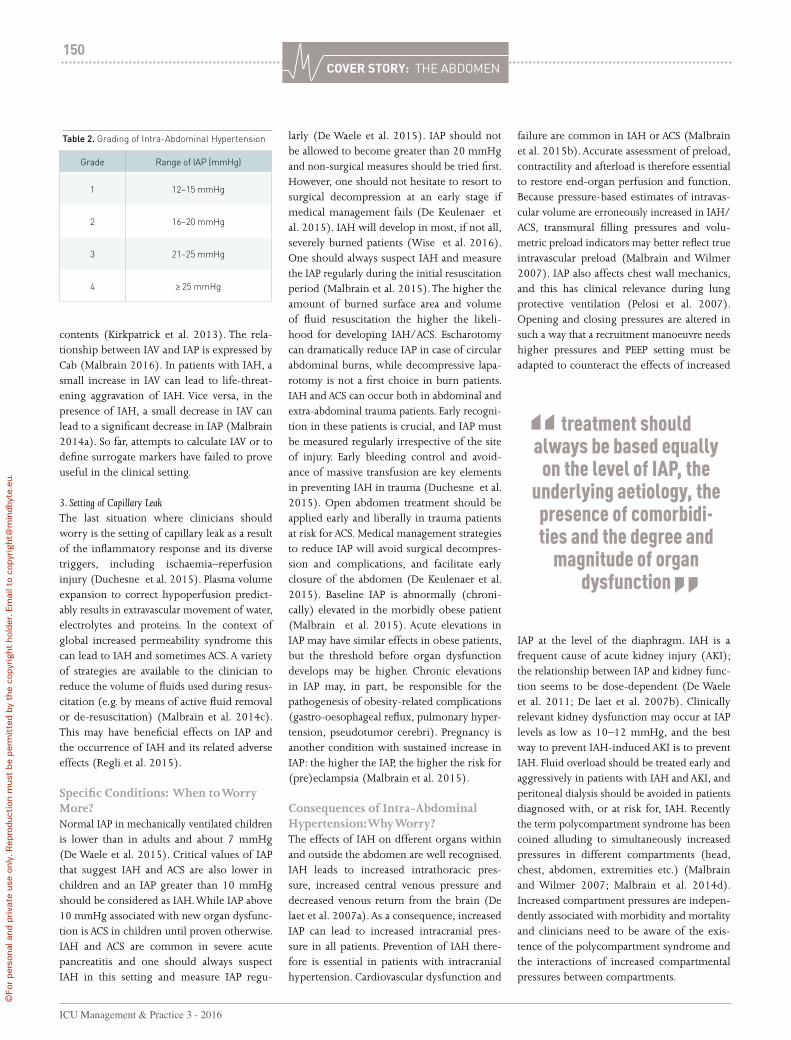
ICU Management & Practice 3 - 2016
150CoVeR StoRY: THE ABDOMEN
©Fo
r pe
rson
al a
nd p
riva
te u
se o
nly.
Rep
rodu
ctio
n m
ust
be p
erm
itte
d by
the
cop
yrig
ht h
olde
r. E
mai
l to
copy
right
@m
indb
yte.
eu.
contents (Kirkpatrick et al. 2013). The rela-tionship between IAV and IAP is expressed by Cab (Malbrain 2016). In patients with IAH, a small increase in IAV can lead to life-threat-ening aggravation of IAH. Vice versa, in the presence of IAH, a small decrease in IAV can lead to a significant decrease in IAP (Malbrain 2014a). So far, attempts to calculate IAV or to define surrogate markers have failed to prove useful in the clinical setting.
3. Setting of Capillary LeakThe last situation where clinicians should worry is the setting of capillary leak as a result of the inflammatory response and its diverse triggers, including ischaemia–reperfusion injury (Duchesne et al. 2015). Plasma volume expansion to correct hypoperfusion predict-ably results in extravascular movement of water, electrolytes and proteins. In the context of global increased permeability syndrome this can lead to IAH and sometimes ACS. A variety of strategies are available to the clinician to reduce the volume of fluids used during resus-citation (e.g. by means of active fluid removal or de-resuscitation) (Malbrain et al. 2014c). This may have beneficial effects on IAP and the occurrence of IAH and its related adverse effects (Regli et al. 2015).
Specific Conditions: When to Worry More?Normal IAP in mechanically ventilated children is lower than in adults and about 7 mmHg (De Waele et al. 2015). Critical values of IAP that suggest IAH and ACS are also lower in children and an IAP greater than 10 mmHg should be considered as IAH. While IAP above 10 mmHg associated with new organ dysfunc-tion is ACS in children until proven otherwise. IAH and ACS are common in severe acute pancreatitis and one should always suspect IAH in this setting and measure IAP regu-
larly (De Waele et al. 2015). IAP should not be allowed to become greater than 20 mmHg and non-surgical measures should be tried first. However, one should not hesitate to resort to surgical decompression at an early stage if medical management fails (De Keulenaer et al. 2015). IAH will develop in most, if not all, severely burned patients (Wise et al. 2016). One should always suspect IAH and measure the IAP regularly during the initial resuscitation period (Malbrain et al. 2015). The higher the amount of burned surface area and volume of fluid resuscitation the higher the likeli-hood for developing IAH/ACS. Escharotomy can dramatically reduce IAP in case of circular abdominal burns, while decompressive lapa-rotomy is not a first choice in burn patients. IAH and ACS can occur both in abdominal and extra-abdominal trauma patients. Early recogni-tion in these patients is crucial, and IAP must be measured regularly irrespective of the site of injury. Early bleeding control and avoid-ance of massive transfusion are key elements in preventing IAH in trauma (Duchesne et al. 2015). Open abdomen treatment should be applied early and liberally in trauma patients at risk for ACS. Medical management strategies to reduce IAP will avoid surgical decompres-sion and complications, and facilitate early closure of the abdomen (De Keulenaer et al. 2015). Baseline IAP is abnormally (chroni-cally) elevated in the morbidly obese patient (Malbrain et al. 2015). Acute elevations in IAP may have similar effects in obese patients, but the threshold before organ dysfunction develops may be higher. Chronic elevations in IAP may, in part, be responsible for the pathogenesis of obesity-related complications (gastro-oesophageal reflux, pulmonary hyper-tension, pseudotumor cerebri). Pregnancy is another condition with sustained increase in IAP: the higher the IAP, the higher the risk for (pre)eclampsia (Malbrain et al. 2015).
Consequences of Intra-Abdominal Hypertension: Why Worry?The effects of IAH on dfferent organs within and outside the abdomen are well recognised. IAH leads to increased intrathoracic pres-sure, increased central venous pressure and decreased venous return from the brain (De laet et al. 2007a). As a consequence, increased IAP can lead to increased intracranial pres-sure in all patients. Prevention of IAH there-fore is essential in patients with intracranial hypertension. Cardiovascular dysfunction and
failure are common in IAH or ACS (Malbrain et al. 2015b). Accurate assessment of preload, contractility and afterload is therefore essential to restore end-organ perfusion and function. Because pressure-based estimates of intravas-cular volume are erroneously increased in IAH/ACS, transmural filling pressures and volu-metric preload indicators may better reflect true intravascular preload (Malbrain and Wilmer 2007). IAP also affects chest wall mechanics, and this has clinical relevance during lung protective ventilation (Pelosi et al. 2007). Opening and closing pressures are altered in such a way that a recruitment manoeuvre needs higher pressures and PEEP setting must be adapted to counteract the effects of increased
IAP at the level of the diaphragm. IAH is a frequent cause of acute kidney injury (AKI); the relationship between IAP and kidney func-tion seems to be dose-dependent (De Waele et al. 2011; De laet et al. 2007b). Clinically relevant kidney dysfunction may occur at IAP levels as low as 10–12 mmHg, and the best way to prevent IAH-induced AKI is to prevent IAH. Fluid overload should be treated early and aggressively in patients with IAH and AKI, and peritoneal dialysis should be avoided in patients diagnosed with, or at risk for, IAH. Recently the term polycompartment syndrome has been coined alluding to simultaneously increased pressures in different compartments (head, chest, abdomen, extremities etc.) (Malbrain and Wilmer 2007; Malbrain et al. 2014d). Increased compartment pressures are indepen-dently associated with morbidity and mortality and clinicians need to be aware of the exis-tence of the polycompartment syndrome and the interactions of increased compartmental pressures between compartments.
treatment should always be based equally
on the level of IAP, the underlying aetiology, the
presence of comorbidi-ties and the degree and
magnitude of organ dysfunction
Grade Range of IAP (mmHg)
1 12–15 mmHg
2 16–20 mmHg
3 21–25 mmHg
4 ≥ 25 mmHg
Table 2. Grading of Intra-Abdominal Hypertension
151CoVeR StoRY: THE ABDOMEN
Management: How to Stop Worrying?Based on the underlying conditions that promote IAH and ACS medical management addresses four therapeutic targets: 1. Improving Cab2. Reducing IAV (either by removing free
abdominal or intraluminal fluid) 3. Correcting capillary leak and 4. Correcting fluid balance. It is beyond the scope of this article to give an extensive overview of the different medical management strategies as these can be found elsewhere (Regli et al. 2015; De Keulenaer et al. 2015). The bottom line is that treatment should always be based equally on the level of
IAP, the underlying aetiology, the presence of comorbidities and the degree and magnitude of organ dysfunction.
ConclusionsIn 2013 the World Society of the Abdominal Compartment Syndrome (WSACS) published evidence-based guidelines on the definitions, diagnosis and management of IAH and ACS (Kirkpatrick et al. 2013). However, bedside decisions regarding correct management in individual patients with IAH or ACS remain difficult. The clinician should be aware of the polycompartment syndrome and interactions between different compartmental pressures.
Cab is one of the most neglected parameters in critically ill patients, although it plays a key role in understanding organ-organ interactions and the deleterious effects of unadapted IAV on IAP and end-organ perfusion.
ReferencesCheatham ML, De Waele JJ, De Laet I et al. (2009) The impact of body position on intra-abdominal pressure measurement: a multi-center analysis. Crit Care Med, 37(7): 2187-90.
De Iaet I, Citerio G, Malbrain ML (2007a) The influence of intraabdominal hypertension on the central nervous system: current insights and clinical recommendations, is it all in the
head? Acta Clin Belg Suppl, 62(1): 89-97.
De laet I, Malbrain ML, Jadoul JL et al. (2007b) Renal implications of increased intra-abdom-inal pressure: are the kidneys the canary for abdominal hypertension? Acta Clin Belg Suppl, 62(1): 119-30.
De Waele JJ, De Laet I, De Keulenaer B et al. (2008) The effect of different reference trans-ducer positions on intra-abdominal pressure
measurement: a multicenter analysis. Intensive Care Med, 34(7): 1299-303.
De Waele JJ, De Laet I, Kirkpatrick AW et al. (2011) Intra-abdominal hypertension and abdominal compartment syndrome. Am J Kidney Dis, 57(1): 159-69.
De Waele JJ, Ejike JC, Leppaniemi A et al. (2015) Intra-abdominal hypertension and abdominal compartment syndrome in pancre-
atitis, paediatrics, and trauma. Anaesthesiol Intensive Ther, 47(3): 219-27.
De Waele JJ, De Laet I, Malbrain ML (2016) Understanding abdominal compartment syndrome. Intensive Care Med, 42(6): 1068-70.
For full references, please email editorial@icu management.org, visit icu-management.org or use the article QR code.
AbbreviationsACS abdominal compartment syndromeAKI acute kidney injuryCab abdominal compliance IAH intra-abdominal hypertension IAV intra-abdominal volumeICU intensive care unit
©Fo
r pe
rson
al a
nd p
riva
te u
se o
nly.
Rep
rodu
ctio
n m
ust
be p
erm
itte
d by
the
cop
yrig
ht h
olde
r. E
mai
l to
copy
right
@m
indb
yte.
eu.

Many patients presenting to the hospital emergency department do not need to stay overnight. Ambulatory emer-
gency care (AEC) may optimise identification and management of such patients by deliv-ering streamlined, efficient patient care within one working day. This may improve clinical outcomes, patient experience and lower costs.
At James Paget University Hospital, the acute medicine unit (AMU) was organised into two units. The Emergency Assessment and Discharge Unit (EADU) immediately reviewed and treated patients who were likely to need therapies and investigations as an inpatient. The Ambulatory Emergency Care (AEC) unit served patients with fewer needs. As the AEC service could not keep up with demand, the hospital management redesigned the patient management streams and established a new unit, the Ambulatory Care Unit (AmbU) where point-of-care testing (POCT) was a core component of the new service.
Process Redesign The project team included hospital staff, with industry partner support and sponsorship from Abbott Point-of-Care, Radiometer® and Operasee Limited.
The redesign process comprised (Figure 1):1. Demand and Process Activity Mapping to understand
how the current system worked and to iden-tify an evidence-based, ideal future state of the service, which could fulfil demand. This identified leverage points on which to focus, such as POCT, to catalyse change.
2. Failure Mode Effect Analysis (FMEA), a step-by-step approach to identifying all possible failures in a design, process, product or service. FMEA was used to get input from multiple stake-holders to identify, quantify, prioritise and resolve potential issues in terms of how severe they might be, how often they might occur and how they could be detected.To reach the goal of reduced length of
stay (LoS), the new service implemented the following:• POCT, including the i-STAT® System*;• Appropriate treatment/discharge planning;• Early senior clinical decision maker input;• Condition-specific management algo-
rithms.POCT fulfilled over 85% of patient diagnostic
needs in the AEC setting. The i-STAT® System was used for a variety of common tests, with the following cartridges used most widely:• CHEM8+ to test blood electrolytes, basic
haematology parameters and TCO2;
• CG4+ to test lactate and blood gases;• PT/INR to test prothrombin time.
In addition the CELL-DYN Emerald® analyser was used for full blood count testing and the Radiometer® AQT90 FLEX for D-dimer tests.
Improved LoSThe new AEC pathway started in August 2014 (Monday through Friday, 08:30–18:30). The three-month pilot led to dramatic improvements in patient flow and reductions in LoS (Figure 2). These improvements continued after the pilot and after the AmbU moved into a newly built space located within the AMU with additional staff to support the extended service (Figure 2).
Emergency care was improved as a result of the new outpatient AEC service, in both the EADU and the AmbU. The rate of patients going
home on the same day increased and the rate of patients admitted for 1, 2 or 3 days decreased, demonstrating that fewer patients required overnight stays. The reduced LoS was achieved despite the patient admission rate remaining constant (Figure 3).
Cost Benefit > £1 millionFollowing the pilot’s success, a business case was approved to provide 7-day AEC services, with operational hours extended from 08:30–18:30 to 08:00–21:30. It was calculated that the extended AEC services would save 15.87 Trust beds per day within the EADU (Figure 4).
Despite the decrease in overnight stays, which reduced Trust income, but represented a £557,146 saving for the Clinical Commissioning Group, and the cost of providing the service (increased staffing and POCT diagnostics, etc.), it was estimated that implementation of the service would result in a net Trust saving of £42,265 per year in the acute setting— through the safe removal of 10 unwarranted escalation beds. The remaining capacity (approximately 5.87 beds) could be used to improve elective service capacity. This equates to a total economic cost benefit of £1,176,751 per annum (Figure 5).
Benefits for Patients, StaffAdditional POCT benefits experienced at this site included:• Patient data integration within the
hospital information system;• Senior clinical decision maker review
within one hour;• Static readmission rates;• Positive patient feedback with 79%
“extremely likely” to recommend the service;
• Under 10 minutes to initial review.
PoInt oF VIeW152
RedeSIGnInG ambULatoRY emeRGenCY CaRe WIth PoCt teStInG REdUCEd COSTS ANd LENGTH OF STAy
Figure 1: POCT was identified as a main way to leverage change in the AEC* For intended use information please see CTI sheets at abbottpointofcare.com
ICU Management & Practice 3 - 2016
Phil Weihser Service Manager AmbU Project [email protected]
Josip Stosic Consultant Acute/Critical Care Medicine, AmbU Clinical [email protected]
dominic GilesAcute Medicine Specialist Trainee
James Paget University HospitalGreat Yarmouth, UK
©Fo
r pe
rson
al a
nd p
riva
te u
se o
nly.
Rep
rodu
ctio
n m
ust
be p
erm
itte
d by
the
cop
yrig
ht h
olde
r. E
mai
l to
copy
right
@m
indb
yte.
eu.
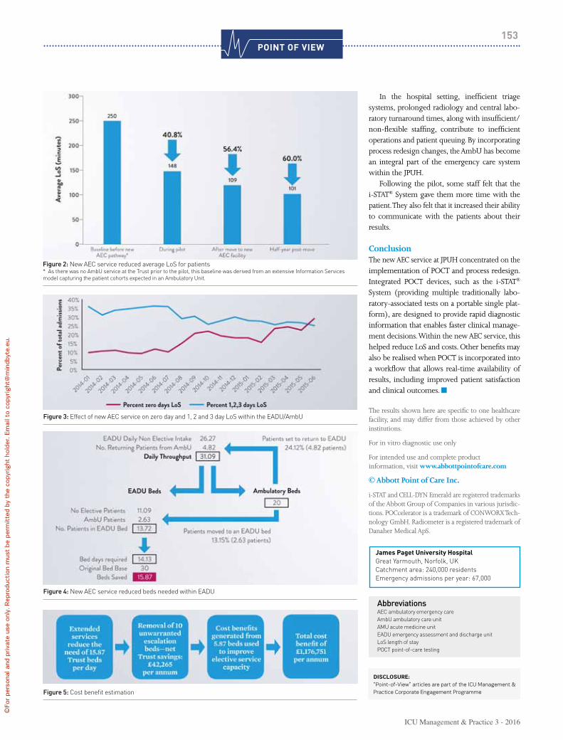
In the hospital setting, inefficient triage systems, prolonged radiology and central labo-ratory turnaround times, along with insufficient/non-flexible staffing, contribute to inefficient operations and patient queuing. By incorporating process redesign changes, the AmbU has become an integral part of the emergency care system within the JPUH.
Following the pilot, some staff felt that the i-STAT® System gave them more time with the patient. They also felt that it increased their ability to communicate with the patients about their results.
ConclusionThe new AEC service at JPUH concentrated on the implementation of POCT and process redesign. Integrated POCT devices, such as the i-STAT® System (providing multiple traditionally labo-ratory-associated tests on a portable single plat-form), are designed to provide rapid diagnostic information that enables faster clinical manage-ment decisions. Within the new AEC service, this helped reduce LoS and costs. Other benefits may also be realised when POCT is incorporated into a workflow that allows real-time availability of results, including improved patient satisfaction and clinical outcomes.
The results shown here are specific to one healthcare facility, and may differ from those achieved by other institutions.
For in vitro diagnostic use only
For intended use and complete productinformation, visit www.abbottpointofcare.com
© Abbott Point of Care Inc.
i-STAT and CELL-DYN Emerald are registered trademarks of the Abbott Group of Companies in various jurisdic-tions. POCcelerator is a trademark of CONWORX Tech-nology GmbH. Radiometer is a registered trademark of Danaher Medical ApS.
James Paget University hospitalGreat Yarmouth, Norfolk, UKCatchment area: 240,000 residentsEmergency admissions per year: 67,000
PoInt oF VIeW153
AbbreviationsAEC ambulatory emergency careAmbU ambulatory care unitAMU acute medicine unitEADU emergency assessment and discharge unitLoS length of stayPOCT point-of-care testing
Figure 2: New AEC service reduced average LoS for patients* As there was no AmbU service at the Trust prior to the pilot, this baseline was derived from an extensive Information Services model capturing the patient cohorts expected in an Ambulatory Unit.
Figure 3: Effect of new AEC service on zero day and 1, 2 and 3 day LoS within the EADU/AmbU
Figure 4: New AEC service reduced beds needed within EADU
Figure 5: Cost benefit estimation
Disclosure:“Point-of-View” articles are part of the ICU Management & Practice Corporate Engagement Programme
ICU Management & Practice 3 - 2016
©Fo
r pe
rson
al a
nd p
riva
te u
se o
nly.
Rep
rodu
ctio
n m
ust
be p
erm
itte
d by
the
cop
yrig
ht h
olde
r. E
mai
l to
copy
right
@m
indb
yte.
eu.
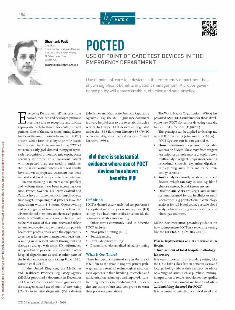
matRIX154
ICU Management & Practice 3 - 2016
©Fo
r pe
rson
al a
nd p
riva
te u
se o
nly.
Rep
rodu
ctio
n m
ust
be p
erm
itte
d by
the
cop
yrig
ht h
olde
r. E
mai
l to
copy
right
@m
indb
yte.
eu.
pOCTedUSE OF POINT OF CARE TEST dEVICES IN THE EMERGENCy dEPARTMENT
Shashank PatilConsultantDepartment of Emergency MedicineChelsea & Westminster Hospital NHS Foundation Trust,London, UK
Use of point-of-care test devices in the emergency department has shown significant benefits in patient management. A proper gover-nance policy will ensure credible, effective and safe practice.
Emergency Department (ED) practices have evolved, modified and developed pathways over the years to recognise and initiate
appropriate early treatment for acutely unwell patients. One of the major contributing factors has been the use of point-of-care test (POCT) devices, which have the ability to provide drastic improvement in the turnaround time (TAT) of test results. Early goal-directed therapy in sepsis, early recognition of neutropenic sepsis, acute coronary syndrome, an unconscious patient with suspected drug use needing antidotes: the list is exhaustive where early test results have shown appropriate treatment has been initiated and has directly affected the outcome.
ED overcrowding is an international problem and waiting times have been increasing over time. France, Sweden, UK, New Zealand and Canada have all passed explicit length-of-stay time targets, requiring that patients leave the department within 4-8 hours. Overcrowding and prolonged wait times have been linked to adverse clinical outcomes and decreased patient satisfaction. While no one factor can be identified as the root cause of this issue, decreased delays in sample collection and test results can provide healthcare professionals with the opportunity to arrive at faster care management decisions, resulting in increased patient throughput and decreased average wait times. ED performance is dependent on processes and capacity in other hospital departments as well as other parts of the health and care system (Kings Fund 2016; Larsson et al 2015).
In the United Kingdom, the Medicines and Healthcare Products Regulatory Agency (MHRA) published a document in December 2013, which provides advice and guidance on the management and use of point-of-care testing (POCT) in in vitro diagnostic (IVD) devices
(Medicines and Healthcare Products Regulatory Agency 2013). The MHRA guidance document is a very helpful text to use to establish such a service. In Europe POCT devices are regulated under the 1998 European Directive 98/79/EC on in vitro diagnostic medical devices (Council Directive 1998).
DefinitionPOCT is defined as any analytical test performed for a patient in primary or secondary care (ED) settings by a healthcare professional outside the conventional laboratory setting.
Other terms commonly used to describe POCT include:• Near patient testing (NPT)• Bedside testing• Extra-laboratory testing• Disseminated/decentralised laboratory testing
What is Out There?There has been a continual rise in the use of POCT due to the drive to improve patient path-ways and as a result of technological advances. Developments in fluid handling, microchip and miniaturisation technology and improved manu-facturing processes are producing POCT devices that are more robust and less prone to error than previous generations.
The World Health Organization (WHO) has provided ASSURED guidelines for those devel-oping new POCT devices for detecting sexually transmitted infections (Figure 1).
This principle can be applied to develop any new POCT device (St John and Price 2014).
POCT Systems can be categorised as: • Non-instrumental systems: disposable
systems or devices. These vary from reagent test strips for a single analyte to sophisticated multi-analyte reagent strips incorporating procedural controls, e.g. urine dipsticks, urinary pregnancy tests and urine toxi-cology screens.
• Small analysers, usually hand- or palm-held devices, which can vary in size, e.g. blood glucose meters, blood ketone meters.
• Desktop analysers are larger and include systems designed for use in clinics or small laboratories, e.g point-of-care haematology analyser for full blood count, portable blood analyser for measuring urea, creatinine, and blood gas analysers.
MHRA documentation provides guidance on how to implement POCT in a secondary setting like the ED (Table 1) (MHRA 2013).
Prior to Implementation of a POCT Service in the Hospital1.Involvement of local hospital pathology laboratoryIt is very important in a secondary setting like the ED to have a close liaison between users and local pathology labs as they can provide advice on a range of issues such as purchase, training, interpretation of results, troubleshooting, quality control, quality assessment and health and safety.2. Identifying the need for POCTIt is essential to establish a clinical need and
there is substantial evidence where use of POCT
devices has shown benefits
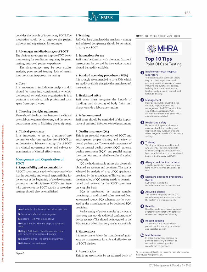
matRIX155
ICU Management & Practice 3 - 2016
©Fo
r pe
rson
al a
nd p
riva
te u
se o
nly.
Rep
rodu
ctio
n m
ust
be p
erm
itte
d by
the
cop
yrig
ht h
olde
r. E
mai
l to
copy
right
@m
indb
yte.
eu.
consider the benefit of introducing POCT. The motivation could be to improve the patient pathway and experience, for example.
3. Advantages and disadvantages of POCTThe obvious advantages are improved TAT, better monitoring for conditions requiring frequent testing, improved patient experience.
The disadvantages may be poor quality analysis, poor record keeping, lack of results interpretation, inappropriate testing.
4. CostsIt is important to include cost analysis and it should be taken into consideration whether the hospital or healthcare organisation is in a position to include variable professional costs apart from capital costs.
5. Choosing the right equipmentThere should be discussion between the clinical users, laboratory, manufacturers, and the estates department prior to finalising the equipment.
6. Clinical governanceIt is important to set up a point-of-care committee who can regulate use of POCT as an alternative to laboratory testing. Use of POCT is a clinical governance issue and subject to examination of clinical effectiveness.
Management and Organisation of POCT1. Responsibility and accountabilityA POCT coordinator needs to be appointed who has the authority and overall responsibility for the service at the beginning of the development process. A multidisciplinary POCT committee who can oversee the POCT activity in secondary settings should also be established.
2. TrainingStaff who have completed the mandatory training and achieved competency should be permitted to carry out POCT.
3. Instructions for useStaff must be familiar with the manufacturer’s instructions for use and the instruction manual should be readily available.
4. Standard operating procedures (SOPs)It is strongly recommended to have SOPs which are readily available alongside the manufacturer’s instructions.
5. Health and safetyStaff users must recognise the hazards of handling and disposing of body fluids and sharps outside a laboratory setting.
6. Infection controlStaff users should be reminded of the impor-tance of universal infection control precautions.
7. Quality assurance (QA)This is an essential component of POCT and encompasses proper training and review of overall performance. The essential components of QA are internal quality control (IQC), external quality assessment (EQA), and parallel testing; they can help ensure reliable results if applied rigorously.
IQC methods primarily ensure that the results obtained are accurate and consistent. This can be achieved by analysis of a set of QC specimens provided by the manufacturer. This can reassure the user. A log of QC activity needs to be main-tained and reviewed by the POCT committee on a regular basis.
EQA is performed by testing samples containing an undisclosed value received from an external source. EQA schemes may be oper-ated by the manufacturer or by dedicated EQA providers.
Parallel testing of patient samples by the central laboratory can provide additional confirmation of device accuracy. This should be integrated in the EQA practice when laboratory results are available.
8. MaintenanceIt is important to follow the manufacturer’s guid-ance on maintenance for safe and effective use of POCT devices.
9. AccreditationThis is an assessment by an external body of
Top 10 TipsPoint Of Care Testing
Involve your local hospital laboratoryYour local hospital pathology labora-tory can play a supportive role in providing advice on a range of issues including the purchase of devices, training, interpretation of results, troubleshooting, quality control, and health and safety.
ManagementMany people will be involved in the creation, implementation andmanagement of a POCT service. It is vital that an appropriate POCT co-ordinator is identified and a POCT committee established.
Health and safetyBe aware of the potential hazards associated with the handling and disposal of body fluids, sharps and waste reagents outside of a laboratory setting.
TrainingTraining must be provided for staff who use POCT devices. Only staff whose training and competence has been established and recorded should be permitted to carry out POCT.
Always read the instructions…and be particularly aware of situa-tions when the device should not be used.
Standard operating procedures (SOPs)SOPs must include the manufacturer’s instructions for use.
Assuring qualityThe analysis of quality control (QC) material can provide assurance that the system is working correctly.
ResultsResults should be reviewed by appro-priately qualified staff with particular reference to the patient’s history.
Record keeping…is essential and must include patient results, test strip lot number and operator identity.
MaintenanceIn order that devices continue to perform accurately they must be maintained according to the manufacturer’s guidance.
1
2
3
4
5
6
7
8
9
10
Figure 1.
A: Affordable - for those at the risk of infection
S: Sensitive - Minimal false negative
S: Specific - Minimal false positive
U: User friendly - Minimal steps to carry out tests
R: Rapid & Robust - Short turnaround time and no need for refrigerated storage
E: Equipment free - no complex equipment
D: Delivered - to end users
© Medicines and Healthcare Products Regulatory Agency. Reproduced with permission.
table 1. Top 10 Tips: Point of Care Testing
matRIX156
ICU Management & Practice 3 - 2016
©Fo
r pe
rson
al a
nd p
riva
te u
se o
nly.
Rep
rodu
ctio
n m
ust
be p
erm
itte
d by
the
cop
yrig
ht h
olde
r. E
mai
l to
copy
right
@m
indb
yte.
eu.
the competence to provide a service to a recog-nised standard. Independent confirmation helps provide reassurance to the users of this service.
10. Record keepingIt is important to keep accurate records of patient results from POCT devices.
11. Information technologySupport from IT systems to manage data, work-stations and laboratory information is crucial for successful use of POCT devices.
12. Adverse incident reportingThe MHRA has two parallel reporting systems for device-related incidents, one for manufac-turers and the other for users.
Clinical Scenarios for Use of POCT in ED Rooney and Schilling (2014) outline several examples where use of POCT has led to improved patient outcomes.
Acute coronary syndromeMany patients present to the ED with symptoms that suggest acute coronary syndrome (ACS). A rapid rule-out protocol using a combination of high-sensitivity cardiac Troponin I testing, risk score and electrocardiogram has been shown to be safe and effective in identifying low-risk patients (Than M et al. 2012). A randomised controlled trial that evaluated the performance of POCT for cardiac biomarkers on patients with suspected myocardial infarction showed a 20% greater discharge rate during the initial evaluation process when POCT was performed (Goodacre et al. 2011). The follow-up study that evaluated the financial implications of POCT found variable results (Bradburn et al. 2012).
Venous thromboembolic diseasePatients who have low risk but are suspected to have a venous thromboembolic disease have a D-Dimer test to rule out the disease. Use of POCT testing has shown a reduction in ED stay by 60 minutes and a 14% reduction in admis-sions (Lee-Lewandrowski et al. 2009).
Severe sepsisElevated blood lactate levels have been shown to be a sensitive marker of impaired tissue perfu-sion and of anaerobic metabolism in patients with suspected sepsis. This is a valid identifica-tion method for patients who will benefit from early aggressive goal-directed therapy.
StrokeEarly thrombolysis in patients presenting with ischaemic stroke has shown significant benefits. It is very important to institute this treatment as soon as possible. Apart from performing an immediate CT scan it is important to know the coagulation status, as there is an increased risk of bleeding if they are deranged, which can cause a potentially serious outcome. A POCT coagula-tion test not only avoids unnecessary delay of a time-critical intervention like thrombolysis, but also ensures that the patient is safe to receive it.
Recognising substance abuse in an uncon-scious patient, metabolic emergencies, ectopic pregnancy—the list goes on of where POCT tests have shown benefits by reducing the diagnosis time and enabling initiation of treatment at the earliest time.
ConclusionThe number of tests that can be performed outside a conventional laboratory, especially in
the ED, has grown significantly over the past two decades. This is primarily driven by the idea that the care provided is brought closer to the patient. There is substantial evidence of where use of POCT devices has shown benefits in recognising the ill patient, helped establish immediate treatment thereby reducing the waiting for diagnosis and also improved the patient journey time from ED to acute medical wards. All this will eventually accumulate into a good patient experience. Patient satisfaction can help gather political support to invest in devel-opment and implementation of this technology.
However, it is of utmost importance that any department or hospital that plans to invest in a POCT device has a strict governance policy.
Conflict of interestShashank Patil declares that he has no conflict of interest.
AbbreviationsED emergency departmentPOCT point of care testingMHRA Medicines and Healthcare products Regulatory AgencyPOCT point-of-care testQA quality assuranceQC quality controlTAT turnaround time
References Bradburn M, Goodacre SW, Fitzgerald P et al. (2012) Interhospital variation in the RATPAC trial (Randomised Assessment of Treatment using Panel Assay of Cardiac markers). Emerg Med J, 29(3): 233-8.
Council Directive (EC) 1998/79/EC of the European Parliament and of the Council of 27 October 1998 on in vitro diagnostic medical devices. [Accessed: 26 August 2016] Avail-able from eur-lex.europa.eu/legal-content/EN/TXT/?uri=CELEX:31998L0079
Goodacre S, Bradburn M, Fitzgerald P et al. (2011) The RATPAC (Randomised Assessment of Treatment using Panel Assay of Cardiac markers) trial: a randomised controlled trial of point-of-care cardiac markers in the emer-gency department. Health Technol Assess, 15(23): iii-xi, 1-102.
King’s Fund (2016) What's going on in A&E? The key questions answered. [Accessed: 26 August 2016] Available from kingsfund.org.uk/projects/urgent-emergency-care/urgent-and-emergency-care-mythbusters
Larsson A, Greig-Pylypczuk R, Huisman A (2015) The state of point-of-care testing: a European perspective. Ups J Med Sci, 120(1): 1–10.
Lee-Lewandrowski E, Nichols J, Van Cott E et al. (2009) Implementation of a rapid whole blood D-dimer test in the emergency depart-ment of an urban academic medical center: impact on ED length of stay and ancillary test utilization. Am J Clin Pathol, 132(3): 326-31.
Medicines and Healthcare Products regula-tory Agency (2013) Management and use of
IVD point of care test devices. MHRA V1.1. [Accessed: 26 August 2016] Available from gov.uk/government/uploads/system/uploads/attachment_data/file/371800/In_vitro_diag-nostic_point-of-care_test_devices.pdf
St John A, Price CP (2014) Existing and emerging technologies for point-of-care testing. Clin Biochem Rev 35 (3) 2014 155-167.
Rooney KD, Schilling U. (2014) Point-of-care testing in the overcrowded emergency department-can it make a difference? Crit Care. 2014; 18(6): 692.

Critical Results Quicker
• Faster & more frequent BGA analysis
• No blood loss through sampling
• Nurse does not need to leave the bedside
• Results on screen at the bedside
• Closed system: minimises infection risk to staff and patients
[email protected] Medical Limited, Harston Mill,
Harston, Cambridge, CB22 7GG
www.spheremedical.com
CE Mark pending
Proxima
©Fo
r pe
rson
al a
nd p
riva
te u
se o
nly.
Rep
rodu
ctio
n m
ust
be p
erm
itte
d by
the
cop
yrig
ht h
olde
r. E
mai
l to
copy
right
@m
indb
yte.
eu.
matRIX158
ICU Management & Practice 3 - 2016
©Fo
r pe
rson
al a
nd p
riva
te u
se o
nly.
Rep
rodu
ctio
n m
ust
be p
erm
itte
d by
the
cop
yrig
ht h
olde
r. E
mai
l to
copy
right
@m
indb
yte.
eu.
Candida spp. in The respiraTOry TraCT A REAL CAUSALITy WITH WORSE OUTCOMES OR JUST A MARKER OF SEVERITy?
Silvia terraneoPhysicianRespiratory UnitOspedale San Paolo Department of Health ScienceUniversità degli Studi di Milano,Milan, Italy
miquel FerrerPhysicianDepartment of PneumologyRespiratory InstituteHospital Clinic, IDIBAPSUniversity of BarcelonaBarcelona, SpainCibeRes-ISCiii ( CB06/06/0028)
antoni torresProfessor of MedicineHead, Respiratory Intensive Care UnitDepartment of PneumologyRespiratory InstituteHospital Clinic, IDIBAPSUniversity of BarcelonaBarcelona, Spain.CibeRes-ISCiii ( CB06/06/0028)
• Candida spp. is the most common cause of intensive care unit (ICU) invasive fungal infections worldwide.
• The isolation of Candida spp. from respiratory tract secretions of non-immunocompromised, mechanically ventilated patients varies between 20% and 55%, but it might represent colonisation rather than infection.
• Candida spp. colonisation promotes bacterial pneumonia in animal models.
• Candida spp. colonisation could clinically increase the risk for Pseu-domonas aeruginosa ventilator-associated pneumonia, prolong mechanical ventilation and stay and worsen outcomes, but to date contrasting data are available.
• Available evidence is not sufficient to support routine antifungal therapy in non-immunocompromised patients.
Candida spp. is part of the normal skin, oropharyngeal, mucosal membranes and upper respiratory tract flora. Candida spp.
can reach the lungs through either haematog-enous dissemination or aspiration of colonised oropharyngeal or gastric contents (Muray et al. 1977). The isolation of Candida spp. from respiratory tract secretions is frequent in non-immunocompromised, mechanically ventilated patients. Several studies have reported the pres-ence of Candida spp. in the sputum of 20-55% of patients receiving antibiotics (Azoulay et al. 2006; Delisle et al. 2008). Candida spp. is the most common cause of invasive fungal infec-tions, with an incidence estimated at 72.8 cases per million inhabitants per year (Guinea 2014). The five main species of Candida spp (C. albicans, C. parapsilosis, C. glabrata, C. tropicalis and C. krusei) are responsible for more than 90% of invasive fungal infections, in both intensive care unit (ICU) and non-ICU patients (Maubon et al. 2014). Candida pneumonia is a rare lung infection with a high morbidity and mortality, commonly observed
as part of a disseminated Candida infection and associated with predisposing clinical circum-stances (i.e. long-term antibiotic use, haemato-logic malignancy or severe immunosuppressive states). The majority of Candida pneumonia cases are secondary to haematological dissemination of Candida spp. (Masur and Rosen 1977). There is no specific clinical or radiological presenta-tion of Candida pneumonia. This aspect of the disease makes the diagnosis difficult to perform. A definitive diagnosis of candida pneumonia is now based on histopathological identification of yeast parenchymal invasion with associated inflammation.
Significance of Candida Spp. Isolation in Non-Immunosuppressed PatientsInvasive lung infection by Candida spp. is a rare event in non-immunocompromised subjects. Several studies showed that the recovery of Candida spp. from sputum and other respiratory tract secretions cultures or lung tissue in non-immunocompromised patients might represent
colonisation of the tracheobronchial tree rather than infection.
El-Elbiary et al. (1997) performed an autopsy study on 25 immunocompetent, mechanically ventilated patients, who died in a medical ICU, in order to assess the real significance of Candida spp. presence in the tracheobronchial tree or lungs. Immediate postmortem respiratory samples and lung tissue specimens were microbiologically and histologically examined. The incidence of Candida spp. isolation from pulmonary biopsies was 40%, while the incidence of Candida pneu-monia was only 8%. The presence of Candida spp. in pulmonary biopsies was always associated with the isolation of the same microorganism from one of another respiratory sample. Furthermore there was a uniform presence of Candida spp. throughout the different lung regions, but the fungal isolation, independently of quantitative cultures, was not recognised as a good marker of Candida pneumonia (el-Ebiary et al. 1997).
In 2009 Meersseman et al. performed a similar study. Data from autopsies of patients, who
matRIX159
ICU Management & Practice 3 - 2016
©Fo
r pe
rson
al a
nd p
riva
te u
se o
nly.
Rep
rodu
ctio
n m
ust
be p
erm
itte
d by
the
cop
yrig
ht h
olde
r. E
mai
l to
copy
right
@m
indb
yte.
eu.
died in a medical ICU and with evidence of pneumonia, were analysed in order to define the value of Candida spp. isolation in airway samples of those patients. Histopathological evidence of pneumonia was found in 58% of patients. Of these, 57% had positive tracheobronchial samples for Candida spp. performed during the preceding two weeks. No cases of candida pneu-monia were identified amongst those cases or in patients without Candida isolation. These results confirmed that the presence of Candida spp. in respiratory samples does not indicate pneumonia and that this is an extremely rare event in ICU patients (Meersseman et al. 2009).
Candida Spp. Colonisation as Risk Factor for P. Aeruginosa Ventilator-Associated Pneumonia OR Multi-Drug Resistant BacteriaAlthough the diagnosis of isolated Candida pneu-monia is rare, the presence of Candida spp. on pathological samples should not be clinically ignored. P. aeruginosa and Candida spp. are among the most prevalent organisms in ICU-acquired infections (Vincent et al. 1995), and they could coexist in the endotracheal tube or medical devices biofilm of patients (Adair et al. 1999). These two pathogens have physical, chemical, environmental and phylogenetic similarities (Ader et al. 2008; Hogan and Kolter 2002). The question of how they interplay in the respiratory tract has been investigated, with contrasting results, in animal studies.
Ader et al. (2011) showed that P. aeruginosa lung injury was reduced in the presence of C. albicans in a mouse model, as well as the amount of alive P. aeruginosa recovered in lungs. Antifungal treatment with caspofungin removed this effect in those cases. However, mortality rate and bacterial dissemination did not vary between colonised and not colonised animals (Ader et al. 2011).
Conversely, in 2009 Roux et al. performed a randomised controlled animal study with the aim of determining the effect of C. albicans pres-ence on P. aeruginosa pneumonia. P. aeruginosa was instilled in the tracheobronchial tree of animals with or without previous C. albicans tracheo-bronchial colonisation. Animals with C. albicans tracheobronchial colonisation developed more frequently P. aeruginosa pneumonia compared with those without. In addition, higher levels of pro-inflammatory cytokines (TNFα, IFγ, IL-6) were measured in the lungs of animals instilled with P. aeruginosa with previous C. albicans colonisa-tion, compared with those without C. albicans colonisation (Roux et al. 2009).
In addition a preliminary report showed that C. albicans colonisation favours the occurrence of pneumonia related to S. aureus and E. coli (Roux et al 2009). Similarly, a recent study suggests that fungal colonisation also facilitated the develop-ment of Acinetobacter baumanii pneumonia in a rat model, with a protective role of antifungal therapy on this event (Tan et al. 2016). Thus the mechanism by which Candida spp. colonisa-tion promotes bacterial pneumonia could be independent of bacterial species.
ICU-acquired pneumonia (ICUAP) is the leading infection in critically ill patients, accounting for prolonged mechanical venti-lation and length of stay, poor outcome and excess costs. There is evidence of interactions between Candida spp. and P. aeruginosa, with fungal colonisation possibly increasing the risk for P. aeruginosa infection. Some clinical reports have shown a possible association between the pres-ence of Candida spp. in respiratory secretions and an increased risk for P. aeruginosa ventilator-associated pneumonia (VAP), longer mechanical ventilation, prolonged stay and worse outcomes.
In a cohort of immunocompetent mechani-cally ventilated patients, Azoulay et al. (2006) found the isolation of Candida spp. in the tracheo-bronchial tree as an independent risk factor for pneumonia, due to P. aeruginosa. Candida spp. colonisation was not associated with higher mortality, but colonised patients showed a significantly longer time on ventilation, and longer ICU and hospital stays compared to patients without Candida spp. isolation from the respiratory tract.
Candida spp. has been identified as a risk factor for multidrug-resistant bacteria. Hamet et al. (2012) conducted a prospective observational study in order to investigate the significance of Candida spp. airway colonisation in patients with suspected VAP and the potential link with isolation of multidrug-resistant (MDR) bacteria. Fifty-six percent of patients with suspected VAP had Candida spp. airway colonisation. Candida spp. airway colonisation was an independent
risk factor for MDR bacteria isolation without significant differences in aetiological pathogens. Colonised patients were similar to non-colonised patients regarding VAP severity; however, in this study mortality rate was greater in patients with fungal airway colonisation than in those without (Hamet et al. 2012).
In a retrospective analysis of the Canadian VAP study, Delisle et al. (2008) found Candida spp. isolation in respiratory samples in 17.8% of all patients. Colonised patients showed longer hospital stay than non-colonised patients and a significant increase in hospital mortality. In that population Candida spp. presence was inde-pendently associated with hospital mortality. Antibiotic administration, co-morbidities and a more severe illness are probable factors associ-ated to Candida spp. isolation (Delisle et al. 2008; Terraneo et al. 2016).
In 2015 we performed a prospective non-interventional study in a medical and surgical ICU of a teaching hospital. The purpose of this study was to compare the characteristics, micro-biology, inflammatory response and outcomes of patients diagnosed with ICUAP (mechanically ventilated or not), with and without Candida spp. presence in lower respiratory tract samples, and to assess the characteristics and outcomes associ-ated with the antifungal therapy. We conducted the study in view of the discrepancy between the uncertain clinical relevance of the isolation of Candida spp. in respiratory tract secretions and its association with adverse clinical outcomes in patients with VAP.
Candida spp.-colonised patients showed higher severity scores than patients without airways fungal colonisation, but similar inflammatory pattern. Clinical outcomes were similar between colonised and non-colonised patients, including 28-day and 90-day mortality, with the exception of an increased risk of intubation in patients with Candida sp. colonisation (Terraneo et al. 2016).
Antifungal TreatmentAlthough Candida spp. is frequently isolated from respiratory tract samples, antifungal treatment is not routinely recommended, because pneumonia caused by this fungal species is exceptional in non-neutropenic patients (Garnacho-Montero et al. 2013). Inappropriate use of antifungal treat-ment could be associated with higher rates of fungal resistance and mortality in ICU patients; therefore, Candida spp. isolation from respira-tory secretions alone should not be promptly treated (Cuenca-Estrella 2012; Rello et al. 1998). Nevertheless antifungal therapy is frequently
further studies are required to understand the real impact of Candida spp.
on respiratory infection development and patients’
outcomes
matRIX160
ICU Management & Practice 3 - 2016
©Fo
r pe
rson
al a
nd p
riva
te u
se o
nly.
Rep
rodu
ctio
n m
ust
be p
erm
itte
d by
the
cop
yrig
ht h
olde
r. E
mai
l to
copy
right
@m
indb
yte.
eu.
prescribed for immunocompetent mechanically ventilated patients with isolation of Candida spp. from respiratory tract samples (Azoulay et al. 2004; van der Geest et al. 2014). The effect of antifungal therapy in patients with Candida spp. airways colonisation has been extensively studied with discordant results.
A retrospective case-control study conducted by Nseir et al. (2007) showed that the prescrip-tion and length of the antifungal treatment were associated with a reduced risk for P. aeruginosa VAP development or tracheobronchial isolation in mechanically ventilated patients colonised by Candida spp.
Wood et al. (2006) performed a retrospective study in trauma ICU patients. Candida spp. was isolated from 8% of diagnostic bronchoalveolar lavages (BALs). Most of the isolations were consid-ered colonisation and no specific therapy was prescribed. No patients developed candidaemia or serious fungal infections after isolation of Candida spp., despite the lack of antifungal therapy. Furthermore, Candida spp. was not isolated in subsequent follow-up BALs. No significantly greater mortality rate was observed in patients with a high level of Candida spp. in BAL, despite the lack of therapy (Wood et al. 2006).
In 2014 van der Geest et al. (2014) performed a retrospective analysis of non-neutropenic mechanically ventilated patients with positive Candida spp. cultures of the respiratory tract treated or not with amphotericin-B deoxy-chlorate inhalation therapy in the context of
selective decontamination of the digestive tract. Treated patients did not decolonise more rapidly as compared to untreated patients. The duration of mechanical ventilation was increased by treat-ment independently of Candida spp. presence, suggesting a direct toxicity of the drug in the lung. No differences in VAP development or overall mortality were observed in this study (van der Geest et al. 2014).
In 2014 Albert et al. performed a double-blind, placebo-controlled, multicentric, pilot randomised clinical trial in order to evaluate inflammatory profiles and clinical outcomes of patients with suspected VAP and Candida spp. presence, treated or not with antifungal therapy. The isolation of Candida spp. was associated with persistent inflam-mation and immunosuppression, but markers of inflammation and all clinical outcomes had similar results between patients treated and not treated with antifungal therapy, both at baseline and over time (Albert et al. 2014).
In our study we observed a more frequent prescription of antifungal therapy in patients with evidence of Candida spp. in respiratory tract samples or patients with multiple co-morbidities or a more severe illness. However, in our group of patients, antifungal therapy was not associated with different outcomes in patients with Candida spp. in respiratory samples (Terraneo et al. 2016).
ConclusionDespite the frequent isolation of Candida spp. from respiratory specimen of ICU patients, the
development of real candida pneumonia is very unlikely when immunocompetent subjects are considered. However, the presence of Candida spp. in pathological samples should not be clinically ignored because it could probably be associ-ated with a more severe illness. What remains unsolved is the question about a real causality between Candida spp. and worse outcomes, since Candida spp. could be simply a marker of severity. As of today, available evidence is not sufficient to support routine antifungal therapy in these patients. In addition, further studies are required to understand the real impact of Candida spp. on respiratory infection development and patients’ outcomes and consequently the possible protec-tive role of antifungal agents’ administration.
AcknowledgementsSupport statement: 2009-SGR-911, IDIBAPS, ICREA academia 2013.
Conflict of InterestSilvia Terraneo, Miquel Ferrer and Antoni Torres declare no conflict of interest.
ReferencesAdair CG, Gorman SP, Feron BM et al. (1999) Implications of endotracheal tube biofilm for ventilator-associated pneumonia. Intensive Care Med, 25(10): 1072–6.
Ader F, Faure K, Guery B et al. (2008) [Pseudo-monas aeruginosa and Candida albicans interac-tion in the respiratory tract: from pathophysiology to a therapeutic perspective]. Pathol Biol (Paris), 56(3): 164–9.
Ader F, Jawhara S, Nseir S et al. (2011) Short term Candida albicans colonization reduces Pseudomonas aeruginosa-related lung injury and bacterial burden in a murine model. Crit Care, 15(3): R150.
Albert M, Williamson D, Muscedere J et al. (2014) Candida in the respiratory tract secretions of critically ill patients and the impact of antifungal treatment: a randomized placebo controlled pilot trial (CANTREAT study). Intensive Care Med, 40(9): 1313–22.
Azoulay E, Timsit JF, Tafflet M et al. (2006) Candida colonization of the respiratory tract and subsequent pseudomonas ventilator-associated pneumonia. Chest, 129(1): 110–7.
Azoulay E, Cohen Y, Zahar JR et al. (2004) Practices in non-neutropenic ICU patients with Candida-positive airway specimens. Intensive
Care Med, 30(7): 1384–9.
Cuenca-Estrella M, Verweij PE, Arendrup MC et al. (2012) ESCMID* guideline for the diagnosis and management of Candida diseases 2012: diagnostic procedures. Clin Microbiol Infect, 18 Suppl 7: 9-18.
Delisle MS, Williamson DR, Perreault MM et al. (2008). The clinical significance of Candida colonization of respiratory tract secretions in critically ill patients. J Crit Care, 23(1): 11–7.
el-Ebiary M, Torres A, Fàbregas N et al. (1997) Significance of the isolation of Candida species from respiratory samples in critically ill, non-neutropenic patients. An immediate postmortem histologic study. Am J Respir Crit Care Med, 156 (2 Pt 1): 583–90.
Garnacho-Montero J, Olaechea P, Alvarez-Lerma F et al. (2013) Epidemiology, diagnosis and treatment of fungal respiratory infections in the critically ill patient. Rev española Quimioter publicación Of la Soc Española Quimioter, 26(2): 173–88.
Guinea J (2014) Global trends in the distribution of Candida species causing candidemia. Clin Microbiol Infect, 20 Suppl 6: 5-10.
Hamet M, Pavon A, Dalle F et al. (2012) Candida spp. airway colonization could promote anti-biotic-resistant bacteria selection in patients
with suspected ventilator-associated pneumonia. Intensive Care Med, 38(8): 1272–9.
Hogan DA, Kolter R (2002) Pseudomonas-Candida interactions: an ecological role for virulence factors. Science, 296(5576): 2229–32.
Maubon D, Garnaud C, Calandra T et al. (2014) Resistance of Candida spp. to antifungal drugs in the ICU: where are we now? Intensive Care Med, 40(9): 1241–55.
Masur H, Rosen PP, Armstrong D (1977) Pulmo-nary disease caused by Candida species. Am J Med, 63(6): 914–25.
Meersseman W, Lagrou K, Spriet I et al. (2009) Significance of the isolation of Candida species from airway samples in critically ill patients: a prospective, autopsy study. Intensive Care Med, 35(9): 1526–31.
Murray PR, Van Scoy RE, Roberts GD (1977) Should yeasts in respiratory secretions be identified? Mayo Clin Proc, 52(1): 42–5.
Rello J, Esandi M-E, Diaz E et al. (1998) The Role of Candida sp isolated from bronchoscopic samples in nonneutropenic patients. Chest, 114(1): 146–9.
al. (2009) Candida albicans impairs macrophage function and facilitates Pseudomonas aeruginosa pneumonia in rat. Crit Care Med, 37: 1062-7.
Roux D, Gaudry S, Khoy-Ear L et al (2009) Candida Albicans airway colonization favors bacterial pneumonia. Am J Respir Crit Care Med 179: A3269.
Tan X, Chen R, Zhu S et al. (2016) Candida albi-cans Airway Colonization Facilitates Subsequent Acinetobacter baumannii Pneumonia in a Rat Model. Antimicrob Agents Chemother, 60(6): 3348–54.
Terraneo S, Ferrer M, Martín-Loeches I et al. (2016) Impact of Candida spp. isolation in the respiratory tract in patients with intensive care unit-acquired pneumonia. Clin Microbiol Infect, 22(1): 94.e1–8.
van der Geest PJ, Dieters EI, Rijnders B et al. (2014) Safety and efficacy of amphotericin-B deoxycholate inhalation in critically ill patients with respiratory Candida spp. colonization: a retrospective analysis. BMC Infect Dis, 14: 575.
Vincent JL, Bihari DJ, Suter PM et al. (1995) The prevalence of nosocomial infection in intensive care units in Europe. Results of the European Prevalence of Infection in Intensive Care (EPIC) Study. EPIC International Advisory Committee. JAMA, 274(8): 639–44..
Wood GC, Mueller EW, Croce MA et al. (2006) Candida sp. isolated from bronchoalveolar lavage: clinical significance in critically ill trauma patients. Intensive Care Med, 32(4): 599–603.
AbbreviationsBAL bronchoalveolar lavageICU intensive care unitICUAP intensive care unit-acquired pneumoniaMDR multidrug-resistantVAP ventilator-associated pneumonia

matRIX161
ICU Management & Practice 3 - 2016
©Fo
r pe
rson
al a
nd p
riva
te u
se o
nly.
Rep
rodu
ctio
n m
ust
be p
erm
itte
d by
the
cop
yrig
ht h
olde
r. E
mai
l to
copy
right
@m
indb
yte.
eu.
Sepsis and especially septic shock is associ-ated with arterial vasodilation refractory to fluid challenge. The use of vasoactive drugs
is strongly recommended by the Surviving Sepsis Campaign (Dellinger et al. 2013) and the Euro-pean consensus on circulatory shock manage-ment and monitoring (Cecconi et al. 2014).
Which Vasopressor Should be Used?Arterial vasoconstrictive response is medi-ated by three physiological pathways involving α1-adrenergic receptors, V1a agonist receptors and angiotensin receptors. To date most studies have examined the use of catecholamines (i.e. dopamine, norepinephrine and epinephrine). De Backer et al. (2012) compared survival in patients with septic shock treated with dopa-mine or norepinephrine in a meta-analysis that included 2,768 patients. In randomised trials dopamine was associated with an increased risk of death (relative risk (RR) 1.12; 95% confi-dence interval (CI) 1.01–1.20; p=0.035) and cardiac arrhythmias (RR 2.34; 95% CI 1.46 –3.77; p=0.001). The most recent meta-analysis focusing on vasopressors in patients with septic shock analysed data from 32 trials including 3544 patients and compared six vasopressors, alone or in combination (Avni et al. 2015). Compared to dopamine (866 patients, 450 events), norepi-nephrine (832 patients, 376 events) was associ-ated with a decrease in all-cause mortality (RR 0.89; 95% CI 0.81-0.98), corresponding to an absolute risk reduction of 11% and a number of patients needed to be treated of 9 to avoid one death. Compared to dopamine, norepinephrine was associated with a lower risk of major adverse events and cardiac arrhythmias.
Epinephrine was compared with norepineph-rine in two double-blind randomised controlled trials, and did not demonstrate a better survival in patients with septic shock (Annane et al. 2007; Myburgh et al. 2008). Of note, both trials were
underpowered, which led the Surviving Sepsis Campaign experts to recommend that epineph-rine “may be added to, or substituted, for norepi-nephrine when an additional agent is needed to maintain adequate blood pressure (grade 2B, weak recommendation based on moderate level of evidence)” (Dellinger et al. 2013).
Catecholamines are associated with an increased risk of cardiac arrhythmias (Asfar et al. 2014) and pro-inflammatory side effects (Andreis and Singer, 2016). A high catechol-amine load is associated with a high mortality rate (Dünser et al. 2009a). These data prompted some authors to assess an alternative approach using V1a agonists. The largest trial, published by Russell et al. (2008), compared the administra-tion of norepinephrine versus a combination of low dose of vasopressin plus norepinephrine in 778 patients with septic shock. Overall, there was no difference in survival rate. However, in the a priori defined strata of less severe patients, the vasopressin-treated patients experienced a lower mortality rate and lower renal replacement
therapy requirements. The reasons for this benefi-cial effect in this subgroup of patients with septic shock is unclear, but could be attributed to the so-called “decatecholaminisation effect” (Asfar et al. 2016), as norepinephrine weaning was faster in this subgroup and may have improved patients’ outcome by reducing norepinephrine side effects. A recent meta-analysis in patients with septic shock by Oba et al. (2014) showed that
norepinephrine and norepinephrine plus low-dose vasopressin was associated with a decreased mortality rate in patients treated with the combi-nation, compared with dopamine (Odds ratio (OR) 0.80 [95% CI 0.65-0.99], 0.69 [0.48-0.98], respectively). In the VANISH trial vaso-pressin was compared to norepinephrine in terms of renal outcome in patients with septic shock (Gordon et al. 2016). Unfortunately, early admin-istration of vasopressin was not associated with a better renal outcome. However, the confidence interval included a potential clinically important benefit for vasopressin. Efficacy of selepressin, a new V1a agonist, is currently being assessed in patients with septic shock in the Selepressin Evaluation Programme for Sepsis-Induced Shock - Adaptive Clinical Trial (SEPSIS-ACT) that aims to recruit 1800 patients, NCT02508649 (clini-caltrials.gov/ct2/show/NCT02508649).According to the latest published data, norepi-nephrine is still the first line vasopressor in patients with septic shock.
VasOaCTiVe drugs in sepsis
Simon bocherResident
François beloncle Registrar
Pierre asfar*Professor of Intensive Care Medicine
Medical Intensive Care DepartmentUniversity Hospital
Angers, France
* corresponding author
In this update review on vasoactive drugs in sepsis, we focus on the most recent data regarding the type of vasopressors that should be used, the timing of infusion, the mean arterial pressure target and the alternative approaches.
we suggest starting norepinephrine after one hour of aggressive fluid
resuscitation

matRIX162
ICU Management & Practice 3 - 2016
©Fo
r pe
rson
al a
nd p
riva
te u
se o
nly.
Rep
rodu
ctio
n m
ust
be p
erm
itte
d by
the
cop
yrig
ht h
olde
r. E
mai
l to
copy
right
@m
indb
yte.
eu.
When to Start?By definition, septic shock is defined as circula-tory impairment associated with hypotension refractory to fluid resuscitation. This definition immediately raises two questions related to the amount of fluid resuscitation and the timing of vasoactive drug initiation. The Surviving Sepsis Campaign recommends in its bundle of resus-citation an amount of 30 mL/Kg (Dellinger et al. 2013). However, this strong recommenda-tion is based on a low level of evidence (grade 1C). The timing of vasoactive drug initiation and amount of fluid resuscitation were recently shown to be strongly associated with mortal-ity in a retrospective study in patients with septic shock (Waechter et al. 2014). The lowest mortality rate was observed for a minimum one litre of fluids administered within the first hour after shock onset and when the vasoactive drug was started within 1-6 hours after the fluid resuscitation. A very early administration of vasoactive drugs within the first hours after hypotension recognition was associated with a higher mortality rate.
Similarly, Bai et al. (2014) reported in a retrospective cohort the effects of early versus late norepinephrine administration. Every one hour of administration delay during the first 6 hours was associated with a 5.3% increase in mortality. The 28-day mortality rate was signifi-cantly higher when norepinephrine administra-tion was started ≥ 2 hours after septic shock onset. Finally, Lee et al. (2014) reported in a retrospective study including 594 patients with septic shock that a high proportion of fluid received within the first 3 hours was associated with a high survival rate.According to the latest published data, we suggest starting norepinephrine after one hour of aggressive fluid resuscitation with at least 1-2 litres of fluids.
Which Mean Arterial Pressure Level Should We Target?Organ perfusion pressure in shock states is driv-en by mean arterial pressure both in pressure-regulated organs (i.e. brain, kidney and heart) as well as in non-pressure-regulated organs. The optimal mean arterial pressure target for every patient is unknown and an individual-ised approach is necessary. As suggested by the Surviving Sepsis Campaign recommendations, the mean arterial pressure target may be set to higher threshold in patients with cardiovascu-lar comorbidities such as chronic hypertension (Dellinger et al. 2013).
Mean Arterial Pressure Target and MortalityBased on observational studies (Dünser et al. 2009b; Varpula et al. 2005), a threshold of 60 to 65mmHg of mean arterial pressure appears suitable in patients with septic shock. Below these values the mortality rate increases proportionally to the time spent under the threshold. Interest-ingly, above the threshold of 70 mm Hg, in a retrospective study, Dünser et al.(2009a) did not report any relationship between mean arterial pressure level and mortality in patients with septic shock, but showed a significant relation between catecholamine load and mortality rate. Finally, the Sepsispam trial assessed two levels of mean arterial pressure (65 to 70 mm Hg versus 80 to 85 mm Hg) in patients with septic shock and did not demonstrate beneficial effect on survival (Asfar et al. 2014). However, patients treated with the higher mean arterial pressure target experienced more cardiac arrhythmias probably due to the higher load of catechol-amines.
Mean Arterial Pressure and Kidney FunctionThe kidney circulation is highly autoregulated. Dünser et al. (2009a) reported that, in patients with septic shock, higher target pressures were associated with better renal outcome. In an observational study, Badin et al. (2011) reported that, in patients with septic shock and initial renal function impairment, those who main-tained their mean arterial pressure between 72 to 82 mm Hg within the first day of septic shock, had a better renal outcome at day 3. Similarly, Poukkanen et al. (2013) reported in a multi-centre study, including 423 patients with severe sepsis, that hypotensive episodes below 73 mm Hg were associated with worse renal outcome. The Sepsispam trial did not report any beneficial effect on kidney function in the overall studied population (Asfar et al. 2014). However, in the a priori defined strata of patients with chronic hypertension, patients who were treated with the higher mean arterial pressure target had less occurrence of renal failure.According to the latest published data, regard-ing the effects of mean arterial pressure on mortality, a target of 65 mm Hg is reasonable as suggested by the Surviving Sepsis Campaign recommendations.
Regarding the prevention of kidney failure occurrence, a higher mean arterial pressure target may be recommended in patients with chronic hypertension. However, this should be weighted with the cardiovascular side effects due to the increase in catecholamine load.
Which Inotropic Agent Should We Add?Haemodynamics targets may not be achieved despite aggressive fluid resuscitation and early vasopressor initiation. Myocardial failure, due to a complex combination of haemodynamic, genetic, molecular, metabolic, and structural alterations is frequent and may often explain this situation. It occurs early in septic shock, but is often silent, as 15 to 50% of patients have overt cardiac failure (Antonucci et al. 2014). Cardiac failure may worsen oxygen delivery to peripheral organs. To maintain the balance between oxygen delivery and oxygen uptake, it is recommended to monitor central venous oxygen or mixed venous oxygen saturations with a target of 70% and 65% respectively (Dellinger et al. 2013 ; Cecconi et al. 2014). Adequate oxygen administration, red blood cells transfusions, fluid challenge to increase cardiac preload and finally inotropic drugs could be used. The Surviving Sepsis Campaign recom-mends the use of a dobutamine test, up to 20 µg/Kg/min, when cardiac filling pressures are high, associated with myocardial failure, and/or when there are persistent signs of low peripheral perfusion despite adequate fluids and pressure resuscitation (Dellinger et al. 2013).
To date only few data from small randomised control trials with limited outcome are avail-able, making it difficult to come to a conclusion about the role of dobutamine in the treatment of patients with septic shock (Levy et al. 1997; Seguin et al. 2002).
Other inotrope agents have been used to support cardiac function, including phospho-diesterase inhibitors, such as milrinone or enoximone, and calcium sensitisers, such as levosimendan. In addition to their inotropic effects, these drugs also have arterial vasodila-tation properties and may worsen hypotension in patients with septic shock. However, these drugs may reduce the catecholamine load and participate in the so-called “decatecholamini-sation effect” of patients with septic shock. In addition, extra haemodynamic properties, such as immunomodulator and anti-oxidative effects of levosimendan (Asfar et al. 2016; Hasslacher et al. 2011), are of potential interest and may also improve survival in patients with septic shock. In a recent meta-analysis, Zangrillo et al. (2015) showed a significant decreased mortal-ity in patients with severe sepsis or septic shock treated by levosimendan (59/125 [47%]) as compared with standard inotropic treatment, dobutamine (74/121 [61%]) (risk difference = − 0.14, risk ratio = 0.79 [0.63-0.98], p = 0.03,
ReferencesAndreis DT, Singer M. (2016) Catechol-amines for inflammatory shock: a Jekyll-and-Hyde conundrum. Intensive Care Med, 42(9): 1387-97.
Annane D, Vignon P, Renault A et al. (2007) Norepinephrine plus dobutamine versus epinephrine alone for management of septic shock: a randomised trial. Lancet, 370(9588): 676-84.
Antonucci E, Fiaccadori E, Donadello K et al. (2014) Myocardial depression in sepsis: from pathogenesis to clinical manifestations and treatment. J Crit Care, 29:500-11.
Asfar P, Meziani F, Hamel JF et al. (2014) High versus low blood-pressure target in patients with septic shock. N Engl J Med, 370(17): 1583-93.
Asfar P, Russell JA, Tuckermann J et al. (2016) Selepressin in septic shock: a step toward decatecholaminisation? Crit Care Med, 44(1): 234-6.
Avni T, Lador A, Lev S et al. (2015) Vaso-pressors for the treatment of septic shock: systematic review and meta-analysis. PLoS One, 10(8): e0129305.
Badin J, Boulain T, Ehrmann S et al. (2011) Relation between mean arterial pressure and renal function in the early phase of shock: a prospective, explorative cohort study. Crit Care, 15(3): R135.
Bai X, Yu W, Ji W et al. (2014) Early versus delayed administration of norepinephrine in patients with septic shock. Crit Care, 18(5): 532.
Cecconi M, De Backer D, Antonelli M et al. (2014) Consensus on circulatory shock and hemodynamic monitoring. Task force of the European Society of Intensive Care Medicine. Intensive Care Med, 40(12): 1795-815.
De Backer D, Aldecoa C, Njimi H et al. (2012) Dopamine versus norepinephrine in the treatment of septic shock: a meta-analysis. Crit Care Med, 40(3): 725–30.
Dellinger RP, Levy MM, Rhodes A et al. (2013) Surviving sepsis campaign: inter-national guidelines for management of severe sepsis and septic shock: 2012. Crit Care Med, 41(2): 580-637.
Dünser MW, Ruokonen E, Pettilä V et al. (2009a) Association of arterial blood pressure and vasopressor load with septic shock mortality: a post hoc analysis of a multicenter trial. Crit Care, 13(6): R181.
Dünser MW, Takala J, Ulmer H et al. (2009b) Arterial blood pressure during early sepsis and outcome. Intensive Care Med, 35(7): 1225-33.
For full references, please email editorial@icu management.org, visit icu-management.org or use the article QR code.
numbers needed to treat = 7). The Levosimendan for the Prevention of Acute oRgan Dysfunction in Sepsis (LeoPARDS) trial is a multicentre randomised control trial, performed in the United Kingdom, aimed at comparing levosimendan for 24 hours versus placebo within 24 hours of septic shock onset (Orme et al. 2014). The recruitment of patients is now completed. The results will probably help us to better delineate levosimendan indications.
Dobutamine remains the first line inotropic drug according to the Surviving Sepsis Campaign recommendations. However, this statement may be challenged by the results of the LeoPARDS trial assessing levosimendan efficacy.
ConclusionWe have focused on the recent literature related to the use of vasoac-tive drugs in patients with septic shock. Recent publications have improved our knowledge regarding norepinephrine, which is still the first line vasoactive drug. To date, the Surviving Sepsis Campaign guidelines (Dellinger et al. 2013) are still relevant. The ongoing trials related to the use of vasoactive drugs in patients with septic shock may alter these recommendations.
Conflict of InterestSimon Bocher declares that he has no conflict of interest. François Beloncle declares that he has no conflict of interest. Pierre Asfar declares that he has no conflict of interest.
©Fo
r pe
rson
al a
nd p
riva
te u
se o
nly.
Rep
rodu
ctio
n m
ust
be p
erm
itte
d by
the
cop
yrig
ht h
olde
r. E
mai
l to
copy
right
@m
indb
yte.
eu.

matRIX164
ICU Management & Practice 3 - 2016
©Fo
r pe
rson
al a
nd p
riva
te u
se o
nly.
Rep
rodu
ctio
n m
ust
be p
erm
itte
d by
the
cop
yrig
ht h
olde
r. E
mai
l to
copy
right
@m
indb
yte.
eu.
COnTrOVersies in VenTilaTOr-assOCiaTed pneuMOnia diagnOsis
Pieter depuydtHead of ClinicDepartment of Intensive CareGhent University HospitalGhent, Belgium
Associate ProfessorDepartment of Internal MedicineFaculty of MedicineGhent University
Liesbet de bus IntensivistDepartment of Intensive CareGhent University HospitalGhent, Belgium
Ventilator-associated pneumonia is a major complication of mechanical ventilation and represents the most
common reason for antibiotic prescription in ventilated patients. Incidence ranges from 1.2 to 8.5 cases per 1000 ventilator days or 9 to 27% cases per mechanically ventilated patient; attributable mortality rates vary between 0% and 70% (Chastre and Fagon 2002; Melsen et al. 2013). The large variability of these figures stems from the fact that both development and outcome of VAP result from a complex interplay between pathogens and host under the influ-ence of many factors: comorbidities, severity and cause of the underlying critical illness, its treatment and its evolution over time. Addi-tionally, uncertainty surrounds diagnosis of VAP and many different diagnostic strategies and criteria prevail. Clinical signs and symp-toms, biochemical markers of inflammation and radiological signs of alveolar consolida-tion, which are highly accurate for a diagno-sis of pneumonia in a walking patient in the community are much less so in the critically ill patient under mechanical ventilation. Clini-cal and biochemical alterations may be absent, or may have an alternative cause that can be infectious or non-infectious. An infiltrate on chest x-ray is required for diagnosis, as it has high sensitivity, but is remarkably non-specific. Inter-observer variability of chest x-ray inter-pretation is large, especially when it comes to deciding whether or not an infiltrate is ‘new’,
‘evolving’ and represents alveolar consolidation. Increasing the number of diagnostic criteria required for diagnosis gains specificity at the cost of reduced sensitivity. The Clinical Pulmo-nary Infection Score (CPIS) is a quantification of these criteria in a summary score: a higher CPIS score increases the likelihood that VAP is present, but no single cut-off combining a high sensitivity with a high or acceptable specificity can be identified (Schurink et al. 2004). Despite decades of study and an impressive amount of published data, the question of how VAP can be accurately diagnosed is not definitively settled. In this contribution, four controversies regarding VAP diagnosis are briefly discussed.
Invasively Obtained Microbiology Allows Accurate Diagnosis of VAPAdding microbiological data increases speci-ficity of VAP diagnosis (Chastre and Fagon 2002). However, the presence of a potential pathogen in a respiratory sample of a mechani-cally ventilated patient is in itself no proof for VAP, as it may represent colonisation of lower respiratory airways or contamination by flora residing in the upper respiratory tract or in the biofilm on the endotracheal tube. Invasive diagnostics in VAP refer to the use of fiberoptic or blind bronchoalveolar lavage or protected specimen brush in order to sample more selec-tively the distal airways and alveoli. Using these samples for direct examination for the presence of intracellular pathogens in alveolar macro-
phages or polymorphonuclears and for quan-titative culturing further helps to distinguish between colonisation and infection (Chastre and Fagon 2002; Torres et al. 1996; Pugin et al. 1991). As such, quantitative cultures of invasively obtained samples may improve the specificity of VAP diagnosis more than qualita-tive culture of routinely obtained endotracheal aspirates. However, the selection of a thresh-old for quantitative cultures to discriminate between infection and colonisation again must strike a balance between specificity and sensitivity. Thresholds for diagnosing VAP may differ between populations. For example, some authors have argued in favour of using a higher threshold (>105 colony-forming units (CFU)/ml) in bronchoalveolar lavage (BAL) samples of trauma patients than the one usually applied in medical patients (>104 CFU/ml, to reduce the number of false positives (Croce et al. 2004). On the other hand, in patients who received antibiotics prior to their BAL, the quantitative threshold for VAP diagnosis should probably be lowered to limit the number of false negatives. However, in the absence of a true gold standard for the diagnosis of VAP, test characteristics of invasive microbiological techniques are not well established. Quantitative cultures themselves are often used as a form of gold standard to which other diagnostic tests are compared, which may lead to a form of circular reasoning (Pugin et al. 1991). Regardless of the higher specificity of invasive microbiology, clinical characteristics must always be taken into account for a diag-nosis of VAP, as many patients with prolonged mechanical ventilation have a high burden of bacteria in the lower airways without signs of infection (Baram et al. 2006).
Invasively Obtained Microbiology Improves Outcome in VAPProponents of invasive diagnostic strategies in VAP have argued that these techniques improve
the question of how VAP can be accurately
diagnosed is not definitively settled
©Fo
r pe
rson
al a
nd p
riva
te u
se o
nly.
Rep
rodu
ctio
n m
ust
be p
erm
itte
d by
the
cop
yrig
ht h
olde
r. E
mai
l to
copy
right
@m
indb
yte.
eu.
patient outcome. The outcome benefit is attributed to the higher diagnostic specificity, which helps the attending physician to avoid unnecessary antibiotics and/or direct a search for alternative diagno-sis if VAP is refuted (Fagon et al. 2000). In a recent study, diagnostic workup of clinically suspected VAP with invasively obtained quan-titative cultures below threshold led to an alternative diagnosis in 60% of cases (Schoemakers et al. 2014). Proponents of noninvasive diagnostics state that the main treatment factor influencing outcome is timely and appropriate empirical antibiotic therapy directed at all likely involved pathogens; microbiological data serve only to guide subsequent de-escalation of antibiotics. For this purpose, routine endotracheal samples and semi-quantitative cultures may suffice (Canadian Critical Care Trials Group 2006). In this view, invasive sampling adds little benefit for the patient and has the disadvan-tage of increased costs and potentially delayed effective therapy. A meta-analysis comparing invasive and noninvasive strategies for VAP diagnosis found no difference in outcome (Shorr et al. 2005), but this has not settled the controversy. Recently, the need for anti-biotic stewardship measures in VAP management has revived the discussion. Identification of the causal pathogen of VAP has been identified as the main factor promoting de-escalation of empirical antibiotics. As invasively obtained microbiological cultures are more likely to represent the true causal pathogens of VAP compared to cultures from noninvasive samples, the physician may be given greater confidence to de-escalate. Giantsou et al. (2007) indeed found higher de-escalation rates in patients subjected to BAL instead of endotracheal aspirates. In addition, the higher specificity of quantitative cultures in suspected VAP, translating into fewer false positives, would also lead to fewer unnecessary antibiotic treat-ments (Sharpe et al. 2015). However, in the Canadian Critical Care Trials Group trial, which randomised between an invasive and a noninvasive strategy for VAP diagnosis, no differences in the rate of de-escalation or antibiotic stop were found between both arms, nor was patient outcome different (Canadian Critical Care Trials Group 2006). In addition, increased focus on antibiotic stopping whenever possible, using repeated clinical evaluations (Micek et al. 2004; Singh et al. 2000), or a protocol guided by sequential procalcitonin measurements (De Jong et al. 2016) may achieve a major effect without the use of invasive microbiology.
Ventilator-Associated Tracheobronchitis (VAT) is a Separate Condition of VAPThe observation that patients may have all clinical signs and symp-toms of VAP and respond to the microbiological criteria of VAP in the absence of unambiguous infiltrates on chest x-ray has led to the concept of ventilator-associated tracheobronchitis (VAT). VAT represents a more limited infection of the lower respiratory tract in ventilated patients. The association between VAT and mortality is less obvious than in VAP, yet VAT appears to be associated with a longer duration of mechanical ventilation (Nseir et al. 2005). It is not clear whether VAT represents a precursor or early stage of VAP, i.e. whether untreated it proceeds to VAP, or whether it is a milder stage of infection, sitting in the continuum between lower respiratory tract colonisation and clear-cut VAP (Rouby et al. 1992). Moreover, as the absence of a new or worsening infiltrate on chest x-ray makes the only distinction between VAT and VAP, inter-observer
TECHFORUM
Political, scientific and technical issueswww.medica.de/MTF2
Platform for overall in-patient carewww.medica.de/DKT2
The medical T5 career forumwww.medica.de/MCF2
ECONFORUM
Healthcare players in dialoguewww.medica.de/MEF2
Every year in November MEDICA provides an exceptional experience for experts from around the world. The World Forum for Medicine presents a broad product spectrum from some 5,000 exhibitors. Take advantage of MEDICA and its special products for your area of expertise, too.
BE PART OF IT!
WORLD FORUM FOR MEDICINE
14 –17 NOVEMBER 2016DÜSSELDORF GERMANY
www.medica-tradefair.com
Messe Düsseldorf GmbH
Postfach 101006 _ 40001 Düsseldorf _ Germany
Tel. +49(0)211/45 60-01 _ Fax +49(0)211/45 60-6 68
www.messe-duesseldorf.de
2016
-07-
25 M
EDIC
A 20
16_I
nter
nati
onal
_Dire
ktor
_105
X 2
76 +
6 m
m_H
ealt
h M
anag
emen
t_4c
_489
1
2016-07-25 MEDICA 2016_International_Direktor_105 X 276 +6 mm_Health Management_4c_4891.indd 1 16.06.16 10:26

matRIX166
ICU Management & Practice 3 - 2016
©Fo
r pe
rson
al a
nd p
riva
te u
se o
nly.
Rep
rodu
ctio
n m
ust
be p
erm
itte
d by
the
cop
yrig
ht h
olde
r. E
mai
l to
copy
right
@m
indb
yte.
eu.
ReferencesBaram D, Hulse G, Palmer LB (2005) Stable patients receiving prolonged mechanical ventilation have a high alveolar burden of bacteria. Chest 127(4): 1353-7.
Bouadma L, Sonneville R, Garrouste-Orgeas M et al. (2015) Ventilator-associated events : prevalence, outcome, and relationship with ventilator-associated pneumonia. Crit Care Med, 43(9): 1798-806.
Canadian Critical Care Trials Group et al. (2006) A randomized trial of diagnostic techniques for ventilator-associated pneumonia. N Engl J Med, 355(25): 2619-30.
Chastre J, Fagon JY (2002) Ventilator-associ-ated pneumonia. Am J Respir Crit Care Med, 165(7): 867-903.
Croce MA, Fabian TC, Mueller EW et al. (2004) The appropriate diagnostic threshold for ventilator-associated pneumonia using quantitative cultures. J Trauma, 56(5): 931-4.
Dallas J, Skrupky L, Abebe N et al. (2011) Ventilator-associated tracheobronchitis in a mixed surgical and medical ICU population. Chest, 139(3): 513-8.
de Jong E, van Oers JA, Beishuizen A et al. (2016) Efficacy and safety of procalcitonin guidance in reducing the duration of anti-biotic treatment in critically ill patients: a randomised, controlled, open-label trial. Lancet Infect Dis, 16(7): 819-27.
Ego A, Preiser JC, Vincent JL (2015) Impact of diagnostic criteria on the incidence of ventilator-associated pneumonia. Chest, 147(2): 347-55.
Fagon JY, Chastre J, Wolff M et al. (2000) Invasive and noninvasive strategies for manage-ment of suspected ventilator-associated pneumonia: a randomized trial. Ann Intern Med, 132(8): 621-30.
Giantsou E, Liratzopoulos N, Efraimidou E et al. (2007) De-escalation therapy rates are significantly higher by bronchoalveolar lavage than by tracheal aspirate. Intensive Care Med, 33(9): 1533-40.
Klein Kouwenberg PMC, Van Mourik MSM, Ong DSY et al. (2013) Validation of a novel surveillance paradigm for ventilator-associated events. Crit Care, 17(Suppl 4).
Klompas M (2012) Is a ventilator-associated pneumonia rate of zero really possible? Curr Opin Infect Dis, 25(2): 176-82.
Klompas M, Anderson D, Trick W et al. (2015) The preventability of ventilator-associated events. The CDC Prevention Epicenters Wake up and Breathe Collaborative. Am J Respir Crit Care Med; 191(3): 292-301.
Melsen WG, Rovers MM, Groenwold RH et al. (2013) Attributable mortality of ventilator-associated pneumonia: a meta-analysis of individual patient data from randomised preven-tion studies. Lancet Infect Dis, 13(8): 665-71.
Micek ST, Ward S, Fraser VJ et al. (2004) A randomized controlled trial of an antibiotic discontinuation policy for clinically suspected ventilator-associated pneumonia. Chest, 125(5): 1791-9.
Nseir S, Di Pompeo C, Soubrier S et al. (2005) Effect of ventilator-associated tracheobron-chitis on outcome in patients without chronic respiratory failure: a case-control study. Crit Care, 9(3): R238-45.
Nseir S, Favory R, Jozefowicz E et al. (2008) Antimicrobial treatment for ventilator-associated tracheobronchitis: a random-ized, controlled, multicenter study. Critical Care, 12(3): R62.
Palmer LB, Smaldone GC, Chen JJ et al. (2008) Aerosolized antibiotics and ventilator-asso-ciated tracheobronchitis in the intensive care unit. Crit Care Med, 36(7): 2008-13.
Pugin J, Auckenthaler R, Mili N et al. (1991) Diagnosis of ventilator-associated pneumonia by bacteriologic analysis of bronchoscopic and non-bronchoscopic “blind” bronchoal-veolar lavage fluid. Am Rev Respir Dis, 143(5 Pt 1): 1121-9.
Rouby JJ, Martin de Lassale E, Poete P et al. (1992) Nosocomial bronchopneumonia in the critically ill. Am Rev Respir Dis, 146(4): 1059-66.
Schoemakers RJ, Schnabel R, Oudhuis G et al. (2014) Alternative diagnosis in the puta-tive ventilator-associated pneumonia patients
not meeting lavage-based diagnostic criteria. Scand J Infect Dis, 46(12): 868-74.
Schurink C, Van Nieuwenhoven CA, Jacobs JA et al. (2004) Clinical pulmonary infection score for ventilator-associated pneumonia: accuracy and inter-observer variability. Intensive Care Med, 30(2): 217-24.
Sharpe JP, Magnotti LJ, Weinberg JA et al. (2015) Adherence to an established diag-nostic threshold for ventilator-associated pneumonia contributes to low false-negative rates in trauma patients. J Trauma Acute Care Surg, 78(3): 468-73.
Shorr AF, Sherner JH, Jackson Wl et al. (2005) Invasive approaches to the diagnosis of ventilator-associated pneumonia: a meta-analysis. Crit Care Med, 33(1): 46-53.
Singh N, Rogers P, Atwood CW et al. (2000) Short-course empiric antibiotic therapy for patients with pulmonary infiltrates in the intensive care unit: a proposed solution for indiscriminate antibiotic prescription. Am J Respir Crit Care Med, 162(2 Pt 1): 505-11.
Torres A, El-Ebiary M, Fábregas N et al. (1996) Value of intracellular bacteria detection in the diagnosis of ventilator associated pneumonia. Thorax, 51(4): 378-84.
variability may lead to false classification of VAP as VAT. VAT may progress to VAP in a third of cases (Dallas et al. 2011); antibiotic treatment of VAT thus may prevent evolution to VAP in some patients but may not influence outcome in others. Given the necessity to restrict antibiotics as part of antibiotic stewardship, treatment of VAT is not straightforward. Antibiotic therapy in VAT, e.g. as delivered by inhalation (Palmer et al. 2008) or systemically as a short course (Nseir et al. 2008), may prevent full VAP and thus have an overall antibiotic-sparing effect. On the other hand, a strategy in which VAT routinely is considered as an indication for antibiotic therapy will increase the number of antibiotic prescriptions in patients who will not directly benefit from it, but still are exposed to the harmful effects of antibiotics, especially increased selection pressure.
Ventilator-Associated Events (VAE) Are a Better Concept for Monitoring of Quality of Intensive CareThe lack of accuracy of diagnostic criteria of VAP, and especially the inter-observer variabil-ity of chest x-ray interpretation hampers the use of VAP as a quality indicator for bench-
marking intensive care unit (ICUs). Ego et al. (2015) found that VAP incidence in their ICU population varied tremendously according to the different sets of diagnostic criteria used. Reports about achieving zero VAP rates may thus reflect the use of overly specific (and too little sensitive) diagnostic criteria rather than true absence of VAP. This has led to a radical change in the Centers for Disease Control and Preven-tion (CDC) approach to surveillance of compli-cations of mechanical ventilation, dismissing subjective criteria (such as chest x-ray inter-pretation) and broadening the concept of VAP to that of ventilator-associated events (VAE). VAE refers to a respiratory deterioration of a mechanically ventilated patient after initial improvement and stabilisation, and is diagnosed on the basis of more objective criteria such as ventilator settings and oxygenation indices: this deterioration may or may not be due to infection. A new definition of VAP is tied within this framework and is defined as VAE together with signs of inflammation or newly started antibiotics, purulent secretions and presence of pathogens in respiratory cultures: the label ‘possible VAP’ and ‘probable VAP’ is applied if only one, and two respectively, of the last two
criteria are met. Studies have shown that VAE poorly correlate with ‘traditionally diagnosed’ VAP (Klein Kouwenberg et al. 2013): less severe VAP is missed by VAE and a large number of VAE are not due to VAP. On the other hand, Bouadma et al. (2015) found a good correla-tion between VAE and antibiotic consumption in their multicentre OUTCOMEREA database, suggesting that VAE could represent a proxy for true VAP. Whether or not VAE is preventable is a matter of discussion (Klompas et al. 2015); this is however a cardinal prerequisite for its use as a quality indicator.
Conflict of InterestPieter Depuydt declares that he has no con-flict of interest. Liesbet De Bus declares that she has no conflict of interest.
AbbreviationsBAL bronchoalveolar lavage CFU colony-forming unitCPIS Clinical Pulmonary Infection ScoreICU intensive care unitVAE ventilator-associated eventVAP ventilator-associated pneumoniaVAT ventilator-associated tracheobronchitis
ESA Focus Meeting onPerioperative Medicine:
The Ageing Patient
www.esahq.org
Contemporary debates about maternal age & outcome, refresher courses, case presentations, panel discussions, pro-con sessions and much more!
Highlights of the programme:• Oldforoldorganallocationfortransplant;• RecentevidenceforAlzheimer&Surgery;• ThePOPS(ProactiveCareofOlderPeopleundergoingSurgery)Service;• ConsensusnomenclatureinPOCD;• Biomarkers for the Elderly
18-19 November 2016Lisbon, Portugal
Registrationis open
Lisbon_ICU_2.indd 1 12/05/2016 11:48:27
©Fo
r pe
rson
al a
nd p
riva
te u
se o
nly.
Rep
rodu
ctio
n m
ust
be p
erm
itte
d by
the
cop
yrig
ht h
olde
r. E
mai
l to
copy
right
@m
indb
yte.
eu.
matRIX168
ICU Management & Practice 3 - 2016
©Fo
r pe
rson
al a
nd p
riva
te u
se o
nly.
Rep
rodu
ctio
n m
ust
be p
erm
itte
d by
the
cop
yrig
ht h
olde
r. E
mai
l to
copy
right
@m
indb
yte.
eu.
Why Might Clinical Assessment of Peripheral Circulation be Helpful? Examination of peripheral circulation is easily done by touching the skin, measuring capil-lary refill time (CRT) or even by observing the skin mottling pattern. The cutaneous vascular bed plays an important role in thermoregula-tion of the body, and this process can result in skin circulation alterations that have direct effects on skin temperature and colour, i.e., a cold, clammy, white and mottled skin. There are different methods to clinically assess the peripheral perfusion. Mottle ScorePallor, mottling and cyanosis are key visual indi-cators of reduced skin circulation, which can be scored by just looking at the skin. Mottling is the result of heterogenic small vessel vaso-constriction and is thought to reflect abnormal skin perfusion. It is defined as a bluish skin discolouration that typically manifests near the elbows or knees and has a distinct patchy pattern.
Capillary Refill Time Capillary refill time (CRT) is defined as the time required for a distal capillary bed (e.g. nail bed, forehead or knee) to regain its colour after pres-sure has been applied to cause blanching. Over the past 30 years, the definition of a delayed CRT has been debated in the literature. Very few studies have addressed the CRT normal
range in adults and its relation to body site, effect of ambient or skin temperature, and its reliability among examiners. Compelling recent studies have demonstrated that the inter-rater reliability for CRT measurement between examiners showed substantial agreement for the strategy of subjective CRT evaluation at the bedside (Ait-Oufella et al. 2014; van Genderen et al. 2014b). Assuming normal core tempera-ture, decreased skin blood flow as the cause of delayed CRT can be estimated by measuring skin temperature, since cold extremities reflect constriction of cutaneous vessels that ultimately decreases the amount of blood volume within peripheral vasculature. By contrast, peripheral vasodilation has the opposite effect. Inducing peripheral vasodilation with nitroglycerin infu-sion in patients with shock after haemodynamic stabilisation and with much delayed CRT resulted in significant decrease in CRT by 51% toward normal compared with baseline values (Lima et al. 2014).
Skin temperatureOne should pay attention to how to evaluate skin temperature. A temperature gradient can better reflect changes in cutaneous blood flow than the absolute skin temperature itself. As periph-
eral temperature may be influenced by ambient temperature, a gradient between forearm and finger temperature may be a more reliable measurement, as the two skin temperatures are exposed to the same ambient temperature. Assessing skin temperature by touching the extremities or measuring a body temperature gradient can assist the physician to recognise a clinically acceptable CRT, which is more predic-tive in warm extremities conditions. Because of a conditional effect, cold extremities will often be related to a delayed CRT. Therefore, if the extremities are cold, one should expect a delayed CRT and CRT will not be much help for the clinician. On the other hand, warm extremities indicate adequate cutaneous blood flow and one should expect a normal CRT, and a delayed CRT in this condition suggests cuta-neous microcirculatory derangement (Lima et al. 2011).
Minimally Invasive Technologies to Assess Peripheral Circulation Despite all the technological innovation in moni-toring peripheral circulation, there has been little success in incorporating these technologies in clinical practice. Many factors contribute to this, but their clinical use still faces some hurdles for adoption. The signal from these devices is accessible at regional level and is often unfamiliar to the doctor, who is unable to contextualise its use in order to influence critical care. This is a barrier against their acceptance, as is the high cost of these devices. To be successful, the technique should be feasible for routine use at the bedside, robust, easy to use and to integrate into care. Minimally invasive technologies that cover some of those criteria include optical monitoring devices and transcutaneous measure-
MOniTOring Of peripheral CirCulaTiOn
alexandre LimaMedical Clinical ResearcherDepartment of Intensive CareErasmus Medical CenterRotterdam, The Netherlands
michel van Genderen Department of Intensive CareErasmus Medical CenterRotterdam, The Netherlands
Department of Internal Medicine and Intensive CareFranciscus GasthuisRotterdam, The Netherlands
Even though systemic haemodynamic variables may be normalised, there could be regions with inadequate regional oxygenation at the tissue level. The most recent developments of noninvasive monitoring of the peripheral circulation have helped physicians to early identify patients at high risk for tissue hypoperfusion, organ failure and poor outcome.
clinical assessment of peripheral circulation
should and will continue to be central to intensive care
clinical practice

matRIX169
ICU Management & Practice 3 - 2016
©Fo
r pe
rson
al a
nd p
riva
te u
se o
nly.
Rep
rodu
ctio
n m
ust
be p
erm
itte
d by
the
cop
yrig
ht h
olde
r. E
mai
l to
copy
right
@m
indb
yte.
eu.
ment of oxygen tension (Table 1) (Lima and Bakker 2005). Optical monitoring utilises the optical properties of haemoglobin to measure partial pressure of oxygen and haemoglobin saturation. Commonly used optical methods in the clinical setting that are able to monitor tissue oxygenation at the bedside include near-infrared spectroscopy and direct visualisation of the sublingual microcirculation. Continuous transcutaneous measurement of oxygen tension is based on the electrochemical properties of noble metals to measure the oxygen content of the tissue.
Near-Infrared Spectroscopy (NIRS)The utility of NIRS for managing critically ill patients remains a matter of debate (Macdonald and Brown 2015). A new trend for the NIRS-derived StO
2 application is in predicting compli-
cations and early identification of septic patients at high risk for microcirculatory failure during specific haemodynamic therapeutic interven-tions, such as vasopressor and blood transfusion therapy (Conrad et al. 2015; Damiani et al. 2015). These studies outline the new trend of the clinical application of NIRS as a potential candidate to evaluate tissue monitoring during clinical treatment of those diseases that impact microvascular function, such as sepsis and hypo-volaemia. However, there are some drawbacks of NIRS technology that still have to be addressed in future studies. Changes in StO
2 values may
be confounded by factors other than the true marker of thenar muscle oxygenation, and StO2 values may mislead the bedside clinician to assume that tissue hypoxia is present, when this change may merely reflect low blood flow to the superficial layers above the muscle capillary beds. In addition, the technique for using vascular occlusion test (VOT) has not been standardised. Currently, various types and degrees of deflation thresholds (StO
2 of 10% or 40%; duration of
3 or 5 minutes) are used and no supporting evidence in the literature shows which of the methods is superior and more reliable to assess the VOT-derived StO
2 slopes. These highlight a
necessary further step in evaluating the NIRS clinical utility and its possible use in predicting complications and early identification of patients at risk for microcirculatory failure.
Direct Visualisation of the Sublingual MicrocirculationDue to rapid advances in technology, microcir-culation evaluation has been dynamic work, and new devices have been introduced that improve microcirculatory image acquisition. Recently,
a more advanced version of handheld micro-scopes (CytoCam, Braedius Medical, Naarden, The Netherlands), based on Incident Dark Field (IDF), has been introduced to overcome persis-tent limitations of the earlier devices (Hutchings et al. 2015). The main technological improve-ments of IDF include higher optical resolution, lower weight of the device and digital signal allowing more vessels to be observed with larger detail. In addition, recording of videos and image frames are directly computer analysed automati-cally at the bedside. Whether IDF measurements can reproduce and confirm similar microcircula-tory patterns in shock from the previous studies with SDF needs to be confirmed. Nevertheless, some studies have compared SDF imaging and CytoCam IDF imaging in healthy subjects and neonates with promising results (Aykut et al. 2015; Gilbert-Kawai et al. 2016; van Elteren et al. 2015). Other studies have focused on the feasibility of monitoring and analysing sublin-
gual microcirculation by nurses at the bedside using the Cytocam IDF (Tanaka et al. 2015). Bringing to the bedside the complete package of microcirculation analysis is an initial step towards the incorporation of physicians and nurses into the measurement and interpreta-tion of microcirculation status at the bedside (Lima et al. 2015).
Continuous PtcO2 transcutaneous measurement
Oxygen sensors for transcutaneous electro-chemical measurements are based on polar-ography: a typical amperometric transducer in which the rate of a chemical reaction is detected by the current drained through an electrode. The sensor heats the skin to 43-45ºC, and as a result the skin surface oxygen tension is increased. These transcutaneous sensors enable us to directly estimate arterial oxygen pressure (PaO
2). However, in adults the skin
is thick, and differences in the skin cause the
Method Variable Advantage LimitationsClinical Assessment Warmth and coolness skin
Capillary refill timeMottle score (on knees)
Depends only on physical examination; Clinical scoring system (from 0 to 5) based on the area of mottling from the knees to the periphery
Difficult interpretation in hypothermia or fever
Body Temperature Gradient
dTc-p dTp-a Tskin-diff
Validated method to estimate dynamic variations in skin blood flow
At least two temperature probes required; Does not reflect the variations in real time
Pulse Oximetry Perfusion Index Easily obtainable; Reflect real-time changes in peripheral blood flow
Not accurate during patient motion
NIRS Hb,HbO2 and HbT variations StO2
Assessment of oxygenation in all vascular compartments; It can be applied to estimate peripheral blood flow and oxygen consumption
Requires specific software to display the variables; Cold or warm extremities can influence StO2 measurements
Cytocam-IDF Functional capillary density Perfusion heterogeneity Proportion of stopped and intermittently perfused capillaries.
Recording of videos and image frames are directly computer analysed automatically at the bedside
Operator dependent; In process of validation to confirm similar microcir-culatory patterns in shock from previous studies with SDF
Transcutaneous Oximetry
Transcutaneous oxygen measurements ( PtcO2) oxygen challenge test
Direct measurement of PtcO2; Use of Oxygen Challenge Test
Necessity to frequently change the sensor position; Not reliable in altered lung function
table 1. Noninvasive Monitoring of Peripheral Circulation
dTc-p temperature gradient central-to-peripheraldTp-a temperature gradient peripheral-to-ambientTskin-diff forearm-to-fingertip skin-temperature gradientNIRS near-infrared spectroscopyHb deoxygenated haemoglobin ; HbO2 oxygenated haemoglobin ; HbT total haemoglobin (HbO2 + Hb)StO2 tissue oxygen saturationIDF incident dark fieldSDF sidestream dark field
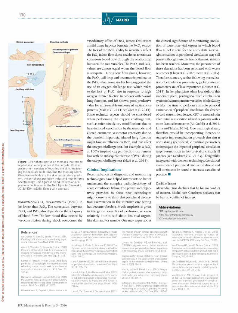
matRIX170
ICU Management & Practice 3 - 2016
©Fo
r pe
rson
al a
nd p
riva
te u
se o
nly.
Rep
rodu
ctio
n m
ust
be p
erm
itte
d by
the
cop
yrig
ht h
olde
r. E
mai
l to
copy
right
@m
indb
yte.
eu.
transcutaneous O2 measurements (PtcO
2) to
be lower than PaO2. The correlation between
PtcO2 and PaO
2 also depends on the adequacy
of blood flow. The low blood flow caused by vasoconstriction during shock overcomes the
vasodilatory effect of PtcO2 sensor. This causes
a mild tissue hypoxia beneath the PtcO2 sensor.
The lack of the PtcO2 ability to accurately reflect
the PaO2 in low flow shock enables us to estimate
cutaneous blood flow through the relationship between the two variables. The PtcO
2 and PaO
2
values are almost equal when the blood flow is adequate. During low flow shock, however, the PtcO
2 will drop and becomes dependent on
the PaO2 value. Some studies have suggested the
use of an oxygen challenge test, which refers to the lack of PtcO
2 rise in response to high
oxygen inspired fraction in patients with normal lung function, and has shown good predictive value for unfavourable outcome of septic shock patients (Mari et al. 2014; Schlager et al. 2014). Some technical aspects should be considered when performing the oxygen challenge test, such as microcirculatory modifications due to heat-induced vasodilation by the electrode, and altered cutaneous vasomotor reactivity due to the transient hyperoxia. Altered lung function might have an influence on PtcO
2 and thus affect
the oxygen challenge test. For example, a PaO2
at 100% inspired oxygen fraction can remain low with no subsequent increase of PtcO
2 during
the oxygen challenge test (Mari et al. 2014).
Clinical ImplicationsRecent advances in diagnostic and monitoring technologies have helped intensivists to better understand the complex pathophysiology of acute circulatory failure. The power and objec-tivity provided by these new technologies might cause us to think that peripheral circula-tion examination in the intensive care setting has become obsolete. Much emphasis is given to the global variables of perfusion, whereas relatively little is said about less vital organs, like skin and/or muscle. One may argue about
the clinical significance of monitoring circula-tion of these non-vital organs in which blood flow is not crucial for the immediate survival. Abnormalities in peripheral circulation may still persist although systemic haemodynamic stability has been reached. Moreover, the persistence of these alterations has been associated with worse outcomes (Chien et al. 2007; Poeze et al. 2005). Therefore, some argue that following normalisa-tion of circulation parameters, global systemic parameters are of less importance (Dunser et al. 2013). In fact physicians often lose sight of this important point, placing too much emphasis on systemic haemodynamic variables while failing to take the time to perform a simple physical examination of peripheral circulation. The absence of cold extremities, delayed CRT or mottled skin after initial resuscitation identifies patients with a more favourable outcome (Ait-Oufella et al. 2011, Lima and Takala, 2014). One next logical step, therefore, would be incorporating therapeutic strategies into resuscitation protocols that aim at normalising (peripheral) circulation parameters to investigate the impact of peripheral circulation target resuscitation in the survival of critically ill patients (van Genderen et al. 2014a). Thoughtfully integrated with the new technology, the clinical assessment of peripheral circulation should and will continue to be central to intensive care clinical practice.
Conflict of InterestAlexandre Lima declares that he has no conflict of interest. Michel van Genderen declares that he has no conflict of interest.
ReferencesAit-Oufella H, Bige N, Boelle PY et al. 2014. Capillary refill time exploration during septic shock. Intensive Care Med, 40(7): 958-64.
Aykut G, Veenstra G, Scorcella C et al. (2015) Cytocam-Idf (incident dark field illumination) imaging for bedside monitoring of the micro-circulation. Intensive Care Med Exp, 3(1): 40.
Conrad M, Perez, P, Thivilier C et al. (2015) Early prediction of norepinephrine dependency and refractory septic shock with a multimodal approach of vascular failure. J Crit Care, 30: 739-43.
Damiani E, Adrario E, Luchetti MM et al. (2015) Plasma free hemoglobin and microcirculatory response to fresh or old blood transfusions in sepsis. PLoS One, 10(5): e0122655.
Gilbert-Kawai E, Coppel J, Bountziouka V et
al. (2016) A comparison of the quality of image acquisition between the incident dark field and sidestream dark field video-microscopes. BMC Med Imaging, 16: 10.
Hutchings S, Watts S, Kirkman E (2015) The Cytocam video microscope. A new method for visualising the microcirculation using Incident Dark Field technology. Clin Hemorheol Microcirc, 62(3): 261-71.
Lima A, Bakker J (2005) Noninvasive monitoring of peripheral perfusion. Intensive Care Med, 31(10): 1316-26.
Lima A, López A, Van Genderen ME et al. (2015) Interrater reliability and diagnostic performance of subjective evaluation of sublingual microcir-culation images by physicians and nurses: a multicenter observational study. Shock, 44(3): 239-44.
Lima A, Van Bommel J, Sikorska K et al. (2011)
The relation of near-infrared spectroscopy with changes in peripheral circulation in critically ill patients. Crit Care Med, 39(7): 1649-54.
Lima A, Van Genderen ME, Van Bommel J et al. (2014) Nitroglycerin reverts clinical manifesta-tions of poor peripheral perfusion in patients with circulatory shock. Crit Care, 18(3): R126.
Macdonald SP, Brown SG (2015) Near-infrared spectroscopy in the assessment of suspected sepsis in the emergency department. Emerg Med J, 32(5): 404-8.
Mari A, Vallée F, Bedel J et al. (2014) Oxygen challenge test in septic shock patients: prog-nostic value and influence of respiratory status. Shock, 41(6): 504-9.
Schlager O, Gschwandtner ME, Willfort-Ehringer A et al. (2014) Transcutaneous oxygen tension monitoring in critically ill patients receiving packed red blood cells. J Crit Care, 29(6): 1057-62.
Tanaka S, Harrois A, Nicolaï C et al. (2015) Qualitative real-time analysis by nurses of sublingual microcirculation in intensive care unit: the MICRONURSE study. Crit Care, 19: 388.
Van Elteren HA, Ince C, Tibboel D et al. (2015) Cutaneous microcirculation in preterm neonates: comparison between sidestream dark field (SDF) and incident dark field (IDF) imaging. J Clin Monit Comput, 29(5): 543-8.
van Genderen ME, Klijn E, Lima A, et al. (2014a) Microvascular perfusion as a target for fluid resuscitation in experimental circulatory shock. Crit Care Med, 42, e96-e105.
van Genderen ME, Paauwe J, de Jonge J et al. (2014b) Clinical assessment of peripheral perfusion to predict postoperative complica-tions after major abdominal surgery early: a prospective observational study in adults. Crit Care, 18(3): R114.
AbbreviationsCRT capillary refill timeNIRS near infrared spectroscopyVOT vascular occlusion test
Figure 1. Peripheral perfusion methods that can be applied in clinical practice at the bedside. Clinical assessment consists of touching the skin, measur-ing the capillary refill time, and the mottling score. Objective methods are the skin temperature gradi-ent, the peripheral perfusion index and near infrared spectroscopy. This figure is an edited version of a previous publication in the Ned Tijdschr Geneeskd. 2013;157(9) :A5338. Edited with approval.
The CytoCam System
Observe the microcirculation with more precision in:
CytoCam-IDF 2nd generation devices
Features of the CytoCam System• Larger Field of View
• Short illumination time, 2ms
• Lower weight, easier to handle,
less pressure artifacts
• Easier focusing
• Higher contrast
• Better depth of focus
• Up to 20% more vessels visible
ESICM booth 612
• (pre) Clinical research
• Hemodynamic monitoring
(sepsis, renal, peri-op, trauma)
• Abdominal surgery
• Neuro surgery
www.braedius.com
©Fo
r pe
rson
al a
nd p
riva
te u
se o
nly.
Rep
rodu
ctio
n m
ust
be p
erm
itte
d by
the
cop
yrig
ht h
olde
r. E
mai
l to
copy
right
@m
indb
yte.
eu.

matRIX172
ICU Management & Practice 3 - 2016
©Fo
r pe
rson
al a
nd p
riva
te u
se o
nly.
Rep
rodu
ctio
n m
ust
be p
erm
itte
d by
the
cop
yrig
ht h
olde
r. E
mai
l to
copy
right
@m
indb
yte.
eu.
In contemporary healthcare, touch—contact between a doctor’s hand and a patient—appears to be on its way out.
The expanding role of CT and MRI imaging is decreasing reliance on touch as a way of making diagnoses. Pressures to move patients through the system more quickly leave health professionals with fewer opportunities to make contact. Our experience suggests that when doctors spend fewer minutes with patients, less time is available for touch (Rabin 2014). Yet despite the rise of scanners, robots and
other new medical technologies, the physician’s hand remains one of medicine’s most valuable diagnostic tools. Touch creates a human bond that is particularly needed in this increasingly hands-off, impersonal age. Medical practice is replete with situations where touch does more than any words to comfort and reassure.
The USC psychologist Leo Buscaglia, whose habit of hugging those he met soon earned him the sobriquet “Doctor Love,” bemoaned our neglect of touch in his book, “Love,” in these terms:
Too often we underestimate the power of a touch, a smile, a kind word, a listening ear, an honest compliment, or the smallest act of caring, all of which have the potential to turn a life around (Buscaglia 1985). For thousands of years, touch has been recog-
nised as an essential part of the healing arts. Native American healers (Cohen 1994) relied on touch to draw out sickness, and kings and queens were long believed to possess the “Royal Touch," through which the mere laying on of hands could heal (Science Museum n.d.). The Bible contains numerous stories of the healing power of touch.
Touch is an Essential Part of our WellbeingAn indication of our need for touch can be found among our primate relatives. Psychologists have
observed that many such species spend upwards of five hours of each day touching one another, partly through grooming (Keltner 2010). For many human beings, however, the daily dose of touching would be measured not in hours but minutes, perhaps even seconds.
Lack of touch can be hazardous to health. In experiments with primates some 60 years ago, researcher Harry Harlow demonstrated that young monkeys deprived of touch did not grow and develop normally (Smuts 2003). Mere food, water and shelter are not sufficient—to thrive, such creatures need to touch and be touched.
The same can be said for human beings. During the 20th century, wars landed many babies in orphanages, where their caretakers observed that no matter how well the infants were fed, they would fail to thrive unless they were held and cuddled on a frequent basis (Rothman 1962). Touch offers no vitamins or calories, yet it plays a vital role in sustaining life.
More recent studies have corroborated these findings. “Kangaroo care,” using papoose-like garments to keep babies close to their mothers, decreases the rate at which they develop blood infections (Larimer 1999). Touching also improves weight gain and decreases the amount of time that newborns need to remain in the hospital.
Touch Creates a Bond Between Doctor and PatientNovelist and physician Abraham Verghese has argued that touching is one of the most impor-tant features of the patient-physician interaction. When he examines a patient, he is not merely collecting information with which to formulate a diagnosis, but also establishing a bond that provides comfort and reassurance.
The notion that touch can reassure and comfort has a scientific basis. Ten years ago researchers used MRI scans to look at the brains of women undergoing painful stimuli
(Coan et al. 2006). When subjects experience pain, certain areas of the brain tend to “light up.” The researchers studied subjects when they were alone, when they were holding a stranger’s hand, and when they were holding their husband’s hand.
They found the highest levels of pain acti-vation when the women were alone. When they were holding a stranger’s hand, the pain response was decreased. And levels of activa-tion were lowest of all when they were holding their husband’s hand. Interesting, the higher the quality of subjects’ marriages, the more pain responses were blunted.
Touch From Parents Helps Kids in Intensive CareWe have been studying this phenomenon in our own institution, looking at the effect of touch not only on patients but on the parents of patients admitted to the pediatric intensive care unit.
The project, called ROSE (Reach Out, Soothe, and Embrace), sought to determine whether increasing opportunities to touch patients could promote parent well-being without compro-mising patient safety (LeLand 2016).
Instead of merely determining whether patients could be taken off the ventilator or fed, we also identified patients who could be safely touched and even held in their parents’ arms. When a patient was deemed safe to hold, a magnet bearing the image of a red rose embraced by two hands was placed on the door to the patient’s room.
While we are still analysing the results and further study is needed to fully delineate the health benefits of touch, several findings are already clear.
First, increasing opportunities for touch does not compromise patient safety. Second, the subjective wellbeing of family members is enhanced when touching is encouraged. Third,
TOuCh CreaTes a healing BOnd in healThCare
Richard Gunderman Chancellor's Professor of Medicine, Liberal Arts, and PhilanthropyIndiana University Indianapolis, USA
brian LeLand FellowSection of Pediatric Critical CareDepartment of PediatricsIndiana University School of Medicine Indianapolis, USA
matRIX173
promoting touch empowers family members to become more involved in their child’s care.
To be sure, inappropriate and unsafe touching can be harmful. But when touch is encouraged in the right ways and for the right reasons, it is good for patients, family, friends and health profes-sionals alike. Touch is one of the most fundamental and effective ways to create a sense of connection and community among human beings.
In the words of the 20th-century theologian Henri Nouwen, who wrote in his book, “Out of Solitude”:
When we honestly ask ourselves which person in our lives
means the most to us, we often find that it is those who, instead of giving advice, solutions, or cures, have chosen rather to share our pain and touch our wounds with a warm and tender hand (Nouwen 2004).So next time you find yourself confronted
by a person in distress, remember the power of touch. Medicines and words both have healing power, but so does touch, and it is perhaps the most widely available, financially responsible and safest tool in the healing arts. When we touch, we connect, and when we connect, we create a healing bond for which there is simply no substitute.
Conflict of InterestBrian LeLand receives funding from Indiana University Health Values Grant Board. 2 year grant awarded through IU Health of $50,000 per year. Grant awarded in 2014.
Reprinted from The Conversation (conversa-tion.com) under Creative Commons CC BY-ND 4.0 (creativecommons.org/licenses/by-nd/4.0).
ReferencesBuscaglia LF (1985) Love: what life is all about. New York: Ballantine.
Coan JA, Schaefer HS, Davidson RJ (2006) Lending a hand: social regulation of the neural response to threat. Psychol Sci, 17(12):1032-9.
Cohen K (1994) Native American healing touch. Bridges, 5(1): 5-7. [Accessed: 19 September 2016] Available from issseem.org/content/uploads/files/Bridges%201994_1.pdf
Keltner D (2010) Hands on research: the science of touch. Greater Good, 29 September. [Accessed: 19 September 2016] Available from: greatergood.berkeley.edu/article/item/hands_on_research
Larimer K (1999) Kangaroo care benefits. [Accessed: 19 September 2016] Available from prematurity.org/baby/kangaroo.html
LeLand B (2016) Fairbanks Ethics Lecture-Project ROSE: Reach Out, Soothe, and Embrace. A focus on caregiver spiritual wellbeing [videorecording].
Vimeo.com. [Accessed: 19 September 2016] Avail-able from vimeo.com/155025967
Nouwen HJM (2004) Out of solitude: three medita-tions on the Christian Life. Notre Dame, Indiana: Ave Maria Press.
Rabin RC (2014) 15-minute visits take a toll on the doctor-patient relationship. KHN: Kaiser Health News, 21 April. [Accessed: 19 September 2016] Available from khn.org/news/15-minute-doctor-visits
Rothman PE (1962) A note on hospitalism. Pediatrics, 30(6): 995-9.
Science Museum (n.d.) King’s evil and the royal touch. [Accessed: 19 September 2016] Avail-able from sciencemuseum.org.uk/broughttolife/techniques/kingsevil
Smuts B (2003) No more wire mothers, ever. New York Times, 2 February. [Accessed: 19 September 2016] Available from nytimes.com/2003/02/02/books/no-more-wire-mothers
Update on Acute Respiratory FailureRome, Italy, December 11-14, 2016
Chairman: Jean-Louis VincentManagement & coordination:Véronique De VlaeminckErasme University HospitalRoute de Lennik 808 – B-1070 BrusselsEmail : [email protected]
©Fo
r pe
rson
al a
nd p
riva
te u
se o
nly.
Rep
rodu
ctio
n m
ust
be p
erm
itte
d by
the
cop
yrig
ht h
olde
r. E
mai
l to
copy
right
@m
indb
yte.
eu.
manaGement174
ICU Management & Practice 3 - 2016
©Fo
r pe
rson
al a
nd p
riva
te u
se o
nly.
Rep
rodu
ctio
n m
ust
be p
erm
itte
d by
the
cop
yrig
ht h
olde
r. E
mai
l to
copy
right
@m
indb
yte.
eu.
Despite an increasing proportion of women in the medical specialty workforce, there are few female doctors
in positions of leadership in intensive care medicine (ICM). This article explores reasons for this gender imbalance, the implications for our specialty and potential solutions. We focus particularly on the situation in Australasia, but present data from other regions where possible to demonstrate that this is a widespread concern.
Where is the Balance? In most developed countries at least half of medical school graduates are women; this has been the case for many years (Amrein et al. 2011; Filardo et al. 2016; Bismark et al. 2015). Despite this, several specialties, including ICM, continue to attract more male than female doctors.
In Australia in 2014 16.6% of ICM specialists and 35.6% of ICM trainees were women (Austra-lian Institute of Health and Welfare 2014). In the
USA in 2013, 24.3% of physicians and 33.1% of trainees in critical care medicine were women (Association of American Medical Colleges 2014). In the UK in 2012, female doctors comprised 26% of the full-time equivalent intensive care specialist workforce (Centre for Workforce Intelligence 2015).
This gender imbalance is more marked in positions of leadership in intensive care medicine. In Australia and New Zealand women make up only 10.5% of Clinical Directors of ICUs accredited by the College of Intensive Care Medicine of Australia and New Zealand (2016).
Female representation on the boards of major intensive care societies is also low (Table 1). Board membership is a key position of influence, as these societies set the academic and political agenda for intensive care medicine.
A similar pattern emerges in academic intensive care medicine. Amrein and colleagues (2011) found that women comprised 17.5% of the editorial boards of the five highest-ranked critical care journals. This was the lowest of all examined specialties, including orthopaedic surgery. Metaxa (2013) reported that between 6 and 14% of speakers at the four largest international intensive care conferences were women. Of six major Australasian specialties, intensive care medicine had the lowest percent-age (8-18%) of female speakers at its annual college conferences (Modra et al. 2016).
Overall, female intensivists are not rising to positions of leadership, nor are they participat-ing equally in the academic discourse in ICM.
It may be argued that the small number of women in leadership positions is not only expected but acceptable, given the low proportion of women within the intensive care workforce. However, in many cases female representation in positions of leadership falls short of female workforce participation (Amrein
et al 2011). In addition, gender imbalance at a leadership level tends to perpetuate workforce gender imbalance, as aspiring female intensiv-ists have few role models. Finally, we believe that increasing the number of female leaders will result in a more innovative and responsive leadership for the specialty.
Why Is It So? Gender BiasGender biases arise from gender schemas: non-conscious hypotheses about sex differences that inform our expectations and evaluations of men and women (Valian 1998). Men and women hold the same gender schemas and use them to efficiently negotiate a complex social world. The World Economic Forum (WEF) (2016) concluded that gender bias is a leading cause of the persistent gender gap in health outcomes, economic and political participation and educational attainment.
Gender schemas lead to measurable biases against women in professional settings even when appraised against apparently objective criteria. Wennerås and Wold (1997) assessed the non-blinded peer review process for scientific grants in Sweden, using multiple regression analysis of the relation between applicants’ scientific productivity and the subjectively assigned ‘competence’ score. They found that a female applicant needed the equivalent of three additional first-author publications in Nature to obtain the same score as a male applicant. Moss-Racusin and colleagues (2012) demonstrated that men were more likely than women to be appointed to a hypothetical science faculty position, despite the male and female candidates having identical CVs.
In academic medicine Jena and colleagues (2015) report that female medical faculty members are “...substantially less likely than men
WOMen in leadership in inTensiVe Care MediCine There is a significant gender imbalance in positions of leadership in intensive care medicine. This complex problem requires action to ensure high quality and sustainable leadership for our specialty in the future.
Lucy J modra*Senior RegistrarIntensive Care UnitAlfred HospitalMelbourne, Australia
Sarah a YongIntensivist Intensive Care UnitAlfred HospitalMelbourne, Australia
danielle e austinFellowIntensive Care UnitRoyal Prince Alfred HospitalSydney, Australia
* corresponding author
@womenintensive
womenintensive.org
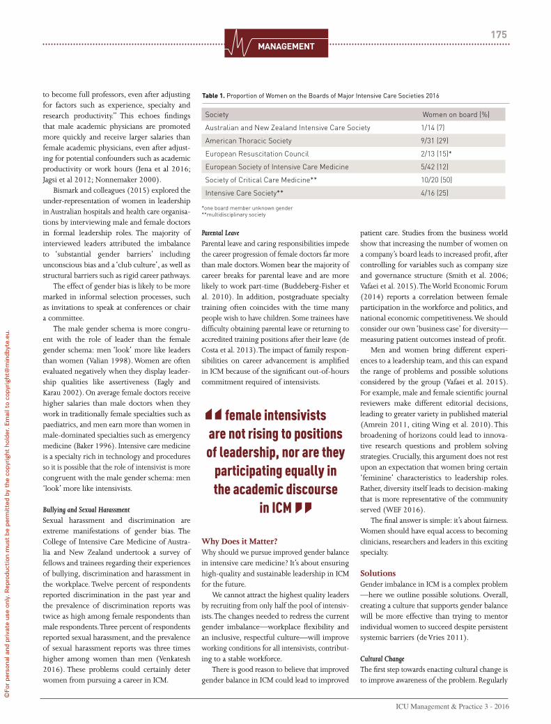
manaGement175
ICU Management & Practice 3 - 2016
©Fo
r pe
rson
al a
nd p
riva
te u
se o
nly.
Rep
rodu
ctio
n m
ust
be p
erm
itte
d by
the
cop
yrig
ht h
olde
r. E
mai
l to
copy
right
@m
indb
yte.
eu.
to become full professors, even after adjusting for factors such as experience, specialty and research productivity.” This echoes findings that male academic physicians are promoted more quickly and receive larger salaries than female academic physicians, even after adjust-ing for potential confounders such as academic productivity or work hours (Jena et al 2016; Jagsi et al 2012; Nonnemaker 2000).
Bismark and colleagues (2015) explored the under-representation of women in leadership in Australian hospitals and health care organisa-tions by interviewing male and female doctors in formal leadership roles. The majority of interviewed leaders attributed the imbalance to ‘substantial gender barriers’ including unconscious bias and a ‘club culture’, as well as structural barriers such as rigid career pathways.
The effect of gender bias is likely to be more marked in informal selection processes, such as invitations to speak at conferences or chair a committee.
The male gender schema is more congru-ent with the role of leader than the female gender schema: men ‘look’ more like leaders than women (Valian 1998). Women are often evaluated negatively when they display leader-ship qualities like assertiveness (Eagly and Karau 2002). On average female doctors receive higher salaries than male doctors when they work in traditionally female specialties such as paediatrics, and men earn more than women in male-dominated specialties such as emergency medicine (Baker 1996). Intensive care medicine is a specialty rich in technology and procedures so it is possible that the role of intensivist is more congruent with the male gender schema: men ‘look’ more like intensivists.
Bullying and Sexual HarassmentSexual harassment and discrimination are extreme manifestations of gender bias. The College of Intensive Care Medicine of Austra-lia and New Zealand undertook a survey of fellows and trainees regarding their experiences of bullying, discrimination and harassment in the workplace. Twelve percent of respondents reported discrimination in the past year and the prevalence of discrimination reports was twice as high among female respondents than male respondents. Three percent of respondents reported sexual harassment, and the prevalence of sexual harassment reports was three times higher among women than men (Venkatesh 2016). These problems could certainly deter women from pursuing a career in ICM.
Parental LeaveParental leave and caring responsibilities impede the career progression of female doctors far more than male doctors. Women bear the majority of career breaks for parental leave and are more likely to work part-time (Buddeberg-Fisher et al. 2010). In addition, postgraduate specialty training often coincides with the time many people wish to have children. Some trainees have difficulty obtaining parental leave or returning to accredited training positions after their leave (de Costa et al. 2013). The impact of family respon-sibilities on career advancement is amplified in ICM because of the significant out-of-hours commitment required of intensivists.
Why Does it Matter? Why should we pursue improved gender balance in intensive care medicine? It’s about ensuring high-quality and sustainable leadership in ICM for the future.
We cannot attract the highest quality leaders by recruiting from only half the pool of intensiv-ists. The changes needed to redress the current gender imbalance—workplace flexibility and an inclusive, respectful culture—will improve working conditions for all intensivists, contribut-ing to a stable workforce.
There is good reason to believe that improved gender balance in ICM could lead to improved
patient care. Studies from the business world show that increasing the number of women on a company’s board leads to increased profit, after controlling for variables such as company size and governance structure (Smith et al. 2006; Vafaei et al. 2015). The World Economic Forum (2014) reports a correlation between female participation in the workforce and politics, and national economic competitiveness. We should consider our own ‘business case’ for diversity—measuring patient outcomes instead of profit.
Men and women bring different experi-ences to a leadership team, and this can expand the range of problems and possible solutions considered by the group (Vafaei et al. 2015). For example, male and female scientific journal reviewers make different editorial decisions, leading to greater variety in published material (Amrein 2011, citing Wing et al. 2010). This broadening of horizons could lead to innova-tive research questions and problem solving strategies. Crucially, this argument does not rest upon an expectation that women bring certain ‘feminine’ characteristics to leadership roles. Rather, diversity itself leads to decision-making that is more representative of the community served (WEF 2016).
The final answer is simple: it’s about fairness. Women should have equal access to becoming clinicians, researchers and leaders in this exciting specialty.
Solutions Gender imbalance in ICM is a complex problem —here we outline possible solutions. Overall, creating a culture that supports gender balance will be more effective than trying to mentor individual women to succeed despite persistent systemic barriers (de Vries 2011).
Cultural ChangeThe first step towards enacting cultural change is to improve awareness of the problem. Regularly
female intensivists are not rising to positions of leadership, nor are they
participating equally in the academic discourse
in ICM
Society Women on board (%)
Australian and New Zealand Intensive Care Society 1/14 (7)
American Thoracic Society 9/31 (29)
European Resuscitation Council 2/13 (15)*
European Society of Intensive Care Medicine 5/42 (12)
Society of Critical Care Medicine** 10/20 (50)
Intensive Care Society** 4/16 (25)
*one board member unknown gender**multidisciplinary society
table 1. Proportion of Women on the Boards of Major Intensive Care Societies 2016
manaGement176
ICU Management & Practice 3 - 2016
©Fo
r pe
rson
al a
nd p
riva
te u
se o
nly.
Rep
rodu
ctio
n m
ust
be p
erm
itte
d by
the
cop
yrig
ht h
olde
r. E
mai
l to
copy
right
@m
indb
yte.
eu.
auditing and publishing data on female represen-tation in all facets of intensive care medicine will increase the visibility of gender imbalance in our profession (Bonomo 2016). Gender bias training, including simple ‘self tests’ such as that developed by Project Implicit, can increase awareness of how gender bias affects us all (Project Implicit 2011).
All intensive care medicine institutions must have a ‘zero tolerance’ approach to sexist behaviour. This should be set out in easily accessible guidelines, with clear consequences for inappropriate behaviour regardless of the status of the perpetrator within the profession (Expert Advisory Committee to the Royal Australasian College of Surgeons 2015).
Overcoming Gender BiasTo minimise the impact of gender biases on leadership appointments, transparent and formal selection processes should be used (Bismark et al. 2015). Selection panels should be ‘blinded’ to gender wherever possible, for example by removing names from CVs in the initial selection process. Introducing the double-blinded peer review process led to an increase in the publica-tion of scientific papers written by women (Budden 2008).
Another solution is to set gender-based targets or quotas for the proportion of women appointed to leadership positions, speaking at conferences or granted research funding (Bonomo 2016). The World Economic Forum (2016) reports that setting targets for recruit-ment and retention of female employees is a crucial strategy to improve female representation across all industries.
Quotas are controversial: some argue they undermine equality and discredit the women appointed to designated female positions.
However, the measurable impact of gender biases on women’s professional advancement demonstrates that the ‘level playing field’ does not actually exist. Even in apparently objective selection processes, men are slightly more likely to be selected simply because they are male. There is evidence that aiming to appoint employees on merit alone amplifies rather than diminishes the effect of covert gender bias in the selection process (Castilla and Benard 2010). Quotas address covert bias by overtly making women more likely to be selected for some positions.
Structural ChangeStructural change in the workplace is crucial for women’s integration and advancement in the workforce (World Economic Forum 2016). This includes transparent parental leave schemes accessible to men and women, workplace flexibility through flexible hours and part-time appointments. A key challenge for ICM is incorporating these changes whilst ensuring continuity of patient care.
On-site childcare in the workplace and at conferences could improve female participation, and encourage the integration of family and professional responsibilities for both mothers and fathers.
Mentoring Mentoring and leadership training can help nurture the careers of individual women (WEF 2016). Mentoring programmes should be carefully tailored to support women, as gender biases can also affect mentoring relationships. For example, men are more likely than women to receive strategic career advice and to be recommended for career opportunities as a result of a mentoring relationship (de Vries 2011).
Advocacy initiativesDeliberate efforts must be made to nominate or encourage talented women to apply for leadership roles, including positions on boards, committees or speaker engagements (Bonomo 2016). Initiatives such as Australia’s Women in Intensive Care Medicine Network can help women to develop effective profes-sional networks and to advocate for key structural change. These initiatives must actively engage men in the change process.
ConclusionIntensive care medicine prides itself on being a dynamic specialty, committed to continuous improvement and understanding of the crucial role of human factors in delivering effective patient care. Despite this, there is a persistent gender imbalance in the leadership of ICM. To ensure the sustainability and quality of ICM leadership in the future, we must commit to urgently redressing this imbalance through cultural change, workplace structural reforms, advocacy and mentoring.
Conflict of InterestAll authors are members of the Women in Intensive Care Medicine network, an organisa-tion dedicated to improving the gender balance in intensive care medicine in Australasia through research, advocacy and networking.
AcknowledgementsThe authors gratefully acknowledge the assistance of Professor Imogen Mitchell in the preparation of this manuscript.
ReferencesAmerican Thoracic Society (2016) [Accessed: April 2016] Available from: thoracic.org/about/governance/board-of-directors.php
Amrein K, Langmann A, Fahrleitner-Pammer A et al. (2011) Women underrepresented on editorial boards of 60 major medical journals. Gend Med, 8(6): 378-87.
Association of American Medical Colleges, Centre for Workforce Studies (2014) Physi-cian specialty data book. [Accessed 23 May 2016] Available from members.aamc.org/eweb/upload/Physician%20Specialty%20Databook%202014.pdf
Australian Institute of Health and Welfare (2014) Medical practitioners overview 2014 tables, Canberra. [Accessed: May 2016] Available from aihw.gov.au/workforce/medical/2014/additional
Australia and New Zealand Intensive Care Society (2016) [Accessed: June 2016] Available from anzics.com.au/about-us
Baker LC (1996) Differences in earnings between male and female physicians. N Engl J Med, 334(15): 960-4.
Bismark et al (2015) Reasons and remedies for under-representation of women in medical lead-ership roles: a qualitative study from Australia, BMJ Open, 5:e009384.
Bonomo Y, Zundel S, Martin JH. (2016) Addressing unconscious bias for female clinical academics, Intern Med J, 46(4): 391-3.
Buddeberg-Fischer B, Stamm M, Buddeberg C et al. (2010) The impact of gender and parent-hood on physician’s careers – professional and personal situation seven years after graduation. BMC Health Serv Res, 10:40.
Budden AE, Tregenza T, Aarssen LW, Koricheva J, Leimu R and Lortie CJ. (2008) Double-blind review favours increased representation of female authors. Trends Ecol Evol, 23(1): 4-6.
Castilla EJ, Benard S (2010) The paradox of meritocracy in organizations, Administrative Science Quarterly, 55: 543-76.
Centre for Workforce Intelligence. (2015) In-depth review of the anaesthetics and intensive care medicine workforce. London: Centre for Work-force Intelligence. [Accessed May 2016] Available from cfwi.org.uk/publications/in-depth-review-of-the-anaesthetics-and-intensive-care-medicine-workforce/@@publication-detail
College of Intensive Care Medicine of Australia and New Zealand. (2016) General accredited units; paediatric accredited units. [Accessed: 28 May 2016] Available from cicm.org.au/Hospitals/Accredited-Sites-Accordion/Accredited-Units
de Costa CM, Permezel M, Farrell LM et al. (2013) Integrating parental leave into specialist training: experience of trainees and recently graduated RANZCOG fellows. Med J Aust, 199(5): 359-62.
De Vries, J. (2011) Mentoring for change. Melbourne: LH Martin Institute, University of Melbourne; Universities Australia Executive Women.. [Accessed: May 2016] Available from lhmartininstitute.edu.au/documents/publications/2011uaewmentoringforchange.pdf
Eagly AH, Karau SJ (2002) Role congruity theory of prejudice toward female leaders. Psychol Rev, 109(3):573–98.
For full references, please email editorial@icu management.org, visit icu-manage-ment.org or use the article QR code.
AbbreviationsCV curriculum vitaeICM intensive care medicineWEF World Economic Forum

Euroanaesthesia The European Anaesthesiology Congress
www.esahq.org
Geneva, Switzerland03 -05 June 2017
Abstract Submission: 1 November - 15 December 2016
2017
©Fo
r pe
rson
al a
nd p
riva
te u
se o
nly.
Rep
rodu
ctio
n m
ust
be p
erm
itte
d by
the
cop
yrig
ht h
olde
r. E
mai
l to
copy
right
@m
indb
yte.
eu.
manaGement178
ICU Management & Practice 3 - 2016
©Fo
r pe
rson
al a
nd p
riva
te u
se o
nly.
Rep
rodu
ctio
n m
ust
be p
erm
itte
d by
the
cop
yrig
ht h
olde
r. E
mai
l to
copy
right
@m
indb
yte.
eu.
inTensiVe Care syndrOMe: prOMOTing independenCe and reTurn TO eMplOyMenT (ins:pire)A NEW MOdEL FOR ICU REHABILITATION
tara QuasimICU ConsultantGlasgow Royal InfirmaryGlasgow, UK
Senior Lecturer in Anaesthesia, Critical Care & Pain Medicine University of Glasgow
Joanne mcPeake Post-doctoral ICU Staff Nurse Glasgow Royal InfirmaryGlasgow, UK
It is now well established that many patients and caregivers suffer physical, psycho-logical and social problems in the years
and months following critical care discharge (Herridge et al. 2011). Similar to many centres, our intensive care unit (ICU) had no follow-up service available to support patients through this difficult recovery period (Griffiths et al. 2006). To understand how best to create a service that was safe, effective and person-centred, two members of our multidisciplinary team (MDT) undertook research programmes to help identify the problems that patients faced after ICU and to help understand the context for change (Quasim et al. 2015; McPeake et al. 2016).
From this work, four main challenges were identified:
1. There is minimal evidence of how and when rehabilitation services should be delivered (Mehlhorn et al. 2010), despite an abundance of literature describing the issues for ICU survivors and their families.
2. The hardships facing ICU patients are often not apparent to hospital management. Readmissions to hospital, increased general practitioner (family physician) visitations and the increased reliance on welfare benefits are distrib-uted amongst a variety of budgets, which do not necessarily appear related to an ICU admission.
3. Finding staff with the time and ability
to do something new that is different from their traditional ICU role can be problematic.
4. Finding physical space with a suitable area to hold a rehabilitation programme can be difficult.
Patient-Focused Solutions Simultaneously the team set up a Patient and Family Council (PFAC). The PFAC was a group of 10-12 previous patients and caregivers who met bimonthly for approximately two hours. This group, which was chaired by a patient or caregiver, helped create potential solutions to the problems encountered by ICU patients and their loved ones.
Utilising peer support as a central compo-nent for rehabilitation is gaining momentum within the ICU community (Mikkelsen et al. 2016). By bringing together a heterogeneous group of ICU survivors as part of our PFAC, the positive effect of peer support was obvi-ous. Without exception, it was the first time that they had spoken to someone with a shared experience of the ICU.
What was also clear from this feedback was that their desire to get home did not live up to expectations. At around a month after hospital discharge, patients realised that their individual recovery was not what had been expected. It was also very clear that both patients and caregivers were frustrated that healthcare professionals and indeed the general public did not understand the difficulties in overcoming a critical illness.
In attempting to move forward, the team were faced with the harsh reality that there is limited funding to establish a new service. Not unreasonably, most healthcare managers require evidence of benefit before there is financial investment.
In 2014 the Health Foundation, a UK char-ity, advertised a series of innovation grants, the Shine Awards. The team successfully applied for one of these awards. The advantage of this opportunity was that the MDT did not have to have the finished, polished intervention and thus were not limited to undertaking traditional research methodologies. This was a ‘learning’ grant, which allowed the team to develop a new service and change things after learning what worked and what didn’t.
InS:PIREUtilising the first hand experience of our Patient and Family Council, the MDT co-created and developed the concept of InS:PIRE (Intensive Care Syndrome: Promoting Independence and Return to Employment). InS:PIRE is a five-week, peer-supported, self-management programme aimed at empowering patients and relatives to take control of their own health and wellbeing by finding community resources to help them. To ensure that we did not create a population of ‘chronic’ ICU patients, the course was kept deliberately short.
Every week there was a group exercise class to stimulate peer support. To encourage peer support further within the programme,

manaGement179
ICU Management & Practice 3 - 2016
©Fo
r pe
rson
al a
nd p
riva
te u
se o
nly.
Rep
rodu
ctio
n m
ust
be p
erm
itte
d by
the
cop
yrig
ht h
olde
r. E
mai
l to
copy
right
@m
indb
yte.
eu.
InS:PIRE adopted patient and carer volunteer roles. These roles were undertaken by ICU survivors further along the recovery trajectory, who could offer hope and support to patients who were still struggling. To help encourage cross-communication we set up a café area; this allowed patients, staff and volunteers to intermingle in a relaxed setting.
As well as the weekly physiotherapy class, patients and caregivers had individual and group sessions (Table 1). These included a physiotherapy consultation to discuss pain, musculoskeletal and balance issues. Patients were then given an individualised programme for their specific needs.
A pharmacy appointment was also provided to ensure that an appropriate medicines recon-ciliation had been undertaken. This was also the opportunity for the pharmacist to educate patients on what they were taking their medi-cines for and how to take them properly. If there were any issues, the pharmacist would write to the GP or use the National Patient Safety Foundation Framework Ask Me 3 (npsf.org/?page=askme3) document to empower patients to discuss their medication queries independently.
At a nursing and medical session, the patient was provided with a lay summary of their ICU stay if they wanted it and offered the oppor-tunity to revisit the ICU. Personal goals were set with the patient and caregiver by asking simple questions such as “What can’t you do
now that you could do before ICU?” or “What would you like to be doing in six months’ time?” Personal outcomes were often centred on issues such as driving, walking and using public transport independently. We would break these challenges into smaller tasks to make them more achievable.
A group psychology session explored ‘coping’ strategies. We also provided patients with information on how to seek further psychological support in the community if they felt this was necessary. We separated the patients from relatives during the psychology sessions as our PFAC felt this was important. This is also reflected within the literature, which demonstrates the challenges which caregivers face during critical care recovery (Haines et al. 2015). During the psychology session, patients were able to ‘normalise’ their feelings and express their concerns without causing
more anxiety for their relatives. For the carers, it was a chance for them to offload exhaustion, anger and frustration without feeling guilty. This process appeared to offer this group an enormous sense of relief.
The fifth and final week of InS:PIRE was named the social prescription week. This session signposted participants to community organ-isations which might help their recovery. For example, we looked at volunteering options as a platform to return to work, community phys-iotherapy classes or places that can help with welfare issues. A fundamental aim of InS:PIRE was to provide patients with the tools to take control of their own health by using existing community resources. Unless absolutely neces-sary, patients were not referred back into the hospital setting.
A vital part of this initial pilot of InS:PIRE was the ‘learning week’, which included a meeting of the entire health and social care team involved in the delivery of InS:PIRE. From this we could find out from patients, staff and the volunteers what worked, what didn’t and what we needed to change. An example of this learning was in our first cohort where attendance was initially low. We had phoned patients a few days in advance to ask if they were attending the clinic, but then often they didn’t attend. When they were contacted to ask why, it was because they didn’t realise what day of the week it was. This is consistent with many of the well-documented ongoing cognitive problems which patients encounter following critical care discharge (Iwashyna et al. 2010). By contacting patients on the morning of the clinic, attendance doubled. We also realised that there was a risk of patients becoming dependent on individual members of the team. As a result of this learning, a generic email account and phone number was created and utilised for all patient interactions.
Learning from InS:PIREThe positive difference in patients that the team witnessed between the first and final week of InS:PIRE was overwhelming. What was unex-pected was the effect InS:PIRE had on the staff attending the clinic. Staff who had been in the same job for decades were expanding their role and working in a different way; they weren’t defined by the role they had trained in.
For patients the biggest issues which were identified and supported during the programme were social: housing; finances; relationships and employment. The skills needed to support these
Innovative strategies that span health and social care are required to support
the holistic needs of patients following critical
care discharge
WEEK 1 2 3 4 5 6
GROUP PHYSIOTHERAPY CLASS X X X X X
MEDIC /NURSE X
PHARMACY X
PHYSIO X
GROUP PSYCHOLOGY X
GROUP SLEEP PSYCHOLOGY/DIETITIAN X
CARERS CENTRE X
CITIZENS ADVICE BUREAU (FINANCIAL HELP) X X
ALISS (SIGNPOSTS TO COMMUNITY ORGANISATIONS) X
GOOD MOVES X
PATIENT VOLUNTEER X X X X X
CARER VOLUNTEER X X X X X
table 1. Demonstrating the Layout of InS:PIRE Over a 6 Week Period
LEARN
ING
SESSION
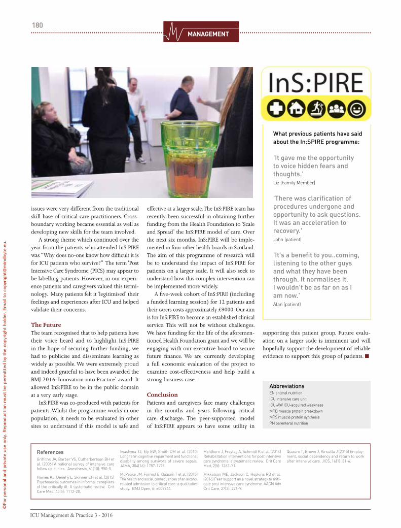
manaGement180
ICU Management & Practice 3 - 2016
©Fo
r pe
rson
al a
nd p
riva
te u
se o
nly.
Rep
rodu
ctio
n m
ust
be p
erm
itte
d by
the
cop
yrig
ht h
olde
r. E
mai
l to
copy
right
@m
indb
yte.
eu.
issues were very different from the traditional skill base of critical care practitioners. Cross-boundary working became essential as well as developing new skills for the team involved.
A strong theme which continued over the year from the patients who attended InS:PIRE was “Why does no-one know how difficult it is for ICU patients who survive?” The term ‘Post Intensive Care Syndrome (PICS) may appear to be labelling patients. However, in our experi-ence patients and caregivers valued this termi-nology. Many patients felt it ‘legitimised’ their feelings and experiences after ICU and helped validate their concerns.
The FutureThe team recognised that to help patients have their voice heard and to highlight InS:PIRE in the hope of securing further funding, we had to publicise and disseminate learning as widely as possible. We were extremely proud and indeed grateful to have been awarded the BMJ 2016 ‘Innovation into Practice’ award. It allowed InS:PIRE to be in the public domain at a very early stage.
InS:PIRE was co-produced with patients for patients. Whilst the programme works in one population, it needs to be evaluated in other sites to understand if this model is safe and
effective at a larger scale. The InS:PIRE team has recently been successful in obtaining further funding from the Health Foundation to ‘Scale and Spread’ the InS:PIRE model of care. Over the next six months, InS:PIRE will be imple-mented in four other health boards in Scotland. The aim of this programme of research will be to understand the impact of InS:PIRE for patients on a larger scale. It will also seek to understand how this complex intervention can be implemented more widely.
A five-week cohort of InS:PIRE (including a funded learning session) for 12 patients and their carers costs approximately £9000. Our aim is for InS:PIRE to become an established clinical service. This will not be without challenges. We have funding for the life of the aforemen-tioned Health Foundation grant and we will be engaging with our executive board to secure future finance. We are currently developing a full economic evaluation of the project to examine cost-effectiveness and help build a strong business case.
ConclusionPatients and caregivers face many challenges in the months and years following critical care discharge. The peer-supported model of InS:PIRE appears to have some utility in
supporting this patient group. Future evalu-ation on a larger scale is imminent and will hopefully support the development of reliable evidence to support this group of patients.
ReferencesGriffiths JA, Barber VS, Cutherbertson BH et al. (2006) A national survey of intensive care follow up clinics. Anesthesia, 61(10): 950-5.
Haines KJ, Denehy L, Skinner EH et al. (2015) Psychosocial outcomes in informal caregivers of the critically ill: A systematic review. Crit Care Med, 43(5): 1112-20.
Iwashyna TJ, Ely EW, Smith DM et al. (2010) Long term cognitive impairment and functional disability among survivors of severe sepsis. JAMA, 304(16): 1787-1794.
McPeake JM, Forrest E, Quasim T et al. (2015) The health and social consequences of an alcohol related admission to critical care: a qualitative study. BMJ Open, 6: e009944.
Mehlhorn J, Freytag A, Schmidt K et al. (2014) Rehabilitation interventions for post intensive care syndrome: a systematic review. Crit Care Med, 2(5): 1263-71.
Mikkelsen ME, Jackson C, Hopkins RO et al. (2016) Peer support as a novel strategy to miti-gate post intensive care syndrome. AACN Adv Crit Care, 27(2): 221-9.
Quasim T, Brown J, Kinsella J (2015) Employ-ment, social dependency and return to work after intensive care. JICS, 16(1): 31-6.
AbbreviationsEN enteral nutritionICU intensive care unitICU-AW ICU-acquired weaknessMPB muscle protein breakdownMPS muscle protein synthesisPN parenteral nutrition
What previous patients have said about the In:SPIRE programme:
'It gave me the opportunity to voice hidden fears and thoughts.'Liz (Family Member)
'There was clarification of procedures undergone and opportunity to ask questions. It was an acceleration to recovery.'John (patient)
'It's a benefit to you..coming, listening to the other guys and what they have been through. It normalises it. I wouldn't be as far on as I am now.' Alan (patient)
©Fo
r pe
rson
al a
nd p
riva
te u
se o
nly.
Rep
rodu
ctio
n m
ust
be p
erm
itte
d by
the
cop
yrig
ht h
olde
r. E
mai
l to
copy
right
@m
indb
yte.
eu.
14TH APRIL 2017
©Fo
r pe
rson
al a
nd p
riva
te u
se o
nly.
Rep
rodu
ctio
n m
ust
be p
erm
itte
d by
the
cop
yrig
ht h
olde
r. E
mai
l to
copy
right
@m
indb
yte.
eu.
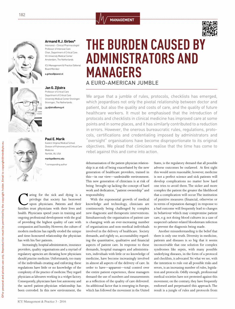
manaGement182
ICU Management & Practice 3 - 2016
©Fo
r pe
rson
al a
nd p
riva
te u
se o
nly.
Rep
rodu
ctio
n m
ust
be p
erm
itte
d by
the
cop
yrig
ht h
olde
r. E
mai
l to
copy
right
@m
indb
yte.
eu.
The Burden Caused By adMinisTraTOrs and ManagersA EURO-AMERICAN JUMBLE
We argue that a jumble of rules, protocols, checklists has emerged, which jeopardises not only the pivotal relationship between doctor and patient, but also the quality and costs of care, and the quality of future healthcare workers. It must be emphasised that the introduction of protocols and checklists in clinical medicine has improved care at some points and in some places, and it has similarly contributed to a reduction in errors. However, the onerous bureaucratic rules, regulations, proto-cols, certifications and credentialing imposed by administrators and “oversight” organisations have become disproportionate to its original objectives. We plead that clinicians realise that the time has come to rebel against this and come into action.
armand R.J. Girbes*Intensivist – Clinical PharmacologistProfessor of Intensive CareChair, Department of Critical CareVU University Medical CenterAmsterdam, The Netherlands
ICU Management & Practice Editorial Board Member
Jan G. ZijlstraProfessor of Critical CareDepartment of Critical CareUniversity Medical Center GroningenGroningen, The Netherlands
Paul e. marikEastern Virginia Medical SchoolDivision of Pulmonary and Critical Care MedicineNorfolk, VA, USA
* corresponding author
Caring for the sick and dying is a privilege that society has bestowed upon physicians. Patients and their
families trust physicians with their lives and health. Physicians spend years in training and ongoing professional development with the goal of providing the highest quality of care with compassion and humility. However, the culture of modern medicine has rapidly eroded the unique and time-honoured relationship the physician has with his/her patients.
Increasingly, hospital administrators, insurance providers, quality organisations and a myriad of regulatory agencies are dictating how physicians should practise medicine. Unfortunately, too many of the individuals creating and enforcing these regulations have little or no knowledge of the complexity of the practice of medicine. They regard physicians as labourers working in a widget factory. Consequently, physicians have lost autonomy and the sacred patient-physician relationship has been corroded. In this new environment, the
dehumanisation of the patient-physician relation-ship is at risk of being exacerbated by the new generation of healthcare providers, trained in this—in our view—undesirable environment. This new generation of clinicians is at risk of being brought up lacking the concept of hard work and dedication, “patient ownership” and responsibility.
With the exponential growth of medical knowledge and technology, clinicians are continuously being challenged by complex new diagnostic and therapeutic interventions. Simultaneously the organisation of patient care is changing with an ever-increasing number of organisations and non-medical individuals involved in the delivery of healthcare. Society demands, and rightly so, accountability regard-ing the quantitative, qualitative and financial aspects of patient care. In response to these demands, hospital managers and administra-tors, individuals with little or no knowledge of medicine, have become increasingly involved in almost all aspects of the delivery of care. In order to have—apparent—total control over the entire patient experience, these managers demand the use of numbers and measurements as a reflection of the quality of care delivered. An additional factor that is emerging in Europe, which has followed the movement in the United
States, is the regulatory demand that all possible adverse outcomes be outlawed. At first sight this would seem reasonable; however, medicine is not a perfect science and sick patients will develop complications no matter how hard one tries to avoid them. The sicker and more complex the patient the greater the likelihood that a complication will occur. The institution of punitive measures (financial, otherwise or in terms of reputation damage) in response to a bad outcome will frequently lead to changes in behaviour which may compromise patient care, e.g. not doing blood cultures in a case of suspected catheter-related bloodstream infection to prevent the diagnosis being made.
Another misunderstanding is the belief that there is only one truth. Diversity in medicine, patients and diseases is so big that it seems inconceivable that one solution for complex syndromes like sepsis, with many possible underlying diseases, in the form of a protocol and checklists, is advocated. Yet what we see, with the intention to rule out all possible risks and errors, is an increasing number of rules, legisla-tion and protocols. Oddly enough, professional medical societies have not protested against this movement; on the contrary, they have frequently endorsed and perpetuated this approach. The result is a jungle of rules and protocols from

manaGement183
ICU Management & Practice 3 - 2016
©Fo
r pe
rson
al a
nd p
riva
te u
se o
nly.
Rep
rodu
ctio
n m
ust
be p
erm
itte
d by
the
cop
yrig
ht h
olde
r. E
mai
l to
copy
right
@m
indb
yte.
eu.
medical and scientific societies, governmental and other non-medical bodies such as insurance companies. Physicians and clinical leaders are confronted with more and more requirements, rules, audits, inspections, compliance training and protocols, imposed by governmental and non-governmental organisations, insurance companies, accreditation organisations, inspec-torates and boards of directors of hospitals. With all the regulatory administrative tasks that physicians are forced to undertake, it is not rocket science to realise that less and less time remains available for the primary process: patient care. Apart from impacting patient care, the time wasted jeopardises clinical research, education and the training of students and registrars. Additionally, research and training are hampered by an increasing number of rules, regulations and mandatory non-functional courses. Many of these mandatory courses are not only meant for the teachers, but also for their PhD students. The distance between workers on the shop floor, the healthcare workers, and on the other hand those people who make the regulations is growing and they speak different languages. All kinds of bodies and commit-tees in hospitals offer training programmes, the additional value of which is questionable in terms of patient outcome or educational quality. It might come to one’s mind that these bodies are mainly preoccupied with providing new work for themselves, creating rules, work and training programmes of unclear benefit.
A simple recent survey that the first author (AG) conducted among some board directors of hospitals, demonstrated that they have insufficient insight into the huge number of obligations imposed by different bodies on medical special-ists and nurses. Table 1 provides an incomplete but illustrative overview of the Dutch situation.
The quality movement has imposed the increased use of protocols and checklists with the intention to improve quality of care. This is accompanied by obligatory ticking off and securing of lists that go through implicit procedures. While protocols were initially intended to provide up-to-date medical knowledge translated into clinically and practically applicable information, currently all kinds of procedures need to be embodied in protocols, which need to be secured by checklists and repeated evaluation according to a plan-do-check-act cycle. Subsequently, compliance to the protocol is used as a marker of quality. Undeniably this approach has induced improvement on certain fronts (Girbes et al. 2015; 2016). But it is now getting out of control. Moreover, a trend can be
observed that for every rare incident a new protocol is created, without taking into account how a new protocol might induce new errors. For example, in addition to double checking the preparation of a medicine by an intensive care nurse, a new additional obligatory protocol was introduced (in the Netherlands) without any evidence or calculation of the consequence. This protocol requires that immediately after the double check of the medication an additional double check is required at the time of administration of the medicine. This of course requires another ICU nurse to abandon their current activity, move to another patient, check what is given, and then go back to continue the interrupted work. It is beyond doubt that frequent interruption of work will induce other errors (Westbrook et al. 2010). Of course continuous double checking would be a dream scenario, if feasible in terms of human factors. This would however require double the number of nurses: one nurse to do the work and another to check the work. Consid-ering the pressure on and shortage of human resources, one wonders whether this is the most effective way to save lives. Furthermore, one of
the nurses would surely become bored, which is not conducive to good concentration on doing the best work they can.
By no means do we want to argue that errors, mistakes and undesirable outcomes should not be investigated to recognise the “holes” in the system. However, the solution is not always the introduction of a new protocol or checklist.
We strongly believe that the policy of increasing the number of protocols and checklists should be reversed if we want to keep good medical care affordable. An issue that is easily forgotten is that we must be able to keep and attract young talented people. Increasing rigidity of the system is, to say the least, not an incentive to motivate young talents to work in medicine. We argue that protocols and checklists are comparable to medicines: it is the dose that makes poison and the indication always remains pivotal. The dose has now reached the level of poison and the indication is too often wrong.
Jumble of Protocols and ChecklistsThe purpose of clinical protocols is to translate the best possible up-to-date medical knowledge
table 1. Examples of Imposed Managerial Tasks, Training and Registration Programmes in the Netherlands.
Quality inventory list of care processes with priority list and improvement actions
(e.g. is the pulmonologist present during lung surgery? Is there a registry of all complications? Is there a
protocol for the treatment of pneumonia? etc.)
Yearly obligatory report of several “performance parameters” (imposed by inspectorate)
Participation in national safety management system
(e.g. participation in and report of Surviving Sepsis Campaign, number of reoperations after hip replace-
ment, number of central venous line infections, yearly training in CPR for all physicians, etc.)
Participation in hospital accreditation programme (e.g. Joint Commission International)
Registry of every employee on knowledge of manuals of all devices in the department
Registry of followed training programme of nurses and physicians
Course for fire extinguisher use
Participation in practice for calamities
Participation in practice for evacuation
Courses to work with electronic patient file
Training in lean management
Audits
(Audits for training programmes, safety audits, audits for employee working condition, audits for material
handling, etc.)
Yearly satisfaction measurements for trainees on a large number of items
(System of evaluation of teaching qualities – SETQ – and Dutch residents’ educational climate test)
Imposed training programmes for PhD students
Critical Performance Indicators (McKinsey & Company)
Teach the teacher courses (level 1, 2 and 3)
Basic Qualification for Education (see text)
Test for English language knowledge
Training programmes for addressing other people / issues
manaGement184
ICU Management & Practice 3 - 2016
©Fo
r pe
rson
al a
nd p
riva
te u
se o
nly.
Rep
rodu
ctio
n m
ust
be p
erm
itte
d by
the
cop
yrig
ht h
olde
r. E
mai
l to
copy
right
@m
indb
yte.
eu.
into practical, clinically applicable instructions. Several studies have shown an improvement in patient outcome with the introduction of a protocol or checklist. Whether a protocol or checklist will introduce an improvement in care largely depends on how good or bad the situation was before the introduction of the protocol. Introduction of a protocol is therefore especially useful in situations of suboptimal circumstances or where inexperienced or less trained healthcare workers are employed. Further-more, checklists are not universal. Checklists need to be intrinsically supported by staff, based on the local applicability of the checklist and support from the leadership.
Protocols will by definition lead to regression to the mean and mediocrity. Rigid application of protocols will hamper progress and innovation, and protocols are by definition not up-to-date. Finally, many protocols are made on the basis of insufficient scientific data, insufficiently possible external validation of studies or even only on the basis of the judgement of self-proclaimed “experts”. Unfortunately, healthcare managers, “organisations for quality”, supervisory bodies and healthcare insurance companies mandato-rily impose the introduction of protocols and checklists for all kinds of aspects of care. The forced introduction on a national level of the Surviving Sepsis Campaign in the United States and in the Netherlands, apart from many other examples, is a tragic example of this. There is insufficient scientific evidence to impose per protocol treatment according to the surviving sepsis guideline in all hospitals and even evidence that it might be harmful (Marik 2016a).
The introduction of protocols with doubtful benefit may lead to waste of time, work and money. The obligatory introduction of the medical emergency team (MET) from the ICU, implementation of all components of the time-out procedure in the operating room, reporting standard screening of feeding condition in the elderly, and scoring of community-acquired pneumonia, are examples of so-called safety programmes that cost a lot of time and money, but are of doubtful benefit for society and individual patients.
Filling in all kinds of lists is promoted by the introduction of electronic patient record programmes. These have been designed for administrative and financial reasons and not, as one would expect, to improve patient care and help healthcare workers to do their work correctly. It is no surprise that the introduction of such electronic health records has been shown
to increase the risk of professional burnout in physicians (Shanafelt et al. 2016).
Treating individual patients optimally will always require aspects of craftsmanship with an academic attitude and thereby individual-ised treatment. Translating the use of protocols and checklists to another craft, food prepara-tion, might clarify some aspects. Application of protocols only works very well in the fast-food industry. In “restaurants” where no chef is needed the employees are easier to handle by the management of the “restaurant” and can be paid less. Food will always be according to the guidelines and protocols and checklists, but in the end will not fit everybody. Likewise, even if written by a great chef, reading and follow-ing the instructions of a cookery book will not match the quality and craft of a real chef.
Proponents of the unrestrained use of protocols and checklists often point to the analogy and similarities between aviation and building construction. We reject that compari-son. Patients are not airplanes and doctors are not pilots. Pilots receive very specific training in general for a single type of airplane. Since every patient is different, it would pose serious problems if doctors were trained like pilots.
Jumble of the Quality MovementThere should be no doubt that doctors and nurses should be accountable to patients and those who pay for them: society. And society is all of us. The healthcare payer has the right to know how their money is spent and where to find quality for the money. However, this is quite difficult to measure and instruments to measure quality are readily available. Neverthe-less the “Quality Movement” has triggered a
“quality tsunami” where multiple organisations have now become preoccupied with developing quality tools, quality indicators and measur-ing the “quality of outcomes.” These quality indicators and scorecards are frequently publicly reported and may influence reimbursement. The scientific validity of most of these quality indica-tors is highly questionable. It would appear that those who expend the most resources measuring quality provide the worst care (Thomson et al. 2013). The refuge that seems to be chosen now by the administrators and managers can best be described as: “If you can’t measure what is important, you make important what you measure”. So orthopaedic surgeons obligatorily record and report on the rate of reoperations for hip fractures. This of course will result in a figure, but this figure is of course full of confounders and biases (e.g. region, population characteristics, referral pattern, etc.) and nobody can tell what the figure means. A rapid survey among chairmen of university departments of orthopaedics in the Netherlands confirmed this. Nevertheless, whenever criticism is expressed about this obligation the answer is: “It is simply an obligation” or “everybody complies with it”.
Registrations furthermore do not take into account the pollution of data that is not expressed in the data. Subjective data are reduced to figures in a spreadsheet, suggesting that different figures and outcomes can be compared. This becomes most hilarious when comparing opinions. For example, during regularly performed so-called employee satisfaction measurements we add the opinions of ambitious, looking for security, lazy, adventurer, genius, hypochondriac, disappointed (in private life or their career) people, divide this by the number of participants and then we conclude that the satisfaction is 7.3! (We do not take into account the number of employees who for several reasons do not wish to participate). The manager will surely advocate a leadership programme to fulfil the goal for next year: 7.8.
In the U.S. Medicare has embarked on hundreds of “quality initiatives”, and records over 1000 “quality measures” with the purported goal of improving the “quality of care” (Casalino et al. 2016). It has been reported that physicians and their staff spend 15.1 hours per physician per week dealing with external quality measures at an annual cost of over $40, 000 per physician. There is scarce data that these quality measures improve patient outcomes. In 2006 the Centers for Medicare and Medicaid Services (CMS) developed the “Surgical Care Improvement Project” (SCIP), which became federally mandated
a jumble of rules, protocols, checklists...
jeopardises not only the pivotal relation-
ship between doctor and patient, but also the quality
and costs of care and the quality of future healthcare
workers

manaGement185
ICU Management & Practice 3 - 2016
©Fo
r pe
rson
al a
nd p
riva
te u
se o
nly.
Rep
rodu
ctio
n m
ust
be p
erm
itte
d by
the
cop
yrig
ht h
olde
r. E
mai
l to
copy
right
@m
indb
yte.
eu.
and linked to pay for performance in 2007 (Joint Commission 2015). SCIP incorporated a number of measures, including glycaemic control and strict timing of prophylactic antibiotics that were required to be performed in every patient undergoing elective surgery. In January 2015 the SCIP project was quietly “retired” (Joint Commission 2015), after it became clear that this very expensive and time-consuming endeavour did not improve patient outcomes (Hawn et al. 2011; Dua et al. 2014; McDonnell et al. 2013). In 2015 CMS adopted the “SEP-1 Early Management Bundle for Severe Sepsis and Septic Shock” for the Hospital Inpatient Quality Reporting Programme. Most alarmingly, it is likely that this “quality” programme” will harm patients (Marik and Varon 2016). In the U.S. and progressively in the Netherlands, physician’s medical records are scrutinised by individu-als with limited educational training to ensure that all elements of the history and physical examination are documented, no matter how irrelevant. Rather than being a tool to communi-cate medical information, the medical record is used as a quality indicator and a means to punish physicians for incomplete documenta-tion. And again a new industry is filling this created gap: a “quality company”. Their slogan is: “Let me measure if you have a quality issue, all your colleagues did it already. Indeed you have a problem and we know people who can solve it”.
Jumble of Obligatory TrainingFortunately, the time of “see one, do one, teach one” is over. Many skills can be learned and improved with good training programmes and simulation sessions. This includes not only hard skills and knowledge but also so-called “soft”
skills such as advanced life support in a team, team performance, bringing bad news to families and patients, and calling someone to account. Complex tasks with a low incidence cannot be dealt with in a training programme. Intentional publication fraud cannot be prevented with a course on ethics in science and neither will a course, obligatory in the Netherlands, with a duration of more than one week on regulations and organisation of clinical research prevent that. However, these rules mean that professors with many publications in leading journals, and with a research desk to guarantee all responsibilities and compliance with regulations, fail an exam because they do not know by heart how many years all records need to be stocked. The goal of good clinical practice and research, will also be missed whenever those who conduct the courses get too much influence on making it an obligation to follow these courses. This again will result in a “course industry” both within and outside the hospital, whose sole purpose is that of self-preservation. In the Netherlands, PhD students in medicine have been guided and supported for decades by established research-ers and professors during their PhD study. The study outline and the interpretation of data were discussed almost on a daily basis. They participated in international congresses and presented their data during national and international meetings. However, all of a sudden specific time-consum-ing courses have been made obligatory for PhD students with no data to support impact on student outcome. Another remarkable obliga-tory regulation without any supporting data was the introduction of the Basic Qualification for Education (BQE). This training programme consists of 5 full days training, 165 hours of study, 90 hours of which are with the help of
an assigned mentor. Someone with more than 30 years of educational experience, educational diplomas outside the field of medicine, who has students who value the courses and applaud during presentations and over 260 international presentations is called to follow this obligatory BQE training programme.
A long list can be generated of time-consuming training programmes with concomitant registra-tion obligation, which can be related to demands by health insurance companies, legal authorities and accreditation programmes. It is beyond the scope of this paper to discuss the benefit-time ratios of these programmes, but in general we would challenge those who make these regula-tions to demonstrate their benefit.
The question remains of how to make progress in medicine and how to prevent errors and wrong treatment. We think the key is good training programmes and a culture where healthcare workers continuously give feedback to each other. Medicine has to stay attractive for young people with an academic mindset that is challenged by all the complex problems encountered in health-care. Whatever protocol or checklist, it should be used as a mental support for highly educated professionals and never get the force of law.
Conflict of interestArmand Girbes declares that he has no conflict of interest. Jan Zijlstra declares that he has no conflict of interest. Paul Marik declares that he has no conflict of interest.
ReferencesCasalino LP, Gans D, Weber R et al. (2016) US physicians’ practices spend more than $15.4 billion annually to report quality measures. Health Aff (Millwood), 35(3): 401-6.
Dua A, Desai SS, Seabrook GR et al. (2014) The effect of Surgical Care Improvement project measures on national trends on surgical site infections in open vascular procedures. J Vasc Surg, 60(6): 1635-9.
Girbes AR, Robert R, Marik PE (2015) Protocols: help for improvement but beware of regression to the mean and mediocrity. Intensive Care Med, 41(12): 2218-20.
Girbes AR, Robert R, Marik PE (2016) The dose makes the poison. Intensive Care Med, 42(4): 632.
Hawn MT, Vick CC, Richman J et al. (2011) Surgical site infection prevention: time to move beyond the surgical care improvement program. Ann Surg, 254(3): 494-9.
Joint Commission (2015) Surgical care Improve-ment Project (SCIP) [Accessed: 23 August 2016] Available from jointcommission.org/surgical_care_improvement_project
Marik PE (2016a) Fluid responsiveness and the six guiding principles of fluid resuscitation. Crit Care Med, Nov 13.
Marik PE (2016b) Tight glycemic control in
acutely ill patients: low evidence of benefit, high evidence of harm! Intensive Care Med, 42(9): 1475-7.
Marik PE, Varon J (2016) Precision medicine and the Federal sepsis initiative! Crit Care Shock, 19: 1-3.
McDonnell ME, Alexanian SM, Junquiera A et al. (2013) Relevance of the Surgical Care Improvement Project on glycemic control in patients undergoing cardiac surgery who receive continuous insulin infusions. J Thorac Cardiavasc Surg, 145(2): 590-4.
Shanafelt TD, Dyrbye LN, Sinsky C et al. (2016) Relationship between clerical burden and char-acteristics of the electronic environment with
physician burnout and professional satisfaction. Mayo Clin Proc, 91(7): 836-48.
Thomson S, Osborn R, Squires D et al., eds (2013) International profiles of health care systems, 2013: Australia, Canada, Denmark, England, France, Germany, Italy, Japan, the Neth-erlands, New Zealand, Norway, Switzerland and the United States. New York: Commonwealth Fund. [Accessed: 23 August 2016] Available from commonwealthfund.org/Publications/Fund-Reports/2013/Nov/International-Profiles-of-Health-Care-Systems.aspx
Westbrook JI, Woods A, Rob MI et al. (2010) Association of interruptions with an increased risk and severity of medication administration errors. Arch Intern Med, 170(8): 683-90.
InteRVIeW186
ICU Management & Practice 3 - 2016
©Fo
r pe
rson
al a
nd p
riva
te u
se o
nly.
Rep
rodu
ctio
n m
ust
be p
erm
itte
d by
the
cop
yrig
ht h
olde
r. E
mai
l to
copy
right
@m
indb
yte.
eu.
TeleMediCine is The fuTureINTERVIEW WITH PROFESSOR GERNOT MARX
Professor Gernot Marx is Director of the Department of Intensive Care Medicine and Intermediate Care, University Hospital Aachen, and Professor of Anaesthesiology and Operative Intensive Care Medicine at RWTH Aachen University, Aachen, Germany. Dr. Marx is a member of the Intensive Care Medicine Scientific Subcommittee of the European Society of Anaesthesiology.
The first tele-ICU service in Germany was set up at the University Hospital Aaachen. What were the main challenges? The first challenge was to install the technol-ogy—to ensure secure connection lines between the telemedical centre and the regional hospitals, set up the audiovisual system and provide the electronic case record. All data is assured under data protection rules.
The second challenge was scepticism from colleagues. This is understandable, because with telemedicine you really ‘open up the doors’ and you can look into the heart of the service, which has pros and cons. You may see some suboptimal issues, so overcoming scepticism was an important hurdle. The other point is that the service is not about the ‘big shots’ from the university hospital telling others how to do it. We wanted to create the situation that together we are better and that we learn from each other. This was very important in order to get everyone engaged in the new service.
The telemedicine project has been running for two years, and we have increased coverage from two to five hospitals. It started as a project funded by research money, but now we are in the process of getting the service reimbursed. We have proved that the service is technically possible, is accepted by colleagues, patients and relatives and that it is improving patient outcomes. In our observational study of 1200
patients (Marx et al. 2015) we could show measurable improvement in patients’ outcome. This is why the insurance companies now support this ongoing process.
Now you are on the way to receiving reim-bursement, where is telemedicine going in Germany? The German Society of Anesthesiology and Intensive Care Medicine has a commission for telemedicine and eHealth, which I lead. We published recommendations (in German) on how to structure telemedicine in the ICU and emergency areas (Marx and Koch 2015), and published a guideline on telemedicine in the ICU (AWMF Leitlinie tele-Intensivmedizin). We applied to create a reimbursement code for telemedicine. The process has been started, and we hope that in 3-4 years there will be a national reimbursement code. Several university hospitals are interested in creating a similar service. My vision is that there will be a national roll out. We have more patients because society is ageing, we have fewer experts and we need to share our expertise. In the USA, it has been already proven to be an intervention associated with a survival benefit in a prospective study including more than 100,000 ICU patients (Lilly et al. 2014). Not only in intensive care is this a problem, so I see this as a role model for different areas of medicine.
Is there potential for cross-border tele-medical collaboration?We have two new projects and we are creating solutions to connect electronic medical systems from different companies (thalea-pcp.eu). We hope to complete this by the end of 2016 so we can then connect intensive care units (ICUs) throughout Europe and the world. From a European perspective, I would like to see the European Society of Anaesthesiol-ogy establish a committee on telemedicine. As intensivists we need to take the future in our hands.
How does telemedicine improve patient care?It is not easy to do randomised controlled trials, but we can use stepped wedge or pre-post designs to test the hypothesis that telemedicine improves patient care. As with our small trial you can see that there is an improvement in measurable outcomes. In a way it is not ‘new medicine’, it is offering access to experts. We have not only the information from the electronic medical record, but we can also see the patient—their eyes, the pupil reaction. We get a very sharp clear clinical picture, and so communication and visualisation is very important. The only thing we cannot do is touch the patient.
Now we have an extra hour at each hospital. If you do this day by day it really creates a
InteRVIeW187
ICU Management & Practice 3 - 2016
©Fo
r pe
rson
al a
nd p
riva
te u
se o
nly.
Rep
rodu
ctio
n m
ust
be p
erm
itte
d by
the
cop
yrig
ht h
olde
r. E
mai
l to
copy
right
@m
indb
yte.
eu.
living quality network, because there is a lot of trust when you see and talk to each other every day. You need a large unit, such as we have at University Hospital Aachen. My unit is comprised of 100 beds and I have 15 ICU consultants. You need a large group of experts with many years of experience to provide this extra service 24/7 in a standardised way to support the doctors, anaesthetists and ICUs in the regional hospitals.
How has patient and family acceptance of the telemedicine service been in regional hospitals?Feedback has been excellent. They feel that they are already part of the university hospital medical service. Previously relatives quite often approached colleagues to ask why we could not transfer patients to the university hospital. That does not happen now because they are already part of the centre. This is an advantage for families and patients, because we provide this top-quality service close to home. It is easier for them to visit their relatives locally, so it really has many advantages.
Clinical Trials
your intensive care unit is participating in the Gelatin in ICU and Sepsis (GENIUS) trial. What is the rationale for this trial? Gelatin in ICU and Sepsis (GENIUS ) (clinical-trials.gov/ct2/show/NCT02715466 or clinicaltrialsregister.eu/ctr-search/trial/2015-000057-20/AT) is the first large trial on gelatin. It is a trial to compare balanced crystalloids against balanced gelatin plus balanced crystalloids. It is scheduled to recruit 600 patients from 10 centres in Germany, Austria, France and Hungary.
There are some differences with previous trials. Instead of central venous pressure (CVP), we’re using the passive leg raising test and advanced monitoring stroke volume measurement in the patients. The evidence in guidelines is that that you cannot assess indication for fluid replacement with CVP nor can you assess the success of the therapy on CVP. This is an important difference.
In addition we decided not to have 28-day mortality as a primary outcome. Many people feel that this is not the right outcome parameter for research in the intensive care area, especially in sepsis (Mebazaa et al. 2016). 28-day mortal-ity is more a measure of safety rather than the outcome parameter. In terms of efficacy
we need other outcomes and priorities, for example organ dysfunction or reduction of organ dysfunction. The GENIUS trial has the time to achieve haemodynamic stabilisation as the primary outcome.
Is the large randomised controlled trial in intensive care still a good model?My vision is to have smaller trials using a complex protocol. We have to stratify patients to move more towards personalised medicine. One example is response to inflammatory response, measured by who has immune competence. Like our oncology colleagues we need to see that not every patient is the same in location of infection and response to infection. We need to have more detailed information and then put those patients in certain trials and investigate. When we have this information and we can connect our databases, we have big data research availabil-ity and we can confirm our data and make smaller trials. I expect to see more pattern recognition type research in the future.
What is the situation in Germany with sepsis?We just conducted a trial about sepsis epidemi-ology in Germany (German Sepnet Critical Care Trial Group, in press). We prospectively put twelve thousand patients into this trial all over Germany in more than 100 units. Mortality is still very high. It is different to examining mortality in randomised trials, where you have protocols and and obviously you look at a very defined group. In this trial we included all septic patients. As we performed the trial at the end of 2013 we were using the old sepsis definition. The lesson is that it is still a very common disease, mortal-ity is still very high and we have to tackle it. The most important point is what we have learnt from the Surviving Sepsis Campaign bundles, that the first hours are very determi-nant for outcome. It requires standard therapy,
recognition, antibiotics, and haemodynamic stabilisation.
Intravascular Volume Therapy Guidelines
you were involved in producing the German guidelines on intravascular volume therapy. How were these developed?The German Society of Anesthesiology and Intensive Care Medicine initiated this guideline and coordinated the process. I was the coordinator from the German Society of Anaesthesiology and Intensive Care Medicine, and we included fourteen medical societ-ies, who all sent an independent expert. We developed the guidelines according to the rules and regulations of the Association of the Scientific Medical Societies in Germany (Arbeitsgemeinschaft der Wissenschaftlichen Medizinischen Fachgesellschaften 2014). We engaged a top expert in methodology with his team. When we screened the publica-tions there was always one physician and one expert in methods and there was always a cross check. That is why it took more than two years to develop the guidelines. The evidence was thoroughly analysed and condensed, and the experts received a very detailed analysis of each paper. Based on that we made our recommendations and suggestions, always after long discussions, with at least 75% consen-sus between all 14 societies. Afterwards each society’s board had to approve the recommen-dations, with the Association of the Scientific Medical Societies in Germany having the final say. It was a very robust process. We have now a thorough evidence-based recommendation in this very important field. The guidelines are published open access in both German (awmf.org/leitlinien/detail/ll/001-020.html) and English in the European Journal of Anaesthesiology (Marx et al. 2016).
In Germany how will you evaluate use of the guidelines and measure process? We had a pre-questionnaire and will have a post-questionnaire and then we will see whether practice has changed or not. This will be our measurement of the process change after introduction of the guidelines.
How do the recommendations differ from current practice?The guidelines have a lot of evidence on the indications for when to use fluids. Therefore
We wanted to create the situation that
together we are better and that we learn from
each other
InteRVIeW188
ICU Management & Practice 3 - 2016
©Fo
r pe
rson
al a
nd p
riva
te u
se o
nly.
Rep
rodu
ctio
n m
ust
be p
erm
itte
d by
the
cop
yrig
ht h
olde
r. E
mai
l to
copy
right
@m
indb
yte.
eu.
ReferencesAWMF Leitlinie tele-Intensivmedizin. [Accessed: 17 September 2016] Available from awmf.org/leitlinien/detail/ll/001-034.html Registernummer 001 - 034
Bingold TM, Bickenbach J, Coburn M et al. (2014) Modulares Zertifikat Intensivmedizin der DGAI.“ Anästh Intensivmedizin, 55: 316-29.
German Sepnet Critical Care Trial Group (In press) Incidence of severe sepsis and septic
shock in German intensive care units: the prospective, multicentre INSEP study. DOI: 10.1007/s00134-016-4504-3.
Lilly CM, McLaughlin JM, H Zhao J et al. (2014) A multicenter study of ICU telemedicine reengineering of adult critical care. CHEST, 145(3): 500–7.
Marx G, Beckers R, Brokmann JC et al. (2015) [Tele-cooperation for innovative care using the example of the University Hospital Aachen.
Telematics in intensive care medicine, emer-gency medicine, and telemedical intersectoral rehabilitation planning in geriatric trauma] Article in German]. Bundesgesundheitsblatt Gesundheitsforschung Gesundheitsschutz, 58 (10): 1056-61.
Marx G, Koch T (2015) Telemedizin in der Inten-sivmedizin: Strukturempfehlungen der DGAI. Anästh Intensivmed, 56: 257-61.
Marx G, Schindler AW, Mosch C et al. (2016)
Intravascular volume therapy in adults: Guide-lines from the Association of the Scientific Medical Societies in Germany. Eur J Anaes-thesiol, 33(7): 488-521.
Mebazaa A, Laterre PF, Russell JA et al. (2016) Designing phase 3 sepsis trials: application of learned experiences from critical care trials in acute heart failure. J Intensive Care, 4: 24.
there is a lot about monitoring. As I indicated before central venous pressure (CVP) has a grade A recommendation not to be used for indication or monitoring of fluids therapy. We have to use dynamic parameters, we have to use flow parameters, and we have to use passive leg raising and ultrasound but not CVP. That is not to say: “never use CVP”, just not for this indication. This is new.
There is a strong recommendation to use balanced fluids and not to use saline. This is in contrast to the previous recommendations. Also there is no evidence that use of colloids has any negative side effect or benefit in terms of outcomes periopratively; thus colloids may be used in the operating room. This is a weak recommendation, because there is not a lot of evidence there. First-line treatment choice in the ICU is crystalloids. But also colloids can be used, gelatin or albumin, if necessary in shock patients. We made an important statement on these issues because all the trials so far had severe methodology problems, e.g. using CVP or recruitment of patients being performed after haemodynamic stabilisation was already done. In contrast, in the GENIUS trial we include patients within 90 minutes while they are still in shock and need fluid therapy. We use passive leg raising not CVP as an evidence-based parameter for indication and monitoring of volume therapy.
you have been involved in a pilot project in Germany on voluntary peer review. How does this work?We have two processes in Germany. One is peer review, which is voluntary. Colleagues visit colleagues to have a look at the unit and its processes and give feedback. This is organised by the German Interdisciplinary Association of Intensive Care Medicine (DIVI), and results in specific recommendations for each particular ICU.
The second established process for quality control is a quality certificate, which has been established by the German Society of Anaesthe-siology and Intensive Care Medicine. ICUs apply and will be audited by two externally trained experts. Certification means that your unit is performing intensive care medicine at a high quality level. We published the standards in 2014 (Bingold et al. 2014), and have started the certification process. Fifteen ICUs have now passed this quality certification. It may take anything from four months up to a year for units to prepare to apply for this process.
I am involved in both receiving and giving peer review and certification. It is very important that we have critical feedback, and it’s important that our specialty defines what is quality, because we know best. Obviously, we have to be in discussions with the authori-ties. I am sure this will come in Germany and
in Europe that there will be certain quality measures that need to be in place to assure that there is a quality ICU service. It would be good to have this in place across Europe.
This interview will appear in ICU Manage-ment & Practice’s issue with a cover story on the abdomen. How do you see the role of haemodynamic monitoring in relation to intra-abdominal pressure?The whole area of advanced haemodynamic monitoring in the operating room for anaesthe-tists needs to be explored further; we need more data and randomised trials to see whether it is a valuable tool with advantages for the patients. So far the ‘noise’ is towards that direction, but as this is a costly investment for the area of anaesthesia then it is important to get more evidence to convince the manage-ment at different sites to invest. In patients with increased intra-abdominal pressure it is a valuable tool to identify haemodynamic disturbances as early as possible.
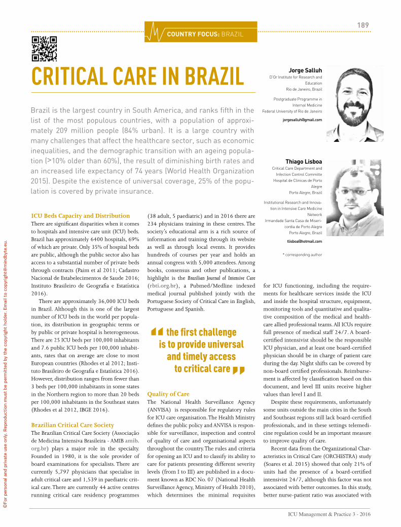
189
ICU Management & Practice 3 - 2016
CoUntRY FoCUS: BRAZIL
©Fo
r pe
rson
al a
nd p
riva
te u
se o
nly.
Rep
rodu
ctio
n m
ust
be p
erm
itte
d by
the
cop
yrig
ht h
olde
r. E
mai
l to
copy
right
@m
indb
yte.
eu.
ICU Beds Capacity and DistributionThere are significant disparities when it comes to hospitals and intensive care unit (ICU) beds. Brazil has approximately 6400 hospitals, 69% of which are private. Only 35% of hospital beds are public, although the public sector also has access to a substantial number of private beds through contracts (Paim et al 2011; Cadastro Nacional de Estabelecimentos de Saude 2016; Instituto Brasileiro de Geografia e Estatística 2016).
There are approximately 36,000 ICU beds in Brazil. Although this is one of the largest number of ICU beds in the world per popula-tion, its distribution in geographic terms or by public or private hospital is heterogeneous. There are 25 ICU beds per 100,000 inhabitants and 7.6 public ICU beds per 100,000 inhabit-ants, rates that on average are close to most European countries (Rhodes et al 2012; Insti-tuto Brasileiro de Geografia e Estatística 2016). However, distribution ranges from fewer than 3 beds per 100,000 inhabitants in some states in the Northern region to more than 20 beds per 100,000 inhabitants in the Southeast states (Rhodes et al 2012, IBGE 2016).
Brazilian Critical Care SocietyThe Brazilian Critical Care Society (Associação de Medicina Intensiva Brasileira - AMIB amib.org.br) plays a major role in the specialty. Founded in 1980, it is the sole provider of board examinations for specialists. There are currently 5,797 physicians that specialise in adult critical care and 1,539 in paediatric crit-ical care. There are currently 44 active centres running critical care residency programmes
(38 adult, 5 paediatric) and in 2016 there are 234 physicians training in these centres. The society’s educational arm is a rich source of information and training through its website as well as through local events. It provides hundreds of courses per year and holds an annual congress with 5,000 attendees. Among books, consensus and other publications, a highlight is the Brazilian Journal of Intensive Care (rbti.org.br), a Pubmed/Medline indexed medical journal published jointly with the Portuguese Society of Critical Care in English, Portuguese and Spanish.
Quality of CareThe National Health Surveillance Agency (ANVISA) is responsible for regulatory rules for ICU care organisation. The Health Ministry defines the public policy and ANVISA is respon-sible for surveillance, inspection and control of quality of care and organisational aspects throughout the country. The rules and criteria for opening an ICU and to classify its ability to care for patients presenting different severity levels (from I to III) are published in a docu-ment known as RDC No. 07 (National Health Surveillance Agency, Ministry of Health 2010), which determines the minimal requisites
for ICU functioning, including the require-ments for healthcare services inside the ICU and inside the hospital structure, equipment, monitoring tools and quantitative and qualita-tive composition of the medical and health-care allied professional teams. All ICUs require full presence of medical staff 24/7. A board-certified intensivist should be the responsible ICU physician, and at least one board-certified physician should be in charge of patient care during the day. Night shifts can be covered by non-board certified professionals. Reimburse-ment is affected by classification based on this document, and level III units receive higher values than level I and II.
Despite these requirements, unfortunately some units outside the main cities in the South and Southeast regions still lack board-certified professionals, and in these settings telemedi-cine regulation could be an important measure to improve quality of care.
Recent data from the Organizational Char-acteristics in Critical Care (ORCHESTRA) study (Soares et al. 2015) showed that only 21% of units had the presence of a board-certified intensivist 24/7, although this factor was not associated with better outcomes. In this study, better nurse-patient ratio was associated with
CriTiCal Care in BraZil Jorge SalluhD’Or Institute for Research and
EducationRio de Janeiro, Brazil
Postgraduate Programme in Internal Medicine
Federal University of Rio de Janeiro
thiago LisboaCritical Care Department and
Infection Control CommitteHospital de Clinicas de Porto
AlegrePorto Alegre, Brazil
Institutional Research and Innova-tion in Intensive Care Medicine
NetworkIrmandade Santa Casa de Miseri-
cordia de Porto AlegrePorto Alegre, Brazil
* corresponding author
Brazil is the largest country in South America, and ranks fifth in the list of the most populous countries, with a population of approxi-mately 209 million people (84% urban). It is a large country with many challenges that affect the healthcare sector, such as economic inequalities, and the demographic transition with an ageing popula-tion (>10% older than 60%), the result of diminishing birth rates and an increased life expectancy of 74 years (World Health Organization 2015). Despite the existence of universal coverage, 25% of the popu-lation is covered by private insurance.
the first challenge is to provide universal
and timely access to critical care
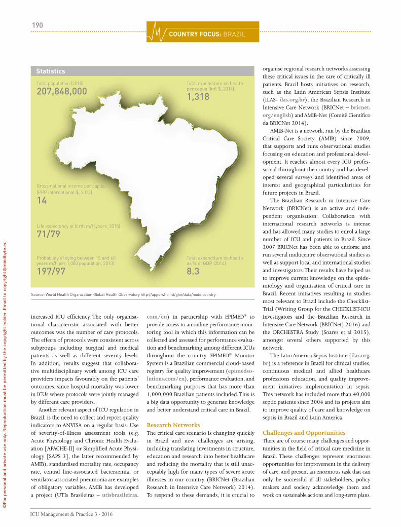
190CoUntRY FoCUS: BRAZIL
ICU Management & Practice 3 - 2016
©Fo
r pe
rson
al a
nd p
riva
te u
se o
nly.
Rep
rodu
ctio
n m
ust
be p
erm
itte
d by
the
cop
yrig
ht h
olde
r. E
mai
l to
copy
right
@m
indb
yte.
eu.
increased ICU efficiency. The only organisa-tional characteristic associated with better outcomes was the number of care protocols. The effects of protocols were consistent across subgroups including surgical and medical patients as well as different severity levels. In addition, results suggest that collabora-tive multidisciplinary work among ICU care providers impacts favourably on the patients’ outcomes, since hospital mortality was lower in ICUs where protocols were jointly managed by different care providers.
Another relevant aspect of ICU regulation in Brazil, is the need to collect and report quality indicators to ANVISA on a regular basis. Use of severity-of-illness assessment tools (e.g. Acute Physiology and Chronic Health Evalu-ation [APACHE-II] or Simplified Acute Physi-ology [SAPS 3], the latter recommended by AMIB), standardised mortality rate, occupancy rate, central line-associated bacteraemia, or ventilator-associated pneumonia are examples of obligatory variables. AMIB has developed a project (UTIs Brasileiras – utisbrasileiras.
com/en) in partnership with EPIMED® to provide access to an online performance moni-toring tool in which this information can be collected and assessed for performance evalua-tion and benchmarking among different ICUs throughout the country. EPIMED® Monitor System is a Brazilian commercial cloud-based registry for quality improvement (epimedso-lutions.com/en), performance evaluation, and benchmarking purposes that has more than 1,000,000 Brazilian patients included. This is a big data opportunity to generate knowledge and better understand critical care in Brazil.
Research NetworksThe critical care scenario is changing quickly in Brazil and new challenges are arising, including translating investments in structure, education and research into better healthcare and reducing the mortality that is still unac-ceptably high for many types of severe acute illnesses in our country (BRICNet (Brazilian Research in Intensive Care Network) 2014). To respond to these demands, it is crucial to
organise regional research networks assessing these critical issues in the care of critically ill patients. Brazil hosts initiatives on research, such as the Latin American Sepsis Institute (ILAS- ilas.org.br), the Brazilian Research in Intensive Care Network (BRICNet – bricnet.org/english) and AMIB-Net (Comitê Científico da BRICNet 2014).
AMIB-Net is a network, run by the Brazilian Critical Care Society (AMIB) since 2009, that supports and runs observational studies focusing on education and professional devel-opment. It reaches almost every ICU profes-sional throughout the country and has devel-oped several surveys and identified areas of interest and geographical particularities for future projects in Brazil.
The Brazilian Research in Intensive Care Network (BRICNet) is an active and inde-pendent organisation. Collaboration with international research networks is intense and has allowed many studies to enrol a large number of ICU and patients in Brazil. Since 2007 BRICNet has been able to endorse and run several multicentre observational studies as well as support local and international studies and investigators. Their results have helped us to improve current knowledge on the epide-miology and organisation of critical care in Brazil. Recent initiatives resulting in studies most relevant to Brazil include the Checklist-Trial (Writing Group for the CHECKLIST-ICU Investigators and the Brazilian Research in Intensive Care Network (BRICNet) 2016) and the ORCHESTRA Study (Soares et al 2015), amongst several others supported by this network.
The Latin America Sepsis Institute (ilas.org.br) is a reference in Brazil for clinical studies, continuous medical and allied healthcare professions education, and quality improve-ment initiatives implementation in sepsis. This network has included more than 40,000 septic patients since 2004 and its projects aim to improve quality of care and knowledge on sepsis in Brazil and Latin America.
Challenges and OpportunitiesThere are of course many challenges and oppor-tunities in the field of critical care medicine in Brazil. These challenges represent enormous opportunities for improvement in the delivery of care, and present an enormous task that can only be successful if all stakeholders, policy makers and society acknowledge them and work on sustainable actions and long-term plans.
Source: World Health Organization Global Health Observatory http://apps.who.int/gho/data/node.country
Total population (2015)
207,848,000Total expenditure on health per capita (Intl $, 2014)
1,318
Gross national income per capita (PPP international $, 2013)
14
Life expectancy at birth m/f (years, 2015)
71/79
Probability of dying between 15 and 60 years m/f (per 1,000 population, 2013)
197/97Total expenditure on health as % of GDP (2014)
8.3
Statistics

191
ICU Management & Practice 3 - 2016
CoUntRY FoCUS: BRAZIL
©Fo
r pe
rson
al a
nd p
riva
te u
se o
nly.
Rep
rodu
ctio
n m
ust
be p
erm
itte
d by
the
cop
yrig
ht h
olde
r. E
mai
l to
copy
right
@m
indb
yte.
eu.
ReferencesAustin S, Murthy S, Wunsch H et al. (2014) Access to urban acute care services in high- vs. middle-income countries: an analysis of seven cities. Intensive Care Med, 40(3): 342-52.
Azevedo LC, Park M, Salluh JI et al. (2013) Clinical outcomes of patients requiring ventilatory support in Brazilian intensive care units: a multicenter, prospective, cohort study. Crit Care, 17(2): R63.
Bozza FA, Grinsztejn B (2016) Key points on Zika infection for the intensivist. Intensive Care Med,42(9): 1490-2.
Bozza FA, Salluh J. (2010) An urban perspec-tive on sepsis in developing countries. Lancet Infect Dis, 10(5): 290-1.
BRICNet (Brazilian Research in Intensive Care Network) (2014) Research networks
and clinical trials in critical care in Brazil: current status and future perspectives. Rev Bras Ter Intensiva, 26(2):79-80.
Cadastro Nacional de Estabelecimentos de Saude (2016) [Accessed: 20 Jul 2016] Available from http://cnes.datasus.gov.br
Conde KA, Silva E, Silva CO et al. (2013) Differences in sepsis treatment and outcomes between public and private hospitals in Brazil: a multicenter observational study. PLoS One, 8(6): e64790.
Instituto Brasileiro de Geografia e Estatística (2016) Observatorio das cidades. [Accessed: 20 July 2016] Available from cidades.ibge.gov.br/xtras/fontes.php
Kaukonen KM, Bailey M, Suzuki S et al. (2014) Mortality related to severe sepsis and septic shock among critically ill patients in Australia and New Zealand, 2000-12. JAMA, 311(13): 1308-16.
Machado FR, Salomão R, Rigato O et al. (2013) Late recognition and illness severity are determinants of early death in severe septic patients. Clinics (Sao Paulo), 68(5): 586-91.
National Health Surveillance Agency, Ministry of Health. (2010) Resolução-RDC Nº 7, de 24 de Fevereiro de 2010. [Accessed: 8 September 2016] Available from www20.anvisa.gov.br/segurancadopaciente/index.php/legislacao/item/rdc-7-de-24-de-fevereiro-de-2010
Paim J, Travassos C, Almeida C et al. (2011) The Brazilian health system: history, advances, and challenges. Lancet, 377(9779): 1778-97.
Rhodes A, Moreno RP (2012) Intensive care provision: a global problem.Rev Bras Ter Intensiva, 24(4): 322-5.
Soares M, Bozza FA, Angus DC et al. (2015) Organizational characteristics, outcomes, and resource use in 78 Brazilian intensive
care units: the ORCHESTRA study. Intensive Care Med, 41(12): 2149-60.
World Health Organization (2015) Brazil: WHO statistical profile. [Accessed: 8 September 2016] Available from http://www.who.int/gho/countries/bra.pdf?ua=1
Writing Group for the CHECKLIST-ICU Investigators and the Brazilian Research in Intensive Care Network (BRICNet) (2016) Effect of a quality improvement intervention with daily round checklists, goal setting, and clinician prompting on mortality of critically ill patients: a randomized clinical trial. JAMA, 315(14): 1480-90.
The first challenge is to provide universal and timely access to critical care. This is espe-cially important to care not only for the daily challenges such as sepsis, trauma and respira-tory failure, but also for emerging threats that include Zika and Dengue (Bozza and Salluh 2010; Bozza and Grinsztejn 2016).
Austin et al. (2014) evaluated acute care services supply in seven cities of diverse economic background, including a city in Brazil (Recife). This demonstrated that urban acute care services vary substantially across economic regions and economic differences play only a partial role. Thus, in some cities, despite adequate provision of ICU beds, there was substantial difference in mortality rates (Austin et al 2014). Certainly, several aspects seem to play a role in the gap observed in mortality rates in sepsis (Kaukonen et al. 2014; Conde et al. 2013; Machado et al. 2013)and acute respiratory failure (Azevedo et al. 2013) between low and middle-income countries as compared to high-income countries. One is the incomplete translation of evidence to practice as observed by the relatively low adherence to best practices such as low tidal volume (Azevedo et al. 2013) and light seda-tion (Writing Group for the CHECKLIST-ICU Investigators and the Brazilian Research in Intensive Care Network (BRICNet) 2016). However, a recent Brazilian study made clear that when protocols and other feasible organ-isational factors are in place, outcomes are improved (Soares et al 2015). Another study showed that checklists may improve adherence to some of the best practices and improve the
safety climate and teamwork (Writing Group for the CHECKLIST-ICU Investigators and the Brazilian Research in Intensive Care Network (BRICNet) 2016). Last, but not least, adequate staffing patterns are to be established with two main special focuses. The first is to make specialists available for rural areas. Whilst this is hard to achieve even in high-income countries with continental dimensions technology can help decrease the gaps with the use of tele-medicine and the availability of specialists for remote consultation. The second is to increase the nurse to bed ratio as well as the number of specialised nurses, as they play a key role in the implementation of protocols and infection control measures.
ConclusionCritical care is a fast-evolving medical field in Brazil that carries opportunities and challenges
as big as the continental dimensions of the country. To address these challenges involve-ment of the main stakeholders is crucial and increased data on epidemiology as well as clinical studies that tackle the aspects of trans-lating evidence to practice are urgently needed.
Conflict of InterestJorge Salluh is founder and shareholder of Epimed Solutions. Thiago Lisboa declares that he has no conflict of interest.
Financial SupportJorge Salluh is supported in part by individual research grants from CNPq and FAPERJ.
directory
Brazilian Association of Intensive Care Medicine amib.org.br
Brazilian Association of Nursing and Intensive Care abenti.org.br
Brazilian Association of Emergency Medicine abramede.com.br
Brazilian Society of Anaesthesiology sba.com.br
Brazilian Research in Intensive Care Network (BRICnet) bricnet.org
Latin American Sepsis Institute ilas.org.br
National Registry of Intensive Care utisbrasileiras.com.br/en

ICU management & Practice is the Official Management Journal of the International Symposium on Intensive Care and Emergency Medicine
edItoR-In ChIeFProf. Jean-Louis Vincent, Consultant, Department of Intensive Care, Erasme Hospital, Free University of Brussels, Belgium [email protected]
edItoRIaL boaRdProf. Antonio Artigas (Spain) [email protected]. Jan Bakker (The Netherlands) [email protected] Prof. Richard Beale (United Kingdom) [email protected]. Rinaldo Bellomo (Australia) [email protected]. Jan de Waele (Belgium) [email protected] Prof. Todd Dorman (United States) [email protected]. Bin Du (China) [email protected] Prof. Hans Kristian Flaatten (Norway) [email protected]. Luciano Gattinoni (Italy) [email protected]. Armand Girbes (Netherlands) [email protected]. Edgar Jimenez (United States) [email protected]. John A. Kellum (United States) [email protected]. Jeff Lipman (Australia) [email protected]. John Marini (United States) [email protected] Prof. John Marshall (Canada) [email protected]. Paul E. Pepe (United States) [email protected]. Paolo Pelosi (Italy) [email protected]. Shirish Prayag (India) [email protected]. Peter Pronovost (United States) [email protected]. Konrad Reinhart (Germany) [email protected]. Gordon Rubenfeld (Canada) [email protected] Prof. Jukka Takala (Switzerland) [email protected]
natIonaL CoRReSPondentSProf. Dr. Dominique Vandijck (Belgium) [email protected]
manaGInG edItoRClaire Pillar [email protected]
GUeSt aUthoRSPierre Asfar, Danielle E. Austin, François Beloncle, Simon Bocher, Liesbet De Bus, Pieter Depuydt, Jan De Waele, Miquel Ferrer, Armand R.J. Girbes, Eric Hoste, James Paget Hospital, Alexandre Lima, Thiago Lisboa, Joanne McPeake, Manu LNG Malbrain, Paul E. Marik, Gernot Marx, Philippe Meersseman, Lucy J. Modra, Heleen M. Oudemans-van Straaten, Shashank Patil, Tara Quasim, Annike Reintam Blaser, Jorge Salluh, Joel Starkopf, Silvia Terraneo, Antoni Torres, Michel van Genderen, Alexander Wilmer, Sarah A. Yong, Jan G. Zijlstra
ICU manaGement and PRaCtICe IS PUbLIShed bYMindBYTE Communications Ltd 9, Vassili Michailidi CY-3026 Limassol, CyprusE-mail: [email protected]: icu-management.org
PUbLISheRMindByte Communications Ltd [email protected]
medIa ContaCt, maRKetInG, adVeRtISInGKatya Mitreva [email protected]
SUbSCRIPtIon RateSOne year 55 Euros + 5% VAT if applicableTwo years 90 Euros + 5% VAT if applicable
Note: Participants of the International Symposium on Intensive Care and Emergency Medicine receive a one year subscription as part of their symposium fee. Prices without VAT.
aRt dIReCtoRMarilena Patatini [email protected]
PRodUCtIon, PRIntInG and dIStRIbUtIonGyomai Kner Nyomda Zrt, Hungary Total classic and digital distribution: 21,500 ISSN = 1377-7564
© ICU management & Practice is published quarterly. The publisher is to be notified of cancella-tions six weeks before the end of the subscription. The reproduction of (parts of) articles without consent of the publisher is prohibited. The publisher does not accept liability for unsolicited materials. The publisher retains the right to republish all contributions and submitted material via the Internet and other media.
LeGaL dISCLaImeRThe Publishers, Editor-in-Chief, Editorial Board, Correspondents and Editors make every effort to see that no inaccurate or misleading data, opinion or statement appears in this publication. All data and opinions appearing in the articles and advertisements herein are the sole responsibility of the contributor or advertiser concerned. There-fore the publishers, Editor-in-Chief, Editorial Board, Correspondents, Editors and their respective employees accept no liability whatsoever for the consequences of any such inaccurate or misleading data, opinion or statement.
VeRIFIed CIRCULatIonaccording to the standards of International Business Press Audits.
ICU management & Practice is independently audited on behalf of ISICEM by AKC.
AGENdAOCTOBER12-15 19th Asia Pacific Conference on Critical Care Medicine 2016 Bangkok, Thailand https://iii.hm/5ds
13-14 Sepsis Unplugged Conference Brighton, UK https://iii.hm/5dt
15-18 American Association for Respiratory Care Congress San Antonio, USA https://iii.hm/5du
20-22 41st ANZICS/ACCCN Intensive Care ASM Perth, Australia https://iii.hm/5dw
21-22 International Conference of Acute Medicine Dubai, UAE https://iii.hm/5dv
21-25 EAPS 2016: European Academy of Paediatric Societies Geneva, Switzerland https://iii.hm/5dx
22-26 Anesthesiology 2016 Chicago, USA https://iii.hm/5dy
NOVEMBER3-5 5th Annual Johns Hopkins Critical Care Rehabilitation Conference Baltimore, USA https://iii.hm/5dz
14-17 Medica Düsseldorf, Germany https://iii.hm/5e0
15-17 Echocardiography for Hemodynamic Monitoring Brussels, Belgium https://iii.hm/5e1
18-19 ESA Focus Meeting on Perioperative Medicine: The Ageing Patient Lisbon, Portugal https://iii.hm/5e2
29 nov-1 dec 22nd Postgraduate Refresher on Cardiovascular and Respiratory Physiology Applied to ICM Brussels, Belgium https://iii.hm/5ep
30 nov-2 dec DIVI 2016 Hamburg, Germany https://iii.hm/5er
DECEMBER1-3 8th International Baltic Congress of Anaesthesiology and Intensive Care Tallinn, Estonia https://iii.hm/5e3
1-3 3rd European Airway Congress Valencia, Spain https://iii.hm/5e4
5-7 Intensive Care Society State-of-the-Art Meeting London, UK https://iii.hm/5e5
6-8 Sepsis 2016 Paris, France https://iii.hm/5ey
11-14 Update on Acute Respiratory Failure Rome, Italy https://iii.hm/5e6
JANUARY 201717-18 ICU Clinical Leadership Brussels, Belgium https://iii.hm/5e7
21-25 46th Annual Meeting of the Society for Critical Care Medicine Honolulu, USA https://iii.hm/5e8
27 Critical Care Reviews Meeting Belfast, UK https://iii.hm/5e9
©Fo
r pe
rson
al a
nd p
riva
te u
se o
nly.
Rep
rodu
ctio
n m
ust
be p
erm
itte
d by
the
cop
yrig
ht h
olde
r. E
mai
l to
copy
right
@m
indb
yte.
eu.
Ambu® aScope™ 3Single-use fl exible scope
Make expensive repair costs a thing of the past with aScope™ 3 single-use fl exible bronchoscope. Sterile straight from the pack, the aScope™ 3 is ready for immediate use whenever needed without any additional costs.
Tried and trusted. See for yourself at: visualisation.ambu.com
My bronchoscope is broken again
The Challenge
The Solution
©Fo
r pe
rson
al a
nd p
riva
te u
se o
nly.
Rep
rodu
ctio
n m
ust
be p
erm
itte
d by
the
cop
yrig
ht h
olde
r. E
mai
l to
copy
right
@m
indb
yte.
eu.