tesis doctoral - UNIVERSIDAD COMPLUTENSE DE MADRID
169
UNIVERSIDAD COMPLUTENSE DE MADRID FACULTAD DE VETERINARIA Departamento de Fisiología (Fisiología Animal) TESIS DOCTORAL Embryo development in vitro in cattle: role of oviduct cells, oviductal fluid and extracellular vesicles Desarrollo embrionario in vitro en bovino : función de las células del oviducto, fluído oviductal y vesículas extracelulares MEMORIA PARA OPTAR AL GRADO DE DOCTOR PRESENTADA POR Ricaurte Lopera Vásquez Directores Dimitrios Rizos Miguel Ángel Ramírez de Paz Madrid, 2015 © Ricaurte Lopera Vásquez, 2015
-
Upload
khangminh22 -
Category
Documents
-
view
2 -
download
0
Transcript of tesis doctoral - UNIVERSIDAD COMPLUTENSE DE MADRID

UNIVERSIDAD COMPLUTENSE DE MADRID
FACULTAD DE VETERINARIA
Departamento de Fisiología (Fisiología Animal)
TESIS DOCTORAL
Embryo development in vitro in cattle: role of oviduct cells, oviductal
fluid and extracellular vesicles
Desarrollo embrionario in vitro en bovino : función de las células del oviducto, fluído oviductal y vesículas extracelulares
MEMORIA PARA OPTAR AL GRADO DE DOCTOR
PRESENTADA POR
Ricaurte Lopera Vásquez
Directores
Dimitrios Rizos Miguel Ángel Ramírez de Paz
Madrid, 2015
© Ricaurte Lopera Vásquez, 2015

UNIVERSIDAD COMPLUTENSE DE MADRID
FACULTAD DE VETERINARIA
Departamento de Fisiología (Fisiología Animal)
EMBRYO DEVELOPMENT IN VITRO IN CATTLE: ROLE
OF OVIDUCT CELLS, OVIDUCTAL FLUID AND
EXTRACELLULAR VESICLES
DESARROLLO EMBRIONARIO IN VITRO EN BOVINO:
FUNCIÓN DE LAS CÉLULAS DEL OVIDUCTO, FLUIDO
OVIDUCTAL Y VESÍCULAS EXTRACELULARES
MEMORIA PARA OPTAR AL GRADO DE DOCTOR
Presentada por
Ricaurte Lopera Vásquez
Madrid, 2015
Supervisors/Directores:
Dimitrios Rizos
Miguel Ángel Ramírez de Paz


UNIVERSIDAD COMPLUTENSE DE MADRID
FACULTAD DE VETERINARIA
Departamento de Fisiología (Fisiología Animal)
EMBRYO DEVELOPMENT IN VITRO IN CATTLE: ROLE
OF OVIDUCT CELLS, OVIDUCTAL FLUID AND
EXTRACELLULAR VESICLES
DESARROLLO EMBRIONARIO IN VITRO EN BOVINO:
FUNCIÓN DE LAS CÉLULAS DEL OVIDUCTO, FLUIDO
OVIDUCTAL Y VESÍCULAS EXTRACELULARES
Ricaurte Lopera Vásquez
PhD. Thesis - Tesis Doctoral
Madrid, 2015


“The results you achieve will be in direct
proportion to the effort you apply”
Denis Waitley


This thesis was done in the Animal Reproduction
Department of the Instituto Nacional de
Investigación Agraria y Tecnología
Agroalimentaria (INIA), and supported by the
projects AGL-2009-11810 and AGL 2012-37510
(Ministry of Economy and Competitiveness, Spain).
Ricaurte Lopera Vásquez was supported by a
Scholarship FPI BES 2010-031873 from Spanish
Ministry of Science and Innovation
Esta memoria se realizó en el Departamento de
Reproducción Animal del Instituto Nacional de
Investigación Agraria y Tecnología
Agroalimentaria INIA, y financiada por los
proyectos AGL-2009- 11810 y AGL 2012-37510
(Ministerio de Economía y Competitividad,
España)
Ricaurte Lopera Vásquez disfrutó de la beca FPI
BES 2010-031873 del Ministerio de Ciencia e
Innovación de España.


A mis padres.


A Cristina.


Aknowledgements / Agradecimientos
Quiero expresar mis agradecimientos a todas las personas que hicieron posible la
culminación de esta tesis doctoral.
Agradezco a mi tutor Dimitrios Rizos por permitirme ser parte de su equipo de
trabajo. Su apoyo constante, paciente e incondicional durante todo el tiempo de
realización de la tesis fue esencial para lograr los frutos aquí reflejados. Gracias por
los valiosos consejos, los conocimientos y la ayuda, sin estos no hubiera sido
posible.
A mi tutor Miguel Ángel Ramírez de Paz, agradezco su total apoyo, conocimiento,
orientación y entusiasmo durante todo este tiempo. Su colaboración y aportes han
sido fundamentales para la culminación de este trabajo. Igualmente, agradezco a
María Yáñez Mó por su esencial colaboración para el desarrollo de este trabajo.
Agradezco a Alfonso Gutiérrez Adán por su apoyo y aportes durante todo este
tiempo, así como por permitirme realizar los experimentos de biología molecular en
su laboratorio.
A Pablo Bermejo Álvarez agradezco su tutoría y apoyo en la parte experimental de
biología molecular.
A todos los compañeros del departamento de Reproducción Animal. A Verónica,
por apoyarme durante todo este tiempo, por tus conocimientos, consejos y tu
incondicional ayuda y amistad. A Ricardo, por la confianza durante los primeros
pasos y la constante cordialidad. A Celia, por la paciencia, conocimientos y sinceros
consejos. A Yosune, por tu incondicional apoyo, ayuda, y consejos y tu siempre
rollo molón. A Carol por el silencioso y muy valioso apoyo durante todo el trabajo,
y por las buenas conversaciones. A Paula por tu apoyo desde el primer dia y
excelente trabajo de laboratorio. A Bea por tu positivismo y apoyo. A la justa
heredera “ciborg” Meriem, por su excepcional amistad y apoyo. A Valeriano, por su
sincera amistad e incondicional apoyo. A Angie por su apoyo, palabras de ánimo y

amistad. A Eva por su apoyo durante las primeras estancias en laboratorio de
biología molecular. A Raúl por las buenas charlas del Real Madrid. A Virginia por
su amistad durante todo este tiempo. A Julio De La Fuente, gracias por los consejos
y las gratas conversaciones, en donde estés un abrazo de despedida. A todas las
personas del departamento de reproducción animal quienes de una u otra forma
estuvieron involucradas durante la realización de esta tesis doctoral.
Al Instituto Nacional de Investigación y Tecnología Agraria y Alimentaria (INIA) y
al Ministerio de Ciencia e Innovación por concederme la beca-contrato predoctoral.
Un agradecimiento al Instituto de Investigaciones del Hospital La Princesa en
Madrid, al Departamento de Microscopia de la Universidad Autónoma de Madrid, y
al Centro Nacional de Investigaciones Cardiovasculares.
Agradezco a Santiago, Lina y Alejandro, por su amistad y el apoyo brindado
durante todo este tiempo
Un agradecimiento a Darío Cárdenas García y a los compañeros de la Universidad
Cooperativa de Colombia quienes en su momento creyeron en este proyecto y lo
apoyaron.
Agradezco especialmente a mis padres por su incondicional apoyo durante toda la
vida y por sus acertados consejos.
A mi compañera de viaje, amiga y esposa, Gloria Cristina por atreverte a ser parte
de esta aventura, por tu paciencia y apoyo incondicional, por estar a mi lado en todo
momento y por darme lo mejor de ti.
Sinceramente, Gracias.

i
INDEX

ii

iii
Index
LIST OF FIGURES ................................................................................................... VII
LIST OF TABLES ...................................................................................................... IX
SUMMARY .......................................................................................................... XV
RESUMEN .......................................................................................................... XXI
1. LITERATURE REVIEW ....................................................................................... 1
1.1. PHYSIOLOGICAL ASPECTS OF GAMETES AND EARLY EMBRYO
DEVELOPMENT IN VIVO ............................................................................................ 3
1.2. EMBRYO DEVELOPMENT IN VITRO .................................................................... 6
1.2.1. In vitro maturation (IVM) .......................................................................... 7
1.2.2. In vitro fertilization (IVF) .......................................................................... 7
1.2.3. In vitro embryo culture .............................................................................. 8
1.2.3.1. Culture conditions and embryo development ..................................... 9
1.2.3.2. Culture conditions and embryo quality ............................................. 10
1.2.3.2.1. Morphology ................................................................................ 11
1.2.3.2.2. Cryotolerance ............................................................................. 12
1.2.3.2.3. Gene Expression ........................................................................ 13
1.2.3.2.4. Embryo cell number ................................................................... 14
1.3. ROLE OF THE OVIDUCT IN EARLY EMBRYO DEVELOPMENT ............................ 15
1.3.1. Anatomophysiological characteristics of the bovine oviduct .................. 15
1.3.1.1. Oviductal epithelium ......................................................................... 17
1.3.1.2. Oviductal fluid .................................................................................. 17
1.3.2. Embryo-maternal dialogue in the oviduct ................................................ 20
1.3.3. Oviductal environment and in vitro models ............................................. 21
1.3.3.1. Bovine oviductal epithelial cells (BOEC) and their
conditioned media .......................................................................... 23
1.4. EXTRACELLULAR VESICLES ............................................................................ 26
1.4.1. Biogenesis ................................................................................................ 28
1.4.2. Characteristics and composition of extracellular vesicles (EV) ............... 29
1.4.3. Role of EV in cell to cell communication ................................................ 31
1.4.4. EV in reproduction ................................................................................... 31

iv
2. JUSTIFICATION AND OBJECTIVES ............................................................... 33
2.1. JUSTIFICATION ................................................................................................. 35
2.2. OBJECTIVES ..................................................................................................... 37
3. MATHERIALS AND METHODS ........................................................................ 39
3.1. OOCYTE COLLECTION AND IN VITRO MATURATION ....................................... 41
3.2. SPERM PREPARATION AND IN VITRO FERTILIZATION ...................................... 41
3.3. IN VITRO CULTURE OF PRESUMPTIVE ZYGOTES .............................................. 41
3.4. ASSESSMENT OF EMBRYO DEVELOPMENT AND QUALITY ............................... 42
3.4.1 Embryo development ................................................................................ 42
3.4.2. Embryo quality ......................................................................................... 42
3.4.2.1.Blastocyst vitrification ....................................................................... 42
3.4.2.2. Differential Staining of Blastocysts .................................................. 42
3.4.2.3. Gene expression analysis .................................................................. 43
3.5. BOEC IN SUSPENSION, MONOLAYERS AND CONDITIONED MEDIA
PREPARATION ................................................................................................ 45
3.6. BOVINE OVIDUCTAL FLUID COLLECTION ....................................................... 46
3.7. EXTRACELLULAR VESICLES ISOLATION AND QUANTIFICATION .................... 46
3.8. TRANSMISSION ELECTRON MICROSCOPY ....................................................... 47
3.9. DEPLETION OF FCS EXTRACELLULAR VESICLES ............................................ 47
3.10. EXPERIMENTAL DESIGN ................................................................................ 47
3.10.1. Experiment 1: Extracellular vesicles from BOEC in in vitro
embryo development and quality .......................................................... 47
3.10.1.1. Experiment 1.1: Effect on embryo development and quality of in vitro
culture with different types of BOEC and Conditioned Media ............................... 47
3.10.1.2. Experiment 1.2: Effect of extracellular vesicles from BOEC on the
development and quality of in vitro produced bovine embryos .............................. 48
3.10.1.3. Experiment 1.3: Effect of extracellular vesicles secreted from BOEC
cultured in different culture media (DMEM or TCM199) on the
development and quality of in vitro produced bovine embryos in the
absence of FCS ....................................................................................................... 49
3.10.1.4. Experiment 1.4: Effect of EV present in FCS on in vitro bovine embryo
development and embryo quality ............................................................................ 49
3.10.2. Experiment 2: Effect of bovine oviductal fluid on development
and quality of bovine embryos in vitro .................................................. 49
3.10.3. Experiment 3: Bovine oviductal fluid extracellular vesicles and
their effect on in vitro embryo development and quality ...................... 50

v
3.11. STATISTICAL ANALYSIS ................................................................................. 50
4. RESULTS ............................................................................................................. 53
4.1. EXTRACELLULAR VESICLES FROM BOEC IN IN VITRO EMBRYO
DEVELOPMENT AND QUALITY ....................................................................... 55
4.1.1. The use of conditioned media from an established BOEC cell line
has a positive effect on the quality of bovine embryos ............................ 55
4.1.2. BOEC conditioned media contain EV ..................................................... 57
4.1.3. Extracellular Vesicles secreted from BOEC in vitro cultures have
a positive effect on the quality of in vitro produced bovine
embryos .................................................................................................... 57
4.1.4. Depletion of extracellular vesicles from fetal calf serum improves
the quality of bovine embryos produced in vitro ..................................... 64
4.2. BOVINE OVIDUCTAL FLUID ON DEVELOPMENT AND QUALITY OF
BOVINE EMBRYOS IN VITRO ........................................................................... 65
4.2.1. Low concentrations of OF has a positive effect on the quality of
bovine embryos ........................................................................................ 65
4.3. BOVINE OVIDUCTAL FLUID EXTRACELLULAR VESICLES AND THEIR
EFFECT ON IN VITRO EMBRYO DEVELOPMENT AND QUALITY ....................... 69
4.3.1. Characteristics of extracellular vesicles present in the oviductal
fluid .......................................................................................................... 69
4.3.2. Extracellular Vesicles present in oviductal fluid improve the
quality of in vitro produced bovine embryos ........................................... 71
5. DISCUSSION ....................................................................................................... 75
6. CONCLUSIONS .................................................................................................. 93
7. CONCLUSIONES ................................................................................................ 97
8. BIBLIOGRAPHY ................................................................................................ 101
9. CURRICULUM VITAE ...................................................................................... 121

vi

vii
List of Figures
Figure 1. Embryo Genome Activation Scheme. ........................................................ 6
Figure 2. Discontinuos density gradient for motile sperm selection -
Bovipure®
. .................................................................................................. 8
Figure 3. In vivo (a) and in vitro (b) derived D7 embryos ....................................... 12
Figure 4. D8 p.i. bovine blastocyst stained with bisbenzimide and propidium
iodide. ....................................................................................................... 15
Figure 5. Schematic representation of the oviduct and its anatomical parts ............ 17
Figure 6. BOEC confluent monolayer. .................................................................... 23
Figure 7. BOEC - Embryo co-culture after 7 days post insemination ..................... 24
Figure 8. Exosome biogenesis. ................................................................................ 29
Figure 9. Experimental design of Experiment 1 ...................................................... 52
Figure 10. Survival rates after vitrification and warming of D7 blastocysts
co-cultured with different types of BOEC (Suspension Cells-SC,
Frozen Monolayer -FrM) or Conditioned Media (from fresh-CM,
or frozen monolayers-FrCM).................................................................... 56
Figure 11. Transmission electron microscopy (TEM) and nanoparticle
tracking analysis (NTA) of vesicles isolated from BOEC-CM. a-
Relation particle size/concentration of EV secreted by BOEC and
measured by Nanosigth®. b- Electron microscope image of BOEC-
EV like exosomes. .................................................................................... 57
Figure 12. Survival rates after vitrification and warming of D7 blastocysts
cultured with different concentrations (100, 50, 25%) of recently
purified (F-EV) or frozen/thawed (Fr-EV) BOEC extracellular
vesicles. .................................................................................................... 59
Figure 13. Survival rates after vitrification and warming of D7-8 blastocysts
cultured with extracellular vesicles (EV) secreted by BOEC
cultured in DMEM and TCM199. ............................................................ 61
Figure 14. Relative mRNA transcription of developmental related genes in
bovine in vitro blastocysts (D7 p.i) cultured with or without (C+)
EV secreted by BOEC fresh (F-EV) and frozen (Fr-EV). ........................ 63
Figure 15. Relative mRNA transcription of developmental related genes in
bovine in vitro blastocysts (D7-8 p.i) cultured with or without (C-)
EV secreted by BOEC cultured in DMEM and TCM199. ....................... 63

viii
Figure 16. Survival rates after vitrification and warming of D7 blastocysts
cultured with normal FCS (containing EV) or EV-depleted FCS. ........... 65
Figure 17. Survival rates after vitrification and warming of D7-8 blastocysts
cultured with low concentrations of bovine oviductal fluid (OF) ............ 67
Figure 18. Relative mRNA transcription of developmental related genes in
bovine in vitro blastocysts (D7 p.i) cultured with low
concentrations of bovine oviductal fluid (OF). ........................................ 69
Figure 19. Electron microscope images of OF-EV isolated from the isthmus
at 10k (a) and 100k (b) g-forces. .............................................................. 70
Figure 20. Relative mRNA transcription of developmental related genes in
bovine in vitro blastocysts (D7-8 p.i) cultured with OF-EV
(isthmus) isolated at different g-forces (10k-100k) .................................. 73
Figure 21. Relative mRNA transcription of developmental related genes in
bovine in vitro blastocysts (D7-8 p.i) cultured with OF-EV
(Isthmus) isolated at different g-forces (10k-100k) .................................. 73

ix
List of Tables
Table 1. Primers used for RT-qPCR .......................................................................... 44
Table 2. Effect of co-culture with different types of BOEC and Conditioned
Media on embryo development in vitro ........................................................ 55
Table 3. Effect of co-culture with different types of BOEC and Conditioned
Media on blastocyst nuclei number ............................................................... 56
Table 4. Effect of culture with BOEC-EV at different concentrations on
embryo development in vitro ........................................................................ 58
Table 5. Effect of culture with BOEC-EV at different concentrations on
blastocyst nuclei number ............................................................................... 60
Table 6. Effect of culture with EV secreted by BOEC cultured in DMEM
and TCM199 on embryo development in vitro ............................................. 60
Table 7. Effect of culture with EV secreted by BOEC cultured in DMEM
and TCM199 on blastocyst nuclei number ................................................... 61
Table 8. Effect of culture in presence (+) or absence (-) of FCS-EV on
embryo development in vitro ........................................................................ 64
Table 9. Effect of in vitro embryo culture with low concentrations of bovine
oviductal fluid (OF) on development in vitro. .............................................. 66
Table 10. Effect of in vitro embryo culture with low concentrations of
bovine oviductal fluid (OF) on blastocyst cell number ................................. 68
Table 11. Size and concentration OF-EV isolated from the ampulla and the
isthmus at different g-forces (10k-100k) ....................................................... 70
Table 12. Effect of in vitro embryo culture with OF-EV (ampulla - isthmus)
isolated at different g-forces (10k-100k xg) on development in vitro. ......... 71
Table 13. Survival rates after vitrification and warming of D7-8 blastocyst
cultured with OF-EV (ampulla - isthmus) isolated at different g-
forces (10k-100k xg) ..................................................................................... 72

x

xi
List of Abbreviations
µm Micrometres
µl Microlitres
100k 10000 xg
10k 10000 xg
18S 18 s ribosomal rna
A Ampulla
ACACA Acetyl-coa carboxylase alpha
AQP11 Aquaporin 11
AQP3 Aquaporin 3
ATP1A1 Atpase, Na+/K
+ transporting alpha 1
BME Basal medium eagle
BOEC Bovine oviduct epithelial cells
bOF Bovine oviduct fluid
BP Binding proteins
BSA Bovine serum albumin
C- Control group without foetal calf serum
C+ Control group with foetal calf serum
Ca2+
Calcium
cAMP Cyclic adenosine monophosphate
CD Cluster of differentiation
cDNA Complementary dna
CL Corpus luteum
CM Conditioned media
CO2 Carbon dioxide
COCs Cumulus oocyte complexes
CT Cycle threshold
CX43 Connexin 43
CYP51 Cytochrome p450 family 51
D7 Day 7
D8 Day 8
D9 Day 9
DMEM Dulbecco’s modified eagle medium
DNMT3A Dna methyltransferase 3a
EDTA Ethylenediaminetetraacetic acid
EGA Embryo genome activation
EGF Epidermal growth factor
EM Electron microscopy
ESCRT Endosomal sorting complexes required for transport
ET-1 Endothelin 1
EV Extracellular Vesicles
F Fresh
FADS1 Fatty acid desaturase 1
FCS Foetal calf serum
FrCM Frozen conditioned media
FrM Frozen monolayer
FSH Follicle stimulating hormone

xii
G6PD Glucose 6 phospathe dehydrogenase
GAGs Glycosaminoglycans
GAPDH Glyceraldehyde-3-phosphate dehydrogenase
GH Growth hormone
GLUT1 Solute carrier family 2 (SCL2A1)
GPR3 G protein-coupled receptor 3
GPX1 Gluthathione peroxidase 1
GRB10 Growth factor receptor-bound protein 10
GV Germinal vesicle
h Hours
H2AFZ Histone 2AFZ
HA Hyaluronic acid
HK Housekeeping
HM Holding medium
I Isthmus
ICM Inner cell mass
IFNT Interferon tau
Ig Immunoglobulin
IGF Insulin-like growth factor
IGF2R Insulin-like growth factor 2 receptor
IL4 Interleukin 4
ILV Intraluminal vesicle
IVC In vitro culture
IVF In vitro fertilization
IVM In vitro maturation
IVP In vitro embryo production
k Kilo (103)
LDHA Lactate dehydrogenase A
LDLR Low-density lipoprotein receptor
LH Luteinizing hormone
LIF Leukaemia inhibitory factor
LN2 Liquid nitrogen
M Molar concentration
min Minutes
miRNA Micro ribonucleic acid
ml Millilitres
mm Millimetres
mM Millimolar
MnSOD Manganese superoxide dismutase
mRNA Messenger ribonucleic acid
mtRNA Mitochondrial rna
MV Microvesicles
MVBs Multi vesicular bodies
n Number
N2 Nitrogen
nm Nanometres
NTA Nanoparticle tracking analysis
O2 Oxigen
ºC Degrees celsius
OEC Oviduct epithelial cells

xiii
OF Oviductal fluid
OPU Ovum pick up
OVGP Oviduct-specific glycoprotein
p.i. Post insemination
PAF Platelets activating factor
PAG1 Pregnancy associated glycoprotein 1
PBS Phosphate buffered saline
PI Propidium iodide
PKA Protein kinase A
PLAC8 Placenta specific 8
PLIN2 Periplin 2
PMCA4a Plasma membrane Ca2+
atpase 4a
RNA Ribonucleic acid
ROS Reactive oxygen species
RT Reverse transcriptase
RT-qPCR Quantitative real time polymerase chain reaction
S.D Standard deviation
S.E Standard error
SC Suspension cells
SCL2A1 Solute carrier family 2 (SCL2A1)
Sec Seconds
SNRPN Small ribonucleoprotein-associated protein N
SOF Synthetic oviduct fluid
TALP Tyrode's albumin lactate pyruvate
TCM-199 Tissue culture medium 199
TE Trophectoderm
TFAM Transcription factor A, mitochondrial
TGF Transforming growth factor
TGFβ2 Transforming growth factor beta 2
tRNA Transfer ribonucleic acid
U Units
UBE2A Ubiquitin-conjugating enzyme e2a
UK United Kingdom
v/v Volume/volume
Vault RNA Vault ribonucleic acid
VEGF Vascular endothelial growth factor
xg Centrifugal force
Y RNA Small noncoding ribonucleic acid
ZP Zona pellucida

xiv

Summary
xv
SUMMARY

Summary
xvi

Summary
xvii
Summary
Early embryo development and its physiological environment have a high
effect on the subsequent embryonic development in short and long term. The
embryo losses before or after implantation are consequences from a number of
factors, some of them still unknown.
The advances in studies on early embryo environmental conditions support
the development of assisted reproductive techniques such as in vitro embryo
production that seeks to mimic the physiological conditions in order to develop an
embryo in a proper stage and quality for transfer into a recipient, or for
cryopreservation. These advances solved fertility problems in humans and other
mammals, like in cattle that they have increased the efficiency in production and
breeding schemes. In research, these techniques are important tools to assess
reproductive processes that under physiological in vivo conditions are difficult to
study.
The study of physiological mechanisms and interactions of the oviductal
environment is essential to understand the fertilization process and early embryo
development. To date, the use of in vitro embryo culture and co-culture systems
with bovine oviductal epithelial cells (BOEC) allowed the discovery of many
secreted components, and their relation with the embryo, associating them as
beneficial (embryotrophic) for embryo development.
The oviductal fluid (OF) as a main component of oviductal environment, and
a secretions vehicle, constitutes an essential element for the study of the
preimplantation development. There are strong evidences of its effect on
fertilization; however, currently the effect of OF during in vitro culture is unknown.
The extracellular vesicles (EV) are somatic cell exocytosis-mediated secretions that
contain lipids, proteins, miRNA and mRNA, and acting as mediators of intercellular
transport. The study of EV advanced rapidly in areas such as immunology, but very
little is known about their role in reproductive sphere and even less about its
implications on early embryo development.

Summary
xviii
Embryo co-culture systems offer advantages attempting to mimic the in vivo
conditions. However, BOEC primary cultures are associated with a lack of
uniformity between replicates, which could be avoided using
standardized/established BOEC lines. Moreover, the ability of conditioned media
(CM) to support cell secretions provides an alternative to co-culture in the study of
unidentified secretions. Currently, the role of the EV as mediators of cell-cell
communication is unknown in the oviductal environment.
In the first experiment of this thesis the conditions to obtain the
embryotrophic factors secreted by the BOEC were standardized, through in vitro
embryo culture, and then the EV secreted by BOEC were characterized and
evaluated their effect on embryo culture environment. For this purpose, different
BOEC co-culture systems, BOEC-CM, BOEC-EV at different concentrations (100,
50, 25%), as well as the effects of EV freezing and culture media used on the
subsequent EV secretion were compared on bovine embryo development and
quality. Embryo development was determined by the number of blastocysts
obtained between days 7 and 9, and embryo quality was determined by blastocyst
cell number, cryotolerance, and gene expression of implantation, epigenetics,
metabolism and oxidative stress related genes. The conditions to obtain BOEC
embryotrophic factors were standardized, and the BOEC-EV were characterized.
The average size of the BOEC-EV was ≈ 220 nm using a nanoparticles tracking
analysis system (NTA) and confirmed with electron microscopy images. The
BOEC, BOEC-CM and BOEC-EV improved embryo development and quality,
reflected in an increased survival rates after vitrification/warming, and higher
embryo cell number. In addition, the BOEC-EV modified the expression of genes
related to intercellular junctions (CX43), implantation (PAG1, IFNT, PLAC8) and
embryo metabolism (GAPDH, G6PD).
The changes on embryo development and quality could be attributed to the
BOEC secretion of embryotrophic factors to the culture environment, which are
present in the CM where BOEC-EV are found. The EV could act as a transporter of
embryotrophic substances constituting an interaction mechanism between the

Summary
xix
BOEC and embryo. The results of this experiment showed that BOEC co-culture
besides to improve embryo quality is a suitable in vitro model to study possible
interaction mechanisms between the mother and embryo. This is the first study
where the BOEC-EV are isolated and characterized, demonstrating their effect on in
vitro embryo development and quality.
In order to reproduce in vitro the physiological conditions of the oviductal
environment, and to assess the effects of oviductal fluid (OF) on bovine embryo
development, the second experiment of the thesis was carried out.
After a preliminary experiment testing the effect of serial dilutions of OF (25
to 0.62%), on embryo development, the lower concentrations were chosen (2.5, 1.25
and 0.62%) based on their positive effect. The optimal concentration of OF was
evaluated on embryo development and quality of the produced blastocysts. The OF
secretions added at low concentrations (1.25 and 0.62%) during in vitro culture,
increased the development and quality of embryos which was reflected in higher
trophectoderm cell number, cryotolerance, and expression of glucose metabolism
(SCL2A1, GAPDH), lipid metabolism (LDLR, CYP51, FADS1), epigenetic
(DNMT3A, IGF2R) and water channels transmembrane (AQP3) related genes. To
our knowledge this is the first study demonstrating that in vitro culture with low
concentrations of OF has a positive effect on the development and quality of bovine
embryos.
It is known that the oviductal epithelium activity between the oviduct regions
is depended on the stage of the estrous cycle, which reflects a dynamic in the
oviductal environment. Similarly, the EV could present different functional profiles
based on population or cell type origin. Based on that and in our previous results in
the third experiment we purify and characterize the EV fractions present in the OF
from the ampulla and isthmus using two different centrifuge forces (10k and 100k
xg) and evaluate their impact during in vitro embryo culture. Embryo development
and quality in terms of cryotolerance and gene expression analysis were assessed.
No differences were observed in embryo development between groups. However,
the oviductal fluid EV from isthmus purified at 100k improve embryo

Summary
xx
cryotolerance, and modify the gene expression patterns of metabolism (LDLR),
epigenetic (DNMT3A, SNRPN) and water channels (AQP3). These changes
demonstrate an essential association between the oviductal environment and the
embryo given from the EV nature. This is the first study that characterizes the OF-
EV and studies its effect on embryo development in vitro.

Resumen
xxi
RESUMEN

Resumen
xxii

Resumen
xxiii
Resumen
El desarrollo embrionario preimplantacional y su ambiente fisiológico tienen
un enorme impacto en el desarrollo embrionario posterior. Las pérdidas
embrionarias antes o después de la implantación son consecuencia de una serie de
factores, algunos de ellos aún desconocidos.
Los avances en estudios sobre el ambiente preimplantacional han permitido el
desarrollo de técnicas de reproducción asistida como la producción in vitro de
embriones, que busca imitar las condiciones fisiológicas preimplantacionales, con el
objeto de crear un embrión en un estadio de desarrollo y con calidad suficiente que
o bien permita su implantación tras la transferencia a una hembra receptora, o bien
permita su criopreservación. Estos avances han logrado, resolver problemas de
fertilidad tanto en humanos como en otras especies de mamíferos, como el vacuno,
en la cual han permitido incrementar la eficiencia en los esquemas productivos y de
mejora genética. En investigación, estas técnicas suponen herramientas importantes,
al permitir evaluar procesos reproductivos que en condiciones fisiológicas in vivo
son difíciles de estudiar.
El estudio de los mecanismos fisiológicos que acontecen en el ambiente
preimplantacional oviductal es de vital importancia, para comprender tanto los
fenómenos de fecundación como de desarrollo embrionario temprano. Hasta la
fecha se han empleado sistemas in vitro de cultivo y co-cultivo embrionario con
células epiteliales del oviducto (BOEC) que permitieron descubrir muchos
componentes secretados, así como los procesos del desarrollo embrionario en los
que intervenían, atribuyéndoles la liberación de sustancias beneficiosas
(embriotróficas) para el desarrollo del embrión.
El fluido oviductal (OF) constituye tanto el componente principal del
ambiente oviductal, como el propio vehículo de las secreciones y lo convierte en
una pieza clave en el estudio del desarrollo preimplantacional. Si bien existen
evidencias importantes de su efecto sobre la fecundación, actualmente no se conoce
su efecto en los sistemas de cultivo in vitro. A su vez, las Vesículas Extracelulares

Resumen
xxiv
(EV) son secreciones de las células somáticas mediadas por mecanismos de
exocitosis que contienen lípidos, proteínas, ARNmi y ARNm, y actúan como
mediadoras del transporte intercelular. Si bien su estudio avanza rápidamente en
áreas como la inmunología, actualmente se sabe muy poco sobre el rol de las EV en
el ámbito reproductivo y menos aún sobre sus implicaciones a nivel
preimplantacional.
Los sistemas de co-cultivo embrionario ofrecen muchas ventajas al buscar
imitar las condiciones in vivo, sin embargo, los cultivos primarios de BOEC
presentan falta de homogeneidad entre réplicas, suceso que se podría evitar usando
líneas estandarizadas / establecidas de BOEC. Por otra parte, la capacidad de los
medios condicionados (CM) para vehiculizar las secreciones celulares, los
constituye en una alternativa a los co-cultivos, y una herramienta de estudio para
secreciones no identificadas. Las EV actualmente son secreciones que actúan como
mecanismos de comunicación intercelular de los cuales se conoce poco en el
ambiente oviductal.
En el primer experimento de esta tesis se estandarizaron las condiciones para
la obtención de factores embriotróficos secretados por las BOEC, y se analizaron
sus efectos sobre el cultivo in vitro de embriones en términos de desarrollo y
calidad. Para este fin, se compararon diferentes sistemas de co-cultivo con BOEC,
de sus CM y de diferentes concentraciones de EV (100, 50, 25%). También se
evaluó el efecto de la congelación de EV, así como el efecto de distintos medios de
cultivo de las BOEC sobre la secreción de EV. El desarrollo embrionario se
determinó con el número de blastocistos obtenidos entre los días 7 y 9, y su calidad
mediante el recuento de las células embrionarias, la tolerancia a la vitrificación, y la
expresión de genes relacionados con implantación, epigenética, metabolismo y
estrés oxidativo entre otros. Además de lograr estandarizar las condiciones para la
obtención de sustancias secretadas a través de líneas de BOEC, se caracterizaron las
EV presentes en los CM de las BOEC. El tamaño medio de las EV fue ≈ 220 µm,
calculado mediante un sistema de análisis de nanopartículas (NTA) y confirmado
con imágenes de microscopía electrónica. Las BOEC, los CM y las EV favorecieron

Resumen
xxv
el desarrollo embrionario in vitro, así como la calidad embrionaria al reflejar una
mayor supervivencia tras la vitrificación y un mayor número de células
embrionarias. Además, las EV modificaron la expresión de genes relacionados con
uniones intercelulares (CX43), implantación (PAG1, IFNT, PLAC8) y metabolismo
embrionario (GAPDH, G6PD).
Como posibles responsables de los efectos en el desarrollo y calidad del
embrión bovino, se encuentran los factores embriotróficos secretados por las BOEC
al sistema de cultivo y presentes en los CM, donde también se encuentran las VE,
cuya naturaleza además de constituirlas en un medio de transporte para sustancias
embriotróficas, puede perfilarlas como un mecanismo de interacción entre las
BOEC y el embrión. Los resultados de este capítulo evidencian cómo el co-cultivo
con BOEC además de modificar la calidad embrionaria, es un modelo in vitro
adecuado para el estudio de posibles mecanismos de interacción entre la madre y el
embrión. Este es el primer estudio de aislamiento y caracterización de EV
provenientes de BOEC, demostrando además su implicación en desarrollo y calidad
embrionaria in vitro.
Con el objeto de reproducir in vitro las condiciones fisiológicas del ambiente
oviductal, para evaluar los efectos del fluido oviductal (OF) sobre el desarrollo
embrionario bovino, se llevó a cabo el segundo experimento de la tesis.
Tras realizar un experimento preliminar testando el efecto de diluciones
seriadas de OF (25 a 0,62%) sobre el desarrollo embrionario, se eligieron aquellas
concentraciones de OF que tuvieron un efecto positivo (2.5, 1.25 y 0.62%). Se
evaluó la concentración óptima sobre el desarrollo y la calidad de los blastocistos
producidos in vitro. Las secreciones presentes en el OF, adicionadas a bajas
concentraciones (1.25 y 0.62%) durante el cultivo in vitro, incrementaron el
desarrollo y la calidad de los embriones, reflejado en un incremento de su
criotolerancia y del número de células de trofoectodermo, y en la expresión de
genes relacionados con metabolismo de glucosa (SCL2A1, GAPDH), metabolismo
de lípidos (LDLR, CYP51, FADS1), epigenética (DNMT3A, IGF2R) y canales de
agua transmembrana (AQP3). Este es el primer estudio que demuestra que el cultivo

Resumen
xxvi
in vitro con bajas concentraciones de OF presenta un efecto positivo sobre el
desarrollo y la calidad de los embriones de bovino.
Existen diferencias en la actividad del epitelio oviductal entre las distintas
zonas del oviducto en función de la fase del ciclo estral, que demuestran una
dinámica en el ambiente oviductal. Igualmente, las EV pueden presentar perfiles
funcionales diferentes en función de la población o tipo celular del que proceden.
En el tercer experimento, y en base a los resultados de los capítulos anteriores, se
aislaron y caracterizaron las fracciones de EV presentes en el OF de las dos
regiones anatómicas del oviducto (ampolla e istmo) usando dos velocidades de
centrifugación diferentes (10k y 100k xg), y se evaluó su repercusión en los
sistemas de cultivo embrionario in vitro, en términos de desarrollo y calidad
embrionaria, a través de los análisis de criotolerancia y de los perfiles de expresión
génica. Las EV procedentes del istmo purificadas a 100k xg, mejoran
significativamente la criotolerancia de los embriones, y los patrones de expresión de
genes relacionados con metabolismo (LDLR), epigenética (DNMT3A, SNRPN) y
canales de agua (AQP3). Estas modificaciones evidencian una asociación esencial
entre el ambiente oviductal y el embrión dada la naturaleza de las EV. Este es el
primer estudio que caracteriza las EV presentes en el OF y estudia su efecto sobre el
desarrollo embrionario in vitro.

Literature Review
1
1. LITERATURE REVIEW

Literature Review
2

Literature Review
3
1. Literature Review
1.1. Physiological aspects of gametes and early embryo development in vivo
Previous to fusion, the gametes experiment different events. In cattle, the
oogenesis starts in the fetal development (day 75-80 of pregnancy) but after that
remains inactive until puberty. The oogonias experiment mitotic divisions and
produce primary oocytes (Gondos et al., 1986). During oogenesis, the meiotic
prophase begins in prepleptotene and ends in arrested metaphase II (Baker and
Franchi, 1967). The primary oocytes are surrounded by mesenchymal cells
constituting the primordial follicles (Rüsse, 1983). The follicular growth begins
when the shape of the granulosa cells change from flattened to cuboidal constituting
the primary follicle (Braw-Tal and Yossefi, 1997). Then, the increase of granulosa
cells in layers in the follicles, originates the secondary follicles. Granulosa cell
proliferation finish in the tertiary follicles, also called antral follicles (Lussier et al.,
1987).
The follicular development is regulated by gonadotropins, the luteinizing
hormone (LH) and follicle-stimulating hormone (FSH). These hormones are
secreted in the pituitary gland. The elevation of FSH during the follicular phase has
a stimulatory factor to recruit a new batch of primary follicles to begin the
development. When a follicle reaches 9-10 mm of diameter (dominant follicle)
(Ginther et al., 1996) the LH receptors increase on its granulosa cells (Adams et al.,
2008) and change its dependence from FSH to LH to continue growing. In addition,
the dominant follicle secretes oestrogens and inhibin that block the secretion of
FSH, necessary for the growth of the subordinate follicles (Ginther et al., 1996) and
therefore they become atretic. The oestrogen secretion is achieved by a coordinated
mechanism between theca cells that produce androgens and granulosa cells, which
aromatize androgens to estradiol. The dominant follicle can secrete more estradiol
because it has more theca and granulosa cells (Fortune, 1994). The increase in
estradiol levels initiates the oestrus behavior, prepares the reproductive tract for
fertilization and initiates the ovulatory peak of LH (Ball and Peters, 2008).

Literature Review
4
It is proposed that LH surge stimulates the process of ovulation by activating
an inflammatory reaction which: (1) thins and ruptures the follicle wall (Espey,
1980); and (2) initiates the luteinisation of the granulosa and theca cells of the
follicle, in preparation for the development of the CL.
The oocyte maturation begins in the meiotic arrest (prophase I - first meiotic
division) and characterized by a large nucleus called germinal vesicle (GV)
(Voronina and Wessel, 2003). Within the possible mechanism of oocyte arrest
resumption, are proposed the GV interaction with stimulant factors as hormones and
paracrine factors (cAMP, PKA, GPR3, ions) (Freudzon et al., 2005) as well as the
cAMP mechanism regulated by FSH (Sutton et al., 2003). During maturation the
oocyte secreted factors as TGF has an effect in the expansion of the surrounding
cumulus cells (Hussein et al., 2006).
During fertilization process many events occur, which end with male and
female gametes fusion. At the time of ejaculation sperm is not able to fertilize. The
sperm require a cascade of changes to acquire the fertilizing ability. These events
take place during sperm transit through the different sections of female reproductive
tract (Machaty et al., 2012). These events are known as capacitation, and are specie-
specific (Florman and First, 1988).
Sperm capacitation begins with seminal plasma proteins removal by
glycosaminoglycans present in the uterus and finishes in a characteristic sperm
motility called hyperactivation (Lin et al., 1994). Hyperactivation makes the
spermatozoa capable of crossing the surrounding cumulus cells to arrive to the zona
pellucida (ZP). Zona pellucida is composed of three types of glycoproteins, ZP1,
ZP2 and ZP3. The contact between sperm and ZP induces sperm acrosome reaction.
During the acrosome reaction the acrosome content are released outwardly
(proteolytic enzymes) (Bleil and Wassarman, 1980). Sperm binding to ZP is
mediated by receptors to stimulate the acrosome reaction and to permit the ZP
penetration (Breitbart et al., 1997; O’Toole et al., 2000). After that, sperm and
oocyte membranes fuse due to associated unclear mechanisms (Primakoff and
Myles, 2000).

Literature Review
5
After gamete fusion, oocyte cytoplasm begins to divide in smaller
compartments due to mitotic events called “cleavage”. The mRNA and proteins
synthesized and stored in the oocyte during oogenesis, support the first stages of
embryo development (Memili and First, 1999). The conditions which the first
mitotic division take place (cleavage) are critical for the subsequent development of
the embryo (Betts and King, 2001). In cattle, when the embryo reached the 8-16 cell
stage (blastomeres) maternal RNA is degraded and the genome of the embryo is
activated (Embryo Genome Activation - EGA) (Barnes and First, 1991) as shown in
figure 1. The oocyte has the ability to support several mitotic divisions without
substantial new mRNA transcription, surviving with maternal mRNA until the
EGA. The mechanism behind this mRNA stability in the oocyte is not yet
understood (Holt and Bullock, 2009). It is believed that the RNA stability is due to
the RNA configuration of oocyte, and may result in interaction with
ribonucleoproteins to suppress translation (Sirard, 2012). Of the maternal
transcriptomes, the majority was not influenced by environmental factors up to a
certain stage (Mamo et al., 2012). Upon EGA, many genes are expressed
differentially due to embryo dynamic and environmental effects (Vallée et al.,
2009). Studies over EGA in in vivo bovine embryos through microarrays, detected
the expression of an average of 15,000 genes or variants in oocytes and early
embryos (Robert et al., 2011). Before EGA, some of the maternal mRNA is
recruited for translation and some is degraded (Tadros and Lipshitz, 2009).
Degradation of maternal transcriptome is not synchronic with the EGA. However
some genes may start to be transcribed at earlier stages (Plante et al., 1994; Memili
and First, 1999); this is based on the presence of novel RNA or proteins in the early
stages (two to four-cell stages) (Memili and First, 1999). EGA constitutes a crucial
event in the early embryo development. If this activation fails, differentiation and
embryo implantation will not occur (Memili and First, 1999; Gad et al., 2012; Graf
et al., 2014).
When the embryo reaches the 32-64 cell stage (morula), the adjacent
blastomeres are tightly bind by gap junctions and desmosomes becoming nearly

Literature Review
6
indistinguishable in a process called compaction (Van Soom et al., 1997). This will
result in the formation of a communicating polarized epithelium (Schultz et al.,
1999). Then, the activation of an ion transport favors the formation of a cavity
inside the morula called blastocoel. During this process the cells start to
differentiate, originating the inner cell mass (ICM) that will give rise to the embryo
and the trophectoderm cells (TE), which will give rise to extra-embryonic tissue
(Van Soom et al., 1997). This embryonic stage is known as blastocyst. In cattle, the
embryo transits in the oviduct until day 3-4 when passes into the uterus during the
morula stage.
Figure 1. Embryo Genome Activation Scheme.
Adapted from Graf et al. (2014)
1.2. Embryo development in vitro
Cattle production plays an important role in food and dairy industry around
the world. To increase the efficiency of breeding schemes in cattle, in vitro embryo
production (IVP) is a powerful tool (Galli et al., 2003). Moreover, the IVP in
different mammals allows us to enhance our understanding of early embryo
development, above all during the preimplantational stages.
In cattle, IVP allows to obtain embryos from: (i) oocytes derived from
slaughtered heifers (Galli et al., 2003); or (ii) live donors by ultrasonography
follicular aspiration (Ovum pick up - OPU) (van Wagtendonk-de Leeuw, 2006).
Besides, IVP offers the opportunity to recover and safe oocytes from high value
animals when they die, as well as to continue the existence of valuable endangered
breeds (Wu and Zan, 2012).

Literature Review
7
The advantages of IVP in cattle are evident; however its efficiency is still
low. When cattle IVP are compared with other species (e.g: mice) is considerably
less efficient. Lonergan and Fair showed that IVP in bovine presented significantly
lower yield of blastocysts and competent embryos than the in vivo process
(Lonergan and Fair, 2008). These deficiencies are subject of research to identify the
mechanisms involved that will permit to improve the conditions of IVP systems and
the subsequent blastocyst yield and quality.
The IVP is divided in three stages that try to mimic what take place in vivo: in
vitro maturation, in vitro fertilization and embryo culture.
1.2.1. In vitro maturation (IVM)
The in vitro maturation is designed to support the development of the oocyte
from the meiotic arrest to the metaphase II stage. During this event, the oocyte
becomes matured (cytoplasmatically and nuclearly) which is determinant for the
subsequent fertilization and further development. It has been demonstrated that
factors related with the oocyte source like follicle size, age, breed and health of
donors influence its competence (Gilchrist et al., 2004). A better understanding of
the in vivo mechanisms and pathways involved are required to improve IVM
(Wrenzycki and Stinshoff, 2013). After 20-24 hours of incubation the oocyte
complete maturation with the extrusion of the first polar body and is ready to be
fertilized (Galli et al., 2003).
1.2.2. In vitro fertilization (IVF)
The IVF is designed to facilitate the union of the gametes. The media used is
designed with a specific ionic balance for oocyte and sperm requirements (TALP or
SOF media) (Parrish et al., 1988). To induce the sperm capacitation media is
supplemented with capacitate substances as glycosaminoglycan (Heparin) (Parrish,
2014). For IVF, frozen sperm is usually used. Dead spermatozoa and diluents are
separated from the motile sperm through a sperm selection method. Techniques like
“swim up” (Parrish et al., 1986) and a discontinuous density gradient (Percoll® or
Bovipure®) are successfully used (Mendes Jr. et al., 2003; Samardzija et al., 2006)
Literature Review
8
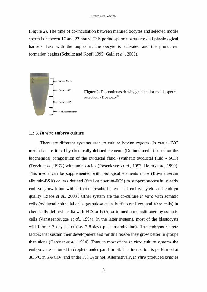
(Figure 2). The time of co-incubation between matured oocytes and selected motile
sperm is between 17 and 22 hours. This period spermatozoa cross all physiological
barriers, fuse with the ooplasma, the oocyte is activated and the pronuclear
formation begins (Schultz and Kopf, 1995; Galli et al., 2003).
Figure 2. Discontinuos density gradient for motile sperm
selection - Bovipure®
.
1.2.3. In vitro embryo culture
There are different systems used to culture bovine zygotes. In cattle, IVC
media is constituted by chemically defined elements (Defined media) based on the
biochemical composition of the oviductal fluid (synthetic oviductal fluid - SOF)
(Tervit et al., 1972) with amino acids (Rosenkrans et al., 1993; Holm et al., 1999).
This media can be supplemented with biological elements more (Bovine serum
albumin-BSA) or less defined (fetal calf serum-FCS) to support successfully early
embryo growth but with different results in terms of embryo yield and embryo
quality (Rizos et al., 2003). Other system are the co-culture in vitro with somatic
cells (oviductal epithelial cells, granulosa cells, buffalo rat liver, and Vero cells) in
chemically defined media with FCS or BSA, or in medium conditioned by somatic
cells (Vansteenbrugge et al., 1994). In the latter systems, most of the blastocysts
will form 6-7 days later (i.e. 7-8 days post insemination). The embryos secrete
factors that sustain their development and for this reason they grow better in groups
than alone (Gardner et al., 1994). Thus, in most of the in vitro culture systems the
embryos are cultured in droplets under paraffin oil. The incubation is performed at
38.5ºC in 5% CO2, and under 5% O2 or not. Alternatively, in vitro produced zygotes

Literature Review
9
can be cultured in vivo after transfer to the oviduct of a temporary recipient as sheep
(Galli and Lazzari, 1996) or cow (Wetscher et al., 2005) or ex vivo in mouse
isolated oviducts (Rizos et al., 2010).
1.2.3.1. Culture conditions and embryo development
The oocyte source and the environment of the ovarian follicle could influence
the transcriptome of the matured oocyte and the subsequent cleavage stage of the
embryo (Sirard, 2012). A correctly programmed oocyte can support the subsequent
embryo development, evidenced in the efficiency and quality of blastocyst produced
in vivo (Graf et al., 2014). The cleavage speed (mitotic divisions) has been
correlated with the subsequent blastocyst rates in bovine (Lonergan et al., 2000) and
humans (Salumets et al., 2003), and related factors as oocyte, spermatozoa and
culture conditions affect the timing of this event (Lechniak et al., 2008). Besides, it
is known that the faster cleaved embryos have a higher chance to develop to the
blastocyst stage (Lonergan et al., 1999) and that IVC environment impacts the
mRNA expression and quality of the embryo (Rizos et al., 2002a). During the
evolution of IVC associated problems like “zygote arrest” or “8-16 cell block” have
been identified (Camous et al., 1984) and resolved (Rosenkrans et al., 1993). Under
in vitro conditions the dynamic of embryo development previous to compaction, are
related to the subsequent developmental stages (Gutiérrez-Adán et al., 2004a).
Crucial processes in early embryo development are compaction and cavitation of
the morula. Compaction is a prerequisite to TE formation, where are involved cell-
cell adhesions between blastomeres, mediated by molecules (E-cadherin) and tight
junction proteins (Van Soom et al., 1997; Gordon, 2003). The blastocoel formation
is accompanied for the formation of TE and the ICM. This process is mediated by
water influx of polarized epithelium were several molecules are involved as
aquoporins (Offenberg et al., 2000). Compaction and cavitation are dependent of
adequate embryonic gene transcription. In IVP embryos the compaction occurs in
lesser degree that in the in vivo counterparts (Van Soom et al., 1997). In vitro
culture systems have advanced on embryo production sacrificing the health of the
embryo and the subsequent fetus and offspring. To obtain a competent embryo

Literature Review
10
under in vitro conditions requires accurately mimicking of most if not all
constituents of the in vivo environment. The differences between in vivo and in vitro
embryo culture systems (Eyestone et al., 1987; Galli et al., 2001) are evidenced in
the pregnancy rates of in vivo derived embryos (Wetscher et al., 2005). All the
knowledge used to develop an optimal embryo culture environment has been
generated from the results obtained with animal models (rodents and mammals)
(Ménézo and Hérubel, 2002). However, we are still far away from the optimum in
vitro culture system such as only 30-40% of the matured oocytes used develop to
blastocyst stage (Rizos et al., 2008).
1.2.3.2. Culture conditions and embryo quality
Despite the considerable improvements of assisted reproductive technologies
in the last two decades, in vitro embryo production processes, designed in simulated
environment to support early embryo development, are yet far to the physiological
conditions (Lonergan and Fair, 2008). These conditions are suboptimal, as is
evidenced by lower blastocyst yields (30 - 40%), lower cryotolerance (Rizos et al.,
2008), altered inner cell mass/trophectoderm cells ratio (Plourde et al., 2012),
altered gene expression patterns (Niemann and Wrenzycki, 2000), and lower
pregnancy rates of transferable embryos (Pontes et al., 2009).
The addition of foetal bovine serum to the culture media accelerates
embryonic developmental kinetics and increases the number of embryonic cells
(Van Langendonckt et al., 1997). However, embryos cultured in the presence of
serum have a lower level of compaction at the morula stage (Thompson, 1997);
exhibit a greater accumulation of lipid droplets in the cytoplasm (Rizos et al.,
2002a); have lower cryotolerance (Rizos et al., 2002a) and present alterations in
gene expression (Rizos et al., 2003) compared to in vivo produced embryos. In
addition, serum has been linked to the "Large Offspring Syndrome" (LOS) (Farin et
al., 2001; Lazzari et al., 2002), that causes the birth of large calves with
musculoskeletal disorders, alterations in the development of the allantois and
defects in vascularization and development of the placenta, showing a smaller area
of maternal-fetal contact (Farin et al., 2001, 2006).
Literature Review
11
It has been showed that the ligated sheep oviduct can provide an adequate
environment, not only for sheep embryos but also for those from other farm species,
including cattle (Rizos et al., 2002a). While culture of in vitro produced zygotes in
the ewe oviduct did not affect blastocyst yields, the oviductal environment of the
intermediate recipient clearly improved the quality of the blastocysts, as measured
by survival after cryopreservation (Rizos et al., 2002b) and pregnancy rates (Lazzari
et al., 2002). Furthermore, the significance influence of culture conditions, in vitro
or in vivo in the homologous bovine oviduct, on the transcriptome of the embryos in
relation to embryonic genome activation has been well demonstrated (Gad et al.,
2012).
The best method of evaluation of each embryo produced is the ability to
establish and maintain a pregnancy after transfer. However, for practical and
economic reasons, it is only ever possible to transfer a subset of embryos.
Therefore, alternative methods for evaluating embryo quality have been developed.
These embryo quality assessments are considered valuable tools to understand or
identify the in vitro culture conditions.
1.2.3.2.1. Morphology
The blastocyst morphology is the first parameter considered for the
immediate transference or cryopreservation. Under light microscope, in vivo
embryos present a lighter color when compared to their in vitro counterparts (Fair et
al., 2001; Rizos et al., 2002a) (Figure 3). These morphological differences between
in vivo and in vitro embryos have been studied at ultrastructural level. Fair et al.
elucidated the effect of culture environment on the blastocyst morphology; showing
that zygotes cultured in vivo (ewe oviduct) had similar morphology to the in vivo
counterparts, and the blastocyst cultured in SOF with FCS present differences from
these two groups, in terms of cell-to-cell contacts (Fair et al., 2001). Rizos et al.
evidenced that in vivo embryos present a higher plasma membrane attachment to
ZP, a dense-continuous cover of microvilli, a closely connected TE an ICM cells
surrounded by small intercellular spaces, a small number of lipid droplets and a
healthy mitochondria (Rizos et al., 2002a). The in vitro counterparts exhibited less

Literature Review
12
and discontinuous microvilli over the plasma membrane, and increased lipid content
(Rizos et al., 2002a). Higher lipid content in blastocyst is related to a lower
cryotolerance (Abe et al., 2002). The differences at ultrastructural level, between in
vivo and in vitro embryos, even between embryos produced in vitro in different
culture systems may in part explain the variation in cryotolerance observed (Fair et
al., 2001; Rizos et al., 2002a).
Figure 3. In vivo (a) and in vitro (b) derived D7 embryos
1.2.3.2.2. Cryotolerance
The ability of the embryo to tolerate the stress caused by cryopreservation
and to continue the development is considered a predictor of embryo quality and
viability (cryotolerance) (Rizos et al., 2002b, 2003; Moore et al., 2007). Culture
conditions influence the survival and subsequent development of in vitro produced
embryos after the cryopreservation. The in vitro embryos are more sensitive to
cryoinjury than the in vivo embryos (Hasler et al., 1995). Mucci et al. compared the
supplementation of Charles Ronsenkrans + amino acids medium (CR1aa) with
estrous cow serum and⁄or BSA, reporting similar blastocyst rates (30.9-33.1%), but
higher post-thaw survival rates (24h) with CR1aa with BSA (51.9 vs. 25%) (Mucci
et al., 2006). Rizos et al. showed that SOF with FCS reduced the cryotolerance of
vitrified bovine blastocysts. Moreover, the presence of FCS during in vitro culture
increase the number and size of lipid droplets (2–6 and >6 mm) in the embryos
when compared with the once produced in FCS free systems (2 mm) (Rizos et al.,

Literature Review
13
2003). This lipid accumulation plays an important role reducing embryo
cryotolerance (Abe et al., 2002). Embryo co-culture with granulosa cells
monolayers reduced blastocyst yiels on day 7 (13 vs. 31%), but had a positive effect
on survival rate after vitrification and warming when compared to SOF with FCS
produced embryos (Rizos et al., 2001). Similarly Mermillod et al. using a BOEC
co-culture improved the blastocyst rates of zygotes cultured in SOF (41 vs. 27%)
and TCM media (28 vs. 10%), and the survival rates after vitrification (69 vs. 22%)
(Mermillod et al., 2010). In vivo culture (ewe oviduct) of IVF zygotes, increase
dramatically the embryo cryotolerance, to a similar levels to their in vivo
counterparts (Galli and Lazzari, 1996; Rizos et al., 2002b). This was confirmed by
Rizos et al who showed that blastocysts cultured either in the ewe oviduct or
produced totally in vivo presented higher cryotolerance than the in vitro once
produced in SOF with FCS (88.0 vs. 5.6%) (Rizos et al., 2002b).
1.2.3.2.3. Gene Expression
Analysis of expression patterns of developmentally important genes essential
in early development provides valuable information about the effect of
environmental factors on the early embryo. The products of these genes are
involved in various biological processes including metabolism, growth factors -
cytokine signaling, stress adaptation, transcription and translation, epigenetic
regulation of transcription, apoptosis, compaction and blastocyst formation
(Wrenzycki et al., 2005).
It has been clearly demonstrated the effect of culture environment on gene
expression in the developing embryo (Lonergan et al., 2003; Wrenzycki et al.,
2005; Cagnone et al., 2012) and this effect is evidenced under in vitro and in vivo
culture systems, and between in vitro systems (Eckert and Niemann, 1998; Natale et
al., 2001; Rizos et al., 2002c; Rinaudo and Schultz, 2004; Wrenzycki et al., 2005).
A very clear example is the presence of FCS in SOF media (Rizos et al., 2003)
modifying the expression level of qualitatively important genes related with
metabolism, compaction and implantation. On the other side, in vivo cultured (e.g.:
Literature Review
14
ewe oviduct) bovine blastocyst had similar expression patterns with totally in vivo
once (Lazzari et al., 2002; Rizos et al., 2002b).
In the same extend, Rizos et al. after culturing bovine zygotes ex vivo in
isolated mouse oviducts showed that most of the gene expression transcripts studied
on these embryos were similar to that of embryos derived either from zygotes
cultured in vivo in the ewe oviduct or in vivo produced; however, the effect was
related with the media used for culturing the oviducts, SOF or KSOM (Rizos et al.,
2007). In a microarray study between in vitro and in vivo produced embryos,
(Corcoran et al., 2006) found that 85% of the differentially expressed transcripts
were downregulated in in vitro blastocysts, suggesting that the causal of lower
developmental competence of in vitro embryos is a deficiency of the machinery
associated with transcription and translation.
1.2.3.2.4. Embryo cell number
The evaluation of the embryo cell number is considered also as a valuable
embryo quality test (Thouas et al., 2001). A possible asynchrony in the mitotic
division in the blastomeres could be reflected in the compaction stage of the morula,
and in the subsequent inner cell mass and trophectoderm cell differentiation
(Johnson and Ziomek, 1981). Differential staining can be used to obtain a better
estimation of embryo quality and embryo differentiation (Figure 4). Therefore, this
technique allows to compare ICM and TE development in different culture
conditions to evidence deviations in embryo development (Van Soom et al., 2001).
Iwasaki et al. evaluated the percentage of ICM to TE cells in bovine embryos
founding them cultured in vitro with fewer total cells as well as a significantly
lower proportion of ICM compared to those cultured in vivo (Iwasaki et al., 1990).
Clemente et al. used differential cell count to evaluate the quality of in vitro bovine
blastocysts in presence or absence of progesterone (Clemente et al., 2009).
Similarly, Rodriguez et al. demonstrated that retinoic acid and specific receptor
agonist increases the number of cells in the ICM and TE of in vitro produced bovine
embryos (Rodríguez et al., 2006, 2007). Recently, Trigal et al. evaluated the use
Activin A under in vitro conditions on bovine embryo development and quality,

Literature Review
15
finding a lower number of TE cells in embryos cultured in absence of Activin A
(Trigal et al., 2011).
Figure 4. D8 p.i. bovine blastocyst stained with bisbenzimide and propidium iodide.
1.3. Role of the oviduct in early embryo development
1.3.1. Anatomophysiological characteristics of the bovine oviduct
The oviduct in bovine is a tubular structure, sustained by the mesosalpinx that
connects the ovary with the uterine horn. The oviduct is divided in five
morphological and functional parts: (i) the infundibulum, (ii) the ampulla, (iii) the
ampullary-isthmic junction, (iv) the isthmus and (v) the utero-tubal junction. The
infundibulum is the most proximal structure to the ovary and is funnel shaped, and
with its fimbriae receives the oocyte after ovulation. The ampulla is the wider part
of the tubal structure. The ampullary-isthmic junction is the place where
fertilization takes place. The isthmus presents a narrow lumen, is the sperm
reservoir previous to fertilization and also where the early stages of embryo
development take place. The utero-tubal junction, connect the isthmus with the
uterus (Yániz et al., 2000).
Histologically, the oviduct in their external surface it is composed by a serosa
layer (Tunica serosa), followed by the Tela subserosa, constituted by muscle fibers
and blood vessels. The subsequent muscle layer (Tunica muscularis) is different in
each oviduct segments, in the infundibulum and the ampulla it is thinner than in the
isthmus. The inner part of the oviduct (Tunica mucosa) is composed by fibrous and
cellular connective tissue and epithelial cell layer (Ellington, 1991).

Literature Review
16
The oviduct is an active organ that maintains and modulates the fluidic milieu
for sperm capacitation, transport and fertilization of the mature oocyte and early
embryonic development (Rodriguez-Martinez, 2007; Leese et al., 2008; Lloyd et
al., 2009). The first stages of bovine embryo development occur in the oviduct,
where the embryo spends around 4 days (Hackett et al., 1993). This, generate an
interest to know its role as multifunctional and specialized reproductive organ
(Rodriguez-Martinez, 2007; Leese et al., 2008).
The infundibulum receives the oocyte in the ovulation with dynamic
movements. The ciliated cells receive and guide the oocyte to the oviduct lumen. In
the ampulla, the final events of oocyte maturation and the fertilization take place
(Talbot et al., 2003). At the other extreme, the sperm remains in the isthmus
particularly in the distal part. The isthmus described as functioning as a sperm
reservoir, sperm adhere transiently to the epithelium, and at the time of ovulation
they are released (Hunter and Wilmut, 1984; Talevi and Gualtieri, 2010). In these
phase, the oviductal environment and sperm-OEC interactions ensures their proper
viability, motility and fertility (Talevi and Gualtieri, 2004). Sperm release from the
OEC is attributed to a mechanism mediated by heparin-like glycosaminoglycans
considered physiological components of the bovine oviductal fluid which
concentrations and activity are dynamic, during the estrous cycle (Bosch et al.,
2001). Oviduct adhesion contact also contributes to sperm selection of higher
quality sperm subpopulation (Gualtieri and Talevi, 2000, 2003) represented by the
ability to bind the zona pellucida and fertilize the oocyte (Talevi and Gualtieri,
2004). After fertilization in the ampullary-isthmic junction, the developing embryo
passes through the isthmus, supported by ciliary beat and muscular contractions,
until it reaches the uterus at about the 16 cell stage on day 4 (Ellington, 1991). A
schematic representation of the oviduct and processes involved are shown in the
figure 5.

Literature Review
17
Figure 5. Schematic representation of the oviduct and its anatomical parts
1.3.1.1. Oviductal epithelium
The oviductal epithelium is the intimate part of the oviduct. Oviductal
epithelium is composed of two different cell types, ciliated and secretory. During
transport processes, the ciliary show a synchronized movement leading a directed
flow of fluids (Abe and Hoshi, 1997). Secretory cells present microvilli on their
apical side and secrete substances (oviduct-specific glycoprotein) and growth
factors, usually by exocytosis, associated with the first days of the estrous cycle,
which contribute on the development of the early embryo (Abe, 1996; Murray,
1997).
Populations of OEC types are dynamic during the phases of the estrous cycle. The
percentage of ciliated cells decreases in the infundibulum and the ampulla during
the luteal phase compared with the follicular phase (Yániz et al., 2000).
Interestingly, it has been suggested that cell morphology is modified in function of
embryo development and cyclic changes (Suuroia et al., 2002). Thus, the height of
ciliated cells decreases in the infundibulum and ampulla in the luteal phase and in
the isthmus the height of secretory cells also diminishes (Abe et al., 1999). Studies
concerning gene expression in bovine oviducts, evidenced the ovary and/or oocyte
effect on the OEC function (Bauersachs et al., 2003), as well as, the differences
during the estrous cycle (Bauersachs et al., 2004)
1.3.1.2. Oviductal fluid
The conditions of oviductal environment are reflected in the oviductal fluid
(OF). The OF is generated by (i) transudation from plasma into the oviductal lumen
together with (ii) the secretion of substances synthesized by the secretory cells
(Menezo and Guerin, 1997). OF composition is very complex, containing simple,

Literature Review
18
and complex carbohydrates, ions, lipids, phospholipids and proteins (Leese et al.,
2001; Avilés et al., 2010). Some of these components are metabolic substrates, such
as lactate, pyruvate, amino acids, and glucose, whose concentrations differ from
those present in the uterine fluid and the serum (Leese, 1988; Leese et al., 2007;
Hugentobler et al., 2008). In bovine, aspartate, glutamate, serine, glycine, tyrosine,
phenylalanine, lysine and alanine of amino acids were present in higher
concentrations in oviductal fluid than in blood plasma (Hugentobler et al., 2007).
Secretions present in the OF affect oocyte and sperm function (Killian, 2011;
Mondéjar et al., 2013) with proteins as glycodelins, and lactoferrin involved on
gamete interaction (Ghersevich et al., 2015), and oviductin, osteopontins and the
complement protein C3 on early embryo development (Tse et al., 2008).
During preimplantation development, bovine embryos obtain energy by
oxidative phosphorylation (pyruvate and amino acids oxidation), while at
compaction and blastulation switch to glycolysis (Kim et al., 1993; Thompson,
2000). Hugentobler et al. show the association in glucose, lactate and pyruvate
concentration between oviductal fluid and plasma, being OF composition mediated
the composition determined by the BOEC (Hugentobler et al., 2008). Georgieu et
al. demonstrated the modulation of the oviductal environment (secretory proteomic
profile) in presence of gametes in a favorable way, as well as prepare the oviduct
milieu for the arrival of the embryo. Twenty proteins were regulated by sperm, and
one protein was regulated by oocytes (Ig kappa light chain). Three proteins were
commonly regulated by both gametes (Complement Component C3, Ig kappa
variable region, and haemoglobin beta chain), and one protein showed regulation by
sperm and oocytes in opposing directions (Complement Component C3) (Georgiou
et al., 2005, 2007). These regulated proteins have been previously reported to play
direct roles in sperm motility, viability, (Kouba et al., 2000) sperm-ZP binding
(Banerjee and Chowdhury, 1994), ZP hardening (Kratz et al., 2003) embryo-
maternal interactions (Reed et al., 1998), oocyte (Hess et al., 1999) and early
embryo development (Lim and Hansel, 1998), cell proliferation (Hulboy et al.,
1997), differentiation and apoptosis, fertilization rates (Dinara et al., 2001) and pH

Literature Review
19
(Ekstedt et al., 2004). The oviduct-specific glycoprotein (OVGP1) is a component
of the OF identified in many species in a highly conserved form. Nowadays it is
clear the interaction with oocytes and sperm (Buhi, 2002). OVPG1 synthesis and
secretion is dynamic and related to estrogen (Buhi, 2002; Killian, 2004) and
luteinizing hormone stimulation (Sun et al., 1997). OVGP1 binds to the zona
pellucida (ZP) of the oocyte and early embryo suggesting a role in early
development (Buhi, 2002). Coy et al., (2008, 2012). demonstrated that OVGP1 and
heparin-like glycosaminoglycans (GAGs) from the oviductal fluid of sows and
cows participate in the functional modification of the ZP, affecting the sperm-
oocyte interaction and contributing to the control of polyspermy. Besides, OVGP1
and sperm interactions increased rates of fertilization and embryonic development
(Killian, 2004). In addition, it is suggested that OVGP1 stabilizes the
microenvironment immediately surrounding gametes and embryo, preventing
dispersal of essential nutrients and ions, particularly during ciliary beating or
muscular contraction, by increasing the viscosity of luminal fluid (Hunter, 1994;
Mondéjar et al., 2012).
Coy et al. evaluate the oviductal fluid (30 min. incubation) influence on the
ZP in pig and cow oocytes, and demonstrate an increasing in the proteolytic
resistant of the ZP reflected in increased pronase digestion times (3-8h.), and a
modulation of sperm-ZP interaction through increased monospermy (Coy et al.,
2008). Lloyd et al. exposed in vitro matured porcine oocytes to bovine OF during
30 min before fertilization increasing the blastocyst rate, and quality in terms of
morphology, cell numbers, as well as gene expression patterns of apoptotic and
developmentally related genes (Lloyd et al., 2009). In the same line, in bovine,
Cebrian Serrano et al. evaluated the effect of short incubation of matured oocytes
with bovine OF with no effect on embryo development but with notable
modification on genes transcripts like G6PD and SOD32 (Cebrian-Serrano et al.,
2013).

Literature Review
20
1.3.2. Embryo-maternal dialogue in the oviduct
For a successful pregnancy establishment, a complex signal exchange
between the newly formed embryo and the maternal reproductive tract is essential.
In ruminants, the principal pregnancy-recognition signal sent by the embryo is
interferon tau, secreted by the trophoblast from day 10 up to Day 21-25 (Spencer
and Bazer, 2004). While, a high proportion of embryonic losses occur between Day
8 and 17 of pregnancy (Humblot, 2001; Thatcher et al., 2001). Alterations in the
environment of the early embryo could have consequences in the subsequent
development. The oviduct as first environment of the embryo development is
considered the starting point to search any signal between the embryo and the
reproductive tract (Wolf et al., 2003). The embryo in the oviduct undergoes
epigenetic changes responsible for further development, implantation and postnatal
phenotype (Wrenzycki et al., 2005). Thus, the exchange of signals between the
embryo and the oviduct are remarkable. However, the mechanisms involved on this
embryo-maternal communication currently are mostly unknown (Fazeli, 2008).
Evidence in vivo in mice, by RT-qPCR showed changes in the oviduct gene
expression depending on the presence or absence of embryos (Lee et al., 2002).
Recently, new sequencing technologies (microarrays) have used to elucidate this
question of a complex molecular dialogue between the maternal tract and embryo in
pigs (Almiñana and Fazeli, 2012). Using these technologies Almiñana et al. showed
that embryo maternal communication exists at the very earliest stages of pregnancy,
before that well-known embryonic signal of maternal recognition. In this
communication, the embryo might play a role as a modulator of the immune system
in the maternal tract, inducing the down-regulation of immune related genes to
allow the refractory uterus to tolerate the embryo and support its development
(Almiñana et al., 2012). In contrast, Maillo et al. (2015) in a microarray study, did
not found differenced in the bovine oviduct transcriptome in the presence or
absence of an 8-16 cell embryo in vivo, concluding that the small embryo may have
a specific side effect which is probably lost in the long oviduct. Obviously, multi-
ovulation species like mice and pigs cannot be directly compared with mono-

Literature Review
21
ovulated once like bovine. Thus, the bovine model would provide new information
on early embryo maternal communications that may be important for humans.
In bovine, substances like growth factors (GH - IGF-system), the hyaluronic
acid (HA) and the platelets activating factor (PAF) could be involved in very early
embryo-maternal communication (Wolf et al., 2003). Equally, OEC express the GH
receptor (Hull and Harvey, 2001) enabling them to react with the GH produced by
the embryo. It has been shown that embryos cultured in vitro in medium with GH
showed similarly characteristics to their in vivo counterparts, in terms of
morphology, cell numbers and apoptosis rates (Kölle et al., 2001, 2002).
Insulin and insulin-like growth factors (IGFs) are family of structurally
related polypeptides that regulate growth of many cell types. The activity of these
peptides is mediated by IGF transport and its binding to membrane receptors. The
function is modulated by interactions with six insulin-like growth factor binding
proteins (IGFBPs) which are present in many extracellular fluids and in early
embryos (Winger et al., 1997; Pushpakumara et al., 2002). Studies in bovine
oviduct in vivo reveal different expression patterns of IGF types, their receptors and
binding proteins (Pushpakumara et al., 2002). Secreted IGFs, regulated by IGFBPs,
may act directly on the embryo or via modulation of oviduct secretions and
muscular activity to positively influence the success of early embryonic
development (Pushpakumara et al., 2002). Hyaluronic acid (HA) is a
glucosaminoglycan and a component of the extracellular matrix that regulates
cellular events such as signaling, gene expression, proliferation, differentiation,
adhesion, migration and morphogenesis. HA is a normal component of mammalian
follicular, oviductal and uterine fluid (Lee and Ax, 1984). The effect of HA in in
vitro embryo culture has been reflected in increased blastocyst rates, cell number
and gene expression of bovine embryos (Stojkovic et al., 2002; Palasz et al., 2006)).
1.3.3. Oviductal environment and in vitro models
In vitro systems are a valuable tool to study pathways and mechanisms,
which are difficult to study in vivo and cell cultures provide valuable aspects of

Literature Review
22
physiologic or pathologic mechanisms. Studying the oviductal environment is
crucial to further our understanding of the underlying regulatory mechanisms
controlling embryo development (Aviles et al., 2015). The advantages of the
oviductal environment have been demonstrated in different models, through the
substances isolated and the effect of environment per se. Many physiological
aspects have been clarified; however, many others are still unknown (Hunter, 2012).
Thus, interactions between oviductal epithelial cells and sperm observed
under in vitro studies (Boquest and Summers, 1999; Boquest et al., 1999; Boilard et
al., 2002; Gualtieri and Talevi, 2003; Hunter, 2003; Talevi and Gualtieri, 2004) find
sperm selection mechanisms (Ellington et al., 1999; Pauw et al., 2002; Talevi and
Gualtieri, 2004). Cell culture has been used for studying physiological processes
and their regulatory mechanisms of embryotrophic function as complement C3b
(Lee et al., 2002). Besides, protein patterns were compared in vivo vs. in vitro using
different culture systems (Woldesenbet and Newton, 2003) as well as the regulatory
effects on single proteins (Eberhardt et al., 1999).
Under in vitro conditions expression of relevant embryotrophic factors as
OVGP1 (Briton-Jones et al., 2004), endothelin-1 (ET-1) (Reinhart et al., 2003),
members of the insulin-like growth factor (IGF) system (Xia et al., 1996; Winger et
al., 1997), or leukaemia inhibitory factor (LIF), vascular endothelial growth factor
(VEGF), epithelial growth factor (EGF), IGF1, tumor growth factor β 2 (TGFβ2),
and interleukin-4 (IL4) (Okada et al., 2005) were applied to obtain information
about the differentiation state of cultured cells.
Gene expression patterns of in vitro cell cultures constitute an excellent tool
for established cell lines characterization. For example, Schmaltz–Panneau et al.
used BOEC monolayers as in vitro models to evidenced differences in the gene
expression of OEC in presence of bovine embryos (Schmaltz-Panneau et al., 2014).
Thus, this model may provide useful information on early embryo maternal
interaction signals.

Literature Review
23
1.3.3.1. Bovine oviductal epithelial cells (BOEC) and their conditioned media
Bovine oviduct epithelial cells (BOEC) as the intimal structure of the oviduct
in contact with gametes and embryos, are considered as the most suitable in vitro
model to study early embryonic events and embryo maternal interactions
approaching to mimic phypsiological conditions (Rottmayer et al., 2006; Ulbrich et
al., 2010). BOEC are usually obtained from oviducts of slaughtered heifers or cows.
The BOEC, could be isolated by enzymatic procedures using trypsin and/or
pancreatin (McNutt-Scott and Harris, 1998) or collagenase (Sun et al., 1997; Bosch
et al., 2001; Mishra et al., 2003) in different concentrations. Also BOEC could be
isolated by mechanical procedures remaining in cell aggregates which are mostly
cultured non-adhesively in a suspension culture system (Lim et al., 1997; Pegoraro
et al., 1998; Yadav et al., 1998; Boquest and Summers, 1999; Kamishita et al.,
1999; Gualtieri and Talevi, 2000; Pauw et al., 2002; Ulbrich et al., 2003). Isolated
cells are seeded to attach to the culture dish and growth to a confluent monolayer
(Figure 6).
Figure 6. BOEC confluent monolayer.
(Day 7 of culture).
When a BOEC line is established for embryo culture in vitro (Figure 7) it is
important to determine the stage of the estrous cycle of the animal of which oviduct
is used. Thus, estrous cycle is estimated by ovarian morphology, ipsilateral to the
oviduct, according criteria described in the literature (Ireland et al., 1979).
The use of BOEC allowed the study of mechanisms involved in sperm storage in
the oviduct (Abe and Hoshi, 1997) and was a major breakthrough in the in vitro
culture of bovine embryos (Gandolfi and Moor, 1987). In co-culture experiments
with sperm, BOEC have been obtained from a luteal phase (Boquest and Summers,

Literature Review
24
1999; Boquest et al., 1999) and from follicular phase (Ellington et al., 1998; Bosch
et al., 2001).
Figure 7. BOEC - Embryo co-culture after 7
days post insemination
In embryo co-culture, BOEC at estrous have been successfully used as in vitro
model for intercept embryo maternal interactions (Rief et al., 2002). Recently,
Cordova et al. used oestrous-metoestrus (Day 0-3) BOEC for early (Day 1-4) or late
(Day 4-7) embryo co-culture showing that the presence of the cells during the four
first days of development, which correspond to the presence of embryos in the
oviduct in vivo, accelerate the kinetics of blastocyst development and induce
changes of genes involved in epigenetic control (Cordova et al., 2014). Moreover,
BOEC co-culture overcomes the early embryo developmental block occurring at the
8-16 cell stage in ruminant, and provide an optimized in vitro environment for
embryo culture until blastocyst stage (Gandolfi et al., 1989; Ellington et al., 1990).
The positive effects of these cells on the embryos are attributed to embryotrophic
substances, such as growth factors secreted by the cells (Nancarrow and Hill, 1994;
Vanroose et al., 2001; Tse et al., 2008). Besides, BOEC modulates the surrounding
environmental conditions, decreasing the oxygen levels in the culture medium,
preventing the formation of deleterious radicals as reactive oxygen species (ROS)
(Thompson, 2000; Vanroose et al., 2001), removing toxic substances from the
medium (ammonia) (Nancarrow and Hill, 1994), decreasing the glucose and ion
levels, and change the culture metabolites (Edwards et al., 1997) that could have
detrimental effects on the embryos (Vanroose et al., 2001). A successfully use of
somatic cells could be attributed to the fact that not only few soluble factors are
responsible for successful development but also the different signals and factors
which act cooperatively (Killian, 2004).

Literature Review
25
Co-culture systems has been associated with methodological complexity, lack
of repeatability and biosanitary risk (Guerin et al., 1997). To avoid the use of
primary cultures that may be a risk of contamination, the use of established cell
lines will allow to have a standardized culture conditions and a better control
(Pegoraro et al., 1998). Established cell lines constitutes an attractive co-culture
system for assisted reproductive technologies by providing a continuous supply of
cells that are easily cultured in vitro, cryopreserved and screened for pathogens
(Eyestone et al., 1991; Ulbrich et al., 2010). The use of co-culture systems offer the
opportunity to do essential and functional studies of candidate genes in embryo-
maternal dialogue (Schmaltz-Panneau et al., 2014) that under in vivo conditions are
difficult to study.
An alternative to co-culture avoiding a direct contact between BOEC and
embryos is the use of conditioned media with positive effects on embryo
development and live calves after transfer (Eyestone and First, 1989; McCaffrey et
al., 1991; Lim et al., 1997). Conditioned media offer advantages over that
monolayer co-culture in that no additional cells are required for the culture system
and the media may be stored frozen, eliminating the need for preparing fresh cells
for each subsequent embryo group (Eyestone et al., 1991; Ramos-Ibeas et al.,
2014). The frozen-thawed conditioned media culture system allows the same batch
of media to be used with sequential groups of embryos. This would potentially
reduce the variation in embryo development due to variations often noted in
monolayer culture systems. The BOEC conditioned media are able to support
embryo development to blastocyst stage (Wang et al., 1990; Mermillod et al., 1993)
through secreted embryotrophic components as, OVGP1 (Briton-Jones et al., 2004),
ET-1 (Reinhart et al., 2003), IGF (Xia et al., 1996; Winger et al., 1997), VEGF,
EGF, IGF1, TGFβ2, and IL4 (Okada et al., 2005) that have been properly identified,
however many other secretions are still unknown. Therefore, conditioned media can
be used to investigate the mechanism(s) by which somatic cells support the
development of embryos in vitro (Rieger et al., 1995).

Literature Review
26
1.4. Extracellular Vesicles
Extracellular environment contains a large number of mobile membrane-
limited vesicles called ‘‘extracellular vesicles’’ (EV). EVs or cell secreted vesicles
contain microvesicles (MV), apoptotic bodies and exosomes. Originally the EV
were associated with removal process of receptors and with cellular waste function,
being unnoticed and remained without importance. Then, the concept changed and
it was found to have immune effects (Raposo et al., 1996). These evidences opened
the possibility that EVs could play a role in intercellular communication (Théry,
2011) generating interest, and discovering different types and tissues in vivo. The
EVs have been found in biological fluids (Simpson et al., 2008) as plasma (Caby et
al., 2005), serum (Taylor et al., 2006; Taylor and Gercel-Taylor, 2008), urine
(Pisitkun et al., 2004) epididymal fluid (Gatti et al., 2005), amniotic fluid (Asea et
al., 2008), follicular fluid (Silveira et al., 2012), and milk (Admyre et al., 2007).
The EVs are classified according to: size, biological content, biogenesis and
way of release from the cell. In relation to the size, EVs are in a range from 30 to
1000 nm and can be divided in:
Exosomes, which size is between 30–90 nm (Théry, 2011), correspond to the
internal vesicles of multivesicular bodies (MVBs) that are released in the
extracellular environment upon fusion of MVBs with the plasma membrane
(Théry et al., 2002).
The MV are over 100 nm (Raposo and Stoorvogel, 2013) and are shed by all
types of cells by outward budding of the cellular plasma membrane and
subsequent fission of the formed vesicle (György et al., 2011a).
The Apoptotic Bodies also are over 100 nm and are released from apoptotic
cells which undergo several steps in their dying process including cell
shrinkage, chromatin condensation, nuclear fragmentation, membrane
blebbing and finally the breakdown of the cell into apoptotic bodies (Reed,
2000).

Literature Review
27
Nowadays, it is known that exosomes contain proteins secreted by epithelial,
tumor and haematopoietic cells, denoting that EV could be released from several
types of cells.
The commonly applied and basic method for separating exosomes, apoptotic
bodies, and microvesicles (fractions) is the differential centrifugation (Théry et al.,
2006). The sedimentation of vesicles depends on their size, density and shape.
Under centrifugation, minor size vesicles (exosomes) are typically isolated by
sedimentation at 100.000-200.000xg (100-200k) (Théry et al., 2009), and the major
size vesicles (MV) are typically reported to be isolated at 10.000-20.000xg (10-20k)
(Witwer et al., 2013).
Quantitative and qualitative assessment of EV Electron microscopy (EM)
techniques are well established and have proven very useful in EV research (Raposo
et al., 1996), providing direct evidence for the presence of vesicular structures.
Heavy metal stains such as osmium tetroxide and uranyl acetate in transmission EM
enables recognition of membrane-surrounded vesicles, and contrast is enhanced by
embedding in methylcellulose. In most cases, concentrated EV suspensions are
applied to grids and fixed, for example with paraformaldehyde. As an alternative
method, EV pellets at the bottom of a centrifuge tube are fixed, and ultrathin cross
sections are analyzed by transmission EM (György et al., 2011b).
Nanoparticle tracking analysis (NTA) is the commercial name of an optical
particle tracking method for obtaining concentration and size distribution of EV
populations (Dragovic et al., 2011). A laser beam is scattered by particles in the
sample, and the mean velocity of each particle is calculated by the Stokes-Einstein
equation on the basis of Brownian motion recorded by a CCD camera. For accurate
quantification of number and size of heterogeneous populations of vesicles, the
procedure requires accurate optimization of camera and analysis settings, and
separate measurements with different settings may be needed to obtain accurate
readings for EV subsets in heterogeneous mixtures.

Literature Review
28
1.4.1. Biogenesis
EV formation is carried out by endocytical pathways of proteins, and usually
are clathrin dependent (Mayor and Riezman, 2004). Endocytic vesicles are released
to early endosomes, and then recycled back to the plasma membrane or transported
to late endosomes together with other proteins and lipids (Maxfield and McGraw,
2004). In late endosomes, two steps have been recognized for sorting protein during
MVBs formation. The first step is the lateral sorting of proteins at the limiting
membrane of endosomal lumen, and the second step is the formation of intraluminal
vesicles (ILV), with the incorporation of proteins, lipids, mRNAs and miRNAs. The
MVBs can either fuse with lysosomes for degradation of proteins, or interact with
the plasma membrane, resulting in the release of the ILVs as exosomes (Lakkaraju
and Rodriguez-Boulan, 2008). The mechanisms of ILVs formation and the sorting
of proteins and lipids into these vesicles, as well as docking and fusion of the MVBs
with the plasma membrane are poorly understood, although the endosomal sorting
complexes required for transport (ESCRT) have been shown to be involved in this
processes (Gassart et al., 2003). The ESCRT machinery are composed by protein
complexes (ESCRT-0, I, II, III) with a sequential recruiting and binding of proteins
that ends in ILV formation (Babst, 2011). Other pathways of protein sorting are
mediated by cholesterol, tetraspanins and lipids. Sphingomyelin, form specialized
microdomains in the endosomal limiting membrane called lipid rafts can bend
inwards and consequently form ILVs (Wubbolts et al., 2003). Biogenesis
mechanisms of EVs are showed in the figure 8. Proteins can also be co-sorted into
ILVs by binding to membrane proteins or lipids (Gassart et al., 2003). The
engulfment of the cell cytoplasm during the vesicle formation can lead to random
packaging of cytosolic proteins. The sorting of RNA into ILVs is a greater enigma,
however the poor correlation between the mRNA content of exosomes and their
donor cells (Mittelbrunn et al., 2011) suggests that the RNA is specifically packed
into the vesicles, rather than randomly engulfed from the cells cytoplasm (Valadi et
al., 2007).
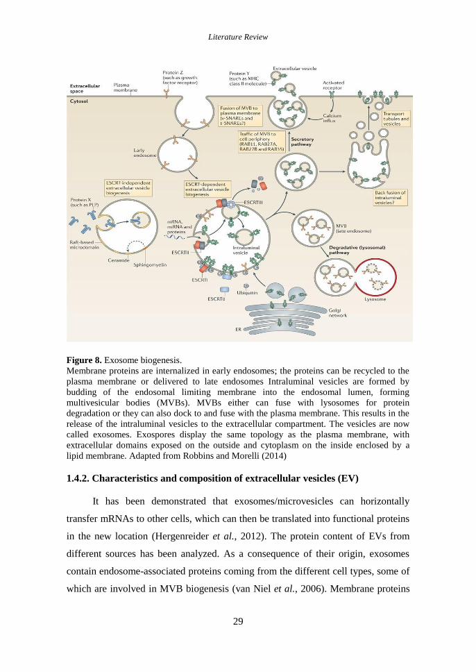
Literature Review
29
Figure 8. Exosome biogenesis.
Membrane proteins are internalized in early endosomes; the proteins can be recycled to the
plasma membrane or delivered to late endosomes Intraluminal vesicles are formed by
budding of the endosomal limiting membrane into the endosomal lumen, forming
multivesicular bodies (MVBs). MVBs either can fuse with lysosomes for protein
degradation or they can also dock to and fuse with the plasma membrane. This results in the
release of the intraluminal vesicles to the extracellular compartment. The vesicles are now
called exosomes. Exospores display the same topology as the plasma membrane, with
extracellular domains exposed on the outside and cytoplasm on the inside enclosed by a
lipid membrane. Adapted from Robbins and Morelli (2014)
1.4.2. Characteristics and composition of extracellular vesicles (EV)
It has been demonstrated that exosomes/microvesicles can horizontally
transfer mRNAs to other cells, which can then be translated into functional proteins
in the new location (Hergenreider et al., 2012). The protein content of EVs from
different sources has been analyzed. As a consequence of their origin, exosomes
contain endosome-associated proteins coming from the different cell types, some of
which are involved in MVB biogenesis (van Niel et al., 2006). Membrane proteins

Literature Review
30
known to cluster into microdomains at the plasma membrane or at endosomes
usually are also enriched on EVs. These include tetraspanins, a family of >30
proteins that are composed by four transmembrane domains (Hemler, 2003).
Tetraspanins such as CD63, CD81, CD82, CD53, and CD37 were first identified in
B cell exosomes in which they can be enriched with >100-fold relative to the
transferrin receptor, which in this cell type can be considered as a genuine marker
for both the plasma membrane and early endosomes (Escola et al., 1998). EVs are
also enriched with proteins associated with lipid rafts, including
glycosylphosphatidylinositol- anchored proteins and flotillin (Wubbolts et al.,
2003).
Exosomes from a variety of cells (Wubbolts et al., 2003; Brouwers et al.,
2013) are highly enriched with cholesterol, sphingomyelin, and hexosylceramides at
the expense of phosphatidylcholine and phosphatidylethanolamine. The fatty acids
in exosomes are mostly saturated or monounsaturated. Together with the high
concentration of cholesterol, this may account for lateral segregation of these lipids
into ILVs/exosomes during their formation at MVB. Less is known about the
protein and lipid contents of MVs and whether particular components are enriched
with MVs relative to their originating plasma membrane.
A major discovery was the demonstration that the content of EVs included
both mRNA and miRNA and that EV associated mRNAs could be translated into
proteins by target cells (Valadi et al., 2007). Later studies reported the RNA
contents of EV isolated from other cell cultures (Skog et al., 2008) and from body
fluids (Hunter et al., 2008; Rabinowits et al., 2009; Michael et al., 2010).
EVs with features of exosomes released by immune cells have been
demonstrated to selectively incorporate miRNA that can be functionally transferred
as a consequence of fusion with recipient cells (Mittelbrunn et al., 2011). Recently,
analysis of RNA from EVs by unbiased deep sequencing approaches demonstrated
that EVs also contain a large variety of other small noncoding RNA species,
including RNA transcripts overlapping with protein coding regions, repeat
sequences, structural RNAs, tRNA fragments, vault RNA, Y RNA, and small
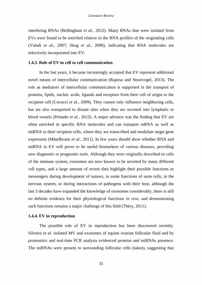
Literature Review
31
interfering RNAs (Bellingham et al., 2012). Many RNAs that were isolated from
EVs were found to be enriched relative to the RNA profiles of the originating cells
(Valadi et al., 2007; Skog et al., 2008), indicating that RNA molecules are
selectively incorporated into EV.
1.4.3. Role of EV in cell to cell communication
In the last years, it became increasingly accepted that EV represent additional
novel means of intercellular communication (Raposo and Stoorvogel, 2013). The
role as mediators of intercellular communication is supported in the transport of
proteins, lipids, nucleic acids, ligands and receptors from their cell of origin to the
recipient cell (Cocucci et al., 2009). They cannot only influence neighboring cells,
but are also transported to distant sites when they are secreted into lymphatic or
blood vessels (Peinado et al., 2012). A major advance was the finding that EV are
often enriched in specific RNA molecules and can transport mRNA as well as
miRNA to their recipient cells, where they are transcribed and modulate target gene
expression (Mittelbrunn et al., 2011). In few years should show whether RNA and
miRNA in EV will prove to be useful biomarkers of various diseases, providing
new diagnostic or prognostic tools. Although they were originally described in cells
of the immune system, exosomes are now known to be secreted by many different
cell types, and a large amount of recent data highlight their possible functions as
messengers during development of tumors, in some functions of stem cells, in the
nervous system, or during interactions of pathogens with their host, although the
last 3 decades have expanded the knowledge of exosomes considerably, there is still
no definite evidence for their physiological functions in vivo, and demonstrating
such functions remains a major challenge of this field (Théry, 2011).
1.4.4. EV in reproduction
The possible role of EV in reproduction has been discovered recently.
Silveira et al. isolated MV and exosomes of equine ovarian follicular fluid and by
proteomics and real-time PCR analysis evidenced proteins and miRNAs presence.
The miRNAs were present in surrounding follicular cells (taken), suggesting that

Literature Review
32
MV and exosomes play a not previously recognized role in mediating cell
communication within the mammalian ovarian follicle (Silveira et al., 2012). Also,
Sohel et al. demonstrated the exosome and non-exosome mediated transport of
miRNAs in bovine follicular microenvironment (Sohel et al., 2013). In the same
line Ng et al. identify and examine the presence and potential role of MV and
exosomes in the uterine cavity. MV and exosome miRNA has enabled
bioinformatic identification of pathways that could be influenced if the exosomes
are taken up by trophectoderm or epithelium at the time of implantation, or
transferred to sperm as they transit the uterine cavity (Ng et al., 2013). The results
of Burns et al. support the hypothesis that exosomes and MV are present in uterine
luminal fluid of pregnant and cyclic ewes, contain specific proteins, miRNAs, and
mRNAs, and are capable of delivering their contents in vitro (Burns et al., 2014).
Differences in the molecular contents by pregnancy status suggest a differential EV
sources (endometrial epithelia-conceptus trophectoderm) serving as evidence of a
communication way between the conceptus and endometrium supporting the notion
that EV in the uterine fluid have a biological role in conceptus-endometrial
interactions important for the establishment and maintenance of pregnancy. Al-
Dossary et al. reveals the expression and secretion via oviductal exosomes of
PMCA4a (Ca2+
homeostasis) in the female reproductive tissues and luminal fluids
during estrous, and their sperm-uptake, with possible roles in sperm viability during
their storage in the oviduct and during capacitation and the acrosome reaction (Al-
Dossary et al., 2013).

Justification and Objectives
33
2. JUSTIFICATION AND
OBJECTIVES

Justification and Objectives
34

Justification and Objectives
35
2.1. Justification
In vitro embryo production (IVP) in cattle is used for research and for applied
purposes. As a research tool, the technology is of great value to address
fundamental questions on endocrine control, molecular switches and metabolic
pathways that modulate early embryo development. As an applied technology,
provides the opportunity to increase the number of offspring from superior
phenotypes. However, several aspects need to be addressed at cellular and
molecular level to increase its efficiency (Galli et al., 2003).
The IVP is a multifactorial process influenced by extrinsic and intrinsic
factors. The embryo development and quality is mainly affected by the origin of the
oocyte and by the post-fertilization culture environment (Lonergan and Fair 2008).
The culture environment affects the viability of the early embryo, its ability to
establish a pregnancy and the characteristics of the newborn calf. This is due to
culture media composition which varies from chemically define to semi-define
normally supplemented with serum, proteins and macromolecules (Rizos et al.
2003).
Embryo development can be assessed by visual observations of blastocysts
formation with stereomicroscope on days 6 to 9. The best measure of embryo
quality is the ability of the embryo to establish a pregnancy after transfer. However,
this is not always feasible due to the big number of embryos produced in vitro and
the high cost of animal maintains. Thus, invasive and not invasive methods have
been used successfully such as the assessment of the ability of the embryo to
withstand cryopreservation (Rizos et al. 2008); the embryo cell number and
proliferation (Trigal et al. 2013); and gene expression patterns of gene transcripts
related with molecular processes, epigenetics, implantation, etc. (Bermejo-Alvarez
et al. 2012).
The oviduct is a dynamic organ where fertilization and early embryo
development occur. Studying the oviductal environment is crucial to further our
understanding of the underlying regulatory mechanisms controlling embryo

Justification and Objectives
36
development (Aviles et al. 2015). The advantages of the oviductal environment
have been demonstrated in different models, through the substances isolated and the
effect of environment per se. Many physiological aspects have been clarified;
however, many others are still unknown (Hunter 2012).
The BOEC as intimal structure of the oviduct has been used as in vitro model
to study the oviduct physiology. In the IVP, the embryo co-culture improved
embryo quality by secretion of embryotrophic substances and regulation of culture
environment, however, it has been associated with a disadvantage, of lack of
repeatability between processes (Myers et al. 1994). On the other hand, many
secretions of cell culture are present in the CM. Thus, CM is an alternative of
embryotrophic substances, which can replace co-culture systems (Mermillod et al.
1993).
The study of the oviductal environment under in vivo conditions is difficult,
so, in vitro models are necessary to study mechanisms, pathways, molecules etc.
related with embryo-maternal interactions. The OF is considered the main
component of oviductal environment. Based on the OF composition, culture media
(e.g. SOF) have been developed supporting early embryo development. Recently, it
was evidenced the positive effect of OF in porcine fertilization (Coy et al. 2008),
however, there are no evidence of its effect on embryo culture in vitro in any
mammalian species.
The EV, as somatic cell secretions are present in the body fluids. The lipids,
proteins, miRNA and mRNA content, convert them as mediators of intercellular
transport (Raposo and Stoorvogel 2013). Although their rapid progress in areas such
as immunology, currently very little is known about the role of these particles in the
reproductive sphere and even less in preimplantation environment.
To summarize, the content of the oviductal environment and its effect on
early embryo development is of great importance and may provide information and
new insights on embryo-maternal communication, which will improve our current
IVP systems. The challenge today is to develop in vitro culture conditions that will

Justification and Objectives
37
allow a physiological growth of the embryo based on the in vivo situation to
enhance the development of high quality embryos.
2.2. Objectives
The main objective of this thesis was to study the role of the oviductal
environment through oviductal cells, oviductal fluid and their extracellular vesicles
on in vitro embryo development and quality in bovine.
To achieve this general objective, specific objectives have been employed to
comprise this thesis:
1. To characterize the extracellular vesicles (EV) secreted by the BOEC
2. To evaluate the effect of different BOEC lines, conditioned media, and
BOEC secreted vesicles (EV) in in vitro culture,
On the developmental capacity of bovine zygotes to reach blastocyst
stage, and
On the quality of the produced blastocysts in terms of:
Cryotolerance
Differential cell number
Gene expression analysis
3. To evaluate the effect of oviductal fluid (OF) supplementation during in
vitro culture,
On the developmental capacity of bovine zygotes to reach blastocyst
stage, and
On the quality of the produced blastocysts in terms of:
Cryotolerance
Differential cell number
Gene expression analysis
4. To characterize the extracellular vesicles (EV) present in the oviductal
fluid

Justification and Objectives
38
5. To evaluate the effect of extracellular vesicles from the ampulla and
isthmus of oviductal fluid isolated at different centrifugal forces in in
vitro culture,
On the developmental capacity of bovine zygotes to reach blastocyst
stage and
On the quality of the produced blastocysts in terms of:
Cryotolerance
Gene expression analysis

Matherials and Methods
39
3. MATHERIALS AND METHODS

Matherials and Methods
40

Matherials and Methods
41
3. Materials and methods
Unless otherwise stated, all chemicals were purchased from Sigma Aldrich
Química S.A Company (Madrid, Spain).
3.1. Oocyte Collection and In vitro Maturation
Immature cumulus oocyte complexes (COCs) were obtained by aspirating
follicles (2-8 mm) from the ovaries of mature heifers and cows collected at
slaughter. COCs were matured for 24 h in 500 µl of maturation media (TCM 199 -
M4530 supplemented with 10% (v⁄v) foetal calf serum (FCS), and 10 ng ⁄ml
epidermal growth factor (E4127) in a four well dish, in groups of 50 COCs per well
at 38.5 ºC under an atmosphere of 5% CO2 in air, with maximum humidity.
3.2. Sperm preparation and In vitro Fertilization
Frozen semen straws (0.25 ml) from Asturian Valley bulls, were thawed at 37
ºC in a water bath for 1 min and centrifuged for 5 min at 280 x g through a gradient
of 1 ml of 40% and 1 ml of 80 % Bovipure according to the manufacturer´s
specification (Nidacon Laboratories AB, Göthenborg, Sweden). The sperm pellet
was isolated and washed in 3 ml of Boviwash by centrifugation at 280 x g for 5
minutes. The pellet was resuspended in the remaining 300 µl of Boviwash. Sperm
concentration was determined and adjusted at a final concentration of 1 x 10⁶
sperm⁄ml for the IVF. Gametes were co-incubated for 18-22 h in 500 µl of
fertilization media (Tyrode's medium with 25 mM bicarbonate, 22 mM Na lactate,
1mM Na-pyruvate, and 6 mg/ml fatty acid-free BSA supplemented with 10 mg/ml
heparin sodium salt, Calbiochem, San Diego, CA) in a four well dish, in groups of
50 COCs per well under an atmosphere of 5% CO2 in air, with maximum humidity
at 38.5 ºC.
3.3. In vitro culture of Presumptive Zygotes
At approximately 20 h post-insemination (p.i.), presumptive zygotes were
denuded of cumulus cell by vortex and cultured in Synthetic Oviduct Fluid, (SOF)
(Holm et al., 1999)- with 4.2 mM sodium lactate, 0.73 mM sodium pyruvate, 30
µl/ml BME amino acids, 10 µl/ml MEM amino acids, 1 µg/ml phenol-red) with or

Matherials and Methods
42
without BOEC or in CM (see experimental design for clarification) at 38.5 ºC under
an atmosphere of 5% CO2, 5% O2 and 90% N , with or without BOEC, BOEC-CM,
BOEC-EV, OF and OF-EV (see experimental design for clarification).
Depending the experiment zygotes were cultured in SOF supplemented or not
with 3mg/ml BSA or 5% FCS in groups of 25 in 25 µl droplets under mineral oil for
the first two experiments. While, in the third experiment were cultured in groups of
50 in 500 µl in a four well dish covered with mineral oil.
3.4. Assessment of embryo development and quality
3.4.1 Embryo development
Cleavage rate was recorded at day 2 (48 h post insemination) and cumulative
blastocyst yield were recorded at days 7, 8, and 9 p.i. under a stereomicroscope.
3.4.2. Embryo quality
3.4.2.1.Blastocyst vitrification
The ability of the blastocyst to withstand cryopreservation was used as
quality indicator. Day 7 and 8 blastocysts were vitrified in holding medium (HM)
(TCM199-M7528 supplemented with 20% (v/v) FCS) and cryoprotectants,
following the procedures of Rizos et al. (2002b), in two steps protocol using the
Cryloop® device (Hampton Research. Aliso Viejo, CA). First step: HM with 7.5%
ethylene glycol, 7.5 % dimethyl sulfoxide and second step final solution: HM with
16.5 % ethylene glycol, 16.5 % dimethyl sulfoxide and 0.5M Sucrose. The
blastocysts are then warmed in two steps in HM with 0.25 M and 0.15 M sucrose
and then cultured in 25 µl droplets of HSOF with 5 % FCS. Survival was defined as
re-expansion of the blastocoel and its maintenance for 24, 48, and 72 h.
3.4.2.2. Differential Staining of Blastocysts
Differential staining of inner cell mass (ICM) and trophectoderm (TE) cells
was carried following the procedures of Thouas et al. (2001). Briefly, blastocyst
were permeabilized and TE cell stained by incubation in 500 μl of PBS with 0.2%
Triton X-100 and 100 μg/ml propidium iodide (PI) in the dark for 60 sec at 37ºC.
For fixation and ICM staining, blastocysts were transferred into 500 μl of ethanol

Matherials and Methods
43
absolute with 25 μg/ml bisbenzimide (Hoechst 33342) for 3 min. Fixed and stained
blastocysts were transferred to glycerol and mounted onto a glass microscope slide,
gently flattened with a coverslip and visualized for cell counting under a fluorescent
microscope.
3.4.2.3. Gene expression analysis
Gene expression analysis was performed using 5 groups of 10 blastocysts per
treatment. Poly(A) RNA was extracted using the Dynabeads mRNA Direct
Extraction Kit (Dynal Biotech, Oslo, Norway) with minor modifications (Bermejo-
Álvarez et al., 2008). Immediately after extraction, the RT reaction was carried out
following the manufacturer´s instructions (Bioline, Ecogen, Madrid, Spain) using
poly(T) primer, random primers, and MMLV reverse transcriptase enzyme. Tubes
were heated to 70 ºC for 5 min to denature the secondary RNA structure and then
the RT mix was completed with the addition of 50 units of reverse transcriptase.
They were then incubated 10 min at 25 ºC, 30 min at 45 ºC to allow the RT of RNA,
followed by 85 ºC for 5 min to denature the RT enzyme. The quantification of all
mRNA transcripts was carried out by qPCR with two repetitions for all genes of
interest. qPCR was performed by adding a 2 µl aliquot of each cDNA sample to the
PCR mix containing the specific primers. Primer sequences and the approximate
sizes of the amplified fragments of all transcripts in the 3 experiments are shown in
table 1. For quantification, qPCR was performed as described previously (Bermejo-
Alvarez et al., 2010); PCR conditions were tested to achieve efficiencies close to 1.
The comparative cycle threshold (CT) method was used to quantify expression
levels. Values were normalized to the endogenous control, H2AFZ. Fluorescence
was acquired in each cycle to determine the threshold cycle or the cycle during the
log-linear phase of the reaction at which fluorescence increased above background
for each sample. Within this region of the amplification curve, a difference of one
cycle is equivalent to a doubling of the amplified PCR product. According to the
comparative CT method, the ΔCT value was determined by subtracting the H2AFZ
CT value for each sample from each gene CT value of the sample. The calculation
of ΔΔCT involved using the highest treatment ΔCT value, i.e. the treatment with the

Matherials and Methods
44
lowest target expression, as an arbitrary constant to substract from all other ΔCT
sample values. Fold changes in the relative gene expression of the target were
determined using the formula 2-ΔΔCT
.
Table 1. Primers used for RT-qPCR
Gene Primer sequence (5´-3´) Fragment Size,
bp
Gene Bank
Accession No.
H2A.Z AGGACGACTAGCCATGGACGTGTG
CCACCACCAGCAATTGTAGCCTTG 212 NM_016750
18S AGAAACGGCTACCACATCCAA
45 NR_036642.1 CCTGTATTGTTATTTTTCGTCACTACCT
PLIN2 ACAACACACCCCTCAACTGG
211 NM_173980.2 CTGCCTGCCTACTTCAGACC
ACACA AAGCAATGGATGAACCTTCTTC
196 FN185963.1 GATGCCCAAGTCAGAGAGC
IFNT GCCCTGGTGCTGGTCAGCTA
564 AF238612 CATCTTAGTCAGCGAGAGTC
PLAC8 CGGTGTTCCAGAGGTTTTTCC
163 NM_016619 AAGATGCCAGTCTGCCAGTCA
PAG1 CAACGTGCCATTTCTGAGCCTG
115 NM_174411.2 TGATGTCCCGGTGTCCACAAGG
DNMT3A CTGGTGCTGAAGGACTTGGGC
318 XM_001252215.1 CAGAAGAAGGGGCGGTCATC
TFAM GGCAGACTGGCAGGTGTA
164 AF311909 CGAGGTCTTTTTGGTTTTCCA
CX43 TGGAATGCAAGAGAGGTTGAAAGAGG
293 NM_174068.2 AACACTCTCCAGAACACATGATCG
GPX1 GCAACCAGTTTGGGCATCA
116 NM_174076.3 CTCGCACTTTTCGAAGAGCATA
MnSOD CCCATGAAGCCTTTCTAATCCTG
307 S67818.1 TTCAGAGGCGCTACTATTTCCTTC
GLUT1 AGCGTCATCTTCATCCCAGC
540 NM_174602.2 CCACAATGCTCAGGTAGGAC
GAPDH GGGCGTGAACCACGAGAAGTA
120 NM_001034034.2 CCCTCCACGATGCCAAAGT
G6PD CGCTGGGACGGGGTGCCCTTCATC
347 NM_001244135.1 CGCCAGGCCTCCCGCAGTTCATCA
SCL2A1 CTGATCCTGGGTCGCTTCAT
ACGTACATGGGCACAAAACCA 68 NM_174602.2
GAPDH ACCCAGAAGACTGTGGATGG
AYGCCTGCTTCACCACCTTC 247 BC102589
LDHA TTCTTAAGGAAGAACATGTC
TTCACGTTACGCTGGACCAA 310 NM_174099.2
LDLR CAAAACCCCGATCATCCCCA
TCGACCCTGAACTGGAAACG 194 NM_001166530
CYP51 GGCCCAADDTDATTTCCATTTC
CTCCCAAGAAACCCTGCACTGG 167 BC149346.1
FADS1 GCTGCCCAATCTGAGCAAAGC
TCCTGTCATGGTGTGGGTCCTG 208 Bt.4959.1.A1_at
DNMT3A CTGGTGCTGAAGGACTTGGGC
CAGAAGAAGGGGCGGTCATC 318 AY271299
IGF2R GCTGCGGTGTGCCAAGTGAAAAAG
AGCCCCTCTGCCGTTGTTACCT 201 NM_174352.2
UBE2A GGGCTCCGTCTGAGAACAACATC
CATACTCCCGCTTGTTCTCCTGG 336 XM_864331
AQP3 CGGTGGTTTCCTCACCATCA
CAGAGGGGTAGGTGGCAAAG 299 NM_001079794.1
AQP11 GCTTCGTATCCACCTGCTGT
TGCAGTGAAAGTGCCAAAGT 102 NM_001110069.1
ATP1A1 GCGGACACGACAGAGAATCA
GAGGCATCACCCGCTACAG 158 NM_001076798.1
LDLR CAAAACCCCGATCATCCCCA
TCGACCCTGAACTGGAAACG 194 NM_001166530
LDHA TTCTTAAGGAAGAACATGTC
TTCACGTTACGCTGGACCAA 310 NM_174099.2
DNMT3A CTGGTGCTGAAGGACTTGGGC
CAGAAGAAGGGGCGGTCATC 318 AY271299
IGF2R GCTGCGGTGTGCCAAGTGAAAAAG
AGCCCCTCTGCCGTTGTTACCT 201 NM_174352.2
GRB10 GAAGACGGGACAAGCAAAGT
CTGGCACCACGTAACCATCTG 291 XM_010803961.1
SNRPN AACAGCACGTACCAGAGGTG
ATGCCTGTCTCAGTTCTGCC 144 NM_001079797.1

Matherials and Methods
45
3.5. BOEC in suspension, monolayers and conditioned media preparation
The establishment of the BOEC cell line was carried out following the
procedures of Ramos-Ibeas et al. (2014) with minor modifications. Briefly,
ipsilateral oviducts at the mid-luteal phase of the estrous cycle were collected from
slaughtered heifers. Oviductal mucosa sample was washed 2 times with PBS by
centrifugation at 300 xg for 10 min. The pellet was resuspended in 2 ml of trypsin /
EDTA and incubated for 3 min at 37°C. After, the action of the trypsin was blocked
by adding 2 ml of SOF + 5% de FCS and pipetting until obtaining a single cell
suspension. Oviductal cells were counted in a hemocytometer and diluted in the
appropriate volume of culture medium to give a final concentration of 1x10⁶
cells/ml, and plated for culture at 38.5°C, 5% CO2 and saturated humidity until
confluence. During BOEC cultures half of the media (SOF+5% FCS) were replaced
every 48 h.
BOEC were cultured in (i) 4-well dishes with SOF+5% FCS for suspension
cells (SC) and conditioned media (CM) production, or in (ii) 100 mm petri dish with
Dulbecco’s modified Eagle medium (DMEM plus 4.5 mg/l glucose, GlutaMAX,
and pyruvate; Invitrogen, Carlsbad, CA) supplemented with 10% FCS, 2mM
glutamine, 1mM MEM nonessential amino acids solution, and antibiotics (100 U/ml
penicillin, 100 mg/ml streptomycin) for freezing after confluence (cell line
establishment).
SC: At day 2 of cell culture (SOF+5% FCS), BOEC-SCs were isolated from
the supernatant media and washed twice before used for embryo culture.
CM–Fresh: At day 5 to 7 of BOEC culture (SOF+5% FCS) cell confluence
was 100%. Supernatant was removed and monolayers were washed with PBS
before new SOF+5% FCS was added for CM production after and additional 72 h
of culture. Then supernatant was filtered through a 0.22 μm nitrocellulose
membrane and used for embryo culture and EV isolation.
BOEC Established line: At day 5 to 7 of BOEC culture (DMEM) cell
confluence was 100%. Monolayers were frozen in FCS+10% DMSO at -80ºC. After

Matherials and Methods
46
thawing cells were cultured until 100% confluence and used for embryo co-culture
or CM production as explained above. Same BOEC frozen/thawed line was used for
all experiments.
3.6. Bovine Oviductal Fluid Collection
Bovine OF (OFA3 - NaturArts, Murcia, Spain) consists of oviductal fluid
collected from oviducts ipsilateral to the corpus luteum from ovaries of slaughtered
heifers at the early-luteal phase of the estrous cycle, with a further process of
purification and freezing (Carrasco et al., 2008).
3.7. Extracellular Vesicles Isolation and Quantification
From BOEC-CM
Extracellular vesicles were isolated from BOEC CM, by ultracentrifugation
following the procedures of Théry et al., (2006). Briefly, filtered BOEC-CM was
centrifuged at 100000 xg (100k) during 60 min at 4ºC (Avanti J30i, Beckman
Coulter). Then, the supernatant was removed and the pellet was re-suspended in
PBS for EV washing by repeating the previous step. A representative part (100 μl)
of resultant pellet (≈400 μl obtained vesicles) was used to determine the size and
number of EV by Nanoparticle Tracking Analysis (NTA) with Nanosight® LM10
and NTA 2.3 Software (Nanosight, Wiltshire, UK). Furthermore, a negative staining
(Uranyl acetate) transmission electron microscopy (JEM-1010, JEOL, Tokio, Japan)
was used to characterize the EV. After quantification, the EV concentration was
standardized and either frozen or used fresh for embryo culture.
From OF
Ipsilateral oviducts from slaughtered heifers at the early-luteal phase of the
estrous cycle (corpus luteum observation) were transported in the laboratory in ice.
Oviducts were washed twice in PBS, and the ampulla and isthmus were separated
on an ice-supported surface. Each oviduct region was washed with 5 mL of PBS
(4ºC). After a first centrifugation for BOEC elimination (400xg), the extracellular
vesicles were isolated under 2 centrifugal (g) forces sequentially: (i) 10000 xg (10k)

Matherials and Methods
47
and (ii) 100k following the procedures of Théry et al. (2006) with minor
modifications as mentioned before. Briefly, after BOEC removal, the OF was
centrifuged at 10k during 60 min at 4ºC. Then, the supernatant was removed and
centrifuged again at 100k during 60 min at 4ºC and the resultant pellet recovered
(Avanti J30i, Beckman Coulter). The EV recovered at 10k and 100k g forces was
washed in PBS under the previous conditions. A representative part of resultant
pellet was used to determine the size and number of EV as mentioned before. After
characterization the EV concentration was standardized, and samples were frozen
for embryo culture. During the procedure of EV isolation the temperature was
maintained at 4ºC.
3.8. Transmission Electron Microscopy
For negative staining of exosomes, ionized carbon and collodion-coated copper
Electron microscopy (EM) grids were floated on a sample drop, washed, and
stained with 2% uranyl acetate (in double-distilled water) for 1 min and visualized
in a JEM-1010 (JEOL, Tokio, Japan) transmission EM.
3.9. Depletion of FCS Extracellular Vesicles
Heat inactivated FCS (F2442) was subjected to overnight (18 h)
centrifugation at 100000 xg at 4ºC (Avanti J30i, Beckman Coulter). Then the
supernatant were aliquoted and stored at -20ºC for embryo culture media
supplement.
3.10. Experimental design
3.10.1. Experiment 1: Extracellular vesicles from BOEC in in vitro embryo
development and quality
For details see Figure 9.
3.10.1.1. Experiment 1.1: Effect on embryo development and quality of in vitro
culture with different types of BOEC and Conditioned Media
In this experiment the developmental capacity of bovine zygotes and the
quality of the produced embryos were assessed on day 7-8 of in vitro culture under

Matherials and Methods
48
the following conditions: SOF+5% FCS, Control group (used as a basic medium for
the remaining groups - C+); BOEC suspension cells (BOEC-SC); frozen/thawed
BOEC monolayer (BOEC-FrM); CM from fresh BOEC monolayer (BOEC-CM);
and CM from frozen/thawed BOEC monolayer (BOEC-FrCM).
BOEC primary cultures were preliminary prepared either 3-4 or 5-7 days
before embryo culture starts. At approximately 20 h p.i., presumptive zygotes were
transferred to culture drops for embryo culture according to the experimental
groups. Half of the media was replaced every 48 h. For all consecutive experiments,
overall cleavage rate was recorded at 48 h pi and blastocyst development was
recorded on days 7, 8, and 9 p.i. To assess blastocyst quality, a representative
number of day 7-8 blastocysts from each group were either vitrified/warmed for
survival rate analysis every 24 h up to 72 h post-warming or fixed for differential
cell count. A total of 11 replicates were carried out.
3.10.1.2. Experiment 1.2: Effect of extracellular vesicles from BOEC on the
development and quality of in vitro produced bovine embryos
In this experiment the developmental capacity and quality of bovine zygotes
cultured in vitro with previously purified EV from BOEC conditioned media were
assessed. At approximately 20 h post insemination presumptive zygotes were
transferred to culture droplets for embryo culture with recently isolated and
characterized EV (“fresh” EV) or frozen/thawed EV (Fr-EV) diluted in SOF+5%
FCS (C+) at different concentrations: 3x10
5 EV/ml; 1.5x10
5 EV/ml; and 7.5x10
4
EV/ml. Due to the absence of information in the literature of EV physiological
concentrations, we took as starting dilution the initial concentration of secreted
vesicles (3x10⁵ EV/ml=100%) recovered from 10 mL of CM produced from
confluent BOEC monolayer in a 100 mm petri dish (≈5.5x10⁶ cells). After isolation
and characterization, BOEC EV were diluted to lower concentrations (1.5x105
=
50% and 7.5x10⁴ EV/ml = 25%) and either frozen/thawed or used fresh for embryo
culture. Blastocyst development and quality was assessed as in Experiment 1.1 and
blastocysts from C+, F-EV and Fr-EV were frozen in LN2 in groups of 10 and stored
at -80°C for gene expression analysis. A total of 13 replicates were carried out.

Matherials and Methods
49
3.10.1.3. Experiment 1.3: Effect of extracellular vesicles secreted from BOEC
cultured in different culture media (DMEM or TCM199) on the
development and quality of in vitro produced bovine embryos in the
absence of FCS
In this experiment the developmental capacity and quality of bovine zygotes
cultured in vitro in absence of FCS, with previously purified extracellular vesicles
from BOEC conditioned media cultured with specific cell media were assessed. At
approximately 20 h post insemination presumptive zygotes were cultured with
frozen EV (from CM of BOEC cultured in DMEM or TCM199) in the absence of
FCS in SOF (C-) with 3x10
5 EV/ml. A positive control of SOF+5% FCS (C
+) was
included as well. Blastocyst development and quality was assessed as in Experiment
1.1 and blastocysts from C-, Fr-EV-DMEM and Fr-EV-TCM199 were frozen in
LN2 in groups of 10 and stored at -80°C for gene expression analysis. A total of 8
replicates were carried out.
3.10.1.4. Experiment 1.4: Effect of EV present in FCS on in vitro bovine
embryo development and embryo quality
In this experiment the developmental capacity and quality of bovine zygotes
cultured in vitro with FCS with or without EV were assessed. At approximately 20
h post insemination presumptive zygotes were cultured in SOF+5% FCS, containing
EV (+) or EV-depleted (-). Embryo development and survival after
vitrification/warming was assessed as in experiment 1.1. A total of 4 replicates were
carried out.
3.10.2. Experiment 2: Effect of bovine oviductal fluid on development and
quality of bovine embryos in vitro
In this experiment the developmental capacity and quality of bovine zygotes
cultured in vitro with SOF supplemented with different concentrations of OF were
assessed. In a preliminary experiment concentrations of 25, 10 and 5% OF were
found to be detrimental for embryo development. Based on that, at 20 h post
insemination presumptive zygotes were cultured in SOF with (C+) or without 5%

Matherials and Methods
50
FCS (C-) and SOF supplemented with 2.5, 1.25 and 0.625% of OF. Overall
cleavage rate was recorded at 48 h p.i. and blastocyst development was recorded on
days 7, 8, and 9 p.i. To assess blastocyst quality a representative number of day 7-8
blastocysts from each group were either vitrified/warmed and survival rate was
recorded every 24 h up to 72 h post-warming or fixed for differential cell count.
Blastocysts from C- , 1.25% OF and C+ were frozen in LN2 in groups of 10 and
stored at -80°C for gene expression analysis. A total of 11 replicates were carried
out.
3.10.3. Experiment 3: Bovine oviductal fluid extracellular vesicles and their
effect on in vitro embryo development and quality
In this experiment the developmental capacity and quality of bovine zygotes
cultured in vitro with previously isolated and characterized EV of oviductal fluid
from the ampulla (A) and the isthmus (I) at 10.000xg (10k) and 100.000xg (100k)
were assessed. At approximately 20 h post insemination presumptive zygotes were
transferred to culture wells for embryo culture with frozen/thawed OF-EV from the
ampulla and isthmus isolated at 10k and 100k and diluted in SOF supplemented
with 3mg/ml BSA (C-) under adjusted concentration of 3x10⁵ EV/ml. A group of
SOF supplemented with 5% fetal calf serum was included (C+). Embryo
development was assessed on days 7, 8, and 9. To assess blastocyst quality a
representative number of day 7-8 blastocysts from each group were either
vitrified/warmed and survival rate was recorded every 24 h up to 72 h post-
warming, and blastocysts from C+, C-, Isthmus 10k and Isthmus 100k were frozen
in LN2 in groups of 10 and stored at -80°C for gene expression analysis. A total of
11 replicates were carried out.
3.11. Statistical analysis
Results of cleavage rates, blastocyst yield, survival after
vitrification/warming, relative mRNA abundance for candidate genes and OF-EV
concentration were analyzed using one-way analysis of variance ANOVA (p<0.05).
The embryo cell number (ICM, TE and Ratio) were analyzed by multiple pair-wise

Matherials and Methods
51
comparisons using a t - test. All analysis was made with the SigmaStat (Jandel
Scientific, San Rafael, CA) software package.

Ma
ther
ials
and
Met
hod
s
52
Fig
ure
9.
Exp
erim
enta
l d
esig
n o
f E
xp
erim
ent
1

Results
53
4. RESULTS

Results
54

Results
55
4. Results
4.1. Extracellular vesicles from BOEC in in vitro embryo development and
quality
4.1.1. The use of conditioned media from an established BOEC cell line has a
positive effect on the quality of bovine embryos
In Experiment 1.1 we analyzed embryos cultured either in SOF+5% FCS (Control
group); in co-culture with BOEC suspension cells (BOEC-SC) or frozen/thawed
BOEC monolayer (BOEC-FrM) or in the presence of CM from fresh BOEC
monolayer (BOEC-CM); or CM from frozen/thawed BOEC monolayer (BOEC-
FrCM). No differences were found in terms of cleavage rates (range: 87.6-89.4%)
and blastocyst yield on day 7 (range: 21.7-27.7%), 8 (range: 31.0-36.0%) or 9
(range: 34.3-39.3%) between groups, as shown in table 2.
Table 2. Effect of co-culture with different types of BOEC and Conditioned Media on
embryo development in vitro
n
Cleavage
n
(% ± S.E)
Blastocyst yield
Day 7
n
(% ± S.E)
Day 8
n
(% ± S.E)
Day 9
n
(% ± S.E)
Control (C+) 682 599
(87.8±1.2)
176
(26.0±2.4)
217
(32.0±2.3)
237
(35.2±2.4)
BOEC-SC 442 387
(87.6±1.2)
105
(24.1±2.0)
145
(33.5±2.8)
147
(34.3±3.8)
BOEC-FrM 424 379
(89.4±1.2)
92
(21.7±3.2)
132
(31.0±4.0)
151
(35.6±3.9)
BOEC-FCM 510 447
(87.9±0.6)
141
(27.7±2.6)
182
(36.0±1.6)
192
(39.3±2.1)
BOEC-FrCM 530 465
(87.6±1.5)
141
(26.8 ±2.2)
174
(33.3±2.5)
186
(35.8±2.7)
n: Total number of presumptive zygotes placed in culture.
The survival rates of vitrified/warmed blastocysts produced in BOEC-FrM
and CM was significantly higher when compared to BOEC-SC and C+ groups at
24h (67.8%; 68.4%; 72.6% vs 49.1%; 54.0% respectively, p<0.05). At 72 h only
blastocysts cultured in CM from fresh or frozen BOEC survived significantly higher
than BOEC-FrM, BOEC-SC and C+ groups (54.0%; 55.4% vs 14.1%; 17.6%;

Results
56
16.7% respectively, p<0.05) (Figure 10).
Figure 10. Survival rates after vitrification and warming of D7 blastocysts co-cultured with
different types of BOEC (Suspension Cells-SC, Frozen Monolayer -FrM) or Conditioned
Media (from fresh-CM, or frozen monolayers-FrCM) a,b,c
Different superscripts indicate significant differences at given time points (p<0.05).
These differences in cryotolerance were also reflected in a significant higher
number of TE cells on embryos cultured in FrM, CM or FrCM compared to SC and
C+ groups (70.2; 72.1; 71.1 vs 68.7; 67.9 respectively, p<0.05). However, the total
embryo cell number was similar for all groups (range: 152.1 to 167.6) (Table 3).
Table 3. Effect of co-culture with different types of BOEC and Conditioned Media on
blastocyst nuclei number
n
Total
nuclei
Mean ± S.E
ICM nuclei
Mean ±
S.E
ICM
% ± S.E
TE nuclei
Mean ±
S.E
TE
% ± S.E
Ratio
ICM/TE
Control (C) 44 152.1±4.7 47.7±1.7 32.1±1.2a 104.4±4.2 67.9±1.2a 0.5±0.02
BOEC-SC 44 158.8±5.3 48.6±1.6 31.3±1.0ac 110.2±4.5 68.7±1.0ac 0.5±0.02
BOEC-FrM 44 161.9±4.1 47.5±1.8 29.8±1.2acd 114.4±3.7 70.2±1.1bc 0.4±0.02
BOEC-FCM 41 163.8±5.4 45.6±1.8 27.8±0.7bd 118.3±4.2 72.1±0.7b 0.4±0.01
BOEC-
FrCM 42 167.6±6.9 46.3±1.5 28.9±1.2bc 121.3±6.5 71.1±1.2b 0.4±0.02
n: Number of blastocysts processed. a,b,c
Values in the same column with different superscripts differ significantly (p< 0.05).
Results
57
No significant differences were found in terms of embryo development and
embryo differential cell count by using 100% conditioned medium or conditioned
medium diluted 50% with DMEM (data not shown).
4.1.2. BOEC conditioned media contain EV
For Experiment 1.2 EV were isolated from BOEC-CM, by
ultracentrifugation. Transmission electron microscopy revealed that the isolates
contained vesicles approximately 200 nm in diameter (Fig. 11B). For quantification
of EV concentration and particle size and distribution, nanoparticle tracking
analysis of the isolates was performed (Fig. 11A). This analysis revealed that the
vesicles collected from BOEC-CM had an average diameter of 220 nm (Fig. 11A).
Figure 11. Transmission electron microscopy (TEM) and nanoparticle tracking analysis
(NTA) of vesicles isolated from BOEC-CM. a- Relation particle size/concentration of EV
secreted by BOEC and measured by Nanosigth®. b- Electron microscope image of BOEC-
EV like exosomes.
4.1.3. Extracellular Vesicles secreted from BOEC in vitro cultures have a
positive effect on the quality of in vitro produced bovine embryos
In Experiment 1.2 we analyzed whether BOEC-derived EV were responsible
for the positive effect of CM in the quality of bovine zygotes cultured in vitro. No
differences were found in terms of cleavage rates (range: 86.2-89.8%) and
blastocyst yield on day 7 (range: 26.8-32.1), 8 (37.8-43.4) and 9 (range: 40.9-46.0)

Results
58
between C+ and the different concentrations of EV groups, either fresh (F-EV) or
frozen (Fr-EV), as shown in table 4.
Table 4. Effect of culture with BOEC-EV at different concentrations on embryo
development in vitro
BOEC-
EV
Dilutions
n
Cleavage
n
(%±S.E)
Blastocyst yield
Day 7
n
(%±S.E)
Day 8
n
(%±S.E)
Day 9
n
(%±S.E)
Control
(C+) 877
778
(88.8±1.0)
241
(27.5±1.2)
329
(37.8±1.7)
357
(40.9±1.8)
100% 777 695
(89.6±1.1)
217
(28.3±1.2)
321
(41.2±2.2)
356
(45.5±2.3)
F-EV 50% 776 688
(88.6±1.1)
236
(31.2±2.4)
331
(43.4±3.1)
355
(46.0±2.9)
25% 772 668
(86.2±1.2)
242
(32.1±2.2)
313
(41.2±2.8)
351
(46.0±2.7)
100% 814 730
(89.9±0.9)
217
(26.8±1.0)
315
(38.7±2.2)
348
(42.7±1.8)
Fr-EV 50% 811 709
(87.2±0.7)
240
(30.2±1.9)
314
(39.7±2.8)
349
(44.1±3.0)
25% 795 703
(88.6±0.9)
245
(30.9±1.7)
324
(40.5±2.0)
359
(45.1±2.0)
F-EV: Fresh EV. Fr-EV: Frozen/thawed EV.
n: Total number of presumptive zygotes placed in culture.
Embryos cultured with EV, irrespective of concentration and processing,
survived significantly higher than C+
group at all-time points (range at 72h: 48.7-
56.5% vs 22.3% respectively, p<0.05) (Figure 12).

Results
59
Figure 12. Survival rates after vitrification and warming of D7 blastocysts cultured with
different concentrations (100, 50, 25%) of recently purified (F-EV) or frozen/thawed (Fr-
EV) BOEC extracellular vesicles. a,b
Different superscripts indicate significant differences at given time points (p<0.05).
Similarly, blastocysts cultured with EV had significantly more cells than the
C+ group (range: 177.1-191.1 vs 160.4 respectively, p<0.05) and also more TE cells
(range: 127.5-131.8 vs 111.5 respectively, p<0.05), as shown in table 5.
When we assessed, in Experiment 1.3 whether the media employed in the
culture of BOEC for the obtention of EV had an effect on embryo development,
again no differences were found in cleavage rates between groups (range: 82.6-
86.8%). However, blastocyst yield of the C+ group was significantly higher than C
-,
and both EV (FrEV-DMEM, FrEV-TCM199) groups, on Days 7, 8 and 9 (Day 9:
29.9% vs 23.6%; 24.5%; 23.3% respectively, p<0.05) as shown in table 6.

Results
60
Table 5. Effect of culture with BOEC-EV at different concentrations on blastocyst
nuclei number
BOEC-
EV
Dilutions
n Total nuclei
Mean ± S.E
ICM
Nuclei
Mean ±
S.E
ICM
% ± S.E
TE nuclei
Mean ±
S.E
TE
% ± S.E
Ratio
ICM/TE
Control
(C+) 40 160.4±7.3a 48.9±3.6 30.3±1.2 111.5±5.4a 69.7±1.2 0.5±0.02
F-EV
100% 40 180.7±8.2b 51.6±2.8 28.8±1.1 129.1±6.3b 71.2±1.1 0.4±0.02
50% 41 175.3±8.0ab 47.9±2.8 27.1±0.8 127.5±5.8b 72.9±0.8 0.4±0.02
25% 41 182.5±7.5b 51.6±3.0 28.1±1.0 130.8±5.4b 71.9±1.0 0.4±0.02
Fr-EV
100% 40 177.1±7.1b 49.0±2.9 27.3±0.9 128.0±5.0b 70.7±0.9 0.4±0.02
50% 39 184.1±9.0b 56.2±3.5 30.6±1.3 127.9±6.5b 69.4±1.3 0.5±0.03
25% 38 191.1±8.6b 59.4±3.7 30.4±1.1 131.8±5.6b 69.6±1.1 0.5±0.02
n: Number of blastocysts processed.
F-EV: Fresh EV. Fr-EV: Frozen/thawed EV. a,b
Values in the same column with different superscripts differ significantly (p< 0.05).
Table 6. Effect of culture with EV secreted by BOEC cultured in DMEM and TCM199
on embryo development in vitro
BOEC-EV
from n
Cleavage
n
(%±S.E)
Blastocyst yield
Day 7
n
(%±S.E)
Day 8
n
(%±S.E)
Day 9
n
(%±S.E)
Control (C+) 490 405
(82.6±1.0)
117
(24.2±1.2) a
134
(27.4±1.0) a
146
(29.9±1.8) a
Control (C-) 590 504
(85.5±1.1)
77
(13.7±1.5) b
122
(20.8±0.6) b
138
(23.6±0.8) b
Fr-EV
DMEM 631 541
(85.1±1.3)
94
(16.1±1.9) b
132
(22.6±2.6) b
144
(24.5±2.3) b
TCM199 630 552
(86.8±1.5)
76
(12.3±1.3) b
122
(20.3±1.9) b
142
(23.3±1.5) b
n: Total number of presumptive zygotes placed in culture. a,b
Values in the same column with different superscripts differ significantly (p< 0.05).
The survival rate after vitrification and warming of embryos cultured with
EV (FrEV-DMEM or FrEV-TCM199) and without FCS was significantly higher
than the C+ group at all-time points (72 h: 37.0%; 36.7% vs 18.4% respectively,
p<0.05). However, no differences were found between C- and EV groups (Figure
13).

Results
61
Figure 13. Survival rates after vitrification and warming of D7-8 blastocysts cultured with
extracellular vesicles (EV) secreted by BOEC cultured in DMEM and TCM199.
C+: Control+FCS. C-: Control-FCS. a,b,
Different superscripts indicate significant differences at given time points (p<0.05).
In terms of cell number, blastocysts produced with EV (FrEV-DMEM or
FrEV-TCM199) and without FCS and C- had lower number of total (range: 133.6-
142.7) and TE cells (range: 101.4-108.6) compared to C+ (155.9 and 117.6
respectively) (Table 7).
Table 7. Effect of culture with EV secreted by BOEC cultured in DMEM and TCM199
on blastocyst nuclei number
BOEC-
EV
from
n
Total
nuclei
Mean ± S.E
ICM
Nuclei
Mean ±
S.E
ICM
% ± S.E
TE nuclei
Mean ±
S.E
TE
% ± S.E
Ratio
ICM/TE
Control
(C+) 32 155.9±10.8 38.2±2.6 25.4±1.1 117.6±9.1 74.5±1.1 0.3±0.02
Control
(C-) 31 133.6±7.7 31.2±2.2 23.9±1.4 101.4±6.5 76.0±1.4 0.3±0.03
Fr-EV DMEM 31 142.7±8.7 34.0±1.8 24.6±1.0 108.6±7.4 75.3±1.0 0.3±0.01
TCM199 31 141.7±7.6 35.2±2.4 24.8±1.2 106.4±5.4 75.0±1.2 0.3±0.02
n: Number of blastocysts processed.

Results
62
On D7 blastocysts from Experiments 1.2 and 1.3 we analyzed the expression
levels of different genes, including housekeeping genes (histone H2AFZ, 18 s
ribosomal RNA (18S), fatty acid related genes such as periplin 2 (PLIN2), and
acetyl-Coa carboxylase alpha (ACACA), implantation-related genes (interferon tau
(IFN-t), placenta specific 8 (PLAC8) and pregnancy associated glycoprotein 1
(PAG1), epigenetics-related genes (DNA methyltransferase 3A (DNMT3A),
transcription factor A, mitochondrial (TFAM), gap junctions gene connexin 43
(Cx43) and genes involved in the regulation of oxidative stress such as gluthathione
peroxidase 1 (GPX1), manganese superoxide dismutase (MnSOD), solute carrier
family 2 (facilitated glucose transporter), member 1 (SCL2A1, previously known as
GLUT1), glyceraldehyde-3-phosphate dehydrogenase (GAPDH) and glucose 6
phospathe dehydrogenase (G6PD). The expression level of PAG1, an implantation
related gene, was upregulated in blastocysts cultured in presence of FCS
supplemented with fresh (F-EV) or Frozen (Fr-EV) extracellular vesicles when
compared with C+. No differences were observed for the rest of transcripts studied
(Figure 14 - Experiment 1.2). When FCS was removed the expression level of IFN-
τ was downregulated in both EV groups (FrEV-DMEM and Fr-EV-TCM199)
compared to C-. PLAC8 was downregulated in Fr-EV-DMEM while PAG1 and
Cx43 were downregulated in FrEV-TCM199 group. GAPDH was upregulated in
both EV groups; and G6PD was downregulated in FrEV-DMEM group (Figure 15
– Experiment 1.3).

Results
63
Figure 14. Relative mRNA transcription of developmental related genes in bovine in vitro
blastocysts (D7 p.i) cultured with or without (C+) EV secreted by BOEC fresh (F-EV) and
frozen (Fr-EV). a.b
Different superscripts indicate significant differences for each gene (p<0.05).
Figure 15. Relative mRNA transcription of developmental related genes in bovine in vitro
blastocysts (D7-8 p.i) cultured with or without (C-) EV secreted by BOEC cultured in
DMEM and TCM199. a.b
Different superscripts indicate significant differences for each gene (p<0.05).

Results
64
4.1.4. Depletion of extracellular vesicles from fetal calf serum improves the
quality of bovine embryos produced in vitro
In Experiment 1.4 we analyzed the effect of EV present in FCS on in vitro
bovine embryo development and embryo quality. No differences were found
between FCS-EV(+) and FCS-EV(-) for cleavage rate (84.0±3.3% vs.82.0±0.7%) or
blastocyst yield on day 7 (22.7±2.3%vs.28.8±5.6%) or day 9
(34.0±3.4vs.38.1±5.4%) (Table 8). However, after vitrification/warming,
significantly more embryos survived at 48 and 72 h in FCS-EV(-) compared with
FCS-EV(+) group (51.2±8.2% and 41.9±2.8% vs. 29.2±6.9% and 19.0±8.6%,
respectively) (p<0.05) (Figure 16).
Table 8. Effect of culture in presence (+) or absence (-) of FCS-EV on embryo
development in vitro
n
Cleavage
n
(% ± S.E)
Blastocyst yield
Day 7
n
(% ± S.E)
Day 8
n
(% ± S.E)
Day 9
n
(% ± S.E)
FCS-EV (+) 333 281
(84.0±3.3)
78
(22.7±2.3)
106
(31.4±4.0)
115
(34.0±3.4)
FCS-EV (-) 321 264
(82.0±0.7)
95
(28.8±5.6)
120
(35.8±4.2)
130
(38.1±5.4)
n: Total number of presumptive zygotes placed in culture.

Results
65
Figure 16. Survival rates after vitrification and warming of D7 blastocysts cultured with
normal FCS (containing EV) or EV-depleted FCS. a,b
Different superscripts indicate significant differences at given time points (p<0.05).
4.2. Bovine oviductal fluid on development and quality of bovine embryos in
vitro
4.2.1. Low concentrations of OF has a positive effect on the quality of bovine
embryos
In Experiment 2, supplementation with 25, 10 and 5% of OF in culture media
was detrimental for embryo development with blastocyst yields of 1, 10 and 11% on
Day 9 respectively, significantly lower compared to control (data not showed).
Therefore, lower concentrations of 2.5, 1.25 and 0.625% were used.
No differences were observed in cleavage rate between both controls and OF
groups (range: 86.7-89.1%). Blastocyst yield of the C+ group was significantly
higher than C- and 2.5% OF group, on days 7, and 8 (26.6% vs 18.3% and 21.2%
respectively, p<0.05). However, no differences were found between C+ and 1.25%
and 0.625% OF groups between Days 7-9 (Table 9). Blastocysts yield of 1.25% OF
group was significantly higher than C+ on days 7, 8 and 9 (Day 9: 27.5% vs 21.5%
respectively p<0.05). No differences were found between 1.25% and 0.625% OF

Results
66
groups on Days 7-9. On Day 9 C+, 1.25% and 0.625% OF rates were similar
(≈27.5%), and significantly higher to C- (21.5%) as shown in Table 9.
Table 9. Effect of in vitro embryo culture with low concentrations of bovine oviductal
fluid (OF) on development in vitro.
OF
Dilutions n
Cleavage
n
% ± S.E
Blastocyst yield
Day 7
n
% ± S.E
Day 8
n
% ± S.E
Day 9
n
% ± S.E
Control (C+) 872 747
(86.7±1.5)
198
(22.9±1.2) a
226
(26.6±1.2)a
236
(27.7±1.9)a
Control (C-) 927 823
(88.8±1.2)
96
(12.0±1.7)c
164
(18.3±1.2)c
195
(21.5±1.4)b
2.5% 855 748
(87.3±1.1)
110
(13.9±1.4)bc
180
(21.2±1.4)bc
192
(22.7±1.5)b
OF 1.25% 964 855
(89.1±1.5)
163
(17.4±1.5)b
236
(24.4±1.7)ab
266
(27.5±1.7)a
0.625% 1011 898
(89.1±1.3)
160
(16.0±1.2)b
230
(22.3±1.0)b
279
(27.5±1.2)a
n: Total number of presumptive zygotes placed in culture. a,b,c,
Values in the same column with different superscripts differ significantly (p<0.05).
C-: SOF.
C+: SOF+5% FCS
The survival rates of vitrified/warmed blastocysts produced in C- and 0.625%
OF was significantly higher when compared to 2.5%, 1.25% OF and C+ groups at
24h (74.1%; 71.7% vs 58.7%; 63.1%; 41.3% respectively, p<0.05). Similarly, at 72
h the survival rates of blastocysts cultured in 1.25%, 0.625% and C- was
significantly higher than 2.5% and C+ groups (53.6%; 57.7%; 56.1% vs. 36.1%;
25.9% respectively, p<0.05) (Fig. 17).

Results
67
Figure 17. Survival rates after vitrification and warming of D7-8 blastocysts cultured with
low concentrations of bovine oviductal fluid (OF) a,b,c
Different superscripts indicate significant differences at given time points (p<0.05). C-:
SOF
C+: SOF+5% FCS
Total cell number was significantly lower in the C+ and C- groups compared
with 2.5% and 1.25% (143.1; 127.7 vs.165.1; 156.2 respectively p<0.05). Similarly,
the number of trophectoderm (TE) cells in embryos in the C+ and C- groups were
significantly lower compared to 1.25% and 0.625% OF groups (107.1; 96.6 vs.
127.0; 119.9 respectively, p<0.05). Difference between 1.25 and 2.5% groups
(127.0 vs. 113.1 p<0.05) were also evident, as shown in Table 10.

Results
68
Table 10. Effect of in vitro embryo culture with low concentrations of bovine oviductal
fluid (OF) on blastocyst cell number
n
Total nuclei
Mean ± S.E
ICM
nuclei
Mean ±
S.E
ICM
% ± S.E
TE nuclei
Mean ± S.E
TE
% ± S.E
Ratio
ICM/TE
Control (C+) 40 143.1±4.9a 36.0±1.4a 25.6±0.9 107.1±4.3a 74.4±0.9 0.3±0.02
Control (C-) 40 127.7±4.9b 31.1±1.4b 24.9±1.1 96.6±4.6a 75.1±1.1 0.3±0.02
OF
2.5% 40 150.2±7.1ac 37.1±1.6a 25.8±1.1 113.1±6.8ab 74.2±1.1 0.3±0.02
1.25% 40 165.1±4.7d 38.0±1.5a 23.5±0.9 127.1±4.5c 76.5±0.9 0.3±0.02
0.62% 40 156.2±4.2cd 36.2±1.3a 23.4±0.8 119.9±3.7bc 76.6±0.8 0.3±0.01
n: Number of blastocysts processed. a,b,c,d
Values in the same column with different superscripts differ significantly (p< 0.05).
C-: SOF
C+: SOF+5% FCS
The expression levels of 3 genes related with glucose metabolism (SCL2A1,
GAPDH and LDHA), three related with lipid metabolism (LDLR, CYP51 and
FADS1), three related with epigenetics (DNMT3A, IGF2R and UBE2A) and AQP3
were determined in blastocysts cultured in C+, C- or C- supplemented with 1.25%
of OF (Fig. 18). Overall, the groups C- and OF showed no differences in the
expression levels of the 10 genes analyzed except for AQP3 which was significantly
upregulated (p<0.05) in OF compared with the other two groups. The glucose
transporter SCL2A1 was significantly upregulated (p<0.05) in both groups cultured
without serum, whereas GAPDH expression levels were significantly higher
(p<0.05) in C- compared with C+ and no differences were observed for LDHA.
Among the genes related with lipid metabolism, LDLR was significantly
upregulated (p<0.05) in OF compared with C+ and the expression CYP51 and
FADS1 was significantly higher (p<0.05) in both groups cultured without serum (C-
and OF). Finally, both DNMT3A and IGF2R were significantly upregulated
(p<0.05) in both groups cultured without serum compared with C+, whereas the
expression levels of UBE2A did not differ between groups.

Results
69
Figure 18. Relative mRNA transcription of developmental related genes in bovine in vitro
blastocysts (D7 p.i) cultured with low concentrations of bovine oviductal fluid (OF). a.b
Different superscripts indicate significant differences for each gene (p<0.05).
C-: SOF
C+: SOF+5% FCS
4.3. Bovine oviductal fluid extracellular vesicles and their effect on in vitro
embryo development and quality
4.3.1. Characteristics of extracellular vesicles present in the oviductal fluid
For Experiment 3, the size of OF-EV between ampulla and isthmus was
similar (≈220 nm) as well as their concentration (≈7.6x10⁸ EV/ml) irrespective the
force used based of nanoparticle analysis system reports (Nanosight®) (Table 11)
and transmission electron microscope images analysis (Figure 19). However, the
concentration of isthmus OF-EV isolated at 10k was significantly lower compared
to ampulla OF-EV at 100k (3.6x10⁸ vs 10.5x10
8 EV/ml respectively, p<0.05) (Table
11).

Results
70
Figure 19. Electron microscope images of OF-EV isolated from the isthmus at 10k (a) and
100k (b) g-forces.
Table 11. Size and concentration OF-EV isolated from the ampulla and the isthmus at
different g-forces (10k-100k)
Centrifugal
force (xg) n
Size Concentration
nm ± S.D
EV8 ± S.D
Ampulla 10k 3 195.0±22.5 9.0±3.4
ab
100k 3 225.6±31.2 10.5±3.1b
Isthmus 10k 3 245.3±63.7 3.6±2.5
ac
100k 3 214.6±32.4 7.5±0.2ab
n: Number of oviductal samples used in nanoparticle analysis system (Nanosigth®).
k: Represents 1x10³
EV8: 1x10
8
a,b,c values with different superscripts differ significantly between groups.

Results
71
4.3.2. Extracellular Vesicles present in oviductal fluid improve the quality of in
vitro produced bovine embryos
In Experiment 3, no differences were found on cleavage rates (range: 88.0-
89.6%) and blastocysts yield at Day 8 (range: 24.6-26.9%) and Day 9 (range: 28.5-
30.8%), as shown in Table 12. However, the blastocyst yield at Day 7 in the C+
group was significantly higher (20.6%) compared with the rest of the groups (range:
12.0-13.8) (p<0.05).
Table 12. Effect of in vitro embryo culture with OF-EV (ampulla - isthmus) isolated at
different g-forces (10k-100k xg) on development in vitro.
Centrifugal
force (xg) N
Cleavage
n
% ± S.E
Blastocyst yield
Day 7
n
% ± S.E
Day 8
n
% ± S.E
Day 9
n
% ± S.E
Control (C+) 490 432
(88.5±2.2)
99
(20.6±2.1)a
129
(26.9±1.9)
143
(29.6±2.3)
Control (C-) 566 498
(88.1±1.3)
75
(13.4±1.8)b
139
(24.6±1.7)
168
(29.7±2.5)
Ampulla
10k 514 460
(89.6±1.3)
62
(12.0±1.6)b
133
(26.0±2.1)
152
(29.7±2.1)
100k 462 406
(88.0±1.1)
63
(13.8±1.9)b
116
(25.3±2.4)
131
(28.5±2.2)
Isthmus
10k 557 494
(88.6±1.5)
75
(13.7±1.1)b
135
(24.6±2.2)
164
(29.6±1.9)
100k 549 488
(89.1±1.2)
68
(14.3±3.0)b
126
(26.1±4.1)
151
(30.8±4.5)
n: Total number of presumptive zygotes placed in culture.
k: Represents 1x10³
a,b Values in the same column with different superscripts differ significantly (p< 0.05).
The survival rate at 24 hours of vitrified/warmed blastocysts produced with
OF-EV from I100k group was significantly higher to C+, C- and A10k groups
(91.3% vs 48.3%; 71.0%; 62.2% respectively, p<0.05). At 48 and 72 hours these
differences were increased with a survival rate of I100k group up to 80-89 %.
Similarly, the survival rate of the blastocysts from I10k group was significantly
higher than C+ in all time points (Table 13).

Results
72
Table 13. Survival rates after vitrification and warming of D7-8 blastocyst cultured
with OF-EV (ampulla - isthmus) isolated at different g-forces (10k-100k xg)
Centrifugal
force (xg) n
Blastocyst survival after vitrification/warming
4 h
n
% ± S.E
24 h
n
% ± S.E
48 h
n
% ± S.E
72 h
n
% ± S.E
Control
(C+) 80
65
(80.4±5.2)
43
(48.3±9.1)a
37
(41.3±7.0)a
31
(34.5±8.0)a
Control
(C-) 105
92
(87.6±4.5)
77
(71.0±6.6)ab
64
(58.0±7.7)ab
56
(50.5±8.1)ab
Ampulla
10k 88 78
(85.9±4.6)
61
(62.2±10.4)ab
52
(50.8±11.5)ab
48
(46.3±12.2)ab
100k 64 59
(89.9±3.6)
50
(73.5±7.4)abc
49
(71.9±7.7)bc
45
(66.8±7.6)bc
Isthmus
10k 119 114
(96.6±1.8)
100
(83.6±4.7)bc
93
(78.6±3.3)bc
86
(70.5±4.4)bc
100k 97 93
(96.7±1.6)
87
(91.3±3.8)c
84
(89.3±4.3)c
79
(80.1±3.9)c
n: Total number of presumptive zygotes placed in culture.
k: Represents 1x10³
a,b,c Values in the same column with different superscripts differ significantly (p<0.05).
The relative transcript abundance of 3 genes related with membrane
trafficking (AQP3, AQP11 and ATP1A1), two related with metabolism (LDLR and
LDHA) and four with epigenetic phenomena (DNMT3A, IGF2R, GRB10 and
SNRPN) were determined in blastocysts cultured in presence of FCS (C+), BSA (C-
), or supplemented with OF-EV isthmus-derived at either 10k (Isthmus 10k) or 100k
(Isthmus 100k) (Figure 20 and 21). The water channel AQP3 was significantly
upregulated in both groups supplemented with isthmus vesicles (10k and 100k)
compared with the group supplemented with serum (C+). The lipid receptor LDLR
was significantly downregulated in C+ compared with the rest of the groups.
DNMT3A and SNRPN were significantly downregulated in C+ compared with
I100k. The other genes displayed no significant differences.

Results
73
Figure 20. Relative mRNA transcription of developmental related genes in bovine in vitro
blastocysts (D7-8 p.i) cultured with OF-EV (isthmus) isolated at different g-forces (10k-
100k)
a.b
Different superscripts indicate significant differences for each gene (p<0.05)
Figure 21. Relative mRNA transcription of developmental related genes in bovine in vitro
blastocysts (D7-8 p.i) cultured with OF-EV (Isthmus) isolated at different g-forces (10k-
100k) a.b
Different superscripts indicate significant differences for each gene (p<0.05)

Results
74

Discussion
75
5. DISCUSSION

Discussion
76

Discussion
77
5. Discussion
The oviduct is the anatomical part of the reproductive tract where fertilization
and early embryo development take place, and provides an optimal environment to
gametes and early embryo development (Hunter, 2012). Events such as sperm
storage, capacitation, sperm release and transport, final oocyte maturation,
fertilization, early embryo development and embryo transport to the uterus occur in
the oviduct. These sequential events require a dynamic and synchronized system,
successfully supported by the oviduct (Aguilar and Reyley, 2005). In the oviductal
milieu, molecular mechanisms and pathways represent the first signal exchange
between the maternal environment and the embryo (Besenfelder et al., 2012). Many
other mechanisms are still unknown that under in vivo conditions are difficult to
study.
BOEC as in vitro model to study the oviductal environment
It is well known that culture environment during embryo development has an
impact on the quality of the produced embryos in terms of cryotolerance (Rizos et
al., 2002b, 2008), ultrastructure morphology (Fair et al., 2001); embryo cell number
(Trigal et al., 2011) and gene expression (Rizos et al., 2002c; Lonergan et al.,
2003). Ellington et al. reported superior development of bovine zygotes and early
embryos in simple medium with BOEC monolayers, compared with a complex
medium (Ellington et al., 1993). Recently, a study by Cordova et al. confirmed that
the presence of BOEC at the early stage of embryo development, up to four days,
improve embryo development and embryo quality in terms of specific gene
transcripts, concluding that this period reflects the in vivo conditions where the
embryo is still in the oviduct (Cordova et al., 2014). We clearly showed in
Experiment 1 that an established BOEC line can be used successful for co-culture
with no differences on embryo development (day 7-9: ≈35%) when compared either
with co-culture with fresh recovered cells or normal culture in SOF. This factor
gives a great advantage over the classical co-culture systems since it helps to
provide homogenous results.

Discussion
78
Conditioned Media as vehicle of embryotrophic oviductal secretions
As mentioned earlier, the main aim of embryo co-culture was the capture of
oviductal embryotrophic substances such as growth factors (Eyestone and First,
1989; Nancarrow and Hill, 1994) by modulating the media and the surrounding
environment. Thus, the use of CM for in vitro culture would avoid undesired
substances and exclude confounding effects of co-cultured cell/tissue presence
(Eyestone et al., 1991). In addition, CM can be prepared in large quantities, freeze
and used when needed (Mermillod et al., 1993). Our results in Experiment 1 indeed
support that CM from the BOEC line had a similar impact on blastocyst yield than
co-culture with fresh BOEC, further reinforcing the advantages of using an
established BOEC line. In the same extend, Mermillod et al. demonstrate that
BOEC CM in absence of FCS induce differential effects of embryonic development
in terms of cleavage and blastocyst rates (Mermillod et al., 1993). Furthermore, we
clearly showed that embryos cultured with CM from frozen/thawed or fresh BOEC
had better cryotolerance with a higher survival rate up to 72h. Similarly, the
percentage of trophectoderm cells in embryos cultures with CM was significantly
higher than the BOEC co-culture and control groups. TE cells are crucial for
blastocoel re-expansion and maintenance after cryopreservation. Thus, higher lipid
contents in TE cells (Abe et al., 2002) makes them particularly damaged during
cryopreservation. Moreover, in cattle, trophoblastic cells play a crucial role around
day 14 when intense trophoblastic proliferation begins together with increased
trophoblastic secretion of the pregnancy recognition factor interferon-tau (IFNT)
(Ealy and Yang, 2009). IFNT regulates the expression of various uterine-derived
factors responsible for placental attachment, modify the uterine immune system,
and regulate early conceptus development (Austin et al., 1996). Therefore,
trophoblastic cells have an essential role on implantation and placentation.
Clearly then, we provide firm evidence that fresh or frozen BOEC CM
improve blastocysts quality to the same extent than classical co-culture with fresh
BOEC monolayers.

Discussion
79
BOEC Extracellular Vesicles
The positive effect of BOEC CM could be due to soluble factors or to the
presence in this medium of extracellular vesicles secreted by these cells. Electron
microscopy and nanoparticle tracking analysis support the conclusion that
extracellular vesicles are present in these conditioned media from the established
BOEC line. By classical ultracentrifugation methods (Théry et al., 2006) we
obtained 3x10⁵ EV/ml from initial 10 ml of BOEC CM as assessed by Nanosight, in
Experiment 1. Transmission electron microscopy confirms their mean size of 220
nm. To date only Burns et al. have demonstrated the presence of extracellular
vesicles in the reproductive tract of ruminants (Burns et al., 2014). These authors
isolated exosomes (30 to 100 nm in diameter) and microvesicles from uterine
luminal fluid of pregnant and cyclic ewes, containing specific proteins, miRNAs,
and mRNAs. These studies provide evidence for pre-implantation communication
of the conceptus and endometrium via cell secreted or shed vesicles. Recently, (Al-
Dossary et al., 2013) showed in mice the effect of oviductal exosomes on sperm
motility and fertility. However, to our knowledge this is the first report where EV
from BOEC have been isolated, morphologically characterized and used in in vitro
embryo culture.
The extracellular vesicles (EV, exosomes - microvesicles) are emerging as
important mediators of cell–cell communication. These EV produced by most cell
types contain both mRNAs and non-coding RNAs such as small regulatory
miRNAs, as well as proteins that can be functionally delivered between different
cell types and across species. In reproduction, EV have been identified in amniotic
fluid (Simpson et al., 2008), uterine fluid (Ng et al., 2013), placenta (Mincheva-
Nilsson and Baranov, 2010), follicular fluid (Silveira et al., 2012) and in oviductal
environment (Al-Dossary et al., 2013).
When fresh or frozen/thawed BOEC EV at different concentrations, were
included in embryo culture in the presence of FCS, the developmental competences
between days 7-9 were homogeneous including the control group (range:
40.9±1.8% - 46.0±2.9%). In the absence of FCS, embryo development of EV

Discussion
80
groups and C- was significantly lower (range: 23.3±1.5% - 25.1±1.8%) compared to
C+ (29.9±1.2%) evidencing the embryotrophic effect of FCS (Bavister, 1995).
These results are in agreement with previous studies comparing serum with serum
free media in in vitro culture (George et al., 2008; Gómez et al., 2008). However,
we showed clearly the positive effect of BOEC EV in embryo cryotolerance with a
significantly higher survival rate after vitrification and warming in serum free media
and bypassing the negative effect of serum. It has been showed that embryos
developed in serum-containing media have more TE cells than embryos produced in
serum-free media (Crosier et al., 2001), which is in line with our results showing
that embryos cultured with BOEC EV either in serum or serum free media had
higher number of total and TE cells.
Gene expression analysis of blastocysts cultured with BOEC EV in the
presence of serum did not show differences compared to control group, apart of one
transcript, PAG1 that was highly expressed in BOEC EV groups. PAG1 is a
member of aspartic proteinase gene family, also related with implantation, which is
primarily expressed in trophoblastic binucleate cells (Wooding et al., 2005;
Garbayo et al., 2008; Ramos-Ibeas et al., 2014). In cows, pregnancy-associated
glycoproteins are released into the maternal circulation soon after implantation (i.e.
around Day 25) and thereafter, concentrations rise until parturition. Plasma PAG1
levels have been used for pregnancy diagnosis and as a marker of placental/foetal
connection (Patel et al., 1997; López-Gatius et al., 2007). The fact that no clear
differences were observed in gene expression, in clear contrast with the significant
effect on cryotolerance and embryo cell number, could be partially explained by the
presence of serum that could mask the effect of EV.
In the absence of serum IFNT and PLAC8 expression levels were down
regulated in EV groups compared to C-, indicating a better quality embryo. A
bovine embryo begins to express IFNT at the blastocyst stage (Farin et al., 1990)
and is primarily dependent on the presence of a functional TE (Hernandez-Ledezma
et al., 1993; Wrenzycki et al., 2003). (Kubisch et al., 1998) reported a negative
relationship between early IFNT production and developmental competence, which
Discussion
81
was later confirmed by comparing in vivo- and in vitro-produced blastocysts,
showing that an early and high expression of IFNT indicates poor quality embryo
(Wrenzycki et al., 2001). PLAC8 is a gene related with successful fetal
development, and plays an important role in placental development and feto-
maternal interaction (Galaviz-Hernandez et al., 2003). It has been linked to cell
invasion, and higher expression in in vitro produced embryos has been associated
with live birth (El-Sayed et al., 2007). Similarly, Hoelker et al. found an up-
regulation of PLAC8 in in vitro produced embryos in groups of 50 when compared
with groups of 16. However, in the same study it was evidenced that in vivo
produced embryos down-regulate PLAC8 compared to in vitro counterparts
(Hoelker et al., 2009). In the same line, Machado et al. showed down-regulation of
PLAC8 in in vivo produced embryos compared to in vitro (Machado et al., 2013).
Additional markers of improved embryo quality would correspond to the
upregulated expression of CX43 and GAPDH. CX43 is a gene related with
compaction and cell to cell adhesion (Wrenzycki et al., 2001; Rizos et al., 2003).
High expression of CX43 has been associated with better quality embryos and
increased cryotolerance (Gutiérrez-Adán et al., 2004b). With regard to GAPDH, it
has multiple functions independent of its role in energy metabolism. Increased
GAPDH gene expression and enzymatic function is associated with cell
proliferation (Nicholls et al., 2012). GAPDH has been used as a housekeeping gene
(Luchsinger et al., 2014). However, Garcia-Herreros et al. found higher level of
GAPDH protein in male embryos compared to female associated with the faster
development of male embryos (Garcia-Herreros et al., 2012).
Our results also suggest that the culture media (DMEM or TCM-199) used to
cultivate BOEC did not affect the capacity of EV in improving embryo
development and quality, although some specific effects were observed on embryo
gene expression. The expression levels of PAG1 from the control and FrEV-DMEM
groups corresponded to late gestation, while that from FrEV-TCM199 group, with
lower expression, would correspond to mid-gestation considering the classification
of (Hashizume et al., 2006).

Discussion
82
The G6PD gene expression was either up-or down-regulated in embryos
cultured with EV depending on the media used for BOEC culture. The G6PD gene
has been coded as an indicator of the pentose phosphate pathway activity (Guérin et
al., 2001). A lower expression of G6PD has been observed in in vitro produced
bovine embryos (Lopes et al., 2007) and has been related with lower quality.
However, in other studies a significantly higher G6PD expression has been
observed in in vitro-produced embryos compared with in vivo cultured in the ewe
oviduct (Lonergan et al., 2003) or obtained in vivo (Wrenzycki et al., 2002;
Balasubramanian et al., 2007). Also, the expression of this gene can be influenced
in vitro by other factors such as sex of embryo, origin of embryo or respiration rate
(Gutiérrez-Adán et al., 2000; Lopes et al., 2007).
Conclusively, we clearly demonstrated that BOEC-CM positive effect on
early embryo development and quality can be reproduced by the sole addition of EV
from an established BOEC line.
FCS Extracellular Vesicles
The FCS is a commonly used supplement in bovine in vitro culture increasing
embryo development and the number of embryonic cells (Van Langendonckt et al.,
1997). However, embryos cultured with serum present a decreased compaction at
the morula stage (Thompson, 1997), greater accumulation of lipid droplets in the
cytoplasm (Rizos et al., 2002a); lower cryotolerance (Rizos et al., 2002a) and
alterations in gene expression (Rizos et al., 2003) compared to in vivo produced
embryos. In addition, serum has been linked to the large offspring syndrome (Farin
et al., 2001; Lazzari et al., 2002). It has been shown that serum contains an
abundance of EV (Caby et al., 2005) with mostly unknown function; however,
serum-derived EV may have an influence in cell biology, such as growth of breast
cancer cell lines (Ochieng et al., 2009). Eitan et al. (2015) showed that EV depleted
from bovine and human serum present a reduced capacity to support the cell
growth.
Today, it is not clear the role of FCS EV in in vitro embryo culture. Thus, in
experiment 1, we evidenced that EV from FCS have a deleterious effect on embryo
Discussion
83
quality, which was avoided by an addition of BOEC EV in the culture media. So,
EVs from FCS may be at least partially responsible for its consequences in short
and long-term embryo/fetus development.
The oviductal fluid and in vitro embryo culture
Over the last two decades, a considerable amount of research has focused on
the quality improvement of the in vitro produced blastocyst, which lags behind their
in vivo counterparts. Studies using the “oviduct” as an intermediate host for embryo
culture in vitro pointed out that the fundamental part of the process responsible for
suboptimal embryo quality is the period after fertilization (Rizos et al., 2010).
Moreover, any modification of the in vitro culture conditions can have a significant
impact for the normality of the embryo. Thus, in an attempt to resemble the in vivo
conditions we studied the effect of the presence of bovine OF during in vitro
embryo culture on the developmental competence and the quality of the blastocysts
produced.
The oviduct is the anatomical part of the reproductive tract where fertilization
and early embryo development take place, and provides an optimal environment to
gametes and early embryos to develop (Hunter, 2012). Critical events such as sperm
storage, capacitation, sperm release and transport, final oocyte maturation,
fertilization, early embryo development and embryo transport to the uterus occur in
the oviduct. These sequential events require a dynamic and synchronized support
system by the oviduct in order to occur successfully (Aguilar and Reyley, 2005). In
the oviductal milieu, molecular mechanisms and pathways represents the first signal
exchange between the maternal environment and the embryo (Besenfelder et al.,
2012). This milieu is composed of the secretions of oviduct epithelial cells and from
blood plasma, and is represented in the OF (Ellington, 1991).
Most media used for in vitro embryo culture (e.g Synthetic Oviduct Fluid -
(Holm et al., 1999)) have been designed based on the ion, energy substrate and
amino acid composition of the OF (Leese et al., 2007). However, the optimization
of an in vitro culture environment that contains all the substances present in the

Discussion
84
oviduct regions may resolve part of the major limitations present in the embryo
culture allowing the production and the quality of embryos with comparable rates
and quality as those that occur in vivo. Thus, by mimicking in vivo conditions using
animal models we could improve assisted reproductive technologies applied to both
domestic species and humans.
Exposure of matured pig and cow oocytes for a short period of time (30 min)
before fertilization to pure OF has been used to improve zona hardening, reduce
polyspermy and improve embryo quality (Coy et al., 2008; Cebrian-Serrano et al.,
2013). To our knowledge, this is the first evidence using OF during the embryo
culture period and measuring embryo development and embryo quality. When a
high concentration of OF was supplemented, a negative effect on blastocyst
development was evident, with a low rate of 7% at Day 7. That may reflect the
continuous renewal of OF in the oviduct in vivo as the reproductive tract modifies
its activity in order to provide the optimal environment for the development of the
embryo (Buhi, 2002), while in vitro degraded with a negative effect on the embryo.
Gradual decreases in OF concentrations (2.5, 1.25 and 0.625%) diminish the
detrimental effects, and improve embryo development rates.
Consistent with our previous reports, we observed that the presence of serum
had a stimulatory effect on the speed of embryo development (Rizos et al., 2003).
However, the effect of serum is detrimental to embryo cryotolerance and gene
expression (Rizos et al., 2008) and even more has been linked to postnatal
consequences for the offspring such as large offspring syndrome (Lazzari et al.,
2002). Thus, replacement of serum with OF in culture media had a stimulatory
effect on blastocysts yield as with serum, but had the added benefit of improving
embryo quality.
As mentioned before, the culture environment during embryo development
has an impact on the quality of the embryos in terms of cryotolerance (Rizos et al.,
2002b, 2008), ultrastructural morphology (Fair et al., 2001), embryo cell number
(Trigal et al., 2011) and gene expression (Rizos et al., 2002c; Wrenzycki et al.,
2007). Here, we observed that replacing serum with 1.25% or 0.625% OF, doubled

Discussion
85
embryo cryotolerance compared to serum group. On the other hand, higher number
of total cells and trophectoderm cells were observed in the embryos cultured with
1.25% OF. It is known in cattle that trophoblastic cells play a crucial role around
day 14 when intense trophoblast proliferation begins together with increased
trophoblast secretion of the pregnancy recognition factor interferon-tau (IFNT)
(Ealy and Yang, 2009). Therefore, trophoblastic cells have an essential role on
implantation and placentation. The positive effect of OF on embryo development
and quality could be attributed to the oviductal embryotrophic secretions which are
absent in the defined and serum supplemented media. Ballester et al. proposed the
OF and/or uterine fluid supplementation as a challenge for improving the in vitro
culture environment (Ballester et al., 2014). However, it is important to note that
many aspects of oviductal environment are still unclear.
Among the OF fluid components that may play a beneficial role on embryo
development, oviduct-specific glycoprotein, specifically OVGP1 has been shown to
play critical role during porcine fertilization, in sperm ZP-binding (Coy et al., 2008)
and ZP hardening (Mondéjar et al., 2013). Martus et al. showed also a positive
effect on fertilization rates when OVGP1 was used before or during the fertilization
process. Thus, OVGPs may have a specific function on the early embryo
developmental events (Martus et al., 1997)..
Recently, Cebrian-Serrano et al. evaluated the effect of a short incubation of
matured oocytes with OF before fertilization on early embryo development and
quality. No effect on embryo development was found; however, embryos derived
from OF pre-incubation showed modification in G6PD and SOD32 transcripts
(Cebrian-Serrano et al., 2013). Similarly, in vitro matured porcine oocytes exposed
to bovine OF during 30 min before fertilization increased blastocyst rates, embryo
cell number, and the gene expression patterns of apoptotic and developmentally
related genes were modified (Lloyd et al., 2009).
Gene expression analysis in our study suggested that glucose and lipid
metabolism is affected by the addition of serum to the culture media. In particular,
the expression of the glucose transporter SCL2A1 and the enzyme GADPH, whose

Discussion
86
expression levels are related with anaerobic glycolysis during preimplantation
development (Bermejo-Álvarez et al., 2010), were lower in C+ compared with C-
suggesting a reduced glucose metabolism in those embryos cultured in the presence
of serum. LDLR, downregulated in C+ compared with OF, has been reported to be
downregulated in embryos derived from obese mice, where it may act as a regulator
of lipid uptake (Bermejo-Alvarez et al., 2012). Taken together, the downregulation
of LDLR in C+ may be the response to an excessive amount of lipids in C+ media.
Two other genes related with lipid metabolism (CYP51 and FADS1) were
upregulated in C- and OF compared with C+. Both genes were reported to be
upregulated in in vitro derived blastocysts compared with their in vivo counterparts
(Clemente et al., 2011), which may lead to think that C+ conditions are closer to the
in vivo situation that C- or OF, and therefore of better quality. However, the
differences in metabolite usage between in vivo and in vitro embryos should
preclude direct extrapolation from the comparison of in vivo vs in vitro to good vs
poor quality embryos (Sturmey et al., 2010). Both enzymes are involved in the
synthesis of molecules required for membrane formation: CYP51 is required for
sterol biosynthesis (Lepesheva and Waterman, 2004) and FADS1 is involved in
unsaturation of fatty acids (de Antueno et al., 2001), key molecules in the regulation
of membrane fluidity and, thereby, related with survival after vitrification, which
was higher in C- and OF groups compared with C+. Similarly, AQP3 expression
was higher in OF compared with C+. Aquaporins are water channel proteins that
have been suggested to play an important role in cryopreservation. The artificial
expression of AQP3 in mouse oocytes (Edashige et al., 2003) improved survival
after cryopreservation and AQP3 has been proposed as a major water and
cryoprotectant transporter in bovine morulae (Jin et al., 2011). Finally, the de novo
DNA methyltransferase (DNMT3A) and the imprinting gene (IGF2R) were
downregulated in C+ compared with the other two groups. These transcriptional
changes are compatible with previous molecular observations of the large offspring
syndrome caused by suboptimal IVC conditions. In particular, loss of methylation
in the differentially methylated region 2 of IGF2R was associated with an increase
of IGF2R transcription and fetal overgrowth (Young et al., 2001).

Discussion
87
Our findings evidenced a positive effect of OF supplementation, as a
replacement of serum during embryo culture, improving the blastocyst development
and quality.
Extracellular vesicles of oviductal fluid
To elucidate the role of bovine oviductal fluid EV on early embryo
development and embryo quality the in vitro culture was used as a tool. We found
that embryo development was not affected from the presence of OF-EV, but the
quality of the produced embryos in terms of cryotolerance as well as the expression
of metabolism and epigenetics related genes was improved. To our knowledge this
is the first attempt isolating and morphologically characterized EV from the
ampullary and isthmic OF and evaluating their effect during in vitro culture.
The first stages of early embryo development in bovine occur in the oviduct,
where the embryo spends around 4 days (Hackett et al., 1993). Embryonic Genome
Activation occurs when the embryo starts to synthetize and use its own mRNA
(Graf et al., 2014) and a correct process ensures normal preimplantation and early
fetal development (Gad et al., 2012). The oviductal environment is represented in
the OF, constituted by the secretions of OEC and the transudation into the oviduct
lumen of blood plasma (Ellington, 1991). So far, the effect of bovine OF in vitro
has been evidenced in porcine and bovine matured oocytes in terms of ZP
modulation and embryo quality (Coy et al., 2008; Lloyd et al., 2009; Cebrian-
Serrano et al., 2013); and during in vitro culture of bovine zygotes with positive
effect on embryo development and quality (Lopera et al., 2014). Also, in vitro
embryo co-culture with BOEC improved the development and quality of the
blastocysts (Cordova et al., 2014) and offers the possibility to study molecules,
mechanisms and functions that in vivo are difficult to study (Ulbrich et al., 2010;
Cordova et al., 2014). This knowledge can be used to improve the current in vitro
production systems and identify models that will provide new information on early
embryo maternal interactions.

Discussion
88
The EV, are membrane vesicles secreted from the cell surface, and actually
are considered mechanisms of intercellular communication (Raposo and Stoorvogel,
2013). A delivery of these components to neighboring cells has effects on recipient
cell function (Lee et al., 2012). The role of these intercellular communicators is
clearly advanced in immunology and cancer fields (György et al., 2011a). The EV
are classified according to their size, biological content, biogenesis and way of
release from the cell, as exosomes (30–90nm) (Théry, 2011), and MV and apoptotic
bodies (>100nm) (Raposo and Stoorvogel, 2013).
The differential centrifugation is a widely used method to isolate EV from
biological fluids (György et al., 2011a). A lower centrifugal forces, sediment the
larger EV (10k -20kxg), and the higher forces isolate the smaller EV (100k -120k
xg) (Witwer et al., 2013). Crescitelli et al. used differential centrifugation steps to
achieve a relative EV separation, distinguishing EV subpopulations (Crescitelli et
al., 2013). Different EV populations associate different morphological
characteristics and RNA profiles. Jeppensen et al. evaluate differential
centrifugation forces (from 33k to 200k xg) on exosome characteristics in two
human cell lines through NTA, protein quantification and immunoblotting,
evidencing that different centrifugal forces influences the purity and yield of
exosomes, and the sedimented MV profile (Jeppesen et al., 2014). For qualitative
and quantitative assessment of EV (characterization), are commonly used the EM to
evidence the presence of the vesicular structures (Raposo et al., 1996), and the
NTA, for obtain concentration and size distribution of EV populations (Dragovic et
al., 2011). The size and concentration of EV from maternal reproductive tract under
NTA measurements are heterogeneous. For example, the human follicular fluid EV
are polydisperse and included MV and exosome sized EV, with an average of
≈220nm and a mean concentration of ≈2.7x10⁶ EV/ml (Tannetta et al., 2014). In the
ewe uterine fluid the EV show a mean size of ≈148nm in a ≈200x109 EV/ml (Burns
et al., 2014). This is in agreement with our findings with an average size of ≈220nm
and average concentration of ≈7.6x10⁸ EV/ml between ampullary and isthmic OF-
EV irrespective the force used for isolation. However, the concentration of isthmic
Discussion
89
OF-EV isolated at 10k (3.6x10⁸ EV/ml) was significantly lower compared to
ampulla OF-EV at 100K (10.5x10⁸ EV/ml) approving the effect of centrifugation
force on EV populations.
The role of EV present in the reproductive tract is a very recently field of
research. Silveira et al. isolated MV and exosomes of equine ovarian follicular fluid
and evidenced the presence of proteins and miRNAs (Silveira et al., 2012). These
miRNAs were present in the follicular cells, suggesting that MV and exosomes play
an important role in cell to cell communication within the mammalian ovarian
follicle. In the same extend, Sohel et al. demonstrated a miRNA mediated transport
by exosome and non-exosome structures in bovine follicular microenvironment, and
associated the extracellular miRNA with the growth status of the oocyte (Sohel et
al., 2013). Using an in vitro model, Ng et al. identify and examine the presence and
role of EV of the uterine cavity. The miRNA present in the uterine EV, allowed a
bioinformatic identification of pathways and the possible influence after the EV
taken-up by trophectoderm or endometrial epithelium at the time of implantation, or
after sperm transfer during the transit across the uterus (Ng et al., 2013). Burns et
al. evidenced the presence of EV in the uterine fluid (UF) of pregnant and cyclic
ewes, which contain specific proteins, miRNAs, and mRNAs, and their capability of
delivering the contents in vitro. Moreover, the differences identified in the
molecular contents by pregnancy status suggest a different EV sources
(endometrium-conceptus), evidencing a conceptus-maternal communication,
supporting the idea of UF-EV have a biological role in interactions relevant for the
establishment and maintenance of pregnancy (Burns et al., 2014). Recently, Al-
Dossary et al. reveals the expression and secretion via oviductal exosomes of
Plasma Membrane Ca2+
-ATPase 4a (PMCA4a - Ca2+
homeostasis) in the female
reproductive tissues and luminal fluids during estrous, and their sperm-uptake with
possible role in sperm viability during storage in the oviduct and during capacitation
and acrosome reaction (Al-Dossary et al., 2013).
Today, the role of bovine OF-EV on early embryo development is unknown.
Thus, our results showed for first time that EV can be isolated from the ampulla or

Discussion
90
isthmus of the bovine oviduct at the early luteal phase by ultracentrifugation and
could be used successfully in in vitro culture with developmental rates similar to
serum containing media. Furthermore, the quality of the blastocysts cultured with
OF-EV was higher compared to those produced in semi-defined media with serum
or BSA in terms of cryotolerance possibly due to cell-cell communication and other
mechanisms related to the dialogue between the mother (oviduct) and the early
embryo. In the same extend could be explained the higher survival rates evidenced
in blastocysts produced with isthmic OF-EV compared to ampullary OF-EV.
Isthmus is the part of the oviduct where in vivo the early embryo develop up to 8-16
cell stage before enter the uterus. Thus, the OF complexity is associated with the
dynamic of oviductal epithelial cell populations at different stages of estrus cycle
(Yániz et al., 2000). In agreement with the above, a recent study from our group
Maillo et al., (2015), characterizing the bovine oviductal transcriptome at the
beginning of the EGA between cyclic and pregnant heifers, showed 4011 genes
differentially expressed between ampulla and isthmus irrespective of the presence
of an embryo.
A plausible molecular explanation for the increased survival rates after
vitrification in the isthmic OF-EV is the upregulation of the water channel AQP3.
We have previously reported a similar small, yet significant, upregulation of AQP3
in blastocysts cultured in the presence of oviductal fluid, which also exhibited
higher cryotolerance than those cultured in the presence of serum (Lopera et al.,
2014). Aquaporins selectively conduct water molecules allowing the rapid
movement of water through the membrane and thereby facilitating survival after
freezing and thawing. In particular, the artificial expression of AQP3 in mouse
oocytes (Edashige et al., 2003) improved survival after cryopreservation and AQP3
has been proposed as a major water and cryoprotectant transporter in bovine
morulae (Jin et al., 2011). In contrast, two other genes related with membrane
trafficking, another aquaporin (AQP11) and the Na/k-ATPase α-1-subunit
(ATP1A1), essential for blastocysts expansion in mice (Barcroft et al., 2004), did
not differ between groups. We also analyzed the expression of a lipid receptor

Discussion
91
(LDLR) and a glycolytic enzyme (LDHA) that has been related with anaerobic
glycolysis in bovine cumulus cells (Bermejo-Alvarez et al., 2010). LDHA did not
display significant differences between groups, but LDLR was significantly
downregulated in the presence of serum compared with the other groups. This result
is in agreement with our previous findings in bovine embryos (Lopera et al., 2014)
and may be the consequence of a higher amount of lipids in the serum
supplemented media. In this perspective, LDLR has been reported to be
downregulated in embryos derived from obese mice and it was proposed as a
regulator of lipid uptake in blastocysts (Bermejo-Alvarez et al., 2012).
Finally, the de novo DNA methyltransferase DNMT3A and the imprinting
gene SNRPN were downregulated in the group supplemented with serum compared
with isthmic 100k OF-EV, whereas the imprinting gene IGF2R and the putatively
imprinting gene GRB10 did not revealed significant differences, despite showing a
similar trend. Altered expression patterns of imprinting genes may be indicative of
imprinting disorders. Imprinting disorders caused by artificial reproductive
techniques are known to cause phenotypic alterations in the offspring such as the
large offspring syndrome (Young et al., 2001). In particular, abnormal SNRPN
imprinting causes Prader-Willi syndrome in humans (Özçelik et al., 1992). Similar
to humans, bovine SNRPN is a paternally expressed imprinting gene (Lucifero et
al., 2006) and its expression is positively linked with methylation in bovine (Suzuki
et al., 2009). Thus, a reduced expression of DNMT3A may have caused a reduction
in methylation and expression of SNRPN, similar to the one observed in bovine
placenta derived from IVF embryos compared with AI (Suzuki et al., 2009).
Conclusively, bovine OF-EV (isthmic) modifies the cryotolerance and gene
expression patterns, providing evidence of an association between the oviductal
environment and the developing embryo confirming an early embryo-maternal
dialogue.

Discussion
92
Overall, the results from this thesis confirm the need to use in vitro models,
which mimic the in vivo environment, to improve our knowledge in events related
to early embryo development and embryo maternal communication and develop
new strategies to enhance assisted reproductive technologies.

Conclusions
93
6. CONCLUSIONS

Conclusions
94

Conclusions
95
6. Conclusions
1. An established BOEC line can be used successfully in in vitro embryo culture
avoiding the reproducibility lack of primary cultures.
2. CM from BOEC primary culture or established line improves embryo quality in
terms of cryotolerance and differential cell number.
3. EV from BOEC CM can be isolated, characterized and used successfully in in
vitro embryo culture as an alternative of FCS, improving the quality of the
produced embryos in terms of cryotolerance, differential cell number and
developmental gene expression patterns.
4. EV from FCS do not affect embryo development but decrease cryotolerance of
the produced embryos.
5. Low concentration of OF from early luteal phase, as a replacement of FCS in
culture medium in vitro, exert a positive effect on embryo development and the
quality of the resulting blastocysts, increasing their cryotolerance and number
of trophectoderm cells and modifying the relative abundance of
developmentally important gene transcripts, including imprinting genes.
6. EV from ampullary and isthmic OF from early luteal phase can be isolated,
characterized and used successfully in in vitro embryo culture improving the
cryotolerance of the resulting blastocysts.
7. EV from isthmic OF from early luteal phase affect the expression of
metabolism and epigenetic related genes in in vitro produced embryos.
8. Essential association of the oviductal environment (cells-fluid-EV) and the
developing embryo was evidenced, confirming an early embryo-maternal
dialogue in bovine.

Conclusions
96

Conclusiones
97
7. CONCLUSIONES

Conclusiones
98

Conclusiones
99
7. Conclusiones
1. Una línea establecida de BOEC permite con éxito el cultivo de embriones in
vitro, evitando la falta de reproducibilidad propia del uso de cultivos primarios.
2. Los CM de cultivos primarios y de líneas establecidas de BOEC mejoran la
calidad embrionaria, en términos de criotolerancia y número de células del
trofoectodermo.
3. Las EV presentes en los CM de BOEC pueden ser aisladas, caracterizadas y
utilizadas con éxito en el cultivo de embriones in vitro como una alternativa al
FCS, mejorando la calidad de los embriones en términos de criotolerancia,
número de células y patrón de expresión de genes relacionados con desarrollo.
4. Las EV presentes en el FCS no afectan al desarrollo del embrión, pero
disminuyen su criotolerancia.
5. El empleo de bajas concentraciones de OF presente en la fase lútea temprana,
como sustituto del FCS en el medio de cultivo in vitro, ejerce un efecto
positivo en el desarrollo embrionario y en la calidad de los blastocistos
incrementando su criotolerancia y el número de células del trofoectodermo, y
modificando la abundancia relativa de genes relacionados con el desarrollo,
incluyendo de impronta genómica parental.
6. Las EV presentes en el fluido del istmo y de la ampolla del oviducto en la fase
lútea temprana pueden ser aisladas, caracterizadas y utilizadas con éxito en el
cultivo de embriones in vitro mejorando la criotolerancia de los blastocistos.
7. Las EV presentes en el fluido del istmo oviductal en la fase lútea temprana
afecta a la expresión de genes relacionados con el metabolismo y la
epigenética en embriones producidos in vitro.
8. Se evidencia una asociación esencial entre el ambiente oviductal (células-
fluido-EV) y el embrión en desarrollo, confirmando un diálogo materno-
embrionario en la especie bovina.

Conclusiones
100

Bibliography
101
8. BIBLIOGRAPHY

Bibliography
102

Bibliography
103
8. Bibliography
Abe H (1996) The mammalian oviductal epithelium: regional variations in cytological and functional
aspects of the oviductal secretory cells. Histology and Histopathology 11 743–768.
Abe H and Hoshi H (1997) Bovine oviductal epithelial cells: their cell culture and applications in
studies for reproductive biology. Cytotechnology 23 171–183.
Abe H, Onodera M, Sugawara S, Satoh T And Hoshi H (1999) Ultrastructural features of goat
oviductal secretory cells at follicular and luteal phases of the oestrous cycle. Journal of
Anatomy 195 515–521.
Abe H, Yamashita S, Satoh T and Hoshi H (2002) Accumulation of cytoplasmic lipid droplets in
bovine embryos and cryotolerance of embryos developed in different culture systems using
serum-free or serum-containing media. Molecular Reproduction and Development 61 57–66.
Adams GP, Jaiswal R, Singh J and Malhi P (2008) Progress in understanding ovarian follicular
dynamics in cattle. Theriogenology 69 72–80.
Admyre C, Johansson SM, Qazi KR, Filén J-J, Lahesmaa R, Norman M, Neve EPA, Scheynius
A and Gabrielsson S (2007) Exosomes with Immune Modulatory Features Are Present in
Human Breast Milk. The Journal of Immunology 179 1969–1978.
Aguilar and Reyley (2005) The uterine tubal fluid: secretion, composition and biological effects.
Anim. Reprod 2 91–105.
Almiñana C and Fazeli A (2012) Exploring the application of high-throughput genomics
technologies in the field of maternal-embryo communication. Theriogenology 77 717–737.
Almiñana C, Heath PR, Wilkinson S, Sanchez-Osorio J, Cuello C, Parrilla I, Gil MA, Vazquez
JL, Vazquez JM, Roca J et al. (2012) Early Developing Pig Embryos Mediate Their Own
Environment in the Maternal Tract. PLoS ONE 7 e33625.
De Antueno RJ, Knickle LC, Smith H, Elliot ML, Allen SJ, Nwaka S and Winther MD (2001)
Activity of human Δ5 and Δ6 desaturases on multiple n-3 and n-6 polyunsaturated fatty
acids. FEBS Letters 509 77–80.
Asea A, Jean-Pierre C, Kaur P, Rao P, Linhares IM, Skupski D and Witkin SS (2008) Heat shock
protein-containing exosomes in mid-trimester amniotic fluids. Journal of Reproductive
Immunology 79 12–17.
Austin KJ, Ward SK, Teixeira MG, Dean VC, Moore DW and Hansen TR (1996) Ubiquitin cross-
reactive protein is released by the bovine uterus in response to interferon during early
pregnancy. Biology of Reproduction 54 600–606.
Avilés M, Gutiérrez-Adán A and Coy P (2010) Oviductal secretions: will they be key factors for the
future ARTs? Molecular Human Reproduction 16 896–906.
Aviles M, Coy P and Rizos D (2015) The oviduct: A key organ for the success of early reproductive
events. Animal Frontiers 5 25–31.
Babst M (2011) MVB Vesicle Formation: ESCRT-Dependent, ESCRT-Independent and Everything
in Between. Current Opinion in Cell Biology 23 452–457.
Baker TG and Franchi LL (1967) The Fine Structure of Oogonia and Oocytes in Human Ovaries.
Journal of Cell Science 2 213–224.
Balasubramanian S, Son WJ, Kumar BM, Ock SA, Yoo JG, Im GS, Choe SY and Rho GJ (2007)
Expression pattern of oxygen and stress-responsive gene transcripts at various
developmental stages of in vitro and in vivo preimplantation bovine embryos.
Theriogenology 68 265–275.
Ball PJH and Peters AR (2008) Reproduction in Cattle. John Wiley & Sons.
Ballester L, Romero-Aguirregomezcorta J, Soriano-Úbeda C, Matás C, Romar R and Coy P (2014) Timing of oviductal fluid collection, steroid concentrations, and sperm preservation
method affect porcine in vitro fertilization efficiency. Fertility and Sterility 102 1762–
1768.e1.

Bibliography
104
Banerjee M and Chowdhury M (1994) Purification and characterization of a sperm-binding
glycoprotein from human endometrium. Human Reproduction (Oxford, England) 9 1497–
1504.
Barcroft LC, Moseley AE, Lingrel JB and Watson AJ (2004) Deletion of the Na/K-ATPase α1-
subunit gene (Atp1a1) does not prevent cavitation of the preimplantation mouse embryo.
Mechanisms of Development 121 417–426.
Barnes FL and First NL (1991) Embryonic transcription in in vitro cultured bovine embryos.
Molecular Reproduction and Development 29 117–123.
Bauersachs S, Blum H, Mallok S, Wenigerkind H, Rief S, Prelle K and Wolf E (2003) Regulation
of Ipsilateral and Contralateral Bovine Oviduct Epithelial Cell Function in the Postovulation
Period: A Transcriptomics Approach. Biology of Reproduction 68 1170–1177.
Bauersachs S, Rehfeld S, Ulbrich SE, Mallok S, Prelle K, Wenigerkind H, Einspanier R, Blum H
and Wolf E (2004) Monitoring gene expression changes in bovine oviduct epithelial cells
during the oestrous cycle. Journal of Molecular Endocrinology 32 449–466.
Bellingham SA, Coleman BM and Hill AF (2012) Small RNA deep sequencing reveals a distinct
miRNA signature released in exosomes from prion-infected neuronal cells. Nucleic Acids
Research 40 10937–10949.
Bermejo-Álvarez P, Rizos D, Rath D, Lonergan P and Gutierrez-Adan A (2008) Epigenetic
differences between male and female bovine blastocysts produced in vitro. Physiological
Genomics 32 264–272.
Bermejo-Alvarez P, Rizos D, Rath D, Lonergan P and Gutierrez-Adan A (2010) Sex determines
the expression level of one third of the actively expressed genes in bovine blastocysts.
Proceedings of the National Academy of Sciences 107 3394–3399.
Bermejo-Álvarez P, Lonergan P, Rizos D and Gutiérrez-Adan A (2010) Low oxygen tension
during IVM improves bovine oocyte competence and enhances anaerobic glycolysis.
Reproductive BioMedicine Online 20 341–349.
Bermejo-Alvarez P, Rosenfeld CS and Roberts RM (2012) Effect of maternal obesity on estrous
cyclicity, embryo development and blastocyst gene expression in a mouse model. Human
Reproduction 27 3513–3522.
Besenfelder U, Havlicek V and Brem G (2012) Role of the Oviduct in Early Embryo Development.
Reproduction in Domestic Animals 47 156–163.
Betts DH and King WA (2001) Genetic regulation of embryo death and senescence. Theriogenology
55 171–191.
Bleil JD and Wassarman PM (1980) Mammalian sperm-egg interaction: Identification of a
glycoprotein in mouse egg zonae pellucidae possessing receptor activity for sperm. Cell 20
873–882.
Boilard M, Bailey J, Collin S, Dufour M and Sirard M-A (2002) Effect of Bovine Oviduct
Epithelial Cell Apical Plasma Membranes on Sperm Function Assessed by a Novel Flow
Cytometric Approach. Biology of Reproduction 67 1125–1132.
Boquest AC and Summers PM (1999) Effects of 17β-oestradiol or oestrous stage-specific cow serum
on the ability of bovine oviductal epithelial cell monolayers to prolong the viability of bull
spermatozoa. Animal Reproduction Science 57 1–14.
Boquest AC, Smith JF, Briggs RM, Duganzich DM and Summers PM (1999) Effects of bovine
oviductal proteins on bull spermatozoal function. Theriogenology 51 583–595.
Bosch P, de Avila JM, Ellington JE and Wright Jr. RW (2001) Heparin and Ca2+-free medium can
enhance release of bull sperm attached to oviductal epithelial cell monolayers.
Theriogenology 56 247–260.
Braw-Tal R and Yossefi S (1997) Studies in vivo and in vitro on the initiation of follicle growth in
the bovine ovary. Journal of Reproduction and Fertility 109 165–171.
Breitbart H, Rubinstein S and Lax Y (1997) Regulatory mechanisms in acrosomal exocytosis.
Reviews of Reproduction 2 165.

Bibliography
105
Briton-Jones C, Lok IH, Cheung CK, Chiu TTY, Cheung LP and Haines C (2004) Estradiol
regulation of oviductin/oviduct-specific glycoprotein messenger ribonucleic acid expression
in human oviduct mucosal cells in vitro. Fertility and Sterility 81, Supplement 1 749–756.
Brouwers JF, Aalberts M, Jansen JWA, van Niel G, Wauben MH, Stout TAE, Helms JB and
Stoorvogel W (2013) Distinct lipid compositions of two types of human prostasomes.
PROTEOMICS 13 1660–1666.
Buhi WC (2002) Characterization and biological roles of oviduct-specific, oestrogen-dependent
glycoprotein. Reproduction 123 355–362.
Burns G, Brooks K, Wildung M, Navakanitworakul R, Christenson LK and Spencer TE (2014)
Extracellular Vesicles in Luminal Fluid of the Ovine Uterus. PLoS ONE 9 e90913.
Caby M-P, Lankar D, Vincendeau-Scherrer C, Raposo G and Bonnerot C (2005) Exosomal-like
vesicles are present in human blood plasma. International Immunology 17 879–887.
Cagnone GLM, Dufort I, Vigneault C and Sirard M-A (2012) Differential Gene Expression Profile
in Bovine Blastocysts Resulting from Hyperglycemia Exposure During Early Cleavage
Stages. Biology of Reproduction 86 50.
Camous S, Heyman Y, Méziou W and Ménézo Y (1984) Cleavage beyond the block stage and
survival after transfer of early bovine embryos cultured with trophoblastic vesicles. Journal
of Reproduction and Fertility 72 479–485.
Carrasco LC, Coy P, Avilés M, Gadea J and Romar R (2008) Glycosidase determination in bovine
oviducal fluid at the follicular and luteal phases of the oestrous cycle. Reproduction, Fertility
and Development 20 808–817.
Cebrian-Serrano A, Salvador I, García-Roselló E, Pericuesta E, Pérez-Cerezales S, Gutierrez-
Adán A, Coy P and Silvestre M (2013) Effect of the Bovine Oviductal Fluid on In Vitro
Fertilization, Development and Gene Expression of In Vitro-Produced Bovine Blastocysts.
Reproduction in Domestic Animals 48 331–338.
Clemente M, Fuente J de L, Fair T, Naib AA, Gutierrez-Adan A, Roche JF, Rizos D and
Lonergan P (2009) Progesterone and conceptus elongation in cattle: a direct effect on the
embryo or an indirect effect via the endometrium? Reproduction 138 507–517.
Clemente M, Lopez-Vidriero I, O’Gaora P, Mehta JP, Forde N, Gutierrez-Adan A, Lonergan P
and Rizos D (2011) Transcriptome Changes at the Initiation of Elongation in the Bovine
Conceptus. Biology of Reproduction 85 285–295.
Cocucci E, Racchetti G and Meldolesi J (2009) Shedding microvesicles: artefacts no more. Trends in
Cell Biology 19 43–51.
Corcoran D, Fair T, Park S, Rizos D, Patel OV, Smith GW, Coussens PM, Ireland JJ, Boland
MP, Evans ACO et al. (2006) Suppressed expression of genes involved in transcription and
translation in in vitro compared with in vivo cultured bovine embryos. Reproduction 131
651–660.
Cordova A, Perreau C, Uzbekova S, Ponsart C, Locatelli Y and Mermillod P (2014) Development
rate and gene expression of IVP bovine embryos cocultured with bovine oviduct epithelial
cells at early or late stage of preimplantation development. Theriogenology 81 1163–1173.
Coy P, Cánovas S, Mondéjar I, Saavedra MD, Romar R, Grullón L, Matás C and Avilés M (2008) Oviduct-specific glycoprotein and heparin modulate sperm–zona pellucida interaction
during fertilization and contribute to the control of polyspermy. Proceedings of the National
Academy of Sciences 105 15809–15814.
Coy P, García-Vázquez FA, Visconti PE and Avilés M (2012) Roles of the oviduct in mammalian
fertilization. Reproduction 144 649–660.
Crescitelli R, Lässer C, Szabó TG, Kittel A, Eldh M, Dianzani I, Buzás EI and Lötvall J (2013)
Distinct RNA profiles in subpopulations of extracellular vesicles: apoptotic bodies,
microvesicles and exosomes. Journal of Extracellular Vesicles 2.

Bibliography
106
Crosier AE, Farin PW, Dykstra MJ, Alexander JE and Farin CE (2001) Ultrastructural
Morphometry of Bovine Blastocysts Produced In Vivo or In Vitro. Biology of Reproduction
64 1375–1385.
Dinara S, Sengoku K, Tamate K, Horikawa M and Ishikawa M (2001) Effects of supplementation
with free radical scavengers on the survival and fertilization rates of mouse cryopreserved
oocytes. Human Reproduction (Oxford, England) 16 1976–1981.
Al-Dossary AA, Strehler EE and Martin-DeLeon PA (2013) Expression and Secretion of Plasma
Membrane Ca2+-ATPase 4a (PMCA4a) during Murine Estrus: Association with Oviductal
Exosomes and Uptake in Sperm. PLoS ONE 8 e80181.
Dragovic RA, Gardiner C, Brooks AS, Tannetta DS, Ferguson DJP, Hole P, Carr B, Redman
CWG, Harris AL, Dobson PJ et al. (2011) Sizing and phenotyping of cellular vesicles
using Nanoparticle Tracking Analysis. Nanomedicine: Nanotechnology, Biology and
Medicine 7 780–788.
Ealy AD and Yang QE (2009) REVIEW ARTICLE: Control of Interferon-Tau Expression During
Early Pregnancy in Ruminants. American Journal of Reproductive Immunology 61 95–106.
Eberhardt DM, Jacobs WG and Godkin JD (1999) Steroid Regulation of Retinol-Binding Protein
in the Ovine Oviduct. Biology of Reproduction 60 714–720.
Eckert J and Niemann H (1998) mRNA expression of leukaemia inhibitory factor (LIF) and its
receptor subunits glycoprotein 130 and LIF-receptor-beta in bovine embryos derived in vitro
or in vivo. Molecular Human Reproduction 4 957–965.
Edashige K, Yamaji Y, Kleinhans FW and Kasai M (2003) Artificial Expression of Aquaporin-3
Improves the Survival of Mouse Oocytes after Cryopreservation. Biology of Reproduction 68
87–94.
Edwards L j., Batt P a., Gandolfi F and Gardner D k. (1997) Modifications made to culture
medium by bovine oviduct epithelial cells: Changes to carbohydrates stimulate bovine
embryo development. Molecular Reproduction and Development 46 146–154.
Eitan E, Zhang S, Witwer KW and Mattson MP (2015) Extracellular vesicle–depleted fetal bovine
and human sera have reduced capacity to support cell growth. Journal of Extracellular
Vesicles 4.
Ekstedt E, Holm L and Ridderstråle Y (2004) Carbonic anhydrase in mouse testis and epididymis;
transfer of isozyme IV to spermatozoa during passage. Journal of Molecular Histology 35
167–173.
Ellington JE (1991) The bovine oviduct and its role in reproduction: a review of the literature. The
Cornell Veterinarian 81 313–328.
Ellington JE, Carney EW, Farrell PB, Simkin ME and Foote RH (1990) Bovine 1-2-cell embryo
development using a simple medium in three oviduct epithelial cell coculture systems.
Biology of Reproduction 43 97–104.
Ellington JE, Ignotz GG, Ball BA, Meyers-Wallen VN and Currie WB (1993) De novo protein
synthesis by bovine uterine tube (oviduct) epithelial cells changes during co-culture with bull
spermatozoa. Biology of Reproduction 48 851–856.
Ellington JE, Jones AE, Davitt CM, Schneider CS, Brisbois RS, Hiss GA and Wright RW (1998)
Human sperm function in co-culture with human, macaque or bovine oviduct epithelial cell
monolayers. Human Reproduction 13 2797–2804.
Ellington JE, Evenson DP, Wright Jr. RW, Jones AE, Schneider CS, Hiss GA and Brisbois RS (1999) Higher-quality human sperm in a sample selectively attach to oviduct (fallopian tube)
epithelial cells in vitro. Fertility and Sterility 71 924–929.
El-Sayed A, Hoelker M, Rings F, Salilew D, Jennen D, Tholen E, Sirard M-A, Schellander K and
Tesfaye D (2007) Large-scale transcriptional analysis of bovine embryo biopsies in relation
to pregnancy success after transfer to recipients. Physiological Genomics 28 84–96.
Escola J-M, Kleijmeer MJ, Stoorvogel W, Griffith JM, Yoshie O and Geuze HJ (1998) Selective
Enrichment of Tetraspan Proteins on the Internal Vesicles of Multivesicular Endosomes and

Bibliography
107
on Exosomes Secreted by Human B-lymphocytes. Journal of Biological Chemistry 273
20121–20127.
Espey LL (1980) Ovulation as an inflammatory reaction--a hypothesis. Biology of Reproduction 22
73–106.
Eyestone WH and First NL (1989) Co-culture of early cattle embryos to the blastocyst stage with
oviducal tissue or in conditioned medium. Journal of Reproduction and Fertility 85 715–
720.
Eyestone WH, Leibfried-Rutledge ML, Northey DL, Gilligan BG and First NL (1987) Culture of
one- and two-cell bovine embryos to the blastocyst stage in the ovine oviduct.
Theriogenology 28 1–7.
Eyestone WH, Jones JM and First NL (1991) Some factors affecting the efficacy of oviduct tissue-
conditioned medium for the culture of early bovine embryos. Journal of Reproduction and
Fertility 92 59–64.
Fair T, Lonergan P, Dinnyes A, Cottell D c., Hyttel P, Ward F a. and Boland M p. (2001)
Ultrastructure of bovine blastocysts following cryopreservation: Effect of method of
blastocyst production. Molecular Reproduction and Development 58 186–195.
Farin CE, Imakawa K, Hansen TR, McDonnell JJ, Murphy CN, Farin PW and Roberts RM (1990) Expression of trophoblastic interferon genes in sheep and cattle. Biology of
Reproduction 43 210–218.
Farin PW, Crosier AE and Farin CE (2001) Influence of in vitro systems on embryo survival and
fetal development in cattle. Theriogenology 55 151–170.
Farin PW, Piedrahita JA and Farin CE (2006) Errors in development of fetuses and placentas from
in vitro-produced bovine embryos. Theriogenology 65 178–191.
Fazeli A (2008) Maternal communication with gametes and embryos. Theriogenology 70 1182–1187.
Florman HM and First NL (1988) The regulation of acrosomal exocytosis: I. Sperm capacitation is
required for the induction of acrosome reactions by the bovine Zona pellucida in vitro.
Developmental Biology 128 453–463.
Fortune JE (1994) Ovarian follicular growth and development in mammals. Biology of Reproduction
50 225–232.
Freudzon L, Norris RP, Hand AR, Tanaka S, Saeki Y, Jones TLZ, Rasenick MM, Berlot CH,
Mehlmann LM and Jaffe LA (2005) Regulation of meiotic prophase arrest in mouse
oocytes by GPR3, a constitutive activator of the Gs G protein. The Journal of Cell Biology
171 255–265.
Gad A, Hoelker M, Besenfelder U, Havlicek V, Cinar U, Rings F, Held E, Dufort I, Sirard M-A,
Schellander K et al. (2012) Molecular Mechanisms and Pathways Involved in Bovine
Embryonic Genome Activation and Their Regulation by Alternative In Vivo and In Vitro
Culture Conditions. Biology of Reproduction 87 100.
Galaviz-Hernandez C, Stagg C, de Ridder G, Tanaka TS, Ko MSH, Schlessinger D and
Nagaraja R (2003) Plac8 and Plac9, novel placental-enriched genes identified through
microarray analysis. Gene 309 81–89.
Galli C and Lazzari G (1996) Practical aspects of IVM/IVF in cattle. Animal Reproduction Science
42 371–379.
Galli C, Crotti G, Notari C, Turini P, Duchi R and Lazzari G (2001) Embryo production by ovum
pick up from live donors. Theriogenology 55 1341–1357.
Galli C, Duchi R, Crotti G, Turini P, Ponderato N, Colleoni S, Lagutina I and Lazzari G (2003)
Bovine embryo technologies. Theriogenology 59 599–616.
Gandolfi F and Moor RM (1987) Stimulation of early embryonic development in the sheep by co-
culture with oviduct epithelial cells. Journal of Reproduction and Fertility 81 23–28.

Bibliography
108
Gandolfi F, Brevini TA, Richardson L, Brown CR and Moor RM (1989) Characterization of
proteins secreted by sheep oviduct epithelial cells and their function in embryonic.
Development 106 303–312.
Garbayo JM, Serrano B and Lopez-Gatius F (2008) Identification of novel pregnancy-associated
glycoproteins (PAG) expressed by the peri-implantation conceptus of domestic ruminants.
Animal Reproduction Science 103 120–134.
Garcia-Herreros M, Aparicio IM, Rath D, Fair T and Lonergan P (2012) Differential glycolytic
and glycogenogenic transduction pathways in male and female bovine embryos produced in
vitro. Reproduction, Fertility and Development 24 344–352.
Gardner DK, Lane M, Spitzer A and Batt PA (1994) Enhanced rates of cleavage and development
for sheep zygotes cultured to the blastocyst stage in vitro in the absence of serum and
somatic cells: amino acids, vitamins, and culturing embryos in groups stimulate
development. Biology of Reproduction 50 390–400.
Gassart A de, Géminard C, Février B, Raposo G and Vidal M (2003) Lipid raft-associated protein
sorting in exosomes. Blood 102 4336–4344.
Gatti J-L, Métayer S, Belghazi M, Dacheux F and Dacheux J-L (2005) Identification, Proteomic
Profiling, and Origin of Ram Epididymal Fluid Exosome-Like Vesicles. Biology of
Reproduction 72 1452–1465.
George F, Daniaux C, Genicot G, Verhaeghe B, Lambert P and Donnay I (2008) Set up of a
serum-free culture system for bovine embryos: Embryo development and quality before and
after transient transfer. Theriogenology 69 612–623.
Georgiou AS, Sostaric E, Wong CH, Snijders APL, Wright PC, Moore HD and Fazeli A (2005)
Gametes Alter the Oviductal Secretory Proteome. Molecular & Cellular Proteomics 4 1785–
1796.
Georgiou AS, Snijders APL, Sostaric E, Aflatoonian R, Vazquez JL, Vazquez JM, Roca J,
Martinez EA, Wright PC and Fazeli A (2007) Modulation of the oviductal environment by
gametes. Journal of Proteome Research 6 4656–4666.
Ghersevich S, Massa E and Zumoffen C (2015) Oviductal secretion and gamete interaction.
Reproduction 149 R1–R14.
Gilchrist RB, Ritter LJ and Armstrong DT (2004) Oocyte–somatic cell interactions during follicle
development in mammals. Animal Reproduction Science 82–83 431–446.
Ginther OJ, Wiltbank MC, Fricke PM, Gibbons JR and Kot K (1996) Selection of the dominant
follicle in cattle. Biology of Reproduction 55 1187–1194.
Gómez E, Rodríguez A, Muñoz M, Caamaño JN, Hidalgo CO, Morán E, Facal N and Díez C (2008) Serum free embryo culture medium improves in vitro survival of bovine blastocysts
to vitrification. Theriogenology 69 1013–1021.
Gondos B, Westergaard L and Byskov AG (1986) Initiation of oogenesis in the human fetal ovary:
Ultrastructural and squash preparation study. American Journal of Obstetrics and
Gynecology 155 189–195.
Gordon I (2003) Laboratory Production of Cattle Embryos. CABI Publishing.
Graf A, Krebs S, Heininen-Brown M, Zakhartchenko V, Blum H and Wolf E (2014) Genome
activation in bovine embryos: Review of the literature and new insights from RNA
sequencing experiments. Animal Reproduction Science 149 46–58.
Gualtieri R and Talevi R (2000) In Vitro-Cultured Bovine Oviductal Cells Bind Acrosome-Intact
Sperm and Retain This Ability upon Sperm Release. Biology of Reproduction 62 1754–
1762.
Gualtieri R and Talevi R (2003) Selection of highly fertilization-competent bovine spermatozoa
through adhesion to the Fallopian tube epithelium in vitro. Reproduction 125 251–258.
Guerin B, Nibart M, Guienne BM-L and Humblot P (1997) Sanitary risks related to embryo
transfer in domestic species. Theriogenology 47 33–42.

Bibliography
109
Guérin P, Mouatassim SE and Ménézo Y (2001) Oxidative stress and protection against reactive
oxygen species in the pre-implantation embryo and its surroundings. Human Reproduction
Update 7 175–189.
Gutiérrez-Adán A, Oter M, Martínez-Madrid B, Pintado B and De La Fuente J (2000)
Differential expression of two genes located on the X chromosome between male and female
in vitro–produced bovine embryos at the blastocyst stage. Molecular Reproduction and
Development 55 146–151.
Gutiérrez-Adán A, Rizos D, Fair T, Moreira P n., Pintado B, Fuente J de la, Boland M p. and
Lonergan P (2004a) Effect of speed of development on mRNA expression pattern in early
bovine embryos cultured in vivo or in vitro. Molecular Reproduction and Development 68
441–448.
Gutiérrez-Adán A, Rizos D, Fair T, Moreira P n., Pintado B, Fuente J de la, Boland M p. and
Lonergan P (2004b) Effect of speed of development on mRNA expression pattern in early
bovine embryos cultured in vivo or in vitro. Molecular Reproduction and Development 68
441–448.
György B, Szabó TG, Pásztói M, Pál Z, Misják P, Aradi B, László V, Pállinger É, Pap E, Kittel Á
et al. (2011a) Membrane vesicles, current state-of-the-art: emerging role of extracellular
vesicles. Cellular and Molecular Life Sciences 68 2667–2688.
György B, Módos K, Pállinger É, Pálóczi K, Pásztói M, Misják P, Deli MA, Sipos Á, Szalai A,
Voszka I et al. (2011b) Detection and isolation of cell-derived microparticles are
compromised by protein complexes resulting from shared biophysical parameters. Blood 117
e39–e48.
Hackett AJ, Durnford R, Mapletoft RJ and Marcus GJ (1993) Location and status of embryos in
the genital tract of superovulated cows 4 to 6 days after insemination. Theriogenology 40
1147–1153.
Hashizume K, Ushizawa K, Patel OV, Kizaki K, Imai K, Yamada O, Nakano H and Takahashi T (2006) Gene expression and maintenance of pregnancy in bovine: roles of trophoblastic
binucleate cell-specific molecules. Reproduction, Fertility and Development 19 79–90.
Hasler JF, Henderson WB, Hurtgen PJ, Jin ZQ, McCauley AD, Mower SA, Neely B, Shuey LS,
Stokes JE and Trimmer SA (1995) Production, freezing and transfer of bovine IVF
embryos and subsequent calving results. Theriogenology 43 141–152.
Hemler ME (2003) Tetraspanin Proteins Mediate Cellular Penetration, Invasion, and Fusion Events
and Define a Novel Type of Membrane Microdomain. Annual Review of Cell and
Developmental Biology 19 397–422.
Hergenreider E, Heydt S, Tréguer K, Boettger T, Horrevoets AJG, Zeiher AM, Scheffer MP,
Frangakis AS, Yin X, Mayr M et al. (2012) Atheroprotective communication between
endothelial cells and smooth muscle cells through miRNAs. Nature Cell Biology 14 249–
256.
Hernandez-Ledezma JJ, Villanueva C, Sikes JD and Roberts RM (1993) Effects of CZB versus
medium 199 and of conditioning culture media with either bovine oviductal epithelial cells
or buffalo rat liver cells on the development of bovine zygotes derived by in vitro
maturation-in vitro fertilization procedures. Theriogenology 39 1267–1277.
Hess KA, Chen L and Larsen WJ (1999) Inter-α-Inhibitor Binding to Hyaluronan in the Cumulus
Extracellular Matrix Is Required for Optimal Ovulation and Development of Mouse
Oocytes. Biology of Reproduction 61 436–443.
Hoelker M, Rings F, Lund Q, Ghanem N, Phatsara C, Griese J, Schellander K and Tesfaye D (2009) Effect of the microenvironment and embryo density on developmental characteristics
and gene expression profile of bovine preimplantative embryos cultured in vitro.
Reproduction 137 415–425.
Holm P, Booth PJ, Schmidt MH, Greve T and Callesen H (1999) High bovine blastocyst
development in a static in vitro production system using sofaa medium supplemented with
Bibliography
110
sodium citrate and myo-inositol with or without serum-proteins. Theriogenology 52 683–
700.
Holt CE and Bullock SL (2009) Subcellular mRNA Localization in Animal Cells and Why It
Matters. Science 326 1212–1216.
Hugentobler SA, Diskin MG, Leese HJ, Humpherson PG, Watson T, Sreenan JM and Morris
DG (2007) Amino acids in oviduct and uterine fluid and blood plasma during the estrous
cycle in the bovine. Molecular Reproduction and Development 74 445–454.
Hugentobler S a., Humpherson P g., Leese H j., Sreenan J m. and Morris D g. (2008) Energy
substrates in bovine oviduct and uterine fluid and blood plasma during the oestrous cycle.
Molecular Reproduction and Development 75 496–503.
Hulboy DL, Rudolph LA and Matrisian LM (1997) Matrix metalloproteinases as mediators of
reproductive function. Molecular Human Reproduction 3 27–45.
Hull KL and Harvey S (2001) Growth hormone: roles in female reproduction. Journal of
Endocrinology 168 1–23.
Humblot P (2001) Use of pregnancy specific proteins and progesterone assays to monitor pregnancy
and determine the timing, frequencies and sources of embryonic mortality in ruminants.
Theriogenology 56 1417–1433.
Hunter RH (1994) Modulation of gamete and embryonic microenvironments by oviduct
glycoproteins. Molecular Reproduction and Development 39 176–181.
Hunter R (2003) Reflections upon Sperm–Endosalpingeal and Sperm–Zona Pellucida Interactions
In Vivo and In Vitro. Reproduction in Domestic Animals 38 147–154.
Hunter R h. f. (2012) Components of oviduct physiology in eutherian mammals. Biological Reviews
87 244–255.
Hunter RH and Wilmut I (1984) Sperm transport in the cow: peri-ovulatory redistribution of viable
cells within the oviduct. Reproduction, Nutrition, Development 24 597–608.
Hunter MP, Ismail N, Zhang X, Aguda BD, Lee EJ, Yu L, Xiao T, Schafer J, Lee M-LT,
Schmittgen TD et al. (2008) Detection of microRNA Expression in Human Peripheral
Blood Microvesicles. PLoS ONE 3 e3694.
Hussein TS, Thompson JG and Gilchrist RB (2006) Oocyte-secreted factors enhance oocyte
developmental competence. Developmental Biology 296 514–521.
Ireland JJ, Coulson PB and Murphree RL (1979) Follicular development during four stages of the
estrous cycle of beef cattle. Journal of Animal Science 49 1261–1269.
Iwasaki S, Yoshiba N, Ushijima H, Watanabe S and Nakahara T (1990) Morphology and
proportion of inner cell mass of bovine blastocysts fertilized in vitro and in vivo. Journal of
Reproduction and Fertility 90 279–284.
Jeppesen DK, Hvam ML, Primdahl-Bengtson B, Boysen AT, Whitehead B, Dyrskjøt L, Ørntoft
TF, Howard KA and Ostenfeld MS (2014) Comparative analysis of discrete exosome
fractions obtained by differential centrifugation. Journal of Extracellular Vesicles 3.
Jin B, Kawai Y, Hara T, Takeda S, Seki S, Nakata Y, Matsukawa K, Koshimoto C, Kasai M and
Edashige K (2011) Pathway for the Movement of Water and Cryoprotectants in Bovine
Oocytes and Embryos. Biology of Reproduction 85 834–847.
Johnson MH and Ziomek CA (1981) The foundation of two distinct cell lineages within the mouse
morula. Cell 24 71–80.
Kamishita H, Takagi M, Choi YH, Wijayagunawardane MPB, Miyazawa K and Sato K (1999)
Development of in vitro matured and fertilized bovine embryos cocultured with bovine
oviductal epithelial cells obtained from oviducts ipsilateral to cystic follicles. Animal
Reproduction Science 56 201–209.
Killian GJ (2004) Evidence for the role of oviduct secretions in sperm function, fertilization and
embryo development. Animal Reproduction Science 82–83 141–153.

Bibliography
111
Killian G (2011) Physiology And Endocrinology Symposium: Evidence that oviduct secretions
influence sperm function: A retrospective view for livestock. Journal of Animal Science 89
1315–1322.
Kim JH, Funahashi H, Niwa K and Okuda K (1993) Glucose requirement at different
developmental stages of in vitro fertilized bovine embryos cultured in semi-defined medium.
Theriogenology 39 875–886.
Kölle S, Stojkovic M, Prelle K, Waters M, Wolf E and Sinowatz F (2001) Growth Hormone
(GH)/GH Receptor Expression and GH-Mediated Effects During Early Bovine
Embryogenesis. Biology of Reproduction 64 1826–1834.
Kölle S, Stojkovic M, Boie G, Wolf E and Sinowatz F (2002) Growth hormone inhibits apoptosis in
in vitro produced bovine embryos. Molecular Reproduction and Development 61 180–186.
Kouba AJ, Abeydeera LR, Alvarez IM, Day BN and Buhi WC (2000) Effects of the Porcine
Oviduct-Specific Glycoprotein on Fertilization, Polyspermy, and Embryonic Development
In Vitro. Biology of Reproduction 63 242–250.
Kratz E, Poland DCW, van Dijk W and Kątnik-Prastowska I (2003) Alterations of branching and
differential expression of sialic acid on alpha-1-acid glycoprotein in human seminal plasma.
Clinica Chimica Acta 331 87–95.
Kubisch HM, Larson MA and Roberts RM (1998) Relationship between age of blastocyst
formation and interferon-τ secretion by in vitro–derived bovine embryos. Molecular
Reproduction and Development 49 254–260.
Lakkaraju A and Rodriguez-Boulan E (2008) Itinerant exosomes: emerging roles in cell and tissue
polarity. Trends in Cell Biology 18 199–209.
Van Langendonckt AV, Donnay I, Schuurbiers N, Auquier P, Carolan C, Massip A and Dessy F (1997) Effects of supplementation with fetal calf serum on development of bovine embryos
in synthetic oviduct fluid medium. Journal of Reproduction and Fertility 109 87–93.
Lazzari G, Wrenzycki C, Herrmann D, Duchi R, Kruip T, Niemann H and Galli C (2002)
Cellular and Molecular Deviations in Bovine In Vitro-Produced Embryos Are Related to the
Large Offspring Syndrome. Biology of Reproduction 67 767–775.
Lechniak D, Pers-Kamczyc E and Pawlak P (2008) Timing of the first zygotic cleavage as a marker
of developmental potential of mammalian embryos. Reproductive Biology 8 23–42.
Lee CN and Ax RL (1984) Concentrations and Composition of Glycosaminoglycans in the Female
Bovine Reproductive Tract1. Journal of Dairy Science 67 2006–2009.
Lee K-F, Yao Y-Q, Kwok K-L, Xu J-S and Yeung WSB (2002) Early Developing Embryos Affect
the Gene Expression Patterns in the Mouse Oviduct. Biochemical and Biophysical Research
Communications 292 564–570.
Lee Y, Andaloussi SE and Wood MJA (2012) Exosomes and microvesicles: extracellular vesicles
for genetic information transfer and gene therapy. Human Molecular Genetics 21 R125–
R134.
Leese HJ (1988) The formation and function of oviduct fluid. Journal of Reproduction and Fertility
82 843–856.
Leese HJ, Tay JI, Reischl J and Downing SJ (2001) Formation of Fallopian tubal fluid: role of a
neglected epithelium. Reproduction 121 339–346.
Leese HJ, Hugentobler SA, Gray SM, Morris DG, Sturmey RG, Whitear S and Sreenan JM (2007) Female reproductive tract fluids: composition, mechanism of formation and potential
role in the developmental origins of health and disease. Reproduction, Fertility and
Development 20 1–8.
Leese HJ, Baumann CG, Brison DR, McEvoy TG and Sturmey RG (2008) Metabolism of the
viable mammalian embryo: quietness revisited. Molecular Human Reproduction 14 667–
672.
Lepesheva GI and Waterman MR (2004) CYP51—the omnipotent P450. Molecular and Cellular
Endocrinology 215 165–170.

Bibliography
112
Lim J m. and Hansel W (1998) Improved development of in vitro–derived bovine embryos by use of
a nitric oxide scavenger in a cumulus–granulosa cell coculture system. Molecular
Reproduction and Development 50 45–53.
Lim JM, Reggio BC, Godke RA and Hansel W (1997) Perifusion culture system for bovine
embryos: improvement of embryo development by use of bovine oviduct epithelial cells, an
antioxidant and polyvinyl alcohol. Reproduction, Fertility, and Development 9 411–418.
Lin Y, Mahan K, Lathrop WF, Myles DG and Primakoff P (1994) A hyaluronidase activity of the
sperm plasma membrane protein PH-20 enables sperm to penetrate the cumulus cell layer
surrounding the egg. The Journal of Cell Biology 125 1157–1163.
Lloyd RE, Romar R, Matás C, Gutiérrez-Adán A, Holt WV and Coy P (2009) Effects of oviductal
fluid on the development, quality, and gene expression of porcine blastocysts produced in
vitro. Reproduction 137 679–687.
Lonergan P and Fair T (2008) In vitro-produced bovine embryos—Dealing with the warts.
Theriogenology 69 17–22.
Lonergan P, Khatir H, Piumi F, Rieger D, Humblot P and Boland MP (1999) Effect of time
interval from insemination to first cleavage on the developmental characteristics, sex ratio
and pregnancy rate after transfer of bovine embryos. Journal of Reproduction and Fertility
117 159–167.
Lonergan P, Gutiérrez-Adán A, Pintado B, Fair T, Ward F, Fuente JDL and Boland M (2000)
Relationship between time of first cleavage and the expression of IGF-I growth factor, its
receptor, and two housekeeping genes in bovine two-cell embryos and blastocysts produced
in vitro. Molecular Reproduction and Development 57 146–152.
Lonergan P, Rizos D, Gutiérrez-Adán A, Moreira PM, Pintado B, Fuente J de la and Boland MP (2003) Temporal Divergence in the Pattern of Messenger RNA Expression in Bovine
Embryos Cultured from the Zygote to Blastocyst Stage In Vitro or In Vivo. Biology of
Reproduction 69 1424–1431.
Lopera R, Hamdi M, Maillo V, Nunez C, Coy P, Gutierrez-Adan A, Bermejo P and Rizos D (2014) 125 effect of bovine oviductal fluid on development and quality of in vitro-produced
bovine embryos. Reproduction, Fertility and Development 27 154–154.
Lopes AS, Wrenzycki C, Ramsing NB, Herrmann D, Niemann H, Løvendahl P, Greve T and
Callesen H (2007) Respiration rates correlate with mRNA expression of G6PD and GLUT1
genes in individual bovine in vitro-produced blastocysts. Theriogenology 68 223–236.
López-Gatius F, Garbayo JM, Santolaria P, Yániz JL, Almería S, Ayad A, de Sousa NM and
Beckers JF (2007) Plasma pregnancy-associated glycoprotein-1 (PAG-1) concentrations
during gestation in Neospora-infected dairy cows. Theriogenology 67 502–508.
Luchsinger C, Arias ME, Vargas T, Paredes M, Sánchez R and Felmer R (2014) Stability of
reference genes for normalization of reverse transcription quantitative real-time PCR (RT-
qPCR) data in bovine blastocysts produced by IVF, ICSI and SCNT. Zygote 22 505–512.
Lucifero D, Suzuki J, Bordignon V, Martel J, Vigneault C, Therrien J, Filion F, Smith LC and
Trasler JM (2006) Bovine SNRPN Methylation Imprint in Oocytes and Day 17 In Vitro-
Produced and Somatic Cell Nuclear Transfer Embryos. Biology of Reproduction 75 531–
538.
Lussier JG, Matton P and Dufour JJ (1987) Growth rates of follicles in the ovary of the cow.
Journal of Reproduction and Fertility 81 301–307.
Machado G m., Ferreira A r., Pivato I, Fidelis A, Spricigo J f., Paulini F, Lucci C m., Franco M
m. and Dode M a. (2013) Post-hatching development of in vitro bovine embryos from day 7
to 14 in vivo versus in vitro. Molecular Reproduction and Development 80 936–947.
Machaty Z, Peippo J and Peter A (2012) Production and manipulation of bovine embryos:
Techniques and terminology. Theriogenology 78 937–950.

Bibliography
113
Maillo V, Gaora PÓ, Forde N, Besenfelder U, Havlicek V, Burns GW, Spencer TE, Gutierrez-
Adan A, Lonergan P and Rizos D (2015) Oviduct-Embryo Interactions in Cattle: Two-
Way Traffic or a One-Way Street? Biology of Reproduction biolreprod.115.127969.
Mamo S, Mehta JP, Forde N, McGettigan P and Lonergan P (2012) Conceptus-Endometrium
Crosstalk During Maternal Recognition of Pregnancy in Cattle. Biology of Reproduction 87
6.
Martus NS, Verhage HG, Mavrogianis PA and Thibodeaux JK (1997) Enhanced in vitro
development of bovine embryos in the presence of a bovine oviductal specific glycoprotein.
Theriogenology 47 334.
Maxfield FR and McGraw TE (2004) Endocytic recycling. Nature Reviews Molecular Cell Biology
5 121–132.
Mayor S and Riezman H (2004) Sorting GPI-anchored proteins. Nature Reviews Molecular Cell
Biology 5 110–120.
McCaffrey C, McEvoy TG, Diskin MG, Gwazdauskas FC, Kane MT and Sreenan JM (1991)
Successful co-culture of 1–4-cell cattle ova to the morula or blastocyst stage. Journal of
Reproduction and Fertility 92 119–124.
McNutt-Scott TL and Harris C (1998) Modulation of Intracellular Glutathione and Cysteine
Metabolism in Bovine Oviduct Epithelial Cells Cultured In Vitro. Biology of Reproduction
59 314–320.
Memili E and First NL (1999) Zygotic and embryonic gene expression in cow: a review of timing
and mechanisms of early gene expression as compared with other species. Zygote 8 87–96.
Mendes Jr. JOB, Burns PD, De La Torre-Sanchez JF and Seidel Jr. GE (2003) Effect of heparin
on cleavage rates and embryo production with four bovine sperm preparation protocols.
Theriogenology 60 331–340.
Menezo Y and Guerin P (1997) The mammalian oviduct: biochemistry and physiology. European
Journal of Obstetrics & Gynecology and Reproductive Biology 73 99–104.
Ménézo YJR and Hérubel F (2002) Mouse and bovine models for human IVF. Reproductive
Biomedicine Online 4 170–175.
Mermillod P, Vansteenbrugge A, Wils C, Mourmeaux JL, Massip A and Dessy F (1993a)
Characterization of the embryotrophic activity of exogenous protein-free oviduct-
conditioned medium used in culture of cattle embryos. Biology of Reproduction 49 582–587.
Mermillod P, Vansteenbrugge A, Wils C, Mourmeaux JL, Massip A and Dessy F (1993b)
Characterization of the embryotrophic activity of exogenous protein-free oviduct-
conditioned medium used in culture of cattle embryos. Biology of Reproduction 49 582–587.
Mermillod, Schmaltz, Perreau, Tsikis, Martinot, Cordova and Locatelli (2010) Early development
of ruminant embryos, autonomous process or the result of a positive dialog with surrounding
maternal tissues? Acta Scientiae Veterinariae 38 521–533.
Michael A, Bajracharya S, Yuen P, Zhou H, Star R, Illei G and Alevizos I (2010) Exosomes from
human saliva as a source of microRNA biomarkers. Oral Diseases 16 34–38.
Mincheva-Nilsson L and Baranov V (2010) REVIEW ARTICLE: The Role of Placental Exosomes
in Reproduction. American Journal of Reproductive Immunology 63 520–533.
Mishra S, Lei ZM and Rao CV (2003) A Novel Role of Luteinizing Hormone in the Embryo
Development in Cocultures. Biology of Reproduction 68 1455–1462.
Mittelbrunn M, Gutierrez-Vazquez C, Villarroya-Beltri C, Gonzalez S, Sanchez-Cabo F,
Gonzalez MA, Bernad A and Sanchez-Madrid F (2011) Unidirectional transfer of
microRNA-loaded exosomes from T cells to antigen-presenting cells. Nature
Communications 2 282.
Mondéjar I, Grullón LA, García-Vázquez FA, Romar R and Coy P (2012) Fertilization outcome
could be regulated by binding of oviductal plasminogen to oocytes and by releasing of
plasminogen activators during interplay between gametes. Fertility and Sterility 97 453–
461.e3.

Bibliography
114
Mondéjar I, Martínez-Martínez I, Avilés M and Coy P (2013) Identification of Potential Oviductal
Factors Responsible for Zona Pellucida Hardening and Monospermy During Fertilization in
Mammals. Biology of Reproduction 89 67.
Moore K, Rodríguez-Sallaberry CJ, Kramer JM, Johnson S, Wroclawska E, Goicoa S and
Niasari-Naslaji A (2007) In vitro production of bovine embryos in medium supplemented
with a serum replacer: Effects on blastocyst development, cryotolerance and survival to
term. Theriogenology 68 1316–1325.
Mucci N, Aller J, Kaiser GG, Hozbor F, Cabodevila J and Alberio RH (2006) Effect of estrous
cow serum during bovine embryo culture on blastocyst development and cryotolerance after
slow freezing or vitrification. Theriogenology 65 1551–1562.
Murray MK (1997) Morphological features of epithelial cells in the sheep isthmus oviduct during
early pregnancy. The Anatomical Record 247 368–378.
Nancarrow CD and Hill JL (1994) Co-culture, oviduct secretion and the function of oviduct-specific
glycoproteins. Cell Biology International 18 1105–1114.
Natale DR, Sousa PD, Westhusin ME and Watson AJ (2001) Sensitivity of bovine blastocyst gene
expression patterns to culture environments assessed by differential display RT-PCR.
Reproduction 122 687–693.
Ng YH, Rome S, Jalabert A, Forterre A, Singh H, Hincks CL and Salamonsen LA (2013)
Endometrial Exosomes/Microvesicles in the Uterine Microenvironment: A New Paradigm
for Embryo-Endometrial Cross Talk at Implantation. PLoS ONE 8 e58502.
Nicholls C, Li H and Liu J-P (2012) GAPDH: a common enzyme with uncommon functions.
Clinical and Experimental Pharmacology & Physiology 39 674–679.
Van Niel G, Porto-Carreiro I, Simoes S and Raposo G (2006) Exosomes: a common pathway for a
specialized function. Journal of Biochemistry 140 13–21.
Niemann H and Wrenzycki C (2000) Alterations of expression of developmentally important genes
in preimplantation bovine embryos by in vitro culture conditions: implications for
subsequent development. Theriogenology 53 21–34.
Ochieng J, Pratap S, Khatua AK and Sakwe. AM (2009) Anchorage Independent Growth of Breast
Carcinoma Cells is Mediated by Serum Exosomes. Experimental Cell Research 315 1875–
1888.
Offenberg H, Barcroft LC, Caveney A, Viuff D, Thomsen PD and Watson AJ (2000) mRNAs
encoding aquaporins are present during murine preimplantation development. Molecular
Reproduction and Development 57 323–330.
Okada H, Hirose Y, Manonmani P, Uda A, Ito M and Sankai T (2005) Characterization of an
immortalized oviduct cell line from the cynomolgus monkey (Macaca fascicularis). Journal
of Medical Primatology 34 67–72.
O’Toole CMB, Arnoult C, Darszon A, Steinhardt RA and Florman HM (2000) Ca2+ Entry
through Store-operated Channels in Mouse Sperm Is Initiated by Egg ZP3 and Drives the
Acrosome Reaction. Molecular Biology of the Cell 11 1571–1584.
Özçelik T, Leff S, Robinson W, Donlon T, Lalande M, Sanjines E, Schinzel A and Francke U (1992) Small nuclear ribonucleoprotein polypeptide N (SNRPN), an expressed gene in the
Prader–Willi syndrome critical region. Nature Genetics 2 265–269.
Palasz AT, Rodriguez-Martinez H, Beltran-Breña P, Perez-Garnelo S, Martinez MF, Gutierrez-
Adan A and De la Fuente J (2006) Effects of hyaluronan, BSA, and serum on bovine
embryo in vitro development, ultrastructure, and gene expression patterns. Molecular
Reproduction and Development 73 1503–1511.
Parrish JJ (2014) Bovine in vitro fertilization: In vitro oocyte maturation and sperm capacitation with
heparin. Theriogenology 81 67–73.
Parrish JJ, Susko-Parrish JL, Leibfried-Rutledge ML, Critser ES, Eyestone WH and First NL (1986) Bovine in vitro fertilization with frozen-thawed semen. Theriogenology 25 591–600.

Bibliography
115
Parrish JJ, Susko-Parrish J, Winer MA and First NL (1988) Capacitation of bovine sperm by
heparin. Biology of Reproduction 38 1171–1180.
Patel OV, Sulon J, Beckers JF, Takahashi T, Hirako M, Sasaki N and Domeki I (1997) Plasma
bovine pregnancy-associated glycoprotein concentrations throughout gestation in
relationship to fetal number in the cow. European Journal of Endocrinology 137 423–428.
Pauw IMCD, Soom AV, Laevens H, Verberckmoes S and Kruif A de (2002) Sperm Binding to
Epithelial Oviduct Explants in Bulls with Different Nonreturn Rates Investigated with a New
In Vitro Model. Biology of Reproduction 67 1073–1079.
Pegoraro LMC, Thuard JM, Delalleau N, Guérin B, Deschamps JC, Marquant - Le Guienne B
and Humblot P (1998) Comparison of sex ratio and cell number of ivm-ivf bovine
blastocysts co-cultured with bovine oviduct epithelial cells or with vero cells.
Theriogenology 49 1579–1590.
Peinado H, Alečković M, Lavotshkin S, Matei I, Costa-Silva B, Moreno-Bueno G, Hergueta-
Redondo M, Williams C, García-Santos G, Ghajar CM et al. (2012) Melanoma
exosomes educate bone marrow progenitor cells toward a pro-metastatic phenotype through
MET. Nature Medicine 18 883–891.
Pisitkun T, Shen R-F and Knepper MA (2004) Identification and proteomic profiling of exosomes
in human urine. Proceedings of the National Academy of Sciences of the United States of
America 101 13368–13373.
Plante L, L. Shepherd D, Allan King W and Plante C (1994) Cleavage and 3H-uridine
incorporation in bovine embryos of high in vitro developmental potential. Molecular
Reproduction and Development 39 375–383.
Plourde D, Vigneault C, Laflamme I, Blondin P and Robert C (2012) Cellular and molecular
characterization of the impact of laboratory setup on bovine in vitro embryo production.
Theriogenology 77 1767–1778.e1.
Pontes JHF, Nonato-Junior I, Sanches BV, Ereno-Junior JC, Uvo S, Barreiros TRR, Oliveira
JA, Hasler JF and Seneda MM (2009) Comparison of embryo yield and pregnancy rate
between in vivo and in vitro methods in the same Nelore (Bos indicus) donor cows.
Theriogenology 71 690–697.
Primakoff P and Myles DG (2000) The ADAM gene family: surface proteins with adhesion and
protease activity. Trends in Genetics 16 83–87.
Pushpakumara PG, Robinson RS, Demmers KJ, Mann GE, Sinclair KD, Webb R and Wathes
DC (2002) Expression of the insulin-like growth factor (IGF) system in the bovine oviduct at
oestrus and during early pregnancy. Reproduction 123 859–868.
Rabinowits G, Gerçel-Taylor C, Day JM, Taylor DD and Kloecker GH (2009) Exosomal
microRNA: a diagnostic marker for lung cancer. Clinical Lung Cancer 10 42–46.
Ramos-Ibeas P, Calle A, Pericuesta E, Laguna-Barraza R, Moros-Mora R, Lopera-Vásquez R,
Maillo V, Yáñez-Mó M, Gutiérrez-Adán A, Rizos D et al. (2014) An Efficient System to
Establish Biopsy-Derived Trophoblastic Cell Lines from Bovine Embryos. Biology of
Reproduction 91 15.
Raposo G and Stoorvogel W (2013) Extracellular vesicles: Exosomes, microvesicles, and friends.
The Journal of Cell Biology 200 373–383.
Raposo G, Nijman HW, Stoorvogel W, Liejendekker R, Harding CV, Melief CJ and Geuze HJ (1996) B lymphocytes secrete antigen-presenting vesicles. The Journal of Experimental
Medicine 183 1161–1172.
Reed JC (2000) Mechanisms of Apoptosis. The American Journal of Pathology 157 1415–1430.
Reed KL, Blaeser LL, Dantzer V, Green ML and Simmen RC (1998) Control of secretory
leukocyte protease inhibitor gene expression in the porcine periimplantation endometrium: a
case of maternal-embryo communication. Biology of Reproduction 58 448–457.
Bibliography
116
Reinhart KC, Dubey RK, Cometti B, Keller PJ and Rosselli M (2003) Differential Effects of
Natural and Environmental Estrogens on Endothelin Synthesis in Bovine Oviduct Cells.
Biology of Reproduction 68 1430–1436.
Rief S, Sinowatz F, Stojkovic M, Einspanier R, Wolf E and Prelle K (2002) Effects of a novel co-
culture system on development, metabolism and gene expression of bovine embryos
produced in vitro. Reproduction 124 543–556.
Rieger D, Grisart B, Semple E, Langendonckt AV, Betteridge KJ and Dessy F (1995) Comparison
of the effects of oviductal cell co-culture and oviductal cell-conditioned medium on the
development and metabolic activity of cattle embryos. Journal of Reproduction and Fertility
105 91–98.
Rinaudo P and Schultz RM (2004) Effects of embryo culture on global pattern of gene expression in
preimplantation mouse embryos. Reproduction 128 301–311.
Rizos D, Ward F, Boland MP and Lonergan P (2001) Effect of culture system on the yield and
quality of bovine blastocysts as assessed by survival after vitrification. Theriogenology 56 1–
16.
Rizos D, Fair T, Papadopoulos S, Boland MP and Lonergan P (2002a) Developmental, qualitative,
and ultrastructural differences between ovine and bovine embryos produced in vivo or in
vitro. Molecular Reproduction and Development 62 320–327.
Rizos D, Ward F, Duffy P, Boland MP and Lonergan P (2002b) Consequences of bovine oocyte
maturation, fertilization or early embryo development in vitro versus in vivo: Implications
for blastocyst yield and blastocyst quality. Molecular Reproduction and Development 61
234–248.
Rizos D, Lonergan P, Boland MP, Arroyo-García R, Pintado B, Fuente J de la and Gutiérrez-
Adán A (2002c) Analysis of Differential Messenger RNA Expression Between Bovine
Blastocysts Produced in Different Culture Systems: Implications for Blastocyst Quality.
Biology of Reproduction 66 589–595.
Rizos D, Gutiérrez-Adán A, Pérez-Garnelo S, Fuente J de la, Boland MP and Lonergan P (2003)
Bovine Embryo Culture in the Presence or Absence of Serum: Implications for Blastocyst
Development, Cryotolerance, and Messenger RNA Expression. Biology of Reproduction 68
236–243.
Rizos D, Pintado B, de la Fuente J, Lonergan P and Gutiérrez-Adán A (2007) Development and
pattern of mRNA relative abundance of bovine embryos cultured in the isolated mouse
oviduct in organ culture. Molecular Reproduction and Development 74 716–723.
Rizos D, Clemente M, Bermejo-Alvarez P, De La Fuente J, Lonergan P and Gutiérrez-Adán A (2008) Consequences of In Vitro Culture Conditions on Embryo Development and Quality.
Reproduction in Domestic Animals 43 44–50.
Rizos D, Ramirez MA, Pintado B, Lonergan P and Gutierrez-Adan A (2010) Culture of bovine
embryos in intermediate host oviducts with emphasis on the isolated mouse oviduct.
Theriogenology 73 777–785.
Robert C, Nieminen J, Dufort I, Gagné D, Grant JR, Cagnone G, Plourde D, Nivet A-L,
Fournier É, Paquet É et al. (2011) Combining resources to obtain a comprehensive survey
of the bovine embryo transcriptome through deep sequencing and microarrays. Molecular
Reproduction and Development 78 651–664.
Rodríguez A, Diez C, Ikeda S, Royo LJ, Caamaño JN, Alonso-Montes C, Goyache F, Alvarez I,
Facal N and Gomez E (2006) Retinoids during the in vitro transition from bovine morula to
blastocyst. Human Reproduction (Oxford, England) 21 2149–2157.
Rodríguez A, Díez C, Caamaño JN, de Frutos C, Royo LJ, Muñoz M, Ikeda S, Facal N, Álvarez-
Viejo M and Gómez E (2007) Retinoid receptor-specific agonists regulate bovine in vitro
early embryonic development, differentiation and expression of genes related to cell cycle
arrest and apoptosis. Theriogenology 68 1118–1127.
Rodriguez-Martinez H (2007) Role of the oviduct in sperm capacitation. Theriogenology 68,
Supplement 1 S138–S146.

Bibliography
117
Rosenkrans CF, Zeng GQ, MCNamara GT, Schoff PK and First NL (1993) Development of
bovine embryos in vitro as affected by energy substrates. Biology of Reproduction 49 459–
462.
Rottmayer R, Ulbrich SE, Kölle S, Prelle K, Neumueller C, Sinowatz F, Meyer HHD, Wolf E
and Hiendleder S (2006) A bovine oviduct epithelial cell suspension culture system suitable
for studying embryo–maternal interactions: morphological and functional characterization.
Reproduction 132 637–648.
Rüsse I (1983) Oogenesis in cattle and sheep. Bibliotheca Anatomica 24 77–92.
Salumets A, Hydén‐Granskog C, Mäkinen S, Suikkari A-M, Tiitinen A and Tuuri T (2003) Early
cleavage predicts the viability of human embryos in elective single embryo transfer
procedures. Human Reproduction 18 821–825.
Samardzija M, Karadjole M, Matkovic M, Cergolj M, Getz I, Dobranic T, Tomaskovic A, Petric
J, Surina J, Grizelj J et al. (2006) A comparison of BoviPure® and Percoll® on bull sperm
separation protocols for IVF. Animal Reproduction Science 91 237–247.
Schmaltz-Panneau B, Cordova A, Dhorne-Pollet S, Hennequet-Antier C, Uzbekova S, Martinot
E, Doret S, Martin P, Mermillod P and Locatelli Y (2014) Early bovine embryos regulate
oviduct epithelial cell gene expression during in vitro co-culture. Animal Reproduction
Science 149 103–116.
Schultz RM and Kopf GS (1995) Molecular basis of mammalian egg activation. Current Topics in
Developmental Biology 30 21–62.
Schultz RM, Davis W, Stein P and Svoboda P (1999) Reprogramming of gene expression during
preimplantation development. Journal of Experimental Zoology 285 276–282.
Silveira JC da, Veeramachaneni DNR, Winger QA, Carnevale EM and Bouma GJ (2012) Cell-
Secreted Vesicles in Equine Ovarian Follicular Fluid Contain miRNAs and Proteins: A
Possible New Form of Cell Communication Within the Ovarian Follicle. Biology of
Reproduction 86 71.
Simpson RJ, Jensen SS and Lim JWE (2008) Proteomic profiling of exosomes: Current
perspectives. PROTEOMICS 8 4083–4099.
Sirard M-A (2012) Factors Affecting Oocyte and Embryo Transcriptomes. Reproduction in Domestic
Animals 47 148–155.
Skog J, Würdinger T, van Rijn S, Meijer DH, Gainche L, Curry WT, Carter BS, Krichevsky
AM and Breakefield XO (2008) Glioblastoma microvesicles transport RNA and proteins
that promote tumour growth and provide diagnostic biomarkers. Nature Cell Biology 10
1470–1476.
Sohel MMH, Hoelker M, Noferesti SS, Salilew-Wondim D, Tholen E, Looft C, Rings F, Uddin
MJ, Spencer TE, Schellander K et al. (2013) Exosomal and Non-Exosomal Transport of
Extra-Cellular microRNAs in Follicular Fluid: Implications for Bovine Oocyte
Developmental Competence. PLoS ONE 8 e78505.
Van Soom A, Boerjan ML, Bols PE, Vanroose G, Lein A, Coryn M and Kruif A de (1997) Timing
of compaction and inner cell allocation in bovine embryos produced in vivo after
superovulation. Biology of Reproduction 57 1041–1049.
Van Soom A, Vanroose G and De Kruif A (2001) Blastocyst Evaluation by Means of Differential
Staining: a Practical Approach. Reproduction in Domestic Animals 36 29–35.
Spencer TE and Bazer FW (2004) Conceptus signals for establishment and maintenance of
pregnancy. Reproductive Biology and Endocrinology 2 49.
Stojkovic M, Kolle S, Peinl S, Stojkovic P, Zakhartchenko V, Thompson JG, Wenigerkind H,
Reichenbach HD, Sinowatz F and Wolf E (2002) Effects of high concentrations of
hyaluronan in culture medium on development and survival rates of fresh and frozen-thawed
bovine embryos produced in vitro. Reproduction 124 141–153.

Bibliography
118
Sturmey R g., Bermejo-Alvarez P, Gutierrez-Adan A, Rizos D, Leese H j. and Lonergan P (2010)
Amino acid metabolism of bovine blastocysts: a biomarker of sex and viability. Molecular
Reproduction and Development 77 285–296.
Sun T, Lei ZM and Rao CV (1997) A novel regulation of the oviductal glycoprotein gene expression
by luteinizing hormone in bovine tubal epithelial cells. Molecular and Cellular
Endocrinology 131 97–108.
Sutton ML, Gilchrist RB and Thompson JG (2003) Effects of in-vivo and in-vitro environments on
the metabolism of the cumulus–oocyte complex and its influence on oocyte developmental
capacity. Human Reproduction Update 9 35–48.
Suuroia T, Aunapuu M, Arend A and Sépp E (2002) [‘Light’ epithelial cells of swine and bovine
oviducts]. Tsitologiia 44 656–660.
Suzuki J, Therrien J, Filion F, Lefebvre R, Goff AK and Smith LC (2009) In vitro culture and
somatic cell nuclear transfer affect imprinting of SNRPN gene in pre- and post-implantation
stages of development in cattle. BMC Developmental Biology 9 9.
Tadros W and Lipshitz HD (2009) The maternal-to-zygotic transition: a play in two acts.
Development 136 3033–3042.
Talbot P, Shur BD and Myles DG (2003) Cell Adhesion and Fertilization: Steps in Oocyte
Transport, Sperm-Zona Pellucida Interactions, and Sperm-Egg Fusion. Biology of
Reproduction 68 1–9.
Talevi R and Gualtieri R (2004) In vivo versus in vitro fertilization. European Journal of Obstetrics
& Gynecology and Reproductive Biology 115, Supplement S68–S71.
Talevi R and Gualtieri R (2010) Molecules involved in sperm-oviduct adhesion and release.
Theriogenology 73 796–801.
Tannetta D, Dragovic R, Alyahyaei Z and Southcombe J (2014) Extracellular vesicles and
reproduction–promotion of successful pregnancy. Cellular & Molecular Immunology 11
548–563.
Taylor DD and Gercel-Taylor C (2008) MicroRNA signatures of tumor-derived exosomes as
diagnostic biomarkers of ovarian cancer. Gynecologic Oncology 110 13–21.
Taylor DD, Akyol S and Gercel-Taylor C (2006) Pregnancy-Associated Exosomes and Their
Modulation of T Cell Signaling. The Journal of Immunology 176 1534–1542.
Tervit HR, Whittingham DG and Rowson LEA (1972) Successful Culture in Vitro of Sheep and
Cattle Ova. Journal of Reproduction and Fertility 30 493–497.
Thatcher WW, Guzeloglu A, Mattos R, Binelli M, Hansen TR and Pru JK (2001) Uterine-
conceptus interactions and reproductive failure in cattle. Theriogenology 56 1435–1450.
Théry C (2011) Exosomes: secreted vesicles and intercellular communications. F1000 Biology
Reports 3.
Théry C, Zitvogel L and Amigorena S (2002) Exosomes: composition, biogenesis and function.
Nature Reviews Immunology 2 569–579.
Théry C, Amigorena S, Raposo G and Clayton A (2006) Isolation and Characterization of
Exosomes from Cell Culture Supernatants and Biological Fluids. In Current Protocols in
Cell Biology. Eds JS Bonifacino, M Dasso, JB Harford, J Lippincott-Schwartz and KM
Yamada. Hoboken, NJ, USA: John Wiley & Sons, Inc.
Théry C, Ostrowski M and Segura E (2009) Membrane vesicles as conveyors of immune responses.
Nature Reviews Immunology 9 581–593.
Thompson JG (1997) Comparison between in vivo-derived and in vitro-produced pre-elongation
embryos from domestic ruminants. Reproduction, Fertility, and Development 9 341–354.
Thompson JG (2000) In vitro culture and embryo metabolism of cattle and sheep embryos — a
decade of achievement. Animal Reproduction Science 60–61 263–275.

Bibliography
119
Thouas GA, Korfiatis NA, French AJ, Jones GM and Trounson AO (2001) Simplified technique
for differential staining of inner cell mass and trophectoderm cells of mouse and bovine
blastocysts. Reproductive Biomedicine Online 3 25–29.
Trigal B, Gómez E, Díez C, Caamaño JN, Martín D, Carrocera S and Muñoz M (2011) In vitro
development of bovine embryos cultured with activin A. Theriogenology 75 584–588.
Tse P-K, Lee Y-L, Chow W-N, Luk JMC, Lee K-F and Yeung WSB (2008) Preimplantation
Embryos Cooperate with Oviductal Cells to Produce Embryotrophic Inactivated
Complement-3b. Endocrinology 149 1268–1276.
Ulbrich SE, Kettler A and Einspanier R (2003) Expression and localization of estrogen receptor α,
estrogen receptor β and progesterone receptor in the bovine oviduct in vivo and in vitro. The
Journal of Steroid Biochemistry and Molecular Biology 84 279–289.
Ulbrich SE, Zitta K, Hiendleder S and Wolf E (2010) In vitro systems for intercepting early
embryo-maternal cross-talk in the bovine oviduct. Theriogenology 73 802–816.
Valadi H, Ekström K, Bossios A, Sjöstrand M, Lee JJ and Lötvall JO (2007) Exosome-mediated
transfer of mRNAs and microRNAs is a novel mechanism of genetic exchange between
cells. Nature Cell Biology 9 654–659.
Vallée M, Dufort I, Desrosiers S, Labbe A, Gravel C, Gilbert I, Robert C and Sirard M-A (2009)
Revealing the bovine embryo transcript profiles during early in vivo embryonic
development. Reproduction 138 95–105.
Vanroose G, Van Soom A and De Kruif A (2001) From Co-culture to Defined Medium: State of the
Art and Practical Considerations. Reproduction in Domestic Animals 36 25–28.
Vansteenbrugge A, Van Langendonckt A, Scutenaire C, Massip A and Dessy F (1994) In vitro
development of bovine embryos in Buffalo rat liver- or bovine oviduct-conditioned medium.
Theriogenology 42 931–940.
Voronina E and Wessel GM (2003) The regulation of oocyte maturation. Current Topics in
Developmental Biology 58 53–110.
Van Wagtendonk-de Leeuw AM (2006) Ovum Pick Up and In Vitro Production in the bovine after
use in several generations: A 2005 status. Theriogenology 65 914–925.
Wang WL, Jiang HS, Lu KH and Gordon I (1990) Effect of condition medium and glucose
concentration on the in vitro development of early bovine embryos. Theriogenology 33 343.
Wetscher F, Havlicek V, Huber T, Gilles M, Tesfaye D, Griese J, Wimmers K, Schellander K,
Müller M, Brem G et al. (2005) Intrafallopian transfer of gametes and early stage embryos
for in vivo culture in cattle. Theriogenology 64 30–40.
Winger QA, Rios P de los, Han VK, Armstrong DT, Hill DJ and Watson AJ (1997) Bovine
oviductal and embryonic insulin-like growth factor binding proteins: possible regulators of
‘embryotrophic’ insulin-like growth factor circuits. Biology of Reproduction 56 1415–1423.
Witwer KW, Buzás EI, Bemis LT, Bora A, Lässer C, Lötvall J, Nolte-’t Hoen EN, Piper MG,
Sivaraman S, Skog J et al. (2013) Standardization of sample collection, isolation and
analysis methods in extracellular vesicle research. Journal of Extracellular Vesicles 2.
Woldesenbet S and Newton GR (2003) Comparison of proteins synthesized by polarized caprine
oviductal epithelial cells and oviductal explants in vitro. Theriogenology 60 533–543.
Wolf E, Arnold G, Bauersachs S, Beier H, Blum H, Einspanier R, Fröhlich T, Herrler A,
Hiendleder S, Kölle S et al. (2003) Embryo-Maternal Communication in Bovine –
Strategies for Deciphering a Complex Cross-Talk. Reproduction in Domestic Animals 38
276–289.
Wooding FBP, Roberts RM and Green JA (2005) Light and electron microscope
immunocytochemical studies of the distribution of pregnancy associated glycoproteins
(PAGs) throughout pregnancy in the cow: possible functional implications. Placenta 26
807–827.

Bibliography
120
Wrenzycki C and Stinshoff H (2013) Maturation Environment and Impact on Subsequent
Developmental Competence of Bovine Oocytes. Reproduction in Domestic Animals 48 38–
43.
Wrenzycki C, Herrmann D, Keskintepe L, Martins A, Sirisathien S, Brackett B and Niemann H (2001) Effects of culture system and protein supplementation on mRNA expression in pre-
implantation bovine embryos. Human Reproduction 16 893–901.
Wrenzycki C, Lucas-Hahn A, Herrmann D, Lemme E, Korsawe K and Niemann H (2002) In
Vitro Production and Nuclear Transfer Affect Dosage Compensation of the X-Linked Gene
Transcripts G6PD, PGK, and Xist in Preimplantation Bovine Embryos. Biology of
Reproduction 66 127–134.
Wrenzycki C, Herrmann D and Niemann H (2003) Timing of Blastocyst Expansion Affects Spatial
Messenger RNA Expression Patterns of Genes in Bovine Blastocysts Produced In Vitro.
Biology of Reproduction 68 2073–2080.
Wrenzycki C, Herrmann D, Lucas-Hahn A, Gebert C, Korsawe K, Lemme E, Carnwath JW and
Niemann H (2005) Epigenetic reprogramming throughout preimplantation development and
consequences for assisted reproductive technologies. Birth Defects Research Part C:
Embryo Today: Reviews 75 1–9.
Wrenzycki C, Herrmann D and Niemann H (2007) Messenger RNA in oocytes and embryos in
relation to embryo viability. Theriogenology 68, Supplement 1 S77–S83.
Wu B and Zan L (2012) Enhance Beef Cattle Improvement by Embryo Biotechnologies.
Reproduction in Domestic Animals 47 865–871.
Wubbolts R, Leckie RS, Veenhuizen PTM, Schwarzmann G, Möbius W, Hoernschemeyer J, Slot
J-W, Geuze HJ and Stoorvogel W (2003) Proteomic and Biochemical Analyses of Human
B Cell-derived Exosomes Potential Implications For Their Function And Multivesicular
Body Formation. Journal of Biological Chemistry 278 10963–10972.
Xia P, Han VK, Viuff D, Armstrong DT and Watson AJ (1996) Expression of insulin-like growth
factors in two bovine oviductal cultures employed for embryo co-culture. The Journal of
Endocrinology 149 41–53.
Yadav PS, Saini A, Kumar A and Jain GC (1998) Effect of oviductal cell co-culture on cleavage
and development of goat IVF embryos. Animal Reproduction Science 51 301–306.
Yániz JL, Lopez-Gatius F, Santolaria P and Mullins KJ (2000) Study of the functional anatomy of
bovine oviductal mucosa. The Anatomical Record 260 268–278.
Young LE, Fernandes K, McEvoy TG, Butterwith SC, Gutierrez CG, Carolan C, Broadbent PJ,
Robinson JJ, Wilmut I and Sinclair KD (2001) Epigenetic change in IGF2R is associated
with fetal overgrowth after sheep embryo culture. Nature Genetics 27 153–154.

Curriculum Vitae
121
9. CURRICULUM VITAE

Curriculum Vitae
122

Curriculum Vitae
123
RICAURTE LOPERA VÁSQUEZ
Personal Details
Date of birth: 9 august 1981
Bogotá D.C. Colombia
Nationality: Colombian
E-mail: [email protected]
Research and Working Experience
PhD student. Preimplantation Embryology Laboratory. Department of Animal
Reproduction (INIA). Madrid, Spain. April 2011 - to date.
PhD thesis: Embryo Development in vitro in Cattle: Role of Oviduct cells, Oviductal
fluid and Extracellular vesicles.
IVF Laboratory Embryo differential staining
Media Preparation Embryo freezing
Ovary selection and recovery at
slaughterhouse
RNA extraction for embryo samples
to study gene expression by
quantitative real time PCR
Maturation, fertilization and embryo
culture
Extracellular vesicles Isolation
Embryo grading Bovine oviduct epithelial cells
culture.
Embryo vitrification
Lecturer. Veterinary Faculty. Universidad Cooperativa de Colombia. Villavicencio,
Colombia. 2010 - 2011.
Teaching and research.
Master student. IVF Laboratory. Animal Technology Center (IVIA). Segorbe, Spain.
October 2008 – 2009.
Master thesis: “Factors affecting goat in vitro fertility after heterologous in vitro
fertilization using zona free bovine oocytes”.
Maturation, fertilization and embryo
culture
Oocyte staining
Embryo grading Goat sperm Recovery
Veterinary student. Genetics and reproduction Laboratory. Universidad de los llanos,
Villavicencio, Colombia. 2005 – 2006.
Final project: Bovine immature oocyte vitrification by open pulled straw method.
Oocyte maturation Oocyte vitrification
Oocyte staining Ovary recovery at slaughterhouse

Curriculum Vitae
124
Education and courses
PhD: Department of Animal Reproduction. Instituto Nacional de Investigaciones
Agropecuarias y tecnología agroalimentaria (INIA). Veterinary Faculty. Universidad
Complutense de Madrid (UCM). Madrid, Spain. April 2011- July 2015.
Master in “Animal Breeding and Reproduction Biotechnology” (Universidad Politecnica de
Valencia - Universidad Autónoma de Barcelona), Valencia, Spain. October 2009.
Specialization in “Animal Breeding and Reproduction Biotechnology”. International Centre
for Advanced Mediterranean Agronomic Studies (IAMZ). Valencia. Spain. June 2008.
Veterinary degree. Universidad de los Llanos. Villavicencio. Colombia. (2008/H16094)
Spain. December 2006.
Languages: Spanish: Mother language. English: Intermediate.
Extracellular Vesicles: Implications in Biomedicine. Universidad Internacional Meléndez
Pelayo. Valencia, Spain. September 2013.
XXXIV International course of Animal Reproduction. (INIA-AECID). Madrid, Spain.
November 2011.
Bovine embryo transfer. Central de transferencia y núcleo de mejoramiento genético las
camelias (CTELCA). Puerto Araujo, Colombia. September 2006.
Teaching experience
Reproduction seminars for veterinary degree students (INIA-UCM). Madrid, Spain.
2013 and 2014.
International course of Bovine Reproduction. INIA - CONIAF - CIMPA - Universidad
ISA. S. de los Caballeros, Dominican Republic. May 2013.
Practical-Commercial assessment of IVF in cattle as reproductive technique (INIA -
Universidad León - In vitro Brasil). León, Spain. April 2013.
Veterinary Faculty. Universidad Cooperativa de Colombia. Villavicencio, Colombia.
2010-2011.
Oral Communications at international conferences
Lopera R, Hamdi M, Fuertes B, Maillo V, Beltran P, Redruello A, Gutierrez-Adan A,
Yañez-Mo M, Ramirez MA, Rizos D (2013). Extracellular vesicles secreted by bovine
oviductal epithelial cells increase the quality of in vitro produced bovine embryos. 29th
Scientific Meeting of the European Embryo Transfer Society (AETE). Istambul, Turkey.
Lopera R, Beltran P, Ramos-Ibeas P, Gutierrez-Adan A, Ramirez MA, Rizos D (2012). The
effect of embryo co-culture with different types of bovine oviductal epithelial cells and
conditioned media in vitro on embryo development and quality. 28th
Scientific Meeting
of the European Embryo Transfer Society (AETE). Saint-Maló, France.
Lopera R, Mendez I, Parra MR, Peña M, Gongora A (2007). Bovine immature oocyte
vitrification by open pulled straw method. II International Meeting of animal science
researchers (ENICIP). Medellín, Colombia.

Curriculum Vitae
125
Posters presented at international conferences
Lopera R, Hamdi M, Maillo V, Lloreda V, Nuñez C, Coy P, Gutierrez-Adan A, Bermejo P,
Rizos D. Effect of bovine oviductal fluid on development and quality of in vitro-
produced bovine embryos. 41th
Annual Conference of the International Embryo
Transfer Society (IETS). Versailles, France. January 2015. Reproduction, Fertility and
Development; 2015. p. 154. doi:http://dx.doi.org/10.1071/RDv27n1Ab125.
Lopera R, Hamdi M, Fuertes B, Maillo V, Beltran P, Redruello A, Yañez Mo M, Ramirez
MA, Rizos D. Depletion of extracellular vesicles from fetal calf serum improves the
quality of bovine embryos produced in vitro. 12º Congreso de la Asociación Española
de Reproducción Animal (AERA). Alicante, Spain. October 2014. Reproduction and
Domestic Animals; 2014. p. 112. doi: 10.1111/rda.12402
Lopera R, Hamdi M, Maíllo V, Lloreda V, Núñez C, Gutiérrez-Adán A, et al. Effect of
extracellular vesicles of bovine oviductal fluid on in vitro embryo development and
quality Conference: Epigenetics and Periconception Enviroment EPICONCEPT -
COST. Vilamoura, Portugal. October 2014. p 45.
Lopera R, Hamdi M, Maíllo V, Lloreda V, Núñez C, Coy P, Rizos D. Effect of bovine
oviductal fluid on in vitro bovine embryo production. 30th Scientific Meeting of the
European Embryo Transfer Society (AETE). Dresden, Germany. September 2014. p.
128. Available: http://www.aete.eu/publications.php
Lopera R, Hamdi M, Fuertes B, Maillo V, Beltran P, Redruello A, Gutierrez-Adan A,
Yañez-Mo M, Ramirez MA, Rizos D. Effect of extracellular vesicles secreted by
bovine oviductal epithelial cells in in vitro bovine embryo production. International
Society for Extracellular Vesicles (ISEV). Rotterdam, the Netherlands. May 2014.
Journal of Extracellular Vesicles; 2014. p. 100.
doi:http://dx.doi.org/10.3402/jev.v3.24214.
Lopera R, Hamdi M, Fuertes B, Maillo V, Beltran P, Redruello A, Gutierrez-Adan A,
Yañez-Mo M, Ramirez MA, Rizos D. Extracellular vesicles secreted by bovine
oviductal epithelial cells increase the quality of in vitro produced bovine embryos. 29th
Scientific Meeting of the European Embryo Transfer Society (AETE). Istambul, Turkey.
September 2013. p. 174. Available: http://www.aete.eu/publications.php
Lopera R, Beltran P, Ramos-Ibeas P, Gutierrez-Adan A, Ramirez MA, Rizos D. The effect
of embryo co-culture with different types of bovine oviductal epithelial cells and
conditioned media in vitro on embryo development and quality. 28th
Scientific Meeting
of the European Embryo Transfer Society (AETE). Saint-Maló, France. September
2012. p. 182. Available: http://www.aete.eu/publications.php

Curriculum Vitae
126
Publications at peer reviewed journals - SCI
Lopera-Vásquez R, Hamdi M, Maillo V, Lloreda V, Coy P, Gutierrez-Adan A, Bermejo-
Álvarez P, Rizos D. Effect of bovine oviductal fluid on development and quality of
bovine embryos in vitro. PLoS ONE. 2015 (Under review).
Lopera-Vásquez R, Hamdi M, Fernandez-Fuertes B, Maillo V, Beltrán-Breña P, Redruello
A, Gutierrez-Adán A, Yañez-Mó M, Ramirez MA, Rizos D. Extracellular vesicles
from BOEC in in vitro embryo development and quality. Reproduction fertility and
development. 2015 (Under review).
Ramos-Ibeas P, Calle A, Pericuesta E, Laguna-Barraza R, Moros-Mora R, Lopera-Vásquez
R, et al. An Efficient System to Establish Biopsy-Derived Trophoblastic Cell Lines
from Bovine Embryos. Biol Reprod. 2014;91: 15. doi:10.1095/biolreprod.114.118430
Ahumada CJ, Salvador I, Cebrian-Serrano A, Lopera R, Silvestre MA (2013) Effect of
supplementation of different growth factors in embryo culture medium with a small
number of bovine embryos on in vitro embryo development and quality. animal 7,
455–462. doi:10.1017/S1751731112001991.
In preparation
Lopera-Vásquez R, Hamdi M, Maillo V, Núñez C, Gutiérrez-Adán A, Bermejo-Álvarez P,
Ramírez Miguel A, Yáñez-Mó M, Rizos D. Bovine oviductal fluid extracellular
vesicles and their effect on in vitro embryo development and quality.