Tese de Doutorado DOUGLAS JARDIM MESSEDER DE ...
230
UNIVERSIDADE FEDERAL DO RIO GRANDE DO SUL DEPARTAMENTO DE GENÉTICA PROGRAMA DE PÓS-GRADUAÇÂO EM GENÉTICA E BIOLOGIA MOLECULAR CARACTERIZAÇÃO DE MECANISMOS ENVOLVIDOS COM A HOMEOSTASE DE ESPÉCIES REATIVAS DE OXIGÊNIO NA RESPOSTA DE PLANTAS A DIFERENTES ESTRESSES Tese de Doutorado DOUGLAS JARDIM MESSEDER DE ALVARENGA Porto Alegre 2016
-
Upload
khangminh22 -
Category
Documents
-
view
3 -
download
0
Transcript of Tese de Doutorado DOUGLAS JARDIM MESSEDER DE ...
UNIVERSIDADE FEDERAL DO RIO GRANDE DO SUL
DEPARTAMENTO DE GENÉTICA
PROGRAMA DE PÓS-GRADUAÇÂO EM GENÉTICA E BIOLOGIA
MOLECULAR
CARACTERIZAÇÃO DE MECANISMOS ENVOLVIDOS
COM A HOMEOSTASE DE ESPÉCIES REATIVAS DE
OXIGÊNIO NA RESPOSTA DE PLANTAS A DIFERENTES
ESTRESSES
Tese de Doutorado
DOUGLAS JARDIM MESSEDER DE ALVARENGA
Porto Alegre
2016

UNIVERSIDADE FEDERAL DO RIO GRANDE DO SUL
DEPARTAMENTO DE GENÉTICA
PROGRAMA DE PÓS-GRADUAÇÂO EM GENÉTICA E BIOLOGIA
MOLECULAR
CARACTERIZAÇÃO DE MECANISMOS ENVOLVIDOS
COM A HOMEOSTASE DE ESPÉCIES REATIVAS DE
OXIGÊNIO NA RESPOSTA DE PLANTAS A DIFERENTES
ESTRESSES
Tese de Doutorado
DOUGLAS JARDIM MESSEDER DE ALVARENGA
Tese submetida ao Programa de Pós-Graduação
em Genética e Biologia Molecular da
Universidade Federal do Rio Grande do Sul,
como requisito para a obtenção do grau de Doutor
em Ciências (Genética e Biologia Molecular)
Orientadoras:
Prof. Dra. Márcia Pinheiro Margis
Dra. Andreia Caverzan
Porto Alegre
2016


iii
Este trabalho foi desenvolvido nas dependências do Laboratório de Genética
Molecular Vegetal do Departamento de Genética do Instituto de Biociências da
Universidade Federal do Rio Grande do Sul e do Laboratório de Genética de
Plantas em Estresse do Instituto de Tecnologia Química e Biológica da
Universidade Nova de Lisboa (Oeiras- Portugal). As agências de financiamento
do Projeto de Pesquisa em que se enquadra o presente trabalho de doutorado
foram Conselho Nacional de Desenvolvimento Científico e Tecnológico (CNPq),
Coordenação de Aperfeiçoamento de Pessoal de Nível Superior (CAPES) e
Fundação para a Ciência e a Tecnologia (FCT-Portugal).
iv
À Zaíra, por meus cromossomos,
minhas mitocôndrias e por minha vida
toda.
v
AGRADECIMENTOS
À minha orientadora Márcia Pinheiro Margis, pelas oportunidades e pela
confiança, pelos ensinamentos e pela amizade.
À minha coorientadora, e grande amiga, Andreia Caverzan. Obrigado pela
paciência, por todos os ensinamentos e por todo o apoio até aqui.
Ao Nelson, pela oportunidade, pelo apoio e por todos os ensinamentos que me
transmitiu. Obrigado por me fazer sentir em casa mesmo quando eu estava tão
longe!
Aos professores Rogério Margis e Albenísio da Silveira, por todos os conselhos,
pelos ensinamentos e pela colaboração, todos essencias.
Ao Elmo, por toda dedicação, atenção e boa vontade!
A todos do Laboratório de Genética Molecular Vegetal. Seria injusto esquecer de
citar alguém, por isso agradeço a todos vocês, especialmente aos colegas do
Núcleo de Genômica Funcional de Plantas.
A todos os colegas do GPlantS.
Aos amigos Rafael e Caroline, pela amizade, pelos momentos únicos, pelas
colaborações e por toda a paciência. Amo vocês!
À Analu por tão instantaneamente se tornar fundamental. Obrigado pela
amizade, pelas catirobices e pelas gordices!
À Leila, pela amizade, por ter organizado todo o período do doutorado sanduiche
e por todos os momentos inesquecíveis que vivemos juntos. Portugal não teria
sido tão divertido se não fosse você! Obrigado, Foca!
vi
Aos colegas, Ana Paula, Gisele, Fernanda, Lauro, Leila e Ronei, por estarem
sempre disponíveis a dicas, discussões e conselhos na bancada, até os mais
obvios.
Aos amigos Clara, Darlan, Luiz e José pelo apoio e torcida, mesmo à distância.
Ao Leonardo, por ter aberto sua casa e sua vida quando cheguei em Porto
Alegre. Obrigado por tudo, Leo!
À minha família, Débora, Diogo, meu pai e, em especial, a minha mãe que
sempre me deu tudo, mesmo não tendo nada. Obrigado por me ensinar a sonhar
e por sonhar comigo. Te amo mais que tudo!
Aos meus avós, Ceny, Maura, Sid e Iara, pelos bolinhos de chuva, pelos
chazinhos de hortelã, pelas caixas de papelão e pelas chimias de goiaba.
A minha madrinha Valéria, por todo carinho ao longo da vida e por me apoiar nos
mais diferentes momentos!
Aos meus sogros Hozinho e Marcinha, aos meus dindos emprestados Lisiane e
Alexandre, ao tio Nilton, Dedei, Lalá e em especial a Sophia, por serem tão
essenciais em minha vida e em minha felicidade. Amo vocês!
Ao meu Yuri, pela dedicação, por toda compreensão e paciência, pelos
conselhos, por todo amor e carinho dedicado. Obrigado por fazer parte da minha
vida, dos meus sonhos e do meu futuro.
À ciência, por ser tão apaixonante, e a Consuelo e Inácia por terem iniciado toda
essa história. Obrigado pela oportunidade e pelos ensinamentos!
Às professoras Magda, Patrícia e Edna, que com seu trabalho e dedicação
mudaram minha vida! Sim, a educação é capaz de mudar realidades!
E a todos, que através de seus impostos, financiaram todos os meus estudos,
da infância até aqui!
vii
SUMÁRIO
RESUMO ...........................................................................................................iv
ABSTRACT ........................................................................................................v
1- INTRODUÇÃO GERAL ..................................................................................1
1.1 Espécies Reativas de Oxigênio
1.2 Geração de ROS em Células Vegetais
1.3 Produção e eliminação de ROS como mecanismos de transdução de
sinais
1.4 Estratégias de Defesa Contra o Estresse Oxidativo em Células Vegetais
1.5 Papel da Ascorbato Peroxidase no metabolismo antioxidante
1.6 Estresse oxidativo como um fator limitante na produção agrícola
2- OBJETIVOS ..................................................................................................13
3.1 CAPÍTULO 1................................................................................................14
succinato desidrogenase (complexo II mitocondrial) é um sítio de geraçâo de
espécies reativas de oxigênio em mitocôndrias de plantas, regulando o
desenvolvimento e a resposta ao estresse
3.2 CAPÍTULO 2 ...............................................................................................36
A evolução da superfamília de fumarato redutases envolveu o estabelecimento
de diferentes rotas do metabolismo primário e processos de endossimbiose
com subsequente transferência de genes para o núcleo
3.3 CAPÍTULO 3 ...............................................................................................78
Silenciamento da ascorbate peroxidase estromal induz tolerância ao estresse
de seca em arroz (Oryza sativa l.)
3.4 CAPÍTULO 4 .............................................................................................121
Silenciamento ou nocaute da ascorbate peroxidase tilacoidal induz padrões
distintos de fechamento estomático e resposta ao estresse de seca em arroz
(Oryza sativa L.)
viii
3.5 CAPÍTULO 5 .............................................................................................180
Identificação de fatores de transcriçâo que potencilamente podem regular a
expressão de OsAPX8
4- DISCUSSÃO GERAL ..................................................................................196
5- CONCLUSÕES GERAIS ............................................................................199
6- REFERÊNCIAS ...........................................................................................200
ix
RESUMO
As espécies reativas de oxigênio (ROS) são importantes moléculas
sinalizadoras, ou mensageiros secundários, em uma complexa rede de
sinalização, que em plantas, é fundamental para o desenvolvimento, e para a
resposta a diferentes estímulos ambientais. Por outro lado, estas moléculas
representam uma ameaça oxidativa à celula, e em altas concentrações podem
danificar diferentes componentes celulares. Dessa forma, as vias de produção e
eliminação de ROS devem ser finamente moduladas. Apesar destas vias terem
sido amplamente estudadas, incontáveis aspectos ainda permanecem
desconhecidos. Este trabalho objetivou estudar os mecanismos de geração e
eliminação de ROS nas mitocôndrias e cloroplastos, e seus efeitos no
desenvolvimento e na resposta de plantas ao estresse.
Assim, foi demonstrado que a enzima succinato desidrogenase
(SDH), correspondente ao complexo II da respiração, é um importante sítio de
geração de ROS em células vegetais. Além disso, a manipulação da geração de
ROS em organelas, como a mitocôndria e o cloroplasto, promoveu alterações
claras no padrão de desenvolvimento e nas respostas de plantas a diferentes
estresses. Enquanto a indução da geração mitocondrial de ROS via SDH inibiu
o desenvolvimento e levou a ativação da expressão de genes de defesa. Por
outro lado, alterações nas respostas antioxidantes no cloroplasto, via
manipulação genética das ascorbato peroxidases cloroplastídicas (OsAPX7 e
OsAPX8), embora tenham afetado em menor grau o desenvolvimento da planta,
modularam os parâmetros fisiológicos e a resposta ao estresse. Desta forma,
plantas de arroz silenciadas, ou nocaute, para a isoformas cloroplastídicas de
APX apresentaram um padrão diferenciado de abertura estomática e tolerância
ao estresse hídrico. Além disso, experimentos de monohíbrido permitiram a
identificação dos fatores de trasncrição OsDST, OsABF7, Os11g28270 e
OsVOZ1, como potenciais reguladores da expressão de OsAPX8.
A complexidade das respostas induzidas por ROS indicam que estas
possuem uma alta especificidade e dependem da localização subcelular e da
atividade de cada um dos componentes dessa intricada rede de sinalização,
assim como o nível de expressão de cada um deles. O conjunto dos resultados
obtidos amplia a visão do papel das ROS no desenvolvimento vegetal e nos
mecanismos de respostas de plantas a estímulos ambientais geradores de
estresse oxidativo
x
ABSTRACT
The reactive oxygen species (ROS) are important signaling molecules, or secondary messengers, involved in a complex signaling network, which, in plants, is essential for development, and different environmental stimuli responses. Moreover, these molecules represent an oxidative risk to the cell, and at high concentrations may damage various cellular components. Thus, the ROS production and elimination routes should be finely regulated. Although these pathways have been extensively studied, many aspects remain unknown. Here we investigated the mechanisms of generation and elimination of ROS in mitochondria and chloroplasts, and its effects in plant development and stress responses.
The results demonstrated that the enzyme succinate dehydrogenase, which corresponds to the mitochondrial complex II, is an important site of ROS production in plant cells. In addition, the control of ROS production in cellular organelles, such as mitochondria and chloroplast, promotes changes in development patterns and in plant stress response. While the induction of mitochondrial ROS production by SDH inhibit the development and activated defense genes expression, changes in chloroplast antioxidant response, by genetic manipulation of chloroplastic ascorbate peroxidases (OsAPX7 e OsAPX8), modulates physiologic parameters and stress response, despite inducing lower changes related to plant development. In this way, rice plants silenced or knockout for chloroplastic isoforms of APX showed a differential stomata opening pattern and drought stress tolerance. In addition, one-hybrid experiments, allowed the identification of the transcription factors OsDST, OsABF7, Os11g28270 e OsVOZ1, as potential regulators of the OsAPX8 expression.
The complexity of the responses induced by ROS indicates that these mechanisms have a high specificity and is dependent of the subcelullar location, their activity, and the expression of each one of the components of this signaling network. The results obtained expands our vision of the role of ROS in plant development and in plant responses to environmental stimuli related to oxidative stress.
1
1- INTRODUÇÃO GERAL
1.1 Espécies Reativas de Oxigênio
O oxigênio molecular se tornou abundante na atmosfera terrestre há
bilhões de anos e sua presença permitiu o surgimento de organismos aeróbios,
capazes de utiliza-lo como aceptor final de elétron em suas reações de oxi-
redução. Esse processo deu origem à respiração, a qual proporcionou um
rendimento energético muito superior ao da fermentação, que até então era a
principal via de transdução de energia presente nos seres vivos. No entanto,
devido a alta eletronegatividade dos átomos de oxigênio, e a sua capacidade de
comportar elétrons, durante as reações de oxi-redução do seres vivos
inevitavelmente ocorre a formação de espécies reativas de oxigênio (ROS, do
inglês Reactive Oxygen Species), que são tidas como subprodutos dessas
reações. As ROS, tais como o radical superóxido (O2.-), peróxido de hidrogênio
(H2O2), radical hidroxil (HO.) e oxigênio singleto (1O2), em geral são formadas por
sucessivas adições de elétrons ao oxigênio molecular ou como resultado de sua
excitação.
O ânion superóxido é resultante da redução monovalente do oxigênio
molecular, que passa a obter carga negativa e torna-se uma espécie radicalar.
Como tal, o superóxido possui natureza polar e não é capaz de atravessar
membranas facilmente. Já, o peróxido de hidrogênio possui natureza apolar e,
portanto, atravessa facilmente as membranas biológicas. O peróxido de
hidrogénio não possui, por si só, muita reatividade, mas é capaz de dar origem
ao radical hidroxil, que além de ser uma espécie radicalar, é a mais reativa de
todas as formas de ROS apresentadas, possuindo o menor tempo de meia vida.
Já o oxigênio singleto, é resultante da excitação da molécula de oxigênio por
energia luminosa, em uma reação conhecida como fotossensibilização. Ao
contrário das demais formas de ROS, o oxigênio singleto não é resultante do
processo de ionização da molécula de oxigênio.
2
Devido à inevitável geração de ROS, para se adaptarem à condição
aeróbica, os seres vivos tiveram que desenvolver diferentes mecanismos para
lidar com a presença destas moléculas, inclusive aproveitando de sua alta
reatividade em processos fisiológicos. De fato, atualmente os mecanismos de
geração de ROS têm uma importante função sinalizadora em diferentes seres
vivos. Em plantas, as ROS podem atuar como segundo mensageiro em
diferentes processos fisiológicos, como crescimento, ciclo celular,
desenvolvimento, senescência, morte celular programada, condutância
estomatal, sinalização hormonal, regulação da expressão gênica, e nas
respostas celulares às condições ambientais e a diferentes estresses bióticos e
abióticos, tais como alta luminosidade, frio, calor, seca, inundação, ataque de
patógenos, etc. (KOVTUN et al., 2000; NEILL et al., 2002; FOYER & NOCTOR,
2005; SLESAK et al., 2007; STONEBLOOM et al., 2009).
Por serem moléculas altamente reativas, em altas concentrações as
ROS são consideradas tóxicas, pois são capazes de causar danos oxidativos em
diferentes componentes celulares, tais como proteínas, lipídios e DNA, podendo
induzir, inclusive, morte celular. Desta forma, os mecanismos de geração e
eliminação de ROS devem ser finamente modulados. A situação em que ocorre
um desbalanço entre a produção de ROS e os mecanismos de defesa
antioxidante é chamada de estresse oxidativo, a qual corresponde a um fator
central para o fenótipo de plantas submetidas a estresses bióticos e abióticos
(MITTLER et al., 2004).
1.2 Geração de ROS em Células Vegetais
É estimado que entre 1 a 3 % de todo o oxigênio consumido por
tecidos vegetais seja convertido em ROS (PUNTARULO et al., 1988). Dados na
literatura demonstram que em células vegetais, as ROS são produzidas
primariamente pelo cloroplasto, pelo peroxissomo e pela mitocôndria (CHANCE
et al., 1979; FOYER et al., 1994; SLESAK et al., 2007).
O cloroplasto, onde ocorre grande absorção de energia luminosa, é
um importante sítio de produção de oxigênio singleto e anion superóxido. A
3
formação de oxigênio singleto ocorre principalmente no fotossistema II (PS-II),
através da transferência da energia não dissipada e armazenada nas moléculas
de clorofila para a molécula de oxigênio (BHATTACHARJEE, 2010). Durante a
fotossíntese também ocorre a formação de superóxido, que é gerado
majoritariamente pela “Reação de Menler”, através da transferência de elétron
da ferrodoxina reduzida para o oxigênio molecular (FOYER & SHIGEOKA, 2011;
FOYER et al., 2012). Embora os cloroplastos tenham um importante papel na
geração de ROS, em tecidos fotosintetizantes, os peroxissomos são
considerados os principais sítios intracelulares de geração de ROS. No
peroxissomo a produção de peróxido de hidrogênio ocorre através da
fotorrespiração, como subproduto da oxidação da molécula de glicolato
(KARUPPANAPANDIAN et al., 2011; SHARMA et al., 2012).
É estimado que em folhas adultas, a produção de peróxido de
hidrogênio nos peroxissomos e clorosplastos pode ser de 30 a 100 vezes maior
do que nas mitocôndrias (FOYER & NOCTOR, 2003; BHATTACHARJEE, 2010).
Por outro lado, em tecidos não fotosintetizantes, a geração de ROS ocorre
principalmente na mitocôndria (PUNTARULO et al., 1991), onde o oxigênio
molecular é capaz de interagir com as formas reduzidas de alguns componentes
da cadeia transportadora de elétrons (CTE), como flavinas e ubiquinona, sendo
reduzido monovalentemente ao ânion superóxido, que posteriormente é
dismutado em peróxido de hidrogênio (MØLLER, 2001). As cadeias
transportadoras de eletrons cloroplastídica e mitocondrial, bem como seus
respectivos sítios de geração de ROS estão indicados na figura 1.
Classicamente é descrito que os principais sítios de geração de ROS
na CTE são o complexo mitocondrial I, através do transporte reverso de elétrons
(CHANCE et al., 1979; TURRENS & BOVERIS, 1980), e o complexo III, através
da formação do radical ubisemiquinona (TURRENS et al., 1985). No entanto, a
análise estrutural do complexo II, correspondente à enzima succinato
desidrogenase (SDH), sugere que esta enzima é capaz de reduzir
monovalentemente a molécula de oxigênio, formando superóxido
(YANKOVSKAYA et al., 2003). De fato, foi demonstrado que a SDH seria um
importante sítio direto da geração de ROS em mamíferos (QUINLAN et al.,
2012). No entanto, em plantas, a contribuição direta da SDH na produção
4
mitocondrial de ROS ainda não foi demonstrada, embora trabalhos em tomate
(Solanum lycopersicum) e Arabidopsis thaliana demonstrem que mutações ou o
silenciamento dos genes de SDH levam a alterações no estado redox das
plantas, decorrentes da diminuição do conteúdo líquido de peróxido de
hidrogênio. Além disso, essas plantas apresentam ainda um padrão alterado de
alguns processos fisiológicos, tais como fotossíntese, produção de pólen e
resposta à estresse biótico (ARAÚJO et al., 2011; LEÓN et al., 2007; FUENTES
et al., 2011).
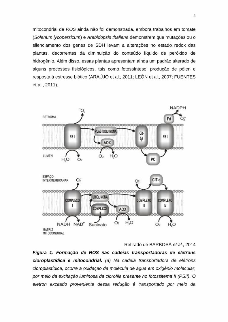
Retirado de BARBOSA et al., 2014
Figura 1: Formação de ROS nas cadeias transportadoras de eletrons
cloroplastídica e mitocondrial. (a) Na cadeia transportadora de elétrons
cloroplastídica, ocorre a oxidaçao da molécula de água em oxigênio molecular,
por meio da excitação luminosa da clorofila presente no fotossitema II (PSII). O
eletron excitado proveniente dessa redução é transportado por meio da
5
plastoquinona, do citocromo b6f (cit-b6f) e da plastocianina (PC), chegando ao
fotossistema I (PSI), onde é novamente excitado e transferido para molécula de
NADP+ via ferrodoxina (Fd). No cloroplasto a formação de oxigênio singleto
ocorre prodominantemente no PS-II e a de superóxido ocorre no PS-I. (b) Na
cadeia transportadora de elétrons mitocondrial, o aceptor final do elétron é o
oxigênio molecular, que é reduzido à agua via citocromo c oxidase (complexo
IV). Este eletron é proveniente da oxidação da moléculas de NADH (via
complexo I) e succinato (via complexo II), e será transportado por meio da
molécula da ubiquinona, do complexo III, e do citocromo C (cit. C), chegando
finalmente no complexo IV onde será transferido ao oxigênio, formando água.
Na respiração a formação de superóxido ocorre nos complexos I e III. Em ambas
as cadeias apresentadas o pool de quinonas reduzidas (plastoquinona ou
ubiquinona), pode ser controlado por meio da atividade de oxidases alternativas
(AOX).
Em plantas, as ROS podem ser produzidas ainda através de outros
processos fisiológicos, como a β-oxidação de ácidos graxos nos glioxissomos, a
ação da NADPH oxidase na membrana plasmática, ou por diferentes enzimas
na matriz extracelular (MYLONA & POLIDOROS, 2010). O citosol não é
considerado uma grande fonte de ROS, no entanto atua como um depósito para
moléculas produzidas por outros compartimentos subcelulares
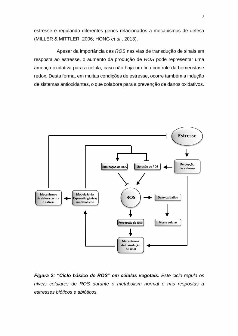
1.3 Produção e eliminação de ROS como mecanismos de transdução de
sinais
Os níveis de ROS nos diferentes compartimentos celulares é
determinado pela interação entre múltiplos mecanismos de produção e
eliminação de ROS. Estes mecanismos são controlados através de vias de
transdução de sinais que constituem o "ciclo básico de ROS" (RASOOL et al.,
2013). Durante o ciclo de vida dos organismos, tais vias são capazes de
monitorar os níveis de ROS através do controle da expressão gênica e da
6
atividade de diferentes enzimas ou rotas metabólicas que participam da
homeostase redox (figura 2)
Em condições de estresse, há um desequilíbrio da homeostase
celular, o que resulta no aumento da produção de ROS. As ROS estão
envolvidas em diferentes vias de transdução de sinal em resposta ao estresse,
e portanto, são essenciais para a indução de vias de defesa. De fato, diferentes
mecanismos de sinalização mediados por ROS vêm sendo propostos, e
diferentes receptores vêm sendo descritos, como canais iônicos, histidinas
kinases e fosfatases (APEL & HIRT, 2004; ASADA, 2006; DEMIDCHIK &
MAATHUIS, 2007; FOYER & NOCTOR, 2009; SIERLA et al., 2013; DEMIDCHIK
et al., 2014).
A modulação da atividade de canais iônicos é considerado o mais
rápido e eficiente mecanismo de resposta a fatores internos e externos, e
envolve principalmente dois mecanismos: a mudança da composição iônica, o
que pode regular a atividade de enzimas e a pressão osmótica, e a mudança do
potencial elétrico através de biomembranas, o que pode modular a atividade de
diferentes transportadores e enzimas associadas (DEMIDCHIK et al., 2015). Os
mecanismos mediados pela modulação de canais iônicos, incluem por exemplo,
o fechamento estomático induzidos por fitohormônios, como ácido abscísico
(ABA) ou jasmonato, em resposta a estresse hídrico (MUNEMASA et al., 2011).
As ROS também são capazes de modular a atividade de outras enzimas
regulatórias, em especial, kinases e fostatases, como MAP kinases, Ser/Thr
kinases, MAPK phosphatases, etc. No entanto, o mecanismo exato pelo qual
esta regulação ocorre ainda não está totalmente descrito (VAN BREUSEGEM
et al., 2008, PITZSCHKE & HIRT, 2009; RODRIGUEZ et al., 2010).
Diferentes fatores de transcrição também podem atuar com
receptores do sinal redox. Por exemplo, o fator de transcrição TGA1 em
Arabidopsis thaliana, que regula a expressão de NPR1, uma proteína de
resistência à doença, possui dois resíduos de histidina específicos e a oxidação
destes resíduos leva à perda da capacidade do fator de transcrição em se ligar
ao DNA (DESPRES et al., 2003). Os fatores de transcrição do tipo Heat shock
também estão envolvidos na sinalização redox, sendo ativados durante o
7
estresse e regulando diferentes genes relacionados a mecanismos de defesa
(MILLER & MITTLER, 2006; HONG et al., 2013).
Apesar da importância das ROS nas vias de transdução de sinais em
resposta ao estresse, o aumento da produção de ROS pode representar uma
ameaça oxidativa para a célula, caso não haja um fino controle da homeostase
redox. Desta forma, em muitas condições de estresse, ocorre também a indução
de sistemas antioxidantes, o que colabora para a prevenção de danos oxidativos.
Figura 2: “Ciclo básico de ROS” em células vegetais. Este ciclo regula os
níveis celulares de ROS durante o metabolism normal e nas respostas a
estresses bióticos e abióticos.
8
1.4 Estratégias de Defesa Contra o Estresse Oxidativo em Células Vegetais
O papel das ROS no metabolismo celular, como reguladoras de
processos fisiológicos ou como produtos tóxicos, é dependente da sua
concentração intracelular (FOYER & NOCTOR, 2005). Desta forma, os seres
vivos desenvolveram, ao longo da evolução, diferentes mecanismos de defesa
antioxidante que os permitem, controlar os níveis celulares de ROS. Em plantas,
existem sistemas enzimáticos e não enzimáticos de controle dos níveis de ROS
e que atuam em diferentes compartimentos subcelulares.
Os componentes não enzimáticos da defesa antioxidante incluem
moléculas como o ascorbato (AsA), glutationa reduzida (GSH), tocoferol,
carotenoides, flavonoides e componentes fenólicos, que reagem diretamente,
neutralizando as ROS e prevenindo a ocorrência de danos aos componentes
celulares (MITTLER et al., 2004; GRATÃO et al., 2005; SCANDALIOS, 2005). Já
o sistema enzimático de detoxificação de ROS em células vegetais inclui
diferentes enzimas, presentes em diferentes compartimentos subcelulares, entre
elas a superóxido dismutase (SOD), a catalase (CAT), a glutationa peroxidase
(GPX) e a ascorbato peroxidase (APX). Essas enzimas atuam coordenadamente
na eliminação de ROS para manter a homeostase redox dos diferentes
compartimentos celulares. A enzima SOD constitui a primeira linha de defesa
convertendo o radical superóxido em peróxido de hidrogênio, o qual é
detoxificado pela atividade da CAT, e também pela ação de diferentes
peroxidases, que requerem a redução de substratos específicos como doadores
de elétrons. Nesta última categoria estão incluídas as APXs, dependentes de
ascorbato, e as GPXs, que dependem de glutationa reduzida (NOCTOR &
FOYER, 1998).
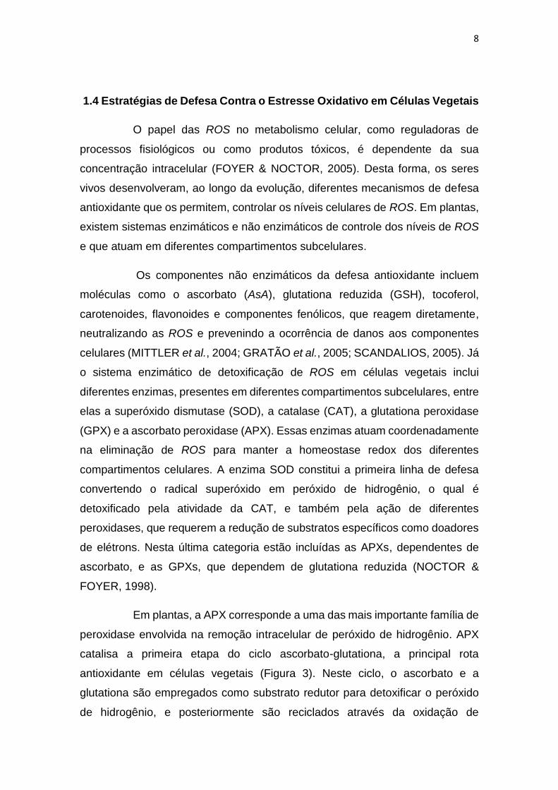
Em plantas, a APX corresponde a uma das mais importante família de
peroxidase envolvida na remoção intracelular de peróxido de hidrogênio. APX
catalisa a primeira etapa do ciclo ascorbato-glutationa, a principal rota
antioxidante em células vegetais (Figura 3). Neste ciclo, o ascorbato e a
glutationa são empregados como substrato redutor para detoxificar o peróxido
de hidrogênio, e posteriormente são reciclados através da oxidação de
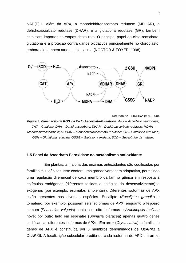
9
NAD(P)H. Além da APX, a monodehidroascorbato redutase (MDHAR), a
dehidroascorbato redutase (DHAR), e a glutationa redutase (GR), também
catalisam importantes etapas desta rota. O principal papel do ciclo ascorbato-
glutationa é a proteção contra danos oxidativos principalmente no cloroplasto,
embora ele também atue no citoplasma (NOCTOR & FOYER, 1998).
Retirado de TEIXEIRA et al., 2004
Figura 3: Eliminação de ROS via Ciclo Ascorbato-Glutationa. APX – Ascorbato peroxidase;
CAT – Catalase; DHA – Dehidroascorbato; DHAR – Dehidroascorbato redutase; MDHA–
Monodehidroascorbato; MDHAR – Monodehidroascorbato-redutase; GR – Glutationa redutase;
GSH – Glutationa reduzida; GSSG – Glutationa oxidada; SOD – Superóxido dismutase.
1.5 Papel da Ascorbato Peroxidase no metabolismo antioxidante
Em plantas, a maioria das enzimas antioxidantes são codificadas por
famílias multigênicas. Isso confere uma grande vantagem adaptativa, permitindo
uma regulação diferencial de cada membro da família gênica em resposta a
estímulos endógenos (diferentes tecidos e estágios do desenvolvimento) e
exógenos (por exemplo, estímulos ambientais). Diferentes isoformas de APX
estão presentes nas diversas espécies. Eucalipto (Eucaliptus grandis) e
tomateiro, por exemplo, possuem seis isoformas de APX, enquanto o feijoeiro
comum (Phaseolus vulgaris) conta com oito isoformas e Arabidopsis thaliana
nove; por outro lado em espinafre (Spinacia oleracea) apenas quatro genes
codificam as diferentes isoformas de APXs. Em arroz (Oryza sativa), a família de
genes de APX é constituída por 8 membros denominados de OsAPX1 a
OsAPX8. A localização subcelular predita de cada isoforma de APX em arroz,
10
baseada na presença de peptídeos sinais adicionais, é descrita a seguir: Os
genes OsAPX1 (cromossomo 3) e OsAPX2 (cromossomo 7) codificam isoformas
citosólicas; OsAPX3 (cromossomo 4) e OsAPX4 (cromossomo 8) codificam
isoformas peroxissomais; as isoformas mitocondriais são codificadas pelos
genes OsAPX5 (cromossomo 12) e OsAPx6 (cromossomo 12), enquanto
OsAPX7 (cromossomo 4) e OsAPX8 (cromossomo 2) codificam isoformas
cloroplastídicas, sendo a primeira, solúvel no estroma, e a segunda, ligada à
membrana tilacoidal. É relevante relatar o alto grau de conservação na estrutura
gênica entre genes de OsAPX que codificam isoformas de um mesmo
compartimento subcelular (TEIXEIRA et al., 2004).
O arroz é uma das principais plantas modelos para estudos
biológicos. E isso se dá principalmente por dois fatores: O arroz apresenta o
menor genoma entre os cereais (OUYANG et al., 2007) e apresenta sintenia na
estrutura e organização do genoma, com outros membros da família Poaceae
(MOORE et al., 1995), permitindo assim, que os resultados obtidos para o arroz
possam ser usados como referência para outras espécies (PATERSON et al. ,
2005). Além disso, o arroz possui uma importância sócio/econômica, uma vez
que entre os cereais, é o alimento mais cultivado no mundo, sendo essencial
para mais de 2,4 bilhões de pessoas (EMBRAPA, 2015).
Diferentes estudos demonstraram que as diferentes isoformas de
APX em arroz apresentam padrões distintos de expressão, estando envolvidas
com a regulação fina dos níveis de peróxido de hidrogênio nos diferentes
compartimentos subcelulares em decorrência do estímulo ambiental
(MENEZES-BENAVENTE et al., 2004; TEIXEIRA et al., 2004; 2006; ROSA et
al., 2010; BONIFACIO et al., 2011; LAZZAROTTO et al., 2011; CAVERZAN et
al., 2014). Estas análises levantam muitas questões sobre o papel
desempenhado pelas diferentes isoformas APX no metabolismo antioxidante em
células vegetais.
Por meio de estudo de genética reversa, usando a tecnologia de RNA
de interferência (RNAi), o papel desempenhado pelas diferentes isorformas de
APX em arroz no metabolismo antioxidante e na resposta ao estresse vem sendo
elucidado. Trabalhos anteriores demonstraram que o duplo silenciamento da

11
isoformas citosólicas de APX (OsAPX1 e OsAPX2), leva ao aumento da
sensibilidade ao estresse por alumínio, além induzir a regulação da expressão
de proteínas relacionadas com vias fotoquímicas, ciclo de Calvin e
fotorespiração. Por outro lado, as plantas silenciadas individualmente para
OsAPX1 e OsAPX2 apresentam problemas no desenvolvimento (ROSA et al.,
2010, BONIFACIO et al., 2011). Já plantas silenciadas para OsAPX3 e OsAPX4
apresentam alterações no padrão de senescência (dados não publicados). Em
relação às isoformas mitocondriais de APX em arroz (OsAPX5 e OsAPX6), calos
transgênicos simultaneamente silenciados para essas isoformas não
regeneraram plantas, indicando que as APXs mitocondriais poderiam exercer um
papel essencial para o desenvolvimento vegetal (dados não publicados).
Quanto às isoformas cloroplastídicas de APX (OsAPX7 e OsAPX8),
foi demonstrado que em diferentes situações de estresse, como alta
luminosidase, frio e seca, estas isoformas possuem a expressão modulada de
forma antagônica: a expressão de OsAPX7 é geralmente induzida, enquanto a
expressão de OsAPX8 é modulada negativamente (ROSA et al., 2010;
CAVERZAN et al., 2014). Além disso, foi demonstrado que o silenciamento duplo
destas isoformas não acarreta mudanças fenotípicas na planta sob condições
normais de crescimento, embora sob condições de estresses abióticos, as
plantas de arroz silenciadas para os ambos os genes cloroplastídicos de APX
sofrem fortes alterações fotossintéticas e bioquímicas, indicando que rotas
fotossintéticas e do metabolismo oxidativo foram afetadas pelo silenciamento
(CAVERZAN et al., 2014). Vale ressaltar que a modulação antagônica da
expressão de OsAPX7 e OsAPX8 que ocorre naturalmente em resposta a
estresses abióticos pode ter dificultado a interpretação do duplo silenciamento
destes genes, e justificar a ausência de diferenças fenotípicas em plantas
crescidas sob condições controle. Além disso, o papel individual de cada
isoforma cloroplastídica de APX em arroz permanece desconhecido.
O papel funcional das isoformas cloroplastídicas de APX vem sendo
estudado também em outras espécies. Em algodão (Gossypium hirsutum), a
superexpressão de APX cloroplastídica induz um aumento na tolerância ao frio
(PAYTON et al., 2001). Similarmente, a superexpressão da isoforma tilacoidal
de APX em tabaco (Nicotiana toabacum) também leva a um aumento da
12
tolerância ao frio, bem como a estresses induzido por metil viologen (YABUTA
et al., 2002). Já em trigo (Triticum aestivum L.), o silenciamento de APX tilacoidal
leva à redução da atividade fotossintética e do crescimento (DANNA et al., 2003).
Estes dados confirmam o importante papel desempenhado por APX no controle
da homeostase redox e da resposta ao estresse em diferentes espécies
vegetais.
1.6 Estresse oxidativo como um fator limitante na produção agrícola
Vários fatores ambientais, tais como estresses bióticos e abióticos,
podem ser considerados fatores limitantes para o crescimento e a produtividade
das diferentes culturas, desta forma, a escassez de áreas para expansão da
agricultura já é um dos grandes desafios deste século (JACQUEMIN et al., 2013).
Ao longo dos anos, a disponibilidade de terra arável vem diminuindo,
principalmente devido a mudanças climáticas e técnicas de gestão
insustentáveis, que intensificaram problemas como a erosão e a degradação do
solo (STOCKING, 2003).
De acordo com a Organização das Nações Unidas para Alimentação
e Agricultura (FAO, do inglês Food and Agriculture Organization), em 2050 a
população mundial deverá chegar a pelo menos 9 bilhões de pessoas, e para
atingir esta demanda, a produção de alimentos deverá crescer pelo menos 60%
(Jacquemin et al., 2013). Desta forma, em um contexto de crescente demanda
mundial de alimentos, desde o início do século vem se estabelecendo uma
crescente necessidade da melhoria do cultivo, seja pela expansão de áreas
destinadas à agropecuária, seja pelo desenvolvimento de estratégias que
aumentem a tolerância de plantas a estresses ambientais (TAKEDA &
MATSUOKA, 2008). Neste contexto, o estudo integrado de transcriptomas,
proteômica e metabolômica vem proporcionando, ao longo dos anos, uma
melhor compreensão das diferentes rotas de sinalização envolvidas na resposta
de plantas ao estresse (CRAMER et al., 2011). No entanto, a compreensão dos
mecanismos subjacentes às respostas de plantas entre as várias pressões
ambientais ainda está longe de ser elucidado. Tendo em vista a importância do
metabolismo antioxidante na manutenção dos níveis de ROS, e a importância

13
destas nos diferentes mecanismos de respostas das plantas ao estresse, é de
grande importância à identificação dos mecanismos modulatórios da expressão
e da atividade de enzimas que atuam na homeostase redox. Dessa forma, os
genes e proteínas envolvidos no metabolismo antioxidante são alvos
interessantes para o melhoramento genético de plantas para características
agronômicas, tais como maior produção e menores perdas causadas por
condições ambientais desfavoráveis.
14
2 OBJETIVOS GERAIS
Objetivo geral: Estudar os mecanismos de geração e eliminação de ROS em
organelas importantes para o balanço redox (mitocôndria e cloroplasto), e seus
efeitos no desenvolvimento e na resposta de plantas ao estresse.
Objetivos específicos:
- Determinar novos sítios de geração de ROS, notadamente a enzima succinato
desidrogenase, conhecida como complexo II mitocondrial;
- Determinar os efeitos moleculares e fisiológicos da geração de ROS pela
succinato desidrogenase;
- Caracterizar do ponto de vista evolutivo a succinato desidrogenase em
diferentes grupos taxonômicos;
- Determinar os efeitos moleculares e fisiológicos da alteração no estado redox
no cloroplasto pelo silenciamento das APX cloroplastídicas, APX7 e APX8;
- Identificar reguladores da expressão de APX8 e seu papel em vias de
transdução de sinal.
15
3.1 CAPÍTULO 1
SUCCINATO DESIDROGENASE (COMPLEXO II MITOCONDRIAL)
É UM SÍTIO DE GERAÇÂO DE ESPÉCIES REATIVAS DE
OXIGÊNIO EM MITOCÔNDRIAS DE PLANTAS, REGULANDO O
DESENVOLVIMENTO E A RESPOSTA AO ESTRESSE
Este capítulo é referente ao artigo “Succinate dehydrogenase
(mitochondrial complex II) is a source of reactive oxygen species in
plants and regulates development and stress responses”, publicado
em 2015 na revista New Phytologist. Neste trabalho identificamos a
enzima succinato desidrogenase (SDH) como um novo e importante
sítio de geração de ROS em células vegetais. De fato, diferentes
trabalhos vêm demonstrando que mutações em subunidades da SDH
afetam diretamente a homeostase redox da célula, no entanto até o
desenvolvimento deste projeto essa hipótese ainda não havia sido
comprovada. Os resultados aqui apresentados demonstram ainda
que a capacidade da SDH em gerar ROS pode ser regulada por
diferentes moléculas, como o ácido salicílico e óxido nítrico (NO).
Além disso, este mecanismo é fundamental na regulação da
expressão gênica, controlando o desenvolvimento vegetal e as
respostas de plantas a diferentes estresses.

36
3.2 CAPÍTULO 2
A EVOLUÇÃO DA SUPERFAMÍLIA DE FUMARATO REDUTASES
ENVOLVEU O ESTABELECIMENTO DE DIFERENTES ROTAS DO
METABOLISMO PRIMÁRIO E PROCESSOS DE ENDOSSIMBIOSE COM
SUBSEQUENTE TRANSFERÊNCIA DE GENES PARA O NÚCLEO
Esse capítulo é referente ao manuscrito “FUMARATE
REDUCTASE SUPERFAMILY EVOLUTION ENCOMPASSED
THE ESTABLISHMENT OF DIFFERENT METABOLIC
PATHWAYS AND ENDOSYMBIOSIS PROCESSES WITH
SUBSEQUENT GENE TRANSFERENCE TO THE NUCLEUS”,
pronto para a submissão. Neste trabalho, demonstramos que a
succinato desidrogenase (SDH), presente em praticamente todos
os seres vivos, é membro da família de succinato:quinona
oxiredutases, composta também pela enzima fumarato redutase
(FRD), presente em bacterias anaeróbicas. Muitos trabalhos vêm
demonstrando a importância destas enzimas em diferentes seres
vivos. No entanto, a literatura ainda carece de um estudo
integrativo desta família, inserida na superfamília de fumarato
redutases, composta por outras fumarato redutases, como as
fumarato redutases solúveis e dependentes de NADH (FRDS), as
fumarato redutases dependente de grupos tiol (TFRD) e as
fumarato redutases dependente de L-aspartato, também
conhecidas com L-aspartato oxidase (LASPO). Nossos dados
demonstram que todas estas enzimas se originaram de um acestral
comum, e foram essenciais na evolução das vias metabolicas
envolvidas na geração de energia. Além disso, a distribuição destas
enzimas nos organismos atuais foi dependente principalmente dos
processos de endossimbiose que originaram a mitocôndria e o
cloroplasto.
37
EVOLUTION OF THE FUMARATE REDUCTASE SUPERFAMILY
ENCOMPASSED THE ESTABLISHMENT OF DIFFERENT METABOLIC
PATHWAYS AND ENDOSYMBIOSIS PROCESSES WITH SUBSEQUENT
GENE TRANSFERENCE TO THE NUCLEUS
Douglas Jardim-Messeder1, Caroline Cabreira-Cagliari1, Rafael Rauber2,
Andreia Carina Turchetto-Zolet1, Rogério Margis2, Marcia Margis-Pinheiro1
1 Departamento de Genética, Universidade Federal do Rio Grande do Sul, Porto
Alegre, RS, Brazil
2 Centro de Biotecnologia, Universidade Federal do Rio Grande do Sul, Porto
Alegre, RS, Brazil
38
ABSTRACT
The organic molecules fumarate and succinate are known to be
present in prebiotic systems essential for the origin of life. The reaction pathways
involved in fumarate and succinate interconversion have been conserved
throughout evolution and are found in virtually all living organisms, including
eubacteria, eukaryotes and Archaea. The process of fumarate and succinate
interconversion is catalyzed by the enzymes succinate dehydrogenase (SDH)
and fumarate reductase (FRD).
SDH, which is found in almost all living organisms, and FRD, which is
found in anaerobic bacteria, are members of the succinato:quinona oxireductase
family. In spite of their importance for the primary metabolism of different
organisms, an integrative study of this family of enzymes is still lacking. They
belong to the fumarate reductase superfamily, which is composed of other
fumarate reductases such as the soluble fumarate reductase dependent of NADH
(FRDS), the soluble fumarate reductase dependent of thiol groups (TFRD) and
the L-aspartate oxidase (LASPO), which shows fumarate reductase activity in the
absence of oxygen.
Our results demonstrate that these enzymes emerged from a common
ancestor and were essential in the evolution of metabolic pathways involved in
energy transduction. In addition, the distribution of these enzymes in present day
organisms was dependent mainly of endosymbiotic processes, which originated
in the mitochondria and chloroplast.
39
INTRODUCTION
The “Origin of Life” is the natural process which gave rise to living
organisms from non-living matter, such as simple organic compounds, and
occurred on Earth about 3.5 billion years ago (AWRAMIK, 1992). In this context,
a non-enzymatic reverse tricarboxylic acid (TCA) cycle that provided a core
mechanism to produce carbon compounds from CO2 and water under prebiotic
conditions has been hypothesized (WACHTERSHAUSER, 1993). Thus, the
intermediate molecules in the TCA cycle, such as succinate and fumarate, are
present in prebiotic systems of organic molecules essential for the origin of life.
Corroborating this hypothesis, the reaction pathways involving fumarate and
succinate have been conserved throughout evolution, and enzymes catalyzing
their interconversion are found in virtually all living organisms, including
eubacteria, eukaryotes and Archaea (HEDERSTEDT AND OHNISHI, 1992).
The succinate:quinone oxidoreductases (SQOR) (EC 1.3.5.1) –
succinate dehydrogenase (SDH) and fumarate reductase (FRD) – are oligomeric,
potentially reversible isoenzymes that catalyze the interconversion of fumarate
and succinate under physiological conditions (HIRSCH et al., 1963). SDH
activity was first detected in frog muscle in 1909 (THUNBERG, 1909). SDH
catalyzes the oxidation of succinate to fumarate (reaction 1), a vital process in
organisms that use the TCA cycle for central carbon metabolism. In addition, this
reaction is coupled to the reduction of ubiquinone (UQ) to ubiquinol (QH2)
(reaction 2), thereby donating electrons to the aerobic electron transport chain
(ETC). On the other hand, FRD is a key component of anaerobic respiration
which catalyzes the reverse reactions. Thus, in the absence of oxygen, fumarate
acts as a final electron acceptor for anaerobic respiration (BLAUT et al., 1989).
The interconversion of succinate and fumarate by SQOR enzymes is reversible,
besides in vivo the oxidation and reduction reactions preferentially occur
according to each enzyme (SDH or FRD). In Escherichia coli, for example, the
FRD is also able to catalyze succinate oxidation; however, with a lower activity
(CECCHINI et al., 1986; SUCHETA et al., 1993).
40
succinate → fumarate + 2H+ + 2e- (reaction 1)
quinone + 2H+ + 2e- → quinol (reaction 2)
SDH preferentially catalyzes the oxidation of succinate to fumarate (E°
= + 30 mV) using an electron acceptor with a high reduction potential, such as
ubiquinone (UQ) (E° = +100 mV). Finally, in the aerobic ETC, oxygen has the
high reduction potential (E° = 820 mV), thus operating as a final electron
acceptor. On the other hand, FRD catalyzes the reverse reaction, oxidizing an
electron donor and reducing fumarate to succinate. For this, the electron donor
used by FRD must possess a lower redox potential than the fumarate/succinate.
Thus, FRD cannot use UQH2 as an electron donor, and the fumarate reduction is
coupled to the oxidation of a lower reduction potential quinol, such as menaquinol
(MQH2) (E° = -73 mV) (Fig. 1A).
In Gammaproteobacteria, the succinate oxidation and fumarate
reduction are carried out by independent enzymes which act as SDH or FRD. On
the other hand, in other bacterial clades and in Archaea, only one enzyme with
both activities can be found. Eukaryotes possess only one enzyme that acts as a
SDH in vivo. Despite the large scale genome sequencing projects which have
provided SDH and FRD sequences from various organisms, the classification of
these enzymes is not obvious because it is based only on biochemical data,
which has not yet been determined for many species. Thus, without prior
knowledge of the physiological role of a particular enzyme in the metabolism of a
given bacterium, it is not possible to predict whether the enzyme functions as a
FRD or a SDH in vivo.
In helminths, such as Ascaris suum and Haemonchus contortus, two
forms of SQORs that are specific to different growth stages have been identified
(ROOS AND TIELES, 1994). During their life cycle, these helminths are able to
change from aerobic to anaerobic metabolism. Thus, in these species there are
two different oligomeric isoforms of SQOR which lead to a predominant SDH
activity during the larval phase (aerobic), and a predominant SDH activity during
adult phase (anaerobic) (AMINO et al., 2000). This adaptive mechanism is similar
41
to that described for prokaryotes, such as Gammaproteobacteria, which have
both SDH and FRD enzymes. Previous work demonstrated that the
Gammaproteobacteria Escherichia coli utilizes transcriptional regulators that, in
the absence of oxygen, induce the expression of FRD and repress SDH, while in
the presence of oxygen, they stimulate the transcription of SDH while repressing
FRD expression (PARK et al., 1995). As expected, the shift between SDH and
FRD expression is also accompanied by changes in the predominant quinone
species. In bacteria such as Escherichia coli, the combination of SDH and UQ is
replaced by FRD and a low-potential quinol, such as MQH2, during metabolic
adaptations to changes in the oxygen supply (COLE et al., 1985). On the other
hand, helminths use rhodoquinol (RQH2) (E° = -63 mV) as the low-potential
quinol, which is essential to FRD activity (VAN HELLEMOND et al., 1995).
Structurally, SQORs are enzymatic complexes anchored to the
cytoplasmic membranes in prokaryotes or the inner mitochondrial membrane in
eukaryotes. SDH generally consists of four protein subunits – referred to as
SDHA, SDHB, SDHC and SDHD, in bacteria and animals, or SDH1, SDH2, SDH3
and SDH4, in fungi and plants, while FRD consist of FRDA, FRDB, FRDC and
FRDD subunits (Fig 1A). The subunit SDHA/FRDA is known as a catalytic
flavoprotein, and possesses a bicarboxylate binding site and a covalently bound
flavin adenine dinucleotide (FAD). The SDHB/FRDB is classified as an iron–sulfur
protein since it contains three iron–sulfur clusters. The subunits SDHC/FRDC and
SDHD/FRDD are the hydrophobic subunits which anchor the SQOR complex to
the membrane and possess the quinone binding site (YANKOVSKAYA et al.,
2003).
In many bacteria, the SQORs do not contain the two hydrophobic
subunits, but instead contain one larger hydrophobic polypeptide, also named
SDHC. Studies of sequence and structure indicate that this larger subunit
originated from fusion of the genes encoding the two smaller subunits
(HAIGERHALL AND HEDERSTEDT, 1996). In addition, the SQOR membrane
bound domain can present zero, one, or two heme prosthetic groups. Thus, the
SQOR complexes can be classified into five types, according to the structure of
the anchor subunit (LANCASTER, 2001). The anchor domain with two
42
polypeptides containing two heme groups is denoted as Type A, one polypeptide
and two heme groups corresponds to Type B, two polypeptide anchors with one
heme group are designated as Type C, whereas anchors with two polypeptides
and no heme group are referred to as Type D (HÄGERHÄLL, 1997). Furthermore,
there is a “non-classical” SQOR Type E, found in some Archaea such as the
Sulfolobales order, which does not contain heme groups. The anchor subunit of
the Type E SQOR is non-classical because it is composed of two polypeptide
subunits, SDHE and SDHF, which are not found in other types of SQOR
complexes (SCHÄFER et al., 2002; LEMOS et al., 2002) (Fig. 1B).
Previous work demonstrated that the reduction of fumarate is also
important in the anaerobic metabolism of eukaryotes such as fungi and some
protists. Despite fungi and protists presenting a SDH located in the mitochondria,
in these organisms the FRD activity is provided by a monomeric soluble FRD
(FRDS) (EC: 1.3.1.6) which possess the conserved catalytic region of
SDHA/FRDA (MURATSUBAKI AND ENOMOTO, 1998; COUSTOU et al., 2005).
This enzyme is also found in the prokaryote Shewanella genus (PEALING et al.,
1992). Curiously, FRDS has no SDH activity in vitro and is not able to oxidize
quinones. This enzyme is involved in the maintenance of the intracellular redox
balance under anaerobic conditions, using NADH as an electron donor for
fumarate reduction (BESTEIRO et al., 2002).
In methanogenic Archaea, such as Methanobacteria, another soluble
fumarate reductase has been found, designated thiol:fumarate reductase (TFRD)
(EC: 1.3.4.1). TFRD is composed of two subunits homologous to SDHA/FRDA
and SDHB/FRDB, and three additional subunits that are not homologous to
SQOR subunits (HEIM et al., 1998). The TFRD catalyzes the reduction of
fumarate using coenzyme M (CoM-S-H) and coenzyme B (CoB-S-H) as electron
donors (BOBIK AND WOLFE, 1989). The reaction catalyzed by the TFRD is
indicated below (reaction 3):
fumarate + CoM-S-H + CoB-S-H → succinate + CoM-S-S-CoB (reaction 3)
43
The heterodisulfide (CoM-S-S-CoB) that is also produced in the last
step of the methanogenesis process (BOBIK et al., 1987; ELLERMANN et al.,
1988) is re-reduced by heterodisulfide reductase (HDR) (EC 1.8.98.1)
(HEDDERICH et al., 1994), in a reaction coupled with ADP phosphorylation
(DEPPENMEIER et al., 1996). Thus, in Methanobacteria the anabolic reduction
of fumarate is indirectly coupled with ATP synthesis. In addition, as demonstrated
for FRDS, the TFRDs do not have SDH activity and are unable to oxidize
succinate in vivo or in vitro (HEIM et al., 1998).
The increasingly amount of available data related to the structure and
function of different FRDs and SDHs in prokaryotes and eukaryotes, and their
respective genes, represents an important tool to understand the evolution of
these proteins. However, despite the few publications describing independent
phylogenetic studies of some FRD/SDH classes, an evolutionary study including
the whole family of FRD/SDH proteins is still lacking. Thus, the aim of this study
was to determine the phylogenetic relationship among different FRD/SDHs in
diverse organisms in order to provide an overview of FRD/SDH evolution and
contribute to the understanding of the evolution of this family. Our results
demonstrated that FRD/SDH is part of a fumarate reductase superfamily,
composed of other fumarate reductases such as NADH:fumarate reductase
(soluble fumarate reductase – FRDS), thiol:fumarate reductase (TFRD) and L-
aspartate oxidase (LASPO). In addition, the evolutionary history of these proteins
encompassed the establishment of different metabolic pathways, endosymbiosis
processes and subsequent massive transference of genes from the mitochondrial
and chloroplastic genomes to the nucleus.
44
METHODS
Sequence retrieval
Flavoproteins homologous to SQORs from representative organisms
were identified by BLAST (ALTSCHUL et al., 1997) searches using databases
from the National Center for Biotechnology Information (NCBI)
(www.ncbi.nlm.nih.gov), UniProt (www.uniprot.org), Phytozome
(www.phytozome.net), Metazome (www.metazome.net) and EuPathDB
(www.eupathdb.org). The sequences of SDHA/SDH1 or FRDA from Escherichia
coli, Saccharomyces cerevisiae, Drosophila melanogaster, Homo sapiens,
Trypanosoma cruzi, Oryza sativa and Arabidopsis thaliana were used as baits.
Sequence alignments and phylogenetic analysis
The protein sequences of the FAD_binding_2 domains (pfam:
PF00890), common to all flavoproteins analyzed and identified using SMART tool
software (SCHULTZ et al., 1998) were used in the phylogenetic analysis. Multiple
sequence alignments were performed using multiple sequence comparison by
log expectation (MUSCLE) (EDGAR, 2004) as implemented in the molecular
evolutionary genetics analysis (MEGA5) software (TAMURA et al., 2011) using
the default parameters. We visually inspected and edited the alignments to
include only unambiguously aligned positions. Phylogenetic analyses were
conducted using the Bayesian inference approach as implemented in BEAST
1.8.1 (DRUMMOND et al., 2012). The Yule tree was selected as a tree prior to
Bayesian analysis, and 50,000,000 generations were performed with Markov
chain Monte Carlo (MCMC) algorithms. The best fit of evolutionary models was
Dayoff as determined with protTest. The TRACER 1.6 (RAMBAUT et al., 2014)
was used to check for the convergence of MCMCs and to ensure adequate
effective sample sizes (EES > 200) after a burning of 100 generations. We
estimated the maximum clade credibility tree with TreeAnnotator, which is part of
the BEAST package, and visualized the trees with Figtree 1.3.1

45
(http://tree.bio.ed.ac.uk/software/figtree/). Statistical support for the clades was
determined by assessing the Bayesian posterior probability (PP).
46
RESULTS AND DISCUSSION
Phylogenetic relationships within the fumarate reductase superfamily
Phylogenetic analyses were carried out with sequences corresponding
to the FAD_binding_domains of SQOR, FRDS and TFRD from 180 species
representing bacteria, Archaea, plants (Glaoucophyta, Rhodophyta and
Viridiplantae), fungi, protists (Excavata and Chromalveolata) and metazoan
phyla. Preliminary analysis by BLAST demonstrated that the
FAD_binding_domain was also conserved in other proteins, such as L-aspartate
oxidase (LASPO) (EC 1.4.3.16), the subunit APRA of adenylyl-sulfate reductase
(EC 1.8.99.2) and the subunit GLPB of anaerobic glycerol-3-phosphate
dehydrogenase (E.C.1.1.1.8), which are apparently not related to FRD activity
(Fig. 2). Thus, these proteins were also included in the phylogenetic analyses,
therefore a total of 380 sequences were analyzed. Finally, a total of 354 amino
acid residues were included in the final dataset used for phylogenetic tree
reconstruction by Bayesian methods. Our results demonstrate that APRA and
GLPB appeared as a basal group within the fumarate reductase superfamily (Fig.
3). The GLPB was restricted to Gammaproteobacteria, while APRA was found in
both Deltaproteobacteria and Archaea, indicating that APRA was possibly
present in an ancestral organism from which both Eubacteria and Archaea
originated.
Analyzing the fumarate reductase superfamily, we observed that
FRDS appeared as a separate group, subdivided in FRDS from bacteria
Shewanella oneidensis and FRDS from fungi and protists (Excavata and
Chromalveolata). The distribution of FRDS in both prokaryotes and eukaryotes,
in addition to the monomeric nature of this enzyme, reinforce the idea that FRDS
was present in basal organisms and evolved into other fumarate reductases, such
as the multimeric SQOR complex (GEST, 1980). Thus, the FRDS of Shewanella
oneidensis, fungi and protists might therefore be close related of an ancestral
form of FRD.

47
Interestingly, the phylogenetic analyses indicated that LASPOs,
encoded by nadB in bacteria and protists, and fin4 in plants, are included in the
fumarate reductase superfamily. LASPOs are able to catalyze the FAD–
dependent oxidation of L-aspartate to iminoaspartate, and are the first enzyme in
the de novo nicotinamide adenine dinucleotide (NAD) biosynthesis by the
aspartate oxidase pathway (MORTARINO et al., 1996, GRIFFITH et al., 1975).
LASPOs are required for NAD biosynthesis under both aerobic and anaerobic
conditions (MESSNER AND IMLAY, 2002). Under aerobic conditions, LASPOs
use oxygen as an electron acceptor, which is then partially reduced to hydrogen
peroxide (reaction 4). However, in the absence of oxygen, LASPOs can also act
as a L-aspartate:fumarate oxidoreductase, using fumarate as an electron
acceptor to produce succinate (MATTEVI et al., 1999) (reaction 5). Thus, in the
absence of oxygen, LASPOs can have FRD activity (TEDESCHI et al., 1996),
demonstrating that LASPOs represent another member of the fumarate
reductase superfamily. This observation is reinforced by the phylogenetic
analysis, which shows that the LASPO and SQOR families form a monophyletic
group.
L-aspartate + O2 → iminoaspartate + O2- (reaction 4)
L-aspartate + fumarate → iminoaspartate + succinate (reaction 5)
Our analysis demonstrates that in eukaryotes, LASPOs are found only
in photosynthetic clades, with the exception of red algae (Rhodophyta). This fact
may suggest that eukaryotes acquired the LASPO during chloroplast
endosymbiosis, as proposed by TERNES AND SCHONKNECHT (2014). The
chloroplast was originated from an endosymbiotic event during evolution, when
a nonphotosynthetic eukaryotic cell took up a photosynthetic cyanobacterium,
which evolved into the chloroplast (REYES-PRIETO et al., 2007). This
endosymbiosis was accompanied by massive gene transfer from the
cyanobacterial genome into the nuclear genome of the host cell. It is estimated
that in land plants, thousands of genes in the nuclear genome originated from
48
gene transfer from a cyanobacterial genome (TIMMIS et al., 2004). In fact, all
plastids found in eukaryotes emerged from a single ancient endosymbiosis event,
with few exceptions, such as the Paulinella chromatophora plastid, which resulted
from a more recent endosymbiosis event (MARIN et al., 2005).
Primary endosymbiosis of a cyanobacterium gave rise to the
Archaeplastida, including Glaoucophyta, Rhodophyta (red algae) and
Viridiplantae (green algae and land plants). In Glaoucophyta and Viridiplantae,
the presence of LASPO is well established (KATOH et al., 2006), while in
Rhodophyta, the aspartate pathway is absence. In addition, LASPO is found in
algae protist, such as diatoms (Bacillariophyceae), Oomycota and brown algae
(Phaeophyceae). These groups, corresponding to Chromalveolata, acquired their
plastid through a secondary endosymbiosis following the uptake of a unicellular
red alga (CAVALIER-SMITH, 1999; BAURAIN et al., 2010), and this fact raises a
question about how Chromalveolata acquired LASPO by endosymbiosis, since
the aspartate pathway is absent in Rhodophyta. The answer to this question
came from previous works which demonstrated that, in Chromalveolata,
thousands of genes potentially originated from green algae, raising a hypothesis
that Chromalveolata were subjected to a cryptic secondary endosymbiosis with
a green algae before acquiring chloroplasts through Rhodophyta endosymbiosis
(MOUSTAFA et al., 2009). Thus, the Chromalveolata could have acquired
LASPO through gene transference with a green algae.
The phylogenetic analysis indicated that the LASPO from
Glaoucophyta and Cyanobacteria is monophyletic, confirming the hypothesis that
LASPO was acquired through gene transfer from a cyanobacterial endosymbiont.
Curiously, as demonstrated by TERNES AND SCHONKNECHT (2014), the
LASPO from Viridiplantae and Chromalveolata, as well as heterokontophyta, are
not closely related to cyanobacterial LASPO sequences. The Viridiplantae
LASPO is grouped with Bacteroidetes, while Chromalveolata LASPO is grouped
with Desulfovibrionales (Deltaproteobacteria) and Halobacteria (Archaea),
suggesting that the ancestor of green plants and Chromalveolata replaced the
cyanobacterial LASPO through a possible horizontal gene transfer with
nonphotosynthetic prokaryotes, as suggested by TERNES AND
49
SCHONKNECHT (2014). On the other hand, the aspartate pathway is not found
in Rhodophyta. Curiously, the phylogenetic analysis of LASPO from Viridiplantae
demonstrated that the LASPO from Fabacea species is placed as an outgroup
from other Magnoliophyta; however, the reason for this fact has no apparent
explanation yet.
Finally, the phylogenetic analysis demonstrated that all SQORs were
organized as a single monophyletic group. The first subgroup we identified
contained the sequences of subunit A of SQOR type B from Chlamydiae,
Firmicutes, Verrucomicrobia, Spirochaetes, Bacteroidetes,
Epsilonproteobacteria and Deltaproteobacteria clades. The following groups
included sequences relative to TFRD. This topology suggests that TFRD
emerged through differentiation of the SQOR complex specifically in
methanogenic Archaea such as Methanobacteria. The next group in the
phylogenetic tree corresponded to a group that was not very well defined, but
was composed of the SQOR type B from Cyanobacteria, the classical Archaeal
SQOR type A, the non-classical Archaeal SQOR type E, and the SQOR type D,
corresponding to Gammaproteobacteria FRD. Curiously, these analyses
indicated that FRD (SQOR type D) from Gammaproteobacteria were closely
related to classical SDH from Archaea (SQOR type A). Two isoforms of SQOR,
SDH and FRD were found in Gammaproteobacteria, thus these data could
indicate that SDH emerged as a duplication of FRD, while the FRD was lost in
other species of bacteria. In addition, the FRDA subunit of FRD from the Aquificae
bacterial group, which does not possess SQOR activity, was also found in this
phylogenetic branch.
In fact, previous work demonstrated that in Aquificae, FRD definitely
is not a SQOR. The functional difference than SQOR, is that Aquificae FRD is
soluble and uses NADH as an electron donor, working as a member of the
enzymatic reductive TCA (rTCA) cycle (MIURA et al., 2008). Aquificae are
chemolithoautotrophs, which assimilate CO2 as a unique carbon source through
the rTCA cycle (SHIBA et al., 1985), which is recognized as an ancestral form of
the TCA cycle (AOSHIMA, 2007). The Aquificae FRD consisted of five subunits,
designated FRDA, FRDB, FRDC, FRDD and FRDE. Among these, the FRDA and
50
FRDB subunits presented high sequence identity to the flavoproteins and iron–
sulfur subunits of SQOR, respectively (MIURA et al., 2008).
Finally, the sequences of flavoproteins of the SQOR type C, which
have only SDH activity in vivo, appeared as a unique group in the phylogenetic
tree. In this group, SDHA from Gammaproteobacteria and Betaproteobacteria
appeared grouped in a unique branch, while SDHA from Alphaproteobacteria and
from all eukaryotes, seemed to have a common origin. In relation to SDH from
eukaryotes, there were groups specific for Excavata, metazoan, Apicomplexa,
Heterokontophyta and plants. The Metazoan clade presented a division of SDH
from different groups, such as Helminth, Arthropoda, Platyhelminthes/Mollusca
and Chordata. Curiously, the SDHAA from Ascaris suum, which is related to FRD
activity, appeared as an outgroup of Helminth SDHA, which is related to SDH
activity. This data suggests that SDHAA may have emerged through
differentiation of SDHAL, due to the necessity of FRD activity, which would allow
the adult worms to live in anaerobic conditions. The same mechanism could also
be proposed for subunit SDHD from Ascaris suum, which also has two alternative
isoforms, SDHDA and SDHDL.
The SDH1 sequences from Viridiplantae also show interesting
features. In the Brassicaceae family there are two isoforms of SDH1 – SDH1-1
and SDH1-2. Our analysis demonstrates that the isoform SDH1-2 forms a
separate group of all eudicot sequences, including the isoform SDH1-1. This data
suggests that SDH1-2 and SDH1-1 emerged after monocot and eudicot division
and before eudicot differentiation, and that SDH1-2 was not maintained in the
other eudicot families. Previous work demonstrates that, in Arabidopsis thaliana,
SDH1-2 is significantly expressed only in the roots, corresponding to less than
10% of SDH1-1 expression. In addition, while SDH1-1 knockout is lethal, the
SDH1-2 lack has no effect on the growth and development of homozygous
mutant plants, indicating that, in Arabidopsis, SDH1-1 is the only functional
flavoprotein (LEÓN et al., 2007).
51
Fumarate reductase superfamily gene arrangement in prokaryotes and in
the mitochondrial genome
In bacteria and Archaea, the protein components of the SQOR and
TFRD complex are typically encoded in a single operon. Thus, the study of gene
arrangement, in addition to the previous phylogenetic analysis, can assist in
elucidating the evolutionary mechanism of the fumarate reductase superfamily.
For this, the arrangement of the operons was analyzed in representative phyla
from bacteria and Archaea by consulting the MBGD: Microbial Genome Database
(mbgd.genome.ad.jp) (Fig. 4).
In Gammaproteobacteria, SDH and FRD, which correspond to SQOR
type C and type D, respectively, are encoded by individual operons. In these
species, SDH is encoded by the SDHC-SDHD-SDHA-SDHB operon, while FRD
is encoded by the FRDA-FRDB-FRDC-FRDD. The order C-D-A-B is also found
in the SDH from Betaproteobacteria and Alphaproteobacteria, which also
corresponds to SQOR type B, reinforcing the phylogenetic results where SQOR
from Alpha, Beta and Gammaproteobacteria form a monophyletic group.
Curiously, in many species of Alphaproteobacteria, the operon structure is not
maintained. While Rickettsia coxiella have a typical operon (SDHC-SDHD-
SDHA-SDHB), in Rickettsia prowazikii, the SDHB gene is separated from the
operon containing the remaining three genes (SDHC-SDHD-SDHA). In
Anaplasma marginale, the SDH operon is divided into two parts, composed of
SDHC-SDHD and SDHA-SDHB. Another exception is Wolbachia sp., where the
SDHC and SDHD genes are separated from the operon containing SDHC-SDHD.
It is important to note that, although there may be some changes in the operon
structure in these species, the order C-D-A-B remains conserved in all of them.
The order C-D-A-B is also found in SQORs from
Epsilonproteobacteria, Deltaproteobacteria, Spirochaetes, Bacteroidetes,
Chlamydiae, Cyanobacterium, Planctomycetes and Firmicutes. In these species,
the SQOR corresponds to Type B, thus the gene SDHD is absent. However, as
52
described previously in SQOR Type B, the subunit SDHC corresponds to a fusion
between SDHC and SDHD (HAIGERHALL AND HEDERSTEDT, 1996),
indicating that the operon involving SQOP type B also follows the C-D-A-B order.
In the SQOR type B group there are also species in which the operon structure
is not maintained, such as Rhodothermus marinus and Chlamydia trachomatis,
which possess an insertion of an additional gene in the SDH operon, and
Cyanobacterium aponinum, which do not possess the SDH subunits arranged in
an operon. As previously discussed, the classification of SQORs into FRD or SDH
is not easy, and in many cases the literature data is contradictory. Due the
similarity with SQOR type C operon, and to facilitate the understanding, in this
work all genes related to SQOR type B were named as SDH, whether or not they
have FRD or SDH activity.
In Archaeal SQOR type A, the operon sequence follows the A-B-C-D
order, and was identified in members of phylum Crenarchaeota and
Euryarchaeota, such as Thermoproteales, Thermoplasmata, and Archaeoglobi.
Unexpectedly, the operon from SQOR type A and SQOR type D, corresponding
to Gammaproteobacteria FRD, possessed the same gene arrangement. This
fact, in addition to their monophyletic nature, suggests a common origin for SQOR
type A and type D. In addition, the presence of this operon in evolutionarily distant
organisms indicates that this operon existed before the division between bacteria
and Archaea. Thus, we suggest that the operon from SQOR type C results from
a duplication of this ancestral operon, which was apparently lost in most bacteria,
with the exception of Gammaproteobacteria. Finally, we assume that, as
previously described, the fusion of SDHC and SDHD genes (HAIGERHALL AND
HEDERSTEDT, 1996) gave rise to the SQOR type B.
In addition to classical SQORs, there is a non-classical SQOR type E,
encoded by the SDHA-SDHB-SDHE-SDHF operon, and found only in
Sulfolobales. Another division in the fumarate reductase superfamily is the TFRD
operon. Its gene arrangement indicates that the TFRD operon from
methanogenic Archaea originated from SQOR type E through a deletion of SDHF
accompanying the SDHB-SDHC gene fusion, giving rise to TFRDB. One question
that remains unanswered: Was the transmembrane domain of the ancestral
53
SQOR SDHC-SDHD or SDHE-SDHF? In fact, homologs of TFRDB have also
been shown in other bacterial and Archaeal enzymes, such as the GLPC subunit
of anaerobic glycerol-3-phosphate dehydrogenase (found in
Gammaproteobacteria) the subunits HDRB and HDRD from CoB-CoM
heterodisulfide reductase (present in methanogenic Archaea and some bacterial
species) and in the subunit GLPC from glycolate oxidase (EC 1.1.3.15) (from
different bacterial species). The presence of the product of SDHB-SDHF fusion
in organisms from different domains indicates that the association between
SDHB and SDHF occurred in an ancestral organism, before the bacteria–
Archaea division, suggesting the possible presence of the SDHA-SDHB-SDHE-
SDHF operon in this organism. Thus our results suggest that SQOR type E could
be structurally similar to ancestral SQOR, which has been lost in most living
organisms, but remained in Sulfolobales.
Interestingly, previous works have demonstrated the presence of SDH
genes in the mitochondrial genomes from different eukaryotes (VIEHMANN et
al., 1996; BURGER et al., 1996; GRAY et al., 1998; CUENCA et al., 2013). Thus,
we also evaluated the arrangement of SDH genes in the mitochondrial genome
in all eukaryotes analyzed, through data available in NCBI. Our analysis
demonstrated the presence of SDH genes in Chromalveolata, Excavata,
Rhodophyta and Viridiplantae mitochondrial genomes, but not in fungi or
metazoa (Fig. 4). In the Chromalveolata Rhodomonas salina we identified the
presence of an operon composed of SDHC-SDHD in the mitochondrial genome,
while SDHA and SDHB were found in the nuclear genome. In the mitochondrial
genome from the Excavata Reclinomonas americana we also identified a SDHC-
SDHD-SDHB operon, while SDHA was present in the nuclear genome. The same
three subunits of SDH are encoded by the mitochondrial DNA of Porphyra
purpurea, a photosynthetic red algae; however, without composing an operon. In
Rhodophyta, the genes SDH3 and SDH2 were also present as an operon in the
mitochondrial genomes of Cyanidium caldarium and Chondrus crispus. On the
other hand, in these species, the SDH1 and SDH4 genes were nuclear. In
Viridiplantae, SDH3 was found in the mitochondrial genome from Cycas
taitungensis, SDH4 was found in the mitochondrial genome from Marchantia
54
polymorpha and Spirodela polyrhiza, and both genes were found in the
mitochondrial genome from Cucumis sativus. In the monocot Butomus
umbellatus, the four SDH subunits were encoded in the nuclear genome;
however, in the mitochondrial genome a pseudogene equivalent to SDH4 subunit
was also identified.
The arrangement of SDH genes in the mitochondrial genome
demonstrates that these genes, in general, also follow the order C-D-A-B (or 3-
4-1-2 in plants). In fact, all SQORs present in eukaryotes are classified as type
C. In addition, phylogenetic analysis has previously demonstrated that the SDHA
subunits from eukaryotes and Alphaproteobacteria form a monophyletic group.
These data reinforce the theory that in eukaryotes, SDH genes were obtained
during mitochondria endosymbiosis, and were subsequently transferred to the
nuclear genome (BURGER et al., 1996). In fact, molecular data support the view
that the mitochondria was originated from an endosymbiosis event involving an
Alphaproteobacteria–like endosymbiont, being usually assumed that after the
endosymbiosis event, there was a massive loss of genes originally encoded in
protomitochondrial genome or their transference to nucleus (GRAY, 1992;
GRAY, 1993). In this context, most mitochondrial genes were relocated to the
nuclear genome, including genes related to components of the ETC, such as
SDH (NUGENT AND PALMER, 1991; COVELLO AND GRAY, 1992). Since
SDHA/SDH1 is not present in the mitochondrial genome of any eukaryote, we
suggest a very ancient transference of this gene to the nucleus.
Evolutionary model of the SDH/FRD family, and its relationship with species
evolution
The evolution of the fumarate reductase superfamily was driven by
several instances of differentiation, neo-functionalization, protein association,
gene transfer, primary and secondary endosymbiotic gene transfer and horizontal
gene transfer from bacteria or Archaea into eukaryotic genomes. The fumarate
reductase superfamily evolved from an ancestral flavoprotein, being proposed
55
that FRDS was present in basal organisms and evolved to actual fumarate
reductases, as the quinol–dependent enzymes (BESTEIRO et al., 2002).
It has been recognized that the most basal organisms were
heterotrophic anaerobes that obtained energy through fermentation of organic
compounds, requiring the constant availability of NAD as an electron acceptor
(OPARIN, 1957). The most common form of fermentation is the anaerobic
conversion of hexose to lactate (lactic fermentation), however in the primitive
anaerobic organisms was also present the “succinic fermentation”, a complex
pathway which is still present in both strictest and facultative anaerobes (GEST,
1980). This pathway contains a CO2 fixation enzyme (PEPCK or pyruvate
carboxylase) which converts pyruvate (or phosphoenolpyruvate) to oxaloacetate;
a NADH–dependent malate dehydrogenase, which converts oxaloacetate to
malate; a fumarase, which converts malate to fumarate; and finally the FRDS,
which is able to oxidize NADH and reduce fumarate to succinate. Thus, this
pathway results in the reoxidation of two NADH molecules (Fig. 5A). This was an
important evolutionary step since it offered the advantage of oxidizing twice as
much NADH, requiring 50% less pyruvate to maintain the cellular redox balance,
and is also coupled to CO2 fixation reaction, which later gave rise to
photosynthesis and TCA cycle. In this way, the actual FRDS may have the
characteristics of an ancestral enzyme, and was conserved in fungi, protists such
as Excavata and Heterokontophyta, and in the prokaryote Shewanella genus,
preserving the ancestral succinic fermentation in these species.
The next step in SQOR evolution is based on the association of FRD
with additional subunits, allowing the binding of FRD to the plasma membrane of
prokaryotes, where FRD became part of the anaerobic ETC. In fact, fermentation
is quite inefficient as an energy conversion pathway, generating only 2 ATP
molecules per molecule of hexose. Thus, the association of FRD with the
anaerobic ETC led to an advantageously increased ATP production. It has been
supposed that in the transition of FRDS to membrane–bound multimeric FRD
(Fig. 5A), additional electron carriers with suitable redox potential were added to
the fumarate reduction pathway (THAUER et al., 1977). Thus, the reduction of
fumarate was associated with the oxidation of MQH2 to menaquinol (MQ), giving
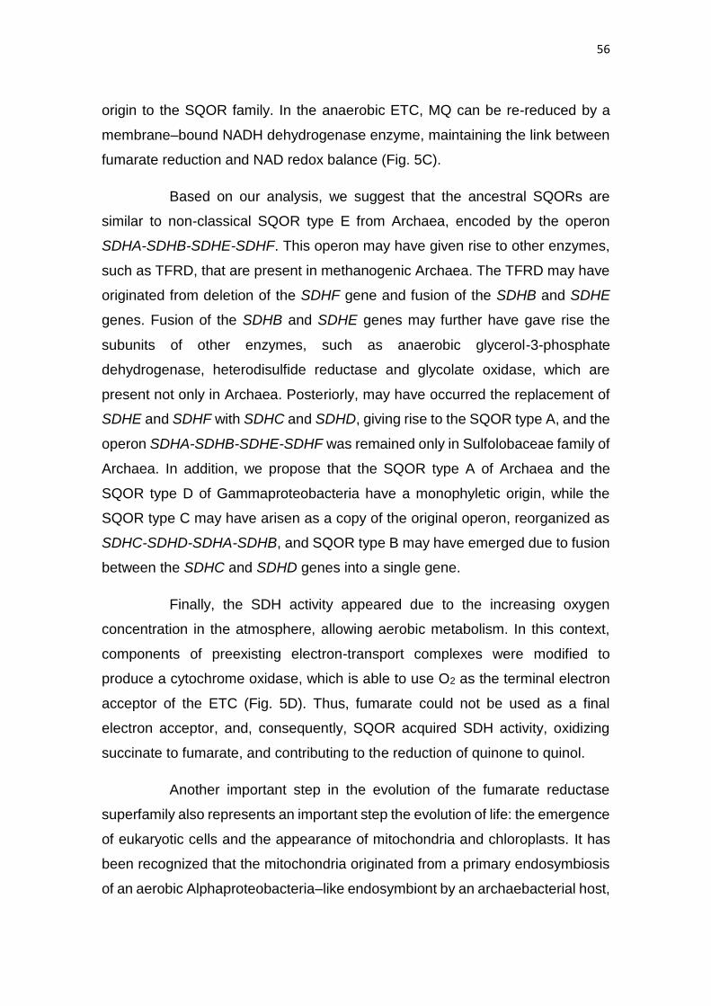
56
origin to the SQOR family. In the anaerobic ETC, MQ can be re-reduced by a
membrane–bound NADH dehydrogenase enzyme, maintaining the link between
fumarate reduction and NAD redox balance (Fig. 5C).
Based on our analysis, we suggest that the ancestral SQORs are
similar to non-classical SQOR type E from Archaea, encoded by the operon
SDHA-SDHB-SDHE-SDHF. This operon may have given rise to other enzymes,
such as TFRD, that are present in methanogenic Archaea. The TFRD may have
originated from deletion of the SDHF gene and fusion of the SDHB and SDHE
genes. Fusion of the SDHB and SDHE genes may further have gave rise the
subunits of other enzymes, such as anaerobic glycerol-3-phosphate
dehydrogenase, heterodisulfide reductase and glycolate oxidase, which are
present not only in Archaea. Posteriorly, may have occurred the replacement of
SDHE and SDHF with SDHC and SDHD, giving rise to the SQOR type A, and the
operon SDHA-SDHB-SDHE-SDHF was remained only in Sulfolobaceae family of
Archaea. In addition, we propose that the SQOR type A of Archaea and the
SQOR type D of Gammaproteobacteria have a monophyletic origin, while the
SQOR type C may have arisen as a copy of the original operon, reorganized as
SDHC-SDHD-SDHA-SDHB, and SQOR type B may have emerged due to fusion
between the SDHC and SDHD genes into a single gene.
Finally, the SDH activity appeared due to the increasing oxygen
concentration in the atmosphere, allowing aerobic metabolism. In this context,
components of preexisting electron-transport complexes were modified to
produce a cytochrome oxidase, which is able to use O2 as the terminal electron
acceptor of the ETC (Fig. 5D). Thus, fumarate could not be used as a final
electron acceptor, and, consequently, SQOR acquired SDH activity, oxidizing
succinate to fumarate, and contributing to the reduction of quinone to quinol.
Another important step in the evolution of the fumarate reductase
superfamily also represents an important step the evolution of life: the emergence
of eukaryotic cells and the appearance of mitochondria and chloroplasts. It has
been recognized that the mitochondria originated from a primary endosymbiosis
of an aerobic Alphaproteobacteria–like endosymbiont by an archaebacterial host,
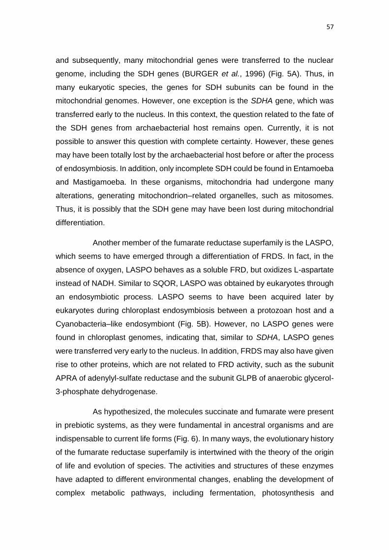
57
and subsequently, many mitochondrial genes were transferred to the nuclear
genome, including the SDH genes (BURGER et al., 1996) (Fig. 5A). Thus, in
many eukaryotic species, the genes for SDH subunits can be found in the
mitochondrial genomes. However, one exception is the SDHA gene, which was
transferred early to the nucleus. In this context, the question related to the fate of
the SDH genes from archaebacterial host remains open. Currently, it is not
possible to answer this question with complete certainty. However, these genes
may have been totally lost by the archaebacterial host before or after the process
of endosymbiosis. In addition, only incomplete SDH could be found in Entamoeba
and Mastigamoeba. In these organisms, mitochondria had undergone many
alterations, generating mitochondrion–related organelles, such as mitosomes.
Thus, it is possibly that the SDH gene may have been lost during mitochondrial
differentiation.
Another member of the fumarate reductase superfamily is the LASPO,
which seems to have emerged through a differentiation of FRDS. In fact, in the
absence of oxygen, LASPO behaves as a soluble FRD, but oxidizes L-aspartate
instead of NADH. Similar to SQOR, LASPO was obtained by eukaryotes through
an endosymbiotic process. LASPO seems to have been acquired later by
eukaryotes during chloroplast endosymbiosis between a protozoan host and a
Cyanobacteria–like endosymbiont (Fig. 5B). However, no LASPO genes were
found in chloroplast genomes, indicating that, similar to SDHA, LASPO genes
were transferred very early to the nucleus. In addition, FRDS may also have given
rise to other proteins, which are not related to FRD activity, such as the subunit
APRA of adenylyl-sulfate reductase and the subunit GLPB of anaerobic glycerol-
3-phosphate dehydrogenase.
As hypothesized, the molecules succinate and fumarate were present
in prebiotic systems, as they were fundamental in ancestral organisms and are
indispensable to current life forms (Fig. 6). In many ways, the evolutionary history
of the fumarate reductase superfamily is intertwined with the theory of the origin
of life and evolution of species. The activities and structures of these enzymes
have adapted to different environmental changes, enabling the development of
complex metabolic pathways, including fermentation, photosynthesis and

58
respiration. This plasticity was essential to living organisms, allowing the
presence of life in different locations and under a wide variety of environmental
conditions.
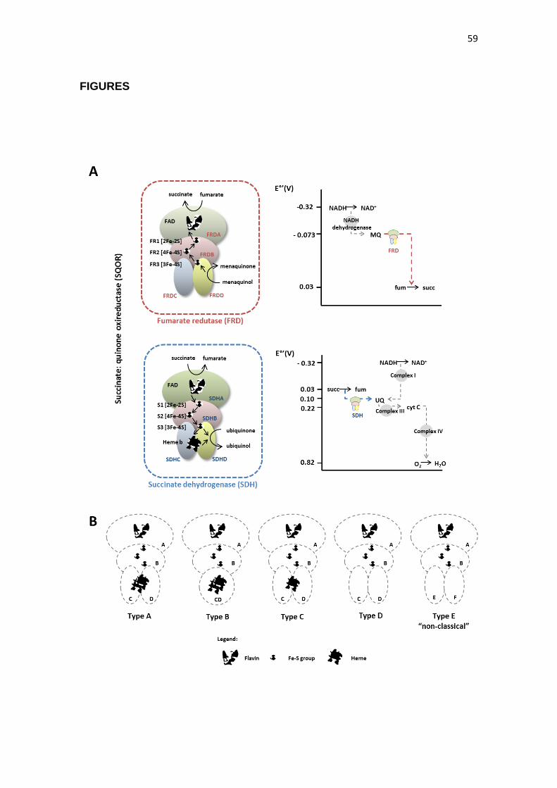
59
FIGURES

60
Figure 1. Schematic representation of succinate:quinone oxidoreductase
family. (A) Schematic representation of FRD and SDH activities. In graphics, at
the right, are represented the reduction potential of different components of
aerobic and anaerobic electron transporter chains. The hydrophilic subunits A
and B are schematically drawn in green and red, and the hydrophobic subunits
C and D in blue and yellow, respectively (B) Classification (type A-E) of
succinate:quinone oxidoreductases (SQOR) based on their hydrophobic domain
and heme content. UQ – ubiquinone; cyt C – cytochrome C; MQ – menoquinone.
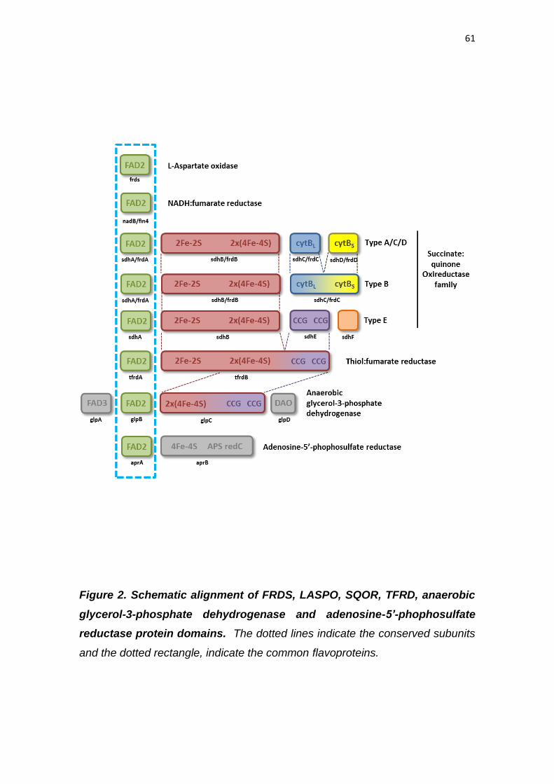
61
Figure 2. Schematic alignment of FRDS, LASPO, SQOR, TFRD, anaerobic
glycerol-3-phosphate dehydrogenase and adenosine-5’-phophosulfate
reductase protein domains. The dotted lines indicate the conserved subunits
and the dotted rectangle, indicate the common flavoproteins.
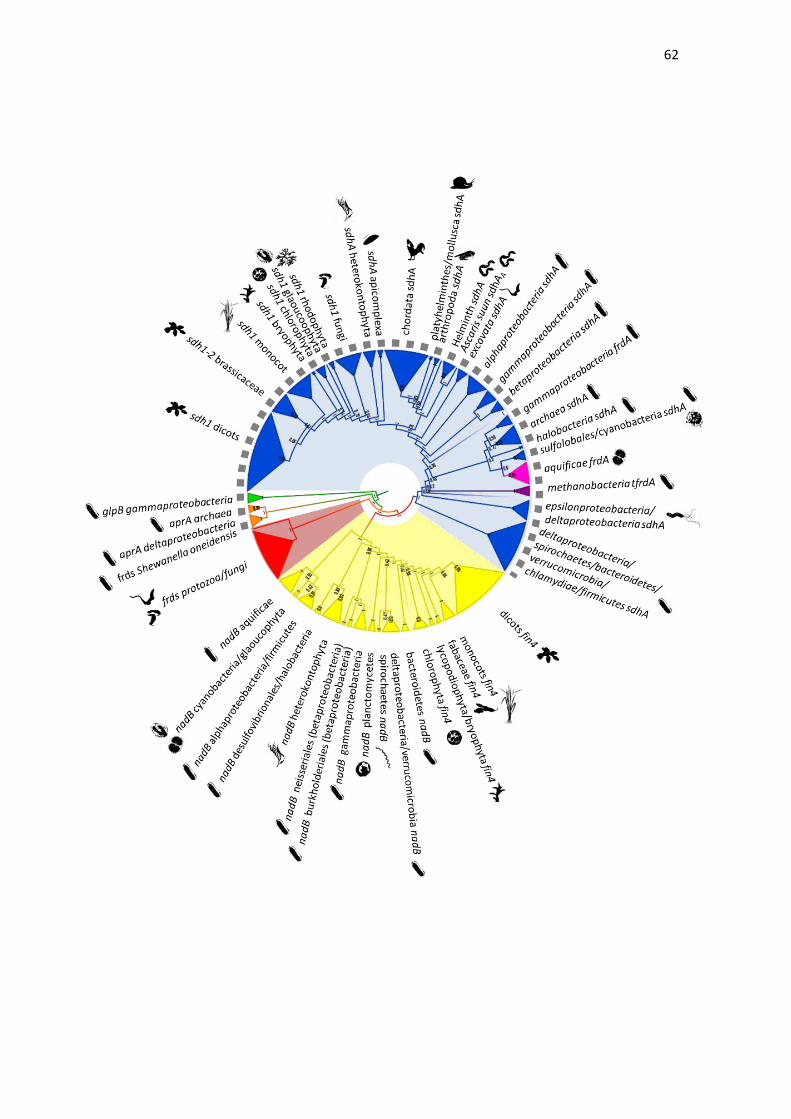
62
63
Figure 3. Phylogenetic relationship between fumarate reductase superfamily.
Clusters corresponding to three well-supported main family classes were indicated with
backgrounds colored in red (FRDS), yellow (LASPO), and blue (SQOR). The activities
of proteins belonging to the superfamily are represented by different colors: anaerobic
glycerol-3-phosphate dehydrogenase (glpB), in green; adenylyl-sulfate reductase
(aprA), in orange; FRDS, in red; LASPO, in yellow; SQOR, in blue; TFRD, in purple and
NADH-dependent fumarate reductase from Aquificae, in pink. The dotted grey line
indicates the subunits of complex components, while the absence indicates monomeric
proteins.
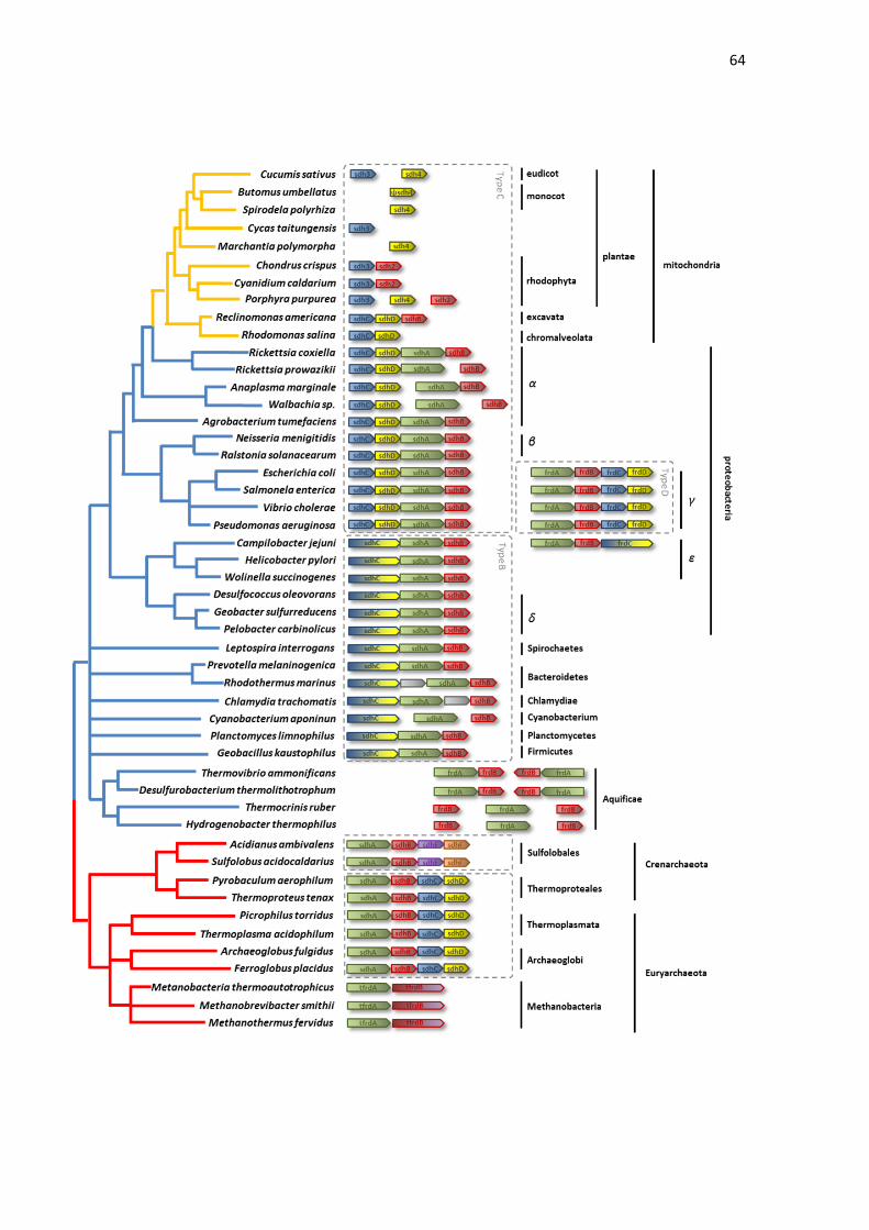
64

65
Figure 4. Arrangement of Sdh/Frd genes in bacterial, archaeal and
mitochondrial genomes. The red lines indicate genes from archaea, the blue
lines from bacteria and the orange lines indicated those genes present in
mitochondrial genome. The classes A, B, C, D and E of succinate:quinone
oxidoreductases are indicate in dotted rectangles,
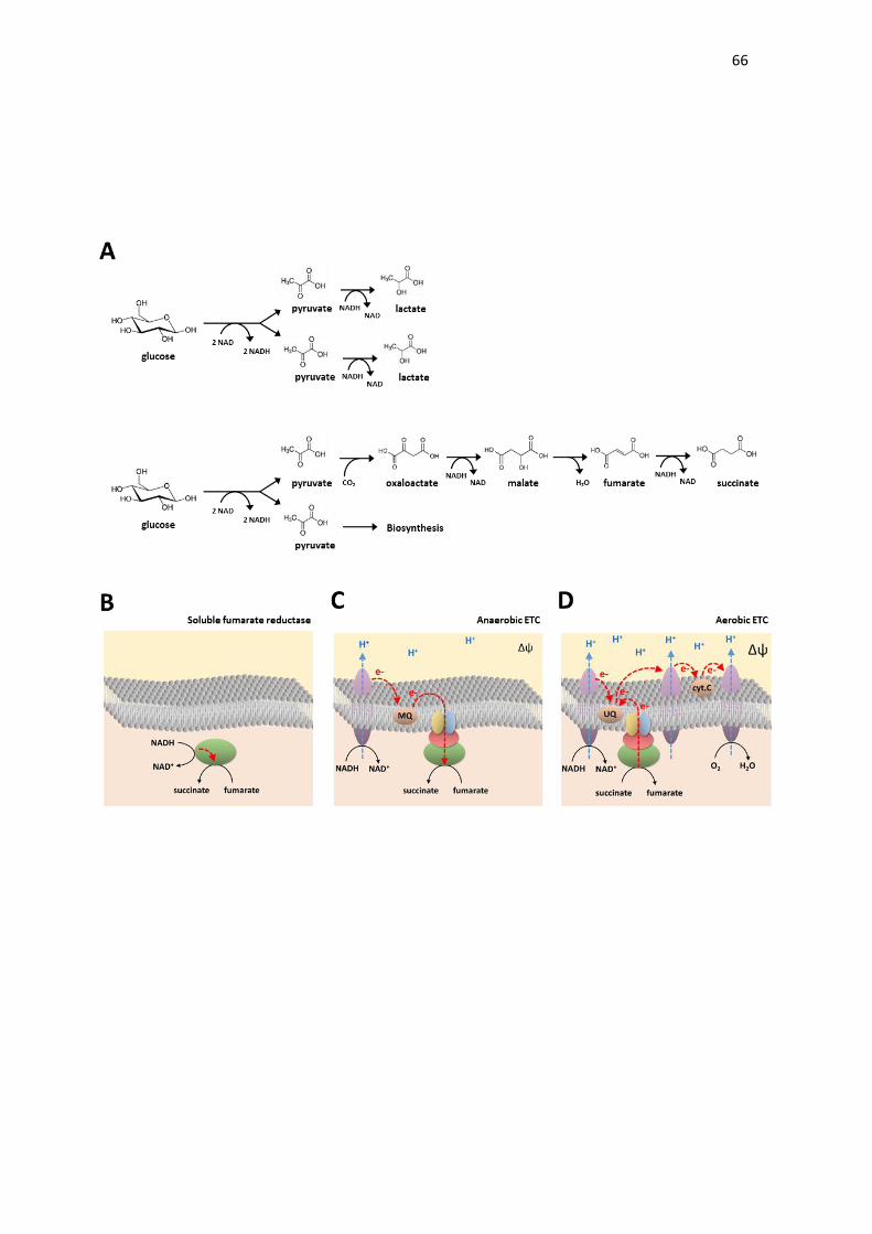
66
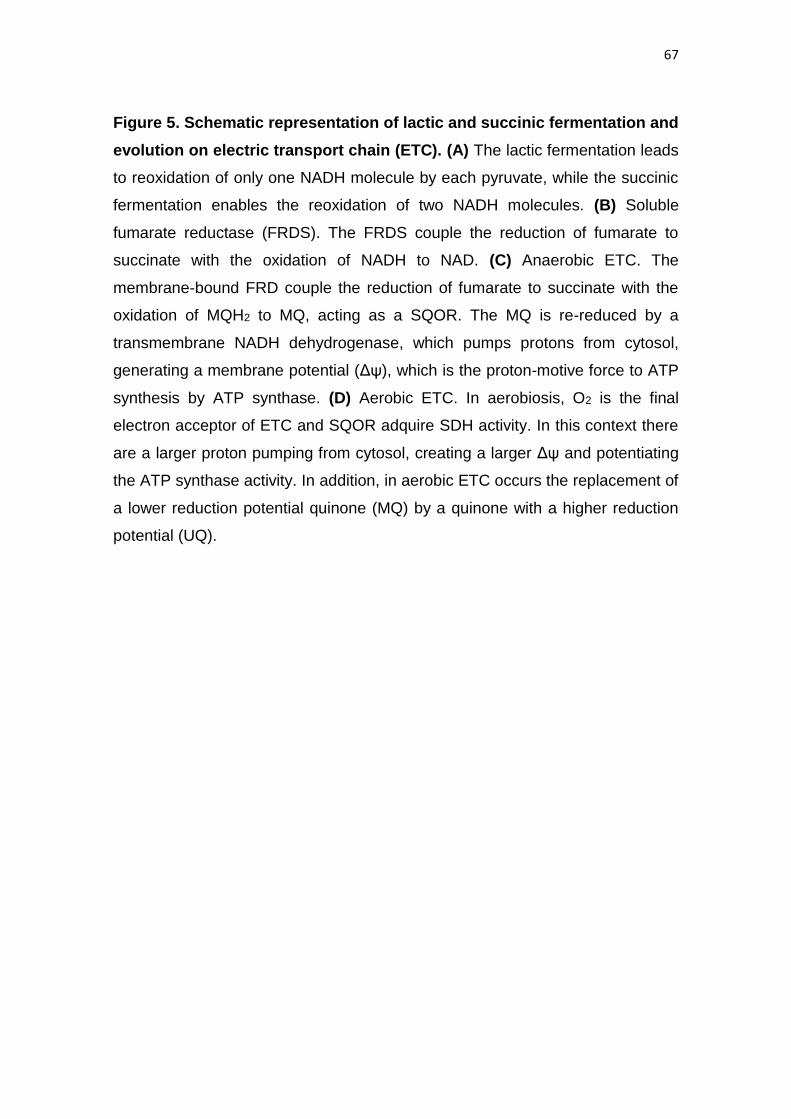
67
Figure 5. Schematic representation of lactic and succinic fermentation and
evolution on electric transport chain (ETC). (A) The lactic fermentation leads
to reoxidation of only one NADH molecule by each pyruvate, while the succinic
fermentation enables the reoxidation of two NADH molecules. (B) Soluble
fumarate reductase (FRDS). The FRDS couple the reduction of fumarate to
succinate with the oxidation of NADH to NAD. (C) Anaerobic ETC. The
membrane-bound FRD couple the reduction of fumarate to succinate with the
oxidation of MQH2 to MQ, acting as a SQOR. The MQ is re-reduced by a
transmembrane NADH dehydrogenase, which pumps protons from cytosol,
generating a membrane potential (Δψ), which is the proton-motive force to ATP
synthesis by ATP synthase. (D) Aerobic ETC. In aerobiosis, O2 is the final
electron acceptor of ETC and SQOR adquire SDH activity. In this context there
are a larger proton pumping from cytosol, creating a larger Δψ and potentiating
the ATP synthase activity. In addition, in aerobic ETC occurs the replacement of
a lower reduction potential quinone (MQ) by a quinone with a higher reduction
potential (UQ).
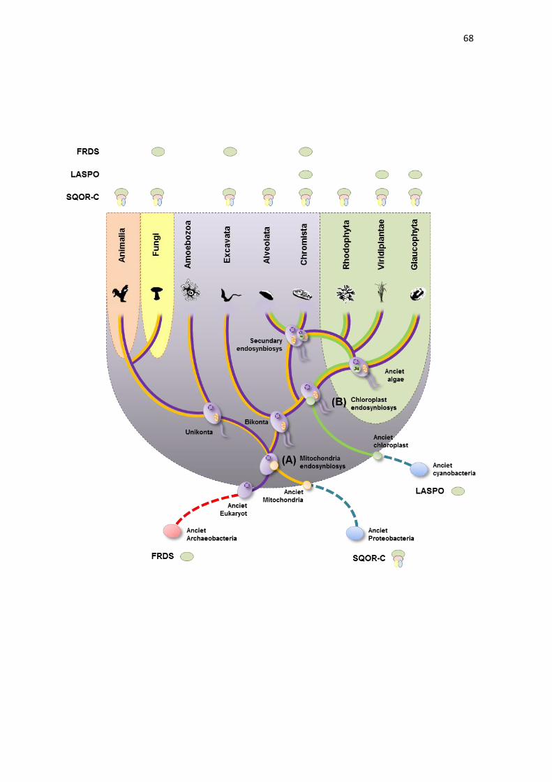
68
69
Figure 6. Schematic representation of life tree and mitochondrial and
chloroplast endosymbiosis processes. The dotted red line indicates Archaea
and the dotted blue line indicates bacteria species. The purple lines indicate all
eukaryotes, the orange lines indicate eukaryotes derived from mitochondrial
endosymbiosis, and the green lines indicate eukaryotes that possess chloroplast.
In addition, the eukaryotic kingdoms are indicated by background color: green to
plants, yellow to fungi, pink to metazoan, and purple to all protozoan. The
presence of LASPO, FRDS and SQOR type C in each eukaryotic group is
indicated on the top.
70
REFERENCES
ALTSCHUL SF, MADDEN TL, SCHÄFFER AA, ZHANG J, ZHANG Z, MILLER
W, LIPMAN DJ. (1997). Gapped BLAST and PSI-BLAST: a new generation of
protein database search programs. Nucleic Acids Res. 25: 3389-3402.
AMINO H, WANG H, HIRAWAKE H, SARUTA F, MIZUCHI D, MINEKI R,
SHINDO N, MURAYAMA K, TAKAMIYA S, AOKI T, KOJIMA S, KITA K. (2000).
Stage specific isoforms of Ascaris suum complex II: the fumarate reductase of
the parasitic adult and the succinate dehydrogenase of free-living larvae share a
common iron–sulfur subunit, Mol. Biochem. Parasitol. 106: 63-76.
AOSHIMA M. (2007). Novel enzyme reactions related to the tricarboxylic acid
cycle: phylogenetic/functional implications and biotechnological applications.
Appl. Microbiol. Biotechnol. 75: 249–255.
AWRAMIK SM. (1992). The oldest records of photosynthesis. Photosynth Res
33:75–89.
BAURAIN D, BRINKMANN H, PETERSEN J, RODRÍGUEZ-EZPELETA N,
STECHMANN A, DEMOULIN V, ROGER AJ, BURGER G, LANG BF, PHILIPPE
H. (2010). Phylogenomic evidence for separate acquisition of plastids in
Cryptophytes, Haptophytes, and Stramenopiles. Mol Biol Evol. 27: 1698-1709.
BESTEIRO S, BIRAN M, BITEAU N, COUSTOU V, BALTZ T, CANIONI P,
BRINGAUD F. (2002). Succinate secreted by Trypanosoma brucei is produced
by a novel and unique glycosomal enzyme, NADH-dependent fumarate
reductase. J. Biol. Chem. 277: 38001-38012.
BLAUT M, WHITTAKER K, VALDOVINOS A, ACKRELL BAC, GUNSALUS
RP, CECCHINI G. (1989). Fumarate reductase mutants of Escherichia coli that
lack covalently bound flavin. J. Biol. Chem. 264: 13599–13604.
BOBIK TA, OLSON KD, NOLL KM, WOLFE RS. (1987). Evidence that the
heterodisulfide of coenzyme M and 7-mercaptoheptanoylthreonine phosphate is
a product of the methylreductase reaction in Methanobacterium, Biochem.
Biophys. Res. Commun. 149: 455-460.
71
BOBIK TA, WOLFE RS. (1989). An unusual thiol-driven fumarate reductase in
Methanobacterium with the production of the heteroditerminal of coenzyme M
and N-(7-mercaptoheptanoyl) threonine-O3-phosphate. J. Biol. Chem. 264:
187142-18718.
BURGER G, LANG BF, REITH M, GRAY MW. (1996). Genes encoding the same
three subunits of respiratory complex II are present in the mitochondrial DNA of
two phylogenetically distant eukaryotes. PNAS 93: 2328-2332.
CAVALIER-SMITH T. (1999). Principles of protein and lipid targeting in
secondary symbiogenesis: euglenoid, dinoflagellate, and sporozoan plastid
origins and the eukaryote family tree. J Eukaryot Microbiol. 46: 347-366.
CECCHINI G, ACKRELL BAC, DESHLER JO, GUNSALUS RP. (1986).
Reconstitution of quinone reduction and characterization of Escherichia coli
fumarate reductase activity.J. Biol. Chem. 261: 1808-1814.
COLE ST, CONDON C, LEMIRE BD, WEINER JH. (1985). Molecular biology,
biochemistry and bioenergetics of fumarate reductase, a complex membrane-
bound iron–sulfur flavoenzyme of Escherichia coli, Biochim. Biophys. Acta 811:
381–403.
COUSTOU V, BESTEIRO S, RIVIERE L, BIRAN M, BITEAU N, FRANCONI JM,
BOSHART M, BALTZ T, BRINGAUD F. (2005). A mitochondrial NADH-
dependent fumarate reductase involved in the production of succinate excreted
by procyclic Trypanosoma brucei. J. Biol. Chem. 280: 16559-16570.
COVELLO PS, GRAY MW. (1992). Silent mitochondrial and active nuclear genes
for subunit 2 of cytochrome c oxidase (cox2) in soybean: evidence for RNA-
mediated gene transfer. EMBO J. 11: 3815-3820.
CUENCA A, PETERSEN G, SEBERG O. (2013). The Complete Sequence of the
Mitochondrial Genome of Butomus umbellatus – A Member of an Early Branching
Lineage of Monocotyledons. PLoS 8(4): e61552.
DEPPENMEIER U, JOHANN A, HARTSCH T, MERKL R, SCHMITZ RA,
MARTINEZ-ARIAS R, HENNE A, WIEZER A, BÄUMER S, JACOBI C,
72
BRÜGGEMANN H, LIENARD T, CHRISTMANN A, BÖMEKE M, STECKEL S,
BHATTACHARYYA A, LYKIDIS A, OVERBEEK R, KLENK HP, GUNSALUS
RP, FRITZ HJ, GOTTSCHALK G. (2002). The genome of Methanosarcina
mazei: evidence for lateral gene transfer between bacteria and archaea. J Mol
Microbiol Biotechnol. 4: 453-61.
DRUMMOND AJ, SUCHARD MA, XIE D, RAMBAUT A. (2012). Bayesian
phylogenetics with BEAUti and the BEAST 1.7. Molecular Biology and Evolution
29: 1969-1973Gest H. (1980). The evolution of biological energy transducing
system. FEMS Microbiol. Lett. 7: 73–77.
EDGAR RC. (2004). MUSCLE: multiple sequence alignment with high accuracy
and high throughput. Nucleic Acids Res. 32: 1792–1797.
ELLERMANN J, HEDDERICH R, BÖCHER R, THAUER RK. (1988). The final
step in methane formation: Investigations with highly purified Methyl-CoM
reductase (component C) from Methanobacterium thermoautotrophicum (strain
Marburg). Eur. J. Biochem 172: 669-677.
GEST, H. (1980). The evolution of biological energy transducing system. FEMS
Microbiol. Lett. 7, 73–77.
GRAY MW, LANG BF, CEDERGREN R, GOLDING GB, LEMIEUX C,
SANKOFF D, TURMEL M, BROSSARD N, DELAGE E, LITTLEJOHN TG,
PLANTE I, RIOUX P, SAINT-LOUIS D, ZHU Y, BURGER G. (1998). Genome
structure and gene content in protist mitochondrial DNAs. Nucleic Acids Res. 26:
865–878;
GRAY MW. (1992). The endosymbiont hypothesis revisited. Int Rev Cytol. 141:
233-357.
GRAY MW. (1993). Origin and evolution of organelle genomes. Curr Opin Genet
Dev. 3(6): 884-890.
GRIFFITH GR, CHANDLER JLR, GHOLSON RK. (1975). Studies on the de
novo biosynthesis of NAD in Escherichia coli. Eur J Biochem. 54: 239–245.
73
HÄGERHÄLL C1. (1997). Succinate: quinone oxidoreductases. Variations on a
conserved theme. Biochim Biophys Acta. 1320(2): 107-141.
HAIGERHALL C, HEDERSTEDT L. (1996). A structural model for the
membrane-integral domain of succinate'quinone oxidoreductases. Lett. 389: 25-
31.
HEDDERICH R, KOCH J, LINDER D, THAUER RK. (1994). The heterodi-sulfide
reductase from Methanobacterium thermoautotrophicum contains sequence
motifs characteristic of pyridine nucleotide-dependent thioredoxin reductases,
Eur. J. Biochem. 225: 253-261.
HEDERSTEDT L, OHNISHI T. (1992). The trinuclear iron-sulfur cluster S3 in
Bacillus subtilis succinate:menaquinone reductase; effects of a mutation in the
putative cluster ligation motif on enzyme activity and EPR properties. Mol. Mech.
Bioenerg. 7: 163-198.
HEIM S, KUNKEL A, THAUER RK, HEDDERICH R. (1998). Thiol:fumarate
reductase (Tfr) from Methanobacterium thermoautotrophicum: identification of
the catalytic sites for fumarate reduction and thiol oxidation. Eur. J. Biochem. 253:
292-299.
HIRSCH CA, RASMINSKY M, DAVIS BD, LIN EC. (1963). A fumarate reductase
in Escherichia coli distinct from succinate dehydrogenase. J Biol Chem. 238:
3770-3774.
KATOH A, UENOHARA K, AKITA M, HASHIMOTO T. (2006). Early steps in the
biosynthesis of NAD in Arabidopsis start with aspartate and occur in the plastid.
Plant Physiol. 14: 851–857.
LANCASTER CR. (2001). Succinate:quinone oxidoreductases--what can we
learn from Wolinella succinogenes quinol:fumarate reductase? FEBS Lett.
504(3):133-141.
LEMOS RS, FERNANDES AS, PEREIRA MM, GOMES CM, TEIXEIRA M.
(2002) Quinol:fumarate oxidoreductases and succinate:quinone
74
oxidoreductases: phylogenetic relationships, metal centres and membrane
attachment. Biochim Biophys Acta. 1553(1-2): 158-70.
LEÓN G, HOLUIGUE L, JORDANA X. (2007). Mitochondrial complex II is
essential for gametophyte development in Arabidopsis. Plant Physiol 143: 1534–
1546.
MARIN B, NOWACK EC, MELKONIAN M. (2005). A plastid in the making:
evidence for a second primary endosymbiosis. Protist 156: 425–432.
MATTEVI A, TEDESCHI G, BACCHELLA L, CODA A, NEGRI A, RONCHI S.
(1999). Structure of L-aspartate oxidase: implications for the succinate
dehydrogenase/fumarate reductase oxidoreductase family. Structure 7(7): 745–
756.
MESSNER KR, IMLAY JA. (2002). Mechanism of superoxide and hydrogen
peroxide formation by fumarate reductase, succinate dehydrogenase, and
aspartate oxidase. J Biol Chem. 277(45): 42563-42571.
MIURA A, KAMEYA M, ARAI H, ISHII M, IGARASHI Y. (2008). A Soluble NADH-
Dependent Fumarate Reductase in the Reductive Tricarboxylic Acid Cycle of
Hydrogenobacter thermophilus TK-6. J. Bacteriology 190 (21): 7170-7177.
MORTARINO M, NEGRI A, TEDESCHI G, SIMONIC T, DUGA S, GASSEN HG,
RONCHI S. (1996). L-aspartate oxidase from Escherichia coli. I. Characterization
of coenzyme binding and product inhibition. Eur J Biochem. 239(2): 418-426.
MOUSTAFA A, BESZTERI B, MAIER UG, BOWLER C, VALENTIN K,
BHATTACHARYA D. (2009). Genomic footprints of a cryptic plastid
endosymbiosis in diatoms. Science 324: 1724-1726.
MURATSUBAKI H, ENOMOTO K. (1998). One of the fumarate reductase
isoenzymes from Saccharomyces cerevisiae is encoded by the OSM1 gene.
Arch. Biochem. Biophys. 352: 175-181.
NUGENT JM, PALMER JD. (1991). RNA-mediated transfer of the gene coxII
from the mitochondrion to the nucleus during flowering plant evolution. Cell.
66(3): 473-481.
75
OPARIN AI. (1957). The Origin of Life on the Earth. Processes. Pergamon,
Oxford. Oliver and Boyd, Edinburgh.
PARK SJ, TSENG CP, GUNSALUS RP. (1995). Regulation of succinate
dehydrogenase (SDHC-D-A-B) operon expression in Escherichia coli in response
to carbon supply and anaerobiosis: role of ArcA and Fnr. Mol. Microbiol. 15: 473-
482.
PEALING SL, BLACK AC, MANSON FDC, WARD FB, CHAPMAN SK, REID
GA. (1992). Sequence of the gene encoding flavocytochrome c from Shewanella
putrefaciens: a tetraheme flavoenzyme that is a soluble fumarate reductase
related to the membrane-bound enzymes from other bacteria. Biochemistry 31:
12132-12140.
RAMBAUT A, SUCHARD MA, XIE D, DRUMMOND AJ. (2014). Tracer v1.6,
Available from http://beast.bio.ed.ac.uk/Tracer
REYES-PRIETO A, WEBER APM, BHATTACHARYA D. (2007). The origin and
establishment of the plastid in algae and plants. Annu Rev Genet. 41: 147–168.
ROOS MH, TIELENS AGM. (1994). Differential expression of two succinate
dehydrogenase subunit-B genes and a transition in energy metabolism during the
development of the parasitic nematode Haemonchus contortus. Mol. Biochem.
Parasitol. 66: 273-281.
SCHÄFER G, ANEMÜLLER S, MOLL R. (2002) Archaeal complex II: 'classical'
and 'non-classical' succinate:quinone reductases with unusual features. Biochim
Biophys Acta. 1553(1-2): 57-73.
SCHULTZ J, MILPETZ F, BORK P, PONTING CP. (1998). SMART, a simple
modular architecture research tool: Identification of signaling domains. PNAS. 95:
5857–5864.
SHIBA H, KAWASUMI T, IGARASHI Y, KODAMA T, MINODA Y. (1985). The
CO2 assimilation via the reductive tricarboxylic acid cycle in an obligately
autotrophic, aerobic hydrogen-oxidizing bacterium, Hydrogenobacter
thermophilus. Arch. Microbiol. 141: 198-203.
76
SUCHETA A, CAMMACK R, WEINER J, ARMSTRONG FA. (1993). Reversible
electrochemistry of fumarate reductase immobilized on an electrode surface.
Direct voltammetric observations of redox centers and their participation in rapid
catalytic electron transport. Biochemistry 32: 5455-5465.
TAMURA K, PETERSON D, PETERSON N, STECHER G, NEI M, KUMAR S.
(2011). MEGA5, molecular evolutionary genetics analysis using maximum
likelihood, evolutionary distance, and maximum parsimony methods. Mol. Biol.
Evol. 28: 2731–2739.
TEDESCHI G, NEGRI A, MORTARINO M, CECILIANI F, SIMONIC T, FAOTTO
L, RONCHI S. (1996). L-Aspartate oxidase from Escherichia coli. Interaction with
C4 dicarboxylic acids and identification of a novel L-aspartate: fumarate
oxidoreductase activity. Eur. J. Biochem. 239: 427-433.
TERNES CM, SCHONKNECHT G. (2014). Gene Transfers Shaped the Evolution
of De Novo NAD+ Biosynthesis in Eukaryotes. Genome Biol. Evol. 6(9): 2335-
2349.
THAUER RK, JUNGERMANN K, DECKER K. (1977). Energy conservation in
chemotrophic anaerobic bacteria. Bacteriol. Rev. 41: 100-180.
THUNBERG T. (1909). Studien uiber die Beeinflussung des Gasaustausches
des uiberlebenden Froschmuskels durch versehiedene Stoffe. II. Die Einwirkung
von Oxalsiiure, Malonsiure und Bernsteinsiiure. Skand. Arch. Physiol. 22: 430-
436.
TIMMIS JN, AYLIFFE MA, HUANG CY, MARTIN W. (2004). Endosymbiotic
gene transfers: organelle genomes forge eukaryotic chromosomes. Nat Rev
Genet. 5: 123–135.
VAN HELLEMOND JJ, KLOCKIEWICZ M, GAASENBEEK CP, ROOS MH,
TIELENS AG. (1995). Rhodoquinone and complex II of the electron transport
chain in anaerobically functioning eukaryotes. J Biol Chem. 270(52): 31065-
31070.
77
VIEHMANN S, RICHARD O, BOYEN C, ZETSCHE K. (1996). Genes for two
subunits of succinate dehydrogenase form a cluster on the mitochondrial genome
of Rhodophyta. Curr Genet 29: 199-201.
WACHTERSHAUSER G. (1993). The cradle chemistry of life: On the origin of
natural-products in a pyrite-pulled chemo-autotrophic origin of life. Pure Appl.
Chem. 65:1343–1348.
YANKOVSKAYA V, HORSEFIELD R, TÖRNROTH S, LUNA-CHAVEZ C,
MIYOSHI H, LÉGER C, BYRNE B, CECCHINI G, IWATA S. (2003). Architecture
of succinate dehydrogenase and reactive oxygen species generation. Science.
299(5607): 700-704.

78
3.3 CAPÍTULO 3
SILENCIAMENTO DA ASCORBATE PEROXIDASE ESTROMAL INDUZ
TOLERÂNCIA AO ESTRESSE DE SECA EM ARROZ (Oryza sativa L.)
Este capítulo é referente à primeira parte do estudo funcional
das isoformas cloroplastídicas de ascorbato peroxidase (APX) em
arroz, e está apresentado na forma do manuscrito “THE SILENCING
OF STROMAL ASCORBATE PEROXIDASE INDUCE DROUGHT
STRESS TOLERANCE IN RICE (Oryza sativa L.)”, a ser submetido.
Neste trabalho, demonstramos que plantas silenciadas para OsAPX7
e as plantas duplamente silenciadas para OsAPX7 e OsAPX8
apresentaram uma maior tolerância à seca. Além disso, verificamos
que o duplo silenciamento resultou em plantas com um maior
conteúdo de ROS e maior fechamento estomático. Esses dados
demonstram que o aumento da tolerância à seca não é consequência
somente do aumento de ROS ou do fechamento estomático, uma vez
que estas alterações não foram verificadas nas plantas com
silenciamento simples para OsAPX7, as quais também apresentam
um aumento na tolerância a seca. Desta forma, a isoforma OsAPX7
estaria envovida em mecanismos ainda inéditos que permitem um
aumento na tolerância a seca, sendo capazes de manter por mais
tempo, parâmetros fisiológicos, como os níveis de transpiração e a
fotossíntese.
79
THE SILENCING OF STROMAL ASCORBATE PEROXIDASE INDUCE
DROUGHT STRESS TOLERANCE IN RICE (Oryza sativa L.)
Douglas Jardim-Messeder1, Andreia Caverzan1, Rafael Rauber2, Juliana
Ribeiro3, Marcos Letaif Gaeta4, J. Miguel Costa5, Jorge Ernesto de Araujo
Mariath4, Nelson Saibo5, Joaquim Albenísio da Silveira3, Marcia Margis-Pinheiro1
1Departamento de Genética, Universidade Federal do Rio Grande do Sul, Porto
Alegre, RS, Brazil
2Centro de Biotecnologia, Universidade Federal do Rio Grande do Sul, Porto
Alegre, RS, Brazil
3Departamento de Bioquímica e Biologia Celular, Universidade Federal do
Ceará, Fortaleza, CE, Brazil
4Departamento de Botânica, Universidade Federal Rio Grande do Sul, Porto
Alegre, RS, Brazil
5Instituto de Tecnologia Química e Biológica, Universidade Nova de Lisboa,
Oeiras, Portugal
80
ABSTRACT
Ascorbate peroxidase (APX) exerts an important role in the
maintenance of the level of reactive oxygen species (ROS), catalyzing the
reduction of H2O2 to H2O and O2, using ascorbate as specific electron donor. In
previous work was demonstrated that the double silencing of OsAPX7 (stromal
APX) and OsAPX8 (thylacoidal APX), located specifically in chloroplast, led to
differences in photosynthetic parameters related to efficiency of utilization of light
and CO2 in plants under stress conditions. However, the functional importance of
each chloroplastic APX isoforms, and their role in plant development and in plant
stress response remains unknown.
In this work, we perform a functional study of OsAPX7 gene in rice
plants, through transgenic lines, where OsAPX7 was silenced by RNAi. In
addition to RNAiOsAPX7 plants, in this work was also used plants double
silenced to OsAPX7 and OsAPX8 genes (RNAiOsAPX7/8). Our results
demonstrate that the both lines show a normal growth, indicating that the
silencing of chloroplastic APX do not led to changes in plants development.
However, in RNAiOsAPX7/8 plants is verified a lower chloroplastic APX activity,
accompanied by a higher H2O2 content and increased stomatal closing.
Independently of this fact, when subjected to drought stress, both transgenic lines
show a higher capacity to maintain stomatal aperture and lower leaf surface
temperature. Consequently, RNAiOsAPX7, as well as RNAiOsAPX7/8 plants,
showed a higher tolerance to drought stress, when compared to non-transformed
plants. These data indicate that the chloroplastic APX can be involved in signaling
transduction pathways which regulate stomatal opening and drought stress
response in rice plants.
81
INTRODUCTION
Reactive oxygen species (ROS), such as superoxide anion (O2.-),
hydrogen peroxide (H2O2) and hydroxyl radical (HO.) are produced from the
monovalent reduction of molecular oxygen (O2), being intrinsic to the aerobic
metabolism. In plants, the ROS production is induced mainly in response to
adverse environmental conditions. ROS are important signaling molecules
involved in key physiological pathways, such as cell cycle, programmed cell death
(PCD), senescence and stress response (DAT et al.,2000; OROZCO-
CARDENAS et al., 2001; MULLINEAUX & KARPINSKI 2002; VANDENABEELE
et al., 2003). However, at high concentration, ROS can diffuse through biological
membranes reacting with biological compounds such as lipids, proteins and
nucleic acids, thus promoting damage and, in many cases, cellular death
(SCANDALIOS, 2002). The control of intracellular ROS levels, and consequently,
the cellular redox homeostasis, is a very complex process involving many
mechanisms and a large network of genes (MITTLER et al., 2004). The main
ROS scavenging mechanism is composed by several enzymes, present in
different subcellular compartments, such as, superoxide dismutases (SOD),
ascorbate peroxidases (APX), catalases (CAT), glutathione peroxidases (GPx),
and peroxidases (Prx) (HALLIWELL & GUTTERIDGE 1999; SCANDALIOS,
2002; MITTLER et al., 2004).
In plants, APX (EC, 1.11.1.11) has an important role in the ROS levels
maintenance, catalyzing the reduction of H2O2 to H2O and O2, using ascorbate
as specific electron donor (ASADA, 1999). In addition, APX is a component of
the ascorbate-glutathione cycle, one of the most important antioxidant systems
in plants. In this cycle, glutathione is used as reducing substrate to restore the
ascorbate levels, and is ultimately recycled at NAD(P)H expense (ALSCHER et
al., 1997; NOCTOR & FOYER, 1998). In rice plants (Oryza sativa L.), APX is
encoded by a small gene family, in which the isoforms are targeted to distinct
subcellular compartments: cytosol (OsAPX1 and OsAPX2), peroxisome
82
(OsAPX3 and OsAPX4), mitochondrial (OsAPX5 and OsAPX6) and chloroplast
(OsAPX5, OsAPX7 and OsAPX8) (TEIXEIRA et al., 2006).
Our group has demonstrated the functional importance of the different
APX isoforms in the cellular homeostasis in rice. Recently, we demonstrated that
rice plants silenced in both exclusive chloroplastic APXs (OsAPX7 and OsAPX8)
exhibited differences in photosynthetic parameters related to efficiency of
utilization of light and CO2 when submitted to stress conditions, suggesting that
a partial deficiency of APX in chloroplast modulate the photosystem activity and
integrity mainly under abiotic stress conditions. In addition, OsAPX7 and OsAPX8
have an antagonic expression pattern under several stress conditions, being
induced and repressed, respectively (CAVERZAN et al.,2012). This data
suggests that chloroplastic APXs exert a different role in stress response,
however, the individual contribution of each chloroplastic APX in normal and in
stress conditions remains unknown.
In this work, we performed a functional study of OsAPX7 gene using
rice plants silenced to OsAPX7 (RNAiOsAPX7) and plants double silenced to
OsAPX7 and OsAPX8 genes (RNAiOsAPX7/8). These transgenic plants have a
growth similar than non-transformed (NT) plants, however, the RNAiOsAPX7/8
plants show lower chloroplastic APX activity and higher H2O2 release, which can
explain the decrease of stomata opening. On the other hand, when subjected to
drought stress, RNAiOsAPX7 and RNAiOsAPX7/8 plants show a higher capacity
to maintain stomatal aperture and lower leaf surface temperature, resulting in a
higher tolerance to drought stress, as compared to NT plants. These results
demonstrate that the silencing of OsAPX7 do not change the redox homeostasis
in chloroplast, but increase the tolerance to drought stress, indicating that
OsAPX7 could be involved in stress signaling pathway. In addition, this work
demonstrate that only OsAPX7 silencing is necessary to improve drought
tolerance in rice.
83
MATERIAL AND METHODS
Plant material and growth conditions
Rice (Oryza sativa L. japonica cv. Nipponbare) seeds were germinated
in MS medium (Sigma-Aldrich) at 25 °C with a 12-h photoperiod. One-week-old
rice seedlings were transferred for hydropony in 200-ml plastic cups (three
seedlings per cup) filled with Hoagland–Arnon’s nutritive solution (Hoagland &
ARNON, 1950). To drought experiments, the one-week-old rice seedlings were
transferred to soil in plastic vessels. Was used a mixture of soil, vermiculite and
peat (2:1:1) and was placed six plants per vessel. The drought experiment was
performed in one-month-old plants, thought the suspense of watering. The
drought stress was maintained until the relative water content in vessel reaches
10% (in about eight days). The control of relative water content during drought
stress experiment is showed in Supplementary figure S1. To ABA treatment,
100µM ABA was dissolved in 0.1% Triton-X-100 and sprayed in excess on the
leaves of the plants until complete wetting was achieved.
Vector construction and plant transformation
A plasmid was constructed to express a hairpin structure (hpRNA)
based on the sequence of OsAPX7 (LOC Os04g35520) gene and was named
RNAiOsAPX7. The following primers were used to amplify a 220 bp fragment
from the OsAPX7 gene to clone into the RNAi vector: 5’-
CTCCGAGCAATCTGGGTGCAAAAAT-3’ and 5’-
GGTACCTCGAGGACTCGTGGTCAGGAAAAGC-3’. The PCR product was
cloned into the Gateway RNAi vector pANDA, in which the expression of the
84
hairpin RNA is driven by the maize ubiquitin promoter (MIKI & SHIMAMOTO,
2004). Agrobacterium tumefaciens-mediated transformation was performed as
previously described (UPADHYAYA et al., 2002).
To make a translational fusion of the OsAPX7 protein with Yellow
Fluorescent Protein (YFP) on the N-terminus and HA on the C-terminus, the
OsAPX7 (LOC_Os04g35520) gene was amplified from rice leaf cDNA using the
primers 5′-ATGGCGGCCCAGCG-3′ and 5′-ACCGTCCAACGTGAATCC-3′ and
cloned into the pART7-HA-YFP plasmid (GALVÁN-AMPUDIA & OFFRINGA,
2007). The gene expression is under control of 35S promoter of CaMV. The
construct was then used to transform rice protoplasts.
To verify the OsAPX7 promoter expression pattern, a 2 kb fragment
upstream of the start codon was amplified by PCR, cloned into pENTR
(Invitrogen®) and recombined with pHGWFS7 (KARIMI et al., 2005) producing
the vector promAPX7-GUS. The promAPX7-GUS vector contains the Gus and
GFP as reporter genes and hpt as selection marker gene that confers plant
resistance to hygromycin. The following primers were used to amplify the
OsAPX7 promoter: 5′-TGATAGAGTTAGTTTATGTCAAACTCG-3′ and 5′-
GACGAGGCGGAGTGAGGT 3′. The plasmid was transformed into rice calli via
Agrobacterium tumefaciens-mediated transformation, as described by
UPADHYAYA et al. (2002).
Isolation and transformation of protoplasts
Protoplast isolation was performed as described by CHEN et al.,
(2006) and protoplast transformation as described by TAO et al., (2002). After
transformation, protoplasts were incubated for 24–48 h in the dark at 28 °C before
imaging. Fluorescence was monitored using an Olympus FluoView 1000 confocal
laser scanning microscope (Olympus, Japan) equipped with a set of filters
capable of distinguishing between green and yellow fluorescente protein (GFP
and YFP, respectively) and plastid autofluorescence. The images were captured
with a high-sensitivity photomultiplier tube detector.
85
Analysis of the OsAPX7 promoter expression pattern in rice plants
Leaves, roots, stem and panicles of transgenic rice plants expressing
Gus gene under the control of the OsAPX7 promoter (promAPX7-GUS) were
sampled and analyzed using the X-Gluc histochemical assay (Fermentas®), as
described in JEFFERSON et al., (1987), with minor modifications. After Gus
staining, the samples were clarified with graded ethanol series (30–70%) and
analyzed in stereomicroscope and bright field microscopy.
Methyl violagen (MV) treatment
The one-month-old plants were grown as previously described. The
50 μM MV was prepared in 0.1% Triton-X-100 and sprayed in excess on the
leaves of the plants until complete wetting. This procedure was repeated twice a
day. The first symptoms of toxicity (brown spots) appeared on the leaves after 24
h of treatment. The control plants were sprayed with 0.1% Triton-X in the same
conditions as the plants submitted to the MV treatment.
Quantitative real-time PCR (RT-qPCR)
Real-time PCR experiments were carried out using cDNA synthesized
from total RNA purified with TRIzol (Invitrogen, Waltham, MA, USA).
Complementary cDNA was obtained using the SuperscriptTMII (Life
Technologies, Carlsbad, CA, USA) reverse transcriptase system and a 24-
polyTV primer (Invitrogen®). After synthesis, cDNAs were diluted 10–100 times
in sterile water for use in PCR reactions. All reactions were repeated four times,
and expression data analyses were performed after comparative quantification of
the amplified products using the 2-ΔΔCt method (LIVAK & SCHMITTGEN, 2001;
SCHMITTGEN & LIVAK, 2008). RT-qPCR reactions were performed in an
Applied Biosystems StepOne plus Real Time PCR system (Applied Biosystems,
Waltham, MA, USA) using SYBR-green intercalating dye for fluorescence
86
detection. The primer sequences and reference genes are listed in Supporting
Information Table S1.
Quantitative measurement of H2O2
The measurement of H2O2 production was performed by extracting
H2O2 from leaves according to a previously described method (RAO et al., 2000)
using the Ampliflu Red (Sigma-Aldrich) oxidation method as previously described
(SMITH et al.,2004). Fluorescence was monitored using a fluorimeter at
excitation and emission wavelengths of 563 nm (slit 5 nm) and 587 nm (slit 5 nm),
respectively. Calibration was performed by the addition of known quantities of
H2O2.
Meansurement of chloroplastic APX activity
The chloroplast was isolated from one-month-old plants as
SEIGNEURIN-BERNY et al. (2008), and APX activity was measured as
CAVERZAN et al. (2012).
Pigment determination
The total chlorophyll, chlorophyll a and b were determined after
extraction in ethanol and measured spectrophotometrically at 665 and 649 nm.
The amount of pigment was calculated using the equations proposed by
LICHTENTHALER & WELLBURN (1983). In addition, the pigment determination
was performed by co-focal microscopy, using the red filter of chlorophyll.
87
Membrane damage determination
The electrolyte leakage (membrane damage) was measured as
described previously by BLUM & EBERCON (1981). Leaf samples were placed
in tubes containing deionized water. The flasks were incubated in a shaker for 12
h, and the electric conductivity in the medium (L1) was measured. Then, the
medium was boiled (95 °C) for 60 min and the electric conductivity (L2) was
measured again. The relative membrane damage (MD) was estimated by MD =
L1/L2×100.
Imaging of rice stomata
The stomatas were visualized by scanning electron microscopy and
quantified. Leaves of 30 day-old plants from different height were detached and
the samples are prepared as described by HUANG et al., (2009).
Gas exchange and photochemical parameters
The following PSII parameters were measured using an Infrared Gas
Analyzer coupled with a leaf chamber fluorometer (Li-6400-XT, LI-COR, Lincoln,
USA) according to the manufacturer's instructions. ΔF/F’m, actual quantum yield
of photosystem II, Fv/Fm, potential quantum yield of photosystem II, and ETR,
apparent electron transport rate. The relative excess energy at the photosystem
II level was calculated as EXC = [(Fv/Fm) − (ΔFv/F’m)]/(Fv/Fm). All of the
parameters were measured with an Infrared Gas Analyzer coupled with a leaf
chamber fluorometer (Li-6400-XT, LI-COR, Lincoln, USA) according to the
manufacturer’s instructions. ETR was calculated as ETR = (ΔF/F’m× PPFD × 0.5
× 0.84), where 0.5 is the presumed fraction of the excitation energy distributed to
PSII and0.84 is the assumed fraction of light absorbed by the leaf. EXC was
calculated according to BILGER et al., (1995) as EXC = [(Fv/Fm)
−(ΔF/F’m)]/(Fv/Fm).The photochemical quenching coefficient [qP = (F’m−
Fs)/(F’m−F’o)] and the non-photochemical quenching coefficient [NPQ =(Fm−
88
F’m)/F’m], where Fm and Fo are, respectively, maximum and minimum
fluorescence of dark-adapted leaves; F’m and Fs are, respectively, maximum and
steady state fluorescence in the light-adapted state, and F’o is minimum
fluorescence after far-red illumination of the previously light exposed leaves. A
saturating pulse of red light (0.8 s, 8000 μmol m−2s−1) was utilized. Leaf gas
exchange measurements were made using an Infra-redGas Analyzer (Li-6400-
XT, LI-COR Biosciences). Light was provided by a red/blue LED light source at
photon irradiance of 1000 μmol m−2s−1. All leaf measurements were taken under
ambient CO2 conditions (380 ppm) at a constant leaf temperature of 28 oC and
relative humidity of 78% and vapor pressure deficit (VPD) of 1.8 Pa. The gas
exchange variables measured were net photosynthesis (PN) and intercellular
CO2 concentration (CI).
Thermal Imaging
Thermography was performed using a ThermaCam B20HS camera
(FLIR Systems) equipped with an uncooled 320 x 240 microbolometer matrix
detector in the 7- to 13-mm band having an improved sensitivity (noise-equivalent
differential temperature below 0.05°C). These subsequent experiments were
carried out in a dedicated darkroom under low wind speed to ensure temperature
contrast between lines.
Protein determination
Protein concentrations were determined as described by BRADFORD
(1976) using BSA as a standard.

89
Statistical analysis
Data were plotted with GRAPHPAD PRISM 5.0 (GraphPad Software
Inc., La Jolla, CA, USA) and analyzed by one-way ANOVA and a posteriori
Tukey’s test. P-values of 0.05 were considered statistically significant.

90
RESULTS
OsAPX7 protein is localized in chloroplast and OsAPX7 gene mainly
expressed in embryo and vascular tissue
In a previous work, eight APX genes were identified in the rice genome
(TEIXEIRA et al., 2006). In silico analysis predicted that the protein encoded by
OsAPX7 is exclusively located in chloroplast. In the present work, the subcellular
localization of OsAPX7 protein was experimentally determined in rice protoplasts.
We constructed a translational fusion of OsAPX7 with YFP protein, driven by the
CaMV 35S promoter. Confocal analysis of protoplasts expressing 35S-
OsAPX7::YFP fusion revealed that OsAPX7 was localized in the chloroplast (Fig.
1A). These data confirmed the previous in silico prediction, which indicated that
OsAPX7 was located in stroma (TEIXEIRA et al., 2006).
To analyse the OsAPX7 gene expression pattern, rice calli were
transformed with the promAPX7-Gus construct and then we analyzed leaves,
roots, stems, and spikelets from two different transgenic rice lines (proAPX7-
Gus). This analysis showed Gus staining in the phloem region of the stem (Fig.
1G and H), in tip lemma/palea of spikelet (Fig. 1I), in the embryo of a fully
expanded rice seed (Fig. 1J), and in young trichomes of rice spikelets (Fig. 1K).
Molecular and morphological analysis of OsAPX7 silenced plants
To determine the functional role of the stromal ascorbate peroxidase,
the OsAPX7 gene was silenced using the RNAi technology to generate the
transgenic rice lines, named RNAiOsAPX7. In addition, in this work, we used
plants that have are both chloroplast APX genes (OsAPX7 and OsAPX8)
silenced. These transgenic plants were previously obtained by CAVERZAN et al.,
(2012) and named RNAiOsAPX7/8.
91
The RT-qPCR analysis of RNAiOsAPX7 T2 plants showed that
OsAPX7 transcript levels were reduced in two lines, which were names as line-a
and line-b. In these lines the OsAPX7 transcript was reduced to 71% and 76%,
respectively, as compared to NT plants. The double silenced plants
(RNAiOsAPX7/8) showed a decrease of about 50% for OsAPX7 transcript level
and 60% for OsAPX8.
RNAiOsAPX7 plants did not show an altered OsAPX8 transcript levels
(Fig. 2G and 2H). On the other hand, in both RNAiOsAPX7 lines, the expression
of OsAPX2 was also reduced, in about 60% (Fig. 2B). In addition, these plants
showed an increase of about 6 and 4-fold in the expression of OsAPX6 (Fig. 2F).
To investigate the effect of OsAPX7 silencing in plant development, the growth
of RNAiOsAPX7 and RNAiOsAPX7/8 plants was analyzed. Our results
demonstrated that, under normal conditions, the transgenic plants did not show
apparent changes in growth when compared with non-transformed plants grown
under control conditions. Thus, all plants showed the same leaf length and growth
rate (Fig. 3). In the future experiments, the RNAi OsAPX7 line-a and RNAi
OsAPX7/8 line-a plants was used as representative lines.
Effect of RNAiOsAPX7 silencing in chloroplastic APX activity and in
response to MV treatment
To analyze the effect of OsAPX7 silencing in chloroplastic APX activity,
we measured the APX activity in isolated chloroplast. The APX activity was
measured using different concentration of H2O2. Our results demonstrate that the
OsAPX7 silencing do not change APX activity (Fig. 4A). On the other hand, the
double silencing led to a decrease of APX activity (Fig. 4B). In addition, we
determined different catalytic parameters of chloroplastic APX activity in NT,
RNAiOsAPX7 and RNAiOsAPX7/8 plants. In both transgenic plants lines there
are not changes in VMAX (Fig. 4C), however in RNAiOsAPX7/8 plants is verify an
increase in about three fold in apparent KM to H2O2 (Fig. 4D), which results in an

92
equivalent decrease of catalytic efficiency of chloroplastic APX activity (VMAX/KM)
(Fig. 4E).
In addition, to verify the effect of APX silencing in chloroplast stress
response, we evaluated the change of chlorophyll content in leaves treated with
methyl viologen (MV), which is able to induce ROS production in the chloroplast.
No differences were verified between NT and RNAi OsAPX7 plants, however,
double-silenced plants showed a higher decrease of chlorophyll content after MV
treatment, however, under control conditions no differences in the chlorophyll
content was observed (Fig. 5A-5C).
RNAiOsAPX7 silenced plants show improved drought tolerance
It has been demonstrated that control of ROS production and
scavenging in the chloroplast are essential for drought tolerance in both
transgenic plants and tolerant cultivars (VAN CAMP et al., 1996; HERNANDEZ
et a.l, 2001; MITTLER & BERKOWITZ, 2001; TSENG et al., 2007). To evaluate
the effect of the OsAPX7 silencing on drought tolerance, we performed a soil
drought experiment. Fig. 6A and 6B show that both RNAiOsAPX7 and
RNAiOsAPX7/8 plants have higher drought tolerance when compared to NT
plants. In addition, we repeat the drought experiment using an additional line of
RNAiOsAPX7. Our results demonstrate that both lines (lina-a and line-b) show
an increased tolerance to drought stress (Supplementary figure S2).
To evaluate the cell membrane damage in leaves of transgenic and NT
plants subjected to drought stress, we have determined the electrolyte leakage.
Under drought stress, both RNAiOsAPX7 and RNAiOsAPX7/8 showed lower
electrolyte leakage under drought stress, as compared to NT plants (Fig. 6C).
These results indicated that silencing of the OsAPX7 gene prevents cell
membrane damage under drought stress, thus contributing to improve drought
tolerance.
93
To elucidate the physiological mechanism of drought tolerance in
RNAiOsAPX7 silenced plants, we compared gas exchange and photochemical
parameters between NT and silenced plants under drought stress condition. NT
plants showed a decrease of apparent transpiration (E), net photosynthesis (Pn),
photosystem II activity (Y) and electron transport rate (ETR) after the fourth day
of drought, while silenced plants maintain the values for these parameters, being
affected by drought stress only at the fifth day (Fig. 7A, 7C, 7D, 7E, respectively).
Consequently, the RNAiOsAPX7 plants show an increased internal CO2
concentration in a later moment than NT pants (Fig. 7B). These results indicate
that the enhanced drought tolerance of the RNAiOsAPX7 silenced plants is due
to an increased ability to maintain water use efficiency (WUS) and the potential
quantum yield of photosystem II (Fv/Fm) under drought stress condition (Fig. 7F
and 7G, respectively).
The OsAPX7 silencing causes decrease of stomatal opening
Due the importance of stomata in the responses to drought
(HETHERINGTON & WOODWARD 2003), we examined the stomata status in
RNAiOsAPX7 plants and RNAiOsAPX7/8 plants under control conditions. Our
results showed that 29.0% and 49.6% of the leaf stomata were completely closed
in the RNAiOsAPX7 silenced plants and in the RNAiOsAPX7/8 plants,
respectively, while only 20,3 % were completely closed in NT plants. In addition,
22.1% of stomata were completely open in RNAiOsAPX7 and only 0.8% in
RNAiOsAPX7/8 plants, while 39.9% of stomata were completely open in NT
plants. The percentage of partially open stomata was 49.6%, 48.8% and 38.8%
in NT, RNAiOsAPX7 and RNAiOsAPX7/8 plants, respectively (Fig. 8B). These
results showed that OsAPX7 silencing do not changes the stomatal opening
pattern, however the double silencing of OsAPX7 and OsAPX8 led to a
decreased stomatal opening, indicating that stomata aperture can be greatly
94
affected by the silencing of both chloroplastic APX in rice plants. Previous work
has demonstrated the ROS involvement in stomata closure, mainly in response
to drought stress (DASZKOWSKA-GOLEC & SZAREJKO, 2013). Thus, we
measured the hydrogen peroxide in transgenic and NT plant leaves. Our results
showed an increase in about 25% in hydrogen peroxide in RNAiOsAPX7/8 plants,
while no differences were observed in the hydrogen peroxide content between
NT and RNAiOsAPX7 plants (Fig. 8C).
In addition, to analyze the effect of OsAPX7 silencing in leaf water loss,
we also subjected rice leaves to a desiccation experiment. Our results
demonstrated that OsAPX7 silencing do not change leaf desiccation, however
leaves from RNAiOsAPX7/8 plants showed a reduction of about 25% in
desiccation as compared to NT plants, consistent with the results observed for
stomatal aperture. Interestingly, the pre-treatment with abscisic acid led to a
decrease of leaf desiccation in NT and in RNAiOsAPX7 plants, and do not change
the leaf desiccation in double knockdown. Under this condition, there are not
differences in leaf desiccation between NT, RNAiOsAPX7 and RNAiOsAPX7/8
plants (Fig. 8D-8F). These results indicate that the silencing of OsAPX7 led to
decrease of stomatal opening but not changes leaf desiccation.
The OsAPX7 silencing led to decrease of leaf surface temperature even in
drought stress condition
An alternative approach for measuring stomatal conductance is the
infrared (IR) thermography in leaves. The leaf surface temperature correlates well
with estimates of stomatal conductance (JONES, 1999; SIRAULT et al., 2009)
and consequently with rice water status (CAO et al., 2013). Thus, we measured
the leave surface temperature in NT, RNAiOsAPX7 and RNAiOsAPX7/8 plants
under control conditions, moderate drought stress (2 days water withholding) and
severe drought stress (7 days water withholding). Our results showed that under
control conditions, RNAiOsAPX7 silenced plants have about 0.85 ºC decrease in
95
leaf surface temperature (Fig. 9A). Under severe drought stress, the
RNAiOsAPX7 silenced plants remains with a lower leaf surface temperature, -
0.61 ºC and -0.34 ºC, respectively (Fig. 9B and 9C). On the other hand, the
RNAiOsAPX7/8 plants did not show an altered leaf surface temperature in either
control or stress conditions. These results indicate that the silencing of OsAPX7
led to a decrease of leaf surface temperature, which remains lower even under
drought stress condition.
96
DISCUSSION
The subcellular localization of all OsAPX family members was
previously predicted by in silico analysis (TEIXEIRA et al., 2006). In the present
study, our data confirmed the predicted subcellular localization of OsAPX7
protein, which is located in the chloroplasts of rice protoplasts. In addition, the
analysis of the OsAPX7 gene promoter demonstrated that OsAPX7 gene is
expressed predominantly in the embryo, phloem region of stem, tip and young
trichrome of rice spikelet. In fact, H2O2 has been recognized as a phloem-mobile
signal, which moves from the local tissue to the entire plant (SZECHYNSKA-
HEBDA et al., 2010; SUZUKI et al., 2013; GILROY et al., 2014). In Arabidopsis
thaliana, using Luciferase gene expression driven by the H2O2 inducible APX2
promoter, was demonstrated that H2O2 have a crucial role in systemic acquired
acclimation (SAA) in response to high light stress (KARPINSKI et al., 1999).
Thus, the presence of APX in the vascular cells could regulate local or systemic
ROS signaling.
We have previously demonstrated that the double silencing of
chloroplastic APXs (OsAPX7 and OsAPX8) does not result in visible growth
changes of plants under control growing conditions, but it causes alterations in
the level of proteins involved in photosynthesis and oxidative metabolism.
Moreover, under abiotic stress conditions, the double knockdown plants showed
many photosynthetic and biochemical changes (CAVERZAN et al., 2012).
Although this work has reported the importance of OsAPX7 and APX8 isoforms
in abiotic stress response in rice plants, the particular role of each APX isoforms
remained unknown. Thus, here we conducted a functional study of OsAPX7,
showing its importance in plant development and in the control of drought stress
response, through the analysis of RNAiOsAPX7 plants.
Our results indicate that, in control conditions, the decrease in about
90% of the OsAPX7 transcript did not impair chloroplastic APX activity and the
normal development of rice plants. In addition, these plants did not exhibit
symptoms of oxidative stress, as membrane damage or reduced growth. In
97
contrast, the RNAiOsAPX7/8 plants, which have both chloroplastic APXs
silenced, showed a decrease of chloroplastic APX activity and a slight increase
of hydrogen peroxide content in leaves when compared to NT plants, as
demonstrated previously by CAVERZAN et al., 2012.
As described by DASZKOWSKA-GOLEC & SZAREJKO (2013) the
increase of ROS production is involved with different stomata closure
mechanisms. Thus, the RNAiOsAPX7/8 plants showed a decrease of stomatal
opening and, consequently, decrease of leaves desiccation. These phenomena
can be justified by an increase of hydrogen peroxide content in leaves of
RNAiOsAPX7/8 plants.
Interestingly, our group demonstrated in a previous work that OsAPX7
expression is highly induced under chloroplast stress condition, as high light or
MV treatment (CAVERZAN et al., 2012). These results indicate that OsAPX7 can
be involved in the chloroplast stress response mechanisms. Due to the
importance of the regulation of ROS production and scavenging by chloroplast in
plant tolerance to drought (VAN CAMP et al., 1996; HERNANDEZ et al., 2001;
MITTLER & BERKOWITZ, 2001; TSENG et al., 2007), we subjected the
RNAiOsAPX7 and RNAiOsAPX7/8 plants to drought stress, to determine the
physiologic role of chloroplastic OsAPX7 in drought stress response.
Surprisingly, both transgenic plants show an increased drought tolerance when
compared to NT plants. To understand the mechanism of drought tolerance in
these transgenic plants, we evaluated photochemical parameters and gas
exchange in plants exposed to drought stress. Our results revealed that
RNAiOsAPX7 and RNAiOsAPX7/8plants are able to maintain the transpiration,
and consequently stomatal conductance, for a longer time under drought stress.
The capacity to maintain the stomatal conductance under stress condition
enables the maintenance of photosynthesis, resulting in a later increase in
intracellular CO2 concentration. Consequently, RNAiOsAPX7 and
RNAiOsAPX7/8 plants are able to maintain for a longer time the water use
efficiency and the potential quantum yield of photosystem II under drought stress
condition. This later effect of drought stress on photosynthesis suggests that the
silencing of OsAPX7 led to a prevention of inhibition of Calvin cycle biochemical
98
reactions, due the ability of transformed plants to maintain the stomatal
conductance and the CO2 assimilation, even under drought stress. The
maintenance of the Calvin cycle, therefore, enables continuous oxidation of
NADPH to NADP, which is required as the final electron acceptor of
photosynthetic electron transporter chain activity.
In addition, we established the rice water status by leaf canopy
temperature in plants subjected or not to drought stress. Surprisingly, the
RNAiOsAPX7 plants showed a decrease of about 0.85 ºC in the leaf surface
temperature. This decrease was maintained even under moderate or severe
drought stress. At first, this result may seem contradictory, since the
RNAiOsAPX7 plants do not show an increase of stomatal opening or
transpiration. However, the rice water status is not only dependent of
transpiration, but is also highly dependent of water absorption. Thus, the
decrease of leaf surface temperature in RNAiOsAPX7 plants can indicates an
increase of water absorption capacity and new studies should be conducted to
test this hypothesis. In fact, lines more productive (and tolerant) to drought stress
condition, especially crops, has been selected based on their lower leaf
temperature. Previous work, in wheat and maize, demonstrate that cooler leaf
surface temperature is favorable for higher yield (JONES, 1999). The higher
transpiration in stress condition indicates a higher stomatal conductance, and
therefore, greater capacities of plants to continue to perform photosynthesis and
being potentially more productive. In potato, for example, it was demonstrated a
clear negative correlation between the temperature of the canopy and the
production of tubers, when cultivated under large moisture supply (PRASHAR et
al., 2014). However, this strategy can be dangerous. A higher stomatal
conductance under stress condition enable a higher photosynthesis, but the
water loss by transpiration is also increased. This can be serious in severe and
prolonged stress situations, which could compromise not only the production but
also the plant survival. This work demonstrates for the first time the functional
study of OsAPX7, and that its manipulation led to rice plants more tolerant to
drought stress. More than a biotechnological application, this work reveals the

99
complexity of the control exercised by the organelle ROS production in the
signaling transduction pathways which regulate the abiotic stresses response.

100
FIGURES

101
Figure 1. Subcellular localization of OsAPX7 protein and analysis of expression
patterns of OsAPX7 promoter merged to the Gus sequence. Subcellular localization
of the OsAPX7 protein in chloroplasts in rice protoplasts throught transient expression
of the 35S-OsAPX7::YFP cassette. (a) green signals indicate YFP fluorescence; (b) red
signals indicate chlorophyll autofluorescence and (c) yellow signlas is the merged
image; (d) positive control of the location using 35S::YFP cassette; (e and f) negative
control; Expression patterns of OsAPX7 promoter gene merged to the Gus sequence.
(g and h) six-month-old rice plants: phloem of stem; (i) tip lemma/palea of spikelet; (j)
embryo of a fully expanded rice seed and (k) young trichomes of rice spikelet.
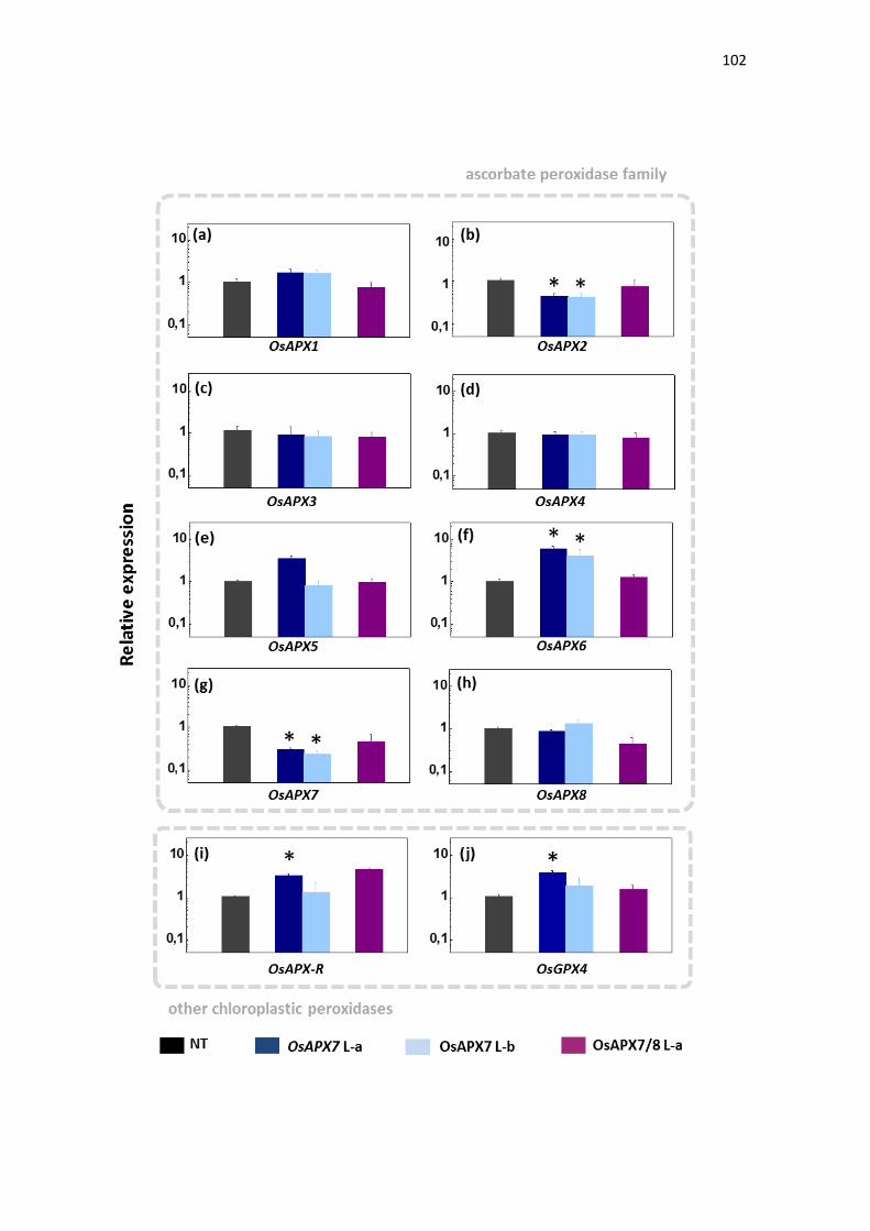
102
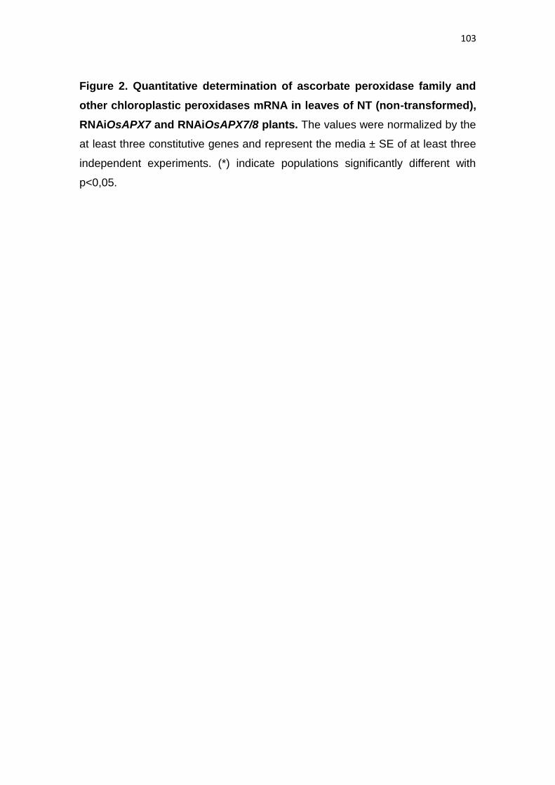
103
Figure 2. Quantitative determination of ascorbate peroxidase family and
other chloroplastic peroxidases mRNA in leaves of NT (non-transformed),
RNAiOsAPX7 and RNAiOsAPX7/8 plants. The values were normalized by the
at least three constitutive genes and represent the media ± SE of at least three
independent experiments. (*) indicate populations significantly different with
p<0,05.
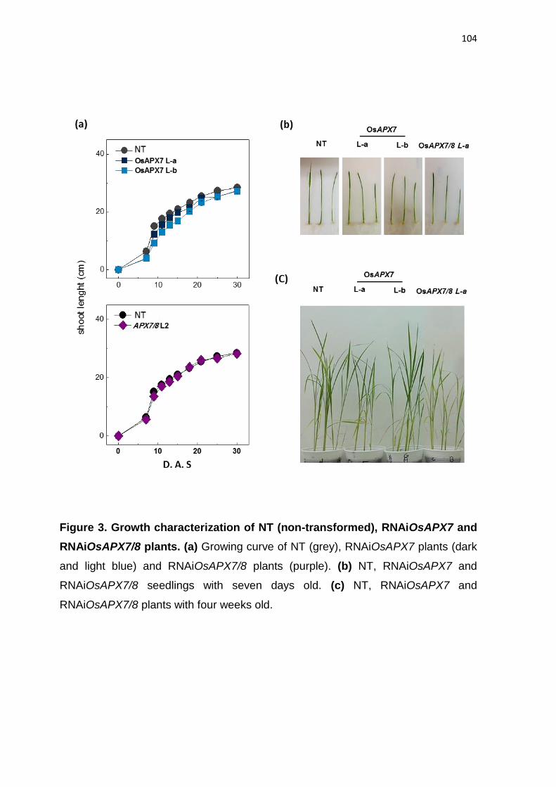
104
Figure 3. Growth characterization of NT (non-transformed), RNAiOsAPX7 and
RNAiOsAPX7/8 plants. (a) Growing curve of NT (grey), RNAiOsAPX7 plants (dark
and light blue) and RNAiOsAPX7/8 plants (purple). (b) NT, RNAiOsAPX7 and
RNAiOsAPX7/8 seedlings with seven days old. (c) NT, RNAiOsAPX7 and
RNAiOsAPX7/8 plants with four weeks old.
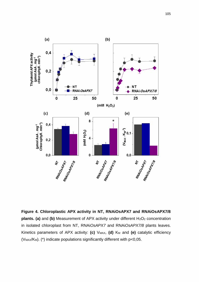
105
Figure 4. Chloroplastic APX activity in NT, RNAiOsAPX7 and RNAiOsAPX7/8
plants. (a) and (b) Measurement of APX activity under different H2O2 concentration
in isolated chloroplast from NT, RNAiOsAPX7 and RNAiOsAPX7/8 plants leaves.
Kinetics parameters of APX activity: (c) VMAX, (d) KM and (e) catalytic efficiency
(VMAX/KM). (*) indicate populations significantly different with p<0,05.
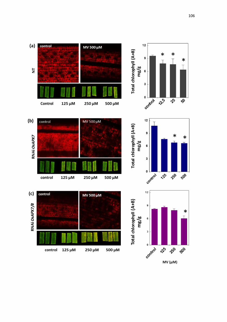
106

107
Figure 5. Effect of MV treatment in chlorophyll content of NT, RNAiOsAPX7
and RNAiOsAPX7/8 plants. The chlorophyll content was evaluated by co-focal
microscopy (left) and quantified by colorimetric method (right) in leaves from NT
(a), RNAiOsAPX7 (b) and RNAiOsAPX7/8 plants (c). (*) indicate populations
significantly different with p<0,05.
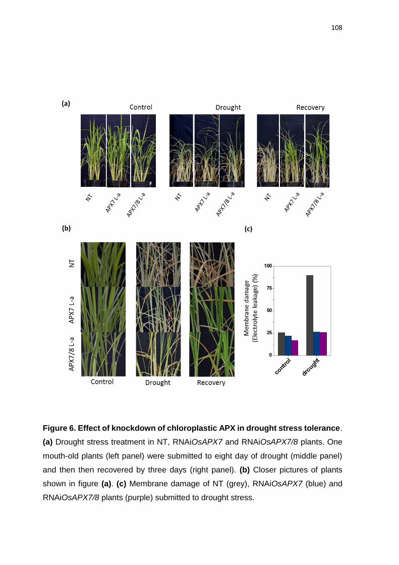
108
Figure 6. Effect of knockdown of chloroplastic APX in drought stress tolerance.
(a) Drought stress treatment in NT, RNAiOsAPX7 and RNAiOsAPX7/8 plants. One
mouth-old plants (left panel) were submitted to eight day of drought (middle panel)
and then then recovered by three days (right panel). (b) Closer pictures of plants
shown in figure (a). (c) Membrane damage of NT (grey), RNAiOsAPX7 (blue) and
RNAiOsAPX7/8 plants (purple) submitted to drought stress.
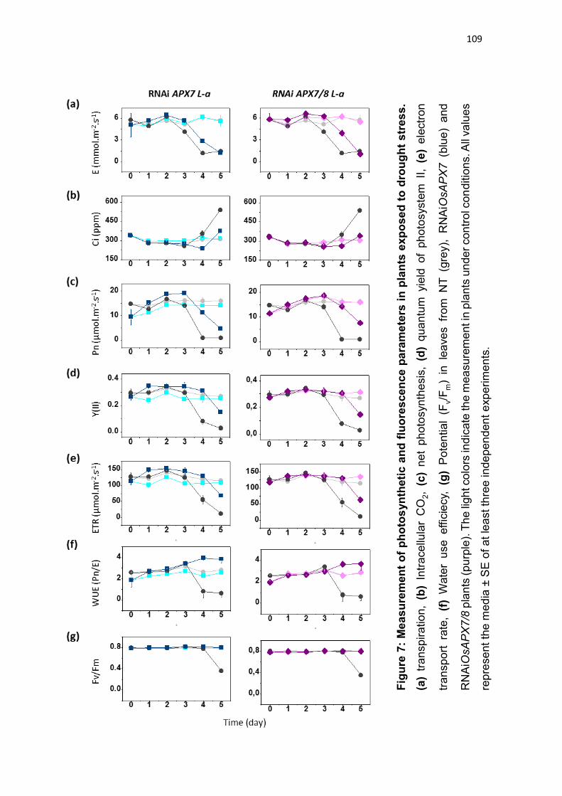
109
Fig
ure
7:
Me
as
ure
men
t o
f p
ho
tos
yn
the
tic
an
d f
luo
res
ce
nc
e p
ara
me
ters
in
pla
nts
exp
ose
d t
o d
rou
gh
t s
tres
s.
(a)
tra
nsp
ira
tio
n,
(b)
Intr
ace
llula
r C
O2,
(c)
ne
t ph
oto
syn
the
sis
, (d
) qu
an
tum
yie
ld of
ph
oto
syste
m II
, (e
) e
lectr
on
tra
nsp
ort
ra
te,
(f)
Wate
r u
se effic
iecy,
(g
) P
ote
ntia
l (F
v/F
m)
in le
ave
s fr
om
N
T (g
rey),
R
NA
iOsA
PX
7 (b
lue
) a
nd
RN
AiO
sA
PX
7/8
pla
nts
(pu
rple
). T
he
ligh
t co
lors
ind
icate
th
e m
ea
su
rem
ent in
pla
nts
un
de
r con
tro
l con
ditio
ns. A
ll va
lue
s
rep
rese
nt
the
me
dia
± S
E o
f a
t le
ast th
ree in
de
pen
den
t e
xp
erim
en
ts.
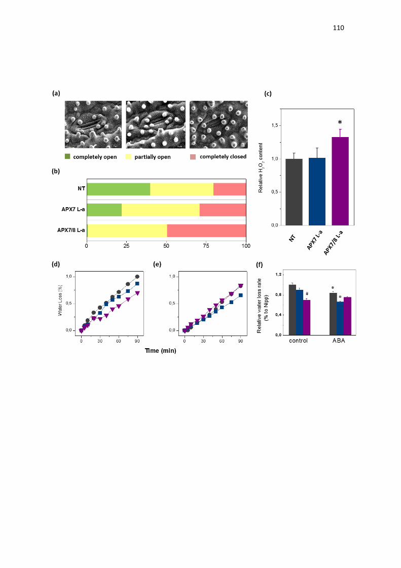
110
111
Figure 8: Effect of chloroplastic APX silencing in stomata opening and in
leaf desiccation. (a) Environmental scanning electron microscopy images of
three levels of stomatal opening. (b) quantification of the percentage of the three
levels of stomatal opening in leaves from NT, RNAiOsAPX7 and RNAiOsAPX7/8
30-d-old plants (n = 200 stomata for each line). (c) Relative content of hydrogen
peroxide in leaves from NT (grey), RNAiOsAPX7 (blue) and double
RNAiOsAPX7/8 (purple) leaves. The values represent the media ± SE of at least
five independent experiments. (d) and (e) Water loss of leaves detached from
plants under normal condition (treated with water) and treated with ABA 100 μM,
respectively. Legend: Grey: NT, Blue: RNAiOsAPX7 and Purple:
RNAiOsAPX7/8. For each repeat, leaves of 30-d-old plants were used in a
triplicate experiment (n = 3). The water loss rate of each conditions was quantified
in the figure (f). (*) indicate populations significantly different of control situation
with p<0,05, and (#) indicate populations significantly different of NT plants with
p<0,05

112
Figure 9: Thermal images of NT, RNAiOsAPX7 and RNAiOsAPX7/8 plants
captured by an infrared thermography device. (a) plants under normal
condition, (b) plants under two days of drought stress, (c) plants under seven
days of drought stress. The values represent the media ± SE of ΔT(˚C) of each
line to NT plants.
113
REFERENCES
ALSCHER RG, DONAHUE JL, CRAMER CL. (1997). Reactive oxygen species
and antioxidants: relationships in green cells. Physiol Plant 100:224–233
ASADA K. (1999). The water-water cycle in chloroplasts: scavenging of active
oxygen and dissipation of excess photons. Annu Rev Plant Physiol Plant Mol Biol
50:601–639.
BLUM A, EBERCON A. (1981). Cell membrane stability as a measure of drought
and heat tolerance in wheat. Crop Science 21: 43–47.
BRADFORD MM. (1976). A rapid and sensitive for the quantitation of microgram
quantitites of protein utilizing the principle of protein-dye binding. Analytical
Biochemistry 72: 248–254.
CAO MC, ZHANG WZ, HAN YD, YAO C, WANG YT, DING GH. (2013). A
theoretical model research of rice water stress index based on automated infrared
thermal imaging. Adv. Mater. Res., 712: 433-438.
CAVERZAN A, BONIFACIO A, CARVALHO FEL, ANDRADE CMB, PASSAIA
G, SCHÜNEMANN M, MARASCHIN FS, MARTINS MO, TEIXEIRA FK,
RAUBER R, MARGIS R, SILVEIRA JAG, MARGIS-PINHEIRO M. (2014). The
knockdown of chloroplastic ascorbate peroxidases reveals its regulatory role in
the photosynthesis and protection underphoto-oxidative stress in rice. Plant
Science 214: 74– 87.
CHEN S, TAO L, ZENG L, VEGA-SANCHEZ ME, KENJI UMEMURA WGL.
(2006). A highly efficient transient protoplast system for analyzing defence gene
expression and protein–protein interactions in rice. Molecular Plant Pathology 7:
417–427.
DASZKOWSKA-GOLEC A, SZAREJKO I. (2013). Open or Close the Gate –
Stomata Action Under the Control of Phytohormones in Drought Stress
Conditions. Front Plant Sci. 4: 138.
114
DAT J, ANDENABEELE S, VRANOVA E, VAN MONTAGU M, INZE D, VAN
BREUSEGEM F. (2000). Dual action of the active oxygen species during plant
stress response. Cell Mol Life Sci 57:779–795.
GALVÁN-AMPUDIA CS, OFFRINGA R. (2007). Plant evolution: AGC kinases
tell the auxin tale, Trends Plant Sci. 12: 541–547.
GILROY S, SUZUKI N, MILLER G, CHOI WG, TOYOTA M, DEVIREDDY AR,
MITTLER R. (2014). A tidal wave of signals: calcium and ROS at the forefront of
rapid systemic signaling. Trends Plant Sci. 19: 623–630.
HALLIWELL B, GUTTERIDGE JM. (1999). Free radicals in biology and
medicine. Oxford University Press, New York p 936
HERNANDEZ JA, FERRER MA, JIMÉNEZ A, BARCELO AR, SEVILLA F.
(2001). Antioxidant systems and O(2)(-)/H(2)O(2) production in the apoplast of
pea leaves. Its relation with salt-induced necrotic lesions in minor veins. Plant
Physiology 127: 817–831.
HETHERINGTON AM, WOODWARD FI. (2003). The role of stomata in sensing
and driving environmental change. Nature 424: 901–908.
HOAGLAND DR, ARNON DI. (1950). The water-culture method for growing
plants without soil. Berkeley, CA, USA: College of Agriculture, University of
California.
HUANG XY, CHAO DY, GAO JP, ZHU MZ, SHI M, LIN HX. (2009). A previously
unknown zinc finger protein, DST, regulates drought and salt tolerance in rice via
stomatal aperture control. Genes Dev. 15:1805-17.
JEFFERSON RA, KAVANAGH TA, BEVAN MW. (1987). GUS fusions: beta-
glucuronidase as a sensitive and versatile gene fusion marker in higher plants,
EMBO J. 6: 3901–3907.
JONES HG. (1999). Use of thermography for quantitative studies of spatial and
temporal variation of stomatal conductance over leaf surfaces. Plant Cell Environ
22:1043–1055.
115
KARIMI M, DE MEYER B, HILSON P. (2005). Modular cloning in plant cells.
Trends Plant Sci., 10: 103–105.
KARPINSKI S, REYNOLDS H, KARPINSKA B, WINGSLE G, CREISSEN G,
MULLINEAUX P. (1999). Systemic signaling and acclimation in response to
excess excitation energy in Arabidopsis. Science 284(5414):654-7.
LICHTENTHALER HK, WELLBURN AR. (1983). Determination of total
carotenoids andchlorophylls a and b of leaf extracts in different solvents,
Biochem. Soc. Trans.11:591–592.
LIVAK KJ, SCHMITTGEN TD. (2001). Analysis of relative gene expression data
using real-time quantitative PCR and the 2-ΔΔCT method. Methods 25: 402–408.
MIKI D, SHIMAMOTO K. (2004). Simple RNAi vectors for stable and transient
suppressionof gene function in rice, Plant Cell Physiol. 45:490–495.
MITTLER R, BERKOWITZ G. (2001). Hydrogen peroxide, a messenger with too
many roles? Redox Report 6: 69–72.
MITTLER R, VANDERAUWERA S, GALLERY M, VAN BREUSEGEM F. (2004).
Reactive oxygen gene network of plants. Trends Plant Sci 9:490–498.
MULLINEAUX P, KARPINSKI S. (2002). Signal transduction in response to
excess light: getting out of the chloroplast. Curr Opin Plant Biol 5:43–48.
NOCTOR G, FOYER CH. (1998). Ascorbate and glutathione: keeping active
oxygen under control. Annu Rev Plant Physiol Plant Mol Biol 49:249–79.
OROZCO-CARDENAS ML, NARVAEZ-VASQUEZ J, RYAN CA. (2001).
Hydrogen peroxide acts as a second messenger for the induction of defense
genes in tomato plants in response to wounding, systemin, and methyl-
jasmonate. Plant Cell 13:179–191.
PRASHAR A, YILDIZ J, MCNICOL JW, BRYAN GJ, JONES HG. (2014). Infra-
red Thermography for High Throughput Field Phenotyping in Solanum
tuberosum. PLoS One. 8(6): e65816.
116
RAO MV, LEE H, CREELMAN RA, MULLET JE, DAVIS KR. (2000). Jasmonic
acid signaling modulates ozone-induced hypersensitive cell death. Plant Cell 12:
1633–1646
SCANDALIOS JG. (2002). The rise of ROS. Trends Biochem Sci 27:483–486.
SCHMITTGEN TD, LIVAK KJ. (2008). Analyzing real-time PCR data by the
comparative CT method. Nature Protocols 3: 1101–1108.
SEIGNEURIN-BERNY D, SALVI D, JOYARD J, ROLLAND N. (2008).
Purification of intact chloroplasts from Arabidopsis and spinach leaves by
isopycnic centrifugation. Curr Protoc Cell Biol. Chapter 3:Unit 3.30.
SIRAULT XRR, JAMES RA, FURBANK RT. (2009). A new screening method
for osmotic component of salinity tolerance in cereals using infrared
thermography. Funct Plant Biol 36:970–977.
SMITH AM, RATCLIFFE RG, SWEETLOVE LJ. (2004). Activation and function
of mitochondrial uncoupling protein in plants. The Journal of Biological Chemistry
279: 51944–51952.
SUZUKI N, MILLER G, SALAZAR C, MONDAL HA, SHULAEV E, CORTES DF,
SHUMAN JL, LUO X, SHAH J, SCHLAUCH K, SHULAEV V, MITTLER R.
(2013). Temporal-spatial interaction between reactive oxygen species and
abscisic acid regulates rapid systemic acclimation in plants. Plant Cell 25: 3553–
3569.
SZECHYNSKA-HEBDA M, KRUK J, GÓRECKA M, KARPINSKA B,
KARPINSKI S. (2010). Evidence for light wavelength-specific
photoelectrophysiological signaling and memory of excess light episodes in
Arabidopsis. Plant Cell 22: 2201–2218.
TAO L, CHEUNG AY, WU H. (2002). Plant rac-like GTPases are activated by
auxin and mediate auxin-responsive gene expression. Plant Cell 14: 2745–2760.
TEIXEIRA FK, MENEZES-BENAVENTE L, GALVAO VC, MARGIS R, MARGIS-
PINHEIRO M. (2006). Rice ascorbate peroxidase gene family encodes
117
functionally diverse isoformslocalized in different subcellular compartments,
Planta 224: 300–314.
TSENG MJ, LIU CW, YIU JC. (2007). Enhanced tolerance to sulfur dioxide and
salt stress of transgenic Chinese cabbage plants expressing both superoxide
dismutase and catalase in chloroplasts. Plant Physiology and Biochemistry 45:
822–833.
UPADHYAYA NM, ZHOU XR, ZHU QH, EAMENS A, WANG MB, WATER-
HOUSE MP, DENNIS ES. (2002). Transgenic rice, in: L. O’Brien, R.J. Henry
(Eds.), TransgenicCereals, AACC, Minnesota pp. 28–87.
VAN CAMP W, CAPIAU K, VAN MONTAGU M, INZE D, SLOOTEN L. (1996).
Enhancement of oxidative stress tolerance in transgenic tobacco plants
overproducing Fe-superoxide dismutase in chloroplasts. Plant Physiology 112:
1703–1714.
VANDENABEELE S, VAN DER KELEN K, DAT J, GADJEV I, BOONEFAES T,
MORSA S, ROTTIERS P, SLOOTEN L, VAN MONTAGU M, ZABEAU M, INZE
D, VAN BREUSEGEM F. (2003). A comprehensive analysis of hydrogen
peroxide-induced gene expression in tobacco. Proc Natl Acad Sci USA
100:16113–16118.

118
SUPPLEMENTARY FIGURE
Figure S1. Relative water content in vessel of NT, RNAiOsAPX7 and
RNAiOsAPX7/8 plants submitted to drought stress. The values represent the
media ± SE of at least four independent experiments
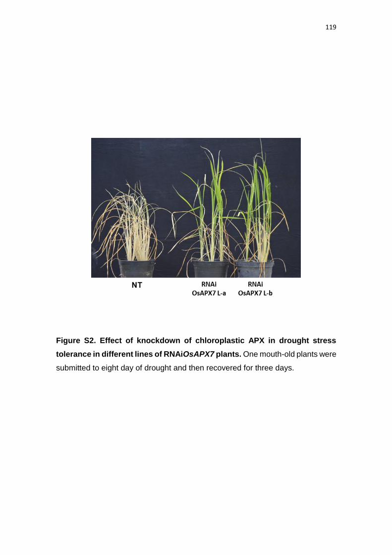
119
Figure S2. Effect of knockdown of chloroplastic APX in drought stress
tolerance in different lines of RNAiOsAPX7 plants. One mouth-old plants were
submitted to eight day of drought and then recovered for three days.
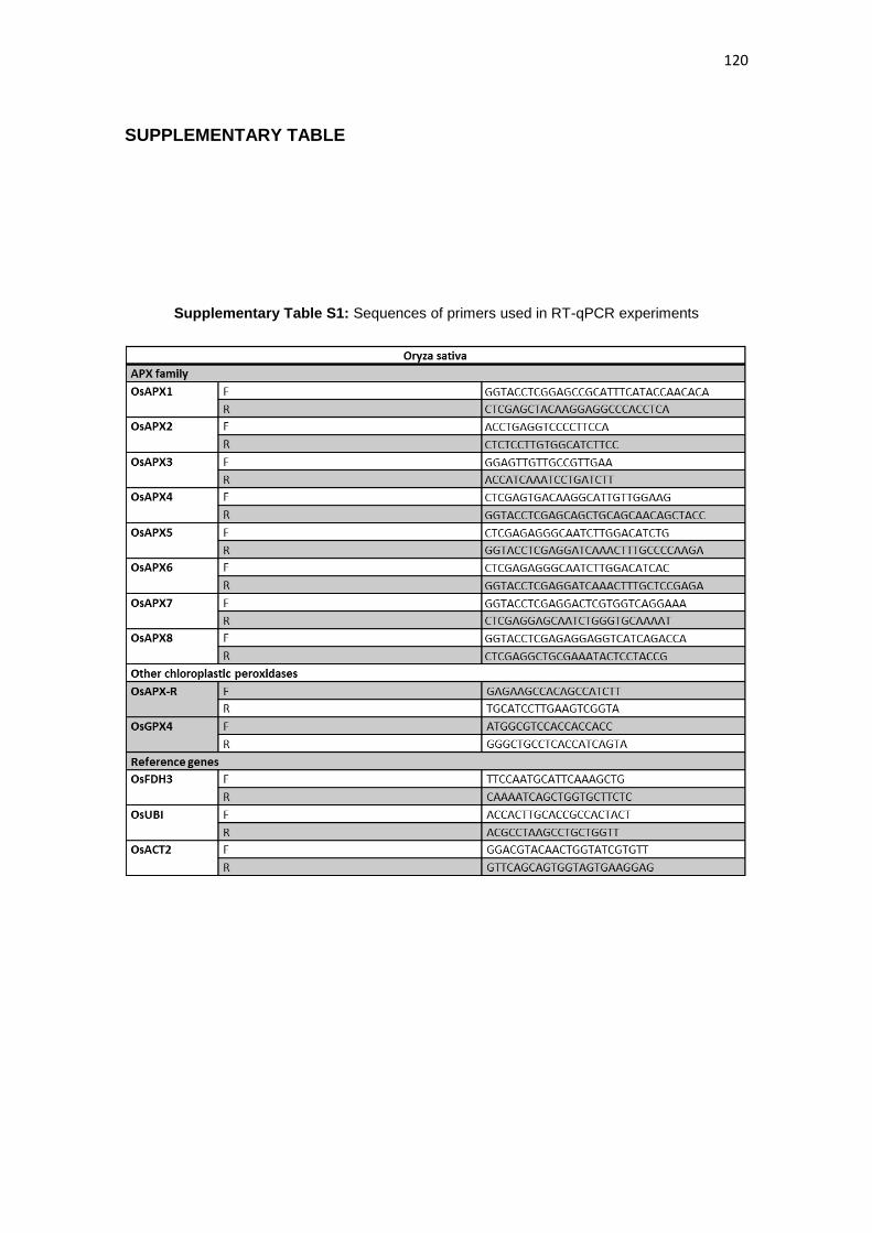
120
SUPPLEMENTARY TABLE
Supplementary Table S1: Sequences of primers used in RT-qPCR experiments
121
3.4 CAPÍTULO 4
SILENCIAMENTO OU NOCAUTE DA ASCORBATE PEROXIDASE
TILACOIDAL INDUZ PADRÕES DISTINTOS DE FECHAMENTO
ESTOMÁTICO E RESPOSTA AO ESTRESSE DE SECA EM ARROZ
(Oryza sativa L.)
Este capítulo é referente ao manuscrito “REDUCTION OR
SUPRESSION OF THYLAKOIDAL APX EXPRESSION PRODUCE
DISTINCT PATTERNS OF STOMATAL CLOSURE AND RESPONSE
TO DROUGHT STRESS IN RICE (Oryza sativa L.)” e apresenta a
segunda etapa do estudo de caracterização das isoformas
cloroplastídicas de ascrbato peroxidase (APX). Neste trabalho,
avaliamos o papel funcional da isoforma OsAPX8 no controle do
balanço redox e do fechamento estomático em plantas de arroz. O
silenciamento individual de OsAPX8 resultou em plantas com um
maior conteúdo de ROS e maior fechamento estomático em plantas
crescidas em codições ideais, como foi também verificado
anteriormente para as plantas duplo silenciadas para OsAPX7 e
OsAPX8. No entanto, as plantas silenciadas na OsAPX8 não
apresentaram uma maior tolerância ao estresse de seca. Por outro
lado, o aumento da tolerância a seca também foi verificado em plantas
nocaute para esse mesmo gene (apx8/apx8), as quais, curiosamente,
não apresentaram as mudanças fenotípicas verificadas nas plantas
silenciadas. Estes dados demonstram que o nocaute de OsAPX8
estaria gerando compensações que não ocorrem nas plantas
silenciadas e que culminam no aumento da tolerância a seca.
122
SILENCING AND KNOCKOUT OF THYLAKOIDAL ASCORBATE
PEROXIDASE LEAD TO DISTINCT PATTERNS OF STOMATAL CLOSING
AND DROUGHT STRESS TOLERANCE IN RICE (Oryza sativa L.)
Douglas Jardim-Messeder1, Andréia Caverzan1,2 , Rafael Rauber2, Juliana
Ribeiro3, Marcos Letaif Gaeta4, Guilherme Cordenonsi da Fonseca2, Miguel
Costa5, Michael Frei6, Joaquim Albenísio da Silveira3, Rogério Margis2, Nelson
Saibo5, Marcia Margis-Pinheiro1,2*
1Departamento de Genética, Universidade Federal do Rio Grande do Sul, Porto
Alegre, RS, Brazil
2Centro de Biotecnologia, Universidade Federal do Rio Grande do Sul, Porto
Alegre, RS, Brazil
3Departamento de Bioquímica e Biologia Celular, Universidade Federal do
Ceará, Fortaleza, CE, Brazil
4Departamento de Botânica, Universidade Federal Rio Grande do Sul, Porto
Alegre, RS, Brazil
5Instituto de Tecnologia Química e Biológica, Universidade Nova de Lisboa,
Oeiras, Portugal
6INRES Plant Nutrition, University of Bonn, Bonn, Germany
123
ABSTRACT
The chloroplast ascorbate peroxidases (chlAPXs) play a central role
in plant stress response. In rice plants (Oryza sativa L.) there are two chlAPXs:
OsAPX7 (stromal) and OsAPX8 (thylakoidal). Previous works demonstrated that
the double silencing of OsAPX7 and OsAPX8 genes led to plant tolerant to
drought stress, with increased H2O2 content and stomatal closing. Curiously, the
individual silencing of OsAPX7 gene is also able to induce tolerance to drought,
but without lead to differences in the biochemical or physiology parameter
evaluated. In this context, the role of OsAPX8, in chloroplast redox metabolism
and in drought stress respond remains unknown.
In this study we have generated rice plants individually silenced to
OsAPX8 gene (RNAiOsAPX8), and analyzed plants knockout to OsAPX8
(apx8/apx8). Our results demonstrate that the silencing of OsAPX8 lead to
increased stomatal closing and a stress like phenotype, possibly due a lower
chlAPX activity, but not induce tolerance to drought stress. Surprisingly,
apx8/apx8 plants do not show biochemical and physiologic changes, but are
more tolerant to drought stress. The OsAPX8 silencing lead also a higher impact
at gene expression levels, inducing mainly the expression of genes related to
photosynthetic process.
In this study we characterized the role of OsAPX8 in plant
development and in drought stress tolerance mechanisms. In addition, we show
important differences between silencing and knockout of OsAPX8 gene. While
the silencing of OsAPX8 induce a stress-like response, the knockout of OsAPX8
demonstrates that this gene is not essential to plant development, but in fact, its
absence improve tolerance to drought stress.
124
INTRODUCTION
The production of reactive oxygen species (ROS) is intrinsic to aerobic
metabolism, and these molecules participate of key physiological pathways, such
as cell cycle, gene regulation, programmed cell death (PCD), senescence and
stress response (DAT et al., 2000; OROZCO-CARDENAS et al., 2001;
MULLINEAUX & KARPINSKI, 2002; VANDENABEELE et al., 2003). However, in
high concentrations, ROS are toxic molecules, leading to oxidation of cellular
structures, including DNA, proteins and membrane lipids, resulting in the
disruption of metabolism and cellular structures ultimately (SCANDALIOS, 2002).
Thus, plants have a range of enzymatic and non-enzymatic antioxidant
mechanisms to prevent ROS from reaching toxic levels to maintain the cellular
redox balance. The enzymatic mechanisms involve the ROS detoxification by
action of antioxidant enzymes, as superoxide dismutases (SOD), ascorbate
peroxidases (APX), catalases (CAT), glutathione peroxidases (GPx), and
peroxidases (Prx) (HALLIWELL & GUTTERIDGE, 1999; SCANDALIOS, 2002;
MITTLER et al., 2004). Among the enzymes involved in ROS metabolism, APX
(EC 1.11.1.11) plays a major antioxidant role in plants, catalyzing the conversion
of H2O2 to H2O and O2, using ascorbate as specific electron donor (ASADA, 1992;
1999).
In plant cell, APX is largely distributed in different subcellular
compartments, as chloroplast, mitochondria, peroxisome and cytosol, where act
as an antioxidant defense (ASADA, 1992). The chloroplastic APXs isoforms
(chlAPX) can be composed of thylakoid-bound (tAPX) and stromal APX (sAPX),
which scavenge the H2O2 generated mainly during photosynthesis, which is the
major H2O2-producing metabolic process in autotrophic tissues (NAKANO &
ASADA, 1981). It is estimated that the rate of accumulation of H2O2 in
chloroplasts of higher plants under normal conditions is about 120 µm sec−1
(ASADA & TAKAHASHI, 1987). Thus, the immediate scavenging of H2O2 is
indispensable to maintain the photosynthetic activity of chloroplasts.
125
Due to the importance of APX activity in H2O2 metabolism, it has been
proposed that APX play a central role in plant stress response. In fact, Cotton
plants (Gossypium hirsutum) that overexpress recombinant APX in chloroplasts
showed an enhanced resistance to chilling-associated oxidative stress (PAYTON
et al., 2001), and a similar result was found in tobacco (Nicotiana toabacum),
where the overexpression of tAPX led to more resistant to chilling and methyl
viologen (MV) treatment (YABUTA et al., 2002). In wheat (Triticum aestivum L.),
knockout to tAPX led to reduced photosynthetic activity and growth (DANNA et
al., 2003). In previous work, our group demonstrated that in rice (Oryza sativa L.)
the chAPX (OsAPX7 and OsAPX8) have a distinct pattern of expression in
response to different abiotic stresses, as drought, high light and treatments with
H2O2 or MV, being OsAPX7 induced and OsAPX8 repressed (TEIXEIRA et al,
2006; ROSA et al, 2010). In addition, we demonstrated that the double silencing
of OsAPX7 and OsAPX8 do not led to phenotype differences in rice plants under
optimal condition of growth, but led to differences in photosynthetic parameters
related to efficiency of light utilization and CO2 in stress conditions (CAVERZAN
et al., 2014). Note that the opposite expression of OsAPX7 and OsAPX8 in
response to stress could hamper the interpretation of double silencing, and justify
the absence of phenotype differences in plants under normal condition. For all
this, it would be important perform the individual knockout or knockdown of each
chAPX to ensure the study of the physiological role of each gene. In fact, recently
we demonstrated that the silencing of OsAPX7 in rice plants improve an
increased tolerance to drought stress, without modify the stomata opening
(JARDIM-MESSEDER et al, not published yet).
Here, we performed a functional study of thylakoidal ascorbate
peroxidase (OsAPX8) gene, through rice plants individually silenced to OsAPX8
(RNAiOsAPX8) or knockout mutant at OsAPX8 gene (apx8/apx8). Our results
demonstrate that RNAiOsAPX8 plants show a decrease of growth rate, and a
high hydrogen peroxide content, even in normal condition. Furthermore, the
silencing of OsAPX8 led to decrease in stomatal opening and increase in leaves
surface temperature. In contrast, this phenotype has not been verified in
apx8/apx8 plants, which are similar to nom-transformed (NT) plants in normal
126
condition. However, beside the silencing of OsAPX8 lead to a stress-like
phenotype, only apx8/apx8 plants show an increased tolerance to drought stress.
These results were also confirmed by physiological parameters, as gas exchange
and chlorophyll fluorescence. In addition, RNAseq analysis confirmed that, in
normal conditions, the silencing of OsAPX8 led to a higher impact on plant than
the knockout, regulating mainly the expression of genes related to photosynthesis
and signal transduction,
127
MATERIAL AND METHODS
Plant material and growth conditions
Rice (Oryza sativa L. japonica cv. Nipponbare) seeds were germinated in MS
medium (Sigma-Aldrich) at 25°C with a 12-h photoperiod. One week after being
sown, the rice seedlings were transferred for hydroponic in 200-ml plastic cups
(three seedlings per cup) filled with Hoagland–Arnon’s nutritive solution
(HOAGLAND & ARNON,1950). To ABA treatment, 100µM ABA was dissolved in
0.1% Triton-X-100 and sprayed in excess on the leaves of the one-month-plants
until complete wetting was achieved.
Methyl viologen (MV) treatment
The one-month-old plants were used to MV treatment. These plants
were grown as described previously. The MV was dissolved in 0.1% Triton-X-100
at 50 μM and sprayed in excess on the leaves of the plants until complete wetting
was achieved. This procedure was repeated twice a day. The first symptoms of
toxicity (brown spots) appeared on the leaves after 24 h of treatment. The control
plants were sprayed with 0.1% Triton-X in the same way as the plants that
underwent MV treatment.
Cold stress
The plants of one-month old were used to cold treatment. The plants
were grown as described previously and submitted to 10°C temperature. Leaves
were collected at 6 and 24 hs after the start of cold treatment.
128
Drought stress
To drought experiments, the one-week-old rice seedlings were transferred to soil
in plastic vessels. Was used a mixture of soil, vermiculite and peat (2:1:1) and
was placed six plants per vessel. The drought experiment was performed in one-
month-old plants, thought the suspense of watering. The drought stress was
maintained until the relative water content in vessel reaches 10% (in about eight
days). The control of relative water content during drought stress experiment is
showed in Supplementary figure S1.
Vector construction and plant transformation
A chimeric gene producing mRNA with a hairpin structure (hpRNA)
was constructed based on the sequence of the OsAPX8 (LOC_Os02g34810)
gene. The following primer pairs were used to amplify a 227 bp RNAiOsAPX8
sequence: 5’- CTCGAGGCTGCGAAATACTCCTACGG-3’ and 5’-
GGTACCTCGAGAGGAGGTCATCAGACCATCG-3’. PCR products were cloned
into the Gateway vector pANDA, which hairpin RNA is driven by the maize
ubiquitin promoter and an intron placed upstream of the inverted repeats (MIKI &
SHIMAMOTO, 2004). The construct was denominated RNAiOsAPX8.
Agrobacterium tumefaciens-mediated transformation was performed as
described previously (UPADHYAYA et al., 2002). The apx8/apx8 knockout plants
were obtained by TOS-17 retrotransposson insertion in exon 4 (Supplementary
fig. S1A). All plants used in this work are homozygous as demonstrated in
Supplementary figure S1.
Quantitative real-time PCR (RT-qPCR)
Real-time PCR experiments were carried out using cDNA synthesized
from total RNA purified with TRIzol (Invitrogen®). Complementary (c) DNA was
obtained using the SuperscriptTMII (Life Technologies®) reverse transcriptase
system and a 24-polyTV primer (Invitrogen®). After synthesis, cDNAs were
diluted 10–100 times in sterile water for use in PCR reactions. All reactions were
129
repeated four times, and expression data analyses were performed after
comparative quantification of the amplified products using the 2-ΔΔCt method
(LIVAK & SCHMITTGEN, 2001; SCHMITTGEN & LIVAK, 2008). RT-qPCR
reactions were performed in an Applied Biosystems StepOne plus Real Time
PCR system (Applied Biosystems®) using SYBR-green intercalating dye for
fluorescence detection. The primer sequences and reference genes are listed in
Supporting Information Table S1. The data from the experiments relative to the
plants submitted to H2O2 treatment and drought were obtained from ROSA et al.
(2010), while experiments relative to MV and high light were obtained from
CAVERZAN et al, 2014.
Diaminobenzidine (DAB) staining and quantitative measurement of H2O2
The hydrogen peroxide was detected by DAB staining as described
previously (THORDALCHRISTENSEN et al., 1997). The fully expanded leaves
of 20 days-old plants were detached and incubated overnight in DAB staining
solution (1 mg ml-1 DAB in 10 mM MES, pH 6.5) at 28 °C and cleared in boiling
propanone for 10 min before photographing.
Measurements of H2O2 production was performed by extracting H2O2
from leaves according to RAO et al. (2000) using the Ampliflu Red (Sigma-
Aldrich) oxidation (SMITH et al., 2004). Fluorescence was monitored using a
fluorometer at excitation and emission wavelengths of 563 nm (slit 5 nm) and 587
nm (slit 5 nm), respectively. Calibration was performed by the addition of known
quantities of H2O2.
Pigment determination
The total chlorophyll, chlorophyll a and b were determined after
extraction in ethanol spectrophotometrically at 663 and 649 nm. The amount of
pigment was calculated using the equations proposed by LICHTENTHALER &
WELLBURN (1983):
130
Chlorophyl a (mg/L) = 12,25 x Abs663 – 2,79 x Abs647
Chlorophyl b (mg/L) = 21,50 x Abs647 – 5,10 x Abs663
Membrane damage determination
Electrolyte leakage (membrane damage) was measured as described
by BLUM & EBERCON (1981). Leaf slices were placed in tubes containing 10mL
of deionized water. The flasks were incubated in a shaker for 12 h, and the electric
conductivity in the medium (L1) was measured. Then, the medium was boiled (95
°C) for 60 min and the electric conductivity (L2) was measured again. The relative
membrane damage (MD) was estimated by MD = L1/L2×100.
Imaging of rice stomata
The stomata were visualized by scanning electron microscopy and
subsequently quantified by counting class. Leaves of 30 day-old plants were
detached and the samples are prepared as HUANG et al., (2009).
Gas exchange and photochemical parameters
The gas exchange measurements and the following photochemistry
parameters associated with the efficiency of photosystem II were measured after
24 h of exposure to MV. The following PSII parameters were measured: ΔF/F’m,
actual quantum yield of photosystem II, Fv/Fm, potential quantum yield of
photosystem II, and ETR, apparent electron transport rate. The relative excess
energy at the photosystem II level was calculated as EXC = [(Fv/Fm) −
(ΔFv/F’m)]/(Fv/Fm). All of the parameters were measured with an Infrared Gas
Analyzer coupled with a leaf chamber fluorometer (Li-6400-XT, LI-COR, Lincoln,
USA) according to the manufacturer’s instructions. ETR was calculated as ETR
= (ΔF/F’m× PPFD × 0.5 × 0.84), where 0.5 is the presumed fraction of the
excitation energy distributed to PSII and 0.84 is the assumed fraction of light
131
absorbed by the leaf. EXC was calculated according to BILGER et al., (1995) as
EXC = [(Fv/Fm) −(ΔF/F’m)]/(Fv/Fm).The photochemical quenching coefficient [qP =
(F’m− Fs)/(F’m−F’o)] and the non-photochemical quenching coefficient [NPQ =(Fm−
F’m)/F’m], where Fm and Fo are, respectively, maximum and minimum
fluorescence of dark-adapted leaves; F’m and Fs are, respectively, maximum and
steady state fluorescence in the light-adapted state, and F’o is minimum
fluorescence after far-red illumination of the previously light exposed leaves. A
saturating pulse of red light (0.8 s, 8000 μmol m−2s−1) was utilized. Leaf gas
exchange measurements were made using an Infra-red Gas Analyzer (Li-6400-
XT, LI-COR Biosciences Inc., Lincoln, USA). Light was provided by a red/blue
LED light source at photon irradiance of 1000 μmol m−2s−1. All leaf measurements
were taken under ambient CO2 conditions (380 ppm) at a constant leaf
temperature of 28 oC and relative humidity of 78% and vapor pressure deficit
(VPD) of 1.8 Pa. The gas exchange variables measured were net photosynthesis
(PN) and intercellular CO2 concentration (CI).
RNAseq analysis
To RNAseq analysis, total RNA was purified from one month old plants, using
TRIzol (Invitrogen®). Was evaluated two simples for lines (NT, RNAiOsAPX8,
apx8/apx8), compoused by mix of total RNA from five plants. The plant growth
condirions was the same showed above. The RNAseq was made by Centro de
Genômica Funcional - USP (Piracicaba, Brasil). In the results is show the average
of the relative values of expression for each line compared to NT plants. Was
considered differently expressed the values with p<0,03.
Gene ontology analysis
Gene ontology analyses were performed by the web-based tool and
database agriGO (DU et al., 2010).
132
Functional protein association networks
The functional protein association network was created using the
STRING database (SNEL et al., 2000) and analyzed by Medusa (HOOPER &
BORK, 2005) and ViaComplex software (CASTRO et al., 2009).
Protein determination
Protein concentrations were determined as described by BRADFORD
(1976) using BSA as a standard.
Statistical analysis
Data were plotted with GRAPHPAD PRISM 5.0 (GraphPad Software
Inc., La Jolla, CA, USA) and analyzed by one-way ANOVA and a posteriori
Tukey’s test. P-values of 0.05 were considered statistically significant.
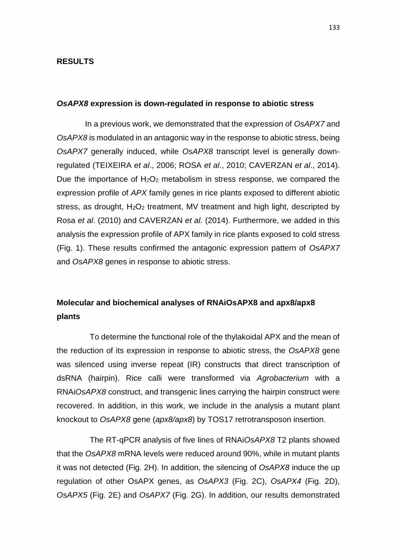
133
RESULTS
OsAPX8 expression is down-regulated in response to abiotic stress
In a previous work, we demonstrated that the expression of OsAPX7 and
OsAPX8 is modulated in an antagonic way in the response to abiotic stress, being
OsAPX7 generally induced, while OsAPX8 transcript level is generally down-
regulated (TEIXEIRA et al., 2006; ROSA et al., 2010; CAVERZAN et al., 2014).
Due the importance of H2O2 metabolism in stress response, we compared the
expression profile of APX family genes in rice plants exposed to different abiotic
stress, as drought, H2O2 treatment, MV treatment and high light, descripted by
Rosa et al. (2010) and CAVERZAN et al. (2014). Furthermore, we added in this
analysis the expression profile of APX family in rice plants exposed to cold stress
(Fig. 1). These results confirmed the antagonic expression pattern of OsAPX7
and OsAPX8 genes in response to abiotic stress.
Molecular and biochemical analyses of RNAiOsAPX8 and apx8/apx8
plants
To determine the functional role of the thylakoidal APX and the mean of
the reduction of its expression in response to abiotic stress, the OsAPX8 gene
was silenced using inverse repeat (IR) constructs that direct transcription of
dsRNA (hairpin). Rice calli were transformed via Agrobacterium with a
RNAiOsAPX8 construct, and transgenic lines carrying the hairpin construct were
recovered. In addition, in this work, we include in the analysis a mutant plant
knockout to OsAPX8 gene (apx8/apx8) by TOS17 retrotransposon insertion.
The RT-qPCR analysis of five lines of RNAiOsAPX8 T2 plants showed
that the OsAPX8 mRNA levels were reduced around 90%, while in mutant plants
it was not detected (Fig. 2H). In addition, the silencing of OsAPX8 induce the up
regulation of other OsAPX genes, as OsAPX3 (Fig. 2C), OsAPX4 (Fig. 2D),
OsAPX5 (Fig. 2E) and OsAPX7 (Fig. 2G). In addition, our results demonstrated
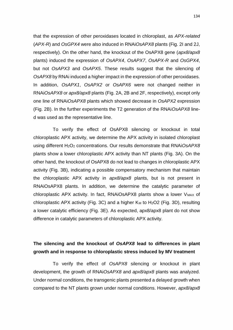
134
that the expression of other peroxidases located in chloroplast, as APX-related
(APX-R) and OsGPX4 were also induced in RNAiOsAPX8 plants (Fig. 2I and 2J,
respectively). On the other hand, the knockout of the OsAPX8 gene (apx8/apx8
plants) induced the expression of OsAPX4, OsAPX7, OsAPX-R and OsGPX4,
but not OsAPX3 and OsAPX5. These results suggest that the silencing of
OsAPX8 by RNAi induced a higher impact in the expression of other peroxidases.
In addition, OsAPX1, OsAPX2 or OsAPX6 were not changed neither in
RNAiOsAPX8 or apx8/apx8 plants (Fig. 2A, 2B and 2F, respectively), except only
one line of RNAiOsAPX8 plants which showed decrease in OsAPX2 expression
(Fig. 2B). In the further experiments the T2 generation of the RNAiOsAPX8 line-
d was used as the representative line.
To verify the effect of OsAPX8 silencing or knockout in total
chloroplastic APX activity, we determine the APX activity in isolated chloroplast
using different H2O2 concentrations. Our results demonstrate that RNAiOsAPX8
plants show a lower chloroplastic APX activity than NT plants (Fig. 3A). On the
other hand, the knockout of OsAPX8 do not lead to changes in chloroplastic APX
activity (Fig. 3B), indicating a possible compensatory mechanism that maintain
the chloroplastic APX activity in apx8/apx8 plants, but is not present in
RNAiOsAPX8 plants. In addition, we determine the catalytic parameter of
chloroplastic APX activity. In fact, RNAiOsAPX8 plants show a lower VMAX of
chloroplastic APX activity (Fig. 3C) and a higher KM to H2O2 (Fig. 3D), resulting
a lower catalytic efficiency (Fig. 3E). As expected, apx8/apx8 plant do not show
difference in catalytic parameters of chloroplastic APX activity.
The silencing and the knockout of OsAPX8 lead to differences in plant
growth and in response to chloroplastic stress induced by MV treatment
To verify the effect of OsAPX8 silencing or knockout in plant
development, the growth of RNAiOsAPX8 and apx8/apx8 plants was analyzed.
Under normal conditions, the transgenic plants presented a delayed growth when
compared to the NT plants grown under normal conditions. However, apx8/apx8
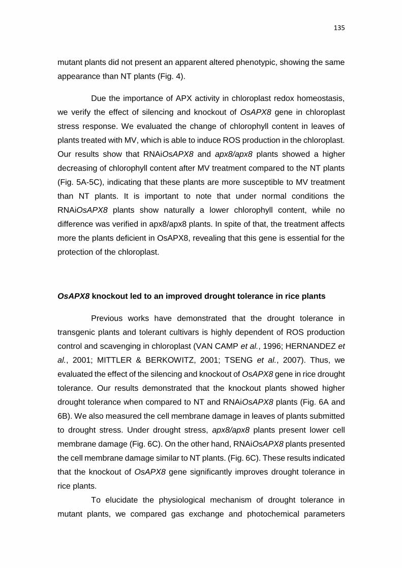
135
mutant plants did not present an apparent altered phenotypic, showing the same
appearance than NT plants (Fig. 4).
Due the importance of APX activity in chloroplast redox homeostasis,
we verify the effect of silencing and knockout of OsAPX8 gene in chloroplast
stress response. We evaluated the change of chlorophyll content in leaves of
plants treated with MV, which is able to induce ROS production in the chloroplast.
Our results show that RNAiOsAPX8 and apx8/apx8 plants showed a higher
decreasing of chlorophyll content after MV treatment compared to the NT plants
(Fig. 5A-5C), indicating that these plants are more susceptible to MV treatment
than NT plants. It is important to note that under normal conditions the
RNAiOsAPX8 plants show naturally a lower chlorophyll content, while no
difference was verified in apx8/apx8 plants. In spite of that, the treatment affects
more the plants deficient in OsAPX8, revealing that this gene is essential for the
protection of the chloroplast.
OsAPX8 knockout led to an improved drought tolerance in rice plants
Previous works have demonstrated that the drought tolerance in
transgenic plants and tolerant cultivars is highly dependent of ROS production
control and scavenging in chloroplast (VAN CAMP et al., 1996; HERNANDEZ et
al., 2001; MITTLER & BERKOWITZ, 2001; TSENG et al., 2007). Thus, we
evaluated the effect of the silencing and knockout of OsAPX8 gene in rice drought
tolerance. Our results demonstrated that the knockout plants showed higher
drought tolerance when compared to NT and RNAiOsAPX8 plants (Fig. 6A and
6B). We also measured the cell membrane damage in leaves of plants submitted
to drought stress. Under drought stress, apx8/apx8 plants present lower cell
membrane damage (Fig. 6C). On the other hand, RNAiOsAPX8 plants presented
the cell membrane damage similar to NT plants. (Fig. 6C). These results indicated
that the knockout of OsAPX8 gene significantly improves drought tolerance in
rice plants.
To elucidate the physiological mechanism of drought tolerance in
mutant plants, we compared gas exchange and photochemical parameters
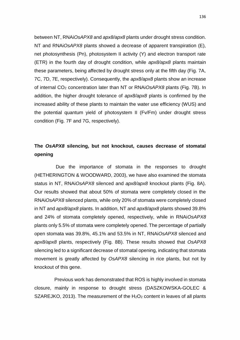
136
between NT, RNAiOsAPX8 and apx8/apx8 plants under drought stress condition.
NT and RNAiOsAPX8 plants showed a decrease of apparent transpiration (E),
net photosynthesis (Pn), photosystem II activity (Y) and electron transport rate
(ETR) in the fourth day of drought condition, while apx8/apx8 plants maintain
these parameters, being affected by drought stress only at the fifth day (Fig. 7A,
7C, 7D, 7E, respectively). Consequently, the apx8/apx8 plants show an increase
of internal CO2 concentration later than NT or RNAiOsAPX8 plants (Fig. 7B). In
addition, the higher drought tolerance of apx8/apx8 plants is confirmed by the
increased ability of these plants to maintain the water use efficiency (WUS) and
the potential quantum yield of photosystem II (Fv/Fm) under drought stress
condition (Fig. 7F and 7G, respectively).
The OsAPX8 silencing, but not knockout, causes decrease of stomatal
opening
Due the importance of stomata in the responses to drought
(HETHERINGTON & WOODWARD, 2003), we have also examined the stomata
status in NT, RNAiOsAPX8 silenced and apx8/apx8 knockout plants (Fig. 8A).
Our results showed that about 50% of stomata were completely closed in the
RNAiOsAPX8 silenced plants, while only 20% of stomata were completely closed
in NT and apx8/apx8 plants. In addition, NT and apx8/apx8 plants showed 39.8%
and 24% of stomata completely opened, respectively, while in RNAiOsAPX8
plants only 5.5% of stomata were completely opened. The percentage of partially
open stomata was 39.8%, 45.1% and 53.5% in NT, RNAiOsAPX8 silenced and
apx8/apx8 plants, respectively (Fig. 8B). These results showed that OsAPX8
silencing led to a significant decrease of stomatal opening, indicating that stomata
movement is greatly affected by OsAPX8 silencing in rice plants, but not by
knockout of this gene.
Previous work has demonstrated that ROS is highly involved in stomata
closure, mainly in response to drought stress (DASZKOWSKA-GOLEC &
SZAREJKO, 2013). The measurement of the H2O2 content in leaves of all plants
137
revealed that, in fact, RNAiOsAPX8 plants have an increase of 60% in H2O2 level,
while no differences were observed between NT and apx8/apx8 plants (Fig. 8C).
To verify the effect of OsAPX8 silencing and knockout in leaf water loss,
we submitted rice leaves to desiccation experiment. The OsAPX8 silencing led
to a lower leaf desiccation (25%), while leaves from apx8/apx8 and NT plants
showed the same water loss rate. Interestingly, the pre-treatment with abscisic
acid (ABA), which induce stomatal closure, led to a decrease of leaf desiccation
in NT and apx8/apx8 plants, but do not change the leaf desiccation in
RNAiOsAPX8 plants. Under this condition, there are not differences in leaf
desiccation between NT, RNAiOsAPX8 and apx8/apx8 plants (Fig. 8E-8G)
indicating that the lower leaf desiccation in RNAiOsAPX8 plants is possibly due
a higher stomatal closure.
Other alternative approach to measure stomatal conductance is the
infrared (IR) thermography. The infrared measurements of leaf temperature
correlate well with estimates of stomatal conductance (JONES, 1999; SIRAULT
et al., 2009) and consequently in rice water status (CAO et al., 2013). Thus, we
measured the leave surface temperature in NT, RNAiOsAPX8 and apx8/apx8
plants under normal condition, moderate drought stress (2 days) and severe
drought stress (7 days). Our results demonstrate that in normal condition, the
RNAiOsAPX8 plants showed naturally an increase of about 0.48 ºC in leaf
surface temperature (Fig. 9A). However, under moderate and severe drought
stress no difference in leaf surface temperature between all plants was observed
(Fig. 9B and 9C). These results indicate that only the silencing of OsAPX8 gene
led to changes in leaf surface temperature in normal condition, and interestingly,
this effect was not induced by the knockout of OsAPX8.
RNAseq analysis of RNAiOsAPX8 and apx8/apx8 plants
RNAseq analyses were performed to identify differentially expressed
genes in RNAiOsAPX8 and apx8/apx8 plants with one-month old grown under
normal conditions. Our results demonstrate differential expression between
RNAiOsAPX8 and NT plants, as well as, apx8/apx8 and NT plants
138
(Supplementary Table S2). In RNAiOsAPX8 plants 222 genes were significantly
upregulated when compared to NT plants (p<0.03), while only 17 genes were
significantly upregulated in apx8/apx8 plants when compared with NT plants (Fig.
10A). On the other hand, 121 genes were down-regulated in RNAiOsAPX8
plants, while only 62 genes were down-regulated in apx8/apx8 plants (Fig. 10B).
A set of proteins which showed significantly difference in expression when
compared to NT plants were also analyzed and classified into groups according
to their ontology (GO: metabolic process). Ontological analysis showed that in
RNAiOsAPX8 plants, among the up-regulated genes, those related to
photosynthesis was well represented (14.8% of the total genes up-regulated). In
addition, there is an increase of genes related to light reaction and harvesting
(7.1% and 6.5%, respectively), generation of energy (8.2%) and genes related to
metabolism of organic molecules. On the other hand, among the down-regulated
genes, the most representative were those related to protein metabolic process
(25.6%) and to signal propagation function, such as genes related to protein
phosphorylation (20.5%) and post-translation modification (20.5%) (Fig. 10C).
The representability of these ontologies among the genes up and down-regulated
is higher than their representability in rice genome, indicating that these
ontological groups are significantly modulated by OsAPX8 silencing. In KO
plants, there is not an enrichment of a specific ontological group in up-regulated
genes. In addition, to down-regulated genes, those related to metabolic process,
primary metabolism and cellular process were well represented (34.4%, 26.5%
and 20.3%, respectively) (Fig. 10D). However, the representability of all
ontologies in the input of analyzed genes is lower than their representability in
rice genome. These data indicate that the knockout of OsAPX8 do not led to
modulation of a specific ontological group.
Due the higher representability of photosynthesis related genes in up-
regulated genes, the transcription profile of an association network of
photosynthesis related genes (GO:0015979) was analyzed comparing NT,
RNAiOsAPX8 and apx8/apx8 plants. This network was created using the
STRING database (SNEL et al., 2000) with genes related to photosynthesis in
rice. Our results demonstrate that the silencing of OsAPX8 led to differential
139
expression of the genes belonging to this network, while in apx8/apx8 plants it
was not verified similar effect (Fig. 11A). In addition, the relative expression of
genes with ontology “photosynthesis”, was quantified. In RNAiOsAPX8 plants,
there are significantly groups of up and down-regulated genes, while in apx8/apx8
plants, in general there are not differential expression of these genes (Fig. 11B).
Finally, these results demonstrate that, the silencing of OsAPX8 also led to a
higher impact in gene expression than the complete OsAPX8 knockout.
140
DISCUSSION
Many studies have demonstrated a close relationship between redox signals
and acclimation to abiotic stresses. The chloroplast plays a central role in
redox/ROS-mediated systemic signaling and acclimation responses (KARPINSKI
et al., 1999; MULLINEAUX et al., 2006; MÜHLENBOCK et al., 2008;
SZECHYŃSKA-HEBDA et al., 2010). The overexpression or knockout of chlAPX
led to an altered response to abiotic stress in different species, as cotton
(PAYTON et al., 2001), tobacco (YABUTA et al., 2002) and wheat (DANNA et al.,
2003). In rice, there are two exclusive isoforms of chlAPX, and we have
previously demonstrated that abiotic stress, as high light or MV, led to contrasting
response related to OsAPX7 and OsAPX8 expression: OsAPX7 expression is
up-regulated, while OsAPX8 expression is downregulated (CAVERZAN et al.,
2014). Similarly, H2O2 treatment and drought also induced OsAPX7 expression
and decreased OsAPX8 expression (ROSA et al., 2010). Here, we have
confirmed that this antagonism also occurred in response to other abiotic stress,
as cold. Due to the general and conserved decrease of OsAPX8 expression in
plants submitted to different abiotic stress we hypothesized that the down
modulation of OsAPX8 expression play an important role in rice stress response.
To understand the physiologic role of OsAPX8, we performed the molecular,
physiological and biochemical characterization of the effect of silencing and
knockout of OsAPX8 gene in rice plants. Our results demonstrated that the
silencing of OsAPX8 led to a compensatory expression of other APX isoforms,
as OsAPX3, OsAPX4, OsAPX5 and OsAPX7, and of other chloroplastic
peroxidases, as OsAPX-R and OsGPX4. In addiyion these plants show a lower
chloroplastic APX activity, accompanied by a a higher H2O2 accumulation, and a
delayed growth, compared to NT plants. As described by DASZKOWSKA-
GOLEC & SZAREJKO (2013) the increase of ROS production is also involved
with stomata closure mechanism, and in fact, the RNAiOsAPX8 plants have a
lower number of opened stomata, which would limit the CO2 assimilation to plant
growth. In addition, the RNAiOsAPX8 plants showed an increased leaf surface
141
temperature, even in normal condition, which confirm the lower stomatal
conductance. Together, these results suggest that the OsAPX8 silencing induce
a stress-like response in rice plant, simulating the natural response of OsAPX8
expression in plants submitted to stress. On the other hand, the complete
knockout of OsAPX8 did not produce any apparent phenotype alteration, and
apx8/apx8plants maintained a normal growth and development, and a similar
H2O2 content, stomatal conductance and leaf surface temperature compared to
NT plants. However, both silencing and knockout plants show a lower tolerance
to MV treatment, demonstrating that OsAPX8 expression is fundamental to
chloroplast stress response, possible by its H2O2 scavenger activity
Previous studies have demonstrated that ROS production control and
scavenging by chloroplast play an important role, not only in stomatal
conductance, but also in plant tolerance to drought (VAN CAMP et al., 1996;
HERNANDEZ et al., 2001; MITTLER & BERKOWITZ, 2001; TSENG et al, 2007).
In fact, previously, we demonstrated that the silencing of OsAPX7, another
chlAPX, led to physiological changes, as increased transpiration, and improve
drought tolerance in rice plants (Jardim-Messeder et al, submitted work). Thus,
we submitted the RNAiOsAPX8 and apx8/apx8 plants to drought stress, to also
determine the role of OsAPX8 in drought stress response. Surprisingly, the
apx8/apx8 plants, but not the RNAiOsAPX8, showed a higher drought tolerance.
This result was confirmed by a lower membrane damage and by the evaluation
of photochemical parameters and gas exchange in plants exposed to drought
stress. Our results revealed that apx8/apx8 plants are able to maintain the
transpiration, and consequently stomatal conductance, for a longer time under
drought stress. The maintenance of stomatal conductance under stress condition
allowed the plant to keep the photosynthesis activity, resulting in an increasing of
the intracellular CO2 concentration. The higher stomatal conductance of
apx8/apx8 plants possibly enables the constant offers of CO2 to RUBISCO
carboxylase activity, leading the ribulose bisphosphate (RuBP) to Calvin cycle
and preventing the photorespiration. Therefore, to promote the re-oxidation of
NADPH to NADP, the maintenance of the Calvin cycle under stress condition
enables the continuous electron transporter chain activity, impairing the
142
photosynthesis inhibition. Consequently, under drought stress condition, the
apx8/apx8 plants are able to maintain for a longer time the water use efficiency
and the potential quantum yield of photosystem II.
The comparative analysis of the transcriptome of RNAiOsAPX8 and
apx8/apx8 plants revealed that the silencing of OsAPX8 caused higher impact in
the global gene expression than the complete gene knockout did. The ontological
analysis of differently expressed genes demonstrated that photosynthesis related
genes are the most well represented ontological group, while the main down-
regulated genes corresponded to protein signaling, as post-transductional
modification, phosphorylation, and to phosphate and phosphorus metabolism. In
addition, among the down-regulated genes there are an increased representation
of genes related to ontologies “photosynthesis” “photosynthetic electron transport
chain”, “light reaction” and “photosynthetic electron transport in photosystem II”,
though these groups were lower represented than other ontology being indicated
as “other”. On the other hand, the knockout of OsAPX8 did not led to a significant
ontological difference. These data confirmed that the RNAiOsAPX8 plants also
showed a higher difference at gene expression level, confirming our hypothesis
that the OsAPX8 silencing led to a stressed-like phenotype in rice plants. In fact,
previous works demonstrated that drought stress significantly affect
photosynthesis related gene expression in rice (HAZEN et al., 2005; WANG et
al., 2011) and this group of genes is highly affected by the OsAPX8 silencing, but
not by the complete knockout of OsAPX8.
In this work we characterized the functional role of OsAPX8 in plant
development and demonstrated the importance of OsAPX8 in drought stress
response. While the silencing of OsAPX8 simulate a natural response to stress,
leading to a stressed-like phenotype, the knockout of OsAPX8 should trigger an
unknown mechanism able to maintain a normal phenotype, and still confer
tolerance to drought stress. This mechanism could involve the genes differently
expressed in apx8/apx8 plants, as for example LOC_Os08g27170.1, which
encode a calmodulin binding protein and is overexpressed in these plants.
Previous work, demonstrated in Arabidopsis thaliana, that the overexpression of
homologue protein CBP60g is a positive regulator of both disease resistance and

143
drought tolerance. Further studies should be conducted to determine the exact
mechanism by which OsAPX8 knockout confers drought tolerance in rice plants.

144
FIGURES
Figure 1. Relative expression of APX family in rice leaves from plants
submitted in to different stress conditions. The values were normalized by
the at least four constitutive genes and represent the media ± SE of at least three
independent experiments.
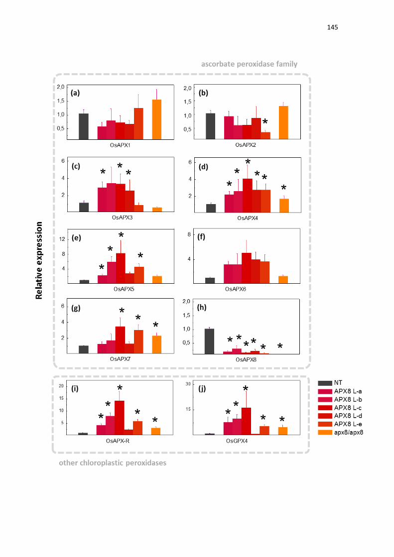
145

146
Figure 2. Quantitative determination of ascorbate peroxidase family and
other chloroplastic peroxidases mRNA in leaves of NT (non-transformed),
RNAiOsAPX8 and apx8/apx8 plants. The values were normalized by the at
least three constitutive genes and represent the media ± SE of at least three
independent experiments. (*) indicate populations significantly different with
p<0,05.
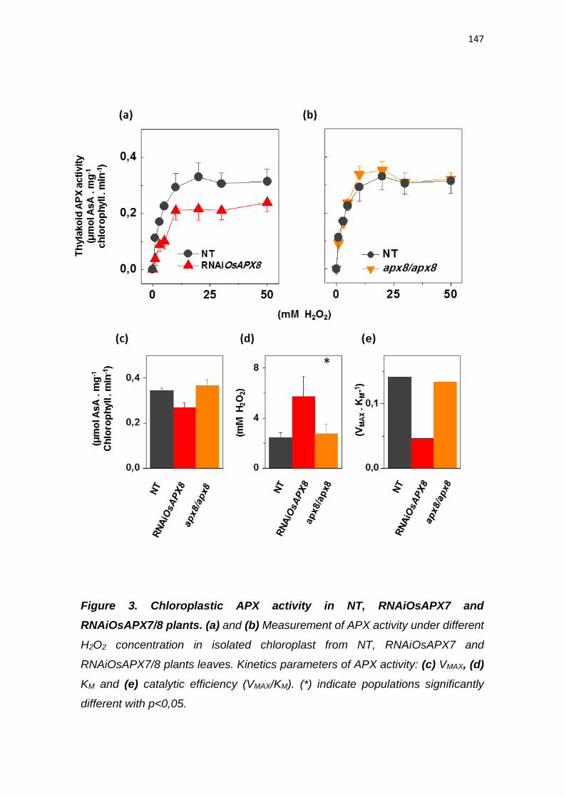
147
Figure 3. Chloroplastic APX activity in NT, RNAiOsAPX7 and
RNAiOsAPX7/8 plants. (a) and (b) Measurement of APX activity under different
H2O2 concentration in isolated chloroplast from NT, RNAiOsAPX7 and
RNAiOsAPX7/8 plants leaves. Kinetics parameters of APX activity: (c) VMAX, (d)
KM and (e) catalytic efficiency (VMAX/KM). (*) indicate populations significantly
different with p<0,05.
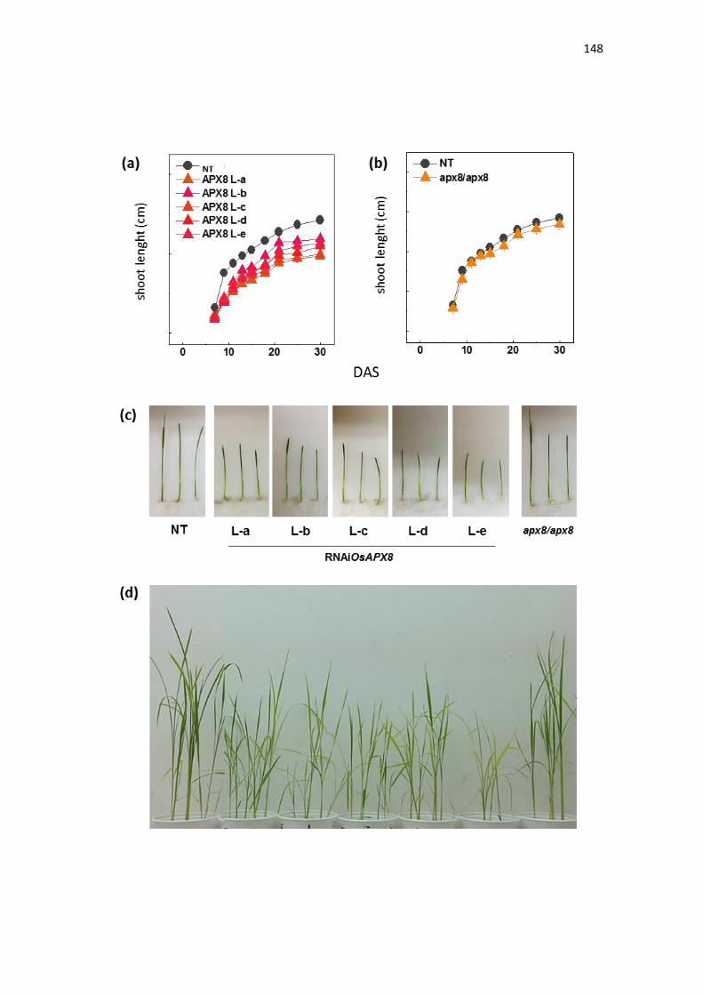
148

149
Figure 4. Growth characterization of NT (non-transformed), RNAiOsAPX8
(silenced) and apx8/apx8 (knockout) plants. (a) Growing curve of NT and
RNAiOsAPX8 plants. (b) Growing curve of NT and apx8/apx8 plants. (c) NT,
RNAiOsAPX8 and apx8/apx8 seedlings with seven days old. (d) NT,
RNAiOsAPX8 and apx8/apx8 seedlings with with four weeks old.
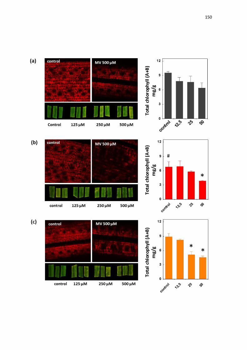
150
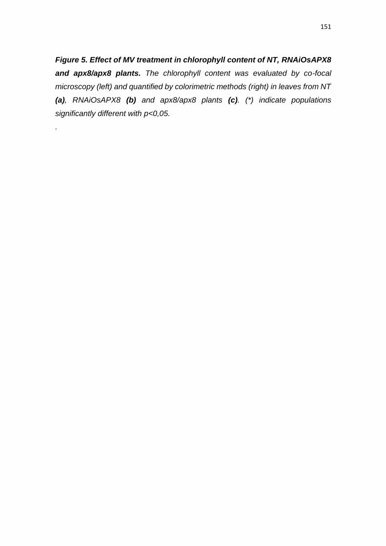
151
Figure 5. Effect of MV treatment in chlorophyll content of NT, RNAiOsAPX8
and apx8/apx8 plants. The chlorophyll content was evaluated by co-focal
microscopy (left) and quantified by colorimetric methods (right) in leaves from NT
(a), RNAiOsAPX8 (b) and apx8/apx8 plants (c). (*) indicate populations
significantly different with p<0,05.
.
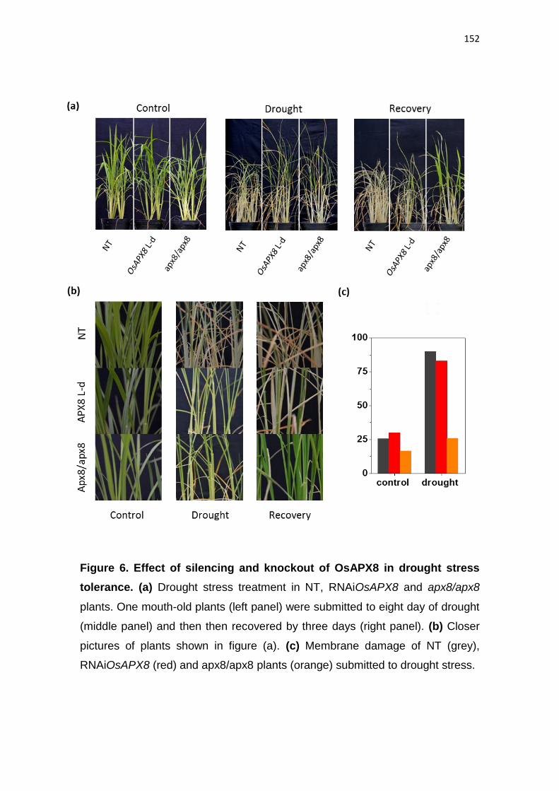
152
Figure 6. Effect of silencing and knockout of OsAPX8 in drought stress
tolerance. (a) Drought stress treatment in NT, RNAiOsAPX8 and apx8/apx8
plants. One mouth-old plants (left panel) were submitted to eight day of drought
(middle panel) and then then recovered by three days (right panel). (b) Closer
pictures of plants shown in figure (a). (c) Membrane damage of NT (grey),
RNAiOsAPX8 (red) and apx8/apx8 plants (orange) submitted to drought stress.
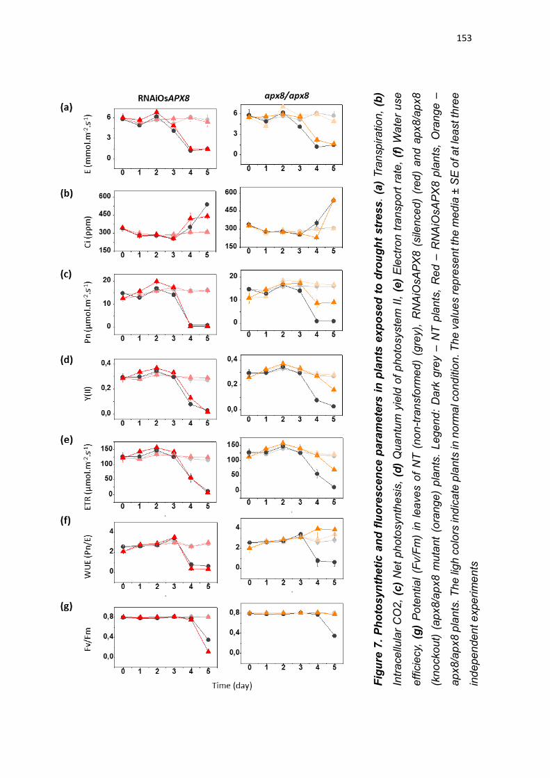
153
Fig
ure
7.
Ph
oto
sy
nth
eti
c a
nd
flu
ore
sce
nce
pa
ram
ete
rs i
n p
lan
ts e
xp
ose
d t
o d
rou
gh
t s
tre
ss
. (a
) Tra
nsp
ira
tio
n,
(b)
Intr
ace
llula
r C
O2
, (c
) N
et
ph
oto
syn
the
sis
, (d
) Q
uan
tum
yie
ld o
f p
ho
tosyste
m I
I, (
e)
Ele
ctr
on
tra
nspo
rt r
ate
, (f
) W
ate
r u
se
eff
icie
cy,
(g
) P
ote
ntia
l (F
v/F
m)
in l
ea
ve
s o
f N
T (
no
n-t
ran
sfo
rme
d)
(gre
y),
RN
AiO
sA
PX
8 (
sile
nce
d)
(red
) an
d a
px8
/ap
x8
(kno
cko
ut)
(ap
x8/a
px8 m
uta
nt
(ora
ng
e)
pla
nts
. L
eg
en
d:
Dark
gre
y –
NT
pla
nts
, R
ed –
RN
AiO
sA
PX
8 p
lan
ts,
Ora
ng
e –
ap
x8
/ap
x8
pla
nts
. T
he
ligh
co
lors
ind
icate
pla
nts
in n
orm
al c
on
ditio
n. T
he
va
lue
s r
ep
resen
t th
e m
ed
ia ±
SE
of a
t le
ast th
ree
ind
epe
nde
nt e
xpe
rim
en
ts
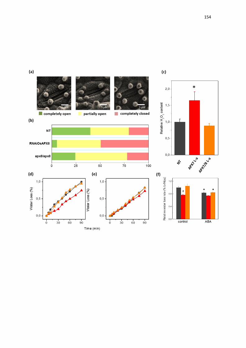
154
155
Figure 8: Effect of silencing and knockout of OsAPX8 in stomata opening
and in leaf desiccation. (a) Environmental scanning electron microscopy
images of three levels of stomatal opening. (b) quantification of the percentage
of the three levels of stomatal opening in leaves from NT, RNAiOsAPX8 and
apx8/apx8 30-d-old plants (n = 200 stomata for each line). (c) Relative content of
hydrogen peroxide in leaves from NT (grey), RNAiOsAPX8 (red) and apx8/apx8
(orange) leaves. The values represent the media ± SE of at least five independent
experiments. (d) and (e) Water loss of leaves detached from plants under normal
condition (treated with water) and treated with ABA 100 μM, respectively. Legend:
Grey: NT, Red: RNAiOsAPX8 and Orange: apx8/apx8. For each repeat, leaves
of 30-d-old plants were used in a triplicate experiment (n = 3). The water loss rate
of each conditions was quantified in the figure (f). (*) indicate populations
significantly different of control situation with p<0,05, and (#) indicate populations
significantly different of NT plants with p<0,05.
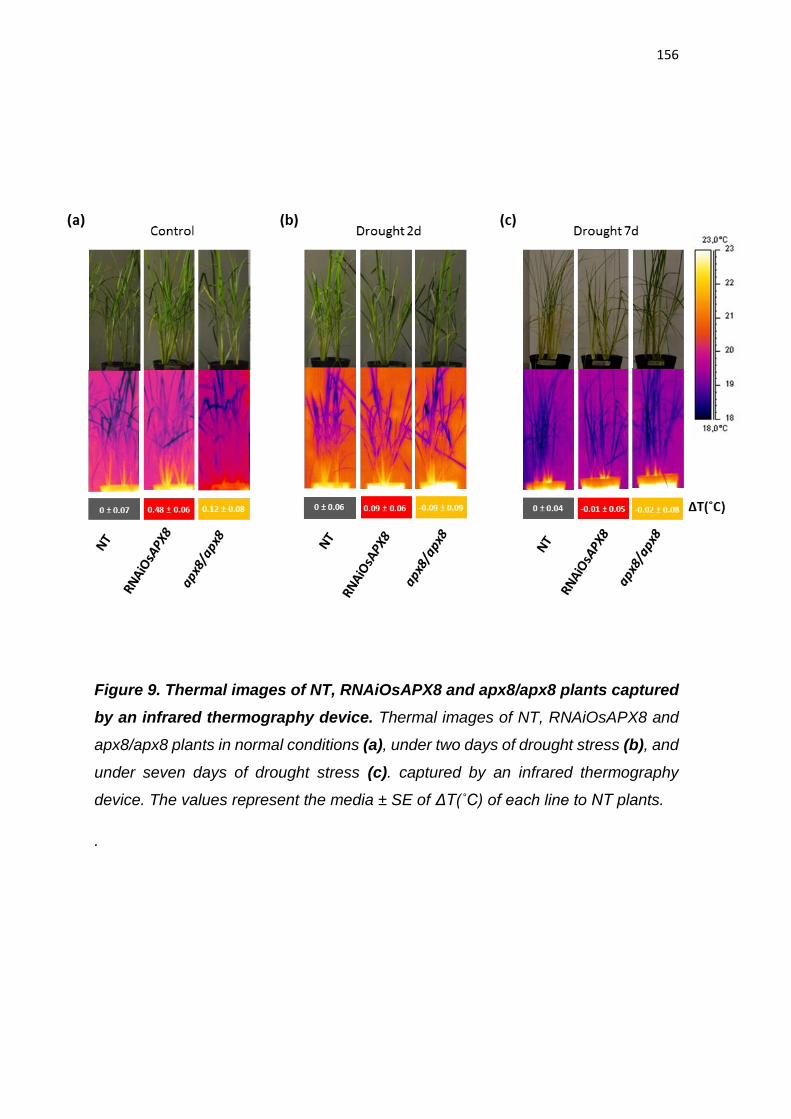
156
Figure 9. Thermal images of NT, RNAiOsAPX8 and apx8/apx8 plants captured
by an infrared thermography device. Thermal images of NT, RNAiOsAPX8 and
apx8/apx8 plants in normal conditions (a), under two days of drought stress (b), and
under seven days of drought stress (c). captured by an infrared thermography
device. The values represent the media ± SE of ΔT(˚C) of each line to NT plants.
.
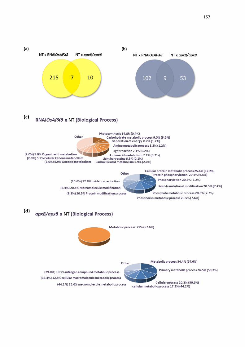
157
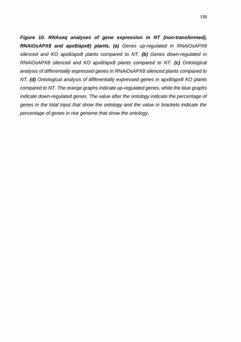
158
Figure 10. RNAseq analyses of gene expression in NT (non-transformed),
RNAiOsAPX8 and apx8/apx8) plants. (a) Genes up-regulated in RNAiOsAPX8
silenced and KO apx8/apx8 plants compared to NT. (b) Genes down-regulated in
RNAiOsAPX8 silenced and KO apx8/apx8 plants compared to NT. (c) Ontological
analysis of differentially expressed genes in RNAiOsAPX8 silenced plants compared to
NT. (d) Ontological analysis of differentially expressed genes in apx8/apx8 KO plants
compared to NT. The orange graphs indicate up-regulated genes, while the blue graphs
indicate down-regulated genes. The value after the ontology indicate the percentage of
genes in the total input that show the ontology and the value in brackets indicate the
percentage of genes in rice genome that show the ontology.
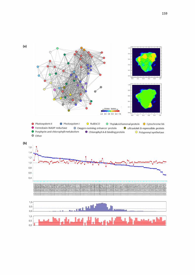
159
160
Figure 11. Effect of silencing and knockout of OsAPX8 on rice
photosynthesis gene network expression. Effect of silencing and KO of
OsAPX8 on rice photosynthesis gene network expression. (a) Heatmap
demonstrating the photosynthesis gene network expression in RNAiOsAPX8 and
apx8/apx8 plants compared to NT (non-transformed) plants. (b) Relative
expression of photosynthesis genes in RNAiOsAPX8 and apx8/apx8 plants
compared to NT. Legend: Blue – RNAiOsAPX8 x NT; Red – apx8/apx8 x NT. The
p-value of each data is indicated by blue and red bars, respectively.
161
REFERENCES
ASADA K, TAKAHASHI M. (1987). Production and scavenging of active oxygen
in photosynthesis. In Photoinhibition (Kyle, D.J., Osmond, C.B. and Arntzen, C.J.,
eds). New York: Elsevier Science Publishers, pp. 227–287.
ASADA K. (1992). Ascorbate Peroxidase - a Hydrogen Peroxide-Scavenging
Enzyme in Plants. Physiologia Plantarum 85: 235-241.
ASADA K. (1999). The water-water cycle in chloroplasts: scavenging of active
oxygen and dissipation of excess photons. Annu Rev Plant Physiol Plant Mol Biol
50:601–639
BILGER W, SCHREIBER U AND BOCK M. (1995). Determination of the
quantum efficiency of photosystem II and non-photochemical quenching of
chlorophyll fluorescence in the field. Oecologia 102: 425-432.
BLUM A, EBERCON A. (1981). Cell membrane stability as a measure of drought
and heat tolerance in wheat. Crop Science 21: 43–47.
BRADFORD MM. (1976). A rapid and sensitive for the quantitation of microgram
quantitites of protein utilizing the principle of protein-dye binding. Analytical
Biochemistry 72: 248–254.
CAO MC, ZHANG WZ, HAN YD, YAO C, WANG YT, DING GH. (2013). A
theoretical model research of rice water stress index based on automated infrared
thermal imaging. Adv. Mater. Res. 712: 433-438.
Castro MA, Filho JL, Dalmolin RJ, Sinigaglia M, Moreira JC, Mombach JC,
de Almeida RM. (2009). ViaComplex: software for landscape analysis of gene
expression networks in genomic context. Bioinformatics. 25(11).:1468-9.
CAVERZAN A, BONIFACIO A, CARVALHO FEL, ANDRADE CMB, PASSAIA
G, SCHÜNEMANN M, MARASCHIN FS, MARTINS MO, TEIXEIRA FK,
RAUBER R, MARGIS R, SILVEIRA JAG, MARGIS-PINHEIRO M. (2014). The
162
knockdown of chloroplastic ascorbate peroxidases reveals itsregulatory role in
the photosynthesis and protection underphoto-oxidative stress in rice. Plant
Science 214: 74– 87.
DANNA CH, BARTOLI CG, SACCO F, INGALA LR, SANTA-MARIA GE,
GUIAMET JJ, UGALDE RA. (2003). Thylakoid-bound ascorbate peroxidase
mutant exhibits impaired electron transport and photosynthetic activity. Plant
physiology 132: 2116-2125.
DASZKOWSKA-GOLEC A, SZAREJKO I. (2013). Open or Close the Gate –
Stomata Action Under the Control of Phytohormones in Drought Stress
Conditions. Front Plant Sci. 4: 138.
DAT J, VANDENABEELE S, VRANOVA E, VAN MONTAGU M, INZE D, VAN
BREUSEGEM F. (2000). Dual action of the active oxygen species during plant
stress response. Cell Mol Life Sci 57:779–795.
DU Z, ZHOU X, LING Y, ZHANG Z, ZHEN S. (2010). agriGO: a GO analysis
toolkit for the agricultural community. Nucl. Acids Res. 38: W64-W70.
HALLIWELL B, GUTTERIDGE JM. (1999). Free radicals in biology and
medicine. Oxford University Press, New York p 936
HAZEN SP, PATHAN MS, SANCHEZ A, BAXTER I, DUNN M, ESTES B,
CHANG H, ZHU T, KREPS JA, NGUYEN HT. (2005). Expression profiling of rice
segregating for drought tolerance QTLs using a rice genome array. Funct Integr
Genomics 5: 104–116.
HERNANDEZ JA, FERRER MA, JIMÉNEZ A, BARCELO AR, SEVILLA F.
(2001). Antioxidant systems and O(2)(-)/H(2)O(2). production in the apoplast of
pea leaves. Its relation with salt-induced necrotic lesions in minor veins. Plant
Physiology 127: 817–831.
HETHERINGTON AM, WOODWARD FI. (2003). The role of stomata in sensing
and driving environmental change. Nature 424: 901–908.
163
HOAGLAND DR, ARNON DI. (1950). The water-culture method for growing
plants without soil. Berkeley, CA, USA: College of Agriculture, University of
California.
HOOPER SD, BORK P. (2005). Medusa: a simple tool for interaction graph
analysis. Bioinformatics. 24:4432-3.
HUANG XY, CHAO DY, GAO JP, ZHU MZ, SHI M, LIN HX. (2009). A previously
unknown zinc finger protein, DST, regulates drought and salt tolerance in rice via
stomatal aperture control. Genes Dev. 15:1805-17.
JEFFERSON RA, KAVANAGH TA, BEVAN MW. (1987). GUS fusions: beta-
glucuronidase as a sensitive and versatile gene fusion marker in higher plants,
EMBO J. 6: 3901–3907.
JONES HG. (1999). Use of thermography for quantitative studies of spatial and
temporal variation of stomatal conductance over leaf surfaces. Plant Cell Environ
22:1043–1055.
KARPINSKI S, REYNOLDS H, KARPINSKA B, WINGSLE G, CREISSEN G,
MULLINEAUX P. (1999). Systemic signaling and acclimation in response to
excess excitation energy in Arabidopsis. Science 284: 654–657.
LICHTENTHALER HK, WELLBURN AR. (1983). Determination of total
carotenoids andchlorophylls a and b of leaf extracts in different solvents,
Biochem. Soc. Trans. 11:591–592.
LIVAK KJ, SCHMITTGEN TD. (2001). Analysis of relative gene expression data
using real-time quantitative PCR and the 2-ΔΔCT method. Methods 25: 402–408.
MIKI D, SHIMAMOTO K. (2004). Simple RNAi vectors for stable and transient
suppressionof gene function in rice, Plant Cell Physiol. 45:490–495.
MILLER G, SUZUKI N, RIZHSKY L, HEGIE A, KOUSSEVITZKY S, MITTLER
R. (2007). Double mutants deficient in cytosolic and thylakoid ascorbate
peroxidase reveal a complex mode of interaction between reactive oxygen
164
species, plant development, and response to abiotic stresses. Plant Physiol.
144:1777–1785.
MITTLER R, BERKOWITZ G. (2001) Hydrogen peroxide, a messenger with too
many roles? Redox Rep. 6(2):69-72.
MITTLER R, VANDERAUWERA S, GALLERY M, VAN BREUSEGEM F. (2004).
Reactive oxygen gene network of plants. Trends Plant Sci 9:490–498.
MÜHLENBOCK P, SZECHYNSKA-HEBDA M, PLASZCZYCA M, BAUDO M,
MATEO A, MULLINEAUX PM, PARKER JE, KARPINSKA B, KARPINSKI S.
(2008). Chloroplast signaling and LESION SIMULATING DISEASE1 regulate
crosstalk between light acclimation and immunity in Arabidopsis. The Plant Cell
20: 2339–2356.
MULLINEAUX P, KARPINSKI S. (2002). Signal transduction in response to
excess light: getting out of the chloroplast. Curr Opin Plant Biol 5:43–48.
MULLINEAUX PM, KARPINSKI S, BAKER NR. (2006). Spatial dependence for
hydrogen peroxide-directed signaling in light-stressed plants. Plant Physiology
141: 346–350.
NAKANO Y, ASADA K. (1981). Hydrogen peroxide is scavenged by ascorbate
peroxidase in spinach chloroplasts. Plant Cell Physiol 22: 867-880.
OROZCO-CARDENAS ML, NARVAEZ-VASQUEZ J, RYAN CA. (2001).
Hydrogen peroxide acts as a second messenger for the induction of defense
genes in tomato plants in response to wounding, systemin, and methyl-
jasmonate. Plant Cell 13:179–191.
PAYTON P, WEBB R, KORNYEYEV D, ALLEN R, HOLADAY AS. (2001).
Protecting cotton photosynthesis during moderate chilling at high light intensity
by increasing chloroplastic antioxidant enzyme activity. J Exp Bot 52: 2345-2354.
PRASHAR A, YILDIZ J, MCNICOL JW, BRYAN GJ, JONES HG. (2014). Infra-
red Thermography for High Throughput Field Phenotyping in Solanum
tuberosum. PLoS One. 8(6): e65816.
165
RAO MV, LEE H, CREELMAN RA, MULLET JE, DAVIS KR. (2000). Jasmonic
acid signaling modulates ozone-induced hypersensitive cell death. Plant Cell 12:
1633–1646.
ROSA SB, CAVERZAN A, TEIXEIRA FK, FERREIRA-SILVA SL,
LAZZAROTTO F, SILVEIRA JAG, ABREU-NETO J, MARGIS R, MARGIS-
PINHEIRO M. (2010). Cytosolic APx knockdown indicates an ambiguous redox
responses in rice. Phytochemistry 71:548–558.
SCANDALIOS JG. (2002). The rise of ROS. Trends Biochem Sci 27:483–486.
SCHMITTGEN TD, LIVAK KJ. (2008). Analyzing real-time PCR data by the
comparative CT method. Nature Protocols 3: 1101–1108.
SIRAULT XRR, JAMES RA, FURBANK RT. (2009). A new screening method
for osmotic component of salinity tolerance in cereals using infrared
thermography. Funct Plant Biol 36: 970–977.
SMITH AM, RATCLIFFE RG, SWEETLOVE LJ. (2004). Activation and function
of mitochondrial uncoupling protein in plants. The Journal of Biological Chemistry
279: 51944–51952.
SNEL B, LEHMANN G, BORK P, HUYNEN MA. (2000). STRING: a web-server
to retrieve and display the repeatedly occurring neighbourhood of a gene. Nucleic
Acids Res. 28(18): 3442-3444.
SNEL B, LEHMANN G, BORK P, HUYNEN MA. (2000). STRING: a web-server
to retrieve and display the repeatedly occurring neighbourhood of a gene. Nucleic
Acids Res. 28(18):3442-4.
SZECHYŃSKA-HEBDA M, KRUK J, GORECKA M, KARPINSKA B,
KARPINSKI S. (2010). Evidence for light wavelength-specific
photoelectrophysiological signaling and memory of excess light episodes in
Arabidopsis. The Plant Cell 22: 2201–2218.
TEIXEIRA FK, MENEZES-BENAVENTE L, GALVAO VC, MARGIS R, MARGIS-
PINHEIRO M. (2006). Rice ascorbate peroxidase gene family encodes
166
functionally diverse isoformslocalized in different subcellular compartments,
Planta 224: 300–314.
THORDALCHRISTENSEN H, ZHANG ZG, WEI YD, COLLINGE DB. (1997).
Subcellular localization of H2O2 in plants. H2O2 accumulation in papillae and
hypersensitive response during the barley-powdery mildew interaction. Plant J.
9: 1187–1194.
TSENG MJ, LIU CW AND YIU JC. (2007). Enhanced tolerance to sulfur dioxide
and salt stress of transgenic Chinese cabbage plants expressing both superoxide
dismutase and catalase in chloroplasts. Plant Physiology and Biochemistry 45:
822–833.
UPADHYAYA NM, ZHOU XR, ZHU QH, EAMENS A, WANG MB, WATER-
HOUSE MP, DENNIS ES. (2002). Transgenic rice, in: L. O’Brien, R.J. Henry.
(Eds.)., TransgenicCereals, AACC, Minnesota pp. 28–87.
VAN CAMP W, CAPIAU K, VAN MONTAGU M, INZE D AND SLOOTEN L.
(1996). Enhancement of oxidative stress tolerance in transgenic tobacco plants
overproducing Fe-superoxide dismutase in chloroplasts. Plant Physiology 112:
1703–1714.
VANDENABEELE S, VAN DER KELEN K, DAT J, GADJEV I, BOONEFAES T,
MORSA S, ROTTIERS P, SLOOTEN L, VAN MONTAGU M, ZABEAU M, INZE
D, VAN BREUSEGEM F. (2003). A comprehensive analysis of hydrogen
peroxide-induced gene expression in tobacco. Proc Natl Acad Sci USA
100:16113–16118.
WANG D, PAN Y, ZHAO X, ZHU L, FU B, LI Z. (2011). Genome-wide temporal-
spatial gene expression profiling of drought responsiveness in rice BMC
Genomics 12: 149.
YABUTA Y, MOTOKI T, YOSHIMURA K, TAKEDA T, ISHIKAWA T,
SHIGEOKA S. (2002). Thylakoid membrane-bound ascorbate peroxidase is a
limiting factor of antioxidative systems under photo-oxidative stress. Plant J 32:
915-925.
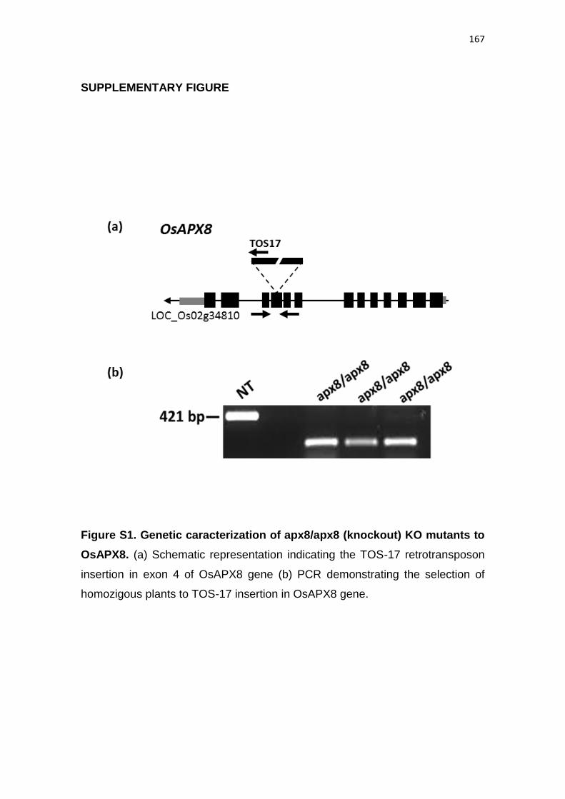
167
SUPPLEMENTARY FIGURE
Figure S1. Genetic caracterization of apx8/apx8 (knockout) KO mutants to
OsAPX8. (a) Schematic representation indicating the TOS-17 retrotransposon
insertion in exon 4 of OsAPX8 gene (b) PCR demonstrating the selection of
homozigous plants to TOS-17 insertion in OsAPX8 gene.
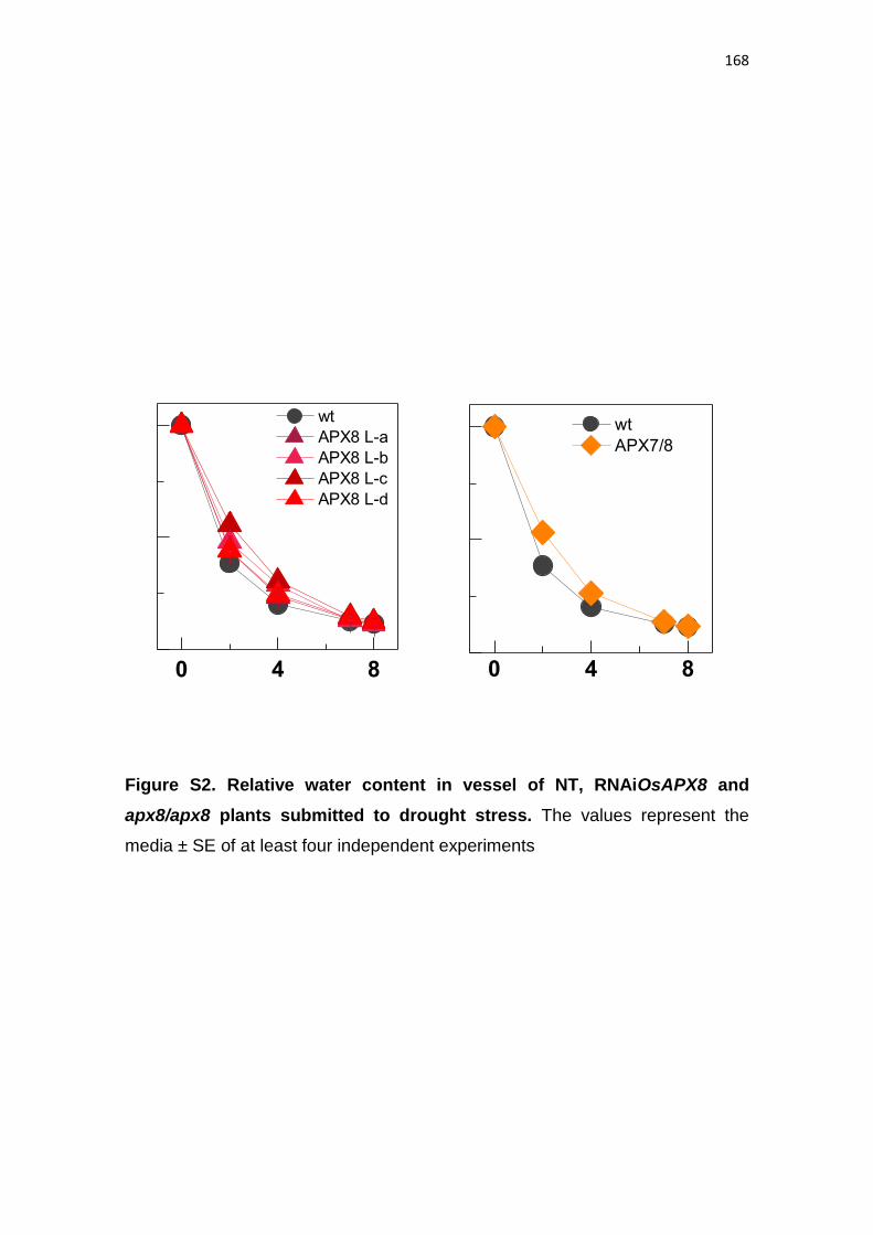
168
Figure S2. Relative water content in vessel of NT, RNAiOsAPX8 and
apx8/apx8 plants submitted to drought stress. The values represent the
media ± SE of at least four independent experiments
0 4 8
wt
APX8 L-a
APX8 L-b
APX8 L-c
APX8 L-d
0 4 8
wt
APX7/8
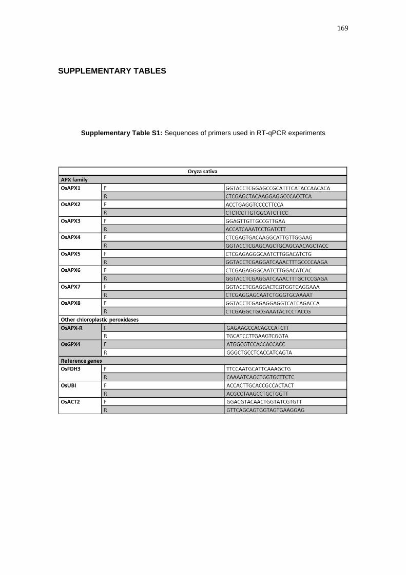
169
SUPPLEMENTARY TABLES
Supplementary Table S1: Sequences of primers used in RT-qPCR experiments
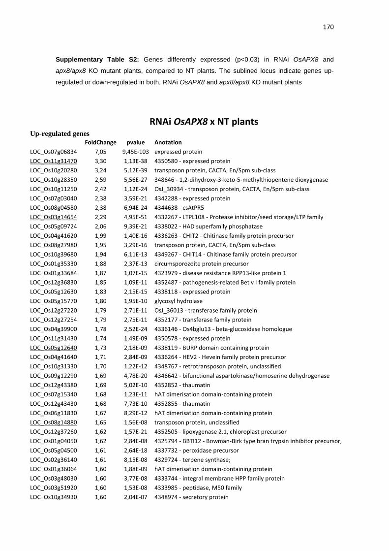
170
Supplementary Table S2: Genes differently expressed (p<0.03) in RNAi OsAPX8 and
apx8/apx8 KO mutant plants, compared to NT plants. The sublined locus indicate genes up-
regulated or down-regulated in both, RNAi OsAPX8 and apx8/apx8 KO mutant plants
RNAi OsAPX8 x NT plants Up-regulated genes
FoldChange pvalue Anotation
LOC_Os07g06834 7,05 9,45E-103 expressed protein
LOC_Os11g31470 3,30 1,13E-38 4350580 - expressed protein
LOC_Os10g20280 3,24 5,12E-39 transposon protein, CACTA, En/Spm sub-class
LOC_Os10g28350 2,59 5,56E-27 348646 - 1,2-dihydroxy-3-keto-5-methylthiopentene dioxygenase
LOC_Os10g11250 2,42 1,12E-24 OsJ_30934 - transposon protein, CACTA, En/Spm sub-class
LOC_Os07g03040 2,38 3,59E-21 4342288 - expressed protein
LOC_Os08g04580 2,38 6,94E-24 4344638 - csAtPR5
LOC_Os03g14654 2,29 4,95E-51 4332267 - LTPL108 - Protease inhibitor/seed storage/LTP family
LOC_Os05g09724 2,06 9,39E-21 4338022 - HAD superfamily phosphatase
LOC_Os04g41620 1,99 1,40E-16 4336263 - CHIT2 - Chitinase family protein precursor
LOC_Os08g27980 1,95 3,29E-16 transposon protein, CACTA, En/Spm sub-class
LOC_Os10g39680 1,94 6,11E-13 4349267 - CHIT14 - Chitinase family protein precursor
LOC_Os01g35330 1,88 2,37E-13 circumsporozoite protein precursor
LOC_Os01g33684 1,87 1,07E-15 4323979 - disease resistance RPP13-like protein 1
LOC_Os12g36830 1,85 1,09E-11 4352487 - pathogenesis-related Bet v I family protein
LOC_Os05g12630 1,83 2,15E-15 4338118 - expressed protein
LOC_Os05g15770 1,80 1,95E-10 glycosyl hydrolase
LOC_Os12g27220 1,79 2,71E-11 OsJ_36013 - transferase family protein
LOC_Os12g27254 1,79 2,75E-11 4352177 - transferase family protein
LOC_Os04g39900 1,78 2,52E-24 4336146 - Os4bglu13 - beta-glucosidase homologue
LOC_Os11g31430 1,74 1,49E-09 4350578 - expressed protein
LOC_Os05g12640 1,73 2,18E-09 4338119 - BURP domain containing protein
LOC_Os04g41640 1,71 2,84E-09 4336264 - HEV2 - Hevein family protein precursor
LOC_Os10g31330 1,70 1,22E-12 4348767 - retrotransposon protein, unclassified
LOC_Os09g12290 1,69 4,78E-20 4346642 - bifunctional aspartokinase/homoserine dehydrogenase
LOC_Os12g43380 1,69 5,02E-10 4352852 - thaumatin
LOC_Os07g15340 1,68 1,23E-11 hAT dimerisation domain-containing protein
LOC_Os12g43430 1,68 7,73E-10 4352855 - thaumatin
LOC_Os06g11830 1,67 8,29E-12 hAT dimerisation domain-containing protein
LOC_Os08g14880 1,65 1,56E-08 transposon protein, unclassified
LOC_Os12g37260 1,62 1,57E-21 4352505 - lipoxygenase 2.1, chloroplast precursor
LOC_Os01g04050 1,62 2,84E-08 4325794 - BBTI12 - Bowman-Birk type bran trypsin inhibitor precursor,
LOC_Os05g04500 1,61 2,64E-18 4337732 - peroxidase precursor
LOC_Os02g36140 1,61 8,15E-08 4329724 - terpene synthase;
LOC_Os01g36064 1,60 1,88E-09 hAT dimerisation domain-containing protein
LOC_Os03g48030 1,60 3,77E-08 4333744 - integral membrane HPP family protein
LOC_Os03g51920 1,60 1,53E-08 4333985 - peptidase, M50 family
LOC_Os10g34930 1,60 2,04E-07 4348974 - secretory protein
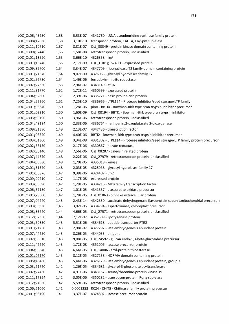
171
LOC_Os06g45250 1,58 5,53E-07 4341760 - tRNA pseudouridine synthase family protein
LOC_Os08g17030 1,58 3,10E-10 transposon protein, CACTA, En/Spm sub-class
LOC_Os11g10710 1,57 8,81E-07 OsJ_33349 - protein kinase domain containing protein
LOC_Os09g07440 1,56 1,58E-08 retrotransposon protein, unclassified
LOC_Os01g13690 1,55 3,66E-10 4326358 - ligA
LOC_Os01g15740 1,55 2,17E-09 LOC_Os01g15740.1 - expressed protein
LOC_Os09g36700 1,54 3,34E-07 4347709 - ribonuclease T2 family domain containing protein
LOC_Os01g71670 1,54 9,07E-09 4326063 - glycosyl hydrolases family 17
LOC_Os02g52730 1,54 1,46E-06 ferredoxin--nitrite reductase
LOC_Os07g27350 1,53 2,94E-07 4343149 - atuA
LOC_Os11g31770 1,52 1,72E-11 4350599 - expressed protein
LOC_Os04g32800 1,51 2,39E-06 4335721 - basic proline-rich protein
LOC_Os04g52260 1,51 7,25E-10 4336966 - LTPL124 - Protease inhibitor/seed storage/LTP family
LOC_Os01g03340 1,50 1,28E-06 pinA - BBTI4 - Bowman-Birk type bran trypsin inhibitor precursor
LOC_Os01g03310 1,50 1,60E-09 OsJ_00194 - BBTI1 - Bowman-Birk type bran trypsin inhibitor
LOC_Os03g59190 1,50 3,96E-06 retrotransposon protein, unclassified
LOC_Os04g49194 1,50 2,33E-06 4336764 - naringenin,2-oxoglutarate 3-dioxygenase
LOC_Os09g31390 1,49 2,13E-07 4347436 - transcription factor
LOC_Os01g03320 1,49 4,40E-06 BBTI2 - Bowman-Birk type bran trypsin inhibitor precursor
LOC_Os03g01300 1,49 3,34E-08 4331302 - LTPL114 - Protease inhibitor/seed storage/LTP family protein precursor
LOC_Os02g53130 1,49 2,17E-06 4330867 - nitrate reductase
LOC_Os02g50140 1,48 7,56E-06 OsJ_08287 - caleosin related protein
LOC_Os07g44670 1,48 2,22E-06 OsJ_27979 - retrotransposon protein, unclassified
LOC_Os04g05580 1,48 1,70E-05 4335018 - kinase
LOC_Os01g51570 1,48 2,03E-05 4325938 - glycosyl hydrolases family 17
LOC_Os01g06876 1,47 9,38E-06 4324407 - Cf-2
LOC_Os09g09210 1,47 1,17E-08 expressed protein
LOC_Os03g55590 1,47 1,29E-05 4334216 - MYB family transcription factor
LOC_Os06g37150 1,47 1,01E-05 4341337 - L-ascorbate oxidase precursor
LOC_Os01g28500 1,47 1,78E-05 OsJ_01863 - SCP-like extracellular protein
LOC_Os07g04240 1,45 2,43E-14 4342350 - succinate dehydrogenase flavoprotein subunit,mitochondrial precursor;
LOC_Os03g63330 1,45 3,92E-05 4334794 - aspartokinase, chloroplast precursor
LOC_Os08g35720 1,44 4,66E-05 OsJ_27571 - retrotransposon protein, unclassified
LOC_Os12g37350 1,44 7,12E-07 4352509 - lipozygenase protein
LOC_Os03g60850 1,43 5,51E-06 4334618 - peptide transporter PTR2
LOC_Os01g21250 1,43 2,98E-07 4327292 - late embryogenesis abundant protein
LOC_Os07g44250 1,43 8,26E-05 4344033 - dirigent
LOC_Os07g35510 1,43 9,08E-05 OsJ_24592 - glucan endo-1,3-beta-glucosidase precursor
LOC_Os11g42220 1,43 1,72E-08 4351006 - laccase precursor protein
LOC_Os04g09540 1,43 6,64E-05 OsJ_14006 - acyl-protein thioesterase
LOC_Os01g07170 1,43 8,12E-05 4327138 - HORMA domain containing protein
LOC_Os05g46480 1,43 5,44E-06 4326129 - late embryogenesis abundant protein, group 3
LOC_Os03g61720 1,42 1,26E-05 4334681 - glycerol-3-phosphate acyltransferase
LOC_Os07g27460 1,42 4,91E-06 4343157 - serine/threonine-protein kinase 19
LOC_Os11g17954 1,42 3,05E-06 4350282 - transposon protein, Pong sub-class
LOC_Os12g24050 1,42 5,59E-06 retrotransposon protein, unclassified
LOC_Os06g51060 1,41 0,0001253 RC24 - CHIT8 - Chitinase family protein precursor
LOC_Os01g63190 1,41 3,37E-07 4324802 - laccase precursor protein
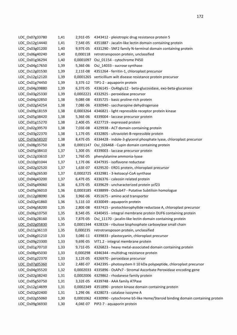
172
LOC_Os07g33780 1,41 2,91E-05 4343412 - pleiotropic drug resistance protein 5
LOC_Os12g14440 1,41 7,54E-05 4351887 - Jacalin-like lectin domain containing protein
LOC_Os03g01200 1,40 9,97E-05 4331290 - SNF2 family N-terminal domain containing protein
LOC_Os06g40240 1,40 0,000118 retrotransposon protein, unclassified
LOC_Os01g36294 1,40 0,0001097 OsJ_01154 - cytochrome P450
LOC_Os04g17650 1,39 5,36E-06 OsJ_14033 - sucrose synthase
LOC_Os12g01530 1,39 2,11E-08 4351264 - ferritin-1, chloroplast precursor
LOC_Os12g12120 1,39 0,0001265 verticillium wilt disease resistance protein precursor
LOC_Os01g74450 1,39 3,37E-12 TIP1-2 - aquaporin protein
LOC_Os04g39880 1,39 6,37E-05 4336145 - Os4bglu12 - beta-glucosidase, exo-beta-glucanase
LOC_Os03g25330 1,39 0,0002221 4332925 - peroxidase precursor
LOC_Os04g32850 1,38 9,08E-08 4335725 - basic proline-rich protein
LOC_Os02g54254 1,38 7,08E-06 4330940 - saccharopine dehydrogenase
LOC_Os09g18159 1,38 0,0003264 4346821 - light repressible receptor protein kinase
LOC_Os05g38420 1,38 5,36E-06 4339004 - laccase precursor protein
LOC_Os01g15270 1,38 2,40E-05 4327719 - expressed protein
LOC_Os02g39570 1,38 7,03E-08 4329938 - ACT domain containing protein
LOC_Os03g22370 1,38 1,17E-05 4332805 - ultraviolet-B-repressible protein
LOC_Os03g58320 1,38 8,47E-05 4334428 - indole-3-glycerol phosphate lyase, chloroplast precursor
LOC_Os08g35750 1,38 0,0001147 OsJ_026468 - Cupin domain containing protein
LOC_Os05g38410 1,37 1,30E-05 4339003 - laccase precursor protein
LOC_Os12g33610 1,37 1,76E-05 phenylalanine ammonia-lyase
LOC_Os10g01044 1,37 1,17E-06 4347935 - isoflavone reductase
LOC_Os02g32520 1,37 1,63E-07 4329520 - ERD1 protein, chloroplast precursor
LOC_Os03g26530 1,37 0,0002725 4332981 - 3-ketoacyl-CoA synthase
LOC_Os04g43200 1,37 6,47E-05 4336376 - caleosin related protein
LOC_Os05g49060 1,36 6,37E-05 4339629 - uncharacterized protein ycf23
LOC_Os05g36010 1,36 0,0003185 4338899 - OsSub47 - Putative Subtilisin homologue
LOC_Os12g08090 1,36 3,96E-06 4351675 - amino acid transporter
LOC_Os02g41860 1,36 5,11E-10 4330049 - aquaporin protein
LOC_Os04g58200 1,35 2,80E-08 4337415 - protochlorophyllide reductase A, chloroplast precursor
LOC_Os06g10750 1,35 8,54E-05 4340455 - integral membrane protein DUF6 containing protein
LOC_Os03g28160 1,35 7,87E-05 OsJ_11170 - jacalin-like lectin domain containing protein
LOC_Os02g05830 1,35 0,0001344 4328326 - ribulose bisphosphate carboxylase small chain
LOC_Os11g36110 1,35 0,000235 retrotransposon protein, unclassified
LOC_Os06g01210 1,33 3,08E-11 4339833 - plastocyanin, chloroplast precursor
LOC_Os09g23300 1,33 9,69E-05 VIT1.2 - integral membrane protein
LOC_Os01g70710 1,33 9,71E-05 4326823 - heavy metal-associated domain containing protein
LOC_Os08g45030 1,33 0,000298 4346344 - multidrug resistance protein
LOC_Os01g22370 1,33 3,12E-05 4326970 - peroxidase precursor
LOC_Os07g05360 1,32 2,48E-07 4342395 - photosystem II 10 kDa polypeptide, chloroplast precursor
LOC_Os04g35520 1,32 0,0002033 4335896 - OsAPx7 - Stromal Ascorbate Peroxidase encoding gene
LOC_Os02g38240 1,31 0,0002006 4329862 - rhodanese family protein
LOC_Os05g50750 1,31 3,32E-05 4339748 - AAA family ATPase
LOC_Os12g14699 1,31 0,0002349 4351890 - protein kinase domain containing protein
LOC_Os02g02400 1,31 1,29E-06 4328073 - catalase isozyme A
LOC_Os02g55060 1,30 0,0001062 4330990 - cytochrome b5-like Heme/Steroid binding domain containing protein
LOC_Os09g36930 1,30 4,04E-07 PIP2-7 - aquaporin protein
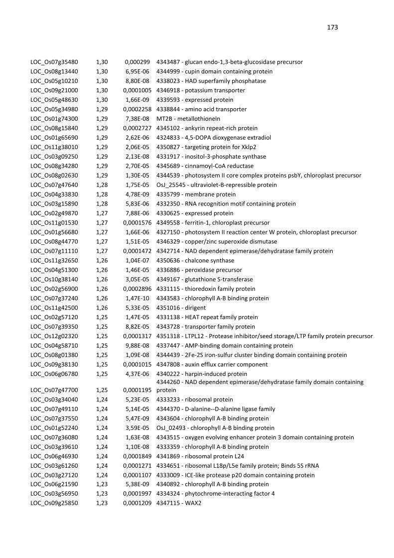
173
LOC_Os07g35480 1,30 0,000299 4343487 - glucan endo-1,3-beta-glucosidase precursor
LOC_Os08g13440 1,30 6,95E-06 4344999 - cupin domain containing protein
LOC_Os05g10210 1,30 8,80E-08 4338023 - HAD superfamily phosphatase
LOC_Os09g21000 1,30 0,0001005 4346918 - potassium transporter
LOC_Os05g48630 1,30 1,66E-09 4339593 - expressed protein
LOC_Os05g34980 1,29 0,0002258 4338844 - amino acid transporter
LOC_Os01g74300 1,29 7,38E-08 MT2B - metallothionein
LOC_Os08g15840 1,29 0,0002727 4345102 - ankyrin repeat-rich protein
LOC_Os01g65690 1,29 2,62E-06 4324833 - 4,5-DOPA dioxygenase extradiol
LOC_Os11g38010 1,29 2,06E-05 4350827 - targeting protein for Xklp2
LOC_Os03g09250 1,29 2,13E-08 4331917 - inositol-3-phosphate synthase
LOC_Os08g34280 1,29 2,70E-05 4345689 - cinnamoyl-CoA reductase
LOC_Os08g02630 1,29 1,30E-05 4344539 - photosystem II core complex proteins psbY, chloroplast precursor
LOC_Os07g47640 1,28 1,75E-05 OsJ_25545 - ultraviolet-B-repressible protein
LOC_Os04g33830 1,28 4,78E-09 4335799 - membrane protein
LOC_Os03g15890 1,28 5,83E-06 4332350 - RNA recognition motif containing protein
LOC_Os02g49870 1,27 7,88E-06 4330625 - expressed protein
LOC_Os11g01530 1,27 0,0001576 4349558 - ferritin-1, chloroplast precursor
LOC_Os01g56680 1,27 1,66E-06 4327150 - photosystem II reaction center W protein, chloroplast precursor
LOC_Os08g44770 1,27 1,51E-05 4346329 - copper/zinc superoxide dismutase
LOC_Os07g11110 1,27 0,0001472 4342714 - NAD dependent epimerase/dehydratase family protein
LOC_Os11g32650 1,26 1,04E-07 4350636 - chalcone synthase
LOC_Os04g51300 1,26 1,46E-05 4336886 - peroxidase precursor
LOC_Os10g38140 1,26 3,05E-05 4349167 - glutathione S-transferase
LOC_Os02g56900 1,26 0,0002896 4331115 - thioredoxin family protein
LOC_Os07g37240 1,26 1,47E-10 4343583 - chlorophyll A-B binding protein
LOC_Os11g42500 1,26 5,33E-05 4351016 - dirigent
LOC_Os02g57120 1,25 1,47E-05 4331138 - HEAT repeat family protein
LOC_Os07g39350 1,25 8,82E-05 4343728 - transporter family protein
LOC_Os12g02320 1,25 0,0001317 4351318 - LTPL12 - Protease inhibitor/seed storage/LTP family protein precursor
LOC_Os04g58710 1,25 9,88E-08 4337447 - AMP-binding domain containing protein
LOC_Os08g01380 1,25 1,09E-08 4344439 - 2Fe-2S iron-sulfur cluster binding domain containing protein
LOC_Os09g38130 1,25 0,0001015 4347808 - auxin efflux carrier component
LOC_Os06g06780 1,25 4,37E-06 4340222 - harpin-induced protein
LOC_Os07g47700 1,25 0,0001195 4344260 - NAD dependent epimerase/dehydratase family domain containing protein
LOC_Os03g34040 1,24 5,23E-05 4333233 - ribosomal protein
LOC_Os07g49110 1,24 5,14E-05 4344370 - D-alanine--D-alanine ligase family
LOC_Os07g37550 1,24 5,47E-09 4343604 - chlorophyll A-B binding protein
LOC_Os01g52240 1,24 3,59E-05 OsJ_02493 - chlorophyll A-B binding protein
LOC_Os07g36080 1,24 1,63E-08 4343515 - oxygen evolving enhancer protein 3 domain containing protein
LOC_Os03g39610 1,24 1,10E-08 4333359 - chlorophyll A-B binding protein
LOC_Os06g46930 1,24 0,0001849 4341869 - ribosomal protein L24
LOC_Os03g61260 1,24 0,0001271 4334651 - ribosomal L18p/L5e family protein; Binds 5S rRNA
LOC_Os03g27120 1,24 0,0001107 4333009 - ICE-like protease p20 domain containing protein
LOC_Os06g21590 1,23 5,38E-09 4340892 - chlorophyll A-B binding protein
LOC_Os03g56950 1,23 0,0001997 4334324 - phytochrome-interacting factor 4
LOC_Os09g25850 1,23 0,0001209 4347115 - WAX2
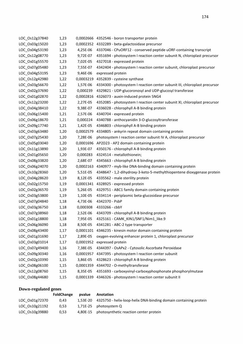
174
LOC_Os12g37840 1,23 0,0002666 4352546 - boron transporter protein
LOC_Os03g15020 1,23 0,0002352 4332289 - beta-galactosidase precursor
LOC_Os04g53190 1,23 4,25E-06 4337046 - CPuORF12 - conserved peptide uORF-containing transcript
LOC_Os12g08770 1,23 9,72E-07 4351694 - photosystem I reaction center subunit N, chloroplast precursor
LOC_Os01g55570 1,23 7,02E-05 4327018 - expressed protein
LOC_Os07g05480 1,23 7,55E-07 4342404 - photosystem I reaction center subunit, chloroplast precursor
LOC_Os04g53195 1,23 9,46E-06 expressed protein
LOC_Os12g42980 1,22 0,0003219 4352839 - cysteine synthase
LOC_Os03g56670 1,22 1,57E-06 4334300 - photosystem I reaction center subunit III, chloroplast precursor
LOC_Os02g37690 1,22 0,000239 4329821 - UDP-glucoronosyl and UDP-glucosyl transferase
LOC_Os01g02870 1,22 0,0002816 4326073 - auxin-induced protein 5NG4
LOC_Os12g23200 1,22 2,27E-05 4352085 - photosystem I reaction center subunit XI, chloroplast precursor
LOC_Os04g38410 1,22 9,38E-07 4336028 - chlorophyll A-B binding protein
LOC_Os06g15400 1,21 2,57E-06 4340704 - expressed protein
LOC_Os06g18670 1,21 0,000224 4340788 - anthocyanidin 3-O-glucosyltransferase
LOC_Os09g17740 1,21 1,42E-05 4346803 - chlorophyll A-B binding protein
LOC_Os03g63480 1,20 0,0002579 4334805 - ankyrin repeat domain containing protein
LOC_Os07g25430 1,20 7,28E-06 photosystem I reaction center subunit IV A, chloroplast precursor
LOC_Os05g03040 1,20 0,0001696 AP2D23 - AP2 domain containing protein
LOC_Os11g13890 1,20 1,93E-07 4350176 - chlorophyll A-B binding protein
LOC_Os01g05650 1,20 0,000283 4324514 - metallothionein;
LOC_Os08g33820 1,20 2,68E-07 4345663 - chlorophyll A-B binding protein
LOC_Os06g24070 1,20 0,0002163 4340977 - myb-like DNA-binding domain containing protein
LOC_Os10g28360 1,20 5,51E-05 4348647 - 1,2-dihydroxy-3-keto-5-methylthiopentene dioxygenase protein
LOC_Os04g28620 1,19 8,12E-05 4335562 - male sterility protein
LOC_Os02g15750 1,19 0,0001341 4328925 - expressed protein
LOC_Os02g36570 1,19 5,26E-05 4329751 - ABC1 family domain containing protein
LOC_Os03g53800 1,19 1,10E-05 4334114 - periplasmic beta-glucosidase precursor
LOC_Os07g04840 1,18 4,73E-06 4342370 - PsbP
LOC_Os03g36750 1,18 0,000308 4333266 - cbbY
LOC_Os07g38960 1,18 2,52E-06 4343709 - chlorophyll A-B binding protein
LOC_Os01g18800 1,18 7,95E-05 4325161 - CAMK_KIN1/SNF1/Nim1_like.9
LOC_Os06g36090 1,18 8,50E-05 4341281 - ABC-2 type transporter
LOC_Os08g43400 1,17 0,0001101 4346235 - kinesin motor domain containing protein
LOC_Os01g31690 1,17 2,89E-05 oxygen-evolving enhancer protein 1, chloroplast precursor
LOC_Os03g01014 1,17 0,0001952 expressed protein
LOC_Os07g49400 1,16 7,38E-05 4344397 - OsAPx2 - Cytosolic Ascorbate Peroxidase
LOC_Os09g30340 1,16 0,0001957 4347395 - photosystem I reaction center subunit
LOC_Os02g10390 1,15 3,86E-05 4328623 - chlorophyll A-B binding protein
LOC_Os08g06100 1,15 0,0001359 4344702 - O-methyltransferase
LOC_Os12g08760 1,15 8,35E-05 4351693 - carboxyvinyl-carboxyphosphonate phosphorylmutase
LOC_Os08g44680 1,15 0,0001339 4346326 - photosystem I reaction center subunit II
Down-regulated genes
FoldChange pvalue Anotation
LOC_Os01g72370 0,43 1,53E-20 4325750 - helix-loop-helix DNA-binding domain containing protein
LOC_Os10g21192 0,53 1,71E-25 photosystem Q
LOC_Os10g39880 0,53 4,80E-15 photosynthetic reaction center protein
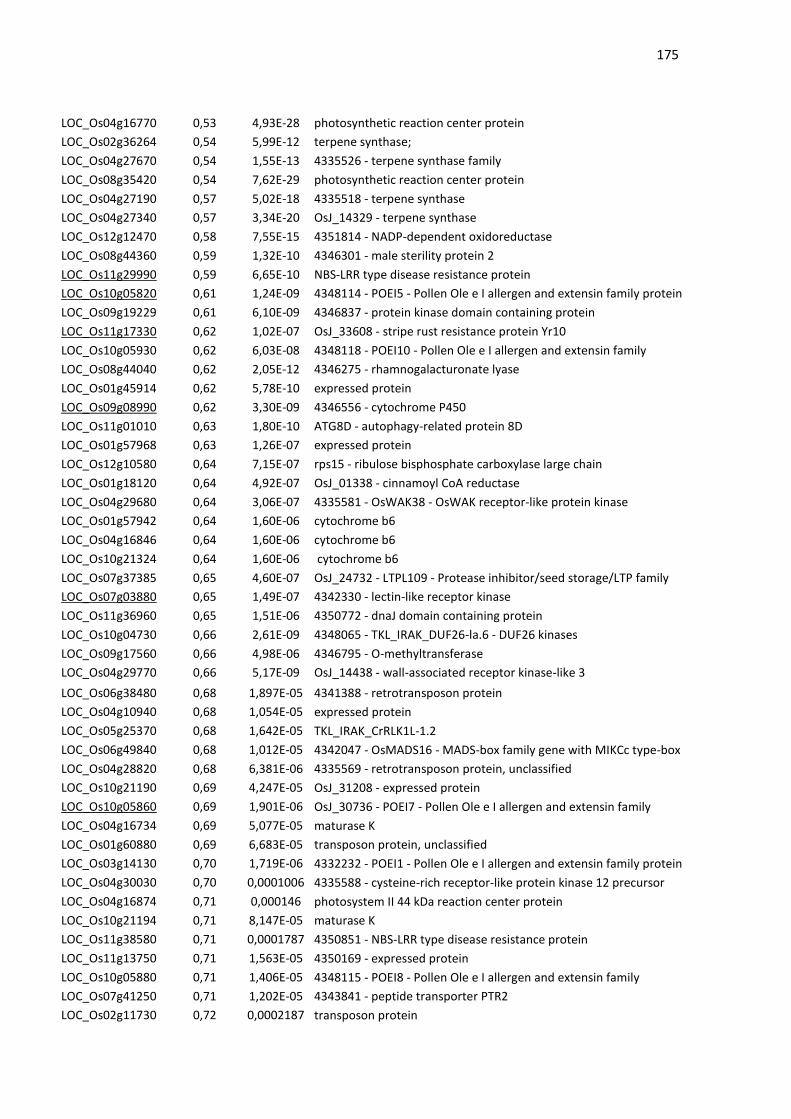
175
LOC_Os04g16770 0,53 4,93E-28 photosynthetic reaction center protein
LOC_Os02g36264 0,54 5,99E-12 terpene synthase;
LOC_Os04g27670 0,54 1,55E-13 4335526 - terpene synthase family
LOC_Os08g35420 0,54 7,62E-29 photosynthetic reaction center protein
LOC_Os04g27190 0,57 5,02E-18 4335518 - terpene synthase
LOC_Os04g27340 0,57 3,34E-20 OsJ_14329 - terpene synthase
LOC_Os12g12470 0,58 7,55E-15 4351814 - NADP-dependent oxidoreductase
LOC_Os08g44360 0,59 1,32E-10 4346301 - male sterility protein 2
LOC_Os11g29990 0,59 6,65E-10 NBS-LRR type disease resistance protein
LOC_Os10g05820 0,61 1,24E-09 4348114 - POEI5 - Pollen Ole e I allergen and extensin family protein
LOC_Os09g19229 0,61 6,10E-09 4346837 - protein kinase domain containing protein
LOC_Os11g17330 0,62 1,02E-07 OsJ_33608 - stripe rust resistance protein Yr10
LOC_Os10g05930 0,62 6,03E-08 4348118 - POEI10 - Pollen Ole e I allergen and extensin family
LOC_Os08g44040 0,62 2,05E-12 4346275 - rhamnogalacturonate lyase
LOC_Os01g45914 0,62 5,78E-10 expressed protein
LOC_Os09g08990 0,62 3,30E-09 4346556 - cytochrome P450
LOC_Os11g01010 0,63 1,80E-10 ATG8D - autophagy-related protein 8D
LOC_Os01g57968 0,63 1,26E-07 expressed protein
LOC_Os12g10580 0,64 7,15E-07 rps15 - ribulose bisphosphate carboxylase large chain
LOC_Os01g18120 0,64 4,92E-07 OsJ_01338 - cinnamoyl CoA reductase
LOC_Os04g29680 0,64 3,06E-07 4335581 - OsWAK38 - OsWAK receptor-like protein kinase
LOC_Os01g57942 0,64 1,60E-06 cytochrome b6
LOC_Os04g16846 0,64 1,60E-06 cytochrome b6
LOC_Os10g21324 0,64 1,60E-06 cytochrome b6
LOC_Os07g37385 0,65 4,60E-07 OsJ_24732 - LTPL109 - Protease inhibitor/seed storage/LTP family
LOC_Os07g03880 0,65 1,49E-07 4342330 - lectin-like receptor kinase
LOC_Os11g36960 0,65 1,51E-06 4350772 - dnaJ domain containing protein
LOC_Os10g04730 0,66 2,61E-09 4348065 - TKL_IRAK_DUF26-la.6 - DUF26 kinases
LOC_Os09g17560 0,66 4,98E-06 4346795 - O-methyltransferase
LOC_Os04g29770 0,66 5,17E-09 OsJ_14438 - wall-associated receptor kinase-like 3
LOC_Os06g38480 0,68 1,897E-05 4341388 - retrotransposon protein
LOC_Os04g10940 0,68 1,054E-05 expressed protein
LOC_Os05g25370 0,68 1,642E-05 TKL_IRAK_CrRLK1L-1.2
LOC_Os06g49840 0,68 1,012E-05 4342047 - OsMADS16 - MADS-box family gene with MIKCc type-box
LOC_Os04g28820 0,68 6,381E-06 4335569 - retrotransposon protein, unclassified
LOC_Os10g21190 0,69 4,247E-05 OsJ_31208 - expressed protein
LOC_Os10g05860 0,69 1,901E-06 OsJ_30736 - POEI7 - Pollen Ole e I allergen and extensin family
LOC_Os04g16734 0,69 5,077E-05 maturase K
LOC_Os01g60880 0,69 6,683E-05 transposon protein, unclassified
LOC_Os03g14130 0,70 1,719E-06 4332232 - POEI1 - Pollen Ole e I allergen and extensin family protein
LOC_Os04g30030 0,70 0,0001006 4335588 - cysteine-rich receptor-like protein kinase 12 precursor
LOC_Os04g16874 0,71 0,000146 photosystem II 44 kDa reaction center protein
LOC_Os10g21194 0,71 8,147E-05 maturase K
LOC_Os11g38580 0,71 0,0001787 4350851 - NBS-LRR type disease resistance protein
LOC_Os11g13750 0,71 1,563E-05 4350169 - expressed protein
LOC_Os10g05880 0,71 1,406E-05 4348115 - POEI8 - Pollen Ole e I allergen and extensin family
LOC_Os07g41250 0,71 1,202E-05 4343841 - peptide transporter PTR2
LOC_Os02g11730 0,72 0,0002187 transposon protein
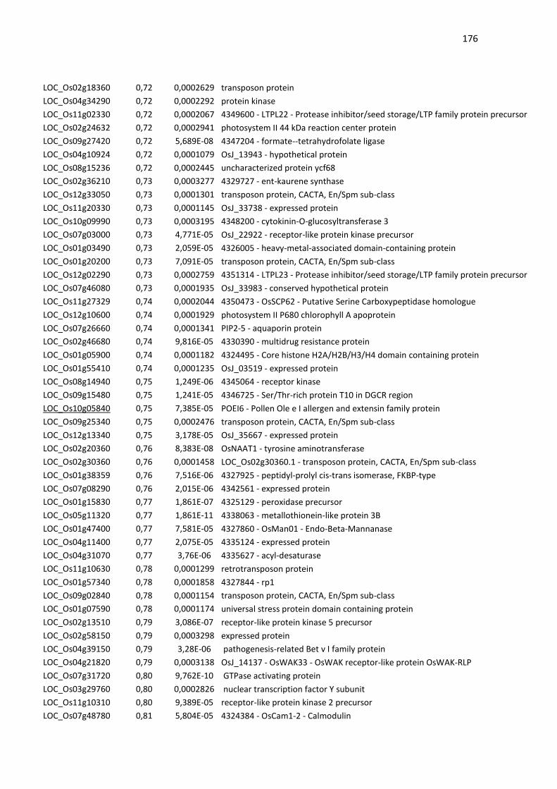
176
LOC_Os02g18360 0,72 0,0002629 transposon protein
LOC_Os04g34290 0,72 0,0002292 protein kinase
LOC_Os11g02330 0,72 0,0002067 4349600 - LTPL22 - Protease inhibitor/seed storage/LTP family protein precursor
LOC_Os02g24632 0,72 0,0002941 photosystem II 44 kDa reaction center protein
LOC_Os09g27420 0,72 5,689E-08 4347204 - formate--tetrahydrofolate ligase
LOC_Os04g10924 0,72 0,0001079 OsJ_13943 - hypothetical protein
LOC_Os08g15236 0,72 0,0002445 uncharacterized protein ycf68
LOC_Os02g36210 0,73 0,0003277 4329727 - ent-kaurene synthase
LOC_Os12g33050 0,73 0,0001301 transposon protein, CACTA, En/Spm sub-class
LOC_Os11g20330 0,73 0,0001145 OsJ_33738 - expressed protein
LOC_Os10g09990 0,73 0,0003195 4348200 - cytokinin-O-glucosyltransferase 3
LOC_Os07g03000 0,73 4,771E-05 OsJ_22922 - receptor-like protein kinase precursor
LOC_Os01g03490 0,73 2,059E-05 4326005 - heavy-metal-associated domain-containing protein
LOC_Os01g20200 0,73 7,091E-05 transposon protein, CACTA, En/Spm sub-class
LOC_Os12g02290 0,73 0,0002759 4351314 - LTPL23 - Protease inhibitor/seed storage/LTP family protein precursor
LOC_Os07g46080 0,73 0,0001935 OsJ_33983 - conserved hypothetical protein
LOC_Os11g27329 0,74 0,0002044 4350473 - OsSCP62 - Putative Serine Carboxypeptidase homologue
LOC_Os12g10600 0,74 0,0001929 photosystem II P680 chlorophyll A apoprotein
LOC_Os07g26660 0,74 0,0001341 PIP2-5 - aquaporin protein
LOC_Os02g46680 0,74 9,816E-05 4330390 - multidrug resistance protein
LOC_Os01g05900 0,74 0,0001182 4324495 - Core histone H2A/H2B/H3/H4 domain containing protein
LOC_Os01g55410 0,74 0,0001235 OsJ_03519 - expressed protein
LOC_Os08g14940 0,75 1,249E-06 4345064 - receptor kinase
LOC_Os09g15480 0,75 1,241E-05 4346725 - Ser/Thr-rich protein T10 in DGCR region
LOC_Os10g05840 0,75 7,385E-05 POEI6 - Pollen Ole e I allergen and extensin family protein
LOC_Os09g25340 0,75 0,0002476 transposon protein, CACTA, En/Spm sub-class
LOC_Os12g13340 0,75 3,178E-05 OsJ_35667 - expressed protein
LOC_Os02g20360 0,76 8,383E-08 OsNAAT1 - tyrosine aminotransferase
LOC_Os02g30360 0,76 0,0001458 LOC_Os02g30360.1 - transposon protein, CACTA, En/Spm sub-class
LOC_Os01g38359 0,76 7,516E-06 4327925 - peptidyl-prolyl cis-trans isomerase, FKBP-type
LOC_Os07g08290 0,76 2,015E-06 4342561 - expressed protein
LOC_Os01g15830 0,77 1,861E-07 4325129 - peroxidase precursor
LOC_Os05g11320 0,77 1,861E-11 4338063 - metallothionein-like protein 3B
LOC_Os01g47400 0,77 7,581E-05 4327860 - OsMan01 - Endo-Beta-Mannanase
LOC_Os04g11400 0,77 2,075E-05 4335124 - expressed protein
LOC_Os04g31070 0,77 3,76E-06 4335627 - acyl-desaturase
LOC_Os11g10630 0,78 0,0001299 retrotransposon protein
LOC_Os01g57340 0,78 0,0001858 4327844 - rp1
LOC_Os09g02840 0,78 0,0001154 transposon protein, CACTA, En/Spm sub-class
LOC_Os01g07590 0,78 0,0001174 universal stress protein domain containing protein
LOC_Os02g13510 0,79 3,086E-07 receptor-like protein kinase 5 precursor
LOC_Os02g58150 0,79 0,0003298 expressed protein
LOC_Os04g39150 0,79 3,28E-06 pathogenesis-related Bet v I family protein
LOC_Os04g21820 0,79 0,0003138 OsJ_14137 - OsWAK33 - OsWAK receptor-like protein OsWAK-RLP
LOC_Os07g31720 0,80 9,762E-10 GTPase activating protein
LOC_Os03g29760 0,80 0,0002826 nuclear transcription factor Y subunit
LOC_Os11g10310 0,80 9,389E-05 receptor-like protein kinase 2 precursor
LOC_Os07g48780 0,81 5,804E-05 4324384 - OsCam1-2 - Calmodulin
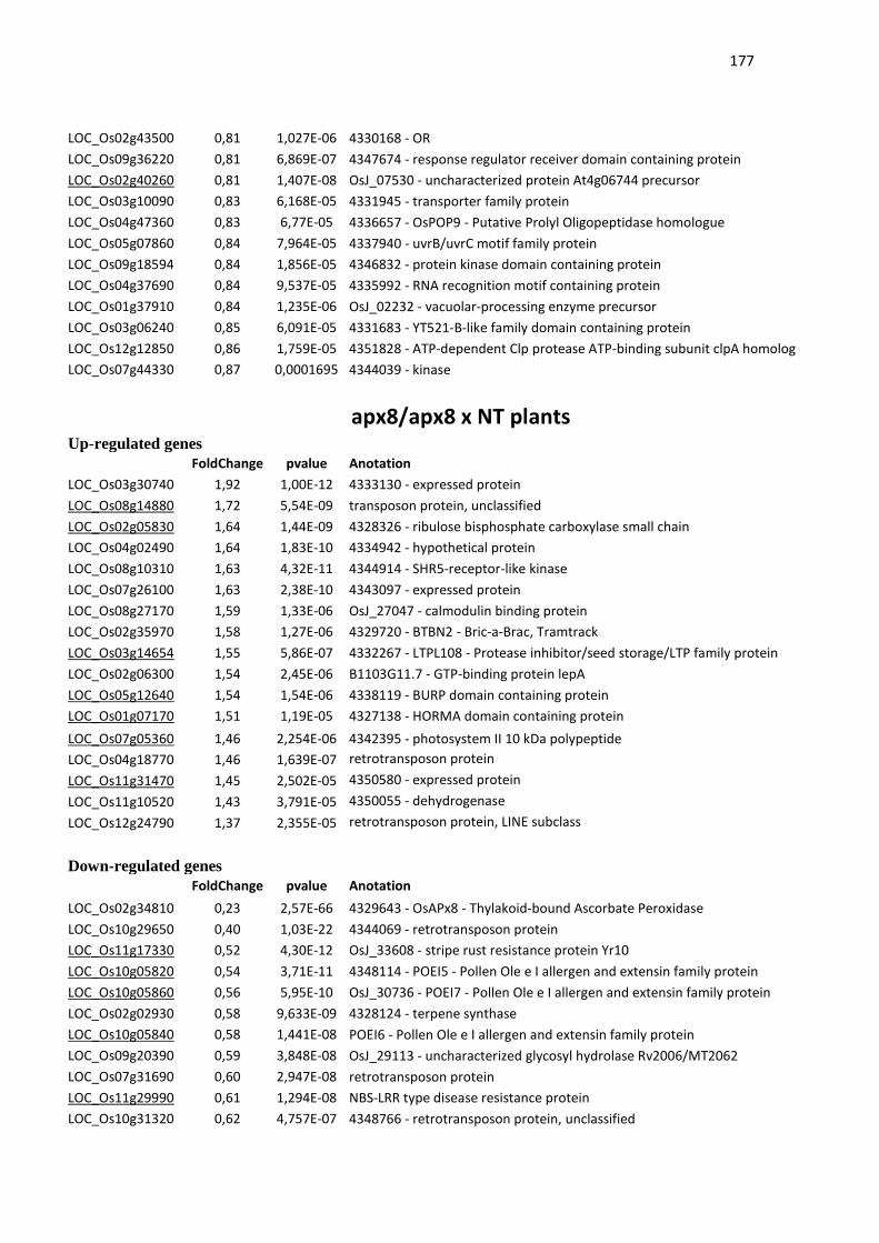
177
LOC_Os02g43500 0,81 1,027E-06 4330168 - OR
LOC_Os09g36220 0,81 6,869E-07 4347674 - response regulator receiver domain containing protein
LOC_Os02g40260 0,81 1,407E-08 OsJ_07530 - uncharacterized protein At4g06744 precursor
LOC_Os03g10090 0,83 6,168E-05 4331945 - transporter family protein
LOC_Os04g47360 0,83 6,77E-05 4336657 - OsPOP9 - Putative Prolyl Oligopeptidase homologue
LOC_Os05g07860 0,84 7,964E-05 4337940 - uvrB/uvrC motif family protein
LOC_Os09g18594 0,84 1,856E-05 4346832 - protein kinase domain containing protein
LOC_Os04g37690 0,84 9,537E-05 4335992 - RNA recognition motif containing protein
LOC_Os01g37910 0,84 1,235E-06 OsJ_02232 - vacuolar-processing enzyme precursor
LOC_Os03g06240 0,85 6,091E-05 4331683 - YT521-B-like family domain containing protein
LOC_Os12g12850 0,86 1,759E-05 4351828 - ATP-dependent Clp protease ATP-binding subunit clpA homolog
LOC_Os07g44330 0,87 0,0001695 4344039 - kinase
apx8/apx8 x NT plants Up-regulated genes
FoldChange pvalue Anotation
LOC_Os03g30740 1,92 1,00E-12 4333130 - expressed protein
LOC_Os08g14880 1,72 5,54E-09 transposon protein, unclassified
LOC_Os02g05830 1,64 1,44E-09 4328326 - ribulose bisphosphate carboxylase small chain
LOC_Os04g02490 1,64 1,83E-10 4334942 - hypothetical protein
LOC_Os08g10310 1,63 4,32E-11 4344914 - SHR5-receptor-like kinase
LOC_Os07g26100 1,63 2,38E-10 4343097 - expressed protein
LOC_Os08g27170 1,59 1,33E-06 OsJ_27047 - calmodulin binding protein
LOC_Os02g35970 1,58 1,27E-06 4329720 - BTBN2 - Bric-a-Brac, Tramtrack
LOC_Os03g14654 1,55 5,86E-07 4332267 - LTPL108 - Protease inhibitor/seed storage/LTP family protein
LOC_Os02g06300 1,54 2,45E-06 B1103G11.7 - GTP-binding protein lepA
LOC_Os05g12640 1,54 1,54E-06 4338119 - BURP domain containing protein
LOC_Os01g07170 1,51 1,19E-05 4327138 - HORMA domain containing protein
LOC_Os07g05360 1,46 2,254E-06 4342395 - photosystem II 10 kDa polypeptide
LOC_Os04g18770 1,46 1,639E-07 retrotransposon protein
LOC_Os11g31470 1,45 2,502E-05 4350580 - expressed protein
LOC_Os11g10520 1,43 3,791E-05 4350055 - dehydrogenase
LOC_Os12g24790 1,37 2,355E-05 retrotransposon protein, LINE subclass
Down-regulated genes
FoldChange pvalue Anotation
LOC_Os02g34810 0,23 2,57E-66 4329643 - OsAPx8 - Thylakoid-bound Ascorbate Peroxidase
LOC_Os10g29650 0,40 1,03E-22 4344069 - retrotransposon protein
LOC_Os11g17330 0,52 4,30E-12 OsJ_33608 - stripe rust resistance protein Yr10
LOC_Os10g05820 0,54 3,71E-11 4348114 - POEI5 - Pollen Ole e I allergen and extensin family protein
LOC_Os10g05860 0,56 5,95E-10 OsJ_30736 - POEI7 - Pollen Ole e I allergen and extensin family protein
LOC_Os02g02930 0,58 9,633E-09 4328124 - terpene synthase
LOC_Os10g05840 0,58 1,441E-08 POEI6 - Pollen Ole e I allergen and extensin family protein
LOC_Os09g20390 0,59 3,848E-08 OsJ_29113 - uncharacterized glycosyl hydrolase Rv2006/MT2062
LOC_Os07g31690 0,60 2,947E-08 retrotransposon protein
LOC_Os11g29990 0,61 1,294E-08 NBS-LRR type disease resistance protein
LOC_Os10g31320 0,62 4,757E-07 4348766 - retrotransposon protein, unclassified
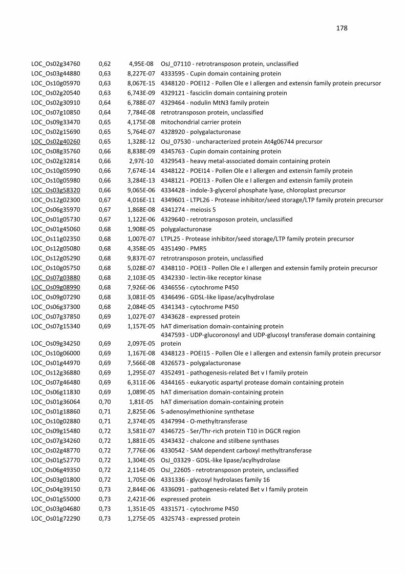
178
LOC_Os02g34760 0,62 4,95E-08 OsJ_07110 - retrotransposon protein, unclassified
LOC_Os03g44880 0,63 8,227E-07 4333595 - Cupin domain containing protein
LOC_Os10g05970 0,63 8,067E-15 4348120 - POEI12 - Pollen Ole e I allergen and extensin family protein precursor
LOC_Os02g20540 0,63 6,743E-09 4329121 - fasciclin domain containing protein
LOC_Os02g30910 0,64 6,788E-07 4329464 - nodulin MtN3 family protein
LOC_Os07g10850 0,64 7,784E-08 retrotransposon protein, unclassified
LOC_Os09g33470 0,65 4,175E-08 mitochondrial carrier protein
LOC_Os02g15690 0,65 5,764E-07 4328920 - polygalacturonase
LOC_Os02g40260 0,65 1,328E-12 OsJ_07530 - uncharacterized protein At4g06744 precursor
LOC_Os08g35760 0,66 8,838E-09 4345763 - Cupin domain containing protein
LOC_Os02g32814 0,66 2,97E-10 4329543 - heavy metal-associated domain containing protein
LOC_Os10g05990 0,66 7,674E-14 4348122 - POEI14 - Pollen Ole e I allergen and extensin family protein
LOC_Os10g05980 0,66 3,284E-13 4348121 - POEI13 - Pollen Ole e I allergen and extensin family protein
LOC_Os03g58320 0,66 9,065E-06 4334428 - indole-3-glycerol phosphate lyase, chloroplast precursor
LOC_Os12g02300 0,67 4,016E-11 4349601 - LTPL26 - Protease inhibitor/seed storage/LTP family protein precursor
LOC_Os06g35970 0,67 1,868E-08 4341274 - meiosis 5
LOC_Os01g05730 0,67 1,122E-06 4329640 - retrotransposon protein, unclassified
LOC_Os01g45060 0,68 1,908E-05 polygalacturonase
LOC_Os11g02350 0,68 1,007E-07 LTPL25 - Protease inhibitor/seed storage/LTP family protein precursor
LOC_Os12g05080 0,68 4,358E-05 4351490 - PMR5
LOC_Os12g05290 0,68 9,837E-07 retrotransposon protein, unclassified
LOC_Os10g05750 0,68 5,028E-07 4348110 - POEI3 - Pollen Ole e I allergen and extensin family protein precursor
LOC_Os07g03880 0,68 2,103E-05 4342330 - lectin-like receptor kinase
LOC_Os09g08990 0,68 7,926E-06 4346556 - cytochrome P450
LOC_Os09g07290 0,68 3,081E-05 4346496 - GDSL-like lipase/acylhydrolase
LOC_Os06g37300 0,68 2,084E-05 4341343 - cytochrome P450
LOC_Os07g37850 0,69 1,027E-07 4343628 - expressed protein
LOC_Os07g15340 0,69 1,157E-05 hAT dimerisation domain-containing protein
LOC_Os09g34250 0,69 2,097E-05
4347593 - UDP-glucoronosyl and UDP-glucosyl transferase domain containing
protein
LOC_Os10g06000 0,69 1,167E-08 4348123 - POEI15 - Pollen Ole e I allergen and extensin family protein precursor
LOC_Os01g44970 0,69 7,566E-08 4326573 - polygalacturonase
LOC_Os12g36880 0,69 1,295E-07 4352491 - pathogenesis-related Bet v I family protein
LOC_Os07g46480 0,69 6,311E-06 4344165 - eukaryotic aspartyl protease domain containing protein
LOC_Os06g11830 0,69 1,089E-05 hAT dimerisation domain-containing protein
LOC_Os01g36064 0,70 1,81E-05 hAT dimerisation domain-containing protein
LOC_Os01g18860 0,71 2,825E-06 S-adenosylmethionine synthetase
LOC_Os10g02880 0,71 2,374E-05 4347994 - O-methyltransferase
LOC_Os09g15480 0,72 3,581E-07 4346725 - Ser/Thr-rich protein T10 in DGCR region
LOC_Os07g34260 0,72 1,881E-05 4343432 - chalcone and stilbene synthases
LOC_Os02g48770 0,72 7,776E-06 4330542 - SAM dependent carboxyl methyltransferase
LOC_Os01g52770 0,72 1,304E-05 OsJ_03329 - GDSL-like lipase/acylhydrolase
LOC_Os06g49350 0,72 2,114E-05 OsJ_22605 - retrotransposon protein, unclassified
LOC_Os03g01800 0,72 1,705E-06 4331336 - glycosyl hydrolases family 16
LOC_Os04g39150 0,73 2,844E-06 4336091 - pathogenesis-related Bet v I family protein
LOC_Os01g55000 0,73 2,421E-06 expressed protein
LOC_Os03g04680 0,73 1,351E-05 4331571 - cytochrome P450
LOC_Os01g72290 0,73 1,275E-05 4325743 - expressed protein
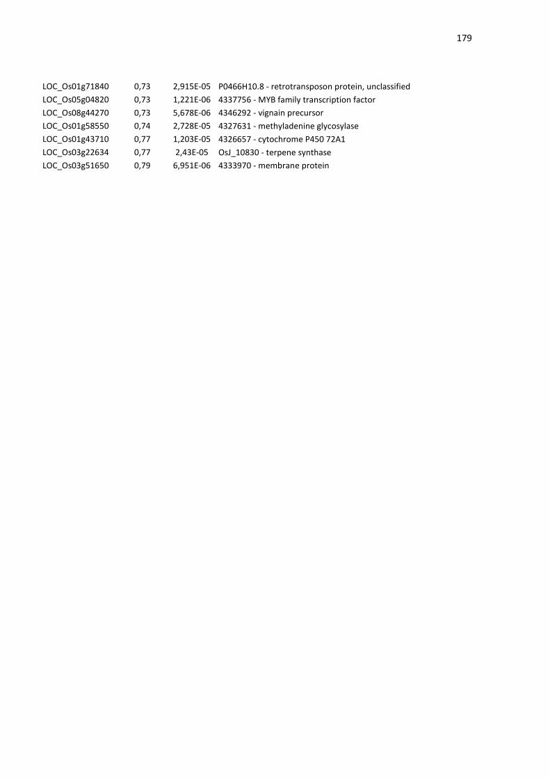
179
LOC_Os01g71840 0,73 2,915E-05 P0466H10.8 - retrotransposon protein, unclassified
LOC_Os05g04820 0,73 1,221E-06 4337756 - MYB family transcription factor
LOC_Os08g44270 0,73 5,678E-06 4346292 - vignain precursor
LOC_Os01g58550 0,74 2,728E-05 4327631 - methyladenine glycosylase
LOC_Os01g43710 0,77 1,203E-05 4326657 - cytochrome P450 72A1
LOC_Os03g22634 0,77 2,43E-05 OsJ_10830 - terpene synthase
LOC_Os03g51650 0,79 6,951E-06 4333970 - membrane protein

180
3.5 CAPÍTULO 5
IDENTIFICAÇÃO DE FATORES DE TRANSCRIÇÂO QUE SE LIGUEM À
REGIAO PROMOTORA DE OsAPX8 E POTENCILAMENTE PODEM
REGULAR A EXPRESSÃO GÊNICA
Conforme demonstrado nos capítulos anteriores, o gene
OsAPX8 desempenha um importante papel no controle da abertura
estomática e na tolerância de plantas ao estresse de seca. Desta
forma, a identificação de fatores de transcrição que regulam a
expressão de OsAPX8 pode fornecer dados importantes a respeito de
mecanismos regulatórios mediadores de tais fenômenos. Neste
capítulo, que corresponde ao trabalho desenvolvido no período de
doutorado sanduiche realizado no Instituto de Tecnologia Química
Biológica da Universidade Nova de Lisboa (Portugal), é descrito o uso
do sistema de monohíbrido em leveduras visando a identificaçao de
fatores de transcrição capazes de se ligar ao promotor de OsAPX8.
Entre os fatores de transcrição identificados, verificamos potenciais
ativadores da expressão gênica, que são fortemente regulados pelo
estresse hídrico, e que, portanto, podem contribuir com o
entendimento dos mecanismos que envolvem a regulação de
OsAPX8 em plantas submetidas ao estresse. Desta forma, os dados
apresentados neste capítulo, apesar de preliminares, podem permitir
a caracterização de novas vias de transdução de sinais que medeiam
a resposta a seca, via modulação da expressão de OsAPX8.

181
INTRODUÇÃO
A seca é um dos principais estresses abióticos que afetam o
desenvolvimento vegetal e limitam a produção de sementes. O controle da perda
de água pelas plantas se dá principalmente pela atividade dos estômatos, que
são poros localizados na epiderme das folhas e que controlam a captação de
CO2 para os processos fotossintéticos e a perda de água durante a transpiração
(SCHROEDER et al., 2001; HETHERINGTON & WOODWARD, 2003). Durante
o estresse de seca, o fechamento dos estômatos é mediado principalmente por
ácido abscísico (ABA) em um mecanismo que permite a adaptação à falta de
água (SCHROEDER et al., 2001). Muitos trabalhos já demonstraram que
diferentes espécies reativas de oxigênino (ROS, do inglês, reactive oxygen
species) incluindo peróxido de hidrogênio (H2O2), gerados em condições de
estresse, podem atuar como segundo mensageiros na sinalização por ABA
(MCAINSH et al., 1996; PEI et al., 2000; KOHLER et al., 2003; BRIGHT et al.,
2006), sendo capazes de promover o fechamento estomático (MCAINSH et al.,
1996; ZHANG et al., 2001). No entanto, os genes que regulam o aumento de
H2O2 em resposta ao estresse ainda não foram totalmente identificados, e os
mecanismos de modulação dos níveis de H2O2, especialmente em estômatos, é
em grande parte desconhecido.
Em arroz (Oryza sativa), a APX é codificada por uma família gênica
composta de oito genes, denominados de OsAPX1 a OsAPX8 e as suas
diferentes isoformas apresentam padrões distintos de expressão, em
decorrência do estímulo ambiental. Ao contrário das demais formas de APX, em
situações de estresses abióticos, como alta luminosidase, frio e seca, a isoforma
cloroplastídica OsAPX8 possui sua expressão modulada negativamente
(MENEZES-BENAVENTE et al., 2004; TEIXEIRA et al., 2004; 2005; ROSA et
al., 2010; BONIFACIO et al., 2011, CAVERZAN et al., 2014). Além disso,
conforme demonstrado nos capítulos anteriores, a análise de plantas de arroz
silenciadas por RNAi nas isoformas cloroplastídicas de APX (OsAPX7 e
OsAPX8), demonstram que o silenciamento de OsAPX8 induz mudanças na
182
geração de ROS e no fechamento estomático. Estes dados indicam uma
possível participação de OsAPX8 nos mecanismos de fechamento estomático
em resposta ao déficit hídrico.
Em arroz, um fator de transcrição do tipo “zinc-finger”, DST (do inglês,
drought and salt tolerance), foi descrito como um regulador da abertura
estomática em resposta a seca e salinidade (HUANG et al., 2009). DST contribui
para a modulação da abertura estomática via regulação de genes envolvidos na
homeostase de ROS e sua expressão é modulada negativamente por ABA e por
estresse de seca, induzindo assim um aumento da geração de H2O2 (LI et al.,
2013). Análises in silico revelaram a presença de pelo menos três possíveis
sítios de ligação ao DST na região promotora de OsAPX8. Esses sítios de
ligação foram denominados DBS (do inglês, DST bindiing site). Esses dados
sugerem que o fator de transcrição DST possa regular a expressão de OsAPX8,
que participaria dos mecanismos de resposta ao estresse de seca através da
modulação dos níveis de ROS no cloroplasto.
183
OBJETIVOS
Dada a modulação da expressão de OsAPX8 em resposta a
diferentes estresses, e sua possível participação nos mecanismos de
fechamento estomático, neste trabalho se objetivou a identificação de fatores de
transcrição capazes de regular a expressão de OsAPX8. Duas abordagens
foram utilizadas:
Avaliar a capacidade do fator de transcrição DST em se ligar ao promotor
de OsAPX8;
Identificar outros fatores de transcrição capazes de se ligar ao promotor
de OsAPX8;
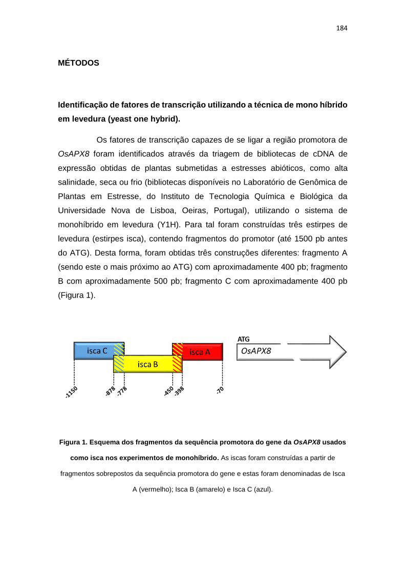
184
MÉTODOS
Identificação de fatores de transcrição utilizando a técnica de mono híbrido
em levedura (yeast one hybrid).
Os fatores de transcrição capazes de se ligar a região promotora de
OsAPX8 foram identificados através da triagem de bibliotecas de cDNA de
expressão obtidas de plantas submetidas a estresses abióticos, como alta
salinidade, seca ou frio (bibliotecas disponíveis no Laboratório de Genômica de
Plantas em Estresse, do Instituto de Tecnologia Química e Biológica da
Universidade Nova de Lisboa, Oeiras, Portugal), utilizando o sistema de
monohíbrido em levedura (Y1H). Para tal foram construídas três estirpes de
levedura (estirpes isca), contendo fragmentos do promotor (até 1500 pb antes
do ATG). Desta forma, foram obtidas três construções diferentes: fragmento A
(sendo este o mais próximo ao ATG) com aproximadamente 400 pb; fragmento
B com aproximadamente 500 pb; fragmento C com aproximadamente 400 pb
(Figura 1).
Figura 1. Esquema dos fragmentos da sequência promotora do gene da OsAPX8 usados
como isca nos experimentos de monohíbrido. As iscas foram construídas a partir de
fragmentos sobrepostos da sequência promotora do gene e estas foram denominadas de Isca
A (vermelho); Isca B (amarelo) e Isca C (azul).

185
Cada fragmento (denominados de isca A, isca B e isca C) foi clonado
em vetor pHISi e integrado no genoma da cepa de levedura Y187 (Clontech).
Cada linhagem foi repetidamente re-transformada com duas bibliotecas de cDNA
(biblioteca de plantas submetidas a estresse de seca e biblioteca de plantas
submetidas a estresse de frio; FIGUEIREDO et al., 2012), e crescida em meio
seletivo com a concentração de 3AT (inibidor da síntese endógenase de
histidina) determinada previamente (isca A: SD-His + 40mM 3AT, isca B: SD-His
+ 20mM 3AT e isca C: SD-His + 20mM 3AT). Dessa forma, as leveduras que
não estiverem expressando o gene HIS (que está sob o constrole dos fragmentos
do promotor de OsAPX8) não será capaz de crescer na ausência de histidina
exógena, a não ser que através do “screening” com a biblioteca a levedura
receba um clone contendo um cDNA que codifique uma proteína capaz de se
ligar aos fragmentos do promotor. As sequencias de cDNA da biblioteca estão
clonadas em vetor pACTII, que codifica um domínio de ativação em fusão com
a proteína da biblioteca, capaz de induzir a expressão de HIS caso a proteína
codificada pelo cDNA selecionado por meio do “screening” se ligue aos
fragmentos do promotor de OsAPX8. Para cada fragmento do promotor foram
triados mais de um milhão de clones de cada biblioteca. As colônias de levedura
que conseguiram crescer no meio seletivo foram analisadas por PCR para
verificação quanto à presença do plasmídeo da biblioteca utilizada e os produtos
de PCR foram sequenciados e analisados por BLAST para a identificação do
cDNA codificante. A validação destas interações foi realizada por re-
transformação com o plasmídeo purificado.
Avaliação da interação entre o fator de transcrição DST e o promotor de
OsAPX8 utilizando a técnica de Yeast One Hybrid
As linhagens de leveduras contendo as regiões do promotor de
OsAPX8 foram co-transformadas com o vetor pDEST 22 contendo o cDNA
codificante do fator de transcrição DST clonado em fusão com um domínio de
ativação da transcrição da proteína GAL4. As leveduras foram selecionadas em
meio SD sem histidina, objetivando analisar a capacidade do fator de transcrição
DST em se ligar ao promotor de OsAPX8.

186
RESULTADOS E DISCUSSÃO
Avaliação da interação entre o fator de transcrição DST e o promotor de
OsAPX8
Após a obtenção das linhagens de leveduras contendo os fragmentos
do promotor de OsAPX8, estas foram transformadas com o vetor pDEST22
contendo a sequencia codificante do fator de transcrição DST. Os resultados
demonstraram uma forte interação entre o DST e a isca A do promotor de
OsAPX8 (Figura 1). Não foram identificadas, no entanto, interações entre DST e
as iscas B e C.
Figura 1. Avaliação da interação entre o fator de transcrição DST e os fragmentos do
promotor de OsAPX8 por monohíbrido em levedura. Foram utilizadas diferentes
concentrações de 3-AT, afim de se determinar a concentração mínima capaz de inibir a
sintese endógena de histida, proveniente de auto-ativação.
187
Dados da literatura, indicam que o fator de transcrição DST contribui
para a regulação da abertura estomática via regulação de genes envolvidos na
homeostase redox, como peroxidases (precursor 24, precursor 12), glutatione S-
transferase (OsGSTU2) e citocromos da família P450 (71D10, 94A2). Sob
condições de estresses de seca e salinidade a expressão de DST é fortemente
inibida, o que, indiretamente, levaria ao acúmulo de ROS (HUANG et al., 2009).
De fato, o gene de OsAPX8 também é reprimido durante o estresse de seca e o
silenciamento deste gene leva ao aumento do acúmulo de ROS e do fechamento
estomático, conforme demonstrado no capítulo 4. Portanto, a ligação desse fator
de transcrição no promotor do gene OsAPX8 e o padrão coincidente da
expressão de ambos em resposta a seca indicam que os mesmos participam
dos mecanismos que culminam com o fechamento dos estômatos na resposta
ao estresse por falta de água.
Identificação de fatores de transcrição capazes de se ligar ao promotor de
OsAPX8
O “screening” da biblioteca de cDNA obtida de plantas submetidas a
estresse de seca não resultou na identificação de fatores de transcrição com
capacidade de se ligar à região promotora de OsAPX8. Mesmo após a triagem
de mais de um milhão de clones para cada fragmento isca, nunhum clone
positivo foi identificado. Devido a isso, novos experimentos foram realizados
visando o “screening” de biblioteca de cDNA de plantas submetidas a frio, tendo
sido triados pelo menos um milhão de clones para cada fragmento isca do
promotor de OsAPX8. Utilizando esta segunda biblioteca foram identificados três
fatores de transcrição com capacidade de se ligar ao promotor de OsAPX8. Os
fatores de transcrição identificados estão listados da tabela 1. A confirmação
destas interações foi realizada através da re-transformação das estirpes isca
com o plasmídeo purificado (vetor pACTII) (Figura 2).
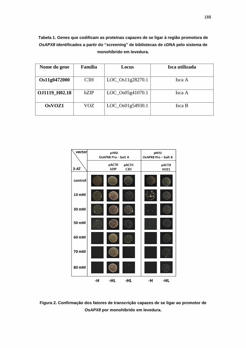
188
Tabela 1. Genes que codificam as proteínas capazes de se ligar à região promotora de
OsAPX8 identificados a partir do “screening” de bibliotecas de cDNA pelo sistema de
monohíbrido em levedura.
Nome do gene Família Locus Isca utilizada
Os11g0472000 C3H LOC_Os11g28270.1 Isca A
OJ1119_H02.18 bZIP LOC_Os05g41070.1 Isca A
OsVOZ1 VOZ LOC_Os01g54930.1 Isca B
Figura 2. Confirmação dos fatores de transcrição capazes de se ligar ao promotor de
OsAPX8 por monohíbrido em levedura.
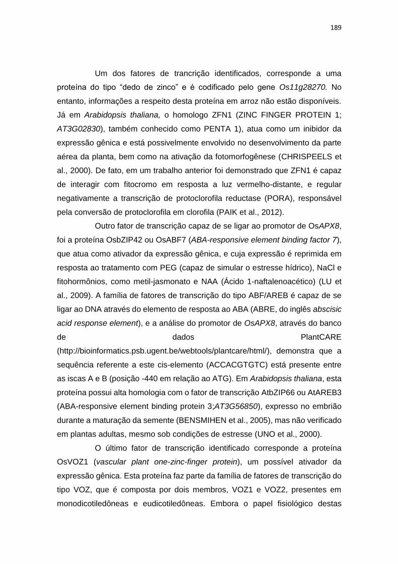
189
Um dos fatores de trancrição identificados, corresponde a uma
proteína do tipo “dedo de zinco” e é codificado pelo gene Os11g28270. No
entanto, informações a respeito desta proteína em arroz não estão disponíveis.
Já em Arabidopsis thaliana, o homologo ZFN1 (ZINC FINGER PROTEIN 1;
AT3G02830), também conhecido como PENTA 1), atua como um inibidor da
expressão gênica e está possivelmente envolvido no desenvolvimento da parte
aérea da planta, bem como na ativação da fotomorfogênese (CHRISPEELS et
al., 2000). De fato, em um trabalho anterior foi demonstrado que ZFN1 é capaz
de interagir com fitocromo em resposta a luz vermelho-distante, e regular
negativamente a transcrição de protoclorofila reductase (PORA), responsável
pela conversão de protoclorofila em clorofila (PAIK et al., 2012).
Outro fator de transcrição capaz de se ligar ao promotor de OsAPX8,
foi a proteína OsbZIP42 ou OsABF7 (ABA-responsive element binding factor 7),
que atua como ativador da expressão gênica, e cuja expressão é reprimida em
resposta ao tratamento com PEG (capaz de simular o estresse hídrico), NaCl e
fitohormônios, como metil-jasmonato e NAA (Ácido 1-naftalenoacético) (LU et
al., 2009). A família de fatores de transcrição do tipo ABF/AREB é capaz de se
ligar ao DNA através do elemento de resposta ao ABA (ABRE, do inglês abscisic
acid response element), e a análise do promotor de OsAPX8, através do banco
de dados PlantCARE
(http://bioinformatics.psb.ugent.be/webtools/plantcare/html/), demonstra que a
sequência referente a este cis-elemento (ACCACGTGTC) está presente entre
as iscas A e B (posição -440 em relação ao ATG). Em Arabidopsis thaliana, esta
proteína possui alta homologia com o fator de transcrição AtbZIP66 ou AtAREB3
(ABA-responsive element binding protein 3;AT3G56850), expresso no embrião
durante a maturação da semente (BENSMIHEN et al., 2005), mas não verificado
em plantas adultas, mesmo sob condições de estresse (UNO et al., 2000).
O último fator de transcrição identificado corresponde a proteína
OsVOZ1 (vascular plant one-zinc-finger protein), um possível ativador da
expressão gênica. Esta proteína faz parte da família de fatores de transcrição do
tipo VOZ, que é composta por dois membros, VOZ1 e VOZ2, presentes em
monodicotiledôneas e eudicotiledôneas. Embora o papel fisiológico destas

190
proteínas em arroz ainda não tenha sido estudado, em Arabidopsis thaliana, os
genes VOZ respondem a diferentes estresses abióticos, sendo fortemente
reprimidos em plantas submetidas a seca e a baixas temperaturas. Além disso,
plantas duplo mutantes para AtVOZ1 e AtVOZ2 (voz1/voz2) apresentaram um
aumento da tolerância a esses estresses. Por outro lado, a resistência destas
plantas, a patógenos como o fungo Colletotrichum higginsianum e a bactéria
Pseudomonas syringae foi fortemente reduzida (NAKAI et al., 2013).
Posteriormente, foi demonstrando que linhagens que superexpressam AtVOZ1
e AtVOZ2 apresentavam uma redução da tolerância a estresses abióticos (seca
e frio), mas em relação a patógenos, elas apresentavam um aumento da
resistência (NAKAI et al., 2013a). Estes dados, portanto, indicam que os fatores
de transcrição do tipo VOZ, possuem um importante papel na resposta a
estresses biótico e abióticos e poderiam regular as respostas mediadas por
OsAPX8.

191
CONCLUSÕES E PERSPECTIVAS
Através de experimentos de monohíbrido em levedura, demonstramos
que o fator de transcrição OsDST é capaz de se ligar ao promotor de OsAPX8.
Além disso identificamos por “screening” de bibliotecas de cDNA, outros três
fatores de transcrição capazes de se ligar ao promotor de OsAPX8:
LOC_Os11g28270, LOC_Os05g41070 (OsABF7) e LOC_Os01g54930
(OsVOZ1). Com base nos dados disponíveis na literatura concluímos que as
proteínas OsDST, OsABF7 e OsVOZ1, são fortes candidatos a serem, de fato,
membros de vias de transdução de sinais mediadas por OsAPX8. Em geral,
todos estes fatores de transcrição são ativadores da expressão gênica, e são
fortemente reprimidos durante o estresse de seca. Se de fato, tais proteínas
forem capazes de ativar a expressão de OsAPX8, a repressão da expressão de
cada fator de transcrição, em condições de estresse, poderia contribuir para a
redução da expressão de OsAPX8, colaborando para o aumento da geração de
ROS e do fechamento estomático nestas condições (Figura 3).
Como perspectiva deste trabalho, pretendemos avaliar, por ensaio de
transativação em protoplastos de arroz, a capacidade dos fatores de transcrição
identificados em ativar ou inibir a expressão gênica sob o controle do promotor
de OsAPX8. Além disso, o próprio ensaio de transativação pode ser utilizado
para confirmar as interaçõeso DNA-proteína, uma vez que, nos experimentos
que utilizam o sistema de protoplastos de plantas, além das proteínas estarem
completas, elas estarão em sua conformação correta e com as modificações
pós-traducionais apropriadas, uma vez que estarão sendo expressas em células
de arroz, e não em leveduras. Além disso, pretendemos avaliar a expressão dos
fatores de transcrição identificados na resposta de plantas de arroz a diferentes
estresses abióticos. Estes ensaios permitirão a caracterização de rotas de
sinalização que modulem a expressão de OsAPX8 em resposta ao estresse.
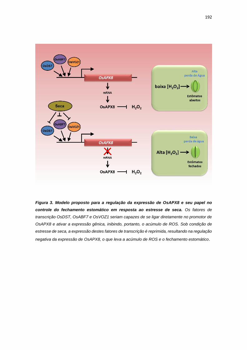
192
Figura 3. Modelo proposto para a regulação da expressão de OsAPX8 e seu papel no
controle do fechamento estomático em resposta ao estresse de seca. Os fatores de
transcrição OsDST, OsABF7 e OsVOZ1 seriam capazes de se ligar diretamente no promotor de
OsAPX8 e ativar a expressão gênica, inibindo, portanto, o acúmulo de ROS. Sob condição de
estresse de seca, a expressão destes fatores de transcrição é reprimida, resultando na regulação
negativa da expressão de OsAPX8, o que leva a acúmulo de ROS e o fechamento estomático.
193
REFERÊNCIAS
BENSMIHEN S, GIRAUDAT J, PARCY F. (2005). Characterization of three
homologous basic leucine zipper transcription factors (bZIP) of the ABI5 family
during Arabidopsis thaliana embryo maturation. J Exp Bot. 56(412):597-603.
BONIFÁCIO A, MARTINS M, RIBEIRO C, FONTENELE A, CARVELHO F,
MARGIS-PINHEIRO M, SILVEIRA J. (2011). Role of peroxidases in the
compensation of cytosolic ascorbate peroxidase knockdown in rice plants under
abiotic stress. Plant, Cell and Environment 34(10):1705-22.
BRIGHT J, DESIKAN R, HANCOCK JT, WEIR IS, NEILL SJ. (2006). ABA
induced NO generation and stomatal closure in Arabidopsis are dependent on
H2O2 synthesis. Plant J 45: 113-22.
CAVERZAN A, BONIFACIO A, CARVALHO FE, ANDRADE CM, PASSAIA G,
SCHÜNEMANN M, MARASCHIN FDOS S, MARTINS MO, TEIXEIRA FK,
RAUBER R, MARGIS R, SILVEIRA JA, MARGIS-PINHEIRO M. (2014). The
knockdown of chloroplastic ascorbate peroxidases reveals its regulatory role in
the photosynthesis and protection under photo-oxidative stress in rice. Plant
Science 214:74-87.
CHRISPEELS HE, OETTINGER H, JANVIER N, TAGUE BW. (2000). AtZFP1,
encoding Arabidopsis thaliana C2H2 zinc-finger protein 1, is expressed
downstream of photomorphogenic activation. Plant Mol Biol.42(2):279-90.
FIGUEIREDO DD, BARROS PM, CORDEIRO AM, SERRA TS, LOURENÇO T,
CHANDER S, OLIVEIRA MM, SAIBO NJ. (2012). Seven zinc-finger transcription
factors are novel regulators of the stress responsive gene OsDREB1B. J Exp Bot.
63(10):3643-56.
HETHERINGTON AM, WOODWARD FI. (2003). The role of stomata in sensing
and driving environmental change. Nature 424: 901-08.

194
HUANG XY, CHAO DY, GAO JP, ZHU MZ, SHI M, LIN HX. (2009). A previously
unknown zinc finger protein, DST, regulates drought and salt tolerance in rice via
stomatal aperturecontrol. Genes Dev 23(15): 1805-17.
KOHLER B, HILLS A, BLATT MR. (2003). Control of guard cell ion channels by
hydrogen peroxide and abscisic acid indicates their action through alternate
signaling pathways. Plant Physiol 131: 385-88.
LI S, ZHAO B, YUAN D, DUAN M, QIAN Q, TANG L, WANG B, LIU X, ZHANG
J, WANG J, SUN J, LIU Z, FENG YQ, YUAN L, LI C. (2013). Rice zinc finger
protein DST enhances grain production through controlling Gn1a/OsCKX2
expression. Proc Natl Acad Sci 110(8): 3167-72.
LU G, GAO C, ZHENG X. HAN B. (2009). Identification of OsbZIP72 as a positive
regulator of ABA response and drought tolerance in rice. Planta 229:605-15.
MCAINSH MR, CLAYTON H, MANSFIELD TA, HETHERINGTON AM. (1996).
Changes in stomatal behavior and guard cell cytosolic free calcium in response
to oxidative stress. Plant Physiol 111: 1031–42.
MENEZES-BENAVENTE L, TEIXEIRA FK, KAMEI CL, MARGIS-PINHEIRO M.
(2004). Salt stress induces expression of genes encoding antioxidant enzymes
in seedlings of a Brazilianindicarice (Oryza sativa L.). Plant Sci 166: 323-31.
NAKAI Y, FUJIWARA S, KUBO Y, SATO MH. (2013a). Overexpression of
VOZ2confers biotic stress tolerance but decreases abiotic stress resistance in
Arabidopsis 8:3, e23358.
NAKAI Y, NAKAHIRA Y, SUMIDA H, TAKEBAYASHI K, NAGASAWA Y,
YAMASAKI K, AKIYAMA M, OHME-TAKAGI M, FUJIWARA S, SHIINA T,
MITSUDA N, FUKUSAKI E, KUBO Y, SATO MH. (2013). Vascular plant one-
zinc-finger protein 1/2 transcription factors regulate abiotic and biotic stress
responses in Arabidopsis. The Plant Journal 73: 761-775.
PAIK I, YANG S, CHOI G. (2012). Phytochrome regulates translation of mRNA
in the cytosol. PNAS 109(4):1335-40.
195
PEI ZM, MURATA Y, BENNING G, THOMINE S, KLUSENER B, ALLEN GJ,
GRILL E, SCHROEDER JI. (2000). Calcium channels activated by hydrogen
peroxide mediate abscisic acid signalling in guard cells. Nature 406: 731–34.
ROSA SB, CAVERZAN A, TEIXEIRA FK, LAZZAROTTO F, SILVEIRA JA,
FERREIRA-SILVA SL, ABREU-NETO J, MARGIS R, MARGIS-PINHEIRO M.
(2010). Cytosolic APx knockdown indicates an ambiguous redox response in rice.
Phytochemistry 71: 548-58.
SCHROEDER JI, ALLEN GJ, HUGOUVIEUX V, KWAK JM, WANER D. (2001).
Guard cell signal transduction. Annu Rev Plant Physiol Plant Mol Biol 52: 627–
58.
TEIXEIRA FK, MENEZES-BENAVENTE L, GALVÃO VC, MARGIS-PINHEIRO
M. (2005). Multigene families encode the major enzymes of antioxidant
metabolism in Eucalyptus grandis L. Genetics and Molecular Biology 28: 529-38;
TEIXEIRA FK, MENEZES-BENAVENTE L, MARGIS R, MARGIS-PINHEIRO M.
(2004). Analysis of the molecular evolutionary history of the ascorbate peroxidase
gene family: inferences from the rice genome. J Mol Evol 59: 761-70;
UNO Y, FURIHATA T, ABE H, YOSHIDA R, SHINOZAKI K, YAMAGUCHI-
SHINOZAKI K. (2000). Arabidopsis basic leucine zipper transcription factors
involved in an abscisic acid-dependent signal transduction pathway under
drought and high-salinity conditions. PNAS 97(21): 11632–11637.
ZHANG X, ZHANG L, DONG F, GAO J, GALBRAITH DW, SONG CP. (2001).
Hydrogen peroxide is involved in abscisic acid-induced stomatal closure in Vicia
faba. Plant Physiol 126: 1438–48.

196
4- DISCUSSÃO GERAL
As ROS são formadas como subproduto do metabolismo aeróbico em
diferentes organismos e atuam como moléculas sinalizadoras, ou mensageiros
secundários, em uma complexa rede de sinalização em diferentes organismos.
Desta forma, em plantas, as vias de produção de ROS, bem como os
mecanismos antioxidantes são fundamentais para o desenvolvimento vegetal, e
para a resposta a diferente estímulos ambientais.
Neste trabalho, identificamos uma nova e importante fonte de geração
de ROS em células vegetais: a enzima SDH, que corresponde ao complexo II da
respiração. Os resultados obtidos demonstram que a geração de ROS pela SDH
pode ser modulada por diferentes inibidores. Enquanto inibidores não-
competitivos, como ácido salicílico e óxido nítrico (NO), são capazes de induzir
a geração de ROS, os inibidores competitivos são capazes de previnir a geração
de ROS pela SDH. Este mecanismo se mostra importante em células vegetais,
pois diferentes moléculas endógenas podem atuar regulando a geração de ROS
pela SDH. Além disso, este mecanismo resulta na regulação da expressão de
genes relacionados com o desenvolvimento vegetal e com a tolerância a
diferentes estresses.
Se por um lado a manipulação da geração de ROS pela mitocôndria
promoveu alterações claras no padrão de desenvolvimento, alterações nas
respostas antioxidantes no cloroplasto afetaram em menor grau o crescimento
da planta. No entanto, impactos nos mecanismos de defesa foram verificados
em resposta às alterações em ambas organelas, de maneira similar. Assim,
nossos dados demonstram que as isoformas cloroplastídicas de APX também
estão envolvidas em mecanismos de tolerância ao estresse, indicando o
potencial para um papel na regulação dos níveis de ROS por enzimas
relacionadas com a produção e a eliminação dessas moléculas reativas. No
entanto, apesar desses pontos comuns, a complexidade das respostas obtidas
indica uma alta especificidade das respostas, que depende da localização
subcelular de cada um dos componentes dessa intricada rede, assim como o
197
nível de expressão de cada um deles. Por exemplo, enquanto o silenciamento
duplo de OsAPX7 e OsAPX8, bem como o silenciamento individual de OsAPX8,
resultou em plantas com um maior conteúdo de ROS e maior fechamento
estomático em codições ideais, o silenciamento individual de OsAPX7 não
resulta em alterações fisiológicas nas plantas silenciadas. No entanto, as plantas
silenciadas para OsAPX7 e as plantas duplamente silenciadas para ambos os
genes demonstram uma maior tolerância ao estresse de seca. Esses dados
demonstram que o aumento da tolerância à seca não é consequência somente
do aumento de ROS ou do fechamento estomático, uma vez que tais fenômenos
não são observados nas plantas silenciadas para OsAPX7.
O aumento da tolerância a seca também foi verificado em plantas
nocaute para OsAPX8 (apx8/apx8). Curiosamente, ao contrário das plantas
silenciadas, as plantas nocaute pra OsAPX8 não apresentaram mudanças
fisiológicas em condições ideias, e dados de RNAseq demonstram que o
silenciamento de OsAPX8 induz uma maior alteração na expressão gênica do
que o nocaute. Aparentemente o silenciamento de OsAPX8 simula uma
condição de estresse, onde a expressão de OsAPX8 seria reduzida
naturalmente. Desta forma, as plantas silenciadas para OsAPX8 apresentam um
fenótipo parecido ao de plantas submetidas a estresse, como um maior conteúdo
de ROS, maior fechamento estomático, crescimento mais lento, bem como uma
maior resposta na expressão gênica. Por outro lado, o nocaute de OsAPX8
estaria gerando compensações não verificadas nas plantas silenciadas, que
permitem as plantas nocaute apresentar um fenótipo mais parecido com plantas
NT, embora sejam mais tolerantes ao estresse de defict hídrico. No entanto, os
mecanismos capazes de induzir o aumento da tolerância dessas plantas ao
estresse de seca ainda são desconhecidos.
Além das avaliações de interações gênicas e efeitos de manipulações
genéticas, a análise de como esses componentes envoluíram nos diferentes
genomas pode trazer novos dados para o entendimento da função de cada um
deles nos mecanismos de defesa das plantas. Nesse sentido, o estudo da
história evolutiva da succinato desidrogenase, descrita no capítulo 2 indicou que
esta enzima é membro da superfamília de fumarato redutases, composta por
198
outras enzimas, como a fumarato redutase dependente de quinona, NADH ou
thiol, além da enzima L-aspartato redudase. Nossos dados demonstram que
todas essas enzimas se originaram de um acestral comum, e os processos de
endossimbiose da mitocôndria e do cloroplasto assumiram um papel central na
distribuição destas enzimas em organismos eucariotos.
Nesta tese, demonstramos que os diferentes mecanismos envolvidos
no balanço redox de células vegetais exercem um importante papel no
desenvolvimento vegetal e na reposta de plantas a diferentes estresses. Além
disso, identificamos diferentes fatores de transcrição capazes de se ligar ao
promotor de OsAPX8, o que indiretamente também poderia regular o balanço
redox celular. A avaliação da capacidade dessas proteínas em regular a
expressão gênica permitirá ainda a indentificação de novas vias de sinalização
envolvidas na resposta ao estresse. Embora ainda existam incontáveis aspectos
a serem explorados nesta área, o presente trabalho contribuiu para a
identificação de novas interações e, consequentemente de vias inéditas de
sinalização nas células vegetais, ampliando a visão do papel das ROS nos
mecanismos de respostas a estímulos ambientais geradores de estresse
oxidativo.
199
5- CONCLUSÕES GERAIS
- Succinato desidrogenase é um importante sítio de geração de ROS em células
vegetais e regula a expressão de genes envolvidos com o desenvolvimento
vegetal e a resposta ao estresse;
- A família de succinato:quinona oxiredutase é membro de uma superfamília de
fumarato redutases, que se originou apartir de um ancestral comum e está
envolvida em processos metabólicos chave ao longo da evolução das espécies;
- As isoformas cloroplastídicas de ascorbato peroxidase desempenham um
importante papel na homestase redox e no controle da abertura estomática e da
tolerância ao estresse hídrico em arroz;
- Os fatores de trancrição, OsDST, OsVOZ1, OsABF7 e LOC_Os01g54930 são
capazes de se ligar ao promotor de OsAPX8 e são potenciais reguladores da
expressão gênica;
- As ROS são essenciais no controle das respostas das plantas a diferentes
estresses.

200
6- REFERÊNCIAS
APEL K, HIRT H. (2004). Reactive oxygen species: metabolism, oxidative stress,
and signal transduction. Annu. Rev. Plant Biol. 55: 373–99.
ARAÚJO WL, NUNES-NESI A, OSORIO S, USADEL B, FUENTES D, NAGY
R, BALBO I, LEHMANN M, STUDART-WITKOWSKI C, TOHGE T, MARTINOIA
E, JORDANA X, DAMATTA FM, FERNIE AR. (2011). Antisense Inhibition of the
Iron-Sulphur Subunit of Succinate Dehydrogenase Enhances Photosynthesis
and Growth in Tomato via an Organic Acid–Mediated Effect on Stomatal
Aperture. Plant Cell. 23(2):600-27.
ASADA K. (2006). Production and scavenging of reactive oxygen species in
chloroplastsand their functions. Plant Physiol. 141: 391–96
BARBOSA MR, SILVA MMA, WILLADINO L, ULISSES C, CAMARAI TR.
(2014). Geração e desintoxicação enzimática de espécies reativas de oxigênio
em plantas Plant generation and enzymatic detoxifi cation of reactive oxygen
species. Ciência Rural. 44(3): 453-60.
BHATTACHARJEE S. (2010). Sites of generation and physicochemical basis of
formation of reactive oxygen species in plant cell. Enfi eld: Science Publishers,
p.1
BONIFACIO A, MARTINS MO, RIBEIRO CW, FONTENELE AV, CARVALHO
FE, MARGIS-PINHEIRO M, SILVEIRA JA. (2011). Role of peroxidases in the
compensation of cytosolic ascorbate peroxidase knockdown in rice plants under
abiotic stress. Plant Cell Environ. 34(10):1705-22.
CAVERZAN A, BONIFACIO A, CARVALHO FE, ANDRADE CM, PASSAIA G,
SCHÜNEMANN M, MARASCHIN FDOS S, MARTINS MO, TEIXEIRA FK,
RAUBER R, MARGIS R, SILVEIRA JA, MARGIS-PINHEIRO M. (2014). The
knockdown of chloroplastic ascorbate peroxidases reveals its regulatory role in
the photosynthesis and protection under photo-oxidative stress in rice. Plant Sci.
214:74-87.

201
CHANCE B, SIES H, BOVERIS A. (1979). Hydroperoxide metabolism in
mammalian organs. Physiol. Rev. 59: 527–605.
CRAMER GR, URANO K, DELROT S, PEZZOTTI M, SHINOZAKI K. (2011).
Effects of abiotic stress on plants: a systems biology perspective. BMC plant
biology. 11:163.
DANNA CH, BARTOLI CG, SACCO F, INGALA LR, SANTA-MARIA GE,
GUIAMET JJ, UGALDE RA. (2003) Thylakoid-bound ascorbate peroxidase
mutant exhibits impaired electron transport and photosynthetic activity. Plant
physiology. 132: 2116-25.
DEMIDCHIK V, MAATHUIS FJM. (2007). Physiological roles of nonselective
cation channels in plants: from salt stress to signalling and development. Tansl.
Rev. New Phytol. 175: 387–405.
DEMIDCHIK V, STRALTSOVA D, MEDVEDEV S, POZHVANOV G, SOKOLIK
A, YURIN V. (2014). Stress-induced electrolyte leakage: the role of K+-
permeable channels and involvement to programmed cell death and metabolic
adjustment. doi:10.1093/jxb/eru004.
DEMIDCHIK V. (2015). Mechanisms of oxidative stress in plants: From classical
chemistry to cell biology. Environmental and Experimental Botany 109: 212–28
DESPRES C, CHUBAK C, ROCHON A, CLARK R, BETHUNE T, DESVEAUX
D, FOBERT PR. (2003). The Arabidopsis NPR1 disease resistance protein is a
novel cofactor that confers redox regulation of DNA binding activity to the basic
domain/leucine zipper transcription factor TGA1. Plant Cell 15: 2181–91.
EMBRAPA. (2015) Cultivo do Arroz Irrigado no Brasil. Disponível em:
<http://sistemasdeproducao.cnptia.embrapa.br/FontesHTML/Arroz/ArrozIrrigado
Brasil/index.htm >. Acessoem: 14 out. 2015.
FOYER CH, LESCURE JC, LEFEBVRE C, MOROT-GAUDRY JF, VINCENTZ
M, VAUCHERET H. (1994). Adaptations of Photosynthetic Electron Transport,
Carbon Assimilation, and Carbon Partitioning in Transgenic Nicotiana

202
plumbaginifolia Plants to Changes in Nitrate Reductase Activity. Plant Physiol.
104(1): 171-78.
FOYER CH, NEUKERMANS J, QUEVAL G, NOCTOR G, HARBINSON J.
(2012). Photosynthetic control of electron transport and the regulation of gene
expression. Journal of Experimental Botany 63: 1637-61.
FOYER CH, NOCTOR G. (2003). Redox sensing and signaling associated with
reactive oxygen in chloroplasts, peroxisomes and mitochondria. Physiologia
Plantarum. 119: 355-64.
FOYER CH, NOCTOR G. (2005). Redox homeostasis and antioxidant signaling:
a metabolic interface between stress perception and physiological responses.
Plant Cell 17: 1866-75.
FOYER CH, NOCTOR G. (2009). Redox regulation in photosynthetic organisms:
signaling, acclimation, and practical implications. Antioxid. Redox Signal. 11:
861-71.
FOYER CH, SHIGEOKA S. (2011). Understanding oxidative stress and
antioxidant functions to enhance photosynthesis. Plant Physiol. 155: 93-100.
FUENTES D, MENESES M, NUNES-NESI A, ARAÚJO WL, TAPIA R, GÓMEZ
I, HOLUIGUE L, GUTIÉRREZ RA, FERNIE AR, JORDANA X. (2011) A
Deficiency in the Flavoprotein of Arabidopsis Mitochondrial Complex II Results in
Elevated Photosynthesis and Better Growth in Nitrogen-Limiting Conditions.
Plant Physiol. 157(3): 1114-27.
GRATÃO PL, POLLE A, LEA PJ, AZEVEDO RA. (2005). Making the life of
heavy metal-stressed plants a little easier. Functional Plant Biology 32: 481-94.
HONG SY, ROZE LV, LINZ JE. (2013). Oxidative stress-related transcription
factors in the regulation of secondary metabolism. Toxins (Basel) 18: 683–702.
JACQUEMIN J, BHATIA D, SINGH K, WING RA. (2013). The International
Oryza Map Alignment Project: development of a genus-wide comparative
203
genomics platform to help solve the 9 billion-people question. CurrOpin Plant Biol
16: 147-56.
KARUPPANAPANDIAN T, MOON JC, KIM C, MANOHARAN K, KIM W. (2011).
Reactive oxygen species in plants: their generation, signal transduction, and
scavenging mechanisms. Australian Journal of Crop Science. 5(6): 709-25.
KOVTUN Y, CHIU WL, TENA G, SHEEN J. (2000). Functional analysis of
oxidative stress-activated mitogen-activated protein kinase cascade in plants.
Proc Natl Acad Sci USA 97: 2940-45.
LAZZAROTTO F, TEIXEIRA FK, ROSA SB, DUNAND C, FERNANDES CL,
FONTENELE ADE V, SILVEIRA JA, VERLI H, MARGIS R, MARGIS-PINHEIRO
M. (2011). Ascorbate peroxidase-related (APx-R) is a new heme-containing
protein functionally associated with ascorbate peroxidase but evolutionarily
divergent. New Phytol. 191(1):234-50.
LEÓN G, HOLUIGUE L, JORDANA X. (2007). Mitochondrial complex II Is
essential for gametophyte development in Arabidopsis. Plant Physiol.
143(4):1534-46.
MENEZES-BENAVENTE L, KERNODLE SP, MARGIS-PINHEIRO M,
SCANDALIOS JG. (2004). Salt-induced antioxidant metabolism defenses in
maize (Zea mays L.) seedlings. Redox Rep. 9(1):29-36.
MILLER G, MITTLER R. (2006). Could heat shock transcription factors function
as hydrogen peroxide sensors in plants? Ann Bot 98: 279–88.
MITTLER R, VANDERAUWERA S, GOLLERY M, VAN BREUSEGEM F. (2004).
Reactive oxygen gene network of plants. Trends Plant Sci 9: 490-98.
MØLLER IM. (2001). Plant mitochondria and oxidative stress: electron transport,
NADPH turnover, and metabolism of reactive oxygen species. Annu Rev Plant
Physiol Plant Mol Biol 52: 561–91.
MOORE G, DEVOS KM, WANG Z, GALE MD. (1995). Cereal Genome
Evolution: Grasses, line up and form a circle. Current Biology 5(7): 737–39.
204
MUNEMASA S, HOSSAIN MA, NAKAMURA Y, MORI IC, MURATA Y. (2011).
The Arabidopsis calcium-dependent protein kinase. CPK6, functions as a positive
regulator of methyl jasmonate signaling in guard cells. Plant Physiol. 155: 553–
61.
MYLONA PV, POLIDOROS AN. (2011). ROS regulation of antioxidant genes.
In: GUPTA, S.D. Reactive oxygen species and antioxidants in higher plants. Enfi
eld: Science Publishers. 6: 101-28.
NEILL S, DESIKAN R, HANCOCK J. (2002). Hydrogen peroxide signalling. Curr
Opin Plant Biol 5: 388-95.
NOCTOR G AND FOYER CH. (1998). Ascorbate glutathione: Keeping active
oxygen under control. Annu Rev Plant Physiol Plant MolBiol 49: 249-79
of networks involving reactive oxygen species in plants. Plant Physiol. 147: 978–
84
OUYANG S, ZHU W, HAMILTON J, LIN H, CAMPBELL M, CHILDS K,
THIBAUD-NISSEN F, MALEK RL, LEE Y, ZHENG L, ORVIS J, HAAS B,
WORTMAN J, BUELL CR. (2007). The TIGR Rice Genome Annotation
Resource: improvements and new features. Nucleic acids research. 35: 883–87.
PATERSON AH, FREELING M, SASAKI T. (2005). Grains of knowledge:
Genomics of model cereals. Genome Res. 15: 1643–50.
PAYTON P, WEBB R, KORNYEYEV D, ALLEN R, HOLADAY AS. (2001).
Protecting cotton photosynthesis during moderate chilling at high light intensity
by increasing chloroplastic antioxidant enzyme activity. J Exp Bot 52: 2345-54.
PITZSCHKE A, HIRT H. (2009). Disentangling the complexity of mitogen-
activated protein kinases and reactive oxygen species signaling. Plant Physiol.
149: 606–15.
PUNTARULO S, GALLEANO M, SANCHEZ RA, BOVERIS A. (1991).
Superoxide anion and hydrogen peroxide metabolism in soybean embryonic
axes during germination. Biochim. Biophys. Acta 1074: 277–83.
205
PUNTARULO S, SANCHEZ RA, BOVERIS A. (1988). Hydrogen peroxide
metabolism in soybean embryonic axes at the onset of germination. Plant
Physiol. 86: 626–30.
QUINLAN CL, ORR AL, PEREVOSHCHIKOVA IV, TREBERG JR, ACKRELL
BA, BRAND MD. (2012). Mitochondrial complex II can generate reactive oxygen
species at high rates in both the forward and reverse reactions. J. Biol. Chem.
287:27255–64.
RASOOL S, REHMAN MU, AZOOZ MM, IQBAL M, SIDDIQI TO, AHMAD P.
(2013). Arsenic toxicity and tolerance mechanisms in plants: An overview. Crop
Improvement: New Appoaches and modern tecniques. Cap. 15: 363-78
RODRIGUEZ MCS, PETERSEN M. MUNDY J. (2010). Mitogen-activated protein
kinase signaling in plants. Annu. Rev. Plant Biol. 61: 621–49.
ROSA SB, CAVERZAN A, TEIXEIRA FK, LAZZAROTTO F, SILVEIRA JA,
FERREIRA-SILVA SL, ABREU-NETO J, MARGIS R, MARGIS-PINHEIRO M.
(2010). Cytosolic APx knockdown indicates an ambiguous redox response in rice.
Phytochemistry. 71(5-6):548-58.
SCANDALIOS JG. (2005). Oxidative stress: molecular perception and
transduction of signals triggering antioxidant gene defenses. Braz J Med Biol Res
38: 995-14.
SHARMA P, JHA AB, DUBEY RS, PESSARAKLI M. (2012) Reactive oxygen
species, oxidative damage, and antioxidative defense mechanism in plants under
stressful conditions. Journal of Botany: 1-26.
SIERLA M, RAHIKAINEN M, SALOJÄRVI J, KANGASJÄRVI J,
KANGASJÄRVI S. (2013). Apoplastic and chloroplastic redox signaling networks
in plant stress responses. Antioxid. Redox Signal. 18: 2220–39.
SLESAK I, LIBIK M, KARPINSKA B, KARPINSKI S, MISZALSKI Z. (2007). The
role of hydrogen peroxide in regulation of plant metabolism and cellular signalling
in response to environmental stresses. Acta Biochim Pol 54: 39-50.

206
STOCKING MA. (2003). Tropical soils and food security: the next 50 years.
Science 302(5649):1356–9135.
STONEBLOOM S, BURCH-SMITHA T, KIMA I, MEINKEB D, MINDRINOSC M,
ZAMBRYSKI P. (2009). Loss of the plant DEAD-box protein ISE1 leads to
defective mitochondria and increased cell-to-cell transport via plasmodesmata.
FEBS 106(40): 17229–34.
TAKEDA S, MATSUOKA M. (2008) Genetic approaches to crop improvement:
responding to environmental and population changes. Nat. Rev. Genetics. 9:444-
56.
TEIXEIRA FK, MENEZES-BENAVENTE L, GALVÃO VC, MARGIS R, MARGIS-
PINHEIRO M. (2006). Rice ascorbate peroxidase gene family encodes
functionally diverse isoforms localized in different subcellular compartments.
Planta. 224(2):300-14.
TEIXEIRA FK, MENEZES-BENAVENTE L, MARGIS R, MARGIS-PINHEIRO M.
(2004). Analysis of the molecular evolutionary history of the ascorbate peroxidase
gene family: inferences from the rice genome. J Mol Evol 59: 761-70;
TURRENS JF, ALEXANDRE A, LEHNINGER AL. (1985). Ubisemiquinone is the
electron donor for superoxide formation by complex III of heart mitochondria. Arch
Biochem Biophys. 237(2):408-14.
TURRENS JF, BOVERIS A. (1980). Generation of superoxide anion by the
NADH dehydrogenase of bovine heart mitochondria. Biochem. J. 191: 421–27.
VAN BREUSEGEM FV, BAILEY-SERRES J. MITTLER R. (2008). Unraveling
the tapestry
VAN VELTHUIZEN H et al. (2007). Mapping biophysical factors that influence
agricultural production and rural vulnerability. FOOD AND AGRICULTURE
ORGANIZATION OF THE UNITED NATIONS and the International Institute of
Applied Systems Analysis.
YABUTA Y, MOTOKI T, YOSHIMURA K, TAKEDA T, ISHIKAWA T,
207
SHIGEOKA S. (2002) Thylakoid membrane-bound ascorbate peroxidase is a
limiting factor of antioxidative systems under photo-oxidative stress. Plant J 32:
915-25.
YANKOVSKAYA V, HORSEFIELD R, TÖRNROTH S, LUNA-CHAVEZ C,
MIYOSHI H, LÉGER C, BYRNE B, CECCHINI G, IWATA S. (2003). Architecture
of succinate dehydrogenase and reactive oxygen species generation. Science.
299(5607):700-04.
36
3.2 CAPÍTULO 2
A EVOLUÇÃO DA SUPERFAMÍLIA DE FUMARATO REDUTASES
ENVOLVEU O ESTABELECIMENTO DE DIFERENTES ROTAS DO
METABOLISMO PRIMÁRIO E PROCESSOS DE ENDOSSIMBIOSE COM
SUBSEQUENTE TRANSFERÊNCIA DE GENES PARA O NÚCLEO
Esse capítulo é referente ao manuscrito “FUMARATE
REDUCTASE SUPERFAMILY EVOLUTION ENCOMPASSED
THE ESTABLISHMENT OF DIFFERENT METABOLIC
PATHWAYS AND ENDOSYMBIOSIS PROCESSES WITH
SUBSEQUENT GENE TRANSFERENCE TO THE NUCLEUS”,
pronto para a submissão. Neste trabalho, demonstramos que a
succinato desidrogenase (SDH), presente em praticamente todos
os seres vivos, é membro da família de succinato:quinona
oxiredutases, composta também pela enzima fumarato redutase
(FRD), presente em bacterias anaeróbicas. Muitos trabalhos vêm
demonstrando a importância destas enzimas em diferentes seres
vivos. No entanto, a literatura ainda carece de um estudo
integrativo desta família, inserida na superfamília de fumarato
redutases, composta por outras fumarato redutases, como as
fumarato redutases solúveis e dependentes de NADH (FRDS), as
fumarato redutases dependente de grupos tiol (TFRD) e as
fumarato redutases dependente de L-aspartato, também
conhecidas com L-aspartato oxidase (LASPO). Nossos dados
demonstram que todas estas enzimas se originaram de um acestral
comum, e foram essenciais na evolução das vias metabolicas
envolvidas na geração de energia. Além disso, a distribuição destas
enzimas nos organismos atuais foi dependente principalmente dos
processos de endossimbiose que originaram a mitocôndria e o
cloroplasto.
78
3.3 CAPÍTULO 3
SILENCIAMENTO DA ASCORBATE PEROXIDASE ESTROMAL INDUZ
TOLERÂNCIA AO ESTRESSE DE SECA EM ARROZ (Oryza sativa L.)
Este capítulo é referente à primeira parte do estudo funcional
das isoformas cloroplastídicas de ascorbato peroxidase (APX) em
arroz, e está apresentado na forma do manuscrito “THE SILENCING
OF STROMAL ASCORBATE PEROXIDASE INDUCE DROUGHT
STRESS TOLERANCE IN RICE (Oryza sativa L.)”, a ser submetido.
Neste trabalho, demonstramos que plantas silenciadas para OsAPX7
e as plantas duplamente silenciadas para OsAPX7 e OsAPX8
apresentaram uma maior tolerância à seca. Além disso, verificamos
que o duplo silenciamento resultou em plantas com um maior
conteúdo de ROS e maior fechamento estomático. Esses dados
demonstram que o aumento da tolerância à seca não é consequência
somente do aumento de ROS ou do fechamento estomático, uma vez
que estas alterações não foram verificadas nas plantas com
silenciamento simples para OsAPX7, as quais também apresentam
um aumento na tolerância a seca. Desta forma, a isoforma OsAPX7
estaria envovida em mecanismos ainda inéditos que permitem um
aumento na tolerância a seca, sendo capazes de manter por mais
tempo, parâmetros fisiológicos, como os níveis de transpiração e a
fotossíntese.

121
3.4 CAPÍTULO 4
SILENCIAMENTO OU NOCAUTE DA ASCORBATE PEROXIDASE
TILACOIDAL INDUZ PADRÕES DISTINTOS DE FECHAMENTO
ESTOMÁTICO E RESPOSTA AO ESTRESSE DE SECA EM ARROZ
(Oryza sativa L.)
Este capítulo é referente ao manuscrito “REDUCTION OR
SUPRESSION OF THYLAKOIDAL APX EXPRESSION PRODUCE
DISTINCT PATTERNS OF STOMATAL CLOSURE AND RESPONSE
TO DROUGHT STRESS IN RICE (Oryza sativa L.)” e apresenta a
segunda etapa do estudo de caracterização das isoformas
cloroplastídicas de ascrbato peroxidase (APX). Neste trabalho,
avaliamos o papel funcional da isoforma OsAPX8 no controle do
balanço redox e do fechamento estomático em plantas de arroz. O
silenciamento individual de OsAPX8 resultou em plantas com um
maior conteúdo de ROS e maior fechamento estomático em plantas
crescidas em codições ideais, como foi também verificado
anteriormente para as plantas duplo silenciadas para OsAPX7 e
OsAPX8. No entanto, as plantas silenciadas na OsAPX8 não
apresentaram uma maior tolerância ao estresse de seca. Por outro
lado, o aumento da tolerância a seca também foi verificado em plantas
nocaute para esse mesmo gene (apx8/apx8), as quais, curiosamente,
não apresentaram as mudanças fenotípicas verificadas nas plantas
silenciadas. Estes dados demonstram que o nocaute de OsAPX8
estaria gerando compensações que não ocorrem nas plantas
silenciadas e que culminam no aumento da tolerância a seca.
180
3.5 CAPÍTULO 5
IDENTIFICAÇÃO DE FATORES DE TRANSCRIÇÂO QUE SE LIGUEM À
REGIAO PROMOTORA DE OsAPX8 E POTENCILAMENTE PODEM
REGULAR A EXPRESSÃO GÊNICA
Conforme demonstrado nos capítulos anteriores, o gene
OsAPX8 desempenha um importante papel no controle da abertura
estomática e na tolerância de plantas ao estresse de seca. Desta
forma, a identificação de fatores de transcrição que regulam a
expressão de OsAPX8 pode fornecer dados importantes a respeito de
mecanismos regulatórios mediadores de tais fenômenos. Neste
capítulo, que corresponde ao trabalho desenvolvido no período de
doutorado sanduiche realizado no Instituto de Tecnologia Química
Biológica da Universidade Nova de Lisboa (Portugal), é descrito o uso
do sistema de monohíbrido em leveduras visando a identificaçao de
fatores de transcrição capazes de se ligar ao promotor de OsAPX8.
Entre os fatores de transcrição identificados, verificamos potenciais
ativadores da expressão gênica, que são fortemente regulados pelo
estresse hídrico, e que, portanto, podem contribuir com o
entendimento dos mecanismos que envolvem a regulação de
OsAPX8 em plantas submetidas ao estresse. Desta forma, os dados
apresentados neste capítulo, apesar de preliminares, podem permitir
a caracterização de novas vias de transdução de sinais que medeiam
a resposta a seca, via modulação da expressão de OsAPX8.
181
INTRODUÇÃO
A seca é um dos principais estresses abióticos que afetam o
desenvolvimento vegetal e limitam a produção de sementes. O controle da perda
de água pelas plantas se dá principalmente pela atividade dos estômatos, que
são poros localizados na epiderme das folhas e que controlam a captação de
CO2 para os processos fotossintéticos e a perda de água durante a transpiração
(SCHROEDER et al., 2001; HETHERINGTON & WOODWARD, 2003). Durante
o estresse de seca, o fechamento dos estômatos é mediado principalmente por
ácido abscísico (ABA) em um mecanismo que permite a adaptação à falta de
água (SCHROEDER et al., 2001). Muitos trabalhos já demonstraram que
diferentes espécies reativas de oxigênino (ROS, do inglês, reactive oxygen
species) incluindo peróxido de hidrogênio (H2O2), gerados em condições de
estresse, podem atuar como segundo mensageiros na sinalização por ABA
(MCAINSH et al., 1996; PEI et al., 2000; KOHLER et al., 2003; BRIGHT et al.,
2006), sendo capazes de promover o fechamento estomático (MCAINSH et al.,
1996; ZHANG et al., 2001). No entanto, os genes que regulam o aumento de
H2O2 em resposta ao estresse ainda não foram totalmente identificados, e os
mecanismos de modulação dos níveis de H2O2, especialmente em estômatos, é
em grande parte desconhecido.
Em arroz (Oryza sativa), a APX é codificada por uma família gênica
composta de oito genes, denominados de OsAPX1 a OsAPX8 e as suas
diferentes isoformas apresentam padrões distintos de expressão, em
decorrência do estímulo ambiental. Ao contrário das demais formas de APX, em
situações de estresses abióticos, como alta luminosidase, frio e seca, a isoforma
cloroplastídica OsAPX8 possui sua expressão modulada negativamente
(MENEZES-BENAVENTE et al., 2004; TEIXEIRA et al., 2004; 2005; ROSA et
al., 2010; BONIFACIO et al., 2011, CAVERZAN et al., 2014). Além disso,
conforme demonstrado nos capítulos anteriores, a análise de plantas de arroz
silenciadas por RNAi nas isoformas cloroplastídicas de APX (OsAPX7 e
OsAPX8), demonstram que o silenciamento de OsAPX8 induz mudanças na

182
geração de ROS e no fechamento estomático. Estes dados indicam uma
possível participação de OsAPX8 nos mecanismos de fechamento estomático
em resposta ao déficit hídrico.
Em arroz, um fator de transcrição do tipo “zinc-finger”, DST (do inglês,
drought and salt tolerance), foi descrito como um regulador da abertura
estomática em resposta a seca e salinidade (HUANG et al., 2009). DST contribui
para a modulação da abertura estomática via regulação de genes envolvidos na
homeostase de ROS e sua expressão é modulada negativamente por ABA e por
estresse de seca, induzindo assim um aumento da geração de H2O2 (LI et al.,
2013). Análises in silico revelaram a presença de pelo menos três possíveis
sítios de ligação ao DST na região promotora de OsAPX8. Esses sítios de
ligação foram denominados DBS (do inglês, DST bindiing site). Esses dados
sugerem que o fator de transcrição DST possa regular a expressão de OsAPX8,
que participaria dos mecanismos de resposta ao estresse de seca através da
modulação dos níveis de ROS no cloroplasto.

183
OBJETIVOS
Dada a modulação da expressão de OsAPX8 em resposta a
diferentes estresses, e sua possível participação nos mecanismos de
fechamento estomático, neste trabalho se objetivou a identificação de fatores de
transcrição capazes de regular a expressão de OsAPX8. Duas abordagens
foram utilizadas:
Avaliar a capacidade do fator de transcrição DST em se ligar ao promotor
de OsAPX8;
Identificar outros fatores de transcrição capazes de se ligar ao promotor
de OsAPX8;

184
MÉTODOS
Identificação de fatores de transcrição utilizando a técnica de mono híbrido
em levedura (yeast one hybrid).
Os fatores de transcrição capazes de se ligar a região promotora de
OsAPX8 foram identificados através da triagem de bibliotecas de cDNA de
expressão obtidas de plantas submetidas a estresses abióticos, como alta
salinidade, seca ou frio (bibliotecas disponíveis no Laboratório de Genômica de
Plantas em Estresse, do Instituto de Tecnologia Química e Biológica da
Universidade Nova de Lisboa, Oeiras, Portugal), utilizando o sistema de
monohíbrido em levedura (Y1H). Para tal foram construídas três estirpes de
levedura (estirpes isca), contendo fragmentos do promotor (até 1500 pb antes
do ATG). Desta forma, foram obtidas três construções diferentes: fragmento A
(sendo este o mais próximo ao ATG) com aproximadamente 400 pb; fragmento
B com aproximadamente 500 pb; fragmento C com aproximadamente 400 pb
(Figura 1).
Figura 1. Esquema dos fragmentos da sequência promotora do gene da OsAPX8 usados
como isca nos experimentos de monohíbrido. As iscas foram construídas a partir de
fragmentos sobrepostos da sequência promotora do gene e estas foram denominadas de Isca
A (vermelho); Isca B (amarelo) e Isca C (azul).
185
Cada fragmento (denominados de isca A, isca B e isca C) foi clonado
em vetor pHISi e integrado no genoma da cepa de levedura Y187 (Clontech).
Cada linhagem foi repetidamente re-transformada com duas bibliotecas de cDNA
(biblioteca de plantas submetidas a estresse de seca e biblioteca de plantas
submetidas a estresse de frio; FIGUEIREDO et al., 2012), e crescida em meio
seletivo com a concentração de 3AT (inibidor da síntese endógenase de
histidina) determinada previamente (isca A: SD-His + 40mM 3AT, isca B: SD-His
+ 20mM 3AT e isca C: SD-His + 20mM 3AT). Dessa forma, as leveduras que
não estiverem expressando o gene HIS (que está sob o constrole dos fragmentos
do promotor de OsAPX8) não será capaz de crescer na ausência de histidina
exógena, a não ser que através do “screening” com a biblioteca a levedura
receba um clone contendo um cDNA que codifique uma proteína capaz de se
ligar aos fragmentos do promotor. As sequencias de cDNA da biblioteca estão
clonadas em vetor pACTII, que codifica um domínio de ativação em fusão com
a proteína da biblioteca, capaz de induzir a expressão de HIS caso a proteína
codificada pelo cDNA selecionado por meio do “screening” se ligue aos
fragmentos do promotor de OsAPX8. Para cada fragmento do promotor foram
triados mais de um milhão de clones de cada biblioteca. As colônias de levedura
que conseguiram crescer no meio seletivo foram analisadas por PCR para
verificação quanto à presença do plasmídeo da biblioteca utilizada e os produtos
de PCR foram sequenciados e analisados por BLAST para a identificação do
cDNA codificante. A validação destas interações foi realizada por re-
transformação com o plasmídeo purificado.
Avaliação da interação entre o fator de transcrição DST e o promotor de
OsAPX8 utilizando a técnica de Yeast One Hybrid
As linhagens de leveduras contendo as regiões do promotor de
OsAPX8 foram co-transformadas com o vetor pDEST 22 contendo o cDNA
codificante do fator de transcrição DST clonado em fusão com um domínio de
ativação da transcrição da proteína GAL4. As leveduras foram selecionadas em
meio SD sem histidina, objetivando analisar a capacidade do fator de transcrição
DST em se ligar ao promotor de OsAPX8.
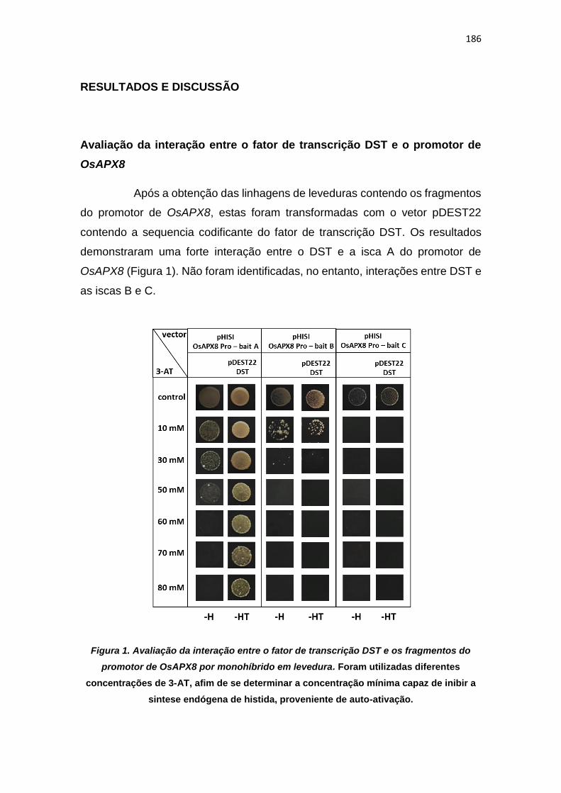
186
RESULTADOS E DISCUSSÃO
Avaliação da interação entre o fator de transcrição DST e o promotor de
OsAPX8
Após a obtenção das linhagens de leveduras contendo os fragmentos
do promotor de OsAPX8, estas foram transformadas com o vetor pDEST22
contendo a sequencia codificante do fator de transcrição DST. Os resultados
demonstraram uma forte interação entre o DST e a isca A do promotor de
OsAPX8 (Figura 1). Não foram identificadas, no entanto, interações entre DST e
as iscas B e C.
Figura 1. Avaliação da interação entre o fator de transcrição DST e os fragmentos do
promotor de OsAPX8 por monohíbrido em levedura. Foram utilizadas diferentes
concentrações de 3-AT, afim de se determinar a concentração mínima capaz de inibir a
sintese endógena de histida, proveniente de auto-ativação.

187
Dados da literatura, indicam que o fator de transcrição DST contribui
para a regulação da abertura estomática via regulação de genes envolvidos na
homeostase redox, como peroxidases (precursor 24, precursor 12), glutatione S-
transferase (OsGSTU2) e citocromos da família P450 (71D10, 94A2). Sob
condições de estresses de seca e salinidade a expressão de DST é fortemente
inibida, o que, indiretamente, levaria ao acúmulo de ROS (HUANG et al., 2009).
De fato, o gene de OsAPX8 também é reprimido durante o estresse de seca e o
silenciamento deste gene leva ao aumento do acúmulo de ROS e do fechamento
estomático, conforme demonstrado no capítulo 4. Portanto, a ligação desse fator
de transcrição no promotor do gene OsAPX8 e o padrão coincidente da
expressão de ambos em resposta a seca indicam que os mesmos participam
dos mecanismos que culminam com o fechamento dos estômatos na resposta
ao estresse por falta de água.
Identificação de fatores de transcrição capazes de se ligar ao promotor de
OsAPX8
O “screening” da biblioteca de cDNA obtida de plantas submetidas a
estresse de seca não resultou na identificação de fatores de transcrição com
capacidade de se ligar à região promotora de OsAPX8. Mesmo após a triagem
de mais de um milhão de clones para cada fragmento isca, nunhum clone
positivo foi identificado. Devido a isso, novos experimentos foram realizados
visando o “screening” de biblioteca de cDNA de plantas submetidas a frio, tendo
sido triados pelo menos um milhão de clones para cada fragmento isca do
promotor de OsAPX8. Utilizando esta segunda biblioteca foram identificados três
fatores de transcrição com capacidade de se ligar ao promotor de OsAPX8. Os
fatores de transcrição identificados estão listados da tabela 1. A confirmação
destas interações foi realizada através da re-transformação das estirpes isca
com o plasmídeo purificado (vetor pACTII) (Figura 2).

188
Tabela 1. Genes que codificam as proteínas capazes de se ligar à região promotora de
OsAPX8 identificados a partir do “screening” de bibliotecas de cDNA pelo sistema de
monohíbrido em levedura.
Nome do gene Família Locus Isca utilizada
Os11g0472000 C3H LOC_Os11g28270.1 Isca A
OJ1119_H02.18 bZIP LOC_Os05g41070.1 Isca A
OsVOZ1 VOZ LOC_Os01g54930.1 Isca B
Figura 2. Confirmação dos fatores de transcrição capazes de se ligar ao promotor de
OsAPX8 por monohíbrido em levedura.

189
Um dos fatores de trancrição identificados, corresponde a uma
proteína do tipo “dedo de zinco” e é codificado pelo gene Os11g28270. No
entanto, informações a respeito desta proteína em arroz não estão disponíveis.
Já em Arabidopsis thaliana, o homologo ZFN1 (ZINC FINGER PROTEIN 1;
AT3G02830), também conhecido como PENTA 1), atua como um inibidor da
expressão gênica e está possivelmente envolvido no desenvolvimento da parte
aérea da planta, bem como na ativação da fotomorfogênese (CHRISPEELS et
al., 2000). De fato, em um trabalho anterior foi demonstrado que ZFN1 é capaz
de interagir com fitocromo em resposta a luz vermelho-distante, e regular
negativamente a transcrição de protoclorofila reductase (PORA), responsável
pela conversão de protoclorofila em clorofila (PAIK et al., 2012).
Outro fator de transcrição capaz de se ligar ao promotor de OsAPX8,
foi a proteína OsbZIP42 ou OsABF7 (ABA-responsive element binding factor 7),
que atua como ativador da expressão gênica, e cuja expressão é reprimida em
resposta ao tratamento com PEG (capaz de simular o estresse hídrico), NaCl e
fitohormônios, como metil-jasmonato e NAA (Ácido 1-naftalenoacético) (LU et
al., 2009). A família de fatores de transcrição do tipo ABF/AREB é capaz de se
ligar ao DNA através do elemento de resposta ao ABA (ABRE, do inglês abscisic
acid response element), e a análise do promotor de OsAPX8, através do banco
de dados PlantCARE
(http://bioinformatics.psb.ugent.be/webtools/plantcare/html/), demonstra que a
sequência referente a este cis-elemento (ACCACGTGTC) está presente entre
as iscas A e B (posição -440 em relação ao ATG). Em Arabidopsis thaliana, esta
proteína possui alta homologia com o fator de transcrição AtbZIP66 ou AtAREB3
(ABA-responsive element binding protein 3;AT3G56850), expresso no embrião
durante a maturação da semente (BENSMIHEN et al., 2005), mas não verificado
em plantas adultas, mesmo sob condições de estresse (UNO et al., 2000).
O último fator de transcrição identificado corresponde a proteína
OsVOZ1 (vascular plant one-zinc-finger protein), um possível ativador da
expressão gênica. Esta proteína faz parte da família de fatores de transcrição do
tipo VOZ, que é composta por dois membros, VOZ1 e VOZ2, presentes em
monodicotiledôneas e eudicotiledôneas. Embora o papel fisiológico destas
190
proteínas em arroz ainda não tenha sido estudado, em Arabidopsis thaliana, os
genes VOZ respondem a diferentes estresses abióticos, sendo fortemente
reprimidos em plantas submetidas a seca e a baixas temperaturas. Além disso,
plantas duplo mutantes para AtVOZ1 e AtVOZ2 (voz1/voz2) apresentaram um
aumento da tolerância a esses estresses. Por outro lado, a resistência destas
plantas, a patógenos como o fungo Colletotrichum higginsianum e a bactéria
Pseudomonas syringae foi fortemente reduzida (NAKAI et al., 2013).
Posteriormente, foi demonstrando que linhagens que superexpressam AtVOZ1
e AtVOZ2 apresentavam uma redução da tolerância a estresses abióticos (seca
e frio), mas em relação a patógenos, elas apresentavam um aumento da
resistência (NAKAI et al., 2013a). Estes dados, portanto, indicam que os fatores
de transcrição do tipo VOZ, possuem um importante papel na resposta a
estresses biótico e abióticos e poderiam regular as respostas mediadas por
OsAPX8.
191
CONCLUSÕES E PERSPECTIVAS
Através de experimentos de monohíbrido em levedura, demonstramos
que o fator de transcrição OsDST é capaz de se ligar ao promotor de OsAPX8.
Além disso identificamos por “screening” de bibliotecas de cDNA, outros três
fatores de transcrição capazes de se ligar ao promotor de OsAPX8:
LOC_Os11g28270, LOC_Os05g41070 (OsABF7) e LOC_Os01g54930
(OsVOZ1). Com base nos dados disponíveis na literatura concluímos que as
proteínas OsDST, OsABF7 e OsVOZ1, são fortes candidatos a serem, de fato,
membros de vias de transdução de sinais mediadas por OsAPX8. Em geral,
todos estes fatores de transcrição são ativadores da expressão gênica, e são
fortemente reprimidos durante o estresse de seca. Se de fato, tais proteínas
forem capazes de ativar a expressão de OsAPX8, a repressão da expressão de
cada fator de transcrição, em condições de estresse, poderia contribuir para a
redução da expressão de OsAPX8, colaborando para o aumento da geração de
ROS e do fechamento estomático nestas condições (Figura 3).
Como perspectiva deste trabalho, pretendemos avaliar, por ensaio de
transativação em protoplastos de arroz, a capacidade dos fatores de transcrição
identificados em ativar ou inibir a expressão gênica sob o controle do promotor
de OsAPX8. Além disso, o próprio ensaio de transativação pode ser utilizado
para confirmar as interaçõeso DNA-proteína, uma vez que, nos experimentos
que utilizam o sistema de protoplastos de plantas, além das proteínas estarem
completas, elas estarão em sua conformação correta e com as modificações
pós-traducionais apropriadas, uma vez que estarão sendo expressas em células
de arroz, e não em leveduras. Além disso, pretendemos avaliar a expressão dos
fatores de transcrição identificados na resposta de plantas de arroz a diferentes
estresses abióticos. Estes ensaios permitirão a caracterização de rotas de
sinalização que modulem a expressão de OsAPX8 em resposta ao estresse.

192
Figura 3. Modelo proposto para a regulação da expressão de OsAPX8 e seu papel no
controle do fechamento estomático em resposta ao estresse de seca. Os fatores de
transcrição OsDST, OsABF7 e OsVOZ1 seriam capazes de se ligar diretamente no promotor de
OsAPX8 e ativar a expressão gênica, inibindo, portanto, o acúmulo de ROS. Sob condição de
estresse de seca, a expressão destes fatores de transcrição é reprimida, resultando na regulação
negativa da expressão de OsAPX8, o que leva a acúmulo de ROS e o fechamento estomático.
193
REFERÊNCIAS
BENSMIHEN S, GIRAUDAT J, PARCY F. (2005). Characterization of three
homologous basic leucine zipper transcription factors (bZIP) of the ABI5 family
during Arabidopsis thaliana embryo maturation. J Exp Bot. 56(412):597-603.
BONIFÁCIO A, MARTINS M, RIBEIRO C, FONTENELE A, CARVELHO F,
MARGIS-PINHEIRO M, SILVEIRA J. (2011). Role of peroxidases in the
compensation of cytosolic ascorbate peroxidase knockdown in rice plants under
abiotic stress. Plant, Cell and Environment 34(10):1705-22.
BRIGHT J, DESIKAN R, HANCOCK JT, WEIR IS, NEILL SJ. (2006). ABA
induced NO generation and stomatal closure in Arabidopsis are dependent on
H2O2 synthesis. Plant J 45: 113-22.
CAVERZAN A, BONIFACIO A, CARVALHO FE, ANDRADE CM, PASSAIA G,
SCHÜNEMANN M, MARASCHIN FDOS S, MARTINS MO, TEIXEIRA FK,
RAUBER R, MARGIS R, SILVEIRA JA, MARGIS-PINHEIRO M. (2014). The
knockdown of chloroplastic ascorbate peroxidases reveals its regulatory role in
the photosynthesis and protection under photo-oxidative stress in rice. Plant
Science 214:74-87.
CHRISPEELS HE, OETTINGER H, JANVIER N, TAGUE BW. (2000). AtZFP1,
encoding Arabidopsis thaliana C2H2 zinc-finger protein 1, is expressed
downstream of photomorphogenic activation. Plant Mol Biol.42(2):279-90.
FIGUEIREDO DD, BARROS PM, CORDEIRO AM, SERRA TS, LOURENÇO T,
CHANDER S, OLIVEIRA MM, SAIBO NJ. (2012). Seven zinc-finger transcription
factors are novel regulators of the stress responsive gene OsDREB1B. J Exp Bot.
63(10):3643-56.
HETHERINGTON AM, WOODWARD FI. (2003). The role of stomata in sensing
and driving environmental change. Nature 424: 901-08.
194
HUANG XY, CHAO DY, GAO JP, ZHU MZ, SHI M, LIN HX. (2009). A previously
unknown zinc finger protein, DST, regulates drought and salt tolerance in rice via
stomatal aperturecontrol. Genes Dev 23(15): 1805-17.
KOHLER B, HILLS A, BLATT MR. (2003). Control of guard cell ion channels by
hydrogen peroxide and abscisic acid indicates their action through alternate
signaling pathways. Plant Physiol 131: 385-88.
LI S, ZHAO B, YUAN D, DUAN M, QIAN Q, TANG L, WANG B, LIU X, ZHANG
J, WANG J, SUN J, LIU Z, FENG YQ, YUAN L, LI C. (2013). Rice zinc finger
protein DST enhances grain production through controlling Gn1a/OsCKX2
expression. Proc Natl Acad Sci 110(8): 3167-72.
LU G, GAO C, ZHENG X. HAN B. (2009). Identification of OsbZIP72 as a positive
regulator of ABA response and drought tolerance in rice. Planta 229:605-15.
MCAINSH MR, CLAYTON H, MANSFIELD TA, HETHERINGTON AM. (1996).
Changes in stomatal behavior and guard cell cytosolic free calcium in response
to oxidative stress. Plant Physiol 111: 1031–42.
MENEZES-BENAVENTE L, TEIXEIRA FK, KAMEI CL, MARGIS-PINHEIRO M.
(2004). Salt stress induces expression of genes encoding antioxidant enzymes
in seedlings of a Brazilianindicarice (Oryza sativa L.). Plant Sci 166: 323-31.
NAKAI Y, FUJIWARA S, KUBO Y, SATO MH. (2013a). Overexpression of
VOZ2confers biotic stress tolerance but decreases abiotic stress resistance in
Arabidopsis 8:3, e23358.
NAKAI Y, NAKAHIRA Y, SUMIDA H, TAKEBAYASHI K, NAGASAWA Y,
YAMASAKI K, AKIYAMA M, OHME-TAKAGI M, FUJIWARA S, SHIINA T,
MITSUDA N, FUKUSAKI E, KUBO Y, SATO MH. (2013). Vascular plant one-
zinc-finger protein 1/2 transcription factors regulate abiotic and biotic stress
responses in Arabidopsis. The Plant Journal 73: 761-775.
PAIK I, YANG S, CHOI G. (2012). Phytochrome regulates translation of mRNA
in the cytosol. PNAS 109(4):1335-40.

195
PEI ZM, MURATA Y, BENNING G, THOMINE S, KLUSENER B, ALLEN GJ,
GRILL E, SCHROEDER JI. (2000). Calcium channels activated by hydrogen
peroxide mediate abscisic acid signalling in guard cells. Nature 406: 731–34.
ROSA SB, CAVERZAN A, TEIXEIRA FK, LAZZAROTTO F, SILVEIRA JA,
FERREIRA-SILVA SL, ABREU-NETO J, MARGIS R, MARGIS-PINHEIRO M.
(2010). Cytosolic APx knockdown indicates an ambiguous redox response in rice.
Phytochemistry 71: 548-58.
SCHROEDER JI, ALLEN GJ, HUGOUVIEUX V, KWAK JM, WANER D. (2001).
Guard cell signal transduction. Annu Rev Plant Physiol Plant Mol Biol 52: 627–
58.
TEIXEIRA FK, MENEZES-BENAVENTE L, GALVÃO VC, MARGIS-PINHEIRO
M. (2005). Multigene families encode the major enzymes of antioxidant
metabolism in Eucalyptus grandis L. Genetics and Molecular Biology 28: 529-38;
TEIXEIRA FK, MENEZES-BENAVENTE L, MARGIS R, MARGIS-PINHEIRO M.
(2004). Analysis of the molecular evolutionary history of the ascorbate peroxidase
gene family: inferences from the rice genome. J Mol Evol 59: 761-70;
UNO Y, FURIHATA T, ABE H, YOSHIDA R, SHINOZAKI K, YAMAGUCHI-
SHINOZAKI K. (2000). Arabidopsis basic leucine zipper transcription factors
involved in an abscisic acid-dependent signal transduction pathway under
drought and high-salinity conditions. PNAS 97(21): 11632–11637.
ZHANG X, ZHANG L, DONG F, GAO J, GALBRAITH DW, SONG CP. (2001).
Hydrogen peroxide is involved in abscisic acid-induced stomatal closure in Vicia
faba. Plant Physiol 126: 1438–48.
196
4- DISCUSSÃO GERAL
As ROS são formadas como subproduto do metabolismo aeróbico em
diferentes organismos e atuam como moléculas sinalizadoras, ou mensageiros
secundários, em uma complexa rede de sinalização em diferentes organismos.
Desta forma, em plantas, as vias de produção de ROS, bem como os
mecanismos antioxidantes são fundamentais para o desenvolvimento vegetal, e
para a resposta a diferente estímulos ambientais.
Neste trabalho, identificamos uma nova e importante fonte de geração
de ROS em células vegetais: a enzima SDH, que corresponde ao complexo II da
respiração. Os resultados obtidos demonstram que a geração de ROS pela SDH
pode ser modulada por diferentes inibidores. Enquanto inibidores não-
competitivos, como ácido salicílico e óxido nítrico (NO), são capazes de induzir
a geração de ROS, os inibidores competitivos são capazes de previnir a geração
de ROS pela SDH. Este mecanismo se mostra importante em células vegetais,
pois diferentes moléculas endógenas podem atuar regulando a geração de ROS
pela SDH. Além disso, este mecanismo resulta na regulação da expressão de
genes relacionados com o desenvolvimento vegetal e com a tolerância a
diferentes estresses.
Se por um lado a manipulação da geração de ROS pela mitocôndria
promoveu alterações claras no padrão de desenvolvimento, alterações nas
respostas antioxidantes no cloroplasto afetaram em menor grau o crescimento
da planta. No entanto, impactos nos mecanismos de defesa foram verificados
em resposta às alterações em ambas organelas, de maneira similar. Assim,
nossos dados demonstram que as isoformas cloroplastídicas de APX também
estão envolvidas em mecanismos de tolerância ao estresse, indicando o
potencial para um papel na regulação dos níveis de ROS por enzimas
relacionadas com a produção e a eliminação dessas moléculas reativas. No
entanto, apesar desses pontos comuns, a complexidade das respostas obtidas
indica uma alta especificidade das respostas, que depende da localização
subcelular de cada um dos componentes dessa intricada rede, assim como o
197
nível de expressão de cada um deles. Por exemplo, enquanto o silenciamento
duplo de OsAPX7 e OsAPX8, bem como o silenciamento individual de OsAPX8,
resultou em plantas com um maior conteúdo de ROS e maior fechamento
estomático em codições ideais, o silenciamento individual de OsAPX7 não
resulta em alterações fisiológicas nas plantas silenciadas. No entanto, as plantas
silenciadas para OsAPX7 e as plantas duplamente silenciadas para ambos os
genes demonstram uma maior tolerância ao estresse de seca. Esses dados
demonstram que o aumento da tolerância à seca não é consequência somente
do aumento de ROS ou do fechamento estomático, uma vez que tais fenômenos
não são observados nas plantas silenciadas para OsAPX7.
O aumento da tolerância a seca também foi verificado em plantas
nocaute para OsAPX8 (apx8/apx8). Curiosamente, ao contrário das plantas
silenciadas, as plantas nocaute pra OsAPX8 não apresentaram mudanças
fisiológicas em condições ideias, e dados de RNAseq demonstram que o
silenciamento de OsAPX8 induz uma maior alteração na expressão gênica do
que o nocaute. Aparentemente o silenciamento de OsAPX8 simula uma
condição de estresse, onde a expressão de OsAPX8 seria reduzida
naturalmente. Desta forma, as plantas silenciadas para OsAPX8 apresentam um
fenótipo parecido ao de plantas submetidas a estresse, como um maior conteúdo
de ROS, maior fechamento estomático, crescimento mais lento, bem como uma
maior resposta na expressão gênica. Por outro lado, o nocaute de OsAPX8
estaria gerando compensações não verificadas nas plantas silenciadas, que
permitem as plantas nocaute apresentar um fenótipo mais parecido com plantas
NT, embora sejam mais tolerantes ao estresse de defict hídrico. No entanto, os
mecanismos capazes de induzir o aumento da tolerância dessas plantas ao
estresse de seca ainda são desconhecidos.
Além das avaliações de interações gênicas e efeitos de manipulações
genéticas, a análise de como esses componentes envoluíram nos diferentes
genomas pode trazer novos dados para o entendimento da função de cada um
deles nos mecanismos de defesa das plantas. Nesse sentido, o estudo da
história evolutiva da succinato desidrogenase, descrita no capítulo 2 indicou que
esta enzima é membro da superfamília de fumarato redutases, composta por

198
outras enzimas, como a fumarato redutase dependente de quinona, NADH ou
thiol, além da enzima L-aspartato redudase. Nossos dados demonstram que
todas essas enzimas se originaram de um acestral comum, e os processos de
endossimbiose da mitocôndria e do cloroplasto assumiram um papel central na
distribuição destas enzimas em organismos eucariotos.
Nesta tese, demonstramos que os diferentes mecanismos envolvidos
no balanço redox de células vegetais exercem um importante papel no
desenvolvimento vegetal e na reposta de plantas a diferentes estresses. Além
disso, identificamos diferentes fatores de transcrição capazes de se ligar ao
promotor de OsAPX8, o que indiretamente também poderia regular o balanço
redox celular. A avaliação da capacidade dessas proteínas em regular a
expressão gênica permitirá ainda a indentificação de novas vias de sinalização
envolvidas na resposta ao estresse. Embora ainda existam incontáveis aspectos
a serem explorados nesta área, o presente trabalho contribuiu para a
identificação de novas interações e, consequentemente de vias inéditas de
sinalização nas células vegetais, ampliando a visão do papel das ROS nos
mecanismos de respostas a estímulos ambientais geradores de estresse
oxidativo.
199
5- CONCLUSÕES GERAIS
- Succinato desidrogenase é um importante sítio de geração de ROS em células
vegetais e regula a expressão de genes envolvidos com o desenvolvimento
vegetal e a resposta ao estresse;
- A família de succinato:quinona oxiredutase é membro de uma superfamília de
fumarato redutases, que se originou apartir de um ancestral comum e está
envolvida em processos metabólicos chave ao longo da evolução das espécies;
- As isoformas cloroplastídicas de ascorbato peroxidase desempenham um
importante papel na homestase redox e no controle da abertura estomática e da
tolerância ao estresse hídrico em arroz;
- Os fatores de trancrição, OsDST, OsVOZ1, OsABF7 e LOC_Os01g54930 são
capazes de se ligar ao promotor de OsAPX8 e são potenciais reguladores da
expressão gênica;
- As ROS são essenciais no controle das respostas das plantas a diferentes
estresses.
200
6- REFERÊNCIAS
APEL K, HIRT H. (2004). Reactive oxygen species: metabolism, oxidative stress,
and signal transduction. Annu. Rev. Plant Biol. 55: 373–99.
ARAÚJO WL, NUNES-NESI A, OSORIO S, USADEL B, FUENTES D, NAGY
R, BALBO I, LEHMANN M, STUDART-WITKOWSKI C, TOHGE T, MARTINOIA
E, JORDANA X, DAMATTA FM, FERNIE AR. (2011). Antisense Inhibition of the
Iron-Sulphur Subunit of Succinate Dehydrogenase Enhances Photosynthesis
and Growth in Tomato via an Organic Acid–Mediated Effect on Stomatal
Aperture. Plant Cell. 23(2):600-27.
ASADA K. (2006). Production and scavenging of reactive oxygen species in
chloroplastsand their functions. Plant Physiol. 141: 391–96
BARBOSA MR, SILVA MMA, WILLADINO L, ULISSES C, CAMARAI TR.
(2014). Geração e desintoxicação enzimática de espécies reativas de oxigênio
em plantas Plant generation and enzymatic detoxifi cation of reactive oxygen
species. Ciência Rural. 44(3): 453-60.
BHATTACHARJEE S. (2010). Sites of generation and physicochemical basis of
formation of reactive oxygen species in plant cell. Enfi eld: Science Publishers,
p.1
BONIFACIO A, MARTINS MO, RIBEIRO CW, FONTENELE AV, CARVALHO
FE, MARGIS-PINHEIRO M, SILVEIRA JA. (2011). Role of peroxidases in the
compensation of cytosolic ascorbate peroxidase knockdown in rice plants under
abiotic stress. Plant Cell Environ. 34(10):1705-22.
CAVERZAN A, BONIFACIO A, CARVALHO FE, ANDRADE CM, PASSAIA G,
SCHÜNEMANN M, MARASCHIN FDOS S, MARTINS MO, TEIXEIRA FK,
RAUBER R, MARGIS R, SILVEIRA JA, MARGIS-PINHEIRO M. (2014). The
knockdown of chloroplastic ascorbate peroxidases reveals its regulatory role in
the photosynthesis and protection under photo-oxidative stress in rice. Plant Sci.
214:74-87.
201
CHANCE B, SIES H, BOVERIS A. (1979). Hydroperoxide metabolism in
mammalian organs. Physiol. Rev. 59: 527–605.
CRAMER GR, URANO K, DELROT S, PEZZOTTI M, SHINOZAKI K. (2011).
Effects of abiotic stress on plants: a systems biology perspective. BMC plant
biology. 11:163.
DANNA CH, BARTOLI CG, SACCO F, INGALA LR, SANTA-MARIA GE,
GUIAMET JJ, UGALDE RA. (2003) Thylakoid-bound ascorbate peroxidase
mutant exhibits impaired electron transport and photosynthetic activity. Plant
physiology. 132: 2116-25.
DEMIDCHIK V, MAATHUIS FJM. (2007). Physiological roles of nonselective
cation channels in plants: from salt stress to signalling and development. Tansl.
Rev. New Phytol. 175: 387–405.
DEMIDCHIK V, STRALTSOVA D, MEDVEDEV S, POZHVANOV G, SOKOLIK
A, YURIN V. (2014). Stress-induced electrolyte leakage: the role of K+-
permeable channels and involvement to programmed cell death and metabolic
adjustment. doi:10.1093/jxb/eru004.
DEMIDCHIK V. (2015). Mechanisms of oxidative stress in plants: From classical
chemistry to cell biology. Environmental and Experimental Botany 109: 212–28
DESPRES C, CHUBAK C, ROCHON A, CLARK R, BETHUNE T, DESVEAUX
D, FOBERT PR. (2003). The Arabidopsis NPR1 disease resistance protein is a
novel cofactor that confers redox regulation of DNA binding activity to the basic
domain/leucine zipper transcription factor TGA1. Plant Cell 15: 2181–91.
EMBRAPA. (2015) Cultivo do Arroz Irrigado no Brasil. Disponível em:
<http://sistemasdeproducao.cnptia.embrapa.br/FontesHTML/Arroz/ArrozIrrigado
Brasil/index.htm >. Acessoem: 14 out. 2015.
FOYER CH, LESCURE JC, LEFEBVRE C, MOROT-GAUDRY JF, VINCENTZ
M, VAUCHERET H. (1994). Adaptations of Photosynthetic Electron Transport,
Carbon Assimilation, and Carbon Partitioning in Transgenic Nicotiana
202
plumbaginifolia Plants to Changes in Nitrate Reductase Activity. Plant Physiol.
104(1): 171-78.
FOYER CH, NEUKERMANS J, QUEVAL G, NOCTOR G, HARBINSON J.
(2012). Photosynthetic control of electron transport and the regulation of gene
expression. Journal of Experimental Botany 63: 1637-61.
FOYER CH, NOCTOR G. (2003). Redox sensing and signaling associated with
reactive oxygen in chloroplasts, peroxisomes and mitochondria. Physiologia
Plantarum. 119: 355-64.
FOYER CH, NOCTOR G. (2005). Redox homeostasis and antioxidant signaling:
a metabolic interface between stress perception and physiological responses.
Plant Cell 17: 1866-75.
FOYER CH, NOCTOR G. (2009). Redox regulation in photosynthetic organisms:
signaling, acclimation, and practical implications. Antioxid. Redox Signal. 11:
861-71.
FOYER CH, SHIGEOKA S. (2011). Understanding oxidative stress and
antioxidant functions to enhance photosynthesis. Plant Physiol. 155: 93-100.
FUENTES D, MENESES M, NUNES-NESI A, ARAÚJO WL, TAPIA R, GÓMEZ
I, HOLUIGUE L, GUTIÉRREZ RA, FERNIE AR, JORDANA X. (2011) A
Deficiency in the Flavoprotein of Arabidopsis Mitochondrial Complex II Results in
Elevated Photosynthesis and Better Growth in Nitrogen-Limiting Conditions.
Plant Physiol. 157(3): 1114-27.
GRATÃO PL, POLLE A, LEA PJ, AZEVEDO RA. (2005). Making the life of
heavy metal-stressed plants a little easier. Functional Plant Biology 32: 481-94.
HONG SY, ROZE LV, LINZ JE. (2013). Oxidative stress-related transcription
factors in the regulation of secondary metabolism. Toxins (Basel) 18: 683–702.
JACQUEMIN J, BHATIA D, SINGH K, WING RA. (2013). The International
Oryza Map Alignment Project: development of a genus-wide comparative
203
genomics platform to help solve the 9 billion-people question. CurrOpin Plant Biol
16: 147-56.
KARUPPANAPANDIAN T, MOON JC, KIM C, MANOHARAN K, KIM W. (2011).
Reactive oxygen species in plants: their generation, signal transduction, and
scavenging mechanisms. Australian Journal of Crop Science. 5(6): 709-25.
KOVTUN Y, CHIU WL, TENA G, SHEEN J. (2000). Functional analysis of
oxidative stress-activated mitogen-activated protein kinase cascade in plants.
Proc Natl Acad Sci USA 97: 2940-45.
LAZZAROTTO F, TEIXEIRA FK, ROSA SB, DUNAND C, FERNANDES CL,
FONTENELE ADE V, SILVEIRA JA, VERLI H, MARGIS R, MARGIS-PINHEIRO
M. (2011). Ascorbate peroxidase-related (APx-R) is a new heme-containing
protein functionally associated with ascorbate peroxidase but evolutionarily
divergent. New Phytol. 191(1):234-50.
LEÓN G, HOLUIGUE L, JORDANA X. (2007). Mitochondrial complex II Is
essential for gametophyte development in Arabidopsis. Plant Physiol.
143(4):1534-46.
MENEZES-BENAVENTE L, KERNODLE SP, MARGIS-PINHEIRO M,
SCANDALIOS JG. (2004). Salt-induced antioxidant metabolism defenses in
maize (Zea mays L.) seedlings. Redox Rep. 9(1):29-36.
MILLER G, MITTLER R. (2006). Could heat shock transcription factors function
as hydrogen peroxide sensors in plants? Ann Bot 98: 279–88.
MITTLER R, VANDERAUWERA S, GOLLERY M, VAN BREUSEGEM F. (2004).
Reactive oxygen gene network of plants. Trends Plant Sci 9: 490-98.
MØLLER IM. (2001). Plant mitochondria and oxidative stress: electron transport,
NADPH turnover, and metabolism of reactive oxygen species. Annu Rev Plant
Physiol Plant Mol Biol 52: 561–91.
MOORE G, DEVOS KM, WANG Z, GALE MD. (1995). Cereal Genome
Evolution: Grasses, line up and form a circle. Current Biology 5(7): 737–39.

204
MUNEMASA S, HOSSAIN MA, NAKAMURA Y, MORI IC, MURATA Y. (2011).
The Arabidopsis calcium-dependent protein kinase. CPK6, functions as a positive
regulator of methyl jasmonate signaling in guard cells. Plant Physiol. 155: 553–
61.
MYLONA PV, POLIDOROS AN. (2011). ROS regulation of antioxidant genes.
In: GUPTA, S.D. Reactive oxygen species and antioxidants in higher plants. Enfi
eld: Science Publishers. 6: 101-28.
NEILL S, DESIKAN R, HANCOCK J. (2002). Hydrogen peroxide signalling. Curr
Opin Plant Biol 5: 388-95.
NOCTOR G AND FOYER CH. (1998). Ascorbate glutathione: Keeping active
oxygen under control. Annu Rev Plant Physiol Plant MolBiol 49: 249-79
of networks involving reactive oxygen species in plants. Plant Physiol. 147: 978–
84
OUYANG S, ZHU W, HAMILTON J, LIN H, CAMPBELL M, CHILDS K,
THIBAUD-NISSEN F, MALEK RL, LEE Y, ZHENG L, ORVIS J, HAAS B,
WORTMAN J, BUELL CR. (2007). The TIGR Rice Genome Annotation
Resource: improvements and new features. Nucleic acids research. 35: 883–87.
PATERSON AH, FREELING M, SASAKI T. (2005). Grains of knowledge:
Genomics of model cereals. Genome Res. 15: 1643–50.
PAYTON P, WEBB R, KORNYEYEV D, ALLEN R, HOLADAY AS. (2001).
Protecting cotton photosynthesis during moderate chilling at high light intensity
by increasing chloroplastic antioxidant enzyme activity. J Exp Bot 52: 2345-54.
PITZSCHKE A, HIRT H. (2009). Disentangling the complexity of mitogen-
activated protein kinases and reactive oxygen species signaling. Plant Physiol.
149: 606–15.
PUNTARULO S, GALLEANO M, SANCHEZ RA, BOVERIS A. (1991).
Superoxide anion and hydrogen peroxide metabolism in soybean embryonic
axes during germination. Biochim. Biophys. Acta 1074: 277–83.

205
PUNTARULO S, SANCHEZ RA, BOVERIS A. (1988). Hydrogen peroxide
metabolism in soybean embryonic axes at the onset of germination. Plant
Physiol. 86: 626–30.
QUINLAN CL, ORR AL, PEREVOSHCHIKOVA IV, TREBERG JR, ACKRELL
BA, BRAND MD. (2012). Mitochondrial complex II can generate reactive oxygen
species at high rates in both the forward and reverse reactions. J. Biol. Chem.
287:27255–64.
RASOOL S, REHMAN MU, AZOOZ MM, IQBAL M, SIDDIQI TO, AHMAD P.
(2013). Arsenic toxicity and tolerance mechanisms in plants: An overview. Crop
Improvement: New Appoaches and modern tecniques. Cap. 15: 363-78
RODRIGUEZ MCS, PETERSEN M. MUNDY J. (2010). Mitogen-activated protein
kinase signaling in plants. Annu. Rev. Plant Biol. 61: 621–49.
ROSA SB, CAVERZAN A, TEIXEIRA FK, LAZZAROTTO F, SILVEIRA JA,
FERREIRA-SILVA SL, ABREU-NETO J, MARGIS R, MARGIS-PINHEIRO M.
(2010). Cytosolic APx knockdown indicates an ambiguous redox response in rice.
Phytochemistry. 71(5-6):548-58.
SCANDALIOS JG. (2005). Oxidative stress: molecular perception and
transduction of signals triggering antioxidant gene defenses. Braz J Med Biol Res
38: 995-14.
SHARMA P, JHA AB, DUBEY RS, PESSARAKLI M. (2012) Reactive oxygen
species, oxidative damage, and antioxidative defense mechanism in plants under
stressful conditions. Journal of Botany: 1-26.
SIERLA M, RAHIKAINEN M, SALOJÄRVI J, KANGASJÄRVI J,
KANGASJÄRVI S. (2013). Apoplastic and chloroplastic redox signaling networks
in plant stress responses. Antioxid. Redox Signal. 18: 2220–39.
SLESAK I, LIBIK M, KARPINSKA B, KARPINSKI S, MISZALSKI Z. (2007). The
role of hydrogen peroxide in regulation of plant metabolism and cellular signalling
in response to environmental stresses. Acta Biochim Pol 54: 39-50.
206
STOCKING MA. (2003). Tropical soils and food security: the next 50 years.
Science 302(5649):1356–9135.
STONEBLOOM S, BURCH-SMITHA T, KIMA I, MEINKEB D, MINDRINOSC M,
ZAMBRYSKI P. (2009). Loss of the plant DEAD-box protein ISE1 leads to
defective mitochondria and increased cell-to-cell transport via plasmodesmata.
FEBS 106(40): 17229–34.
TAKEDA S, MATSUOKA M. (2008) Genetic approaches to crop improvement:
responding to environmental and population changes. Nat. Rev. Genetics. 9:444-
56.
TEIXEIRA FK, MENEZES-BENAVENTE L, GALVÃO VC, MARGIS R, MARGIS-
PINHEIRO M. (2006). Rice ascorbate peroxidase gene family encodes
functionally diverse isoforms localized in different subcellular compartments.
Planta. 224(2):300-14.
TEIXEIRA FK, MENEZES-BENAVENTE L, MARGIS R, MARGIS-PINHEIRO M.
(2004). Analysis of the molecular evolutionary history of the ascorbate peroxidase
gene family: inferences from the rice genome. J Mol Evol 59: 761-70;
TURRENS JF, ALEXANDRE A, LEHNINGER AL. (1985). Ubisemiquinone is the
electron donor for superoxide formation by complex III of heart mitochondria. Arch
Biochem Biophys. 237(2):408-14.
TURRENS JF, BOVERIS A. (1980). Generation of superoxide anion by the
NADH dehydrogenase of bovine heart mitochondria. Biochem. J. 191: 421–27.
VAN BREUSEGEM FV, BAILEY-SERRES J. MITTLER R. (2008). Unraveling
the tapestry
VAN VELTHUIZEN H et al. (2007). Mapping biophysical factors that influence
agricultural production and rural vulnerability. FOOD AND AGRICULTURE
ORGANIZATION OF THE UNITED NATIONS and the International Institute of
Applied Systems Analysis.
YABUTA Y, MOTOKI T, YOSHIMURA K, TAKEDA T, ISHIKAWA T,

207
SHIGEOKA S. (2002) Thylakoid membrane-bound ascorbate peroxidase is a
limiting factor of antioxidative systems under photo-oxidative stress. Plant J 32:
915-25.
YANKOVSKAYA V, HORSEFIELD R, TÖRNROTH S, LUNA-CHAVEZ C,
MIYOSHI H, LÉGER C, BYRNE B, CECCHINI G, IWATA S. (2003). Architecture
of succinate dehydrogenase and reactive oxygen species generation. Science.
299(5607):700-04.