tepzz 4z748zb_t - ep 2 407 480 b1
242
Note: Within nine months of the publication of the mention of the grant of the European patent in the European Patent Bulletin, any person may give notice to the European Patent Office of opposition to that patent, in accordance with the Implementing Regulations. Notice of opposition shall not be deemed to have been filed until the opposition fee has been paid. (Art. 99(1) European Patent Convention). Printed by Jouve, 75001 PARIS (FR) (19) EP 2 407 480 B1 (Cont. next page) TEPZZ 4Z748ZB_T (11) EP 2 407 480 B1 (12) EUROPEAN PATENT SPECIFICATION (45) Date of publication and mention of the grant of the patent: 16.07.2014 Bulletin 2014/29 (21) Application number: 11003016.0 (22) Date of filing: 19.10.2006 (51) Int Cl.: C07K 14/11 (2006.01) A61K 39/145 (2006.01) C12N 7/00 (2006.01) (54) Materials for respiratory disease control in canines Materialien zur Kontrolle von Atemwegserkrankungen in Hunden Matériaux de contrôle de maladie respiratoire chez les canins (84) Designated Contracting States: AT BE BG CH CY CZ DE DK EE ES FI FR GB GR HU IE IS IT LI LT LU LV MC NL PL PT RO SE SI SK TR (30) Priority: 19.10.2005 US 728449 P 29.12.2005 US 754881 P 14.01.2006 US 759162 P 23.01.2006 US 761451 P 03.03.2006 US 779080 P 21.04.2006 US 409416 (43) Date of publication of application: 18.01.2012 Bulletin 2012/03 (62) Document number(s) of the earlier application(s) in accordance with Art. 76 EPC: 06826359.9 / 1 945 659 (73) Proprietors: • University of Florida Research Foundation, Incorporated Gainesville, Florida 32611 (US) • The Government of the United States of America as represented by the Secretary of the Department of Health and Human Services Atlanta, GA 30341 (US) • Cornell Research Foundation, Inc. Ithaca, NY 14850 (US) (72) Inventors: • Crawford, Patti, C. Gainesville, FL 32607 (US) • Gibbs, Paul, J. Gainesville, FL 32605 (US) • Dubovi, Edward, J. Ithaca, NY 14850-1087 (US) • Donis, Ruben, O Atlanta, GA 30307 (US) • Katz, Jacqueline Atlanta, GA 30307 (US) • Klimov, Alexander, I. Atlanta, GA 30345 (US) • Lakshmanan, Nallakannu, P. Millsboro, DE 19966 (US) • Lum, Melissa, Anne Millsboro, DE 19966 (US) • Goovaerts, Daniel, Ghislena, Emiel 5831 AN Boxmeer (NL) • Mellencamp, Mark, William Desoto, KS 66018 (US) • Castleman, William, L Gainesville, FL 32608 (US) • Cox, Nancy, J. Atlanta, GA 30307 (US) (74) Representative: Roberts, Michael Austin Reddie & Grose LLP 16 Theobalds Road London WC1X 8PL (GB)
-
Upload
khangminh22 -
Category
Documents
-
view
0 -
download
0
Transcript of tepzz 4z748zb_t - ep 2 407 480 b1

Note: Within nine months of the publication of the mention of the grant of the European patent in the European PatentBulletin, any person may give notice to the European Patent Office of opposition to that patent, in accordance with theImplementing Regulations. Notice of opposition shall not be deemed to have been filed until the opposition fee has beenpaid. (Art. 99(1) European Patent Convention).
Printed by Jouve, 75001 PARIS (FR)
(19)E
P2
407
480
B1
(Cont. next page)
TEPZZ 4Z748ZB_T(11) EP 2 407 480 B1
(12) EUROPEAN PATENT SPECIFICATION
(45) Date of publication and mention of the grant of the patent: 16.07.2014 Bulletin 2014/29
(21) Application number: 11003016.0
(22) Date of filing: 19.10.2006
(51) Int Cl.:C07K 14/11 (2006.01) A61K 39/145 (2006.01)
C12N 7/00 (2006.01)
(54) Materials for respiratory disease control in canines
Materialien zur Kontrolle von Atemwegserkrankungen in Hunden
Matériaux de contrôle de maladie respiratoire chez les canins
(84) Designated Contracting States: AT BE BG CH CY CZ DE DK EE ES FI FR GB GR HU IE IS IT LI LT LU LV MC NL PL PT RO SE SI SK TR
(30) Priority: 19.10.2005 US 728449 P29.12.2005 US 754881 P14.01.2006 US 759162 P23.01.2006 US 761451 P03.03.2006 US 779080 P21.04.2006 US 409416
(43) Date of publication of application: 18.01.2012 Bulletin 2012/03
(62) Document number(s) of the earlier application(s) in accordance with Art. 76 EPC: 06826359.9 / 1 945 659
(73) Proprietors: • University of Florida Research Foundation,
IncorporatedGainesville, Florida 32611 (US)
• The Government of the United States of America as represented by the Secretary of the Department of Health and Human ServicesAtlanta, GA 30341 (US)
• Cornell Research Foundation, Inc.Ithaca, NY 14850 (US)
(72) Inventors: • Crawford, Patti, C.
Gainesville, FL 32607 (US)• Gibbs, Paul, J.
Gainesville, FL 32605 (US)
• Dubovi, Edward, J.Ithaca, NY 14850-1087 (US)
• Donis, Ruben, OAtlanta, GA 30307 (US)
• Katz, JacquelineAtlanta, GA 30307 (US)
• Klimov, Alexander, I.Atlanta, GA 30345 (US)
• Lakshmanan, Nallakannu, P.Millsboro, DE 19966 (US)
• Lum, Melissa, AnneMillsboro, DE 19966 (US)
• Goovaerts, Daniel, Ghislena, Emiel5831 AN Boxmeer (NL)
• Mellencamp, Mark, WilliamDesoto, KS 66018 (US)
• Castleman, William, LGainesville, FL 32608 (US)
• Cox, Nancy, J.Atlanta, GA 30307 (US)
(74) Representative: Roberts, Michael AustinReddie & Grose LLP 16 Theobalds RoadLondon WC1X 8PL (GB)

2
EP 2 407 480 B1
(56) References cited: • CRAWFORD P C ET AL: "Transmission of Equine
influenza virus to dogs", SCIENCE, AMERICAN ASSOCIATION FOR THE ADVANCEMENT OF SCIENCE,, US, vol. 310, no. 5747, 21 October 2005 (2005-10-21), pages 482-485, XP003003531, ISSN: 0036-8075, DOI: 10.1126/SCIENCE.1117950 -& CRAWFORD P C ET AL: "Transmission of Equine influenza virus to dogs", SCIENCEXPRESS (10.1126/SCIENCE.1117950), 29 September 2005 (2005-09-29), XP002664250, DOI: 10.1126/SCIENCE.1117950 -& CRAWFORD P C ET AL: "Supporting Online Material: Transmission of Equine influenza virus to dogs", SCIENCE, 29 September 2005 (2005-09-29), pages S1-S19, XP002664251, -& DATABASE UniProt 8 November 2005 (2005-11-08), "Hemagglutinin", XP002664252, retrieved from EBI Database accession no. Q3I3Q4 -& DATABASE UniProt [Online] 8 November 2005 (2005-11-08), "M1", XP002664253, retrieved from EBI Database accession no. Q313U6
• PEEK SIMON F ET AL: "Acute respiratory distress syndrome and fatal interstitial pneumonia associated with equine influenza in a neonatal foal", JOURNAL OF VETERINARY INTERNAL MEDICINE, LIPPINCOTT, PHILADELPHIA, US, vol. 18, no. 1, January 2004 (2004-01), pages 132-134, XP008080707, ISSN: 0891-6640 -& DATABASE UniProt 6 December 2005 (2005-12-06), "Matrix Protein 1", XP002664254, retrieved from EBI Database accession no. Q30BG2

EP 2 407 480 B1
3
5
10
15
20
25
30
35
40
45
50
55
Description
BACKGROUND OF THE INVENTION
[0001] "Kennel cough" or infectious tracheobronchitis (ITB) is an acute, contagious respiratory infection in dogs char-acterized mainly by coughing (Ford et al, 1998). Canine ITB is considered one of the most prevalent infectious respiratorydiseases of dogs worldwide, and outbreaks can reach epidemic proportions when dogs are housed in high-densitypopulation environments such as kennels. Most outbreaks are due to direct dog-to-dog contact or aerosolization ofrespiratory secretions (Ford et al, 1998). The clinical signs arc caused by infection with one or a combination of bacterialand viral agents that colonize the epithelium of the upper and lower respiratory tract. Canine parainfluenza virus (CPiV)and Bordetella bronchiseptica bacteria are the most common organisms isolated from affected dogs, but several otherviruses such as canine distemper virus (CDV) and canine adenoviruses-1 and -2 (CAV-1, CAV-2), along with bacteriasuch as Streptococcus sp., Pasteurella multicoda and Escherichia coli, can influence the clinical course and outcome(Ford et al, 1998). While outbreaks occur most efficiently and rapidly in high-density populations with high morbidity,complicated respiratory infections and death are uncommon. Although life-threatening secondary bacterial pneumoniacan develop, the majority of ITB cases are self-limiting and resolve without any treatment (Ford et al, 1998).[0002] In July 1992, a respiratory infection presumed to be "kennel cough" became epidemic at several greyhoundtracks in New England, Florida, West Virginia, Wisconsin, Kansas, Colorado, Oklahoma and Arizona. According toveterinarians, most of the affected dogs had a mild cough that resolved, but more than a dozen greyhounds developedan acute hemorrhagic pneumonia followed by rapid death (Greyhound Daily News, 1999).[0003] In late 1998 to early 1999, several outbreaks of "kennel cough" occurred in racing greyhound kennels acrossthe country, resulting in mandatory closure of tracks and quarantine of all racing greyhounds in the U.S. for severalweeks (Greyhound Daily News, 1999). At one track in Florida (Palm Beach Kennel Club), coughing was recorded innearly 40% of the dog population on a single day (Personal communication from Dr. William Duggar). Similar to theoutbreak in 1992, the coughing resolved in most greyhounds, but 10 dogs in Florida died from a hemorrhagic pneumoniasyndrome uncharacteristic of "kennel cough" (Putnam, 1999).[0004] In March-April 2003, another outbreak of "kennel cough" occurred at greyhound tracks in the eastern U.S. Theoutbreak is believed to have originated in kennels at four tracks in Florida and caused the suspension of racing andquarantine of dogs for almost three weeks. Nearly 25% of the dogs at the track in West Palm Beach were affected, whilealmost 50% of the 1400 dogs at Derby Lane in St. Petersburg developed coughing. Again, most dogs recovered, butseveral dogs have died from the respiratory infection. The estimated economic impact of the respiratory outbreak at theDerby Lane track alone was $2 million.[0005] There are no published reports documenting the etiology or clinicopathology of the "kennel cough" epidemicsin racing greyhound kennels in 1992, 1998-1999, or 2003. The assumption has been that the infections were due toCPiV and/or B. bronchiseptica, the two most common causes of kennel cough. Unsubstantiated communications suchas web sites have attributed the fatal hemorrhagic pneumonias reported in some of the coughing dogs to infection withβ-hemolytic Streptococcus equi subspecies zooepidemicus, and refer to the syndrome as "canine streptococcal toxicshock."[0006] Transmission of virus from one host species to another is a crucial feature of the ecology and epidemiology ofinfluenza virus (Webster, 1998). Two basic mechanisms of interspecies transmission of influenza virus are possible(Webster et al., 1992; Lipatov et al., 2004). One is the direct transfer of an essentially unaltered virus from one speciesto another. Examples of this mechanism include the recent human infections with the H5N1 subtype of avian influenzavirus (Subbarao et al., 1998; Peiris et al., 2004; Guan et al., 2004) and possibly the pandemic of 1918, known as Spanishflu (Reid et al., 2004). The second mechanism is a consequence of the segmented nature of the influenza genome. Co-infection of a host with viruses from different species can result in reassortment of the segmented viral genes and thegeneration of a recombinant with the ability to infect other species. For example, novel viruses generated by genereassortment between avian and human influenza viruses resulted in human influenza pandemics in 1957 and 1968(Webster et al., 1992; Lipatov et al., 2004; Kawaoka et al., 1989).[0007] Most direct transmissions of unaltered influenza viruses from the natural host species to a different speciesare terminal events because sustained transmission between individuals of the new species fails to occur. Multiple virus-host interactions are necessary for replication and horizontal transmission and provide a formidable barrier to perpetuationof influenza viruses in the new host (Webby et al., 2004). Therefore, establishment of new host-specific lineages ofinfluenza virus is uncommon and has only occurred in domestic poultry, pigs, horses, and humans (Webster et al., 1992;Lipatov et al., 2004).[0008] Because of the serious nature of influenza virus infection, there remains a need for methods for diagnosing,preventing, and treating infection by influenza virus.

EP 2 407 480 B1
4
5
10
15
20
25
30
35
40
45
50
55
BRIEF SUMMARY OF THE INVENTION
[0009] The subject invention pertains to isolated influenza virus that is capable of infecting canids and causing respi-ratory disease in the canid.[0010] The invention is the following:
1. An isolated canine influenza virus that is capable of infecting a canid animal, wherein said influenza virus comprisesa polynucleotide which encodes a hemagglutinin (HA) polypeptide having an amino acid sequence shown in SEQID NO: 62, or a mature sequence thereof where the N-terminal 16 amino acid signal sequence of the full-lengthsequence has been removed.
2. The influenza virus according to item 1, wherein said influenza virus comprises a polynucleotide which encodesa polypeptide having the amino acid sequence shown in any of SEQ ID NOs: 48, 50, 52, 54, 56, 58, or 60, or afunctional and/or immunogenic fragment thereof, or said polynucleotide encodes a polypeptide having 95% or greatersequence identity with the amino acid sequence shown in any of SEQ ID NOs: 48, 50, 52, 54, 56, 58, or 60.
3. The influenza virus according to item 1, where said HA polypeptide of said viral isolate comprises the amino acidsequence of SEQ ID NO: 62.
4. The influenza virus according to item 1, wherein said influenza virus comprises a polynucleotide having thenucleotide sequence shown in any of SEQ ID NOs: 47, 49, 51, 53, 55, 57, 59, or 61, or said polynucleotide has 98%or greater sequence identity with the nucleotide sequence shown in any of SEQ ID NOs: 47, 49, 51, 53, 55, 57, 59,or 61.
5. The influenza virus according to item 1, wherein said influenza virus is inactivated or attenuated.
6. A composition comprising an immunogen of an influenza virus of item 1, wherein said immunogen is capable ofinducing an immune response against an influenza virus that is capable of infecting a canid animal, and whereinsaid immunogen comprises:
(a) an HA polypeptide as defined in item 1 or item 3; and/or(b) a polynucleotide encoding an HA polypeptide as defined in item 1 or item 3.
7. The composition according to item 6, wherein said immunogen comprises cell-free whole virus, or a portionthereof; a viral polynucleotide; a viral protein; a viral polypeptide or peptide; a virus infected cell; a recombinant viralvector based construct; a reassortant virus; or naked nucleic acid of said virus.
8. The composition according to item 7, wherein said viral protein, polypeptide, or peptide comprises an amino acidsequence shown in any of SEQ ID NOs: 48, 50, 52, 54, 56, 58, or 60, or a functional and/or immunogenic fragmentthereof, or said polynucleotide encodes a polypeptide having 95% or greater sequence identity with the amino acidsequence shown in any of SEQ ID NOs: 48, 50, 52, 54, 56, 58, or 60, or wherein said viral polynucleotide encodesa polypeptide comprising an amino acid sequence shown in any of SEQ ID NOs: 48, 50, 52, 54, 56, 58, or 60, or afunctional and/or immunogenic fragment thereof, or said polynucleotide encodes a polypeptide having 95% or greatersequence identity with the amino acid sequence shown in any of ID NOs: 48, 50, 52, 54, 56, 58, or 60, or whereinsaid viral polynucleotide comprises the nucleotide sequence shown in any of SEQ ID NOs: 47, 49, 51, 53, 55, 57,59, or 61, or a functional fragment thereof.
9. A canine influenza vaccine, wherein the vaccine comprises:
a therapeutically effective amount of an antigen of at least one influenza virus of item 1, andat least one pharmaceutically acceptable excipient,wherein said antigen comprises:
(a) an HA polypeptide as defined in item 1 or item 3; and/or(b) a polynucleotide encoding an HA polypeptide as defined in item 1 or item 3.
10. The vaccine according to item 9, wherein the virus antigen comprises an inactivated virus or a live attenuated virus.

EP 2 407 480 B1
5
5
10
15
20
25
30
35
40
45
50
55
11. An isolated polynucleotide that comprises all or part of a genomic segment or gene of an influenza virus of item1, wherein the polynucleotide comprises a nucleic acid sequence which encodes an HA polypeptide as defined initem 1 or item claim 3.
12. The polynucleotide according to item 11, wherein said polynucleotide is formulated in a pharmaceutically ac-ceptable carrier or diluent.
13. A polynucleotide expression construct comprising a polynucleotide of item 11.
14. An isolated HA polypeptide encoded by a polynucleotide of item 11.
15. The polypeptide according to item 14, wherein said polypeptide is formulated in a pharmaceutically acceptablecarrier or diluent.
BRIEF DESCRIPTION OF THE DRAWINGS
[0011]
Figures 1A-1B show phylogenetic relationships among the hemagglutinin genes. Figure 1A shows a tree of HAgenes from representative canine, human, avian, swine, and equine isolates, including A/Budgerigar/Hokkaido/1/77(H4) as outgroup. Figure 1B shows a tree of the canine influenza virus HA genes with contemporary and olderequine HA genes, using A/Duck/Ukraine/63 (H3) as outgroup. Phylogenetic trees were inferred from nucleotidesequences by the neighbor joining method and bootstrap analysis values ≥90% are shown. The bar denotes thenumber of nucleotide changes per unit length of the horizontal tree branches.Figures 2A-2B show immunohistochemical detection of influenza H3 antigen in the lungs. Lung tissue sectionswere probed with a mouse monoclonal antibody to H3 hemagglutinin and binding was detected by immunoperoxidasereaction (brown precipitate). Figure 2A shows bronchial epithelium from a greyhound with spontaneous disease.Viral H3 antigen was detected in bronchial epithelial cell cytoplasm and in macrophages in airway lumens and inalveolar spaces. Figure 2B shows bronchial epithelium from a dog 5 days after inoculation with A/canine/Flori-da/43/2004 (H3N8). Viral H3 antigen was detected in bronchial epithelial cell cytoplasm. Scale bar, 66 mm.Figure 3 shows the characteristic histological changes in the bronchi of greyhounds that died from hemorrhagicpneumonia associated with influenza virus infection. The tissues are stained with H&E. Upper panel: Normal bronchuswith ciliated epithelial cells, mucous cells, and basal cells. Lower panel: Bronchus from a greyhound with spontaneousinfluenza. There is necrosis and erosion of the bronchial ciliated epithelial cells. Scale bar, 100 mm.Figures 4A-4B shows phylogenetic relationships among the H3 hemagglutinin genes. Figure 4A shows a phylo-genetic tree of the canine influenza virus HA genes with contemporary and older equine HA genes. Figure 4B showsa phylogenetic tree of the canine influenza virus HA protein with contemporary and older equine HA. Phylogenetictrees were inferred from genetic or amino acid sequences by the neighbor joining method and bootstrap analysisvalues ≥80% are shown. The bar denotes the number of amino acid changes per unit length of the horizontal treebranches.Figure 5 shows Influenza virus H3 protein in epithelial cells of bronchi and bronchial glands in lungs of dogs thatdied of pneumonia associated with influenza virus infection. Upper panels: Erosion of ciliated bronchial epithelialcells in bronchi. Tissues were stained with H&E. Lower panels: Influenza virus H3 protein in the cytoplasm of bronchial(left) and bronchial gland (right) epithelial cells. Tissues were stained with a monoclonal antibody to influenza H3detected by immunoperoxidase reaction (brown precipitate) and counterstained with hematoxylin.Figures 6A-6D show amplification plots of H3 and Matrix genes (Figure 6A and Figure 6B) obtained from theamplification of 10-fold serially diluted in vitro transcribed RNA standards. Standard curves of H3 and Matrix genes(Figure 6C and Figure 6D) constructed by plotting the log of starting RNA concentrations against the thresholdcycle (Ct) obtained from each dilution.Figure 7 shows sensitivity of Directigen Flu A was tested with 10-fold serially diluted virus stocks including A/Wy-oming/3/2003 and A/canine/FL/242/2003. The purple triangle indicates positive result.
BRIEF DESCRIPTION OF THE SEQUENCES
[0012]
SEQ ID NO: 1 is a nucleotide sequence of a canine influenza virus (Florida/43/04) encoding a PB2 protein.SEQ ID NO: 2 is the amino acid sequence encoded by SEQ ID NO: 1.

EP 2 407 480 B1
6
5
10
15
20
25
30
35
40
45
50
55
SEQ ID NO: 3 is a nucleotide sequence of a canine influenza virus (Florida/43/04) encoding a PB1 protein.SEQ ID NO: 4 is the amino acid sequence encoded by SEQ ID NO: 3.SEQ ID NO: 5 is a nucleotide sequence of a canine influenza virus (Florida/43/04) encoding a PA protein.SEQ ID NO: 6 is the amino acid sequence encoded by SEQ ID NO: 5.SEQ ID NO: 7 is a nucleotide sequence of a canine influenza virus (Florida/43/04) encoding an NS protein.SEQ ID NO: 8 is the amino acid sequence encoded by SEQ ID NO: 7.SEQ ID NO: 9 is a nucleotide sequence of a canine influenza virus (Florida/43/04) encoding an NP protein.SEQ ID NO: 10 is the amino acid sequence encoded by SEQ ID NO: 9.SEQ ID NO: 11 is a nucleotide sequence of a canine influenza virus (Florida/43/04) encoding an NA protein.SEQ ID NO: 12 is the amino acid sequence encoded by SEQ ID NO: 11.SEQ ID NO: 13 is a nucleotide sequence of a canine influenza virus (Florida/43/04) encoding an MA protein.SEQ ID NO: 14 is the amino acid sequence encoded by SEQ ID NO: 13.SEQ ID NO: 15 is a nucleotide sequence of a canine influenza virus (Florida/43/04) encoding an HA protein.SEQ ID NO: 16 is the amino acid sequence encoded by SEQ ID NO: 15.SEQ ID NO: 17 is a nucleotide sequence of a canine influenza virus (FL/242/03) encoding a PB2 protein.SEQ ID NO: 18 is the amino acid sequence encoded by SEQ ID NO: 17.SEQ ID NO: 19 is a nucleotide sequence of a canine influenza virus (FL/242/03) encoding a PB1 protein.SEQ ID NO: 20 is the amino acid sequence encoded by SEQ ID NO: 19.SEQ ID NO: 21 is a nucleotide sequence of a canine influenza virus (FL/242/03) encoding a PA protein.SEQ ID NO: 22 is the amino acid sequence encoded by SEQ ID NO: 21.SEQ ID NO: 23 is a nucleotide sequence of a canine influenza virus (FL/242/03) encoding an NS protein.SEQ ID NO: 24 is the amino acid sequence encoded by SEQ ID NO: 23.SEQ ID NO: 25 is a nucleotide sequence of a canine influenza virus (FL/242/03) encoding an NP protein.SEQ ID NO: 26 is the amino acid sequence encoded by SEQ ID NO: 25.SEQ ID NO: 27 is a nucleotide sequence of a canine influenza virus (FL/242/03) encoding an NA protein.SEQ ID NO: 28 is the amino acid sequence encoded by SEQ ID NO: 27.SEQ ID NO: 29 is a nucleotide sequence of a canine influenza virus (FL/242/03) encoding an MA protein.SEQ ID NO: 30 is the amino acid sequence encoded by SEQ ID NO: 29.SEQ ID NO: 31 is a nucleotide sequence of a canine influenza virus (FL/242/03) encoding an HA protein.SEQ ID NO: 32 is the amino acid sequence encoded by SEQ ID NO: 31.SEQ ID NO: 33 is the mature form of the HA protein shown in SEQ ID NO: 16 wherein the N-terminal 16 amino acidsignal sequence has been removed.SEQ ID NO: 34 is the mature form of the HA protein shown in SEQ ID NO: 32 wherein the N-terminal 16 amino acidsignal sequence has been removed.SEQ ID NO: 35 is an oligonucleotide.SEQ ID NO: 36 is an oligonucleotide.SEQ ID NO: 37 is an oligonucleotide.SEQ ID NO: 38 is an oligonucleotide.SEQ ID NO: 39 is an oligonucleotide.SEQ ID NO: 41 is an oligonucleotide.SEQ ID NO: 42 is an oligonucleotide.SEQ ID NO: 43 is an oligonucleotide.SEQ ID NO: 44 is an oligonucleotide.SEQ ID NO: 45 is an oligonucleotide.SEQ ID NO: 46 is an oligonucleotide.SEQ ID NO: 47 is a nucleotide sequence of a canine influenza virus (Miami/2005) encoding a PB2 protein that canbe used according to the present invention.SEQ ID NO: 48 is the amino acid sequence encoded by SEQ ID NO: 47.SEQ ID NO: 49 is a nucleotide sequence of a canine influenza virus (Miami/2005) encoding a PB1 protein that canbe used according to the present invention.SEQ ID NO: 50 is the amino acid sequence encoded by SEQ ID NO: 49.SEQ ID NO: 51 is a nucleotide sequence of a canine influenza virus (Miami/2005) encoding a PA protein that canbe used according to the present invention.SEQ ID NO: 52 is the amino acid sequence encoded by SEQ ID NO: 51.SEQ ID NO: 53 is a nucleotide sequence of a canine influenza virus (Miami/2005) encoding an NS protein that canbe used according to the present invention.SEQ ID NO: 54 is the amino acid sequence encoded by SEQ ID NO: 53.SEQ ID NO: 55 is a nucleotide sequence of a canine influenza virus (Miami/2005) encoding an NP protein that can

EP 2 407 480 B1
7
5
10
15
20
25
30
35
40
45
50
55
be used according to the present invention.SEQ ID NO: 56 is the amino acid sequence encoded by SEQ ID NO: 55.SEQ ID NO: 57 is a nucleotide sequence of a canine influenza virus (Miami/2005) encoding an NA protein that canbe used according to the present invention.SEQ ID NO: 58 is the amino acid sequence encoded by SEQ ID NO: 57.SEQ ID NO: 59 is a nucleotide sequence of a canine influenza virus (Miami/2005) encoding an MA protein that canbe used according to the present invention.SEQ ID NO: 60 is the amino acid sequence encoded by SEQ ID NO: 59.SEQ ID NO: 61 is a nucleotide sequence of a canine influenza virus (Miami/2005) encoding an HA protein that canbe used according to the present invention.SEQ ID NO: 62 is the amino acid sequence encoded by SEQ ID NO: 61.SEQ ID NO: 63 is a nucleotide sequence of a canine influenza virus (Jacksonville/2005) encoding a PB2 protein.SEQ ID NO: 64 is the amino acid sequence encoded by SEQ ID NO: 63.SEQ ID NO: 65 is a nucleotide sequence of a canine influenza virus (Jacksonville/2005) encoding a PB1 protein.SEQ ID NO: 66 is the amino acid sequence encoded by SEQ ID NO: 65.SEQ ID NO: 67 is a nucleotide sequence of a canine influenza virus (Jacksonville/2005) encoding a PA protein.SEQ ID NO: 68 is the amino acid sequence encoded by SEQ ID NO: 67.SEQ ID NO: 69 is a nucleotide sequence of a canine influenza virus (Jacksonville/2005) encoding an NS protein.SEQ ID NO: 70 is the amino acid sequence encoded by SEQ ID NO: 69.SEQ ID NO: 71 is a nucleotide sequence of a canine influenza virus (Jacksonville/2005) encoding an NP protein.SEQ ID NO: 72 is the amino acid sequence encoded by SEQ ID NO: 71.SEQ ID NO: 73 is a nucleotide sequence of a canine influenza virus (Jacksonville/2005) encoding an NA protein.SEQ ID NO: 74 is the amino acid sequence encoded by SEQ ID NO: 73.SEQ ID NO: 75 is a nucleotide sequence of a canine influenza virus (Jacksonville/2005) encoding an MA protein.SEQ ID NO: 76 is the amino acid sequence encoded by SEQ ID NO: 75.SEQ ID NO: 77 is a nucleotide sequence of a canine influenza virus (Jacksonville/2005) encoding an HA protein.SEQ ID NO: 78 is the amino acid sequence encoded by SEQ ID NO: 77.SEQ ID NO: 79 is an oligonucleotide.SEQ ID NO: 80 is an oligonucleotide.SEQ ID NO: 81 is an oligonucleotide.SEQ ID NO: 82 is an oligonucleotide.SEQ ID NO: 83 is an oligonucleotide.SEQ ID NO: 84 is an oligonucleotide.SEQ ID NO: 85 is an oligonucleotide.SEQ ID NO: 86 is an oligonucleotide.SEQ ID NO: 87 is an oligonucleotide.SEQ ID NO: 88 is an oligonucleotide.
DETAILED DISCLOSURE OF THE INVENTION
[0013] The present disclosure concerns isolated influenza virus that is capable of infecting canids and causing respi-ratory disease. An influenza virus can comprises a polynucleotide which encodes a protein having an amino acid sequenceshown in any of SEQ ID NOs: 2, 4, 6, 8, 10, 12, 14, 16, 18, 20, 22, 24, 26, 28, 30, 32, 33, 34, 48, 50, 52, 54, 56, 58, 60,62, 64, 66, 68, 70, 72, 74, 76, or 78, or a functional and/or immunogenic fragment or variant thereof. The polynucleotidecan comprise the nucleotide sequence shown in any of SEQ ID Nos: 1, 3, 5, 7, 9, 11, 13, 15, 17, 19, 21, 23, 25, 27, 29,31, 47, 49, 51, 53, 55, 57, 59, 61, 63, 65, 67, 69, 71, 73, 75, or 77, or a fragment or variant thereof. Influenza virus canhave an HA subtype of H1, H2, H3, H4, H5, H6, H7, H8, and H9, H10, H11, H12, H13, H14, H15, or H16 or an NAsubtype of N1, N2, N3, N4, N5, N6, N7, N8, OR N9. The influenza virus may be a subtype H3. Virus can be isolatedfrom infected dogs and cultured in cells or eggs according to methods described herein. The influenza virus may be aninfluenza A virus.[0014] The disclosure also concerns polynucleotides that comprise all or part of a gene or genes or a genomic segmentof an influenza virus of the present invention. A polynucleotide may comprises an influenza hemagglutinin (HA) gene,neuraminidase (NA) gene, nucleoprotein (NP) gene, matrix protein (MA or M) gene, polymerase basic (PB) protein gene,polymerase acidic (PA) protein gene, non-structural (NS) protein gene, or a functional fragment or variant of any of thesegenes. In a specific embodiment a polynucleotide of the invention comprise the hemagglutinin (HA) gene. In the disclosure,the HA gene encodes a hemagglutinin protein having one or more of the following: a serine at position 83; a leucine atposition 222; a threonine at position 328; and/or a threonine at position 483, versus the amino acid sequence of equineH3 consensus sequence. In one disclosure, the HA gene encodes a polypeptide having an amino acid sequence shown

EP 2 407 480 B1
8
5
10
15
20
25
30
35
40
45
50
55
in SEQ ID NOs: 16, 32, 62, or 78, or a functional and/or immunogenic fragment or variant thereof. In the disclosure, theHA gene comprises a nucleotide sequence shown in SEQ ID NOs: 15, 31, 61, or 77.[0015] In the disclosure, a polynucleotide encodes a polypeptide having the amino acid sequence shown in any ofSEQ ID NOs: 2, 4, 6, 8, 10, 12, 14, 16, 18, 20, 22, 24, 26, 28, 30, 32, 33, 34, 48, 50, 52, 54, 56, 58, 60, 62, 64, 66, 68,70, 72, 74, 76, or 78, or a functional and/or immunogenic fragment or variant thereof. In the disclosure, the polynucleotideencoding the amino acid sequence shown in SEQ ID NOs: 2, 4, 6, 8, 10, 12, 14, 16, 18, 20, 22, 24, 26, 28, 30, 32, 33,34, 48, 50, 52, 54, 56, 58, 60, 62, 64, 66, 68, 70, 72, 74, 76, or 78, comprises the nucleotide sequence shown in SEQID NOs: 1, 3, 5, 7, 9, 11, 13, 15, 17, 19, 21, 23, 25, 27, 29, 31, 47, 49, 51, 53, 55, 57, 59, 61, 63, 65, 67, 69, 71, 73, 75,or 77, respectively, or a sequence encoding a functional and/or immunogenic fragment or variant of any of SEQ ID NOs:2, 4, 6, 8, 10, 12, 14, 16, 18, 20, 22, 24, 26, 28, 30, 32, 33, 34, 48, 50, 52, 54, 56, 58, 60, 62, 64, 66, 68, 70, 72, 74, 76,or 78. The disclosure concerns polynucleotide sequences comprising the nucleotide sequence shown in any of SEQ IDNOs: 1, 3, 5, 7, 9, 11, 13, 15, 17, 19, 21, 23, 25, 27, 29, 31, 47, 49, 51, 53, 55, 57, 59, 61, 63, 65, 67, 69, 71, 73, 75, or77, or a fragment or variant, including a degenerate variant, of any of SEQ ID NOs: 1, 3, 5, 7, 9, 11, 13, 5, 17, 19, 21,23, 25, 27, 29, 31, 47, 49, 51, 53, 55, 57, 59, 61, 63, 65, 67, 69, 71, 73, 75, or 77. In the disclosure, a polynucleotidecan comprise: Nucleotides 1-2271 of SEQ ID NO: 3; Nucleotides 1-2148 of SEQ ID NO: 5; Nucleotides 1-657 of SEQID NO: 7; Nucleotides 1-1494 of SEQ ID NO: 9; Nucleotides 1-1410 of SEQ ID NO: 11; Nucleotides 1-756 of SEQ IDNO: 13; Nucleotides 1-1695 of SEQ ID NO: 15; Nucleotides 1-2271 of SEQ ID NO: 19; Nucleotides 1-2148 of SEQ IDNO: 21; Nucleotides 1-657 of SEQ ID NO: 23; Nucleotides 1-1494 of SEQ ID NO: 25; Nucleotides 1-756 of SEQ ID NO:29; Nucleotides 1-1695 of SEQ ID NO: 31; Nucleotides 1-2277 of SEQ ID NO: 47; Nucleotides 1-2271 of SEQ ID NO:49; Nucleotides 1-2148 of SEQ ID NO: 51; Nucleotides 1-690 of SEQ ID NO: 53; Nucleotides 1-1494 of SEQ ID NO:55; Nucleotides 1-1410 of SEQ ID NO: 57; Nucleotides 1-756 of SEQ ID NO: 59; Nucleotides 1-1695 of SEQ ID NO:61; Nucleotides 1-2277 of SEQ ID NO: 63; Nucleotides 1-2271 of SEQ ID NO: 65; Nucleotides 1-2148 of SEQ ID NO:67; Nucleotides 1-690 of SEQ ID NO: 69; Nucleotides 1-1494 of SEQ ID NO: 71; Nucleotides 1-1410 of SEQ ID NO:73; Nucleotides 1-756 of SEQ ID NO: 75; and Nucleotides 1-1695 of SEQ ID NO: 77. Nucleotide and amino acidsequences of viral polynucleotide and polypeptide sequences contemplated within the disclosure have also been de-posited with GenBank at accession Nos. DQ124147 through DQ124161 and DQ124190.[0016] The disclosure also concerns polypeptides encoded by polynucleotides of an influenza virus. The disclosurealso concerns functional and/or immunogenic fragments and variants of the subject polypeptides. Polypeptides contem-plated include HA protein, NA protein, NS protein, nucleoprotein, polymerase basic protein, polymerase acidic protein,and matrix protein of an influenza virus. An polypeptide may have an amino acid sequence shown in any of SEQ IDNOs: 2, 4, 6, 8, 10, 12, 14, 16, 18, 20, 22, 24, 26, 28, 30, 32, 33, 34, 48, 50, 52, 54, 56, 58, 60, 62, 64, 66, 68, 70, 72,74, 76, or 78, or a functional and/or immunogenic fragment or variant thereof.[0017] The disclosure also concerns polynucleotide expression constructs comprising a polynucleotide sequence. Anexpression construct may comprise a polynucleotide sequence encoding a polypeptide comprising an amino acid se-quence shown in any of SEQ ID NOs: 2, 4, 6, 8, 10, 12, 14, 16, 18, 20, 22, 24, 26, 28, 30, 32, 33, 34, 48, 50, 52, 54, 56,58, 60, 62, 64, 66, 68, 70, 72, 74, 76, or 78, or a functional and/or immunogenic fragment or variant thereof. Thepolynucleotide encoding the amino acid sequence shown in SEQ ID NOs: 2, 4, 6, 8, 10, 12, 14, 16, 18, 20, 22, 24, 26,28, 30, 32, 33, 34, 48, 50, 52, 54, 56, 58, 60, 62, 64, 66, 68, 70, 72, 74, 76, or 78 may comprise the nucleotide sequenceshown in SEQ ID NOs: 1, 3, 5, 7, 9, 11, 13, 15, 17, 19, 21, 23, 25, 27, 29, 31, 47, 49, 51, 53, 55, 57, 59, 61, 63, 65, 67,69, 71, 73, 75, or 77, respectively, or a sequence encoding a functional and/or immunogenic fragment or variant of anyof SEQ ID NOs: 2, 4, 6, 8, 10, 12, 14, 16, 18, 20, 22, 24, 26, 28, 30, 32, 33, 34, 48, 50, 52, 54, 56, 58, 60, 62, 64, 66,68, 70, 72, 74, 76, or 78. The disclosure concerns expression constructs comprising a polynucleotide sequence com-prising the nucleotide sequence shown in any of SEQ ID NOs: 1, 3, 5, 7, 9, 11, 13, 15, 17, 19, 21, 23, 25, 27, 29, 31,47, 49, 51, 53, 55, 57, 59, 61, 63, 65, 67, 69, 71, 73, 75, or 77, or a fragment or variant, including a degenerate variant,of any of SEQ ID NOs: 1, 3, 5, 7, 9, 11, 13, 15, 17, 19, 21, 23, 25, 27, 29, 31, 47, 49, 51, 53, 55, 57, 59, 61, 63, 65, 67,69, 71, 73, 75, or 77. An expression construct provides for overexpression of an operably linked polynucleotide.[0018] Expression constructs generally include regulatory elements that are functional in the intended host cell inwhich the expression construct is to be expressed. Thus, a person of ordinary skill in the art can select regulatoryelements for use in, for example, human host cells, mammalian host cells, insect host cells, yeast host cells, bacterialhost cells, and plant host cells. In one embodiment, the regulatory elements are ones that are functional in canine cells.Regulatory elements include promoters, transcription termination sequences, translation termination sequences, en-hancers, and polyadenylation elements. As used herein, the term "expression construct" refers to a combination ofnucleic acid sequences that provides for transcription of an operably linked nucleic acid sequence. As used herein, theterm "operably linked" refers to a juxtaposition of the components described wherein the components are in a relationshipthat permits them to function in their intended manner. In general, operably linked components are in contiguous relation.[0019] An expression construct can comprise a promoter sequence operably linked to a polynucleotide sequenceencoding a polypeptide of the invention. Promoters can be incorporated into a polynucleotide using standard techniquesknown in the art. Multiple copies of promoters or multiple promoters can be used in an expression construct of the

EP 2 407 480 B1
9
5
10
15
20
25
30
35
40
45
50
55
invention. In a preferred embodiment, a promoter can be positioned about the same distance from the transcription startsite in the expression construct as it is from the transcription start site in its natural genetic environment. Some variationin this distance is permitted without substantial decrease in promoter activity. A transcription start site is typically includedin the expression construct. Preferably, the promoter associated with an expression construct of the invention providesfor overexpression of an operably linked polynucleotide of the invention.[0020] Promoters for use with an expression construct of the invention in eukaryotic cells can be of viral or cellularorigin. Viral promoters include, but are not limited to, cytomegalovirus (CMV) gene promoters, SV40 early or late pro-moters, or Rous sarcoma virus (RSV) gene promoters. Promoters of cellular origin include, but are not limited to, desmingene promoter and actin gene promoter Promoters suitable for use with an expression construct of the invention in yeastcells include, but are not limited to, 3-phosphoglycerate kinase promoter, glyceraldehyde-3-phosphate dehydrogenasepromoter, metallothionein promoter, alcohol dehydrogenase-2 promoter, and hexokinase promoter.[0021] If the expression construct is to be provided in or introduced into a plant cell, then plant viral promoters, suchas, for example, a cauliflower mosaic virus (CaMV) 35S (including the enhanced CaMV 35S promoter (see, for exampleU.S. Patent No. 5,106,739 and An, 1997)) or a CaMV 19S promoter can be used. Other promoters that can be used forexpression constructs in plants include, for example, prolifera promoter, Ap3 promoter, heat shock promoters, T-DNA1’- or 2’-promoter of A. tumefaciens, polygalacturonase promoter, chalcone synthase A (CHS-A) promoter from petunia,tobacco PR-1a promoter, ubiquitin promoter, actin promoter, alcA gene promoter, pin2 promoter (Xu et al., 1993), maizeWipI promoter, maize trpA gene promoter (U.S. Patent No. 5,625,136), maize CDPK gene promoter, and RUBISCOSSU promoter (U.S. Patent No. 5,034,322) can also be used. Root-specific promoters, such as any of the promotersequences described in U.S. Patent No. 6,455,760 or U.S. Patent No. 6,696,623, or in published U.S. patent applicationNos. 20040078841; 20040067506; 20040019934; 20030177536; 20030084486; or 20040123349, can be used with anexpression construct of the invention. Constitutive promoters (such as the CaMV, ubiquitin, actin, or NOS promoter),developmentally-regulated promoters, and inducible promoters (such as those promoters than can be induced by heat,light, hormones, or chemicals) are also contemplated for use with polynucleotide expression constructs of the invention.Tissue-specific promoters, for example fruit-specific promoters, such as the E8 promoter of tomato (accession number:AF515784; Good et al. (1994)) can also be used. Seed-specific promoters such as the promoter from a β-phaseolingene (for example, of kidney bean) or a glycinin gene (for example, of soybean), and others, can also be used.[0022] For expression in prokaryotic systems, an expression construct of the invention can comprise promoters suchas, for example, alkaline phosphatase promoter, tryptophan (trp) promoter, lambda PL promoter, β-lactamase promoter,lactose promoter, phoA promoter, T3 promoter, T7 promoter, or tac promoter (de Boer et al., 1983).[0023] Expression constructs may optionally contain a transcription termination sequence, a translation terminationsequence, a sequence encoding a signal peptide, and/or enhancer elements. Transcription termination regions cantypically be obtained from the 3’ untranslated region of a eukaryotic or viral gene sequence. Transcription terminationsequences can be positioned downstream of a coding sequence to provide for efficient termination. A signal peptidesequence is a short amino acid sequence typically present at the amino terminus of a protein that is responsible for therelocation of an operably linked mature polypeptide to a wide range of post-translational cellular destinations, rangingfrom a specific organelle compartment to sites of protein action and the extracellular environment. Targeting geneproducts to an intended cellular and/or extracellular destination through the use of an operably linked signal peptidesequence is contemplated for use with the polypeptides of the invention. Classical enhancers are cis-acting elementsthat increase gene transcription and can also be included in the expression construct. Classical enhancer elements areknown in the art, and include, but are not limited to, the CaMV 35S enhancer element, cytomegalovirus (CMV) earlypromoter enhancer element, and the SV40 enhancer element. Intron-mediated enhancer elements that enhance geneexpression are also known in the art. These elements must be present within the transcribed region and are orientationdependent.[0024] DNA sequences which direct polyadenylation of mRNA transcribed from the expression construct can also beincluded in the expression construct, and include, but are not limited to, an octopine synthase or nopaline synthase signal.[0025] Expression constructs can also include one or more dominant selectable marker genes, including, for example,genes encoding antibiotic resistance and/or herbicide-resistance for selecting transformed cells. Antibiotic-resistancegenes can provide for resistance to one or more of the following antibiotics: hygromycin, kanamycin, bleomycin, G418,streptomycin, paromomycin, neomycin, and spectinomycin. Kanamycin resistance can be provided by neomycin phos-photransferase (NPT II). Herbicide-resistance genes can provide for resistance to phosphinothricin acetyltransferase orglyphosate. Other markers used for cell transformation screening include, but are not limited to, genes encoding β-glucuronidase (GUS), β-galactosidase, luciferase, nopaline synthase, chloramphenicol acetyltransferase (CAT), greenfluorescence protein (GFP), or enhanced GFP (Yang et al., 1996).[0026] The disclosure also concerns polynucleotide vectors comprising a polynucleotide sequence that encodes apolypeptide. Unique restriction enzyme sites can be included at the 5’ and 3’ ends of an expression construct or poly-nucleotide of the invention to allow for insertion into a polynucleotide vector. As used herein, the term "vector" refers toany genetic element, including for example, plasmids, cosmids, chromosomes, phage, virus, and the like, which is

EP 2 407 480 B1
10
5
10
15
20
25
30
35
40
45
50
55
capable of replication when associated with proper control elements and which can transfer polynucleotide sequencesbetween cells. Vectors contain a nucleotide sequence that permits the vector to replicate in a selected host cell. A numberof vectors are available for expression and/or cloning, and include, but are not limited to, pBR322, pUC series, M 13series, pGEM series, and pBLUESCRIPT vectors (Stratagene, La Jolla, CA and Promega, Madison, WI).[0027] The disclosure also concerns oligonucleotide probes and primers, such as polymerase chain reaction (PCR)primers, that can hybridize to a coding or non-coding sequence of a polynucleotide of the present invention. Oligonu-cleotide probes can be used in methods for detecting influenza virus nucleic acid sequences. Oligonucleotide primerscan be used in PCR methods and other methods involving nucleic acid amplification. A probe or primer can hybridizeto a polynucleotide under stringent conditions. Probes and primers can optionally comprise a detectable label or reportermolecule, such as fluorescent molecules, enzymes, radioactive moiety, and the like. Probes and primers can be of anysuitable length for the method or assay in which they are being employed. Typically, probes and primers will be 10 to500 or more nucleotides in length. Probes and primers that are 10 to 20, 21 to 30, 31 to 40, 41 to 50, 51 to 60, 61 to 70,71 to 80, 81 to 90, 91 to 100, or 101 or more nucleotides in length are contemplated. Probes and primers are any of 9,10, 11, 12, 13, 14, 15, 16, 17, 18, 19, 20, 21, 22, 23, 24, 25, 26, 27, 28, 29, or 30 nucleotides in length. Probes andprimers can have complete (100%) nucleotide sequence identity with the polynucleotide sequence, or the sequenceidentity can be less than 100%. For example, sequence identity between a probe or primer and a sequence can be 99%,98%, 97%, 96%, 95%, 90%, 85%, 80%, 75%, 70% or any other percentage sequence identity so long as the probe orprimer can hybridize under stringent conditions to a nucleotide sequence of a polynucleotide. Exemplified probes andprimers include those having the nucleotide sequence shown in any of SEQ ID NO: 35, SEQ ID NO: 36, SEQ ID NO:37, SEQ ID NO: 38, SEQ ID NO: 39, SEQ ID NO: 40, SEQ ID NO: 41, SEQ ID NO: 42, SEQ ID NO: 43, SEQ ID NO:44, SEQ ID NO: 45, and SEQ ID NO: 46, or a functional fragment or variant of any of the SEQ ID NOs: 35-46.[0028] As used herein, the terms "nucleic acid," "polynucleotide," and "oligonucleotide" refer to a deoxyribonucleotide,ribonucleotide, or a mixed deoxyribonucleotide and ribonucleotide polymer in either single- or double-stranded form,and unless otherwise limited, would encompass known analogs of natural nucleotides that can function in a similarmanner as naturally-occurring nucleotides. Polynucleotide sequences include the DNA strand sequence that can betranscribed into RNA and the RNA strand that can be translated into protein. The complementary sequence of anynucleic acid, polynucleotide, or oligonucleotide is contemplated. Polynucleotide sequences also include both full-lengthsequences as well as shorter sequences derived from the full-length sequences. Polynucleotides that are complementaryin sequence to the polynucleotides disclosed are contemplated. Polynucleotides and polypeptides invention can beprovided in purified or isolated form.[0029] Because of the degeneracy of the genetic code, a variety of different polynucleotide sequences can encode apolypeptide. A table showing all possible triplet codons (and where U also stands for T) and the amino acid encoded byeach codon is described in Lewin (1985). In addition, it is well within the skill of a person trained in the art to createalternative polynucleotide sequences encoding the same, or essentially the same, polypeptides of the subject invention.These degenerate variant and alternative polynucleotide sequences are within the scope of the subject invention. Asused herein, references to "essentially the same" sequence refers to sequences which encode amino acid substitutions,deletions, additions, or insertions which do not materially alter the functional and/or immunogenic activity of the polypep-tide encoded by the polynucleotides.[0030] The disclosure also concerns variants of the polynucleotides that encode polypeptides. Variant sequencesinclude those sequences wherein one or more nucleotides of the sequence have been substituted, deleted, and/orinserted. The nucleotides that can be substituted for natural nucleotides of DNA have a base moiety that can include,but is not limited to, inosine, 5-fluorouracil, 5-bromouracil, hypoxanthine, 1-methylguanine, 5-methylcytosine, and trit-ylated bases. The sugar moiety of the nucleotide in a sequence can also be modified and includes, but is not limited to,arabinose, xylulose, and hexose. In addition, the adenine, cytosine, guanine, thymine, and uracil bases of the nucleotidescan be modified with acetyl, methyl, and/or thio groups. Sequences containing nucleotide substitutions, deletions, and/orinsertions can be prepared and tested using standard techniques known in the art.[0031] Substitution of amino acids other than those specifically exemplified or naturally present in a polypeptide arealso contemplated. For example, non-natural amino acids can be substituted for the amino acids of a polypeptide, solong as the polypeptide having the substituted amino acids retains substantially the same functional activity as thepolypeptide in which amino acids have not been substituted. Examples of non-natural amino acids include, but are notlimited to, ornithine, citrulline, hydroxyproline, homoserine, phenylglycine, taurine, iodotyrosine, 2,4-diaminobutyric acid,α-amino isobutyric acid, 4-aminobutyric acid, 2-amino butyric acid, γ-amino butyric acid, ε-amino hexanoic acid, 6-aminohexanoic acid, 2-amino isobutyric acid, 3-amino propionic acid, norleucine, norvaline, sarcosine, homocitrulline, cysteicacid, τ-butylglycine, τ-butylalanine, phenylglycine, cyclohexylalanine, β-alanine, fluoro-amino acids, designer aminoacids such as β-methyl amino acids, C-methyl amino acids, N-methyl amino acids, and amino acid analogues in general.Non-natural amino acids also include amino acids having derivatized side groups. Furthermore, any of the amino acidsin the protein can be of the D (dextrorotary) form or L (levorotary) form. Allelic variants of a protein sequence of apolypeptide are also contemplated.

EP 2 407 480 B1
11
5
10
15
20
25
30
35
40
45
50
55
[0032] Amino acids can be generally categorized in the following classes: non-polar, uncharged polar, basic, andacidic. Conservative substitutions whereby a polypeptide having an amino acid of one class is replaced with anotheramino acid of the same class are contemplated so long as the polypeptide having the substitution still retains substantiallythe same functional activity as the polypeptide that does not have the substitution. Polynucleotides encoding a polypeptidehaving one or more amino acid substitutions in the sequence are contemplated. Table 11 below provides a listing ofexamples of amino acids belonging to each class. Single letter amino acid abbreviations are defined in Table 12.[0033] Fragments and variants of polypeptides of influenza virus can be generated using standard methods known inthe art and tested for the presence of function or immunogenecity using standard techniques known in the art. Forexample, for testing fragments and/or variants of a neuraminidase polypeptide of the invention, enzymatic activity canbe assayed. Thus, an ordinarily skilled artisan can readily prepare and test fragments and variants of a polypeptide ofthe invention and determine whether the fragment or variant retains activity relative to full-length or a non-variant polypep-tide.[0034] Polynucleotides and polypeptides can be defined in terms of more particular identity and/or similarity ranges.The sequence identity will typically be greater than 60%, preferably greater than 75%, more preferably greater than80%, even more preferably greater than 90%, and can be greater than 95%. The identity and/or similarity of a sequencecan be 49, 50, 51, 52, 53, 54, 55, 56, 57, 58, 59, 60, 61, 62, 63, 64, 65, 66, 67, 68, 69, 70, 71, 72, 73, 74, 75, 76, 77,78, 79, 80, 81, 82, 83, 84, 85, 86, 87, 88, 89, 90, 91, 92, 93, 94, 95, 96, 97, 98, or 99% as compared to a sequenceexemplified herein. Unless otherwise specified, as used herein percent sequence identity and/or similarity of two se-quences can be determined using the algorithm of Karlin and Altschul (1990), modified as in Karlin and Altschul (1993).Such an algorithm is incorporated into the NBLAST and XBLAST programs of Altschul et al. (1990). BLAST searchescan be performed with the NBLAST program, score = 100, wordlength = 12, to obtain sequences with the desired percentsequence identity. To obtain gapped alignments for comparison purposes, Gapped BLAST can be used as describedin Altschul et al. (1997). When utilizing BLAST and Gapped BLAST programs, the default parameters of the respectiveprograms (NBLAST and XBLAST) can be used. See NCBI/NIH website.[0035] Also contemplated are those polynucleotide molecules having sequences which are sufficiently homologouswith the polynucleotide sequences exemplified herein so as to permit hybridization with that sequence under standardstringent conditions and standard methods (Maniatis et al., 1982). As used herein, "stringent" conditions for hybridizationrefers to conditions wherein hybridization is typically carried out overnight at 20-25 C below the melting temperature(Tm) of the DNA hybrid in 6x SSPE, 5x Denhardt’s solution, 0.1 % SDS, 0.1 mg/ml denatured DNA. The meltingtemperature, Tm, is described by the following formula (Beltz et al., 1983):
[0036] Washes are typically carried out as follows:
(1) Twice at room temperature for 15 minutes in 1x SSPE, 0.1% SDS (low stringency wash).(2) Once at Tm-20 C for 15 minutes in 0.2x SSPE, 0.1% SDS (moderate stringency wash).
[0037] The disclosure also concerns viral proteins and peptides encoded by the genes of an influenza virus. The viralprotein may be a mature HA protein. The mature HA protein may comprise one or more of the following: a serine atposition 82; a leucine at position 221; a threonine at position 327; and/or a threonine at position 482. The mature HAprotein may have an amino acid sequence shown in SEQ ID NO: 33 or SEQ ID NO: 34, or a functional and/or immunogenicfragment or variant of SEQ ID NO: 33 or SEQ ID NO: 34. The viral protein may be an NA protein, NS protein, PB protein,PA protein, or MA protein. Viral proteins and peptides can be used to generate antibodies that bind specifically to theprotein or peptide. Viral proteins and peptides can also be used as immunogens and in vaccine compositions.[0038] The disclosure also concerns compositions and methods for inducing an immune response against an influenzavirus that is capable of infecting a susceptible host animal and causing respiratory disease. Induction of an immuneresponse against an influenza virus of any subtype in a susceptible host animal can be carried out. For example, theinfluenza virus can be an HA subtype of H1, H2, H3, H4, H5, H6, H7, H8, H9, H10, H11, H12, H13, H14, H15, or H16,and an NA subtype of N1, N2, N3, N4, N5, N6, N7, N8, or N9. In one embodiment, the HA subtype is H3 or H5. The NAsubtype may be N7 or N8. An immune response may be induced against an influenza virus of subtype H3N8. The hostanimal may be a canid. Canines include wild, zoo, and domestic canines, such as wolves, coyotes, and foxes. Caninesalso include dogs, particularly domestic dogs, such as, for example, pure-bred and/or mongrel companion dogs, showdogs, working dogs, herding dogs, hunting dogs, guard dogs, police dogs, racing dogs, and/or laboratory dogs. Thehost animal may be a domesticated dog, such as a greyhound. The animal may be administered an effective amountof an immunogenic composition sufficient to induce an immune response against an influenza virus. The immune re-

EP 2 407 480 B1
12
5
10
15
20
25
30
35
40
45
50
55
sponse can be a humoral and/or cellular immune response. The immune response may be a protective immune responsethat is capable of preventing or minimizing viral infection in the immunized host animal for some period of time subsequentto the immunization. The disclosure concerns vaccine compositions and methods that can provide a vaccinated animalwith a protective immune response to a virus.[0039] As described herein, the vaccine or immunogenic compositions may comprise cell-free whole virus, includingattenuated or inactivated virus, or portions of the virus, including subvirion particles (including "split vaccine" wherein avirion is treated to remove some or all viral lipids), viral proteins (including individual proteins and macromolecularcomplexes of multiple proteins), polypeptides, and peptides, as well as virus-infected cell lines, or a combination of anyof these. Vaccine or immunogenic compositions comprising virus-infected cell lines may comprise multiple cell lines,each infected with a different viral strain.[0040] A canine may be immunized with one or more inactivated (i.e., killed) and/or live attenuated influenza virusvaccines or vaccines comprising one or a multiplicity of influenza virus antigens from one or more virus isolates. Theinfluenza virus may be canine influenza virus. The influenza virus may be an equine influenza virus that encodes orexpresses a polypeptide that has at least about 90%, or at least about 95%, or at least about 96%, or 97%, or 98%, or99% or more amino acid sequence identity with a canine influenza virus polypeptide. An influenza antigen used in avaccine may have at least about 96% sequence identity with an HA antigen and/or NA antigen of a canine influenza virus.[0041] An example of an inactivated vaccine is EQUICINE II™, which has been marketed by Intervet Inc. (Millsboro,DE, USA) as a liquid vaccine. EQUICINE II™ contains inactivated A/Pennsylvania/63 influenza virus ("A/Pa/63") andA/equine/Kentucky/93 influenza virus ("A/KY/93") with carbopol (i.e., HAVLOGEN® (Intervet Inc.)). More specifically, adose of EQUICINE II™ contains: inactivated A/Pa/63 at 106.0 EID50, inactivated A/KY/93 at 106.7 EID50, 0.25% by volumecarbopol, and sufficient PBS to create a total volume of 1 ml.[0042] Another example of an inactivated vaccine is equine flu virus A/equine/Ohio/03 ("Ohio 03"). In some embodi-ments, such a vaccine contains CARBIGEN™, which is an emulsified polymer-based adjuvant commercially availablefrom MVP Laboratories, Inc. (Ralston, NE). In such vaccines, a dosage unit typically comprises at least about 250 HAunits of the virus, from about 250 to about 12,500 HA units of the virus, or from about 1000 to about 6200 HA units ofthe virus. The recommended concentration of CARBIGEN™ is from about 5 to about 30% (by mass).[0043] An example of a live attenuated vaccine is modified live equine/Kentucky/91 ("A/KY/91") influenza in the formof a freeze-dried vaccine that may be reconstituted with water. Reconstitution may be conducted using vaccine-gradewater sufficient to bring the vaccine dosage to a total volume of 1 ml. Aspects of such vaccines are discussed in, forexample, U.S. Patent Nos. 6,436,408; 6,398,774; and 6,177,082. When reconstituted, a dose of such a vaccine may,for example, contain A/KY/91 at 107.2 TCID50 per ml, 0.015 grams N-Z AMINE AS™ per ml, 0.0025 grams gelatin perml, and 0.04 grams D lactose per ml. N-Z AMINE AS™ is a refined source of amino acids and peptides produced byenzymatic hydrolysis of casein. N-Z AMINE AS™ is marketed by Kerry Bio-Science (Norwich, NY, USA).[0044] In the disclosure, vaccine may comprise an H3 influenza antigen having at least about 93% homology withFlorida/43/2004 in HA coding sequences, such as, for example, the equine/New Market/79 strain. Preferred homologyis at least about 96%, such as, for example, the equine/Alaska/1/91 and equine/Santiago/85 strains. In the examplesthat follow, the equine/Kentucky/ 91, equine-2/Kentucky/93, equine-1/Pennsylvania/63, and equine Ohio/03 influenzaantigens were incorporated into vaccines. Vaccines may include vaccines comprising equine/Wisconsin/03, equine/Ken-tucky/02, equine/Kentucky/93, and equine/New Market 2/93. H3N8 viruses may be used. Other H3 influenza virusescan be used.[0045] Live attenuated vaccines can be prepared by conventional means. Such means generally include, for example,modifying pathogenic strains by in vitro passaging, cold adaptation, modifying the pathogenicity of the organism bygenetic manipulation, preparation of chimeras, insertion of antigens into viral vectors, selecting non-virulent wild typestrains, etc.[0046] Live attenuated virus strain may be derived by serial passage of the wild-type virus through cell culture, laboratoryanimals, non-host animals, or eggs. The accumulation of genetic mutation during such passage(s) typically leads toprogressive loss of virulence of the organism to the original host.[0047] Live attenuated virus strain may be prepared by co-infection of permissible cells with an attenuated mutantvirus and pathogenic virus. The desired resultant recombinant virus has the safety of the attenuated virus with genescoding for protective antigens from the pathogenic virus.[0048] Live attenuated virus strain may be prepared by cold adaptation. A cold-adapted virus has an advantage ofreplicating only at the temperature found in upper respiratory tract. A method of generation of a cold-adapted equineinfluenza virus has been described in U.S. Patent No. 6,177,082. A desired resulting cold-adapted virus confers one ormore of the following phenotypes: cold adaptation, temperature sensitivity, dominant interference, and/or attenuation.[0049] Live attenuated virus strain may be prepared by molecular means, such as point mutation, deletion, or insertionto convert a pathogenic virus to a non-pathogenic or less-pathogenic virus compared to the original virus, while preservingthe protective properties of the original virus.[0050] Live attenuated virus may be prepared by cloning the candidate of genes of protective antigens into a genome

EP 2 407 480 B1
13
5
10
15
20
25
30
35
40
45
50
55
of a non-pathogenic or less-pathogenic virus or other organism.[0051] Inactivated (i.e., "killed") virus vaccines may be prepared by inactivating the virus using conventional methods.Typically, such vaccines include excipients that may enhance an immune response, as well as other excipients that areconventionally used in vaccines. For example, in the examples that follow, EQUICINE II™ comprises HALOGEN®.Inactivation of the virus can be accomplished by treating the virus with inactivation chemicals (e.g., formalin, betapropiolactone ("BPL"), bromoethylamine ("BEA"), and binary ethylenimine ("BEI")) or by non-chemical methods (e.g.,heat, freeze/thaw, or sonication) to disable the replication capacity of the virus.[0052] Equine/Ohio/03 may be used as a challenge virus. It is known to have about 99% homology with Florida/43/04isolates, and has been shown to induce symptoms of infection and seroconversion in dogs. Example 18 illustrates theefficacy of equine influenza vaccine in dogs, showing hemagglutination inhibition (or "HI" or "HAI") titers in dogs vaccinatedwith inactivated Ohio 03 antigen in a vaccine composition comprising CARBIGEN™ adjuvant. Table 29 shows titers pre-vaccination, post-vaccination, and post-second vaccination, as well as post-challenge. The results indicate HI titers ateach stage post-vaccination for the vaccinated dogs, with little or no increase for controls. Table 30 illustrates the clinicalsigns, virus isolation, and histopathology results from the same study. Although challenged animals did not show clinicalsigns, virus shedding, or positive histopathology, the positive HI titers (Table 29) indicate significant antibody titers inimmunized animals.[0053] Other H3 influenza virus antigen vaccines are disclosed. The following examples insofar as relating to thesubject matter of the claims are provided to illustrate the invention and its preferred embodiments.[0054] Influenza antigens other than H3 influenza virus antigens may be used. Such antigens include, for example,those from equine/PA/63, which is an equine A 1 subtype (H7N7). It is contemplated that one or more of such antigensmay be used with or without one or more H3 influenza antigens.[0055] In general, the vaccine is administered in a therapeutically effective amount. A "therapeutically effective amount"is an amount sufficient to induce a protective response in the canine patient against the target virus. Typically, a dosageis "therapeutically effective" if it prevents, reduces the risk of, delays the onset of, reduces the spread of, ameliorates,suppresses, or eradicates the influenza or one or more (typically two or more) of its symptoms. Typical influenza symptomsinclude, for example, fever (for dogs, typically ≥103.0°F; ≥39.4°C), cough, sneezing, histopathological lesions, oculardischarge, nasal discharge, vomiting, diarrhea, depression, weight loss, gagging, hemoptysis, and/or audible rates.Other often more severe symptoms may include, for example, hemorrhage in the lungs, mediastanum, or pleural cavity;tracheitis; bronchitis; bronchiolitis; supportive bronchopneumonia; and/or infiltration of the epithelial lining and airwaylumens of the lungs with neutrophils and/or macrophages.[0056] The vaccine may be administered as part of a combination therapy, i.e., a therapy that includes, in addition tothe vaccine itself, administering one or more additional active agents, adjuvants, therapies, etc. In that instance, it shouldbe recognized the amount of vaccine that constitutes a "therapeutically effective" amount may be less than the amountof vaccine that would constitute a "therapeutically effective" amount if the vaccine were to be administered alone. Othertherapies may include those known in the art, such as, for example, anti-viral medications, analgesics, fever-reducingmedications, expectorants, anti-inflammation medications, antihistamines, antibiotics to treat bacterial infection thatresults from the influenza virus infection, rest, and/or administration of fluids. The vaccine may be administered incombination with a bordetella vaccine, adenovirus vaccine, and/or parainfluenza virus vaccine.[0057] A typical dose for a live attenuated vaccine may be at least about 103 pfu/canine, and more typically from about103 to about 109 pfu/canine. In this patent, "pfu" means "plaque forming units". A typical dose for a live attenuated vaccinemay be at least about 103 TCID50/canine, and more typically from about 103 to about 109 TCID50/canine. A typical dosefor a live attenuated vaccine may be at least about 103 EID50/canine, and more typically from about 103 to about 109
EID50/canine. A typical dose for a killed vaccine may be at least about 40 HA units, typically from about 40 to about10,000 HA units, and more typically from about 500 to about 6200 HA units. The dose may be from about 6100 to about6200 HA units.[0058] The vaccine may comprise a live attenuated vaccine at a concentration which is at least about 100.5 pfu/caninegreater than the immunogenicity level. The vaccine may comprise a live attenuated vaccine at a concentration which isat least about 100.5 TCID50/canine greater than the immunogenicity level. The vaccine may comprise a live attenuatedvaccine at a concentration which is at least about 100.5 EID50/canine greater than the immunogenicity level.[0059] The immunogenicity level may be determined experimentally by challenge dose titration study techniquesgenerally known in the art. Such techniques typically include vaccinating a number of canines with the vaccine at differentdosages, and then challenging the canines with the virulent virus to determine the minimum protective dose.[0060] Factors affecting the preferred dosage regimen may include, for example, the type (e.g., species and breed),age, weight, sex, diet, activity, lung size, and condition of the subject; the route of administration; the efficacy, safety,and duration-of-immunity profiles of the particular vaccine used; whether a delivery system is used; and whether thevaccine is administered as part of a drug and/or vaccine combination. Thus, the dosage actually employed can vary forspecific animals, and, therefore, can deviate from the typical dosages set forth above. Determining such dosage adjust-ments is generally within the skill of those in the art using conventional means. It should further be noted that live

EP 2 407 480 B1
14
5
10
15
20
25
30
35
40
45
50
55
attenuated viruses are generally self-propagating; thus, the specific amount of such a virus administered is not necessarilycritical.[0061] A vaccine may be administered to the canine patient a single time; or, alternatively, two or more times overdays, weeks, months, or years. The vaccine may be administered at least two times. The vaccine may be administeredtwice, with the second dose (e.g., the booster) being administered at least about 2 weeks after the first. The vaccinemay be administered twice, with the second dose being administered no greater than 8 weeks after the first.The seconddose may be administered at from about 2 weeks to about 4 years after the first dose, from about 2 to about 8 weeksafter the first dose, or from about 3 to about 4 weeks after the first dose. The second dose may be administered about4 weeks after the first dose. The first and subsequent dosages may vary, such as, for example, in amount and/or form.Often, however, the dosages are the same as to amount and form. When only a single dose is administered, the amountof vaccine in that dose alone generally comprises a therapeutically effective amount of the vaccine. When, however,more than one dose is administered, the amounts of vaccine in those doses together may constitute a therapeuticallyeffective amount.[0062] The vaccine may be administered before the canine recipient is infected with influenza. The vaccine may, forexample, be administered to prevent, reduce the risk of, or delay the onset of influenza or one or more (typically two ormore) influenza symptoms.[0063] The vaccine may be administered after the canine recipient is infected with influenza. The vaccine may, forexample, ameliorate, suppress, or eradicate the influenza or one or more (typically two or more) influenza symptoms.[0064] The composition of a vaccine depends on, for example, whether the vaccine is an inactivated vaccine, liveattenuated vaccine, or both. It also depends on the method of administration of the vaccine. It is contemplated that avaccine will comprise one or more conventional pharmaceutically acceptable carriers, adjuvants, other immune-responseenhancers, and/or vehicles (collectively referred to as "excipients"). Such excipients are generally selected to be com-patible with the active ingredient(s) in the vaccine. Use of excipients is generally known to those skilled in the art.[0065] The term "pharmaceutically acceptable" is used adjectivally to mean that the modified noun is appropriate foruse in a pharmaceutical product. When it is used, for example, to describe an excipient in a pharmaceutical vaccine, itcharacterizes the excipient as being compatible with the other ingredients of the composition and not disadvantageouslydeleterious to the intended recipient canine.[0066] Vaccines may be administered by conventional means, including, for example, mucosal administration, (suchas intranasal, oral, intratracheal, and ocular), and parenteral administration. Mucosal administration is often particularlyadvantageous for live attenuated vaccines. Parenteral administration is often particularly advantageous for inactivatedvaccines.[0067] Mucosal vaccines may be, for example, liquid dosage forms, such as pharmaceutically acceptable emulsions,solutions, suspensions, syrups, and elixirs. Excipients suitable for such vaccines include, for example, inert diluentscommonly used in the art, such as, water, saline, dextrose, glycerol, lactose, sucrose, starch powder, cellulose estersof alkanoic acids, cellulose alkyl esters, talc, stearic acid, magnesium stearate, magnesium oxide, sodium and calciumsalts of phosphoric and sulfuric acids, gelatin, acacia gum, sodium alginate, polyvinylpyrrolidone, and/or polyvinyl alcohol.Excipients also can comprise various wetting, emulsifying, suspending, flavoring (e.g., sweetening), and/or perfumingagents.[0068] Oral mucosal vaccines also may, for example, be tableted or encapsulated for convenient administration. Suchcapsules or tablets can contain a controlled-release formulation. In the case of capsules, tablets, and pills, the dosageforms also can comprise buffering agents, such as sodium citrate, or magnesium or calcium carbonate or bicarbonate.Tablets and pills additionally can be prepared with enteric coatings.[0069] Vaccines may be administered via the canine patient’s drinking water and/or food. The vaccine may be admin-istered in the form of a treat or toy.[0070] "Parenteral administration" includes subcutaneous injections, submucosal injections, intravenous injections,intramuscular injections, intrastemal injections, transcutaneous injections, and infusion. Injectable preparations (e.g.,sterile injectable aqueous or oleaginous suspensions) can be formulated according to the known art using suitableexcipients, such as vehicles, solvents, dispersing, wetting agents, emulsifying agents, and/or suspending agents. Thesetypically include, for example, water, saline, dextrose, glycerol, ethanol, corn oil, cottonseed oil, peanut oil, sesame oil,benzyl alcohol, benzyl alcohol, 1,3-butanediol, Ringer’s solution, isotonic sodium chloride solution, bland fixed oils (e.g.,synthetic mono- or diglycerides), fatty acids (e.g., oleic acid), dimethyl acetamide, surfactants (e.g., ionic and non-ionicdetergents), propylene glycol, and/or polyethylene glycols. Excipients also may include small amounts of other auxiliarysubstances, such as pH buffering agents.[0071] Vaccines may include one or more excipients that enhance a canine patient’s immune response (which mayinclude an antibody response, cellular response, or both), thereby increasing the effectiveness of the vaccine. Use ofsuch excipients (or "adjuvants") may be particularly beneficial when using an inactivated vaccine. The adjuvant(s) maybe a substance that has a direct (e.g., cytokine or Bacillé Calmette-Guerin ("BCG")) or indirect effect (liposomes) oncells of the canine patient’s immune system. Examples of often suitable adjuvants include oils (e.g., mineral oils), metallic

EP 2 407 480 B1
15
5
10
15
20
25
30
35
40
45
50
55
salts (e.g., aluminum hydroxide or aluminum phosphate), bacterial components (e.g., bacterial liposaccharides, Freund’sadjuvants, and/or MDP), plant components (e.g., Quil A), and/or one or more substances that have a carrier effect (e.g.,bentonite, latex particles, liposomes, and/or Quil A, ISCOM). As noted above, adjuvants also include, for example,CARBIGEN™ and carbopol. It should be recognized that this invention encompasses both vaccines that comprise anadjuvant(s), as well as vaccines that do not comprise any adjuvant.[0072] It is contemplated that vaccines may be freeze-dried (or otherwise reduced in liquid volume) for storage, andthen reconstituted in a liquid before or at the time of administration. Such reconstitution may be achieved using, forexample, vaccine-grade water.[0073] Further disclosed are kits that are suitable for use in performing the methods described above. The kit comprisesa dosage form comprising a vaccine. The kit also comprises at least one additional component, and, typically, instructionsfor using the vaccine with the additional component(s). The additional component(s) may, for example, be one or moreadditional ingredients (such as, for example, one or more of the excipients discussed above, food, and/or a treat) thatcan be mixed with the vaccine before or during administration. The additional component(s) may alternatively (or addi-tionally) comprise one or more apparatuses for administering the vaccine to the canine patient. Such an apparatus maybe, for example, a syringe, inhaler, nebulizer, pipette, forceps, or any medically acceptable delivery vehicle. The apparatusmay be suitable for subcutaneous administration of the vaccine. The apparatus may be suitable for intranasal adminis-tration of the vaccine.[0074] Other excipients and modes of administration known in the pharmaceutical or biologics arts also may be used.[0075] The vaccine or immunogenic compositions also encompass recombinant viral vector-based constructs thatmay comprise, for example, genes encoding HA protein, NA protein, nucleoprotein, polymerase basic protein, polymeraseacidic protein, and/or matrix protein of an influenza virus of the present invention. Any suitable viral vector that can beused to prepare a recombinant vector/virus construct is contemplated. For example, viral vectors derived from adenovirus,avipox, herpesvirus, vaccinia, canarypox, entomopox, swinepox, West Nile virus and others known in the art can beused with the compositions and methods. Recombinant polynucleotide vectors that encode and express componentscan be constructed using standard genetic engineering techniques known in the art. In addition, the various vaccinecompositions described herein can be used separately and in combination with each other. For example, primary im-munizations of an animal may use recombinant vector-based constructs, having single or multiple strain components,followed by secondary boosts with vaccine compositions comprising inactivated virus or inactivated virus-infected celllines. Other immunization protocols with vaccine compositions are apparent to persons skilled in the art.[0076] The disclosure also concerns reassortant virus comprising at least one gene or genomic segment of oneinfluenza virus and the remainder of viral genes or genomic segments from a different influenza virus. Reassortant viruscan be produced by genetic reassortant of nucleic acid of a donor influenza virus of the present invention with nucleicacid of a recipient influenza virus and then selecting for reassortant virus that comprises the nucleic acid of the donorvirus. Methods to produce and isolate reassortant virus are well known in the art (Fields et al., 1996). A reassortant virusmay comprise genes or genomic segments of a human, avian, swine, or equine influenza virus. A reassortant virus caninclude any combination of nucleic acid from donor and recipient influenza virus. A recipient influenza virus can be anequine influenza virus.[0077] Natural, recombinant or synthetic polypeptides of viral proteins, and peptide fragments thereof, can also beused as vaccine compositions. A vaccine composition may comprise a polynucleotide or a polypeptide of a canineinfluenza virus. A vaccine composition may comprise a polynucleotide encoding a polypeptide having the amino acidsequence shown in any of SEQ ID NOs: 2, 4, 6, 8, 10, 12, 14, 16, 18, 20, 22, 24, 26, 28, 30, 32, 33, 34, 48, 50, 52, 54,56, 58, 60, 62, 64, 66, 68, 70, 72, 74, 76, or 78, or a functional and/or immunogenic fragment or variant thereof. Thepolynucleotide encoding the amino acid sequence shown in SEQ ID NOs: 2, 4, 6, 8, 10, 12, 14, 16, 18, 20, 22, 24, 26,28, 30, 32, 33, 34, 48, 50, 52, 54, 56, 58, 60, 62, 64, 66, 68, 70, 72, 74, 76, or 78, comprises the nucleotide sequenceshown in SEQ ID NOs: 1, 3, 5, 7, 9, 11, 13, 15, 17, 19, 21, 23, 25, 27, 29, 31, 47, 49, 51, 53, 55, 57, 59, 61, 63, 65, 67,69, 71, 73, 75, or 77, respectively, or a sequence encoding a functional and/or immunogenic fragment or variant of anyof SEQ ID NOs: 2, 4, 6, 8, 10, 12, 14, 16, 18, 20, 22, 24, 26, 28, 30, 32, 33, 34, 48, 50, 52, 54, 56, 58, 60, 62, 64, 66,68, 70, 72, 74, 76, or 78. A polynucleotide can comprise: Nucleotides 1-2271 ofSEQ ID NO: 3; Nucleotides 1-2148 ofSEQ ID NO: 5; Nucleotides 1-657 of SEQ ID NO: 7; Nucleotides 1-1494 of SEQ ID NO: 9; Nucleotides 1-1410 of SEQID NO: 11; Nucleotides 1-756 of SEQ ID NO: 13; Nucleotides 1-1695 of SEQ ID NO: 15; Nucleotides 1-2271 of SEQ IDNO: 19; Nucleotides 1-2148 of SEQ ID NO: 21; Nucleotides 1-657 of SEQ ID NO: 23; Nucleotides 1-1494 of SEQ IDNO: 25; Nucleotides 1-756 of SEQ ID NO: 29; Nucleotides 1-1695 of SEQ ID NO: 31; Nucleotides 1-2277 of SEQ IDNO: 47; Nucleotides 1-2271 of SEQ ID NO: 49; Nucleotides 1-2148 of SEQ ID NO: 51; Nucleotides 1-690 of SEQ IDNO: 53; Nucleotides 1-1494 of SEQ ID NO: 55; Nucleotides 1-1410 of SEQ ID NO: 57; Nucleotides 1-756 of SEQ IDNO: 59; Nucleotides 1-1695 of SEQ ID NO: 61; Nucleotides 1-2277 of SEQ ID NO: 63; Nucleotides 1-2271 of SEQ IDNO: 65; Nucleotides 1-2148 of SEQ ID NO: 67; Nucleotides 1-690 of SEQ ID NO: 69; Nucleotides 1-1494 of SEQ IDNO: 71; Nucleotides 1-1410 of SEQ ID NO: 73; Nucleotides 1-756 of SEQ ID NO: 75; and Nucleotides 1-1695 of SEQID NO: 77. A vaccine composition may comprise a polypeptide having the amino acid sequence shown in any of SEQ

EP 2 407 480 B1
16
5
10
15
20
25
30
35
40
45
50
55
ID NOs: 2, 4, 6, 8, 10, 12, 14, 16, 18, 20, 22, 24, 26, 28, 30, 32, 33, 34, 48, 50, 52, 54, 56, 58, 60, 62, 64, 66, 68, 70,72, 74, 76, or 78, or a functional and/or immunogenic fragment or variant thereof. A vaccine composition may comprisea polynucleotide or a polypeptide of an equine influenza virus wherein the polynucleotide or polypeptide has at leastabout 90%, or at least about 95%, or at least about 96%, or 97%, or 98%, or 99% or more sequence identity with acanine influenza polynucleotide or polypeptide. Viral polypeptides may be derived from multiple strains and combinedin a vaccine composition and used to vaccinate a host animal. For example, polypeptides based on the viral HA proteinfrom at least two different strains of influenza virus can be combined in a vaccine. The polypeptides may be homologousto one strain or may comprise hybrid or chimeric polypeptides whose amino acid sequence is derived from joining orlinking polypeptides from at least two distinct strains. Procedures for preparing viral polypeptides are well known in theart. For example, viral polypeptides and peptides can be synthesized using solid-phase synthesis methods (Merrifield,1963). Viral polypeptides and peptides can also be produced using recombinant DNA techniques wherein a polynucleotidemolecule encoding an viral protein or peptide is expressed in a host cell, such as bacteria, yeast, or mammalian celllines, and the expressed protein purified using standard techniques of the art.[0078] Vaccine compositions include naked nucleic acid compositions. A nucleic acid may comprise a nucleotidesequence encoding an HA and/or an NA protein of an influenza virus. Methods for nucleic acid vaccination are knownin the art and are described, for example, in U.S. Patent Nos. 6,063,385 and 6,472,375. The nucleic acid can be in theform of a plasmid or a gene expression cassette. The nucleic acid may be provided encapsulated in a liposome whichis administered to an animal.[0079] Vaccine compositions and immunogens, such as polypeptides and nucleic acids, can be provided with a phar-maceutically-acceptable carrier or diluent. Compounds and compositions can be formulated according to known methodsfor preparing pharmaceutically useful compositions. Formulations are described in detail in a number of sources whichare well known and readily available to those skilled in the art. For example, Remington’s Pharmaceutical Science byE.W. Martin, Easton Pennsylvania, Mack Publishing Company, 19th ed., 1995, describes formulations which can beused. In general, compositions will be formulated such that an effective amount of an immunogen is combined with asuitable carrier in order to facilitate effective administration of the composition. The compositions used can be in a varietyof forms. These include, for example, solid, semi-solid, and liquid dosage forms, such as tablets, pills, powders, liquidsolutions or suspension, suppositories, injectable and infusible solutions, and sprays. The preferred form depends onthe intended mode of administration and therapeutic application. The compositions also preferably include conventionalpharmaceutically acceptable carriers and diluents which are known to those skilled in the art. Examples of carriers ordiluents for use with the subject peptidomimetics include, but are not limited to, water, saline, oils including mineral oil,ethanol, dimethyl sulfoxide, gelatin, cyclodextrans, magnesium stearate, dextrose, cellulose, sugars, calcium carbonate,glycerol, alumina, starch, and equivalent carriers and diluents, or mixtures of any of these. Formulations of an immunogencan also comprise suspension agents, protectants, lubricants, buffers, preservatives, and stabilizers. To provide for theadministration of such dosages for the desired therapeutic treatment, pharmaceutical compositions of the invention willadvantageously comprise between about 0.1 % and 45%, and especially, 1 and 15% by weight of the immunogen orimmunogens based on the weight of the total composition including carrier or diluent.[0080] Vaccine and immunogenic compositions can be prepared by procedures well known in the art. For example,the vaccine or immunogens are typically prepared as injectables, e.g., liquid solutions or suspensions. The vaccine orimmunogens are administered in a manner that is compatible with dosage formulation, and in such amount as will betherapeutically effective and immunogenic in the recipient. The optimal dosages and administration patterns for a par-ticular vaccine or immunogens formulation can be readily determined by a person skilled in the art.[0081] Peptides and/or polypeptides can also be provided in the form of a multiple antigenic peptide (MAP) construct.The preparation of MAP constructs has been described in Tam (1988). MAP constructs utilize a core matrix of lysineresidues onto which multiple copies of an immunogen are synthesized (Posnett et al., 1988). Multiple MAP constructs,each containing the same or different immunogens, can be prepared and administered in a vaccine composition. AnMAP construct may be provided with and/or administered with one or more adjuvants. Influenza polypeptides can alsobe produced and administered as macromolecular protein structures comprising one or more polypeptides. PublishedU.S. Patent Application US2005/0009008 describes methods for producing virus-like particles as a vaccine for influenzavirus.[0082] Vaccine and immunogenic compositions described herein can be administered to susceptible hosts, typicallycanids, and more typically domesticated dogs, in an effective amount and manner to induce protective immunity againstsubsequent challenge or infection of the host by virus. The host animal may be a canid. Canines include wild, zoo, anddomestic canines, such as wolves, coyotes, and foxes. Canines also include dogs, particularly domestic dogs, such as,for example, pure-bred and/or mongrel companion dogs, show dogs, working dogs, herding dogs, hunting dogs, guarddogs, police dogs, racing dogs, and/or laboratory dogs. The host animal may be a domesticated dog, such as a greyhound.The vaccines or immunogens may be administered parenterally, by injection, for example, either subcutaneously, intra-peritoneally, or intramuscularly. Other suitable modes of administration include oral or nasal administration. Usually, thevaccines or immunogens are administered to an animal at least two times, with an interval of one or more weeks between

EP 2 407 480 B1
17
5
10
15
20
25
30
35
40
45
50
55
each administration. However, other regimens for the initial and booster administrations of the vaccine or immunogensare contemplated, and may depend on the judgment of the practitioner and the particular host animal being treated.[0083] Virus and virus-infected cells in a vaccine formulation may be inactivated or attenuated using methods knownin the art. For example, whole virus and infected cells can be inactivated or attenuated by exposure to paraformaldehyde,formalin, beta propiolactone (BPL), bromoethylamine (BEA), binary ethylenimine (BEI), phenol, UV light, elevated tem-perature, freeze thawing, sonication (including ultrasonication), and the like. The amount of cell-free whole virus in avaccine dose can be in the range from about 0.1 mg to about 5 mg, and more usually being from about 0.2 mg to about2 mg. The dosage for vaccine formulations comprising virus-infected cell lines will usually contain from about 106 toabout 108 cells per dose, and more usually from about 5 x 10⁶ to about 7.5 x 10⁷ cells per dose. The amount of proteinor peptide immunogen in a dose for an animal can vary from about 0.1 mg to 10000 mg, or about 1 mg to 5000 mg, orabout 10 mg to 1000 mg, or about 25 mg to 750 mg, or about 50 mg to 500 mg, or 100 mg to 250 mg, depending upon thesize, age, etc., of the animal receiving the dose.[0084] An immunogenic or vaccine composition, such as virus or virus-infected cells or viral proteins or peptides, canbe combined with an adjuvant, typically just prior to administration. Adjuvants contemplated for use in the vaccineformulations include threonyl muramyl dipeptide (MDP) (Byars et al., 1987), saponin, Cornebacterium parvum, Freund’scomplete and Fruend’s incomplete adjuvants, aluminum, or a mixture of any of these. A variety of other adjuvants, suchas alum, are well known in the art.[0085] The disclosure also concerns antibodies that bind specifically to a protein or a peptide. Antibodies includemonoclonal and polyclonal antibody compositions. Preferably, the antibodies are monoclonal antibodies. Whole anti-bodies and antigen binding fragments thereof are contemplated. Thus, for example, suitable antigen binding fragmentsinclude Fab2, Fab and Fv antibody fragments. Antibodies can be labeled with a detectable moiety, such as a fluorescentmolecule (e.g., fluorescein or an enzyme).[0086] The disclosure also concerns methods and compositions for detection and identification of an influenza virusand for diagnosis of infection of an animal with an influenza virus. The methods include detection of the presence ofcanine influenza, in a biological sample from an animal. The detection of canine influenza in a sample, is useful todiagnose canine influenza in an animal. In turn, this information can provide the ability to determine the prognosis of ananimal based on distinguishing levels of canine influenza present over time, and can assist in selection of therapeuticagents and treatments for the animal, and assist in monitoring therapy. The method also provides the ability to establishthe absence of canine influenza in an animal tested.[0087] The ability to detect canine influenza in an animal permits assessment of outbreaks of canine influenza indifferent geographical locations. This information also permits early detection so that infected animals can be isolated,to limit the spread of disease, and allows early intervention for treatment options. In addition, having this informationavailable can provide direction to medical personnel for preparing to treat large numbers of ill animals, including assem-bling medical supplies, and, if available, vaccines.[0088] A method of the present disclosure involves the collection of a biological sample from a test animal, such as acanine. The biological sample may be any biological material, including, cells, tissue, hair, whole blood, serum, plasma,nipple aspirate, lung lavage, cerebrospinal fluid, saliva, sweat and tears.[0089] The animal test sample may come from an animal suspected of having canine influenza virus, whether or notthe animal exhibits symptoms of the disease. Control samples can also be provided or collected from animals known tobe free of canine influenza. Additional controls may be provided, e.g., to reduce false positive and false negative results,and verify that the reagents in the assay are actively detecting canine influenza A virus.[0090] In addition to detecting the presence or absence of canine influenza in a biological sample, methods of detectioncan detect mutations in canine influenza virus, such as changes in nucleic acid sequence, that may result from theenvironment, drug treatment, genetic manipulations or mutations, injury, change in diet, aging, or any other character-istic(s) of an animal. Mutations may also cause canine influenza A to become resistant to a drug that was formerlyeffective, or to enable the virus to infect and propagate in a different species of animal, or human. For example, avianinfluenza A virus has been shown to infect other animals and humans.[0091] In one disclosed means for detecting an influenza virus in an animal, diagnosis is facilitated by the collectionof high-quality specimens, their rapid transport to a testing facility, and appropriate storage, before laboratory testing.Virus is best detected in specimens containing infected cells and secretions. Specimens for the direct detection of viralantigens and/or for nucleic acids and/or virus isolation in cell cultures are taken during the first 3 days after onset ofclinical symptoms. A number of types of specimens are suitable to diagnose virus infections of the upper respiratorytract, including, but not limited to, nasal swab, nasopharyngeal swab, nasopharyngeal aspirate, nasal wash and throatswabs. In addition to swabs, samples of tissue or serum may be taken, and invasive procedures can also be performed.[0092] Respiratory specimens may be collected and transported in 1-5 ml of virus transport media. A number of mediathat are satisfactory for the recovery of a wide variety of viruses are commercially available. Clinical specimens areadded to transport medium. Nasal or nasopharyngeal swabs can also be transported in the virus transport medium. Oneexample of a transport medium is 10 gm of veal infusion broth and 2 gm of bovine albumin fraction V, added to sterile

EP 2 407 480 B1
18
5
10
15
20
25
30
35
40
45
50
55
distilled water to 400 m. Antibiotics such as 0.8 ml gentamicin sulfate solution (50 mg/ml) and 3.2 ml amphotericin B(250 mg/ml) can also be added. The medium is preferably sterilized by filtration. Nasal washes, such as sterile saline(0.85% NaCl), can also be used to collect specimens of respiratory viruses.[0093] Sera may be collected in an amount of from 1-5 ml of whole blood from an acute-phase animal, soon after theonset of clinical symptoms, and preferably not later than 7 days. A convalescent-phase serum specimen can also becollected, for example at about 14 days after onset of symptoms. Serum specimens can be useful for detecting antibodiesagainst respiratory viruses in a neutralization test.[0094] Samples may be collected from individual animals over a period of time (e.g., once a day, once a week, oncea month, biannually or annually). Obtaining numerous samples from an individual animal, over a period of time, can beused to verify results from earlier detections, and/or to identify response or resistance to a specific treatment, e.g., aselected therapeutic drug.[0095] The methods disclosed can be used to detect the presence of one or more pathological agents in a test samplefrom an animal, and the level of each pathological agent. Any method for detecting the pathological agent can be used,including, but not limited to, antibody assays including enzyme-linked immunosorbent assays (ELISAs), indirect fluo-rescent antibody (IFA) tests, hemagglutinating, and inhibition of hemagglutination (HI) assays, and Western Blot. Knowncell-culture methods can also be used. Positive cultures can be further identified using immunofluorescence of cellcultures or HI assay of the cell culture medium (supernatant).[0096] In addition, methods for detecting nucleic acid (DNA or RNA) or protein can be used. Such methods include,but are not limited to, polymerase chain reaction (PCR), and reverse transcriptase (RT) PCR tests and real time tests,and quantitative nuclease protection assays. There are commercially available test kits available to perform these assays.For example, QIAGEN (Valencia, CA) sells a one step RT-PCR kit, and viral RNA extraction kit.[0097] The method may utilize an antibody specific for a virus or viral protein. An antibody specific for an HA proteinmay be utilized. An antibody specific for an NP protein of a virus may be used. A suitable sample, such as from the nasalor nasopharyngeal region, is obtained from an animal and virus or viral protein is isolated therefrom. The viral componentsare then screened for binding of an antibody specific to a protein, such as HA or NP, of a virus of the invention. A serumsample (or other antibody containing sample) may be obtained from an animal and the serum screened for the presenceof antibody that binds to a protein of a virus. For example, an ELISA assay can be performed where the plate walls haveHA and/or NP protein, or a peptide fragment thereof, bound to the wall. The plate wall is then contacted with serum orantibody from a test animal. The presence of antibody in the animal that binds specifically to the HA and/or NP proteinis indicative that the test animal is infected or has been infected with an influenza virus.[0098] The presence of a pathological agent may be detected by determining the presence or absence of antibodiesagainst the agent, in a biological sample. It can take some time (e.g. months) after an animal is infected before antibodiescan be detected in a blood test. Once formed, antibodies usually persist for many years, even after successful treatmentof the disease. Finding antibodies to canine influenza A may not indicate whether the infection was recent, or sometimein the past.[0099] Antibody testing can also be done on fluid(s). Antibody assays include enzyme-linked immunosorbent assays(ELISAs), indirect fluorescent antibody (IFA) assays, and Western Blot. Preferably, antibody testing is done using multipleassays, for example ELISA or IFA followed by Western blot. Antibody assays can be done in a two-step process, usingeither an ELISA or IFA assay, followed by a Western blot assay. ELISA is considered a more reliable and accurate assaythan IFA, but IFA may be used if ELISA is not available. The Western blot test (which is a more specific test) can alsobe done in all animals, particularly those that have tested positive or borderline positive (equivocal) in an ELISA or IFAassay.[0100] Other antibody-based tests that can be used for detection of influenza virus include hemagglutination inhibitionassays. Hemagglutination activity can be detected in a biological sample from an animal, using chicken or turkey redblood cells as described (Burleson et al., 1992) and Kendal et al., 1982). An influenza or an HA protein or peptide maybe contacted with a test sample containing serum or antibody. Red blood cells (RBC) from an animal, such as a bird,are then added. If antibody to HA is present, then the RBC will not agglutinate. If antibody to HA is not present, the RBCwill agglutinate in the presence of HA. Variations and modifications to standard hemagglutination inhibition assays areknown in the art.[0101] Infection of an animal can also be determined by isolation of the virus from a sample, such as a nasal ornasopharyngeal swab. Viral isolation can be performed using standard methods, including cell culture and egg inoculation.[0102] A nucleic acid-based assay can be used for detection of a virus. A nucleic acid sample may be obtained froman animal and the nucleic acid subjected to PCR using primers that will generate an amplification product if the nucleicacid contains a sequence specific to an influenza virus of the present invention. RT-PCR may be used in an assay forthe subject virus. Real-time RT-PCR may be used to assay for an influenza virus. PCR, RT-PCR and real-time PCRmethods are known in the art and have been described in U.S. Patent Nos. 4,683,202; 4,683,195; 4,800,159; 4,965,188;5,994,056; 6,814,934; and in Saiki et al. (1985); Sambrook et al. (1989); Lee et al. (1993); and Livak et al. (1995). ThePCR assay may use oligonucleotides specific for an influenza matrix (MA) gene and/or HA gene. The amplification

EP 2 407 480 B1
19
5
10
15
20
25
30
35
40
45
50
55
product can also be sequenced to determine if the product has a sequence of an influenza virus of interest. Other nucleicacid-based assays can be used for detection and diagnosis of viral infection by a virus. A sample containing a nucleicacid may be subjected to a PCR-based amplification using forward and reverse primers where the primers are specificfor a viral polynucleotide or gene sequence. If the nucleic acid in the sample is RNA, then RT-PCR can be performed.For real-time PCR, a detectable probe is utilized with the primers.[0103] Primer sets specific for the hemagglutinin (HA) gene of many of the circulating influenza viruses are known,and are continually being developed. The influenza virus genome is single-stranded RNA, and a DNA copy (cDNA) mustbe made using a reverse transcriptase (RT) polymerase. The amplification of the RNA genome, for example using RT-PCR, requires a pair of oligonucleotide primers, typically designed on the basis of the known HA sequence of influenzaA subtypes and of neuraminadase (NM)-1. The primers can be selected such that they will specifically amplify RNA ofonly one virus subtype. DNAs generated by using subtype-specific primers can be further analyzed by molecular genetictechniques such as sequencing. The test is preferably run with a positive control, or products are confirmed by sequencingand comparison with known sequences. The absence of the target PCR products (i.e, a "negative" result) may not ruleout the presence of the virus. Results can then be made available within a few hours from either clinical swabs or infectedcell cultures. PCR and RT-PCR tests for influenza A virus are described by Fouchier et al., 2000 and Maertzdorf et al.,2004.[0104] The disclosure also concerns methods for screening for compounds or drugs that have antiviral activity againsta virus. Cells infected with a virus may be contacted with a test compound or drug. The amount of virus or viral activityfollowing contact is then determined. Those compounds or drugs that exhibit antiviral activity can be selected for furtherevaluation.[0105] The disclosure also concerns isolated cells infected with an influenza virus. The cell may be a canine cell, suchas canine kidney epithelial cells.[0106] The disclosure also concerns cells transformed with a polynucleotide. Preferably, the polynucleotide sequenceis provided in an expression construct. More preferably, the expression construct provides for overexpression in the cellof an operably linked polynucleotide. The cell may be transformed with a polynucleotide sequence comprising a sequenceencoding the amino acid sequence shown in any of SEQ ID NOs: 2, 4, 6, 8, 10, 12, 14, 16, 18, 20, 22, 24, 26, 28, 30,32, 33, 34, 48, 50, 52, 54, 56, 58, 60, 62, 64, 66, 68, 70, 72, 74, 76, or 78, or a functional fragment or variant thereof.The cell may be transformed with a polynucleotide encoding the amino acid sequence shown in SEQ ID NOs: 2, 4, 6,8, 10, 12, 14, 16, 18, 20, 22, 24, 26, 28, 30, 32, 33, 34, 48, 50, 52, 54, 56, 58, 60, 62, 64, 66, 68, 70, 72, 74, 76, or 78,or which comprises the nucleotide sequence shown in SEQ ID NOs: 1, 3, 5, 7, 9, 11, 13, 15, 17, 19, 21, 23, 25, 27, 29,31, 47, 49, 51, 53, 55, 57, 59, 61, 63, 65, 67, 69, 71, 73, 75, or 77, respectively, or a sequence encoding a functionalfragment or variant of any of SEQ ID NOs: 2, 4, 6, 8, 10, 12, 14, 16, 18, 20, 22, 24, 26, 28, 30, 32, 33, 34, 48, 50, 52,54, 56, 58, 60, 62, 64, 66, 68, 70, 72, 74, 76, or 78. The disclosure concerns cells transformed with a polynucleotidesequence comprising the nucleotide sequence shown in any of SEQ ID NOs: 1, 3, 5, 7, 9, 11, 13, 15, 17, 19, 21, 23,25, 27, 29, 31, 47, 49, 51, 53, 55, 57, 59, 61, 63, 65, 67, 69, 71, 73, 75, or 77, or a fragment or variant, including adegenerate variant, of any of SEQ ID NOs: 1, 3, 5, 7, 9, 11, 13, 15, 17, 19, 21, 23, 25, 27, 29, 31, 47, 49, 51, 53, 55, 57,59, 61, 63, 65,67, 69, 71, 73, 75, or 77.[0107] The transformed cell can be a eukaryotic cell, for example, a plant cell, including protoplasts, or the transformedcell can be a prokaryotic cell, for example, a bacterial cell such as E. coli or B. subtilis. Animal cells include human cells,mammalian cells, partially canine cells, avian cells, and insect cells. Plant cells include, but are not limited to, dicotyle-donous, monocotyledonous, and conifer cells.[0108] The disclosure concerns plants, including transgenic plants that express and produce a viral protein or polypep-tide. Plants, plant tissues, and plant cells transformed with or bred to contain a polynucleotide are contemplated. Apolynucleotide may be overexpressed in the plant, plant tissue, or plant cell. Plants can be used to produce influenzavaccine compositions and the vaccines can be administered through consumption of the plant (see, for example, U.S.Patent Nos. 5,484,719 and 6,136,320).[0109] The disclosure also concerns kits for detecting a virus or diagnosing an infection by a virus. A kit may comprisean antibody that specifically binds to an influenza virus, or an antigenic portion thereof. A kit may comprises one or morepolypeptides or peptides. The polypeptides may have an amino acid sequence shown in any of SEQ ID NOs. 2, 4, 6,8, 10, 12, 14, 16, 18, 20, 22, 24, 26, 28, 30, 32, 33, 34, 48, 50, 52, 54, 56, 58, 60, 62, 64, 66, 68, 70, 72, 74, 76, or 78,or a functional and/or immunogenic fragment or variant thereof. A kit may comprise one or more polynucleotides oroligonucleotides. The polynucleotides may have a nucleotide sequence shown in any of SEQ ID NOs. 1, 3, 5, 7, 9, 11,13, 15, 17, 19, 21, 23, 25, 27, 29, 31, 47, 49, 51, 53, 55, 57, 59, 61, 63, 65, 67, 69, 71, 73, 75, or 77, or a fragment orvariant thereof. A kit may optionally comprise one or more control antibody, control polypeptide or peptide, and/or controlpolynucleotide or oligonucleotide. The antibody, polypeptides, peptides, polynucleotides, and/or oligonucleotides of thekit can be provided in a suitable container or package.[0110] The disclosure also concerns the use of mongrel dogs as a model for infection and pathogenesis of influenzavirus. A mongrel dog may be inoculated with an influenza virus, such as a canine influenza virus. Optionally, the dog

EP 2 407 480 B1
20
5
10
15
20
25
30
35
40
45
50
55
can be administered therapeutic agents subsequent to inoculation. The dog can also have been administered a com-position for generating an immune response against an influenza virus prior to inoculation with virus. Tissue, blood,serum, and other biological samples can be obtained before and/or after inoculation and examined for the presence ofvirus and pathogenesis of tissue using methods known in the art including, but not limited to, PCR, RT-PCR, nucleicacid sequencing, and immunohistochemistry.[0111] Canine influenza virus strains (designated as "A/canine/Florida/43/2004" and "A/canine/Florida/242/2003")were deposited with American Type Culture Collection (ATCC), P.O. Box 1549, Manassas, VA 20108, on October 9,2006. Canine influenza virus strains (designated as "canine/Jax/05" and "canine/Miami/05"), were deposited with Amer-ican Type Culture Collection (ATCC), P.O. Box 1549, Manassas, VA 20108, on October 17, 2006. The virus strainshave been deposited under conditions that assure that access to the cultures will be available during the pendency ofthis patent application to one determined by the Commissioner of Patents and Trademarks to be entitled thereto under37 CFR 1.14 and 35 U.S.C. 122. The deposit will be available as required by foreign patent laws in countries whereincounterparts of the subject application, or its progeny, are filed. However, it should be understood that the availabilityof a deposit does not constitute a license to practice the subject invention in derogation of patent rights granted bygovernmental action.[0112] Further, the subject virus deposits will be stored and made available to the public in accord with the provisionsof the Budapest Treaty for the Deposit of Microorganisms, i.e., it will be stored with all the care necessary to keep itviable and uncontaminated for a period of at least five years after the most recent request for the furnishing of a sampleof the deposit, and in any case, for a period of at least thirty (30) years after the date of deposit or for the enforceablelife of any patent which may issue disclosing the culture. The depositor acknowledges the duty to replace the depositshould the depository be unable to furnish a sample when requested, due to the condition of the deposit. All restrictionson the availability to the public of the subject culture deposit will be irrevocably removed upon the granting of a patentdisclosing it.[0113] Table 57 illustrates the similarities among the amino acid sequences encoded by the hemagglutinin (or "HA"),neuraminidase (or "NA"), and nucleoprotein (NP) genes of the canine influenza virus identified as A/canine/Flori-da/43/2004 (Ca/ Fla/43/04) with H3N8 equine isolates, as well as the canine/Florida/242/2003 isolate.[0114] Insofar as the following examples relate to the subject matter of the claims, such are illustrative of the presentinvention. Otherwise, the examples are provided for information purposes only.
MATERIALS AND METHODS FOR EXAMPLES 1-6
Blood and Nasal Swab Collection from Greyhounds.
[0115] Acute and convalescent blood samples were collected by jugular venipuncture from clinically diseased or normalgreyhounds in racing kennels experiencing outbreaks of respiratory disease. Convalescent samples were collected 4to 12 weeks after the acute sample. Serum was harvested and stored at -80°C. Nasal swabs were collected and placedin Amies transport medium with charcoal (Becton Dickinson Biosciences) pending submission for bacterial isolation.
Postmortem examination of greyhounds.
[0116] Complete postmortem examinations were performed by the Anatomic Pathology Service at the University ofFlorida College of Veterinary Medicine (UF CVM) on 5 of the 8 greyhounds that died in the January 2004 outbreak at aFlorida track. Postmortem examination of another dog was performed at a private veterinary clinic with submission oftissues to the UF CVM for histopathologic diagnosis. Tissues were fixed in 10% neutral buffered formalin, embedded inparaffin, and 5-mm sections were either stained with hematoxylin and eosin for histopathologic diagnosis or processedfor immunohistochemistry as described below. Unfixed tissues were submitted for bacterial culture and also stored at -80°C.
Serological tests for canine viral respiratory pathogens.
[0117] Paired acute and convalescent serum samples were submitted to the Animal Health Diagnostic Laboratory(AHDL) at the Cornell University College of Veterinary Medicine for serum neutralization assays against canine distempervirus, adenovirus type 2, and parainfluenza virus. Antibody titers were expressed as the last dilution of serum thatinhibited viral infection of cell cultures. Seroconversion, defined as a ≥ 4-fold increase in antibody titer between the acuteand convalescent sample, indicated viral infection. No seroconversions to these viral pathogens were detected.

EP 2 407 480 B1
21
5
10
15
20
25
30
35
40
45
50
55
Microbial tests for canine bacterial respiratory pathogens.
[0118] Paired nasal swabs and postmortem tissues were submitted to the Diagnostic Clinical Microbiology/Parasitol-ogy/Serology Service at the UF CVM for bacterial isolation and identification. The samples were cultured on nonselectivemedia as well as media selective for Bordetella species (Regan-Lowe; Remel) and Mycoplasma species (Remel). Allcultures were held for 21 days before reporting no growth. Nasal swabs from some of the greyhounds were also submittedto the Department of Diagnostic Medicine/Pathobiology at the Kansas State University College of Veterinary Medicinefor bacterial culture. Of 70 clinically diseased dogs tested, Bordetella bronchiseptica was isolated from the nasal cavityof 1 dog, while Mycoplasma spp. were recovered from the nasal cavity of 33 dogs. Pasteurella multocida was commonlyrecovered from the nasal cavity of dogs with purulent nasal discharges. Two of the dogs that died in the January 2004outbreak had scant growth of Escherichia coli in the lungs postmortem, one dog had scant growth of E. coli and Strep-tococcus canis, and another had scant growth of Pseudomonas aeruginosa and a yeast. Neither Bordetella bronchisepticanor Mycoplasma was isolated from the trachea or lungs of dogs that died.
Virus isolation from postmortem tissues.
[0119] Frozen tissues were thawed and homogenized in 10 volumes of minimum essential medium (MEM) supple-mented with 0.5% bovine serum albumin (BSA) and antibiotics. Solid debris was removed by centrifugation and super-natants were inoculated onto cultured cells or into 10-day old embryonated chicken eggs. Tissue homogenates fromgreyhounds that died were inoculated into diverse cell cultures that supported the replication of a broad range of viralpathogens. The cell cultures included Vero (African green monkey kidney epithelial cells, ATCC No. CCL-81), A-72(canine tumor fibroblasts, CRL-1542), HRT-18 (human rectal epithelial cells, CRL-11663), MDCK (canine kidney epithelialcells, CCL-34), primary canine kidney epithelial cells (AHDL, Cornell University), primary canine lung epithelial cells(AHDL), and primary bovine testicular cells (AHDL). MDCK and HRT cells were cultured in MEM supplemented with 2.5ug/mL TPCK-treated trypsin (Sigma); the remaining cell lines were cultured in MEM supplemented with 10% fetal calfserum and antibiotics. Cells were grown in 25 cm2 flasks at 37°C in a humidified atmosphere containing 5% CO2. Acontrol culture was inoculated with the supplemented MEM. The cultures were observed daily for morphologic changesand harvested at 5 days post inoculation. The harvested fluids and cells were clarified by centrifugation and inoculatedonto fresh cells as described for the initial inoculation; two blind passages were performed. Hemagglutination activity inthe clarified supernatants was determined using chicken or turkey red blood cells as described (Burleson et al., 1992;Kendal et al., 1982). For virus isolation in chicken embryos, 0.1 mL of tissue homogenate was inoculated into the allantoicsac and incubated for 48 hours at 35°C. After two blind passages, the hemagglutination activity in the allantoic fluidswas determined as described (Burleson et al., 1992; Kendal et al., 1982).
RT-PCR, nucleotide sequencing, and phylogenetic analyses.
[0120] Total RNA was extracted from tissue culture supernatant or allantoic fluid using the RNeasy kit (Qiagen, Valencia,CA) according to manufacturer’s instructions. The total RNA (10 ng) was reverse transcribed to cDNA using a one-stepRT-PCR Kit (Qiagen, Valencia, CA) according to manufacturer’s instructions. PCR amplification of the coding region ofthe 8 influenza viral genes in the cDNA was performed as previously described (Klimov et al., 1992a), using universalgene-specific primer sets. The resulting DNA amplicons were used as templates for automated sequencing on an AppliedBiosystems 3100 automated DNA sequencer using cycle sequencing dye terminator chemistry (ABI). Nucleotide se-quences were analyzed using the GCG Package©, Version 10.0 (Accelyrs) (Womble, 2000). The Phylogeny InferencePackage© Version 3.5 was used to estimate phylogenies and calculate bootstrap values from the nucleotide sequences(Felsenstein, 1989). Phylogenetic trees were compared to those generated by neighbor-joining analysis with the Tamura-Nei gamma model implemented in the MEGA© program (Kumar et al., 2004) and confirmed by the PAUP© 4.0 Betaprogram (Sinauer Associates).
Experimental inoculation of dogs.
[0121] Four 6-month old specific pathogen-free beagles [(2 males and 2 females (Liberty Research)] were used.Physical examination and baseline blood tests including complete blood cell count/differential, serum chemistry panel,and urinalysis determined that the animals were healthy. They were housed together in a BSL 2-enhanced facilityaccredited by the Association for Assessment and Accreditation of Laboratory Animal Care. Baseline rectal temperatureswere recorded twice daily for 7 days. The dogs were anesthetized by intravenous injection of propofol (Diprivan®, ZenecaPharmaceuticals, 0.4 mg/kg body weight to effect) for intubation with endotracheal tubes. Each dog was inoculated witha total dose of 106.6 median tissue culture infectious doses (TCID50) of A/Canine/Florida/43/2004 (Canine/FL/04) (H3N8)virus with half the dose administered into the distal trachea through the endotracheal tube and the other half administered

EP 2 407 480 B1
22
5
10
15
20
25
30
35
40
45
50
55
into the deep nasal passage through a catheter. Physical examinations and rectal temperature recordings were performedtwice daily for 14 days post inoculation (p.i.). Blood samples (4 mL) were collected by jugular venipuncture on days 0,3, 5, 7, 10, and 14 p.i. Nasal and oropharyngeal specimens were collected with polyester swabs (Fisher Scientific) fromeach dog on days 0 to 5, 7, 10, and 14 p.i. The swabs were placed in viral transport medium (Remel) and stored at-80°C. Two dogs (1 male and 1 female) were euthanatized by intravenous inoculation of Beuthanasia-D® solution (1mL/5 kg body weight; Schering-Plough Animal Health Corp) on day 5 p.i. and the remaining 2 dogs on day 14 forpostmortem examination. Tissues for histological analysis were processed as described. Tissues for virus culture werestored at -80°C. This study was approved by the University of Florida Institutional Animal Care and Use Committee.
Virus shedding from experimentally inoculated dogs.
[0122] Serial dilutions of lung homogenates and swab extracts, prepared by clarification of the swab transport mediaby centrifugation, were set up in MEM supplemented with 0.5% BSA and antibiotics. Plaque assays were performed asdescribed (Burleson et al., 1992) using monolayers of MDCK cells in 6-well tissue culture plates. Inoculated cell mon-olayers were overlaid with supplemented MEM containing 0.8% agarose and 1.5 ug/mL of TPCK-trypsin. Cells werecultured for 72 hours at 37°C in a humidified atmosphere containing 5% CO2 prior to fixation and staining with crystalviolet. Virus concentration was expressed as plaque forming units (PFU) per gram of tissue or per swab.
Immunohistochemistry.
[0123] Deparaffinized and rehydrated 5-mm lung tissue sections from the greyhounds and beagles were mounted onBond-Rite™ slides (Richard-Allan Scientific, Kalamazoo, MI) and subsequently treated with proteinase K (DakoCyto-mation, Carpenteria, CA) followed by peroxidase blocking reagent (Dako® EnVision™ Peroxidase Kit, Dako Corp.). Thesections were incubated with 1:500 dilutions of monoclonal antibodies to canine distemper virus (VMRD, Inc.), canineadenovirus type 2 (VMRD, Inc.), canine parainfluenza virus (VMRD, Inc.), or influenza A H3 (Chemicon International,Inc.) for 2 hours at room temperature. Controls included incubation of the same sections with mouse IgG (1 mg/mL,Serotec, Inc.), and incubation of the monoclonal antibodies with normal canine lung sections. Following treatment withthe primary antibodies, the sections were incubated with secondary immunoperoxidase and peroxidase substrate rea-gents (Dako® EnVision™ Peroxidase Kit, Dako Corp.) according to the manufacturer’s instructions. The sections werecounterstained with hematoxylin, treated with Clarifier #2 and Bluing Reagent (Richard-Allan Scientific, Kalamazoo, MI),dehydrated, and coverslips applied with Permount (ProSciTech).
Hemagglutination inhibition (HI) assay.
[0124] Serum samples were incubated with receptor destroying enzyme (RDE, Denka) (1 part serum: 3 parts RDE)for 16 hours at 37°C prior to heat inactivation for 60 minutes at 56°C. Influenza A/Canine/FL/04 (H3N8) virus was grownin MDCK cells for 36-48 hr at 37°C. Virus culture supernatants were harvested, clarified by centrifugation, and storedat - 80°C. The HI assay was performed as described previously (Kendal et al., 1982). Briefly, 4 hemagglutinating unitsof virus in 25ml were added to an equal volume of serially diluted serum in microtiter wells and incubated at roomtemperature for 30 minutes. An equal volume of 0.5% v/v turkey erythrocytes was added and the hemagglutination titerswere estimated visually after 30 minutes. The endpoint HI titer was defined as the last dilution of serum that completelyinhibited hemagglutination. Seroconversion was defined as ≥ 4-fold increase in HI titer between paired acute and con-valescent samples. Seropositivity of a single sample was defined as a HI antibody titer ≥1:32.
Microneutralization (MN) assay.
[0125] Neutralizing serum antibody responses to A/Canine/FL/04 (H3N8) were detected by a MN assay as describedpreviously (Rowe et al., 1999) except that canine sera were RDE-treated as described above prior to the assay. Theendpoint titer was defined as the highest dilution of serum that gave 50% neutralization of 100 TCID50 of virus. Sero-conversion was defined as ≥ 4-fold increase in MN titer between paired acute and convalescent samples. Seropositivityof a single sample was defined as a MN titer ≥1:80.[0126] Following are examples which illustrate procedures for practicing the invention and the accompanying disclo-sure. These examples should not be construed as limiting. All percentages are by weight and all solvent mixture pro-portions are by volume unless otherwise noted.
EXAMPLE 1
[0127] In January 2004, an outbreak of respiratory disease occurred in 22 racing greyhounds housed in 2 kennels at

EP 2 407 480 B1
23
5
10
15
20
25
30
35
40
45
50
55
a Florida track and the local farm that supplied dogs to these kennels. There were approximately 60 dogs in each kennelbuilding and 300 dogs at the farm. The outbreak occurred over a 6-day period after which no new cases were identified.Fourteen of the 22 dogs had fevers of 39.5 to 41.5°C, a soft, gagging cough for 10 to 14 days, and eventual recovery.Of the remaining 8 dogs, 6 apparently healthy dogs died unexpectedly with hemorrhage from the mouth and nose. Twoother dogs were euthanatized within 24 hours of onset of hemorrhage from the mouth and nose due to rapid deterioration.Both of these dogs had fevers of 41°C. Four of the 8 deaths occurred in the kennel buildings and 4 occurred at the farm.Fifty percent of the deaths occurred on day 3 of the outbreak. The 22 dogs ranged in age from 17 months to 4 years,but 73% were 17 to 33 months old.[0128] Two clinical syndromes were evident: a milder illness characterized by initial fever and then cough for 10-14days (14 dogs) with subsequent recovery, or a peracute death associated with hemorrhage in the respiratory tract (8dogs for a mortality rate of 36%). Postmortem examinations were performed on 6 of the 8 fatal cases. All dogs hadextensive hemorrhage in the lungs, mediastinum, and pleural cavity. Histological examination of the respiratory tractrevealed that in addition to pulmonary hemorrhage, all dogs had tracheitis, bronchitis, bronchiolitis, and suppurativebronchopneumonia (Figure 3). The epithelial lining and airway lumens in these tissues were infiltrated by neutrophilsand macrophages. Lung homogenates prepared from these dogs were inoculated into a variety of monkey, human,bovine, and canine cell lines for virus culture. The lung homogenate from one dog caused cytopathic effects in Madin-Darby canine kidney epithelial cells (MDCK) cultured in the presence of trypsin, and the cell culture supernatant agglu-tinated chicken red blood cells. Preliminary evidence of an influenza type A virus was provided by a commercial ELISAfor detection of the nucleoprotein of influenza A and B viruses, and by PCR analysis using primers specific for the matrixgene of influenza A viruses. In addition, the hemagglutinating activity was inhibited by reference antisera to the equineinfluenza A H3 subtype, but not by antisera specific for human influenza A subtypes H1-H11 and H13 (Table 3). Tocharacterize the molecular properties of the virus, we determined the nucleotide sequences of the 8 RNA segments ofthe viral genome. Sequence comparisons with known influenza virus genes and phylogenetic analyses indicated thatthe 8 genes of the canine isolate were most similar to those from contemporary equine influenza A (H3N8) viruses, withwhich they shared ≥96-97% sequence identity (Figure 1A, Table 4). In contrast, representative genes from avian, swine,and human influenza A isolates had ≤94% identity with the canine isolate (Table 4). These data identified the canineisolate A/Canine/Florida/43/2004 (Canine/FL/04) as an influenza A H3N8 virus closely related to contemporary lineagesof equine influenza viruses. Since all genes of the canine isolate were of equine influenza virus origin, we concludedthat the entire genome of an equine influenza virus had been transmitted to the dog.
EXAMPLE 2
[0129] To investigate the role of the Canine/FL/04 virus in the clinical and pathological observations in the greyhounds,we performed immunohistochemical staining (IHC) on lung tissues using a monoclonal antibody to influenza A H3. ViralH3 antigen was consistently detected in the cytoplasm of bronchial and bronchiolar epithelial cells, bronchial glandepithelial cells, and macrophages in airway lumens and alveolar spaces (Figure 2A). These data support a diagnosis ofpulmonary infection with influenza virus of the H3 subtype in multiple dogs.
EXAMPLE 3
[0130] To determine involvement of a Canine/FL/04-like virus in the etiology of the respiratory disease outbreak, weanalyzed paired acute and convalescent sera from 11 sick dogs and 16 asymptomatic contacts by hemagglutinationinhibition (HI) and microneutralization (MN). Seroconversion, defined as a ≥ 4-fold rise in antibody titer to Canine/FL/04from the acute to convalescent phase, occurred in 8 of 11 (73%) sick dogs in both assays (Table 1). Seroconversionoccurred in 6 of 16 (38%) asymptomatic contacts in the HI assay, while 8 of 16 (50%) seroconverted in the MN assay(Table 1). The seroconversion data demonstrated infection of the dogs with a Canine/FL/04-like virus which coincidedtemporally with the onset of respiratory disease in most animals.[0131] Single serum samples were collected 3 months after the outbreak from an additional 46 asymptomatic dogshoused with the sick dogs. Of these, 43 (93%) were seropositive in both assays. For the total population of 73 dogstested, 93% were seropositive in both assays, including 82% (9/11) of the sick dogs and 95% (59/62) of the healthycontacts. The high seroprevalence in dogs with no history of respiratory disease indicates that most infections withcanine influenza virus are subclinical and suggest efficient spread of the virus among dogs. It is not known if subclinicalinfections contribute to the spread of the virus.
EXAMPLE 4
[0132] To better understand the capacity of the Canine/FL/04 virus to infect dogs, four 6-month old purpose-bredbeagles were each inoculated with 106.6 median tissue culture infectious doses (TCID50) by the intratracheal and intra-

EP 2 407 480 B1
24
5
10
15
20
25
30
35
40
45
50
55
nasal routes. All dogs developed a fever (rectal temperature ≥39°C) for the first 2 days postinoculation (p.i.), but noneexhibited respiratory symptoms such as cough or nasal discharge over a 14 day observation period. Virus sheddingwas examined by quantification of virus in nasal and oropharyngeal swabs. Only 2 of the 4 dogs shed detectable amountsof virus. One dog shed virus on days 1 and 2 p.i. (1.0-2.5 log10 PFU per swab), whereas the other dog shed virus for 4consecutive days after inoculation (1.4-4.5 log10 PFU per swab). Postmortem examination of 2 dogs on day 5 p.i. revealednecrotizing and hyperplastic tracheitis, bronchitis, and bronchiolitis similar to that found in the spontaneous disease ingreyhounds, but there was no pulmonary hemorrhage or bronchopneumonia. Viral H3 antigen was detected in thecytoplasm of epithelial cells of bronchi, bronchioles, and bronchial glands by IHC (Figure 2B). Infectious virus wasrecovered from the lung tissue of one of the dogs. Postmortem examination of the remaining 2 dogs on day 14 p.i.showed minimal histological changes in respiratory tissues, no viral H3 antigen by IHC, and no recovery of virus fromlung homogenates. Seroconversion in these latter 2 dogs was detected in MN assays by day 7 p.i., with a further 2-to3-fold increase in antibody titers by day 14. These results established the susceptibility of dogs to infection with Ca-nine/FL/04, as evidenced by the febrile response, presence of viral antigen and infectious virus in the lung parenchyma,histopathological findings typical for influenza, and seroconversion. The failure to reproduce severe disease and deathin the experimentally inoculated beagles is not surprising since a large proportion of the naturally infected greyhoundswere asymptomatic.
EXAMPLE 5
[0133] To investigate whether a Canine/FL/04-like influenza virus had circulated among greyhound populations inFlorida prior to the January 2004 outbreak, archival sera from 65 racing greyhounds were tested for the presence ofantibodies to Canine/FL/04 using the HI and MN assays. There were no detectable antibodies in 33 dogs sampled from1996 to 1999. Of 32 dogs sampled between 2000 and 2003, 9 were seropositive in both assays - 1 in 2000, 2 in 2002,and 6 in 2003 (Table 5). The seropositive dogs were located at Florida tracks involved in outbreaks of respiratory diseaseof unknown etiology from 1999 to 2003, suggesting that a Canine/FL/04-like virus may have been the causative agentof those outbreaks. To investigate this possibility further, we examined archival tissues from greyhounds that died fromhemorrhagic bronchopneumonia in March 2003. Lung homogenates inoculated into MDCK cells and chicken embryosfrom one dog yielded H3N8 influenza virus, termed A/Canine/Florida/242/2003 (Canine/FL/03). Sequence analysis ofthe complete genome of Canine/FL/03 revealed >99% identity to Canine/FL/04 (Table 4), indicating that Canine/FL/04-like viruses had infected greyhounds prior to 2004.
EXAMPLE 6
[0134] From June to August 2004, respiratory disease outbreaks occurred in thousands of racing greyhounds at 14tracks in Florida, Texas, Alabama, Arkansas, West Virginia, and Kansas.[0135] Officials at some of these tracks estimated that at least 80% of their dog population had clinical disease. Mostof the dogs had clinical signs of fever (≥ 39°C) and cough similar to the dogs in the January 2004 outbreak, but manydogs also had a mucopurulent nasal discharge. Multiple deaths were reported but an accurate mortality rate could notbe determined.[0136] We collected paired acute and convalescent sera from 94 dogs located at 4 Florida tracks: 56% of these dogshad ≥4-fold rises in antibody titers to Canine/FL/04, and 100% were seropositive (Table 6). Convalescent sera from 29dogs in West Virginia and Kansas also had antibodies to Canine/FL/04. We isolated influenza A (H3N8) virus from thelungs of a greyhound that died of hemorrhagic bronchopneumonia at a track in Texas. Sequence analysis of the entiregenome of this isolate, named A/Canine/Texas/1/2004 (Canine/TX/04), revealed ≥99% identity to Canine/FL/04 (Table4). The isolation of three closely related influenza viruses from fatal canine cases over a 13-month period and fromdifferent geographic locations, together with the substantial serological evidence of widespread infection among racinggreyhounds, suggested sustained circulation of a Canine/FL/04-like virus in the dog population.[0137] Phylogenetic analysis of the HA genes of Canine/FL/03, Canine/FL/04, and Canine/TX/04 showed that theyconstitute a monophyletic group with robust bootstrap support that was clearly distinct from contemporary H3 genes ofequine viruses isolated in 2002 and 2003 (Figure 1B). Phylogentic analysis and pairwise nucleotide sequence compar-isons of the other 7 genomic segments supported the segregation of the canine genes as a distinct sub-lineage mostclosely related to the equine virus lineage (data not shown, and Table 4). The clustering of the canine influenza genesas a monophyletic group separate from equine influenza is also supported by the presence of 4 signature amino acidchanges in the HA (Table 2). Together with the serological results from 2003 and 2004, these data are consistent witha single virus transmission event from horses to dogs with subsequent horizontal spread of the virus in the greyhoundpopulation. However, repeated introductions of this unique lineage of influenza virus from an unidentified reservoirspecies can not be formally excluded, unlikely as it may be.[0138] The viral HA is a critical determinant of host species specificity of influenza virus (Suzuki et al., 2000). To identify

EP 2 407 480 B1
25
5
10
15
20
25
30
35
40
45
50
55
residues within HA that may be associated with adaptation to the canine host, we compared the deduced amino acidsequence of canine HAs to those of contemporary equine viruses. Four amino acid changes differentiate the equineand canine mature HA consensus amino acid sequences: N83S, W222L, I328T, and N483T (see Table 2). The canineviruses have an amino acid deletion when compared to the consensus equine sequences. Therefore, amino acid position7 in the HA equine sequence is position 6 in the HA canine sequence, amino acid position 29 in the HA equine sequenceis position 28 in the HA canine sequence, amino acid position 83 in the HA equine sequence is position 82 in the HAcanine sequence, etc. Thus, the four substituted amino acids are at position 82, 221, 327, and 482 of the amino acidsequence shown in SEQ ID NO: 33 and SEQ ID NO: 34. The substitution of serine for asparagine at consensus sequenceposition 83 is a change of unknown functional significance since various polar residues are found in H3 molecules fromother species. The strictly conserved isoleucine at consensus sequence position 328 near the cleavage site of the H3HA has been replaced by threonine. The pivotal role of HA cleavage by host proteases in pathogenesis suggests thatthis change merits further study. The substitution of leucine for tryptophan at consensus sequence position 222 is quiteremarkable because it represents a non-conservative change adjacent to the sialic acid binding pocket which couldmodulate receptor function (Weis et al., 1988). Interestingly, leucine at position 222 is not unique to canine H3 HA sinceit is typically found in the H4, H8, H9, and H12 HA subtypes (Nobusawa et al., 1991; Kovacova et al., 2002). The leucinesubstitution may be more compatible with virus specificity for mammalian hosts since infections of swine with subtypeH4 (Karasin et al., 2000) and humans and swine with subtype H9 (Peiris et al., 1999) viruses have been reported. Thesubstitution of asparagine with threonine at consensus sequence position 483 resulted in the loss of a glycosylation sitein the HA2 subunit that is conserved in all HA subtypes (Wagner et al., 2002). Although the importance of these aminoacid changes in the HA for adaptation of an equine virus to dogs remains to be determined, similar amino acid changeshave been observed previously in association with interspecies transfer (Vines et al., 1998; Matrosovich et al., 2000).Amino acid differences between other influenza viral proteins of the invention and equine consensus sequence areshown in Tables 19 to 25.[0139] The source of the equine influenza virus that initially infected racing greyhounds remains speculative. Kennelsat greyhound racetracks are not located near horses or horse racetracks, suggesting that contact between greyhoundsand shedding horses is not a sufficient explanation for the multiple outbreaks in different states in 2004. A potentialsource of exposure to the equine virus is the feeding of horsemeat to greyhounds, whose diet is supplemented with rawmeat supplied by packing houses that render carcasses, including horses which could carry influenza. Precedents forthis mode of infection include reports of interspecies transmission of H5N1 avian influenza virus to pigs and zoo felidsfed infected chicken carcasses (Webster, 1998; Keawcharoen et al., 2004; Kuiken et al., 2004). Although this is aplausible route for the initial introduction of equine influenza into dogs, it does not explain the recent multiple influenzaoutbreaks in thousands of dogs in different states. Our experimental inoculation study demonstrated the presence ofvirus in the nasal passages and oropharynx of dogs, albeit at modest titers. Nevertheless, these results indicate thatshedding is possible, and that dog-to-dog transmission of virus by large droplet aerosols, fomites, or direct mucosalcontact could play a role in the epizootiology of the disease.[0140] The interspecies transfer of a whole mammalian influenza virus to an unrelated mammal species is a rareevent. Previous studies have provided limited serological or virological evidence, but not both, of transient infection ofdogs with human influenza A (H3N2) viruses (Nikitin et al., 1972, Kilbourne, et al., 1975; Chang et al., 1976; Houser etal., 1980). However, there was no evidence of sustained circulation in the canine host. Although direct transfer of swineinfluenza viruses from pigs to people is well-documented - (Dacso et al., 1984; Kimura et al., 1998; Patriarca et al., 1984;Top et al., 1977), there is no evidence for adaptation of the swine viruses in human hosts. In this report, we providevirological, serological, and molecular evidence for interspecies transmission of an entire equine influenza A (H3N8)virus to another mammalian species, the dog. Unique amino acid substitutions in the canine virus HA, coupled withserological confirmation of infection of dogs in multiple states in the U.S., suggest adaptation of the virus to the caninehost. Since dogs are a primary companion animal for humans, these findings have implications for public health; dogsmay provide a new source for transmission of novel influenza A viruses to humans.
Table 1. Antibody response to A/Canine/Florida/43/2004 (H3N8).
Sick Dogs (11)a Healthy Contacts (16)b
Response HIc SNd HI SN
Seroconversion (%)e 73 73 38 50
Seropositive (%)f 82 82 100 100

EP 2 407 480 B1
26
5
10
15
20
25
30
35
40
45
50
55
(continued)
Table 1. Antibody response to A/Canine/Florida/43/2004 (H3N8).
Sick Dogs (11)a Healthy Contacts (16)b
Geometric mean titerg 329 424 268 431
a Number of dogs with clinical signs of disease.b Number of asymptomatic dogs housed in contact with clinically diseaseddogs.c Hemagglutination-inhibition (HI) assay using A/Canine/Florida/43/2004virus.d Microneutralization (MN) assay using A/Canine/Florida/43/2004 virus.e Percentage of dogs with at least a 4-fold increase in antibody titer inpaired acute and convalescent sera.f Percentage of dogs with a positive antibody titer (HI titer ≥32: MN titer≥80) in the convalescent sera.g Geometric mean antibody titer for the convalescent sera.
Table 2. Amino acid differences between the canine and equine H3 hemagglutinins.
Equine H3 consensus
Can/FL/03 Can/FL/04 Can/TX/04 Potential functional significance
G7* D - † - D also found in duck and human H3 HA
129 - M M I is conserved in H3 HAs from all species
N83 S S SVarious polar amino acids present at this position in H3 HAs of other species
S92 - N - N is present in some duck H3 HAs
L118 - - V L is conserved in all H3 HAs
W222 L L LW is conserved in most H3 HAs of all species; located near the receptor binding site
A272 V A V V is present in some recent equine isolates
I328 T T TT is strictly conserved in all avian, swine or humans H3 HAs
N483 T T TN occurs in all H3 and other HA subtypes. Replacement results in loss of a glycosylation site.
K541 - R - Basic amino acid conservative change
* Amino acid residue (single letter code) and position in the mature H3 HA. The amino acid code is: A=alanine,D=aspartic acid, G=glycine, I=isoleucine, K=lysine, L=leucine, M=methionine, N=asparagine, R=arginine, S=serine,T=threonine, V=valine, W=tryptophan .† Denotes no change from the consensus equine H3 HAs.
Table 3. Hemagglutination inhibition of a virus isolate by reference antisera to different HA subtypes.
Reference Antisera HA Specificity HI titera
Puerto Rico/8/34 H1 5
Swine/Iowa15/30 H1 5
Singapore/01/57 H2 5

EP 2 407 480 B1
27
5
10
15
20
25
30
35
40
45
50
55
(continued)
Table 3. Hemagglutination inhibition of a virus isolate by reference antisera to different HA subtypes.
Reference Antisera HA Specificity HI titera
Shanghai/11/87 H3b 5
Equine/Miami/1/63 H3 160
Duck/Czechoslovakia/56 H4 5
Tern/South Africa/61 H5 5
Turkey/Massachussetts/65 H6 5
Fowl Plague/Dutch/27 H7 5
Fowl Plague/Rostock/34 H7 5
Equine/Prague/1/56 H7 5
Turkey/Ontario/6118/68 H8 5
Quail/Hong Kong/G1/97 H9b 5
Chicken/Hong Kong/G9/97 H9b 5
Chicken/Germany/49 H10 5
Duck/England/56 H11 5
Gull/Maryland/704/77 H13 5
Normal sheep serum - 5
Normal ferret serum - 5
a Hemagglutination inhibition titer to virus isolate from dog # 43.b Polyclonal antisera were produced in ferrets, whereas all other antisera were produced in sheepor goats.
Table 4. Sequence homology of A/Canine/Florida/43/2004 (H3N8) genes to equine, avian, swine, and human strains of influenza A.
Gene Equine Avian Swine Human
PB2 DQ124147
96.9 (98.7)a Eq/Kentucky/2/8
M73526
88.6 (96.8) Mall/Alberta/98/85
AY633315
87.9 (96.8) Sw/Ontario/ 01911-1/99 AF285892
86.2 (96.4) PR/8/34
(HK/213/03) AF389115
(AY576381)
PB1 DQ124148
97.1 (98.8) Eq/Tennessee/5/86
M25929
83.9 (97.1) Ck/BritishColumbia/04
(Gull/Md/704/77) AY61675 (M25933)
83.9 (97.1) Sw/Korea/S 109/04 (Sw/Saskatch/ 18789/02) AY790287
(AY619955)
83.9 (97.1) WSN/33
(Sing/1/57) J02178
(M25924)
PA DQ124149
96.3 (97.5) M26082 Eq/Tennesee/5/86
87.0 (94.3) Ck/Chile/4591/02
(Ostrich/SA/08103/95) AY303660(AF508662)
84.3 (94.6) Sw/Hong Kong/ 126/02 M26081
83.8 (93.4) Taiwan/2/70 (Viet Nam/ 1203/04)
AY210199 (AY818132)
HA (H3) DQ124190
97.4(97.1) Eq/FL/1/93 L39916
80.7 (89.0) Dk/Norway/1/03
AJ841293
80.0 (87.7) Sw/Ontario/42729a/01
AY619977
81.8 (87.9) HK/1/68
AF348176

EP 2 407 480 B1
28
5
10
15
20
25
30
35
40
45
50
55
(continued)
Table 4. Sequence homology of A/Canine/Florida/43/2004 (H3N8) genes to equine, avian, swine, and human strains of influenza A.
Gene Equine Avian Swine Human
NP DQ124150
96.6 (97.9) Eq/Tennesee/5/86
M30758
87.9 (95.1) Ck/Chile/176822/02
AY303658
85.4 (93.5) Sw/Ontario/42729a/01
(Sw/Fujian/1/2003) AY619974 (AY747611)
84.7 (93.0) HK/1073/99 (Hong Kong
/538/97) AF255742
(AF255751)
NA (N8) DQ124151
96.8 (97.0) Eq/Tennesee/5/86
L06583
84.0 (85.2) Dk/NJ/2000 L06583
b nab nab na
M DQ124152
97.9 (95.7) Eq/Tennesee/5/86 (Eq/Kentucky/92)
M63529 (AF001683)
94.1 (94.0) Tky/Mn/833/80
AF001683
93.7 (93.5) Sw/Saskatchewan/ 18789/02 M63527
91.2 (95.4) WSN/33 (Hong Kong/ 1073/99)
J02177 (AJ278646)
NS DQ124153
97.5 (95.7) Eq/Tn/5/86 (Eq/Kentucky/92)
M80973 (AF001671)
92.0 (90.4) Mal/NY/6750/78 M80945
91.1 (89.1) Sw/China/8/78 (Sw/Korea/s452/04) M80968 (AY790309)
91.4 (90.0) Brevig
Mission/1/18 AF333238
a Percent nucleotide and amino acid (in parentheses) sequence identity of A/Canine/Florida/43/2004 (H3N8) genesto the most homologous gene of influenza virus virus isolates from the species, followed by their Genbank sequencedatabase accession numbers.b Not applicable: N8 neuraminidase was never reported in human or swine viruses
Table 5. Antibody titers to A/canine/Florida/43/2004 (H3N8) in greyhound serum collected from 1996 to 2003.
Yeara
1996 1997 1998 2000 2002 2003
No. of dogs tested 8 6 19 4 6 22
No. of seropositive dogs 0 0 0 1 2 6
Antibody titersb 512 232, 524 280-2242
a The year of serum sample collection from racing greyhounds in Florida.b Microneutralization assay antibody titers for seropositive dogs, including the range for the six 2003 seropositivedogs.
Table 6. Antibody response to A/canine/Florida/43/2004 (H3N8) in racing greyhounds at 4 Florida tracks in June 2004.
Response Track A Track B Track C Track D
Number of dogs testeda 37 10 22 25
Seroconversion (%)b 46 90 100 64
Seropositive (%)c 100 100 100 100

EP 2 407 480 B1
29
5
10
15
20
25
30
35
40
45
50
55
MATERIALS AND METHODS FOR EXAMPLES 7-11
Canine tissues
[0141] Postmortem examinations were performed by the Anatomic Pathology Service at the University of FloridaCollege of Veterinary Medicine on 6 mixed breed dogs that died in April/May 2005 during an influenza outbreak in ashelter facility in northeast Florida, and on a pet Yorkshire Terrier dog that died in May 2005 during an influenza outbreakin a veterinary clinic in southeast Florida. Tissues were fixed in 10% neutral buffered formalin, embedded in paraffin,and 5-mm sections were stained with hematoxylin and eosin for histopathologic diagnosis. Unfixed tissues were storedat -80°C pending virological analyses.
RNA extraction from canine tissue samples
[0142] Frozen lung tissues from each of the 7 dogs were thawed and homogenized in minimum essential medium(MEM) supplemented with 0.5% bovine serum albumin (BSA) and antibiotics (gentamycin and ciprofloxacin) using adisposable tissue grinder (Kendall, Lifeline Medical Inc., Danbury, CT). Total RNA was extracted using a commercialkit (RNeasy® Mini Kit, QIAGEN Inc., Valencia, CA) according to manufacturer’s instructions and eluted in a final volumeof 60 mL of buffer. Total RNA was also extracted from lung tissue collected from dogs without respiratory disease.
Real-time RT-PCR
[0143] A single-step quantitative real-time RT-PCR was performed on total RNA extracted from the canine tissuesamples using the QuantiTect® Probe RT-PCR Kit containing ROX as a passive reference dye (QIAGEN Inc., Valencia,CA). Briefly, 2 primer-probe sets were used for detection of influenza A sequences in each sample (Table 7). One primer-probe set was selective for canine hemagglutinin (H3) gene sequences. The other primer-probe set targeted a highlyconserved region of the matrix (M) gene of type A influenza virus. For each real-time RT-PCR reaction, 5 mL of extractedtotal RNA were added to a reaction mixture containing 12.5 mL of 2X QuantiTech® Probe RT-PCR Master Mix, 0.25 mLof QuantiTech® RT Mix, forward and reverse primers (0.4 mM final concentration for each), probe (0.1 mM final concen-tration) and RNase-free water in a final volume of 25 mL. The TaqMan® Ribosomal RNA Control Reagents (AppliedBiosystems, Foster City, CA) were used according to manufacturer’s instructions for detection of 18S rRNA as anendogenous internal control for the presence of RNA extracted from the canine tissue samples.[0144] Quantitative one-step real-time RT-PCR was performed on the reaction mixtures in a Mx3000P® QPCR System(Stratagene, La Jolla, CA). Cycling conditions included a reverse transcription step at 50°C for 30 minutes, an initialdenaturation step at 95°C for 15 minutes to activate the HotStarTaq® DNA polymerase, and amplification for 40 cycles.Each amplification cycle included denaturation at 94°C for 15 seconds followed by annealing/extension at 60°C for 1minute. The FAM (emission wavelength 518 nm) and VIC (emission wavelength 554 nm) fluorescent signals wererecorded at the end of each cycle. The threshold cycle (Ct) was determined by setting the threshold fluorescence (dR)at 1000 in each individual experiment. The Mx3000P® version 2.0 software program (Stratagene, La Jolla, CA) wasused for data acquisition and analysis. Samples were considered positive for influenza A virus when the threshold cycle(Ct) for the H3 or M gene was 3 units smaller than the Ct for lung tissues from dogs without respiratory disease. Thepositive control consisted of amplification of RNA extracted from A/canine/FL/242/03 (H3N8) virus.
Virus isolation in MDCK cells
[0145] Frozen lung tissues from each of the 7 dogs were thawed and homogenized in 10 volumes of Dulbecco’sModified Eagle Medium (DMEM) supplemented with 0.5% (BSA) and antibiotics (gentamycin and ciprofloxacin). Solid
(continued)
Table 6. Antibody response to A/canine/Florida/43/2004 (H3N8) in racing greyhounds at 4 Florida tracks in June 2004.
Response Track A Track B Track C Track D
Geometric mean titerd 401 512 290 446
a Number of clinically diseased dogs tested by HI using A/canine/Florida/43/2004 (H3N8).b Percentage of dogs with ≥4-fold increase in antibody titer between acute and convalescent sera.c Percentage of dogs with a positive antibody titer (HI titer>16) in the convalescent sera.d Geometric mean antibody titer for the convalescent sera.

EP 2 407 480 B1
30
5
10
15
20
25
30
35
40
45
50
55
debris was removed by centrifugation and supernatants were inoculated onto Madin-Darby canine kidney (MDCK) cellscultured in DMEM supplemented with 1 mg/mL TPCK-treated trypsin (Sigma-Aldrich Corp., St. Louis, MO) and antibiotics(gentamycin and ciprofloxacin). Cells were grown in 25 cm2 flasks at 37°C in a humidified atmosphere containing 5%CO2. The cultures were observed daily for morphologic changes and harvested at 5 days post inoculation. The harvestedcultures were clarified by centrifugation and the supernatants inoculated onto fresh MDCK cells as described for theinitial inoculation; two additional passages were performed for samples that did not show evidence of influenza virus byhemagglutination or RT-PCR. Hemagglutination activity in the clarified supernatants was determined using 0.5% turkeyred blood cells as previously described (Burleson, F. et al., 1992; Kendal, P. et al., 1982). RT-PCR was performed asdescribed below.
Virus isolation in embryonated chicken eggs
[0146] Homogenates were prepared from frozen lung tissues as described above for inoculation of MDCK cells. Thehomogenates (0.2 mL) were inoculated into the allantoic sac of 10-day old embryonated chicken eggs. After 48 hoursof incubation at 35°C, the eggs were chilled at 4°C overnight before harvesting the allantoic fluid. Hemagglutinationactivity in the clarified supernatants was determined using 0.5% turkey red blood cells as previously described (Burleson,F. et al., 1992; Kendal, P. et al., 1982). RT-PCR was performed as described below. Two additional passages in em-bryonated eggs were performed for samples that did not show evidence of influenza virus after the initial inoculation. ,
RT-PCR, nucleotide sequencing, and Phylogenetic analyses
[0147] Viral RNA was extracted from MDCK supernatant or allantoic fluid using the QIAamp® Viral RNA Mini Kit(QIAGEN Inc., Valencia, CA) according to manufacturer’s instructions. The viral RNA was reverse transcribed to cDNAusing the QIAGEN® OneStep RT-PCR Kit (QIAGEN Inc., Valencia, CA) according to manufacturer’s instructions. PCRamplification of the coding region of the 8 influenza viral genes in the cDNA was performed as previously described(Klimov, A. et al., 1992b), using universal gene-specific primer sets (primer sequences available on request). The resultingDNA amplicons were used as templates for automated sequencing in the ABI PRISM® 3100 automated DNA sequencerusing cycle sequencing dye terminator chemistry (Applied Biosystems, Foster City, CA). Nucleotide sequences wereanalyzed using the Lasergene 6 Package® (DNASTAR, Inc., Madison, WI). The PHYLIP Version 3.5© software programwas used to estimate phylogenies and calculate bootstrap values from the nucleotide sequences (Felsenstein, J., 1989).Phylogenetic trees were compared to those generated by neighbor-joining analysis with the Tamura-Nei model imple-mented in the MEGA© program (Kumar, S. et al., 2004) and confirmed by the PAUP© 4.0 Beta program (SinauerAssociates, Inc., Sunderland, MA).
Hemagglutination inhibition (HI) assay
[0148] Serum samples were incubated with receptor destroying enzyme (RDE, DENKA SEIKEN Co., Ltd., Tokyo,Japan) (1 part serum: 3 parts RDE) for 16 hours at 37°C prior to heat inactivation for 30 minutes at 56°C. InfluenzaA/Canine/Jacksonville/05 (H3N8) virus was grown in MDCK cells for 72 hrs at 37°C in 5% CO2. Virus culture supernatantswere harvested, clarified by centrifugation, and stored at -80°C. All other viruses used in the HI assay were grown in10-day old embryonated chicken eggs from which allantoic fluid was collected and stored at -80°C. The HI assay wasperformed as described previously (Kendal, P. et al., 1982). Briefly, 4 hemagglutinating units of virus in 25 ml were addedto an equal volume of serially diluted serum in 96-well plastic plates and incubated at room temperature for 30 minutes.An equal volume of 0.5% turkey erythrocytes was added and the hemagglutination titers were estimated visually after30 minutes. The endpoint HI titer was defined as the last dilution of serum that completely inhibited hemagglutination.
EXAMPLE 7-CLINICAL CASES
[0149] In April and May 2005, a previously described (Crawford, P.C. et al., 2005) respiratory disease outbreak occurredin dogs housed in a shelter facility in northeast Florida. The outbreak involved at least 58 dogs ranging in age from 3months to 9 years, and included purebred dogs as well as mixed breeds. The most common clinical signs were purulentnasal discharge and a cough for 7 to 21 days. Of the 43 dogs that had clinical disease for ≥7 days, 41 had HI antibodytiters to canine/FL/04 (H3N8) ranging from 32 to >1024. At least 10 dogs progressed to pneumonia, of which 6 wereeuthanized. These 6 mixed breed dogs included 3 males and 3 females ranging in age from 4 months to 3 years. Theduration of clinical signs ranged from 2 to 10 days at the time of euthanasia. On postmortem examination, these dogshad pulmonary congestion and edema. Histological examination of the respiratory tract revealed rhinitis, tracheitis,bronchitis, bronchiolitis, and suppurative bronchopneumonia. There was epithelial cell necrosis and erosion in the trachea,bronchi, bronchioles, and bronchial glands. The respiratory tissues were infiltrated by neutrophils and macrophages.

EP 2 407 480 B1
31
5
10
15
20
25
30
35
40
45
50
55
[0150] In May 2005, a respiratory disease outbreak occurred in 40 pet dogs at a veterinary clinic in southeast Florida.The most common clinical signs were purulent nasal discharge and a cough for 10 to 30 days. Of the 40 dogs, 17 wereseropositive for ca1nine/FL/04 (H3N8) with HI antibody tiers ranging from 32 to >1024. Seroconversion occurred in 10dogs for which paired acute and convalescent sera were available. Three dogs progressed to pneumonia. One of thesedogs, a 9-year old male Yorkshire Terrier, died 3 days after onset of clinical signs. This dog had tracheobronchitis,pulmonary edema and congestion, and severe bronchopneumonia. Similar to the 6 shelter dogs, there was epithelialcell necrosis and erosion of the airways and neutrophilic infiltrates in the tissues.
EXAMPLE 8-REAL-TIME RT-PCR AND VIRAL ISOLATION
[0151] Lung tissues from the 7 dogs were analyzed by quantitative real-time RT-PCR assays that detect the M geneof influenza type A and the H3 gene of canine H3N8 influenza A virus. The lungs from all 7 dogs were positive for boththe influenza A M gene and the canine influenza H3 gene (Table 8). After 3 passages in MDCK cells, influenza A subtypeH3N8 virus was isolated from the lungs of a shelter dog that died after 3 days of pneumonia. This virus was namedA/canine/Jacksonville/05 (H3N8) (canine/Jax/05). After 2 passages in embryonated chicken eggs, influenza A subtypeH3N8 virus was recovered from the lungs of the pet dog that also died after 3 days of pneumonia. This virus was namedA/canine/Miami//05 (H3N8) (canine/Miami/05).
EXAMPLE 9-GENETIC ANALYSES OF THE CANINE INFLUENZA A H3N8 ISOLATES
[0152] Sequence analyses of canine/Jax/05 and canine/Miami/05 revealed that their hemagglutinin (HA) genes were98% identical to the canine/FL/04, canine/TX/04, and canine/Iowa/05 isolates recovered from the lungs of racing grey-hounds that died of pneumonia during influenza outbreaks at tracks in 2004 and 2005 (Crawford, P.C. et al., 2005; YoonK-Y. et al., 2005). In addition, the HA genes of canine/Jax/05 and canine/Miami/05 were 98% identical to contemporaryequine influenza viruses isolated after the year 2000. Phylogenetic comparisons of the HA genes showed that thecanine/Jax/05 and canine/Miami/05 viruses were clustered with the canine/FL/04, canine/TX/04, and canine/Iowa/05greyhound isolates and contemporary equine isolates, forming a distinct group from the older equine viruses isolatedin the early 1990’s (Figure 4). Furthermore, the canine/Jax/05, canine/Miami/05, and canine/Iowa/05 isolates were moreclosely related to canine/Tx/04 than to either canine/FL/04 or canine/FL/03. The 2005 isolates formed a subgroup thatappears to branch off from the earlier 2003 and 2004 canine viruses with differences at approximately 10 parsimony-informative sites. These differences support the hypothesis that canine influenza virus is being transmitted horizontallyfrom dog-to-dog as opposed to being reintroduced periodically from an outside source. The accumulation of mutationsfrom 2003 to 2005 illustrates the ongoing process of adaptation that the virus must undergo after being transmitted toa new host, as is expected to have happened for the canine influenza viruses.
EXAMPLE 10-AMINE ACID ANALYSES OF THE CANINE INFLUENZA A H3N8 ISOLATES.
[0153] There were conserved amino acid substitutions in all 6 canine isolates that differentiated them from contem-porary equine influenza viruses (Table 9). These conserved substitutions were I15M, N83S, W222L, I328T, and N483T.Phylogenetic comparisons of the mature HA protein showed that the canine/Jax/05, canine/Miami/05, and canine/Iowa/05viruses formed a subgroup with the canine/TX/04 isolate (Figure 4). There were 3 amino acid changes (L118V, K261N,and G479E) that differentiated this subgroup from the other canine viruses (Table 9). There were 2 amino acid changes(F79L and G218E) that differentiated the 2005 isolates from their canine/TX/04 root. Furthermore, the 2005 isolatesfrom non-greyhound dogs, canine/Jax/05 and canine/Miami/05, differed from the canine/Iowa/05 greyhound isolate byone amino acid change, R492K. Finally, canine/Jax/05 differed from canine/Miami/05 at a single amino acid, S107P. Inall other H3N8 equine and canine viruses, S is conserved at position 107 except for A/Equine/Jilin/1/89 which has a T(Guo Y. et al., 1992).
EXAMPLE 11-ANTIGENIC ANALYSES OF THE CANINE INFLUENZA A H3N8 ISOLATES
[0154] Hemagglutination inhibition (HI) tests were performed using an antigen panel of older and contemporary equineinfluenza viruses and the canine influenza viruses, and serum collected in 2005 from horses and dogs that had beeninfected with influenza virus (Table 10). Serum from ferrets immunized against canine/FL/04 was also included in theanalyses. The HI antibody titers in equine serum were 8 to 16-fold higher when tested with contemporary equine virusescompared to older isolates, but decreased by at least 4-fold when tested with the canine viruses. The canine serum wasnonreactive with the older equine viruses, but the antibody titers increased 4-fold when tested with contemporary equineisolates and canine isolates. This was also observed for the serum from ferrets immunized against canine influenzavirus. These seroreactivity patterns demonstrated the antigenic similarity between the canine influenza viruses andcontemporary equine influenza viruses and were consistent with the phylogenetic analyses. The antibody titers in equine,canine, and ferret sera to the canine/Miami/05 isolate were similar to those for the 2003 and 2004 canine isolates.

EP 2 407 480 B1
32
5
10
15
20
25
30
35
40
45
50
55
However, the titers were 2 to 4-fold lower for the canine/Jax/05 isolate. This suggests that canine/Jax/05 is antigenicallydistinct from the other canine isolates, which may in part be related to the single amino acid change at position 107 inthe mature HA.
Table 7. Primers and probes for quantitative real-time RT-PCR analysis for the matrix gene of influenza A virus and the H3 gene of canine influenza A (H3N8).
Primer Target Sequence Application
Ca-H3-F387 H3 (nt 387-406)
5’-tatgcatcgctccgatccat-3’ (SEQ ID NO: 79) Forward primer for H3
Ca-H3-R487 H3 (nt 487-467)
5’-gctccacttcttccgttttga-3’ (SEQ ID NO: 80) Reverse primer for H3
Ca-H3-P430 H3 (nt 430-459)
FAM-aattcacagcagagggattcacatggacag-BHQI (SEQ ID NO: 81)
TaqMan® probe
FluA-M-F151 M (nt 151-174)
5’-catggartggctaaagacaagacc-3’ (SEQ ID NO: 82) Forward primer for M
FluA-M-R276 M (nt 276-253)
5’-agggcattttggacaaakcgtcta-3’ (SEQ ID NO: 83) Reverse primer for M
FluA-M-P218 M (nt 218-235)
FAM-acgcTcaccgTgcccAgt-BHQ1 b (SEQ ID NO: 84) TaqMan® probe
a Underlined letter r represents nucleotide a or g and underlined letter k represents nucleotide g or t.b Uppercase letters represent locked nucleic acid residues.
Table 8. Quantitative real-time RT-PCR and viral isolation performed on lung tissues from dogs that died from pneumonia during respiratory disease outbreaks in a shelter and veterinary clinic in Florida.
Dog ID LocationDuration of
clinical disease
Real-time RT-PCRVirus Isolation
M (Ct) HA (Ct)
A/canine/FL/242/03 positive control 28.15 27.36
1079 Shelter (NE FL) 2 days 29.81 28.84 none
1078 Shelter (NE FL) 3 days 30.37 29.71MDCK 3rd passage
318 Shelter (NE FL) 9 days 33.89 32.97 none
320 Shelter (NE FL) 10 days 39.44 37.09 none
319 Shelter (NE FL) 6 days 33.87 32.23 none
1080 Shelter (NE FL) 6 days 38.87 38.23 none
374Veterinary clinic (SE FL)
3 days 24.05 22.65 Egg 2nd passage

EP 2 407 480 B1
33
5
10
15
20
25
30
35
40
45
50
55
Tab
le 9
. Am
ino
acid
com
paris
on o
f the
mat
ure
HA
for
cani
ne in
fluen
za v
iruse
s an
d co
ntem
pora
ry e
quin
e in
fluen
za v
iruse
s.
Am
ino
Aci
d
715
5478
7983
9210
711
815
921
822
226
132
847
948
349
254
1
A/e
qu
ine/
KY
/5/0
2G
IN
VF
NS
SL
NG
WK
IG
NR
K
A/e
qu
ine/
MA
/213
/03
..
.A
..
..
.S
..
..
..
..
A/e
qu
ine/
OH
/1/0
3D
.K
A.
..
..
S.
..
..
..
.
A/c
anin
e/F
L/2
42/0
3.
MK
A.
S.
..
S.
L.
T.
T.
.
A/c
anin
e/F
L/4
3/04
.M
KA
.S
N.
.S
.L
.T
.T
.R
A/c
anin
e/T
X/1
/04
.M
KA
.S
..
VS
.L
NT
ET
..
A/c
anin
e/lo
wa/
05.
MK
AL
S.
.V
SE
LN
TE
T.
.
A/c
anin
e/M
iam
i/05
.M
KA
LS
..
VS
EL
NT
ET
K.
A/c
anin
e/Ja
ckso
nvi
lle/0
5.
MK
AL
S.
PV
SE
LN
TE
TK
.

EP 2 407 480 B1
34
5
10
15
20
25
30
35
40
45
50
55
MATERIALS AND EXAMPLES METHODS FOR EXAMPLES 12-15
Canine influenza virus inoculum.
[0155] The virus inoculum was prepared by inoculation of Madin-Darby canine kidney (MDCK) epithelial cells with astock of A/canine/FL/43/04 (H3N8) representing passage 3 of the original isolate previously described (Crawford et al.,2005). The inoculated MDCK cells in Dulbecco’s Minimal Essential Media (DMEM) supplemented with 1 mg/mL TPCK-treated trypsin (Sigma-Aldrich Corp., St. Louis, MO) and antibiotics (gentamycin and ciprofloxacin) were grown in 250cm2 flasks at 37°C in a humidified atmosphere containing 5% CO2. The cultures were observed daily for morphologicchanges and harvested at 5 days post inoculation. The harvested cultures were clarified by centrifugation and thesupernatants were stored at -80°C pending inoculation of dogs. An aliquot of supernatant was used for determinationof virus titer by the Reed and Muench method. The titer was 107 median tissue culture infectious doses (TCID50) ofA/canine/Florida/43/2004 (canine/FL/04) per mL.
Experimental inoculation.
[0156] Eight 4-month old colony bred mongrel dogs (Marshall BioResources, North Rose, NY) (4 males and 4 females)were used for the experimental inoculation study approved by the University of Florida Institutional Animal Care andUse Committee. The dogs’ body weights ranged from 13 to 17 kg. The dogs were healthy based on physical examinations,baseline blood tests, and recording of body temperatures for 2 weeks prior to inoculation. All dogs were free from priorexposure to canine influenza virus based on serology tests performed on paired serum samples collected at the time ofarrival into the facility and 2 weeks later. The dogs were anesthetized by intravenous injection of propofol (Diprivan®,Zeneca Pharmaceuticals, 0.4 mg/kg body weight to effect) for intubation with endotracheal tubes. Six dogs (3 malesand 3 females) were each inoculated with 107 TCID50 of canine/FL/04 virus in 5 mL of sterile saline administered intothe distal trachea through a small diameter rubber catheter inserted into the endotracheal tube. Two dogs (1 male and1 female) were sham-inoculated with an equal volume of sterile saline. The sham-inoculated control dogs were housedin a different room from the virus-inoculated dogs and cared for by different personnel. Physical examinations and rectaltemperature recordings were performed twice daily for 6 days post inoculation (p.i.).
Table 10. Antibody titers in equine, canine, and ferret serum to older and contemporary equine influenza viruses and canine influenza viruses.
Serum antibody titersa
Antigens Equine Canine Ferretb
equine/Miami/63 40 <10 16
equine/Ky/86 40 40 32
equine/KY/92 40 <10 32
equine/NY/99 320 40 128
equine/KY/05/02 320 160 256
equine/MA/213/03 640 160 512
equine/OH/01/03 640 160 512
canine/FL/03 160 160 512
canine/FL/04 160 80 512
canine/Tx/04 160 160 512
canine/Miami/05 160 80 256
canine/Jax/05 40 40 128
a Antibody titers were determined in a hemagglutination inhibition assay performed with serial dilutions of equine,canine, or ferret serum and the viruses listed in the antigen column.b Serum from ferrets immunized with canine/FL/04 virus.
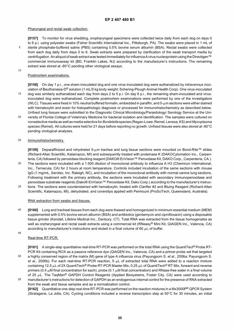
EP 2 407 480 B1
35
5
10
15
20
25
30
35
40
45
50
55
Pharyngeal and rectal swab collection.
[0157] To monitor for virus shedding, oropharyngeal specimens were collected twice daily from each dog on days 0to 6 p.i. using polyester swabs (Fisher Scientific International Inc., Pittsburgh, PA). The swabs were placed in 1 mL ofsterile phosphate-buffered saline (PBS) containing 0.5% bovine serum albumin (BSA). Rectal swabs were collectedfrom each dog daily from days 0 to 6. Swab extracts were prepared by clarification of the swab transport media bycentrifugation. An aliquot of swab extract was tested immediately for influenza A virus nucleoprotein using the Directigen™commercial immunoassay kit (BD, Franklin Lakes, NJ) according to the manufacturer’s instructions. The remainingextract was stored at -80°C pending other virological assays.
Postmortem examinations.
[0158] On day 1 p.i., one sham-inoculated dog and one virus-inoculated dog were euthanatized by intravenous inoc-ulation of Beuthanasia-D® solution (1 mL/5 kg body weight; Schering-Plough Animal Health Corp). One virus-inoculateddog was similarly euthanatized each day from days 2 to 5 p.i. On day 6 p.i., the remaining sham-inoculated and virus-inoculated dog were euthanatized. Complete postmortem examinations were performed by one of the investigators(WLC). Tissues were fixed in 10% neutral buffered formalin, embedded in paraffin, and 5-mm sections were either stainedwith hematoxylin and eosin for histopathologic diagnosis or processed for immunohistochemistry as described below.Unfixed lung tissues were submitted to the Diagnostic Clinical Microbiology/Parasitology/ Serology Service at the Uni-versity of Florida College of Veterinary Medicine for bacterial isolation and identification. The samples were cultured onnonselective media as well as media selective for Bordetella species (Regan-Lowe; Remel, Lenexa, KS) and Mycoplasmaspecies (Remel). All cultures were held for 21 days before reporting no growth. Unfixed tissues were also stored at -80°Cpending virological analyses.
Immunohistochemistry.
[0159] Deparaffinized and rehydrated 5-mm trachea and lung tissue sections were mounted on Bond-Rite™ slides(Richard-Allan Scientific, Kalamazoo, MI) and subsequently treated with proteinase K (DAKOCytomation Inc., Carpen-teria, CA) followed by peroxidase blocking reagent (DAKO® EnVision™ Peroxidase Kit, DAKO Corp., Carpenteria, CA ).The sections were incubated with a 1:500 dilution of monoclonal antibody to influenza A H3 (Chemicon International,Inc., Ternecula, CA) for 2 hours at room temperature. Controls included incubation of the same sections with mouseIgG (1 mg/mL, Serotec, Inc. Raleigh, NC), and incubation of the monoclonal antibody with normal canine lung sections.Following treatment with the primary antibody, the sections were incubated with secondary immunoperoxidase andperoxidase substrate reagents (Dako® EnVision™ Peroxidase Kit, Dako Corp.) according to the manufacturer’s instruc-tions. The sections were counterstained with hematoxylin, treated with Clarifier #2 and Bluing Reagent (Richard-AllanScientific, Kalamazoo, MI), dehydrated, and coverslips applied with Permount (ProSciTech, Queensland, Australia).
RNA extraction from swabs and tissues.
[0160] Lung and tracheal tissues from each dog were thawed and homogenized in minimum essential medium (MEM)supplemented with 0.5% bovine serum albumin (BSA) and antibiotics (gentamycin and ciprofloxacin) using a disposabletissue grinder (Kendall, Lifeline Medical Inc., Danbury, CT). Total RNA was extracted from the tissue homogenates aswell as orpharyngeal and rectal swab extracts using a commercial kit (RNeasy® Mini Kit, QIAGEN Inc., Valencia, CA)according to manufacturer’s instructions and eluted in a final volume of 60 mL of buffer.
Real-time RT-PCR.
[0161] A single-step quantitative real-time RT-PCR was performed on the total RNA using the QuantiTect® Probe RT-PCR Kit containing ROX as a passive reference dye (QIAGEN Inc., Valencia, CA) and a primer-probe set that targeteda highly conserved region of the matrix (M) gene of type A influenza virus (Payungporn S. et al., 2006a; Payungpom S.et al., 2006b). For each real-time RT-PCR reaction, 5 mL of extracted total RNA were added to a reaction mixturecontaining 12.5 mL of 2X QuantiTech® Probe RT-PCR Master Mix, 0.25 mL of QuantiTech® RT Mix, forward and reverseprimers (0.4 mM final concentration for each), probe (0.1 mM final concentration) and RNase-free water in a final volumeof 25 mL. The TaqMan® GAPDH Control Reagents (Applied Biosystems, Foster City, CA) were used according tomanufacturer’s instructions for detection of GAPDH as an endogenous internal control for the presence of RNA extractedfrom the swab and tissue samples and as a normalization control.[0162] Quantitative one-step real-time RT-PCR was performed on the reaction mixtures in a Mx3000P® QPCR System(Stratagene, La Jolla, CA). Cycling conditions included a reverse transcription step at 50°C for 30 minutes, an initial

EP 2 407 480 B1
36
5
10
15
20
25
30
35
40
45
50
55
denaturation step at 95°C for 15 minutes to activate the HotStarTaq® DNA polymerase, and amplification for 40 cycles.Each amplification cycle included denaturation at 94°C for 15 seconds followed by annealing/extension at 60°C for 1minute. The FAM (emission wavelength 518 nm) and VIC (emission wavelength 554 nm) fluorescent signals wererecorded at the end of each cycle. The threshold cycle (Ct) was determined by setting the threshold fluorescence (dR)at 1000 in each individual experiment. The Mx3000P® version 2.0 software program (Stratagene, La Jolla, CA) wasused for data acquisition and analysis. The positive control consisted of amplification of RNA extracted from A/ca-nine/FL/242/03 (H3N8) virus. The results were normalized by dividing the M Ct value by the corresponding GAPDH Ctvalue for each sample.
Virus re-isolation from tissues.
[0163] Frozen lung and trachea tissues from virus-inoculated dogs were thawed and homogenized in 10 volumes ofDMEM supplemented with 0.5% BSA and antibiotics. Solid debris was removed by centrifugation and supernatants wereinoculated onto MDCK cells cultured in DMEM supplemented with 1 mg/mL TPCK-treated trypsin (Sigma-Aldrich Corp.,St. Louis, MO) and antibiotics as described above. Cells were grown in 25 cm2 flasks at 37°C in a humidified atmospherecontaining 5% CO2. The cultures were observed daily for morphologic changes and harvested at 5 days post inoculation.The harvested cultures were clarified by centrifugation and the supernatants inoculated onto fresh MDCK cells asdescribed for the initial inoculation; two additional passages were performed for samples that did not show evidence ofinfluenza virus by hemagglutination or RT-PCR. Hemagglutination activity in the clarified supernatants was determinedusing 0.5% turkey red blood cells as previously described (Crawford et al., 2005). RT-PCR was performed as describedbelow.
RT-PCR, nucleotide sequencing, and phylogenetic analyses.
[0164] Viral RNA was extracted from MDCK supernatant using the QIAamp® Viral RNA Mini Kit (QIAGEN Inc., Valencia,CA) according to manufacturer’s instructions. The viral RNA was reverse transcribed to cDNA using the QIAGEN®
OneStep RT-PCR Kit (QIAGEN Inc., Valencia, CA) according to manufacturer’s instructions. PCR amplification of thecoding region of the 8 influenza viral genes in the cDNA was performed as previously described (Crawford et al., 2005),using universal gene-specific primer sets (primer sequences available on request). The resulting DNA amplicons wereused as templates for automated sequencing in the ABI PRISM® 3100 automated DNA sequencer using cycle sequencingdye terminator chemistry (Applied Biosystems, Foster City, CA). Nucleotide sequences were analyzed using the Laser-gene 6 Package® (DNASTAR, Inc., Madison, WI). The nucleotide sequences for viruses recovered from infected dogswere compared to the sequences of the virus in the inoculum to determine if any changes had occurred during replicationin the respiratory tract.
EXAMPLE 12-CLINICAL DISEASE
[0165] All 6 virus-inoculated dogs developed a transient fever (rectal temperature ≥39°C) for the first 2 days p.i., butnone exhibited respiratory signs such as cough or nasal discharge over the 6-day observation period. The sham-inoc-ulated dogs remained clinically healthy.
EXAMPLE 13-VIRUS SHEDDING
[0166] Influenza A nucleoprotein was detected in the oropharyngeal swab collected from one of the virus-inoculateddogs at 24 hours p.i. The oropharyngeal swabs collected from one dog at 72, 84, and 120 hours p.i., and another dogat 108, 120, and 132 hours p.i., were positive for virus by quantitative real-time RT-PCR (Table 11). The absolute numberof influenza M gene copies per mL of swab extract increased with time from 3 to 6 days p.i. No virus was detected in therectal swabs.
EXAMPLE 14-POSTMORTEM EXAMINATIONS
[0167] In contrast to the previous experimental infection using specific pathogen-free Beagles (Crawford et al., 2005),the virus-inoculated mongrel dogs had pneumonia as evidenced by gross and histological analyses of the lungs fromdays 1 to 6 p.i. In addition to pneumonia, the dogs had rhinitis, tracheitis, bronchitis, and bronchiolitis similar to thatdescribed in naturally infected dogs (Crawford et al., 2005). There was epithelial necrosis and erosion of the lining ofthe airways and bronchial glands with neutrophil and macrophage infiltration of the submucosal tissues (Figure 5, upperpanels). Immunohistochemistry detected viral H3 antigen in the epithelial cells of bronchi, bronchioles, and bronchialglands (Figure 5, lower panels). No bacterial superinfection was present. The respiratory tissues from the 2 sham-

EP 2 407 480 B1
37
5
10
15
20
25
30
35
40
45
50
55
inoculated dogs were normal.
EXAMPLE 15-VIRUS REPLICATION IN TRACHEA AND LUNGS
[0168] The trachea and lungs were positive for virus by quantitative real-time RT-PCR in all dogs from 1 to 6 days p.i.(Table 12). The absolute number of influenza M gene copies per mL of trachea homogenate increased from 1 to 5 daysp.i., then decreased on day 6. The absolute number of M gene copies per mL of lung homogenate decreased from 1 to6 days p.i. In general, the trachea contained ≥ one log10 more virus than the lung on each of the 6 days p.i.
MATERIALS AND EXAMPLES METHODS FOR EXAMPLE 16
Virus strains
[0169] Canine influenza virus strains as well as those of avian, equine and human origin (listed in Table 15) werepropagated in embryonated eggs or MDCK cells and their infectivity was titrated by endpoint dilution in chicken embryos,or plaque assay. Rapid virus quantification was performed by hemagglutination assay using turkey red blood cell eryth-rocytes.
Table 11. Detection of virus shedding in the oropharynx of mongrel dogs inoculated with canine influenza virus by quantitative real-time RT-PCR.
Dog ID Time p.i. (hours)a M/GAPDH ratiob Matrix gene (copies / uL)c
860
72 1.20 1.57E+02
84 1.30 8.25E+02
120 1.23 1.47E+03
894
108 1.17 1.17E+02
120 1.41 1.37E+02
132 1.27 3.74E+02
a Time that oropharyngeal swabs were collected from the dogs following inoculation with A/canine/FL/43/04 (H3N8)virus.b Normalization ratios were calculated by dividing the M (Ct) by the GAPDH (Ct) for each swab extract.c The absolute number of matrix gene copies per uL of swab extract.
Table 12. Detection of virus replication in the trachea and lung of mongrel dogs inoculated with canine influenza virus by quantitative real-time RT-PCR.
M/GAPDH ratiob Matrix gene (copies / uL)c
Dog ID Time p.i. (hours)a Lung Trachea Lung Trachea
797 24 1.20 1.43 8.22E+05 3.11E+04
801 48 1.33 0.99 1.15E+05 6.52E+06
789 72 1.44 1.12 2.39E+04 1.56E+05
819 96 1.40 1.27 3.19E+04 1.43E+05
860 120 1.59 1.04 3.48E+03 1.17E+06
894 144 1.70 1.15 4.78E+02 1.50E+03
a Time that tissues were collected from the dogs following inoculation with A/canine/FL/43/04 (H3N8) virus.b Normalization ratios were calculated by dividing the M (Ct) by the GAPDH (Ct) for each tissue homogenate.c The absolute number of matrix gene copies per uL of tissue homogenate.

EP 2 407 480 B1
38
5
10
15
20
25
30
35
40
45
50
55
Diagnostic specimens
[0170] A Total of 60 canine’s lung tissues collected from suspect cases of viral respiratory disease during the year of2005 were tested for the presence of canine influenza virus.
RNA extraction from canine tissue samples
[0171] Blocks of lung tissue weighing between 20 and 30 mg were homogenized in a disposable tissue grinder (Kendal).Total RNA was extracted using a commercial kit (RNeasy Mini Kit, Qiagen, Valencia, CA) and eluted in a final volumeof 60 mL, following the manufacturer’s recommendations.
Primers and probes design
[0172] Multiple sequence alignments of the H3 and M genes from various subtypes and from diverse species wereperformed using the CLUSTAL X program (Version 1.8). Matrix (M) primers and probe were selected from the conservedregions of over the known sequences corresponding to different subtypes of influenza A virus, whereas the H3 hemag-glutinin gene-specific primers and probe set were selected to specifically match equine and canine influenza A virusgenes and mismatch the homologous avian and human genes (Table 13). Primer design software (OLIGOS Version9.1) and the web based analysis tools provided by EXIQON (http://lnatools.com) was used for Tm calculation andprediction of secondary structure as well as self hybridization. A conserved region of an 18S rRNA gene was used asendogenous internal control for the presence of RNA extracted from canine tissue sample. The Pre-Developed TaqMan®Assay Reagents for Eukaryotic 18S rRNA (VIC/TAMRA) (Applied Biosystems) was used for the real-time detection of18S rRNA in tissue samples.
Real-time RT-PCR condition
[0173] A single-step real-time RT-PCR was performed by using the Quantitect Probe RT-PCR Kit containing ROX asa passive reference dye (Qiagen, Valencia, CA). In each real-time RT-PCR reaction, 5 mL of RNA sample were usedas a template to combine with a reaction mixture containing 10 mL of 2X QuantiTech Probe RT-PCR Master Mix, 0.2mL of QuantiTech RT Mix, primers (0.4 mM final conc. for H3 gene or 0.6 mM final conc. for M gene), probe (0.1 mM finalconc. for H3 gene or 0.2 mM final conc. for M gene) and RNase-free water in a final volume of 20 mL. One-step real-time RT-PCR was performed in the Mx3005P Real-Time QPCR System (Stratagene). Cycling conditions included areverse transcription step at 50°C for 30 minutes. After an initial denaturation step at 95°C for 15 minutes in order toactivate the HotStarTaq DNA polymerase, amplification was performed during 40 cycles including denaturation (94°Cfor 15 seconds) and annealing/extension (60°C for 30 seconds). The FAM (emission wavelength 516 nm for H3 and Mdetection) and VIC (emission wavelength 555 nm for 18S rRNA detection) fluorescent signals were obtained once percycle at the end of the extension step. Data acquisition and analysis of the real-time PCR assay were performed usingthe Mx3005P software version 2.02 (Stratagene).
Specificity of H3 primers/ probe set for canine influenza (H3N8) and universality of M primers/probe set for type A influenza virus
[0174] In order to test the specificity of each primers/probe set, RNA extracted from several known subtypes of influenzaA viruses were used as a template in the real-time RT-PCR assay (Table 15).
RNA standard for determination of the real-timer RT-PCR performance
[0175] The genes of canine influenza A virus (A/canine/Florida/242/2003(H3N8)) were used to generate the PCRamplicons for H3 (nt 1-487) and M (nt 1-276) by using primers linked with T7 promoter (Table 13). Then the purifiedPCR amplicons of H3 and M genes were used as templates for in vitro transcription by using Riboprobe in vitro Tran-scription System-T7 (Promega) following the manufacturer’s recommendations. The concentration of the transcribedRNAs was calculated by measuring absorbance at 260 nm. The RNAs were then serially diluted 10-fold, ranging from108 to 10 copies/ mL to perform sensitivity tests. Moreover, a standard curve was generated by plotting the log of initialRNA template concentrations (copies/ mL) against the threshold cycle (Ct) obtained from each dilution in order to de-termine the overall performance of real-time RT-PCR.

EP 2 407 480 B1
39
5
10
15
20
25
30
35
40
45
50
55
Comparative sensitivity tests between real-time RT-PCR and Directgen Flu A test kit
[0176] Stock viruses of two viral strains including A/Wyoming/3/2003 (H3N2) at 106.67 EID50/mL (HA=64) and A/ca-nine/Florida/242/2003(H3N8) at 107.17 EID50/mL (HA=16) were used for the detection threshold assay. Logarithmicdilution of specimens in phosphate-buffered saline (PBS) (125 mL) were used in a rapid influenza A antigen detectionkit, Directigen Flu A, (Becton, Dickinson and Company) following the manufacturer’s instructions. Each Directigen FluA test device has an H1N1 influenza antigen spot in the center of the membrane which develops as a purple dot andindicates the integrity of the test, which is based on a monoclonal antibody to the nucleoprotein (NP). The developmentof a purple triangle surrounding the dot is indicative of the presence of influenza NP in the tested specimen. The intensityof the purple signal from the triangle was scored as + (outline of triangle), ++ (lightly colored triangle), +++ (dark-purpletriangle) and ++++ (very dark-purple triangle). Viral RNA was extracted 125 mL aliquots of each virus dilution by usingQIAamp Viral RNA Mini Kit (Qiagen, Valencia, CA) and eluting in a final volume of 50 mL. A volume of 5 uL of theextracted viral RNAs were tested by real-time RT-PCR for comparative sensitivity test with Directigen Flu A kit.
EXAMPLE 16
[0177] The real-time RT-PCR assay for canine influenza relies on information from three molecular probes whichtarget 18S rRNA from host cell was well as M and H3 from the influenza A virus genome (Table 14). Amplification of thehost gene is a reporter of specimen quality and integrity. Clinical, necropsy or laboratory samples containing canineinfluenza (H3N8) virus are expected to yield amplification signal with the three probes. Specimens yielding amplificationsignal with M and 18S rRNA probes but negative for H3 would be indicative of an influenza virus subtype H3 from human,swine or avian origin or from non-H3 subtypes. These rare cases could be resolved by RT-PCR using HA universalprimers to generate amplicon cDNA that can be analyzed by sequencing. Properly collected and handled specimenslacking influenza A virus yield 18S rRNA amplification signal only. Situations in which only the 18S rRNA probe and theH3 probes yield amplification signal are indicative of faulty technique, unless proven otherwise; either a false negativewith M probes or false positive for H3 need to be demostrated. Finally, specimens failing to yield amplification signalswith the three probes are indicative of defective sample collection, degradation, faulty RNA extraction or the presenceof inhibitors the polymerases used in PCR.[0178] In order to test the specificity of the H3 primers/probe set for canine influenza A virus (H3N8) and the universalityof M primers/probe set for type A influenza, several subtypes of influenza A viruses were tested by real-time RT-PCR.The results show that H3 primers/probe set yielded a positive amplification signal only with canine influenza (H3N8). Nosignificant false positive or non-specific amplification signals were observed in other subtypes or human H3 strains. TheM primers/probe set yielded positive amplification signal with all of the strains tested (Table 15). These results indicatedthat H3 primers/probe specifically detects canine influenza A virus (H3N8) whereas M primers/probe detect multiplesubtypes of type A influenza viruses.[0179] The performance of real-time RT-PCR assays was evaluated by endpoint dilution of M and H3 in vitro transcribedRNAs. As expected, the threshold cycle (Ct) increased in direct correlation with the dilution of the RNA standards. Thefluorescent signals can be detected at RNA standard dilutions of M and H3 as low as 103 and 102 copies/mL, respectively(Figure 6A and 6B). The standard curves of M and H3 genes were constructed by plotting the log of starting RNAconcentrations against the threshold cycle (Ct) obtained from each dilution (Figure 6C and 6D). The slope of the standardcurve is used to determine the PCR reaction efficiency, which is theoretically exponential; 100% amplification efficiencywould imply doubling of amplicon cocentration each cycle. The standard curves with a slope between approximately-3.1 and -3.6 are typically acceptable for most applications requiring accurate quantification (90-110 % reaction efficiency).An Rsq value is the fit of all data to the standard curve plot. If all the data lie perfectly on the line, the Rsq will be 1.00.As the data fall further from the line, the Rsq decreases. An Rsq value ≥ 0.985 is acceptable for most assays. The Mstandard curve revealed a slope of -3.576 (efficiency= 90.4 %) and Rsq= 1.00 whereas H3 standard curve yielded aslope of -3.423 (efficiency= 95.9%) and Rsq= 0.999. These values indicate satisfactory amplification efficiency andoverall performance of the real-time RT-PCR assays. We attribute the lower efficiency and sensitivity of M primers/probeset as compared to H3 primers/probe set to the N-fold degeneracy of M primer sequences required to ensure broadcoverage of M gene sequences variability across viruses of multiple subtypes, hosts and lineages.[0180] The sensitivity of real-time RT-PCR assay was also compared with the commercial rapid antigen detectionassay (Directigen Flu A). Logarithmic dilutions of A/Wyoming/3/2003 (H3N2) and A/canine/Florida/242/2003(H3N8) wereanalyzed with Directigen Flu A and by real-time RT-PCR. The results of Directigen Flu A showed that the sensitivitiesagainst both viral strains are approximately 100-fold dilution from the stock viruses used in these experiments (Figure7). The signals (purple color) generated by the canine virus (A/canine/Florida/242/2003: 106.x PFU/ml) samples weremuch weaker than those found in human virus (A/Wyoming/3/2003: 107.x PFU/ml), in agreement with the lower virusconcentration in these samples. Alternatively, lower signal for canine influenza could be attributed to the molecularspecificity of monoclonal antibodies against the NP; i.e. poor conservation of the amino acids within the NP epitope of

EP 2 407 480 B1
40
5
10
15
20
25
30
35
40
45
50
55
canine influenza A viruses.[0181] Real-time RT-PCR of the M gene yielded Ct values above threshold with virus 10 and 30 PFU equivalents perreaction of A/canine/Florida/242/2003 and A/Wyoming/3/2003, respectively (Table 16). The differences between thesensitivity value of 2 viral strains because the differences in the original viral titers. The H3 gene detection comparisonbetween canine and human influenza viruses was not performed because the H3 primers/probe in our realtime RT-PCRassay amplifies exclusively canine influenza A virus. RT-PCR was 105 times more sensitive than the rapid antigendetection kit.[0182] To evaluate the performance of the RT-PCR test in necropsy specimens from dogs with acute respiratorydisease, sixty canine lung tissue samples submitted during the year of 2005 were tested for the presence of canineinfluenza A virus by real-time RT-PCR. A total of 12 out of 60 samples (20%) were positive with both M and H3 geneswhereas the remaining 48 samples yielded negative result for both M and H3 gene. Virus isolation attempts wereconducted by egg and MDCK cell inoculation, to evaluate the specificity of the realtime assay; 2 out 12 samples thatwere positive for canine influenza by RT-PCR yielded canine influenza virus (data not shown, manuscript in preparation).Although all of the tissues were collected from dogs with a history of severe respiratory disease, most of the samplesyielded no canine influenza virus by either realtime PCR or conventional isolation, suggesting a high incidence of otherrespiratory pathogens such as Bordetella bronchiseptica, canine distemper or parainfluenza virus. The single step real-time RT-PCR assay herein provides a rapid, sensitive and cost-effective approach for canine influenza A virus (H3N8)detection. Rapid laboratory diagnosis of canine influenza A virus (H3N8) infections in the early stage of the disease canyield information relevant to clinical patient and facility management.
Table 13: Primers and probes used for real-time RT-PCR detection and in vitro transcription
Oligo name Type Target Sequence * Application
Ca-H3-F387 Forward primer
H3 (nt 387-406)
5’-tatgcatcgctccgatccat-3’ (SEQ ID NO: 79) Real-time PCR
Ca-H3-R487 Reverse primer
H3 (nt 487-467)
5’-gctccacttcttccgttttga-3’ (SEQ ID NO: 80)
Ca-H3-P430 TaqMan probe
H3 (nt 430-459)
FAM-aattcacagcagagggattcacatggacag-BHQ1 (SEQ ID NO: 81)
FluA-M-F151 Forward primer
M (nt 151-174)
5’-catggartggctaaagacaagacc-3’ (SEQ ID NO: 82) Real-time PCR
FluA-M-R276 Reverse primer
M (nt 276-253)
5’-agggcattttggacaaakcgtcta-3’ (SEQ ID NO: 83)
FluA-M-P218 LNA TaqMan probe
M (nt 218-235)
FAM-acgcTcaccgTgcccAgt-BHQ1 (SEQ ID NO: 84)
H3-F1 Forward primer
H3 (nt 1-14)
5’-tattcgtctcagggagcaaaagcagggg-3’ (SEQ ID NO: 85)
In vitro transcription
T7/H3-R490 Reverse primer
T7 / H3 (nt 487-467)
5’-tgtaatacgactcactatagggctccacttcttccgttttga-3’ (SEQ ID NO: 86)
M-F1 Forward primer
M (nt 1-15) 5’-gatcgctcttcagggagcaaaagcaggtag-3’ (SEQ ID NO: 87)
In vitro transcription
T7/M-R276 Reverse primer
M (nt 276-253)
5’-tgtaatacgactcactatagggcattttggacaaagcgtc-3’ (SEQ ID NO: 88)
* Note: Uppercases = LNA (Locked Nucleic Acid) residues, r = a or g, k= g or t, underline= T7 promoter sequence
Table 14: Interpretation of the real-time RT-PCR assay
Interpretation Results
M H3 18S rRNA
Positive for canine influenza A virus (H3N8) + + +

EP 2 407 480 B1
41
5
10
15
20
25
30
35
40
45
50
55
(continued)
Table 14: Interpretation of the real-time RT-PCR assay
Interpretation Results
M H3 18S rRNA
Positive for influenza A virus (unknown subtype) + - +
Negative for influenza A virus - - +
Error in RNA extraction or presence of PCR inhibitor - - -
Table 15: Specificity test of canine H3 primers/probe set and universality test of M primers/probe set with several subtypes of influenza A viruses
Subtypes Real-time RT-PCR detection
Strain Name Host H3 gene (Ct) M gene (Ct)
H1A/Ohio/1983 Human No Ct 15.40
A/WSN/1933 Human No Ct 20.09
H3
A/Wyoming/3/2003 Human No Ct 28.85
A/Victoria/3/1975 Human No Ct 16.62
A/canine/FL/242/2003 Canine 28.43 29.25
H4Turkey/MN/1066/1980 Avian No Ct 17.49
Clinical sample* Avian No Ct 20.87
H5AChicken/Thailand/CUK2/2004 Avian No Ct 20.13
A/Pheasant/NJ/1335/1998 Avian No Ct 16.64
H6 Clinical sample* Avian No Ct 19.52
H10Clinical sample* Avian No Ct 25.64
Clinical sample* Avian No Ct 19.59
H11Clinical sample* Avian No Ct 15.72
Clinical sample* Avian No Ct 24.55
* Note that subtypes of clinical samples were confirmed by nucleotide sequencing.
Table 16: Comparative sensitivity tests for influenza A virus detection between real-time RT-PCR and Directigen Flu A
Virus dilutions Directigen Flu A Real-time RT-PCR of M (Ct)
A/canine/242/03 A/Wyoming/3/03 A/canine/242/03 A/Wyoming/3/2003
10-1 ++ ++++ 22.42 19.48
10-2 + +++ 25.85 22.66
10-3 - - 29.27 25.76
10-4 Not done Not done 32.66 28.66
10-5 Not done Not done 35.48 33.14
10-6 Not done Not done 37.51 35.06
10-1 Not done Not done 39.09 36.44
10-8 Not done Not done No Ct 38.93

EP 2 407 480 B1
42
5
10
15
20
25
30
35
40
45
50
55
Table 17.
Class of Amino Acid Examples of Amino Acids
Nonpolar Ala, Val, Leu, Ile, Pro, Met, Phe, Trp
Uncharged Polar Gly, Ser, Thr, Cys, Tyr, Asn, GlnAcidic Asp, GluBasic Lys, Arg, His
Table 18.
Letter Symbol Amino Acid Letter Symbol Amino Acid
A Alanine M Methionine
B Asparagine or aspartic acid N Asparagine
C Cysteine P Proline
D Aspartic Acid Q Glutamine
E Glutamic Acid R Arginine
F Phenylalanine S Serine
G Glycine T Threonine
H Histidine V Valine
I Isoleucine W Tryptophan
K Lysine Y Tyrosine
L Leucine Z Glutamine or glutamic acid
Table 19. Amino acid differences between PB2 proteins of H3N8 equine and canine influenza viruses
Position Equine Consensus * Canine/FL/03 Canine/FL/04
5 K K E
12 S L L
37 G G E
175 R R I
374 L I I
375 R R K
447 Q Q H
Table 20. Amino acid differences between PB1 proteins of H3N8 equine and canine influenza viruses
Position Equine Consensus * Canine/FL/03 Canine/FL/04
114 V I I
154 D G G
221 A T T
317 M I I
459 I I V

EP 2 407 480 B1
43
5
10
15
20
25
30
35
40
45
50
55
(continued)
Table 20. Amino acid differences between PB1 proteins of H3N8 equine and canine influenza viruses
Position Equine Consensus * Canine/FL/03 Canine/FL/04
682 I I V
Table 21. Amino acid differences between PA proteins of H3N8 equine and canine influenza viruses
Position Equine Consensus * Canine/FL/03 Canine/FL/04
27 D N N
62 I V V
213 R K K
337 A T T
343 A E E
345 L I I
353 K R R
400 T T A
450 V I I
460 M M I
673 R R K
675 N D D
*Based on available genes of viruses isolated between 1963 and 1998.
Table 22. Amino acid differences between NP proteins of H3N8 equine and canine influenza viruses
Position Equine Consensus * Canine/FL/03 Canine/FL/04
16 G D D
157 A T T
214 R R K
285 V V I
286 A T T
359 A T T
375 D D N
384 R K K
452 R K K
Table 23. Amino acid differences between NA proteins of H3N8 equine and canine influenza viruses
Position Equine Consensus * Canine/FL/03 Canine/FL/04
9 A/T T A
12 S F F
20 L I I

EP 2 407 480 B1
44
5
10
15
20
25
30
35
40
45
50
55
EXAMPLES 17 - CANINE INFLUENZA CHALLENGE MODEL DEVELOPMENT.
[0183] The canine influenza (canine flu) virus, which was isolated from flu outbreaks in Florida, was observed to bea H3N8 type influenza virus, and closely related to equine flu virus strain, A/equine/Ohio/03 (Crawford et al., SCIENCEVol. 309, September 2005, incorporated by reference in its entirety into this patent). The potential of using the equineflu virus strain A/equine/Ohio/03 to induce influenza-like disease in dogs was investigated in this study.
Procedure:
[0184] Ten 13-week-old beagles of mixed sex were obtained from a commercial supplier, and housed in individualcages in a BSL-2 facility. The dogs were randomly assigned to two groups of 5 dogs each. As shown in Table 26, one
(continued)
Table 23. Amino acid differences between NA proteins of H3N8 equine and canine influenza viruses
Position Equine Consensus * Canine/FL/03 Canine/FL/04
40 G R R
42 G D D
46 N K K
52 E E K
61 R K K
69 N S S
72 E K K
201 V I I
261 I V V
301 I I V
396 N D D
397 L P P
Table 24. Amino acid differences between M1 proteins of H3N8 equine and canine influenza viruses
Position Equine Consensus * Canine/FL/03 Canine/FL/04
M1 161 S S A
M1 208 K/Q R R
*Based on available genes of viruses isolated between 1963 and 1998.
Table 25. Amino acid differences between NS1 proteins of H3N8 equine and canine influenza viruses
Position Equine Consensus * Canine/FL/03 Canine/FL/04
44 K R R
59 R H H
71 E K K
86 A T T
88 R R L
140 R G G
216 P S S
* Based on available genes of viruses isolated between 1963 and 1998.

EP 2 407 480 B1
45
5
10
15
20
25
30
35
40
45
50
55
group was subjected to a intratracheal challenge, and the other group was subjected to an oronasal challenge. The dogswere challenged at 14 weeks-of-age.
[0185] A cell culture grown equine flu virus A/equine/Ohio/03 was used as the challenge virus. For intratrachealchallenge, the challenge virus was administered via a delivery tube, which consisted of a cuffed tracheal tube (Size4.0/4.5, Sheridan, USA) and feeding tube (size 5Fr, 1.7 mm, /16 inches in length, Kendall, USA) in 0.5 to 1.0 ml volume.For oronasal challenge, the challenge virus (107 to 108 TCID50 per dog) was administered as a mist using a nebulizer(DeVilbiss Ultra-Neb®99 ultrasonic nebulizer, Sunrise Medical, USA) in a 2 to 3 ml volume.[0186] The dogs were observed for flu related clinical signs for 14 days post-challenge. Serum samples were collectedfrom each dog on day zero (before challenge), and days 7 and 14 post-challenge for determining the HI titer using aH3N8 equine influenza virus with a standard protocol (SAM 124, CVB, USDA, Ames, IA). All dogs were humanelyeuthanized and lung tissues were collected in 10% buffered formalin for histopathological evaluation.
Results:
[0187] The results of this experiment are summarized in Table 27. Influenza related clinical signs were observed in afew dogs after challenge. These signs included fever (>103°F; >39.4°C) and cough. Two of 5 dogs (i.e., 40%) had fevers(>103°F; >39.4oC) in Group 1, compared to 1 of 5 (i.e., 20%) dogs in Group 2. One dog from the oronasal challengegroup had sneezing, and another had cough following the challenge. An HI titer range from 10 to 80, with a geometricmean titer (GMT) of 20, was observed for Group 1. A titer range of 40 to 160, with a GMT of 86, was observed for Group2. One dog from each group had histopathological lesions compatible with or pathognomic for influenza.
Table 26: Experimental Design
Group Number of Dogs Challenge Route
1 5 Intratracheal
2 5 Oronasal

EP 2 407 480 B1
46
5
10
15
20
25
30
35
40
45
50
55
Tab
le 2
7. C
anin
e flu
cha
lleng
e -
clin
ical
sig
ns, v
irus
isol
atio
n, h
isto
path
olog
y re
sults
and
ser
olog
y re
sults
Do
g*
IDC
hal
len
ge
met
ho
d*
Clin
ical
sig
ns
Vir
us
iso
lati
on
Mic
rosc
op
ic le
sio
n
(his
top
ath
olo
gy)
Ser
olo
gy
(HI t
iter
)
Nas
al/o
ral
swab
Tra
chea
l sc
rap
ing
Lu
ng
ti
ssu
esP
re-c
hal
len
ge
7-d
ays
po
st
chal
len
ge
14-d
ays
po
st
chal
len
ge
AA
HIn
trat
rach
eal
none
nega
tive
nega
tive
nega
tive
nega
tive
1010
20
AD
BIn
trat
rach
eal
none
nega
tive
nega
tive
nega
tive
nega
tive
1080
20
AD
CIn
trat
rach
eal
Fev
er*
nega
tive
nega
tive
nega
tive
nega
tive
1020
20
AE
BIn
trat
rach
eal
Fev
erne
gativ
ene
gativ
ene
gativ
epo
sitiv
e10
4020
AE
EIn
trat
rach
eal
none
nega
tive
nega
tive
nega
tive
inco
nclu
sive
1020
10
AA
EO
rona
sal
none
nega
tive
nega
tive
nega
tive
nega
tive
1080
80
AA
GO
rona
sal
none
nega
tive
nega
tive
nega
tive
nega
tive
1040
80
AB
YO
rona
sal
Occ
asio
nal s
neez
e,
occa
sion
al c
ough
nega
tive
nega
tive
nega
tive
posi
tive
1080
160
AD
YO
rona
sal
Fev
er, o
ccas
iona
l sn
eeze
nega
tive
nega
tive
nega
tive
nega
tive
1080
80
AD
ZO
rona
sal
none
nega
tive
nega
tive
nega
tive
nega
tive
1080
160
* T
he a
nim
als
wer
e ch
alle
nged
with
an
Equ
ine
flu is
olat
e O
hio
03.
** R
ecta
l tem
pera
ture
≥10
3°F
; ≥39
.4°C

EP 2 407 480 B1
47
5
10
15
20
25
30
35
40
45
50
55
EXAMPLE 18 - EFFICACY OF AN EQUINE INFLUENZA VIRUS VACCINE FOR DOGS.
[0188] The canine influenza (canine flu) virus isolated from flu outbreaks in Florida was observed to be a H3N8 typeinfluenza virus, and was closely related to equine flu virus, A/equine/Ohio/03 based on the sequence similarity. Thefollowing study was conducted to determine the efficacy of an experimental inactivated equine influenza virus vaccine.
Procedure:
[0189] Nine 7-week-old beagles of mixed sex were obtained from a commercial supplier, and housed in individualcages in a BSL-2 facility. These dogs were randomly assigned to two groups, as summarized in Table 28:
The first group consisted of 5 dogs, which were vaccinated with an inactivated, CARBIGEN™ adjuvanted, equine fluvirus A/equine/Ohio/03 vaccine at 8 and 12 weeks-of-age via subcutaneous (SQ) route. The A/equine/Ohio/03 wasinactivated by binary ethylenimine ("BEI") using a standard method. Each dose of the vaccine contained 5% by massCARBIGEN™, 4096 HA units of the inactivated virus, sufficient PBS to bring the total volume of the dose to 1 ml, andsufficient NaOH to adjust the pH to between 7.2 and 7.4. Serum samples were collected from all dogs on the day of firstand second vaccination and day 7 and 14, post-first and -second vaccination, and at pre-challenge for determining theHI titer using a H3N8 equine influenza virus a standard protocol (SAM 124, CVB, USDA, Ames, IA). At 3 weeks post-second vaccination, the 5 vaccinated dogs and the second group (i.e., the control group) consisting of 4 age-matcheddogs were challenged oronasally with a cell-culture-grown equine influenza virus A/equine/Ohio/03 (107 to 108 TCID50per dog) in a 1-2 ml volume per dose. The challenge virus was administered to the dogs as a mist using a nebulizer(DeVilbiss Ultra-Neb®99 ultrasonic nebulizer, Sunrise Medical, USA). The dogs were observed for flu-related clinicalsigns for 14 days post-challenge. Five dogs (3 vaccinates and 2 controls) 7 days post-challenge and 4 dogs (2 controlsand 2 vaccinates) 14 days post-challenge were humanely euthanized for collection of lung tissues in 10% bufferedformalin for histopathological evaluation.
Results:
[0190] The results of this experiment are summarized in Tables 29 and 30. All vaccinated dogs seroconverted followingthe vaccination. An HI titer range from 40 to 640, with the GMT of 129, was observed during the post-vaccination periodwith equine influenza virus A/equine/Ohio/03, and a HI titer of 160 to 320, with a geometric mean titer of 211, wasobserved with canine flu isolate, A/canine/Florida/242/03. Two of 6 vaccinates had a fever of >103°F (>39.4°C) for oneday and no other clinical signs were observed in any of the dogs following challenge.
Conclusion:
[0191] All the vaccinated dogs responded to the inactivated, CARBIGEN™ adjuvanted equine influenza vaccine. TheHI titer results with a canine influenza virus isolate suggest that the inactivated equine influenza vaccine did induce adetectable level of cross reactive antibody to canine influenza virus. Even though the challenge virus used in this didnot induce any noticeable clinical disease in beagle dogs, based on the HI titer with a canine influenza virus isolate, itwas concluded that inactivated equine vaccine could be used in dogs to induce cross reactive antibodies, which couldpotentially protect dogs against "canine flu" disease caused by H3N8 type canine influenza viruses.
Table 28: Experimental Design
Group Number of Dogs Treatment
1 5 Vaccine
2 4 Control
Table 29. Serology - Pre- and post-vaccination and post-challenge HI titers
Dog* Group
HI titers
Pre-vaccination Post-1st vaccination Post-2nd vaccination Post-challenge*
7-d 14-d 7-d 14-d 21-d 7-d 14-d
AKT Vaccinate** < 10 40 80 640 640 640 320 320

EP 2 407 480 B1
48
5
10
15
20
25
30
35
40
45
50
55
EXAMPLE 19 - EFFICACY OF AN EQUINE INFLUENZA VIRUS VACCINE FOR DOGS.
[0192] The canine influenza virus isolated from flu outbreaks in Florida was characterized is closely related to a numberof H3N8 type equine influenza virus isolates. By DNA and amino acid sequence similarity analysis it was demonstratedthat the canine influenza virus is very similar to an equine influenza virus, A/equine/Ohio/03. The following study wasconducted in dogs to determine the efficacy of commercially available equine influenza vaccines in dogs.
(continued)
Table 29. Serology - Pre- and post-vaccination and post-challenge HI titers
Dog* Group
HI titers
Pre-vaccination Post-1st vaccination Post-2nd vaccination Post-challenge*
7-d 14-d 7-d 14-d 21-d 7-d 14-d
ALH Vaccinate** < 10 40 80 320 160 160 80 ***
ALU Vaccinate** < 10 40 80 320 160 160 80 80
ANJ Vaccinate** < 10 40 80 320 160 80 320 ***
ANU Vaccinate** < 10 40 80 320 160 80 160 ***
AJW Control <10 < 10 < 10 10 < 10 < 10 10 ***
AKR Control < 10 < 10 < 10 < 10 < 10 < 10 10 ***
ALZ Control < 10 < 10 < 10 < 10 < 10 < 10 20 20
ARC Control < 10 < 10 < 10 < 10 < 10 < 10 10 10
* The animals were challenged with an equine flu isolate Ohio 03** CARBIGEN™ adjuvanted inactivated equine flu virus Ohio 03 vaccine was used for vaccination*** Euthanized 7-days post-challenge
Table 30. Canine flu challenge* - clinical signs, virus isolation, histopathology results
Dog ID Treatment group Clinical signs
Virus isolation Microscopic lesion (histopathology)Nasal swab Tracheal
scrapingLung tissues
AKT Vaccinate** none negative negative negative negative
ALH Vaccinate** none negative negative negative negative
ALU Vaccinate** none negative negative negative negative
ANJ Vaccinate** none negative negative negative negative
ANU Vaccinate** none negative negative negative negative
AJW Control none negative negative negative negative
AKR Control none negative negative negative negative
ALZ Control none negative negative negative negative
ARC Control none negative negative negative negative
* The animals were challenged with an Equine flu isolate Ohio 03** CARBIGEN™ adjuvanted inactivated equine flu virus Ohio 03 vaccine was used for vaccination

EP 2 407 480 B1
49
5
10
15
20
25
30
35
40
45
50
55
Procedure:
[0193] Approximately 16 month old, 20 mongrels and 20 beagles of mixed sex were used in the study. The dogs wererandomly assigned to 6 groups (Table 31) of 6-7 dogs each. Dogs in groups 1 and 4 were vaccinated with a commerciallyavailable inactivated, adjuvanted equine influenza vaccine (EQUICINE II™, Intervet Inc., Millsboro, DE) at 16 and 17months of age via subcutaneous (SQ) route. The dogs in groups 2 and 5 were vaccinated with a modified live equine/Ken-tucky/91 influenza vaccine in a 1 ml volume via intranasal route (single nostril). Blood samples were collected on theday of vaccination, day 7 and 14 post first vaccination (groups 1, 2, 4, and 5) and post second vaccination (groups 1and 4) for determining the HI titer using an H3N8 equine influenza virus and a canine influenza virus using per a standardprotocol (SAM 124, CVB, USDA, Ames, IA).[0194] Vaccinates (at 72 days post final vaccination) and the controls were challenged oronasally with a cell-culturegrown equine influenza virus strain A/equine/Ohio/03 (107 to 108 TCID50 per dog) in a 1-2 ml volume. The challengevirus was administered to the dogs as mist using a nebulizer (DeVilbiss Ultra-Neb®99 ultrasonic nebulizer, SunriseMedical, USA). The dogs were observed for influenza-related clinical signs for 12 days post-challenge. The nasal andoropharyngeal swabs were collected in Earl’s MEM medium with antibiotics (neomycin and polymyxin B) daily from day-1 to day 12 post challenge for virus isolation. The presence of virus in the swabs indicates that the animal is excretingthe virus in nasal/oral secretions. All dogs were humanely euthanized on day 12 post-challenge and lung tissues werecollected in 10% buffered formalin for histopathological evaluation.
Results:
[0195] All vaccinated dogs seroconverted following the vaccination and the HI titers ranged from 10 to 80 for EQUICINEII™ vaccine group dogs compared to 10 to 40 for the A/KY/91 vaccine group dogs using an equine influenza virus (H3N8type).[0196] The samples collected at 2 weeks post vaccination (post second vaccination for EQUICINE II™ vaccine) wereanalyzed for HI titer determination with a canine influenza as well as with an equine influenza virus (H3N8 type). TheHI results are shown in Table 32. The clinical signs include fever (>103°F; >39.4oC), occasional cough, and mild nasaldischarge observed following the challenge.
Table 31. Experimental design
Group Number of dogs Breed Treatment Number of doses Route of vaccination
1 7 Beagle EQUICINE II™** 2 Subcutaneous
2 7 Beagle A/KY/91*** 1 Intranasal
3 6 Beagle Control N/A* N/A*
4 7 Mongrel EQUICINE II™ 2 Subcutaneous
5 7 Mongrel A/KY/91 1 Intranasal
6 6 Mongrel Control N/A* N/A*
* Not applicable** EQUICINE II™ is marketed by Intervet Inc. as a liquid vaccine. EQUICINE II™ contains inactivated A/Penn-sylvania/63 influenza (or "A/Pa/63") virus and A/equine/Kentucky/93 influenza (or "A/KY/93") virus with car-bopol (i.e., HALOGEN® (Intervet Inc.)). More specifically, a dose of EQUICINE II™ contains: inactivatedA/Pa/63 at 106.0 EID50, inactivated A/KY/93 at 106.7 EID50, 0.25% by volume carbopol, and sufficient PBSto create a total volume of 1 ml.*** A/KY/91 is a freeze-dried vaccine that was reconstituted with water. Such reconstitution was conductedusing vaccine-grade water sufficient to bring the vaccine dosage to a total volume of 1 ml. The vaccinecontained equine/Kentucky/91 influenza (or "A/KY/91 ") virus, and is discussed in, for example, U.S. PatentNos. 6,436,408; 6,398,774; and 6,177,082, which are incorporated by reference in their entirety into thispatent. When reconstituted, a dose of the vaccine contained A/KY/91 at 107.2 TCID50 per ml, 0.015 gramsN-Z AMINE AS™ per ml, 0.0025 grams gelatin per ml, and 0.04 grams D lactose per ml. N-Z AMINE AS™
is a refined source of amino acids and peptides produced by enzymatic hydrolysis of casein. N-Z AMINEAS™ is marketed by Kerry Bio-Science (Norwich, NY, USA).

EP 2 407 480 B1
50
5
10
15
20
25
30
35
40
45
50
55
[0197] Among beagles, 2 of 6 dogs in the EQUICINE II ™ vaccine group (Group 1), 1 of 7 dogs in the A/KY/91 vaccinegroup (Group 2) and 2 of 6 dogs in the control group (Group 3) had fever. One of 6 dogs in Group 3 (control) was positivefor virus in the cell culture supernatant of nasal swab material by hemagglutination assay with 0.25% chicken red bloodcells (CRBC). One of 6 dogs in the control group (Group 3) and 1 of 7 dogs in the A/KY/91 vaccine group (Group 2) hadmild nasal discharge during the post challenge observation period. There was no statistical significant difference (P >0.05) between control and vaccine groups for beagle dogs.[0198] Among mongrels, 5 of 7 dogs in the EQUICINE II™ vaccine group (Group 4), 1 of 7 dogs in the A/KY/91 vaccinegroup (Group 5) and 5 of 6 dogs in the control group (Group 6) had fever. One dog from each of Group 4 and 6 had amild nasal discharge, and one dog from Group 5 had an occasional cough. Two of 7 dogs in the EQUICINE II™ vaccinegroup (Group 4) and 3 of 6 dogs in the control group (Group 6) were positive for influenza virus in the nasal swab byHA assay. None of the dogs from the A/KY/91 group (Group 5) were positive for influenza virus in the nasal swab materials.
Conclusion:
[0199] By serology, it was demonstrated that vaccination of dogs with commercially available equine influenza vaccinesstimulated a moderate level influenza antibody response. There may be some breed difference in development ofinfluenza-related clinical signs in dogs following a challenge with H3N8 type influenza virus. The live attenuated equineinfluenza vaccine (A/KY/91) provided a significant (P< 0.05) protection from clinical disease development in rectaltemperature in mongrels. Also, the live attenuated viral vaccine prevented the shedding of influenza virus in the nasalsecretions.
EXAMPLE 20 - CANINE INFLUENZA CHALLENGE MODEL DEVELOPMENT
[0200] In view of reports that inducing disease in canines for purposes of study had not proven successful, the potentialfor using a canine influenza virus, H3N8, to develop a canine influenza challenge model in dogs was investigated in thefollowing study.
Procedure:
[0201] Ten mongrels of mixed sex were obtained from a commercial supplier, and housed in cages in a BSL-2 facility.The dogs were randomly assigned to two groups of 5 dogs each. As shown in Table 33, one group was subjected to anintratracheal/intranasal challenge, and the other group was subjected.
Table 32. Serology - HI titers at 2 weeks post vaccination
Group Number of dogs Breed Treatment HI titer with
Equine influenza virus Canine influenza virus
Range GMT Range GMT
1 7 Beagle Equicine II™ 10-80 36 10-80 33
2 7 Beagle A/KY/91 10-20 12 20-160 54
3 6 Beagle Control N/A* N/A* N/A* N/A*
4 7 Mongrel Equicine II™ 40-80 54 40-80 50
5 7 Mongrel A/KY/91 10-40 24 40-80 49
6 6 Mongrel Control N/A* N/A* N/A* N/A*
* Not applicable
Table 33. Experimental design
Group Number of dogs Challenge route
1 5 Intratracheal/intranasal
2 5 Oronasal

EP 2 407 480 B1
51
5
10
15
20
25
30
35
40
45
50
55
[0202] The dogs were challenged at approximately12 weeks-of-age. Embryonated-chicken-egg grown canine influ-enza virus (A/canine/Florida/242/03) virus was used as challenge virus. Each dog received a total of approximately 107.2
TCID50 of virus in either 2 ml (for oronasal route) or 4 ml (intratracheal/intranasal route) volume.[0203] For intratracheal/intranasal challenge, 3 ml of the challenge virus was administered into the trachea first, followedby 5 ml of PBS using a delivery tube, which consisted of a cuffed tracheal tube (Size 4.5/5.0, Sheridan, USA) and feedingtube (size 5Fr, 1.7 mm; 16 inches (41 cm) in length, Kendall, USA), and a 1 ml challenge virus, followed by 3 ml ofatmospheric air was administered into nostrils using a syringe.[0204] For oronasal challenge, the challenge virus was administered as a mist using a nebulizer (Nebulair™, DVMPharmaceuticals, Inc., Miami, FL) in approximately 2 ml volume. The dogs were observed for flu-related clinical signsfor 14 days post-challenge. The dogs were euthanized at day 14 post challenge, and tissue (lung and trachea) sampleswere collected in 10% buffered formalin for histopathological examination.
Results:
[0205] All dogs in groups 1 and 2 developed canine influenza clinical signs within 24 to 48 hours. Each dog had 2 ormore of the following clinical signs: fever (>103.0°F; >39.4°C), cough, serous or mucopurulent ocular discharge, serousor mucopurulent nasal discharge, vomiting, diarrhea, depression, weight loss, gagging, hemoptysis, and audible rales.Lung tissues from 5 of 5 dogs from group 1 and 4 of 5 dogs from group 2 had histopathological lesions which includedone or more of the following: diffuse suppurative bronchopneumonia, bronchitis/bronchoiolitis with plugs of neutrophilicexudate in the lumina and marked mononuclear cell aggregation in mucosa and peribronchiolar tissue, mixed exudatewithin alveoli with large numbers of foamy macrophages, lymphocellular and plasma cellular as well as granulocytic cellinfiltration, and thickening of alveolar septa with proliferation of type II pneumocytes compatible with or pathognomic toan influenza virus infection. The trachea tissue samples were normal.
Conclusion:
[0206] An H3N8 canine influenza isolate such as the one used in this study may be used for inducing canine influenzadisease in dogs using one of the methods described in this study or a similar method.
EXAMPLE 21 - CANINE INFLUENZA CHALLENGE MODEL DEVELOPMENT.
[0207] The potential for using a canine influenza virus, H3N8, to develop a canine influenza challenge model in dogswas further investigated in the following study.
Procedure:
[0208] Fifteen 17- to 18-week-old mongrels and five 15-week-old beagles were obtained from commercial suppliers,and were housed in cages in a BSL-2 facility. The mongrels were randomly assigned to 3 groups (Groups 1 to 3) of 5dogs each. All beagles were assigned to one group (Group 4) as shown in Table 34:
The dogs were challenged oronasally with a virulent canine influenza virus, A/Canine/Florida/242/2003 (isolated fromlung of a greyhound dog with canine influenza disease (provided by Dr. Cynda Crawford at the University of Florida)).The challenge virus was administered as a mist using a nebulizer (Nebulair™) in approximately 2 ml volume. The dogswere observed for flu-related clinical signs for 14 days post-challenge.
Results:
[0209] Eighty percent (4 of 5) of the dogs in Group 1 and 4, 100% of the dogs in Group 2 and 3, developed canine
Table 34. Experimental design
Group Breed Number of dogs Challenge virus dose
1 Mongrels 5 106.8 TCID50
2 Mongrels 5 105.8 TCID50
3 Mongrels 5 104.8 TCID50
4 Beagles 5 106.8 TCID50

EP 2 407 480 B1
52
5
10
15
20
25
30
35
40
45
50
55
influenza clinical signs within 48 hours. Each dog had one or more of the following clinical signs: fever (>103.0°F;>39.4°C), cough, serous or mucopurulent ocular discharge, serous or mucopurulent nasal discharge, vomiting, diarrhea,depression, weight loss, gagging, and rales. The clinical signs observed in beagles were generally milder and short-course compared to mongrels.
Conclusion:
[0210] An H3N8 canine influenza isolate such as the one used in this study may be used for inducing canine-influenza-like or kennel-cough-like disease in dogs using method described in this study or a similar method with a challenge doserange from 104.8 to 106.8 TCID50. There were some differences in clinical signs observed in mongrels and beagles. Ingeneral, beagles tend to have milder flu-related clinical signs compared to mongrels.
EXAMPLE 22 - CANINE INFLUENZA VACCINE EFFICACY STUDY.
[0211] The following study was conducted to assess the efficacy of an H3N8 equine influenza vaccine in dogs againstcanine influenza virus.
Procedure:
[0212] Seventeen 14-week-old mongrels and ten 8-week-old beagles were obtained from commercial suppliers. Thedogs were randomly assigned to 5 groups as shown in Table 35, and housed in a research facility.
[0213] The vaccine used in this study was a HAVLOGEN®-adjuvanted, inactivated equine influenza virus(A/equine/KY/02) vaccine. To prepare this vaccine, the virus was inactivated by binary ethylenimine (BEI) using astandard method. Each vaccine dose contained HAVLOGEN® (10% v/v), 6144 HA units of the inactivated virus, 0.1%(v/v) of 10% thimerosal, 0.1 % (v/v) of phenol red, sufficient NaOH to adjust the pH to from 6.8 to 7.2, and sufficient PBSto bring the total dose volume to 1 ml.[0214] The dogs in Groups 1 and 4 were vaccinated with 2 doses of the vaccine. The second dose (i.e., the booster)was administered 4 weeks after the first. The dogs in Group 2 were vaccinated with 1 dose at 18 weeks-of-age. Bloodsamples were collected to assess HI titer using a standard protocol (e.g., SAM 124, CVB, USDA, Ames, IA) with anH3N8 canine influenza isolate on days zero (before vaccination), 7, and 14 post first and second vaccinations. Approx-imately 5 days before challenge, the dogs were moved to a BSL-2 facility and housed in individual cages.[0215] All vaccinates and age-matched control dogs were challenged oronasally with a virulent canine influenza virus(107.7 TCID50 of A/Canine/Florida/242/2003 per dog) at 2 weeks post second vaccination of Groups 1 and 4 and firstvaccination of Group 2. The challenge virus was administered as a mist using a nebulizer (Nebulair™) at 2 ml per dog.The dogs were observed for influenza-related clinical signs for 17 days post-challenge. Nasal and oropharygeal swabswere collected in tubes containing 2 ml of virus transport medium for virus isolation from day -1 (i.e., one day beforechallenge) to day 17 days post-challenge. All dogs were euthanized at day 17 post-challenge and lung and trachealsamples were collected in 10% buffered formalin for histopathology. Blood samples were collected on days 7 and 14post challenge for HI titer determination. The clinical sign score assignments used for the post challenge observationare shown in Table 36.
Results:
[0216] All dogs in 2-dose vaccination groups (Group 1 and 4) developed HI antibody titer responses to the canineinfluenza virus isolate (Table 37). Following the challenge, approximately a 4-fold increase in titer on day 14 post challenge
Table 35. Experimental design
Group Age Number of dogs Treatment Number of doses Age at Vaccination (weeks)
1 14 weeks 7 Vaccinate 2 14 & 18
2 14 weeks 5 Vaccinate 1 18
3 14 weeks 5 Control -- --
4 8 weeks 5 Vaccinate 2 8 & 12
5 8 weeks 5 Control -- --

EP 2 407 480 B1
53
5
10
15
20
25
30
35
40
45
50
55
in all groups indirectly indicated that all dogs were exposed to the challenge virus. All dogs exhibited one or more of thefollowing signs of canine influenza: fever (>103.0°F; >39.4°C), cough, serous or mucopurulent ocular discharge, serousor mucopurulent nasal discharge, vomiting, diarrhea, depression, weight loss, and dyspnea. Vaccinates had less severeclinical signs, compared to age-matched controls (Table 38). There was a significant reduction in clinical signs due tothe 2-dose vaccination in both 8-week-old (P = 0.040) and 14-week-old (P = 0.003) dogs (Groups 4 and 1 respectively).In this experiment, one-dose vaccination did not provide a significant (P = 0.294) reduction in clinical signs (Group 2)[0217] Virus isolation results are shown in Table 39. Among 14-week-old dogs, canine influenza virus was isolatedfrom swab samples collected from 2 of 7 dogs (29%) from the 2-dose vaccine group (Group 1), 3 of 5 dogs (60%) fromthe 1-dose vaccine group (Group 2), and 5 of 5 dogs (100%) from the control group (Group 3). Among 8-week-old dogs,the virus was isolated from 1 of 5 dogs (20%) from the 2-dose vaccine group (Group 4), and 4 of 5 dogs (80%) from thecontrol group (Group 5). There was a significant reduction (P = 0.003) in the number of dogs positive for canine influenzavirus in swab samples due to 2-dose vaccination (Groups 1 and 4) compared to unvaccinated controls (Groups 3 and5). Although there was a reduction in the number of dogs (60% vs. 100%) positive for canine influenza virus in swabsamples between 1-dose vaccine group (Group 2) and the control group (Group 3), the difference was not statisticallysignificant (P = 0.222).[0218] Histopathological evaluation of lung and tracheal tissue samples for lesions was conducted to identify lesionscompatible with or pathognomic to canine influenza disease. This includes, for example, determination of whether oneor more of the following exist: areas with suppurative bronchopneumonia; peribronchitis/peribronchiolitis with mononu-clear cell aggregation (lymphocytes, plasma cells); presence of plugs of granulocytic cellular debris in the lumina; hy-perplasia of respiratory epithelium; mixed exudate in the alveoli with large amount of granulocytic cells and cell debris;aggregates of (foamy) macrophages, plasma cells, and lymphocytes; and thickening of alveolar septa with proliferationof type II pneumocytes.[0219] Table 40 provides a summary of the extent of lesions in this experiment for the dogs. Among 14-week-old dogs,the lung lesions were less extensive and less severe in 5 of 7 dogs in the 2-dose vaccination group (Group 2), and 4 of5 dogs in the 1-dose vaccination group (Group 1). All controls dogs (Group 3) had severe and extensive lesions suggestiveof no protection. There was no difference in tracheal lesions due to 1- or 2-dose vaccination among 14- week-old dogs.Among 8-week-old dogs, there was no difference in lung lesions between 2-dose vaccinates and control dogs. None ofthe dogs had any tracheal lesions.
Conclusion:
[0220] The results from this study demonstrate that: (1) inactivated H3N8 equine influenza virus can induce canineinfluenza virus cross reactive HI antibody responses in vaccinated dogs, (2) use of an H3N8 equine influenza virusvaccine can reduce the severity of canine influenza virus disease in dogs, and (3) use of an H3N8 equine influenza virusvaccine can reduce virus excretion in nasal and/or oral secretions.
Table 36. Clinical signs and scoring system
Clinical signs Score per day
Temp
<103.0°F (<39.4°C) 0
103.0 - 103.9°F (39.4- 2
104.0-104.9°F (40.0-40.5°C) 3
> 105.0°F (>40.6°C) 4
Coughing
No coughing 0
Occasional 2
Paroxysmal 4
Sneezing
No sneezing 0
Occasional 1
Paroxysmal 2

EP 2 407 480 B1
54
5
10
15
20
25
30
35
40
45
50
55
(continued)
Table 36. Clinical signs and scoring system
Clinical signs Score per day
Nasal discharge
No discharge 0
Serous -slight 1
Serous -copious 1
Mucopurulent-slight 2
Mucopurulent-copious 3
Ocular discharge
No discharge 0
Serous -slight 1
Serous -copious 1
Mucopurulent-slight 2
Mucopurulent-copious 3
Hemoptysis
No 0
Yes 5
Depression
No 0
Yes 1
Anorexia
No 0
Yes 1
Respiratory signs
None 0
Rales 3
Dyspnea 4
Gasping 5
Mucous expectorate
No 0
Yes 2
Vomiting
No 0
Yes 1
Fecal abnormalities
No 0
Yes 1

EP 2 407 480 B1
55
5
10
15
20
25
30
35
40
45
50
55
Table 37. Serology - Hemagglutination inhibition titers
Group No
Dog ID
Age (week)
Treatment Number of doses
HI titer
Days post first vaccination of Groups 1 and 4
Days post challenge
0* 7 14 28** 35 42*** 7 14
1 921 14 Vaccinate 2 < 10
< 10
10 20 40 20 160 320
1 926 14 Vaccinate 2 < 10
< 10
< 10
40 40 80 80 > 640
1 931 14 Vaccinate 2 < 10
< 10
< 10
10 20 20 80 > 640
1 955 14 Vaccinate 2 < 10
< 10
< 10
10 40 40 160 320
1 011 14 Vaccinate 2 < 10
< 10
< 10
10 20 40 160 320
1 013 14 Vaccinate 2 < 10
< 10
< 10
20 40 40 160 320
1 019 14 Vaccinate 2 < 10
< 10
< 10
10 20 40 80 > 640
2 922 14 Vaccinate 1 < 10
< 10
< 10
< 10 < 10
< 10 > 640 > 640
2 953 14 Vaccinate 1 < 10
< 10
< 10
< 10 < 10
< 10 320 > 640
2 015 14 Vaccinate 1 < 10
< 10
< 10
< 10 < 10
< 10 320 > 640
2 016 14 Vaccinate 1 < 10
< 10
< 10
< 10 < 10
< 10 160 320
2 017 14 Vaccinate 1 < 10
< 10
< 10
< 10 < 10
< 10 320 > 640
3 923 14 Control N/A < 10
< 10
< 10
< 10 < 10
< 10 40 160
3 012 14 Control N/A < 10
< 10
< 10
< 10 < 10
< 10 40 320
3 014 14 Control N/A < 10
< 10
< 10
< 10 < 10
< 10 40 160
3 018 14 Control N/A < 10
< 10
< 10
< 10 < 10
< 10 40 160
3 01A 14 Control N/A < 10
< 10
< 10
< 10 < 10
< 10 40 160
4 406 8 Vaccinate 2 < 10
< 10
10 40 80 80 160 > 640
4 407 8 Vaccinate 2 < 10
20 20 40 40 40 320 > 640
4 504 8 Vaccinate 2 < 10
< 10
10 20 20 80 160 > 640

EP 2 407 480 B1
56
5
10
15
20
25
30
35
40
45
50
55
(continued)
Table 37. Serology - Hemagglutination inhibition titers
Group No
Dog ID
Age (week)
Treatment Number of doses
HI titer
Days post first vaccination of Groups 1 and 4
Days post challenge
0* 7 14 28** 35 42*** 7 14
4 704 8 Vaccinate 2 < 10
< 10
10 40 80 160 160 > 640
4 705 8 Vaccinate 2 < 10
< 10
< 10
40 80 160 160 > 640
5 404 8 Control N/A < 10
< 10
< 10
< 10 < 10
< 10 80 160
5 405 8 Control N/A < 10
< 10
< 10
< 10 < 10
< 10 80 80
5 610 8 Control N/A < 10
< 10
< 10
< 10 < 10
< 10 20 40
5 702 8 Control N/A < 10
< 10
< 10
< 10 < 10
< 10 80 160
5 703 8 Control N/A < 10
< 10
< 10
< 10 < 10
< 10 40 160
*First vaccination - Groups 1 and 4; **Second vaccination - Groups 1 and 4; First vaccination - Group 2; ***Day ofchallenge
Table 38. Analysis of total canine influenza disease clinical scores
Group Treatment Number of doses of vaccine
Age at first vaccination of Groups 1 and 4
Average total Score per dog
P-value*
1 Vaccinate 2 14 weeks 8.7 0.003 (Group 1
vs. 3)
2 Vaccinate 1 14 weeks (these dogs were vaccinated once, when they were 18
weeks old)
21.8 0.294 (Group 2
vs. 3)
3 Control -- 14 weeks (these dogs were not vaccinated)
25.4 --
4 Vaccinate 2 8 weeks 2.0 0.040 (Group 4
vs. 5)
5 Control -- 8 weeks (these dogs were not vaccinated)
5.4 --
*Analyzed using a NPARIWAY procedure of SAS® Version 8.2 (the vaccine groups were compared using the Wilcoxonrank sum test)

EP 2 407 480 B1
57
5
10
15
20
25
30
35
40
45
50
55

EP 2 407 480 B1
58
5
10
15
20
25
30
35
40
45
50
55
EXAMPLE 23 - CANINE INFLUENZA VACCINE EFFICACY STUDY
[0221] The following study was conducted to determine the efficacy of a multivalent H3N8 equine influenza vaccineagainst canine influenza virus in dogs.
Procedure:
[0222] Seventeen 15-week-old beagles were obtained from a commercial supplier. The dogs were randomly assignedto 3 groups as shown in Table 41, and housed in a research facility.
Table 40. Histopathological evaluation of tissue samples
Group No Dog ID Age (week) Treatment Number of doses Microscopic lesion (Histopathology)
Lungs Trachea
1 921 14 Vaccinate 2 +/- -
1 926 14 Vaccinate 2 - +/-
1 931 14 Vaccinate 2 - -
1 955 14 Vaccinate 2 +/- -
1 011 14 Vaccinate 2 +/- -
1 013 14 Vaccinate 2 +/- +/-
1 019 14 Vaccinate 2 +/- +/-
2 922 14 Vaccinate 1 +/- -
2 953 14 Vaccinate 1 +/- +/-
2 015 14 Vaccinate 1 +/- +
2 016 14 Vaccinate 1 - -
2 017 14 Vaccinate 1 +/- +/-
3 923 14 Control N/A + +/-
3 012 14 Control N/A + -
3 014 14 Control N/A + -
3 018 14 Control N/A + -
3 01A 14 Control N/A + +/-
4 406 8 Vaccinate 2 +/- -
4 407 8 Vaccinate 2 - -
4 504 8 Vaccinate 2 +/- -
4 704 8 Vaccinate 2 - -
4 705 8 Vaccinate 2 - -
5 404 8 Control N/A - -
5 405 8 Control N/A - -
5 610 8 Control N/A +/- -
5 702 8 Control N/A +/- -
5 703 8 Control N/A - -
"+" Severe lesion consistent or pathognomic to an influenza infection"+/-" Mild lesion (inconclusive)"-" Normal

EP 2 407 480 B1
59
5
10
15
20
25
30
35
40
45
50
55
[0223] The vaccine used in this study was a HAVLOGEN® adjuvanted, inactivated equine influenza (Alequine/KY/02,A/equine/KY/93, and A/equine/NM/2/93) vaccine. To prepare this vaccine, the viruses were inactivated by binary ethyl-enimine (BEI) using a standard method. Each vaccine dose contained HAVLOGEN® (10% v/v), 2048 HA units of eachof the inactivated virus, 0.1% (v/v) of 10% thimerosal, 0.1 % (v/v) of phenol red, sufficient NaOH to adjust the pH to 6.8to 7.2, and sufficient PBS to bring the total dose volume to 1 ml.[0224] The dogs in Group 1 were vaccinated with 2 doses of the vaccine. The second (i.e., booster) dose was admin-istered 4 weeks after the first dose. The dogs in Group 2 were vaccinated with 1 dose of vaccine at 19 weeks-of-age.Blood samples were collected to assess HI titer using a standard protocol with an H3N8 canine influenza isolate on dayszero (before vaccination), 7, and 14 post first and second vaccinations. Seven days before challenge, the dogs weremoved to a BSL-2 facility and housed in individual cages.[0225] All vaccinates and age-matched control dogs were challenged oronasally with a virulent canine influenza virus(107.3 TCID50 of A/Canine/Florida/242/2003 per dog) at 2 weeks post second vaccination of Group 1 and first vaccinationof Group 2. The challenge virus was administered as a mist using a nebulizer (Nebulair™) at 2 ml per dog. The dogswere observed for influenza-related clinical signs for 14 days post challenge. All dogs were euthanized at day 14 post-challenge, and lung and trachea samples were collected in 10% buffered formalin for histopathology. Blood sampleswere collected on days 7 and 14 post challenge for HI titer determination. The clinical sign score assignments used forthe post challenge observation are shown in Table 42.
Results:
[0226] All vaccinated dogs developed HI antibody titer responses to the canine influenza virus isolate (Table 43).Following the challenge, approximately a 4 fold increase in HI titer on day 14 post challenge compared to the pre-challenge HI titer in all groups indirectly indicate that all dogs were exposed to the challenge virus. All dogs exhibitedsigns canine influenza disease with each dog demonstrating one or more of the following clinical signs: fever (>103.0°F;>39.4°C), cough, serous or mucopurulent ocular discharge, serous or mucopurulent nasal discharge, vomiting, diarrhea,depression, weight loss, and dyspnea. Vaccinates had less severe clinical signs, compared to age-matched controls(Table 44). There was a significant (P = 0.028) reduction in clinical signs due to the 2-dose vaccination in dogs (Group1). One dose vaccination did not provide a significant (P = 0.068) reduction in clinical signs (Group 2).[0227] As in Example 22, histopathological evaluation of lung and tracheal tissue samples for lesions was conductedto identify lesions compatible with or pathognomic to canine influenza disease. Table 45 provides a summary of theextent of lesions in this experiment for the dogs. Among 15-week-old dogs, vaccination of dogs with either 1 dose or 2doses prevented the lung lesions in all dogs. Four of 5 control dogs (80%) had severe suppurative bronchopneumoniaconsistent with an influenza disease. One of 7 dogs from the 2-dose vaccine group (Group 1) and 1 of 5 dogs from thecontrol group (Group 3) had mild trachea lesions suggestive of tracheitis which could be attributed to influenza disease.
Conclusion:
[0228] The results from this study demonstrate that 1) inactivated H3N8 equine influenza virus can induce canineinfluenza virus cross reactive HI antibody responses in vaccinated dogs, and 2) Use of a H3N8 equine influenza virusvaccine can reduce the severity of canine influenza virus disease in dogs.
Table 41. Experimental design
Group Number of dogs Treatment Number of doses Age at Vaccination (weeks)
1 7 Vaccinate 2 15 & 19
2 5 Vaccinate 1 19
3 5 Control -- --
Table 42. Clinical signs and scoring system
Clinical signs Score per day
Temp
<103.0°F (<39.4°C) 0
103.0 - 103.9°F (39.4- 2

EP 2 407 480 B1
60
5
10
15
20
25
30
35
40
45
50
55
(continued)
Table 42. Clinical signs and scoring system
Clinical signs Score per day
Temp
104.0-104.9°F (40.0- 3
>105.0°F (>40.6°C) 4
Coughing
No coughing 0
Occasional 2
Paroxysmal 4
Sneezing
No sneezing 0
Occasional 1
Paroxysmal 2
Nasal discharge
No discharge 0
Serous -slight 1
Serous -copious 1
Mucopurulent-slight 2
Mucopurulent-copious 3
Ocular discharge
No discharge 0
Serous -slight 1
Serous -copious 1
Mucopurulent-slight 2
Mucopurulent-copious 3
Hemoptysis
No 0
Yes 5
Depression
No 0
Yes 1
Anorexia
No 0
Yes 1
Respiratory signs
None 0
Rales 3
Dyspnea 4
Gasping 5

EP 2 407 480 B1
61
5
10
15
20
25
30
35
40
45
50
55
(continued)
Mucous expectorate
No 0
Yes 2
Vomiting
No 0
Yes 1
Fecal abnormalities
No 0
Yes 1
Table 43. Serology - Hemagglutination inhibition titers
Group No Dog ID Treatment Number of doses HI titer
Days post first vaccination of Group 1 Days post challenge
0* 7 14 28** 35 42*** 7 14
1 ALK Vaccinate 2 < 10 < 10 20 20 80 40 160 320
1 AMF Vaccinate 2 < 10 < 10 10 20 20 40 160 320
1 AKY Vaccinate 2 < 10 20 20 20 40 40 160 80
1 ALC Vaccinate 2 < 10 10 10 10 40 40 160 160
1 ALL Vaccinate 2 < 10 < 10 10 10 40 20 160 320
1 ALM Vaccinate 2 < 10 < 10 10 20 40 40 80 160
1 AMU Vaccinate 2 < 10 20 40 40 40 40 40 160
2 ALA Vaccinate 1 < 10 < 10 < 10 < 10 < 10 10 320 160
2 AMA Vaccinate 1 < 10 < 10 < 10 < 10 < 10 20 > 640 80
2 APD Vaccinate 1 < 10 < 10 < 10 < 10 < 10 10 > 640 320
2 APG Vaccinate 1 < 10 < 10 < 10 < 10 < 10 10 320 80
2 APT Vaccinate 1 < 10 < 10 < 10 < 10 < 10 10 320 320
3 ALT Control N/A < 10 < 10 < 10 < 10 < 10 < 10 40 160
3 AMS Control N/A < 10 < 10 < 10 < 10 < 10 < 10 80 160
3 AKX Control N/A < 10 < 10 < 10 < 10 < 10 < 10 20 80
3 ALX Control N/A < 10 < 10 < 10 < 10 < 10 < 10 80 80
3 AMI Control N/A < 10 < 10 < 10 < 10 < 10 < 10 40 80
*First vaccination - Group 1**Second vaccination - Group 1; First vaccination - Group 2***Day of challenge

EP 2 407 480 B1
62
5
10
15
20
25
30
35
40
45
50
55
EXAMPLE 24 - CANINE INFLUENZA VACCINE EFFICACY STUDY
[0229] The following study was conducted to determine: (1) the efficacy of monovalent versus multivalent H3N8 equineinfluenza vaccines against canine influenza virus in dogs, and (2) the effect of route of administration on vaccine efficacy.
Table 44. Analysis of total canine influenza disease clinical scores
Group Treatment Number of doses
Age at first vaccination of Group 1 Average total Score per dog
P-value*
1 Vaccinate 2 15 weeks 6.3 0.028 (Group 1
vs. 3)
2 Vaccinate 1 15 weeks (these dogs were vaccinated once, when they were 19 weeks old)
14.2 0.068 (Group 2
vs. 3)
3 Control -- 15 weeks (these dogs were not vaccinated) 24.4 --
* Analyzed using a NPARIWAY procedure of SAS® Version 8.2 (the vaccine groups were compared using theWilcoxon rank sum test)
Table 45. Histopathological evaluation of tissue samples
Group No Dog ID Treatment Number of doses Microscopic lesion (Histopathology)
Lung Trachea
1 ALK Vaccinate 2 - +/-
1 AMF Vaccinate 2 - -
1 AKY Vaccinate 2 - -
1 ALC Vaccinate 2 - -
1 ALL Vaccinate 2 - -
1 ALM Vaccinate 2 - -
1 AMU Vaccinate 2 - -
2 ALA Vaccinate 1 - -
2 AMA Vaccinate 1 - -
2 APD Vaccinate 1 - -
2 APG Vaccinate 1 - -
2 APT Vaccinate 1 - -
3 ALT Control N/A +/- -
3 AMS Control N/A + -
3 AKX Control N/A + -
3 ALX Control N/A + +/-
3 AMI Control N/A - -
"+" Severe lesion consistent or pathognomic to an influenza infection"+/-" Mild lesions (inconclusive)"-" Normal

EP 2 407 480 B1
63
5
10
15
20
25
30
35
40
45
50
55
Procedure:
[0230] Thirty 10-week old mongrels were obtained from a commercial supplier. The dogs were randomly assigned to6 groups as shown in Table 46, and housed in a research facility.
[0231] Three types of vaccines (VAX-1, VAX-2, and VAX-3) were used. The VAX-1 was a HAVLOGEN®-adjuvanted,inactivated equine influenza virus (A/equine/KY/02) monovalent vaccine, and each dose contained HAVLOGEN® (10%v/v), 6144 HA units of the inactivated virus, 0.1% (v/v) of 10% thimerosal, 0.1 % (v/v) of phenol red, sufficient NaOH toadjust the pH to 6.8 to 7.2, and sufficient PBS to bring the total dose volume to 1 ml. The VAX-2 was a HAVLOGEN®-adjuvanted, inactivated equine influenza virus (A/equine/KY/02) monovalent vaccine, and each dose of vaccine containedHAVLOGEN® (10% v/v), 4096 HA units of the inactivated virus, 0.1% (v/v) of 10% thimerosal, 0.1% (v/v) of phenol red,sufficient NaOH to adjust the pH to 6.8 to 7.2, and sufficient PBS to bring the total dose volume to 1 ml. The VAX-3 wasa HAVLOGEN®-adjuvanted, inactivated equine influenza (A/equine/KY/02, A/equine/KY/93, and A/equine/NM/2/93 )multivalent vaccine, and contained HAVLOGEN® (10% v/v), 2048 HA units of inactivated virus per strain, 0.1% (v/v) of10% thimerosal, 0.1% (v/v) of phenol red, sufficient NaOH to adjust the pH to 6.8 to 7.2, and sufficient PBS to bring thetotal dose volume to 1 ml. All influenza viruses used for the vaccine formulation were inactivated by binary ethylenimine(BEI) using a standard method.[0232] The vaccines and routes of administration for each group are described in Table 46. All dogs in the vaccinatedgroups were vaccinated either via the intranasal (IN) or the subcutaneous (SQ) route, and each dog received 2 doses.The second (i.e., booster) dose was administered 4 weeks after the first dose. Blood samples were collected to assessHI titer using a standard protocol with an H3N8 canine influenza isolate on days zero (before vaccination), 7, and 14post first and second vaccinations. Seven days before challenge, the dogs were moved to a BSL-2 facility and housedin individual cages.[0233] All vaccinates and age-matched control dogs were challenged oronasally with a virulent canine influenza virus(107.4 TCID50 of A/Canine/Florida/242/2003 per dog) at 2 weeks post second vaccination. The challenge virus wasadministered as a mist using a nebulizer (Nebulair™) in a 2 ml volume per day. The dogs were observed for influenza-related clinical signs for 14 days post-challenge. Blood samples were collected on days 7 and 14 post challenge for HItiter determination. All dogs were euthanized at day 14 post-challenge, and lung and trachea samples were collectedin 10% buffered formalin for histopathology. The clinical sign score assignments used for the post challenge observationare shown in Table 47.
Results:
[0234] All dogs vaccinated via the SQ route developed HI antibody titer responses to the canine influenza virus isolate,regardless of the vaccine type (Table 48). None of the dogs from the IN vaccination groups (i.e., Groups 1, 3, and 5)developed HI antibody titer responses to the canine influenza virus isolate, regardless of the vaccine type, during thepost vaccination period. There was, however, a 4-fold increase in titer by day 14 post challenge in all dogs indirectly,indicating that all dogs were exposed to the challenge virus (Table 47).[0235] All dogs exhibited one or more of the following clinical signs of canine influenza: fever (>103.0°F; >39.4°C),cough, serous or mucopurulent ocular discharge, serous or mucopurulent nasal discharge, vomiting, diarrhea, depres-sion, weight loss, and dyspnea. Vaccinates had less severe clinical signs, compared to age-matched controls (Table49). There was a significant reduction in clinical signs in dogs vaccinated with VAX-3 via the SQ route (Group 4). In thisexperiment, IN administration of either VAX-1, VAX-2, or VAX-3 did not provide a significant reduction in clinical signsof canine influenza virus.[0236] As in Examples 22 and 23, histopathological evaluation of lung and tracheal tissue samples for lesions was
Table 46. Experimental design
Group Number of dogs Treatment Route of vaccination Number of doses Age at Vaccination (weeks)
1 5 VAX-1 IN 2 10 & 14
2 5 VAX-2 SQ 2 10 & 14
3 5 VAX-2 IN 2 10 & 14
4 5 VAX-3 SQ 2 10 & 14
5 5 VAX-3 IN 2 10 & 14
6 5 Control -- -- --

EP 2 407 480 B1
64
5
10
15
20
25
30
35
40
45
50
55
conducted to identify lesions compatible with or pathognomic to canine influenza disease. Table 50 provides a summaryof the extent of lesions in this experiment for the dogs. Five of 5 control dogs (Group 6) had lung lesions consistencewith an influenza infection. Two of 5 dogs vaccinated with VAX-2 via the SC route (Group 2) and 3 of 5 dogs vaccinatedwith VAX-3 via the SC route (Group 4) were free of any influenza-related lung lesions. All the dogs that received thevaccine via the intranasal route, irrespective of the vaccine type, had severe lung lesions consistent with an influenzainfection. The trachea lesions observed in this study were very mild.
Conclusion:
[0237] The results from this study demonstrate that: (1) inactivated H3N8 equine influenza virus can induce canineinfluenza virus cross reactive HI antibody responses in dogs vaccinated via the SQ route, (2) intranasal administrationof either monovalent (VAX-1 and VAX-2) or multivalent vaccine (VAX-3) was not efficacious in dogs, and (3) subcutaneousadministration of multivalent vaccine (VAX-3) provided a significant (P=0.016) reduction in severity of canine influenzavirus disease in dogs.
Table 47. Clinical signs and scoring system
Clinical signs Score per day
Temp
<103.0°F (<39.4°C) 0
103.0 - 103.9°F (39.4- 2
104.0-104.9°F (40.0- 3
> 105.0°F (>40.6°C) 4
Coughing
No coughing 0
Occasional 2
Paroxysmal 4
Sneezing
No sneezing 0
Occasional 1
Paroxysmal 2
Nasal discharge
No discharge 0
Serous -slight 1
Serous -copious 1
Mucopurulent-slight 2
Mucopurulent-copious 3
Ocular discharge
No discharge 0
Serous -slight 1
Serous -copious 1
Mucopurulent-slight 2
Mucopurulent-copious 3
Hemoptysis
No 0

EP 2 407 480 B1
65
5
10
15
20
25
30
35
40
45
50
55
(continued)
Hemoptysis
Yes 5
Depression
No 0
Yes 1
Anorexia
No 0
Yes 1
Respiratory signs
None 0
Rales 3
Dyspnea 4
Gasping 5
Mucous expectorate
No 0
Yes 2
Vomiting
No 0
Yes 1
Fecal abnormalities
No 0
Yes 1

EP 2 407 480 B1
66
5
10
15
20
25
30
35
40
45
50
55 Tab
le 4
8. S
erol
ogy
- H
emag
glut
inat
ion
inhi
bitio
n tit
ers
Gro
up N
oD
og ID
Tre
atm
ent
Rou
te o
f vac
cina
tion
Num
ber
of d
oses
HI t
iter
Day
s po
st v
acci
natio
nD
ays
post
cha
lleng
e
0*7
1428
**35
42**
*7
14
124
8V
acci
nate
IN2
< 1
0<
10
< 1
0<
10
< 1
0<
10
8040
150
1V
acci
nate
IN2
< 1
010
< 1
0<
10
< 1
0<
10
160
160
150
2V
acci
nate
IN2
< 1
0<
10
< 1
0<
10
< 1
0<
10
8016
0
146
9V
acci
nate
IN2
< 1
0<
10
< 1
0<
10
< 1
0<
10
8016
0
146
AV
acci
nate
IN2
< 1
0<
10
< 1
0<
10
< 1
0<
10
8080
223
2V
acci
nate
SQ
2<
10
< 1
0<
10
2020
4032
064
0
251
1V
acci
nate
sS
Q2
< 1
010
1020
2020
160
640
251
4V
acci
nate
SQ
2<
10
< 1
040
4080
4016
032
0
246
1V
acci
nate
SQ
2<
10
1010
2020
20>
640
> 6
40
246
3V
acci
nate
SQ
2<
10
1040
8080
4080
320
324
6V
acci
nate
IN2
< 1
010
< 1
0<
10
< 1
0<
10
4040
350
5V
acci
nate
IN2
< 1
0<
10
< 1
0<
10
< 1
0<
10
8080
350
6V
acci
nate
IN2
< 1
0<
10
< 1
0<
10
< 1
0<
10
8016
0
346
4V
acci
nate
IN2
< 1
0<
10
< 1
0<
10
< 1
0<
10
8080
346
5V
acci
nate
IN2
< 1
0<
10
< 1
0<
10
< 1
0<
10
8016
0
423
BV
acci
nate
SQ
2<
10
1010
4040
2016
016
0
424
7V
acci
nate
SQ
2<
10
< 1
0<
10
2020
2016
032
0
450
8V
acci
nate
SQ
2<
10
1040
4080
8032
032
0
451
2V
acci
nate
SQ
2<
10
< 1
020
2080
8032
016
0
451
6V
acci
nate
SQ
2<
10
1010
2080
8016
0>
640
550
3V
acci
nate
IN2
< 1
010
< 1
0<
10
< 1
0<
10
8016
0
551
3V
acci
nate
IN2
< 1
0<
10
< 1
0<
10
< 1
0<
10
8080
5’46
2V
acci
nate
IN2
< 1
0<
10
< 1
0<
10
< 1
0<
10
8032
0

EP 2 407 480 B1
67
5
10
15
20
25
30
35
40
45
50
55
(con
tinue
d)
Tab
le 4
8. S
erol
ogy
- H
emag
glut
inat
ion
inhi
bitio
n tit
ers
Gro
up N
oD
og ID
Tre
atm
ent
Rou
te o
f vac
cina
tion
Num
ber
of d
oses
HI t
iter
Day
s po
st v
acci
natio
nD
ays
post
cha
lleng
e
0*7
1428
**35
42**
*7
14
546
6V
acci
nate
IN2
< 1
0<
10
< 1
0<
10
< 1
0<
10
8080
546
BV
acci
nate
IN2
< 1
0<
10
< 1
0<
10
< 1
0<
10
8016
0
623
6C
ontr
ol--
2<
10
< 1
0<
10
< 1
0<
10
< 1
080
160
650
4C
ontr
ol--
2<
10
< 1
0<
10
< 1
0<
10
< 1
016
016
0
650
7C
ontr
ol--
2<
10
< 1
0<
10
< 1
0<
10
< 1
080
160
651
5C
ontr
ol--
2<
10
< 1
0<
10
< 1
0<
10
< 1
080
160
646
8C
ontr
ol--
2<
10
< 1
0<
10
< 1
0<
10
< 1
080
160
* F
irst v
acci
natio
n**
Sec
ond
vacc
inat
ion
***
Day
of c
halle
nge

EP 2 407 480 B1
68
5
10
15
20
25
30
35
40
45
50
55
Table 49. Analysis of total canine influenza disease clinical scores
Group Treatment Route of vaccination Average total Score per dog P-value*
1 VAX-1 IN 35.2 0.500 (Group 1 vs. 6)
2 VAX-2 SQ 31.0 0.345 (Group 2 vs. 6)
3 VAX-2 IN 39.4 0.631 (Group 3 vs. 6)
4 VAX-3 SQ 13.0 0.016 (Group 4 vs. 6)
5 VAX-3 IN 42.6 0.790 (Group 4 vs. 6)
6 Control -- 36.8 --
* Analyzed using a NPARIWAY procedure of SAS® Version 8.2 (the vaccine groups were comparedusing the Wilcoxon rank sum test)
Table 50. Histopathological evaluation of tissue samples
Group No Dog ID Treatment Route of vaccination Number of doses Microscopic lesion (Histopathology)
Lung Trachea
1 248 Vaccinate IN 2 + -
1 501 Vaccinate IN 2 + -
1 502 Vaccinate IN 2 + -
1 469 Vaccinate IN 2 + +
1 46A Vaccinate IN 2 + +
2 232 Vaccinate SQ 2 + -
2 511 Vaccinate SQ 2 + -
2 514 Vaccinate SQ 2 - -
2 461 Vaccinate SQ 2 + -
2 463 Vaccinate SQ 2 - -
3 246 Vaccinate IN 2 + -
3 505 Vaccinate IN 2 + -
3 506 Vaccinate IN 2 + +
3 464 Vaccinate IN 2 + -
3 465 Vaccinate IN 2 + +
4 23B Vaccinate SQ 2 - -
4 247 Vaccinate SQ 2 +/- -
4 508 Vaccinate SQ 2 - -
4 512 Vaccinate SQ 2 - +/-
4 516 Vaccinate SQ 2 + +
5 503 Vaccinate IN 2 + +/-
5 513 Vaccinate IN 2 + +
5 462 Vaccinate IN 2 + +/-
5 466 Vaccinate IN 2 + +
5 46B Vaccinate IN 2 + -

EP 2 407 480 B1
69
5
10
15
20
25
30
35
40
45
50
55
EXAMPLE 25 - CANINE INFLUENZA VACCINE EFFICACY STUDY
[0238] Canine influenza disease is caused by an H3N8 influenza virus (CIV). CIV is very closely related to equineH3N8 viruses (Crawford et al., 2005) and infects all exposed dogs. Approximately 80% of exposed dogs develop clinicalsigns. In the following study the efficacy of an inactivated H3N8 equine influenza virus vaccine and a canine influenzavirus vaccine were determined.
Procedure:
[0239] Thirty-five beagles and five mongrels were used in this study. Beagles were randomly assigned to three groups(Table 51). All mongrels were assigned to control group (Group 3). All dogs were fed with a standard growth diet andwater was available as libitum.
The dogs in Groups 1 and 2 were vaccinated with either VAX-1 or VAX-2 (Table 51). VAX-1 was a HAVLOGEN®adjuvanted, inactivated equine influenza virus (A/equine/KY/02) vaccine. For vaccine preparation, the vaccine virus wasinactivated by binary ethylenimine (BEI) using a standard method. Each dose of vaccine contained HAVLOGEN® (10%v/v), 6144 HA units of the inactivated virus, 0.1% (v/v) of 10% thimerosal, 0.1 % (v/v) of phenol red and sufficient PBSto bring the total dose volume to 1 ml and sufficient NaOH to adjust the pH to 6.8 to 7.2.[0240] VAX-2 was an inactivated, CARBIGENTM adjuvanted, canine influenza antigen vaccine (A/canine/Fl/43/2004).The A/canine/Fl/43/2004 was inactivated by binary ethylenimine ("BEI") using a standard method. Each dose of thevaccine contained 5% by mass CARBIGENTM, approximately1280 HA units of the inactivated virus, sufficient PBS tobring the total volume of the dose to 1 ml, and sufficient NaOH to adjust the pH to between 7.2 and 7.4. Serum sampleswere collected from all dogs on the day of first and second vaccination, days 7 and 14 post first and second vaccinations,and at pre-challenge to determine the HI titers using an H3N8 equine influenza virus standard protocol (SAM 124, CVB,USDA, Ames, IA). Seven days before challenge, the dogs were moved to a ABSL-2 facility and housed in individual cages.[0241] All vaccinates and age-matched control dogs were challenged oronasally with virulent canine influenza virus(107.2 TCID50 of A/Canine/Florida/242/2003 per dog) at 2 weeks post second vaccination. The challenge virus wasadministered as a mist (2ml/dog) using a nebulizer (Nebulair™). The dogs were observed for influenza-related clinicalsigns for 14 days post-challenge. Nasal and oropharyngeal swabs were collected daily in tubes containing 2 ml of virustransport medium for virus isolation from day -1 (i.e., one day before challenge) through day 14 post-challenge. Bloodsamples were collected on days 7 and 14 post challenge for HI titer determination. The clinical sign score assignments
(continued)
Table 50. Histopathological evaluation of tissue samples
Group No Dog ID Treatment Route of vaccination Number of doses Microscopic lesion (Histopathology)
Lung Trachea
6 236 Control -- 2 + -
6 504 Control -- 2 + +
6 507 Control -- 2 + +
6 515 Control -- 2 + +/-
6 468 Control -- 2 + +
"+" Severe lesion consistent or pathognomic to an influenza infection"+/-" Mild lesion (inconclusive)"-" Normal
Table 51. Experimental design
Group Treatment Vaccination route Number of dogs Age at vaccination (weeks) Challenge
1 VAX-1 IM 15 8 & 12 Yes
2 VAX-2 SC 5 8 & 12 Yes
3 Control N/A 20 N/A Yes

EP 2 407 480 B1
70
5
10
15
20
25
30
35
40
45
50
55
used for post challenge observation are shown in Table 52.
Results:
[0242] All vaccinated dogs (Groups 1 and 2) developed HI antibody titer responses to the canine influenza virus isolate(Table 53). All dogs exhibited one or more of the following signs of canine influenza: fever (>103.0°F; >39.4°C), cough,serous or mucopurulent ocular discharge, serous or mucopurulent nasal discharge, vomiting, diarrhea, depression, andanorexia. Vaccinates had less severe clinical signs, compared to age-matched controls (Table 54). There was a significant(P < 0.001) reduction in clinical signs in dogs vaccinated with either VAX-1 (Group 1) or VAX-2 (Group 2).[0243] Virus isolation results are shown in Tables 55 and 56. Following a virulent canine influenza virus challenge,the canine influenza virus was isolated from 5 of 15 (33%) dogs from Group 1 (VAX-1), 0 of 5 (0%) dogs from Group 2(VAX-2) and 17 of 20 (85%) controls (Group 3). Both inactivated equine influenza vaccine (VAX-1) and canine influenzavirus (VAX-2) vaccinates demonstrated a significant (P = 0.004) reduction in virus shedding in nasal or oral secretionsor both (Table 55) compared to controls.
Conclusion:
[0244] The results from this study demonstrate that: (1) inactivated H3N8 equine influenza virus and canine influenzavirus vaccines can induce canine influenza virus reactive HI antibody responses in vaccinated dogs, (2) use of an H3N8equine influenza virus or canine influenza virus vaccine can reduce the severity of canine influenza virus disease indogs, and (3) use of an H3N8 equine influenza virus or canine influenza virus vaccine can reduce virus excretion innasal and/or oral secretions.
Table 52. Clinical signs and scoring system
Clinical signs Score per day
Temp
<103.0°F (<39.4°C) 0
103.0 - 103.9°F (39.4- 2
104.0-104.9°F (40.0-40.5°C) 3
>105.0°F (>40.6°C) 4
Coughing
No coughing 0
Occasional 2
Paroxysmal 4
Sneezing
No sneezing 0
Occasional 1
Paroxysmal 2
Nasal discharge
No discharge 0
Serous -slight 1
Serous -copious 1
Mucopurulent-slight 2
Mucopurulent-copious 3
Ocular discharge
No discharge 0
Serous -slight 1

EP 2 407 480 B1
71
5
10
15
20
25
30
35
40
45
50
55
(continued)
Ocular discharge
Serous -copious 1
Mucopurulent-slight 2
Mucopurulent-copious 3
Hemoptysis
No 0
Yes 5
Depression
No 0
Yes 1
Anorexia
No 0
Yes 1
Respiratory signs
None 0
Rales 3
Dyspnea 4
Gasping 5
Mucous expectorate
No 0
Yes 2
Vomiting
No 0
Yes 1
Fecal abnormalities
No 0
Yes 1
Table 53. Serology - Hemagglutination inhibition titers
Group No Dog ID Treatment Vaccination route HI titer
Days post vaccination Days post challenge
0* 7 14 28** 35 42*** 7 14
1 AYS Vaccinate IM < 10 < 10 < 10 20 40 40 80 > 640
1 AZV Vaccinate IM < 10 < 10 < 10 20 40 40 160 ≥ 640
1 BAD Vaccinate IM < 10 < 10 < 10 40 40 80 80 320
1 BAE Vaccinate IM < 10 < 10 10 20 20 20 40 320
1 BAH Vaccinate IM < 10 < 10 10 10 40 40 160 ≥ 640
1 BAJ Vaccinate IM < 10 < 10 10 20 80 80 40 320

EP 2 407 480 B1
72
5
10
15
20
25
30
35
40
45
50
55
(continued)
Table 53. Serology - Hemagglutination inhibition titers
Group No Dog ID Treatment Vaccination route HI titer
Days post vaccination Days post challenge
0* 7 14 28** 35 42*** 7 14
1 BAN Vaccinate IM < 10 10 10 20 40 40 40 320
1 BBN Vaccinate IM < 10 10 10 20 80 80 40 320
1 BBT Vaccinate IM < 10 < 10 < 10 20 40 40 40 160
1 BBY Vaccinate IM < 10 < 10 < 10 20 80 80 160 ≥ 640
1 BCS Vaccinate IM < 10 10 40 40 160 160 160 160
1 BCZ Vaccinate IM < 10 10 10 20 80 40 160 160
1 BDP Vaccinate IM < 10 < 10 < 10 20 40 40 80 ≥ 640
1 BEE Vaccinate IM < 10 10 20 40 80 80 160 320
1 BEY Vaccinate IM < 10 < 10 10 10 40 40 160 160
2 AZH Vaccinate SC < 10 < 10 10 20 80 80 160 160
2 AZT Vaccinate SC < 10 < 10 10 10 40 80 320 ≥ 640
2 BBC Vaccinate SC < 10 < 10 20 40 160 160 80 160
2 BCM Vaccinate sc < 10 < 10 10 20 80 40 80 160
2 BEB Vaccinate SC < 10 < 10 < 10 10 20 40 80 160
3 AYT Control N/A < 10 < 10 < 10 < 10 < 10 < 10 40 320
3 AZJ Control N/A < 10 < 10 < 10 < 10 < 10 < 10 20 160
3 AZL Control N/A < 10 < 10 < 10 < 10 < 10 < 10 40 160
3 AZN Control N/A < 10 < 10 < 10 < 10 < 10 < 10 160 160
3 BAB Control N/A < 10 < 10 < 10 < 10 < 10 < 10 40 320
3 BBD Control N/A < 10 < 10 < 10 < 10 < 10 < 10 320 ≥ 640
3 BBU Control N/A < 10 < 10 < 10 < 10 < 10 < 10 160 160
3 BBZ Control N/A < 10 < 10 <10 < 10 < 10 < 10 20 160
3 BCC Control N/A < 10 < 10 < 10 < 10 < 10 < 10 40 320
3 BCD Control N/A < 10 < 10 < 10 < 10 < 10 < 10 80 ≥ 640
3 BCG Control N/A < 10 < 10 to < 10 < 10 < 10 40 ≥ 640
3 BCI Control N/A < 10 < 10 < 10 < 10 < 10 < 10 20 320
3 BCL Control N/A < 10 < 10 < 10 < 10 < 10 < 10 80 ≥ 640
3 BCV Control N/A < 10 < 10 < 10 < 10 < 10 < 10 40 320
3 BDU Control N/A < 10 < 10 < 10 <10 < 10 < 10 80 ≥ 640
3 MFI Control N/A NT NT NT NT < 10 < 10 80 320
3 MFJ Control N/A NT NT NT NT < 10 < 10 40 320
3 MFK Control N/A NT NT NT NT < 10 < 10 80 320
3 MFR Control N/A NT NT NT NT < 10 < 10 80 320

EP 2 407 480 B1
73
5
10
15
20
25
30
35
40
45
50
55
(continued)
Table 53. Serology - Hemagglutination inhibition titers
Group No Dog ID Treatment Vaccination route HI titer
Days post vaccination Days post challenge
0* 7 14 28** 35 42*** 7 14
3 MFS Control N/A NT NT NT NT < 10 < 10 160 ≥ 640
* First vaccination** Second vaccination*** Day of challenge
Table 54. Analysis of total canine influenza disease clinical scores
Group Treatment Average total Score per dog P-value*
1 VAX-1 9.1 < 0.001(Group 1 vs. 3)
2 VAX-2 5.4 < 0.001(Group 2 vs. 3)
3 Control 24.1 --
* Analyzed using a NPARIWAY procedure of SAS® Version 9.1 (the vaccinegroups were compared using the GLM procedure)
Table 55. Post-challenge virus shedding
Group Treatment Percent dogs excreted the virus P-value*
1 VAX-1 33% (5/15) 0.004(Group 1 vs. 3)
2 VAX-2 0% (0/5) 0.004(Group 2 vs. 3)
3 Control 85% (17/20) --
* Analyzed using a FREQ procedure of SAS® (Version 9.1) and P-valueassociated with Fisher’s exact test
EP 2 407 480 B1
74
5
10
15
20
25
30
35
40
45
50
55 Tab
le 5
6. S
erol
ogy
- H
emag
glut
inat
ion
inhi
bitio
n tit
ers
Gro
up
No
Do
g ID
Tre
atm
ent
Vac
cin
atio
n r
ou
teD
ays
po
st-c
hal
len
ge
-10
12
34
56
78
910
1112
1314
1A
YS
Vac
cina
teIM
NN
NN
NN
NN
NN
NN
NN
NN
1A
ZV
Vac
cina
teIM
NN
NP
NN
NN
NN
NN
NN
NN
1B
AD
Vac
cina
teIM
NN
NN
NN
NN
NN
NN
NN
NN
1B
AE
Vac
cina
teIM
NN
NP
PN
NN
NN
NN
NN
NN
1B
AH
Vac
cina
teIM
NN
NN
NN
NN
NN
NN
NN
NN
1B
AJ
Vac
cina
teIM
NN
NP
NN
NN
NN
NN
NN
NN
1B
AN
Vac
cina
teIM
NN
NN
NN
NN
NN
NN
NN
NN
1B
BN
Vac
cina
teIM
NN
NN
NN
NN
NN
NN
NN
NN
1B
BT
Vac
cina
teIM
NN
NN
NN
NN
NN
NN
NN
NN
1B
BY
Vac
cina
teIM
NN
NN
NN
NN
NN
NN
NN
NN
1B
CS
Vac
cina
teIM
NN
NN
NN
NN
NN
NN
NN
NN
1B
CZ
Vac
cina
teIM
NN
NN
NN
NN
NN
NN
NN
NN
1B
DP
Vac
cina
teIM
NN
NP
NN
NN
NN
NN
NN
NN
1B
EE
Vac
cina
teIM
NN
NN
NN
NN
NN
NN
NN
NN
1B
EY
Vac
cina
teIM
NN
NP
NN
NN
NN
NN
NN
NN
2A
ZH
Vac
cina
teS
CN
NN
NN
NN
NN
NN
NN
NN
N
2A
ZT
Vac
cina
teS
CN
NN
NN
NN
NN
NN
NN
NN
N
2B
BC
Vac
cina
teS
CN
NN
NN
NN
NN
NN
NN
NN
N
2B
CM
Vac
cina
teS
CN
NN
NN
NN
NN
NN
NN
NN
N
2B
EB
Vac
cina
teS
CN
NN
NN
NN
NN
NN
NN
NN
N
3A
YT
Con
trol
N/A
NN
NN
NN
NN
NN
NN
NN
NN
3A
ZJ
Con
trol
N/A
NN
NN
NP
PN
NN
NN
NN
NN
3A
ZL
Con
trol
N/A
NN
NN
NN
PN
NN
NN
NN
NN
3A
ZN
Con
trol
N/A
NN
NP
NN
NN
NN
NN
NN
NN

EP 2 407 480 B1
75
5
10
15
20
25
30
35
40
45
50
55
(con
tinue
d)
Tab
le 5
6. S
erol
ogy
- H
emag
glut
inat
ion
inhi
bitio
n tit
ers
Gro
up
No
Do
g ID
Tre
atm
ent
Vac
cin
atio
n r
ou
teD
ays
po
st-c
hal
len
ge
-10
12
34
56
78
910
1112
1314
3B
AB
Con
trol
N/A
NN
NP
NN
NN
NN
NN
NN
NN
3B
BD
Con
trol
N/A
NN
NN
NN
NN
NN
NN
NN
NN
3B
BU
Con
trol
N/A
NN
NN
NN
PN
NN
NN
NN
NN
3B
BZ
Con
trol
N/A
NN
NP
NN
PN
NN
NN
NN
NN
3B
CC
Con
trol
N/A
NN
NP
NP
NN
NN
NN
NN
NN
3B
CD
Con
trol
N/A
NN
NN
NP
PN
NN
NN
NN
NN
3B
CG
Con
trol
N/A
NN
NP
NP
NN
NN
NN
NN
NN
3B
CI
Con
trol
N/A
NN
NP
PP
NN
NN
NN
NN
NN
3B
CL
Con
trol
N/A
NN
NP
PN
PN
NN
NN
NN
NN
3B
CV
Con
trol
N/A
NN
NP
PN
NN
NN
NN
NN
NN
3B
DU
Con
trol
N/A
NN
NP
PN
NN
NN
NN
NN
NN
3M
FI
Con
trol
N/A
NN
NP
PP
PN
NN
NN
NN
NN
3M
FJ
Con
trol
N/A
NN
NP
NN
NN
NN
NN
NN
NN
3M
FK
Con
trol
N/A
NN
NP
NN
NN
NN
NN
NN
NN
3M
FR
Con
trol
N/A
NN
NN
NN
NN
NN
NN
NN
NN
3M
FS
Con
trol
N/A
NN
NP
PP
NN
NN
NN
NN
NN
N-
No
viru
s is
olat
ed fr
om o
ral o
r na
sal s
wab
sP
- V
irus
isol
ated
from
nas
al o
r or
al o
r na
sal a
nd o
ral s
wab
s.

EP 2 407 480 B1
76
5
10
15
20
25
30
35
40
45
50
55
[0245] The words "comprise," "comprises," and "comprising" in this patent (including the claims) are to be interpretedinclusively rather than exclusively.[0246] The above detailed description of preferred embodiments is intended only to acquaint others skilled in the artwith the invention, its principles, and its practical application so that others skilled in the art may adapt and apply theinvention in its numerous forms, as they may be best suited to the requirements of a particular use. This invention,therefore, is not limited to the above embodiments, and may be variously modified.
Table 57. Hemagglutinin, neuraminidase and nucleoprotein gene amino acid sequence similarities among influenza viruses
Gene (Canine /Florida/43/2004) Amino acid sequence similarity
Gene of influenza virus used for comparison
Hemagglutinin 88 equine/Algiers/72
HA 90 equine/Sao paulo/6/69
HA 91 equine/Miami/1/63
HA 93 equine/Newmarket/79
HA 94 equine/Kentucky/1/81
HA 95 Equi-2/Ludhiana/87
HA 96 Equine/Alaska/1/91
HA 97 equine/Tennessee/5/86
HA 98 equine/Kentucky/5/02
HA 99 equine/Ohio/1/2003
HA 99 canine/Florida/242/2003
Neuraminidase 88 Eq/Algiers/72
NA 90 equine/Sao Paulo/6/69
NA 91 equine/Miami/1/63
NA 93 equine/Newmarket/79
NA 94 equine/Kentucky/1/81
NA 95 Equi-2/Ludhiana/87
NA 96 equine/Santiago/85
NA 97 equine/Tennessee/5/86
NA 98 equine/Kentucky/5/2002
NA 99 equine/Ohio/1/2003
NA 99 canine/Florida/242/2003
Nucleoprotein ("NP") 94 equi/Miami/1/63
NP 97 equine/Kentucky/1/81
NP 99 equine/Kentucky/5/02
NP 99 equine/Ohio/1/2003
NP 99 canine/Florida/242/2003

EP 2 407 480 B1
77
5
10
15
20
25
30
35
40
45
50
55
REFERENCES
[0247]
U.S. Patent No. 5,106,739, U.S. Patent No. 5,034,322, U.S. Patent No. 6,455,760, U.S. Patent No. 6,696,623, U.S.Patent No. 4,683,202, U.S. Patent No. 4,683,195, U.S. Patent No. 4,800,159, U.S. Patent No. 4,965,188, U.S.Patent No. 5,994,056, U.S. Patent No. 6,814,934, U.S. Patent No. 6,436,408, U.S. Patent No. 6,398,774, U.S.Patent No. 6,177,082, Published U.S. Application No. 20040078841, Published U.S. Application No. 20040067506,Published U.S. Application No. 20040019934, Published U.S. Application No. 20030177536, Published U.S. Appli-cation No. 20030084486, Published U.S. Application No. 20040123349.Greyhound Daily News, 1/28/99. National Greyhound Association (NGA), Abilene, Kansas: http://www.NGAgrey-hounds.com.Personal communication, Dr. William Duggar, veterinarian at Palm Beach Kennel Club, West Palm Beach, Florida.Altschul, S. F. et al. (1990) "Basic Local Alignment Search Tool" J. Mol. Biol. 215:402-410.Altschul, S. F. et al. (1997) "Gapped BLAST and PSI-BLAST: A New Generation of Protein Database SearchPrograms" Nucl. Acids Res. 25:3389-3402.An, G. (1987) "Binary Ti vectors for plant transformation and promoter analysis" Methods Enzymol. 153:292-305.Beltz, G. A., Jacobs, K. A., Eickbush, T. H., Cherbas, P. T., Kafatos, F. C. (1983) "Isolation of multigene familiesand determination of homologies by filter hybridization methods" Methods of Enzymology, R. Wu, L. Grossman andK. Moldave [eds.] Academic Press, New York 100:266-285.Burleson, F. et al. (1992) Virology: A Laboratory Manual (Academic Press).Byars, N.E., A.C. Allison (1987) "Adjuvant formulation for use in vaccines to elicit both cell-mediated and humoralimmunity" Vaccine 5:223-228.Crawford, P.C. et al. (2005) "Transmission of equine influenza virus to dogs" Science 310:482-485.Chang, C.P. et al. (1976) "Influenza virus isolations from dogs during a human epidemic in Taiwan" Int J Zoonoses3:61-64.Dacso, C.C. et al. (1984) "Sporadic occurrence of zoonotic swine influenza virus infections" J Clin Microbiol20:833-835.de Boer, H. A., Comstock, L. J., Vasser, M. (1983) "The tac promoter: a functional hybrid derived from the trp andlac promoters" Proc. Natl. Acad. Sci. USA 80(1):21-25.Felsenstein, J. (1989) Cladistics 5:164.Fields et al. (1946) Fields Virology, 3rd ed., Lippincott-Raven publishers.Ford, R.B., Vaden, S.L. (1998) "Canine infectious tracheobronchitis" In Infectious Diseases of the Dog and Cat, 2ndedition, C.E. Greene, editor, W.B. Saunders Co., Philadelphia, PA, pp. 33-38.Fouchier et al., (2000) Journal of Clinical Microbiology 38 (11):4096-4101.Good, X. et al. (1994) "Reduced ethylene synthesis by transgenic tomatoes expressing S-adenosylmethioninehydrolase" Plant Molec. Biol. 26:781-790.Guan, Y. et al. (2004) "H5N1 influenza: a protean pandemic threat" Proc Natl Acad Sci U S A 101:8156-8161.Guo, Y. et al. (1992) "Characterization of a new avian-like influenza A virus from horses in China" Virology188:245-255.Houser, R.E. et al. (1980) "Evidence of prior infection with influenza A/Texas/77 (H3N2) virus in dogs with clinicalparainfluenza" Can J Comp Med 44:396-402.Karasin, A.I. et al. (2000) "Isolation and characterization of H4N6 avian influenza viruses from pigs with pneumoniain Canada" J Virol 74:9322-9327.Karlin S. and Altschul, S. F. (1990) "Methods for Assessing the Statistical Significance of Molecular SequenceFeatures by Using General Scoring Schemes" Proc. Natl. Acad. Sci. USA 87:2264-2268.Karlin S. and Altschul, S. F. (1993) "Applications and Statistics for Multiple High-Scoring Segments in MolecularSequences" Proc. Natl. Acad. Sci. USA 90:5873-5877.Kawaoka, Y. et al., (1989) "Avian-to-human transmission of the PB1 gene of influenza A viruses in the 1957 and1968 pandemics" J Virol 63:4603-4608.Keawcharoen, J. et al. (2004) "Avian influenza H5N1 in tigers and leopards" Emerg Infect Dis 10:2189-2191.Kendal, A. P. et al. (1982) In Concepts and Procedures for Laboratory-based Influenza Surveillance. A. P. Kendal,M. S. Pereira, J. J. Skehel, Eds. (U.S. Department of Health and Human Services, Centers for Disease Control andPrevention and Pan-American Health Organization, Atlanta, GA, United States) pp. B17-B35.Kilbourne, E.D. et al. (1975) "Demonstration of antibodies to both hemagglutinin and neuraminidase antigens ofH3N2 influenza A virus in domestic dogs" Intervirology 6:315-318.Kimura, K. et al. (1998) "Fatal case of swine influenza virus in an immunocompetent host" Mayo Clin Proc 73:243-245.Klimov, A. I. et al. (1992a) "Sequence changes in the live attenuated, cold-adapted variants of influenza A/Lenin-
EP 2 407 480 B1
78
5
10
15
20
25
30
35
40
45
50
55
grad/134/57 (H2N2) virus" Virology 186:795-797.Klimov A. et al. (1992b) "Subtype H7 influenza viruses: comparative antigenic and molecular analysis of the HA-,M-, and NS-genes" Arch Virol. 122:143-161.Kovacova, A. et al. (2002) "Sequence similarities and evolutionary relationships of influenza virus A hemagglutinins"Virus Genes 24:57-63.Kuiken, T. et al. (2004) "Avian H5N1 influenza in cats" Science 306:241.Kumar, S. et al. (2004) "MEGA3: Integrated software for Molecular Evolutionary Genetics Analysis and sequencealignment" Brief Bioinform 5:150-163.Lee, L.G. et al. (1993) "Allelic discrimination by nick-translation PCR with fluorogenic probes" Nucleic Acids Res.21(16):3761-3766.Lewin, B. (1985) Genes II, John Wiley & Sons, Inc., p. 96.Lipatov, A.S. et al. (2004) "Influenza: emergence and control" J Virol 78:8951-8959.Livak, K. J. et al. (1995) "Oligonucleotides with fluorescent dyes at opposite ends provide a quenched probe systemuseful for detecting PCR product and nucleic acid hybridization" PCR Methods Appl. 4(6):357-362.Maniatis, T., E.F. Fritsch, J. Sambrook (1982) "Nuclease Bal31" Molecular Cloning: A Laboratory Manual, ColdSpring Harbor Laboratory, Cold Spring Harbor, NY.Matrosovich, M. et al. (2000) "Early alterations of the receptor-binding properties of H1, H2, and H3 avian influenzavirus hemagglutinins after their introduction into mammals" J Virol 74:8502-8512.Maertzdorf et al., (2004) Clin Microbiol. 42(3):981-986.Merrifield, R.B. (1963) "Solid Phase Peptide Synthesis. I. The Synthesis of a Tetrapeptide" J. Amer. Chem. Soc.85:2149-2154.Nikitin, A. et al. (1972) "Epidemiological studies of A-Hong Kong-68 virus infection in dogs" Bull World Health Organ47:471-479.Nobusawa, E. et al. (1991) "Comparison of complete amino acid sequences and receptor-binding properties among13 serotypes of hemagglutinins of influenza A viruses" Virology 182:475-485.Patriarca, P.A. et al. (1984) "Lack of significant person-to person spread of swine influenzalike virus following fatalinfection in an immunocompromised child" Am J Epidemiol 119:152-158.Payungporn S. et al. (2006a) "Detection of canine influenza A virus (H3N8) RNA by realtime RT-PCR" (in preparationfor Journal of Clinical Microbiology).Payungporn S, et al. (2006b) "Isolation and characterization of influenza A subtype H3N8 viruses from dogs withrespiratory disease in a shelter and veterinary clinic in Florida" (in preparation for Emerging Infectious Diseases).Peiris, M. et al. (1999) "Human infection with influenza H9N2" Lancet 354:916-917.Peiris, J.S. et al. (2004) "Re-emergence of fatal human influenza A subtype H5N1 disease" Lancet 363:617-619.Posnett, D. N. et al. (1988) "A Novel Method for Producing Anti-peptide Antibodies" J. Biol. Chem. 263(4):1719-1725.Putnam, Bob (February 10, 1999) "Two illnesses seen in death of dogs" St. Petersburg Times.Reid, A.H. et al. (2004) "Evidence of an absence: the genetic origins of the 1918 pandemic influenza virus" Nat RevMicrobiol 2:909-914.Rowe, T. et al. (1999) "Detection of antibody to avian influenza A (H5N1) virus in human serum by using a combinationof serologic assays" J Clin Microbiol 37: 937-943.Saiki, R. (1985) "Enzymatic amplification of beta-globin genomic sequences and restriction site analysis for diagnosisof sickle cell anemia" Science 230:1350-1354.Sambrook, J. et al. (1989) "Plasmid Vectors" In: Molecular Cloning: A Laboratory Manual, 2d Edition, pp. 1.82-1.104.Cold Spring Harbor Laboratory Press, Cold Spring Harbor, New York.Subbarao, K. et al. (1998) "Characterization of an avian influenza A (H5N1) virus isolated from a child with a fatalrespiratory illness" Science 279:393-396.Suzuki, Y. et al. (2000) "Sialic acid species as a determinant of the host range of influenza A viruses" J Virol74:11825-11831.Tam, J. P. (1988) "Synthetic Peptide Vaccine Design: Synthesis and Properties of a High-Density Multiple AntigenicPeptide System" PNAS USA 85(15):5409-5413.Top, Jr., F.H. et al. (1977) "Swine influenza A at Fort Dix, New Jersey (January-February 1976). IV. Summary andspeculation" J Infect Dis 136 Suppl:S376-S380.Vines, A. et al. (1998) "The role of influenza A virus hemagglutinin residues 226 and 228 in receptor specificity andhost range restriction" J Virol 72:7626-7631.Wagner, R. et al. (2002) "N-Glycans attached to the stem domain of haemagglutinin efficiently regulate influenza Avirus replication" J Gen Virol 83:601-609.Webby, R. et al. (2004) "Molecular constraints to interspecies transmission of viral pathogens" Nat Med 10:S77-S81.Webster, R.G. (1998) "Influenza: an emerging disease" Emerg Infect Dis. 4:436-441.Webster, R.G. et al. (1992) "Evolution and ecology of influenza A viruses" Microbiol Rev. 56 :152-179.

EP 2 407 480 B1
79
5
10
15
20
25
30
35
40
45
50
55
Weis, W. et al. (1988) "Structure of the influenza virus haemagglutinin complexed with its receptor, sialic acid" Nature333:426-431.Womble, D.D. (2000) "GCG: The Wisconsin Package of sequence analysis programs" Methods Mol Biol 132:3-22.Xu, D., McElroy, D., Thornburg, R. W., Wu, R. et al. (1993) "Systemic induction of a potato pin2 promoter by wounding,methyl jasmonate, and abscisic acid in transgenic rice plants" Plant Molecular Biology 22:573-588.Yang, T. T. et al. (1996) "Optimized Codon Usage and Chromophore Mutations Provide Enhanced Sensitivity withthe Green Fluorescent Protein" Nucleic Acid Research 24(22):4592-4593.Yoon K-Y. et al. (2005) "Influenza virus infection in racing greyhounds" Emerg Infect Dis. 11:1974-1975.
SEQUENCE LISTING
[0248]
<110> University of Florida Research Foundation, Inc. et al.
<120> Materials and Methods for Respiratory Disease Control in Canines
<130> P/60826.EP02/MAR
<150> EP 06826359.9<151> 2006-10-19
<150> PCT/US2006/041061<151> 2006-10-19
<150> US 11/409,416<151> 2006-04-21
<150> US 60/728,449<151> 2005-10-19
<150> US 60/754,881<151> 2005-12-29
<150> US 60/759,162<151> 2006-01-14
<150> US 60/761,451<151> 2006-01-23
<150> US 60/779,080<151> 2006-03-03
<160> 88
<170> PatentIn version 3.3
<210> 1<211> 2277<212> DNA<213> Influenza virus
<220><221> CDS<222> (1)..(2277)
<400> 1
EP 2 407 480 B1
80
5
10
15
20
25
30
35
40
45
50
55

EP 2 407 480 B1
81
5
10
15
20
25
30
35
40
45
50
55

EP 2 407 480 B1
82
5
10
15
20
25
30
35
40
45
50
55

EP 2 407 480 B1
83
5
10
15
20
25
30
35
40
45
50
55
<210> 2<211> 759<212> PRT<213> Influenza virus
<400> 2

EP 2 407 480 B1
84
5
10
15
20
25
30
35
40
45
50
55

EP 2 407 480 B1
85
5
10
15
20
25
30
35
40
45
50
55

EP 2 407 480 B1
86
5
10
15
20
25
30
35
40
45
50
55
<210> 3<211> 2274<212> DNA<213> Influenza virus
<220><221> CDS<222> (1)..(2274)
<400> 3

EP 2 407 480 B1
87
5
10
15
20
25
30
35
40
45
50
55

EP 2 407 480 B1
88
5
10
15
20
25
30
35
40
45
50
55

EP 2 407 480 B1
89
5
10
15
20
25
30
35
40
45
50
55
<210> 4<211> 757<212> PRT<213> Influenza virus
<400> 4

EP 2 407 480 B1
90
5
10
15
20
25
30
35
40
45
50
55

EP 2 407 480 B1
91
5
10
15
20
25
30
35
40
45
50
55

EP 2 407 480 B1
92
5
10
15
20
25
30
35
40
45
50
55
<210> 5<211> 2151<212> DNA<213> Influenza virus
<220><221> CDS<222> (1)..(2151)
<400> 5

EP 2 407 480 B1
93
5
10
15
20
25
30
35
40
45
50
55

EP 2 407 480 B1
94
5
10
15
20
25
30
35
40
45
50
55

EP 2 407 480 B1
95
5
10
15
20
25
30
35
40
45
50
55
<210> 6<211> 716<212> PRT<213> Influenza virus
<400> 6

EP 2 407 480 B1
96
5
10
15
20
25
30
35
40
45
50
55

EP 2 407 480 B1
97
5
10
15
20
25
30
35
40
45
50
55

EP 2 407 480 B1
98
5
10
15
20
25
30
35
40
45
50
55
<210> 7<211> 838<212> DNA<213> Influenza virus
<220><221> CDS<222> (1)..(657)
<400> 7

EP 2 407 480 B1
99
5
10
15
20
25
30
35
40
45
50
55
<210> 8<211> 219<212> PRT<213> Influenza virus
<400> 8

EP 2 407 480 B1
100
5
10
15
20
25
30
35
40
45
50
55
<210> 9<211> 1497<212> DNA<213> Influenza virus
<220><221> CDS<222> (1)..(1497)
<400> 9

EP 2 407 480 B1
101
5
10
15
20
25
30
35
40
45
50
55

EP 2 407 480 B1
102
5
10
15
20
25
30
35
40
45
50
55
<210> 10<211> 498<212> PRT<213> Influenza virus
<400> 10

EP 2 407 480 B1
103
5
10
15
20
25
30
35
40
45
50
55

EP 2 407 480 B1
104
5
10
15
20
25
30
35
40
45
50
55
<210> 11<211> 1413<212> DNA<213> Influenza virus
<220><221> CDS<222> (1)..(1413)
<400> 11

EP 2 407 480 B1
105
5
10
15
20
25
30
35
40
45
50
55

EP 2 407 480 B1
106
5
10
15
20
25
30
35
40
45
50
55
<210> 12<211> 470<212> PRT<213> Influenza virus
<400> 12

EP 2 407 480 B1
107
5
10
15
20
25
30
35
40
45
50
55

EP 2 407 480 B1
108
5
10
15
20
25
30
35
40
45
50
55
<210> 13<211> 982<212> DNA<213> Influenza virus
<220><221> CDS<222> (1)..(756)
<400> 13

EP 2 407 480 B1
109
5
10
15
20
25
30
35
40
45
50
55
<210> 14<211> 252<212> PRT<213> Influenza virus
<400> 14

EP 2 407 480 B1
110
5
10
15
20
25
30
35
40
45
50
55
<210> 15<211> 1698<212> DNA<213> Influenza virus
<220><221> CDS<222> (1)..(1698)
<400> 15

EP 2 407 480 B1
111
5
10
15
20
25
30
35
40
45
50
55

EP 2 407 480 B1
112
5
10
15
20
25
30
35
40
45
50
55
<210> 16<211> 565<212> PRT<213> Influenza virus
<400> 16

EP 2 407 480 B1
113
5
10
15
20
25
30
35
40
45
50
55

EP 2 407 480 B1
114
5
10
15
20
25
30
35
40
45
50
55

EP 2 407 480 B1
115
5
10
15
20
25
30
35
40
45
50
55
<210> 17<211> 2277<212> DNA<213> Influenza virus
<220><221> CDS<222> (1)..(2277)
<220><221> misc_feature<222> (731)..(731)<223> The ’Xaa’ at location 731 stands for Val, or Ile
<400> 17

EP 2 407 480 B1
116
5
10
15
20
25
30
35
40
45
50
55

EP 2 407 480 B1
117
5
10
15
20
25
30
35
40
45
50
55

EP 2 407 480 B1
118
5
10
15
20
25
30
35
40
45
50
55
<210> 18<211> 759<212> PRT<213> Influenza virus
<220><221> misc_feature<222> (731)..(731)<223> The ’Xaa’ at location 731 stands for Val, or Ile.
<400> 18

EP 2 407 480 B1
119
5
10
15
20
25
30
35
40
45
50
55

EP 2 407 480 B1
120
5
10
15
20
25
30
35
40
45
50
55

EP 2 407 480 B1
121
5
10
15
20
25
30
35
40
45
50
55
<210> 19<211> 2274<212> DNA<213> Influenza virus
<220><221> CDS<222> (1)..(2274)
<400> 19

EP 2 407 480 B1
122
5
10
15
20
25
30
35
40
45
50
55

EP 2 407 480 B1
123
5
10
15
20
25
30
35
40
45
50
55

EP 2 407 480 B1
124
5
10
15
20
25
30
35
40
45
50
55
<210> 20<211> 757<212> PRT<213> Influenza virus
<400> 20

EP 2 407 480 B1
125
5
10
15
20
25
30
35
40
45
50
55

EP 2 407 480 B1
126
5
10
15
20
25
30
35
40
45
50
55

EP 2 407 480 B1
127
5
10
15
20
25
30
35
40
45
50
55
<210> 21<211> 2151<212> DNA<213> Influenza virus
<220><221> CDS<222> (1)..(2151)
<400> 21

EP 2 407 480 B1
128
5
10
15
20
25
30
35
40
45
50
55

EP 2 407 480 B1
129
5
10
15
20
25
30
35
40
45
50
55

EP 2 407 480 B1
130
5
10
15
20
25
30
35
40
45
50
55
<210> 22<211> 716<212> PRT<213> Influenza virus
<400> 22

EP 2 407 480 B1
131
5
10
15
20
25
30
35
40
45
50
55

EP 2 407 480 B1
132
5
10
15
20
25
30
35
40
45
50
55
<210> 23<211> 838<212> DNA<213> Influenza virus
<220><221> CDS

EP 2 407 480 B1
133
5
10
15
20
25
30
35
40
45
50
55
<222> (1)..(657)
<400> 23
<210> 24<211> 219<212> PRT

EP 2 407 480 B1
134
5
10
15
20
25
30
35
40
45
50
55
<213> Influenza virus
<400> 24
<210> 25<211> 1497<212> DNA<213> Influenza virus
<220><221> CDS<222> (1)..(1497)

EP 2 407 480 B1
135
5
10
15
20
25
30
35
40
45
50
55
<400> 25

EP 2 407 480 B1
136
5
10
15
20
25
30
35
40
45
50
55

EP 2 407 480 B1
137
5
10
15
20
25
30
35
40
45
50
55
<210> 26<211> 498<212> PRT<213> Influenza virus
<400> 26

EP 2 407 480 B1
138
5
10
15
20
25
30
35
40
45
50
55

EP 2 407 480 B1
139
5
10
15
20
25
30
35
40
45
50
55
<210> 27<211> 1410<212> DNA<213> Influenza virus
<220><221> CDS<222> (1)..(1410)
<400> 27

EP 2 407 480 B1
140
5
10
15
20
25
30
35
40
45
50
55

EP 2 407 480 B1
141
5
10
15
20
25
30
35
40
45
50
55
<210> 28<211> 470<212> PRT<213> Influenza virus
<400> 28

EP 2 407 480 B1
142
5
10
15
20
25
30
35
40
45
50
55 <210> 29<211> 982<212> DNA<213> Influenza virus

EP 2 407 480 B1
143
5
10
15
20
25
30
35
40
45
50
55
<220><221> CDS<222> (1)..(756)
<400> 29

EP 2 407 480 B1
144
5
10
15
20
25
30
35
40
45
50
55
<210> 30<211> 252<212> PRT<213> Influenza virus
<400> 30

EP 2 407 480 B1
145
5
10
15
20
25
30
35
40
45
50
55
<210> 31<211> 1698<212> DNA<213> Influenza virus
<220><221> CDS<222> (1)..(1698)
<400> 31

EP 2 407 480 B1
146
5
10
15
20
25
30
35
40
45
50
55

EP 2 407 480 B1
147
5
10
15
20
25
30
35
40
45
50
55
<210> 32<211> 565<212> PRT<213> Influenza virus
<400> 32

EP 2 407 480 B1
148
5
10
15
20
25
30
35
40
45
50
55

EP 2 407 480 B1
149
5
10
15
20
25
30
35
40
45
50
55
<210> 33<211> 549<212> PRT<213> Influenza virus
<400> 33
EP 2 407 480 B1
150
5
10
15
20
25
30
35
40
45
50
55

EP 2 407 480 B1
151
5
10
15
20
25
30
35
40
45
50
55

EP 2 407 480 B1
152
5
10
15
20
25
30
35
40
45
50
55
<210> 34<211> 549<212> PRT<213> Influenza virus
<400> 34

EP 2 407 480 B1
153
5
10
15
20
25
30
35
40
45
50
55

EP 2 407 480 B1
154
5
10
15
20
25
30
35
40
45
50
55
<210> 35<211> 9<212> DNA<213> Influenza virus
<400> 35gagagttgg 9
<210> 36<211> 9<212> DNA<213> Influenza virus
<400> 36ccgttggtc 9
<210> 37<211> 9<212> DNA<213> Influenza virus
<400> 37caaaccaga 9
<210> 38<211> 9<212> DNA<213> Influenza virus
<400> 38agaactggg 9
<210> 39<211> 15<212> DNA<213> Influenza virus
<400> 39tatgagagtt gggac 15
<210> 40<211> 15<212> DNA

EP 2 407 480 B1
155
5
10
15
20
25
30
35
40
45
50
55
<213> Influenza virus
<400> 40agaccgttgg tcaga 15
<210> 41<211> 15<212> DNA<213> Influenza virus
<400> 41aagcaaacca gagga 15
<210> 42<211> 15<212> DNA<213> Influenza virus
<400> 42ataagaactg ggaca 15
<210> 43<211> 9<212> DNA<213> Influenza virus
<400> 43acaatgagt 9
<210> 44<211> 15<212> DNA<213> Influenza virus
<400> 44aaaacaatga gtgat 15
<210> 45<211> 9<212> DNA<213> Influenza virus
<400> 45gatgtaccc 9
<210> 46<211> 15<212> DNA<213> Influenza virus
<400> 46tcagatgtac ccata 15
<210> 47<211> 2280<212> DNA<213> Influenza virus

EP 2 407 480 B1
156
5
10
15
20
25
30
35
40
45
50
55
<220><221> CDS<222> (1)..(2280)
<400> 47
EP 2 407 480 B1
157
5
10
15
20
25
30
35
40
45
50
55

EP 2 407 480 B1
158
5
10
15
20
25
30
35
40
45
50
55
<210> 48<211> 759<212> PRT<213> Influenza virus
<400> 48

EP 2 407 480 B1
159
5
10
15
20
25
30
35
40
45
50
55

EP 2 407 480 B1
160
5
10
15
20
25
30
35
40
45
50
55

EP 2 407 480 B1
161
5
10
15
20
25
30
35
40
45
50
55
<210> 49<211> 2274<212> DNA<213> Influenza virus
<220><221> CDS<222> (1)..(2274)
<400> 49

EP 2 407 480 B1
162
5
10
15
20
25
30
35
40
45
50
55

EP 2 407 480 B1
163
5
10
15
20
25
30
35
40
45
50
55

EP 2 407 480 B1
164
5
10
15
20
25
30
35
40
45
50
55
<210> 50<211> 757<212> PRT<213> Influenza virus
<400> 50

EP 2 407 480 B1
165
5
10
15
20
25
30
35
40
45
50
55

EP 2 407 480 B1
166
5
10
15
20
25
30
35
40
45
50
55

EP 2 407 480 B1
167
5
10
15
20
25
30
35
40
45
50
55
<210> 51<211> 2151<212> DNA<213> Influenza virus
<220><221> CDS<222> (1)..(2151)
<400> 51

EP 2 407 480 B1
168
5
10
15
20
25
30
35
40
45
50
55

EP 2 407 480 B1
169
5
10
15
20
25
30
35
40
45
50
55

EP 2 407 480 B1
170
5
10
15
20
25
30
35
40
45
50
55
<210> 52<211> 716<212> PRT<213> Influenza virus
<400> 52

EP 2 407 480 B1
171
5
10
15
20
25
30
35
40
45
50
55

EP 2 407 480 B1
172
5
10
15
20
25
30
35
40
45
50
55

EP 2 407 480 B1
173
5
10
15
20
25
30
35
40
45
50
55
<210> 53<211> 844<212> DNA<213> Influenza virus
<220><221> CDS<222> (1)..(690)
<400> 53

EP 2 407 480 B1
174
5
10
15
20
25
30
35
40
45
50
55
<210> 54<211> 230<212> PRT<213> Influenza virus
<400> 54

EP 2 407 480 B1
175
5
10
15
20
25
30
35
40
45
50
55
<210> 55<211> 1497<212> DNA<213> Influenza virus
<220><221> CDS<222> (1)..(1497)
<400> 55

EP 2 407 480 B1
176
5
10
15
20
25
30
35
40
45
50
55

EP 2 407 480 B1
177
5
10
15
20
25
30
35
40
45
50
55
<210> 56<211> 498<212> PRT<213> Influenza virus
<400> 56

EP 2 407 480 B1
178
5
10
15
20
25
30
35
40
45
50
55

EP 2 407 480 B1
179
5
10
15
20
25
30
35
40
45
50
55
<210> 57<211> 1413<212> DNA<213> Influenza virus
<220><221> CDS<222> (1)..(1413)
<400> 57

EP 2 407 480 B1
180
5
10
15
20
25
30
35
40
45
50
55

EP 2 407 480 B1
181
5
10
15
20
25
30
35
40
45
50
55
<210> 58<211> 470<212> PRT<213> Influenza virus
<400> 58

EP 2 407 480 B1
182
5
10
15
20
25
30
35
40
45
50
55

EP 2 407 480 B1
183
5
10
15
20
25
30
35
40
45
50
55
<210> 59<211> 981<212> DNA<213> Influenza virus
<220><221> CDS<222> (1)..(756)
<400> 59

EP 2 407 480 B1
184
5
10
15
20
25
30
35
40
45
50
55
<210> 60<211> 252<212> PRT<213> Influenza virus
<400> 60

EP 2 407 480 B1
185
5
10
15
20
25
30
35
40
45
50
55
<210> 61<211> 1698<212> DNA<213> Influenza virus
<220><221> CDS

EP 2 407 480 B1
186
5
10
15
20
25
30
35
40
45
50
55
<222> (1) .. (1698)
<400> 61

EP 2 407 480 B1
187
5
10
15
20
25
30
35
40
45
50
55

EP 2 407 480 B1
188
5
10
15
20
25
30
35
40
45
50
55
<210> 62<211> 565<212> PRT<213> Influenza virus
<400> 62

EP 2 407 480 B1
189
5
10
15
20
25
30
35
40
45
50
55

EP 2 407 480 B1
190
5
10
15
20
25
30
35
40
45
50
55
<210> 63<211> 2280<212> DNA<213> Influenza virus
<220><221> CDS<222> (1) .. (2280)
<400> 63

EP 2 407 480 B1
191
5
10
15
20
25
30
35
40
45
50
55

EP 2 407 480 B1
192
5
10
15
20
25
30
35
40
45
50
55

EP 2 407 480 B1
193
5
10
15
20
25
30
35
40
45
50
55
<210> 64<211> 759<212> PRT<213> Influenza virus
<400> 64

EP 2 407 480 B1
194
5
10
15
20
25
30
35
40
45
50
55

EP 2 407 480 B1
195
5
10
15
20
25
30
35
40
45
50
55

EP 2 407 480 B1
196
5
10
15
20
25
30
35
40
45
50
55
<210> 65<211> 2274<212> DNA<213> Influenza virus
<220><221> CDS<222> (1) .. (2274)
<400> 65

EP 2 407 480 B1
197
5
10
15
20
25
30
35
40
45
50
55

EP 2 407 480 B1
198
5
10
15
20
25
30
35
40
45
50
55

EP 2 407 480 B1
199
5
10
15
20
25
30
35
40
45
50
55
<210> 66<211> 757<212> PRT<213> Influenza virus
<400> 66

EP 2 407 480 B1
200
5
10
15
20
25
30
35
40
45
50
55

EP 2 407 480 B1
201
5
10
15
20
25
30
35
40
45
50
55

EP 2 407 480 B1
202
5
10
15
20
25
30
35
40
45
50
55
<210> 67<211> 2151<212> DNA<213> Influenza virus
<220><221> CDS<222> (1)..(2151)
<400> 67

EP 2 407 480 B1
203
5
10
15
20
25
30
35
40
45
50
55

EP 2 407 480 B1
204
5
10
15
20
25
30
35
40
45
50
55

EP 2 407 480 B1
205
5
10
15
20
25
30
35
40
45
50
55
<210> 68<211> 716<212> PRT<213> Influenza virus
<400> 68

EP 2 407 480 B1
206
5
10
15
20
25
30
35
40
45
50
55

EP 2 407 480 B1
207
5
10
15
20
25
30
35
40
45
50
55

EP 2 407 480 B1
208
5
10
15
20
25
30
35
40
45
50
55
<210> 69<211> 844<212> DNA<213> Influenza virus
<220><221> CDS<222> (1) .. (690)
<400> 69

EP 2 407 480 B1
209
5
10
15
20
25
30
35
40
45
50
55
<210> 70<211> 230<212> PRT<213> Influenza virus
<400> 70

EP 2 407 480 B1
210
5
10
15
20
25
30
35
40
45
50
55
<210> 71<211> 1497<212> DNA<213> Influenza virus
<220><221> CDS<222> (1)..(1497)
<400> 71

EP 2 407 480 B1
211
5
10
15
20
25
30
35
40
45
50
55

EP 2 407 480 B1
212
5
10
15
20
25
30
35
40
45
50
55
<210> 72<211> 498<212> PRT<213> Influenza virus
<400> 72

EP 2 407 480 B1
213
5
10
15
20
25
30
35
40
45
50
55

EP 2 407 480 B1
214
5
10
15
20
25
30
35
40
45
50
55
<210> 73<211> 1413<212> DNA<213> Influenza virus
<220><221> CDS<222> (1) .. (1413)
<400> 73

EP 2 407 480 B1
215
5
10
15
20
25
30
35
40
45
50
55

EP 2 407 480 B1
216
5
10
15
20
25
30
35
40
45
50
55
<210> 74<211> 470<212> PRT<213> Influenza virus
<400> 74

EP 2 407 480 B1
217
5
10
15
20
25
30
35
40
45
50
55

EP 2 407 480 B1
218
5
10
15
20
25
30
35
40
45
50
55
<210> 75<211> 981<212> DNA<213> Influenza virus
<220><221> CDS<222> (1)..(756)
<400> 75

EP 2 407 480 B1
219
5
10
15
20
25
30
35
40
45
50
55
<210> 76<211> 252<212> PRT<213> Influenza virus
<400> 76

EP 2 407 480 B1
220
5
10
15
20
25
30
35
40
45
50
55
<210> 77<211> 1698<212> DNA<213> Influenza virus
<220><221> CDS<222> (1)..(1698)
<400> 77
EP 2 407 480 B1
221
5
10
15
20
25
30
35
40
45
50
55

EP 2 407 480 B1
222
5
10
15
20
25
30
35
40
45
50
55

EP 2 407 480 B1
223
5
10
15
20
25
30
35
40
45
50
55
<210> 78<211> 565<212> PRT<213> Influenza virus
<400> 78

EP 2 407 480 B1
224
5
10
15
20
25
30
35
40
45
50
55

EP 2 407 480 B1
225
5
10
15
20
25
30
35
40
45
50
55
<210> 79<211> 20<212> DNA<213> Influenza virus
<400> 79tatgcatcgc tccgatccat 20
<210> 80<211> 21<212> DNA<213> Influenza virus
<400> 80gctccacttc ttccgttttg a 21
<210> 81<211> 30<212> DNA<213> Influenza virus
<400> 81aattcacagc agagggattc acatggacag 30
<210> 82<211> 24<212> DNA<213> Influenza Virus
<220><221> variation<222> (7)..(7)<223> r = a or g
<400> 82catggartgg ctaaagacaa gacc 24
<210> 83<211> 24<212> DNA<213> Influenza virus

EP 2 407 480 B1
226
5
10
15
20
25
30
35
40
45
50
55
<220><221> variation<222> (18)..(18)<223> k = g or t
<400> 83agggcatttt ggacaaakcg tcta 24
<210> 84<211> 18<212> DNA<213> Influenza virus
<400> 84acgctcaccg tgcccagt 18
<210> 85<211> 28<212> DNA<213> Influenza virus
<400> 85tattcgtctc agggagcaaa agcagggg 28
<210> 86<211> 42<212> DNA<213> Influenza virus
<400> 86tgtaatacga ctcactatag ggctccactt cttccgtttt ga 42
<210> 87<211> 30<212> DNA<213> Influenza virus
<400> 87gatcgctctt cagggagcaa aagcaggtag 30
<210> 88<211> 40<212> DNA<213> Influenza virus
<400> 88tgtaatacga ctcactatag ggcattttgg acaaagcgtc 40
Claims
1. An isolated canine influenza virus that is capable of infecting a canid animal, wherein said influenza virus comprisesa polynucleotide which encodes a hemagglutinin (HA) polypeptide having an amino acid sequence shown in SEQID NO: 62; or a mature sequence thereof where the N-terminal 16 amino acid signal sequence of the full-lengthsequence has been removed.
2. The influenza virus according to claim 1, wherein said influenza virus comprises a polynucleotide which encodes apolypeptide having the amino acid sequence shown in any of SEQ ID NOs: 48, 50, 52, 54, 56, 58, or 60, or a

EP 2 407 480 B1
227
5
10
15
20
25
30
35
40
45
50
55
functional and/or immunogenic fragment thereof, or said polynucleotide encodes a polypeptide having 95% or greatersequence identity with the amino acid sequence shown in any of SEQ ID NOs: 48, 50, 52, 54, 56, 58, or 60.
3. The influenza virus according to claim 1, where said HA polypeptide of said viral isolate comprises the amino acidsequence of SEQ ID NO: 62.
4. The influenza virus according to claim 1, wherein said influenza virus comprises a polynucleotide having the nucle-otide sequence shown in any of SEQ ID NOs: 47, 49, 51, 53, 55, 57, 59, or 61, or said polynucleotide has 98% orgreater sequence identity with the nucleotide sequence shown in any of SEQ ID NOs: 47, 49, 51, 53, 55, 57, 59, or 61.
5. The influenza virus according to claim 1, wherein said influenza virus is inactivated or attenuated.
6. A composition comprising an immunogen of an influenza virus of claim 1, wherein said immunogen is capable ofinducing an immune response against an influenza virus that is capable of infecting a canid animal, and whereinsaid immunogen comprises:
(a) an HA polypeptide as defined in claim 1 or claim 3; and/or(b) a polynucleotide encoding an HA polypeptide as defined in claim 1 or claim 3.
7. The composition according to claim 6, wherein said immunogen comprises cell-free whole virus, or a portion thereof;a viral polynucleotide; a viral protein; a viral polypeptide or peptide; a virus infected cell; a recombinant viral vectorbased construct; a reassortant virus; or naked nucleic acid of said virus.
8. The composition according to claim 7, wherein said viral protein, polypeptide, or peptide comprises an amino acidsequence shown in any of SEQ ID NOs: 48, 50, 52, 54, 56, 58, or 60, or a functional and/or immunogenic fragmentthereof, or said polynucleotide encodes a polypeptide having 95% or greater sequence identity with the amino acidsequence shown in any of SEQ ID NOs: 48, 50, 52, 54, 56, 58, or 60, or wherein said viral polynucleotide encodesa polypeptide comprising an amino acid sequence shown in any of SEQ ID NOs: 48, 50, 52, 54, 56, 58, or 60, or afunctional and/or immunogenic fragment thereof, or said polynucleotide encodes a polypeptide having 95% or greatersequence identity with the amino acid sequence shown in any of ID NOs: 48, 50, 52, 54, 56, 58, or 60, or whereinsaid viral polynucleotide comprises the nucleotide sequence shown in any of SEQ ID NOs: 47, 49, 51, 53, 55, 57,59, or 61, or a functional fragment thereof.
9. A canine influenza vaccine, wherein the vaccine comprises:
a therapeutically effective amount of an antigen of at least one influenza virus of claim 1, andat least one pharmaceutically acceptable excipient,wherein said antigen comprises:
(a) an HA polypeptide as defined in claim 1 or claim 3; and/or(b) a polynucleotide encoding an HA polypeptide as defined in claim 1 or claim 3.
10. The vaccine according to claim 9, wherein the virus antigen comprises an inactivated virus or a live attenuated virus.
11. An isolated polynucleotide that comprises all or part of a genomic segment or gene of an influenza virus of claim 1,wherein the polynucleotide comprises a nucleic acid sequence which encodes an HA polypeptide as defined inclaim 1 or claim 3.
12. The polynucleotide according to claim 11, wherein said polynucleotide is formulated in a pharmaceutically acceptablecarrier or diluent.
13. A polynucleotide expression construct comprising a polynucleotide of claim 11.
14. An isolated HA polypeptide encoded by a polynucleotide of claim 11.
15. The polypeptide according to claim 14, wherein said polypeptide is formulated in a pharmaceutically acceptablecarrier or diluent.

EP 2 407 480 B1
228
5
10
15
20
25
30
35
40
45
50
55
Patentansprüche
1. Isoliertes Hundegrippevirus, das in der Lage ist, ein hundeartiges Tier zu infizieren, wobei das genannte Grippevirusein Polynucleotid beinhaltet, das ein Hämagglutinin-(HA)-Polypeptid mit einer in SEQ ID Nr. 62 gezeigten Amino-säuresequenz oder einer reifen Sequenz davon kodiert, wobei die N-terminale 16-Aminosäure-Signalsequenz derVolllängensequenz entfernt wurde.
2. Grippevirus nach Anspruch 1, wobei das genannte Grippevirus ein Polynucleotid beinhaltet, das ein Polypeptid mitder in einer der SEQ ID Nrn. 48, 50, 52, 54, 56, 58 oder 60 gezeigten Aminosäuresequenz oder einem funktionellenund/oder immunogenen Fragment davon kodiert, oder das genannte Polynucleotid ein Polypeptid mit 95 % odermehr Sequenzidentität mit der in einer der SEQ ID Nrn. 48, 50, 52, 54, 56, 58 oder 60 gezeigten Aminosäuresequenzkodiert.
3. Grippevirus nach Anspruch 1, wobei das genannte HA-Polypeptid des genannten viralen Isolats die Aminosäure-sequenz der SEQ ID Nr. 62 beinhaltet.
4. Grippevirus nach Anspruch 1, wobei das genannte Grippevirus ein Polynucleotid mit der in einer der SEQ ID Nrn.47, 49, 51, 53, 55, 57, 59 oder 61 gezeigten Nucleotidsequenz beinhaltet oder das genannte Polynucleotid 98 %oder mehr Sequenzidentität mit der in einer der SEQ ID Nrn. 47, 49, 51, 53, 55, 57, 59 oder 61 gezeigten Nucleo-tidsequenz hat.
5. Grippevirus nach Anspruch 1, wobei das genannte Grippevirus inaktiviert oder abgeschwächt ist.
6. Zusammensetzung, die ein Immunogen eines Grippevirus nach Anspruch 1 beinhaltet, wobei das genannte Immu-nogen in der Lage ist, eine Immunreaktion gegen ein Grippevirus auszulösen, das in der Lage ist, ein hundeartigesTier zu infizieren, und wobei das genannte Immunogen Folgendes beinhaltet:
(a) ein HA-Polypeptid nach Anspruch 1 oder Anspruch 3; und/oder(b) ein Polynucleotid, das ein HA-Polypeptid nach Anspruch 1 oder Anspruch 3 kodiert.
7. Zusammensetzung nach Anspruch 6, wobei das genannte Immunogen Folgendes beinhaltet: zellfreies Vollvirusoder einen Teil davon; ein virales Polynucleotid; ein virales Protein; ein virales Polypeptid oder Peptid; eine virus-infizierte Zelle; ein auf einem rekombinanten viralen Vektor basierendes Konstrukt; ein neusortiertes Virus; odernackte Nucleinsäure des genannten Virus.
8. Zusammensetzung nach Anspruch 7, wobei das genannte virale Protein, Polypeptid oder Peptid eine in einer derSEQ ID Nrn. 48, 50, 52, 54, 56, 58 oder 60 gezeigte Aminosäuresequenz oder ein funktionelles und/oder immuno-genes Fragment davon beinhaltet oder das genannte Polynucleotid ein Polypeptid mit 95 % oder mehr Sequenzi-dentität mit der in einer der SEQ ID Nrn. 48, 50, 52, 54, 56, 58 oder 60 gezeigten Aminosäuresequenz kodiert, oderwobei das genannte virale Polynucleotid ein Polypeptid kodiert, das eine in einer der SEQ ID Nrn. 48, 50, 52, 54,56, 58 oder 60 gezeigte Aminosäuresequenz oder ein funktionelles und/oder immunogenes Fragment davon bein-haltet, oder das genannte Polynucleotid ein Polypeptid mit 95 % oder mehr Sequenzidentität mit der in einer derSEQ ID Nrn. 48, 50, 52, 54, 56, 58 oder 60 gezeigten Aminosäuresequenz kodiert, oder wobei das genannte viralePolynucleotid die in einer der SEQ ID Nrn. 47, 49, 51, 53, 55, 57, 59 oder 61 gezeigte Nucleotidsequenz oder einfunktionelles Fragment davon beinhaltet.
9. Hundegrippeimpfstoff, wobei der Impfstoff Folgendes beinhaltet:
eine therapeutisch wirksame Menge eines Antigens von wenigstens einem Grippevirus nach Anspruch 1 undwenigstens einen pharmazeutisch akzeptablen Exzipienten,wobei das genannte Antigen Folgendes beinhaltet:
(a) ein HA-Polypeptid nach Anspruch 1 oder Anspruch 3; und/oder(b) ein Polynucleotid, das ein HA-Polypeptid nach Anspruch 1 oder Anspruch 3 kodiert.
10. Impfstoff nach Anspruch 9, wobei das Virusantigen ein inaktiviertes Virus oder ein lebendes, abgeschwächtes Virusbeinhaltet.

EP 2 407 480 B1
229
5
10
15
20
25
30
35
40
45
50
55
11. Isoliertes Polynucleotid, das das gesamte oder einen Teil eines genomische(n) Segment(s) oder Gen(s) einesGrippevirus nach Anspruch 1 beinhaltet, wobei das Polynucleotid eine Nucleinsäuresequenz beinhaltet, die ein HA-Polypeptid nach Anspruch 1 oder Anspruch 3 kodiert.
12. Polynucleotid nach Anspruch 11, wobei das genannte Polynucleotid in einem pharmazeutisch akzeptablen Trägeroder Verdünnungsmittel formuliert ist.
13. Polynucleotid-Expressionskonstrukt, das ein Polynucleotid nach Anspruch 11 beinhaltet.
14. Isoliertes HA-Polypeptid, das durch ein Polynucleotid nach Anspruch 11 kodiert ist.
15. Polypeptid nach Anspruch 14, wobei das genannte Polypeptid in einem pharmazeutisch akzeptablen Träger oderVerdünnungsmittel formuliert ist.
Revendications
1. Virus de la grippe canine isolé qui est capable d’infecter un canidé, dans lequel ledit virus de la grippe comprendun polynucléotide codant pour un polypeptide de l’hémagglutinine (HA) dont la séquence d’acides aminés estreprésentée à la SÉQ ID N° : 62 ou pour une séquence mature de celui-ci de laquelle la séquence signal de 16acides aminés à l’extrémité N-terminale de la séquence complète a été éliminée.
2. Virus de la grippe selon la revendication 1, dans lequel ledit virus de la grippe comprend un polynucléotide qui codepour un polypeptide répondant à la séquence d’acides aminés représentée à l’une quelconque des SÉQ ID N° :48, 50, 52, 54, 56, 58 ou 60 ou pour un fragment fonctionnel et/ou immunogène de celui-ci, ou dans lequel leditpolynucléotide code pour un polypeptide ayant une identité de séquence de 95 % ou plus avec la séquence d’acidesaminés représentée à l’une quelconque des SÉQ ID N° : 48, 50, 52, 54, 56, 58 ou 60.
3. Virus de la grippe selon la revendication 1, dans lequel ledit polypeptide de l’HA dudit isolat viral comprend laséquence d’acides aminés de la SÉQ ID N° : 62.
4. Virus de la grippe selon la revendication 1, dans lequel ledit virus de la grippe comprend un polynucléotide répondantà la séquence de nucléotides représentée à l’une quelconque des SÉQ ID N° : 47, 49, 51, 53, 55, 57, 59 ou 61 oudans lequel ledit polynucléotide a une identité de séquence de 98 % ou plus avec la séquence de nucléotidesreprésentée à l’une quelconque des SÉQ ID N° : 47, 49, 51, 53, 55, 57, 59 ou 61.
5. Virus de la grippe selon la revendication 1, dans lequel ledit virus de la grippe est inactivé ou atténué.
6. Composition comprenant un immunogène d’un virus de la grippe de la revendication 1, dans laquelle ledit immu-nogène est capable d’induire une réponse immunitaire à un virus de la grippe qui est capable d’infecter un canidéet dans laquelle ledit immunogène comprend :
(a) un polypeptide de l’HA selon la revendication 1 ou la revendication 3 ; et/ou(b) un polynucléotide codant pour un polypeptide de l’HA selon la revendication 1 ou la revendication 3.
7. Composition selon la revendication 6, dans laquelle ledit immunogène comprend un virus acellulaire entier ou uneportion d’un tel virus ; un polynucléotide viral ; une protéine virale ; un polypeptide ou peptide viral ; une celluleinfectée par un virus ; un construit basé sur un vecteur viral recombinant ; un virus réassorti ; ou un acide nucléiquenu dudit virus.
8. Composition selon la revendication 7, dans laquelle ladite protéine ou ledit polypeptide ou peptide viral comprendune séquence d’acides aminés représentée à l’une quelconque des SÉQ ID N° : 48, 50, 52, 54, 56, 58 ou 60 ou unfragment fonctionnel et/ou immunogène de celle-ci ou ledit polynucléotide code pour un polypeptide ayant uneidentité de séquence de 95 % ou plus avec la séquence d’acides aminés représentée à l’une quelconque des SÉQID N° : 48, 50, 52, 54, 56, 58 ou 60, ou dans laquelle ledit polynucléotide viral code pour un polypeptide qui comprendune séquence d’acides aminés représentée à l’une quelconque des SÉQ ID N° : 48, 50, 52, 54, 56, 58 ou 60 oupour un fragment fonctionnel et/ou immunogène de celui-ci ou ledit polynucléotide code pour un polypeptide ayantune identité de séquence de 95 % ou plus avec la séquence d’acides aminés représentée à l’une quelconque des

EP 2 407 480 B1
230
5
10
15
20
25
30
35
40
45
50
55
SÉQ ID N° : 48, 50, 52, 54, 56, 58 ou 60, ou dans laquelle ledit polynucléotide viral comprend la séquence denucléotides représentée à l’une quelconque des SÉQ ID N° : 47, 49, 51, 53, 55, 57, 59 ou 61 ou un fragmentfonctionnel de celle-ci.
9. Vaccin contre la grippe canine, dans lequel le vaccin comprend :
une quantité thérapeutiquement efficace d’un antigène d’au moins un virus de la grippe de la revendication 1 etau moins un excipient pharmaceutiquement acceptable,ledit antigène comprenant:
(a) un polypeptide de l’HA selon la revendication 1 ou la revendication 3 ; et/ou(b) un polynucléotide codant pour un polypeptide de l’HA selon la revendication 1 ou la revendication 3.
10. Vaccin selon la revendication 9, dans lequel l’antigène du virus comprend un virus inactivé ou un virus vivant atténué.
11. Polynucléotide isolé qui comprend l’ensemble ou une partie d’un segment génomique ou gène d’un virus de lagrippe de la revendication 1, dans lequel le polynucléotide comprend une séquence d’acide nucléique codant pourun polypeptide de l’HA selon la revendication 1 ou la revendication 3.
12. Polynucléotide selon la revendication 11, dans lequel ledit polynucléotide est formulé dans un véhicule ou diluantpharmaceutiquement acceptable.
13. Construit d’expression polynucléotidique comprenant un polynucléotide de la revendication 11.
14. Polypeptide de l’HA isolé codé par un polynucléotide de la revendication 11.
15. Polypeptide selon la revendication 14, dans lequel ledit polypeptide est formulé dans un véhicule ou diluant phar-maceutiquement acceptable.

EP 2 407 480 B1
231

EP 2 407 480 B1
232

EP 2 407 480 B1
233

EP 2 407 480 B1
234

EP 2 407 480 B1
235

EP 2 407 480 B1
236

EP 2 407 480 B1
237

EP 2 407 480 B1
238

EP 2 407 480 B1
239

EP 2 407 480 B1
240
REFERENCES CITED IN THE DESCRIPTION
This list of references cited by the applicant is for the reader’s convenience only. It does not form part of the Europeanpatent document. Even though great care has been taken in compiling the references, errors or omissions cannot beexcluded and the EPO disclaims all liability in this regard.
Patent documents cited in the description
• US 5106739 A, An [0021] [0247]• US 5625136 A [0021]• US 5034322 A [0021] [0247]• US 6455760 B [0021] [0247]• US 6696623 B [0021] [0247]• US 20040078841 A [0021] [0247]• US 20040067506 A [0021] [0247]• US 20040019934 A [0021] [0247]• US 20030177536 A [0021] [0247]• US 20030084486 A [0021] [0247]• US 20040123349 A [0021] [0247]• US 6436408 B [0043] [0194] [0247]• US 6398774 B [0043] [0194] [0247]• US 6177082 B [0043] [0048] [0194] [0247]• US 20050009008 A [0081]• US 4683202 A [0102] [0247]
• US 4683195 A [0102] [0247]• US 4800159 A [0102] [0247]• US 4965188 A [0102] [0247]• US 5994056 A [0102] [0247]• US 6814934 A [0102]• US 5484719 A [0108]• US 6136320 A [0108]• US 6814934 B [0247]• EP 06826359 A [0248]• US 2006041061 W [0248]• US 11409416 B [0248]• US 60728449 B [0248]• US 60754881 B [0248]• US 60759162 B [0248]• US 60761451 B [0248]• US 60779080 B [0248]
Non-patent literature cited in the description
• E.W. MARTIN. Remington’s Pharmaceutical Sci-ence. Mack Publishing Company, 1995 [0079]
• CRAWFORD et al. SCIENCE, September 2005, vol.309 [0183]
• Greyhound Daily News. National Greyhound Asso-ciation, 28 January 1999 [0247]
• veterinarian at Palm Beach Kennel Club [0247]• ALTSCHUL, S. F. et al. Basic Local Alignment
Search Tool. J. Mol. Biol., 1990, vol. 215, 402-410[0247]
• ALTSCHUL, S. F. et al. Gapped BLAST andPSI-BLAST: A New Generation of Protein DatabaseSearch Programs. Nucl. Acids Res, 1997, vol. 25,3389-3402 [0247]
• AN, G. Binary Ti vectors for plant transformation andpromoter analysis. Methods Enzymol., 1987, vol.153, 292-305 [0247]
• Isolation of multigene families and determination ofhomologies by filter hybridization methods. BELTZ,G. A. ; JACOBS, K. A. ; EICKBUSH, T. H. ; CHER-BAS, P. T. ; KAFATOS, F. C. Methods of Enzymol-ogy. Academic Press, 1983, vol. 100, 266-285 [0247]
• BURLESON, F. et al. Virology: A Laboratory Manual.Academic Press, 1992 [0247]
• BYARS, N.E. ; A.C. ALLISON. Adjuvant formulationfor use in vaccines to elicit both cell-mediated andhumoral immunity. Vaccine, 1987, vol. 5, 223-228[0247]
• CRAWFORD, P.C. et al. Transmission of equine in-fluenza virus to dogs. Science, 2005, vol. 310,482-485 [0247]
• CHANG, C.P. et al. Influenza virus isolations fromdogs during a human epidemic in Taiwan. Int J Zoon-oses, 1976, vol. 3, 61-64 [0247]
• DACSO, C.C. et al. Sporadic occurrence of zoonoticswine influenza virus infections. J Clin Microbiol,1984, vol. 20, 833-835 [0247]
• DE BOER, H. A. ; COMSTOCK, L. J. ; VASSER, M.The tac promoter: a functional hybrid derived fromthe trp and lac promoters. Proc. Natl. Acad. Sci. USA,1983, vol. 80 (1), 21-25 [0247]
• FELSENSTEIN, J. Cladistics, 1989, vol. 5, 164[0247]
• FIELDS et al. Fields Virology. Lippincott-Raven pub-lishers, 1946 [0247]
• Canine infectious tracheobronchitis. FORD, R.B. ;VADEN, S.L. Infectious Diseases of the Dog and Cat.W.B. Saunders Co, 1998, 33-38 [0247]
• FOUCHIER et al. Journal of Clinical Microbiology,2000, vol. 38 (11), 4096-4101 [0247]
• GOOD, X. et al. Reduced ethylene synthesis bytransgenic tomatoes expressing S-adenosylmethio-nine hydrolase. Plant Molec. Biol., 1994, vol. 26,781-790 [0247]
• GUAN, Y. et al. H5N1 influenza: a protean pandemicthreat. Proc Natl Acad Sci U S A, 2004, vol. 101,8156-8161 [0247]

EP 2 407 480 B1
241
• GUO, Y. et al. Characterization of a new avian-likeinfluenza A virus from horses in China. Virology,1992, vol. 188, 245-255 [0247]
• HOUSER, R.E. et al. Evidence of prior infection withinfluenza A/Texas/77 (H3N2) virus in dogs with clin-ical parainfluenza. Can J Comp Med, 1980, vol. 44,396-402 [0247]
• KARASIN, A.I. et al. Isolation and characterizationof H4N6 avian influenza viruses from pigs with pneu-monia in Canada. J Virol, 2000, vol. 74, 9322-9327[0247]
• KARLIN S. ; ALTSCHUL, S. F. Methods for Assess-ing the Statistical Significance of Molecular Se-quence Features by Using General ScoringSchemes. Proc. Natl. Acad. Sci. USA, 1990, vol. 87,2264-2268 [0247]
• KARLIN S. ; ALTSCHUL, S. F. Applications and Sta-tistics for Multiple High-Scoring Segments in Molec-ular Sequences. Proc. Natl. Acad. Sci. USA, 1993,vol. 90, 5873-5877 [0247]
• KAWAOKA, Y. et al. Avian-to-human transmissionof the PB1 gene of influenza A viruses in the 1957and 1968 pandemics. J Virol, 1989, vol. 63,4603-4608 [0247]
• KEAWCHAROEN, J. et al. Avian influenza H5N1 intigers and leopards. Emerg Infect Dis, 2004, vol. 10,2189-2191 [0247]
• KENDAL, A. P. et al. Concepts and Procedures forLaboratory-based Influenza Surveillance. U.S. De-partment of Health and Human Services, 1982,B17-B35 [0247]
• KILBOURNE, E.D. et al. Demonstration of antibod-ies to both hemagglutinin and neuraminidase anti-gens of H3N2 influenza A virus in domestic dogs.Intervirology, 1975, vol. 6, 315-318 [0247]
• KIMURA, K. et al. Fatal case of swine influenza virusin an immunocompetent host. Mayo Clin Proc, 1998,vol. 73, 243-245 [0247]
• KLIMOV, A. I. et al. Sequence changes in the liveattenuated, cold-adapted variants of influenza A/Len-ingrad/134/57 (H2N2) virus. Virology, 1992, vol. 186,795-797 [0247]
• KLIMOV A. et al. ubtype H7 influenza viruses: com-parative antigenic and molecular analysis of the HA-,M-, and NS-genes. Arch Virol., 1992, vol. 122,143-161 [0247]
• KOVACOVA, A. et al. Sequence similarities and ev-olutionary relationships of influenza virus A hemag-glutinins. Virus Genes, 2002, vol. 24, 57-63 [0247]
• KUIKEN, T. et al. Avian H5N1 influenza in cats. Sci-ence, 2004, vol. 306, 241 [0247]
• KUMAR, S. et al. MEGA3: Integrated software forMolecular Evolutionary Genetics Analysis and se-quence alignment. Brief Bioinform, 2004, vol. 5,150-163 [0247]
• LEE, L.G. et al. Allelic discrimination by nick-trans-lation PCR with fluorogenic probes. Nucleic AcidsRes., 1993, vol. 21 (16), 3761-3766 [0247]
• LEWIN, B. Genes II. John Wiley & Sons, Inc, 1985,96 [0247]
• LIPATOV, A.S. et al. Influenza: emergence and con-trol. J Virol, 2004, vol. 78, 8951-8959 [0247]
• LIVAK, K. J. et al. Oligonucleotides with fluorescentdyes at opposite ends provide a quenched probe sys-tem useful for detecting PCR product and nucleic acidhybridization. PCR Methods Appl, 1995, vol. 4 (6),357-362 [0247]
• Nuclease Bal31. MANIATIS, T. ; E.F. FRITSCH ; J.SAMBROOK. Molecular Cloning: A Laboratory Man-ual. Cold Spring Harbor Laboratory, 1982 [0247]
• MATROSOVICH, M. et al. Early alterations of thereceptor-binding properties of H1, H2, and H3 avianinfluenza virus hemagglutinins after their introductioninto mammals. J Virol, 2000, vol. 74, 8502-8512[0247]
• MAERTZDORF et al. Clin Microbiol, 2004, vol. 42(3), 981-986 [0247]
• MERRIFIELD, R.B. Solid Phase Peptide Synthesis.I. The Synthesis of a Tetrapeptide. J. Amer. Chem.Soc., 1963, vol. 85, 2149-2154 [0247]
• NIKITIN, A. et al. Epidemiological studies of A-HongKong-68 virus infection in dogs. Bull World HealthOrgan, 1972, vol. 47, 471-479 [0247]
• NOBUSAWA, E. et al. Comparison of complete ami-no acid sequences and receptor-binding propertiesamong 13 serotypes of hemagglutinins of influenzaA viruses. Virology, 1991, vol. 182, 475-485 [0247]
• PATRIARCA, P.A. et al. Lack of significant person-toperson spread of swine influenzalike virus followingfatal infection in an immunocompromised child. AmJ Epidemiol, 1984, vol. 119, 152-158 [0247]
• PAYUNGPORN S. et al. Detection of canine influen-za A virus (H3N8) RNA by realtime RT-PCR. Journalof Clinical Microbiology, 2006 [0247]
• PAYUNGPORN S et al. Isolation and characteriza-tion of influenza A subtype H3N8 viruses from dogswith respiratory disease in a shelter and veterinaryclinic in Florida. Emerging Infectious Diseases, 2006[0247]
• PEIRIS, M. et al. Human infection with influenzaH9N2. Lancet, 1999, vol. 354, 916-917 [0247]
• PEIRIS, J.S. et al. Re-emergence of fatal human in-fluenza A subtype H5N1 disease. Lancet, 2004, vol.363, 617-619 [0247]
• POSNETT, D. N. et al. A Novel Method for ProducingAnti-peptide Antibodies. J. Biol. Chem., 1988, vol.263 (4), 1719-1725 [0247]
• PUTNAM, BOB. Two illnesses seen in death of dogs.St. Petersburg Times, 10 February 1999 [0247]
• REID, A.H. et al. Evidence of an absence: the geneticorigins of the 1918 pandemic influenza virus. Nat RevMicrobiol, 2004, vol. 2, 909-914 [0247]
• ROWE, T. et al. Detection of antibody to avian influ-enza A (H5N1) virus in human serum by using a com-bination of serologic assays. J Clin Microbiol, 1999,vol. 37, 937-943 [0247]

EP 2 407 480 B1
242
• SAIKI, R. Enzymatic amplification of beta-globin ge-nomic sequences and restriction site analysis for di-agnosis of sickle cell anemia. Science, 1985, vol. 230,1350-1354 [0247]
• Plasmid Vectors. SAMBROOK, J. et al. MolecularCloning: A Laboratory Manual. Cold Spring HarborLaboratory Press, Cold Spring Harbor, 1989,1.82-1.104 [0247]
• SUBBARAO, K. et al. Characterization of an avianinfluenza A (H5N1) virus isolated from a child with afatal respiratory illness. Science, 1998, vol. 279,393-396 [0247]
• SUZUKI, Y. et al. Sialic acid species as a determinantof the host range of influenza A viruses. J Virol, 2000,vol. 74, 11825-11831 [0247]
• TAM, J. P. Synthetic Peptide Vaccine Design: Syn-thesis and Properties of a High-Density Multiple An-tigenic Peptide System. PNAS USA, 1988, vol. 85(15), 5409-5413 [0247]
• TOP, JR., F.H. et al. Swine influenza A at Fort Dix,New Jersey. J Infect Dis, January 1976, vol. 136,S376-S380 [0247]
• VINES, A. et al. The role of influenza A virus hemag-glutinin residues 226 and 228 in receptor specificityand host range restriction. J Virol, 1998, vol. 72,7626-7631 [0247]
• WAGNER, R. et al. N-Glycans attached to the stemdomain of haemagglutinin efficiently regulate influen-za A virus replication. J Gen Virol, 2002, vol. 83,601-609 [0247]
• WEBBY, R. et al. Molecular constraints to interspe-cies transmission of viral pathogens. Nat Med, 2004,vol. 10, S77-S81 [0247]
• WEBSTER, R.G. Influenza: an emerging disease.Emerg Infect Dis., 1998, vol. 4, 436-441 [0247]
• WEBSTER, R.G. et al. Evolution and ecology of in-fluenza A viruses. Microbiol Rev., 1992, vol. 56,152-179 [0247]
• WEIS, W. et al. Structure of the influenza virus hae-magglutinin complexed with its receptor, sialic acid.Nature, 1988, vol. 333, 426-431 [0247]
• WOMBLE, D.D. GCG: The Wisconsin Package ofsequence analysis programs. Methods Mol Biol,2000, vol. 132, 3-22 [0247]
• XU, D. ; MCELROY, D. ; THORNBURG, R. W. ; WU,R. et al. Systemic induction of a potato pin2 promoterby wounding, methyl jasmonate, and abscisic acid intransgenic rice plants. Plant Molecular Biology, 1993,vol. 22, 573-588 [0247]
• YANG, T. T. et al. Optimized Codon Usage andChromophore Mutations Provide Enhanced Sensitiv-ity with the Green Fluorescent Protein. Nucleic AcidResearch, 1996, vol. 24 (22), 4592-4593 [0247]
• YOON K-Y. et al. Influenza virus infection in racinggreyhounds. Emerg Infect Dis., 2005, vol. 11,1974-1975 [0247]