tepzz ¥z_587b_t - ep 2 301 587 b1
152
Note: Within nine months of the publication of the mention of the grant of the European patent in the European Patent Bulletin, any person may give notice to the European Patent Office of opposition to that patent, in accordance with the Implementing Regulations. Notice of opposition shall not be deemed to have been filed until the opposition fee has been paid. (Art. 99(1) European Patent Convention). Printed by Jouve, 75001 PARIS (FR) (19) EP 2 301 587 B1 (Cont. next page) TEPZZ ¥Z_587B_T (11) EP 2 301 587 B1 (12) EUROPEAN PATENT SPECIFICATION (45) Date of publication and mention of the grant of the patent: 25.06.2014 Bulletin 2014/26 (21) Application number: 10185498.2 (22) Date of filing: 03.03.2003 (51) Int Cl.: A61K 49/22 (2006.01) C07K 7/08 (2006.01) C07K 14/00 (2006.01) C07K 14/52 (2006.01) C07K 14/71 (2006.01) (54) KDR and VEGF/KDR binding peptides and their use in diagnosis KDR- und VEGF/KDR-bindende Peptide sowie ihre Verwendung bei der Diagnose und Therapie Peptides de liaison KDR et VEGF/KDR et leur utilisation pour le diagnostic et la thérapie (84) Designated Contracting States: AT BE BG CH CY CZ DE DK EE ES FI FR GB GR HU IE IT LI LU MC NL PT RO SE SI SK TR (30) Priority: 01.03.2002 US 360851 P 15.01.2003 US 440411 P (43) Date of publication of application: 30.03.2011 Bulletin 2011/13 (62) Document number(s) of the earlier application(s) in accordance with Art. 76 EPC: 08008365.2 / 2 014 310 03711418.8 / 1 572 724 (73) Proprietors: • Dyax Corp. Burlington, MA 01803 (US) • Bracco Suisse SA 6928 Manno (CH) (72) Inventors: • Sato, Aaron K. Richmond, CA 94805 (US) • Sexton, Daniel J. Melrose, CA 02176 (US) • Dransfield, Daniel T. Hanson, MA 02341 (US) • Ladner, Robert C. Ijamsville, MD 21754 (US) • Arbogast, Christophe 74250 Viuz-en-Sallaz (FR) • Bussat, Philippe 74800 La Roche-sur-Foron (FR) • Fan, Hong Shanghai, PuDong 201203 (CN) • Linder, Karen E. Kingston, NJ 08528 (US) • Marinelli, Edmund R. Tucson, AZ 85704 (US) • Nanjappan, Palaniappa Princeton, NJ 08540 (US) • Nunn, Adrian D. Lambertville, NJ 08530 (US) • Pillai, Radhakrishna K. Cranbury, NJ 08512 (US) • Pochon, Sibylle 1256 Troinex (CH) • Ramalingam, Kondareddiar Dayton, NJ 08810 (US) • Shrivastava, Ajay Princeton, NJ 08540 (US) • Song, Bo Princeton, NJ 08540 (US) • Swenson, Rolf E. Princeton, NJ 08540 (US) • Von Wronski, Mathew A. Geneva 1205 (CH) • Yan, Feng Grand-Lancy 1216 (CH) (74) Representative: Krauss, Jan Boehmert & Boehmert Pettenkoferstrasse 20-22 80336 München (DE) (56) References cited: WO-A-01/52875 WO-A-99/58162
-
Upload
khangminh22 -
Category
Documents
-
view
0 -
download
0
Transcript of tepzz ¥z_587b_t - ep 2 301 587 b1
Note: Within nine months of the publication of the mention of the grant of the European patent in the European PatentBulletin, any person may give notice to the European Patent Office of opposition to that patent, in accordance with theImplementing Regulations. Notice of opposition shall not be deemed to have been filed until the opposition fee has beenpaid. (Art. 99(1) European Patent Convention).
Printed by Jouve, 75001 PARIS (FR)
(19)E
P2
301
587
B1
(Cont. next page)
TEPZZ ¥Z_587B_T(11) EP 2 301 587 B1
(12) EUROPEAN PATENT SPECIFICATION
(45) Date of publication and mention of the grant of the patent: 25.06.2014 Bulletin 2014/26
(21) Application number: 10185498.2
(22) Date of filing: 03.03.2003
(51) Int Cl.:A61K 49/22 (2006.01) C07K 7/08 (2006.01)
C07K 14/00 (2006.01) C07K 14/52 (2006.01)
C07K 14/71 (2006.01)
(54) KDR and VEGF/KDR binding peptides and their use in diagnosis
KDR- und VEGF/KDR-bindende Peptide sowie ihre Verwendung bei der Diagnose und Therapie
Peptides de liaison KDR et VEGF/KDR et leur utilisation pour le diagnostic et la thérapie
(84) Designated Contracting States: AT BE BG CH CY CZ DE DK EE ES FI FR GB GR HU IE IT LI LU MC NL PT RO SE SI SK TR
(30) Priority: 01.03.2002 US 360851 P15.01.2003 US 440411 P
(43) Date of publication of application: 30.03.2011 Bulletin 2011/13
(62) Document number(s) of the earlier application(s) in accordance with Art. 76 EPC: 08008365.2 / 2 014 31003711418.8 / 1 572 724
(73) Proprietors: • Dyax Corp.
Burlington, MA 01803 (US)• Bracco Suisse SA
6928 Manno (CH)
(72) Inventors: • Sato, Aaron K.
Richmond, CA 94805 (US)• Sexton, Daniel J.
Melrose, CA 02176 (US)• Dransfield, Daniel T.
Hanson, MA 02341 (US)• Ladner, Robert C.
Ijamsville, MD 21754 (US)• Arbogast, Christophe
74250 Viuz-en-Sallaz (FR)• Bussat, Philippe
74800 La Roche-sur-Foron (FR)• Fan, Hong
Shanghai, PuDong 201203 (CN)
• Linder, Karen E.Kingston, NJ 08528 (US)
• Marinelli, Edmund R.Tucson, AZ 85704 (US)
• Nanjappan, PalaniappaPrinceton, NJ 08540 (US)
• Nunn, Adrian D.Lambertville, NJ 08530 (US)
• Pillai, Radhakrishna K.Cranbury, NJ 08512 (US)
• Pochon, Sibylle1256 Troinex (CH)
• Ramalingam, KondareddiarDayton, NJ 08810 (US)
• Shrivastava, AjayPrinceton, NJ 08540 (US)
• Song, BoPrinceton, NJ 08540 (US)
• Swenson, Rolf E.Princeton, NJ 08540 (US)
• Von Wronski, Mathew A.Geneva 1205 (CH)
• Yan, FengGrand-Lancy 1216 (CH)
(74) Representative: Krauss, JanBoehmert & Boehmert Pettenkoferstrasse 20-2280336 München (DE)
(56) References cited: WO-A-01/52875 WO-A-99/58162
2
EP 2 301 587 B1
• HAIYAN JIA ET AL: "PEPTIDES ENCODED BY EXON 6 OF VEGF INHIBIT ENDOTHELIAL CELL BIOLOGICAL RESPONSES AND ANGIOGENESIS INDUCED BY VEGF", BIOCHEMICAL AND BIOPHYSICAL RESEARCH COMMUNICATIONS, ACADEMIC PRESS INC. ORLANDO, FL, US, vol. 283, no. 1, 2001, pages 164-173, XP002941058, ISSN: 0006-291X
EP 2 301 587 B1
3
5
10
15
20
25
30
35
40
45
50
55
Description
BACKGROUND OF THE INVENTION
[0001] In the developing embryo, the primary vascular network is established by in situ differentiation of meso-dermalcells in a process called vasculogenesis. After embryonic vasculogenesis however, it is believed that all subsequentgeneration of new blood vessels, in the embryo or in adults, is governed by the sprouting or splitting of new capillariesfrom the pre-existing vasculature in a process called angiogenesis (Pepper, M. et al., 1996. Enzyme Protein, 49:138-162;Risau, W., 1997. Nature, 386:671-674). Angiogenesis is not only involved in embryonic development and normal tissuegrowth and repair, it is also involved in the female reproductive cycle, establishment and maintenance of pregnancy,and in repair of wounds and fractures. In addition to angiogenesis that takes place in the normal individual, angiogenicevents are involved in a number of pathological processes, notably tumor growth and metastasis, and other conditionsin which blood vessel proliferation is increased, such as diabetic retinopathy, psoriasis and arthropathies. Angiogenesisis so important in the transition of a tumor from hyperplastic to neoplastic growth, that inhibition of angiogenesis hasbecome an active cancer therapy (Kim, K. et al., 1993. Nature, 362:841-844).[0002] Tumor-induced angiogenesis is thought to depend on the production of pro-angiogenic growth factors by thetumor cells, which overcome other forces that tend to keep existing vessels quiescent and stable (Hanahan, D. andFolkman, J., 1996. Cell, 86:353-364). The best characterized of these pro-angiogenic agents is vascular endothelialgrowth factor (VEGF) (Neufeld, G. et al., 1999. FASEB J., 13:9-22).[0003] VEGF is produced naturally by a variety of cell types in response to hypoxia and some other stimuli. Manytumors also produce large amounts of VEGF, and/or induce nearby stromal cells to make VEGF (Fukumura, D. et al.,1998. Cell, 94:715-725). VEGF, also referred to as VEGF-A, is synthesized as five different splice isoforms of 121, 145,165, 189, and 206 amino acids. VEGF121 and VEGF165 are the main forms produced, particularly in tumors (see, Neufeld,G. et al. 1999, supra). VEGF121 lacks a basic domain encoded by exons 6 and 7 of the VEGF gene and does not bindto heparin or extracellular matrix, unlike VEGF165.[0004] VEGF family members act primarily by binding to receptor tyrosine kinases. In general, receptor tyrosine kinasesare glycoproteins having an extracellular domain capable of binding one or more specific growth factors, a transmembranedomain (usually an alpha helix), a juxtamembrane domain (where the receptor may be regulated, e.g., by phosphoryla-tion), a tyrosine kinase domain (the catalytic component of the receptor), and a carboxy-terminal tail, which in manyreceptors is involved in recognition and binding of the substrates for the tyrosine kinase. There are three endothelialcell-specific receptor tyrosine kinases known to bind VEGF: VEGFR-1 (Flt-1), VEGFR-2 (KDR or Flk-1), and VEGFR-3(Flt4). Flt-1 and KDR have been identified as the primary high affinity VEGF receptors. While Flt-1 has higher affinity forVEGF, KDR displays more abundant endothelial cell expression (Bikfalvi, A. et al., 1991. J. Cell. Physiol., 149:50-59).Moreover, KDR is thought to dominate the angiogenic response and is therefore of greater therapeutic and diagnosticinterest (see, Neufeld, G. et al. 1999, supra). Expression of KDR is highly upregulated in angiogenic vessels, especiallyin tumors that induce a strong angiogenic response (Veikkola, T. et al., 2000. Cancer Res., 60:203-212).[0005] WO99/58162 discloses ultrasound contrast agents binding to KDR comprising a cyclic peptide. Thus, theseligands are structurally different to the ones according to the present invention.[0006] KDR is made up of 1336 amino acids in its mature form. Because of glycosylation, it migrates on an SDS-PAGE gel with an apparent molecular weight of about 205 kDa. KDR contains seven immunoglobulin-like domains inits extracellular domain, of which the first three are the most important in VEGF binding (Neufeld, G. et al. 1999, supra).VEGF itself is a homodimer capable of binding to two KDR molecules simultaneously. The result is that two KDRmolecules become dimerized upon binding and autophosphorylate, becoming much more active. The increased kinaseactivity in turn initiates a signaling pathway that mediates the KDR-specific biological effects of VEGF.[0007] From the foregoing, it can be seen that not only is the VEGF binding activity of KDR in vivo critical to angiogenesis,but the ability to detect KDR upregulation on endothelial cells or to detect VEGF/KDR binding complexes would beextremely beneficial in detecting or monitoring angiogenesis, with particular diagnostic applications such as detectingmalignant tumor growth. It would also be beneficial in therapeutic applications such as targeting tumorcidal agents orangiogenesis inhibitors to a tumor site or targeting KDR, VEGF/KDR, or angiogenesis agonists to a desired site.
SUMMARY OF THE INVENTION
[0008] The present invention relates to a compound comprising an optical label according to the appending claimsuseful for detecting and targeting primary receptors on endothelial cells for vascular endothelial growth factor (VEGF),i.e., vascular endothelial growth factor receptor-2 (VEGFR-2, also known as kinase domain region (KDR) and fetal liverkinase-1 (Flk-1)), and for imaging complexes formed by VEGF and KDR. The involvement of VEGF and KDR in angio-genesis makes the VEGF/KDR and KDR binding polypeptides of the present invention particularly useful for imagingimportant sites of angiogenesis, e.g., neoplastic tumors.

EP 2 301 587 B1
4
5
10
15
20
25
30
35
40
45
50
55
[0009] A group of polypeptides has been discovered that bind to KDR or VEGF/KDR complex (referred to herein as"KDR binding polypeptides" or "KDR binding moieties" and homologues thereof). Such KDR and VEGF/KDR bindingpolypeptides will concentrate at the sites of angiogenesis, thus providing a means for detecting and imaging sites ofactive angiogenesis, which may include sites of neoplastic tumor growth. Such KDR and VEGF/KDR binding polypeptidesprovide novel therapeutics to inhibit or promote, e.g., angiogenesis. The preparation, use and screening of such polypep-tides, for example as imaging agents or as fusion partners for KDR or VEGF/KDR-homing therapeutics, is described indetail herein.[0010] In answer to the need for improved materials and methods for detecting, localizing, measuring and possiblyinhibiting affecting, e.g., angiogenesis, we have now surprisingly discovered seven families of non-naturally occurringpolypeptides that bind specifically to KDR or VEGF/KDR complex. Appropriate labeling of such polypeptides providesdetectable imaging agents that can bind, e.g., at high concentration, to KDR-expressing endothelial cells or cells exhibitingVEGF/KDR complexes, providing angiogenesis specific imaging agents. The KDR and VEGF/KDR binding polypeptidesof the instant invention can thus be used in the detection and diagnosis of such angiogenesis-related disorders. Conju-gation or fusion of such polypeptides with effective agents such as VEGF inhibitors or tumorcidal agents can also beused to treat pathogenic tumors, e.g., by causing the conjugate or fusion to "home" to the site of active angiogenesis,thereby providing an effective means for treating pathogenic conditions associated with angiogenesis.[0011] This invention pertains to at least one KDR and VEGF/KDR binding polypeptide comprising an optical labelconjugated directly or via a linker or spacer to said at least one polypeptide, and includes use of a single bindingpolypeptide as a monomer or in a multimeric or polymeric construct as well as use of more than one binding polypeptideof the invention in multimeric or polymeric constructs. Binding polypeptides according to this invention are useful in anyapplication where binding, detecting or isolating KDR or VEGF/KDR complex, or fragments thereof retaining the polypep-tide binding site, is advantageous. A particularly advantageous use of the binding polypeptides disclosed herein is in amethod of imaging angiogenesis in vivo. The method entails the use of specific binding polypeptides according to theinvention for detecting a site of angiogenesis, where the binding polypeptides have been detectably labeled for use asimaging agents, including optical imaging agents.[0012] Another advantageous use of the KDR and VEGF/KDR complex binding polypeptides disclosed herein is totarget therapeutic agents, (including compounds capable of providing a therapeutic, radiotherapeutic or cytotoxic effect.)or delivery vehicles for therapeutics (including drugs, genetic material, etc.) to sites of angiogenesis or other tissueexpressing KDR.[0013] Constructs comprising two or more KDR or KDR/VEGF binding polypeptides show improved ability to bind thetarget molecule compared to the corresponding monomeric binding polypeptides. For example, as shown in Experiment5, tetrameric constructs of KDR binding polypeptides provided herein showed improved ability to bind KDR-transfected293H cells. Combining two or more binding polypeptides in a single molecular construct appears to improve the avidityof the construct over the monomeric binding polypeptides as shown by a decrease in KD.[0014] In addition, as demonstrated herein, constructs comprising two or more binding polypeptides specific for differentepitopes of KDR and/or KDR/VEGF (e.g., "heteromeric" or "heteromultimeric" constructs, see U.S. application number60/440,201 published as US 2004018974) were made. Constructs comprising two or more binding polypeptides providedherein are expected to block multiple sites on KDR or VEGF/KDR. The heteromeric constructs show superior bindingability over both the corresponding monomers, as well as multimeric constructs comprising multiple copies of the samebinding polypeptide. Furthermore, heteromeric constructs comprising two or more binding peptides specific for differentepitopes, together with a control peptide were also able to efficiently bind KDR-transfected 293H cells. Thus, inclusionof two or more binding polypeptides that recognize different epitopes further improves the avidity of the construct for thetarget molecule, as demonstrated by a decrease in KD.[0015] Therefore, the present invention is drawn to constructs comprising two or more binding polypeptides. In oneembodiment, the multimeric constructs comprise two or more copies of a single binding polypeptide. In another embod-iment, the multimeric constructs of the present invention comprise two or more binding polypeptides, such that at leasttwo of the binding polypeptides in the construct are specific for different epitopes of KDR and/or KDR/VEGF. Theseconstructs are also referred to herein as "heteromeric constructs," "heteromultimers," etc. The constructs of the presentinvention can also include unrelated, or control peptide. The constructs can include two or more, three or more, or fouror more binding polypeptides. Based on the teachings provided herein, one of ordinary skill in the art is able to assemblethe binding polypeptides provided herein into multimeric constructs and to select multimeric constructs having improvedproperties, such as improved ability to bind the target molecule. Such multimeric constructs having improved propertiesare included in the present invention, as defined in the claims.[0016] Examination of the sequence information and binding data identifies a series of KDR or VEGF/KDR complexbinding polypeptides that may form loop structures.[0017] The KDR and VEGF/KDR binding polypeptides described above can optionally have additional amino acidsattached at either or both of the N- and C-terminal ends[0018] Another aspect of the present invention relates to modifications of the foregoing polypeptides to provide specific

EP 2 301 587 B1
5
5
10
15
20
25
30
35
40
45
50
55
angiogenesis imaging agents by detectably labeling a polypeptide according to the present invention. Such detectablelabeling involves conjugation with optical dyes.[0019] These and other aspects of the present invention will become apparent with reference to the following detaileddescription.[0020] Also, methods of screening polypeptides identified by phage display for their ability to bind to cells expressingthe target are described. These methods permit rapid screening of the binding ability of polypeptides, including polypep-tides with monomeric affinities that are too low for evaluation in standard cell-binding assays. Additionally, these methodsmay be used to rapidly assess the stability of the peptides in the presence of serum.
BRIEF DESCRIPTION OF THE DRAWINGS
[0021]
FIG. 1 (panels A and B) are graphs illustrating the saturation binding curves of binding peptide/neutravidin-HRPcomplexes. FIG. 1A illustrates the saturation binding curve for SEQ ID NO:5 and SEQ ID NO:31. FIG. 1B illustratesthe saturation binding curve for SEQ ID NO:17 and SEQ ID NO:66. All peptides had a C-terminal biotin and JJ spacer.FIG. 2 is a graph illustrating the binding of peptide/neutravidin-HRP complexes: control (biotinylated with spacer,and SEQ ID NOS:5, 31, 17 and 66) to KDR-transfected and Mock-transfected 293H cells at a single concentration(5.55 nM). All peptides had a C-terminal biotin and JJ spacer.FIG. 3 is illustrates peptide structures, with and without both spacer (di(8-amino-3,6-dioxaoctanoic acid) "JJ") andbiotin tested in Example 5((a) biotinylated SEQ ID NO:5 with a JJ spacer; (b) SEQ ID NO:5 with an N-terminal biotin;(c) biotinylated SEQ ID NO:31 with the JJ spacer (d) biotinylated SEQ ID NO:31).FIG. 4 is a bar graph illustrating binding of peptide/neutravidin HRP complexes to KDR-transfected and mock-transfected 293H cells at single a concentration (2.78 nM); peptides include (a) control (with spacer); (b) control;(c) biotinylated SEQ ID NO:5 with a JJ spacer; (d) SEQ ID NO:5 with an N-terminal biotin; and (e) biotinylated SEQID NO:31 with the JJ spacer; and biotinylated SEQ ID NO:31.FIG. 5 is a bar graph illustrating specific binding (binding to KDR transfected cells minus binding to Mock transfectedcells) of peptide/neutravidin-HRP complexes with and without 40% rat serum. (a) SEQ ID NO:31; (b) SEQ ID NO:5;(c) SEQ ID NO:17; (d) SEQ ID NO:66. Concentration of peptide/avidin HRP solutions was 6.66 nM for (a) and (b),3.33 nM (c), and 2.22 nM for (d). ). All peptides had a C-terminal biotin and JJ spacer.FIG. 6 is a bar graph illustrating binding of polypeptide/avidin-HRP solutions (SEQ ID NO:31 and/or SEQ ID NO:5)to mock- and KDR-transfected cells plotted as absorbance at 450 nm. The proportions of control and KDR bindingpeptides used to form each tetrameric complex are indicated in the legend for each tested multimer.FIG. 7 is a bar graph illustrating specific binding of a biotinylated SEQ ID NO:5 with a JJ spacer/avidin-HRP complexto KDR transfected cells (background binding to mock-transfected cells subtracted), plotted as absorbance at 450nm. Increasing concentrations (as indicated in the X axis) of uncomplexed peptides were added to the assay asindicated in the legend. Only free SEQ ID NO:5 was able to decrease the binding of the SEQ ID NO:5 complex toKDR-transfected cells.FIG. 8 illustrates structures of binding polypeptide sequences tested in Example 6: SEQ ID NOS:31 and 76-80.FIG. 9 is a bar graph illustrating the binding of fluorescent beads to KDR-transfected and mock-transfected cells.Neutravidin-coated beads with the indicated biotinylated ligands attached were tested for binding to KDR-expressingand non-expressing 293H cells.FIG. 10 is a bar graph illustrating percent inhibition of 125I-labeled VEGF binding by binding polypeptides (a) acetylatedSEQ ID NO:31 (without the modified C-terminus, "P6", GDSRVCWEDSWGGEVCFRYDP; SEQ ID NO:81); (b) SEQID NO:4 (without the modified C-terminus, "P4", AGDSWCSTEYTYCEMIGT; SEQ ID NO:82); (c) biotinylated SEQID NO:5 with a JJ spacer; and (d) SEQ ID NO:17 (biotinylated with the JJ spacer), at two concentrations (30 mMand 0.3 mM), to KDR-expressing 293H transfectants.FIG. 11 depicts chemiluminescent detection on film demonstrating that activated (phosphorylated) KDR was notdetected in immunoprecipitates from unstimulated (-V) HUVECs, but was abundant in immunoprecipitates fromVEGF-stimulated (+V) HUVECs (upper panel). Reprobing the blot with anti-KDR demonstrated that comparableamounts of total KDR were present in both immunoprecipitates (lower panel).FIG. 12 depicts chemiluminescent detection on film demonstrating the ability of an anti-KDR antibody (1 mg/mL) topartially block VEGF-mediated phosphorylation.FIG. 13 depicts chemiluminescent detection on film demonstrating the ability of a KDR-binding polypeptide SEQ IDNO:38 (10 mM) to block VEGF-mediated KDR phosphorylation.FIG. 14 is a graph showing the percentage inhibition of 125I-labeled VEGF binding by peptides P12-XB (SEQ IDNO:17) D2, D1, D3, and P13-D (AQDWYYDEILSMADQLRHAFLSGG; SEQ ID NO:83) at three different concen-trations (10 mM, 0.3 mM, and 0.03 mM) to KDR-transfected 293H cells. The results are from one experiment carried
EP 2 301 587 B1
6
5
10
15
20
25
30
35
40
45
50
55
out in triplicate +/- S.D.FIG. 15 is a photograph showing the ability of D1 to completely block the VEGF-induced phosphorylation of KDRin HUVECs at 10 nM and the majority of phosphorylation at 1 nM. Reprobing the blot for total KDR (lower panel)demonstrated that the effects of the tested compounds was not due to reduced sample loading. Homodimerscomposed of the two binding sequences contained in D1 did not interfere with the phosphorylation at up to 100 nM.FIG. 16 is a graph showing that D1 potently blocks the migration/invasion of endothelial cells induced by VEGF.Migrating cells were quantitated by fluorescence measurement after staining the migrated cells with a fluorescent dye.FIG. 17 is a graph of plasma clearance as percent injected dose per mL versus time.FIG. 18 shows SE-HPLC profiles of plasma from the Superdex peptide column. Top panel, sample injected; followedby 0min, 30min, and 90min. The insert within each panel shows time point, animal number and volume injected forHPLC analysis.FIG. 19 is a graph showing the results of testing of KDR peptides in HUVEC proliferation assay. A: D6; B: SEQ IDNO:17; C: SEQ ID NO:84 (AEGTGDLHCYFPWVCSLDPGPEGGGK; negative control); F: SEQ ID NO:84; negativecontrol.FIG. 20 shows the kinetic analysis of D1 (see FIG. 25), binding to murine KDR-Fc. All sensograms are fit to thebivalent analyte model.FIG. 21 shows the kinetic analysis of D7, a heterodimer of SEQ ID NO:5 and SEQ ID NO:31. All sensograms arefit to the bivalent analyte model.FIG. 22 shows Kinetic analysis of fluorescein labeled SEQ ID NO:17 binding to murine KDR-Fc. All sensogramsare fit to the 1:1 Langmuir model.FIG. 23 shows an oxime linker. The amino acids containing an aminoalcohol function (4), and containing analkoxyamino function (5), are incorporated into the peptide chain, not necessarily at the end of the peptide chain.FIG. 24 shows phospholipid structures.FIG. 25 shows dimer 1 (D1; Ac-AGPTWCEDDWYYCWLFGTGGGK(SEQ ID NO:17)[(Biotin-JJK-(O=)C(CH2)3C(=O)-JJ-NH(CH2)4-(S)-CH((Ac-VCWEDSWGGEVCFRYDPGGG(SEQ ID NO:78))-NH)CONH2]-NH2).FIG. 26 shows dimer 2 (D2; Ac-AGPTWCEDDWYYCWLFGTGGGK(SEQ ID NO:17) [(Biotin-JJK-(O=)C(CH2)3C(=O)-JJ-NH(CH2)4-(S)-CH((Ac-AGPTWCEDDWYYCWLFGTJ(SEQ ID NO:50))-NH)CONH2]-NH2).FIG. 27 shows dimer 3 (D3; Ac-VCWEDSWGGEVCFRYDPGGGK(SEQ ID NO:49)[(Biotin-JJK-(O=)C(CH2)3C(=O)-JJ-NH(CH2)4-(S)-CH((Ac-VCWEDSWGGEVCFRYDPGGG(SEQ ID NO: 78))-NH)CONH2 ]-NH2).FIG. 28 shows dimer 4 (D4; Ac-AGPTWCEDDWYYCWLFGTJK(SEQ ID NO:50)[DOTA-JJK-(O=)C(CH2)3C(=O)-JJ-NH(CH2)4-(S)-CH((Ac-VCWEDSWGGEVCFRYDPGGG(SEQ ID NO: 78))-NH)CONH2 ]-NH2).FIG. 29 shows dimer 5 (D5; Ac-VCWEDSWGGEVCFRYDPGGGK(SEQ ID NO:49) (JJ-C(=O)(CH2)3C(=O)-K-NH(CH2)4-(S)-CH((Ac-AGPTWCEDDWYYCWLFGTGGG(SEQ ID NO:85))-NH)CONH2)-NH2).FIG. 30 shows dimer 8 (D8; Ac-AQDWYYDEILSMADQLRHAFLSGGGGGK(SEQ ID NO:66){Ac-AQDWYYDEILS-MADQLRHAFLSGGGGGK(SEQ ID NO:66)(J-Glut-)-NH2}K(Biotin-JJ)-NH2).FIG. 31 shows dimer 9 (D9; Ac-AQDWYYDEILSMADQLRHAFLSGGGGGK(SEQ ID NO:66){[Ac-GD-SRVCWEDSWGGEVCFRYDPGGGK(SEQ ID NO:31)(JJ-Glut-)]-NH2}K-NH2).FIG. 32 shows dimer 10 (D10Ac-AGPTWCEDDWYYCWLFGTGGGK(SEQ ID NO:17){[Ac-GDSRVCWEDSWG-GEVCFRYDPGGGK(SEQ ID NO:31) (JJ-Glut-NH(CH2)4-(S)-CH(PnAO6-Glut-NH)(C=O-)]-NH2}-NH2).FIG. 33 shows dimer 11 (D11; Ac-AGPTWCEDDWYYCWLFGTGGGK(SEQ ID NO:17){Ac-VCWEDSWEDSWG-GEVCFRYDPGGGK(SEQ ID NO:97)[JJ-Glut-NH(CH2)4-(S)-CH(DOTA-JJ-NH-)(C=O)-]-NH2}-NH2).FIG. 34 shows dimer 12 (D12; Ac-AGPTWCEDDYCWLFGTGGGK(SEQ ID NO:98){[PnA06-Glut-K(Ac-VCWEDSWGGEVCFRYDPGGGK(SEQ ID NO:49)(-C(=O)CH2(OCH2CH2)2OCH2C(=O)-)-NH2]}-NH2).FIG. 35 shows dimer 13 (D13; Ac-AGPTWCEDDWYYCWLFGTGGGK(SEQ ID NO:17){Ac-VCWEDSWGGEVC-FRYDPGGGK(SEQ ID NO:49)[JJ-Glut-K(BOA)]-NH2}-NH2).FIG. 36 shows dimer 14 (D14; Ac-AQDWYYDEILSMADQLRHAFLSGGGGGK(SEQ ID NO:66){PnAO6-Glut-K[Ac-GSDRVCWEDSWGGEVCFRYDPGGGK(SEQ ID NO:99) (JJ-Glut)-NH2]}-NH2).FIG. 37 shows dimer 15 (D15; Ac-AGPTWCEDDWYYCWLFGTGGGK(SEQ ID NO:17){[[Ac-GDSRVCWEDSWG-GEVCFRYDPGGGKJJ(SEQ ID NO:31)-Glut]-NH2]-K(PnAO6-Glut)}-NH2).FIG. 38 shows dimer 16 (D16; Ac-AGPTWCEDDWYYCWLFGTGGGGK(SEQ ID NO:17)(PnAO6-Glut-K [Ac-GD-SRVCWEDSWGGEVCFRYDPGGGK(SEQ ID NO:31)[-C(=O)CH2O(CH2CH2O)2CH2C(=O)NH(CH2)3O(CH2CH2O)2(CH2)3NH C(=O)CH2O(CH2CH2O)2CH2C(=O)-]-NH2]}-NH2).FIG. 39 shows dimer 17 (D17; Ac-AQDWYYDEILJGRGGRGGRGGK(SEQ ID NO:100){K[Ac-VCWEDSWGGEVC-FRYDPGGGK(SEQ ID NO:31)(JJ-Glut)-NH2]}-NH2).FIG. 40 shows dimer 18 (D18; Ac-AGPTWCDYDWEYCWLGTFGGGK(SEQ ID NO: 101){PnAO6-Glut-K[Ac-GVD-FRCEWSDWGEVGCRSPDYGGGK (SEQ ID NO:106)(JJ-Glut)-NH2]}-NH2).FIG. 41 shows dimer 19 (D19; Ac-AGPTWCEDDWYYCWLFGTGGGK(SEQ ID NO:31){Biotin-K[Ac-VCWEDSWG-GEVCFRYDPGGGK(JJ-Glut)-NH2]}-NH2) (SEQ ID NO:49).
EP 2 301 587 B1
7
5
10
15
20
25
30
35
40
45
50
55
FIG. 42 shows dimer 20 (D20; (-JJAGPTWCEDDWYYCWLFGTGGGGK(SEQ ID NO: 102)-NH2)-Glut-VCWEDSWGGEVCFRYDPGGG(SEQ ID NO:78)-NH2).FIG. 43 shows dimer 21 (D21; [-JJAGPTWCEDDWYYCWLFGTGGGGK(SEQ ID NO: 102)(PnAO6-Glut)-NH2]-Glut-VCWEDSWGGEVCFRYDPGGG(SEQ ID NO:78)-NH2).FIG. 44 shows dimer 22 (D22; Ac-GDSRVCWEDSWGGEVCFRYDPGGGK(SEQ ID NO:31){JJ-Glut-JJ-AGPT-WCEDDWYYCWLFTGGGK(SEQ ID NO:103)-NH2}-NH2)FIG. 45 shows dimer 23 (D23; Ac-AGPTWCEDDWYYCWLFGTGGGK(SEQ ID NO:17){Ac-VCWEDSWGGEVC-FRYDPGGGK(SEQ ID NO:49) [JJ-Glut-K(SATA)]-NH2}-NH2. D23 is also D5 functionalized with the SATA (S-Acetylthioacetyl) group).FIG. 46 shows dimer 24 (D24; Ac-AGPTWCEDDWYYCWLFGTGGGK(SEQ ID NO:17){SATA-JJK[Ac-VCWEDSWGGEVCFRYDPGGGK(SEQ ID NO:49)(JJ-Glut)-NH2]}-NH2).FIG. 47 shows dimer 25 (D25; Ac-AGPTWCEDDWYYCWLFGTGGGK(SEQ ID NO: 17){Ac-GDSRVCWEDSWG-GEVCFRYDPGGGK(SEQ ID NO:31)[JJ-Glut-NH(CH2)4-(S)-CH(NH2)C(=O)-]-NH2}-NH2).FIG. 48 shows dimer 26 (D26; AGPTWCEDDWYYCWLFGTGGGGK(SEQ ID NO:17){(-Glut-JJ-VCWEDSWG-GEVCFRYDPGGG(SEQ ID NO:78)-NH2)-K}-NH2).FIG. 49 shows dimer 27 (D27; Ac-AGPTWCEDDWYYCWLFGTGGGGK(SEQ ID NO:17){Ac-VCWEDSWGGEVC-FRYDPGGGK(SEQ ID NO:49)[S(GalNAc(Ac)3-alpha-D)-G-S(GalNAc(Ac)3-alpha-D)-Glut-S(GalNAc(Ac)3-alpha-D)-G-S(GalNAc(Ac)3-alpha-D)-NH(CH2)4-(S)-CH(Biotin-JJNH-)C(=O)-]-NH2}-NN2).FIG. 50 shows a dimeric binding peptide of the invention.FIG. 51 shows a dimeric binding peptide of the invention.FIG. 52 shows that D26 (squares) with its glycosylation and modified spacer is able to block the effects of VEGF inthe migration assay to block VEGF-stimulated migration even more potently than D24 (diamonds), which lacks thosechemical modifications.FIG. 53 shows that Adjunct A enhances the potency of D6 in blocking the biological effects of VEGF in a migrationassay with cultured HUVECs. Diamonds: D6 alone at the indicated concentrations. Squares: D6 at the indicatedconcentrations plus 100nM Adjunct A (constant).FIG. 54 is a schematic showing Scheme 1 (synthesis of Peptide 2).FIG. 55 is a schematic showing Scheme 2 (synthesis of Peptide 4).FIG. 56 is a schematic showing Scheme 3 (synthesis of D26).
DEFINITIONS
[0022] In the following sections, the term "recombinant" is used to describe non-naturally altered or manipulated nucleicacids, host cells transfected with exogenous nucleic acids, or polypeptides expressed non-naturally, through manipulationof isolated DNA and transformation of host cells. Recombinant is a term that specifically encompasses DNA moleculeswhich have been constructed in vitro using genetic engineering techniques, and use of the term "recombinant" as anadjective to describe a molecule, construct, vector, cell, polypeptide or polynucleotide specifically excludes naturallyoccurring such molecules, constructs, vectors, cells, polypeptides or polynucleotides.[0023] The term "bacteriophage" is defined as a bacterial virus containing a DNA core and a protective shell built upby the aggregation of a number of different protein molecules. The terms "bacteriophage" and "phage" are used hereininterchangeably.[0024] The term "polypeptide" is used to refer to a compound of two or more amino acids joined through the mainchain (as opposed to side chain) by a peptide amide bond (-C(:O)NH-). The term "peptide" is used interchangeablyherein with "polypeptide" but is generally used to refer to polypeptides having fewer than 40, and preferably fewer than25 amino acids.[0025] The term "binding polypeptide" as used herein refers to any polypeptide capable of forming a binding complexwith another molecule. An equivalent term sometimes used herein is "binding moiety". "KDR binding polypeptide" is apolypeptide that forms a complex in vitro or in vivo with vascular endothelial growth factor receptor-2 (or KDR, Flk-1);"VEGF/KDR complex binding polypeptide" is a polypeptide that forms a complex in vitro or in vivo with a binding complexformed between vascular endothelial growth factor (VEGF) and KDR, in particular the complex of homodimeric VEGFand one or two KDR molecules that is believed to form at the surface of endothelial cells during angiogenesis. Specificexamples of KDR and VEGF/KDR binding polypeptides include but are not limited to the peptides presented in Tables1-7, infra, and include hybrid and chimeric polypeptides incorporating such peptides. Also included within the definitionof KDR and VEGF/KDR complex binding polypeptides are polypeptides which are modified or optimized as disclosedherein.[0026] Specific examples of such modifications are discussed in detail infra, but include substitution of amino acidsfor those in the parent polypeptide sequence to optimize properties, obliterate an enzyme cleavage site, etc.; C- or N-terminal amino acid substitutions or elongations, e.g., for the purpose of linking the binding polypeptide to a detectable
EP 2 301 587 B1
8
5
10
15
20
25
30
35
40
45
50
55
imaging label or other substrate, examples of which include, e.g., addition of a polyhistidine "tail" in order to assist inpurification; truncations; amide bond changes; translocations; retroinverso peptides; peptoids; retroinversopeptoids; theuse of N-terminal or C-terminal modifications or linkers, such as polyglycine or polylysine segments; alterations to includefunctional groups, notably hydrazide (-NH-NH2) functionalities or the C-terminal linker -Gly-Gly-Gly-Lys (SEQ ID NO:18),to assist in immobilization of binding peptides according to this invention on solid supports or for attachment of fluorescentdyes; pharmacokinetic modifications, structural modifications to retain structural features, formation of salts to increasewater solubility or ease of formulation, and the like.[0027] In addition to the detectable labels described further herein, other suitable substrates for the binding polypeptidesinclude a tumorcidal agent or enzyme, a liposome (e.g., loaded with a therapeutic agent, an ultrasound appropriate gas,or both), or a solid support, well, plate, bead, tube, slide, filter, or dish. Moreover, dimers or multimers of one or moreKDR or VEGF/KDR binding polypeptides may be formed. Such constructs may, for example, exhibit increased abilityto bind to KDR. All such modified binding polypeptides are also considered KDR or VEGF/KDR complex binding polypep-tides so long as they retain the ability to bind the KDR or VEGF/KDR targets.[0028] "Homologues" of the binding polypeptides described herein may be produced using any of the modification oroptimization techniques described herein or known to those skilled in the art. Such homologous polypeptides will beunderstood to fall within the scope of the present invention and the definition of KDR and VEGF/KDR complex bindingpolypeptides so long as the substitution, addition, or deletion of amino acids or other such modification does not eliminateits ability to bind either KDR or VEGF/KDR complex. The term "homologous", as used herein, refers to the degree ofsequence similarity between two polymers (i.e., polypeptide molecules or nucleic acid molecules). When the samenucleotide or amino acid residue or one with substantially similar properties (i.e., a conservative substitution) occupiesa sequence position in the two polymers under comparison, then the polymers are homologous at that position. Forexample, if the amino acid residues at 60 of 100 amino acid positions in two polypeptide sequences match or arehomologous then the two sequences are 60% homologous. The homology percentage figures referred to herein reflectthe maximal homology possible between the two polymers, i.e., the percent homology when the two polymers are soaligned as to have the greatest number of matched (homologous) positions. Polypeptide homologues within the scopeof the present invention will be at least 70% and preferably greater than 80% homologous to at least one of the KDR orVEGF/KDR binding sequences disclosed herein.[0029] The term "binding" refers to the determination by standard assays, including those described herein, that abinding polypeptide recognizes and binds reversibly to a given target. Such standard assays include, but are not limitedto equilibrium dialysis, gel filtration, and the monitoring of spectroscopic changes that result from binding.[0030] The term "specificity" refers to a binding polypeptide having a higher binding affinity for one target over another.The term "KDR specificity" refers to a KDR binding moiety having a higher affinity for KDR over an irrelevant target. Theterm "VEGF/KDR specificity" refers to a VEGF/KDR complex binding moiety having a higher affinity for a VEGF/KDRcomplex over an a given target. Binding specificity may be characterized by a dissociation equilibrium constant (KD) oran association equilibrium constant (Ka) for the two tested target materials, or can be any measure of relative bindingstrength. The binding polypeptides according to the present invention are specific for KDR or VEGF/KDR complex andpreferably have a KD for KDR or VEGF/KDR complex that is lower than 10mM, more preferably less than 1.0mM, mostpreferably less than 0.5mM or even lower.[0031] The term "patient" as used herein refers to any mammal, especially humans.[0032] The term "pharmaceutically acceptable" carrier or excipient refers to a non-toxic carrier or excipient that maybe administered to a patient, together with a compound of this invention, and which does not destroy the biological orpharmacological activity thereof.[0033] The following common abbreviations are used throughout this specification: 9-fluorenylmethyloxycarbonyl (fmocor Fmoc), 1-hydroxybenzotriazole (HOBt), N,N’-diisopropylcarbodiimide (DIC), acetic anhydride (Ac2O), (4,4-dimethyl-2,6-dioxocyclohex-1-ylidene)-3-methylbutyl (ivDde), trifluoroacetic acid (TFA), Reagent B (TFA:H2O:phenol:triisopropyl-silane, 88:5:5:2), N,N-diisopropylethylamine (DIEA), O-(1H-benzotriazole-1-yl)-N,N,N’,N’-tetramethyluronium hexafluor-ophosphate (HBTU),O-(7-azabenzotriazol-1-yl)-1,1,3,3-tetramethyluronium hexafluorphosphate (HATU), N-hydroxy-succinimide (NHS), solid phase peptide synthesis (SPPS), dimethyl sulfoxide (DMSO), dichloromethane (DCM), dimeth-ylformamide (DMF), and N-methylpyrrolidinone (NMP).
DETAILED DESCRIPTION OF THE INVENTION
[0034] The present invention provides to at least one polypeptide comprising an optical label conjugated directly orvia a linker or spacer to said at least one polypeptide that is binding to KDR or a complex of VEGF and KDR as definedin the claims. Such binding moieties make possible the efficient detection, imaging and localization of activated endothelialcells exhibiting upregulated KDR expression and binding to VEGF. Such endothelial cells are characteristic of activeangiogenesis, and therefore the polypeptides described herein provide a means of detecting, monitoring and localizingsites of angiogenesis. In particular, the binding polypeptides of this invention, when appropriately labeled, are useful for

EP 2 301 587 B1
9
5
10
15
20
25
30
35
40
45
50
55
detecting, imaging and localizing tumor-induced angiogenesis. Thus, the binding polypeptides can be used to form avariety of diagnostic agents for diagnosing neoplastic tumor growth or other pathogenic instances of angiogenesis.[0035] Specific labeled KDR and VEGF/KDR complex binding polypeptides according to the present invention wereisolated initially by screening of phage display libraries, that is, populations of recombinant bacteriophage transformedto express an exogenous peptide on their surface. In order to isolate new polypeptide binding moieties for a particulartarget, such as KDR or VEGF/KDR, screening of large peptide libraries, for example using phage display techniques,is especially advantageous, in that very large numbers (e.g., 5 3 109) of potential binders can be tested and successfulbinders isolated in a short period of time.[0036] In order to prepare a phage library of displaying polypeptides to screen for binding polypeptides such as KDRor VEGF/KDR complex binding polypeptides, a candidate binding domain is selected to serve as a structural templatefor the peptides to be displayed in the library. The phage library is made up of a multiplicity of analogues of the parentaldomain or template. The binding domain template may be a naturally occurring or synthetic protein, or a region or domainof a protein. The binding domain template may be selected based on knowledge of a known interaction between thebinding domain template and the binding target, but this is not critical. In fact, it is not essential that the domain selectedto act as a template for the library have any affinity for the target at all: Its purpose is to provide a structure from whicha multiplicity (library) of similarly structured polypeptides (analogues) can be generated, which multiplicity of analogueswill hopefully include one or more analogues that exhibit the desired binding properties (and any other properties screenedfor).[0037] In selecting the parental binding domain or template on which to base the variegated amino acid sequencesof the library, the most important consideration is how the variegated peptide domains will be presented to the target,i.e., in what conformation the peptide analogues will come into contact with the target. In phage display methodologies,for example, the analogues will be generated by insertion of synthetic DNA encoding the analogues into phage, resultingin display of the analogue on the surfaces of the phage. Such libraries of phage, such as M13 phage, displaying a widevariety of different polypeptides, can be prepared using techniques as described, e.g., in Kay et al., Phage Display ofPeptides and Proteins: A Laboratory Manual (Academic Press, Inc., San Diego, 1996) and US 5,223,409 (Ladner et al.).[0038] In isolating the labeled specific polypeptides according to this invention, seven cyclic peptide (or "loop") libraries,designated TN8/IX, TN12/I, and a linear library, designated Lin20, were used. Each library was constructed for expressionof diversified polypeptides on M13 phage. The seven libraries having a "TN" designation were designed to display ashort, variegated exogenous peptide loop of 6, 7, 8, 9, 10, 12 or 13 amino acids, respectively, on the surface of M13phage, at the amino terminus of protein III. The libraries are designated TN8/IX (having a potential 2.2 3 1015 aminoacid sequence diversity), TN12/I (having a sequence diversity of 4.6 3 1019), and Lin20 (having a potential 3.8 3 1025
amino acid sequence diversity).[0039] The TN8/IX library was constructed to display a single microprotein binding loop contained in a 14-amino acidtemplate. The TN8/IX library utilized a template sequence of Xaa1-Xaa2-Xaa3-Cys-Xaa5-Xaa6-Xaa7-Xaa8-Xaa9-Xaa10-Cys-Xaa12-Xaa13-Xaa14. The amino acids at position 1, 2, 3, 5, 6, 7, 8, 9, 10, 12, 13, and 14 in the template were variedto permit any amino acid except cysteine (Cys).[0040] The TN12/I library was constructed to display a single microprotein binding loop contained in an 18-amino acidtemplate. The TN12/I library utilized a template sequence Xaa1-Xaa2-Xaa3-Cys-Xaa5-Xaa6-Xaa7-Xaa8-Xaa9-Xaa10-Xaa11-Xaa12-Xaa13-Xaa14-Cys-Xaa16-Xaa17-Xaa18. The amino acids at position 1,2, 17, and 18 in the template werevaried to permit any amino acid selected from a group of 12 amino acids: A, D, F, G, H, L, N, P, R, S, W, or Y). Theamino acids at positions 3, 5, 6, 7, 8, 9, 10, 11, 12, 13, 14, and 16 were varied to permit any amino acid except cysteine(Cys).[0041] The Lin20 library was constructed to display a single linear peptide in a 20-amino acid template. The aminoacids at each position in the template were varied to permit any amino acid except cysteine (Cys).[0042] The labeled binding polypeptides provided herein can include additions or truncations in the N- and/or C- termini.Such modified binding polypeptides are expected to bind KDR or VEGF/KDR complex. For example, the -GGGK linkerpresent at the N-terminus of some of the binding polypeptides provided herein is an optional linker. Therefore, polypeptideshaving the same sequence, except without the terminal -GGGK sequences as far as defined in the claims are alsoencompassed by the present invention. In addition, binding polypeptides comprising the loop portion of the templatesand sequences as described herein are expected to bind KDR and/or VEGF/KDR complex. The loop portion of thetemplates and sequences includes the sequences between and including the two cysteine residues that are expectedto form a disulfide bond, thereby generating a peptide loop structure. Furthermore, the binding polypeptides of the presentinvention can include additional amino acid residues at the N- and/or C-termini.[0043] The phage display libraries were created by making a designed series of mutations or variations within a codingsequence for the polypeptide template, each mutant sequence encoding a peptide analogue corresponding in overallstructure to the template except having one or more amino acid variations in the sequence of the template. The novelvariegated (mutated) DNA provides sequence diversity, and each transformant phage displays one variant of the initialtemplate amino acid sequence encoded by the DNA, leading to a phage population (library) displaying a vast number

EP 2 301 587 B1
10
5
10
15
20
25
30
35
40
45
50
55
of different but structurally related amino acid sequences.[0044] As indicated previously, the techniques discussed in Kay et al., Phage Display of Peptides and Proteins: ALaboratory Manual (Academic Press, Inc., San Diego, 1996) and US 5,223,409 are particularly useful in preparing alibrary of potential binders corresponding to the selected parental template. The libraries discussed above were preparedaccording to such techniques, and they were screened for KDR or VEGF/KDR complex binding polypeptides againstan immobilized target, as explained in the examples to follow.[0045] In a typical screen, a phage library is contacted with and allowed to bind the target, or a particular subcomponentthereof. To facilitate separation of binders and non-binders, it is convenient to immobilize the target on a solid support.Phage bearing a target-binding moiety form a complex with the target on the solid support whereas non-binding phageremain in solution and may be washed away with excess buffer. Bound phage are then liberated from the target bychanging the buffer to an extreme pH (pH 2 or pH 10), changing the ionic strength of the buffer, adding denaturants, orother known means. To isolate the binding phage exhibiting the polypeptides of the present invention, a protein elutionwas performed, i.e., some phage were eluted from target using VEGF in solution (competitive elution); and also, veryhigh affinity binding phage that could not be competed off incubating with VEGF overnight were captured by using thephage still bound to substrate for infection of E.coli cells.[0046] The recovered phage may then be amplified through infection of bacterial cells and the screening processrepeated with the new pool that is now depleted in non-binders and enriched in binders. The recovery of even a fewbinding phage is sufficient to carry the process to completion. After a few rounds of selection, the gene sequencesencoding the binding moieties derived from selected phage clones in the binding pool are determined by conventionalmethods, described below, revealing the peptide sequence that imparts binding affinity of the phage to the target. Whenthe selection process works, the sequence diversity of the population falls with each round of selection until desirablebinders remain. The sequences converge on a small number of related binders, typically 10-50 out of the more than 10million original candidates from each library. An increase in the number of phage recovered at each round of selection,and of course, the recovery of closely related sequences are good indications that convergence of the library has occurredin a screen. After a set of binding polypeptides is identified, the sequence information may be used to design othersecondary phage libraries, biased for members having additional desired properties.[0047] Formation of the disulfide binding loop is advantageous because it leads to increased affinity and specificityfor such peptides. However, in serum, the disulfide bond might be opened by free cysteines or other thiol-containingmolecules. Thus, it may be useful to modify the cysteine residues to replace the disulfide cross-link with another lessreactive linkage. The -CH2-S-S-CH2- cross-link has a preferred geometry in which the dihedral bond between sulfurs isclose to 90 degrees, but the exact geometry is determined by the context of other side groups and the binding state ofthe molecule. Preferred modifications of the closing cross-link of the binding loop will preserve the overall bond lengthsand angles as much as possible. Suitable such alternative cross-links include thioether linkages such as -CH2-S-CH2-CH2-, -CH2-CH2-S-CH2-, -CH2-CH2-S-CH2-CH2-; lactam linkages such as -CH2-NH-CO-CH2- and -CH2-CO-NH-CH2-; ether linkages such as -CH2-CH2-O-CH2-CH2-; alkylene bridges such as -(CH2)n- (where n = 4, 5, or 6); the linkage-CH2-NH-CO-NH-CH2-, and similar groups known in the art.[0048] Direct synthesis of the polypeptides of the invention may be accomplished using conventional techniques,including solid-phase peptide synthesis, solution-phase synthesis, etc. Solid-phase synthesis is preferred. See Stewartet al., Solid-Phase Peptide Synthesis (W. H. Freeman Co., San Francisco, 1989); Merrifield, J. Am. Chem. Soc.,85:2149-2154 (1963); Bodanszky and Bodanszky, The Practice of Peptide Synthesis (Springer-Verlag, New York, 1984).[0049] Polypeptides according to the invention may also be prepared commercially by companies providing peptidesynthesis as a service (e.g., BACHEM Bioscience, Inc., King of Prussia, PA; Quality Controlled Biochemicals, Inc.,Hopkinton, MA). Automated peptide synthesis machines, such as manufactured by Perkin-Elmer Applied Biosystems,also are available.[0050] The polypeptide compound is preferably purified once it has been isolated or synthesized by either chemicalor recombinant techniques. For purification purposes, there are many standard methods that may be employed, includingreversed-phase high-pressure liquid chromatography (RP-HPLC) using an alkylated silica column such as C4-, C8- orC18-silica. A gradient mobile phase of increasing organic content is generally used to achieve purification, for example,acetonitrile in an aqueous buffer, usually containing a small amount of trifluoroacetic acid. Ion-exchange chromatographycan also be used to separate peptides based on their charge. The degree of purity of the polypeptide may be determinedby various methods, including identification of a major large peak on HPLC. A polypeptide that produces a single peakthat is at least 95% of the input material on an HPLC column is preferred. Even more preferable is a polypeptide thatproduces a single peak that is at least 97%, at least 98%, at least 99% or even 99.5% or more of the input material onan HPLC column.[0051] In order to ensure that the peptide obtained using any of the techniques described above is the desired peptidefor use in compositions of the present invention, analysis of the peptide composition may be carried out. Such compositionanalysis may be conducted using high resolution mass spectrometry to determine the molecular weight of the peptide.Alternatively, the amino acid content of the peptide can be confirmed by hydrolyzing the peptide in aqueous acid, and

EP 2 301 587 B1
11
5
10
15
20
25
30
35
40
45
50
55
separating, identifying and quantifying the components of the mixture using HPLC, or an amino acid analyzer. Proteinsequenators, which sequentially degrade the peptide and identify the amino acids in order, may also be used to determinethe sequence of the peptide.[0052] Labeled KDR or VEGF/KDR complex binding polypeptides according to the present invention also may beproduced using recombinant DNA techniques, utilizing nucleic acids (polynucleotides) encoding the polypeptides ac-cording to this invention and then expressing them recombinantly, i.e., by manipulating host cells by introduction ofexogenous nucleic acid molecules in known ways to cause such host cells to produce the desired KDR or VEGF/KDRcomplex binding polypeptides. Such procedures are within the capability of those skilled in the art (see Davis et al.,Basic Methods in Molecular Biology, (1986)). Recombinant production of short peptides such as those described hereinmay not be practical in comparison to direct synthesis, however recombinant means of production may be very advan-tageous where a KDR or VEGF/KDR complex binding moiety of this invention is incorporated in a hybrid polypeptide orfusion protein.[0053] In the practice of the present invention, a determination of the affinity of the KDR or VEGF/KDR complex bindingmoiety for KDR or VEGF/KDR complex relative to another protein or target is a useful measure, and is referred to asspecificity for KDR or VEGF/KDR complex. Standard assays for quantitating binding and determining affinity includeequilibrium dialysis, equilibrium binding, gel filtration, or the monitoring of numerous spectroscopic changes (such as achange in fluorescence polarization) that may result from the interaction of the binding moiety and its target. Thesetechniques measure the concentration of bound and free ligand as a function of ligand (or protein) concentration. Theconcentration of bound polypeptide ([Bound]) is related to the concentration of free polypeptide ([Free]) and the con-centration of binding sites for the polypeptide, i.e., on KDR or VEGF/KDR complex, (N), as described in the followingequation:
[0054] A solution of the data to this equation yields the association constant, Ka, a quantitative measure of the bindingaffinity. The association constant, Ka is the reciprocal of the dissociation constant, KD. The KD is more frequently reportedin measurements of affinity. Preferred KDR or VEGF/KDR complex binding polypeptides have a KD for KDR or VEGF/KDRcomplex in the range of 1 nanomolar (nM) to 100 micromolar (mM), which includes KD values of less than 10 nM, lessthan 20 nM, less than 40 nM, less than 60 nM, less than 80 nM, less than 1 mM, less than 5 mM, less than 10 mM, lessthan 20 mM, less than 40 mM, less than 60 mM, and less than 80 mM.[0055] Where KDR or VEGF/KDR complex binding moieties are employed as imaging agents, other aspects of bindingspecificity may become more important: Imaging agents operate in a dynamic system in that binding of the imagingagent to the target (KDR or VEGF/KDR complex, e.g., on activated endothelium) may not be in a stable equilibrium statethroughout the imaging procedure. For example, when the imaging agent is initially injected, the concentration of imagingagent and of agent-target complex rapidly increases. Shortly after injection, however, the circulating (free) imaging agentstarts to clear through the kidneys or liver, and the plasma concentration of imaging agent begins to drop. This drop inthe concentration of free imaging agent in the plasma eventually causes the agent-target complex to dissociate. Theusefulness of an imaging agent depends on the difference in rate of agent-target dissociation relative to the clearingrate of the agent. Ideally, the dissociation rate will be slow compared to the clearing rate, resulting in a long imagingtime during which there is a high concentration of agent-target complex and a low concentration of free imaging agent(background signal) in the plasma.[0056] Quantitative measurement of dissociation rates may be easily performed using several methods known in theart, such as fiber optic fluorimetry (see, e.g., Anderson & Miller, Clin. Chem., 34(7):1417-21 (1988)), surface plasmonresonance (see, Malmborg et al., J. Immunol. Methods, 198(1):51-7 (1996) and Schuck, Current Opinion in Biotechnology,8:498-502 (1997)), resonant mirror, and grating coupled planar waveguiding (see, e.g., Hutchinson, Molec. Biotechnol-ogy, 3:47-54 (1995)). Automated biosensors are commercially available for measuring binding kinetics: BIAcore surfaceplasmon resonance sensor (Biacore AB, Uppsala SE), IAsys resonant mirror sensor (Fisons Applied Sensor Technology,Cambridge GB), BIOS-1 grated coupled planar waveguiding sensor (Artificial Sensor Instruments, Zurich CH).
EP 2 301 587 B1
12
5
10
15
20
25
30
35
40
45
50
55
Methods of Screening Polypeptides Identified by Phage Display For Their Ability To Bind To Cells Expressing The Target:
[0057] Furthermore, methods of screening binding polypeptides identified by phage display for their ability to bind tocells expressing the target (and not to cells which do not express the target) are disclosed. These methods address asignificant problem associated with screening peptides identified by phage display: frequently the peptides so identifieddo not have sufficient affinity for the target to be screened against target-expressing cells in conventional assays.However, ascertaining that a particular phage-identified peptide binds to cells that express the target (and does not bindto cells that do not) is a critical piece of information in identifying binding peptides which are potential in vivo targetingmoieties. The method takes advantage of the increase in affinity and avidity associated with multivalent binding andpermit screening of polypeptides with low affinities against target-expressing cells.[0058] The method generally consists of preparation and screening of multimeric constructs including one or morebinding polypeptides. For example, polypeptides identified by phage display as binding to a target are biotinylated andcomplexed with avidin, streptavidin or neutravidin to form tetrameric constructs. These tetrameric constructs are thenincubated with cells that express the desired target and cells that do not, and binding of the tetrameric construct isdetected. Binding may be detected using any method of detection known in the art. For example, to detect binding theavidin, streptavidin, or neutravidin may be conjugated to a detectable marker (e.g., a radioactive label, a fluorescentlabel, or an enzymatic label which undergoes a color change, such as HRP (horse radish peroxidase), TMB (tetramethylbenzidine) or alkaline phosphatase).[0059] The biotinylated peptides are preferably complexed with neutravidin-HRP. Neutravidin exhibits lower non-specific binding to molecules than the other alternatives due to the absence of lectin binding carbohydrate moieties andcell adhesion receptor-binding RYD domain in neutravidin. See, Hiller et al., Biochem. J., 248:167-171 (1987); Alon etal., Biochem. Biophys. Res. Commun., 170:1236-41 (1990).[0060] The tetrameric constructs may be screened against cells which naturally express the target or cells which havebeen engineered via recombinant DNA technologies to express the target (e.g., transfectants, transformants, etc.). Ifcells which have been transfected to express the target are used, mock transfected cells (i.e., cells transfected withoutthe genetic material encoding the target) may be used as a control.[0061] The tetrameric complexes may optionally be screened in the presence of serum. Thus, the assay may also beused to rapidly evaluate the effect of serum on the binding of peptides to the target.[0062] The methods disclosed herein are particularly useful in preparing and evaluating combinations of distinct bindingpolypeptides for use in dimeric or multimeric targeting contructs which contain two or more binding polypeptides. Useof biotin/avidin complexes allows for relatively easy preparation of tetrameric constructs containing one to four differentbinding peptides. Furthermore, it has now been found that affinity and avidity of a targeting construct may be increasedby inclusion of two or more targeting moieties which bind to different epitopes on the same target. The screening methodsdescribed herein are useful in identifying combinations of binding polypeptides which may have increased affinity whenincluded in such multimeric constructs.[0063] The screening methods described herein may be used to screen KDR and VEGF/KDR complex binding polypep-tides identified by phage display, such as those described herein. As described in more detail in Example 5 infra, thesemethods may be used to assess the specific binding of KDR binding polypeptides to cells which express KDR or havebeen engineered to express KDR. Tetrameric complexes of labeled KDR binding polypeptides of the invention andneutravidin-HRP may be prepared and screened against cells transfected to express KDR as well as mock transfectedcells (without any KDR).[0064] As shown in Example 5, the assay may be used to identify KDR binding polypeptides which bind specificallyto KDR-expressing cells (and do not bind to cells that do not express KDR) even when the monodentate KD of thepolypeptide is on the order of 200nM-300nM. The assay may be used to screen homotetrameric constructs containingfour copies of a single KDR binding polypeptide of the invention as well as heterotetrameric (constructs containing twoor more different KDR binding polypeptides. The methods described herein are particularly useful for assessing combi-nations of KDR binding polypeptides for use in multimeric constructs, particularly constructs containing two or more KDRbinding polypeptides which bind to different epitopes of KDR.[0065] The assay may also be used to assess the effect of serum on the KDR binding polypeptides. Indeed, using thescreening methods disclosed herein, KDR binding polypeptides, such as SEQ ID NOS: 4 and 31, were identified whosebinding is not significantly affected by serum.
Substitution of Amide Bonds
[0066] A type of modification within the scope of the patent is to substitute the amide bonds within the backbone ofthe polypeptide. For example, to reduce or eliminate undesired proteolysis, or other degradation pathways which diminishserum stability, resulting in reduced or abolished bioactivity, or to restrict or increase conformational flexibility, it is
EP 2 301 587 B1
13
5
10
15
20
25
30
35
40
45
50
55
common to substitute amide bonds within the backbone of the peptides with functionality that mimics the existing con-formation or alters the conformation in the manner desired. Such modifications may produce increased binding affinityor improved pharmacokinetic behavior. It is understood that those knowledgeable in the art of peptide synthesis canmake the following amide bond-changes for any amide bond connecting two amino acids with the expectation that theresulting peptides could have the same or improved activity: insertion of alpha-N-methylamides or peptide amide back-bone thioamides, removal of the carbonyl to produce the cognate secondary amines, replacement of one amino acidwith an aza-aminoacid to produce semicarbazone derivatives, and use of E-olefins and substituted E-olefins as amidebond surrogates.
Modifications To Improve Pharmacokinetic or Pharmacodynamic Properties
[0067] It is also understood that use of the KDR or VEGF/KDR complex binding polypeptide in a particular applicationmay necessitate modifications of the peptide or formulations of the peptide to improve pharmacokinetic and pharmaco-dynamic behavior. It is expected that the properties of the peptide may be changed by attachment of moieties anticipatedto bring about the desired physical or chemical properties. Such moieties may be appended to the peptide using acidsor amines, via amide bonds or urea bonds, respectively, to the N- or C-terminus of the peptide, or to the pendant aminogroup of a suitably located lysine or lysine derivative, 2, 3-diaminopropionic acid, ornithine, or other amino acid in thepeptide that possesses a pendant amine group or a pendant alkoxyamine or hydrazine group. The moieties introducedmay be groups that are hydrophilic, basic, or nonpolar alkyl or aromatic groups depending on the peptide of interest andthe extant requirements for modification of its properties.
Glycosylation of Amino Acid Residues
[0068] Yet another modification within the scope of the invention is to employ glycosylated amino acid residues (e.g.serine, threonine or asparagine residues), singly or in combination in the either the binding moiety (or moieties) or thelinker moiety or both. Glycosylation, which may be carried out using standard conditions, can be used to enhancesolubility, alter pharmacokinetics and pharmacodynamics or to enhance binding via a specific or non-specific interactioninvolving the glycosidic moiety. In another approach glycosylated amino acids such as O-(2-acetamido-2-deoxy-3,4,6-tri-O-acetyl-β-D-glucopyranosyl) serine or the analogous threonine derivative (either the D- or L- amino acids) can beincorporated into the peptide during manual or automated solid phase peptide synthesis, or in manual or automatedsolution phase peptide synthesis. Similarly D- or L-Nγ-(2-acetamido-2-deoxy-3,4,6-tri-O-acetyl-β-D-glucopyranosyl)-as-paragine can be employed. The use of amino acids glycosylated on a pendant oxygen, nitrogen or sulfur function bythe agency of suitably functionalized and activated carbohydrate moieties that can be employed in glycosylation isanticipated. Such carbohydrate functions could be monosaccharides, disaccharides or even larger assemblies of oli-gosaccharides (Kihlberg, Jan. (2000) Glycopeptide synthesis. In: Fmoc Solid Phase Peptide Synthesis - A PracticalApproach (Chan, W.C. and White, P.D. Eds) Oxford University Press, New York, NY Chap. 8, pp195-213).[0069] Also anticipated is the appendage of carbohydrate functions to amino acids by means other than glycosylationvia activation of a leaving group at the anomeric carbon. Linkage of the amino acid to the glycoside is not limited to theformation of a bond to the anomeric carbon of the carbohydrate function. Instead, linkage of the carbohydrate moiety tothe amino acid could be through any suitable, sufficiently reactive oxygen atom, nitrogen atom, carbon atom or otherpendant atom of the carbohydrate function via methods employed for formation of C-heteroatom, C-C or heteroatom-heteroatom (examples are S-S, O-N, N-N, P-O, P-N) bonds known in the art.
Formation of Salts
[0070] It is also within the scope of the invention to form different salts that may increase the water solubility or theease of formulation of these peptides. These may include, but are not restricted to, N-methylglucamine (meglumine),acetate, oxalates, ascorbates, etc.
1.) Oxime Linker
[0071] The oxime moiety has been employed as a linker by investigators in a number of contexts. Of the most interestis the work by Mutter et. al. (Wahl and Mutter, Tetrahedron Lett., 37:6861-6864 (1996)). The amino acids 4, containingan aminoalcohol function, and 5, containing an alkoxyamino function, are incorporated into the peptide chain, not nec-essarily at the end of the peptide chain (FIG. 27). After formation of the peptide the sidechain protecting groups areremoved. The aldehyde group is unmasked and an oxime linkage is formed.
EP 2 301 587 B1
14
5
10
15
20
25
30
35
40
45
50
55
2.) Lanthionine Linker
[0072] Lanthionines are cyclic sulfides, wherein the disulfide linkage (S-S) is replaced by a carbon-sulfur (C-S) linkage.Thus, the lability to reduction is far lower. Lanthionines have been prepared by a number of methods since 1971.
Linkers
[0073] Additional modifications within the scope of the invention include introduction of linkers or spacers between thetargeting sequence of the KDR or VEGF/KDR complex binding peptide and the detectable label. Use of such linkers/spac-ers may improve the relevant properties of the binding peptide (e.g., increase serum stability, etc.). These linkers mayinclude, but are not restricted to, substituted or unsubstituted alkyl chains, polyethylene glycol derivatives, amino acidspacers, sugars, or aliphatic or aromatic spacers common in the art. Furthermore, linkers which are combinations of themoieties described above, can also be employed to confer special advantage to the properties of the peptide. Lipidmolecules with linkers may be attached to allow formulation of ultrasound bubbles, liposomes or other aggregation basedconstructs. Such constructs could be employed as agents for targeting and delivery of a diagnostic reporter, a therapeuticagent (e.g., a chemical "warhead" for therapy) or a combination of these.
Multimeric Constructs of KDR and VEGF/KDR Complex Binding Polypeptides
[0074] Constructs employing dimers, multimers or polymers of one or more VEGF or VEGF/KDR complex bindingpolypeptides of the invention are also contemplated. Indeed, there is ample literature evidence that the binding of lowpotency peptides or small molecules can be substantially increased by the formation of dimers and multimers. Thus,dimeric and multimeric constructs (both homogeneous and heterogeneous) are within the scope of the instant invention.Indeed, as discussed in more detail in the Examples, it is within the scope of the present invention to include multipleKDR or VEGF/KDR complex binding polypeptide sequences in a dimeric or multimeric construct. Moreover, as shownin Example 4 infra, these constructs may exhibit improved binding compared to a monomeric construct. The polypeptidesequences in the dimeric constructs may be attached at their N- or C- terminus or the N-epsilon nitrogen of a suitablyplaced lysine moiety (or another function bearing a selectively derivatizable group such as a pendant oxyamino or othernucleophilic group), or may be joined together via one or more linkers employing the appropriate attachment chemistry.This coupling chemistry may include amide, urea, thiourea, oxime, or aminoacetylamide (from chloro- or bromoacetamidederivatives, but is not so limited. For example, any of the following methods may be utilized to prepare dimeric ormultimeric constructs of KDR or VEGF/KDR complex binding polypeptides of the invention.
Method A
[0075] Fully protected KDR-binding peptides can be built up on Ellman-type safety catch resin using automated ormanual Fmoc peptide synthesis protocols. Backes et al, J. Am. Chem. Soc., 118(12):3055-56 (1996). Separately, usingstandard methods known in the art of peptide synthesis, a di-lysine derivative can be constructed on 2-chlorotrityl resin.See, for example, Fields et al, "Principles and Practice of Solid Phase Synthesis" in Synthetic Peptides, A Users Guide,Grant, Ed. (W.H. Freeman Co., New York, 1992), Chapt. 3, pp. 77-183; Barlos et al., "Convergent Peptide Synthesis"in Fmoc Solid Phase Peptide Synthesis, Chan, W.C. and White, P.D., Eds. (Oxford University Press, New York, 2000),Chapt. 9, pp. 215-228. Liberation of this from the 2-chlorotrityl resin without removal of the side-chain protecting groups,activation of the carboxyl group and coupling to any amine-functionalized labeling group provides a di-lysine derivativewhose protected pendant nitrogen atoms may be unmasked to give two free amino groups. The prior-mentioned safety-catch resin is activated and the desired N-deprotected labeling group-functionalized di-lysine derivative is added to theactivated safety-catch resin. The pendant amino groups are acylated by the carboxy-terminus of the safety-catch resin-bound peptide which is now detached from the resin and an integral part of the di-lysine structure. An excess of thesafety-catch resin-bound peptide can be employed to insure complete reaction of the amino groups of the di-lysineconstruct. Optimization of the ratio of the reacting partners in this scheme optimizes the yield. The protecting groups onthe KDR-binding peptides are removed employing trifluoroacetic acid based cleavage protocols.[0076] The synthesis of dimeric and multimeric constructs wherein two or more KDR-binding peptides are present inone construct is easily accomplished. Orthogonal protection schemes (such as an allyloxycarbonyl group on one nitrogenand an Fmoc group on the other, or employing the Fmoc group in conjunction with the iV-Dde protecting group on theother, for example) can be employed to distinguish the pendant nitrogen atoms of the di-lysine derivatives describedabove. Unmasking of one of the amino groups, followed by reaction of the resulting product with an activated safety-catch resin-bound KDR-binding peptide as described above, provides a di-lysine construct having a single KDR-bindingpeptide attached. Removal of the second protecting group unmasks the remaining nitrogen. See, also, Mellor et al.,"Synthesis of Modified Peptides" in Fmoc Solid Phase Peptide Synthesis, Chan, W.C. and White, P.D., Eds. (Oxford

EP 2 301 587 B1
15
5
10
15
20
25
30
35
40
45
50
55
University Press, New York, 2000), Chapt. 6, pp. 169-176. The resulting product may be reacted with a second safety-catch resin bearing another KDR-binding peptide to provide a fully-protected homodimeric construct, which after removalof protecting groups with trifluoroacetic acid, provides the desired material.
Method B
[0077] A KDR-binding peptide is assembled on a Rink-amide resin by automated or manual peptide coupling methods,usually employing Fmoc peptide synthesis protocols. The peptide may possess a C-terminus or N-terminus functionalizedwith a linker or a linker-labeling group construct that may possess an additional nucleophilic group such as the ε-aminogroup of a lysine moiety, for example. Cleavage of the protecting groups is accomplished employing trifluoroacetic acidwith appropriate modifiers depending on the nature of the peptide. The fully deprotected peptide is then reacted with alarge excess of a bifunctional electrophile such as the commercially available glutaric acid bis-N-hydroxysuccinimideester (Tyger Scientific, Inc.). The resulting monoamidated, mono-N-hydroxysuccinimidyl ester of glutaric acid is thentreated with an additional equivalent of the same peptide, or an equivalent of a different KDR-binding peptide. Purificationof the resulting material by HPLC affords the desired homo-dimeric construct bearing a suitable labeling group.
Method C
[0078] A modular scheme can be employed to prepare dimeric or higher multimeric constructs bearing suitable labelinggroups as defined above. In a simple illustration, fmoc-lysine(iV-Dde) Rink amide resin is treated with piperidine toremove the fmoc moiety. Then a labeling function, such as biotin, 5-carboxyfluorescein or N,N-Dimethyl-Gly-Ser(O-t-Bu)-Cys(Acm)-Gly-OH is coupled to the nitrogen atom. The resin is next treated with hydrazine to remove the iV-Ddegroup. After thorough washing, the resin is treated with cyanuric chloride and a hindered base such as diisopropylethyl-amine in a suitable solvent such as DMF, NMP or dichloromethane to provide a monofunctionalized dichlorotriazinebound to the resin. Subsequent successive displacement of the remaining chlorine atoms by two equivalents of a KDR-binding peptide provides a resin-bound homo-dimeric labeling group-functionalized construct. Falorni et al., TetrahedronLett., 39(41):7607-7610 (1998); Johnson et al., Tetrahedron Lett., 54(16):4097-4106 (1998); Stankova et al., Mol. Di-versity, 2(1/2):75-80 (1996). The incoming peptides may be protected or unprotected as the situation warrants. Cleavageof protecting groups is accomplished employing trifluoroacetic acid-based deprotection reagents as described above,and the desired materials are purified by high performance liquid chromatography.[0079] It is understood that in each of these methods lysine derivatives may be serially employed to increase themultiplicity of the multimers. The use of related, more rigid molecules bearing the requisite number of masked, ororthogonally protected nitrogen atoms to act as scaffolds to vary the distance between the KDR-binding peptides, toincrease the rigidity of the construct (by constraining the motion and relative positions of the KDR-binding peptidesrelative to each other and the reporter) is entirely within the scope of methods A-C and all other methods described herein.
Uses for KDR or VEGF/KDR Complex Binding Polypeptides
[0080] The KDR or VEGF/KDR complex binding moieties according to this invention will be extremely useful for imagingof KDR or VEGF/KDR complex in vitro or in vivo, and particularly for detection and/or imaging of sites of angiogenesis,in which VEGF and KDR are intimately involved, as explained above.
In vitro:
[0081] For detection of KDR or VEGF/KDR complex in solution, a binding polypeptide according to the invention canbe detectably labeled, e.g., fluorescently labeled, then contacted with the solution, and thereafter formation of a complexbetween the binding polypeptide and the KDR or VEGF/KDR complex target can be detected. As an example, a fluo-rescently labeled KDR or VEGF/KDR complex binding peptide may be used for in vitro KDR or VEGF/KDR complexdetection assays, wherein the peptide is added to a solution to be tested for KDR or VEGF/KDR complex under conditionsallowing binding to occur. The complex between the fluorescently labeled KDR or VEGF/KDR complex binding peptideand KDR or VEGF/KDR complex target can be detected and quantified by measuring the increased fluorescence po-larization arising from the KDR or VEGF/KDR complex-bound peptide relative to that of the free peptide.[0082] For detection of soluble KDR or VEGF/KDR complex in or from a solution, binding polypeptides of the inventioncan be immobilized on a solid substrate such as a chromatographic support or other matrix material, then the immobilizedbinder can be loaded or contacted with the solution under conditions suitable for formation of a binding polypeptide:KDRcomplex or binding polypeptide:VEGF/KDR complex. The non-binding portion of the solution can be removed and thecomplex may be detected, or the KDR or VEGF/KDR complex target may be released from the binding moiety atappropriate elution conditions.

EP 2 301 587 B1
16
5
10
15
20
25
30
35
40
45
50
55
[0083] The biology of angiogenesis and the roles of VEGF and KDR in initiating and maintaining it have been inves-tigated by many researchers and continues to be an active field for research and development.
In vivo:
Diagnostic Imaging
[0084] A particularly preferred use for the labeled polypeptides according to the present invention is for creating visuallyreadable images of KDR expressing tissue, such as, for example, neoplastic tumors, which require angiogenesis forsurvival and metastasis, or other sites of angiogenic activity. The KDR and VEGF/KDR complex binding polypeptidesdisclosed herein may be converted to imaging reagents by conjugating the polypeptides with a label appropriate fordiagnostic detection, optionally via a linker. Preferably, a peptide exhibiting much greater specificity for KDR or VEGF/KDRcomplex than for other serum proteins is conjugated or linked to a label appropriate for the detection methodology to beemployed. For example, the KDR or VEGF/KDR complex binding polypeptide may be conjugated with or without a linkerto an optical imaging dye.[0085] Suitable linkers can be substituted or unsubstituted alkyl chains, amino acid chains (e.g., polyglycine), poly-ethylene glycols, polyamides, and other simple polymeric linkers known in the art.[0086] In general, the technique of using a detectably labeled KDR or VEGF/KDR complex binding moiety is basedon the premise that the label generates a signal that is detectable outside the patient’s body. For example, when thedetectably labeled KDR or VEGF/KDR complex binding moiety is administered to the patient in which it is desirable todetect, e.g., angiogenesis, the high affinity of the KDR or VEGF/KDR complex binding moiety for KDR or VEGF/KDRcomplex causes the binding moiety to bind to the site of angiogenesis and accumulate label at the site of angiogenesis.Sufficient time is allowed for the labeled binding moiety to localize at the site of angiogenesis. The signal generated bythe labeled peptide is detected by a scanning device which will vary according to the type of label used, and the signalis then converted to an image of the site of angiogenesis.
C. Optical Imaging, Sonoluminescence or Photoacoustic Imaging
[0087] In accordance with the present invention, a number of optical parameters may be employed to determine thelocation of KDR or VEGF/KDR complex with in vivo light imaging after injection of the subject with an optically-labeledKDR or VEGF/KDR complex binding polypeptide. Optical parameters to be detected in the preparation of an image mayinclude transmitted radiation, absorption, fluorescent or phosphorescent emission, light reflection, changes in absorbanceamplitude or maxima, and elastically scattered radiation. For example, biological tissue is relatively translucent to lightin the near infrared (NIR) wavelength range of 650-1000 nm. NIR radiation can penetrate tissue up to several centimeters,permitting the use of the KDR or VEGF/KDR complex binding polypeptides of the present invention for optical imagingof KDR or VEGF/KDR complex in vivo.[0088] The KDR or VEGF/KDR complex binding polypeptides may be conjugated with photolabels, such as opticaldyes, including organic chromophores or fluorophores, having extensive delocalized ring systems and having absorptionor emission maxima in the range of 400-1500 nm. The KDR or VEGF/KDR complex binding polypeptide may alternativelybe derivatized with a bioluminescent molecule. The preferred range of absorption maxima for photolabels is between600 and 1000 nm to minimize interference with the signal from hemoglobin. Preferably, photoabsorption labels havelarge molar absorptivities, e.g., > 105 cm-1M-1, while fluorescent optical dyes will have high quantum yields. Examplesof optical dyes include, but are not limited to those described in WO 98/18497, WO 98/18496, WO 98/18495, WO98/18498, WO 98/53857, WO 96/17628, WO 97/18841, WO 96/23524, WO 98/47538, and references cited therein. Thephotolabels may be covalently linked directly to the KDR or VEGF/KDR complex binding peptide or linked to the KDRor VEGF/KDR complex binding peptide via a linker, as described previously.[0089] After injection of the optically-labeled KDR or VEGF/KDR complex binding moiety, the patient is scanned withone or more light sources (e.g., a laser) in the wavelength range appropriate for the photolabel employed in the agent.The light used may be monochromatic or polychromatic and continuous or pulsed. Transmitted, scattered, or reflectedlight is detected via a photodetector tuned to one or multiple wavelengths to determine the location of KDR or VEGF/KDRcomplex in the subject. Changes in the optical parameter may be monitored over time to detect accumulation of theoptically-labeled reagent at the site of angiogenesis. Standard image processing and detecting devices may be used inconjunction with the optical imaging reagents of the present invention.[0090] The optical imaging reagents described above may also be used for acousto-optical or sonoluminescent imagingperformed with optically-labeled imaging agents (see, US 5,171,298, WO 98/57666, and references cited therein). Inacousto-optical imaging, ultrasound radiation is applied to the subject and affects the optical parameters of the transmitted,emitted, or reflected light. In sonoluminescent imaging, the applied ultrasound actually generates the light detected.Suitable imaging methods using such techniques are described in WO 98/57666.
EP 2 301 587 B1
17
5
10
15
20
25
30
35
40
45
50
55
[0091] Other Therapeutic Applications (for reference only) The labeled KDR or VEGF/KDR complex binding polypep-tides of the present invention can be used to monitor the activity of therapeutic agents such as anti-angiogenic ortumorcidal agents against undesired angiogenesis such as occurs in neoplastic tumors.[0092] Suitable therapeutic agents include, but are not limited to: antineoplastic agents, such as platinum compounds(e.g., spiroplatin, cisplatin, and carboplatin), methotrexate, adriamycin, mitomycin, ansamitocin, bleomycin, cytosine,arabinoside, arabinosyl adenine, mercaptopolylysine, vincristine, busulfan, chlorambucil, melphalan (e.g., PAM, L-PAM,or phenylalanine mustard), mercaptopurine, mitotane, procarbazine hydrochloride, dactinomycin (actinomycin D), dau-norubcin hydrochloride, doxorubicin hydrochloride, taxol, mitomycin, plicamycin (mithramycin), aminoglutethimide, es-tramustine phosphate sodium, flutamide, leuprolide acetate, megestrol acetate, tamoxifen citrate, testoiactone, trilostane,amsacrine (m-AMSA), aparaginase (L-aparaginase), Erwina aparaginase, etoposide (VP-16), interferon cx-2a, Interferoncx-2b, teniposide (VM-26, vinblastine sulfate (VLB), vincristine sulfate, bleomycin sulfate, adriamycin, and arabinosyl;anti-angiogenic agents such as tyrosine kinase inhibitors with activity toward signaling molecules important in angio-genesis and/or tumor growth such as SU5416 and SU6668 (Sugen/Pharmacia & Upjohn), endostatin (EntreMed), an-giostatin (EntreMed), Combrestatin (Oxigene), cyclosporine, 5-fluorouracil, vinblastine, doxorubicin, paclitaxel, dau-norubcin, immunotoxins; coagulation factors; antivirals such as acyclovir, amantadine azidothymidine (AZT or Zidovu-dine), ribavirin and vidarabine monohydrate (adenine arahinoside, ara-A); antibiotics, antimalarials, antiprotozoans suchas chloroquine, hydroxychloroquine, metroidazole, quinine and meglumine antimonate; anti-inflammatories such asdiflunisal, ibuprofen, indomethacin, meclofenamate, mefenamic acid, naproxen, oxyphenbutazone, phenylbutazone,piroxicam, sulindac, tolmetin, aspirin and salicylates.[0093] The binding polypeptides and constructs thereof can be administered by any suitable route. Suitable routes ofadministration include, but are not limited to, topical application, transdermal, parenteral, gastrointestinal, intravaginal,and transalveolar. Compositions for the desired route of administration can be prepared by any of the methods wellknown in the pharmaceutical arts, for example, as described in Remington: The Science and Practice of Pharmacy, 20thed., Lippincott, Williams and Wilkins, 2000.[0094] For topical application, the binding polypeptides can be suspended, for example, in a cream, gel or rinse whichallows the polypeptides or constructs to penetrate the skin and enter the blood stream, for systemic delivery, or contactthe area of interest, for localized delivery. Compositions suitable for topical application include any pharmaceuticallyacceptable base in which the polypeptides are at least minimally soluble.[0095] For transdermal administration, the polypeptides can be applied in pharmaceutically acceptable suspensiontogether with a suitable transdermal device or "patch." Examples of suitable transdermal devices for administration ofthe polypeptides of the present invention are described, for example, in U.S. Patent No. 6,165,458, issued December26, 2000 to Foldvari, et al., and U.S. Patent No. 6,274,166B1, issued August 4, 2001 to Sintov, et al.[0096] For parenteral administration, the polypeptides can be injected intravenously, intramuscularly, intraperitoneally,or subcutaneously. Typically, compositions for intravenous administration are solutions in sterile isotonic aqueous buffer.Other pharmaceutically acceptable carriers include, but are not limited to, sterile water, saline solution, and bufferedsaline (including buffers like phosphate or acetate), alcohol, vegetable oils, polyethylene glycols, gelatin, lactose, amylose,magnesium stearate, talc, silicic acid, paraffin, etc. Where necessary, the composition may also include a solubilizingagent and a local anaesthetic such as lidocaine to ease pain at the site of the injection, preservatives, stabilizers, wettingagents, emulsifiers, salts, lubricants, etc. as long as they do not react deleteriously with the active compounds. Similarly,the composition may comprise conventional excipients, i.e. pharmaceutically acceptable organic or inorganic carriersubstances suitable for parenteral, enteral or intranasal application which do not deleteriously react with the activecompounds. Generally, the ingredients will be supplied either separately or mixed together in unit dosage form, forexample, as a dry lyophilized powder or water free concentrate in a hermetically sealed container such as an ampouleor sachette indicating the quantity of active agent in activity units. Where the composition is to be administered byinfusion, it can be dispensed with an infusion bottle containing sterile pharmaceutical grade "water for injection" or saline.Where the composition is to be administered by injection, an ampoule of sterile water for injection or saline may beprovided so that the ingredients may be mixed prior to administration.[0097] For gastrointestinal and intravaginal administration, the polypeptides can be incorporated into pharmaceuticallyacceptable powders, pills or liquids for ingestion, and suppositories for rectal or vaginal administration.[0098] For transalveolar, buccal or pulmonary administration, the polypeptides can be suspended in a pharmaceuticallyacceptable excipient suitable for aerosolization and inhalation or as a mouthwash. Devices suitable for transalveolaradministration such as atomizers and vaporizers are also included within the scope of the invention. Suitable formulationsfor aerosol delivery of polypeptides using buccal or pulmonary routes can be found, for example in U.S. Patent No.6,312,665B1, issued November 6, 2001 to Pankaj Modi.[0099] In addition, the polypeptides of the present invention can be administered nasally or ocularly, where the polypep-tide is suspended in a liquid pharmaceutically acceptable agent suitable for drop wise dosing.[0100] The polypeptides of the present invention can be administered such that the polypeptide is released in theindividual over an extended period of time (sustained or controlled release). For example, the polypeptide can be for-
EP 2 301 587 B1
18
5
10
15
20
25
30
35
40
45
50
55
mulated into a composition such that a single administration provides delivery of the polypeptide for at least one week,or over the period of a year or more. Controlled release systems include monolithic or reservoir-type microcapsules,depot implants, osmotic pumps, vesicles, micelles, liposomes, transdermal patches and iontophoretic devices. In oneembodiment, the polypeptides of the present invention are encapsulated or admixed in a slowly degrading, non-toxicpolymer. Additional formulations suitable for controlled release of the polypeptides provided herein are described in U.S.Patent No. 4,391,797, issued July 5, 1983, to Folkman, et al..[0101] Isolation of KDR or VEGF/KDR complex binding moieties in accordance with this invention will be furtherillustrated in the following examples. The specific parameters included in the following examples are intended to illustratethe practice of the invention, and they are not presented to in any way limit the scope of the invention as defined in theclaims.
EXAMPLES (subject-matter not claimed is present for reference purposes)
Example 1: Library Screening Against KDR and KDR/VEGF Complex Targets
[0102] Chimeric fusions of Ig Fc region with human KDR (#357-KD-050), murine KDR (#443-KD-050), human VEGFR-1 (#321-FL-050), human VEGFR-3 (#349-F4-050), and human Trail R4 (#633-TR-100) were purchased in carrier-freeform (no BSA) from R & D Systems (Minneapolis, MN). Trail R4 Fc is an irrelevant Fc fusion protein with the same Fcfusion region as the target Fc fusion (KDR Fc) and is used to deplete the libraries of Fc binders. VEGF165 (#100-20)was purchased in carrier-free form from Peprotech (Rocky Hill, NJ). Protein A Magnetic Beads (#100.02) were purchasedfrom Dynal (Oslo, Norway). Heparin (#H-3393) was purchased from Sigma Chemical Company (St. Louis, MO). A 2-component tetramethyl benzidine (TMB) system was purchased from KPL (Gaithersburg, MD).[0103] In the following procedures, microtiter plates were washed with a Bio-Tek 404 plate washer (Winooski, VT).ELISA signals were read with a Bio-Tek plate reader (Winooski, VT). Agitation of 96-well plates was on a LabQuakeshaker (Labindustries, Berkeley, CA).[0104] Eight M13 phage display libraries were prepared for screening against immobilized KDR and VEGF/KDRtargets: Cyclic peptide display libraries TN6/VI, TN7/IV, TN8/IX, TN9/IV, TN10/IX, TN12/I, and MTN13/I, and a lineardisplay library, Lin20. The design of these libraries has been described, supra.[0105] The DNA encoding the library was synthesized with constant DNA on either side so that the DNA can be PCRamplified using Taq DNA polymerase (Perkin-Elmer, Wellesley, MA), cleaved with NcoI and PstI, and ligated to similarlycleaved phage display vector. XL1-Blue MFR’ E. coli cells were transformed with the ligated DNA. All of the librarieswere constructed in same manner.
KDR Selection Protocol in the Presence of Heparin
[0106] Protein A Magnetic Beads were blocked once with 1X PBS (pH 7.5), 0.01% Tween-20, 0.1% HSA (BlockingBuffer) for 30 minutes at room temperature and then washed five times with 1X PBS (pH 7.5), 0.01% Tween-20, 5 mg/mlheparin (PBSTH Buffer).[0107] The cyclic peptide, or "constrained loop", libraries were pooled for the initial screening into two pools: TN6/VI,TN7/IV and TN8/IX were in one pool; TN9/IV, TN10/IX and TN12/I were in the second pool. The two pooled librariesand the linear library (Lin20) were depleted against Trail R4 Fc fusion (an irrelevant Fc fusion) and then selected againstKDR Fc fusion. 1011 plaque forming units (pfu) from each library per 100 ml PBSTH were pooled together, e.g., 3 pooledlibraries would result in a total volume of ∼350 ml in PBSTH.[0108] To prepare the irrelevant Fc fusion beads, 500 ml of Trail R4-Fc fusion (0.1mg/ml stock in PBST (no heparin))were added to 1000 ml of washed, blocked protein A magnetic beads. The fusion was allowed to bind to the beadsovernight with agitation at 4°C. The next day, the magnetic beads were washed 5 times with PBSTH. Each phage poolwas incubated with 50 ml of Trail R4 Fc fusion beads on a Labquake shaker for 1 hour at room temperature (RT). Afterincubation, the phage supernatant was removed and incubated with another 50 ml of Trail R4 beads. This was repeatedfor a total of 5 rounds of depletion, to remove non-specific Fc fusion and bead binding phage from the libraries.[0109] To prepare the KDR target beads, 500 ml of KDR-Fc fusion (0.1 mg/ml stock in PBST (no heparin)) were addedto 500 ml of washed, blocked beads. The KDR-Fc fusion was allowed to bind overnight with agitation at 4°C. The nextday, the beads were washed 5 times with PBSTH. Each depleted library pool was added to 100 ml of KDR-Fc beadsand allowed to incubate on a LabQuake shaker for 1 hour at RT. Beads were then washed as rapidly as possible with5 X 1 ml PBSTH using a magnetic stand (Promega) to separate the beads from the wash buffer. Phage still bound tobeads after the washing were eluted once with 250 ml of VEGF (50 mg/ml, ∼1 mM) in PBSTH for 1 hour at RT on aLabQuake shaker. The 1-hour elution was removed and saved. After the first elution, the beads were incubated againwith 250 ml of VEGF (50mg/ml, ∼1mM) overnight at RT on a LabQuake shaker. The two VEGF elutions were kept separateand a small aliquot taken from each for titering. Each elution was mixed with an aliquot of XL1-Blue MRF’ (or other F’
EP 2 301 587 B1
19
5
10
15
20
25
30
35
40
45
50
55
cell line) E. coli cells which had been chilled on ice after having been grown to mid-logarithmic phase. The remainingbeads after VEGF elution were also mixed with cells to amplify the phage still bound to the beads, i.e., KDR-bindingphage that had not been competed off by the two VEGF incubations (1-hour and overnight (O/N) elutions). After ap-proximately 15 minutes at room temperature, the phage/cell mixtures were spread onto Bio-Assay Dishes (243 X 243X 18 mm, Nalge Nunc) containing 250 ml of NZCYM agar with 50 mg/ml of ampicillin. The plate was incubated overnightat 37°C. The next day, each amplified phage culture was harvested from its respective plate. Over the next day, theinput, output and amplified phage cultures were titered for FOI (i.e., Fraction of Input = phage output divided by phageinput).[0110] In the first round, each pool yielded three amplified eluates. These eluates were panned for 2-3 more additionalrounds of selection using ~1010 input phage/round according to the same protocol as described above. For each additionalround, the KDR-Fc beads were prepared the night before the round was initiated. For the elution step in subsequentrounds, the amplified elution re-screen on KDR-Fc beads was always eluted in the same manner and all other elutionswere treated as washes. For example, for the amplified elution recovered by using the still-bound beads to infect E. coli,the 1-hour and overnight VEGF elutions were performed and then discarded as washes. Then the beads were used toagain infect E. coli and produce the next round amplified elution. Using this procedure, each library pool only yieldedthree final elutions at the end of the selection. Two pools and one linear library, therefore, yielded a total of 9 final elutionsat the end of the selection.[0111] This selection procedure was repeated for all libraries in the absence of heparin in all binding buffers, i.e.,substituting PBST (PBS (pH 7.5), 0.01% Tween-20) for PBSTH in all steps.
KDR:VEGF Complex Selection Protocol in the Presence of Heparin
[0112] Protein A magnetic beads were blocked once with Blocking Buffer for 30 minutes at room temperature andthen washed five times with PBSTH.[0113] Two pools of constrained loop libraries and a linear library (Lin20) were prepared as before and then depletedagainst KDR Fc fusion alone, instead of Trail-R4 Fc fusion, to remove binders to the receptor without bound VEGF.Once depleted, the libraries were selected against the KDR:VEGF165 complex.[0114] To prepare KDR-Fc fusion depletion beads, 1 mL of KDR-Fc fusion (0.1 mg/ml stock in PBST (no heparin)) wasadded to 1 mL of washed, blocked beads. The fusion was allowed to bind overnight with agitation at 4°C. The next day,the beads were washed 5 times with PBSTH. Each phage pool was incubated with 50 ml of KDR-Fc fusion beads on aLabQuake shaker for 1 hour at RT. After incubation, the phage supernatant was removed and incubated with another50ml of KDR-Fc beads. This was repeated for a total of 5 rounds of depletion.[0115] To prepare the KDR:VEGF complex beads, 300 ml of KDR-Fc fusion beads from above were incubated with15 ml of VEGF (1 mg/ml). VEGF was allowed to bind for 1 hour at RT. The beads were washed 5 times with PBSTH.Each depleted library pool was added to 100 ml of KDR:VEGF complex beads and allowed to incubate on a LabQuakeshaker for 1 hour at RT. Beads were then washed as rapidly as possible with 5 3 1 mL PBSTH using a magnetic stand(Promega) to separate the beads from the wash buffer. To elute the phage still bound after washing, the beads weremixed with cells to amplify the phage still bound to the beads. After approximately 15 minutes at room temperature, thephage/cell mixtures were spread onto Bio-Assay Dishes (243 3 243 3 18 mm, Nalge Nunc) containing 250 ml of NZCYMagar with 50 mg/ml of ampicillin. The plate was incubated overnight at 37°C. The next day, each amplified phage culturewas harvested from its respective plate. Over the next day, the input, output and amplified phage cultures were titeredfor FOI. This selection protocol was repeated for two additional rounds using 1010 input phage from each amplified elution.
KDR and KDR/VEGF Screening Assay
[0116] 100 ml of KDR-Fc fusion or Trail R4-Fc fusion (1mg/ml) were added to duplicate Immulon II plates, to everywell, and allowed to incubate at 4°C overnight. Each plate was washed twice with PBST (PBS, 0.05% Tween-20). Thewells were filled to the top with 1X PBS, 1% BSA and allowed to incubate at RT for 2 hours. Each plate was washedonce with PBST (PBS, 0.05% Tween-20).[0117] To assess binding to KDR:VEGF complex, another set of KDR plates was prepared as above and then 100 mlof VEGF (1mg/ml) in PBST was added to each KDR well and allowed to incubate at RT for 30 minutes. Each plate wasthen washed with PBST (PBS, 0.05% Tween-20).[0118] Once the plates were prepared, each overnight phage culture was diluted 1:1 (or to 1010 pfu if using purifiedphage stock) with PBS, 0.05% Tween-20, 1% BSA. 100 ml of each diluted culture was added and allowed to incubateat RT for 2-3 hours. Each plate was washed 5 times with PBST. The binding phage were visualized by adding 100 mlof a 1:10,000 dilution of HRP-anti-M13 antibody conjugate (Pharmacia), diluted in PBST, to each well, then incubatingat room temperature for 1 hr. Each plate was washed 7 times with PBST (PBS, 0.05% Tween-20), then the plates weredeveloped with HRP substrate (∼10 minutes) and the absorbance signal (630 nm) detected with plate reader.
EP 2 301 587 B1
20
5
10
15
20
25
30
35
40
45
50
55
[0119] KDR and VEGF/KDR complex binding phage were recovered, amplified, and the sequences of the displaypeptides responsible for the binding were determined by standard DNA sequencing methods.[0120] After isolation of KDR and VEGF/KDR complex isolates in initial selection rounds, certain isolates were selectedto act as templates for the construction of secondary libraries, from which additional high affinity binding polypeptideswere isolated. In a secondary TN8 library, the phage isolate sequence PKWCEEDWYYCMIT (SEQ ID NO:1) was usedas a template to construct a library that allowed one-, two-, and three-base mutations to the parent sequence at eachvariable codon. In a secondary TN12 library, the phage isolate sequence SRVCWEDSWGGEVCFRY (SEQ ID NO:3)was used as a template to construct a library that allowed one-, two-, and three-base mutations to the parent sequenceat each variable codon. In a another TN8 secondary library, a recurrent motif from the initial TN8 sequences was keptconstant (WVEC---TG-C---) and all of the other codon positions (i.e., at "-") were allowed to vary (all possible 20 aminoacids) using NNK codon substitution, where N stands for any nucleotide and K stands for any keto nucleotide (G or T).[0121] Using a method of peptide optimization by soft randomization as described by Fairbrother et al., Biochemistry,37(51): 17754-17764 (1998), two libraries were prepared based on the SEQ ID NO: 1 and SEQ ID NO:3 sequences. Ateach residue position, each nucleotide within a particular codon was allowed to evolve by adding fixed amounts of theother three nucleotides that did not correspond to the nucleotide of the parent codon. This nucleotide mixing is accom-plished in the synthesis of the template DNA used to make the library. For these libraries, the parent nucleotide withineach codon was maintained at 64% for SEQ ID NO:1 and 67% for SEQ ID NO:3, whereas the other nucleotides wereadded at the remainder frequency divided by three. Since the parent nucleotides are in the majority, the overall consensussequence for the whole library should still contain the parental sequence. Inspection of individual isolates, however,shows that multiple mutations are possible, thus allowing selection of peptides with improved binding ability comparedto the parent sequence.[0122] For the third library, the TN8 motif described above was kept constant and all of the other positions in wereallowed to vary with NNK substitution in the template oligonucleotide. To extend the substitution, NNK diversity was alsopermitted in the two flanking amino acid positions, thus adding variable amino acid positions N-terminal and C-terminalto the display peptide. Unlike the previous two libraries, where the consensus sequence remains the parental sequence,this library was quite diverse in all allowed positions and only resembled the parent motif in the residues that were heldconstant.[0123] A total of 2 3 1011 pfu from each library was used as before, except the elution strategy was changed. Competitionelution of bound phage was performed using the parental peptide (50 mM) that was used to make the particular secondarylibrary (i.e., peptides of SEQ ID NOS:21, 88, and 40, respectively). Binding phage were eluted through three steps: (1)elution for 1 hour at room temperature, the eluted phage being used to infect cells for amplification, (2) elution overnight,wherein fresh competition elution peptide was added to the bound phage and incubated at 4°C overnight with mixing,the eluted phage being then used to infect cells for amplification, and (3) the remaining beads (bearing uneluted bindingphage) were used to infect cells directly. Three rounds of selections were performed. Plaques were picked from rounds2 and 3 and analyzed by ELISA and sequencing. KDR positive isolates were assayed further for competition with 50mM free parent peptide. Those peptides that showed minimal competition with the parent peptide were deemed higheraffinity binders and were synthesized.
Example 2: Peptide Synthesis and Fluorescein Labeling
[0124] Selected KDR or VEGF/KDR complex binding peptides corresponding to positive phage isolates were synthe-sized on solid phase using 9-fluorenylmethoxycarbonyl protocols and purified by reverse phase chromatography. Peptidemasses were confirmed by electrospray mass spectrometry, and peptides were quantified by absorbance at 280 nm.For synthesis, two N-terminal and two C-terminal amino acids from the phage vector sequence from which the peptidewas excised were retained and a -Gly-Gly-Gly-Lys-NH2 linker was added to the C-terminus of each peptide. Each peptidewas N-terminally acetylated. For peptides with selected lysine residues, these were protected with 1-(4,4-dimethyl-2,6-dioxocyclohex-1-ylidene)-3-methylbutyl (ivDde), which allows selective coupling to the C-terminal lysine, is not removedduring peptide cleavage, and can be removed after coupling with 2% hydrazine in DMF or 0.5 M hydroxylamine, pH 8,in water.[0125] Each peptide was labeled with fluorescein on the C-terminal lysine using fluorescein (N-hydroxysuccinimideester derivative) or fluorescein isothiocyanate (FITC) in DMF, 2% diisopropylethylamine (DIPEA). If the peptide containedan ivDde protected lysine, the reaction was quenched by the addition of 2% hydrazine, which reacts with all free NHS-fluorescein and removes the internal protecting group. For all other peptides, the reaction was quenched by the additionof an equal volume of 0.5 M hydroxylamine, pH 8. The quenched reactions were then diluted with water to less than10% DMF and then purified using C18 reverse phase chromatography. The peptides were characterized for purity andcorrect mass on an LC-MS system (HP1100 HPLC with in-line SCIEX AP150 single quadrapole mass spectrometer).
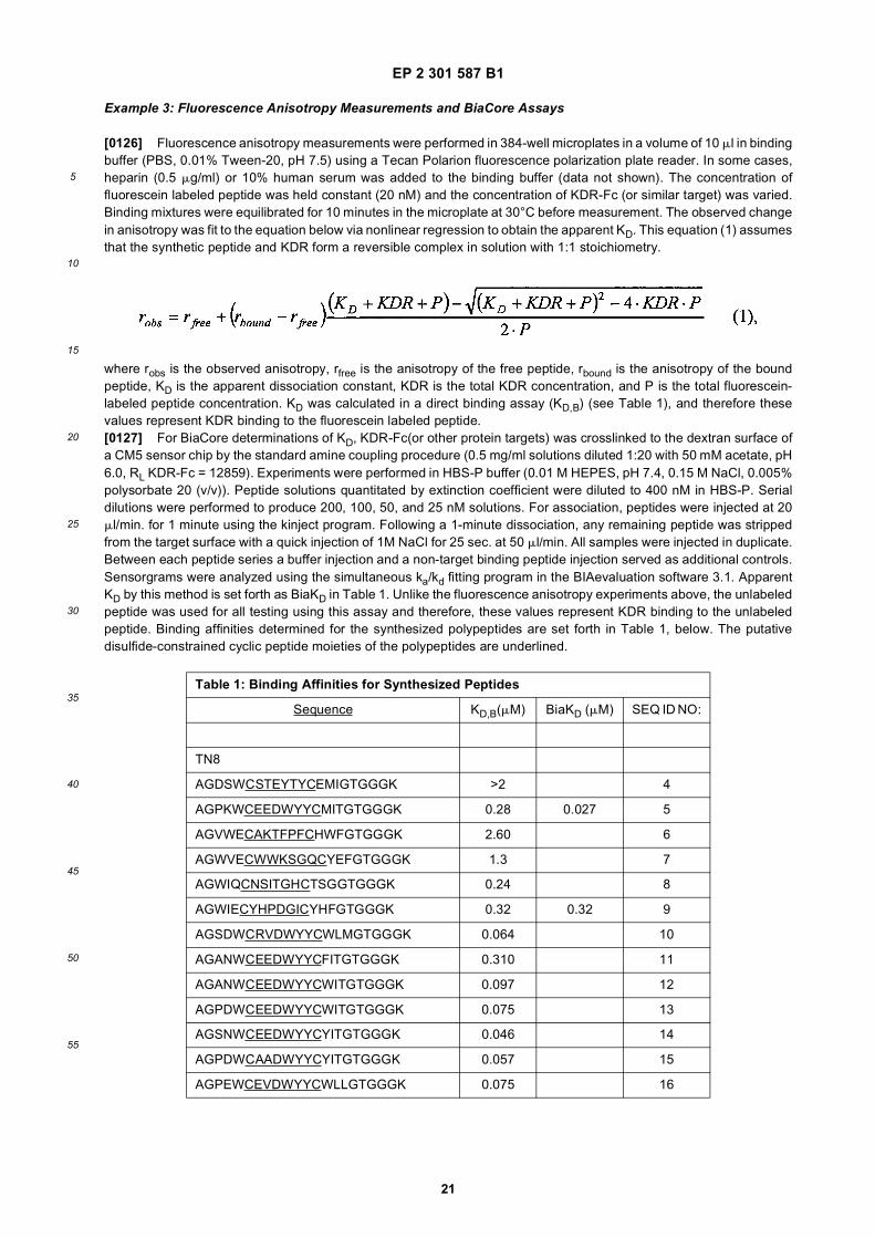
EP 2 301 587 B1
21
5
10
15
20
25
30
35
40
45
50
55
Example 3: Fluorescence Anisotropy Measurements and BiaCore Assays
[0126] Fluorescence anisotropy measurements were performed in 384-well microplates in a volume of 10 ml in bindingbuffer (PBS, 0.01% Tween-20, pH 7.5) using a Tecan Polarion fluorescence polarization plate reader. In some cases,heparin (0.5 mg/ml) or 10% human serum was added to the binding buffer (data not shown). The concentration offluorescein labeled peptide was held constant (20 nM) and the concentration of KDR-Fc (or similar target) was varied.Binding mixtures were equilibrated for 10 minutes in the microplate at 30°C before measurement. The observed changein anisotropy was fit to the equation below via nonlinear regression to obtain the apparent KD. This equation (1) assumesthat the synthetic peptide and KDR form a reversible complex in solution with 1:1 stoichiometry.
where robs is the observed anisotropy, rfree is the anisotropy of the free peptide, rbound is the anisotropy of the boundpeptide, KD is the apparent dissociation constant, KDR is the total KDR concentration, and P is the total fluorescein-labeled peptide concentration. KD was calculated in a direct binding assay (KD,B) (see Table 1), and therefore thesevalues represent KDR binding to the fluorescein labeled peptide.[0127] For BiaCore determinations of KD, KDR-Fc(or other protein targets) was crosslinked to the dextran surface ofa CM5 sensor chip by the standard amine coupling procedure (0.5 mg/ml solutions diluted 1:20 with 50 mM acetate, pH6.0, RL KDR-Fc = 12859). Experiments were performed in HBS-P buffer (0.01 M HEPES, pH 7.4, 0.15 M NaCl, 0.005%polysorbate 20 (v/v)). Peptide solutions quantitated by extinction coefficient were diluted to 400 nM in HBS-P. Serialdilutions were performed to produce 200, 100, 50, and 25 nM solutions. For association, peptides were injected at 20ml/min. for 1 minute using the kinject program. Following a 1-minute dissociation, any remaining peptide was strippedfrom the target surface with a quick injection of 1M NaCl for 25 sec. at 50 ml/min. All samples were injected in duplicate.Between each peptide series a buffer injection and a non-target binding peptide injection served as additional controls.Sensorgrams were analyzed using the simultaneous ka/kd fitting program in the BIAevaluation software 3.1. ApparentKD by this method is set forth as BiaKD in Table 1. Unlike the fluorescence anisotropy experiments above, the unlabeledpeptide was used for all testing using this assay and therefore, these values represent KDR binding to the unlabeledpeptide. Binding affinities determined for the synthesized polypeptides are set forth in Table 1, below. The putativedisulfide-constrained cyclic peptide moieties of the polypeptides are underlined.
Table 1: Binding Affinities for Synthesized Peptides
Sequence KD,B(mM) BiaKD (mM) SEQ ID NO:
TN8
AGDSWCSTEYTYCEMIGTGGGK >2 4
AGPKWCEEDWYYCMITGTGGGK 0.28 0.027 5
AGVWECAKTFPFCHWFGTGGGK 2.60 6
AGWVECWWKSGQCYEFGTGGGK 1.3 7
AGWIQCNSITGHCTSGGTGGGK 0.24 8
AGWIECYHPDGICYHFGTGGGK 0.32 0.32 9
AGSDWCRVDWYYCWLMGTGGGK 0.064 10
AGANWCEEDWYYCFITGTGGGK 0.310 11
AGANWCEEDWYYCWITGTGGGK 0.097 12
AGPDWCEEDWYYCWITGTGGGK 0.075 13
AGSNWCEEDWYYCYITGTGGGK 0.046 14
AGPDWCAADWYYCYITGTGGGK 0.057 15
AGPEWCEVDWYYCWLLGTGGGK 0.075 16

EP 2 301 587 B1
22
5
10
15
20
25
30
35
40
45
50
55
[0128] For the analysis of those peptides that bind specifically to KDR/VEGF complex, each peptide was tested forbinding to the complex in both assays (fluorescence anisotropy/Biacore) as above. In the anisotropy assay, KDR-VEGFcomplex was formed by mixing together a two fold molar excess of VEGF with KDR-Fc. This mixture was then used inthe direct binding titration using a fluorescein labeled peptide as done previously. As a control, each peptide was alsotested for binding to KDR and VEGF alone to assess their specificity for complex. Since none of the peptides boundVEGF to any extent, the presence of excess VEGF in the assay should not affect the KD determination. As shown inTable 2, below, all of the peptides showed a dramatic binding preference, binding for KDR/VEGF complex over VEGF.
(continued)
Table 1: Binding Affinities for Synthesized Peptides
Sequence KD,B(mM) BiaKD (mM) SEQ ID NO:
AGPTWCEDDWYYCWLFGTGGGK 0.0032 0.079 17
AGSKWCEQDWYYCWLLGTGGGK 0.400 18
AGRNWCEEDWYYCFITGTGGGK 0.190 19
AGVNWCEEDWYYCWITGTGGGK 0.260 20
AGANWCEEDWYYCYITGTGGGK 0.180 21
AGQAWVECYAETGYCWPRSWGTGGGK 0.71 22
AGQAWIECYAEDGYCWPRSWGTGGGK 1.40 23
AGVGWVECYQSTGFCYHSRDGTGGGK 1.30 24
AGDWWVECRVGTGLCYRYDTGTGGGK 0.93 25
AGDSWVECDAQTGFCYSFLYGTGGGK 2.30 26
AGERWVECRAETGFCYTWVSGTGGGK 2.10 27
AGGGWVECRAETGHCQEYRLGTGGGK 1.60 28
AGVAWVECYQTTGKCYTFRGGTGGGK ∼2 29
AGEGWVECFANTGACFTYPRGTGGGK 2.10 30
TN12
GDSRVCWEDSWGGEVCFRYDPGGGK 0.069 0.12 31
GDDSYCMMNEKGWWNCYLYDPGGGK 1.3 32
GDPAQCWESNYQGIFFCDNPDPGGGK 2.3 33
GDGWACAKWPWGGEICQPSDPGGGK 1.0 1.5 34
GDSSVCFEYSWGGEVCFRYDPGGGK 0.058 104
GDSRVCWEYSWGGQICLGYDPGGGK 0.32 105
Lin20
zIQQVQYQFFLGTPRYEQWDLDKGGK 1.7 35
AQEPEGYAYWEVITLYHEEDGDGGK 0.27 0.73 36
AQAFPRFGGDDYWIQQYLRYTDGGK 0.53 0.25 37
AQGDYVYWEIIELTGATDHTPPGGK 0.18 38
AQRGDYQEQYWHQQLVEQLKLLGGK 0.31 5.3 39
AQRSWYLGPPYYEEWDPIPNGGK 1.8 40
AQDWYYDEILSMADQLRHAFLSGGGK 0.05 41
EP 2 301 587 B1
23
5
10
15
20
25
30
35
40
45
50
55
Some of them, however, did show some residual binding to free KDR. To confirm the anisotropy results, the unlabeledpeptides were tested in Biacore as before, except the chip was saturated with VEGF to form KDR/VEGF complex priorto the injection of the peptides. In the peptides tested, the BiaKD was within at least 2-fold of the anisotropy measurement.
METHODS FOR EXAMPLES 4-10
[0129] The following methods were employed in Examples 4-10. The following common abbreviations are used: 9-fluorenylmethyloxycarbonyl (Fmoc), 1-hydroxybenzotriazole (HOBt), N,N’-diisopropylcarbodiimide (DIC), N-methylpyr-rolidinone (NMP), acetic anhydride (Ac2O), (4,4-dimethyl-2,6-dioxocyclohex-1-ylidene)-3-methylbutyl (ivDde), trifluoro-acetic acid (TFA), Reagent B (TFA: H2O: phenol: triisopropylsilane 88:5:5:2), diisopropylethylamine (DIEA), O-(1H-benzotriazole-1-yl)-N,N,N’,N’-tetramethyluronium hexafluorophosphate (HBTU), O-(7-azabenzotriazol-1-yl)-1,1,3,3-te-tramethyluronium hexafluorophosphate (HATU), N-hydroxysuccinimide (NHS), solid phase peptide synthesis (SPPS),dimethyl sulfoxide (DMSO), dichloromethane (DCM), dimethylformamide (DMF), human serum albumin (HSA), andradiochemical purity (RCP).
Method 1 for the ACT 357 MPS and ACT 496 MOS Synthesizers
[0130] The peptides were synthesized on NovaSyn TGR (Rink amide) resin (0.2 mmol/g) using the AdvancedChemTech ACT 357 or ACT 496 Synthesizers employing Fmoc peptide synthesis protocols, specifically using, HOBt/DICas the coupling reagents and NMP as the solvent. The Fmoc was removed by treating the Nova-Syn TGR (Rink amide-available from NovaBiochem, San Diego CA) resin-bound peptide with 25% piperidine in DMF twice (4 min and 10 min).All amino acids were dissolved in NMP (DMF was added when the amino acid was not soluble in pure NMP). Theconcentration of the amino acid was 0.25M, and the concentrations for HOBt and DIC respectively were 0.5 M.For a 0.04 mmol scale synthesis:
A typical amino acid coupling cycle (not including wash steps) was to dispense piperidine solution (2.4 mL) to eachwell and mix for 4 min, then empty all wells. NMP (320 mL), HOBt solution (320mL, 4 eq), amino acid (640mL, 4 eq)and DIC (320 mL, 4 eq) solutions were dispensed to each well. The coupling time was 3h; then the resin was washed.The cycle was repeated for each amino acid. After the last amino acid coupling, the resin-bound peptide was treatedwith 25% piperidine to remove the Fmoc protecting group. After washing, the resin bound peptide was capped with1.0M Ac2O (1.2 ml per well) and diisopropylethylamine in DMF, optionally including varying amounts of HOBt in themixture for 30 min. The resin was washed with methanol and then dichloromethane and dried. Cleavage of thepeptides from the resin and side-chain deprotection was accomplished using Reagent B for 4.5 h. The cleavagesolutions were collected and the resins were washed with an additional aliquot of Reagent B. The combined solutionswere concentrated to dryness. Ether was added to the residue with swirling or stirring to precipitate the peptides.The ether was decanted, and solid was collected. This procedure was repeated 2-3 times to remove impurities. Thecrude linear peptides were dissolved in DMSO and water mixtures, and purified by HPLC (column: Waters AssociatesXterra C18, 19 3 50 mm; solvents: H2O with 0.1% TFA and CH3CN with 0.1% TFA; UV 220 mm; Flow rate: 50-60ml/min). The solutions containing the peptide were lyophilized to give the desired peptides as white fluffy lyophilizates(> 90% purity). The purified linear di-cysteine containing peptides were dissolved in water, mixtures of water-ace-tonitrile, or mixtures of water-DMSO at concentrations between 0.1 mg/ml and 2.0 mg/ml. The choice of solventwas a function of the solubility of the crude peptide in the solvent. The pH of the solution was adjusted to pH 7.5-8.5with aqueous ammonia, aqueous ammonium carbonate or aqueous ammonium bicarbonate. The mixture was stirredvigorously in air for 24-48 hrs. In the case of non-DMSO containing solvent systems, the pH of the solution wasadjusted to pH 2 with aqueous trifluoroacetic acid. The mixture was lyophilized to provide the crude cyclic disulfidecontaining peptide. The cyclic disulfide peptide was then dissolved to a volume of 1-2 ml in aqueous (0.1% TFA)containing a minimum of acetonitrile (0.1% TFA). The resulting solution was loaded onto a reverse phase columnand the desired compound obtained by a gradient elution of acetonitrile into water, employing a C18, or C8 reversephase semipreparative or preparative HPLC column. In the case of the DMSO-containing solutions, the solution
Table 2: KDR/VEGF Complex Specific Peptides
SEQ ID
NO:
Sequence KD, B (KDR)
KD, B (VEGF)
KD, B (KDR/ VEGF)
BiaKD (KDR/ VEGF)
42 AQAPAWTFGTNWRSIQRVDSLTGGGGGK NB NB 0.84
43 AQEGWFRNPQEIMGFGDSWDKPGGGGGK NB NB 1.4

EP 2 301 587 B1
24
5
10
15
20
25
30
35
40
45
50
55
was diluted until the DMSO concentration was minimal without precipitation of the peptide. The resulting mixturewas quickly acidified to pH 2 with dilute trifluoroacetic acid and loaded onto the reverse phase HPLC system andpurified as described. Fractions containing the desired materials were pooled and the peptides isolated by lyophi-lization.
Method 2 for the ACT 357 MPS and ACT 496 MOS Synthesizers
[0131] The peptides were synthesized as in Method 1 with the following changes. HBTU/HOBt/DIEA were used asthe coupling reagent and NMP as the solvent. A low load (∼0.2 mmol/g) Fmoc-GGGK(Boc)-NovSyn-TGR-resin-preparedfrom the above-described Nova-Syn TGR resin was employed for peptide synthesis on 0.01 mmol scale.For a 0.01mmol scale synthesis:
After the Fmoc group was removed, a standard coupling procedure used a solution of HOBt (720 ml, 6 eq), aminoacid (804ml, 6.6 eq), HBTU (720ml, 6 eq) and DIEA (798 ml, 13.3 eq). The mixture was agitated for 15 min., emptiedand the resin washed. After all couplings and after cleavage and purification as above, the solutions containingdesired linear peptides were lyophilized to give the peptides (> 90% purity) as white fluffy solids. The crude ether-precipitated linear di-cysteine containing peptides were cyclized by dissolution in water, mixtures of aqueous ace-tonitrile (0.1% TFA), or aqueous DMSO and adjustment of the pH of the solution to pH 7.5 - 8.5 by addition ofaqueous ammonia, aqueous ammonium carbonate, or aqueous ammonium bicarbonate solution. The peptide con-centration was between 0.1 and 2.0 mg/ml. The mixture was stirred in air for 24-48 hrs., acidified to a pH 2 withaqueous trifluoroacetic acid, and then purified by preparative reverse phase HPLC employing a gradient of acetonitrileinto water. Fractions containing the desired material were pooled and the peptides were isolated by lyophilization.
Method 3 for the ACT 496 MOS Synthesizer
[0132] The peptides were synthesized by using an Advanced ChemTech ACT 496 MOS Synthesizer as in method 1.The low load (∼0.2 mmol/g) GGGK(Boc)-NovaSyn-TGR resin was employed for peptide synthesis. The coupling solventwas NMP/DMSO 8:2. The synthesis was performed at a 0.02 mmol scale using a coupling time of 3h. The crude linearpeptides were further processed as described for Method 1.
Method 4 for the ACT 496 MOS Synthesizer
[0133] The peptides were synthesized using method 3 on the ACT 496 with HBTU/DIEA as the coupling reagents,and NMP as the solvent. 2,4,6-collidine as a 1 M solution was used as the base. The low load Fmoc-GGGK(ivDde)-Novsyn-TGR resin (∼0.2 mmol/g) was used for peptide synthesis. The coupling time was 30 minutes.The crude linear peptides were further processed as described for Method 1.
Method 5 for the ABI 433A Synthesizer
[0134] Synthesis of peptides was carried out on a 0.25 mmol scale using the FastMoc protocol (Applied BiosystemsInc). In each cycle of this protocol, 1.0 mmol of a dry protected amino acid in a cartridge was dissolved in a solution of0.9 mmol of HBTU, 2 mmol of DIEA, and 0.9 mmol of HOBt in DMF with additional NMP added. The peptides weremade using 0.1 mmol ofNovaSyn TGR (Rink amide) resin (resin substitution 0.2 mmol/g). The coupling time in thisprotocol was 21 min. Fmoc deprotection was carried out with 20% piperidine in NMP. At the end of the last cycle, thesynthesized peptide was acetylated using acetic anhydride/DIEA/HOBt/NMP. The peptide resin was washed and driedfor further manipulations or cleaved from the resin (using reagent B). Generally, the cleaved peptides were cyclized asin Method 1 before purification.
Method 6: Biotinylation of Resin-Bound Peptides
[0135] The peptides were prepared using Method 5. The ivDde protecting group on the C-terminal lysine was selectivelyremoved by treatment with 10% hydrazine in DMF. The resin was then treated with a solution of Biotin-N-hydroxysuc-cinimidyl ester in DMF in the presence of DIEA. After washing, the resin was dried and cleavage was performed usingwith Reagent B. The resin was filtered off and the filtrate concentrated to dryness. The biotinylated peptide was dissolvedin neat DMSO and treated with DIEA and stirred for 4-6 hours to effect disulfide cyclization. The crude mixture waspurified by preparative HPLC.[0136] In a typical experiment, 200 mg of the resin -bound peptide was treated with 10% hydrazine in DMF (2 3 20mL) and washed with DMF (2 3 20 mL) and then with dichloromethane (1 3 20 mL). The resin was resuspended in

EP 2 301 587 B1
25
5
10
15
20
25
30
35
40
45
50
55
DMF (10 mL) and treated with a solution of Biotin-NHS ester (0.2 mmol, 5 equivalents) and DIEA (0.2 mmol) and theresin was mixed with the reagents for 4 h. The completion of the reaction was checked by the ninhydrin test. The peptidewas then released from the resin by treatment with Reagent B (10 mL) for 4 h. The resin was filtered off, Reagent B wasremoved in vacuo and the peptide was precipitated by addition of anhydrous ether. The solid formed was collected,washed with ether and dried. The solid was dissolved in anhydrous DMSO and the mixture was adjusted to pH 7.5 withDIEA and stirred for 4-6 h to effect disulfide cyclization. The disulfide cyclization reaction was monitored by analyticalHPLC. After completion of the cyclization, the mixture solution was diluted with 25% acetonitrile in water and directlypurified by HPLC on a reverse phase C18 column using a gradient of acetonitrile into water (both containing 0.1 % TFA).Fractions were analyzed by analytical HPLC and those containing the pure product were collected and lyophilized toobtain the required biotinylated peptide.
Method 7: Biotinylation of Purified Peptides
[0137] The purified peptide (10 mg, prepared by methods 1-5) containing a free amino group was dissolved in anhydrousDMF or DMSO (1 ml) and Biotin-NHS ester (5 equivalents) and DIEA (5 equivalents) were added. The reaction wasmonitored by HPLC and after the completion of the reaction (1-2 h.), the crude reaction mixture was directly purified bypreparative HPLC. Fractions were analyzed by analytical HPLC and those containing the pure product were collectedand lyophilized to obtain the required biotinylated peptide.
Method 8: Biotinylation of Resin-Bound Peptides Containing Linkers
[0138] In a typical experiment, 400 mg of the resin- containing peptide (made using the ABI 433A Synthesizer andbearing an ivDde-protected lysine) was treated with 10% hydrazine in DMF (2 3 20 ml). The resin was washed withDMF (2 3 20 ml) and DCM (1 3 20 ml). The resin was resuspended in DMF (10 ml) and treated with Fmoc-aminodiox-aoctanoic acid (0.4 mmol), HOBt (0.4 mmol), DIC (0.4 mmol), DIEA (0.8 mmol) with mixing for 4 h. After the reaction,the resin was washed with DMF (2 3 10 ml) and with DCM (1 3 10 ml). The resin was then treated with 20% piperidinein DMF (2 3 15 ml) for 10 min. each time. The resin was washed and the coupling with Fmoc-diaminodioxaoctanoicacid and removal of the Fmoc protecting group were repeated once more. The resulting resin, containing a peptide witha free amino group, was treated with a solution of Biotin-NHS ester (0.4 mmol, 5 equivalents) and DIEA (0.4 mmol, 5equivalents) in DMF for 2 hours. The peptide-resin was washed and dried as described previously and then treated withreagent B (20 mL) for 4h. The mixture was filtered, and the filtrate concentrated to dryness. The residue was stirred withether to produce a solid that was collected, washed with ether and dried. The solid was dissolved in anhydrous DMSOand the pH adjusted to 7.5 with DIEA. The mixture was stirred for 4-6 hr to effect the disulfide cyclization reaction, whichwas monitored by analytical HPLC. After the completion of the cyclization, the DMSO solution was diluted with 25%acetonitrile in water and applied directly to a reverse phase C-18 column. Purification was effected using a gradient ofacetonitrile into water (both containing 0.1 % TFA). Fractions were analyzed by analytical HPLC and those containingthe pure product were collected and lyophilized to provide the required biotinylated peptide.
Method 9: Formation of 5-Carboxyfluorescein-Labeled Peptides
[0139] Peptide-resin obtained via Method 5, containing an ivDde protecting group on the epsilon nitrogen of lysine,was mixed with a solution of hydrazine in DMF (10% hydrazine/DMF, 2 3 10 ml, 10 min) to remove the ivDde group.The epsilon nitrogen of the lysine was labeled with fluorescein-5-isothiocyanate (0.12 mmol) and diisopropylethylamine(0.12 mmol) in DMF. The mixture was agitated for 12 h (fluorescein-containing compounds were protected from light).The resin was then washed with DMF (3 3 10 mL) and twice with CH2Cl2 (10 mL) and dried under nitrogen for 1h. Thepeptide was cleaved from the resin using reagent B for 4h and the solution collected by filtration. The volatiles wereremoved under reduced pressure and the residue was dried under vacuum. The peptide was precipitated with ether,collected and the precipitate was dried under a stream of nitrogen. The precipitate was added to water (1 mg/ml) andthe pH of the mixture was adjusted to 8 with 10% aqueous meglumine. Cyclization of the peptide was carried out for 48h and the solution was freeze-dried. The crude cyclic peptide was dissolved in water and purified by RP-HPLC on a C18column with a linear gradient of acetonitrile into water (both phases contained 0.1%TFA). Fractions containing the pureproduct were collected and freeze-dried. The peptides were characterized by ES-MS and the purity was determined byRP-HPLC (linear gradient of acetonitrile into water/0.1% TFA).

EP 2 301 587 B1
26
5
10
15
20
25
30
35
40
45
50
55
Method 11: Formation of Mercaptoacetylated Peptides
Using S-Acetylthioglycolic acid N-Hydoxysuccinimide Ester
[0140] S-acetylthioglycolic acid N-hydroxysuccinimide ester (SATA) (0.0055mmol) was added to a solution of a peptide(0.005 mmol, obtained from Methods 1-5 with a free amine) in DMF (0.25 mL) and the reaction mixture was stirred atambient temperature for 6 h. The volatiles were removed under vacuum and the residue was purified by preparativeHPLC using acetonitrile-water containing 0.1%TFA. Fractions containing the pure product were collected and freeze-dried to yield the mercaptoacetylated peptide. The mercaptoacetylated peptide was characterized by ESI-MS and thepurity was determined by reverse phase HPLC analysis employing a linear gradient of acetonitrile into water (bothcontaining 0.1% TFA).
Method 12: Formation of Mercaptoacetylated Peptides using S-Acetylthioglycolic acid
[0141] Purified peptides from method 5, after disulfide cyclization, was coupled with S-acetylthioglycolic acid(1.5-10eq.)/HOBt (1.5-10 eq.)/DIC (1.5-10 eq.) in NMP for 2-16 hours at room temperature. The mixture was then purified bypreparative HPLC; the fractions containing pure peptide were combined and lyophilized. In the case of compounds withanother lysine protected by an ivDde group, the deprotection reaction employed 2% hydrazine in DMSO for 3h at roomtemperature. Purification of the reaction mixture afforded pure peptide.[0142] In the case when a preparing a compound with S-acetylthioglycolic acid coupled to two aminodioxaoctanoicacid groups and the peptide, the purified peptide from method 5 (having a free amino group, was coupled toAcSCH2CO-(NH-CH2-CH2-O-CH2-CH2-O-CH2-CO)2-OH (30 eq.)/HOBt (30 eq.)/DIC (30 eq.) in NMP for 40 hours atroom temperature. The mixture was purified and the ivDde group was removed. A second purification gave the finalproduct as a white lyophilizate.[0143] Alternatively Fmoc aminodioxaoctanoic acid was coupled twice successively to the peptide (produced by method5) followed by Fmoc removal and coupling to S-acetylthioglycolic acid.
Method 13: Preparation of Homo and Heterodimers
[0144] The required purified peptides were prepared by SPPS using Method 5. To prepare homodimers, half of thepeptide needed to prepare the dimer was dissolved in DMF and treated with 10 equivalents of glutaric acid bis N-hydoxysuccinimidyl ester. The progress of the reaction was monitored by HPLC analysis and mass spectroscopy. Atcompletion of the reaction, The volatiles were removed in vacuo and the residue was washed with ethyl acetate toremove unreacted bis-NHS ester. The residue was dried, re-dissolved in anhydrous DMF and treated with another halfportion of the peptide in the presence of 2 equivalents of DIEA. The reaction was allowed to proceed for 24 h. Thismixture was applied directly to a Waters Associates C-18 XTerra reverse phase HPLC column and purified by elutionwith a linear gradient of acetonitrile into water (both containing 0.1% TFA).[0145] In the case of heterodimers, one of the monomers was reacted with the bis NHS ester of glutaric acid and afterwashing off the excess of bis NHS ester, the second peptide was added in the presence of DIEA. After the reaction, themixture was purified by preparative HPLC.
Example 4: Preparation of KDR and VEGF/KDR Complex Binding Polypeptides
[0146] Utilizing the methods set forth above, biotinylated versions the KDR and VEGF/KDR complex binding polypep-tides set forth in Table 3 were prepared. The letter "J" in the peptide sequences refers to a spacer or linker group, 8-amino-3,6-dioxaoctanoyl.[0147] The ability of the biotinylated polypeptides (with the JJ spacer) to bind to KDR was assessed using the assayset forth in Example 5, following the procedures disclosed therein. Several biotinylated peptides bound well to the KDR-expressing cells: SEQ ID NO:66 (KD 1.81 nM +/- 0.27), SEQ ID NO:5 (KD 14.87+/- 5.0 nM, four experiment average),SEQ ID NO:31 + spacer (KD 10.00+/-2.36 nM, four experiment average),-SEQ ID NO:49 (KD 6.94+/-1.94 nM, oneexperiment), and SEQ ID NO:52 (KD 3.02+/-0.75 nM, one experiment).

EP 2 301 587 B1
27
5
10
15
20
25
30
35
40
45
50
55
Table 3: KDR, VEGF/KDR Complex Binding Polypeptides
SEQ ID
NO:
Structure (or) Sequence Mol. Wt. MS
31 Ac-GDSRVCWEDSWGGEVCFRYDPGGGK-NH2 2801.98 1399.6 [M-H]-
5 Ac-AGPKWCEEDWYYCMITGTGGGK-NH2 2361 -
7 Ac-AGWVFCWWKSGQCYEFGTGGGK-NH2 2474.06 -
44 Ac-AQEGWFRNPQEIMGFGDSWDKPGGGK-NH2 2934.35 -
31 Ac-GDSRVCWEDSWGGEVCFRYDPGGGK(Biotin)-NH2 3030.29 1512.4 [M2-]
45 Ac-AQRGDYQEQYWHQQLVEQLK(iv-Dde)LLGGGK-NH2 3318.71 1659.1 [M2-]
45 Ac-AQRGDYQEQYWHQQLVEQLKLLGGGK-NH2 3112.58 -
46 Ac-AGWYWCDYYGIGCK(iv-Dde)WTGGGK-NH2 2673.18
47 Ac-AGWYWCDYYGIGCKWTGTGGGK-NH2 2467.05
48 Ac-AQWYYDWFHNQRKPPSDWIDNLGGGK-NH2 3218.51 -
5 Ac-AGPK(iv-Dde)WCEEDWYYCMITGTGGGK-NH2 2698.11 2695.7 [M-H]-; 1347.8 [M-2H]2-/2
5 Ac-AGPKWCEEDWYYCMITGTGGGK(Biotin)-NH2 2718.13 2833.4 (M-H)-
5 Ac-AGPKWCEEDWYYCMITGTGGGK(Biotin-JJ-)-NH2 3008.44 1502.6.4 (M-2H)2-/2
5 Ac-AGPKWCEEDWYYCMITGTGGGK (AcSCH2C(=O)-)-NH2 2608.96 1304, [M-2H]2-/2
31 Ac-GDSRVCWEDSWGGEVCFRYDPGGGK(Biotin-JJ-)-NH2 3316.4 1657.8, [M-2H]2-/2
31 Ac-GDSRVCWEDSWGGEVCFRYDPGGGK (AcSCH2C(=O)-)-NH2 2917.15 1457.4, [M-2H]2-/2
31 Biotin-JJGDSRVCWEDSWGGEVCFRYDPGGGK-NH2 3272.34 1636.7, [M-2H]2-/2
5 Ac-AGPKWCEEDWYYCMITGT-GGGK(AcSCH2C(=O)-JJ-)-NH2 2899.28 1449.2, [M-2H]2-/2
17 Ac-AGPTWCEDDWYYCWLFGTGGGK(Biotin-JJ-)-NH2 3066.27 1532.8, [M-2H]2-/2
49 Ac-VCWEDSWGGEVCFRYDPGGGK(Biotin-JJ-)-NH2 2903.24 1449.3, (M-2H)2-/2; 965.8, (M-3H)3-/3
50 Ac-AGPTWCEDDWYYCWLFGTJK(Biotin-JJ-)-NH2 3042.44 1519.7, (M-2H)2-/2-; 1012.8 (M-3H)3-/3
31 Ac-GDSRVCWEDSWGGEVCFRYDPGGGK (AcSCH2C(=O)-JJ-)-NH2
3208.48 1602.6, [M-2H]2-/2
17 Ac-AGPTWCEDDWYYCWLFGTGGGK (AcSCH2C(=O)-JJ-)-NH2 2907.29 1453.1, [M-2H]2-/2
51 Ac-AQAHMPPWRPVAVDALFDWVEGG-GGGK(Biotin-JJ-)-NH2 3404.64 1701.6, [M-2H]2-/2
52 Ac-AQAHMPPWWPLAVDAQEDWFEGGGGGK(Biotin-JJ-)-NH2 3493.59 1746.2, [M-2H]2-/2
52 Ac-AQAQMPPWWPLAVDALFDWFEGGGGGK(Biotin-JJ-)-NH2 3487.64 1743.2, [M-2H]2-/2
53 Ac-AQDWYWREWMPMHAQFLADDWGGGGGK(Biotin-JJ-)-NH2 3751.64 1874.3, [M-2H]2-/2
55 Ac-AQPVTDWTPHHPK(iv-Dde)APDVWLFYTGGGGGK(Biotin-JJ-)-NH2
3781.86 1890.4, [M-2H]2-/2
56 Ac-AQDALEAPK(iv-Dde)RDWYYDWFLNHSPGGGGGK(Biotin-JJ-)-NH2
3897.85 1948.0, [M-2H]2-/2
57 Ac-KWCEEDWYYCMITGTGGGK(Biotin-JJ-)-NH2 2781.2 1390.0, [M-2H]2-/2
58 Ac-AGPKWCEEDWYYCMIGGGK(Biotin-JJ-)-NH2 2747.15 1373.5, [M-2H]2-/2
59 Ac-KWCEEDWYYCMIGGGK(Biotin-JJ-)-NH2 2522.04 1260.8, [M-2H]2-/2

EP 2 301 587 B1
28
5
10
15
20
25
30
35
40
45
50
55
Example 5: Binding of KDR binding peptides/avidin HRP complex to KDR transfected 293H cells
[0148] To determine the binding of peptides identified by phage display to KDR expressed in transiently-transfected293H cells, a novel assay that measures the binding of biotinylated peptides complexed with neutravidin HRP to KDRon the surface of the transfected cells was developed. This assay was used to screen the biotinylated peptides set forthin Example 4. Neutravidin HRP was used instead of streptavidin or avidin because it has lower non-specific binding tomolecules other than biotin due to the absence of lectin binding carbohydrate moieties and also due to the absence ofthe cell adhesion receptor-binding RYD domain in neutravidin.[0149] In the experiments described herein, tetrameric complexes of KDR-binding peptides SEQ ID NO:31, SEQ IDNO:5, SEQ ID NO:17 and SEQ ID NO:66 and a control peptide, which does not bind to KDR, were prepared and testedfor their ability to bind 293H cells that were transiently-transfected with KDR. All four tetrameric complexes of KDR-binding peptides were niotinylated and contained the JJ spacer, and bound to the KDR-expressing cells; however, SEQID NO:66 exhibited the best KD (1.81nM). The tetrameric complexes of KDR-binding peptides SEQ ID NO:31, SEQ IDNO:5 exhibited improved binding over monomers of the same peptides. Moreover, inclusion of a spacer between theKDR-binding peptide and the biotin was shown to improve binding in Experiment B.[0150] In Experiment C, it was shown that this assay can be used to assess the effect of serum on binding of peptidesas used in the invention to KDR and VEGF/KDR complex. The binding of SEQ ID NO:5, SEQ ID NO:31, and SEQ IDNO:66 was not significantly affected by the presence of serum, while the binding of SEQ ID NO:17 was reduced morethan 50% in the presence of serum.[0151] In Experiment D, it was shown that this assay is useful in evaluating distinct combinations of KDR and VEGF/KDRcomplex binding polypeptides for use in multimeric targeting constructs which contain more than one KDR and VEGF/KDRcomplex binding polypeptide. Moreover, Experiments D and E establish that tetrameric constructs including two or moreKDR binding peptides which bind to different epitopes exhibited superior binding to "pure" tetrameric constructs of thetargeting peptides alone.
Experiment A
Preparation of m-RNA & 5’ RACE ready cDNA library
[0152] HUVEC cells were grown to almost 80% confluence in 175 cm2 tissue culture flasks (Becton Dickinson, Biocoat,cat # 6478) and then 10 ng/ml of bFGF (Oncogene, cat # PF003) was added for 24 h to induce expression of KDR.mRNA was isolated using the micro-fast track 2.0 kit from Invitrogen (cat. # K1520-02). 12 mg of mRNA (measured byabsorbance at 260 nM) was obtained from two flasks (about 30 million cells) following the kit instructions. Reversetranscription to generate cDNA was performed with 2 mg of mRNA, oligo dT primer (5’-(T)25GC-3’) and/or smart II oligo(5’AAGCAGTGGTAACAACGCAGAGTACGCGGG-3’) (SEQ ID NO:67) using Moloney Murine Leukemia Virus (MMLV)reverse transcriptase. The reaction was performed in a total volume of 20 ml and the reaction mix contained 2 ml of RNA,
(continued)
Table 3: KDR, VEGF/KDR Complex Binding Polypeptides
SEQ ID
NO:
Structure (or) Sequence Mol. Wt. MS
60 Ac-AQPDNWK(iv-Dde)EFYESGWK(iv-Dde)-YPSLYK(iv-Dde)PLGGGGGK(Biotin-JJ-)-NH2
4377.2 2188.4, [M-2H]2-/2
61 Ac-AQMPPGFSYWEQVVLHDDAQVLGG-GGGK(Biotin-JJ-)-NH2 3499.7 1749.2, [M-2H]2-/2
62 Ac-AQARMGDDWEEAPPHEWGWADGGGGGK(Biotin-JJ-)-NH2 3480.5 1740.2, [M-2H]2-/2
63 Ac-AQPEDSEAWYWLNYRPTMFHQLGG-GGGK(Biotin-JJ-)-NH2 3751.7 1875.8, [M-2H]2-/2
64 Ac-AQSTNGDSFVYWEEVELVDHPGGGGGK(Biotin-JJ-)-NH2 3554.6 1776.4, [M-2H]2-/2
65 Ac-AQWESDYWDQMRQQLK(iv-Dde)TAYMK(iv-Dde)VGGGGGK(Biotin-JJ-)-NH2
4187.02 2093.0, [M-2H]2-/2
66 Ac-AQDWYYDEILSMADQLRHAFLSGGGGGK(Biotin-JJ-)-NH2 3641.69 1820.9, [M-2H]2-/2
The putative disulfide constrained cyclic peptide is indicated by underlining.

EP 2 301 587 B1
29
5
10
15
20
25
30
35
40
45
50
55
1 ml smart II oligo, 1 ml of oligo dT primer, 4 ml of 5X first-strand buffer (250 mM Tris HCl pH 8.3, 375 mM KCl, 30 mMMgCl2) 1 ml DTT (20 mM, also supplied with reverse transcriptase), 1 ml dNTP mix (10 mM each of dATP, dCTP, dGTP,and dTTP in ddH2O, Stratagene, cat. # 200415), 9 ml ddH2O and 1 ml MMLV reverse transcriptase (Clonetech, cat#8460-1). The reverse transcription reaction was performed for 90 minutes at 42°C, and the reaction was stopped byadding 250 ml of tricine-EDTA buffer (10 mM tricine, 1.0 mM EDTA). The reverse transcription product, a 5’ RACE readycDNA library, can be stored for 3 months at -20°C. Note that all water used for DNA and RNA application was DNAseand RNAse free from USB (cat. # 70783).
Cloning of s-KDR into TOPOII Vector
[0153] In order to clone s-KDR, a 5’ oligo (G ATG GAG AGC AAG GTG CTG CTG G) (SEQ ID NO:68) and a 3’ oligo(C CAA GTT CGT CTT TTC CTG GGC A) (SEQ ID NO:69) were used. These were designed to amplify the completeextracellular domain of KDR (∼2.2 kbps) from the 5’ RACE ready cDNA library (prepared above) using polymerase chainreaction (PCR) with pfu polymerase (Stratagene, cat. # 600135). The PCR reaction was done in total volume of 50 mland the reaction mix contained 2 ml 5’ RACE ready cDNA library, 1 ml 5’ oligo (10 mM), 1 ml 3’ oligo (10 mM), 5 ml 10XPCR buffer [PCR buffer (200 mM Tris-HCl pH 8.8, 20 mM MgSO4, 100 mM KCl, 100 mM (NH4)2SO4) supplied with pfuenzyme plus 1% DMSO and 8% glycerol], 1 ml dNTP mix (10 mM) and 40 ml ddH20. The PCR reaction was performedby using a program set for 40 cycles of 1 minute at 94C, 1 minute at 68C and 4 minutes at 72C. The PCR product waspurified by extraction with 1 volume of phenol, followed by extraction with 1 volume of chloroform and precipitated using3 volume of ethanol and 1/10 volume of 3M sodium acetate. The PCR product was resuspended in 17 ml of ddH2O, the2 ml of 10X Taq polymerase buffer (100 mM Tris-HCl pH 8.8, 500 mM KCl, 15 mM MgCl2, 0.01% gelatin) and 1 ml ofTaq polymerase (Stratagene, cat. # 600131) was added to generate an A overhang to each end of the product. Afterincubating for 1 hour at 72C the modified product was cloned directly into a TOPOII vector (InVitrogen, Carlsbad, CA)following the manufacturer’s protocol to give TOPO-sKDR. The TOPO vector allows easy cloning of PCR productsbecause of the A-overhang in Taq (PCR enzyme)-treated PCR products.
Cloning the transmembrane and cytoplasmic domains of KDR into TOPO II Vector
[0154] In order to clone the transmembrane and cytoplasmic domains of KDR, a 5’ oligo (TCC CCC GGG ATC ATTATT CTA GTA GGC ACG GCG CTG) (SEQ ID NO:70) and a 3’ oligo (C AGG AGG AGA GCT CAG TGT GGT C) (SEQID NO:71) were used. These were designed to amplify the complete transmembrane and cytoplasmic domains of KDR(∼1.8 kbps) from the 5’ RACE ready cDNA library (described above) using polymerase chain reaction (PCR) with pfupolymerase. PCR reaction conditions and the program were exactly the same as described above for s-KDR. Just aswith the s-KDR sequence, the PCR product was purified using phenol chloroform extraction, treated with Taq polymeraseand cloned into TOPOII vector from Invitrogen to give TOPO-CYTO.
Cloning of full-length KDR into pcDNA6 Vector
[0155] To create the full-length receptor, the extra-cellular domain and the cytoplasmic domain (with trans-membranedomain) were amplified by PCR separately from TOPO-sKDR and TOPO-CYTO respectively and ligated later to createthe full-length receptor. An oligo with a Not1 site at the 5’ end of the extracellular domain (A TAA GAA TGC GGC CGCAGG ATG GAG AGC AAG GTG CTG CTG G) (SEQ ID NO:72) and an oligo complimentary to the 3’ end of the extracellulardomain (TTC CAA GTT CGT CTT TTC CTG GGC ACC) (SEQ ID NO:73) were used to amplify by PCR the extracellulardomain from TOPO-sKDR. Similarly, the 5’ oligo (ATC ATT ATT CTA GTA GGC ACG GCG GTG) (SEQ ID NO:74) andthe 3’ oligo, with a Not1 site (A TAA GAA TGC GGC CGC AAC AGG AGG AGA GCT CAG TGT GGT C) (SEQ IDNO:75), were used to amplify by PCR the cytoplasmic domain of KDR (with transmembrane domain) from TOPO-CYTO.Both PCR products were digested with Not1 and ligated together to create the full-length receptor. The cDNA encodingthe full-length receptor was purified on an agarose gel and ligated into the Not I site of the pcDNA6/V5-HisC vector.Purification of DNA and ligation was done as described earlier for psKDR. The ligation reaction was used to transforma culture of DH5α bacteria and a number of individual clones were analyzed for the presence and orientation of insertby restriction analysis of purified plasmid from each clone with EcoRI enzyme.
Cell Culture
[0156] 293H cells were obtained from Invitrogen (cat. # 11631) and grown as monolayer culture in their recommendedmedia plus 1ml/L pen/strep (Invitrogen, cat. # 15140-148). All the cells were grown in presence of antibiotic for everydayculture but were split into antibiotic free media for 16-20 hours prior to transfection.
EP 2 301 587 B1
30
5
10
15
20
25
30
35
40
45
50
55
Preparation of DNA for Transfection
[0157] E. coli bacteria DH5α containing pf-KDR was streaked onto LB with 50 mg/ml ampicillin (LB agar from USbiologicals, cat. # 75851 and ampicillin from Sigma, cat. #A2804) plates from a glycerol stock and plates were left in a37°C incubator to grow overnight. Next morning, a single colony was picked from the plate and grown in 3 ml of LB/amp-icillin media (LB from US biologicals, cat. # US75852) at 37°C. After 8 hours, 100 ml of bacterial culture from the 3 mltube was transferred to 250 ml of LB/ampicillin media for overnight incubation at 37°C. Bacteria were grown up withcircular agitation in a 500 ml bottle (Beckman, cat. # 355605) at 220 rpm in a Lab-Line incubator shaker. The next day,the bacterial culture was processed using maxi-prep kit (QIAGEN, cat. # 12163). Generally, about 1mg of plasmid DNA(as quantitated by absorbance at 260 nm) was obtained from 250 ml of bacterial culture.
Transfection of 293H cells in 96 well plate
[0158] Transfection was done as recommended in the lipofectamine 2000 protocol (Invitrogen, cat# 11668-019) usinga poly-D-lysine-coated 96 well plate. 320 ng of KDR DNA (pc-DNA6-fKDR)/per well in 0.1 ml was used for 96 welltransfection. Transfection was done in serum-containing media, the transfection reagent mix was removed from cellsafter 6-8 hours and replaced with regular serum-containing medium. Transfection was done in black/clear 96-well plates(Becton Dickinson, cat. # 354640). The left half of the plate (48 wells) were mock-transfected (with no DNA) and theright half of the plate was transfected with KDR cDNA. The cells were 80-90% confluent at the time of transfection andcompletely confluent next day, at the time of the assay, otherwise the assay was aborted.
Preparation of M199 media
[0159] In order to prepare M199 media for the assay, one M199 medium packet (GIBCO, cat. # 31100-035), 20 ml of1 mM HEPES (GIBCO, cat. #15630-080) and 2 gm of DIFCO Gelatin (DIFCO, cat. # 0143-15-1) were added to 950 mlof ddH2O and the pH of the solution was adjusted to 7.4 by adding approximately 4 ml of 1N NaOH. After pH adjustment,the M199 media was warmed to 37°C in a water bath for 2 hours to dissolve the gelatin, then filter sterilized using 0.2mm filters (Corning, cat. # 43109), and stored at 4°C to be used later in the assay.
Preparation of SoftLink soft release avidin-sepharose
[0160] SoftLink soft release avidin-sepharose was prepared by centrifuging the sepharose obtained from Promega(cat. # V2011) at 12,000 rpm for 2 minutes, washing twice with ice cold water (centrifuging in-between the washes) andresuspending the pellet in ice cold water to make a 50% slurry in ddH2O. A fresh 50% slurry of avidin-sepharose wasprepared for each experiment.
Preparation of peptide/neutravidin HRP solution
[0161] Biotinylated peptides SEQ ID NOS:294, 264, 277, 356, and the non-binding biotinylated control peptide wereused to prepare 250 mM stock solutions in 50% DMSO and a 33 mM stock solution of neutravidin-HRP was preparedby dissolving 2 mg of neutravidin-HRP (Pierce, cat. # 31001) in 1 mL of ddH2O (all polypeptides contained the JJ spacer).Peptide stock solutions were stored at -20°C, whereas the Neutravidin HRP stock solution was stored at -80°C. Toprepare peptide/neutravidin-HRP complexes, 10 ml of 250 mM biotinylated peptide stock solution and 10 ml of 33 mMneutravidin-HRP were added to 1 ml of M199 medium. This mixture was incubated on a rotator at 4°C for 60 minutes,followed by addition of 50 ml of soft release avidin-sepharose (50% slurry in ddH20) to remove excess peptides andanother incubation for 30 minutes on a rotator at 4°C. Finally, the soft release avidin-sepharose was pelleted by centri-fuging at 12,000 rpm for 5 minutes at room temperature, and the resulting supernatant was used for the assays. Freshpeptide/neutravidin-HRP complexes were prepared for each experiment.
Preparation of peptide/neutravidin HRP dilutions for the assay
[0162] For saturation binding experiments, 120 ml, 60 ml, 20 ml, 10 ml, 8 ml, 6 ml, 4 ml, and 1 ml of peptide/neutravidinHRP complex were added to 1.2 ml aliquots of M199 medium to create dilutions with final concentrations of 33.33 nM,16.65 nM, 5.55 nM, 2.78 nM, 1.67 nM, 1.11 nM and 0.28 nM complex, respectively.
Preparation of blocking solution for transfected 293H cells
[0163] Blocking solution was prepared by adding 20 ml of M199 medium to 10 mg of lyophilized unlabeled neutravidin
EP 2 301 587 B1
31
5
10
15
20
25
30
35
40
45
50
55
(Pierce, cat. # 31000). Fresh blocking solution was used for each experiment.
Assay to detect the binding of peptide/neutravidin-HRP
[0164] 24 hours after transfection, each well of the 293H cells was washed once with 100 ml of M199 medium andincubated with 80 ml of blocking solution at 37°C. After one hour, cells were washed twice with 100 ml of M199 mediaand incubated with 70 ml of peptide/neutravidin-HRP dilutions of control peptide, SEQ ID NO:5, SEQ ID NO:31, SEQID NO:17, and SEQ ID NO:66 for two and half hours at room temperature. Each dilution was added to three separatewells of mock as well as KDR-transfected 293H cells (two plates were used for each saturation binding experiment).After incubation at room temperature, plates were transferred to 4°C for another half-hour incubation. Subsequently,cells were washed 5 times with ice-cold M199 media and once with ice-cold PBS (in that order). After the final wash,100 ml of ice cold TMB solution (KPL, cat. # 50-76-00) was added to each well and each plate was incubated for 30minutes at 37°C in an air incubator. Finally, the HRP enzyme reaction was stopped by adding 50 ml of 1N phosphoricacid to each well, and binding was quantitated by measuring absorbance at 450 nm using a microplate reader (BioRadModel 3550).
Binding of peptide/neutravidin HRP to KDR-transfected cells
[0165] In this assay, complexes of control peptide, SEQ ID NO:5, SEQ ID NO:31, SEQ ID NO:17, and SEQ ID NO:66peptides, each biotinylated with the JJ spacer and conjugated with neutravidin-HRP, were prepared as described aboveand tested for their ability to bind 293H cells that were transiently-transfected with KDR. During the peptide/neutravidincomplex preparation, a 7.5-fold excess of biotinylated peptides over neutravidin-HRP was used to make sure that allfour biotin binding sites on neutravidin were occupied. After complex formation, the excess of free biotinylated peptideswas removed using soft release avidin-sepharose to avoid any competition between free biotinylated peptides andneutravidin HRP-complexed biotinylated peptides. The experiment was performed at several different concentrationsof peptide/neutravidin-HRP, from 0.28 nM to 33.33 nM, to generate saturation binding curves for SEQ ID NO:5 and SEQID NO:31 (FIG. 1A) and 0.28 to 5.55 nM to generate saturation binding curve for SEQ ID NO:17 and SEQ ID NO:66(FIG. 1B). In order to draw the saturation binding curve, the background binding to mock-transfected cells was subtractedfrom the binding to KDR-transfected cells for each distinct peptide/neutravidin HRP complex at each concentrationtested. Therefore, absorbance on the Y-axis of FIG. 1 (below) is differential absorbance (KDR minus mock) and not theabsolute absorbance. Analysis of the saturation binding data in FIG. 1 using Graph Pad Prism software (version 3.0)yielded a KD of 10.00 nM (+/-2.36) for the tetrameric SEQ ID NO:31, 14.87 nM (+/- 5.066) for the tetrameric SEQ IDNO:5, 4.031 nM (+/- 0.86) for the tetrameric SEQ ID NO:17, and 1.814 nM (+/- 0.27) for the tetrameric SEQ ID NO:66peptide complexes. These binding constants are, as expected, lower than those measured by FP against the KDRFcconstruct for the related monodentate peptides SEQ ID NO:31 (69 nM), SEQ ID NO:5 (280 nM), SEQ ID NO:42 (51nM), but similar to monodentate peptide SEQ ID NO:17 (3 nM). As expected, no saturation of binding for the control(non-binder) peptide/neutravidin HRP-complex was observed. The binding of peptide/neutravidin HRP complexes (FIG.2) at a single concentration (5.55 nM) was plotted to demonstrate that a single concentration experiment can be usedto differentiate between a KDR binding peptide (SEQ ID NOS:5, 31 and 17) from a non-binding peptide.
Experiment B
[0166] Experiment B was designed to look at the requirement of spacer (JJ, Table 3) between the KDR bindingsequences (SEQ ID NOS:31 and 5) and biotin. In this experiment, biotinylated peptides with and without spacer JJ weretested (e.g., biotinylated SEQ ID NO:5 with the JJ spacer, biotinylated SEQ ID NO:5 without the JJ spacer, SEQ IDNO:31 with a spacer, and biotinylated SEQ ID NO:31 without the spacer), and a non-KDR binding, biotinylated controlpeptide (with and without spacer, prepared as set forth above) was used as a control. The peptide structure of all theKDR-binding sequences tested in this experiment is shown in FIG. 3.[0167] This experiment was performed as set forth in Experiment A described above, except that it was only done ata single concentration of 2.78 nM.[0168] Results: It is evident from the results shown in the FIG. 4 that the spacer is enhances binding of SEQ ID NO:31and SEQ ID NO:5. The spacer between the binding sequence and biotin can be helpful in enhancing binding to targetmolecule by multiple mechanisms. First, it could help reduce the steric hindrance between four biotinylated peptide aftertheir binding to single avidin molecule. Second, it could provide extra length necessary to reach multiple binding sitesavailable on a single cell.

EP 2 301 587 B1
32
5
10
15
20
25
30
35
40
45
50
55
Experiment C
[0169] Experiment C was designed to look at the serum effect on the binding of SEQ ID NOS: 31, 5, 17 and 66. Inthis procedure, biotinylated peptide/avidin HRP complexes of SEQ ID NOS:31, 5, 17 and 66 were tested in M199 media(as described above in Experiment A) with and without 40% rat serum. This experiment was performed as describedfor Experiment A except that it was only done at single concentration of 6.66 nM for SEQ ID NOS: 31 and 5, 3.33 nMfor SEQ ID NO:17 and 2.22 nM for SEQ ID NO:66. Each of the polypeptides were biotinylated and had the JJ spacer.[0170] Results: Results in FIG. 5 indicate that binding of SEQ ID NO:5, SEQ ID NO:31, and SEQ ID NO:66 was notsignificantly affected by 40% rat serum, whereas binding of SEQ ID NO:17 was more than 50% lower in presence of40% rat serum. More than an 80% drop in the binding of Tc-labeled SEQ ID NO:17 with Tc-chelate was observed in thepresence of 40% rat serum (FIG. 24). Since the serum effect on the binding of Tc-labeled SEQ ID NO:17 is mimickedin the avidin HRP assay disclosed herein, this assay may be used to rapidly evaluate the serum effect on the bindingof peptide(s) to KDR.
Experiment D
[0171] Experiment D was designed to evaluate the binding of tetrameric complexes of KDR and VEGF/KDR complex-binding polypeptides SEQ ID NO:31 and SEQ ID NO:5, particularly where the constructs included at least two KDRbinding polypeptides. The KDR binding peptides and control binding peptide were prepared as described above. Thisexperiment was performed using the protocol set forth for Experiment A, except the procedures set forth below wereunique to this experiment.[0172] Preparation of Peptide/Neutravidin Solutions: 250 mM stock solutions of biotinylated peptides SEQ ID NOs:5,31 and control peptide were prepared in 50% DMSO and a 33 mM stock solution of Neutravidin HRP was prepared bydissolving 2 mg of Neutravidin HRP (Pierce, cat. # 31001) in 1 mL of ddH2O. Peptide stock solutions were stored at-20C, whereas the Neutravidin HRP stock solution was stored at -80C. The sequences of the biotinylated peptides areshown above. To prepare peptide/neutravidin HRP complexes, a total 5.36 mL of 250 mM biotinylated peptide stocksolution (or a mixture of peptide solutions, to give peptide molecules four times the number of avidin HRP molecules)and 10 mL of 33 mM Neutravidin HRP were added to 1 mL of M199 medium. This mixture was incubated on a rotator at4C for 60 minutes, followed by addition of 50 mL of soft release avidin-sepharose (50% slurry in ddH20) to remove excesspeptides and another incubation for 30 minutes on a rotator at 4C. Finally, the soft release avidin-sepharose was pelletedby centrifuging at 12,000 rpm for 5 minutes at room temperature, and the resulting supernatant was used for the assays.Fresh peptide/neutravidin HRP complexes were prepared for each experiment.[0173] Assay to Detect the Binding of Peptide/Neutravidin HRP: 24 hours after transfection, each well of the 293Hcells was washed once with 100 mL of M199 medium and incubated with 80 mL of blocking solution at 37C. After onehour, cells were washed twice with 100 mL of M199 media and incubated with 70 mL of 3.33 nM peptide (or peptidemix)/neutravidin HRP solutions (prepared by adding 10 mL of stock prepared earlier to 1 mL of M199 media) for two andhalf hours at room temperature. Each dilution was added to three separate wells of mock as well as KDR-transfected293H cells. After incubation at room temperature, plates were transferred to 4C for another half-hour incubation. Sub-sequently, cells were washed five times with ice-cold M199 media and once with ice-cold PBS (in that order). After thefinal wash, 100 mL of ice cold TMB solution (KPL, Gaithersburg, MD) was added to each well and each plate wasincubated for 30 minutes at 37C in an air incubator. Finally, the HRP enzyme reaction was stopped by adding 50 mL of1N phosphoric acid to each well, and binding was quantitated by measuring absorbance at 450 nm using a microplatereader (BioRad Model 3550).[0174] Results: This experiment establishes that SEQ ID NO:31 and SEQ ID NO:5 bind to KDR in multimeric fashion,and cooperate with each other for binding to KDR in 293H transfected cells. A biotinylated control peptide that does notbind to KDR was used. As expected, a tetrameric complex of the control peptide with avidin-HRP did not show enhancedbinding to KDR-transfected cells. Tetrameric complexes of SEQ ID NO:31 and SEQ ID NO:5 bound to KDR-transfectedcells significantly better than to mock-transfected cells (see FIG. 6). SEQ ID NO:31 tetramers, however, bound muchbetter than SEQ ID NO:5 tetramers. If the control peptide was added to the peptide mixture used to form the tetramericcomplexes, the binding to the KDR-transfected cells decreased. The ratio of specific binding of tetramer to monomer,dimer and trimer was calculated by dividing the specific binding (obtained by subtracting the binding to mock transfectedcells from KDR transfected cells) of tetramer, trimer and dimer with that of monomer. Results indicate that there is co-operative effect of multimerization of SEQ ID NOS:5, 31 and 67 on the binding to KDR-transfected cells.
Tetramer Trimer DimerSEQ ID NO: 5 45.4 5 4.3
SEQ ID NO:31* 38.6 7.1 2.7
EP 2 301 587 B1
33
5
10
15
20
25
30
35
40
45
50
55
[0175] A mixture of 25% non-binding control peptide with 75% SEQ ID NO:5 did not bind significantly over backgroundto KDR-transfected cells, indicating that multivalent binding is critical for the SEQ ID NO:5/avidn-HRP complex to remainbound to KDR throughout the assay. This phenomenon also held true for SEQ ID NO:31, where substituting 50% of thepeptide with control peptide in the tetrameric complex abolished almost all binding to KDR on the transfected cells.[0176] Surprisingly, a peptide mixture composed of 50% control peptide with 25% SEQ ID NO:31 and 25% SEQ IDNO:5 bound quite well to KDR-transfected cells relative to mock-transfected cells, indicating that there is a great advantageto targeting two sites or epitopes on the same target molecule. Furthermore, it was noted that tetrameric complexescontaining different ratios of SEQ ID NO:31 and SEQ ID NO:5 (3:1, 2:2, and 1:3) all bound much better to KDR-transfectedcells than pure tetramers of either peptide, in agreement with the idea that targeting two distinct sites on a single targetmolecule is superior to multimeric binding to a single site. This may be because multimeric binding to a single targetrequires that the multimeric binding entity span two or more separate target molecules which are close enough togetherfor it to bind them simultaneously, whereas a multimeric binder which can bind two or more distinct sites on a singletarget molecule does not depend on finding another target molecule within its reach to achieve multimeric binding.
Experiment E
[0177] Experiment E was designed to confirm that SEQ ID NO:31 and SEQ ID NO:5 bind to distinct sites (epitopes)on KDR. If these peptides bind to the same site on KDR, then they should be able to compete with each other; however,if they bind to different sites they should not compete. This experiment was performed using a single concentration ofSEQ ID NO:5/avidin HRP (3.33 nM) solution in each well and adding a varying concentration (0-2.5 mM) of biotinylatedcontrol peptide with spacer, SEQ ID NO:5 and SEQ ID NO:31, none of which were complexed with avidin.[0178] Results: It is evident from FIG. 7 that SEQ ID NO:5 does compete with SEQ ID NO:5/avidin HRP solution forbinding to KDR transfected cells whereas control peptide and SEQ ID NO:31 do not compete with SEQ ID NO:5/avidinHRP solution for binding to KDR transfected cells. Thus, SEQ ID NO:5 and SEQ ID NO:31 bind to distinct and comple-mentary sites on KDR.
Example 6: Binding of Analogs of a KDR-binding Peptide to KDR-expressing Cells
[0179] N-terminal and C-terminal truncations of a KDR binding polypeptide were made and the truncated polypeptidestested for binding to KDR-expressing cells. The synthesized polypeptides are shown in FIG. 8. Binding of the polypeptidesto KDR-expressing cells was determined following the procedures of Example 3.[0180] All of the peptides were N-terminally acetylated and fluoresceinated for determining apparent KD according tothe method described above (Example 3). The results indicate that, for the SEQ ID NO:31 (FIG. 8) polypeptide, the C-terminal residues outside the disulfide-constrained loop contribute to KDR binding.
Example 7: Bead-binding Assay to Confirm Ability of Peptides Identified by Phage Display to Bind KDR-expressing Cells
[0181] The following procedures were performed to assess the ability of KDR-binding peptides to bind to KDR-ex-pressing cells. In this procedure, KDR-binding peptides containing SEQ ID NOS:5, and 49 were conjugated to fluorescentbeads and their ability to bind to KDR-expressing 293H cells was assessed. The experiments show these peptides canbe used to bind particles such as beads to KDR-expressing sites. The results indicate that the binding of both KDRbinding sequences improved with the addition of a spacer.
(continued)
Tetramer Trimer DimerSEQ ID NO:17 1 1.1 1.1SEQ ID NO:66 16 5.7 2.3
*monomeric peptide binding at 2.22 nM was zero,therefore ratios were calculated using binding at5.55 nM.
EP 2 301 587 B1
34
5
10
15
20
25
30
35
40
45
50
55
Protocol
Biotinylation of an anti-KDR antibody: Anti-KDR from Sigma (V-9134), as ascites fluid, was biotinylated using a kit from Molecular Probes (F-6347) according to the manufacturer’s instructions.
[0182] Preparation of peptide-conjugated fluorescent beads: 0.1 mL of a 0.2 mM stock solution of each biotinylatedpeptide (prepared as set forth above, in 50% DMSO) was incubated with 0.1 ml of Neutravidin-coated red fluorescentmicrospheres (2 micron diameter, custom-ordered from Molecular Probes) and 0.2 ml of 50mM MES (Sigma M-8250)buffer, pH 6.0 for 1 hour at room temperature on a rotator. As a positive control, biotinylated anti-KDR antibody wasincubated with the Neutravidin-coated beads as above, except that 0.03 mg of the biotinylated antibody preparation inPBS (Gibco #14190-136) was used instead of peptide solution. Beads can be stored at 4°C until needed for up to 1 week.[0183] Binding Assay: From the above bead preparations, 0.12 mL was spun for 10 minutes at 2000 rpm in a micro-centrifuge at room temperature. The supernatant was removed and 0.06 ml of MES pH 6.0 was added. Each beadsolution was then vortexed and sonicated in a water bath 15 min. To 1.47 ml of DMEM, high glucose (GIBCO #11965-084)with 1X MEM Non-Essential Amino Acids Solution (NEAA) (GIBCO 11140-050) and 40% FBS (Hyclone SH30070.02)0.03 ml of the sonicated bead preparations was added. 96-well plates seeded with 293H cells which have been mock-transfected in columns 1 to 6, and KDR-transfected in columns 7 to 12 (as in Example 5), were drained and washedonce with DMEM, high glucose with 1X NEAA and 40% FBS. To each well 0.1 ml of bead solution was added, six wellsper bead preparation. After incubating at room temperature for 30 minutes, the wells were drained by inverting the platesand washed four times with 0.1 ml PBS with Ca++Mg++ (GIBCO #14040-117) with shaking at room temperature for 5minutes each wash. After draining, 0.1 ml of PBS was added per well. The plates were then read on a Packard FluoroCountfluorometer at excitation 550nm/emission 620nm. Unconjugated neutravidin beads were used as a negative controlwhile beads conjugated with a biotinylated anti-KDR antibody were used as the positive control for the assay.[0184] To calculate the number of beads bound per well, a standard curve with increasing numbers of the samefluorescent beads was included in each assay plate. The standard curve was used to calculate the number of beadsbound per well based on the fluorescence intensity of each well.[0185] Results: The positive control beads with anti-KDR attached clearly bound preferentially to the KDR-expressingcells while avidin beads with nothing attached did not bind to either cell type (FIG. 9). Biotinylated SEQ ID NO:5 beadsdid not bind to the KDR-transfected cells significantly more than to mock-transfected cells, but adding a hydrophilicspacer between the peptide moiety and the biotin group (biotinylated SEQ ID NO:5 with a JJ spacer beads) enhancedbinding to KDR cells without increasing the binding to mock-transfected cells. Biotinylated SEQ ID NO:31 beads showedgreater binding to KDR-transfected cells, and adding a hydrophilic spacer between the peptide portion and the biotin ofthe molecule (biotinylated SEQ ID NO:31 with the JJ spacer) significantly improved the specific binding to KDR in thetransfected cells. Thus, the peptide sequences of both SEQ ID NO:5 and SEQ ID NO:31 can be used to bind particlessuch as beads to KDR expressing sites. Addition of a hydrophilic spacer between the peptide and the group used forattachment to the particle should routinely be tested with new targeting molecules as it improved the binding for both ofthe peptides evaluated here.
Example 8: Competition of KDR binding peptides and 125I-labeled VEGF for binding to KDR-transfected 293H cells
[0186] KDR-binding polypeptides were next assessed for their ability to compete with 125I-labelled VEGF for bindingto KDR expressed by transfected 293H cells. The results indicate that KDR-binding polypeptide SEQ ID NO:4 (Ac-AGDSWCSTEYTYCEMIGTGGGK-NH2) did not compete significantly with 125I-labeled VEGF, and SEQ ID NOS:31, 5,and SEQ ID NO:17 competed very well with 125I-labeled VEGF, inhibiting 96.2962.97% and 104.4862.074% of 125I-labeled VEGF binding.[0187] Transfection of 293H cells: 293H cells were transfected using the protocol described in Example 5. Transfectionwas done in black/clear 96-well plates (Becton Dickinson, cat. # 354640). The left half of the plates (48 wells) weremock-transfected (with no DNA) and the right half of the plates were transfected with KDR cDNA. The cells were 80-90%confluent at the time of transfection and completely confluent the next day, at the time of the assay; otherwise the assaywas aborted.
Preparation of M199 media: M199 medium was prepared as described in Example 5.
Preparation of peptide solutions: 3 mM stock solutions of peptides SEQ ID NO:31, SEQ ID NO:4, SEQ ID NO:5 and SEQ ID NO:17 were prepared as described above in 50% DMSO.
[0188] Preparation of 125I-labeled VEGF solution for the assay: 25 mCi of lyophilized 125I-labeled VEGF (Amersham,cat. # IM274) was reconstituted with 250 ml of ddH2O to create a stock solution, which was stored at -80C for later use.

EP 2 301 587 B1
35
5
10
15
20
25
30
35
40
45
50
55
For each assay, a 300 pM solution of 121I-labeled VEGF was made fresh by diluting the above stock solution in M199medium. The concentration of 125I-labeled VEGF was calculated daily based on the specific activity of the material onthat day.[0189] Preparation of 30 mM and 0.3 mM peptide solution in 300 pM 125I-labeled VEGF: For each 96 well plate, 10 mlof 300 pM 125I-labeled VEGF in M199 medium was prepared at 4°C. Each peptide solution (3 mM, prepared as describedabove) was diluted 1:100 and 1:10000 in 300 ml of M199 media with 300 pM 125I-labeled VEGF to prepare 30 mM and0.3 mM peptide solutions containing 300 mM of 125I-labeled VEGF. Once prepared, the solutions were kept on ice untilready to use. The dilution of peptides in M199 media containing 300 mM 125I-labelled VEGF was done freshly for eachexperiment.[0190] Assay to detect competition with 125I-labeled VEGF in 293H cells: Cells were used 24 hours after transfection,and to prepare the cells for the assay, they were washed 3 times with room temperature M199 medium and placed inthe refrigerator. After 15 minutes, the M199 medium was removed from the plate and replaced with 75 ml of 300 pM125I-labeled VEGF in M199 medium (prepared as above) with the polypeptides. Each dilution was added to three separatewells of mock and KDR transfected cells. After incubating at 4°C for 2 hours, the plates were washed 5 times with coldbinding buffer, gently blotted dry and checked under a microscope for cell loss. 100 ml of solubilizing solution (2% TritonX-100, 10% Glycerol, 0.1% BSA) was added to each well and the plates were incubated at room temperature for 30minutes. The solubilizing solution in each well was mixed by pipeting up and down, and transferred to 1.2 ml tubes. Eachwell was washed twice with 100 ml of solubilizing solution and the washes were added to the corresponding 1.2 ml tube.Each 1.2 ml tube was then transferred to a 15.7 3 100 cm tube to be counted in an LKB Gamma Counter using program54 (125I window for 1 minute).[0191] Competition of peptides with 125I-labeled VEGF in 293H cells: The ability of KDR-binding peptides SEQ IDNO:31, SEQ ID NO:4, SEQ ID NO:5 and SEQ ID NO:17 to specifically block 125I-labeled VEGF binding to KDR wasassessed in mock-transfected and KDR-transfected cells. SEQ ID NO:4 was used in the assay as a negative control,as it exhibited poor binding to KDR in the FP assays described herein and would therefore not be expected to displaceor compete with VEGF. To calculate the specific binding to KDR, the binding of 125I-labeled VEGF to mock-transfectedcells was subtracted from KDR-transfected cells. Therefore, the binding of 125I-labeled VEGF to sites other than KDR(which may or may not be present in 293H cells) is not included when calculating the inhibition of 125I-labeled VEGFbinding to 293H cells by KDR-binding peptides. Percentage inhibition was calculated using formula [(Y1-Y2)*100/Y1],where Y1 is specific binding to KDR-transfected 293H cells in the absence peptides, and Y2 is specific binding to KDR-transfected 293H cells in the presence of peptides or DMSO. Specific binding to KDR-transfected 293H cells wascalculated by subtracting binding to mock-transfected 293H cells from binding to KDR-transfected 293H cells.[0192] As shown in FIG. 10, in 293 cells, SEQ ID NO:4, which due to its relatively high Kd (>2 mM) was used as anegative control, did not compete significantly with 125I-labeled VEGF, 12.69+7.18% at 30 mM and -5.45+9.37% at 0.3mM (FIG. 10). At the same time, SEQ ID NOS:294 and 277 competed very well with 125I-labeled VEGF, inhibiting96.2962.97% and 104.4862.074% of 125I-labeled VEGF binding at 30 mM and 52.2763.78% and 80.9663.8% at 0.3mM (FIG. 10) respectively. The percentage inhibition with SEQ ID NO:5 was 47.9565.09% of 125I-labelled VEGF bindingat 30mM and 24.4168.43% at 0.3 mM (FIG. 10). Thus, the three strongly KDR-binding polypeptides did compete withVEGF, and their potency increased with their binding affinity. This assay will be useful for identifying peptides that bindtightly to KDR but do not compete with VEGF, a feature that may be useful for imaging KDR in tumors, where there isfrequently a high local concentration of VEGF that would otherwise block the binding of KDR-targeting molecules.
Example 9: Inhibition of VEGF-induced KDR receptor activation by peptides identified by phage display
[0193] The ability of KDR-binding peptides identified by phage display to inhibit VEGF induced activation (phosphor-ylation) of KDR was assessed using the following assay. A number of peptides of the invention were shown to inhibitactivation of KDR in monomeric and/or tetrameric constructs. As discussed supra, peptides that inhibit activation of KDRmay be useful as anti-angiogenic agents.
Protocol
[0194] Human umbilical vein endothelial cells (HUVECs) (Biowhittaker Catalog #CC-2519) were obtained frozen ondry ice and stored in liquid nitrogen until thawing. These cells were thawed, passaged, and maintained as described bythe manufacturer in EGM-MV medium (Biowhittaker Catalog #CC-3125). Cells seeded into 100 mm dishes were allowedto become confluent, then cultured overnight in basal EBM medium lacking serum (Biowhittaker Catalog #CC-3121).The next morning, the medium in the dishes was replaced with 10 ml fresh EBM medium at 37C containing either noadditives (negative control), 5 ng/ml VEGF (Calbiochem Catalog #676472 or Peprotech Catalog #100-20) (positivecontrol), or 5 ng/ml VEGF plus the indicated concentration of the KDR-binding peptide (prepared as described above).In some cases, a neutralizing anti-KDR antibody (Catalog #AF357, R&D Systems) was used as a positive control inhibitor

EP 2 301 587 B1
36
5
10
15
20
25
30
35
40
45
50
55
of activation. In such cases, the antibody was pre-incubated with the test cells for 30 min at 37°C prior to the additionof fresh medium containing both VEGF and the antibody. After incubating the dishes 5 min. in a 37°C tissue cultureincubator they were washed three times with ice-cold D-PBS containing calcium and magnesium and placed on icewithout removing the last 10 ml of Delbecco’s phosphate buffered saline (D-PBS). The first dish of a set was drainedand 0.5 ml of Triton lysis buffer was added (20 mM Tris base pH 8.0, 137 mM NaCl, 10% glycerol, 1% Triton X-100, 2mM EDTA (ethylenediaminetetraacetic acid), 1 mM PMSF(phenylmethylsulfonylfluoride), 1 mM sodium orthovanadate,100 mM NaF, 50 mM sodium pyrophosphate, 10 mg/ml leupeptin, 10 mg/ml aprotinin). The cells were quickly scrapedinto the lysis buffer using a cell scraper (Falcon, Cat No. #353087), dispersed by pipeting up and down briefly, and theresulting lysate was transferred to the second drained dish of the pair. Another 0.5 ml of lysis buffer was used to rinseout the first dish then transferred to the second dish, which was then also scraped and dispersed. The pooled lysatefrom the two dishes was transferred to a 1.5 ml Eppindorf tube. The above procedure was repeated for each of thecontrols and test samples (KDR-binding peptides), one at a time. The lysates were stored on ice until all the sampleshad been processed. At this point samples were either stored at -70° C or processed to the end of the assay withoutinterruption.[0195] The lysates, freshly prepared or frozen and thawed, were precleared by adding 20 ml of protein A-sepharosebeads (Sigma 3391, preswollen in D-PBS, washed three times with a large excess of D-PBS, and reconstituted with 6ml D-PBS to generate a 50% slurry) and rocking at 4° C for 30 min. The beads were pelleted by centrifugation for 2 min.in a Picofuge (Stratgene, Catalog #400550) at 2000 3g and the supernatants transferred to new 1.5 ml tubes. Twentymg of anti-Flk-1 antibody (Santa Cruz Biotechnology, Catalog #sc-504) was added to each tube, and the tubes wereincubated overnight (16-18 hr.) at 4C on a rotator to immunoprecipitate KDR. The next day 40 ml of protein A-sepharosebeads were added to the tubes which were then incubated 4C for 1 hr. on a rotator. The beads in each tube weresubsequently washed three times by centrifuging for 2 min. in a Picofuge, discarding the supernatant, and dispersingthe beads in 1 ml freshly added TBST buffer (20 mM Tris base pH 7.5, 137 mM NaCl, and 0.1% Tween 20). Aftercentrifuging and removing the liquid from the last wash, 40 ml of Laemmli SDS-PAGE sample buffer (Bio-Rad, Catalog#161-0737) was added to each tube and the tubes were capped and boiled for 5 min. After cooling, the beads in eachtube were pelleted by centrifuging and the supernatants containing the immunoprecipitated KDR were transferred tonew tubes and used immediately or frozen and stored at -70C for later analysis.[0196] Detection of phosphorylated KDR as well as total KDR in the immunoprecipitates was carried out by immunoblotanalysis. Half (20 mL) of each immunoprecipitate was resolved on a 7.5% precast Ready Gel (Bio-Rad, Catalog#161-1154) by SDS-PAGE according to the method of Laemmli (Nature, 227:680-685 (1970)).[0197] Using a Bio-Rad mini-Protean 3 apparatus (Catalog #165-3302). The resolved proteins in each gel were elec-troblotted to a PVDF membrane (Bio-Rad, Cat. No. 162-0174) in a Bio-Rad mini Trans-Blot cell (Catalog #170-3930) inCAPS buffer (10 mM CAPS, Sigma Catalog #C-6070, 1% ACS grade methanol, pH 11.0) for 2 hr. at 140 mA accordingto the method of Matsudaira (J. Biol. Chem., 262:10035-10038 (1987)). Blots were blocked at room temperature in 5%Blotto-TBS (Pierce Catalog #37530) pre-warmed to 37° C for 2 hr. The blots were first probed with an anti-phosphotyrosineantibody (Transduction Labs, Catalog #P11120), diluted 1:200 in 5% Blotto-TBS with 0.1% Tween 20 added for 2 hr. atroom temp. The unbound antibody was removed by washing the blots four times with D-PBS containing 0.1% Tween20 (D-PBST), 5 min. per wash. Subsequently, blots were probed with an HRP-conjugated sheep anti-mouse antibody(Amersham Biosciences Catalog #NA931) diluted 1:25,000 in 5% Blotto-TBS with 0.1% Tween 20 added for 1 hr. atroom temp., and washed four times with D-PBST. Finally, the blots were incubated with 2 ml of a chemiluminescentsubstrate (ECL Plus, Amersham Catalog #RPN2132) spread on top for 2 min., drip-drained well, placed in plastic sheetprotector (C-Line Products, Catalog #62038), and exposed to X-ray film (Kodak BioMax ML, Cat No. 1139435) for varyinglengths of time to achieve optimal contrast.[0198] To confirm that similar amounts of KDR were compared in the assay, the blots were stripped by incubating for30 min. at 37° C in TBST with its pH adjusted to 2.4 with HCl, blocked for 1 hr. at room temp. with 5% Blotto-TBS with0.1% Tween 20 (Blotto-TBST), and reprobed with an anti-Flk-1 polyclonal antibody (Catalog #sc-315 from Santa CruzBiotech), 1:200 in 5% Blotto-TBST with 1% normal goat serum (Life Tech Catalog #16210064) for 2 hr. at room temp.The unbound antibody was removed by washing the blots four times with D-PBST, 5 min. per wash. Subsequently, theblots were probed with an HRP-conjugated donkey anti-rabbit secondary antibody (Amersham Biosciences Catalog#NA934) diluted 1:10,000 in 5% Blotto-TBST for 1 hr. at room temp., and washed four times with D-PBST. Finally, theblots were incubated with 2 ml of chemiluminescent substrate and exposed to X-ray film as described above.[0199] Results: Immunoblots of KDR immunoprecipitates prepared from HUVECs with and without prior VEGF stim-ulation demonstrated that activated (phosphorylated) KDR could be detected when the HUVECs were stimulated withVEGF. An anti-phosphotyrosine antibody (PY-20) detected no phosphorylated proteins close to the migration positionof KDR from unstimulated HUVECs on the blots, but after five minutes of VEGF stimulation, an intense band wasconsistently observed at the expected location (FIG. 11, upper panel). When the blots were stripped of bound antibodiesby incubation in acidic solution then reprobed with an anti-KDR antibody (sc-315), the identity of the phosphorylatedprotein band was confirmed to be KDR. Moreover, it was observed that immunoprecipitates from unstimulated HUVECs
EP 2 301 587 B1
37
5
10
15
20
25
30
35
40
45
50
55
contained about as much total KDR as immunoprecipitates from VEGF-stimulation HUVECs (FIG. 11, lower panel).[0200] The foregoing results indicate that the phosphorylated KDR detected was formed from pre-existing KDR throughautophosphorylation of KDR dimers resulting from VEGF binding, as five minutes is not enough time to synthesize andprocess a large glycosylated cell-surface receptor such as KDR.[0201] The ability of this assay to detect agents capable of blocking the VEGF activation of KDR was assessed byadding a series of compounds to HUVECs in combination with VEGF and measuring KDR phosphorylation with theimmunoblot assay described above. As negative and positive controls, immunoprecipitates from unstimulated HUVECsand from HUVECs stimulated with VEGF in the absence of any test compounds were also tested in every assay. Whena neutralizing anti-KDR antibody (Catalog #AF-357 from R&D Systems) was combined with the VEGF, the extent ofKDR phosphorylation was greatly reduced (FIG. 12, upper panel), indicating that the antibody was able to interfere withthe ability of VEGF to bind to and activate KDR. This result was expected since the ability of the antibody to block VEGF-induced DNA synthesis is part of the manufacturer’s quality control testing of the antibody. Re-probing the blot with ananti-KDR antibody (FIG. 12, lower panel) indicated that slightly less total KDR was present in the VEGF+antibody-treatedlane (+V+α-KDR) relative to the VEGF-only-treated lane (+V), but the difference was not great enough to account forthe much lower abundance of phosphorylated KDR in the antibody-treated lane.[0202] Repeating the foregoing assay, the following polypeptides demonstrated at least a 50% inhibition of VEGF-induced KDR phosphorylation at 10 mM:
Ac-AGWIECYHPDGICYHFGTGGGK-NH2 (SEQ ID NO:9)Ac-GDSRVCWEDSWGGEVCFRYDPGGGK-NH2 (SEQ ID NO:31)Ac-AQEPEGYAYWEVITLYHEEDGDGGK-NH2 (SEQ ID NO:36)Ac-AQAFPRFGGDDYWIQQYLRYTDGGK-NH2 (SEQ ID NO:37)Ac-AQGDYVYWEIIELTGATDHTPPGGK-NH2 (SEQ ID NO:38)
[0203] SEQ ID NOS: 9 and 31 were the most potent compounds in the assay, producing at least a 50% inhibition ofVEGF-induced KDR phosphorylation at 1 mM.[0204] The following peptides were tested in the assay and did not produce significant inhibition of KDR activation at10 mM:
Ac-AGPK(ivDde)WCEEDWYYCMITGTGGGK-NH2 (SEQ ID NO:5)Ac-AQRGDYQEQYWHQQLVEQLK(ivDde)LLGGGK-NH2 (SEQ ID NO:45)
[0205] In addition, tetrameric complexes of biotinylated derivatives of SEQ ID NOS:31 and 17 (prepared as describedabove) produced at least a 50% inhibition of VEGF-induced KDR phosphorylation at 10 nM.
Example 11: Preparation of peptides and dimeric peptide construction
[0206] The following methods were used for the preparation of individual peptides and dimeric peptide constructsdescribed in the following Examples (11-15).
Automated Peptide Synthesis
[0207] Peptide synthesis was carried out on a ABI-433A Synthesizer (Applied Biosystems Inc., Foster City, CA) on a0.25 mmol scale using the FastMoc protocol. In each cycle of this protocol preactivation was accomplished by dissolutionof 1.0 mmol of the requisite dry Nα-Fmoc side-chain protected amino acid in a cartridge with a solution of 0.9 mmol ofHBTU, 2 mmol of DIEA, and 0.9 mmol of HOBt in a DMF-NMP mixture. The peptides were assembled on NovaSyn TGR(Rink amide) resin (substitution level 0.2 mmol/g). Coupling was conducted for 21 min. Fmoc deprotection was carriedout with 20% piperidine in NMP. At the end of the last cycle, the N-terminal Fmoc group was removed and the fullyprotected resin-bound peptide was acetylated using acetic anhydride / DIEA / HOBt / NMP.
Cleavage, Side-chain Deprotection and Isolation of Crude Peptides
[0208] Cleavage of the peptides from the resin and side-chain deprotection was accomplished using Reagent B for4.5h at ambient temperature. The cleavage solutions were collected and the resins were washed with an additionalaliquot of Reagant B. The combined solutions were concentrated to dryness. Diethyl ether was added to the residuewith swirling or stirring to precipitate the peptides. The liquid phase was decanted, and solid was collected. This procedurewas repeated 2-3 times to remove impurities and residual cleavage cocktail components.

EP 2 301 587 B1
38
5
10
15
20
25
30
35
40
45
50
55
Cyclization of Di-cysteine Peptides
[0209] The crude ether-precipitated linear di-cysteine containing peptides were cyclized by dissolution in water, mix-tures of aqueous acetonitrile (0.1% TFA), aqueous DMSO or 100% DMSO and adjustment of the pH of the solution to7.5 - 8.5 by addition of aqueous ammonia, aqueous ammonium carbonate, aqueous ammonium bicarbonate solutionor DIEA. The mixture was stirred in air for 16-48 h, acidified to pH 2 with aqueous trifluoroacetic acid and then purifiedby preparative reverse phase HPLC employing a gradient of acetonitrile into water. Fractions containing the desiredmaterial were pooled and the purified peptides were isolated by lyophilization.
Preparation of Peptides Containing Linkers
[0210] In a typical experiment, 400 mg of the resin-bound peptide bearing an ivDde-protected lysine) was treated with10% hydrazine in DMF (2 x 20 mL). The resin was washed with DMF (2 x 20 mL) and DCM (1 x 20 mL). The resin wasresuspended in DMF (10 mL) and treated with Fmoc-8-amino-3,6-dioxaoctanoic acid (0.4 mmol), HOBt (0.4 mmol), DIC(0.4 mmol) and DIEA (0.8 mmol) with mixing for 4 h. After the reaction, the resin was washed with DMF (2 3 10 mL)and with DCM (1 3 10 mL). The resin was then treated with 20% piperidine in DMF (2 3 15 mL) for 10 min each time.The resin was washed and the coupling with Fmoc-8-amino-3,6-dioxaoctanoic acid and Fmoc protecting group removalwere repeated once more.[0211] The resulting resin-bound peptide with a free amino group was washed and dried and then treated with reagentB (20 mL) for 4 h. The mixture was filtered and the filtrate concentrated to dryness. The residue was stirred with etherto produce a solid, which was washed with ether and dried. The solid was dissolved in anhydrous DMSO and the pHadjusted to 7.5 with DIEA. The mixture was stirred for 16h to effect the disulfide cyclization and the reaction was monitoredby analytical HPLC. After completion of the cyclization, the reaction mixture was diluted with 25% acetonitrile in waterand applied directly to a reverse phase C-18 column. Purification was effected using a gradient of acetonitrile into water(both containing 0.1% TFA). Fractions were analyzed by HPLC and those containing the pure product were combinedand lyophilized to provide the required peptide.
Preparation of Biotinylated Peptides Containing Linkers
[0212] In a typical experiment, 400 mg of the resin-bound peptide bearing an ivDde-protected lysine, was treated with10% hydrazine in DMF (2 x 20 mL). The resin was washed with DMF (2 x 20 mL) and DCM (1 x 20 mL). The resin wasresuspended in DMF (10 mL) and treated with Fmoc-8-amino-3,6-dioxaoctanoic acid (0.4 mmol), HOBt (0.4 mmol), DIC(0.4 mmol) and DIEA (0.8 mmol) with mixing for 4 h. After the reaction, the resin was washed with DMF (2x10 mL) andwith DCM (1x 10 mL). The resin was then treated with 20% piperidine in DMF (2 x 15 mL) for 10 min each time. Theresin was washed and the coupling with Fmoc-8-amino-3,6-dioxaoctanoic acid and removal of the Fmoc protectinggroup were repeated once more.[0213] The resulting resin-bound peptide with a free amino group was treated with a solution of Biotin-NHS ester (0.4mmol, 5 equiv.) and DIEA (0.4 mmol, 5 equiv.) in DMF for 2 h. The resin was washed and dried as described previouslyand then treated with Reagent B (20 mL) for 4 h. The mixture was filtered and the filtrate concentrated to dryness. Theresidue was stirred with ether to produce a solid that was collected, washed with ether, and dried. The solid was dissolvedin anhydrous DMSO and the pH adjusted to 7.5 with DIEA. The mixture was stirred for 4-6 h to effect the disulfidecyclization which was monitored by HPLC. Upon completion of the cyclization, the reaction mixture was diluted with25% acetonitrile in water and applied directly to a reverse phase C-18 column. Purification was effected using a gradientof acetonitrile into water (both containing 0.1% TFA). Fractions were analyzed by HPLC and those containing the pureproduct were collected and lyophilized to provide the required biotinylated peptide.
Preparation of DOTA-Conjugated Peptides for Labeling with Selected Gadolinium or Indium Isotopes
[0214] In a typical experiment, 400 mg of the resin-bound peptide bearing an Nε-ivDde-protected lysine moiety wastreated with 10% hydrazine in DMF (2 3 20 mL). The resin was washed with DMF (2 3 20 mL) and DCM (1 3 20 mL).The resin was resuspended in DMF (10 mL) and treated with Fmoc-8-amino-3,6-dioxaoctanoic acid (0.4 mmol), HOBt(0.4 mmol), DIC (0.4 mmol), DIEA (0.8 mmol) with mixing for 4 h. After the reaction, the resin was washed with DMF (23 10 mL) and with DCM (1 3 10 mL). The resin was then treated with 20% piperidine in DMF (2 3 15 mL) for 10 mineach time. The resin was washed and the coupling with Fmoc-8-amino-3,6-dioxaoctanoic acid and removal of the Fmocprotecting group were repeated once. The resulting resin-bound peptide with a free amino group was resuspended inDMF (10 mL) and treated with a solution of 1,4,7,10-tetraazacyclododecane-1,4,7,10-tetraacetic acid,-1,4,7-tris-t-butylester (DOTA-tris-t-butyl ester, 0.4 mmol, 5 equiv.), HOBt (0.4 mmol), DIC (0.4 mmol) and DIEA (0.8 mmol) in DMF (10mL) with mixing for 4 h. Upon completion of the reaction, the resin was washed with DMF (2 3 10 mL) and with DCM

EP 2 301 587 B1
39
5
10
15
20
25
30
35
40
45
50
55
(1 x 10 mL) and treated with Reagent B (20 mL) for 4 h. The mixture was filtered and the filtrate concentrated to dryness.The residue was stirred in ether to produce a solid that was collected, washed with ether, and dried. The solid wasdissolved in anhydrous DMSO and the pH adjusted to 7.5 with DIEA. The mixture was stirred for 16 h to effect thedisulfide cyclization, which was monitored by HPLC. Upon completion of the cyclization, the mixture was diluted with25% acetonitrile in water and applied directly to a reverse phase C-18 HPLC column. Purification was effected using agradient of acetonitrile into water (both containing 0.1% TFA). Fractions were analyzed by HPLC and those containingthe pure product were combined and lyophilized to provide the required biotinylated peptide.[0215] The following monomeric peptides of Table 4 were prepared by the above methods, "PnAO6", as used herein,refers to 3-(2-amino-3-(2-hydroxyimino-1,1-dimethyl-propylamino)-propylamino)-3-methyl-butan-2-one oxime.
Example 12: Preparation of homodimeric and heterodimeric constructs
[0216] The purified peptide monomers mentioned above in Example 8 were used in the preparation of various ho-modimeric and heterodimeric constructs.
Preparation ofHomodimer-Containing Constructs
[0217] To prepare homodimeric compounds, half of the peptide needed to prepare the dimer was dissolved in DMFand treated with 10 equivalents of glutaric acid bis-N-hydoxysuccinimidyl ester. The progress of the reaction was mon-itored by HPLC analysis and mass spectroscopy. At completion of the reaction, the volatiles were removed in vacuoand the residue was washed with ethyl acetate to remove the unreacted bis-NHS ester. The residue was dried, re-dissolved in anhydrous DMF and treated with another half portion of the peptide in the presence of 2 equivalents ofDIEA. The reaction was allowed to proceed for 24 h. This mixture was applied directly to a YMC reverse phase HPLC
Table 4. Sequence or Structure of Monomeric Peptides and Peptide Derivatives
Structure or Sequence SEQ. ID NO: or dimer
Ac-AGPTWCEDDWYYCWLFGTGGGK(BiotinJJ-K)-NH2 17
Ac-VCWEDSWGGEVCFRYDPGGGK(Biotin-JJK)-NH2 49
Ac-VCWEDSWGGEVCFRYDPGGGK(JJ)-NH2 49
Ac-AQDWYYDEILSMADQLRHAFLSGGGGGK(J)-NH2 Seq 12 derivative 66
Ac-GDSRVCWEDSWGGEVCFRYDPGGGK(JJ)-NH2 Seq 5 derivative 31
Ac-GDSRVCWEDSWGGEVCFRYDPGGGK(JJ)-NH2 Seq 5 deriv 31/D10
Ac-AGPTWCEDDWYYCWLFGTGGGK[(PnAO6-C(=O)(CH2)3C(=O)-K]-NH2 A Seq 11 derivative 17/D10
Ac-AGPTWCEDDWYYCWLFGTGGGK[(DOTA-JJK(iV-Dde)]-NH2 A Seq 11 derivative 17/D11
Ac-VCWEDSWGGEVCFRYDPGGGK-NH2 A Seq 5 derivative specifically: Seq 5 residues 5-25 49/D12
Ac-AGPTWCEDDWYYCWLFGTGGGK[K(BOA)]-NH2 Seq 11 derivative 17/D13
Ac-AQDWYYDEILSMADQLRHAFLSGGGGGK[PnAO6-C(=O)(CH2)3C(=O)-K(iV-Dde)]-NH2 Application seq 12 derivative
66/D 14
Ac-GDSRVCWEDSWGGEVCFRYDPGGGK(JJ)-NH2 Seq 5 deriv linker = Glut 31/D15
Ac-AGPTWCEDDWYYCWLFGTGGGK-[PnAO6-C(=O)(CH2)3C(=O)-K]-NH2 A Seq 11 derivative, new sequence
17/D16
Ac-AGPTWCEDDWYYCWLFGTGGGK(Biotin-K)-NH2, A Seq 11 derivative 31/D19
JJAGPTWCEDDWYYCWLFGTGGGK(iV-Dde)-NH2 (SEQ ID NO:17) 17/D20
JJAGPTWCEDDWYYCWLFGTGGGK(iV-Dde)-NH2 17/D21
Ac-GDSRVCWEDSWGGEVCFRYDPGGGK(JJ)-NH2 31/D24
Ac-VCWEDSWGGEVCFRYDPGGGK(NH2-Ser(GalNAc(Ac)3-alpha-D)-Gly-Ser(GalNAc(Ac)3-alpha-D)-NH2
49/D26
EP 2 301 587 B1
40
5
10
15
20
25
30
35
40
45
50
55
column and purified by elution with a linear gradient of acetonitrile into water (both containing 0.1% TFA).
Preparation of Heterodimer-Containing Constructs
[0218] In the case of heterodimers, one of the monomers ("A") was reacted with the bis-NHS ester of glutaric acid andafter washing off the excess of bis-NHS ester (as described for the homodimeric compounds), the second monomer("B") was added in the presence of DIEA. After the reaction the mixture was purified by preparative HPLC. Typically, toa solution of glutaric acid bis N-hydoxysuccinimidyl ester (0.02 mmol, 10 equivalents) in DMF (0.3 mL) was added asolution of peptide "A" and DIEA (2 equiv) in DMF (0.5mL) and the mixture was stirred for 2 h. The progress of thereaction was monitored by HPLC analysis and mass spectroscopy. At completion of the reaction, the volatiles wereremoved in vacuo and the residue was washed with ethyl acetate (3 x 1.0 mL) to remove the unreacted bis-NHS ester.The residue was dried, re-dissolved in anhydrous DMF (0.5 mL) and treated with a solution of peptide "B" and DIEA (2equiv) in DMF (0.5 mL) for 24 h. The mixture was diluted with water (1:1,v/v and applied directly to a YMC C-18 reversephase HPLC column and purified by elution with a linear gradient of acetonitrile into water (both containing 0.1% TFA).Fractions were analyzed by analytical HPLC and those containing the pure product were combined and lyophilized toobtain the required dimer. The dimers depicted in FIGS. 25-49 were prepared by this method (structure, name, compoundreference number as described in the "Brief Description of the Drawings").[0219] For the preparation of the dimer D5, after the coupling reaction of the individual peptides, 50 mL of hydrazinewas added to the reaction mixture (to expose the, lysine Nε-amino group) and the solution was stirred for 2 min. Thereaction mixture was diluted with water (1.0 mL) and the pH was adjusted to 2 with TFA. This was then purified by themethod described above.[0220] The HPLC analysis data and mass spectral data for the dimeric peptides are given in Table 5 below.
Table 5. Analytical Data for Homodimeric and Heterodimeric Peptide Constructs HPLC Analysis System
Retention Time (System) Mass Spectral data (API-ES, - ion)
D1 8.98 min. (A) 1987.7 (M-3H)/3, 1490.6 (M-4H)/4, 1192.3 (M-5H)/5
D2 16.17 min (B) 2035.3 (M-3H)/3, 1526.1 (M-4H)/4, 1220.7 (M-5H)/5
D3 8.74 min (C) 1933.6 (M-3H)/3, 1449.9 (M-4H)/4, 1159.4 (M-5H)/5
D4 10.96 min (D) 2032.8 (M-3H)/3
D5 6.57 min (E) 1816.2 (M-3H)/3, 1361.8 (M-4H)/4, 1089.4 (M-5H)/5, 907.7 (M-6H)/6
D8 4.96 min; (F) 2379.3 [M-3H]/3
D9 5.49 min; (G) 2146.4 [M-3H]/3
D10 5.44 min; (H) 2082.7 [M-3H]/3, 1561.7 [M-4H]/4, 1249.1 [M-5H]/5, 1040.7 [M-6H]/6
D11 7.23 min; (E) 2041.8 [M-3H]/3, 1531.1 [M-4H]/4, 1224.6 [M-5H]/5
D12 5.84 min; (H) 1877.1 [M-3H]/3, 1407.6 [M-4H]/4, 1125.9 [M-5H]/5, 938.1 [M-6H]/6.
D13 5.367 min; (E) 1965.3 [M-3H]/3, 1473.8 [M-4H]/4, 1178.8 [M-5H]/5, 982.2 [M-6H]/6
D14 4.78 min; (I) 2275.0 [M-3H]/3, 1362.8 [M-5H]/5
D15 5.41 min; (H) 1561.3 [M-4H]/4, 1249.1 [M-5H]/5, 1040.8 [M-6H]/6, 891.8 [M-7H]/7.
D16 5.44 min; (J) 2150.8 [M-3H]/3, 1613.1 [M-4H]/4, 1289.9 [M-5H]/5, 1074.8 [M-6H]/6, 920.9 [M-7H]/7.
D17 4.78 min; (K) 1789.4 [M-3H]/3, 1347.7 [M-4H]/4.
D18 4.74 min; (L) 2083.1 [M-3H]/3, 1562.7 [M-4H]/4, 1249.5 [M-5H]/5.
D19 7.13 min; (O) 1891.9 [M-3H]/3, 1418.4 [M-4H]/4, 1134.8 [M-5H]/5, 945.5 [M-6H]/6.
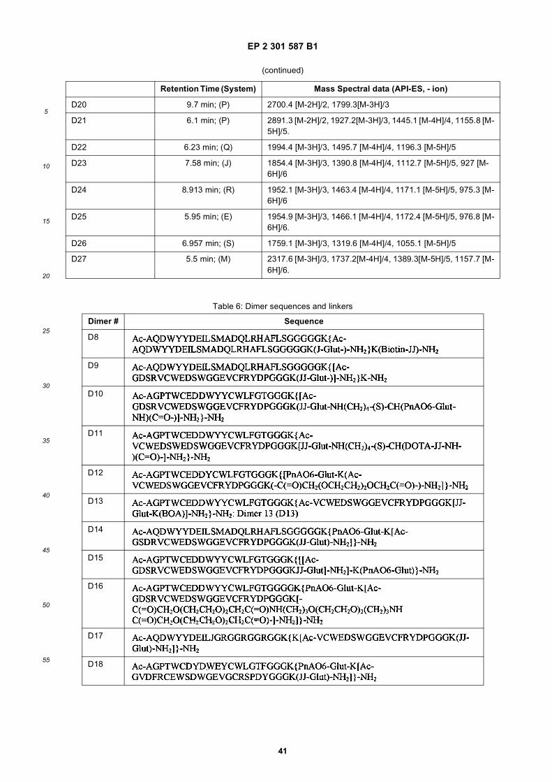
EP 2 301 587 B1
41
5
10
15
20
25
30
35
40
45
50
55
(continued)
Retention Time (System) Mass Spectral data (API-ES, - ion)
D20 9.7 min; (P) 2700.4 [M-2H]/2, 1799.3[M-3H]/3
D21 6.1 min; (P) 2891.3 [M-2H]/2, 1927.2[M-3H]/3, 1445.1 [M-4H]/4, 1155.8 [M-5H]/5.
D22 6.23 min; (Q) 1994.4 [M-3H]/3, 1495.7 [M-4H]/4, 1196.3 [M-5H]/5
D23 7.58 min; (J) 1854.4 [M-3H]/3, 1390.8 [M-4H]/4, 1112.7 [M-5H]/5, 927 [M-6H]/6
D24 8.913 min; (R) 1952.1 [M-3H]/3, 1463.4 [M-4H]/4, 1171.1 [M-5H]/5, 975.3 [M-6H]/6
D25 5.95 min; (E) 1954.9 [M-3H]/3, 1466.1 [M-4H]/4, 1172.4 [M-5H]/5, 976.8 [M-6H]/6.
D26 6.957 min; (S) 1759.1 [M-3H]/3, 1319.6 [M-4H]/4, 1055.1 [M-5H]/5
D27 5.5 min; (M) 2317.6 [M-3H]/3, 1737.2[M-4H]/4, 1389.3[M-5H]/5, 1157.7 [M-6H]/6.
Table 6: Dimer sequences and linkers
Dimer # Sequence
D8
D9
D10
D11
D12
D13
D14
D15
D16
D17
D18
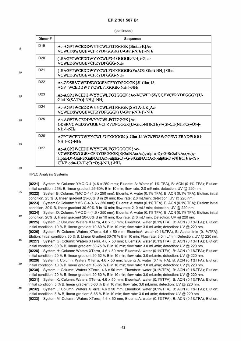
EP 2 301 587 B1
42
5
10
15
20
25
30
35
40
45
50
55
HPLC Analysis Systems
[0221] System A: Column: YMC C-4 (4.6 x 250 mm); Eluents: A: Water (0.1% TFA), B: ACN (0.1% TFA); Elution:initial condition, 25% B, linear gradient 25-60% B in 10 min; flow rate: 2.0 ml/ min; detection: UV @ 220 nm.[0222] System B: Column: YMC C-4 (4.6 x 250 mm); Eluents: A: water (0.1% TFA), B: ACN (0.1% TFA); Elution: initialcondition, 25 % B, linear gradient 25-60% B in 20 min; flow rate: 2.0 mL/min; detection: UV @ 220 nm.[0223] System C: Column: YMC C-4 (4.6 x 250 mm); Eluents: A: water (0.1% TFA), B: ACN (0.1% TFA); Elution: initialcondition, 30% B, linear gradient 30-60% B in 10 min; flow rate: 2. 0 mL/ min; detection: UV @ 220 nm.[0224] System D: Column: YMC C-4 (4.6 x 250 mm); Eluents: A: water (0.1% TFA), B: ACN (0.1% TFA); Elution: initialcondition, 20% B, linear gradient 20-60% B in 10 min; flow rate: 2. 0 mL/ min; Detection: UV @ 220 nm.[0225] System E: Column: Waters XTerra, 4.6 x 50 mm; Eluents:A: water (0.1%TFA), B: ACN (0.1%TFA); Elution:initial condition, 10 % B, linear gradient 10-60 % B in 10 min; flow rate: 3.0 mL/min; detection: UV @ 220 nm.[0226] System F: Column: Waters XTerra, 4.6 x 50 mm; Eluents:A: water (0.1%TFA), B: Acetonitrile (0.1%TFA);Elution: Initial condition, 30 % B, Linear Gradient 30-70 % B in 10 min; Flow rate: 3.0 mL/min; Detection: UV @ 220 nm.[0227] System G: Column: Waters XTerra, 4.6 x 50 mm; Eluents:A: water (0.1%TFA), B: ACN (0.1%TFA); Elution:initial condition, 30 % B, linear gradient 30-75 % B in 10 min; flow rate: 3.0 mL/min; detection: UV @ 220 nm.[0228] System H: Column: Waters XTerra, 4.6 x 50 mm; Eluents:A: water (0.1%TFA), B: ACN (0.1%TFA); Elution:initial condition, 20 % B, linear gradient 20-52 % B in 10 min; flow rate: 3.0 mL/min; detection: UV @ 220 nm.[0229] System I: Column: Waters XTerra, 4.6 x 50 mm; Eluents:A: water (0.1%TFA), B: ACN (0.1%TFA); Elution:initial condition, 10 % B, linear gradient 10-65 % B in 10 min; flow rate: 3.0 mL/min; detection: UV @ 220 nm.[0230] System J: Column: Waters XTerra, 4.6 x 50 mm; Eluents:A: water (0.1%TFA), B: ACN (0.1%TFA); Elution:initial condition, 20 % B, linear gradient 20-60 % B in 10 min; flow rate: 3.0 mL/min; detection: UV @ 220 nm.[0231] System K: Column: Waters XTerra, 4.6 x 50 mm; Eluents:A: water (0.1%TFA), B: ACN (0.1%TFA); Elution:initial condition, 5 % B, linear gradient 5-60 % B in 10 min; flow rate: 3.0 mL/min; detection: UV @ 220 nm.[0232] System L: Column: Waters XTerra, 4.6 x 50 mm; Eluents:A: water (0.1%TFA), B: ACN (0.1%TFA); Elution:initial condition, 5 % B, linear gradient 5-65 % B in 10 min; flow rate: 3.0 mL/min; detection: UV @ 220 nm.[0233] System M: Column: Waters XTerra, 4.6 x 50 mm; Eluents:A: water (0.1%TFA), B: ACN (0.1%TFA); Elution:
(continued)
Dimer # Sequence
D19
D20
D21
D22
D23
D24
D25
D26
D27
EP 2 301 587 B1
43
5
10
15
20
25
30
35
40
45
50
55
initial condition, 15 % B, linear gradient 15-50 % B in 10 min; flow rate: 3.0 mL/min; detection: UV @ 220 nm.[0234] System N: Column: Waters XTerra, 4.6 x 50 mm; Eluents:A: water (0.1%TFA), B: ACN (0.1%TFA); Elution:initial condition, 10 % B, linear gradient 20-80 % B in 10 min; flow rate: 3.0 mL/min; detection: UV @ 220 nm.[0235] System O: Column: YMC-C18, 4.6 x 250 mm; Eluents:A: water (0.1%TFA), B: ACN (0.1%TFA); Elution: initialcondition, 30 % B, linear gradient 30-60 % B in 10 min; flow rate: 2.0 mL/min; detection: UV @ 220 nm.[0236] System P: Column: YMC-C18, 4.6 x 250 mm; Eluents:A: water (0.1%TFA), B: ACN (0.1%TFA); Elution: initialcondition, 20 % B, linear gradient 20-80 % B in 20 min; flow rate: 2.0 mL/min; detection: UV @ 220 nm.[0237] System Q: Column: YMC-C18, 4.6 x 250 mm; Eluents:A: water (0.1%TFA), B: ACN (0.1%TFA); Elution: initialcondition, 20 % B, linear gradient 20-60 % B in 6 min; flow rate: 2.0 mL/min; detection: UV @ 220 nm.[0238] System R: Column: YMC-C18, 4.6 x 250 mm; Eluents:A: water (0.1%TFA), B: ACN (0.1%TFA); Elution: initialcondition, 25 % B, linear gradient 25-60 % B in 10 min; flow rate: 2.0 mL/min; detection: UV @ 220 nm.[0239] System S: Column: YMC-C18, 4.6 x 100 mm; Eluents:A: water (0.1%TFA), B: ACN (0.1%TFA); Elution: initialcondition, 10 % B, linear gradient 10-60 % B in 10 min; flow rate: 3.0 mL/min; detection: UV @ 220 nm.
Example 13: Competition with 125I-VEGF for binding to KDR on HUVECs and KDR-transfected cells
[0240] The following experiment assessed the ability of KDR-binding peptides to compete with 125I-labeled VEGF forbinding to KDR expressed by transfected 293H cells.
Protocol:
[0241] 293H cells were transfected with the KDR cDNA or mock-transfected by standard techniques. The cells wereincubated with 125I-VEGF in the presence or absence of competing compounds (at 10 mM, 0.3 mM, and 0.03 mM). Afterwashing the cells, the bound radioactivity was quantitated on a gamma counter. The percentage inhibition of VEGFbinding was calculated using the formula [(Y1-Y2) 3 100/Y1], where Y1 is specific binding to KDR-transfected 293Hcells in the absence peptides, and Y2 is specific binding to KDR-transfected 293H cells in the presence of peptidecompetitors. Specific binding to KDR-transfected 293H cells was calculated by subtracting the binding to mock-trans-fected 293H cells from the binding to KDR-transfected 293H cells.
Results
[0242] As shown in FIG. 15, all of the KDR-binding peptides assayed were able to compete with 125I-VEGF for bindingto KDR-transfected cells. The heterodimer (D1) was clearly the most effective at competing with 125I-VEGF, even overthe two homodimers (D2 and D3), confirming the superior binding of D1.
Example 14. Receptor Activation Assay
[0243] The ability of KDR-binding peptides to inhibit VEGF induced activation (phosphorylation) of KDR was assessedusing the following assay.
Protocol
[0244] Dishes of nearly confluent HUVECs were placed in basal medium lacking serum or growth factors overnight.The dishes in group (c), below were then pretreated for 15 min in basal medium with a KDR-binding peptide, and thenthe cells in the dishes in groups (a), (b), and (c) were placed in fresh basal medium containing:
(a) no additives (negative control),(b) 5 ng/ mL VEGF (positive control), or(c) 5 ng/ mL VEGF plus the putative competing/inhibiting peptide.
After 5 min of treatment, lysates were prepared from each set of dishes. KDR was immunoprecipitated from the lysateswas analyzed sequentially by immunoblotting for phosphorylation with an anti-phosphotyrosine antibody, and for totalKDR with an anti-KDR antibody (to control for sample loading).
Results
[0245] As shown in FIG. 15, D1 was able to completely block the VEGF-induced phosphorylation of KDR in HUVECsat 10 nM. More than half of the phosphorylation was inhibited by the compound at 1 nM. Homodimers D2 and D3, made

EP 2 301 587 B1
44
5
10
15
20
25
30
35
40
45
50
55
up of the two individual binding moieties that are contained in D1, had no effect on phosphorylation at up to 100 nM,demonstrating the benefit of heterodimer constructs in blocking a receptor-ligand interaction. In multiple experiments,the IC50 for D 1 in this assay varied between 0.5 and 1 nM. A different heterodimer containing unrelated binding sequences,a tail-to-tail heterodimer comprising the polypeptides of SEQ ID NO:36 and SEQ ID NO:37 (FIG. 51), had no effect onphosphorylation at 100 nM in spite of it’s high binding affinity (11 nM for KDR by SPR), suggesting that the choice ofKDR-binding moieties is important when constructing a multimer to compete with VEGF for binding to KDR. One ofordinary skill in the art would be able to construct suitable heteromultimers using the binding polypeptides providedherein and routine screening assays.[0246] Even though the affinity of D1 for KDR is 10-fold higher than that of D2 (by SPR analysis), the IC50 of D1 inthe activation assay is at least 100-fold lower. Without wishing to be bound by theory, this suggests that targeting twodistinct epitopes on KDR with a single binding molecule can generate greater steric hindrance than a molecule withsimilar affinity that only binds to a single epitope on KDR and, therefore, improve the ability to inhibit VEGF inducedKDR activity. Similarly, it should be pointed out that the two KDR-binding moieties within D1, when tested as monomericfree peptides (SEQ ID NO:17 and SEQ ID NO:49 in the receptor activation assay, had IC50s of 0.1 and 1 micromolar,respectively. The IC50 for the monomeric free peptides were 100 to 1000-fold higher than the IC50 for D1 in the assayand 14 to 30-fold higher than the KDs for the fluoresceinated derivatives of the monomeric peptides. Thus, creating adimer containing two peptides with weak VEGF-blocking activity has resulted in a molecule with very potent VEGF-blocking activity that goes well beyond the increased binding affinity of D1.
Example 15: Migration Assay
[0247] The following experiment assessed the ability of D1 to block the VEGF-induced migration of HUVECs in culture.
Protocol
[0248] Serum-starved HUVECs were placed, 100,000 cells per well, into the upper chambers of BD Matrigel-coatedFluoroBlok 24-well insert plates (#354141). Basal medium, containing either nothing or different attractants such asVEGF (10 ng/mL) or serum (5% FBS) in the presence or absence of potential VEGF-blocking/inhibiting compounds,was added to the lower chamber of the wells. After 22 hours, quantitation of cell migration/invasion was achieved bypost-labeling cells in the insert plates with a fluorescent dye and measuring the fluorescence of the invading/migratingcells in a fluorescent plate reader. The VEGF-induced migration was calculated by subtracting the migration that occurredwhen only basal medium was placed in the lower chamber of the wells.
Results:
[0249] VEGF induced a large increase in endothelial cell migration in the assay, which was potently blocked by D1.At 5 nM D1, the VEGF-stimulated endothelial cell migration was 84% blocked (see FIG. 17). At 25 nM D1, this migrationwas almost completely blocked. In other experiments, a known KDR inhibitor, SU-1498 (Strawn, L. et al., 1996, CancerRes., 56:3540-3545) was tested in the assay. SU-1498 at 3 micromolar did not block the VEGF-induced migration aswell as D1 (47% blocked at 3 micromolar). D6 (structure shown below in Example 18), at 50 nM, also produced essentiallycomplete inhibition of the migration stimulated by VEGF. Serum was a very powerful attractant in the assay when usedin place of VEGF, but its effect was not significantly diminished by D1, indicating that D1 specifically inhibits endothelialmigration induced by VEGF.
Example 18: Biacore analysis of heterodimer binding to KDR-Fc and determination of affinity constant
[0250] A peptide heterodimer (FIG. 50) composed of SEQ ID NO:17 and SEQ ID NO:31 was prepared as previouslydescribed in Example 12 using glutaric acid bis N-hydoxysuccinimidyl ester. The heterodimer was tested for binding toKDR-Fc using Biacore and an affinity constant was determined as follows.[0251] Three densities of KDR-Fc were cross-linked to the dextran surface of a CM5 sensor chip by the standardamine coupling procedure (0.5 mg/mL solution diluted 1:100 or 1:50 with 50 mM acetate, pH 6.0). Flow cell 1 wasactivated and then blocked to serve as a reference subtraction. Final immobilization levels achieved:
RL Fc 2 KDR-Fc = 1607RL Fc 3 KDR-Fc = 3001RL Fc 4 KDR-Fc = 6319
[0252] Experiments were performed in PBS (5.5 mM phosphate, pH 7.65, 0.15 M NaCl) + 0.005% P-20 (v/v)). D6 was

EP 2 301 587 B1
45
5
10
15
20
25
30
35
40
45
50
55
diluted to 250 nM in PBS and serial dilutions were performed to produce 125, 62.5, 31.3 15.6, 7.8, and 3.9 nM solutions.All samples were injected in duplicate. For association, peptides were injected at 20 mL/min for 12.5 minutes using thekinject program. Following a 10 minute dissociation, any remaining peptide was stripped from the KDR surface with aquickinject of 50 mM NaOH +1 M NaCl for 12 s at 75 mL/min. Sensorgrams were analyzed using BIAevaluation software3.1 and a hyperbolic double rectangular regression equation in SigmaPlot 6.0. Heterodimer steady state binding affinities(KDAV) were determined at all three KDR immobilization densities (Table 7).
[0253] From this data, it appears that at the higher immobilization densities, the heterodimer binds KDR with a sub-nanomolar affinity (∼0.6 nM).[0254] To assess the in vivo clearance of this peptide heterodimer, a small amount of material was iodinated usingiodogen and Na125I according to standard protocols (Pierce). One tube coated with the iodogen reagent was pre-wetwith 1 mL of 25 mM Tris, 0.4M NaCl, pH 7.5. This was discarded and 100 ml of the same buffer added. Using a Hamiltonsyringe 11 mL of the 125I-NaI was transferred to the reaction tube. Based on original estimates of the No125I concentrationof 143.555 mCi/ml, the 11 mL should contain about 1.5 mCi. After addition, the sample was swirled and set in a lead pigto incubate for 6min with a swirl every 30 sec. After 6 min, the entire sample was transferred to the protein that was inan Eppendorf tube. The sample was swirled and set to incubate for 8 min, with a swirl every 30 sec. After 8 min thereaction was quenched (terminated) with tyrosine (10mg/mL, a saturated solution), allowed to sit for 5 min, and then 2mL was removed for a standard.[0255] For purification a 10 mL column of the D-salt polyacrylamide 1800 was used to separate the labeled peptidefrom labeled tyrosine. The column was first washed with 10 mL saline, then 5 mL of 25 mM Tris, 0.4M NaCl, pH 7.5containing 2.5% HSA to block non-specific sites. After the HSA buffer wash, the column was eluted with 60mL of the25 mM Tris, 0.4 M NaCl buffer, and the column was stored overnight at 4°C. The labeled sample contained 1.355 mCi,as determined by the dose calibrator. The 2 ml sample that was removed as a standard contained 8.8 mCi. The peptidesample was applied to the D-salt 1800 column and eluted with the Tris/NaCl buffer, pH 7.5. The flow was controlled byapplying single 0.5ml aliquots for each fraction, #1-14, and then 1.0 mL for fractions 25-43. FIG. 18 shows the elutionprofile of activity versus fraction number. The peak of activity in fractions # 9, 10, and 11, was assumed to be the peptide.The radioactivity in 24 through ∼40 is likely the labeled tyrosine. From this purification, fractions #9-12 were pooledtogether and used for the subsequent clearance study (concentration of 125I-D6 in pool is 7.023 mg/mL; 100 mL = 0.702mgwith 8.6mCi).[0256] A total of 15 mice were injected with 100 mL 125I-D6 and mice (in sets of 3) were sacrificed at the following timepoints: 0, 7, 15, 30, 90 minutes. After injection more than 2 mCi was found remaining in the syringe, so actual activityinjected was about 6 mCi. With 6 mCi injected, the corresponding protein administered was ∼ 0.5 mg per animal. Oncesacrificed, the counts were determined in a 50 mL plasma sample from each animal. For each set of three animals ateach time point, the counts were averaged, converted to % injected dose/ml plasma (ID%/mL), and then plotted toassess the rate of clearance (FIG. 18). This data was fit to either a 4 or 5 parameter equation to determine the biphasichalf life of this molecule. The 4 parameter fit resulted in a T1/2α of 2.55 minutes and a T1/2β of 64.66 minutes. The 5parameter fit resulted in a T1/2α of 2.13 minutes and a T1/2β of 23.26 minutes.[0257] Larger volumes of plasma were also taken from mice sacrificed at the 0, 30, and 90 minute time points. Thesesamples were injected onto a Superdex peptide column (Pharmacia) coupled to a radioactivity detector to assess theassociation of the peptide with serum proteins (FIG. 19). As shown, the labeled peptide does associate with higher MWproteins, which could explain its biphasic half life clearance behavior.[0258] To help assess the potency of the peptide as an anti-angiogenesis inhibitor, D6 was tested in an endothelialcell proliferation assay using HUVECs and BrdU detection. Briefly, freshly isolated HUVECs (between p3 - 6) werecultured in RPMI + 10% FCS +1% antibiotics + 1% L-glutamine + 0.4% BBE (bovine brain extract) and seeded per well,5000-10000/well in 100 mL. The cells were allowed to recover for 24 hrs prior to use. Then the cells were washed withPBS twice and treated for 48 hrs with anti-VEGF antibody (positive control) or peptides A, B and C (0.1 and 10 ug/mL)in RPMI + 0.1% BSA + 1% L-glutamine. The following 6 variables were tested in 2 series (n=4):
Series I: w/o VEGF
Table 7. Summary of Parameters
KD1 (nM) Rmax1 KDAV(nM) RmaxAV R2*
D6
Vs. 1600RU 46 13.1 1.5 12.6 0.995
Vs. 3000RU 25.5 21.2 0.665 22.7 0.991
Vs. 6000RU 17 61.3 0.662 62.2 0.993
EP 2 301 587 B1
46
5
10
15
20
25
30
35
40
45
50
55
Series II: w/ VEGF (30 ng/mL)
1. Standard medium: RPMI + 10% FCS +1% antibiotics + 1% L-glutamine + 0.4% BBE2. Negative control 1: RPMI (true starvation)3. Negative control 2: RPMI + 0.1% BSA + 1% L-glutamine4. Positive control: anti-VEGF 10 mg/ml in RPMI + 0.1% BSA + 1% L-glutamine5. 0.1 mg/ml KDR peptides in RPMI + 0.1% BSA + 1% L-glutamine6. 10 mg/ml KDR peptides in RPMI + 0.1 % BSA + 1% L-glutamine
Protocol:
[0259]
1) cells are incubated for 48 hours under various conditions2) 10mL BrdU dilution (1:100 in EBM) is added to each well at 24 hours3) incubate for another 24 hours (total 48 hrs)4) aspirate the culture medium5) add 100mL FixDenat (Roche Applied Science, Indianapolis, IN) to each well, incubate at room temperature for30 min.6) Discard FixDenat solution7) 100 mL antibody-solution (PBS 1% BSA and anti-BrdU PO) added to each well.8) incubate at RT for 90 minutes.9) wash 3 times with PBS, 200 mL/well, 5 min.10) add substrate solution (TMB), incubate for 10-30 minutes11) transfer all to a flexible plate12) stop the reaction by adding 2 M H2SO4, 25 mL/well13) read absorbance at 450 nm within 5 minutes after stopping the reaction.
[0260] Background binding was determined by omitting the anti-BrdU antibody in 4 wells with control cells (culturedin complete medium; EBM + BulletKit (Clonetics, BioWhittaker, Inc., MD) and by complete labeling of cells that was notexposed to BrdU.[0261] Of the two KDR binding peptide tested (D6 and SEQ ID NO:17) as shown in FIG. 19, D6 completely inhibitsHUVEC proliferation at 10 mg/mL in the presence of VEGF, similar to an anti-VEGF antibody (positive control). On theother hand, SEQ ID NO:17 (one of the peptides that make up the heterodimer) did not inhibit proliferation in this assayat the highest concentration tested (10 mg/mL). As a result, the heterodimer shows an enhanced ability to compete withVEGF in comparison with SEQ ID NO:17 alone.
Example 19: BIAcare Analysis-murine KDR-Fc Binding of Peptide Dimers D1 and D7
[0262] Using BIAcore, the binding constants of peptide dimers D1 (a heterodimer of SEQ ID NO:17 and SEQ ID NO:31and D7 (a heterodimer of SEQ ID NO:5 and SEQ ID NO 31; see FIG. 51) for murine KDR-Fc were determined.
Procedure
[0263] Three densities of recombinant murine KDR-Fc were cross-linked to the dextran surface of a CM5 sensor chipby the standard amine coupling procedure (0.5 mg/mL solution diluted 1:100 or 1:40 with 50 mM acetate, pH 6.0). Flowcell 1 was activated and then blocked to serve as a reference subtraction. Final immobilization levels achieved:
RL Fc 2 KDR-Fc = 2770RL Fc 3 KDR-Fc = 5085RL Fc 4 KDR-Fc = 9265
[0264] Experiments were performed in PBS buffer (5.5 mM phosphate, pH 7.65, 0.15 M NaCl) + 0.005% P-20 (v/v)).SEQ ID NO:17, run as a control, was diluted to 125 nM in PBS. Serial dilutions were performed to produce 62.5, 31.3,15.6, 7.8, and 3.9 nM solutions. D1 and D6 were diluted to 50 nM in PBS and serial dilutions were performed to produce25, 12.5, 6.25, 3.13, 1.56, 0.78, and 0.39 nM solutions. All samples were injected in duplicate. For association, peptideswere injected at 30 mL/min for 3 minutes using the kinject program. Following a 10 minute dissociation, any remainingpeptide was stripped from the rmKDR-Fc surface with a quickinject of 50 mM NaOH + 1 M NaCl for 12s at 75 mL/min.
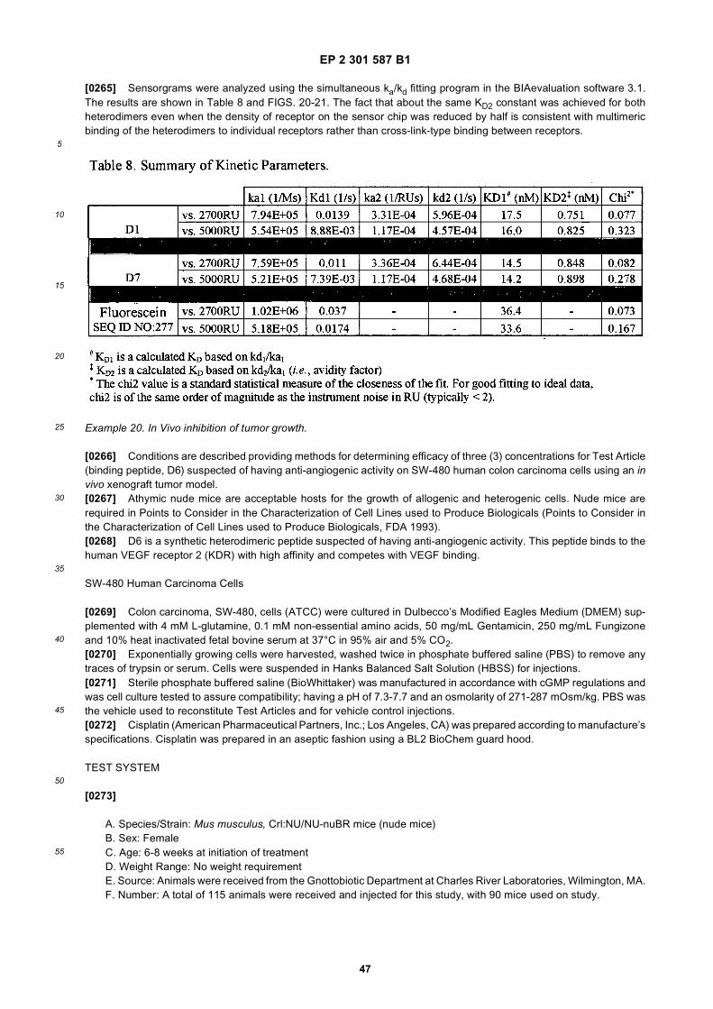
EP 2 301 587 B1
47
5
10
15
20
25
30
35
40
45
50
55
[0265] Sensorgrams were analyzed using the simultaneous ka/kd fitting program in the BIAevaluation software 3.1.The results are shown in Table 8 and FIGS. 20-21. The fact that about the same KD2 constant was achieved for bothheterodimers even when the density of receptor on the sensor chip was reduced by half is consistent with multimericbinding of the heterodimers to individual receptors rather than cross-link-type binding between receptors.
Example 20. In Vivo inhibition of tumor growth.
[0266] Conditions are described providing methods for determining efficacy of three (3) concentrations for Test Article(binding peptide, D6) suspected of having anti-angiogenic activity on SW-480 human colon carcinoma cells using an invivo xenograft tumor model.[0267] Athymic nude mice are acceptable hosts for the growth of allogenic and heterogenic cells. Nude mice arerequired in Points to Consider in the Characterization of Cell Lines used to Produce Biologicals (Points to Consider inthe Characterization of Cell Lines used to Produce Biologicals, FDA 1993).[0268] D6 is a synthetic heterodimeric peptide suspected of having anti-angiogenic activity. This peptide binds to thehuman VEGF receptor 2 (KDR) with high affinity and competes with VEGF binding.
SW-480 Human Carcinoma Cells
[0269] Colon carcinoma, SW-480, cells (ATCC) were cultured in Dulbecco’s Modified Eagles Medium (DMEM) sup-plemented with 4 mM L-glutamine, 0.1 mM non-essential amino acids, 50 mg/mL Gentamicin, 250 mg/mL Fungizoneand 10% heat inactivated fetal bovine serum at 37°C in 95% air and 5% CO2.[0270] Exponentially growing cells were harvested, washed twice in phosphate buffered saline (PBS) to remove anytraces of trypsin or serum. Cells were suspended in Hanks Balanced Salt Solution (HBSS) for injections.[0271] Sterile phosphate buffered saline (BioWhittaker) was manufactured in accordance with cGMP regulations andwas cell culture tested to assure compatibility; having a pH of 7.3-7.7 and an osmolarity of 271-287 mOsm/kg. PBS wasthe vehicle used to reconstitute Test Articles and for vehicle control injections.[0272] Cisplatin (American Pharmaceutical Partners, Inc.; Los Angeles, CA) was prepared according to manufacture’sspecifications. Cisplatin was prepared in an aseptic fashion using a BL2 BioChem guard hood.
TEST SYSTEM
[0273]
A. Species/Strain: Mus musculus, Crl:NU/NU-nuBR mice (nude mice)B. Sex: FemaleC. Age: 6-8 weeks at initiation of treatmentD. Weight Range: No weight requirementE. Source: Animals were received from the Gnottobiotic Department at Charles River Laboratories, Wilmington, MA.F. Number: A total of 115 animals were received and injected for this study, with 90 mice used on study.

EP 2 301 587 B1
48
5
10
15
20
25
30
35
40
45
50
55
G. Method of Identification:
Mice were uniquely numbered using an ear tag system. Additionally, cages were marked with cage cardsminimally identifying group number, animal number, study number and IACUC protocol number.
H. Randomization:
Animals were randomly assigned to treatment groups using Microsoft® Excel 97 SR-1 program.
I. Humane Care of Animals:
Treatment and care of the animals were in accordance with the standard operating procedures of Charles RiverLaboratories, which adheres to the regulations outlined in the USDA Animal Welfare Act (9 CFR, Parts 1, 2,and 3) and the Guide for the Care and Use of Laboratory Animals.
[0274] This study protocol was covered under the Charles River Laboratories Institutional Animal Care and Use Com-mittee (IACUC Protocol Number: P07182001I).
ANIMAL CARE
A. Diet and Drinking Water:
[0275] Mice were fed gamma-irradiated rodent chow ad libitum. Tap water was sterilized and supplied via bottle andsipper tube ad libitum.
B. Animal Environment:
[0276] Animals were housed by groups in semi-rigid isolators. Mice were housed in flat bottom caging containing fiveto ten animals. Cages contained gamma-irradiated contact bedding. The number of mice in each cage may have beenaltered due to the behavior of the mice, changes were noted in the isolator inventory. The housing conforms to therecommendations set forth in the Guide for the Care and Use of Laboratory Animals, National Academy Press, Wash-ington, D.C., 1996 and all subsequent revisions.[0277] Environmental controls were set to maintain a temperature of 16-26°C (70 6 8°F) with a relative humidity of30-70. A 12:12 hour light: dark cycle was maintained.
C. Acclimation:
[0278] Once animals were received, they were allowed to acclimate to the laboratory environment for 24-hours priorto the study start. Mice were observed for signs of disease, unusual food and/or water consumption or other generalsigns of poor condition. At the time of animal receipt, animals were clinically observed and appeared to be healthy.
EXPERIMENTAL DESIGN
A. General Description:
[0279] Female athymic nude mice (Crl:NU/NU-nuBR) at 6-8 weeks of age were used in this study. A total of 115 micewere injected subcutaneously into the right lateral thorax with 5 3 106 SW-480, human colon carcinoma cells. Whentumors reached a target window size of approximately 150 6 75 mg, 90 tumor-bearing mice were randomly selectedand distributed into one of nine groups. Test Articles and vehicle were administered intraperitoneally (IP), Cisplatin wasadministered intravenously (IV). Tumor measurements were recorded twice weekly using hand-held calipers. Mice weremonitored daily for signs of toxicity and morbidity. At study termination, animals were euthanized by carbon dioxideoverdose and necropsied for tissue collection.
B. Group Assignments:
[0280] A total of nine (9) groups were used in this study. Each group contained ten (10) tumor-bearing mice. Groups1 and 2 contained untreated and vehicle treated negative control mice, respectively. Groups 3, 4, and 5 contained micethat received one of three different concentrations of the D6 anti-angiogenic peptide. Group 9 contained mice that

EP 2 301 587 B1
49
5
10
15
20
25
30
35
40
45
50
55
received cisplatin, a standard chemotherapeutic compound as a positive control.
C. Dosing Levels and Regiment:
[0281] Dose levels for each group are provided in Table 9. Dosing began the same day that animals were randomlysorted into groups (Study Day 7). Each dose was removed from the dose vial using aseptic technique for each animaland the injection site was wiped with an alcohol swab prior to dose administration. Doses were administered with a 1.0mL syringe and a 27-gauge x ©" needle for each mouse[0282] The Test Article- and vehicle-treated mice received daily intraperitoneal (IP) injections for 15 days. Cisplatinwas administered every other workday for a total of five (5) doses via an intravenous route.
D. Clinical Observations of Animals:
[0283] Clinical Observations of each animal were performed and recorded at least once daily for toxicity, morbidityand mortality. Morbidity included signs of illness such as, but not limited to, emaciation, dehydration, lethargy, hunchedposture, unkempt appearance, dyspnea and urine or fecal staining.
E. Tumor Measurements:
[0284] In accordance with the protocol tumor measurements were taken twice weekly throughout the study by meas-uring the length and width of tumors with calibrated calipers. Measurements occurred a minimum of 3-4 days apart,except when animals were euthanized and measurements were taken; this sometimes resulted in an interval of lessthan 3 days. Tumor weights were calculated using the following formula: mg = (L 3 W2)/2. Animals were euthanizedeither when mean tumor weight was ≥ 1000 mg per group over two (2) consecutive measurements, or if tumors becameulcerated, impaired the animal’s ability to ambulate or obtain food and water.
F. Unscheduled Euthanasia and Unexpected Deaths:
1. Unscheduled Euthanasia:
[0285] None of the animals required unscheduled euthanasia while on study.
2. Unexpected Deaths:
[0286] None of the animals died while on study.
G. Necropsy:
1. Euthanasia and Necropsy Order:
[0287] All mice in groups 1, 2, 3, 4, and 5 (50 total) were submitted for necropsy when tumors reached a group meantarget size of ≥ 1000 mg over two (2) consecutive measurements within a group. Animals were submitted for necropsyto the Charles River Laboratories Health Monitoring Laboratory (HM), Wilmington, MA. All animals were euthanized onStudy Day 22, short of received the full 28 day treatment regiment with Test Articles because mean tumor size was ≥1000 mg in Test Article Treated Groups 3-8.
Table 9. Study Treatment Groups
Group Test Article Concentration mg/kg Number of Animals
1 Untreated - 10
2 Vehicle 0 10
3 D6 0.05 10
4 D6 0.5 10
5 D6 5.0 10
9 Cisplatin 6.0 10
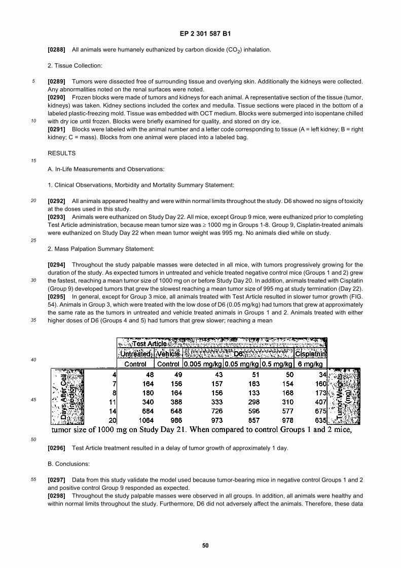
EP 2 301 587 B1
50
5
10
15
20
25
30
35
40
45
50
55
[0288] All animals were humanely euthanized by carbon dioxide (CO2) inhalation.
2. Tissue Collection:
[0289] Tumors were dissected free of surrounding tissue and overlying skin. Additionally the kidneys were collected.Any abnormalities noted on the renal surfaces were noted.[0290] Frozen blocks were made of tumors and kidneys for each animal. A representative section of the tissue (tumor,kidneys) was taken. Kidney sections included the cortex and medulla. Tissue sections were placed in the bottom of alabeled plastic-freezing mold. Tissue was embedded with OCT medium. Blocks were submerged into isopentane chilledwith dry ice until frozen. Blocks were briefly examined for quality, and stored on dry ice.[0291] Blocks were labeled with the animal number and a letter code corresponding to tissue (A = left kidney; B = rightkidney; C = mass). Blocks from one animal were placed into a labeled bag.
RESULTS
A. In-Life Measurements and Observations:
1. Clinical Observations, Morbidity and Mortality Summary Statement:
[0292] All animals appeared healthy and were within normal limits throughout the study. D6 showed no signs of toxicityat the doses used in this study.[0293] Animals were euthanized on Study Day 22. All mice, except Group 9 mice, were euthanized prior to completingTest Article administration, because mean tumor size was ≥ 1000 mg in Groups 1-8. Group 9, Cisplatin-treated animalswere euthanized on Study Day 22 when mean tumor weight was 995 mg. No animals died while on study.
2. Mass Palpation Summary Statement:
[0294] Throughout the study palpable masses were detected in all mice, with tumors progressively growing for theduration of the study. As expected tumors in untreated and vehicle treated negative control mice (Groups 1 and 2) grewthe fastest, reaching a mean tumor size of 1000 mg on or before Study Day 20. In addition, animals treated with Cisplatin(Group 9) developed tumors that grew the slowest reaching a mean tumor size of 995 mg at study termination (Day 22).[0295] In general, except for Group 3 mice, all animals treated with Test Article resulted in slower tumor growth (FIG.54). Animals in Group 3, which were treated with the low dose of D6 (0.05 mg/kg) had tumors that grew at approximatelythe same rate as the tumors in untreated and vehicle treated animals in Groups 1 and 2. Animals treated with eitherhigher doses of D6 (Groups 4 and 5) had tumors that grew slower; reaching a mean
[0296] Test Article treatment resulted in a delay of tumor growth of approximately 1 day.
B. Conclusions:
[0297] Data from this study validate the model used because tumor-bearing mice in negative control Groups 1 and 2and positive control Group 9 responded as expected.[0298] Throughout the study palpable masses were observed in all groups. In addition, all animals were healthy andwithin normal limits throughout the study. Furthermore, D6 did not adversely affect the animals. Therefore, these data

EP 2 301 587 B1
51
5
10
15
20
25
30
35
40
45
50
55
would suggest that animals treated with D6 Test Article had tumors that grew slowly (approximately 1 day slower overthe 22 day test period than controls). Also, since the Test Article did not show any significant toxic effects, higherconcentrations of Test Article could also be used with potentially better tumor regression.
Example 21: In vitro cell proliferation assay
[0299] Microvascular endothelial cells (MVECs, Cascade Biologics, Portland, OR) were used to assess the in vitroefficacy of D6 and related analogues for their ability to inhibit VEGF-stimulated proliferation. MVECs (passage 2) weregrown to 90% confluency, trypsinized and plated in gelatin-coated 96-well microtiter plates at a density of 4-8 3 103
cells/well. Sixteen to 24 hours after plating, the cells were washed one time (200mL/well) with media devoid of fetalbovine serum but containing 0.1% bovine serum albumin (BSA). Fresh BSA-containing media was added to each welland the cells were incubated for an additional 24 hours. After this 24 hour period of starvation, fresh BSA-containingmedia (containing 25 ng/mL VEGF) with or without D6 was added and the cells were incubated for an additional 48hours at 37°C. To assess the dose reponse in this assay, multiple D6 concentrations were tested in duplicate wells. Themedia was removed and fresh BSA-containing media was added with or without BrdU and the cells were incubated foran additional 24 hours prior to determining the level of incorporation exactly as described by the manufacturer. Resultsare shown in FIG. 62.
Example 22.
[0300] The following experiment assessed the ability of D25 and D27 to block the VEGF-induced migration of HUVECsin culture and demonstrated that the added glycosylation and/or distinct spacer structure used in D27 enhanced itspotency.[0301] Protocol: Serum-starved HUVECs were placed, 100,000 cells per well, into the upper chambers of BD fibronec-tin-coated FluoroBlok 24-well insert plates. Basal medium, with or without VEGF (10 ng/mL) in the presence or absenceof D25 or D27, was added to the lower chamber of the wells. After 22 hours, quantitation of cell migration/invasion wasachieved by post-labeling cells in the insert plates with a fluorescent dye and measuring the fluorescence of the invad-ing/migrating cells in a fluorescent plate reader. The VEGF-induced migration was calculated for each experimentalcondition by subtracting the amount of migration that occurred when only basal medium was added to the lower chamberof the wells.
Results: VEGF induced a large increase in endothelial cell migration in the assay, which
[0302] was potently blocked by both D25 and D27. D27 was ten-fold more potent than D25 (IC50 0.5 nM and 5 nMrespectively), indicating that the glycosylation of D27 and/or its distinct spacer properties has enhanced its ability to bindKDR and block the effects of VEGF.
Example 23.
[0303] The following experiment assessed the ability of "Adjunct A" multimeric construct of TKPPR peptide (binds toNP-1, a VEGF receptor that enhances the effects of VEGF mediated by KDR),to enhance the inhibition of the VEGF-induced migration of HUVECs in culture produced by D6. Adjunct A = 5CF-Gly-N{[CH2CH2C(=O)-Gly-N(CH2CH2C(=O)-Adoa-Thr-Lys-Pro-Pro-Arg-OH]2}2 where Adoa = 3,6-dioxa-8-aminooctanoyl, 5CF = 5-carboxyfluo-resceinyl.[0304] Protocol: Serum-starved HUVECs were placed, 100,000 cells per well, into the upper chambers of BD fibronec-tin-coated FluoroBlok 24-well insert plates. Basal medium, containing with or without VEGF (10 ng/mL) in the presenceor absence of varying concentrations of D6 , or varying concentrations of D6 in combination with a constant 100 nMAdjunct A (synthesized as described in WO 01/91805 A2), was added to the lower chamber of the wells. After 22 hours,
EP 2 301 587 B1
52
5
10
15
20
25
30
35
40
45
50
55
quantitation of cell migration/invasion was achieved by post-labeling cells in the insert plates with a fluorescent dye andmeasuring the fluorescence of the invading/migrating cells in a fluorescent plate reader. VEGF-induced migration wascalculated for each experimental condition by subtracting the amount of migration observed in the absence of VEGF.[0305] Results: VEGF induced a large increase in endothelial cell migration in the assay, which was potently blockedby D6 (IC50 about 12.5 nM), but not by 100 nM Adjunct A alone (FIG. 53). Surprisingly however, Adjunct Awas able toenhance the potency of D6 by about ten-fold when used in the assay simultaneously with D6 (IC50 about 2.5 nM). Thisindicates that compounds containing the TKPPR sequence (or similar) found in Adjunct A can be used to enhance thepotency of certain compounds such as D6, which compete with VEGF for binding to KDR. In addition, a heteromultimercontaining the peptide sequences found in D6 or similar) as well as the TKPPR sequence (or similar), in one or morerepetitions, would likely possess enhanced activity in this assay.
Example 24: Synthesis of D27
Synthesis of 1 and 3 (see FIGS. 54 and 55)
[0306] Synthesis of the monomers were carried out as described in Method 5 on a 0.25 mmol scale employing as thestarting resin Fmoc-GGGK(iV-Dde)NH-PAL-PEG-PS resin. The peptide resin was washed and dried before cleavageor further derivatization by automated or manual methods.
Procedure Synthesis of Peptide 2 and Peptide 4 (see FIGS. 54 and 55)
[0307] Appendage of Biotin-JJ, Lysyl, Glycyl and Serinyl(GalNAc(Ac)3-α-D moieties onto 1 and 3 was done by manualSPPS such as described in Method 6 and Method 8. The coupling of amino acids was carried out in DMF using HOBt/DICactivation (except for Ser(GalNAc(Ac)3-α-D). Fmoc removal was carried out with 20% piperidine in DMF. All couplingswere 5-16 hours duration. After each coupling, the completion was confirmed by the Kaiser test. In the case of Ser(Gal-NAc(Ac)3-α-D, the coupling was performed in DMF employing HATU/DIEA as the coupling agent. In cases where theKaiser test indicated unreacted amino groups the coupling was repeated. Removal of the N-terminal Fmoc group andcleavage from resin was performed. The crude peptide was precipitated in ether and washed twice by ether and driedunder vacuum. The linear crude peptide was directly cyclized by dissolving the peptide in DMSO (40 mg/mL). The pHof the solyution was adjusted to 8 by addition of aqueous N-methylglucamine.and the solution was was stirred in air for48h at room temperature. The peptides were then purified employing gradient HPLC as described in Method 1 employinga Waters-YMC C-18 ODS preparative column (250 mm x 4.6 mm i.d.). The pure product-containing fractions werecombined and lyophilized to provide the needed peptides.
Procedure: Synthesis of D27 - Compound 6
[0308] To a solution of glutaric acid bis-NHS ester (0.122 mmol, Pierce Scientific Co.) in anhydrous DMF was addeddropwise a solution of 4 in DMF (40 mg, 0.0122 mmol, DIEA was added to neutralize the trifluoroacetic acid bound tothe peptide and N-hydroxysuccinimide formed during the reaction). This 0.7 mL solution was stirred for 4h. The reactionwas monitored by HPLC and mass spectroscopy. DMF was removed under vacuum. The excess diester was removedby addition of ethyl acetate which precipitated the peptide-monoester 5 while dissolving glutaric acid bis-NHS ester. Themixture was centrifuged and the liquid portion decanted. This was repeated twice. The residue was kept under vacuumfor 10 min. The residue was dissolved in DMF and mixed with a solution of 2 (37 mg, 0.009 mmol) in DMF (pH 7). It wasstirred at ambient temperature for 16 h. The volatiles were removed under high vacuum and the acetate functions wereremoved by treatment of the residue with 1 mL of hydrazine/MeOH (15/85, v/v) solution with stirring for 2.5 h at ambienttemperature. Acetone was added to quench the excess of hydrazine and the volatiles were removed under vacuum.The resulting residue was dissolved in DMSO and purified by preparative HPLC as described above to provide 9 mg ofthe pure material.
Sequence and Analytical Data for Peptides 2, 4 and 6
[0309]

EP 2 301 587 B1
53
5
10
15
20
25
30
35
40
45
50
55
[0310] System T: Column: Waters XTerra, 4.6 3 50 mm; Eluents:A: Water (0.1%TFA), B: Acetonitrile (0.1%TFA) :Elution: Initial condition, 15 % B, Linear Gradient 15-50 % B in 8 min; Flow rate: 3.0 mL/min; Detection: UV @ 220 nm.
Example 41: Enhancing the serum residence of KDR-binding peptides
[0311] It is known in the art that compounds that contain maleimide and other groups that can react with thiols reactwith thiols on serum proteins, especially serum albumin, when the compounds are injected. The adducts have serumlife times similar to serum albumin, more than 14 days in humans for example.
Conjugation to maleimide
[0312] Methods are available that allow for the direct synthesis of maleimide-labeled linear peptides encompassed bythe present invention (Holmes, D. et al., 2000. Bioconjug. Chem., 11:439-444).[0313] Peptides that include disulfides can be derivatized with maleimide in one of several ways. For example, a thirdcysteine can be added at the carboxy terminus. The added cysteine is protected with protecting group that is orthogonalto the type of groups used for the cysteines that are to form the disulfide. The disulfide is formed by selectively deprotectingthe intended cysteines and oxidizing the peptide. The final cysteine is then deprotected and the peptide reacted with alarge molar excess of a bismaleimide. The resulting compound has one of the maleimides free to react with serumalbumin or other thiol-containing serum proteins.Alternatively, a cyclic peptide of the present invention is synthesized with a lysine-containing C-terminal extention, suchas -GGGK. Lysines of the KDR-binding motif are protected with ivDde and the C-terminal lysine is deprotected. Thislysine is reacted with a maleimide-contining compound, such as N-[e-maleimidocaproyloxy]succinimide ester (PierceBiotechnology, Rockford, IL) or N-(a-Maleimidoacetoxy)succinimide ester (Pierce Biotechnology).
Conjugation to a moiety that binds serum albumin non-covelently
[0314] Polypeptides having a molecular weight less than 50-60 kDa are rapidly excreted. Many small molecules, suchas fatty acids, bind to serum albumin. Fatty acids containing 10 to 20 carbon atoms have substantial affinity for serumalbumin. Linear and branched fatty acids can be used. This binding in serum can reduce the rate of excretion. Usingmethods known in the art, serum-albumin-binding moieties can be conjugated to any one of the peptides herein disclosed.The serum-ablumin-binding moiety can be joined to the KDR-binding peptide through a linker. The linker can be peptidicor otherwise, such as PEG. Linkers of zero to about thirty atoms are preferred. It is preferred that the linker be hydrophilic.The serum-albumin-binding moiety can be conjugated to the KDR-binding peptide at either end or though a side groupof an appended amino acid. Suitable side groups include lysine and cysteine. Such compounds can also comprisechelators for radionuclides, as discussed herein. A KDR-binding peptide joined to a serum-ablumin-binding moiety willbind KDR.

EP 2 301 587 B1
54
5
10
15
20
25
30
35
40
45
50
55
Conjugation to PEG
[0315] As is well known in the art, attachment of poly(ethyleneglycol) (PEG) to proteins and peptides enhances theserum residence of these molecules. Attachment of PEG (linear or branched) to a KDR-binding peptide is expected givesubstantial enhancement of serum residence time. The molecular weight of the PEG should be at least 10 kDA, morepreferably at least 20 kDa, and most preferably 30 kDa or more. The PEG could be attached at the N- or C-terminus.Methods of attaching PEG to peptides are well known in the art (Roberts M. et al., 2002. Adv. Drug. Deliv. Rev.,54:459-476). PEG can be attached to reactive side groups such as lysine or cysteine.
Fusion to serum protein
[0316] It is known in the art that proteins comprising serum albumin (SA) and other proteins have enhanced serumresidence times. The amino-acid sequence of human SA (hSA) is shown in Table 22. Table 23 shows a fusion proteincomprisingAGDWWVECRVGTGLCYRYDTGTGGGK(SEQ ID NO:25)::
PGGSGGEGGSGGEGGRPGGSEGGTGG::mature hSA::GGSGGEGGSGGEGGSGPGEGGEGSGGRP ::GDSRVCWEDSWGGEVCFRYDPGGGK(SEQ ID NO:31). The KDR-binding peptides are separated from mature hSAby linkers that are rich in glycine to allow flexible spacing. It is known in the art that one need not use all of hSA to obtainan injectable protein that will have an enhanced serum residence time. It is also known in the art that chemical groups,such as maleimide and alpha bromo carboxylates, react with the unpaired cysteine (residue 34) to form stable adducts.Thus, one could attach a single chelator to hSA fusion proteins so that the adduct will bind a radionuclide. One couldprepare a chelator with a maleimide group and couple that to hSA or an hSA derivative. Alternatively, hSA or an hSAderivative could be reacted with a bismaleimide and a chelator carrying a reactive thiol could be reacted with the bis-maleimide-derivatized hSA.[0317] Construction of genes that encode a given amino-acid sequence are known in the art. Expression of HSAfusions in Saccharomyces cerevisiae is known in the art (Sleep, D et al., 1991. Biotechnology (NY), 9:183-187).
SEQUENCE LISTING
[0318]
<110> Dyax Corp. BRACCO International B.V.
<120> KDR and VEGF/KDR Binding Peptides and Their Use in Diagnosis and Therapy
<130> FB14882/B
<140> EP 10185498.2<141> 2003-03-03
<150> US 60/360,851<151> 2002-03-01
<150> US 60/440,411<151> 2003-01-15
<160> 107
<170> FastSEQ for Windows Version 4.0
<210> 1<211> 14<212> PRT<213> Artificial Sequence
<220>
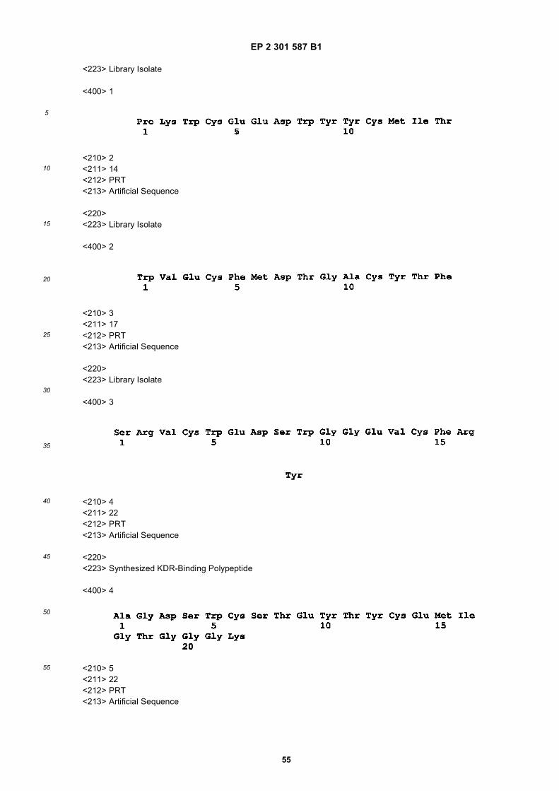
EP 2 301 587 B1
55
5
10
15
20
25
30
35
40
45
50
55
<223> Library Isolate
<400> 1
<210> 2<211> 14<212> PRT<213> Artificial Sequence
<220><223> Library Isolate
<400> 2
<210> 3<211> 17<212> PRT<213> Artificial Sequence
<220><223> Library Isolate
<400> 3
<210> 4<211> 22<212> PRT<213> Artificial Sequence
<220><223> Synthesized KDR-Binding Polypeptide
<400> 4
<210> 5<211> 22<212> PRT<213> Artificial Sequence

EP 2 301 587 B1
56
5
10
15
20
25
30
35
40
45
50
55
<220><223> Synthesized KDR-Binding Polypeptide
<400> 5
<210> 6<211> 22<212> PRT<213> Artificial Sequence
<220><223> Synthesized KDR-Binding Polypeptide
<400> 6
<210> 7<211> 22<212> PRT<213> Artificial Sequence
<220><223> Synthesized KDR-Binding Polypeptide
<400> 7
<210> 8<211> 22<212> PRT<213> Artificial Sequence
<220><223> Synthesized KDR-Binding Polypeptide
<400> 8

EP 2 301 587 B1
57
5
10
15
20
25
30
35
40
45
50
55
<210> 9<211> 22<212> PRT<213> Artificial Sequence
<220><223> Synthesized KDR-Binding Polypeptide
<400> 9
<210> 10<211> 22<212> PRT<213> Artificial Sequence
<220><223> Synthesized KDR-Binding Polypeptide
<400> 10
<210> 11<211> 22<212> PRT<213> Artificial Sequence
<220><223> Synthesized KDR-Binding Polypeptide
<400> 11
<210> 12<211> 22<212> PRT<213> Artificial Sequence
<220><223> Synthesized KDR-Binding Polypeptide
<400> 12

EP 2 301 587 B1
58
5
10
15
20
25
30
35
40
45
50
55
<210> 13<211> 22<212> PRT<213> Artificial Sequence
<220><223> Synthesized KDR-Binding Polypeptide
<400> 13
<210> 14<211> 22<212> PRT<213> Artificial Sequence
<220><223> Synthesized KDR-Binding Polypeptide
<400> 14
<210> 15<211> 22<212> PRT<213> Artificial Sequence
<220><223> Synthesized KDR-Binding Polypeptide
<400> 15
<210> 16<211> 22<212> PRT<213> Artificial Sequence
<220><223> Synthesized KDR-Binding Polypeptide
EP 2 301 587 B1
59
5
10
15
20
25
30
35
40
45
50
55
<400> 16
<210> 17<211> 22<212> PRT<213> Artificial Sequence
<220><223> Synthesized KDR-Binding Polypeptide
<400> 17
<210> 18<211> 22<212> PRT<213> Artificial Sequence
<220><223> Synthesized KDR-Binding Polypeptide
<400> 18
<210> 19<211> 22<212> PRT<213> Artificial Sequence
<220><223> Synthesized KDR-Binding Polypeptide
<400> 19
<210> 20<211> 22<212> PRT<213> Artificial Sequence
<220>

EP 2 301 587 B1
60
5
10
15
20
25
30
35
40
45
50
55
<223> Synthesized KDR-Binding Polypeptide
<400> 20
<210> 21<211> 22<212> PRT<213> Artificial Sequence
<220><223> Synthesized KDR-Binding Polypeptide
<400> 21
<210> 22<211> 26<212> PRT<213> Artificial Sequence
<220><223> Synthesized KDR-Binding Polypeptide
<400> 22
<210> 23<211> 26<212> PRT<213> Artificial Sequence
<220><223> Synthesized KDR-Binding Polypeptide
<400> 23
<210> 24<211> 26<212> PRT<213> Artificial Sequence

EP 2 301 587 B1
61
5
10
15
20
25
30
35
40
45
50
55
<220><223> Synthesized KDR-Binding Polypeptide
<400> 24
<210> 25<211> 26<212> PRT<213> Artificial Sequence
<220><223> Synthesized KDR-Binding Polypeptide
<400> 25
<210> 26<211> 26<212> PRT<213> Artificial Sequence
<220><223> Synthesized KDR-Binding Polypeptide
<400> 26
<210> 27<211> 26<212> PRT<213> Artificial Sequence
<220><223> Synthesized KDR-Binding Polypeptide
<400> 27
<210> 28<211> 26<212> PRT
EP 2 301 587 B1
62
5
10
15
20
25
30
35
40
45
50
55
<213> Artificial Sequence
<220><223> Synthesized KDR-Binding Polypeptide
<400> 28
<210> 29<211> 26<212> PRT<213> Artificial Sequence
<220><223> Synthesized KDR-Binding Polypeptide
<400> 29
<210> 30<211> 26<212> PRT<213> Artificial Sequence
<220><223> Synthesized KDR-Binding Polypeptide
<400> 30
<210> 31<211> 25<212> PRT<213> Artificial Sequence
<220><223> Synthesized KDR-Binding Polypeptide
<400> 31
<210> 32

EP 2 301 587 B1
63
5
10
15
20
25
30
35
40
45
50
55
<211> 25<212> PRT<213> Artificial Sequence
<220><223> Synthesized KDR-Binding Polypeptide
<400> 32
<210> 33<211> 26<212> PRT<213> Artificial Sequence
<220><223> Synthesized KDR-Binding Polypeptide
<400> 33
<210> 34<211> 25<212> PRT<213> Artificial Sequence
<220><223> Synthesized KDR-Binding Polypeptide
<400> 34
<210> 35<211> 25<212> PRT<213> Artificial Sequence
<220><223> Synthesized KDR-Binding Polypeptide
<400> 35

EP 2 301 587 B1
64
5
10
15
20
25
30
35
40
45
50
55
<210> 36<211> 25<212> PRT<213> Artificial Sequence
<220><223> Synthesized KDR-Binding Polypeptide
<400> 36
<210> 37<211> 25<212> PRT<213> Artificial Sequence
<220><223> Synthesized KDR-Binding Polypeptide
<400> 37
<210> 38<211> 25<212> PRT<213> Artificial Sequence
<220><223> Synthesized KDR-Binding Polypeptide
<400> 38
<210> 39<211> 25<212> PRT<213> Artificial Sequence
<220><223> Synthesized KDR-Binding Polypeptide
<400> 39

EP 2 301 587 B1
65
5
10
15
20
25
30
35
40
45
50
55
<210> 40<211> 23<212> PRT<213> Artificial Sequence
<220><223> Synthesized KDR-Binding Polypeptide
<400> 40
<210> 41<211> 26<212> PRT<213> Artificial Sequence
<220><223> Synthesized KDR-Binding Polypeptide
<400> 41
<210> 42<211> 28<212> PRT<213> Artificial Sequence
<220><223> KDR or KDR/VEGF Complex Binding Polypeptide
<400> 42
<210> 43<211> 28<212> PRT<213> Artificial Sequence
<220><223> KDR or KDR/VEGF Complex Binding Polypeptide
EP 2 301 587 B1
66
5
10
15
20
25
30
35
40
45
50
55
<400> 43
<210> 44<211> 26<212> PRT<213> Artificial Sequence
<220><223> KDR or KDR/VEGF Complex Binding Polypeptide
<400> 44
<210> 45<211> 26<212> PRT<213> Artificial Sequence
<220><223> KDR or KDR/VEGF Complex Binding Polypeptide
<400> 45
<210> 46<211> 20<212> PRT<213> Artificial Sequence
<220><223> KDR or KDR/VEGF Complex Binding Polypeptide
<400> 46
<210> 47<211> 22<212> PRT<213> Artificial Sequence
<220>
EP 2 301 587 B1
67
5
10
15
20
25
30
35
40
45
50
55
<223> KDR or KDR/VEGF Complex Binding Polypeptide
<400> 47
<210> 48<211> 26<212> PRT<213> Artificial Sequence
<220><223> KDR or KDR/VEGF Complex Binding Polypeptide
<400> 48
<210> 49<211> 21<212> PRT<213> Artificial Sequence
<220><223> KDR or KDR/VEGF Complex Binding Polypeptide
<400> 49
<210> 50<211> 19<212> PRT<213> Artificial Sequence
<220><223> KDR or KDR/VEGF Complex Binding Polypeptide
<400> 50
<210> 51<211> 27<212> PRT<213> Artificial Sequence

EP 2 301 587 B1
68
5
10
15
20
25
30
35
40
45
50
55
<220><223> KDR or KDR/VEGF Complex Binding Polypeptide
<400> 51
<210> 52<211> 27<212> PRT<213> Artificial Sequence
<220><223> KDR or KDR/VEGF Complex Binding Polypeptide
<400> 52
<210> 53<211> 27<212> PRT<213> Artificial Sequence
<220><223> KDR or KDR/VEGF Complex Binding Polypeptide
<400> 53
<210> 54<211> 27<212> PRT<213> Artificial Sequence
<220><223> KDR or KDR/VEGF Complex Binding Polypeptide
<400> 54
<210> 55<211> 28<212> PRT
EP 2 301 587 B1
69
5
10
15
20
25
30
35
40
45
50
55
<213> Artificial Sequence
<220><223> KDR or KDR/VEGF Complex Binding Polypeptide
<400> 55
<210> 56<211> 28<212> PRT<213> Artificial Sequence
<220><223> KDR or KDR/VEGF Complex Binding Polypeptide
<400> 56
<210> 57<211> 19<212> PRT<213> Artificial Sequence
<220><223> KDR or KDR/VEGF Complex Binding Polypeptide
<400> 57
<210> 58<211> 19<212> PRT<213> Artificial Sequence
<220><223> KDR or KDR/VEGF Complex Binding Polypeptide
<400> 58
<210> 59
EP 2 301 587 B1
70
5
10
15
20
25
30
35
40
45
50
55
<211> 16<212> PRT<213> Artificial Sequence
<220><223> KDR or KDR/VEGF Complex Binding Polypeptide
<400> 59
<210> 60<211> 29<212> PRT<213> Artificial Sequence
<220><223> KDR or KDR/VEGF Complex Binding Polypeptide
<400> 60
<210> 61<211> 28<212> PRT<213> Artificial Sequence
<220><223> KDR or KDR/VEGF Complex Binding Polypeptide
<400> 61
<210> 62<211> 27<212> PRT<213> Artificial Sequence
<220><223> KDR or KDR/VEGF Complex Binding Polypeptide
<400> 62

EP 2 301 587 B1
71
5
10
15
20
25
30
35
40
45
50
55
<210> 63<211> 28<212> PRT<213> Artificial Sequence
<220><223> KDR or KDR/VEGF Complex Binding Polypeptide
<400> 63
<210> 64<211> 27<212> PRT<213> Artificial Sequence
<220><223> KDR or KDR/VEGF Complex Binding Polypeptide
<400> 64
<210> 65<211> 28<212> PRT<213> Artificial Sequence
<220><223> KDR or KDR/VEGF Complex Binding Polypeptide
<400> 65
<210> 66<211> 28<212> PRT<213> Artificial Sequence
<220><223> KDR or KDR/VEGF Complex Binding Polypeptide
EP 2 301 587 B1
72
5
10
15
20
25
30
35
40
45
50
55
<400> 66
<210> 67<211> 30<212> DNA<213> Artificial Sequence
<220><223> Smart II Oligonucleotide
<400> 67aagcagtggt aacaacgcag agtacgcggg 30
<210> 68<211> 23<212> DNA<213> Artificial Sequence
<220><223> Oligonucleotide for Cloning
<400> 68gatggagagc aaggtgctgc tgg 23
<210> 69<211> 23<212> DNA<213> Artificial Sequence
<220><223> Oligonucleotide for Cloning
<400> 69ccaagttcgt cttttcctgg gca 23
<210> 70<211> 36<212> DNA<213> Artificial Sequence
<220><223> Oligonucleotide for Cloning
<400> 70tcccccggga tcattattct agtaggcacg gcggtg 36
<210> 71<211> 23<212> DNA<213> Artificial Sequence
<220><223> Oligonucleotide for Cloning

EP 2 301 587 B1
73
5
10
15
20
25
30
35
40
45
50
55
<400> 71caggaggaga gctcagtgtg gtc 23
<210> 72<211> 41<212> DNA<213> Artificial Sequence
<220><223> Oligonucleotide for Cloning
<400> 72ataagaatgc ggccgcagga tggagagcaa ggtgctgctg g 41
<210> 73<211> 27<212> DNA<213> Artificial Sequence
<220><223> Oligonucleotide for Cloning
<400> 73ttccaagttc gtcttttcct gggcacc 27
<210> 74<211> 27<212> DNA<213> Artificial Sequence
<220><223> Oligonucleotide for Cloning
<400> 74atcattattc tagtaggcac ggcggtg 27
<210> 75<211> 41<212> DNA<213> Artificial Sequence
<220><223> Oligonucleotide for Cloning
<400> 75ataagaatgc ggccgcaaca ggaggagagc tcagtgtggt c 41
<210> 76<211> 17<212> PRT<213> Artificial Sequence
<220><223> KDR-Binding Polypeptide
<400> 76

EP 2 301 587 B1
74
5
10
15
20
25
30
35
40
45
50
55
<210> 77<211> 21<212> PRT<213> Artificial Sequence
<220><223> KDR-Binding Polypeptide
<400> 77
<210> 78<211> 20<212> PRT<213> Artificial Sequence
<220><223> KDR-Binding Polypeptide
<400> 78
<210> 79<211> 22<212> PRT<213> Artificial Sequence
<220><223> KDR-Binding Polypeptide
<400> 79
<210> 80<211> 23<212> PRT<213> Artificial Sequence
EP 2 301 587 B1
75
5
10
15
20
25
30
35
40
45
50
55
<220><223> KDR-Binding Polypeptide
<400> 80
<210> 81<211> 21<212> PRT<213> Artificial Sequence
<220><223> KDR-Binding Polypeptide
<400> 81
<210> 82<211> 18<212> PRT<213> Artificial Sequence
<220><223> KDR-Binding Polypeptide
<400> 82
<210> 83<211> 24<212> PRT<213> Artificial Sequence
<220><223> KDR-Binding Polypeptide
<400> 83
<210> 84<211> 26<212> PRT

EP 2 301 587 B1
76
5
10
15
20
25
30
35
40
45
50
55
<213> Artificial Sequence
<220><223> Negative Control Polypeptide
<400> 84
<210> 85<211> 22<212> PRT<213> Artificial Sequence
<220><223> KDR-Binding Polypeptide
<400> 85
<210> 86<211> 26<212> PRT<213> Artificial Sequence
<220><223> Library Isolate
<400> 86
<210> 87<211> 26<212> PRT<213> Artificial Sequence
<220><223> Library Isolate
<400> 87
<210> 88

EP 2 301 587 B1
77
5
10
15
20
25
30
35
40
45
50
55
<211> 17<212> PRT<213> Artificial Sequence
<220><223> Library Isolate
<400> 88
<210> 89<211> 18<212> PRT<213> Artificial Sequence
<220><223> Library Isolate
<400> 89
<210> 90<211> 22<212> PRT<213> Artificial Sequence
<220><223> Library Isolate
<400> 90
<210> 91<211> 22<212> PRT<213> Artificial Sequence
<220><223> Library Isolate
<400> 91
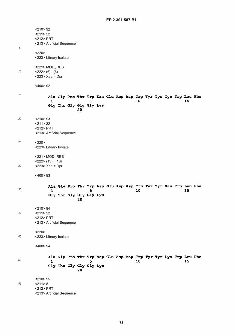
EP 2 301 587 B1
78
5
10
15
20
25
30
35
40
45
50
55
<210> 92<211> 22<212> PRT<213> Artificial Sequence
<220><223> Library Isolate
<221> MOD_RES<222> (6)...(6)<223> Xaa = Dpr
<400> 92
<210> 93<211> 22<212> PRT<213> Artificial Sequence
<220><223> Library Isolate
<221> MOD_RES<222> (13)...(13)<223> Xaa = Dpr
<400> 93
<210> 94<211> 22<212> PRT<213> Artificial Sequence
<220><223> Library Isolate
<400> 94
<210> 95<211> 8<212> PRT<213> Artificial Sequence

EP 2 301 587 B1
79
5
10
15
20
25
30
35
40
45
50
55
<220><223> Library Isolate
<400> 95
<210> 96<211> 19<212> PRT<213> Artificial Sequence
<220><223> Library Isolate
<400> 96
<210> 97<211> 25<212> PRT<213> Artificial Sequence
<220><223> Library isolate
<400> 97
<210> 98<211> 22<212> PRT<213> Artificial Sequence
<220><223> Library Isolate
<400> 98
<210> 99<211> 25<212> PRT<213> Artificial Sequence

EP 2 301 587 B1
80
5
10
15
20
25
30
35
40
45
50
55
<220><223> Library Isolate
<400> 99
<210> 100<211> 21<212> PRT<213> Artificial Sequence
<220><223> Synthesized KDR-Binding Polypeptide
<400> 100
210> 101
<211> 22<212> PRT<213> Artificial Sequence
<220><223> Library isolate
<400> 101
<210> 102<211> 23<212> PRT<213> Artificial Sequence
<220><223> Synthesized KDR-Binding Polypeptide
<400> 102
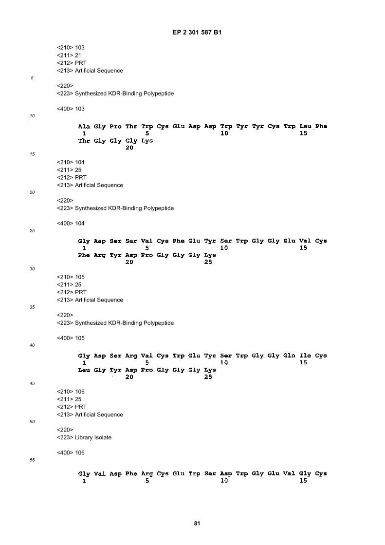
EP 2 301 587 B1
81
5
10
15
20
25
30
35
40
45
50
55
<210> 103<211> 21<212> PRT<213> Artificial Sequence
<220><223> Synthesized KDR-Binding Polypeptide
<400> 103
<210> 104<211> 25<212> PRT<213> Artificial Sequence
<220><223> Synthesized KDR-Binding Polypeptide
<400> 104
<210> 105<211> 25<212> PRT<213> Artificial Sequence
<220><223> Synthesized KDR-Binding Polypeptide
<400> 105
<210> 106<211> 25<212> PRT<213> Artificial Sequence
<220><223> Library Isolate
<400> 106

EP 2 301 587 B1
82
5
10
15
20
25
30
35
40
45
50
55
<210> 107<211> 19<212> PRT<213> Artificial Sequence
<220><223> Mature HSA
<400> 107
Claims
1. A compound comprising an optical label conjugated directly or via a linker or spacer to at least one polypeptidehaving the ability to bind to KDR or VEGF/KDR complex comprising an amino acid sequence selected from:
AGDSWCSTEYTYCEMIGTGGGK (SEQ ID NO: 4);AGPKWCEEDWYYCMITGTGGGK (SEQ ID NO: 5);AGVWECAKTFPFCHWFGTGGGK (SEQ ID NO: 6);AGWVECWWKSGQCYEFGTGGGK (SEQ ID NO: 7);AGWIQCNSITGHCTSGGTGGGK (SEQ ID NO: 8);AGWIECYHPDGICYHFGTGGGK (SEQ ID NO: 9);AGSDWCRVDWYYCWLMGTGGGK (SEQ ID NO: 10);AGANWCEEDWYYCFITGTGGGK (SEQ ID NO: 11);AGANWCEEDWYYCWITGTGGGK (SEQ ID NO: 12);AGPDWCEEDWYYCWITGTGGGK (SEQ ID NO: 13);AGSNWCEEDWYYCYITGTGGGK (SEQ ID NO: 14);AGPDWCAADWYYCYITGTGGGK (SEQ ID NO: 15);AGPEWCEVDWYYCWLLGTGGGK (SEQ ID NO: 16);AGPTWCEDDWYYCWLFGTGGGK (SEQ ID NO: 17);AGSKWCEQDWYYCWLLGTGGGK (SEQ ID NO: 18);AGRNWCEEDWYYCFITGTGGGK (SEQ ID NO: 19);AGVNWCEEDWYYCWITGTGGGK (SEQ ID NO: 20);AGANWCEEDWYYCYITGTGGGK (SEQ ID NO: 21);AGQAWVECYAETGYCWPRSWGTGGGK (SEQ ID NO: 22);AGQAWIECYAEDGYCWPRSWGTGGGK (SEQ ID NO: 23);AGVGWVECYQSTGFCYHSRDGTGGGK (SEQ ID NO: 24);AGDWWVECRVGTGLCYRYDTGTGGGK (SEQ ID NO: 25);AGDSWVECDAQTGFCYSFLYGTGGGK (SEQ ID NO: 26);AGERWVECRAETGFCYTWVSGTGGGK (SEQ ID NO: 27);AGGGWVECRAETGHCQEYRLGTGGGK (SEQ ID NO: 28);AGVAWVECYQTTGKCYTFRGGTGGGK (SEQ ID NO: 29);AGEGWVECFANTGACFTYPRGTGGGK (SEQ ID NO: 30);GDSRVCWEDSWGGEVCFRYDPGGGK (SEQ ID NO: 31);GDDSYCMMNEKGWWNCYLYDPGGGK (SEQ ID NO: 32);GDPAQCWESNYQGIFFCDNPDPGGGK (SEQ ID NO: 33);GDGWACAKWPWGGEICQPSDPGGGK (SEQ ID NO: 34);GDSSVCFEYSWGGEVCFRYDPGGGK (SEQ ID NO: 104);GDSRVCWEYSWGGQICLGYDPGGGK (SEQ ID NO: 105);

EP 2 301 587 B1
83
5
10
15
20
25
30
35
40
45
50
55
AQQVQYQFFLGTPRYEQWDLDKGGK (SEQ ID NO: 35);AQEPEGYAYWEVITLYHEEDGDGGK (SEQ ID NO: 36);AQAFPRFGGDDYWIQQYLRYTDGGK (SEQ ID NO: 37);AQGDYVYWEIIELTGATDHTPPGGK (SEQ ID NO: 38);AQRGDYQEQYWHQQLVEQLKLLGGK (SEQ ID NO: 39);AQRSWYLGPPYYEEWDPIPNGGK (SEQ ID NO: 40);AQDWYYDEILSMADQLRHAFLSGGGK (SEQ ID NO: 41);AQAPAWTFGTNWRSIQRVDSLTGGGGGK (SEQ ID NO: 42);AQEGWFRNPQEIMGFGDSWDKPGGGGGK (SEQ ID NO: 43);AQEGWFRNPQEIMGFGDSWDKPGGGK (SEQ ID NO: 44);AQRGDYQEQYWHQQLVEQLKLLGGGK (SEQ ID NO: 45);Ac-AGWYWCDYYGIGCK(ivDde)WTGGGK-NH2(SEQ ID NO: 46);Ac-AGWYWCDYYGIGCKWTGTGGGK-NH2(SEQ ID NO: 47);Ac-AQWYYDWFHNQRKPPSDWIDNLGGGK-NH2(SEQ ID NO: 48);Ac-VCWEDSWGGEVCFRYDPGGGK(biotin-JJ-)-NH2 (SEQ ID NO: 49);Ac-AGPTWCEDDWYYCWLFGTJK(biotin-JJ-)-NH2 (SEQ ID NO: 50);Ac-AQAHMPPWRPVAVDALFDWVEGG-GGGK(biotin-JJ-)-NH2 (SEQ ID NO: 51);Ac-AQAHMPPWWPLAVDAQEDWFEGG-GGGK(biotin-JJ-)-NH2 (SEQ ID NO: 52);Ac-AQAQMPPWWPLAVDALFDWFEGG-GGGK(biotin-JJ-)-NH2 (SEQ ID NO: 53);Ac-AQDWYWREWMPMHAQFLADDWGG-GGGK(biotin-JJ-)-NH2 (SEQ ID NO: 54);Ac-AQPVTDWTPHHPK(ivDde)APDVWLFYT-GGGGGK(biotin-JJ-)-NH2 (SEQ ID NO: 55);Ac-AQDALEAPK(ivDde)RDWYYDWFLNHSP-GGGGGK(biotin-JJ-)-NH2 (SEQ ID NO: 56);Ac-KWCEEDWYYCMITGTGGGK(biotin-JJ-)-NH2 (SEQ ID NO: 57);Ac-AGPKWCEEDWYYCMIGGGK(biotin-JJ-)-NH2 (SEQ ID NO. 58);Ac-KWCEEDWYYCMIGGGK(biotin-JJ-)-NH2 (SEQ ID NO: 59);Ac-AQPDNWK(ivDde)EFYESGWK(ivDde)-YPSLYK(ivDde)PLGGGGGK(biotin-JJ-)-NH2 (SEQ ID NO: 60);Ac-AQMPPGFSYWEQWLHDDAQVLGG-GGGK(biotin-JJ-)-NH2 (SEQ ID NO: 61);Ac-AQARMGDDWEEAPPHEWGWADGG-GGGK(biotin-JJ-)-NH2 (SEQ ID NO: 62);Ac-AQPEDSEAWYWLNYRPTMFHQLGG-GGGK(biotin-JJ-)-NH2 (SEQ ID NO: 63);Ac-AQSTNGDSFVYWEEVELVDHPGG-GGGK(biotin-JJ-)-NH2 (SEQ ID NO: 64);Ac-AQWESDYWDQMRQQLK(iv-Dde)TAYMK(iv-Dde)VGGGGGK(biotin-JJ-)-NH2 (SEQ ID NO: 65);Ac-AQDWYYDEILSMADQLRHAFLSGGGGGK(biotin-JJ-)-NH2 (SEQ ID NO: 66);VCWEDSWGGEVCFGGGK (SEQ ID NO: 76);GDSRVCWEDSWGGEVCFGGGK (SEQ ID NO: 77);SRVCWEDSWGGEVCFRYGGGGK (SEQ ID NO: 79);GDSRVCWEDSWGGEVCFRYGGGK (SEQ ID NO: 80);DWYYGGGK (SEQ ID NO: 95);AQDWYYDEILGRGRGGRGG (SEQ ID NO: 96);AGPTWEEDDWYYKWLFGTGGGK (SEQ ID NO: 90);AGPTWKEDDWYYEWLFGTGGGK (SEQ ID NO: 91);AGPTWDprEDDWYYDWLFGTGGGK (SEQ ID NO: 92);AGPTWDEDDWYYDprWLFGTGGGK (SEQ ID NO: 93); andAGPTWDEDDWYYKWLFGTGGGK (SEQ ID NO: 94), wherein J is the spacer orlinker group, 8-amino-3,6-dioxaoctanoyl, and iv-Dde is 1-(4,4-dimethyl-2,6-dioxocyclohex-1-ylidene)-3-methyl-butyl.
2. The compound of claim 1, wherein the optical label is conjugated directly or via a linker or spacer to a dimeric ormultimeric compound comprising two or more polypeptides having the ability to bind KDR or VEGF/KDR complex.
3. The compound of claim 1, wherein the compound comprises two or more polypeptides and the polypeptides havespecificity for different epitopes on KDR, wherein the poly-peptides are preferably independently selected from thegroup consisting of:
AGPKWCEEDWYYCMITGTGGGK (SEQ ID NO: 5);GDSRVCWEDSWGGEVCFRYDPGGGK (SEQ ID NO: 31);AQDWYYDEILSMADQLRHAFLSGGK (SEQ ID NO: 41);AGPTWCEDDWYYCWLFGTGGGK (SEQ ID NO: 17);AGDWWVECRVGTGLCYRYDTGTGGGK (SEQ ID NO: 25); and

EP 2 301 587 B1
84
5
10
15
20
25
30
35
40
45
50
55
Ac-VCWEDSWGGEVCFRYDPGGGK(biotin-JJ-)-NH2 (SEQ ID NO: 49).
4. The compound of any of claims 1 or 3, wherein one or more of the polypeptides do not include the C terminal GGGKextension in their amino acid sequence.
5. The compound of claim 3, wherein the polypeptides comprise any of the following combinations:
AGPTWCEDDWYYCWLFGTGGGK (SEQ ID NO: 17) andAc-VCWEDSWGGEVCFRYDPGGGK(biotin-JJ-)-NH2 (SEQ ID NO: 49);AGPTWCEDDWYYCWLFGTGGGK (SEQ ID NO: 17) andGDSRVCWEDSWGGEVCFRYDPGGGK (SEQ ID NO: 31);AGPKWCEEDWYYCMITGTGGGK (SEQ ID NO: 5) andGDSRVCWEDSWGGEVCFRYDPGGGK (SEQ ID NO: 31); orAQDWYYDEILSMADQLRHAFLSGGK (SEQ ID NO: 41) andAc-VCWEDSWGGEVCFRYDPGGGK(biotin-JJ-)-NH2 (SEQ ID NO: 49).
6. The compound of claim 1, wherein the polypeptide comprisesAGPKWCEEDWYYCMITGTGGGK (SEQ ID NO: 5);AGPTWCEDDWYYCWLFGTGGGK (SEQ ID NO: 17);GDSRVCWEDSWGGEVCFRYDPGGGK (SEQ ID NO: 31);AQDWYYDEILSMADQLRHAFLSGGK (SEQ ID NO: 41); orAc-VCWEDSWGGEVCFRYDPGGGK(biotin-JJ-)-NH2 (SEQ ID NO: 49).
7. A compound comprising an optical label conjugated directly or via a linker or spacer to a dimer, the dimer comprisingtwo polypeptides having the ability to bind KDR or VEGF/KDR complex, and wherein the dimer is selected from thegroup consisting of D1 (FIG.25), D2 (FIG.26), D3 (FIG.27), D5 (FIG.29), D6 (FIG.50), D7 (FIG.51), D8 (FIG.30), D9(FIG.31), D17 (FIG.39), D19 (FIG.41), D20 (FIG.42), D22 (FIG.44), D23 (FIG.45), D24 (FIG.46), D25 (FIG.47), D26(FIG.48), and D27 (FIG.49).
8. The compound of any one of the preceding claims, wherein the polypeptide is linked to the optical label via a linkeror spacer, wherein said linker or spacer is preferably selected from the group consisting of a substituted alkyl chain,an unsubstituted alkyl chain, a polyethylene glycol derivative, an amino acid spacer, a sugar, an aliphatic spacer,an aromatic spacer, a lipid molecule, or combination thereof.
9. The compound of any one of the preceding claims, wherein the optical label comprises an optical dye, a biolumi-nescent molecule, a photoabsorption label or a fluorescent bead.
10. The compound of claim 9, wherein the optical label is an optical dye such as an organic chromophore or fluorophore,such as, for example, a fluorescent compound or wherein the optical label is a fluorescent bead.
11. The compound of claim 9, wherein the optical label comprises a bioluminescent molecule.
12. Use of a compound according to any one of claims 1 to 11 for producing a diagnostic composition for imaging, inparticular for imaging important sites of angiogenesis, e.g., neoplastic tumors.
13. Use according to claim 12, wherein said imaging is for monitoring of a therapy with a tumorcidal agent or an anti-angiogenesis agent.
14. Use according to claim 12 or 13, wherein said label is detected by a parameter selected from transmitted radiation,absorption, fluorescent or phosphorescent emission, light reflection, changes in absorbance amplitude or maximaand elastically-scattered radiation.
Patentansprüche
1. Eine Verbindung umfassend ein optisches Label, das direkt oder über einen Linker oder Spacer konjugiert ist mitmindestens einem Polypeptid mit der Fähigkeit KDR oder den VEGF/KDR-Komplex zu binden, umfassend einerAminosäuresequenz ausgesucht aus:
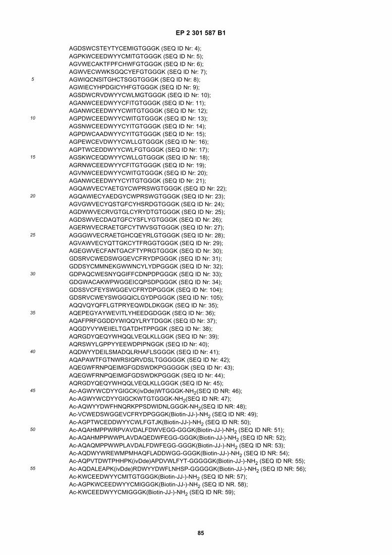
EP 2 301 587 B1
85
5
10
15
20
25
30
35
40
45
50
55
AGDSWCSTEYTYCEMIGTGGGK (SEQ ID Nr: 4);AGPKWCEEDWYYCMITGTGGGK (SEQ ID Nr: 5);AGVWECAKTFPFCHWFGTGGGK (SEQ ID Nr: 6);AGWVECWWKSGQCYEFGTGGGK (SEQ ID Nr: 7);AGWIQCNSITGHCTSGGTGGGK (SEQ ID Nr: 8);AGWIECYHPDGICYHFGTGGGK (SEQ ID Nr: 9);AGSDWCRVDWYYCWLMGTGGGK (SEQ ID Nr: 10);AGANWCEEDWYYCFITGTGGGK (SEQ ID Nr: 11);AGANWCEEDWYYCWITGTGGGK (SEQ ID Nr: 12);AGPDWCEEDWYYCWITGTGGGK (SEQ ID Nr: 13);AGSNWCEEDWYYCYITGTGGGK (SEQ ID Nr: 14);AGPDWCAADWYYCYITGTGGGK (SEQ ID Nr: 15);AGPEWCEVDWYYCWLLGTGGGK (SEQ ID Nr: 16);AGPTWCEDDWYYCWLFGTGGGK (SEQ ID Nr: 17);AGSKWCEQDWYYCWLLGTGGGK (SEQ ID Nr: 18);AGRNWCEEDWYYCFITGTGGGK (SEQ ID Nr: 19);AGVNWCEEDWYYCWITGTGGGK (SEQ ID Nr: 20);AGANWCEEDWYYCYITGTGGGK (SEQ ID Nr: 21);AGQAWVECYAETGYCWPRSWGTGGGK (SEQ ID Nr: 22);AGQAWIECYAEDGYCWPRSWGTGGGK (SEQ ID Nr: 23);AGVGWVECYQSTGFCYHSRDGTGGGK (SEQ ID Nr: 24);AGDWWVECRVGTGLCYRYDTGTGGGK (SEQ ID Nr: 25);AGDSWVECDAQTGFCYSFLYGTGGGK (SEQ ID Nr: 26);AGERWVECRAETGFCYTWVSGTGGGK (SEQ ID Nr: 27);AGGGWVECRAETGHCQEYRLGTGGGK (SEQ ID Nr: 28);AGVAWVECYQTTGKCYTFRGGTGGGK (SEQ ID Nr: 29);AGEGWVECFANTGACFTYPRGTGGGK (SEQ ID Nr: 30);GDSRVCWEDSWGGEVCFRYDPGGGK (SEQ ID Nr: 31);GDDSYCMMNEKGWWNCYLYDPGGGK (SEQ ID Nr: 32);GDPAQCWESNYQGIFFCDNPDPGGGK (SEQ ID Nr: 33);GDGWACAKWPWGGEICQPSDPGGGK (SEQ ID Nr: 34);GDSSVCFEYSWGGEVCFRYDPGGGK (SEQ ID Nr: 104);GDSRVCWEYSWGGQICLGYDPGGGK (SEQ ID Nr: 105);AQQVQYQFFLGTPRYEQWDLDKGGK (SEQ ID Nr: 35);AQEPEGYAYWEVITLYHEEDGDGGK (SEQ ID Nr: 36);AQAFPRFGGDDYWIQQYLRYTDGGK (SEQ ID Nr: 37);AQGDYVYWEIIELTGATDHTPPGGK (SEQ ID Nr: 38);AQRGDYQEQYWHQQLVEQLKLLGGK (SEQ ID Nr: 39);AQRSWYLGPPYYEEWDPIPNGGK (SEQ ID Nr: 40);AQDWYYDEILSMADQLRHAFLSGGGK (SEQ ID Nr: 41);AQAPAWTFGTNWRSIQRVDSLTGGGGGK (SEQ ID Nr: 42);AQEGWFRNPQEIMGFGDSWDKPGGGGGK (SEQ ID Nr: 43);AQEGWFRNPQEIMGFGDSWDKPGGGK (SEQ ID Nr: 44);AQRGDYQEQYWHQQLVEQLKLLGGGK (SEQ ID Nr: 45);Ac-AGWYWCDYYGIGCK(ivDde)WTGGGK-NH2(SEQ ID NR: 46);Ac-AGWYWCDYYGIGCKWTGTGGGK-NH2(SEQ ID NR: 47);Ac-AQWYYDWFHNQRKPPSDWIDNLGGGK-NH2(SEQ ID NR: 48);Ac-VCWEDSWGGEVCFRYDPGGGK(Biotin-JJ-)-NH2 (SEQ ID NR: 49);Ac-AGPTWCEDDWYYCWLFGTJK(Biotin-JJ-)-NH2 (SEQ ID NR: 50);Ac-AQAHMPPWRPVAVDALFDWVEGG-GGGK(Biotin-JJ-)-NH2 (SEQ ID NR: 51);Ac-AQAHMPPWWPLAVDAQEDWFEGG-GGGK(Biotin-JJ-)-NH2 (SEQ ID NR: 52);Ac-AQAQMPPWWPLAVDALFDWFEGG-GGGK(Biotin-JJ-)-NH2 (SEQ ID NR: 53);Ac-AQDWYWREWMPMHAQFLADDWGG-GGGK(Biotin-JJ-)-NH2 (SEQ ID NR: 54);Ac-AQPVTDWTPHHPK(ivDde)APDVWLFYT-GGGGGK(Biotin-JJ-)-NH2 (SEQ ID NR: 55);Ac-AQDALEAPK(ivDde)RDWYYDWFLNHSP-GGGGGK(Biotin-JJ-)-NH2 (SEQ ID NR: 56);Ac-KWCEEDWYYCMITGTGGGK(Biotin-JJ-)-NH2 (SEQ ID NR: 57);Ac-AGPKWCEEDWYYCMIGGGK(Biotin-JJ-)-NH2 (SEQ ID NR. 58);Ac-KWCEEDWYYCMIGGGK(Biotin-JJ-)-NH2 (SEQ ID NR: 59);

EP 2 301 587 B1
86
5
10
15
20
25
30
35
40
45
50
55
Ac-AQPDNWK(ivDde)EFYESGWK(ivDde)-YPSLYK(ivDde)PLGGGGGK(Biotin-JJ-)-NH2 (SEQ ID NR: 60);Ac-AQMPPGFSYWEQVVLHDDAQVLGG-GGGK(Biotin-JJ-)-NH2 (SEQ ID NR: 61);Ac-AQARMGDDWEEAPPHEWGWADGG-GGGK(Biotin-JJ-)-NH2 (SEQ ID NR: 62);Ac-AQPEDSEAWYWLNYRPTMFHQLGG-GGGK(Biotin-JJ-)-NH2 (SEQ ID NR: 63);Ac-AQSTNGDSFVYWEEVELVDHPGG-GGGK(Biotin-JJ-)-NH2 (SEQ ID NR: 64);Ac-AQWESDYWDQMRQQLK(iv-Dde)TAYMK(iv-Dde)VGGGGGK(Biotin-JJ-)-NH2 (SEQ ID NR: 65);Ac-AQDWYYDEILSMADQLRHAFLSGGGGGK(Biotin-JJ-)-NH2 (SEQ ID NR: 66);VCWEDSWGGEVCFGGGK (SEQ ID Nr: 76);GDSRVCWEDSWGGEVCFGGGK (SEQ ID Nr: 77);SRVCWEDSWGGEVCFRYGGGGK (SEQ ID Nr: 79);GDSRVCWEDSWGGEVCFRYGGGK (SEQ ID Nr: 80);DWYYGGGK (SEQ ID Nr: 95);AQDWYYDEILGRGRGGRGG (SEQ ID Nr: 96);AGPTWEEDDWYYKWLFGTGGGK (SEQ ID Nr: 90);AGPTWKEDDWYYEWLFGTGGGK (SEQ ID Nr: 91);AGPTWDprEDDWYYDWLFGTGGGK (SEQ ID Nr: 92);AGPTWDEDDWYYDprWLFGTGGGK (SEQ ID Nr: 93); undAGPTWDEDDWYYKWLFGTGGGK (SEQ ID Nr: 94), wobei J die Spacer oder Linker Gruppe ist, 8-Amino-3,6-Dioxaoctanoyl, und iv-Dde 1-(4,4-Dimethyl-2,6-Dioxocyclohex-1-Yliden)-3-Methylbutyl ist.
2. Die Verbindung nach Anspruch 1, wobei das optische Label direkt oder über einen Linker oder Spacer konjugiertist mit einer dimeren- oder multimeren Verbindung umfassend zwei oder mehr Polypeptide mit der Fähigkeit KDRoder den VEGF/KDR-Komplex zu binden
3. Verbindung nach Anspruch 1, wobei die Verbindung zwei oder mehr Polypeptide umfasst und die PolypeptideSpezifität für unterschiedliche Epitope auf KDR haben, wobei die Polypeptide vorzugsweise unabhängig ausgewähltwerden aus der Gruppe bestehend aus:
AGPKWCEEDWYYCMITGTGGGK (SEQ ID Nr: 5);GDSRVCWEDSWGGEVCFRYDPGGGK (SEQ ID Nr: 31);AQDWYYDEILSMADQLRHAFLSGGK (SEQ ID Nr: 41);AGPTWCEDDWYYCWLFGTGGGK (SEQ ID Nr: 17);AGDWWVECRVGTGLCYRYDTGTGGGK (SEQ ID Nr: 25); undAc-VCWEDSWGGEVCFRYDPGGGK(Biotin-JJ-)-NH2 (SEQ ID Nr: 49).
4. Verbindung nach einem der Ansprüche 1 oder 3, wobei ein oder mehrere der Polypeptide nicht die C-terminaleVerlängerung GGGK in ihrer Aminosäuresequenz enthalten.
5. Die Verbindung nach Anspruch 3, wobei die Polypeptide eine der folgenden Kombinationen umfassen:
AGPTWCEDDWYYCWLFGTGGGK (SEQ ID Nr: 17) undAc-VCWEDSWGGEVCFRYDPGGGK(Biotin-JJ-)-NH2 (SEQ ID NR: 49);AGPTWCEDDWYYCWLFGTGGGK (SEQ ID Nr: 17) undGDSRVCWEDSWGGEVCFRYDPGGGK (SEQ ID Nr: 31);AGPKWCEEDWYYCMITGTGGGK (SEQ ID Nr: 5) undGDSRVCWEDSWGGEVCFRYDPGGGK (SEQ ID Nr: 31); oderAQDWYYDEILSMADQLRHAFLSGGK (SEQ ID Nr: 41) undAc-VCWEDSWGGEVCFRYDPGGGK(Biotin-JJ-)-NH2 (SEQ ID NR: 49).
6. Verbindung nach Anspruch 1, wobei das Polypeptid umfasst:
AGPKWCEEDWYYCMITGTGGGK (SEQ ID Nr: 5);AGPTWCEDDWYYCWLFGTGGGK (SEQ ID Nr: 17);GDSRVCWEDSWGGEVCFRYDPGGGK (SEQ ID Nr: 31);AQDWYYDEILSMADQLRHAFLSGGK (SEQ ID Nr: 41); oderAc-VCWEDSWGGEVCFRYDPGGGK(Biotin-JJ-)-NH2 (SEQ ID NR: 49).
7. Eine Verbindung umfassend ein optisches Label, das direkt oder über einen Linker oder Spacer mit einem Dimer
EP 2 301 587 B1
87
5
10
15
20
25
30
35
40
45
50
55
konjugiert ist, das Dimer umfassend zwei Polypeptide mit der Fähigkeit KDR oder den VEGF/KDR-Komplex zubinden, und wobei das Dimer ausgesucht wird aus der Gruppe bestehend aus D1 (FIG.25), D2 (FIG.26), D3 (FIG.27),D5 (FIG.29), D6 (FIG.50), D7 (FIG.51), D8 (FIG.30), D9 (FIG.31), D17 (FIG.39), D19 (FIG.41), D20 (FIG.42), D22(FIG.44), D23 (FIG.45), D24 (FIG.46), D25 (FIG.47), D26 (FIG.48), und D27 (FIG.49).
8. Verbindung nach einem der vorhergehenden Ansprüche, wobei das Polypeptid an ein optisches Label über einenLinker oder Spacer gebunden ist, wobei der Linker oder Spacer vorzugsweise aus der Gruppe ausgewählt wirdbestehend aus einer substituierten AlkylKette, eine unsubstituierte Alkylkette, einem Polyethylen-Glykolderivat, einAminosäure-Spacer, ein Zucker, ein aliphatischer Spacer, ein aromatischer Spacer, ein Lipidmolekül oder eineKombination davon.
9. Die Verbindung nach einem der vorhergehenden Ansprüche, wobei das optische Label ein optischer Farbstoff, einbiolumineszierendes Molekül, ein Fotoabsorptions-Label oder ein fluoreszierendes Bead ist.
10. Verbindung nach Anspruch 9, wobei das optische Label ein optischer Farbstoff ist, wie ein organisches Chromophoroder Fluorophor, wie beispielsweise eine fluoreszierende Verbindung, oder wobei das optische Label ein fluores-zierendes Bead ist.
11. Die Verbindung nach Anspruch 9, wobei das optische Label ein biolumineszentes Molekül umfasst.
12. Verwendung einer Verbindung nach einem der Ansprüche 1 bis 11 zum Herstellen einer diagnostischen Zusam-mensetzung zur Bildgebung, insbesondere zur Bildgebung wichtiger Stellen der Angiogenese, z.B. neoplastischeTumore.
13. Verwendung nach Anspruch 12, wobei genannte Bildgebung zur Überwachung einer Therapie mit einem tumorizi-dalen Mittel oder einem Anti-Angiogenese-Mittel ist.
14. Verwendung nach Anspruch 12 oder 13, wobei das Label durch einen Parameter detektiert wird ausgesucht ausübertragener Strahlung, Absorption, Fluoreszenz-oder Phosphoreszenz-Emissions, Lichtreflexion, Änderungen derAbsorption oder Amplitudenmaxima und elastisch gestreute Strahlung.
Revendications
1. Composé comprenant un marqueur optique conjugué directement ou par l’intermédiaire d’un lieur ou d’un espaceurà au moins un polypeptide ayant la capacité de se lier à KDR ou à un complexe VEGF/KDR comprenant uneséquence d’acides aminés choisie parmi :
AGDSWCSTEYTYCEMIGTGGGK (SEQ ID NO : 4) ;AGPKWCEEDWYYCMITGTGGGK (SEQ ID NO : 5) ;AGVWECAKTFPFCHWFGTGGGK (SEQ ID NO : 6) ;AGWVECWWKSGQCYEFGTGGGK (SEQ ID NO : 7) ;AGWIQCNSITGHCTSGGTGGGK (SEQ ID NO : 8) ;AGWIECYHPDGICYHFGTGGGK (SEQ ID NO : 9) ;AGSDWCRVDWYYCWLMGTGGGK (SEQ ID NO : 10) ;AGANWCEEDWYYCFITGTGGGK (SEQ ID NO : 11) ;AGANWCEEDWYYCWITGTGGGK (SEQ ID NO : 12) ;AGPDWCEEDWYYCWITGTGGGK (SEQ ID NO : 13) ;AGSNWCEEDWYYCYITGTGGGK (SEQ ID NO : 14) ;AGPDWCAADWYYCYITGTGGGK (SEQ ID NO : 15) ;AGPEWCEVDWYYCWLLGTGGGK (SEQ ID NO : 16) ;AGPTWCEDDWYYCWLFGTGGGK (SEQ ID NO : 17) ;AGSKWCEQDWYYCWLLGTGGGK (SEQ ID NO : 18) ;AGRNWCEEDWYYCFITGTGGGK (SEQ ID NO : 19) ;AGVNWCEEDWYYCWITGTGGGK (SEQ ID NO : 20) ;AGANWCEEDWYYCYITGTGGGK (SEQ ID NO : 21) ;AGQAWVECYAETGYCWPRSWGTGGGK (SEQ ID NO : 22) ;AGQAWIECYAEDGYCWPRSWGTGGGK (SEQ ID NO : 23) ;

EP 2 301 587 B1
88
5
10
15
20
25
30
35
40
45
50
55
AGVGWVECYQSTGFCYHSRDGTGGGK (SEQ ID NO : 24) ;AGDWWVECRVGTGLCYRYDTGTGGGK (SEQ ID NO : 25) ;AGDSWVECDAQTGFCYSFLYGTGGGK (SEQ ID NO : 26) ;AGERWVECRAETGFCYTWVSGTGGGK (SEQ ID NO : 27) ;AGGGWVECRAETGHCQEYRLGTGGGK (SEQ ID NO : 28) ;AGVAWVECYQTTGKCYTFRGGTGGGK (SEQ ID NO : 29) ;AGEGWVECFANTGACFTYPRGTGGGK (SEQ ID NO : 30) ;GDSRVCWEDSWGGEVCFRYDPGGGK (SEQ ID NO : 31) ;GDDSYCMMNEKGWWNCYLYDPGGGK (SEQ ID NO : 32) ;GDPAQCWESNYQGIFFCDNPDPGGGK (SEQ ID NO : 33) ;GDGWACAKWPWGGEICQPSDPGGGK (SEQ ID NO : 34) ;GDSSVCFEYSWGGEVCFRYDPGGGK (SEQ ID NO : 104) ;GDSRVCWEYSWGGQICLGYDPGGGK (SEQ ID NO : 105) ;AQQVQYQFFLGTPRYEQWDLDKGGK (SEQ ID NO : 35) ;AQEPEGYAYWEVITLYHEEDGDGGK (SEQ ID NO : 36) ;AQAFPRFGGDDYWIQQYLRYTDGGK (SEQ ID NO : 37) ;AQGDYVYWEIIELTGATDHTPPGGK (SEQ ID NO : 38) ;AQRGDYQEQYWHQQLVEQLKLLGGK (SEQ ID NO : 39) ;AQRSWYLGPPYYEEWDPIPNGGK (SEQ ID NO : 40) ;AQDWYYDEILSMADQLRHAFLSGGGK (SEQ ID NO : 41) ;AQAPAWTFGTNWRSIQRVDSLTGGGGGK (SEQ ID NO : 42) ;AQEGWFRNPQEIMGFGDSWDKPGGGGGK (SEQ ID NO : 43) ;AQEGWFRNPQEIMGFGDSWDKPGGGK (SEQ ID NO : 44) ;AQRGDYQEQYWHQQLVEQLKLLGGGK (SEQ ID NO : 45) ;Ac-AGWYWCDYYGIGCK(ivDde)WTGGGK-NH2 (SEQ ID NO : 46) ;Ac-AGWYWCDYYGIGCKWTGTGGGK-NH2 (SEQ ID NO : 47) ;Ac-AQWYYDWFHNQRKPPSDWIDNLGGGK-NH2 (SEQ ID NO : 48) ;Ac-VCWEDSWGGEVCFRYDPGGGK(biotine-JJ-)-NH2 (SEQ ID NO : 49) ;Ac-AGPTWCEDDWYYCWLFGTJK(biotine-JJ-)-NH2 (SEQ ID NO : 50) ;Ac-AQAHMPPWRPVAVDALFDWVEGG-GGGK(biotine-JJ-)-NH2 (SEQ ID NO : 51) ;Ac-AQAHMPPWWPLAVDAQEDWFEGG-GGGK(biotine-JJ-)-NH2 (SEQ ID NO : 52) ;Ac-AQAQMPPWWPLAVDALFDWFEGG-GGGK(biotine-JJ-)-NH2 (SEQ ID NO : 53) ;Ac-AQDWYWREWMPMHAQFLADDWGG-GGGK(biotine-JJ-)-NH2 (SEQ ID NO : 54) ;Ac-AQPVTDWTPHHPK(ivDde)-APDVWLFYT-GGGGGK(biotine-JJ-)-NH2 (SEQ ID NO : 55) ;Ac-AQDALEAPK(ivDde)RDWYYDWFLNHSP-GGGGGK(biotine-JJ-)-NH2 (SEQ ID NO : 56) ;Ac-KWCEEDWYYCMITGTGGGK(biotine-JJ-)-NH2 (SEQ ID NO : 57) ;Ac-AGPKWCEEDWYYCMIGGGK(biotine-JJ-)-NH2 (SEQ ID NO : 58) ;Ac-KWCEEDWYYCMIGGGK(biotine-JJ-)-NH2 (SEQ ID NO : 59);Ac-AQPDNWK(ivDde)EFYESGWK(ivDde)-YPSLYK(ivDde) PLGGGGGK(biotine-JJ-)-NH2 (SEQ ID NO : 60) ;Ac-AQMPPGFSYWEQVVLHDDAQVLGG-GGGK(biotine-JJ-)-NH2 (SEQ ID NO : 61) ;Ac-AQARMGDDWEEAPPHEWGWADGG-GGGK(biotine-JJ-)-NH2 (SEQ ID NO : 62) ;Ac-AQPEDSEAWYWLNYRPTMFHQLGG-GGGK(biotine-JJ-)-NH2 (SEQ ID NO : 63) ;Ac-AQSTNGDSFVYWEEVELVDHPGG-GGGK(biotine-JJ-)-NH2 (SEQ ID NO : 64) ;Ac-AQWESDYWDQMRQQLK(iv-Dde)TAYMK(iv-Dde)VGGGGGK (biotine-JJ-)-NH2 (SEQ ID NO : 65) ;Ac-AQDWYYDEILSMADQLRHAFLSGGGGGK(biotine-JJ-)-NH2 (SEQ ID NO : 66) ;VCWEDSWGGEVCFGGGK (SEQ ID NO : 76) ;GDSRVCWEDSWGGEVCFGGGK (SEQ ID NO : 77) ;SRVCWEDSWGGEVCFRYGGGGK (SEQ ID NO : 79) ;GDSRVCWEDSWGGEVCFRYGGGK (SEQ ID NO : 80) ;DWYYGGGK (SEQ ID NO : 95) ;AQDWYYDEILGRGRGGRGG (SEQ ID NO : 96) ;AGPTWEEDDWYYKWLFGTGGGK (SEQ ID NO : 90) ;AGPTWKEDDWYYEWLFGTGGGK (SEQ ID NO : 91) ;AGPTWDprEDDWYYDWLFGTGGGK (SEQ ID NO : 92) ;AGPTWDEDDWYYDprWLFGTGGGK (SEQ ID NO : 93) ; etAGPTWDEDDWYYKWLFGTGGGK (SEQ ID NO : 94), où J représente le groupe espaceur ou lieur, 8-amino-3,6-dioxaoctanoyle, et iv-Dde représente le 1-(4,4-diméthyl-2,6-dioxocyclohex-1-ylidène)-3-méthylbutyle.
EP 2 301 587 B1
89
5
10
15
20
25
30
35
40
45
50
55
2. Composé de la revendication 1, dans lequel le marqueur optique est conjugué directement ou par l’intermédiaired’un lieur ou d’un espaceur à un composé dimère ou multimère comprenant deux polypeptides ou plus ayant lacapacité de lier KDR ou un complexe VEGF/KDR.
3. Composé de la revendication 1, dans lequel le composé comprend deux polypeptides ou plus et les polypeptidesont une spécificité pour des épitopes différents sur KDR, où les polypeptides sont de préférence choisis indépen-damment dans le groupe constitué de :
AGPKWCEEDWYYCMITGTGGGK (SEQ ID NO : 5) ;GDSRVCWEDSWGGEVCFRYDPGGGK (SEQ ID NO : 31) ;AQDWYYDEILSMADQLRHAFLSGGK (SEQ ID NO : 41) ;AGPTWCEDDWYYCWLFGTGGGK (SEQ ID NO : 17) ;AGDWWVECRVGTGLCYRYDTGTGGGK (SEQ ID NO : 25) ; etAc-VCWEDSWGGEVCFRYDPGGGK(biotine-JJ-)-NH2 (SEQ ID NO : 49).
4. Composé de l’une des revendications 1 ou 3, dans lequel un ou plusieurs polypeptide(s) parmi les polypeptides necomporte/comportent pas l’extension GGGK C terminale dans sa/leurs séquence(s) d’acides aminés.
5. Composé de la revendication 3, dans lequel les polypeptides comprennent l’une des combinaisons suivantes :
AGPTWCEDDWYYCWLFGTGGGK (SEQ ID NO : 17) etAc-VCWEDSWGGEVCFRYDPGGGK(biotine-JJ-)-NH2 (SEQ ID NO : 49) ;AGPTWCEDDWYYCWLFGTGGGK (SEQ ID NO : 17) etGDSRVCWEDSWGGEVCFRYDPGGGK (SEQ ID NO : 31) ;AGPKWCEEDWYYCMITGTGGGK (SEQ ID NO : 5) etGDSRVCWEDSWGGEVCFRYDPGGGK (SEQ ID NO : 31) ; ouAQDWYYDEILSMADQLRHAFLSGGK (SEQ ID NO : 41) etAc-VCWEDSWGGEVCFRYDPGGGK(biotine-JJ-)-NH2 (SEQ ID NO : 49).
6. Composé de la revendication 1, dans lequel le polypeptide comprendAGPKWCEEDWYYCMITGTGGGK (SEQ ID NO : 5) ;AGPTWCEDDWYYCWLFGTGGGK (SEQ ID NO : 17) ;GDSRVCWEDSWGGEVCFRYDPGGGK (SEQ ID NO : 31) ;AQDWYYDEILSMADQLRHAFLSGGK (SEQ ID NO : 41) ; ouAc-VCWEDSWGGEVCFRYDPGGGK(biotine-JJ-)-NH2 (SEQ ID NO : 49).
7. Composé comprenant un marqueur optique conjugué directement ou par l’intermédiaire d’un lieur ou d’un espaceurà un dimère, le dimère comprenant deux polypeptides ayant la capacité de lier KDR ou un complexe VEGF/KDR,et où le dimère est choisi dans le groupe constitué de D1 (figure 25), D2 (figure 26), D3 (figure 27), D5 (figure 29),D6 (figure 50), D7 (figure 51), D8 (figure 30), D9 (figure 31), D17 (figure 39), D19 (figure 41), D20 (figure 42), D22(figure 44), D23 (figure 45), D24 (figure 46), D25 (figure 47), D26 (figure 48), et D27 (figure 49).
8. Composé de l’une quelconque des revendications précédentes, dans lequel le polypeptide est lié au marqueuroptique par l’intermédiaire d’un lieur ou d’un espaceur, où ledit lieur ou espaceur est de préférence choisi dans legroupe constitué d’une chaîne alkyle substituée, d’une chaîne alkyle non substituée, d’un dérivé de polyéthylèneglycol, d’un espaceur d’acides aminés, d’un sucre, d’un espaceur aliphatique, d’un espaceur aromatique, d’unemolécule lipidique, ou d’une combinaison de ceux-ci.
9. Composé de l’une quelconque des revendications précédentes, dans lequel le marqueur optique comprend uncolorant optique, une molécule bioluminescente, un marqueur de photoabsorption ou une bille fluorescente.
10. Composé de la revendication 9, dans lequel le marqueur optique est un colorant optique tel qu’un chromophore ouun fluorophore organique, comme, par exemple, un composé fluorescent ou dans lequel le marqueur optique estune bille fluorescente.
11. Composé de la revendication 9, dans lequel le marqueur optique comprend une molécule bioluminescente.
12. Utilisation d’un composé selon l’une quelconque des revendications 1 à 11 pour la production d’une composition

EP 2 301 587 B1
90
5
10
15
20
25
30
35
40
45
50
55
de diagnostic pour l’imagerie, en particulier pour l’imagerie de sites importants de l’angiogenèse, par exemple, destumeurs néoplasiques.
13. Utilisation selon la revendication 12, dans laquelle ladite imagerie est destinée à la surveillance d’une thérapie avecun agent tumoricide ou un agent anti-angiogenèse.
14. Utilisation selon la revendication 12 ou 13, dans laquelle ledit marqueur est détecté par un paramètre choisi parmiun rayonnement transmis, une absorption, une émission fluorescente ou phosphorescente, une réflexion de lumière,des changements de maximums ou d’amplitude d’absorbance et un rayonnement diffusé élastiquement.
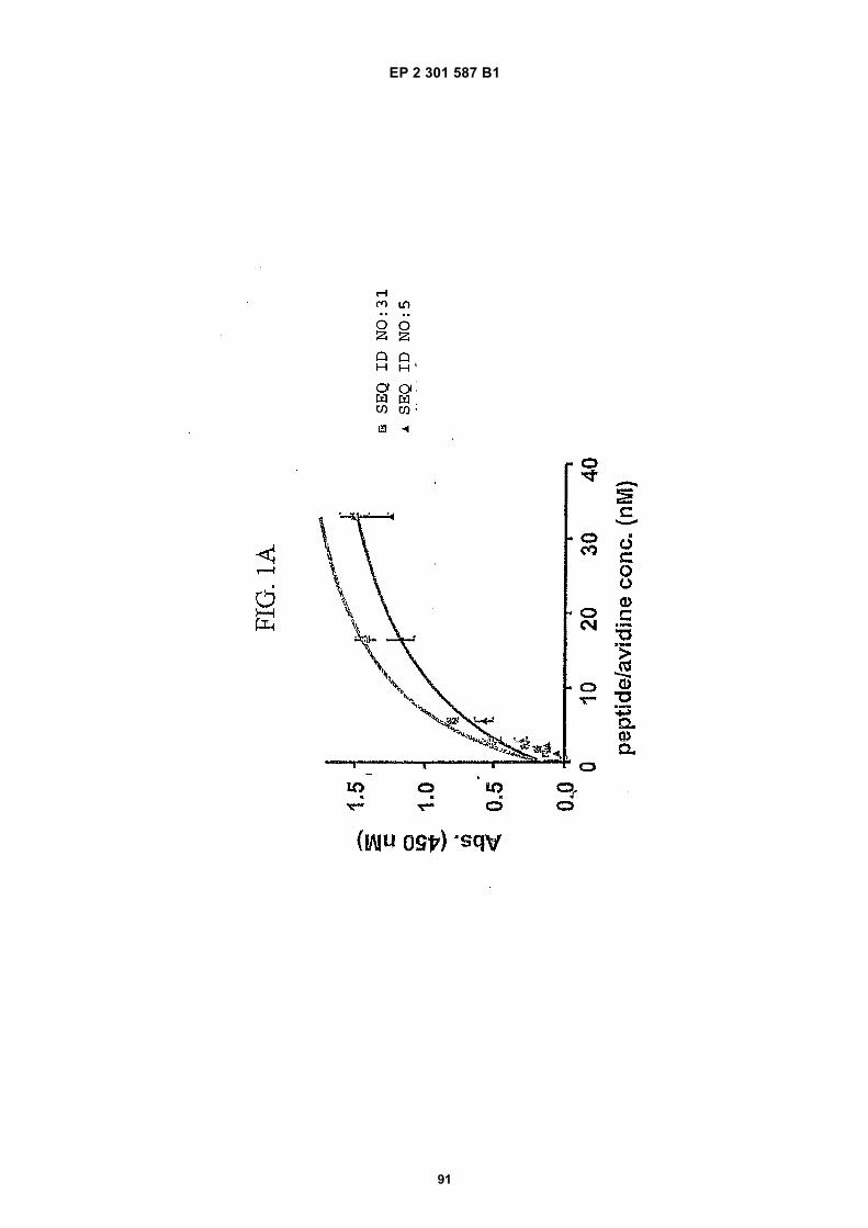
EP 2 301 587 B1
91
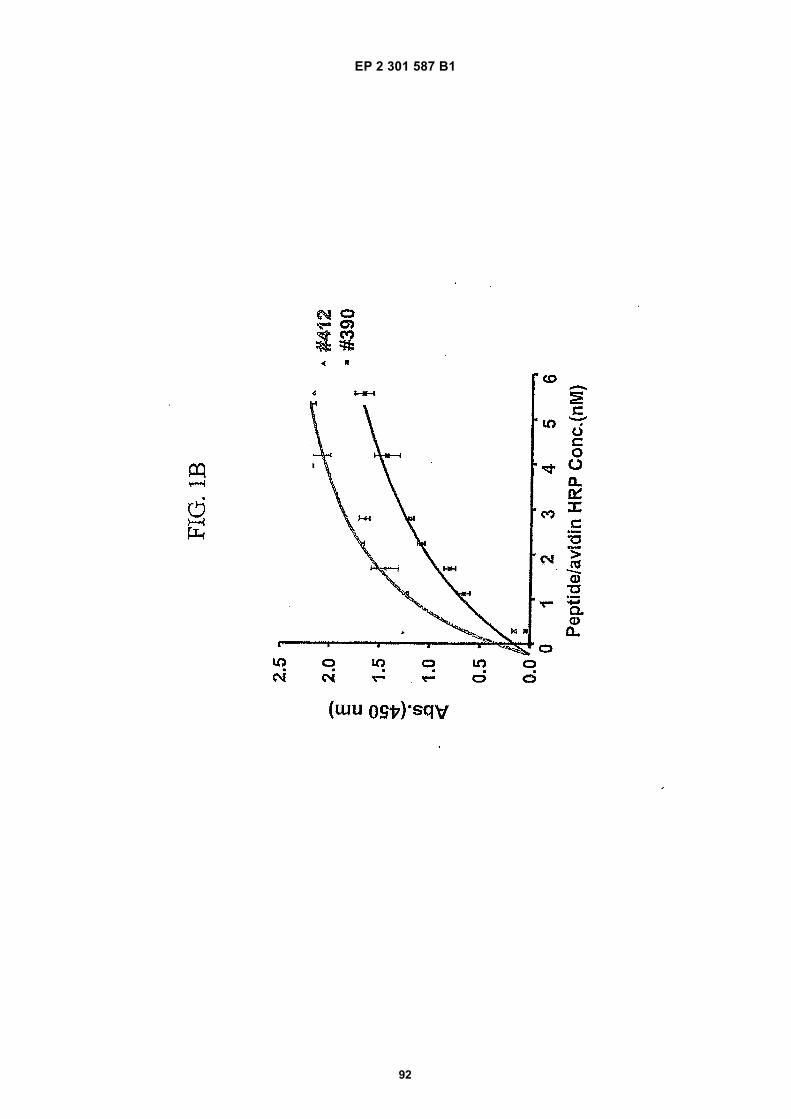
EP 2 301 587 B1
92

EP 2 301 587 B1
93

EP 2 301 587 B1
94
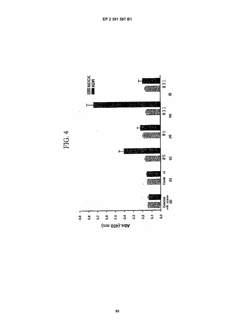
EP 2 301 587 B1
95

EP 2 301 587 B1
96
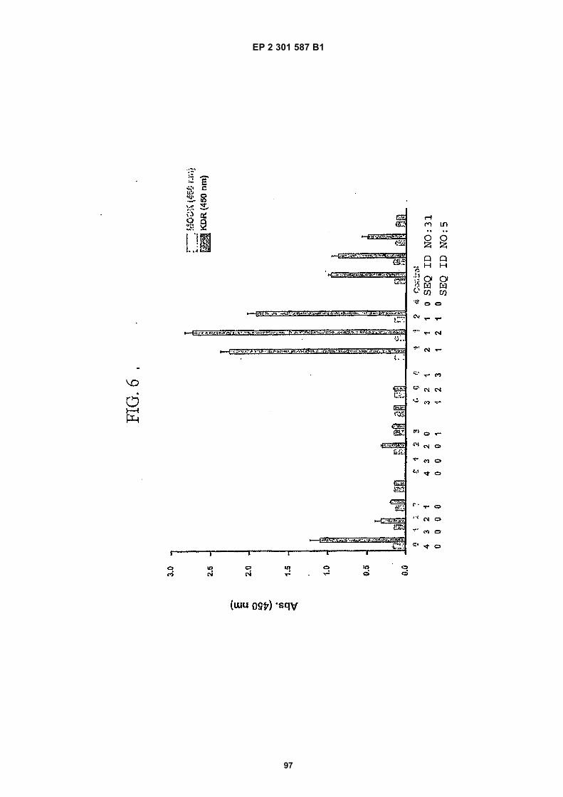
EP 2 301 587 B1
97
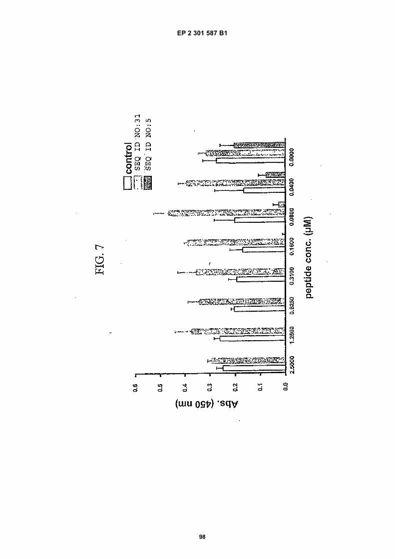
EP 2 301 587 B1
98
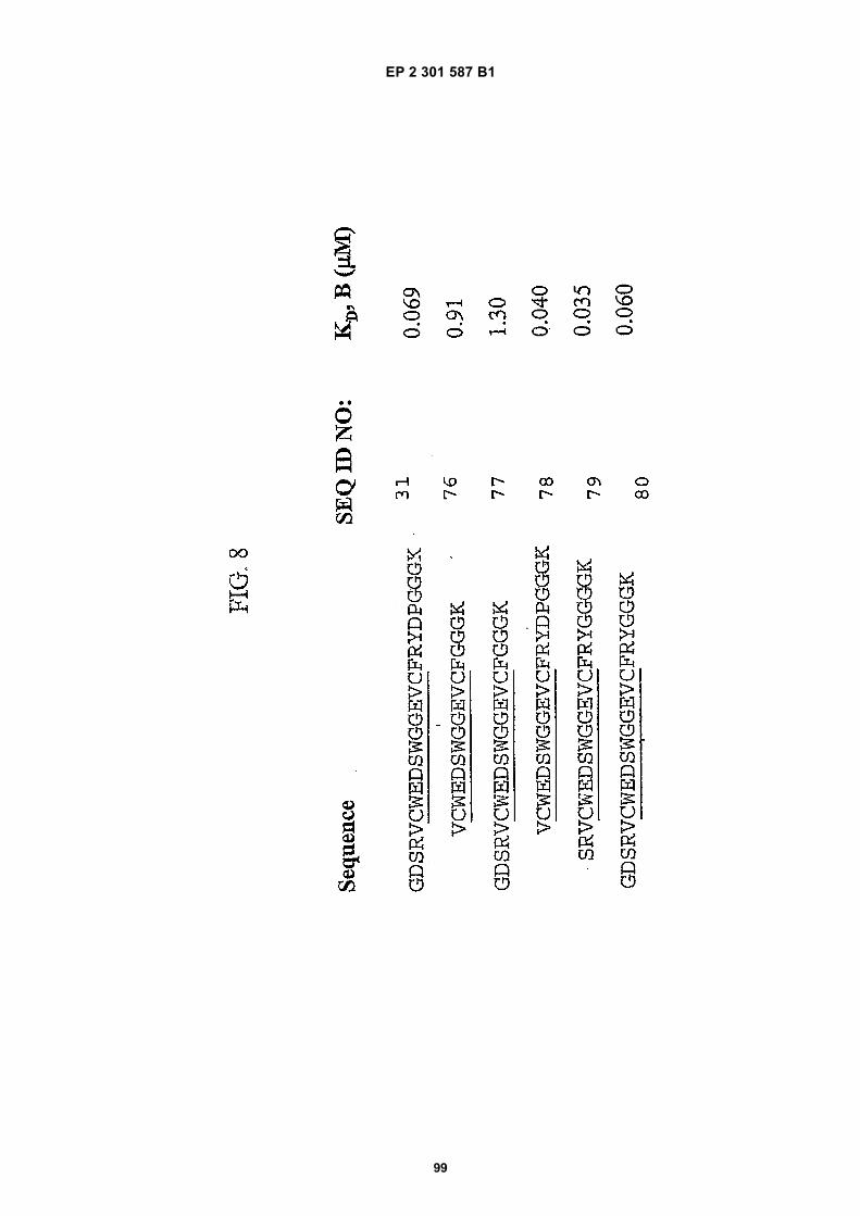
EP 2 301 587 B1
99
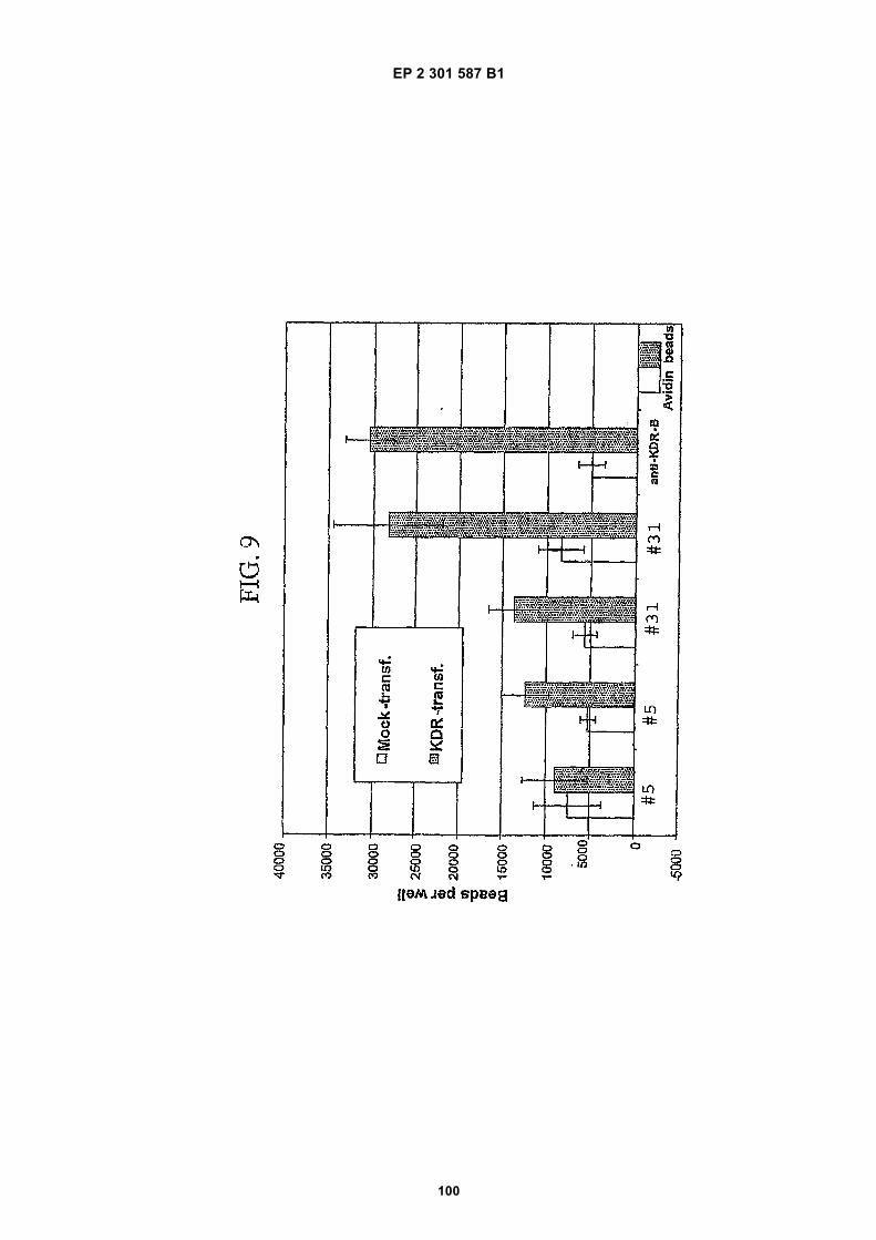
EP 2 301 587 B1
100
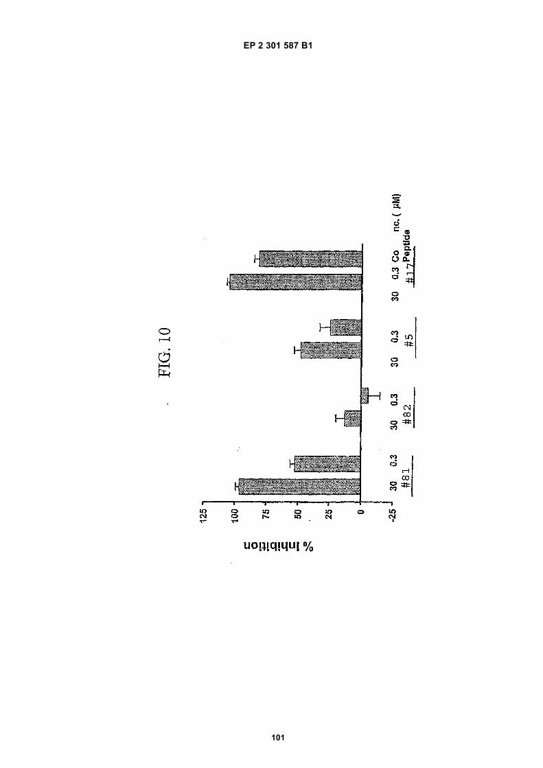
EP 2 301 587 B1
101

EP 2 301 587 B1
102
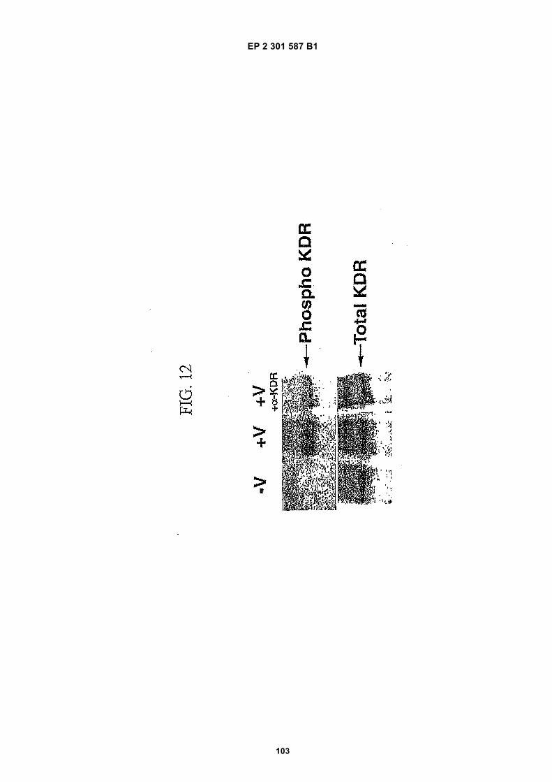
EP 2 301 587 B1
103
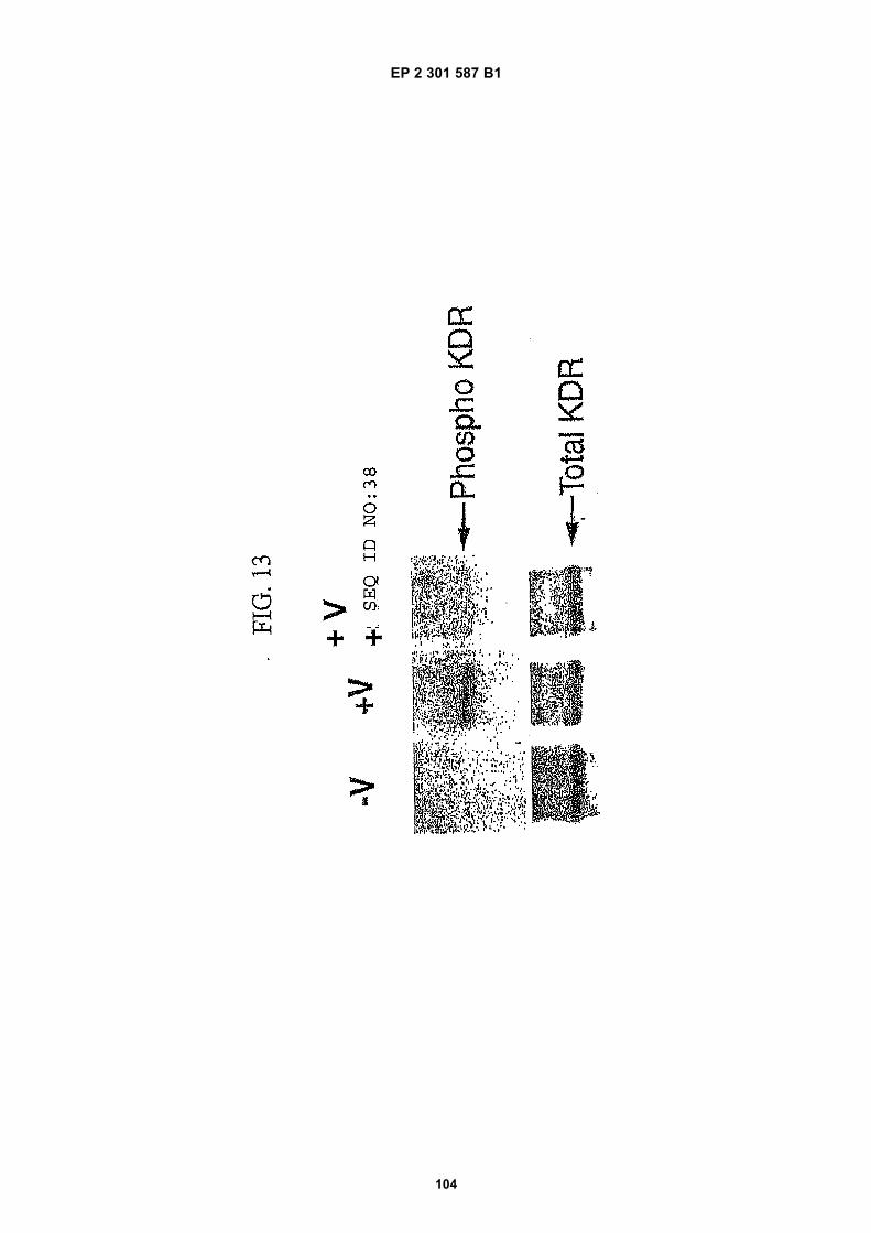
EP 2 301 587 B1
104

EP 2 301 587 B1
105

EP 2 301 587 B1
106
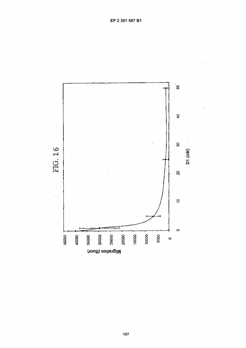
EP 2 301 587 B1
107

EP 2 301 587 B1
108

EP 2 301 587 B1
109

EP 2 301 587 B1
110
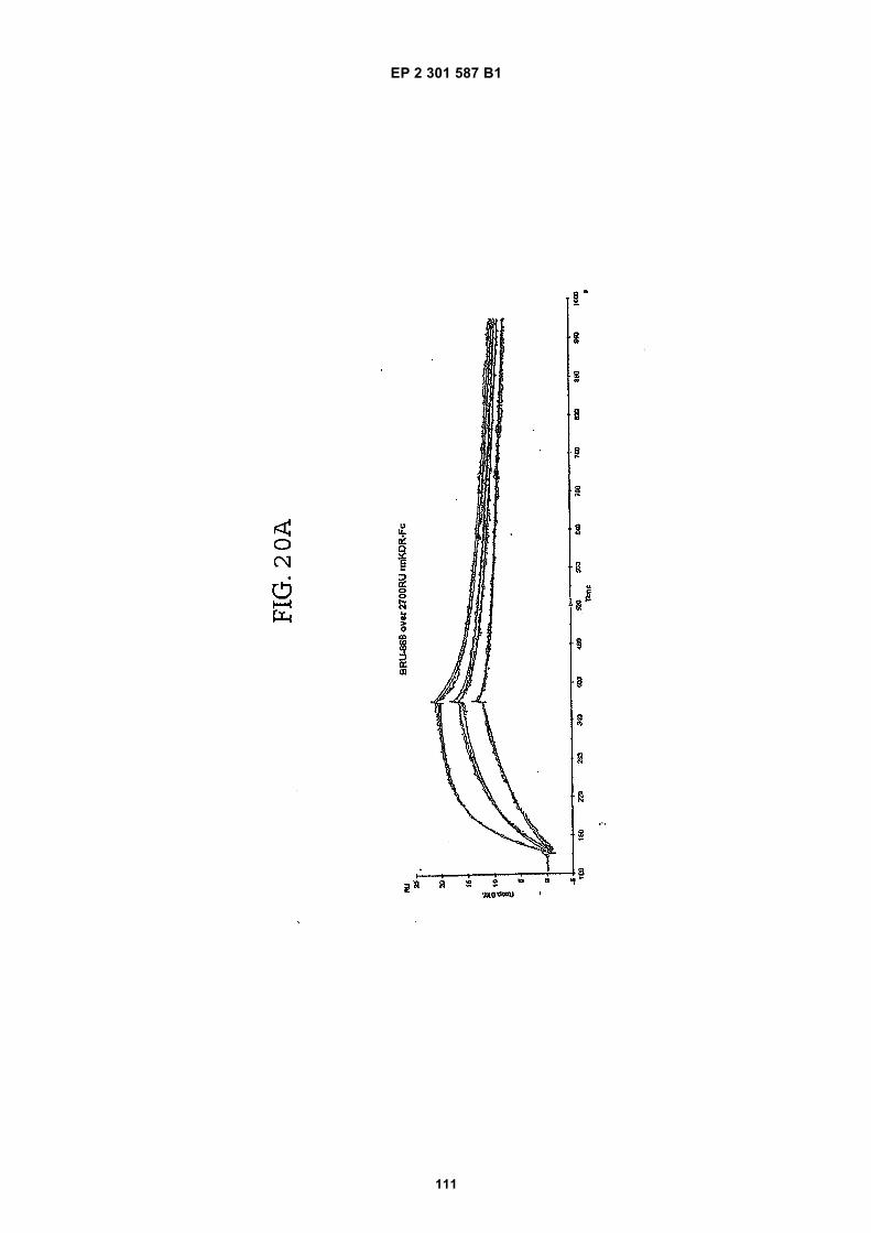
EP 2 301 587 B1
111

EP 2 301 587 B1
112
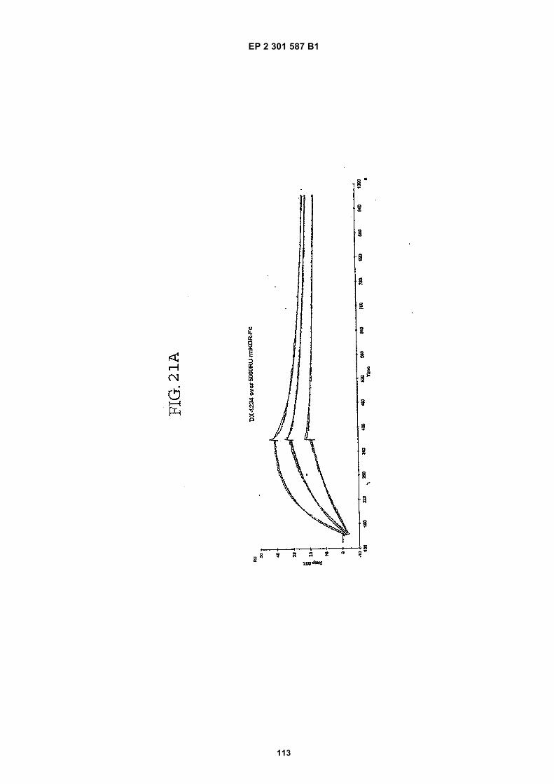
EP 2 301 587 B1
113

EP 2 301 587 B1
114
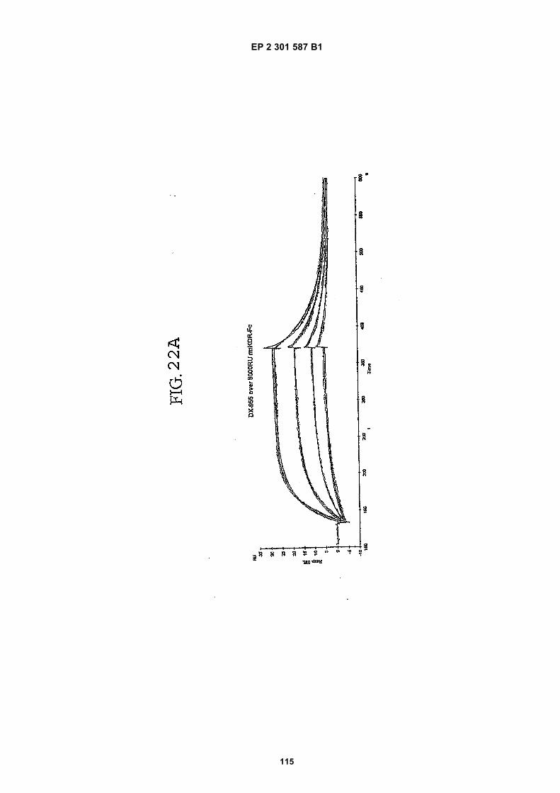
EP 2 301 587 B1
115
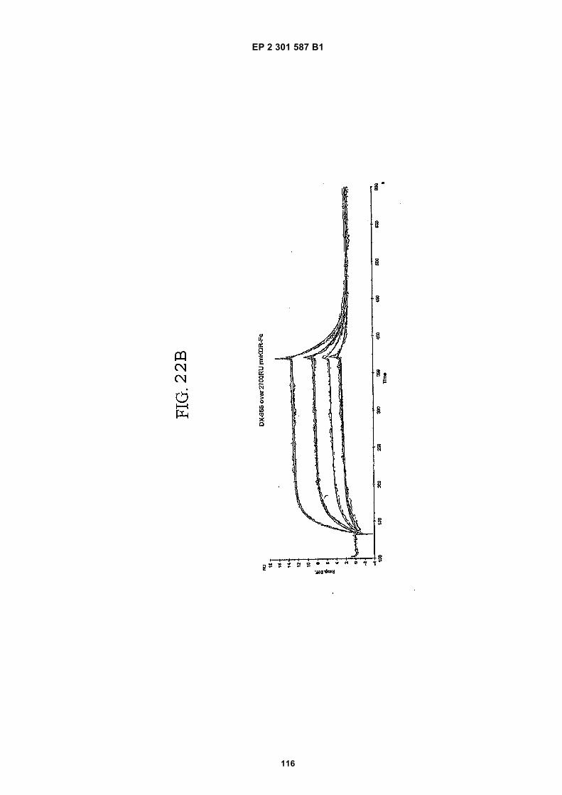
EP 2 301 587 B1
116

EP 2 301 587 B1
117
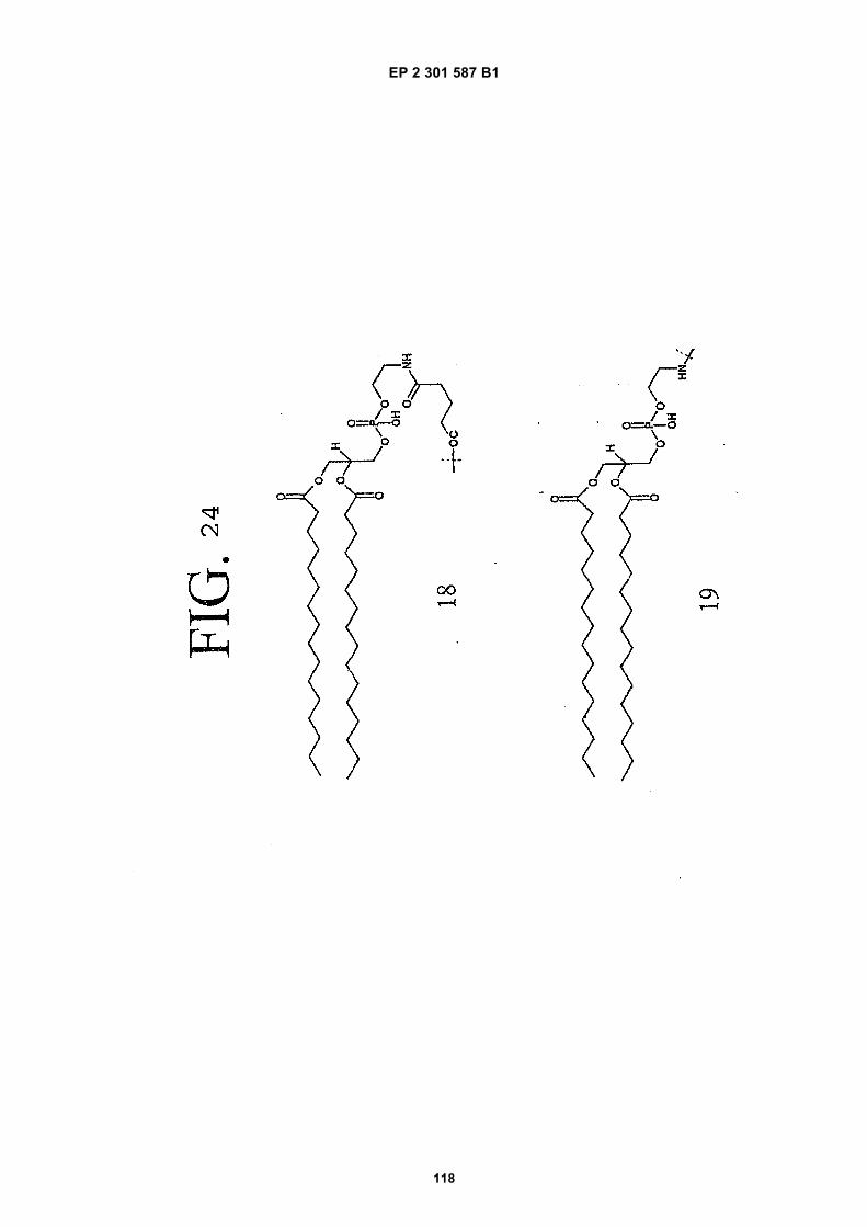
EP 2 301 587 B1
118
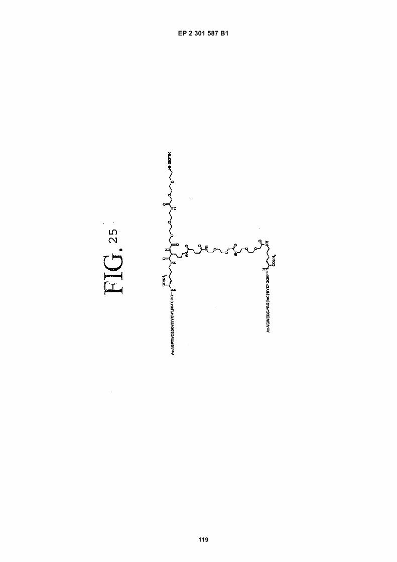
EP 2 301 587 B1
119

EP 2 301 587 B1
120

EP 2 301 587 B1
121

EP 2 301 587 B1
122
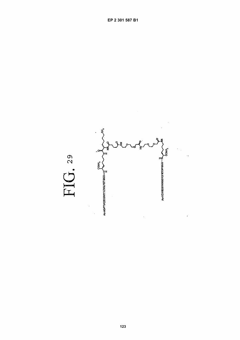
EP 2 301 587 B1
123
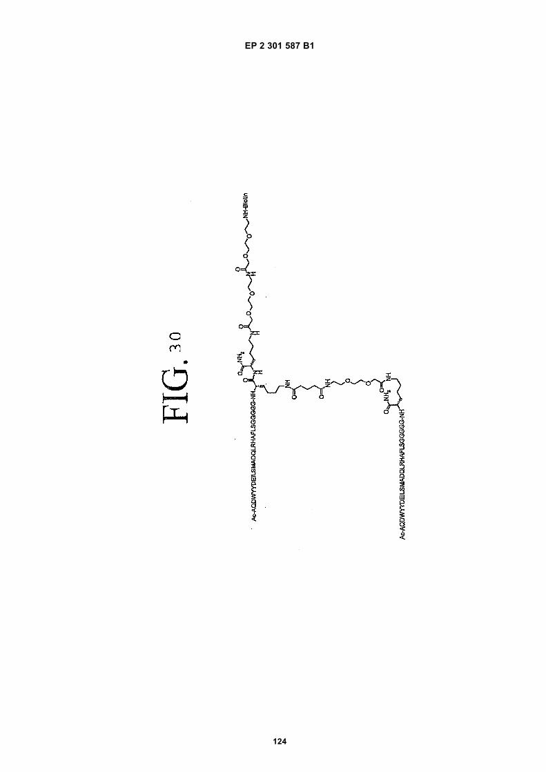
EP 2 301 587 B1
124

EP 2 301 587 B1
125

EP 2 301 587 B1
126

EP 2 301 587 B1
127

EP 2 301 587 B1
128

EP 2 301 587 B1
129

EP 2 301 587 B1
130
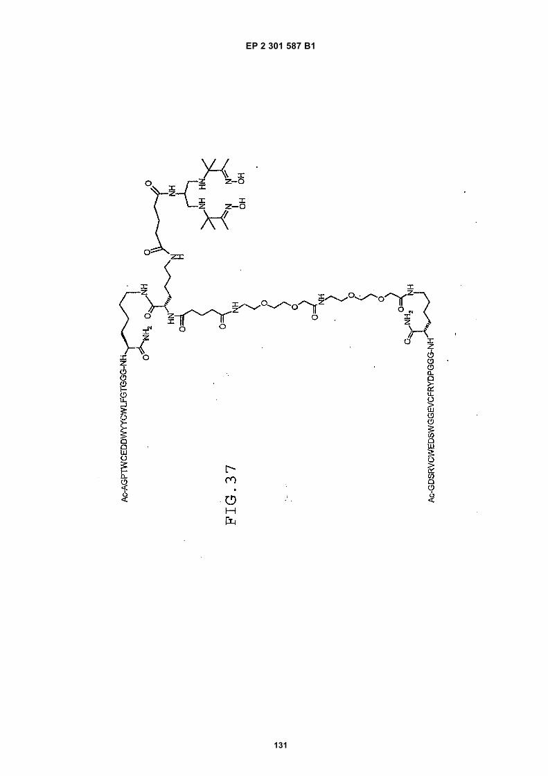
EP 2 301 587 B1
131
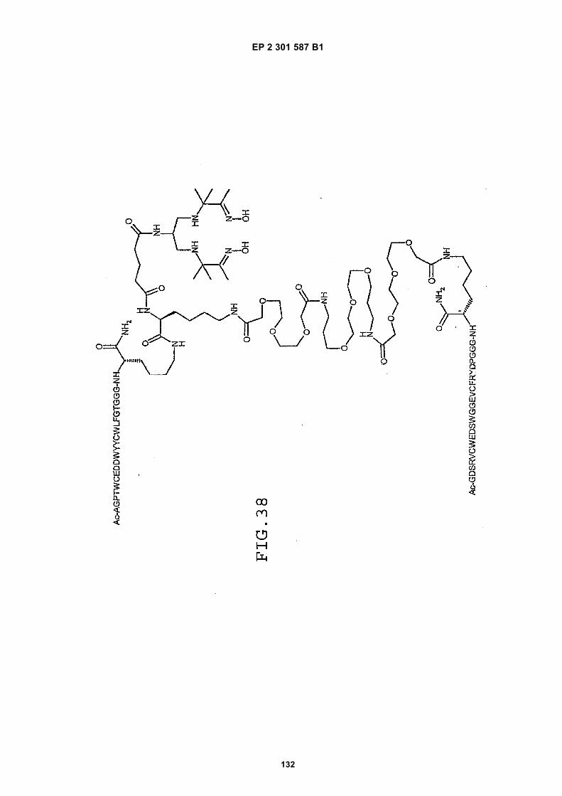
EP 2 301 587 B1
132
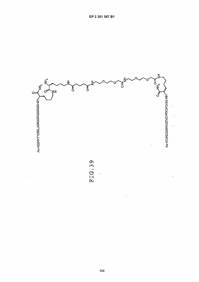
EP 2 301 587 B1
133
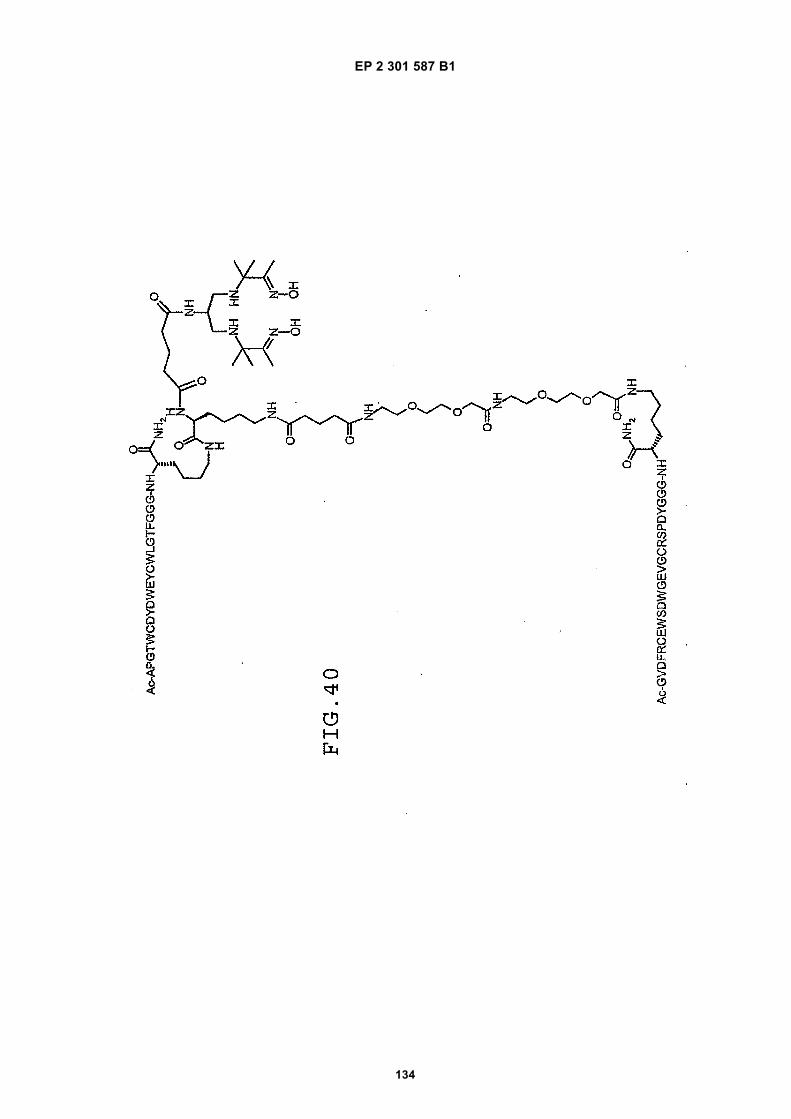
EP 2 301 587 B1
134
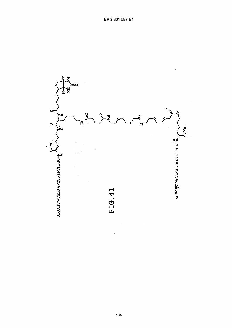
EP 2 301 587 B1
135
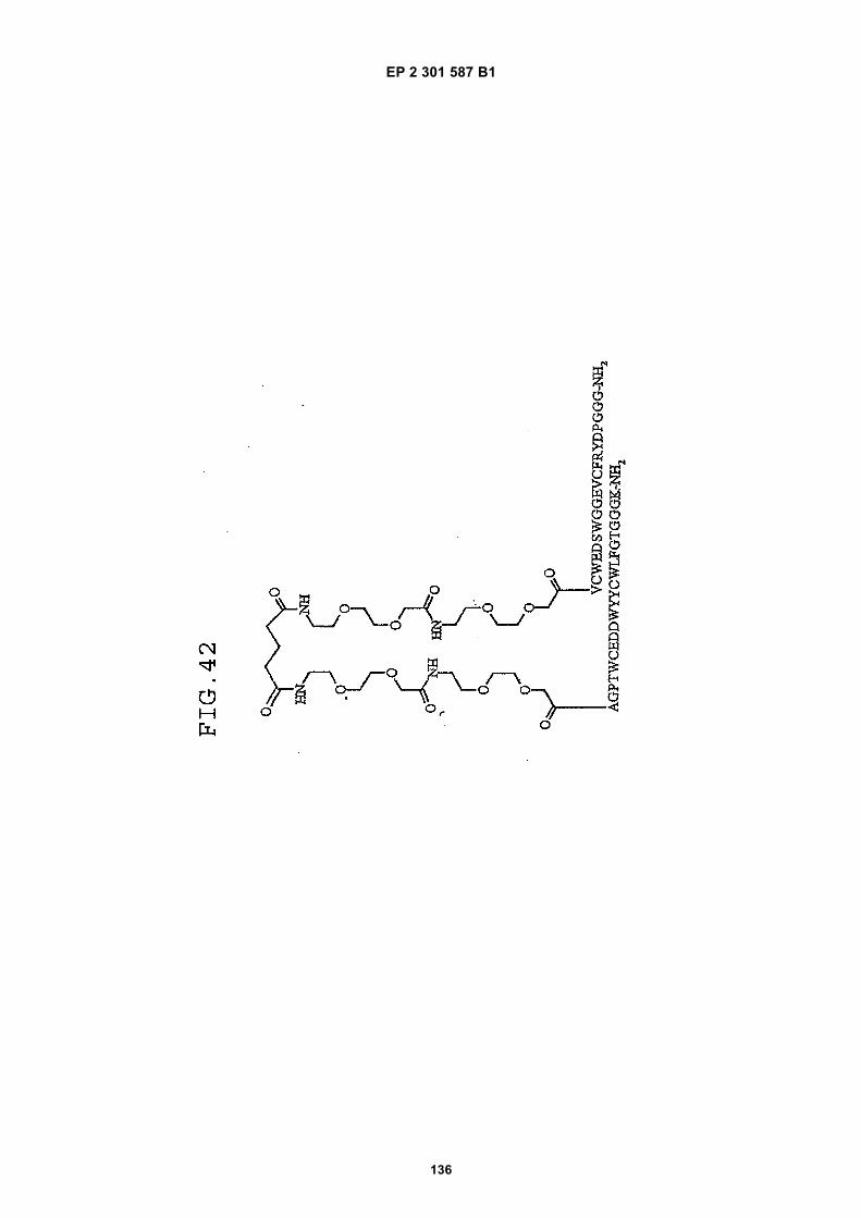
EP 2 301 587 B1
136
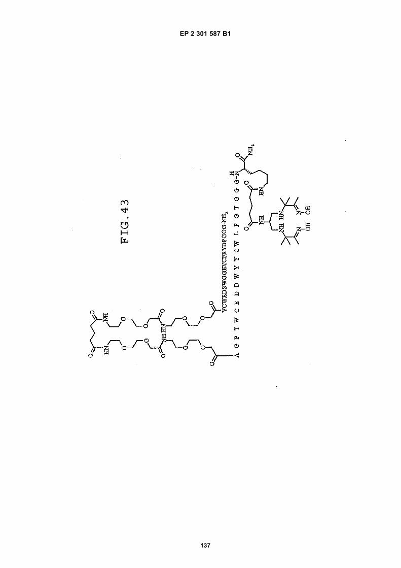
EP 2 301 587 B1
137
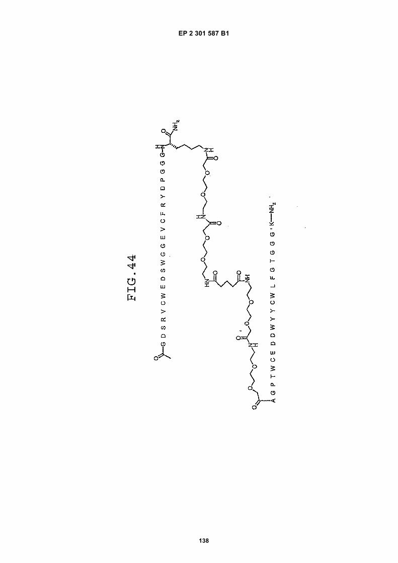
EP 2 301 587 B1
138

EP 2 301 587 B1
139
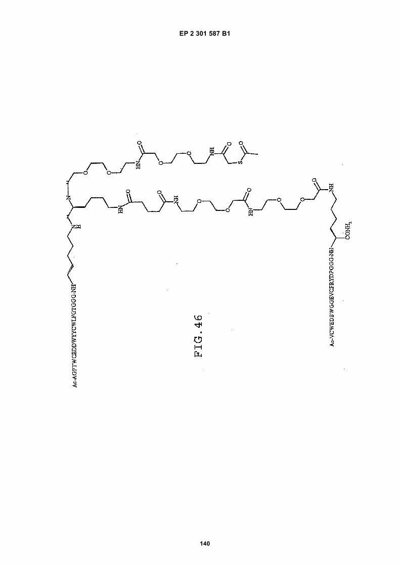
EP 2 301 587 B1
140
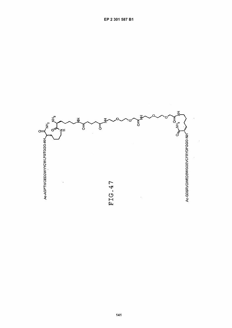
EP 2 301 587 B1
141
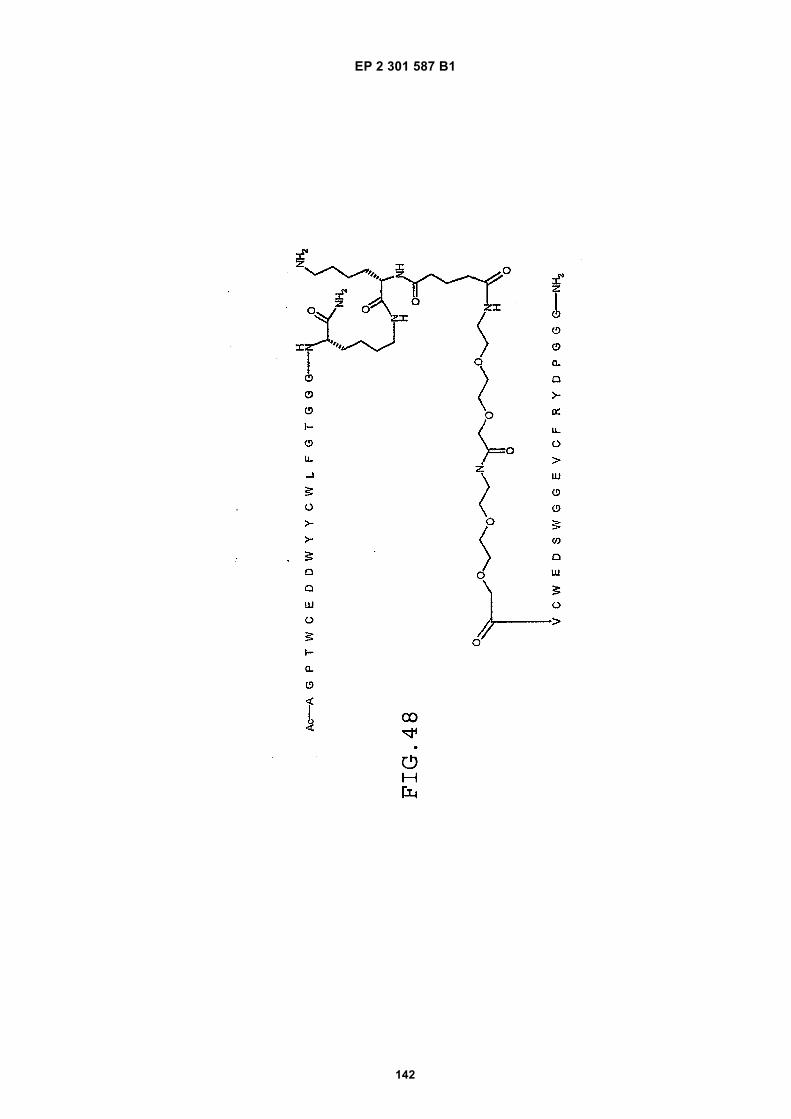
EP 2 301 587 B1
142
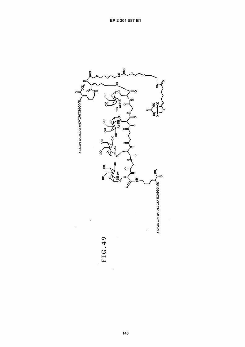
EP 2 301 587 B1
143

EP 2 301 587 B1
144
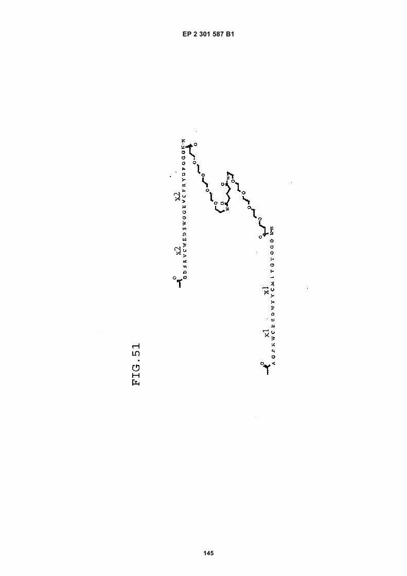
EP 2 301 587 B1
145
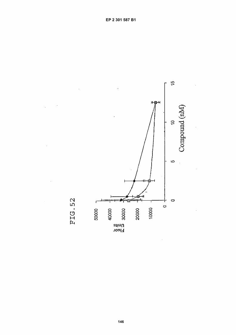
EP 2 301 587 B1
146
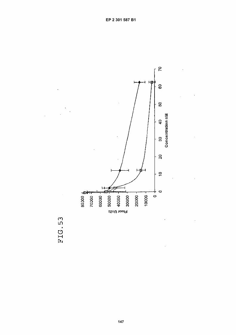
EP 2 301 587 B1
147
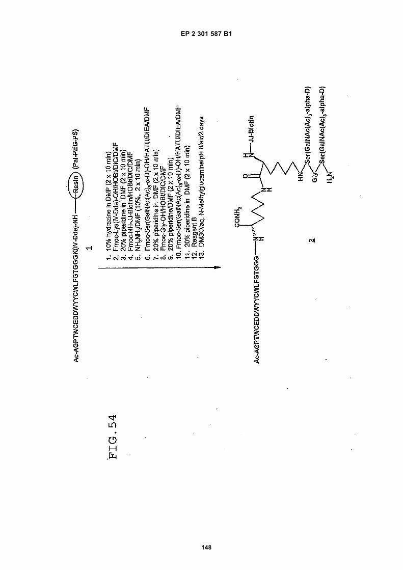
EP 2 301 587 B1
148
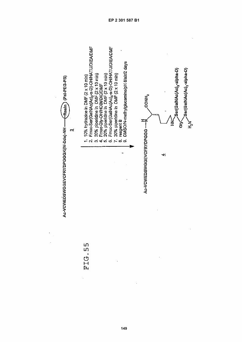
EP 2 301 587 B1
149
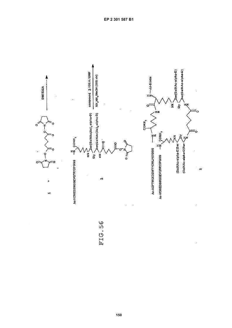
EP 2 301 587 B1
150
EP 2 301 587 B1
151
REFERENCES CITED IN THE DESCRIPTION
This list of references cited by the applicant is for the reader’s convenience only. It does not form part of the Europeanpatent document. Even though great care has been taken in compiling the references, errors or omissions cannot beexcluded and the EPO disclaims all liability in this regard.
Patent documents cited in the description
• WO 9958162 A [0005]• US 440201 P [0014]• US 2004018974 A [0014]• US 5223409 A, Ladner [0037] [0044]• WO 9818497 A [0088]• WO 9818496 A [0088]• WO 9818495 A [0088]• WO 9818498 A [0088]• WO 9853857 A [0088]• WO 9617628 A [0088]• WO 9718841 A [0088]• WO 9623524 A [0088]
• WO 9847538 A [0088]• US 5171298 A [0090]• WO 9857666 A [0090]• US 6165458 A, Foldvari [0095]• US 6274166 B1, Sintov [0095]• US 6312665 B1, Pankaj Modi [0098]• US 4391797 A, Folkman [0100]• WO 0191805 A2 [0304]• EP 10185498 A [0318]• US 60360851 B [0318]• US 60440411 B [0318]
Non-patent literature cited in the description
• PEPPER, M. et al. Enzyme Protein, 1996, vol. 49,138-162 [0001]
• RISAU, W. Nature, 1997, vol. 386, 671-674 [0001]• KIM, K. et al. Nature, 1993, vol. 362, 841-844 [0001]• HANAHAN, D. ; FOLKMAN, J. Cell, 1996, vol. 86,
353-364 [0002]• NEUFELD, G. et al. FASEB J., 1999, vol. 13, 9-22
[0002]• FUKUMURA, D. et al. Cell, 1998, vol. 94, 715-725
[0003]• BIKFALVI, A. et al. J. Cell. Physiol., 1991, vol. 149,
50-59 [0004]• VEIKKOLA, T. et al. Cancer Res., 2000, vol. 60,
203-212 [0004]• KAY et al. Phage Display of Peptides and Proteins:
A Laboratory Manual. Academic Press, Inc, 1996[0037] [0044]
• STEWART et al. Solid-Phase Peptide Synthesis. W.H. Freeman Co, 1989 [0048]
• MERRIFIELD. J. Am. Chem. Soc., 1963, vol. 85,2149-2154 [0048]
• BODANSZKY ; BODANSZKY. The Practice of Pep-tide Synthesis. Springer-Verlag, 1984 [0048]
• DAVIS et al. Basic Methods in Molecular Biology,1986 [0052]
• ANDERSON ; MILLER. Clin. Chem., 1988, vol. 34(7), 1417-21 [0056]
• MALMBORG et al. J. Immunol. Methods, 1996, vol.198 (1), 51-7 [0056]
• SCHUCK. Current Opinion in Biotechnology, 1997,vol. 8, 498-502 [0056]
• HUTCHINSON. Molec. Biote, 1995, vol. 3, 47-54[0056]
• HILLER et al. Biochem. J., 1987, vol. 248, 167-171[0059]
• ALON et al. Biochem. Biophys. Res. Commun.,1990, vol. 170, 1236-41 [0059]
• KIHLBERG. Glycopeptide synthesis. In: Fmoc SolidPhase Peptide Synthesis - A Practical Approach. Ox-ford University Press, January 2000, 195-213 [0068]
• WAHL ; MUTTER. Tetrahedron Lett., 1996, vol. 37,6861-6864 [0071]
• BACKES et al. J. Am. Chem. Soc., 1996, vol. 118(12), 3055-56 [0075]
• Principles and Practice of Solid Phase Synthesis.FIELDS et al. Synthetic Peptides, A Users Guide.W.H. Freeman Co, 1992, 77-183 [0075]
• Convergent Peptide Synthesis. BARLOS et al. FmocSolid Phase Peptide Synthesis. Oxford UniversityPress, 2000, 215-228 [0075]
• Synthesis of Modified Peptides. MELLOR et al. FmocSolid Phase Peptide Synthesis. Oxford UniversityPress, 2000, 169-176 [0076]
• FALORNI et al. Tetrahedron Lett., 1998, vol. 39 (41),7607-7610 [0078]
• JOHNSON et al. Tetrahedron Lett., 1998, vol. 54(16), 4097-4106 [0078]
• STANKOVA et al. Mol. Diversity, 1996, vol. 2, 75-80[0078]
• Remington: The Science and Practice of Pharmacy.Lippincott, Williams and Wilkins, 2000 [0093]
• FAIRBROTHER et al. Biochemistry, 1998, vol. 37(51), 17754-17764 [0121]
• LAEMMLI. Nature, 1970, vol. 227, 680-685 [0196]• MATSUDAIRA. J. Biol. Chem., 1987, vol. 262,
10035-10038 [0197]

EP 2 301 587 B1
152
• STRAWN, L. et al. Cancer Res., 1996, vol. 56,3540-3545 [0249]
• Points to Consider in the Characterization of CellLines used to Produce Biologicals. Points to Considerin the Characterization of Cell Lines used to ProduceBiologicals, 1993 [0267]
• Guide for the Care and Use of Laboratory Animals.National Academy Press, 1996 [0276]
• HOLMES, D. et al. Bioconjug. Chem., 2000, vol. 11,439-444 [0312]
• ROBERTS M. et al. Adv. Drug. Deliv. Rev., 2002,vol. 54, 459-476 [0315]
• SLEEP, D et al. Biotechnology (NY), 1991, vol. 9,183-187 [0317]