tepzz 77 758a_t - ep 2 772 758 a1
24
Printed by Jouve, 75001 PARIS (FR) (19) EP 2 772 758 A1 TEPZZ 77 758A_T (11) EP 2 772 758 A1 (12) EUROPEAN PATENT APPLICATION (43) Date of publication: 03.09.2014 Bulletin 2014/36 (21) Application number: 13156865.1 (22) Date of filing: 27.02.2013 (51) Int Cl.: G01N 33/574 (2006.01) (84) Designated Contracting States: AL AT BE BG CH CY CZ DE DK EE ES FI FR GB GR HR HU IE IS IT LI LT LU LV MC MK MT NL NO PL PT RO RS SE SI SK SM TR Designated Extension States: BA ME (71) Applicant: Oncotyrol Center for Personalized Cancer Medicine GmbH 6020 Innsbruck (AT) (72) Inventors: • Spizzo, Gilbert 39012 Meran (IT) • Fong, Dominic 39028 Kortsch/Schlanders (IT) (74) Representative: Sonn & Partner Patentanwälte Riemergasse 14 1010 Wien (AT) (54) EpCAM tumour diagnosis (57) The invention discloses a method for in vitro di- agnosing a pancreas or colon cancer tissue specimen wherein the specimen is analysed for the presence or absence of the ectodomain of EpCAM (EpEX) as well as for the presence or absence of the intracellular domain of EpCAM (EpICD).
-
Upload
khangminh22 -
Category
Documents
-
view
2 -
download
0
Transcript of tepzz 77 758a_t - ep 2 772 758 a1
Printed by Jouve, 75001 PARIS (FR)
(19)E
P2
772
758
A1
TEPZZ 77 758A_T(11) EP 2 772 758 A1
(12) EUROPEAN PATENT APPLICATION
(43) Date of publication: 03.09.2014 Bulletin 2014/36
(21) Application number: 13156865.1
(22) Date of filing: 27.02.2013
(51) Int Cl.:G01N 33/574 (2006.01)
(84) Designated Contracting States: AL AT BE BG CH CY CZ DE DK EE ES FI FR GB GR HR HU IE IS IT LI LT LU LV MC MK MT NL NO PL PT RO RS SE SI SK SM TRDesignated Extension States: BA ME
(71) Applicant: Oncotyrol Center for Personalized Cancer Medicine GmbH6020 Innsbruck (AT)
(72) Inventors: • Spizzo, Gilbert
39012 Meran (IT)• Fong, Dominic
39028 Kortsch/Schlanders (IT)
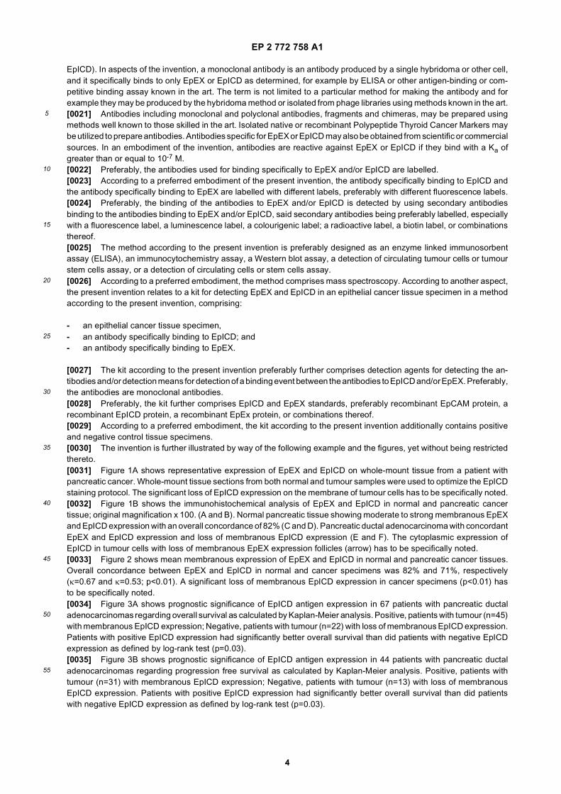
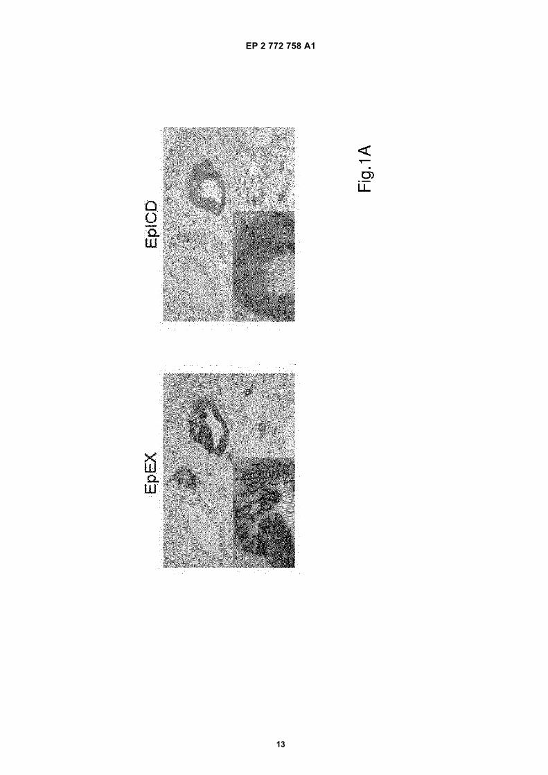
(74) Representative: Sonn & Partner PatentanwälteRiemergasse 141010 Wien (AT)
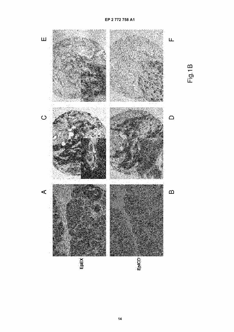
(54) EpCAM tumour diagnosis
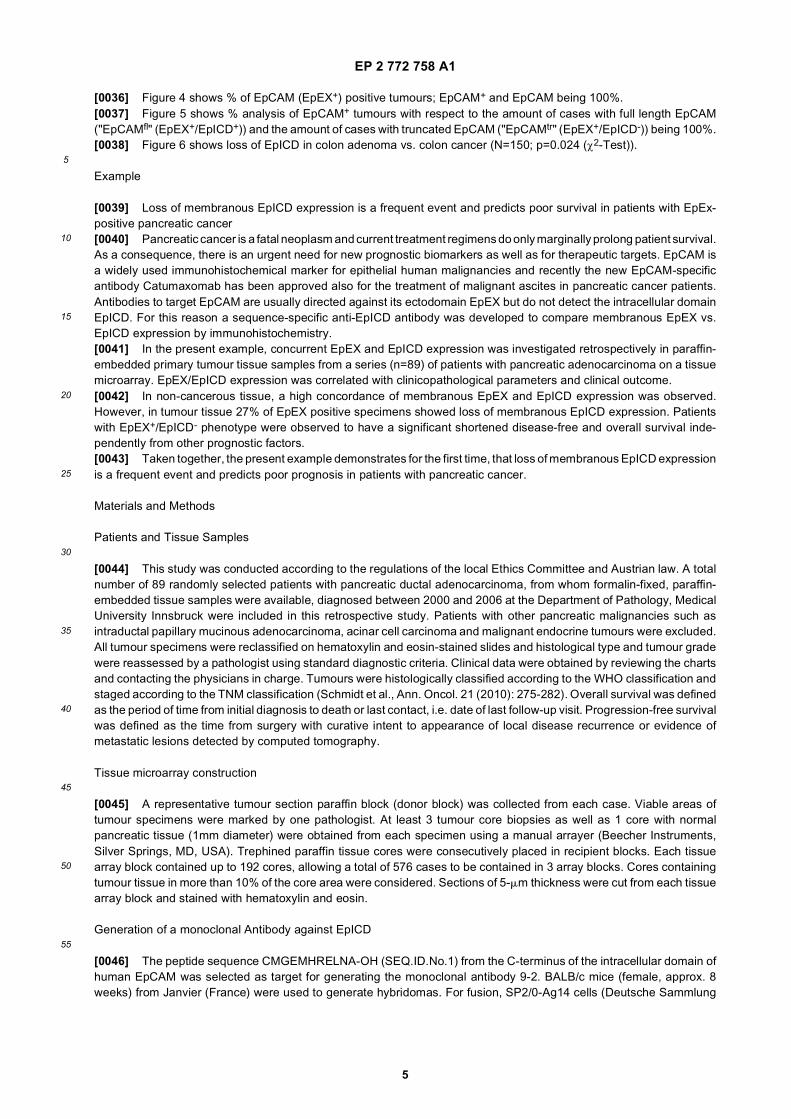
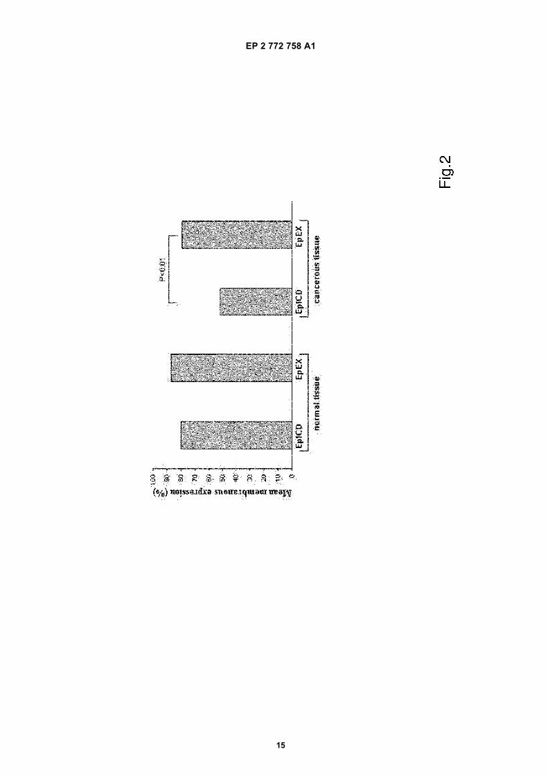
(57) The invention discloses a method for in vitro di-agnosing a pancreas or colon cancer tissue specimenwherein the specimen is analysed for the presence or
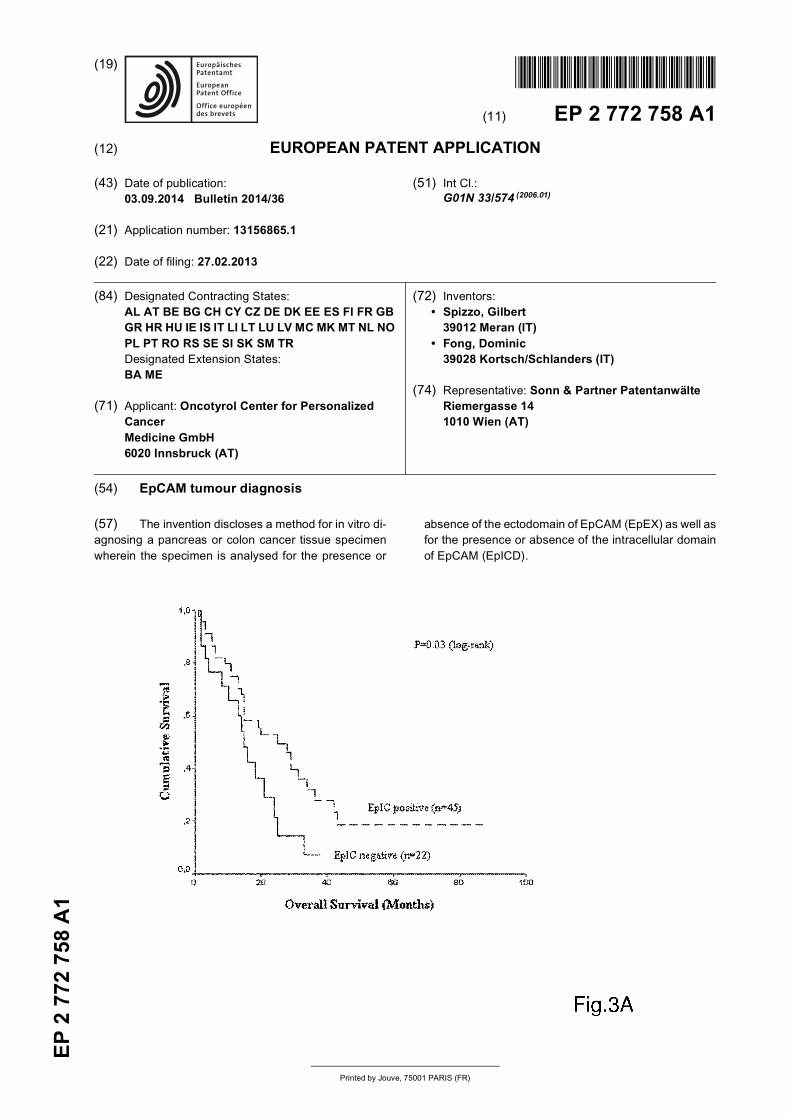
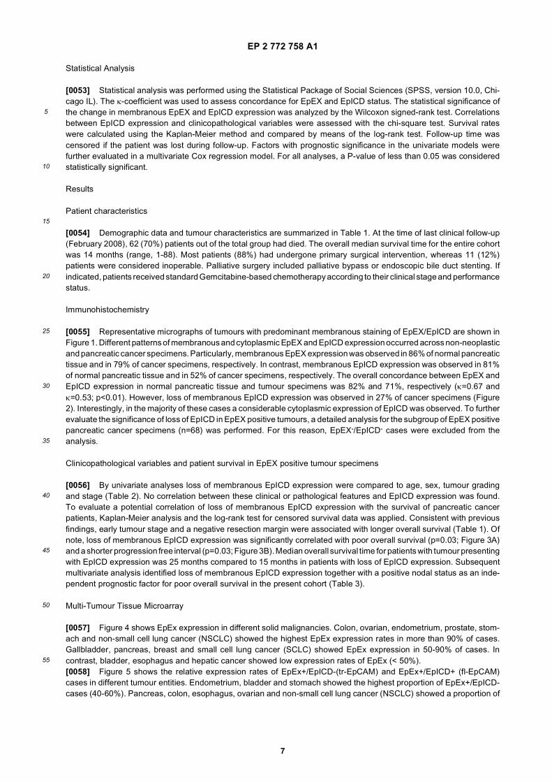
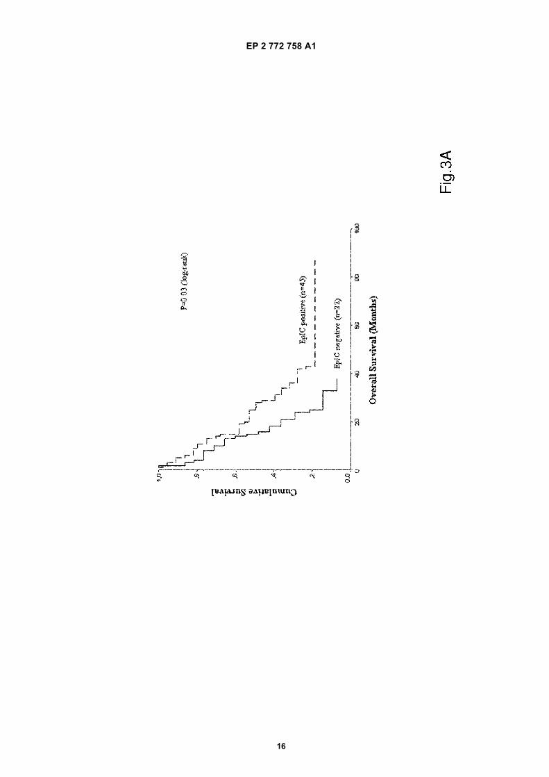
absence of the ectodomain of EpCAM (EpEX) as well asfor the presence or absence of the intracellular domainof EpCAM (EpICD).
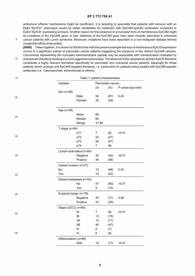
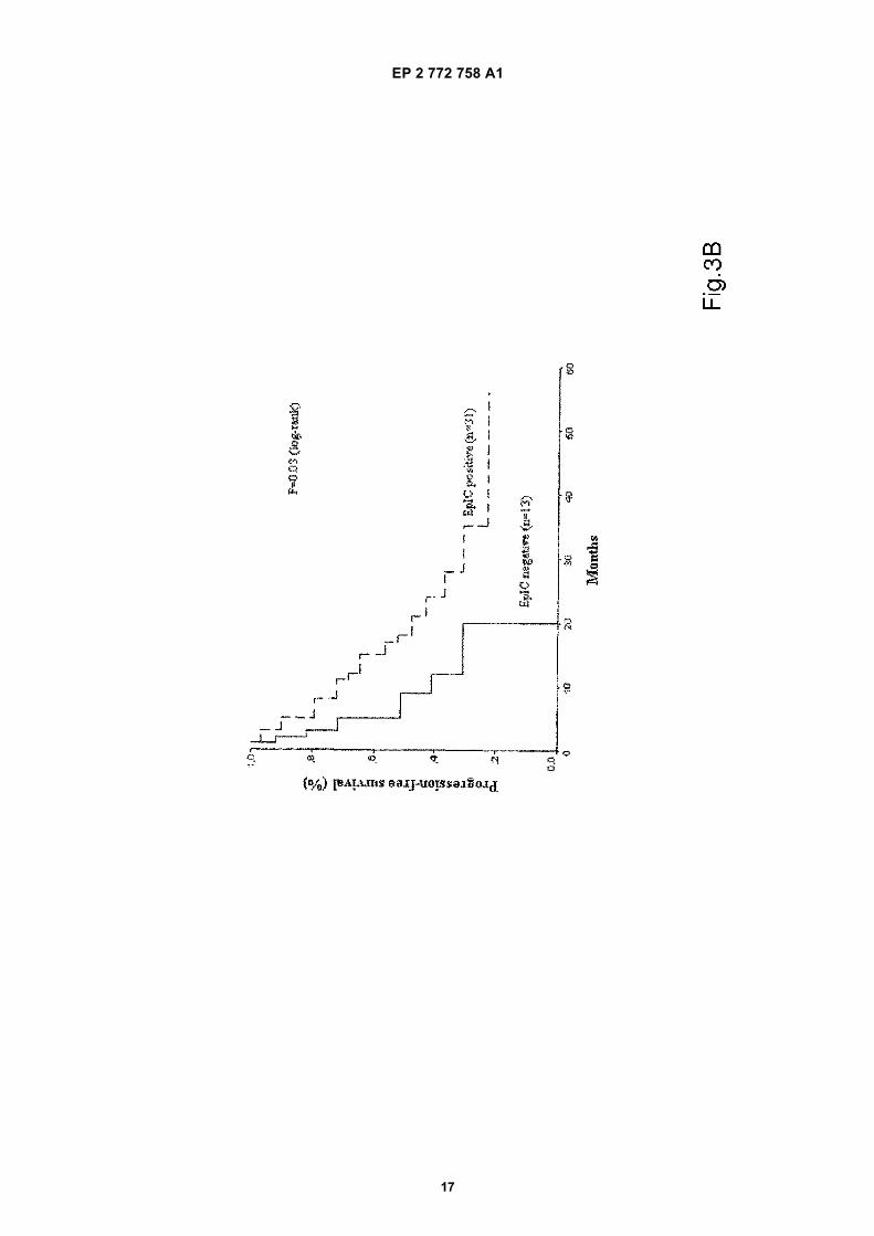
EP 2 772 758 A1
2
5
10
15
20
25
30
35
40
45
50
55
Description
[0001] The present invention relates to the field of tumour prognosis.[0002] Characterised by its aggressive tumour biology most pancreatic cancer patients already have locally advancedor metastatic disease at the time of diagnosis. While surgical resection offers the only potentially curative treatment, themajority of patients experience recurrence within a short period even after complete (R0) resection. Current Gemcitabine-based chemotherapy provides only marginal survival benefit, and even the addition of novel, molecularly targeted ther-apies do not prolong patient survival significantly. Moreover, in advanced and metastatic pancreatic cancer more recenttrials have included the control of disease-related symptoms defined by the clinical benefit response rate (CBR) as anendpoint for evaluating therapeutic success in this disease. Therefore, definition of reliable prognostic and predictivemarkers is fundamental to allow the selection of patients who are most likely benefit from novel therapeutic approaches.[0003] Tumour antigen specific monoclonal antibodies (mAb) have been successfully implemented into standardtreatment regimens for patients with different malignant diseases. However, clinical efficacy of these cancer specifictherapies is only seen in a subset of patients. Moreover, only little is known about the mechanisms underlying the clinicalresponse of patients to mAb-based immunotherapy.[0004] The epithelial cell adhesion molecule/antigen EpCAM (also known as GA733-2, TACSTD1, ESA, KS1/4) is a40 kDa protein that is frequently expressed in normal epithelia and that has been described to be widely overexpressedin carcinomas of various origin. As a consequence, the presence of high amounts of membranous EpCAM in multipletumours, have rendered EpCAM an ideal target for antibody-based therapeutics. For this reason, EpCAM was describedbeing one of the first antigens to be targeted by monoclonal antibodies in cancer patients. While the murine monoclonalanti-EpCAM antibody Edrecolomab (17-1A, Panorex®) showed anti-tumoural activity in gastrointestinal cancer patients,clinical trials are currently evaluating different immunotherapeutic approaches against this common tumour antigen.Only recently, the trifunctional EpCAM/CD3-specific antibody Catumaxomab could show meaningful clinical activity bythe addition of co-adjuvant mechanisms and has been approved for the treatment of malignant ascites in cancer patientswith EpCAM-positive tumours. In a phase II/III study, quality of life was improved as symptoms of ascites were significantlydecreased compared with the control group.[0005] However, despite its broad distribution in human malignancies, EpCAM-specific therapeutic antibodies stillhave not hold promise and results from recent clinical trials have been disappointing. Therefore, identification of themechanisms underlying the therapeutic efficacy of EpCAM targeted therapies is crucial to optimize the selection ofpatients most likely to benefit from their use.[0006] The object of the present invention is to provide improved means for diagnosing cancer patients and providesignificant prognostic and/or predictive markers which allow a personalised optimization of treatment regimen.[0007] Therefore, the present invention provides a method for in vitro diagnosing a pancreas or colon cancer tissuespecimen wherein the specimen is analysed for the presence or absence of the ectodomain of EpCAM (EpEX) as wellas for the presence or absence of the intracellular domain of EpCAM (EpICD).[0008] Therapeutic antibodies that target EpCAM usually bind the ectodomain of the antigen termed EpEX but do notrecognize the intracellular domain EpICD. For this reason the present invention applies EpICD specific antibodies whichassess membranous EpICD expression by immunohistochemistry. Such expression analyses can be compared withthe staining patterns obtained by conventional EpEX antibodies. The present invention specifically investigated in themembranous presence of full-length EpCAM or truncated variants in pancreatic cancer and defined the prognostic impactthereof.[0009] In the course of the present invention it could be shown that expression of membranous EpEX/EpICD expressionis varying significantly in different tumour tissue. In fact, there are tumours in which presence of membranous EpICD islost, whereas in other tumours, such loss is not observed. This loss of presence of membranous EpICD has biologicaland clinical relevance, because patients who show such loss have to face a worse development (disease course) thanthose who show EpICD presence. In fact, the data gathered in the course of the present invention show that medianoverall survival is significantly different for the two groups of patients. According to the present invention, patients whoshow a loss of presence of membranous EpICD have a worse prognosis than patients who have membranous EpICDin the cancer tissue specimen. This allows a targeted more aggressive treatment of the patients with a worse prognosis,whereas patients with a good prognosis can be relieved from such an aggressive treatment. This has been shownaccording to the present invention for tissue cancer specimen from patients with pancreas carcinoma and for patientswith colon carcinoma. From the medical point of view, it is clear that patients with membranous EpEX+/EpICD- phenotypemetastatic pancreatic cancer showing reduced survival should not be treated with the antibody Catumaxomab or anti-bodies with similar EpCAM specificity, since these patients are not supposed to benefit from such a treatment. On theother side, pancreatic cancer patients with EpEX+/EpICD+ phenotype are more appropriated for such a treatment andshould therefore be treated with Catumaxomab (or antibodies recognising the same epitope as Catumaxomab (Remov-ab®; a trispecific antibody; mouse IgG2a/rat IgG2b hybrid), such as Proxinium®, Vivendium® (VB4-845; immunotoxin;single-chain antibody pseudomonas exotoxin fusion), IGN-101 (edrecolomab; a vaccine for induction of antiidiotypic

EP 2 772 758 A1
3
5
10
15
20
25
30
35
40
45
50
55
antibody response), Adecatumumab (MT201; fully human IgG1 mAb), ING-1 (human engineered IgG1 mAb), EMD 273066 (huKS-IL2; fusion of humanized mAb KS1/4 with human IL-2); see: Baeuerle et al., BJC 96 (2007): 417-423).Moreover, pancreatic cancer patients with EpEX+/EpICD- phenotype are candidates for - compared to the standardtreatments (e.g. Seeber/Schutte "Therapiekonzepte Onkologie" 5th edition (2007): 713ff., 747ff.) - more aggressivechemotherapy regimens (e.g. GemOx (Louvet et al., JCO 20 (2002): 1512-1518), GemCap (Cunningham et al., JCO27 (2009): 5513-5518) or Folfirinox (Conroy et al., New England Journal of Medicine 364 (2011): 1817-1825)) andpotentially for inhibitors of EpCAM cleavage (Maetzel el al., Nat. Cell Biol. 11 (2009): 162-171) such as TACE inhibitors(for review see: Das Gupta et al., Bioorg. Med. Chem. 17 (2009): 444-459).[0010] For patients with colorectal cancer, many clinical studies have been performed with the anti-EpCAM antibodyEdrecolomab. Most of these studies have not shown improved survival of these patients (Niedzwiecki et al., J. Clin.Oncol. 29 (2011): 3146-3152). According to the teachings of the present invention, it is evident that the efficacy of sucha treatment is limited to patients with EpEX+/EpICD+ phenotype and that the studies might have failed upon bad selectionof patients.[0011] Preferred cancer tissue specimens according to the present invention are tissue sections and cytocentrifugatesfrom pleura and ascites fluids. Preferred tissue section specimens are paraffin-embedded tissue sections and freshfrozen sections.[0012] Regulated intramembrane proteolysis activates EpCAM as a mitogenic signal transducer in vitro and in vivo.This involves shedding of its ectodomain EpEX and nuclear translocation of its intracellular domain EpICD.[0013] Determination of EpCAM in cancer tissue specimen according to the present invention can, in principle, beestablished by various methods including immunohistochemistry (Spizzo et al., 2004; Slanchev et al., PLoS Genetics5 (2009), e1000563; WO 2011/032296 A1, PCT/EP2012/062397), ELISA (Abe et al., J. Immunol. Meth. 270 (2002),227-233; Petsch et al., mAbs 3 (2011), 31-37); Schmetzer et al, Immunol. Lett. 143 (2012), 184-192), immunofluores-cence, immunoprecipitation (Maetzel et al., Nat. Cell Biol. 11 (2009), 162-171), flow cytometry, etc..[0014] The method according to the present invention has turned out to be highly relevant for correlating the prognosisand aggressiveness of treatment of patients suffering from colon carcinoma and pancreas cancer. The present inventiontherefore specifically provides a method for in vitro diagnosing a colon carcinoma (CC) tissue specimen or a pancreascancer (PC) tissue specimen wherein the specimen is analysed for the presence or absence of the ectodomain of EpCAM(EpEX) as well as for the presence or absence of the intracellular domain of EpCAM (EpICD).[0015] As already mentioned, the presence of EpEX and the absence of EpICD in the specimen is indicative of ashortened disease-free survival of a patient with CC or PC from which the specimen had been obtained, preferably forpatients, with exocrine pancreas cancer, especially patients with pancreatic ductal adenocarcinomas. Endocrine pan-creas cancers or other special forms of pancreas cancer have other progression mechanisms and are less suited.[0016] Preferably, the presence or absence of EpEX or the presence or absence of EpICD is analysed by use of anantibody binding specifically to EpEX and/or EpICD, respectively. Specificity of the antibodies to EpEX and EpICD havetherefore to be confirmed carefully (e.g. the antibodies M2-5 and M4-10 of Abe et al. (2002) are not able to bind to EpICD).[0017] Antibodies for use in the present invention include synthetic antibodies, monoclonal antibodies, polyclonalantibodies, recombinant antibodies, antibody fragments (such as Fab, Fab’, F(ab’)2), dAb (domain antibody), antibodyheavy chains, intrabodies, humanized antibodies, human antibodies, antibody light chains, single chain Fvs (scFv) (e.g.,including monospecific, bispecific etc), anti-idiotypic (ant-Id) antibodies, proteins comprising an antibody portion, chimericantibodies (for example, antibodies which contain the binding specificity of murine antibodies, but in which the remainingportions are of human origin), derivatives, such as enzyme conjugates or labelled derivatives, diabodies, linear antibodies,disulfide-linked Fvs (sdFv), multispecific antibodies (e.g., bispecific antibodies), epitope-binding fragments of any of theabove, and any other modified configuration of an immunoglobulin molecule that comprises an antigen recognition siteof the required specificity. An antibody includes an antibody of any type (e.g. IgA, IgD, IgE, IgG, IgM and IgY), any class(e.g. IgG1, IgG2, IgG3, IgG4, IgA1 and IgA2), or any subclass (e.g. IgG2a and IgG2b), and the antibody need not be ofany particular type, class or subclass. In certain embodiments of the invention the antibodies are IgG antibodies or aclass or subclass thereof. An antibody may be from any animal origin including birds and mammals (e.g. human, murine,donkey, sheep, rabbit, goat, guinea pig, camel, horse, or chicken).[0018] A "recombinant antibody" includes antibodies that are prepared, expressed, created or isolated by recombinantmeans, such as antibodies expressed using a recombinant expression vector transfected into a host cell, antibodiesisolated from recombinant, combinatorial antibody libraries, antibodies isolated from an animal (e.g. a mouse or cow)that is transgenic and/or transchromosomal for human immunoglobin genes, or antibodies prepared, expressed, createdor isolated by any other means that involves slicing of immunoglobulin gene sequences to other DNA sequences.[0019] It is even more preferred to analyse the presence or absence of EpEX and the presence or absence of EpICDby use of antibodies binding specifically to EpEX and EpICD, respectively, and wherein the antibodies are preferablymonoclonal antibodies.[0020] A "monoclonal antibody" refers to an antibody obtained from a population of homogenous or substantiallyhomogenous antibodies. Generally each monoclonal antibody recognizes a single epitope on an antigen (i.e. EpEX or
EP 2 772 758 A1
4
5
10
15
20
25
30
35
40
45
50
55
EpICD). In aspects of the invention, a monoclonal antibody is an antibody produced by a single hybridoma or other cell,and it specifically binds to only EpEX or EpICD as determined, for example by ELISA or other antigen-binding or com-petitive binding assay known in the art. The term is not limited to a particular method for making the antibody and forexample they may be produced by the hybridoma method or isolated from phage libraries using methods known in the art.[0021] Antibodies including monoclonal and polyclonal antibodies, fragments and chimeras, may be prepared usingmethods well known to those skilled in the art. Isolated native or recombinant Polypeptide Thyroid Cancer Markers maybe utilized to prepare antibodies. Antibodies specific for EpEX or EpICD may also be obtained from scientific or commercialsources. In an embodiment of the invention, antibodies are reactive against EpEX or EpICD if they bind with a Ka ofgreater than or equal to 10-7 M.[0022] Preferably, the antibodies used for binding specifically to EpEX and/or EpICD are labelled.[0023] According to a preferred embodiment of the present invention, the antibody specifically binding to EpICD andthe antibody specifically binding to EpEX are labelled with different labels, preferably with different fluorescence labels.[0024] Preferably, the binding of the antibodies to EpEX and/or EpICD is detected by using secondary antibodiesbinding to the antibodies binding to EpEX and/or EpICD, said secondary antibodies being preferably labelled, especiallywith a fluorescence label, a luminescence label, a colourigenic label; a radioactive label, a biotin label, or combinationsthereof.[0025] The method according to the present invention is preferably designed as an enzyme linked immunosorbentassay (ELISA), an immunocytochemistry assay, a Western blot assay, a detection of circulating tumour cells or tumourstem cells assay, or a detection of circulating cells or stem cells assay.[0026] According to a preferred embodiment, the method comprises mass spectroscopy. According to another aspect,the present invention relates to a kit for detecting EpEX and EpICD in an epithelial cancer tissue specimen in a methodaccording to the present invention, comprising:
- an epithelial cancer tissue specimen,- an antibody specifically binding to EpICD; and- an antibody specifically binding to EpEX.
[0027] The kit according to the present invention preferably further comprises detection agents for detecting the an-tibodies and/or detection means for detection of a binding event between the antibodies to EpICD and/or EpEX. Preferably,the antibodies are monoclonal antibodies.[0028] Preferably, the kit further comprises EpICD and EpEX standards, preferably recombinant EpCAM protein, arecombinant EpICD protein, a recombinant EpEx protein, or combinations thereof.[0029] According to a preferred embodiment, the kit according to the present invention additionally contains positiveand negative control tissue specimens.[0030] The invention is further illustrated by way of the following example and the figures, yet without being restrictedthereto.[0031] Figure 1A shows representative expression of EpEX and EpICD on whole-mount tissue from a patient withpancreatic cancer. Whole-mount tissue sections from both normal and tumour samples were used to optimize the EpICDstaining protocol. The significant loss of EpICD expression on the membrane of tumour cells has to be specifically noted.[0032] Figure 1B shows the immunohistochemical analysis of EpEX and EpICD in normal and pancreatic cancertissue; original magnification x 100. (A and B). Normal pancreatic tissue showing moderate to strong membranous EpEXand EpICD expression with an overall concordance of 82% (C and D). Pancreatic ductal adenocarcinoma with concordantEpEX and EpICD expression and loss of membranous EpICD expression (E and F). The cytoplasmic expression ofEpICD in tumour cells with loss of membranous EpEX expression follicles (arrow) has to be specifically noted.[0033] Figure 2 shows mean membranous expression of EpEX and EpICD in normal and pancreatic cancer tissues.Overall concordance between EpEX and EpICD in normal and cancer specimens was 82% and 71%, respectively(κ=0.67 and κ=0.53; p<0.01). A significant loss of membranous EpICD expression in cancer specimens (p<0.01) hasto be specifically noted.[0034] Figure 3A shows prognostic significance of EpICD antigen expression in 67 patients with pancreatic ductaladenocarcinomas regarding overall survival as calculated by Kaplan-Meier analysis. Positive, patients with tumour (n=45)with membranous EpICD expression; Negative, patients with tumour (n=22) with loss of membranous EpICD expression.Patients with positive EpICD expression had significantly better overall survival than did patients with negative EpICDexpression as defined by log-rank test (p=0.03).[0035] Figure 3B shows prognostic significance of EpICD antigen expression in 44 patients with pancreatic ductaladenocarcinomas regarding progression free survival as calculated by Kaplan-Meier analysis. Positive, patients withtumour (n=31) with membranous EpICD expression; Negative, patients with tumour (n=13) with loss of membranousEpICD expression. Patients with positive EpICD expression had significantly better overall survival than did patientswith negative EpICD expression as defined by log-rank test (p=0.03).
EP 2 772 758 A1
5
5
10
15
20
25
30
35
40
45
50
55
[0036] Figure 4 shows % of EpCAM (EpEX+) positive tumours; EpCAM+ and EpCAM being 100%.[0037] Figure 5 shows % analysis of EpCAM+ tumours with respect to the amount of cases with full length EpCAM("EpCAMfl" (EpEX+/EpICD+)) and the amount of cases with truncated EpCAM ("EpCAMtr" (EpEX+/EpICD-)) being 100%.[0038] Figure 6 shows loss of EpICD in colon adenoma vs. colon cancer (N=150; p=0.024 (χ2-Test)).
Example
[0039] Loss of membranous EpICD expression is a frequent event and predicts poor survival in patients with EpEx-positive pancreatic cancer[0040] Pancreatic cancer is a fatal neoplasm and current treatment regimens do only marginally prolong patient survival.As a consequence, there is an urgent need for new prognostic biomarkers as well as for therapeutic targets. EpCAM isa widely used immunohistochemical marker for epithelial human malignancies and recently the new EpCAM-specificantibody Catumaxomab has been approved also for the treatment of malignant ascites in pancreatic cancer patients.Antibodies to target EpCAM are usually directed against its ectodomain EpEX but do not detect the intracellular domainEpICD. For this reason a sequence-specific anti-EpICD antibody was developed to compare membranous EpEX vs.EpICD expression by immunohistochemistry.[0041] In the present example, concurrent EpEX and EpICD expression was investigated retrospectively in paraffin-embedded primary tumour tissue samples from a series (n=89) of patients with pancreatic adenocarcinoma on a tissuemicroarray. EpEX/EpICD expression was correlated with clinicopathological parameters and clinical outcome.[0042] In non-cancerous tissue, a high concordance of membranous EpEX and EpICD expression was observed.However, in tumour tissue 27% of EpEX positive specimens showed loss of membranous EpICD expression. Patientswith EpEX+/EpICD- phenotype were observed to have a significant shortened disease-free and overall survival inde-pendently from other prognostic factors.[0043] Taken together, the present example demonstrates for the first time, that loss of membranous EpICD expressionis a frequent event and predicts poor prognosis in patients with pancreatic cancer.
Materials and Methods
Patients and Tissue Samples
[0044] This study was conducted according to the regulations of the local Ethics Committee and Austrian law. A totalnumber of 89 randomly selected patients with pancreatic ductal adenocarcinoma, from whom formalin-fixed, paraffin-embedded tissue samples were available, diagnosed between 2000 and 2006 at the Department of Pathology, MedicalUniversity Innsbruck were included in this retrospective study. Patients with other pancreatic malignancies such asintraductal papillary mucinous adenocarcinoma, acinar cell carcinoma and malignant endocrine tumours were excluded.All tumour specimens were reclassified on hematoxylin and eosin-stained slides and histological type and tumour gradewere reassessed by a pathologist using standard diagnostic criteria. Clinical data were obtained by reviewing the chartsand contacting the physicians in charge. Tumours were histologically classified according to the WHO classification andstaged according to the TNM classification (Schmidt et al., Ann. Oncol. 21 (2010): 275-282). Overall survival was definedas the period of time from initial diagnosis to death or last contact, i.e. date of last follow-up visit. Progression-free survivalwas defined as the time from surgery with curative intent to appearance of local disease recurrence or evidence ofmetastatic lesions detected by computed tomography.
Tissue microarray construction
[0045] A representative tumour section paraffin block (donor block) was collected from each case. Viable areas oftumour specimens were marked by one pathologist. At least 3 tumour core biopsies as well as 1 core with normalpancreatic tissue (1mm diameter) were obtained from each specimen using a manual arrayer (Beecher Instruments,Silver Springs, MD, USA). Trephined paraffin tissue cores were consecutively placed in recipient blocks. Each tissuearray block contained up to 192 cores, allowing a total of 576 cases to be contained in 3 array blocks. Cores containingtumour tissue in more than 10% of the core area were considered. Sections of 5-mm thickness were cut from each tissuearray block and stained with hematoxylin and eosin.
Generation of a monoclonal Antibody against EpICD
[0046] The peptide sequence CMGEMHRELNA-OH (SEQ.ID.No.1) from the C-terminus of the intracellular domain ofhuman EpCAM was selected as target for generating the monoclonal antibody 9-2. BALB/c mice (female, approx. 8weeks) from Janvier (France) were used to generate hybridomas. For fusion, SP2/0-Ag14 cells (Deutsche Sammlung

EP 2 772 758 A1
6
5
10
15
20
25
30
35
40
45
50
55
von Mikroorganismen und Zellkulturen) were used. After immunisation and test of serum, isolated thymocytes werefused with SP2/0 cells. Positive clones were further subcloned. Immunisation, fusion and subcloning was performed byBiogenes GmbH, Berlin. Peptides for immunisation were produced by Biosyntan GmbH, Berlin.[0047] Hybridoma were grown in ISF-1 medium (Biochrom AG, Berlin) and supernatants were harvested and sterilefiltered. Antibodies were isolated from the supernatant by protein G chromatography and subsequently concentratedand rebuffered in phosphate buffered saline at pH 7, sterile filtered and aliquots thereof were stored at -35°C. Purity ofmAbs was controlled by SDS-PAGE and concentration of mAbs was determined with a ND-1000 spectrophotometer(Peqlab Biotechnologie GmbH, Erlangen).
Anti-EpICD antibody validation
[0048] Antibody specificity was also confirmed on histological tissue sections by pre-absorption with the peptides. Theantibody was diluted to its working concentration and incubated with a tenfold excess of relevant peptide overnight at4°C before application on the tissue. To optimise the EpICD staining protocol, full sections of paraffin-embedded tissuefrom both normal and tumour samples were stained using a range of primary antibody dilutions, incubation times andantigen retrieval methods and were compared to commercial available anti-EpCAM antibodies.
Immunohistochemistry
[0049] Immunohistochemistry (IHC) of tumour samples with the antibody clone directed against the extracellular do-main of EpCAM (EpEx) MOC31 was performed according to manufacturer’s instructions. This staining protocol is usedroutinely in the evaluation of pathologic specimens.[0050] Immunostaining using the EpCAM antibody (clone 9-2) binding to the intracellular domain of EpCAM (EpICD)was performed as follows:
Three mm thick sections from formalin-fixed paraffin-embedded tissue specimens were stained using the automatedimmunostainer Leica BOND-MAX™ (Leica Microsystems, Wetzlar, Germany). Leica provides specifically for thisstaining machine all the solutions used, with the exception of the primary antibody clone 9-2 directed against EpICD.Following steps were programmed on the staining machine: dewaxing at 72°C, antigen retrieval with ER2 (epitoperetrieval pH9) 20 minutes at 100°C, wash solution, peroxide blocking solution for 5 minutes at room temperature,wash solution, treatment with the primary antibody 9-2 against EpICD at a concentration of 1:5, wash solution, postprimary antibody treatment over 8 minutes at room temperature, wash solution, Leica BOND-MAX™ Polymer treat-ment over 8 minutes at room temperature, wash solution, Mixed DAB treatment over 10 minutes at room temperature,washing with distilled water, counterstaining with HTX over 8 minutes at room temperature, washing with distilledwater, dehydration in alcohol and xilene and mounting on glass slides with Entelan.
Scoring
[0051] EpCAM antigen expression was evaluated by two independent observers using light microscopy in a blindedfashion. Discordant cases were reevaluated on a double-headed microscope to achieve a consensus. Antigen expressionwas defined as the presence of specific staining on surface membranes of tumour cells as described previously (Baeuerleet al., Br. J. Cancer 96 (2007): 417-423). Briefly, membranous expression of EpEx and EpICD was scored as 0=negative,1=weak, 2=moderate or 3=strong based on staining intensity in at least 10% of tumour cells. The median staining intensityof the tumour cells was calculated for each case and categorized as 0, (no reactivity); 1+ (median reactivity >0-1); 2+,(median reactivity >1-2) and 3+, (median reactivity >2). Cases with median intensity scores ≥1+ were considered positivefor EpEX and EpICD expression. Concordance rate of EpEX and EpICD expression was assessed for normal tissueand tumour cells.
Multi-Tumour Tissue Microarray
[0052] For multi-tumour tissue microarray construction, a hematoxylin and eosin-stained slide was made from eachblock to define representative tumor regions. Tissue cylinders (cores) with a diameter of 0.6 mm were punched from thetumor areas of each block and brought into a recipient paraffin block using a precision instrument (Beecher Instruments,Silver Spring, MD). Five-micrometer sections of these tissue microarray blocks were transferred to an adhesive-coatedglass slide system (Instrumedics Inc., Hackensack, NJ). The microarrays contained more than 10 cases from each ofmost important human tumour entities.
EP 2 772 758 A1
7
5
10
15
20
25
30
35
40
45
50
55
Statistical Analysis
[0053] Statistical analysis was performed using the Statistical Package of Social Sciences (SPSS, version 10.0, Chi-cago IL). The κ-coefficient was used to assess concordance for EpEX and EpICD status. The statistical significance ofthe change in membranous EpEX and EpICD expression was analyzed by the Wilcoxon signed-rank test. Correlationsbetween EpICD expression and clinicopathological variables were assessed with the chi-square test. Survival rateswere calculated using the Kaplan-Meier method and compared by means of the log-rank test. Follow-up time wascensored if the patient was lost during follow-up. Factors with prognostic significance in the univariate models werefurther evaluated in a multivariate Cox regression model. For all analyses, a P-value of less than 0.05 was consideredstatistically significant.
Results
Patient characteristics
[0054] Demographic data and tumour characteristics are summarized in Table 1. At the time of last clinical follow-up(February 2008), 62 (70%) patients out of the total group had died. The overall median survival time for the entire cohortwas 14 months (range, 1-88). Most patients (88%) had undergone primary surgical intervention, whereas 11 (12%)patients were considered inoperable. Palliative surgery included palliative bypass or endoscopic bile duct stenting. Ifindicated, patients received standard Gemcitabine-based chemotherapy according to their clinical stage and performancestatus.
Immunohistochemistry
[0055] Representative micrographs of tumours with predominant membranous staining of EpEX/EpICD are shown inFigure 1. Different patterns of membranous and cytoplasmic EpEX and EpICD expression occurred across non-neoplasticand pancreatic cancer specimens. Particularly, membranous EpEX expression was observed in 86% of normal pancreatictissue and in 79% of cancer specimens, respectively. In contrast, membranous EpICD expression was observed in 81%of normal pancreatic tissue and in 52% of cancer specimens, respectively. The overall concordance between EpEX andEpICD expression in normal pancreatic tissue and tumour specimens was 82% and 71%, respectively (κ=0.67 andκ=0.53; p<0.01). However, loss of membranous EpICD expression was observed in 27% of cancer specimens (Figure2). Interestingly, in the majority of these cases a considerable cytoplasmic expression of EpICD was observed. To furtherevaluate the significance of loss of EpICD in EpEX positive tumours, a detailed analysis for the subgroup of EpEX positivepancreatic cancer specimens (n=68) was performed. For this reason, EpEX-/EpICD- cases were excluded from theanalysis.
Clinicopathological variables and patient survival in EpEX positive tumour specimens
[0056] By univariate analyses loss of membranous EpICD expression were compared to age, sex, tumour gradingand stage (Table 2). No correlation between these clinical or pathological features and EpICD expression was found.To evaluate a potential correlation of loss of membranous EpICD expression with the survival of pancreatic cancerpatients, Kaplan-Meier analysis and the log-rank test for censored survival data was applied. Consistent with previousfindings, early tumour stage and a negative resection margin were associated with longer overall survival (Table 1). Ofnote, loss of membranous EpICD expression was significantly correlated with poor overall survival (p=0.03; Figure 3A)and a shorter progression free interval (p=0.03; Figure 3B). Median overall survival time for patients with tumour presentingwith EpICD expression was 25 months compared to 15 months in patients with loss of EpICD expression. Subsequentmultivariate analysis identified loss of membranous EpICD expression together with a positive nodal status as an inde-pendent prognostic factor for poor overall survival in the present cohort (Table 3).
Multi-Tumour Tissue Microarray
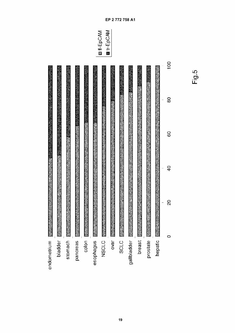
[0057] Figure 4 shows EpEx expression in different solid malignancies. Colon, ovarian, endometrium, prostate, stom-ach and non-small cell lung cancer (NSCLC) showed the highest EpEx expression rates in more than 90% of cases.Gallbladder, pancreas, breast and small cell lung cancer (SCLC) showed EpEx expression in 50-90% of cases. Incontrast, bladder, esophagus and hepatic cancer showed low expression rates of EpEx (< 50%).[0058] Figure 5 shows the relative expression rates of EpEx+/EpICD-(tr-EpCAM) and EpEx+/EpICD+ (fl-EpCAM)cases in different tumour entities. Endometrium, bladder and stomach showed the highest proportion of EpEx+/EpICD-cases (40-60%). Pancreas, colon, esophagus, ovarian and non-small cell lung cancer (NSCLC) showed a proportion of

EP 2 772 758 A1
8
5
10
15
20
25
30
35
40
45
50
55
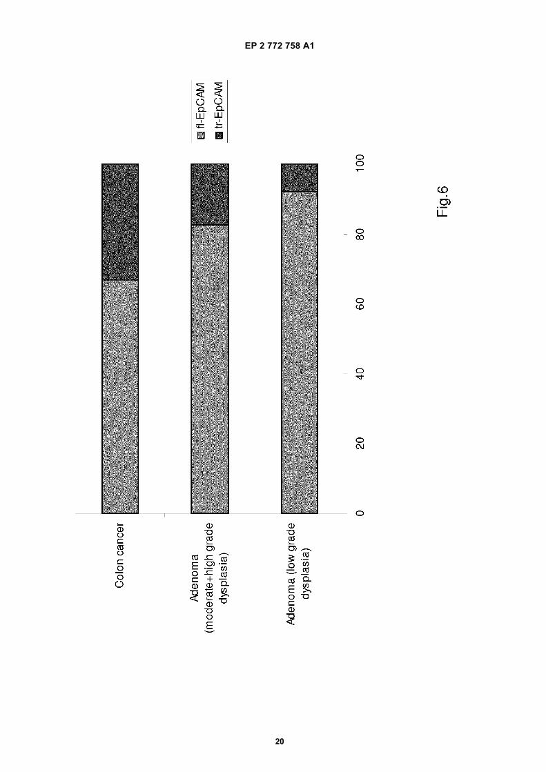
EpEx+/EpICD- in 20-40% of cases. Small cell lung cancer (SCLC), gallbladder, breast, prostate and hepatic cancershow an EpEx+/EpICD- expression rate varying from 0 to 20%.[0059] Figure 6 shows the proportion of tr-EpCAM in different stages of pre-cancerous lesions compared to coloncancer. The proportion of tr-EpCAM increases from < 10% of cases with low grade dysplastic adenomas to 18% inmoderate and high grade dysplasias to 34% in colon carcinoma, respectively. The grade of dysplasia is directly correlatedto prognosis of the disease. For this reason, the proportion of tr-EpCAM can be used as a prognostic marker in patientswith colon adenomas or carcinomas.
Discussion
[0060] EpCAM is one of the most widely investigated tumour associated antigens in human malignancies. However,the prognostic value of EpCAM expression is still a matter of debate. It was observed that EpCAM overexpression isassociated with poor clinical outcome in some epithelial cancers, while additional reports demonstrated EpCAM expres-sion to good prognosis. In the past, it was demonstrated that EpCAM overexpression is detectable in the majority ofpancreatic carcinomas and therefore may represents an attractive target for immune-based therapeutic interventions inthese tumours.[0061] In the present example the membranous expression pattern of EpCAM in pancreatic cancer was investigatedfor the first time using two different antibodies directed against EpEX and EpICD domains of the EpCAM antigen. Innon-neoplastic pancreatic tissue a high concordance between EpEX and EpICD was observed. In contrast, loss ofmembranous EpICD expression could be detected in 27% of cancer specimens which was significantly associated withdecreased overall survival and correlated with a shortened progression-free interval. It can be concluded that two differentEpCAM variants may occur during epithelial carcinogenesis, the inactivated EpEX+/EpICD+ phenotype representing thefull-length protein and an activated remnant transmembrane peptide (EpEX+/EpICD-) characterized by its cleaved cy-toplasmic domain. These two EpCAM variants may have not only prognostic but also therapeutic consequences.[0062] Regulated intramembrane proteolysis activates EpCAM as a mitogenic signal transducer through nuclear trans-location of its intracellular domain EpICD. The oncogenic potential of both proteins, full-length EpCAM but also cleavedEpICD on its own was demonstrated by using stable transfected colon carcinoma cells in vivo. In addition, EpICD wasfound as nuclear complexes in colon carcinoma but not in normal tissue. Moreover, a focal loss of membrane EpEXwas described at the invasive margin in colorectal cancer. A reciprocal loss of membrane EpEX but increased nuclearand cytoplasmic accumulation of EpICD was reported in a variety of epithelial carcinomas. Interestingly, compared tothe remaining epithelial tumours, in pancreatic cancer a reduced nuclear and cytoplasmic staining pattern for EpICDwas observed.[0063] However, despite the interesting observation that reduced or absent membranous expression of either EpEXor EpICD is a frequent event in epithelial carcinomas, none of the prior art studies directly compared the expressionpattern of the full-length protein EpCAM with possible truncated variants within the same tumour. Only recently it wasshown that high EpCAM-expression was associated with good prognosis in resectable pancreatic cancer by suppressingcell activity. Hence, it was interesting to assess the EpICD status in the EpCAM positive group within this cohort.[0064] As current strategies for EpCAM-based therapies are focusing on monoclonal antibodies against EpEX furtherstudies are warranted to define not only the level of EpCAM target expression within the tumour but also the impact oftruncated EpCAM on treatment outcome. One reason for the so far moderate success of EpCAM targeted therapiesmight be the fact that shedding of EpEX to a soluble ligand seems to be a precondition for subsequent intramembranecleavage and release of EpICD to the nucleus. The findings according to the present invention that none of the tumoursin the present study displayed an EpEX- /EpICD+ phenotype may support this assumption. Transactivation followed byectodomain shedding of the epidermal growth factor receptor (EGFR) family through regulated intramembrane proteolysisgenerating intracellular signals has received significant attention in understanding the complex signaling networks withincells. In addition, a dual function of the cell adhesion molecule E-Cadherin has been described recently by cleavage ofthe major protein and the release of an intracellular fragment. As a consequence, cell proliferation and migration arestimulated whereas cell-cell adhesion is inhibited.[0065] Beside of ectodomain shedding by different sheddases, receptor cross talk and remnant peptide signalingwithin the cell membrane through other transmembrane proteins seems to be crucial steps during carcinogenesis.Importantly, novel insights into the accessibility of EpCAM on normal epithelia vs tumour cells come from recent studiesidentifying a number of other proteins found in close association with EpCAM within the cell membrane. In pancreaticcancer a complex formation of transmembrane proteins including EpCAM, claudin-7, CD44 and tetraspanin was reportedto promote tumour progression rather than the individual molecules on their own. In addition, claudin-7 recruits EpCAMinto tetraspanin-enriched membrane microdomains which were shown to be associated with poor prognosis and areduced disease-free survival in colon cancer. Consequently, efficient cleavage of EpCAM may also depend on inter-actions with other transmembrane proteins to successfully execute its surface to nucleus missile. Hence, processed forshedding but still membrane anchored remnant EpEX may be recognized by EpCAM specific antibodies but subsequent
EP 2 772 758 A1
9
5
10
15
20
25
30
35
40
45
50
55
antitumour effector mechanisms might be insufficient. It is tempting to speculate that patients with tumours with anEpEx+/EpICD+ phenotype would be better candidates for treatment with EpCAM-specific antibodies compared toEpEx+/EpICD- expressing tumours. Another reason for the presence of a truncated form of membranous EpCAM mightbe mutations of the EpCAM gene. In fact, deletions of the EpCAM gene have been recently described in colorectalcancer patients with Lynch syndrome. Moreover, mutations have been described in a non-malignant disease termedcongenital tufting enteropathy.[0066] Taken together, it is shown for the first time with the present example that loss of membranous EpICD expressionoccurs in a significant subset of pancreatic cancer patients suggesting the presence of two distinct EpCAM variants.Carcinomas representing the truncated transmembrane peptide may be associated with transactivation mediated byectodomain shedding resulting in a more aggressive phenotype. The absence of the cytoplasmic domain EpICD thereforeconstitutes a highly relevant biomarker specifically for pancreatic and colorectal cancer patients, especially for thosepatients which undergo anti-EpCAM targeted therapies, i.e. particularly for patients being treated with EpCAM-specificantibodies (i.e. Catumaxomab, edrecolomab or others).
Table 1: patient characteristics
Variable Pancreatic cancer(n) (%) P -value (log-rank)
Sex (n=89)
Male 54 (61) 0.48Female 35 (39)
Age (n=89)Mean 66Median 68Range 37-86
T stage (n=84)pT1 7 (8) <0.01
pT2 22 (27)pT3 48 (57)pT4 7 (8)
Lymph node status (n=84)Negative 35 (42) <0.01Positive 49 (58)
Vessel invasion (n=27)No 13 (48) 0.03Yes 14 (52)
Distant metastasis (n=52)No 47 (90) <0.01Yes 5 (10)
Surgical margin (n=78)Negative 55 (71) 0.08
Positive 23 (29)
Stage (UICC) (n=85)
IA 7 (8) <0.01IB 13 (15)IIA 14 (17)IIB 40 (47)III 6 (7)IV 5 (6)
Differentiation (n=89)Well 15 (17) <0.01
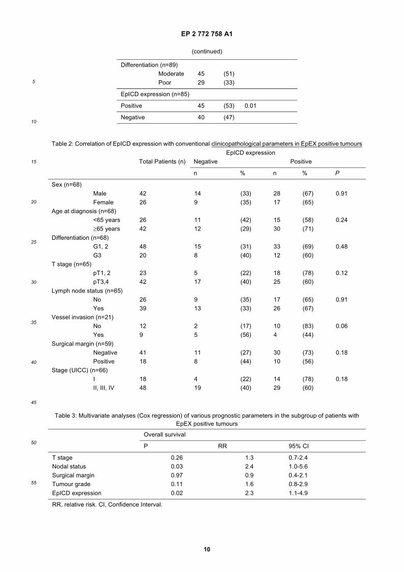
EP 2 772 758 A1
10
5
10
15
20
25
30
35
40
45
50
55
(continued)
Differentiation (n=89)Moderate 45 (51)Poor 29 (33)
EpICD expression (n=85)
Positive 45 (53) 0.01
Negative 40 (47)
Table 2: Correlation of EpICD expression with conventional clinicopathological parameters in EpEX positive tumours
EpICD expressionTotal Patients (n) Negative Positive
n % n % P
Sex (n=68)
Male 42 14 (33) 28 (67) 0.91Female 26 9 (35) 17 (65)
Age at diagnosis (n=68)<65 years 26 11 (42) 15 (58) 0.24≥65 years 42 12 (29) 30 (71)
Differentiation (n=68)G1, 2 48 15 (31) 33 (69) 0.48G3 20 8 (40) 12 (60)
T stage (n=65)pT1, 2 23 5 (22) 18 (78) 0.12pT3,4 42 17 (40) 25 (60)
Lymph node status (n=65)No 26 9 (35) 17 (65) 0.91Yes 39 13 (33) 26 (67)
Vessel invasion (n=21)No 12 2 (17) 10 (83) 0.06Yes 9 5 (56) 4 (44)
Surgical margin (n=59)Negative 41 11 (27) 30 (73) 0.18Positive 18 8 (44) 10 (56)
Stage (UICC) (n=66)I 18 4 (22) 14 (78) 0.18II, III, IV 48 19 (40) 29 (60)
Table 3: Multivariate analyses (Cox regression) of various prognostic parameters in the subgroup of patients with EpEX positive tumours
Overall survival
P RR 95% CI
T stage 0.26 1.3 0.7-2.4Nodal status 0.03 2.4 1.0-5.6Surgical margin 0.97 0.9 0.4-2.1Tumour grade 0.11 1.6 0.8-2.9
EpICD expression 0.02 2.3 1.1-4.9
RR, relative risk. CI, Confidence Interval.
EP 2 772 758 A1
11
5
10
15
20
25
30
35
40
45
50
55
Claims
1. Method for in vitro diagnosing a colon carcinoma (CC) tissue specimen or a pancreas cancer (PC) tissue specimenwherein the specimen is analysed for the presence or absence of the ectodomain of EpCAM (EpEX) as well as forthe presence or absence of the intracellular domain of EpCAM (EpICD).
2. Method according to claim 1 wherein specimen is a tissue section, especially a paraffin-embedded tissue sectionor a fresh frozen section; or a cytocentrifugate from pleura or ascites fluids.
3. Method according to claim 1 or claim 2 wherein the presence of EpEX and the absence of EpICD in the specimenis indicative of a shortened disease-free survival of a patient from which the specimen had been obtained.
4. Method according to any one of claims 1 to 3, wherein the presence or absence of EpEX or the presence or absenceof EpICD is analysed by use of an antibody binding specifically to EpEX and/or EpICD, respectively.
5. Method according to any one of claims 1 to 4, wherein the presence or absence of EpEX and the presence orabsence of EpICD is analysed by use of antibodies binding specifically to EpEX and EpICD, respectively, andwherein the antibodies are preferably monoclonal antibodies.
6. Method according to claim 4 or 5, wherein the antibodies are labelled.
7. Method according to any one of claims 4 to 6, wherein the antibody specifically binding to EpICD and the antibodyspecifically binding to EpEX are labelled with different labels, preferably with different fluorescence labels.
8. Method according to any one of claims 4 to 7, wherein the binding of the antibodies to EpEX and/or EpICD is detectedby using secondary antibodies binding to the antibodies binding to EpEX and/or EpICD, said secondary antibodiesbeing preferably labelled, especially with a fluorescence label, a luminescence label, a colourigenic label; a radio-active label, a biotin label, or combinations thereof.
9. Method according to any one of claims 1 to 8, wherein the method is an enzyme linked immunosorbent assay(ELISA), an immunocytochemistry assay, a detection of circulating tumour cells or tumour stem cells assay, or adetection of circulating cells or stem cells assay.
EP 2 772 758 A1
12
5
10
15
20
25
30
35
40
45
50
55
10. Method according to any one of claims 1 to 9, wherein the method comprises mass spectroscopy.
11. Kit for detecting EpEX and EpICD in an epithelial cancer tissue specimen in a method according to any one of claims1 to 10, comprising:
- an epithelial cancer tissue specimen,- an antibody specifically binding to EpICD; and- an antibody specifically binding to EpEX.
12. Kit according to claim 11, further comprising detection agents for detecting the antibodies and/or detection meansfor detection of a binding event between the antibodies to EpICD and/or EpEX.
13. Kit according to claim 11 or 12, wherein the antibodies are monoclonal antibodies.
14. Kit according to any one of claims 11 to 13, wherein it further comprises EpICD and EpEX standards, preferablyrecombinant EpCAM protein, a recombinant EpICD protein, a recombinant EpEx protein, or combinations thereof.
15. Kit according to any one of claims 11 to 14, wherein the kit additionally contains positive and negative control tissuespecimens.
EP 2 772 758 A1
13
EP 2 772 758 A1
14
EP 2 772 758 A1
15
EP 2 772 758 A1
16
EP 2 772 758 A1
17

EP 2 772 758 A1
18
EP 2 772 758 A1
19
EP 2 772 758 A1
20
EP 2 772 758 A1
21
5
10
15
20
25
30
35
40
45
50
55
EP 2 772 758 A1
22
5
10
15
20
25
30
35
40
45
50
55
EP 2 772 758 A1
23
5
10
15
20
25
30
35
40
45
50
55
EP 2 772 758 A1
24
REFERENCES CITED IN THE DESCRIPTION
This list of references cited by the applicant is for the reader’s convenience only. It does not form part of the Europeanpatent document. Even though great care has been taken in compiling the references, errors or omissions cannot beexcluded and the EPO disclaims all liability in this regard.
Patent documents cited in the description
• WO 2011032296 A1 [0013] • EP 2012062397 W [0013]
Non-patent literature cited in the description
• BAEUERLE et al. BJC, 2007, vol. 96, 417-423 [0009]• SEEBER/SCHUTTE. Therapiekonzepte Onkologie.
2007, 713ff, 747ff [0009]• LOUVET et al. JCO, 2002, vol. 20, 1512-1518 [0009]• CUNNINGHAM et al. JCO, 2009, vol. 27, 5513-5518
[0009]• CONROY et al. New England Journal of Medicine,
2011, vol. 364, 1817-1825 [0009]• MAETZEL. Nat. Cell Biol., 2009, vol. 11, 162-171
[0009]• DAS GUPTA et al. Bioorg. Med. Chem., 2009, vol.
17, 444-459 [0009]• NIEDZWIECKI et al. J. Clin. Oncol., 2011, vol. 29,
3146-3152 [0010]
• SLANCHEV et al. PLoS Genetics, 2009, vol. 5,1000563 [0013]
• ABE et al. J. Immunol. Meth., 2002, vol. 270, 227-233[0013]
• PETSCH et al. mAbs, 2011, vol. 3, 31-37 [0013]• SCHMETZER et al. Immunol. Lett., 2012, vol. 143,
184-192 [0013]• MAETZEL et al. Nat. Cell Biol., 2009, vol. 11,
162-171 [0013]• ABE et al. antibodies M2-5 and M4-10, 2002 [0016]• SCHMIDT et al. Ann. Oncol., 2010, vol. 21, 275-282
[0044]• BEECHER. Instruments, Silver. Springs [0045]• BAEUERLE et al. Br. J. Cancer, 2007, vol. 96,
417-423 [0051]