Transcranial magnetic stimulation studies in Alzheimer's disease
Journal of Physiology (1995), 488.3, pp.795-801
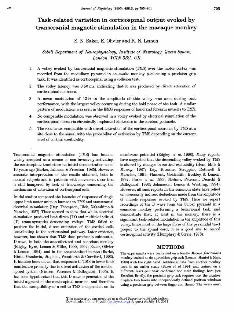
Task-related variation in corticospinal output evoked bytranscranial magnetic stimulation in the macaque monkey
S. N. Baker, E. Olivier and R. N. Lemon
Sobell Department of Neurophysiology, Institute of Neurology, Queen Square,London WCJN 3BG, UK
1. A volley evoked by transcranial magnetic stimulation (TMS) over the motor cortex wasrecorded from the medullary pyramid in an awake monkey performing a precision griptask. It was identified as corticospinal using a collision test.
2. The volley latency was 0 50 ms, indicating that it was produced by direct activation ofcorticospinal neurones.
3. A mean modulation of 13% in the amplitude of this volley was seen during taskperformance, with the largest volley occurring during the hold phase of the task. A similarpattern of modulation was seen in the EMG responses of hand and forearm muscles to TMS.
4. No comparable modulation was observed in a volley evoked by electrical stimulation of thecorticospinal fibres via chronically implanted electrodes in the cerebral peduncle.
5. The results are compatible with direct activation of the corticospinal neurones by TMS at asite close to the soma, with the probability of activation by TMS depending on the currentlevel of cortical excitability.
Transcranial magnetic stimulation (TMS) has becomewidely accepted as a means of non-invasively activatingthe corticospinal tract since its initial demonstration some10 years ago (Barker, Jalinous & Freeston, 1985). However,accurate interpretation of the results obtained, both innormal subjects and in patients with movement disorders,is still hampered by lack of knowledge concerning themechanism of activation of corticospinal cells.
Initial studies compared the latencies of responses of singleupper limb motor units in humans to TMS and transcranialelectrical stimulation (Day, Thompson, Dick, Nakashima &Marsden, 1987). These seemed to show that whilst electricalstimulation produced both direct ('D') and multiple indirect('I', trans-synaptic) descending volleys, TMS failed toproduce the initial, direct excitation of the cortical cellscontributing to the corticospinal pathway. Later evidence,however, has shown that TMS does produce a substantialD wave, in both the anaesthetized and conscious monkey(Edgley, Eyre, Lemon & Miller, 1990, 1992; Baker, Olivier& Lemon, 1994), and in the anaesthetized human (Burke,Hicks, Gandevia, Stephen, Woodforth & Crawford, 1993).It has also been shown that responses to TMS in lower limbmuscles are probably due to direct activation of the cortico-spinal system (Nielsen, Petersen & Ballegaard, 1995). Ithas been hypothesized that this D wave is generated at theinitial segment of the corticospinal neurone, and thereforethat the susceptibility of a cell to TMS is dependent on its
membrane potential (Edgley et al. 1990). Many reportshave suggested that the descending volley evoked by TMSis altered by changes in cortical excitability (Hess, Mills &Murray, 1987; Day, Riescher, Struppler, Rothwell &Marsden, 1991; Flament, Goldsmith, Buckley & Lemon,1992; Burke et al. 1993; Nielsen, Petersen, Deuschl &Ballegaard, 1993; Johansson, Lemon & Westling, 1994).However, all such reports in the conscious state have reliedon necessarily indirect deductions made from the amplitudeof muscle responses evoked by TMS. Here we reportrecordings of the D wave from the bulbar pyramid in aconscious monkey performing a behavioural task, anddemonstrate that, at least in the monkey, there is asignificant task-related modulation in the amplitude of thisvolley. Since most of the large fibres of the pyramidal tractproject to the spinal cord, it is a good site to recordcorticospinal activity (Humphrey & Corrie, 1978).
METHODSThe experiments were performed on a female Macaca fascicularismonkey trained to do a precision grip task (Lemon, Mantel & Muir,1986) with the right hand. Additional data from another monkeyused in an earlier study (Baker et al. 1994) and trained on adifferent, lever-pull task confirmed the main findings here (seeResults). Briefly, the precision grip task requires that the monkeydisplace two levers into independently defined position windowsusing a precision grip between finger and thumb. The levers must
This manuscript was accepted as a Short Paper for rapid publication.
4771 795
) by guest on July 14, 2011jp.physoc.orgDownloaded from J Physiol (
S. N Baker, E. Olivier and R. N Lemon
then be held in position for at least 0-8 s (the 'hold period'), beforebeing released to earn the animal a food reward. After trainingwas complete, the animal was implanted under full surgicalanaesthesia (3% isoflurane in 50:50 N20 :02) and asepticconditions with a stainless-steel headpiece for head fixation. In thesame surgery, two epoxylite insulated 150 ,um shank diametertungsten electrodes (impedance at 1 kHz, 10-20 kQl) werechronically implanted in the left pyramid under stereotaxic controlat Horsley-Clarke co-ordinates APO and P4 mm and two in theleft cerebral peduncles at A7 and AIO mm. These locations wereconfirmed histologically: the tip of the rostral peduncle electrodewas found in the dorsal third of the peduncle at the level of therostral pole of the lateral geniculate nucleus, and the caudalelectrode was located in corticofugal fibres as they entered the ponsat a level just rostral to the posterior commissure. The rostral andcaudal pyramidal electrodes were located in the pyramidal tract atlevels just caudal to the trapezoid body, and at the caudal end ofinferior olive, respectively. A full program of postoperativeanalgesia and antibiotics was administered. All experiments wereperformed under licence from the United Kingdom Home Office.
After recovery from surgery, multi-unit activity was recordedfrom the pyramid electrodes using a specially constructedamplifier which could be 'muted' to reduce the stimulus artifact(see Baker et al. 1994). The posterior electrode was connected tothe non-inverting input of the amplifier. The head was restrainedby bars fixed to the experimental cage. The animal performed thetask whilst magnetic stimuli were delivered using a Magstim 200stimulator (Magstim Ltd, Dyfed, UK) and 7 cm outer diameterfigure-of-eight coil (2-2 T maximum magnetic field). The coil waspositioned with the handle orientated mediolaterally, and theinduced current flowing away from the mid-line. The coil positionwas chosen to optimize the amplitude of the short-latency EMGresponse evoked in a subset of up to four hand and forearmmuscles. Stimuli were given at a constant rate of 0 3-0'5 Hzwhilst the animal performed the task in a self-paced fashion.
Once the optimal coil position was found, the coil was clamped tothe experimental cage, fixing it relative to the head. The positionof the lead connecting the pyramid electrodes to the amplifier was
1500 uVTMS alone | \
0-4 ms
0-8 ms
then adjusted, particularly where it ran close to the coil, until thesize of the stimulus artifact became acceptable. This was the singlemost important factor in obtaining an artifact-free recording. Theamplified volley was digitized on-line at 80 kHz by a personalcomputer with a 1401 interface (Cambridge Electronic Design,Cambridge, UK). EMG, task position signals and stimulus triggerpulses were recorded on an FM tape recorder (bandpass 200 Hz to10 kHz for EMG, DC-100 Hz for position) and digitized off-line.
The volley evoked by TMS could be identified as corticospinal inorigin by collision with an appropriately timed volley excited bystimulation of the cerebral peduncle through the chronicallyimplanted electrodes (Baker et al. 1994).
Trial-by-trial variation in task performance made it likely thatany modulation in the size of the volley evoked by TMS would beobscured by intertrial averaging. The position signals for fingerand thumb levers were therefore examined on a single trial basisusing an interactive computer program. This allowed theexperimenter to select a trial, and place cursors at the start andend of the movement. A normalization procedure was then used inwhich the cursor marking the movement start was designated'relative time' 0. The interval between this and the cursor markingthe movement end defined a time unit in the relative time scale forthat trial. Analysis extended from relative time -1 to relative time+2. A similar normalization approach has been used in the studyof locomotion (Drew & Doucet, 1991) to correct for differences inthe duration of individual step cycles.
The amplitude of the volley evoked by each stimulus was thenmeasured as the peak-to-peak height of the initialpositive-negative wave (see Fig. 2), and listed with the relativetime of stimulus delivery during the task. These were then sortedin ascending order in relative time, and a 30 point moving windowused to compile a mean and standard error for the volleyamplitude. This smoothed volley height was then plotted versusthe mean relative time of each set of 30 points.
EMG responses to TMS were treated similarly. An average ofrectified EMG was compiled, triggered by all available stimuli.This was used to define the response onset latency and duration.
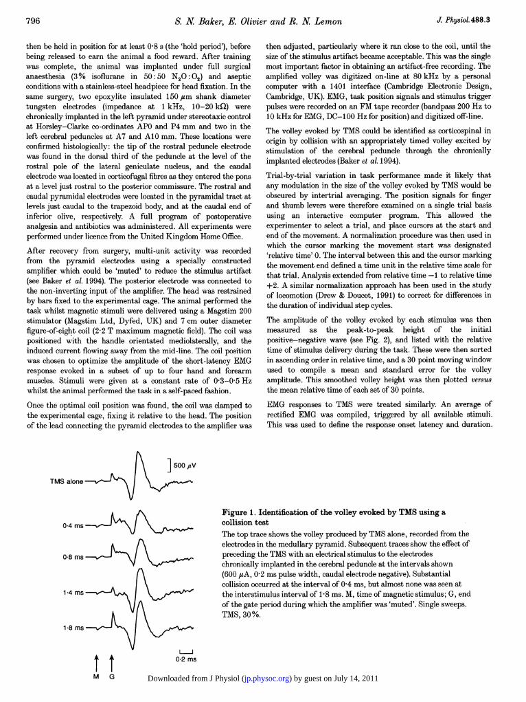
Figure 1. Identification of the volley evoked by TMS using acollision testThe top trace shows the volley produced by TMS alone, recorded from theelectrodes in the medullary pyramid. Subsequent traces show the effect ofpreceding the TMS with an electrical stimulus to the electrodeschronically implanted in the cerebral peduncle at the intervals shown(600 ,4A, 0-2 ms pulse width, caudal electrode negative). Substantialcollision occurred at the interval of 0 4 ms, but almost none was seen atthe interstimulus interval of 18 ms. M, time of magnetic stimulus; G, endof the gate period during which the amplifier was 'muted'. Single sweeps.TMS, 30%.
14 msi
18 ms
tIt 02 ms
M G
J Physiol.488.3796
) by guest on July 14, 2011jp.physoc.orgDownloaded from J Physiol (
Task dependence of (
As above, stimuli were sorted according to relative time ofoccurrence, and a moving average compiled of EMG sweeps withrespect to sets of thirty stimuli. The average height of thepreviously defined response region above the background level ofEMG (assessed over the 25 ms period before the stimulus) wasmeasured from these averages, and again plotted versus meanrelative time of stimulus occurrence. The response size was notnormalized by dividing by the background, as other studies havedone (e.g. Flament et al. 1992), due to the presence of periods ofnear-zero background EMG, which would have produced anartifactually high response amplitude.
RESULTSThe onset latency of the volley evoked by TMS was0 50 ms. During the implant surgery, the latency of theantidromic field potential recorded from the surface of themotor cortex following stimulation through the anteriorpyramidal electrode was 0 55 ms. These values areconsistent with our previous observations (Baker et al.1994). The TMS volley latency is too short to have beenproduced by trans-synaptic activation; it is a 'D wave'response. Later 'I waves' were not seen in single sweeps ofthe recording (see Discussion).
Figure 1 shows a collision test between the volley producedby TMS and that produced by electrical stimulation of thechronically implanted peduncle electrodes. The amplitudeof the TMS volley was reduced by more than half when apeduncle stimulus preceded TMS by 0 4 ms. The failure of
A1750
1700
I 1650
m 1600
1550
1500
Finger
Thumb
B
Ivw] 0 5 mm
, ] 1 mm
-1 0 1 2
t
corticospinal volley 797
the collision test to abolish the volley completely can beexplained if the peduncle stimulus (600 ,sA, duration0-2 ms) did not excite all of the corticospinal axonsrecruited by the TMS. It seems likely that the activitywhich was not collided was also corticospinal, since it hadthe same latency and time course. When the interstimulusinterval was increased to 1 8 ms there was little evidence ofcollision, as previously documented (Baker et al. 1994).
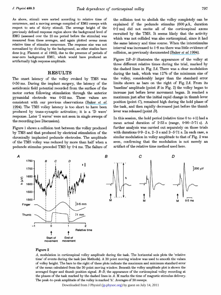
Figure 2B-D illustrates the appearance of the volley atthree different relative times during the trial, marked bythe dashed lines in Fig. 2A. There was a clear modulationduring the task, which was 12% of the minimum size ofthe volley, considerably larger than the standard errorlimits shown as bars on the right of Fig. 2A. From its'baseline' amplitude (point B in Fig. 2) the volley began toincrease just before lever movement began. It reached amaximum just after the initial rapid change in thumb leverposition (point C), remained high during the hold phase ofthe task, and then rapidly decreased just before the thumblever was released (point D).
In this session, the hold period (relative time 0 to +1) had amean actual duration of 2f53 s (range, 0f90-5f71 s). Afurther analysis was carried out separately on those trialswith durations 0f9-2 s, 2-3 s and 3-5-71 s. In each case, asimilar modulation in volley amplitude to that of Fig. 2 wasseen, confirming that the modulation is not merely anartifact of the relative time method used here.
C D
tM
tM
] 200 1sV
0-5 ms
Relative time
Start of End ofmovement movement
Figure 2A, modulation in corticospinal volley amplitude during the task. The horizontal axis plots the 'relativetime' of events during the task (see Methods). A 30 point moving window was used to smooth the valuesof volley height. The bars to the right of these plots indicate the maximum and minimum standard errorof the mean calculated from the 30 point moving window. Beneath the volley amplitude plot is shown theaveraged finger and thumb position signal. B-D, the appearance of the corticospinal volley recording atthe phases of the task marked by the dashed lines in A. M marks the time of magnetic stimulus delivery.The peak-to-peak amplitude of the volley is marked 'h'. Averages of 30 sweeps.
J Phy8iol.488.3
) by guest on July 14, 2011jp.physoc.orgDownloaded from J Physiol (
S. N Baker, E. Olivier and R. N. Lemon
A B C D1Dl 30 B B C D
P-B
(,UV) 2]5
-10
B
FDS
B 50(1UV) 25-
20
(juV) 100
B200 10 sB 50-
(,UV) 1100D50
-1 0 1 2 M M M20mRelative time
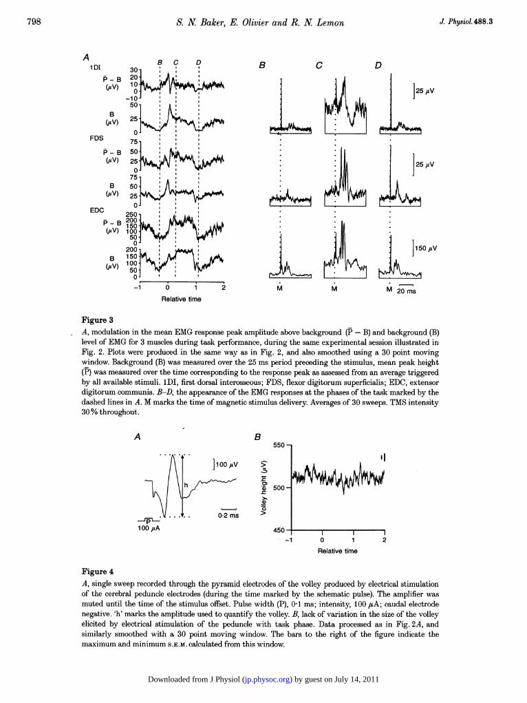
Figure 3A, modulation in the mean EMG response peak amplitude above background (P- B) and background (B)level of EMG for 3 muscles during task performance, during the same experimental session illustrated inFig. 2. Plots were produced in the same way as in Fig. 2, and also smoothed using a 30 point movingwindow. Background (B) was measured over the 25 ms period preceding the stimulus, mean peak height(P) was measured over the time corresponding to the response peak as assessed from an average triggeredby all available stimuli. 1DI, first dorsal interosseous; FDS, flexor digitorum superficialis; EDC, extensordigitorum communis. B-D, the appearance of the EMG responses at the phases of the task marked by thedashed lines in A. M marks the time of magnetic stimulus delivery. Averages of 30 sweeps. TMS intensity30% throughout.
A B550
h *~~~~~~500-
0
. . 002 ms
100 ,uA 450--1 0 1 2
Relative time
Figure 4A, single sweep recorded through the pyramid electrodes of the volley produced by electrical stimulationof the cerebral peduncle electrodes (during the time marked by the schematic pulse). The amplifier wasmuted until the time of the stimulus offset. Pulse width (P), 0.1 ms; intensity, 100 ,4A; caudal electrodenegative. 'h' marks the amplitude used to quantify the volley. B, lack of variation in the size of the volleyelicited by electrical stimulation of the peduncle with task phase. Data processed as in Fig. 2A, andsimilarly smoothed with a 30 point moving window. The bars to the right of the figure indicate themaximum and minimum S.E.M. calculated from this window.
798 J Phy8iol.488.3
) by guest on July 14, 2011jp.physoc.orgDownloaded from J Physiol (

UTask dependence of corticospinal volley
A clear modulation of the volley following TMS was seenduring the task in each of five recording sessions, varyingbetween 8 2 and 21 % (mean, 13 %). In all sessions the volleywas largest during the hold period (relative time 0 to +1).This was the case even though the TMS intensity wasaltered (range, 17-30% of maximum stimulator output),producing a volley whose mean amplitude varied from 370to 1620 ,uV; no consistent change in the pattern or level ofmodulation was seen with different mean volley amplitudes.
Figure 3 shows a plot of the EMG background level (B) andthe mean response above background (P- B) for threemuscles, recorded in the same session as the data in Fig. 2.Points B, C and D are placed at the same relative times asin Fig. 2, to allow comparison with the volley data. Frompoint B (the 'baseline' level) to point C, the measure P- Bchanged by 560, 970 and 480% for the first dorsalinterosseous (1DI), flexor digitorum superficialis (FDS) andextensor digitorum communis (EDC) muscles, respectively.A pattern of modulation broadly similar to that seen in thevolley recording was observed. Both volley data and theEMG show an increase just before movement onset, then adecrease, then an increase which is sustained until the endof movement. The main difference is in the relative size ofthe initial peak before movement onset compared with thatseen during the hold period. In the volley data and in theP- B plot for EDC, the hold period response is larger; inthe P- B plots for the other two muscles, the initial, pre-movement response is larger. This is probably a result ofthe non-linear nature of the spinal response to a cortico-spinal input, especially when the response is superimposedon a widely changing background level.
In addition, data from another animal which havepreviously been published (Baker et al. 1994) were analysedusing the 'relative time' procedure described under Methods.Significant task-dependent changes in the volley were alsoseen in this case, although they were less pronounced,possibly due to the highly phasic and less precise nature ofthe lever pull task performed by this animal.
In a control experiment, electrical stimuli were delivered tothe cerebral peduncle via the chronically implantedelectrodes at a rate of 2 Hz, whilst the monkey performedthe task. The volley recorded from the pyramid followingthis stimulation was then processed in exactly the sameway as the TMS volley data to investigate whether it wouldshow any task-dependent modulation. The results of thisexperiment are given in Fig. 4. This volley (shown inFig. 4A) had a similar time course and waveform to thatelicited by TMS, as expected if both stimuli excite a similarpopulation of fibres. Figure 4B presents the variation ofthis volley throughout the task. The scale of this figure hasbeen chosen so that height of the maximal standard error isthe same as in Fig. 2A. Unlike the volley evoked by TMS,there was no consistent change with task phase.
DISCUSSIONThe results reported here demonstrate that the size of theD wave volley excited by TMS changes during the course ofa voluntary movement. This is not likely to be due to achange in the stimulation parameters (e.g. a change inprecise coil position), even during the long experimentsrecorded here. Firstly, the coil was firmly clamped abovethe head, which was itself rigidly fixed. Secondly, thestimuli were delivered at a constant rate whilst the animalworked in a self-paced manner; stimuli were thuseffectively given at random with respect to task phase. Anychanges in stimulation parameters over the course of theexperimental session would be cancelled by the post hocselection procedure used here, as the sweeps comprisingeach average plotted in Fig. 2 were drawn equally from thebeginning, middle and end of the recording session.
The modulation observed is most likely, therefore, to resultfrom changes in the level of cortical activity accompanyingtask performance. Such a modulation was not seen in thevolley produced by direct electrical stimulation of cortico-spinal fibres at the cerebral peduncle. These findingssupport the hypothesis of Edgley et al. (1990) that theD wave following TMS is produced by excitation ofcorticospinal neurones at a point close to their cell body,although we have used a different stimulating coil andorientation from that study. The amplitude of the cortico-spinal volley presented here should be related to thenumber of corticospinal neurones excited by the magneticstimulus, and hence a more reliable indicator of changesdependent on cortical excitability than muscle responses.Such a modulation in cortical excitability would also beexpected to alter the synchrony of neuronal response toTMS, and hence reduce cancellation of individual actionpotentials in the recording. The contribution of this effectto the amplitude changes in the corticospinal volley isunknown.
The modulation of only 12% shown in Fig. 2A on a muchexpanded scale might seem on initial examination to bedisappointingly small, especially given the much largertask-related modulation in EMG responses shown in Fig. 3(up to tenfold). This is likely to reflect the heterogeneousnature of the cells contributing to the recorded volley,many of which will not modulate their activity withperformance of this task. By contrast, evidence from singlecell recording in the motor cortex during performance ofthe precision grip task suggests that almost all the fastcorticospinal cells projecting to hand and forearm moto-neurones modulate their activity in a task-dependent way(Lemon et al. 1986). It is therefore likely that the task-related modulation in recruitment of this subpopulation byTMS will be greater than that in the global activitymeasured from the entire tract. In addition, the modulationin EMG response will reflect changes in spinal excitability
799J Physiol.488.3
) by guest on July 14, 2011jp.physoc.orgDownloaded from J Physiol (

800 S. N Baker, E. Olivier and R. N Lemon J Physiol.488.3
compounded with the changes in the size of the descendingvolley.
At present we have little information as to how the currentsinduced by TMS will interact with the natural firinghistory of a corticospinal cell. The present data seem toshow that corticospinal cells are more susceptible to TMSduring the hold period of the task than during the initialmovement, in which the monkey positions its digits on thetwo levers. Corticospinal cell activity is largest during thisinitial phase (Bennett & Lemon, 1994). This implies thatthe period of maximum corticospinal susceptibility to TMSmay not coincide with the time of maximum corticospinalcell activity; possible mechanisms include after-hyperpolarization and refractory period when the cells fireat high rates.
I waves were not visible in single sweeps of the recordingspresented here. In averages over all stimulus presentationsfor one session (i.e. around 2000 sweeps), small laterdeflections could be seen. However, they were of the orderof only 3% of the size of the initial, D wave, renderingdetailed analysis of their variability impossible. This isinitially a surprising finding, since indirect activationmight be expected to be more important in the consciousstate than under anaesthesia, where I waves havepreviously been seen (Amassian, Stewart, Quirk &Rosenthal, 1987; Edgley et at. 1990; Burke et al. 1993).There are two possible explanations. Firstly, the lack ofI waves may reflect the recording arrangement. Thepyramid recording electrodes were spaced only 4 mmapart, so that the duration of the recorded triphasic volleywas very brief at approximately 0 5 ms. The jitter in theresponse latency of a single corticospinal axon respondingindirectly can be as much as 1 ms (S. A. Edgley, J. A. Eyre,S. Miller & R. N. Lemon, unpublished observations), whichwould cause substantial cancellation of positive andnegative phases of I waves. In reports where I waves havebeen recorded, electrodes were placed at the spinal levelwith presumably larger spacings, for the duration of theD wave is often as much as 1 ms; less cancellation wouldthen occur. The greater sensitivity of I waves to therecording arrangement has been noted by Inghilleri,Berardelli, Cruccu, Priori & Manfredi (1989) when usingtranscranial electrical stimulation in humans.
Alternatively, it may be that indirect activation is relativelyunimportant with a mediolateral coil orientation (Amassian,Quirk & Stewart, 1990). With this orientation, Werhahn etal. (1994) showed that single 1DI motor units in manrespond to TMS with a single sub-peak. Preliminaryobservations in the monkey have also shown that singlemotor units responded to TMS delivered as described abovewith a single subpeak, at a latency consistent with directactivation (S. N. Baker, E. Olivier & R. N. Lemon,unpublished observations). Thus direct corticospinalexcitation may be more important than indirect under thestimulation conditions used in this experiment.
This report provides direct evidence that changes in corticalexcitability associated with the performance of theprecision grip task are reflected in significant variation inthe amplitude of the corticospinal volley evoked by TMS. Itfurther shows that the time course of the EMG responsesfollows these changes, suggesting that modulation in theamplitude of short-latency EMG responses to TMS is due,at least in part, to changes in the size of the descendingcorticospinal volley.
AMASSIAN, V. E., QUIRK, G. J. & STEWART, M. (1990). A comparisonof corticospinal activation by magnetic coil and electricalstimulation of monkey motor cortex. Electroencephalography andClinical Neurophysiology 77, 390-401.
AMASSIAN, V. E., STEWART, M., QUIRK, G. J. & ROSENTHAL, J. L.(1987). Physiological basis of motor effects of a transient stimulus tocerebral cortex. Neurosurgery 20, 74-93.
BAKER, S. N., OLIVIER, E. & LEMON, R. N. (1994). Recording anidentified pyramidal volley evoked by transcranial magneticstimulation in a conscious macaque monkey. Experimental BrainResearch 99, 529-532.
BARKER, A. T., JALINOUS, R. & FREESTON, I. L. (1985). Non-invasivemagnetic stimulation of the human motor cortex. Lancet i,1106-1107.
BENNETT, K. M. B. & LEMON, R. N. (1994). The influence of singlemonkey cortico-motoneuronal cells at different levels of activity intarget muscles. Journal of Physiology 477, 291-307.
BURKE, D., HICKS, R., GANDEVIA, S. C., STEPHEN, J., WOODFORTH, I.& CRAWFORD, M. (1993). Direct comparison of corticospinal volleysin human subjects to transcranial magnetic and electricalstimulation. Journal of Physiology 470, 383-393.
DAY, B. L., RIESCHER, H., STRUPPLER, A., ROTHWELL, J. C. &MARSDEN, C. D. (1991). Changes in the response to magnetic andelectrical stimulation of the motor cortex following muscle stretch inman. Journal of Physiology 433, 41-57.
DAY, B. L., THOMPSON, P. D., DICK, J. P., NAKASHIMA, K. &MARSDEN, C. D. (1987). Different sites of action of electrical andmagnetic stimulation of the human brain. Neuroscience Letters 75,101-106.
DREW, T. & DOUCET, S. (1991). Application of circular statistics to thestudy of neuronal discharge during locomotion. Journal ofNeuroscience Methods 38, 171-181.
EDGLEY, S. A., EYRE, J. A., LEMON, R. N. & MILLER, S. (1990).Excitation of the corticospinal tract by electromagnetic andelectrical stimulation of the scalp in the macaque monkey. Journalof Physiology 425, 301-320.
EDGLEY, S. A., EYRE, J. A., LEMON, R. N. & MILLER, S. (1992). Directand indirect activation of corticospinal neurones by electrical andmagnetic stimulation in the anaesthetized macaque monkey. Journalof Physiology 446, 224P.
FLAMENT, D., GOLDSMITH, P., BUCKLEY, J. C. & LEMON, R. N. (1992).Task dependence of responses in first dorsal interosseous muscle tomagnetic brain stimulation in man. Journal of Physiology 464,361-378.
HESS, C. W., MILLS, K. R. & MURRAY, N. M. F. (1987). Responses insmall hand muscles from magnetic stimulation of the human brain.Journal of Physiology 388, 397-419.
) by guest on July 14, 2011jp.physoc.orgDownloaded from J Physiol (

'I'-Task dependence of corticospinal volley
HUMPHREY, D. R. & CORRIE, W. S. (1978). Properties of pyramidaltract neuron system within a functionally defined subregion ofprimate motor cortex. Journal of Neurophysiology 41, 216-243.
INGHILLERI, M., BERARDELLI, A., CRuccu, G., PRIORI, A. &MANFREDI, M. (1989). Corticospinal potentials after transcranialstimulation in humans. Journal of Neurology, Neurosurgery andPsychiatry 52, 970-974.
JOHANSSON, R. S., LEMON, R. N. & WESTLING, G. (1994). Time-varying enhancement of human cortical excitability mediated bycutaneous inputs during precision grip. Journal of Physiology 481,761-775.
LEMON, R. N., MANTEL, G. W. H. & MUIR, R. B. (1986). Corticospinalfacilitation of hand muscles during voluntary movement in theconscious monkey. Journal of Physiology 381, 497-527.
NIELSEN, J., PETERSEN, N. & BALLEGAARD, M. (1995). Latency ofeffects evoked by electrical and magnetic brain stimulation in lowerlimb motoneurones in man. Journal of Physiology 483, 791-802.
NIELSEN, J., PETERSEN, N., DEUSCHL, G. & BALLEGAARD, M. (1993).Task-related changes in the effect of magnetic brain stimulation onspinal neurones in man. Journal of Physiology 471, 223-243.
WERHAHN, K. J., FONG, J. K. Y., MEYER, B. U., PRIORI, A.,ROTHWELL, J. C., DAY, B. L. & THOMPSON, P. D. (1994). The effect ofmagnetic coil orientation on the latency of surface EMG and singlemotor unit responses in the first dorsal interosseous muscle.Electroencephalography and Clinical Neurophysiology 93, 138-146.
AcknowledgementsThe authors would like to thank Miss Nora Philbin for technicalassistance, and Miss Natalia Ognjevich for help with histology.This work was supported by the Wellcome Trust, Action Research,the MRC and the Magstim Company Limited.
Received 4 July 1995; accepted 7 September 1995.
J Physiol. 488.3 801
) by guest on July 14, 2011jp.physoc.orgDownloaded from J Physiol (
Copyright © 2022 FDOKUMEN