Shaping pseudoneglect with transcranial cerebellar direct current stimulation and music listening
Upload
khangminh22Category
view
0download
0
REVIEW
Transcranial magnetic stimulation and amyotrophiclateral sclerosis: pathophysiological insightsSteve Vucic,1,2 Ulf Ziemann,3 Andrew Eisen,4 Mark Hallett,5 Matthew C Kiernan2,6
1Sydney Medical SchoolWestmead, University ofSydney, Sydney, New SouthWales, Australia2Neuroscience ResearchAustralia, Sydney, New SouthWales, Australia3Department of Neurology andStroke, Hertie Institute forClinical Brain Research,Eberhard-Karls University,Tübingen, Germany4Division of Neurology, TheUniversity of British Columbia,Vancouver, British Columbia,Canada5Human Motor ControlSection, NINDS, NIH,Bethesda, Maryland, USA6Prince of Wales ClinicalSchool, University of NewSouth Wales, Sydney, NewSouth Wales, Australia
Correspondence toAssociate Professor SteveVucic, Sydney Medical SchoolWestmead, University ofSydney, Darcy and HawkesburyRd, Wentworthville, Sydney,NSW 2045, Australia; [email protected]
Received 20 September 2012Revised 20 November 2012Accepted 25 November 2012Published Online First21 December 2012
To cite: Vucic S,Ziemann U, Eisen A, et al. JNeurol Neurosurg Psychiatry2013;84:1161–1170.
ABSTRACTAmyotrophic lateral sclerosis (ALS) is a rapidlyprogressive neurodegenerative disorder of the motorneurons in the motor cortex, brainstem and spinal cord.A combination of upper and lower motor neurondysfunction comprises the clinical ALS phenotype.Although the ALS phenotype was first observed byCharcot over 100 years ago, the site of ALS onset andthe pathophysiological mechanisms underlying thedevelopment of motor neuron degeneration remain to beelucidated. Transcranial magnetic stimulation (TMS)enables non-invasive assessment of the functionalintegrity of the motor cortex and its corticomotoneuronalprojections. To date, TMS studies have established motorcortical and corticospinal dysfunction in ALS, withcortical hyperexcitability being an early feature insporadic forms of ALS and preceding the clinical onset offamilial ALS. Taken together, a central origin of ALS issupported by TMS studies, with an anterogradetranssynaptic mechanism implicated in ALS pathogenesis.Of further relevance, TMS techniques reliably distinguishALS from mimic disorders, despite a compatibleperipheral disease burden, thereby suggesting a potentialdiagnostic utility of TMS in ALS. This review will focuson the mechanisms underlying the generation of TMSmeasures used in assessment of cortical excitability, thecontribution of TMS in enhancing the understanding ofALS pathophysiology and the potential diagnostic utilityof TMS techniques in ALS.
INTRODUCTIONThe term amyotrophic lateral sclerosis (ALS) wasfirst coined by Charcot, who postulated theprimacy of the upper motor neuron (UMN) in ALSpathogenesis.1 Assessment of cortical function inALS and identification of the characteristic clinicalphenotype involving combined upper and lowermotor neuron abnormalities remain the key forALS diagnosis.2–4 However, despite Charcot’sinitial observations, the site of disease onset andmechanisms underlying ALS pathophysiologyremain areas of intense study and debate.5 In thissetting, assessment of motor cortical and corticosp-inal function using non-invasive techniques, such astranscranial magnetic stimulation (TMS), hasenhanced our understanding of ALS pathophysi-ology and resulted in novel diagnostic approaches.Single-, paired- and multiple-pulse TMS techni-
ques have all been used (figure 1) with the follow-ing measures taken to reflect corticomotoneuronalfunction: motor threshold (MT), motor evokedpotential (MEP) amplitude, central motor conduc-tion time (CMCT), cortical silent period (CSP),
intracortical inhibition and facilitation. The presentreview will focus on the mechanisms underlyingthe generation of these TMS measures, while at thesame time assessing the contributions TMS hasmade in the understanding of ALS pathophysiology.With an eye towards the future, the review will alsoconsider the potential diagnostic utility of TMS inALS and incorporation of TMS as a disease bio-marker in the assessment of neuroprotective medi-cations in a clinical trial setting.
BACKGROUND TMS TERMINOLOGY ANDPATHOPHYSIOLOGYMT reflects the ease with which corticomotoneur-ons are excited and is proposed to be assessedby the International Federation of ClinicalNeurophysiology as the minimum stimulus intensityrequired to elicit a small (usually >50 μV) MEP inthe target muscle in 50% of trials.6 With the recentadaptation of threshold tracking techniques, MTcanalso be measured as the stimulus intensity requiredto elicit and maintain a target MEP response of0.2 mV.7–9 MT reflects the density of corticomoto-neuronal projections onto the spinal motor neuronwith the highest density of projections to intrinsichand muscles having the lowest MTs.10–12 MTs arelower in the dominant hand12 and correlate withthe ability to perform fine (fractionated) fingertasks,13 so that MT has the potential to map cortico-motoneuronal representation and function.As well as reflecting the density of corticomoto-
neuronal projections, MTs may also be a biomarkerof cortical neuronal membrane excitability.14–16
MTs are influenced by the glutamatergic neuro-transmitter system, through α-amino-3-hydroxy-5-methyl-4-isoxazoleproprionic acid (AMPA) receptors,whereby excessive glutamate activity reduces MTs.17
In contrast, pharmacological blockade of voltage-gated sodium channels raises MT.18
In ALS, abnormalities in MT have been inconsist-ent. While some TMS studies reported an increasedMTor even an inexcitable motor cortex,19–26 othershave documented either normal or reducedMT.27–32 These discrepancies likely relate to hetero-geneity of the ALS phenotype and the stage ofdisease at time of testing and rate of progression.Longitudinal studies have documented a reductionof MTs early in the disease course, increasing to thepoint of cortical inexcitability with disease progres-sion.29 The early reduction in MTappears most pro-nounced in ALS patients with profuse fasciculations,preserved muscle bulk and hyper-reflexia.33
Fasciculations may precede other features of ALS bymany months and taken in association with reduced
Open AccessScan to access more
free content
Vucic S, et al. J Neurol Neurosurg Psychiatry 2013;84:1161–1170. doi:10.1136/jnnp-2012-304019 1161
Neurodegeneration
MT suggest a cortical origin of fasciculations in ALS.34 ReducedMT may be modulated by increased glutamate excitation,reduced gamma-Aminobutyric acid (GABA) inhibition or a com-bination of both. Reduced MT early in ALS supports an antero-grade transsynaptic process, whereby cortical hyperexcitabilityunderlies the development of progressive neurodegeneration.
MEP amplitude reflects a summation of complex corticospinalvolleys consisting of D (direct)- and I (indirect)-waves.14 35 Atthreshold, TMS elicits I-waves at intervals of 1.5 ms, whichincrease in amplitude with increasing stimulus intensity.35 Theincrease in MEP amplitude with increasing stimulus intensitymay be used to generate a stimulus–response curve that followsa sigmoid function.36 As with MT, the MEP amplitude reflectsthe density of corticomotoneuronal projections onto motorneurons.37 When compared with MT, the MEPs probably assessthe function of neurons that are less excitable or further awayfrom the centre of the TMS induced electrical field.38 The MEP
amplitude should be expressed as a percentage of the maximumcompound muscle action potential (CMAP) evoked by electricalperipheral nerve stimulation.6 Doing so takes into account anylower motor neuron pathology and provides insight into thepercentage of the motor neurone pool activated in the MEP.Normative values for the MEP to CMAP ratio demonstrate alarge inter-subject variability thereby reducing the sensitivity andlimiting the value of this measure for detecting abnormalities ofthe corticomotoneurons.38 39
The MEP responses are modulated by a variety of neurotrans-mitter systems within the central nervous system.37 40
Specifically, GABAergic neurotransmission via GABAA receptorssuppresses while glutamatergic and noradrenergic neurotransmis-sion enhances the MEP amplitude.41 Of interest, these changes inMEP amplitude occur independently of changes in MT, suggest-ing that physiological mechanisms underlying the generation ofthe MEP amplitude and MTare varied.
Figure 1 Transcranial magnetic stimulation excites a network of neurons in the underlying motor cortex with motor evoked potentials recordedover the contralateral abductor pollicis brevis muscle. The motor cortex is preferentially stimulated when the current flows in a posterior–anteriordirection within the motor cortex.
1162 Vucic S, et al. J Neurol Neurosurg Psychiatry 2013;84:1161–1170. doi:10.1136/jnnp-2012-304019
Neurodegeneration

Abnormalities of MEPs have been extensively documented inALS.38 Increases in MEP amplitude have been reported in spor-adic and familial forms of ALS (figure 2A), most prominentlyearly in the disease course.30 31 42 MEP amplitude correlateswith surrogate biomarkers of axonal degeneration, such as thestrength duration time constant, thereby providing an associ-ation between cortical hyperexcitability and motor neurondegeneration.30 43 The increase in MEP amplitude in ALS is notseen in mimic disorders despite a comparable degree of lowermotor neuron dysfunction (figure 2B). This suggests that theMEP amplitude changes in ALS are excitotoxic in nature.44–47
CMCT represents the time from stimulation of the motorcortex to the arrival of corticospinal volley at the spinal motorneuron.6 Multiple factors contribute to the CMCT includingtime to activate the corticospinal cells, conduction time of thedescending volley down the corticospinal tract, synaptic trans-mission and activation of spinal motor neurons.48 CMCT maybe measured using either the F-wave or cervical (or lumbar)nerve root stimulation methods;49 50 both methods provideonly an estimation of the CMCT,48 51 and given that a varietyof technical, physiological and pathological factors influenceCMCT,48 there is a range of normative data.
In ALS, CMCT is typically modestly prolonged,21 29 52 prob-ably reflecting axonal degeneration of the fastest conducting cor-ticomotoneuronal fibres and increased desynchronisation ofcorticomotoneuronal volleys secondary to axonal loss.28 53 54
The D90A-SOD1 ALS mutation is a unique exception; in thisdisorder CMCT is typically very prolonged.55 The sensitivity ofdetecting a prolonged CMCT may be improved by recordingfrom both upper and lower limb muscles, or from cranialmuscles in ALS patients with bulbar onset disease.26 56
CSP refers to the interruption of voluntary electromyographyactivity in a target muscle induced by stimulation of the
contralateral motor cortex.57 The CSP duration is measuredfrom the onset of the MEP response to resumption of voluntaryelectromyography activity37 57 and increases with stimulusintensity.57–59
The CSP is mediated by both spinal mechanisms, in its earlypart, and cortical inhibitory neurons acting via GABAB receptorsin the latter part.57 58 60–63 Since the duration is determined bythe latter part, the CSP is a measure of cortical inhibition. Inaddition, the density of the corticomotoneuronal projectionsonto motor neurons also influences the CSP, with the CSP dur-ation being the longest for upper limb muscles.38
Abnormalities of the CSP duration are well established inALS.37 Absence or reduction in CSP duration has been reportedin both sporadic and familial ALS, with the reduction of CSPduration being the most prominent early in the diseasecourse.30–32 44 46 52 64–67 The reduction of CSP durationappears to be specific for ALS among neuromuscular disorders,being normal in X-linked bulbospinal muscular atrophy(Kennedy’s disease), acquired neuromyotonia and distal heredi-tary motor neuronopathy with pyramidal features.44–47
Although the mechanisms underlying CSP duration reduction inALS remain to be established, decreased motor drive andreduced GABAergic inhibition, either due to degeneration ofinhibitory interneurons or dysfunction of GABAB receptors,may underlie the reduction of CSP duration in ALS.
An absent or delayed ipsilateral CSP has also been reported asan early abnormality in ALS.67 68 The ipsilateral CSP dependson functioning of transcallosal glutamatergic fibres projectingonto inhibitory interneurons in the non-stimulated motorcortex,69 and degeneration of these transcallosal fibres or theirtargeted inhibitory interneurons may account for abnormalitiesof the ipsilateral CSP in ALS.
PAIRED-PULSE TMS TECHNIQUESThe previous section has covered conventional TMS parametersthat can be assessed through activation of the motor cortex bysingle impulses. Motor cortical excitability may also be assessedusing paired-pulse techniques, in which a conditioning stimulusmodulates the effect of a second test stimulus. Several differentpaired-pulse paradigms have been developed,37 38 but shortinterval intracortical inhibition (SICI), intracortical facilitation(ICF) and long interval intracortical inhibition have been mostfrequently used in ALS clinical research as methods to deter-mine cortical excitability.
To identify SICI and ICF, a subthreshold conditioning stimu-lus is typically delivered at predetermined time intervals prior toa suprathreshold test stimulus.8 70–72 In the early TMS para-digms,70 72 73 the conditioning and test stimuli were kept con-stant, and changes in the test MEP amplitude were evaluated.Typically, if the interstimulus interval (ISI) was between 1 and5 ms, the test response was inhibited (SICI). Increasing the ISIto between 7 and 30 ms resulted in the facilitation of the testresponse (ICF).38
By recording the descending corticospinal volleys through epi-dural electrodes at the level of the cervical spinal cord, it hasbeen deduced that both SICI and ICF originate at the level ofthe motor cortex.35 72 Specifically, SICI is associated with areduction in the number and amplitude of late I-waves, namelyI2 and I3, with I-wave suppression remaining up to an ISI of20 ms, which is the typical duration of the inhibitory postsynap-tic potential mediated through GABAA receptors.71 74 SICI andICF appear to be physiologically distinct processes as evident bylower thresholds for activation of SICI and SICI remains inde-pendent of the direction of current flow in the motor cortex
Figure 2 (A) The motor evoked potential (MEP) amplitude, expressedas a percentage of compound muscle action potential (CMAP)response, is significantly increased in sporadic amyotrophic lateralsclerosis (ALS) and familial ALS (FALS) when compared with healthycontrols. (B) The MEP amplitude is significantly increased in ALS whencompared with pathological and healthy controls, therebydistinguishing ALS from ALS mimic disorders.* p<0.05; ***p<0.001.RMT, resting motor threshold.
Vucic S, et al. J Neurol Neurosurg Psychiatry 2013;84:1161–1170. doi:10.1136/jnnp-2012-304019 1163
Neurodegeneration

induced by a subthreshold conditioning pulse in healthy sub-jects, while ICF appears to be preferentially generated bycurrent flowing in a posterior–anterior direction.73
A limitation of the ‘constant stimulus’ paired-pulse technique hasbeen the marked variability in MEP amplitudes with consecutivestimuli.71 75 To overcome this limitation, a threshold tracking tech-nique was developed whereby a constant target MEP response(0.2 mV) was tracked by a test stimulus.7 8 Using threshold tracking,two phases of SICI were identified,7 8 76 77 a smaller phase at ISI≤1 ms and a larger phase at ISI 3 ms (figure 3A). Although synapticneurotransmission through the GABAA receptor mediates thesecond phase of SICI,74 78–80 the precise mechanisms underlyingthe first phase of SICI remain uncertain. It was initially suggestedthat the first phase of SICI reflected local excitability properties,particularly relative refractoriness of cortical axons, with resultantresynchronisation of cortico-cortical and corticomotoneuronalvolleys.7 81 Subsequently, it has been argued that synaptic processesbest explain the development of the initial phase of SICI, possiblydriven by activation of cortical inhibitory circuits that were distinctto those that mediated the later SICI phase.76 77 82
A reduction or absence of SICI, together with an increase inICF, indicative of cortical hyperexcitability has been documentedin cohorts of sporadic and familial ALS patients (figure 3A).30–32 44 45 83–88 Of relevance, cortical hyperexcitability appears tobe an early feature in sporadic ALS, correlating with measures ofsubsequent peripheral neurodegeneration.30 In addition, corticalhyperexcitability appeared as an early feature in familial ALS dueto mutations linked to the superoxide dismutase-1 (figure 3A)and fused in sarcoma (FUS) genes,31 preceding the clinical devel-opment of familial ALS (figure 3B).31
Neuropathological studies in ALS have identified degener-ation of inhibitory cortical interneurons89 and this could
account for the reduction in SICI. Separately, glutamate-mediated excitotoxicity may also contribute to SICI reduction,as was suggested by partial correction of SICI abnormalities inALS patients treated with the glutamate antagonist riluzole.87 Arecent study documenting SICI reduction at low (40% of restingMT (RMT)), medium (70% of RMT) and high (90% of RMT)conditioning stimulus intensities in ALS patients providedfurther support for the notion that abnormalities in SICIappeared to be mediated by a combination of glutamate excito-toxicity and degeneration of inhibitory cortical circuits.90 Assuch, preserving the integrity of intracortical inhibitory circuits,and counteracting excitatory cortical circuits, may serve aspotential therapeutic options in ALS.
UTILITY OF PERISTIMULUS TIME HISTOGRAMSA peristimulus time histogram technique can assess the functionof a select subset of corticomotoneurons by recording the per-turbation of voluntarily recruited motor units induced by athreshold cortical stimulation.53 In healthy controls, there is awell synchronised primary peak with a latency of approximately20–30 ms recording from hand or forearm muscles.28 53
Analysis of this primary peak in disease states such as ALS pro-vides information on corticomotoneuronal conduction time, theextent of desynchronisation of corticomotoneuronal descendingvolleys, the degree of corticomotoneuronal synaptic input ontothe anterior horn cell and the timing of excitatory and inhibi-tory inputs to the motor neuron.33
In ALS, the primary peak becomes desynchronised, prolongedin duration and delayed.28 91 92 In addition, the amplitude ofthe primary peak may be increased with additional subcompo-nents both suggestive of corticomotoneuronal hyperexcitabil-ity.53 93 These primary peak abnormalities appear early in ALS,
Figure 3 (A) Short interval intracortical inhibition (SICI), defined as the stimulus intensity required to maintain a target motor evoked potential of0.2 mV, as assessed by the threshold tracking transcranial magnetic stimulation technique. Intracortical inhibition is illustrated by an increase in theconditioned test stimulus intensity required to track the target response, while intracortical facilitation is indicated by a reduction in test stimulusintensity. In healthy controls, SICI develops between interstimulus intervals (ISI) of 1 and 7 ms, with two peaks evident at 1 and 3 ms as indicatedby the arrows. Intracortical facilitation developed between ISIs of 10 and 30 ms. SICI is significantly reduced in both sporadic amyotrophic lateralsclerosis (SALS) and familial amyotrophic lateral sclerosis (FALS). (B) Averaged SICI, between ISI 1 and 7 ms, was reduced in two presymptomaticsuperoxide dismutase-1 (SOD-1) mutation carriers 6 months prior to the development of ALS. (C) Normalised SICI, expressed as a fraction of the SICIvalue measured at the first study, was reduced 8 months prior to development of ALS in a third presymptomatic SOD-1 mutation carrier.
1164 Vucic S, et al. J Neurol Neurosurg Psychiatry 2013;84:1161–1170. doi:10.1136/jnnp-2012-304019
Neurodegeneration
accompanied by reduced MTs. With progression of disease,there is prolongation and increased desynchronisation of theprimary peak, findings possibly specific to ALS when comparedwith healthy controls and Kennedy’s disease.53 94
TRIPLE STIMULATION TECHNIQUEOver recent years, collision techniques such as the triple stimula-tion technique (TST) have been used to reduced the degree ofMEP desynchronisation which normally occurs following asingle cortical stimulus.95 96 This complex technique is per-formed by first delivering a high-intensity magnetic stimulus tomotor cortex followed by supramaximal electrical stimulation ofthe peripheral nerve supplying the target muscle at the wristsuch that the descending corticomotoneuronal volley is ‘col-lided’ out by the antidromic action potentials. Collision takesplace along the proximal segment of the peripheral nerve at theupper arm. A third stimulus is subsequently delivered to Erb’spoint (axilla) after an appropriate delay, eliciting a highly syn-chronised motor response in those fibres in which the collisionhad occurred. The amplitude and area of this test CMAPresponse are compared with the response induced by the condi-tioned TST paradigm (Erb’s point-wrist–Erb’s point stimulation)yielding an amplitude ratio of >93% and area ratio of >92% inhealthy controls.95 96
In ALS, the TST is sensitive at detecting subclinical cortico-motoneuronal dysfunction.54 97 Corticomotoneuronal dysfunc-tion was also reported in Kennedy’s disease using the TSTtechnique,98 99 potentially limiting the diagnostic utility of TSTin ALS. Recently, however, a combination of TST with single-and paired-pulse TMS techniques has reaffirmed the functionalintegrity of corticomotoneuronal tracts in Kennedy’s disease,100
and thereby the diagnostic utility of TST.
DIAGNOSTIC BIOMARKER IN ALSGiven the well documented TMS abnormalities in ALS patients,the TMS techniques may be of utility in the diagnostic processof ALS. Although UMN signs may be clinically evident in ALS,in some phenotypes such as the flail arm variant, this may notbe the case, and detection of subclinical UMN dysfunction mayfacilitate the diagnosis.42 Abnormalities of cortical excitability,including an increase in MEP amplitude along with reduction ofSICI and RMTs, have been reported in the flail-arm variant ofALS, underscoring the utility of TMS in detecting subclinicalUMN dysfunction.42 Of further relevance, subclinical UMNdysfunction has been reported in progressive muscular atrophy
(PMA),101–103 suggesting that PMA may be a phenotype of ALS.While corticomotoneuronal integrity was recently reported tobe intact in PMA using a β-band intermuscular coherence tech-nique,104 assessment of cortical function with TMS techniquesmay be of diagnostic utility, especially in light of presence ofsubclinical UMN pathology in PMA.102 103
Importantly, single- and paired-pulse TMS techniques reliablydistinguish ALS from the mimic disorders (table 1), hasteningthe diagnosis of ALS by up to 8 months.47 A reduction in aver-aged SICI, between ISI 1 and 7 ms, and peak SICI at ISI 3 mswere the most robust diagnostic TMS parameters, with thefinding of absent SICI exhibiting a sensitivity of 97%.47 Offurther relevance, TMS studies have established the presence ofearly and subclinical dysfunction of cortico-bulbar andcortico-respiratory tracts in ALS,26 105–107 thereby suggesting apotential diagnostic utility of bulbar and diaphragmatic MEPrecordings. In addition, combining TMS with radiological tech-niques, such as MR spectroscopy, may further add to the diag-nostic yield especially given the sensitivity of MR spectroscopyin detecting subclinical UMN dysfunction.108–110 Consequently,combining TMS techniques, in particular the recording of SICIas well as bulbar and diaphragmatic MEPs, together with radio-logical techniques, such as MR spectroscopy, may enable anearlier diagnosis of ALS and thereby commencement of neuro-protective therapies and recruitment into clinical trials.
In addition to its diagnostic utility, it has been suggested thatTMS may exhibit a clinical utility in assessing disease progres-sion in ALS.111 Specifically, longitudinal TMS studies in ALSpatients reported a significant reduction in MEP amplitude, MTand CMCT, and suggested that reduction in MEP amplitudemay be an objective biomarker of disease progression in ALS.111
In contrast, others have failed to document any significant longi-tudinal changes in TMS parameters, thereby arguing againstTMS utility in the monitoring of disease progression in ALS.52
Prospective longitudinal studies are indicated to further clarifythe role for TMS in monitoring disease progression in ALS.
CONCEPTS OF ALS PATHOPHYSIOLOGYIn his original writings, Charcot concluded that ALS was a dis-order of the brain and that the lower motor neuron componentresulted from a downstream affect. Not all his contemporariesagreed and in particular Gowers was adamant that the demiseof upper and lower motor neurons were independent events. Inthe past 2 decades the site of ALS onset has been revisited, to alarge extent precipitated by the advent of TMS. Three schools
Table 1 Transcranial magnetic stimulation (TMS) techniques in amyotrophic lateral sclerosis (ALS) mimic disorders
Mimic disorder RMT MEP amplitude CMCT CSP duration SICI ICF TST
Kennedy’s disease Normal Normal Normal Normal Normal Normal Normal and abnormalAcquired neuromyotonia Normal Normal Normal Normal Normal Normal Not doneDHMNP Normal Normal Prolonged Normal Normal Normal Not doneSMA Normal Increased Normal Normal Normal Normal Not doneFOSMN syndrome Normal Normal Normal Normal Normal Normal Not doneNeuromuscular disorders* Normal Normal Normal Normal Normal Normal Not done
Single-pulse TMS studies have established a normal resting motor threshold (RMT) and cortical silent period (CSP) duration in all ALS mimic disorders. The motor evoked potential (MEP)amplitude was reported to be increased in spinal muscular atrophy (SMA), a finding attributed to greater corticomotoneuronal projections onto the surviving motor neurons. In addition,the central motor conduction time (CMCT) was reportedly prolonged in distal hereditary motor neuronopathy with pyramidal features (DHMNP). Short interval intracortical inhibition(SICI) and intracortical facilitation (ICF), assessed by the paired-pulse TMS technique, have been universally normal in ALS mimic disorders. In contrast, triple stimulation techniques(TST) have been reportedly abnormal in Kennedy’s disease, suggesting subclinical upper motor neuron dysfunction, although a recent study has reaffirmed functional integrity ofcorticomotoneuronal tracts in Kennedy’s disease (see Utility of peristimulus time histograms section). Single- and paired-pulse techniques have also been normal in facial onset sensorymotor neuronopathy (FOSMN) syndrome.*Neuromuscular disorders include demyelinating neuropathy, myasthenia gravis, lead toxicity and Hirayama’s disease.
Vucic S, et al. J Neurol Neurosurg Psychiatry 2013;84:1161–1170. doi:10.1136/jnnp-2012-304019 1165
Neurodegeneration
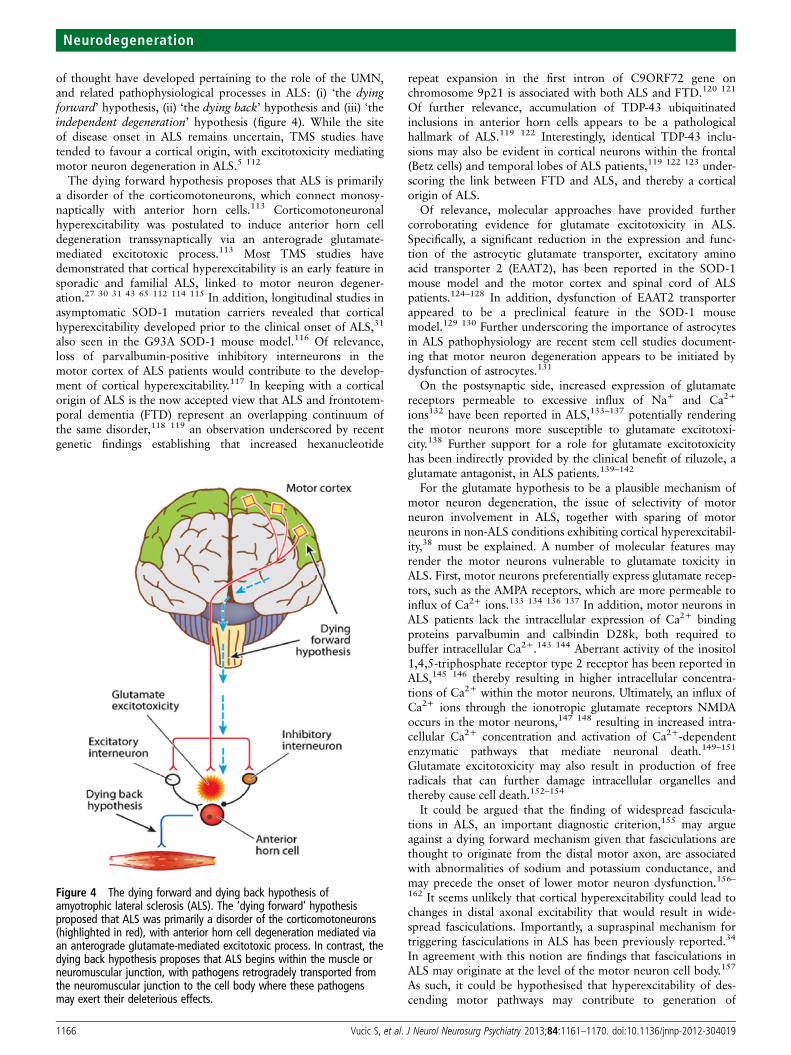
of thought have developed pertaining to the role of the UMN,and related pathophysiological processes in ALS: (i) ‘the dyingforward’ hypothesis, (ii) ‘the dying back’ hypothesis and (iii) ‘theindependent degeneration’ hypothesis (figure 4). While the siteof disease onset in ALS remains uncertain, TMS studies havetended to favour a cortical origin, with excitotoxicity mediatingmotor neuron degeneration in ALS.5 112
The dying forward hypothesis proposes that ALS is primarilya disorder of the corticomotoneurons, which connect monosy-naptically with anterior horn cells.113 Corticomotoneuronalhyperexcitability was postulated to induce anterior horn celldegeneration transsynaptically via an anterograde glutamate-mediated excitotoxic process.113 Most TMS studies havedemonstrated that cortical hyperexcitability is an early feature insporadic and familial ALS, linked to motor neuron degener-ation.27 30 31 43 65 112 114 115 In addition, longitudinal studies inasymptomatic SOD-1 mutation carriers revealed that corticalhyperexcitability developed prior to the clinical onset of ALS,31
also seen in the G93A SOD-1 mouse model.116 Of relevance,loss of parvalbumin-positive inhibitory interneurons in themotor cortex of ALS patients would contribute to the develop-ment of cortical hyperexcitability.117 In keeping with a corticalorigin of ALS is the now accepted view that ALS and frontotem-poral dementia (FTD) represent an overlapping continuum ofthe same disorder,118 119 an observation underscored by recentgenetic findings establishing that increased hexanucleotide
repeat expansion in the first intron of C9ORF72 gene onchromosome 9p21 is associated with both ALS and FTD.120 121
Of further relevance, accumulation of TDP-43 ubiquitinatedinclusions in anterior horn cells appears to be a pathologicalhallmark of ALS.119 122 Interestingly, identical TDP-43 inclu-sions may also be evident in cortical neurons within the frontal(Betz cells) and temporal lobes of ALS patients,119 122 123 under-scoring the link between FTD and ALS, and thereby a corticalorigin of ALS.
Of relevance, molecular approaches have provided furthercorroborating evidence for glutamate excitotoxicity in ALS.Specifically, a significant reduction in the expression and func-tion of the astrocytic glutamate transporter, excitatory aminoacid transporter 2 (EAAT2), has been reported in the SOD-1mouse model and the motor cortex and spinal cord of ALSpatients.124–128 In addition, dysfunction of EAAT2 transporterappeared to be a preclinical feature in the SOD-1 mousemodel.129 130 Further underscoring the importance of astrocytesin ALS pathophysiology are recent stem cell studies document-ing that motor neuron degeneration appears to be initiated bydysfunction of astrocytes.131
On the postsynaptic side, increased expression of glutamatereceptors permeable to excessive influx of Na+ and Ca2+
ions132 have been reported in ALS,133–137 potentially renderingthe motor neurons more susceptible to glutamate excitotoxi-city.138 Further support for a role for glutamate excitotoxicityhas been indirectly provided by the clinical benefit of riluzole, aglutamate antagonist, in ALS patients.139–142
For the glutamate hypothesis to be a plausible mechanism ofmotor neuron degeneration, the issue of selectivity of motorneuron involvement in ALS, together with sparing of motorneurons in non-ALS conditions exhibiting cortical hyperexcitabil-ity,38 must be explained. A number of molecular features mayrender the motor neurons vulnerable to glutamate toxicity inALS. First, motor neurons preferentially express glutamate recep-tors, such as the AMPA receptors, which are more permeable toinflux of Ca2+ ions.133 134 136 137 In addition, motor neurons inALS patients lack the intracellular expression of Ca2+ bindingproteins parvalbumin and calbindin D28k, both required tobuffer intracellular Ca2+.143 144 Aberrant activity of the inositol1,4,5-triphosphate receptor type 2 receptor has been reported inALS,145 146 thereby resulting in higher intracellular concentra-tions of Ca2+ within the motor neurons. Ultimately, an influx ofCa2+ ions through the ionotropic glutamate receptors NMDAoccurs in the motor neurons,147 148 resulting in increased intra-cellular Ca2+ concentration and activation of Ca2+-dependentenzymatic pathways that mediate neuronal death.149–151
Glutamate excitotoxicity may also result in production of freeradicals that can further damage intracellular organelles andthereby cause cell death.152–154
It could be argued that the finding of widespread fascicula-tions in ALS, an important diagnostic criterion,155 may argueagainst a dying forward mechanism given that fasciculations arethought to originate from the distal motor axon, are associatedwith abnormalities of sodium and potassium conductance, andmay precede the onset of lower motor neuron dysfunction.156–162 It seems unlikely that cortical hyperexcitability could lead tochanges in distal axonal excitability that would result in wide-spread fasciculations. Importantly, a supraspinal mechanism fortriggering fasciculations in ALS has been previously reported.34
In agreement with this notion are findings that fasciculations inALS may originate at the level of the motor neuron cell body.157
As such, it could be hypothesised that hyperexcitability of des-cending motor pathways may contribute to generation of
Figure 4 The dying forward and dying back hypothesis ofamyotrophic lateral sclerosis (ALS). The ‘dying forward’ hypothesisproposed that ALS was primarily a disorder of the corticomotoneurons(highlighted in red), with anterior horn cell degeneration mediated viaan anterograde glutamate-mediated excitotoxic process. In contrast, thedying back hypothesis proposes that ALS begins within the muscle orneuromuscular junction, with pathogens retrogradely transported fromthe neuromuscular junction to the cell body where these pathogensmay exert their deleterious effects.
1166 Vucic S, et al. J Neurol Neurosurg Psychiatry 2013;84:1161–1170. doi:10.1136/jnnp-2012-304019
Neurodegeneration
fasciculation in ALS, thereby providing additional support for adying forward process.
In conjunction with glutamate excitotoxicity, there is compel-ling evidence that mitochondrial dysfunction may exert animportant role in the pathophysiology of ALS.163–168 Underconditions of excessive Ca2+ load, as may be evident with glu-tamate excitotoxicity,169 mitochondrial production of free radi-cals increases resulting in injury of critical neuronal cellularproteins and DNA. Mitochondrial dysfunction may in turnenhance glutamate excitotoxicity by disrupting the normalresting membrane potential, resulting in loss of the voltage-dependent Mg2+-mediated block of NMDA receptor chan-nels.170 From a therapeutic perspective, dexpramipexole, apharmacological agent that enhances mitochondrial function,171
was effective in slowing ALS progression in a recent phase IItrial.172 A phase III, multicentre, international trial was com-menced in March 2011 to determine the clinical efficacy of dex-pramipexole in ALS (ClinicalTrials.gov-NCT01281189). Takenfurther, it is anticipated that TMS studies will be used to deter-mine the efficacy of dexpramipexole in the modulation of cor-tical excitability in an attempt to provide further insight intoALS pathophysiology.
The dying back hypothesis proposes that ALS is primarily adisorder of the lower motor neurons, with pathogens retro-gradely transported from the neuromuscular junction to the cellbody where they exert their deleterious effects.173 Althoughsome pathological studies have indirectly supported a dyingback process,174–176 no pathogens of any type have been identi-fied in relation to ALS. The presence of widespread dysfunctionwithin the frontal cortex, including the primary, supplementaryand prefrontal motor cortices in ALS, remains difficult to recon-cile with a dying back process.3 110 177 In addition, the absenceof central pathology in other lower motor neuron disorderssuch as Kennedy’s disease or poliomyelitis provides a furtherargument against a dying back process.33 44
The independent degeneration hypothesis suggests that theupper and lower motor neurons degenerate independently.178
Some 100 years after the original Gowers publication, neuro-pathological studies provided support for the independentdegeneration hypothesis whereby the degeneration of upper andlower motor neurons appeared to be independent.179 180 Thesecorrelative morphological techniques, however, may be con-founded by the anatomical and functional complexity of thecorticomotoneuronal system.181 In particular, there remainsconsiderable variability in the corticomotoneuronal to anteriorhorn cell ratio, due to synaptic changes, and as such attempts tocorrelate upper and lower motor neurons on autopsy studiesmay not be meaningful.33
In addition to the three competing theories of ALS pathogenesis,a prion-like propagation hypothesis has also been suggested.182
Specifically, the previously documented contiguous spread ofALS5 183 could be explained by direct neuron-to-neuron transmis-sion of pathogenic proteins via exosomes, defined as small lipidmembranous microvesicles.182 The pathogenic exosomes couldspread in either a rostral direction, explaining the rostral-to-caudalspread of ALS, or in a lateral–horizontal direction accounting forthe lateral-to-medial spread of disease. In addition, non-contiguouspropagation of ALS could also be explained by spread of patho-genic proteins or toxic molecules through the blood or CSF viaexosomes.182 Interestingly, the genes implicated in ALS pathogen-esis, including TDP-43 and FUS, possess a putative priondomain.184 Although a prion-like propagation mechanism mayseem an attractive explanation for the spread of ALS, at presentthere is no direct evidence to support such a process in ALS.
Future clinical utility of TMSAlthough first described by Charcot some 150 years ago, thepathophysiological mechanisms underlying ALS, variability, rateof progression and site of disease onset remain obscure.Objective assessment of UMN function in ALS remains a difficulttask in clinical neurophysiology.185 While TMS is mainly used asa clinical research tool, conducted in specialised neurophysio-logical laboratories, there is an urgent need to objectively assessUMN function in ALS. This has been underscored by the recentAwaji diagnostic criteria.155 186 Although needle electromyogra-phy is used by the criteria to objectively assess lower motorneuron dysfunction, the detection of UMN involvement is basedsolely on clinical examination. Much has recently been learntabout ALS fromMRI, especially diffusion tensor MRI, functionalimaging and network analysis,110 187–198 but these tools remainprohibitively expensive, not readily available and may exhibit amodest diagnostic sensitivity.190 Commercially available TMSsystems that will enable an objective assessment of UMN functioncould be readily developed, facilitating the diagnosis of ALS.Such TMS systems may result in the development of more func-tional ALS biomarkers that could be used in future drug trials forearly patient recruitment and monitoring of drug efficacy.
Acknowledgements Funding support from the Motor Neuron Disease ResearchInstitute of Australia (MNDRIA), Sylvia and Charles Viertel Charitable FoundationClinical Investigator grant, Ramaciotti Foundation and National Health and MedicalResearch Council of Australia (Project grant numbers 510233 and 1024915) isgratefully acknowledged.
Contributors SV was involved in the design, research and writing of the review. UZ,AE, MH and MK were involved in the design, critiquing and editing of the review.
Funding Support from the Motor Neuron Disease Research Institute of Australia(MNDRIA), Sylvia and Charles Viertel Charitable Foundation Clinical Investigatorgrant, Ramaciotti Foundation and National Health and Medical Research Council ofAustralia (Project grant numbers 510233 and 1024915) is gratefully acknowledged.
Competing interests None.
Ethics approval Local ethics committee.
Provenance and peer review Commissioned; externally peer reviewed.
Open Access This is an Open Access article distributed in accordance with theCreative Commons Attribution Non Commercial (CC BY-NC 3.0) license, whichpermits others to distribute, remix, adapt, build upon this work non-commercially, andlicense their derivative works on different terms, provided the original work is properlycited and the use is non-commercial. See: http://creativecommons.org/licenses/by-nc/3.0/
REFERENCES1 Charcot J, Joffroy A. Deux cas d’atrophie musculaire progressive avec lesion de la
substance grise et des faisceaux antero-lateraux de la moelle epiniere. Arch PhysiolNeurol Pathol 1869;2:744–54.
2 Kiernan MC, Vucic S, Cheah BC, et al. Amyotrophic lateral sclerosis. Lancet2011;377:942–55.
3 Vucic S, Burke D, Kiernan MC. Diagnosis of motor neuron disease. In: KiernanMC. ed. The motor neuron disease handbook. Sydney: Australasian MedicalPublishing Company Limited, 2007:89–115.
4 Winhammar JM, Rowe DB, Henderson RD, et al. Assessment of diseaseprogression in motor neuron disease. Lancet Neurology 2005;4:229–38.
5 Ravits J, Paul P, Jorg C. Focality of upper and lower motor neuron degeneration atthe clinical onset of ALS. Neurology 2007;68:1571–5.
6 Rossini PM, Berardelli A, Deuschl G, et al. Applications of magnetic corticalstimulation. The International Federation of Clinical Neurophysiology.Electroencephalogr Clin Neurophysiol Suppl 1999;52:171–85.
7 Fisher RJ, Nakamura Y, Bestmann S, et al. Two phases of intracortical inhibitionrevealed by transcranial magnetic threshold tracking. Exp Brain Res2002;143:240–8.
8 Vucic S, Howells J, Trevillion L, et al. Assessment of cortical excitability usingthreshold tracking techniques. Muscle Nerve 2006;33:477–86.
9 Groppa S, Oliviero A, Eisen A, et al. A practical guide to diagnostic transcranialmagnetic stimulation: report of an IFCN committee. Clin Neurophysiol2012;123:858–82.
10 Brouwer B, Ashby P. Corticospinal projections to upper and lower limb spinalmotoneurons in man. Electroencephalogr Clin Neurophysiol 1990;76:509–19.
Vucic S, et al. J Neurol Neurosurg Psychiatry 2013;84:1161–1170. doi:10.1136/jnnp-2012-304019 1167
Neurodegeneration

11 Chen R, Tam A, Butefisch C, et al. Intracortical inhibition and facilitation indifferent representations of the human motor cortex. J Neurophysiol1998;80:2870–81.
12 Macdonell RA, Shapiro BE, Chiappa KH, et al. Hemispheric threshold differencesfor motor evoked potentials produced by magnetic coil stimulation. Neurology1991;41:1441–4.
13 Triggs WJ, Calvanio R, Levine M. Transcranial magnetic stimulation reveals ahemispheric asymmetry correlate of intermanual differences in motor performance.Neuropsychologia 1997;35:1355–63.
14 Amassian VE, Stewart M, Quirk GJ, et al. Physiological basis of motor effects of atransient stimulus to cerebral cortex. Neurosurgery 1987;20:74–93.
15 Epstein CM, Schwartzberg DG, Davey KR, et al. Localizing the site of magneticbrain stimulation in humans. Neurology 1990;40:666–70.
16 Rudiak D, Marg E. Finding the depth of magnetic brain stimulation: are-evaluation. Electroencephalogr Clin Neurophysiol 1994;93:358–71.
17 Di Lazzaro V, Oliviero A, Profice P, et al. Ketamine increases human motor cortexexcitability to transcranial magnetic stimulation. Journal of Physiology2003;547:485–96.
18 Ziemann U. TMS and drugs. Clin Neurophysiol 2004;115:1717–29.19 Attarian S, Azulay JP, Lardillier D, et al. Transcranial magnetic stimulation in lower
motor neuron diseases. Clin Neurophysiol 2005;116:35–42.20 Berardelli A, Inghilleri M, Cruccu G, et al. Electrical and magnetic transcranial
stimulation in patients with corticospinal damage due to stroke or motor neuronedisease. Electroencephalogr Clin Neurophysiol 1991;81:389–96.
21 Eisen A, Shytbel W, Murphy K, et al. Cortical magnetic stimulation in amyotrophiclateral sclerosis. Muscle Nerve 1990;13:146–51.
22 de Carvalho M, Turkman A, Swash M. Motor responses evoked by transcranialmagnetic stimulation and peripheral nerve stimulation in the ulnar innervation inamyotrophic lateral sclerosis: the effect of upper and lower motor neuron lesion.J Neurol Sci 2003;210:83–90.
23 Miscio G, Pisano F, Mora G, et al. Motor neuron disease: usefulness of transcranialmagnetic stimulation in improving the diagnosis. Clin Neurophysiol1999;110:975–81.
24 Triggs WJ, Macdonell RA, Cros D, et al. Motor inhibition and excitation areindependent effects of magnetic cortical stimulation. Annals of Neurol1992;32:345–51.
25 Triggs WJ, Menkes D, Onorato J, et al. Transcranial magnetic stimulation identifiesupper motor neuron involvement in motor neuron disease. Neurology1999;53:605–11.
26 Urban P, Wicht S, Hopf H. Sensitivity of transcranial magnetic stimulation ofcortico-bulbar vs. cortico-spinal tract involvement in ALS. J Neurol2001;248:850–5.
27 Caramia MD, Cicinelli P, Paradiso C, et al. ‘Excitability changes of muscularresponses to magnetic brain stimulation in patients with central motor disorders.Electroencephalogr Clin Neurophysiol 1991;81:243–50.
28 Kohara N, Kaji R, Kojima Y, et al. Abnormal excitability of the corticospinalpathway in patients with amyotrophic lateral sclerosis: a single motor unit studyusing transcranial magnetic stimulation. Electroencephalogr Clin Neurophysiol1996;101:32–41.
29 Mills KR, Nithi KA. Corticomotor threshold is reduced in early sporadicamyotrophic lateral sclerosis. Muscle Nerve 1997;20:1137–41.
30 Vucic S, Kiernan MC. Novel threshold tracking techniques suggest that corticalhyperexcitability is an early feature of motor neuron disease. Brain 2006;129:2436–46.
31 Vucic S, Nicholson GA, Kiernan MC. Cortical hyperexcitability may precedethe onset of familial amyotrophic lateral sclerosis. Brain 2008;131:1540–50.
32 Zanette G, Tamburin S, Manganotti P, et al. Different mechanisms contribute tomotor cortex hyperexcitability in amyotrophic lateral sclerosis. Clin Neurophysiol2002;113:1688–97.
33 Eisen A, Weber M. The motor cortex and amyotrophic lateral sclerosis. MuscleNerve 2001;24:564–73.
34 Hirota N, Eisen A, Weber M. Complex fasciculations and their origin in amyotrophiclateral sclerosis and Kennedy’s disease. Muscle Nerve 2000;23:1872–5.
35 Di Lazzaro V, Restuccia D, Oliviero A, et al. Magnetic transcranial stimulation atintensities below active motor threshold activates intracortical inhibitory circuits.Exp Brain Res 1998;119:265–8.
36 Devanne H, Lavoie BA, Capaday C. Input-output properties and gain changes inthe human corticospinal pathway. Exp Brain Res 1997;114:329–38.
37 Ziemann U. Cortical threshold and excitability measurements. In: Eisen A. ed.Clinical neurophysiology of motor neuron diseases handbook of clinicalneurophysiology. Amsterdam: Elsevier, 2004:317–35.
38 Chen R, Cros D, Curra A, et al. The clinical diagnostic utility of transcranialmagnetic stimulation: report of an IFCN committee. Clin Neurophysiol2008;119:504–32.
39 Hess CW, Mills KR, Murray NM, et al. Magnetic brain stimulation: central motorconduction studies in multiple sclerosis. Ann Neurol 1987;22:744–52.
40 Paulus W, Classen J, Cohen LG, et al. State of the art: pharmacologic effects oncortical excitability measures tested by transcranial magnetic stimulation. BrainStimul 2008;1:151–63.
41 Boroojerdi B, Battaglia F, Muellbacher W, et al. Mechanisms influencingstimulus-response properties of the human corticospinal system. Clin Neurophysiol2001;112:931–7.
42 Vucic S, Kiernan MC. Abnormalities in cortical and peripheral excitability in flailarm variant amyotrophic lateral sclerosis. J Neurol Neurosurg Psychiatry2007;78:849–52.
43 Vucic S, Kiernan MC. Upregulation of persistent sodium conductances in familialALS. J Neurol Neurosurg Psychiatry 2010;81:222–7.
44 Vucic S, Kiernan MC. Cortical excitability testing distinguishes Kennedy’s diseasefrom amyotrophic lateral sclerosis. Clin Neurophysiol 2008;119:1088–96.
45 Vucic S, Cheah BC, Yiannikas C, et al. Corticomotoneuronal function andhyperexcitability in acquired neuromyotonia. Brain 2010;133:2727–33.
46 Vucic S, Nicholson GA, Kiernan MC. Cortical excitability in hereditary motorneuronopathy with pyramidal signs: comparison with ALS. J Neurol NeurosurgPsychiatry 2010;81:97–100.
47 Vucic S, Cheah BC, Yiannikas C, et al. Cortical excitability distinguishes ALS frommimic disorders. Clin Neurophysiol 2011;122:1860–6.
48 Mills K. Magnetic stimulation and central conduction time. In: Eisen A. ed. Clinicalneurophysiology of motor neuron diseases handbook of clinical neurophysiology.Amsterdam: Elsevier, 2004:283–93.
49 Claus D. Central motor conduction: method and normal results. Muscle Nerve1990;13:1125–32.
50 Mills KR, Murray NM. Electrical stimulation over the human vertebral column:which neural elements are excited? Electroencephalogr Clin Neurophysiol1986;63:582–9.
51 Rossini PM, Barker AT, Berardelli A, et al. Non-invasive electrical and magneticstimulation of the brain, spinal cord and roots: basic principles and procedures forroutine clinical application. Report of an IFCN committee. Electroencephalogr ClinNeurophysiol 1994;91:79–92.
52 Mills KR. The natural history of central motor abnormalities in amyotrophic lateralsclerosis. Brain 2003;126:2558–66.
53 Eisen A, Entezari-Taher M, Stewart H. Cortical projections to spinal motoneurons:changes with aging and amyotrophic lateral sclerosis. Neurology1996;46:1396–404.
54 Komissarow L, Rollnik JD, Bogdanova D, et al. Triple stimulation technique (TST) inamyotrophic lateral sclerosis. Clin Neurophysiol 2004;115:356–60.
55 Turner MR, Osei-Lah AD, Hammers A, et al. Abnormal cortical excitability insporadic but not homozygous D90A SOD1 ALS. J Neurol Neurosurg Psychiatry2005;76:1279–85.
56 Mills K. Magnetic stimulation and central conduction time. Amsterdam: Elsevier B.V., 2004.
57 Cantello R, Gianelli M, Civardi C, et al. Magnetic brain stimulation: the silentperiod after the motor evoked potential. Neurology 1992;42:1951–9.
58 Inghilleri M, Berardelli A, Cruccu G, et al. Silent period evoked by transcranialstimulation of the human cortex and cervicomedullary junction. J Physiol (Lond)1993;466:521–34.
59 Triggs WJ, Kiers L, Cros D, et al. Facilitation of magnetic motor evoked potentialsduring the cortical stimulation silent period. Neurology 1993;43:2615–20.
60 Chen R, Lozano AM, Ashby P. Mechanism of the silent period followingtranscranial magnetic stimulation. Evidence from epidural recordings. Exp Brain Res1999;128:539–42.
61 Siebner HR, Dressnandt J, Auer C, et al. Continuous intrathecal baclofen infusionsinduced a marked increase of the transcranially evoked silent period in a patientwith generalized dystonia. Muscle Nerve 1998;21:1209–12.
62 Werhahn KJ, Kunesch E, Noachtar S, et al. Differential effects on motorcorticalinhibition induced by blockade of GABA uptake in humans. J Physiol (Lond)1999;517:591–7.
63 Stetkarova I, Kofler M. Differential effects of cortical and spinal inhibitory circuits.Clin Neurophysiol 2013;124:339–45.
64 Desiato MT, Caramia MD. Towards a neurophysiological marker of amyotrophiclateral sclerosis as revealed by changes in cortical excitability. ElectroencephalogrClin Neurophysiol 1997;105:1–7.
65 Prout AJ, Eisen A. The cortical silent period and ALS. Muscle Nerve1994;17:217–23.
66 Siciliano G, Manca ML, Sagliocco L, et al. Cortical silent period in patients withamyotrophic lateral sclerosis. J Neurol Sci 1999;169:93–7.
67 Wittstock M, Wolters A, Benecke R. Transcallosal inhibition in amyotrophic lateralsclerosis. Clin Neurophysiol 2007;118:301–7.
68 Wittstock M, Meister S, Walter U, et al. Mirror movements in amyotrophic lateralsclerosis. Amyotroph Lateral Scler 2011;12:393–7.
69 Meyer BU, Roricht S, Grafin von Einsiedel H, et al. Inhibitory and excitatoryinterhemispheric transfers between motor cortical areas in normal humansand patients with abnormalities of the corpus callosum. Brain 1995;118:429–40.
70 Kujirai T, Caramia MD, Rothwell JC, et al. Corticocortical inhibition in humanmotor cortex. J Physiol (Lond) 1993;471:501–19.
71 Hanajima R, Ugawa Y, Terao Y, et al. Paired-pulse magnetic stimulation of thehuman motor cortex: differences among I waves. J Physiol (Lond)1998;509:607–18.
1168 Vucic S, et al. J Neurol Neurosurg Psychiatry 2013;84:1161–1170. doi:10.1136/jnnp-2012-304019
Neurodegeneration

72 Nakamura H, Kitagawa H, Kawaguchi Y, et al. Intracortical facilitation andinhibition after transcranial magnetic stimulation in conscious humans. J Physiol(Lond) 1997;498:817–23.
73 Ziemann U, Rothwell JC, Ridding MC. Interaction between intracortical inhibitionand facilitation in human motor cortex. J Physiol (Lond) 1996;496:873–81.
74 Di Lazzaro V, Oliviero A, Meglio M, et al. Direct demonstration of the effect oflorazepam on the excitability of the human motor cortex. Clin Neurophysiol2000;111:794–9.
75 Kiers L, Cros D, Chiappa KH, et al. Variability of motor potentials evoked bytranscranial magnetic stimulation. Electroencephalogr Clin Neurophysiol1993;89:415–23.
76 Vucic S, Cheah BC, Krishnan AV, et al. The effects of alterations in conditioningstimulus intensity on short interval intracortical inhibition. Brain Res 2009;1273:39–47.
77 Vucic S, Cheah BC, Kiernan MC. Dissecting the Mechanisms UnderlyingShort-Interval Intracortical Inhibition Using Exercise. Cereb Cortex 2011:1639–44.
78 Ziemann U, Lonnecker S, Steinhoff BJ, et al. The effect of lorazepam on the motorcortical excitability in man. Exp Brain Res 1996;109:127–35.
79 Ilic TV, Meintzschel F, Cleff U, et al. Short-interval paired-pulse inhibition andfacilitation of human motor cortex: the dimension of stimulus intensity. J Physiol(Lond) 2002;545:153–67.
80 Di Lazzaro V, Pilato F, Dileone M, et al. GABAA receptor subtype specificenhancement of inhibition in human motor cortex. J Physiol 2006;575:721–6.
81 Hanajima R, Furubayashi T, Iwata NK, et al. Further evidence to support differentmechanisms underlying intracortical inhibition of the motor cortex. Exp Brain Res2003;151:427–34.
82 Roshan L, Paradiso GO, Chen R. Two phases of short-interval intracorticalinhibition. Exp Brain Res 2003;151:330–7.
83 Hanajima R, Ugawa Y, Terao Y, et al. Ipsilateral cortico-cortical inhibition of themotor cortex in various neurological disorders. J Neurol Sci 1996;114:109–16.
84 Yokota T, Yoshino A, Inaba A, et al. Double cortical stimulation in amyotrophiclateral sclerosis. J Neurol Neurosurg Psychiatry 1996;61:596–600.
85 Ziemann U, Winter M, Reimers CD, et al. Impaired motor cortex inhibition inpatients with amyotrophic lateral sclerosis. Evidence from paired transcranialmagnetic stimulation. Neurology 1997;49:1292–8.
86 Sommer M, Tergau F, Wischer S, et al. Riluzole does not have an acute effect onmotor thresholds and the intracortical excitability in amyotrophic lateral sclerosis.J Neurol 1999;246(Suppl 3):III22–26.
87 Stefan K, Kunesch E, Benecke R, et al. Effects of riluzole on cortical excitability inpatients with amyotrophic lateral sclerosis. Ann Neurol 2001;49:536–9.
88 Blair IP, Williams KL, Warraich ST, et al. FUS mutations in amyotrophic lateralsclerosis: clinical, pathological, neurophysiological and genetic analysis. J NeurolNeurosurg Psychiatry 2010;81:1286–8.
89 Nihei K, McKee AC, Kowall NW. Patterns of neuronal degeneration in the motor cortexof amyotrophic lateral sclerosis patients. Acta Neuropathologica 1993;86:55–64.
90 Vucic S, Cheah BC, Kiernan MC. Defining the mechanisms that underlie corticalhyperexcitability in amyotrophic lateral sclerosis. Exp Neurol 2009;220:177–82.
91 Eisen A, Nakajima M, Weber M. Electrophysiological studies of thecorticomotoneuron in ALS. Rinsho Shinkeigaku -Clinical Neurology 1999;39:99.
92 Mills KR. Motor neuron disease. Studies of the corticospinal excitation of singlemotor neurons by magnetic brain stimulation. Brain 1995;118:971–82.
93 Eisen A, Nakajima M, Weber M. Corticomotorneuronal hyper-excitability inamyotrophic lateral sclerosis. J Neurol Sci 1998;160(Suppl 1):S64–68.
94 Weber M, Eisen A. Assessment of upper and lower motor neurons in Kennedy’sdisease: implications for corticomotoneuronal PSTH studies. Muscle Nerve1999;22:299–306.
95 Magistris MR, Rosler KM, Truffert A, et al. A clinical study of motor evokedpotentials using a triple stimulation technique. Brain 1999;122:265–79.
96 Magistris MR, Rosler KM, Truffert A, et al. Transcranial stimulation excites virtuallyall motor neurons supplying the target muscle. A demonstration and a methodimproving the study of motor evoked potentials. Brain 1998;121:437–50.
97 Kleine BU, Schelhaas HJ, van Elswijk G, et al. Prospective, blind study of the triplestimulation technique in the diagnosis of ALS. Amyotroph Lateral Scler 2010;11:67–75.
98 Attarian S, Vedel JP, Pouget J, et al. Cortical versus spinal dysfunction inamyotrophic lateral sclerosis. Muscle Nerve 2006;33:677–90.
99 Pachatz C, Terracciano C, Desiato MT, et al. Upper motor neuron involvement inX-linked recessive bulbospinal muscular atrophy. Clin Neurophysiol 2007;118:262–8.
100 Vucic S, Kiernan M. Clarifying variability of corticomotoneuronal function inKennedy’s disease. Muscle Nerve 2011;44:197–201.
101 Kim W-K, Liu X, Sandner J, et al. Study of 962 patients indicates progressivemuscular atrophy is a form of ALS. Neurology 2009;73:1686–92.
102 Geser F, Stein B, Partain M, et al. Motor neuron disease clinically limited to thelower motor neuron is a diffuse TDP-43 proteinopathy. Acta Neuropathologica2011;121:509–17.
103 Ince PG, Evans J, Knopp M, et al. Corticospinal tract degeneration in theprogressive muscular atrophy variant of ALS. Neurology 2003;60:1252–8.
104 Fisher KM, Zaaimi B, Williams TL, et al. Beta-band intermuscular coherence: anovel biomarker of upper motor neuron dysfunction in motor neuron disease. Brain2012;135:2849–64.
105 Miscio G, Gukov B, Pisano F, et al. The cortico-diaphragmatic pathwayinvolvement in amyotrophic lateral sclerosis: neurophysiological, respiratory andclinical considerations. J Neurol Sci 2006;251:10–6.
106 Similowski T, Attali V, Bensimon G, et al. Diaphragmatic dysfunction and dyspnoeain amyotrophic lateral sclerosis. Eur Respir J 2000;15:332–7.
107 Urban PP, Vogt T, Hopf HC. Corticobulbar tract involvement in amyotrophic lateralsclerosis. A transcranial magnetic stimulation study. Brain 1998;121:1099–108.
108 Mitsumoto H, Ulug AM, Pullman SL, et al. Quantitative objective markers forupper and lower motor neuron dysfunction in ALS. Neurology 2007;68:1402–10.
109 Kaufmann P, Pullman SL, Shungu DC, et al. Objective tests for upper motorneuron involvement in amyotrophic lateral sclerosis (ALS). Neurology2004;62:1753–7.
110 Turner MR, Kiernan MC, Leigh PN, et al. Biomarkers in amyotrophic lateralsclerosis. Lancet Neurol 2009;8:94–109.
111 Floyd AG, Yu QP, Piboolnurak P, et al. Transcranial magnetic stimulation in ALS:utility of central motor conduction tests. Neurology 2009;72:498–504.
112 Vucic S, Kiernan M. Pathophysiology of degeneration in familial amyotrophiclateral sclerosis. Curr Mol Med 2009;9:255–72.
113 Eisen A, Kim S, Pant B. Amyotrophic lateral sclerosis (ALS): a phylogenetic diseaseof the corticomotoneuron? Muscle Nerve 1992;15:219–24.
114 Desiato M, Bernardi G, Hagi AH, et al. Transcranial magnetic stimulation of motorpathways directed to muscles supplied by cranial nerves in ALS. Clin Neurophysiol2002;113:132–40.
115 Eisen A, Pant B, Stewart H. Cortical excitability in amyotrophic lateral sclerosis: aclue to pathogenesis. Can J Neurol Sci 1993;20:11–6.
116 Browne SE, Yang L, DiMauro JP, et al. Bioenergetic abnormalities in discretecerebral motor pathways presage spinal cord pathology in the G93A SOD1 mousemodel of ALS. Neurobiol Dis 2006;22:599–610.
117 Nihei K, McKee AC, Kowall NW. Patterns of neuronal degeneration in the motorcortex of amyotrophic lateral sclerosis patients. Acta Neuropathologica1993;86:55–64.
118 Lillo P, Hodges JR. Frontotemporal dementia and motor neurone disease:overlapping clinic-pathological disorders. J Clin Neurosci 2009;16:1131–5.
119 Neumann M, Sampathu DM, Kwong LK, et al. Ubiquitinated TDP-43 inFrontotemporal Lobar Degeneration and Amyotrophic Lateral Sclerosis. Science2006;314:130–3.
120 Renton Alan E, Majounie E, Waite A, et al. A Hexanucleotide Repeat Expansion inC9ORF72 Is the Cause of Chromosome 9p21-Linked ALS-FTD. Neuron2011;72:257–68.
121 DeJesus-Hernandez M, Mackenzie Ian R, Boeve Bradley F, et al. ExpandedGGGGCC Hexanucleotide Repeat in Noncoding Region of C9ORF72 CausesChromosome 9p-Linked FTD and ALS. Neuron 2011;72:245–56.
122 Arai T, Hasegawa M, Akiyama H, et al. TDP-43 is a component of ubiquitin-positivetau-negative inclusions in frontotemporal lobar degeneration and amyotrophic lateralsclerosis. Biochem and Biophy Res Commun 2006;351:602–11.
123 Tsuji H, Arai T, Kametani F, et al. Molecular analysis and biochemical classificationof TDP-43 proteinopathy. Brain 2012;135:3380–91.
124 Boillee S, Vande Velde C, Cleveland DW. ALS: a disease of motor neurons andtheir nonneuronal neighbors. Neuron 2006;52:39–59.
125 Ionov ID. Survey of ALS-associated factors potentially promoting Ca(2+) overloadof motor neurons. Amyotroph Lateral Scler 2007;8:260–5.
126 Rothstein JD, Jin L, Dykes-Hoberg M, et al. Chronic inhibition of glutamate uptakeproduces a model of slow neurotoxicity. Proc Natl Acad Sci U S A1993;90:6591–5.
127 Rothstein JD, Van Kammen M, Levey AI, et al. Selective loss of glial glutamatetransporter GLT-1 in amyotrophic lateral sclerosis. Ann Neurol 1995;38:73–84.
128 Trotti D, Rolfs A, Danbolt NC, et al. SOD1 mutants linked to amyotrophic lateralsclerosis selectively inactivate a glial glutamate transporter. Nat Neurosci1999;2:848.
129 Boston-Howes W, Gibb SL, Williams EO, et al. Caspase-3 cleaves and inactivatesthe glutamate transporter EAAT2. J Biol Chem 2006;281:14076–84.
130 Gibb SL, Boston-Howes W, Lavina ZS, et al. A Caspase-3-cleaved Fragment of theGlial Glutamate Transporter EAAT2 Is Sumoylated and Targeted to PromyelocyticLeukemia Nuclear Bodies in Mutant SOD1-linked Amyotrophic Lateral Sclerosis. JBiol Chem 2007;282:32480–90.
131 Haidet-Phillips AM, Hester ME, Miranda CJ, et al. Astrocytes from familial andsporadic ALS patients are toxic to motor neurons. Nat Biotech 2011;29:824–8.
132 Simeone TA, Sanchez RM, Rho JM. Molecular biology and ontogeny of glutamatereceptors in the mammalian central nervous system. J Child Neurol2004;19:343–60.
133 Kawahara Y, Ito K, Sun H, et al. Glutamate receptors: RNA editing and death ofmotor neurons. Nature 2004;427:801.
134 Kwak S, Kawahara Y. Deficient RNA editing of GluR2 and neuronal death inamyotropic lateral sclerosis. J Mol Med 2005;83:110–20.
135 Takuma H, Kwak S, Yoshizawa T, et al. Reduction of GluR2 RNA editing, amolecular change that increases calcium influx through AMPA receptors, selectivein the spinal ventral gray of patients with amyotrophic lateral sclerosis. Ann Neurol1999;46:806–15.
Vucic S, et al. J Neurol Neurosurg Psychiatry 2013;84:1161–1170. doi:10.1136/jnnp-2012-304019 1169
Neurodegeneration

136 Van Damme P, Braeken D, Callewaert G, et al. GluR2 deficiency accelerates motorneuron degeneration in a mouse model of amyotrophic lateral sclerosis.J Neuropathol Exp Neurol 2005;64:605–12.
137 Van Damme P, Van Den Bosch L, Van Houtte E, et al. GluR2-dependent propertiesof AMPA receptors determine the selective vulnerability of motor neurons toexcitotoxicity. J Neurophysiol 2002;88:1279–87.
138 Williams TL, Day NC, Ince PG, et al. Calcium-permeablealpha-amino-3-hydroxy-5-methyl-4-isoxazole propionic acid receptors: a moleculardeterminant of selective vulnerability in amyotrophic lateral sclerosis. Ann Neurol1997;42:200–7.
139 Bensimon G, Lacomblez L, Meininger V, ALS/Riluzole Study Group. A controlledtrial of riluzole in amyotrophic lateral sclerosis. N Engl J Med 1994;330:585–91.
140 Gurney ME, Cutting FB, Zhai P, et al. Benefit of vitamin E, riluzole, andgabapentin in a transgenic model of familial amyotrophic lateral sclerosis. AnnNeurol 1996;39:147–57.
141 Gurney ME, Fleck TJ, Himes CS, et al. Riluzole preserves motor function in atransgenic model of familial amyotrophic lateral sclerosis. Neurology 1998;50:62–6.
142 Lacomblez L, Bensimon G, Leigh PN, et al. Amyotrophic Lateral Sclerosis/RiluzoleStudy Group II. Dose-ranging study of riluzole in amyotrophic lateral sclerosis.Lancet 1996;347:1425–31.
143 Alexianu ME, Ho BK, Mohamed AH, et al. The role of calcium-binding proteins inselective motoneuron vulnerability in amyotrophic lateral sclerosis. Ann Neurol1994;36:846–58.
144 Ince P, Stout N, Shaw P, et al. Parvalbumin and calbindin D-28k in the humanmotor system and in motor neuron disease. Neuropathol Appl Neurobiol1993;19:291–9.
145 Choe CU, Ehrlich BE. The inositol 1,4,5-trisphosphate receptor (IP3R) and itsregulators: sometimes good and sometimes bad teamwork. Sci STKE 2006;2006:re15.
146 van Es MA, Van Vught PW, Blauw HM, et al. ITPR2 as a susceptibility gene insporadic amyotrophic lateral sclerosis: a genome-wide association study. LancetNeurol 2007;6:869–77.
147 Choi DW. Ionic dependence of glutamate neurotoxicity. J Neurosci 1987;7:369–79.148 Miller RJ, Murphy SN, Glaum SR. Neuronal Ca2+ channels and their regulation by
excitatory amino acids. Ann N Y Acad Sci 1989;568:149–58.149 Meldrum B, Garthwaite J. Excitatory amino acid neurotoxicity and
neurodegenerative disease. Trends Pharmacol Sci 1990;11:379–87.150 Regan RF, Panter SS, Witz A, et al. Ultrastructure of excitotoxic neuronal death in
murine cortical culture. Brain Res 1995;705:188–98.151 Shaw P, Kuncl WR. Current concepts in the pathogenesis of ALS. In: WR K. ed.
Motor neuron disease. Lodon: WB Saunders, 2002:37–73.152 Bondy SC, Lee DK. Oxidative stress induced by glutamate receptor agonists. Brain
Res 1993;610:229–33.153 Lees GJ. Contributory mechanisms in the causation of neurodegenerative disorders.
Neuroscience 1993;54:287–322.154 Maher P, Davis JB. The role of monoamine metabolism in oxidative glutamate
toxicity. J Neurosci 1996;16:6394–401.155 de Carvalho M, Dengler R, Eisen A, et al. Electrodiagnostic criteria for diagnosis of
ALS. Clin Neurophysiol 2008;119:497–503.156 de Carvalho M, Naka D, Mills KR, et al. Pathophysiological significance of
fasciculations in the early diagnosis of ALS. Amyotroph Lateral Scler Other MotorNeuron Disord 2000;1(Suppl 1):S43–6.
157 Kleine BU, Stegeman DF, Schelhaas HJ, et al. Firing pattern of fasciculations inALS: evidence for axonal and neuronal origin. Neurology 2008;70:353–9.
158 Mills KR. Characteristics of fasciculations in amyotrophic lateral sclerosis and thebenign fasciculation syndrome. Brain 2010;133:3458–69.
159 Bostock H, Sharief MK, Reid G, et al. Axonal ion channel dysfunction inamyotrophic lateral sclerosis. Brain 1995;118:217–25.
160 Kanai K, Kuwabara S, Misawa S, et al. Altered axonal excitability properties inamyotrophic lateral sclerosis: impaired potassium channel function related todisease stage. Brain 2006;129:953–62.
161 Kanai K, Shibuya K, Sato Y, et al. Motor axonal excitability properties are strongpredictors for survival in amyotrophic lateral sclerosis. J Neurol NeurosurgPsychiatry 2012;83:734–8.
162 Vucic S, Kiernan MC. Axonal excitability properties in amyotrophic lateral sclerosis.Clin Neurophysiol 2006;117:1458–66.
163 Boillee S, Vande Velde C, Cleveland DW. ALS: a disease of motor neurons andtheir nonneuronal neighbors. Neuron 2006;52:39–59.
164 Chung MJ, Suh YL. Ultrastructural changes of mitochondria in the skeletal muscleof patients with amyotrophic lateral sclerosis. Ultrastruct Pathol 2002;26:3–7.
165 Higgins CM, Jung C, Xu Z. ALS-associated mutant SOD1G93A causesmitochondrial vacuolation by expansion of the intermembrane space and byinvolvement of SOD1 aggregation and peroxisomes. BMC Neurosci 2003;4:16.
166 Kirkinezos IG, Bacman SR, Hernandez D, et al. Cytochrome c association with theinner mitochondrial membrane is impaired in the CNS of G93A-SOD1 mice. JNeurosci 2005;25:164–72.
167 Lederer CW, Torrisi A, Pantelidou M, et al. Pathways and genes differentiallyexpressed in the motor cortex of patients with sporadic amyotrophic lateralsclerosis. BMC Genomics 2007;8:26.
168 Xu Z, Jung C, Higgins C, et al. Mitochondrial degeneration in amyotrophic lateralsclerosis. J Bioenerg Biomembr 2004;36:395–9.
169 Dugan LL, Choi DW. Excitotoxicity, free radicals, and cell membrane changes. AnnNeurol 1994;35:S17–21.
170 Heath PR, Tomkins J, Ince PG, et al. Quantitative assessment of AMPA receptormRNA in human spinal motor neurons isolated by laser capture microdissection.Neuroreport 2002;13:1753–7.
171 Cheah BC, Kiernan MC. Dexpramipexole, the R(+) enantiomer of pramipexole, forthe potential treatment of amyotrophic lateral sclerosis. IDrugs 2010;13:911–20.
172 Cudkowicz M, Bozik ME, Ingersoll EW, et al. The effects of dexpramipexole(KNS-760704) in individuals with amyotrophic lateral sclerosis. Nat Med2011;17:1652–6.
173 Chou SM, Norris FH. Amyotrophic lateral sclerosis: lower motor neuron diseasespreading to upper motor neurons. Muscle Nerve 1993;16:864–9.
174 Gould TW, Buss RR, Vinsant S, et al. Complete dissociation of motor neuron deathfrom motor dysfunction by Bax deletion in a mouse model of ALS. J Neurosci2006;26:8774–86.
175 Pagani MR, Reisin RC, Uchitel OD. Calcium signaling pathways mediating synapticpotentiation triggered by amyotrophic lateral sclerosis IgG in motor nerveterminals. J Neurosci 2006;26:2661–72.
176 Pun S, Santos AF, Saxena S, et al. Selective vulnerability and pruning of phasicmotoneuron axons in motoneuron disease alleviated by CNTF. Nat Neurosci2006;9:408–19.
177 Miller RG, Jackson CE, Kasarskis EJ, et al. Practice parameter update: the care ofthe patient with amyotrophic lateral sclerosis: multidisciplinary care, symptommanagement, and cognitive/behavioral impairment (an evidence-based review):report of the Quality Standards Subcommittee of the American Academy ofNeurology. Neurology 2009;73:1227–33.
178 Gowers WR. A Manual of Diseases of the Nervous System: spinal cord and nerves.In. London: Churchill, 1888:356–381.
179 Kiernan J, Hudson A. Changes in sizes of cortical and lower motor neurons inamyotrophic lateral sclerosis. Brain 1991:843–53.
180 Pamphlett R, Kril J, Hng T. Motor neuron disease: a primary disorder ofcorticomotoneurons? Muscle Nerve 1995:314–18.
181 Flament D, Goldsmith P, Buckley CJ, et al. Task dependence of responses in firstdorsal interosseous muscle to magnetic brain stimulation in man. J Physiol1993;464:361–78.
182 Kanouchi T, Ohkubo T, Yokota T. Can regional spreading of amyotrophic lateralsclerosis motor symptoms be explained by prion-like propagation? J NeurolNeurosurg Psychiatry 2012;83:739–45.
183 Fujimura-Kiyono C, Kimura F, Ishida S, et al. Onset and spreading patterns oflower motor neuron involvements predict survival in sporadic amyotrophic lateralsclerosis. J Neurol Neurosurg Psychiatry 2011;82:1244–9.
184 King OD, Gitler AD, Shorter J. The tip of the iceberg: RNA-binding proteins withprion-like domains in neurodegenerative disease. Brain Res 2012;1462:61–80.
185 de Carvalho M. Testing upper motor neuron function in amyotrophic lateralsclerosis: the most difficult task of neurophysiology. Brain 2012;135:2581–2.
186 Costa J, Swash M, de Carvalho M. Awaji criteria for the diagnosis of amyotrophiclateral sclerosis: a systematic review. Arch Neurol 2012:1–7.
187 Udaka F, Sawada H, Seriu N, et al. MRI and SPECT findings in amyotrophic lateralsclerosis. Demonstration of upper motor neurone involvement by clinicalneuroimaging. Neuroradiology 1992;34:389–93.
188 Ellis CM, Simmonds A, Jones D, et al. Diffusion tensor MRI assesses corticospinaltract damage in ALS. Neurology 1999;53:1051–8.
189 Ellis CM, Simmons A, Andrews C, et al. A proton magnetic resonance spectroscopicstudy in ALS: correlation with clinical findings. Neurology 1998;51:1104–9.
190 Foerster BR, Dwamena BA, Petrou M, et al. Diagnostic accuracy using diffusiontensor imaging in the diagnosis of ALS: a meta-analysis. Acad Radiol2012;19:1075–86.
191 Verstraete E, Veldink JH, Hendrikse J, et al. Structural MRI reveals cortical thinningin amyotrophic lateral sclerosis. J Neurol Neurosurg Psychiatry 2012;83:383–8.
192 Cosottini M, Pesaresi I, Piazza S, et al. Structural and functional evaluation ofcortical motor areas in Amyotrophic Lateral Sclerosis. Exp Neurol 2012;234:169–80.
193 Rose S, Pannek K, Bell C, et al. Direct evidence of intra- and interhemisphericcorticomotor network degeneration in amyotrophic lateral sclerosis: an automatedMRI structural connectivity study. NeuroImage 2012;59:2661–9.
194 Turner MR, Hammers A, Al-Chalabi A, et al. Distinct cerebral lesions in sporadicand ‘D90A’ SOD1 ALS: studies with [11C]flumazenil PET. Brain2005;128:1323–9.
195 Turner MR, Rabiner EA, Hammers A, et al. [11C]-WAY100635 PET demonstratesmarked 5-HT1A receptor changes in sporadic ALS. Brain 2005;128:896–905.
196 Tsujimoto M, Senda J, Ishihara T, et al. Behavioral changes in early ALS correlatewith voxel-based morphometry and diffusion tensor imaging. J Neurol Sci2011;307:34–40.
197 Turner MR, Grosskreutz J, Kassubek J, et al. Towards a neuroimaging biomarkerfor amyotrophic lateral sclerosis. Lancet Neurol 2011;10:400–3.
198 Turner MR, Modo M. Advances in the application of MRI to amyotrophic lateralsclerosis. Expert Opin Med Diagn 2010;4:483–96.
1170 Vucic S, et al. J Neurol Neurosurg Psychiatry 2013;84:1161–1170. doi:10.1136/jnnp-2012-304019
Neurodegeneration
Copyright © 2022 FDOKUMEN