table of contents - Philippine Journal of Otolaryngology Head ...
117
-
Upload
khangminh22 -
Category
Documents
-
view
0 -
download
0
Transcript of table of contents - Philippine Journal of Otolaryngology Head ...


NECKSURGERY,It,.
Iq4LIPl:qut=S
TABLE OF CONTENTS
President's Page ................................................................................................................................................. 3
Another Otolzuryngologic Etiology of Diplopia .................. .......... .................................................................... S
Chondrosarcoma of the Maxilla .............................. ........................................................................................ II
Brown Tumor of the Maxilla in primary Hypcrparathyroidism: A case Report ........................................... 17
A Massive Pyogemc grannloma in the Gingiva ............................................................................................... 26
Unusual Case of Bronchopncumonia in an Infant., .......................................................................... i...... ...... 31
Cancer and Keloid or Kimura and Steroid ............................ :.......................................................................... 34
NGT Induced Sinisitis: A Prospective Cohort Study ............................................................................... •...... 39
Hereditary Hemontmgic Tclagiectasia ............................................................................................................ 46
The Neck Mass, A diagnostic Challenge ......................................................................................................... 49
Cleft Lip Surgery for Rural Filipinos ............................................................ i................................................. 52
,//Mi¢/"robial Flora in Chronic Otitis Media" Value of Ear ........... ...................................................................... 58
Randomized Clinical Trial on the Efficacy and Safety of Loratadine, versus Astemizole in Allegi¢ Rhinitis .......................................................................................................... 67
An Evaluation of the Efficacy, Tolerance and Safetyof Loratidine-D vs. Actifed in Acute 17d3inJtis.............................................................................................. 77
A Comparative Study on the Efficacy of Medic_!e_ Medium-strip Gauze and MedicatedFinger-cot as Antrior Packs in Minor Nasal Surgery ................ .................................................................... 87
Collision Tumor of the Nasopharynx (Liposarcoma, Undifferentiated Ca) Report of a Case.. ...................... 96The Lateral Nasal Wall in Filipinos; A study based on Fifty Consecutive
Cadaver Dissections ................................................................................................................................ .... 102

THE PHILIPPINE JOURNAL OF OTOLARYNGOLOGYHEAD AND NECK SURGERY
THE EDITORIAL STAFF
Joselito C. Jamir, M.D. Editor-in-Chief
Alfredo Q.Y. Pontejos, Jr., M.D. Assistance EditorVictoria C. Sarmiento, M.D. Managing Editor
BOARD OF EDITORS
Remigio I.Jarin, M.D. Head & Neck SurgeryGeneroso T. Abes, M.D. Otology
Mariano B. Caparas, M.D. Maxillofacial SurgeryMilagros S. Lopez, M.D. RhinophamgologyNorberto V. Martinez, M.D. Audiology & Neuro-OtologyCesar V, ViUafuerte, Jr., M.D. Reconstructive & Plastic SurgeryRene S. Tuazon, M.D. Bronchoesophagology&Laryngology
All manuscripts and other editorial matter should be addressed to Joselito C. Jamir, M.D.,Editor-in-Chief, The Philippine Journal of Otolaryngology - Head and Neck Surgery, De-
partment of Otolaryngology, UP-PGH Medical Center, Taft Avenue, Manila.
2512,25/g, _ 9_ta/z,z6,,_i_,a,,,San_;g,,-_ due.,(Y_ia_eta.,5Pa_/_P__flee.:633.2783,633-8344,0920.906-6652ee_leA;a.: JOO,4_r_ • OU>I_'
HEAD AND NECK SURGERY - TO ADD OR NOT TO ADD:A REBUTTAL OF THE PCS AND PMA COMMITTEE POSITIONS
Guest Editorial: ANGEL E. ENRI(]UEZ, MD
A year ago (1989), the Philippine College of cling to obsolete ideas suffer obsolescence. Even theirSurgeons caused to be sent, to all medical institutions thoughts are affected by competition. Small wonderand hospitals all over the archipelago a position paper the Philippine College of Surgeons opted for a status
on the proposal of some Departments of Otolaryn- quo, which, at best, is anathema to progress. Unimag-gology to add "Head and Neck Surgery" to their present inable still is the PMA's recommendation that the
departmental name. Briefly, the College (Philippine specialty drop the caption Head and Neck SurgeryCollege of Surgeons) thru the Board of Regents and without giving the society or the specialty board "duein consultation with the Advisory Council of Past process". When the Committee on affiliated Medical
Presidents and after meeting with the Philippine Society Societies of the PMA was reminded that this is beingof Otorhinolaryngology and the Philippine Society of threshed out with the Phlipppine College of Surgeons,Plastic and Reconstructive Surgery has resolved that said committee immediately rescinded its letter ad-"Head and Neck Surgery should not be added in dressed to the Chairman of the Philippine Board of(to) the present Department of Otorhinolaryngol- Surgery thus rendering its ruling null and void. Angogy". In August 16, 1990, perhaps to prove that there nanghimasok sa pagkakabinyag ng dalawang pangalan
are aggrupation of General Surgeons other than those sa sinuman ay hindi lamang nararapat kundi ay kabas-listed above who will always be the last to give up tusan.what is old and slow to accept the new, the Philippine While it is true that the term "head and neck
Board of Surgery through the PMS's Committee on surgery" evolved as operations of radical extent in
Affiliate Medical Societies, recommended that the ENT the head and neck areas developed, the term is byspecialty board drop the caption Head and Neck no means vague as claimed by our well meaningSurgery. (Underlining ours) colleagues in general surgery. It encompassess sur-
gery of the cranial base, maxillo-facial, temporal bone,These are extremely serious statements as it poorly facial plastic and reconstructive surgery not only for
disguised an unprofessional attempt to paint us oto- malignant lesions but also for benign tumors, congeni-laryngologists as not qualified to perform head and tallesions from the hase of the skull down to theneck surgery, clavicle, etc.
How did it happen that departments of Otolar- The name change of the original Philippine Societyyngology are proposing a name change. Perhaps the of Otolaryngology & Bronchoesophagology to thebest answer is to "represent an intent to accurately Philippine Society of Otolaryngology- Head & Neckcharacterize the current scope of (the) specialty. The Surgery, Inc. and for the Philippine Board of Oto-name is a clear reflection of current practice activities laryngology to include Head & Neck Surgery as welland should not be perceived as a descriptor (that is as the name of the society's journal (The Philippinean attempt to expand the horizons of Otolaryngologic Journal of Otolaryngology - Head & Neck Surgery)surgery at the expense of other surgical specialties'3, all registered with the Securities and ExchangeIt makes good sense in that the major part of what Commission represent an intent, as previously stated,otolaryngology consists of is surgery of the head and to accurately characterize the current scope of theneck areas and does not imply exclusiveness as there specialty. Bronchoesophagology was deleted from theis no intent to exclude others from receiving training original name of the society with the advent of newin or practicing head and .neck surgery, technology that allowed other specialties including
general surgery to include this in their current practiceOld ways of thinking do die hard indeed. Worse, activities. The concerns, therefore, about the scope
it is no longer relevant with the new wave of advances of the specialty appear exaggerated and unjustifiedin medicine and surgery. And those who adamantly and to delete "head and neck surgery" now from
1

otolaryngology is to make an amputee of the spe- "It is absurd to believe that it would be possiblecialty, or practical for the general surgeon ---.to properly
manage complicated patients in all fields.""The simple fact of the matter is that the science
and practice of medicine and surgery are not static." Loring Pratt, MD
Loring Pratt, MDBeing truly reflective of our activities in the
specialty, the Philippine Board of Otolaryngology -The allegation of the Philippine College of Head and Neck Surgery, Inc. holds that the name
Surgeons that "additional training is necessary whether change from Otolaryngology to Otolaryngology - Headthey be graduates of residency programs in general & Neck Surgery is appropriate, upbeat and clearlysurgery, plastic surgery or otorhinolaryngology is a reflects the regional scope of the specialty.redundancy as far as otolaryngology is concerned sincethe required training program for accreditation callsfor proficiency to thyroid and parotid surgery, laryn-gectomies, etc. Enrollment in approved post graduatecourses in head and neck oncology is mandatory.
These requirements are so stringent that, to thisday, only six (6) institutions are accredited for resi- ANGEL E. ENRIQUEZ. MDdency training in otolaryngology. These are Depts,of Otolaryngology - Head & Neck Surgery of thefollowing medical institutions:
1. U.P. College of Medicine - Philippine GeneralHospital
2. U.S.T. Faculty of Medicine and Surgery3. P.L.M. College of Medicine - Ospital ng
Maynila E_,',Note:
4. Jose Reyes Memorial Medical Center (or. Angel E. Eafiquez i_ one _ lira e.lderl of this specialty and5. East Avenue Medical Center and has _d as pn_skl_t of the PSO-HNS, e.ditor-in-ehiefofthcPhilippine
6. M.C.U.-F.D.T. Medical Foundation Journal of PSO-HN$ and, presently, it Ihe pruident of the PhilippineBoard of ORL-.-HNS,)
Of all the different surgical aggrupation withinterest in the head and neck areas, it is inarguablethat the otolaryngologists, by virtue of their trainingand orientation, are very familiar with the"anatomyand instrumentation employed in these areas and, there-fore, the best qualified to evaluate and manage patho-logical lesions in these regions. That significant numberof neck masses are metastatic from primaries situatedwithin the realm of otolaryngology places the oto-laryngologist in the best position to diagnose and treatsuch cases. Although actual figures are not available,there is a growing suspicion that more head and necksurgical procedures being performed by otlaryngolo-gist - head and neck surgery residents. This impliesthat there is greater operative experience in Otolar-yngology considering the fact that is involved onlysome 6 institutions compared to more than 45 insti-
tutions accredited in general surgery.
2

PRESIDENT'S PAGE
The years 1989-90 have proven to be very eventful, even tumultous years forour country. Each of us has a role and a story to tell concerning these events -devastating array of coups, destabilizing attempts; earthquake, floods, welga etc.
The PSO-HNS has been equally colorful during these years perhaps influencedby the fast phase of these happenings and the challenge to deliver or else be a lag-gard in an era of turbulent changes.
The first test of the Society's mettle was the disruptionof its Annual Conventionin December 8-9, 1989 when the puschists choose the 1st week of December to launch
its bloodiest coup. All of us, particularly in the organizing commitee had nothingbut curse for these counter productive measures but the committee headed by itsable chairman, Dr. Dominador Almeda, could not be cowed by these events. Afterall the ideas, efforts and time invested in preparation for these first big time AnnualConvention, nothing could dampen the spirits of the organizers. It finally pushedthrough a month later on January 26-27, 1990, ushered in by the first ENT week
and initiated by a festive one day sports fest at the Astra Sucat Compound.
After the initial debacle, the events took a rapid fire proportion:
The interhospital grand rounds, a simple but novel way of scientific exchangeparticulary beneficial to our residents, became an on going quarterly affair, Thanksto the organizers headed by Dr. Rene Tuazon and to the recent hosts - The ENTdepartments of UST and JRMaMC.
These initial success fuelled the enthusiasm of the leadership of the societywith the cooperation of members from Region ! particularly Drs. Carlos Dumlao,Zen Wi, Leonardo Mangahas launched the first out of town mid-year conventionat the Nevada Hotel , Baguio, last June. (This was the last medical convention
that Hotel ever hosted before it was totally flattened by the destructive earthquake).As a consequence, the provincial members became enthusiastic in forming chapters.Whether decentralization will be productive or not, is now under study.
Earlier this year, a new organizing committee was fomaed, chaired by the energeticDr. Cesar Villafuerte, Jr. to prepare for the forthcoming annual convention and thesecond ENT Week on December 2 to 8 this year. Prominent personalities fromEurope, Japan, United States, and Hongkong will grace the occasion. This is probablythe time to solicit Divine Providence to prevent any untoward event that will spoil
the occasion together with the energies invested on it.
3

Occasions like these tend to project a glowing image for our society and likethe provervial ripe mango fruit, with its striking yellow color and sweet fruity odor,critics try to pull it down and even casst doubts as to its technical ability in the
field of head and neck surgery. The main contention is our title "Philippine Societyof Otolaryngology-Head & Neck Surgery, Inc.". which according to to these samecritics, is a claim to exclisivity to head and neck surgery in this country. The English
in this title is very simple and elementary and therefore the charge is baseless.
Nevertheless, in the spirit of cooperation, our society has joined the "Councilof Head & Neck Surgery", an Ad Hoc committee created by the Philippine Collegeof Surgeons. This council is tasked to formulate guidelines in the practice of Head& Neck Surgery in the country particularly in the training of future head & necksurgeons and their eventual practice. Drs. Manuel Lira, Alfredo Pontejos and myselfare our country's representatives to this body and have been attending monthlymeetings.
Meanwhile, the routine scientific meetings on Interesting Cases, Clinical Re-search, Surgical and Instrument Innovations, are all on going projects.
Our ties with the ASEAN Otorhinolarygology Federation was renewed April lastyear when the sizeable delegation from our country went to Singapore and presentedinteresting scientific papers.
The 10th issue of Philippine Joumalof Otolaryngology-Head and Neck Surgery
just came out or the press and as usual is filled with scientific and literary gems,a product of the ability of its new editor, Dr. Alfie Pontejos.
All these milestones in the history of our society could not have been realized
if not for the selfless cooperation of each individual members guided by the wisdomand experience of our elders. To cite other names will be tantamount to mentioningthe rank and file of the organization.
It is therefore, my wish as your outgoing president, that the future leadershipof our society will continue exploring the path in pursuit of a bigger tasks and greater
accomplishments and for the past leadership, always to tend a guiding hand.
TEODORO P. LLAMANZARES, M.D.President, PSO-HNS, Inc.
4

RIZAL MEDICAL CENTER
DEPARTMENT OF EENT
TITLE : Another OtolaryngolooicEtiologyof DiplopiaAUTHOR : Ma. Liza Villanueva-Sarenasm,MD
INTRODUCTION: which was mucopurulent in consistency. Other find-ings were the presence of maxillary tenderness on the
Diplopia secondary to ophthalmoplegia which is fight and dental caries on the first (lst), and secondthe limitation of movement of the extrocular muscle (2nd) upper molar, fight, patient was diagnosed tois a very prominent sign in two (2) otolaryngologic have acute maxillary sinusitis, diplopia etiology (7).disease entities. We have diplopia secondary to directinvolvement of the alxlucens nerve thereby resulting An x-ray of the paranasal sinuses (Water's view)to lateral rectus paralysis as in Gradenigo syndrome was requested which revealed haziness of the rightand in nasopharyngeal carcinoma. This report will maxillary sinus. Antral puncture and aspiration of theshow that diplopia can result from another otolaryn- maxillary sinus was done and 1.5-2.0 cc of turbidgologic pathology, which is very common in our daily aspirate was evacuated. Aspirate was then sent to theENT practice. The opthalmoplegia described here is laboratory for no growth or isolate of any organism.transient but persistent infection may possibly causeotherwise. Patient was then advised to continue the presribed
medications. Tooth extraction of the first (lst) and
Therefore this report is written so that another second (2nd)upper molars right was then scheduled,otolaryngologic etiology of diplopia may be reported and an oral steroid was also prescribed.and to make one aware of the possibility that this
might happen to any patient. One weck after, the patient came back andimprovement was noted. Diplopia or upward gazewas no longer median and lateral gazes. A repeat x-
CASE REPORT: ray of the maxillary sinus was done and revealeddecreased haziness of the right maxillary sinus. A
This is a case of twenty four (24) year old male, combination of the previously prescribed medicationwho presented with a chief complaint of double vision, was advised.History revealed that the condition started two (2)
weeks prior to OPD consultation as nasal stuffiness, Two (2) weeks later, the patient came back andnasal discharge which is whitish to yellowish in all the signs and symptoms were no longer present.appearance, low grade fever and a frontal headache. A repeat x-ray showed resolution of the haziness ofHe was then diagnosed to have sinusitis and was the maxillary sinus.
prescribed antibiotics, decongestant and antipyretic.One week later, patient started to develop diplopia DISCUSSION:
and thus he sought consultation at the Department ofEENT. Past Medical history was unremarkable. Per- Ophthalmoplegia which is the limitation of
anent physical findings revealed limitation of the movement of the extrocular muscle can cause diplopiainferior, median lateral and superior gaze on the fight or double vision in layman's term. In our patient, thereeye, with diplopia on all directions of gaze. ENT is limitation of the inferior, median and lateral gazefindings on anterior rhinoscopy ghowed congested and on the right eye resulting in diplopia.swollen turbinates on both sides, with whitish to
yellowish discharge more on the right side. Posterior Three (3) causes to be considered include me-rhinoscopy revealed the presence of a postnasal drip chanical, myogenic and neurogenic etiology. A
5

mechanical cause is considered, when some factors out. Nevertheless patient is still consistent withinterfere with the flee patient movement of muscle, maxillary sinusitis.A force duction test was done on this patient and theresults was negative, hence this has been ruled out. Tooth extraction was done together with our
A myogenic cause results when a disease entity directly medical management of the sinusitis consisting ofaffects gravis. Historical and clinical investigations antibiotics, mucolytics, decongestants, and steroids.done does not indicate the presence of this disease. The nasal discharge gradually disappered, haziness or
x-ray resolved and diplopia improved. This wouldNeurogenic etiology is lastly considered. This point to the maxillary sinusitis causing inflammation
could either be congenital or acquired. Congenital of the nerve supplying the extraocular muscle andneurogenic cause is ruled out since the disease process resulting into ophthalmoplegia causing diplopia.started just two weeks prior to consultation. Under According to W. Jarred Goodwin, Jr. in 1975, "orbitalthe acquired classification, there are four (4) possi- infections is a threat to both vision and life and isbilities (1) trauma, which the patient denies, (2) vascular caused by paranasal infections in seventy five (75%)and/or metabolic disorders. Laboratory work-ups for percent patients". This is commonly periorbitalhypertension and diabetes were done and all results cellulitis resulting from ethmoidal sinusitis. In ourwere negative, (3) aneurysm or space occupying patient the problem could have started from the dentallesions, in the presence of only an extraocular muscle caries affecting the maxillary sinus. On x-ray, noparalysis and in the absence of other neurologic findings haziness was noted on the ethmoid sinus and clinicalthis cannot be considered as an etiologic factor. Hence, investigation was done and other possible cause ofthe first three (3) causes have been ruled out. The the inflammation into which ophthalmoplegia can befourth (4th) cause is the one considered in our patient, attributed were noted, hence everything points to ainflammation. Any infection in the area which may maxillary sinus problem.spread can cause inflammation of the nerve itself.
Review of the different literatures revealed that
Basing on the history of a two (2) week duration there has been no known report of maxillary sinusitisof nasal stufiness, whitish to yellowish nasal and causing ophthalmoplegia. This could may well be thepostnasal discharge, headache and low grade fever;, first known report. Considering the comment of
physical findings of discharge on anterior and pos- Goodwin, we began to think of the possible pathwayterior rhinoscopy, dental caries on the first (lst) and for maxillary sinusitis to cause ophthalmoplegia Kelvin,
second (2nd) upper molars, right maxillary tender- et al in a related study of one hundred twenty (120)hess, the patient has been diagnosed to have Acute case of optic neuritis showed twenty six (26) casesmaxillary sinusitis, right, x-ray studies showed haziness to be second try to sphenoethmoiditis and two (2)of the maxillary sinus consistent with sinusitis, were associated with maxillary sinusitis.
Result of the culture and sensitivity study of the In a textbook by Mackay and Bull, it was notedmaxillary aspirate is negative. Pekka, Karma M.D. et that the orbital floor, which is the roof of the maxillary
al in an article entitled "Bacteria in maxillary sinusi- sinus is incomplete at its central portion and is trav-tis", noted that forty six (46) percent of sinus secre- ersed by a grove known as the infraorbital fissure.
tions didn't grow any bacteria. Similar/unpublished This fissure does not only house the infraorbital nervestudies by one of the authors showed eighty (80) but also the inferior ophthalmic vein with the tribu-
percent to grow no organisms, taries to the superior ophthalmic vein. These veinsare included in the extensive system of valveless veins
Antibiotics taken prior to obtaining of the speci- between the nose, paranasal sinuses, orbit and cav-men and also the involvement of a festidious anaero- ernous sinus which are considered as one of the
bic organism could also play a role. In a study done preformed pathways for an infection to penetrate theby Frederick and Brandenique, it states that the mucosal adjacent orbit.culture described twenty-five (25) anaerobic bacterialstrains in seventeen (17) sinuses (28%), but they never There is also a clear cut connection between the
grew heavily. Possibility and/or probability of the inferior orbital fissure and the superior orbital fissureinvolvement of a viral organism cannot also be ruled which contains the nerve supply to the extraocular-

muscles. Again it is likely possible that the infection c. through the inferior opthalmic vein whichcould have passed through the inferior orbital fissure passes through the inferior orbital fissure.affecting the superior orbital fissure resulting in theinvolvement of the nerve supply to the extraocularmuscles.
Because of the high incidence of maxillarysinusitis, it is possible that this complication may BIBLIOGRAPHYactually happen again. This patient may land on thehands of the ophthalmologist who may be treating this 1. Karma,P., Jokipii, L., Sipila, P., Bacteria in
problem. A previous case which unfortunately has maxillary sinusitis. Archives of Otolaryngol-been documented of, has prompted the authors to have ogy, Vol. 105, July 1979 pp. 386-390a high index of suspicion regarding this unusualcomplication. A similar case may just be waiting at 2. Sanborn, G. et al. Optic Neuritis secondaryyour consultation rooms or a referral from an oph- to sinus disease. Archives of Otolaryngology,thalmologist may be forthcoming. This can make us Vol 110, Dec 1984 pp. 816-819.aware of such complication and not necessarily at aloss for its etiology. 3. Wilkein, R. et al. Spontaneous Enopthalmus
associated with chronic maxillary sinusitis.
This paper will show to you the diplopia secon- American Academy of Opthalmology, Vol.dary to ophthalmoplegia can develop in maxillary si- 88, 1981 pp 981-985.nusitis patients.
4. Slavin, M., et al. Acute severe irreversible
CONCLUSIONS visual loss with spheno-ethmoiditis-posteriororbital cellulitis. Archives of Opthalmology.
Two other otolaryngologic causes of diplopia are Vol. 105, 1987 pp 345-348.
common knowledge to most of us. Another cause,acute maxillary sinusitis is presented. 5. Jacobson, A. et al. Bilateral pansinus muco-
coele with bilateral orbital and intra-cranial
Maxillary sinusitis can cause spontaneous oph- exten,_ion. Otolaryngology, Head and Neck
thalmoplegia by the spread of bacterial toxin through Surgery, 1982 pp 507-509.several probable preformed pathways.
6. Goodwin, W., et al. Orbital complication of
a. orbital floor which is incomplete at its central ethmoiditis. Otolaryngologic Clinics of Northportion and is traversed by a groove, the America. Vol. 18, February 1985, pp 139-inferior orbital fissure. 147.
b. clear cut connection between the inferior and 7. Mein, J., et al. Paralytic strabismus, Diag-
superior orbital fissure which contains the nosis and management of ocular motility
nerve supply to the extraocular muscles, disorder. 19867 pp 262-322.
7


UNIVERSITY OF THE EASTRAMON MAGSAYSAY MEMORIAL MEDICAL CENTER
DEPARTMENT OF OTOLARYNGOLOGYHEAD AND NECK SURGERY
TITLE : Chondrosarcomaof the maxilla ( A case report)
AUTHORS : Tristan G. Custodio, MD; Rhodora LL. Ballestero, MD; Michael B. Pies, MDElmo R. Lago, Jr., MD; Alejandro P. Opulencia, MD; Teodoro P. Llamanzares, MD
INTRODUCTION: its etiology, histopathologic grading, management andprognosis.
Chondrosarcoma is a neoplasm known for thewide variability of its morphology and clinical course 2. to present the first reported case of chondro-ranging from the locally aggressive type with no sarcoma managed thru a midfacial degloving prove-metastatic potential to the high grade malignancies dure at the UERMMMC.with marked propensity to metastasize. Approximately,10% of primary malignant tumors of bone are chon- CASE REPORT:drosarcomas, comprising the second most commonform of bone cancer. Major sites of lesion usually D.M., 26 years old, female, single from Daraga,occur in long bones of the upper and lower extremities Albay was admitted for the first time at theas well as the pelvis. UERMMMC Department of Otolaryngology, Head
and Neck Surgery because of a left maxillary massPrimary maxillary chondrosarcoma has been of 2 1/2 years duration.
reported in foreign literature. In 1950, Miles in areview of literature accepted only s):_cases of maxillary One year PTA, she consulted a dentist in Albaychondrosarcoma. Batsakis and Dito (1961), reported .who did tooth extraction and a tissue biopsy of the
ten cases of head and neck chondrosarcoma at the left gingival area. Histopath showed chondrosarcomaMayo Clinic from 1907 to March 1957, only two of of the maxilla. During this time she started complain-these originated from the maxilla. Likewise, 1961 at ing of left sided headaches and pricking sensation ofthe Walter Reed General Hospital three treated cases the left side of the face for which she was referredwere seen, thus bringing a total of 15 cases as of to the UERMMMC Department of Otolaryngology,1961. Paddeson and Hanks (1971), after reviewing Head and Neck Surgery for further work-up andthe English literature disclosed only 16 reported cases, management.In 1972, Alen et al. reported 18 cases of head and
neck chondrosarcoma, 10 of which were located in Pertinent physical examination showed a firm,the maxilla.. Vener et al. (1984), had three documented non-tender, non-movable left maxillary mass approxi-cases of maxillary chondrosarcoma at the UCLA Health mately 4x3.5 era. On anterior rhinoscopy the lateralServices from 1960 to 1984. After reviewing some nasal wall was medially. A bulge at the left palatal
of the local journals, there has not been a single report area was noted. The rest of the ENT examination wasof chondrosarcoma of the maxilla and probably this unremarkable.is the first reported case.
Radiologic examination of the paranasal sinuses
The objectives of the paper are : showed a soft tissue mass density on the left maxillarysinus. Computerized tomography of the maxilla showed
1. to review some of the world literature on a large nonhomogenous mass with areas of relativechondrosarcoma with regards to possible theories of hypodensity and calcification in the left maxillary
11

area measuring 4.8 x 3.7 cm extending to the left nasal Maffucci's syndrome. 23 to 50% of enchondromas
cavity, maxilla and anteriormedial wall of the maxillary may undergo malignant degeneration to chondrosar-antrum deviating the posterior aspect of the nasal comas. Likewise, OUier's disease, a ram congenitalseptum to the right. Biopsy of the mass revealed disorder charaterized by enchondromatosis has achondrosarcoma, potential for malignant degeneration. (5) head trauma,
ischemias, pressure and shearing forces also have beenAfter confirmation of diagnosis, patient under- implicated as origins of chondrosarcomas but their
went a left antero-medial maxillectomy utilizing the exact mechanism to this date is unknown.midfacial degloving procedure. The tumor consistedof encapsulated,lobulated, firm, grayish-white tissues, This tumor may be confused with some chondro-measuring 4 x 3 cm and 2 x 1 cm. sarcomatous and osseous tumors like chordomas,
chordoid chordomas and even osteogenic sarcoma.The post-operative course was unevenful. Final Seidman (1989), provided a list of the histologic
bistopathology report shows a malignant tumor proc- differentiation of benign and well-defferentiated
ess composed of numerous cartilage cells supported malignant cartilaginous neoplasm (Table 1)..by bluish myxoid stroma. The individual cells varyin sized and contained pleomorphic, large, occasion- Chondrosarcomas show histologic changes ofaly bilobed and bissare nuclei, mitotic figures and varying degrees and grades. Evans (1977), evaluatednecrosis are infrequent. Section taken from tissue the following histologic features: character of inter-around the whole specimen revealed negative for tumor cellular background, nuclear size, cellularity, mitoticcell. rate and frequency of lacunae containing multiple
nuclei. Seidman (1989), devised a criteria patterned
DISCUSSION after Evans (1977) in the histological differentiationof the separate grades of chondrosareomas (Table 11).
Chondrosareoma has only been recognized as a Low grade tumors have well formed chondroid matricesdistinct neoplasm since 1939 when Ewing separated and lacunar spaces. They may show subtle changesthe cartilaginous form the osteogenic group of bone such as increased cellularity, nuclear enlargement,neoplasm. To this day, the nomenclature of malignant pleomorphism and hyperchromatism. Mitoses are rareneoplasm of osseous connective tissue is confused by or absent. There may be cell necrosis, calcificationdifference in opinion concerning the proper basis for and metaplastic bone formation. Higher grades areclassification. Lichenstein and Jaffe (1943), justified easier to diagnose because of their loss of lacunarthis revised classification through ther anatomical and spaces, naked nuclei, mitoses and myxoid changes.histological studies. They further pointed out the The high grade undifferentiated tumors are identifiedclinical and prognostic importance of chondrosarco- as cartilaginous by the presence of chondroid matri-mas as a distinct pathological and clinical group, it ces.has been stated that maxillary chondrosarcomas arevery rare entities and their etiology and pathogenesis MANAGEMENT AND PROGNOSISare not fully understood although several theories have
been proposed. Morous Jones (1972) gave several Review of literature confirms the opinion of mostpossible etiologies. (1) they may arise from tissue authors that surgical resection is the treatment of choiceknown to be formed of cartilage. (2) they may arise for chondrosarcomas. Batsakis and Dito (1961), statedin bones which develops from a cartilaginous plate, that the excision must be radical including a large(3) they mary arise in tissue not normally harboring margin of normal bone or tissue on each side of thecartilage. These tissues normally have their cell origin tumor bed. Additionally surgery and radiation therapycoming from primitive mesenchymal cells which are were employed for recurrent tumors. Harwood et al.
the forerunner of chondroblasts and osteoblasts. This (1980), in their experience suggested the followingmultidirectional differentiation of mesenchymal cells indications for the use of radiotherapy in this disease:is thought by some to give rise to these tumors. (4) (1) As primary treatment when surgical resection isCartilagenous tumors can also arise from the cartilage not possible or grossly mutilating. (2) Post-opera-cap ol an exostosis or may arise as a manilestation tively in all rases in which gross residual tumor isof multiple endochondromatosis as in cases of left behind or in which there is doubt as to the adequacy
12

of surgical tumor is left behind or in which there in 50% in tumor size were observe in these patients. Hedoubt as to the following local extension, also recommended the use of chemotherapy for grades
11 and 111 mesenchymal and dedifferentiated chon-
Arien et al. (1970), suggested that radiotherapy drosarcomas.at 4000 rads to 6000 fads must be given in 4 to 5
weeks interval or its equivalent. Althought the tumor Alen et al. (1970), also suggested that these tumorshave been considered to be radioresistant, recent are stimulated by growth hormone and as such may
studies tend to dispute this impression. Harwood et benefit by suppression of the pituitary activity byal. (1980), observed a 50% complete remission rate hypophysectomy. There were also indications thatin Grade 1 tumors with a disease free status in 25% progrestational agents were employed to suppress these
of these patients for 15 years or more with adjuvant tumors.radiotherapy.
As stated by Vener et al. (1984), the prognosis
Chemotherapy has also been used as part of the of these tumors of the head and neck is related totreatment for chondrosarcomas. Finn et al. (1984), the location of the primary lesion, presence nor absence
observed good response of patients who received pre- of pain, adequacy of initial surgical excision and finally
operative treatment with agents like cis-platinum, on the histologic grade of the neoplasm. Several authorscyclophosphamide, vincristinem, doxorubicin and give a five-year survival of chondrosarcoma of thedecarbizine. Partial response meaning a decrease of face and jaw at 40 to 60%. However, these lesions
TABLE1: Differentiationof BenignandWell-differentiatedCartilaginousTumor=($iedman,1989).
....... i ....
FEATURE Chondrosarcoma Chrodoma OstSarc ChondroidChordoma tl ,.,.. = , ..
Cell of origin Chondroblast Notochord Osteoblast Possiblynotochordand Chondroblast
Location Facialbones Midline(but may Possiblyanywherein the head and mandible extend) but primarilyandneck Mandible temporalbone
and midline
Ageof patient 3rd-Sthdecade 3rd.4th 3rd decade 3rd-5thdecadedecade
Radiographic 'Calcification Calcification, Dependsonfindings bonydestruc- bonydestruction state of Calcification;
tion usually hyperostolJc, mineralization bonydestructionavascular,may avascular,may usuallyavascular;enhancew/CT enhancew/minimalCT enhancementw/CT
Generally excisionw/ or excision+ radical_'esectionaccepted w/o radiation radiation (+) or (-)treatment radiation; excisionand
possible therapychemo.Tx
Primarysite of local local local with localrecurrence metastasis
Distant usuallyw/metastasis rare rare in fst & rare
2ridyrs.(lung& brain)
13

tend to recur locally, even after prolonged periods of 4. Firm, DG, Goepfert, H. Batsakis JG. Chondro
time. Vener et al. (1984), put the recurrence rate at sarcoma of the Head and Neck. Laryngo-85% for chondrosarcomas of the head and neck as scope: 1984: 94:1539-1544oppossed to 15% elsewhere in the body. Metastasis
occur in 18% of patients, mainly through the hema- 5. Harwood, AR, Krajbids JI, Fornasier, VL.togenous route especially to the lungs and brain. Radiotherapy of Chondrosarcoma of Bone,
Cancer: 2769-2777:1980SUMMARY
6. Liechenstein, L and Jaffe, NL. Chondrosar-This is the first locally documented case of coma of Bone, American Journal of Pathol-
chondrosarcoma of the maxilla, managed via a ogy, 19: 553-589, 1943midfacial degloving procedure. Its possible etiology,
7. Miles, AEW. Chondrosarcoma of the Maxilla,histopathologic grading, treatment and prognosis arediscussed. Presently, radical surgery appears to be the British Journal of Dentistry, 99: 257_269, 1950
most widely accepted modality of treatment. Radio- 8. Morus Jones, B. Cartilaginous Tumors of thetherapy and chemotherapy have been accepted as forms Nead and Neck, Journal of Laryngology,of adjunctive treatment. Otology and Rhinology, 24: 135-150, 1972
TABLEI1:Criteriafor the Differentiationo! the GradesofChondrosareoma(Sledman,1989).
GRADE CRITERIA
1 Usuallyuniformandnot denselycelullar;chondroidor myxoid-chondroidmatrixwell developed;rareto absent mitosis; small, luniform nuclei;often two or more nuclei in a single lacuna
11 Lessthan two mitosisper 10 high powerfields; denseclustersof clustersof nuclei, usuallyat theperiphery;nuclei larger and lessuniform than Grade1; matrix less Ichondroid than Grade1.
11
111 Two or more mitosesper 10 high powerfields; prominent,denseclusters of nuclei,usuallyat theperiphery;nuclei largest of the grades;spindlecell forms and poorly developedmatrix.
9. Paddeson, GM and Harkis, GE. Chondrosar-
BIBLIOGRAPHY coma of the Maxilla. Report of a caseresponding to supervoltage irradiation andreview of literature. Cancer, 28: 616-618,
1. Arlen, M. Tollefson, HR, Huvos, AG et al. 1971Chondrosarcoma of the Head and Neck,
American Journal of Surgery: 120: 456-460, 10 Seidman, MD, Nichols, RD, Usha, RB, Mehta,1970 B. and Levy, HG. Extracranial Skull base
Chondrosarcoma, Ear, Nose, Throat Journal.
2. Batsakis, JG, and Dito, WR. Chondrosarcoma 68 (8) 6267-635, 1989of the Maxilla, Archives of Otolaryngology:
75: 55-61, 1962 11 Vener, JR, Newman, AN. Osteosarcoma and
Chondrosarcoma of the Head and Neck, La-3. Evas, HL, Ayala, AG and Rohmsdal, MM. ryngoscope, 94: 240-242, 1984
Prognostic Factors in Chondrosarcoma of the
Bone, A clinicopathologic analysis withemphasis on grading, Caner: 45: 2796-2777,1977
14

Figure 1
Slide shows numerous cartilage cells supported by bluish myxoid stroma, cell size vary containingpleomorphic, large bilobed & bizarre nuclei (arrow)
Figure 2
High powt¢ view
15


UNIVERSITY OF THE PHILIPPINESPHILIPPINE GENERAL HOSPITAL
DEPARTMENT OF OTOLARYNGOLOGY
TITLE : BrownTumorof the Maxillain PrimaryHyperparathyroidism:A CaseReport
AUTHORS: Juank. Rosas,MDJoseAntonioM. Santos,MD
JesusM. Jardin,MDJoseL. MontillaIII, MD
AbnerL. Chart,MD
ABSTRACT To put this condition in proper perspective, a casewill be presented with the following objectives in
Primary hyperparathyroidism presenting with mind.advanced bone disease is an important differentialdiagnosis of tumors affecting the craniofacial skele- I. to present a rare and interesting case of aton. A case of primary hyperparathymidism, initially maxillary mass which is the first reported case in ourpresenting with a brown tumor of the maxilla is institution.described. There was a delay in the diagnosis despitetwo operations primarily due to nonspccificity of 2. to discuss the clinical signs and symptoms andclinical, radiologic, and histopathologic features, diagnostic modalities leading to the diagnosisDifferentiation from other giant c¢11lesions of maxillais necessary. Diagnosis of brown tumor of primary 3. to discuss the differential diagnosis and thehyperparathyrodism relies on plasma calcium estima- difficulties of initial assessment and managementtion and is confirmed by serum parathyroid hormone
assay. 4. to emphasize the importance of histopathologyconcomitant with the need for close communication
between clinician and pathologist.INTRODUCTION
Tumors of the facial bones have always been a CASE REPORTchallenge to both the Otolaryngologist and the pa-thologist. They may present with the same clinical A.M., 41 year old, housewife from San Pedro,manifestations and almost similar radiologic and Laguna was admitted for. the third time on April 25,pathologic features, and yet the approach to manage- 1989 for recurrent left maxillary mass.merit may differ considerably. Bone tumors may be
primary or metastatic or even manifestations of a History started 1 year prior to admission, whensystemic illness, she noted a wound on her left gingiva attributed to
wearing cracked dentures for two weeks. AlthoughAs Mederjahn (1979) has aptly stated, "A lack the wound he',tied spontaneously, she noted a progres-
of knowledge of etiology, extraordinary rarity, poly- sive swelling over her left maxillary area, with nomorphism in their nature and lack of agreement on other associated symptoms. On consultation with ana commonly accepted nomenclature and classification Otolaryngologist, a Waters view was requested whichput nearly insurmountable difficulties in the way of revealed an expansile soft tissue opacity on the leftevery experiment to gather and analyze the clinical maxillary area. She was eventually advised excisionbehavior of the different form of these tumors." biopsy.
17

On her 1st admission (July, 1989), findings showed following: (1) serial serum calcium determination was
3 cm. fixed, hard and nontender mass on the left persistently elevated (3.27, 3.06, 3.12); (2) serummaxillary area. Excision biopsy via Caldwell-Luc phosphate waslow at0.761 mmolXl (normal value=0.8-approach was done and histopathologic diagnosis was 1.6 mmolXl); (3) skeletal survey revealed a general-Giant Cell Granuloma. She was discharged asymp- ized skeletal demineralization, granular localizationtomatic only to be readmitted 2 months later because (salt and pepper appearance) of the skull and neph-of a recurrent progressive growth over the same site. roclacinosis with the renal calculus (see Figure 1);
(4) ultrasonography of the neck revealed a 1.7 x 1.2On her second admission (Sept., 1989), the patient x 0.9 era. hypoechoic mass in the inferior portion of
presented a slightly firm, non tender 7 x 4 cm. left the right thyroid probably an enlarged parathyroid,
maxillary mass which was encroaching on the leR thyroid gland essentially normal. All the other labo-orbit causing diplopia on downward gaze. The left ratory examinations (FBS, BUNXCrea, Na, K, CI, urinegingivobuccal gutter was obliterated anteriorly. No calcium, creatine clearance) were normal.
paresthesia were noted. Routine laboratory examina-tions (urinalysis, CBC, BUNXCreatinine, serum Na, The patient underwent neck exploration flow-collarK, CI, Chest PA) for pre-operative clearance were incision) which revealed a 2 x 1.5 x 1 cm. firm solid
unremarkable..Waters and Basal views revealed an mass under the right inferior thyroid vessels. The massexpansile soft tissue opacity in the left maxillary area was removed and frozen section was read as Para-extending upward to the inferior aspect of the orbit thyroid Adenoma. The right superior parathyroid andlaterally, mucoperiosteal thickening was noted in the the two left parathyroid glands were explored andright maxillary sinus, the inferior rim of the orbit and identified and were found to be normal in size and
anterior aspect of the left zygomatic bone were not appearance. The maxillary mass was left untouched.delineated. She underwent a second operation (Weber-Ferguson incision with lip splitting) with the follow- The patient tolerated the procedure very well. Theing findings: the mass eroded the anterior and lateral post operative serum calcium level was low at 1.97maxillary walls, filled the maxillary antrum and mmolM, She manifested symptoms of hypocalcemiapartially eroded the orbital floor. Frozen section of which was readily controlled with calcium supple-
the mass was read as Giant Cell Tumor. The bed was ments. The final histopathology report was read as:then extensively cleaned of tumor. The patients post- Parathyroid tissue, in the absence of capsule in theoperative course was uneventful. She was discharged section submitted, distinction between Parathyroidafter 9 days. The final histopathologic report was Giant Adenoma and Hyperplasia must be clinically corre-Cell Tumor. lated. (See Figure 2). Review of slides of 2 previous
operations was read as consistent with Brown Tumor
Two months later, .the patient again noted swel- of Hyperparathyroidism. (See Figure 3).ling of the left maxillary area lateral to the previous
site. By this time, she was lost to follow-up only to The patient was eventually discharged and sub-
come back with markedly enlarged maxillary mass. sequent follow-ups revealed a progressive decreaseDifferential diagnosis for Giant Cell Tumor were con- in the size of the maxillary mass with resolution ofsidered. Subsequent work- ups revealed serum cal- diplopia. (See Figure 4).cium to be abnormally high at 3.27 mmolXl (normalvalue----=2.35-2.75 mmolM). She was then referred to
the Endocrine section for parathyroid hormone level DISCUSSIONwhich likewise turned out to be markedly elevated
at 47.69 mg'_ml (normal value=0.4-1.4 tug'real). She Hyperparathyroidism is a disorder of the paraty-was eventually admitted for the third time. roid glands characterized by the abnormal secretion
of parathyroid hormone leading to hypercalcema. TheOn her present admission (April, 1989), physical incidence of primary hyperparathyroidism has been
examination showed a 7 x 8 cm. firm, slightly tender, reported between 25 and 50 per 100,000 populationill-defined mass extending from left molar to the left per year. The highest incidence occurs in women inzygomatic area with persistence of diplopia on the fourth to sixth decade of life approaching 200downward gaze. The rest of the examination was cases per 100,000 population per year.essentially normal. Diagnostic studies showed the
18

The majority of patients with hyperparathyroidism more emphasis on the color imparted by the degen-are asymptomatic and the disease is often detected eration of extravasated blood.incidentally during laboratory radiographic examina-tion for unrelated conditions. Symptoms that prompt Brown tumor is often missed on initial diagnosisthe patient to seek medical care reflect the hypcrcal- of patient. It is imperative that physicians should becemia and hypophosphatemia caused by excess para- aware that such condition exists since such diseasethyroid hom_one. Symptoms may be divided into the is difficult to diagnose without a high index ofskeletal, urinary , and nonspecific symptoms like suspicion. Aside from its rarity, the histological andnausea, vomiting, hematcmeses, etc. In the retrospec- radiological features of Brown Tumor are somewhat
tive study by Breslau, et al; nonspecific symptoms similar to other diseases particulary with Giant Cellaccount for 50% to 70% of cases, urologic symptoms Tumor and reparative granuloma. To quote Batsakis,for 20% to 30% and only 5% for skeletal symptoms. "the presence of multinucleated giant ceils in fibroos-Likewise, Saaka et al; in their review of 316 patients seus lesion of the jaws has led to considerablewith primary hyperparathyroidism, 52% manifested nosological confusion, and in fact, too much emphasisurologic symptoms, 13% lbr gastrointestinal, 14% to has been placed on this histopathologic finding. Theskeletal and 3% for pancreatitis, giant cell themselves are of little diagnostic
importance,and they may be found in a variety of boneAs stated above, skeletal changes occur in 5% to lesions affecting the jaws. Most often, they represent
14% ot' thc total reported cases. Of these skeletal osteoclasts and are secondary to the basic underlyingchanges seen in Primary Hyperparathyroidism, only process affecting the jaws. Because there is consid-
4.5% occurs on the facial bones mainly in the mandible erable histological and radiological overlap in appear-and rarely in the maxilla. The skeletal changes vary ance of the fibroosseous lesions of the jaw, with orfrom generalized demineralization of bone in early without giant ceils, uncritical interpretation of suchcases to resorption of bone marrow and replacement lesions must be avoided."
by fibrous tissue with cystic changes, the latter being Misdiagnosis, both clinically and histopathologi-termed osteitis librosa cystica. In rare instances, local cally was the usual cause of delay in the proper andaccumulation of fibrous and giant cells appears as a early management. Therefore, this would entail a closesingle or multiple well delined lesion of bone as a communication between clinician and pathologist forresult of hyperparathyroidism or the so called Brown the reason stated above.Tumor. Brown Tumor very rarely presents in themaxilla as the initial symptom ofhy, perparathyroidism. In some instances, brown tumor may actually be
the 9,10,11 earliest clinical manifestation of hyper-
Brown Tumor is a focal bony lesion of hyper- parathyrodism. The impo_ance of biochemical studyparathyroidism. It results from the direct effect of (serum calcium and parathyroid hormone level) of the
parathyroid hormone on bone, causing the conversion patient is underlined by the difficulty of differenti-of potentially osteogenic cells from osteoblasts to ating between giant cell tumor and reparative gran-osteoclasts. Dual process ofosteoblastic and osteoclas- Uloma and the brown tumor of hyperparathyroidism.
tic activity take place with the latter exceeding the The diagnosis of hyperparathyroidism should alwaysformation of new osseous tissue. Osteoid is elaborated be suspected or at least considered if patient belongswith a vascular, fibrosblastic tissue with abortive to older age bracket presenting with bone lesions.
attempts at bony trabecular formation. Cyst may Giant cell tumor and reparative granuloma are usuallydevelop as a result of bleeding and tissue degenera- seen in 1,5,6,7 a much younger age group.tion. Giant Cell masses or brown tumors maybe seenas focal bony lesions. Treatment of brown tumor is directed towards the
management of hyperparathyroidism.
Brown tumor represents hypcrplastic prolifera-tions rather than true neoplasm. Extravasation of blood The first step in the diagnosis of hyperparathy-is a histologic feature that gives_ the mass the brown roidism is a careful history of uncovering typical mani-color observed surgically and histologically, hence festation such as presence of band keratopathy,the name "brown tumor". This term is unsatisfactory proximal muscle weakness in lower extremities,to many authorities because it is nonspccific and places peculiar fine fascirculation of tongue and bone ten-
19

derness. The classical laboratory findings in hypcr- are asymptomatic and have no metabolic complica-parathyroidism are hypercalcemia and hypo- tion pose an entirely differcnt poblem. No hard andphosphatemia.. An elevated serum calcium level in fast rule exists concerning whether asymptomaticthe absence of malignancy, sarcoidosis, hypervitami- patients should undergo surgery, although currentnosis D, hyperthyroidism, thiazides, milk-alkali trend seems to favor early surgical intervention. In
syndrome or prolonged immobilization is still the best approximately 25% of asymptomatic patients, thetest for this disease. Owing to the phophaturic effect disease will progress and they will develop some formof parathyroid hormone, hypophosphatemia is com- of metabolic complications within 5 years. The extent
monly observed in primary hypcrparathyroidism. In of surgical management of hyperparathyroidism mustthe absence of renal impairment, hypophosphatemia be based on pathological entities of the disease. Ifis demonstrable in approximately 70% of patients, the diagnosis of an adenoma is irreputable, excision
The definitive diagnosis of primary hyperparathy- of the diseased gland is all that is required. If primaryroidism rests on the simultaneous demonstration of hyperplasia is confirmed by the presence of morehypercalcemia together with an index of abnormal or than 2 diseased glands, a resection of the three andinappropriate parathyroid function - an increase serum part Of the fourth gland is mandatory. The manage-parathyroid hormone level, merit of a parathyroid carcinoma requires en bloc
resection including excision of a wide margin of normalThe radiologic findings of primary hyperparathy- tissue. Routine radical neck dissection is not recom-
roidism are as diverse as the symptoms. Subperioteal mended since spread is by local extension sparing theresorption is virtually pathognomic of the disease, cervical lymph nodes until a later date.Other radiologic findings are "salt and pepper"appearance of calvarium, bone 12.13 cyst, calcinosis, As regards to Brown Tumor, many cascs haveand "tugger jersey" sclerosis of spine, been reported to undergo spantaneous regression
following excision of the 3.7 disease parathyroid. InThe most common pathology in patient with a review of literature by Parrish, 3 et al; many surgeons
primary hyperparathyroidism is a single parathyroid still opted an excision of brown tumor primarily foradenoma found in 80%, parathyroid hyperplasia (chief immediate debulking of the mass. Bohlman et al, haveand clear cells) accounted for 9%and parathyroid cited faster resolution of brown tumor with adjuvantcarcinoma accounted for 3% of patients. 2 corticosteroid therapy.
Preoperative localization is important in the
surgical 14,15 management of primary hyperpara- The prognosis of primary hyperparathyroidism isthyroidism. Parathyroid glands abnormalities are rarely generally very good. Formation of renal calculus andpalpable. Many procedures have been devised in the some of the late skeletal changes caused by hyper-quest for a sensitive means of localization. Davidson parathyroidism would require a surgical management.et al, in their review of parathyroid imaging, reveals However, most of the skeletal changes and otherthat high frequency ultrasonography has a sensitivity nonspecific complications would revert back to nor-of 69-88% for most parathyroid adenoma of greater mal once the diseased parathyroid is removed.than 5 mm. In the same study, thallium-pertechnetateradionuclide substraction scan has a diagnosticaccuracy between 50-95%, digital stubstraction angi- SUMMARYography has a sensitivity of 61)-70% while computed
tomography has 50 to 70% sensitivity. In this study, This is the first reported case in our institutionit was recommended that ultrasound be the first line of primary hyperparathyroidism masquerading asof imaging modality because of relatively acceptable maxillary mass-Brown Tumor. The diagnosis of brown
sensitivity index and more simplified and inexpensive tumor was delayed despite 2 operations primarily duemethod of imaging, to nonspecificity of clinical, radiologic and histopa-
thologic manifestations and possibly due to unaware-Surgery of the parathyroid is clearly indicated in ness on the part of the clinician and pathologist. Only
patients with one or more of the metabolic compli- detection and medical evaluation of hypercalcemia,cations of hyperparathyroidism such as bone disease, demonstrating elevation of both serum calcium andrenal calculi, ulcers and pancreatitis. PatienLs who parathyroid hormone prompted search for parathyroid
20

mass wa uttrasonograImy. Neck exploranon contlrmeo
a solitary right inferior parathyroid adenoma. The 8. Saaka MB, et al. Primary hyperparathy-maxillary mass or brown tumor spontaneously re-
roidism: Surg Gynecol Obstet. 166:333-337,gressed despite the absence of any debulking proce- Apt 1988.dure after the parathyroidectomy.
9. Mundy MR, et al. Primary hyperparathy-roidism: changes in the pattern of clinical pros-
BIBLIOGRAPHY ¢ntation, Lancet, 8182:1317-1320, June 1980.
10. Genant HK, et al. Primary hyperparathy-1. Batsakis JG. Tumor of the Head and Neck.
roidism: a comprehensive study of clinical,Baltimore, William and Wilkins Co. 2nd ed, biochemical and radiographic manifestations.301-306, 1974. Diagnostic Radiology, 109:513-524, Dec 1973.
2. Bohlman M, et al. Brown tumor in secondary 11. Mallete LE, et al. Primary Hyperparathy-hyperparathyroidism causing acute paraple- roidism: Clinical and Biochemical features.gia. American Journal of Medicine. 81:545- Medicine. 53(2):127-144, 1974.547, Sept 1986.
12. Ragozzino MN, et al. Primary Hyperpara-3. Parrish C, et al. Brown tumor of the orbit.
thyroidism, Radiographic Highlights,Archives of Ophthalmology. 104: 1199-1202, 30(3):155-158, Sept 1984.Aug 1986.
13. Dobbcn GP, et al. Radiologic diagnosis of4. Robinson PJ, et al. Primary hyperparathy- hypcrparathyroidism. Otol Clinics of North
roidism presenting with a maxillary tumor and America. 12(I): 147-157, Feb 1980.hypdrocephalus. Laryngol Otol, 98(4):417-
420, Apt 1984. 14. Edis AJ, et al. Results of rcoperation for
5. Friedman et al. Brown tumor of maxilla in hyperparathyroidism, with evaluation ofproperative localization studies. Surgery.
secondary hypcrparathyroidism. Arch ofOtol. 84(3): 384-390, Sept 1978.110:157-159, Aug 1974.
6. Bedard CA, et al. Brown tumor of the maxilla. 15. Davidson J. et al. The parathyroid adenoma:an imaging\surgical perspective. The Journal
Laryngoscope. 84:2093-2100, Dec 1974.of Otolaryngology. 17(6):282-287, 1988.
7. Roscnberg E, et al. Hyperparathyroidism: a 16. Breslau NA, et al. Clinical Evaluation ofreview of 226 cases with special emphasis on
Parathyroid Tumor. Comprehensive Manage-findings int he jaw. Oral Surg, Suppl 2, 15:84- ment of Head and Neck Tumors, Philadelphia94, 1962. W. B Saunders Co.,p. 1635, 1987.
21
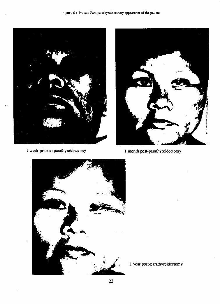
_- Figure $ : Pre and Post-parathyroidectomy appearance of the patient
1 week prior to parathyroidectomy 1 month post-parathyroidectomy
_ 1 year post-parathyroidectomy
22
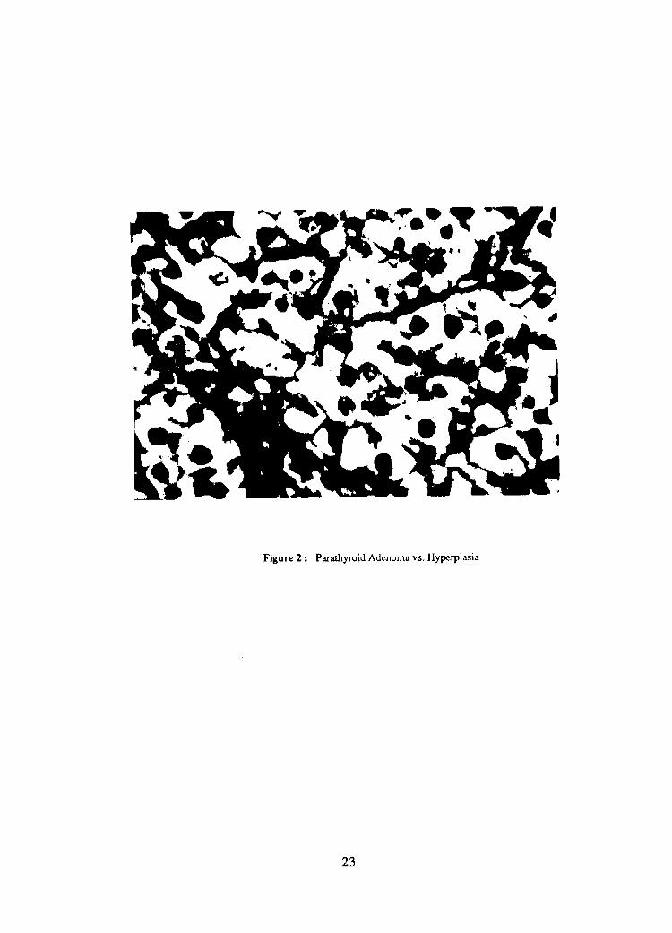
Figure 2 : Parathyroid Adcnoma vs. Hyperplasia
23

ltistopath on the first operation
Histopath on the second operation
Figure 3 : Brown tumor as seen in the patient. Note of the giant cells predominantly occupying the stroma.
24

E.wa_ileMassonLe_Maxilla "salt& pepper"appearanceofCalvarium
Figure 1 : RadiographicFindingsin PrimsryHyl_erparathyrodism
-_, _ as seen in u_ patient
Nephrocaleinosis
25

UNIVERSITY OF THE PHILIPPINESPHILIPPINE GENERAL HOSPITAL
DEPARTMENT OF OTOLARYNGOLOGY
TITLE : A MassivePyogenicgranulomain the Gingiva*
AUTHORS: ArmancloM. Chiong,Jr., MD; CharlotteM. Chiong,MD;ArthurY. Dy MD; Pio R. Pajarillo,MD
ABSTRACT the oral cavity of six years duration, described initiallyas 2 X 2 cm in size, firm, reddish and non-tender.
A large exophytic mass was seen in the oral cavity Eventually, there was note of frequent bleeding onof a 22 year old male. Repeated biopsies of the mass minimal trauma as well as progressive ulceration withshowed chronic inflammation with granulation tissue, non-foul mucopumlent discharge. On physical exami-An excisional biopsy confirmed the presence of nation, there was a 10 x 5 x 4 cm. mass in the rightpyogenic granuloma. This lesion is unusual as it upper gingiva protuding out of the oral cavity (Fig-attained a very large size causing considerable facial ure.1). There was displacement of the molars mediallydeformity. Intraoperatively, the lesion was noted to and the incisors anteriorly and outward. A bulge on
be highly vascular, this case may prove to be of great the right nasal floor and the right cheek was notedinterest to otolaryngologists who often deal with oral although the overlying skin was normal. Submentallesions, as it demonstrates an extreme and unusual and submandibular nodes measuring 0.5 x 0.5 cm.presentation of a definitely benign lesion, were movable and non-tender on palpation. There was
no history of smoking, betel nut chewing, chronicbeverage intake nor was there a history of trauma or
INTRODUCTION toothache preceeding the appearance of the lesion.
Pyogenic granulomas in the oral cavity are rela- A Water's x-ray revealed a soft tissue density overtively common lesions encountered by otolaryngolo- the right maxillary area with no distinct evidence ofgists. They are well circumscribed, soft tissue tumors bone resorption. On suspicion of malignancy, biopsyof inflammatory rather than neoplastic nature? While was repeated three times but consistently showedsuch lesions usually do not pose diagnostic problems, chronic inflammation with granulation tissue. Thethis case report illustrates an extreme and unusual patient was subsequendy admitted. Clinical impres-presentation of pyogenic granuloma where repeated sion varied, ranging from benign lesions such asbiopsies had to be done in order to confirm the fibromas, fibrous dysplasia to malignant lesions likediagnosis, fibrosarcoma or squamous cell carcinoma. An
exeisional biopsy under general anesthesia was done(Fig. 2). Intraoperatively, a solid encapsulated tumor
REPORT OF A CASE broadly pedicled on the rightmaxilla with bony spiculesin its substance was noted (Fig. 3). The mass was
A 22 year old Filipino male consulted the outpa- highly vascular such that total blood loss amounted
tient clinic of the Philippine General Hospital on June to 2 liters.17, 1986 for a large progressively enlarging mass in
Histophatology revealed a highly vascular fibromawith large areas of ulceration and intense chronicinflammatory exudates totalised of lymphocytes,
*l_e.eemtedat the PhilippineSociety of Otolaqmgology - Headand plasma cells and polymorphonuclear neutrophils.Nec_SurgeryRmearc, hFccumv_nIntere=tingCasei, September9,1988atthe There was no evidence of malignancy. On the basisManila Garden HowL
26

of some clinical features as well as the histologic the local irritant are eliminated and the pregRancy ter-picture, the final diagnosis was pyogenic granuloma, minated? Otherwise, treatment of PG involves
conservative surgical exicison.DISCUSSION
Differential diagnosis in this case included benignPyogenic granuloma (PG) of the oral cavity, lesions such a fibrous epulis, peripheral giant cell
although a relatively common lesion, may sometimes granuloma, calcigying fibrooblastic granuloma andpresent in an unusual manner. It is described to be ameloblastic fibroma. Anneroth and Sigurdson 2 pro-a well cricumscribed, soft tissue tumor with a size vided an excellent review and classification of the
varying from 0.5 cm to 2 cm or more. Trauma and above-mentioned hyperplastic lesions of the gingiva.
microtrauma due to toothbrushing and gingival in- In this case, a malignant process was also highlyflammation were cited as pathogenic elements? His- considered because of the unusually large size of thetologically, PG arises from the com_ective tissue of mass with its ulcerative surface and friability on tissuethe skin and more commonly of the oral mucosa where biopsy. Among the malignant lesions, a fibrosarcomait can be partly or completely covered by squamous as well as a squamous cell carcinoma were consid-epithelium. In 65% of cases, there is a varying degree ered. All these lesions will however, have distinct
of ulceration covered by a mucinousexudate. Usually, histologic features quite different from PG.lobulated lesions are Composed of solid ennthelialproliferation or proliferation of capillary sized blood The case presented is very unusual in that lesionvessels? Clinically, it appears as an elevated, sessile has attained a large size, unprecedented in worldor pedunculated soft tissue mass. It may have a smooth literature. Of particular interest is its highly vacularlobulated appearance sometimes with ulcerative sur- nature observed intraoperatively. Surprisingly, thereface which tends to bleed spontaneously. 5 Occasion- was no radiographic evidence of bone resorption in
ally, white sloughy material resembling pus maybe this case despite its size and duration. The clinicalobserved, thus the term "pyogenic" granuloma given appearance was however suggestive of a malignancyby earlier clinicians. In a review of 762 cases, PG such that repeated biopsies were done.involves in decreasing order of frequency the gingiva,lips, tongue, buccal mucosa, palate, mucobuccal foldand alveolar mucosa ofedentulous areas. From further SUMMARY
analysis of gingival involvement, the upper gingivaare more common site than the lower gingiva. Sta- A case illustrating a huge mass in the oral cavityblein and Silverglade in their comparative anlaysis of has been presented. A rather aggressive and malig-a total of 834 consecutive biopsy specimens from the nant process was considered because of the unusuallygingiva and 448 from the alveolar mucosa reported large size and the ulcerative surface of the lesion.an 85% incidence of inflammatory or reactive hyper-plasia with PG being most common in the gingiva Histophatologic examination often does notwith 23.6% incidence. There was a 10:l ratio of conform with the clinical impression thus resulting :benign to malignant neoplasia in the gingiva which in repeated biopsies to establish the definitive diag-was greater than in the alveolar (almost 1;1). Moriconi nosis.and Popowich 6 reported a case of alveolar granuloma
measuring 4 x 6 cm in size. Previous to this report, Finally, management was simple and straightfor-this case represents the largest PG to be reported in ward as conservative surgical excision need be doneworld literature. The lesion occurs in all ages with when histologic identification of a benign process ispeak incidence in the third decade as in this patient, confirmed.Radiographically, there maybe evidence of alveolar
bone resorption in large and long-standing gingivalmasses. There is a reported female preponderance ACKNOWLEDGEMENTespecially in pregnant women presumably due tohormonal factors. It is therefore known as a preg- The authors wish to tank Dr. Joselito Jamir andnancy or hormonal tumor. This is one instance when R. Armando T. Chiong of the Department of Oto-pyogenic granulomas need not be excised surgically laryngology, Jniversity of the Philippines for their
since regression and resolution of the mass occurs as invaluable advice.
27

._ REFERENCES 5. Kfir, Y., Buchner, A. and Hansen, L.: Reactive
lesions of the gingiva: A clinical
1. Angelopoulos, A.: Pyogenic granulomas of the pathological study of 741 cases. Joral cavity: statistical analysis of its Periodontol, 51:655-661,1980.clinical features. J Oral Surg., 29:840-
847, 1971. 6. Moriconi, E. and Popowich, L.: Alveolar pyogenicgranuloma: Review and report of case.
2. Anneroth, G. and Sigurdson A.: Hyperplasia lesions Laryngoscope, 94:807-909, 1984.of the gingiva and laveolar mucosa:
A study of 175 cases. Acta Odontol 7. Stablein, M. and Silverglade, L.: ComparativeScan(I, 41 (2):75-86, 1983. analysis of biopsy specimens from
gingiva alveolar mucosa. J. 07 33. Bhaskar, S.; Pyogenic granuloma - clinical featurers, Pciodontol, 56:671-678, 1985.
incidence, histology and result of
trcatment: Report of 242 cascs. JOral 8. Vilmarm, A., Vilmann, P. and Vilmann, H.:Surg, 24:391-398, 1966. Pyogenic granuloma: evaluation of
oral conditions. Br J Oral Maxillofac
4. Eversole, L. and Rovin, S,: Reactive lesions of Surg, 24:376-382, 1986.the gingiva. Oral Surg, 1:30, 1972.
28


MCU HOSPITALDEPARTMENT OF OTOLARYNGOLOGY
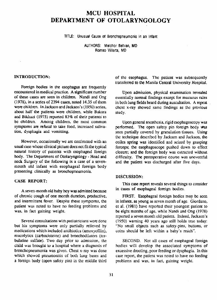
TITLE: UnusualCauseof Bronchopneumoniain an Infant
AUTHORS:MelchorBeltran,MDRomeoVillarta,MD
INTRODUCTION:_ of the esophagus. The patient was subsequentlytransferred to theManila Central University Hospital.
Foreign bodies in the esophagus are frequently
encountered in medical practice. A significazlt number Upon admission, physical examination revealed
of these cases are seen in children. Nandi and Ong essentially normal findings except for mucuous tales(1978), in a series of 2394 cases, noted 14.35 of them in both lung fields heard during auscultation. A repeatwere children, ln JacksonandJackson's (1950)series, chext x-ray showed same findings as the previousabout half the patients were children, while Bakara study.and Bikhazi (1975) reported 83% of their patients to
be children. Among children, the most common Upon general anesthesia, rigid esophagoscopywassymptoms are refusal to take food, increased saliva- performed. The open safety pin foreign body _astion, dysphagia and vomitting, seen partially covered by granulation tissues. Using
the technique described by Jackson and Jackson, the
However, occasionally we are confronted with an coiles spring was identified and seized by graspingusual case whose clinical picture does not fit the typical forceps; the esophagoscope pushed down to effectnatural history of patients with esophageal foreign closure; and the foreign body was extracted withoutbody. The Department of Otolaryngology - Head and difficulty. The postoperative course was uneventfulneck Surgery of the following is a case of a seven- and the patient was discharged after five days.month old infant with esophageal foreign bodypresenting clinically as bronchopneumonia.
DISCUSSION:
CASE REPORT: This case report reveals several things to considerin cases of esophageal foreign bodies.A seven-month old baby boy was admitted because
of chronic cough of one month duration, productive, FIRST. Esophageal foreign bodies may be seenand intermittent fever. Despite these symptoms, the in infants, as young as seven month of age. Giordano,patient was noted to have no feeding problems and et al. (1981) have reported their youngest patient towas, in fact gaining weight, be eight months of age, while Nandi and Ong (1978)
reported a seven month old patient. Indeed, Jackson'sSeveral consultations with pediatricians were done (1950) warning 40 years ago still holds true today:
but his symptoms were only partially relieved by "No small objects such as safety-pins, buttons, ormedications which included antibiotics (amoxycillin), coins should be left within a baby's reach".mucolytics (carbocisteine) and bronchodilators (ter-
bulatine sulfate). Two day prior to admission, the SECOND. Not all cases of esophageal foreignchild was brought to a hospital where a diagnosis of bodies will develop the associated symptoms ofbronchopneumonia was given. Chest x-ray was done excessive drooling, poor feeding or dysphagia. In thiswhich showed pneumonitis of both lung bases and case report, the patient was noted to have no feedinga foreign body (open safety pin) in the middle third problems and was, in fact, gaining weight.
31

Glass and Goodman (1974) have postulated that as Jackson and Jackson succinctly puts it, has notsince the diet of these young patients consisted of given rise to possible complications like ulcerations,liquid food, it can easily pass through the esophagus fistula and esophageal cicatrical stenosis (Jackson,even with large foreign bodies. 1950).
THIRD. The only presenting symptom that the SIXTH. The diagnosis of esophageal foreignpatient manifested was prolonged one-month duration bodies is by radiographic studies, first without, thenof cough unrelieved by medications if necessary with a capsule filled with an opaque
substance. If the opaque capsule seems to lodge, theIt may be challenged that the bronchopneumonia patient should have a diagnostic esophagoscopy.
of the patient was secondary to the foreign body.
However, Newman (1978) has showed that this may In conclusion, esophageal foreign bodies may bebe possible. He enumerated the possible pathogenesis overlooked while treating a patient with respiratoryof respiratory symptoms secondary to esophageal symptoms. Some esophageal foreign bodies mayforeign bodies, present with stridor, wheezing, chronic pneumonia,
or may simulate asthma, croup, bronchitis, and bron-1. compression of the trachea by the posteriorly chopneumonia particularly in children under three
place esophagus years of age. (Newman, 1978). As Jackson has said,
2. aspiration of pooled secretions in the pyriform "failure to consider a foreign body as a diagnostic.....sinus can occur from esophageal obstruction and lead possibility is one of the commonest causes of its
to pneumonitis or tracheobronchitis; oversight." (Jackson, 1950).3. long standing esophageal foreign bodies may
produce respiratory symptoms from cricoid perichon-dritis or periesophagitis; and REFERENCES:
4. very rarely, the esophageal foreign bodies maypass through the acquired tracheo-esophageal fistula 1. Nandi P. and Ong G.B (1978) Foreign body inand obstruct the airway, the esophagus: review of 2394 cases.
Br. J. Surg. C5:5-9.FOURTH. Although uncommon, sharp and
pointed forcing bodies can be difficult to manage. It 2. Jackson C. and Jackson C.L. (1950) Bronchoe-
is important to be careful not to make the situation sophagoscopy. Philadelphia, Saun-worse or to cause a complication, such as a perforated ders.esophagus. It is always well to remember Jackson's
dictum: "Advancing points puncture, trailing points 3. Bakara A. and Bikhazi G. (1975) Esophagealdo not." foreign bodies. Br. reed. J.I. 561-
563.
__: The open safety pin presents a special problem.Fortunately for us, Jackson and Jackson have dis- 4. Giordano A., Adams G. Boies L., Meyehoff W.cussed the various ways of removing an open safety (1981) Current management of eso-pin, For the open safety pin with point down, they phageal foreign bodies. Arc Otolsuggest: "the coiled spring is to be sought and, when 107:249-251.found, seized with the rotation forceps and the eso-
phagoscope pushed down over it to effect closure." 5. Glass W.M. and Goodman M. (1966) Unsus-For the open safety pin with point upward, they pected bodies in the young child's
suggested sixteen methods of safe removal (Jackson, esophagus presenting with respiratory1950). symptoms. Laryngoscope. 76:605.
FIFTH. The presence of granulation tissue 6. Newman D.E. (1978) The radioluscent esoph-surrounding the foreign body suggest that the open ageal foreign body: an often forgottensafety pin had been in the esophagus for a prolonged cause of respiratory symptoms. J. pedperiod of time. It is fortunate for the patient that this 92:1
"prolonged sojourn of foreign body in the esophagus"
32


JOSE REYES MEMORIAL MEDICAL CENTERDEPARTMENT OF OTOLARYNGOLOGY
TITLE: CancerandKeloidor KimuraandSteroid
AUTHORS: Henry/L Rossi,MDWilliamB.Dy,MD
INTRODUCTION 40 gm. was removed. Surprisingly, histopath showedno salivary tissue. The result was read as
There are times when an Otorhinolaryngologist- Angiolymphoid Hyperplasia with Eosinophilia. NoHead and Neck Surgeon is at a loss as to how intervention was made however on the earlobe mass.to manage a lesion mimicking numerous diseaseentities. The more confused he becomes when, in This seemed to be a mere descriptive termspite of previous standard therapy, the lesion of the entire microscopic picture. No further
.... keeps on recurring. Soft tissue tumors, in some investigation was made and patient was advisedof its unusual forms, demand that this special group to follow up regularly without any medication.of lesions be investigated as exemplified by the fol-lowing case. The earlobe mass continously increased in size.
Patient sought consultation from a private physicianCASE REPORT where fine needle aspiration biopsy was done on
the earlobe mass. Results revealed: bcnignlymphoidNine years ago, a 15 year old male consulted elements in the bloody background. No evidence
the JRRMC due to a corn size mass on his left of malignancy. No surgical intervention was doneearlobe. There were no other associated signs and nor any meds given.symptoms except occasional pruritis. Excisionbiopsy was done but the result was not known. There was no apparent improvement in the patientsLittle did the patient know that this would only condition. Although there was no pain andbe the first of a series of recurrences and surgical tenderness the patient was bothered by its graduallyinterventions, increasing size. He again sought consultation at
our institution where PE revealed a hard, non-Immediately after the first excision, the mass tender, keloid-like smooth mass on the left lobule,
recurred. It gradually increased in size making him about 3.5 _ 2.5 cms. in size. By this time thereseek consultation at Our institution five years later, were also two palpable lymph nodes on the samePhysical examination revealed a 2 x 2 cm. hard, non- side.tender smooth mass on the left lobule with a 1.5 x
1.5 cm mass of the same consistency on the infra- The case was presented at the grand roundsauricular region. A diagnosis of sebaceous cyst was and the following differential diagnosis was made:made. Excision was done and histopath revealedchronic inflammation. 1. Keloid formation or
2. Malignancy of the ear with neck node metastasisThree months later, the left earlobe mass again
started to gradually increase in size. This time Analytic disse_tion shows equivocal outcome withit extended to the pre-auricular region. The patient no definite advantage of one.sought consultation at another government medicalinstitution where it was thought to be a parotidtumor. Superficial parotidectomy was then done.Record showed a 6 x 6 x 2.5 cm. mass weighing
34

Appearance Presenceof Duration Recurrent thologically recognized entity that is characterizedNatureLymph
Nodes by cutaneous nodules, single or multiple, 1-10 cmsin diameter often located in the cheek and auricular
Keloid + + + region. It is also characterized by cutaneousMalignancy + + + proliferating blood vessels with a typical histiocyte-
like endothelial cells and numerous eosinophils.
Besides, previous histopath does not favor keloid A peripheral eosinophilia may be present but therebecause there was no collagen formation and neither are no known systemic manifestations. 85% occurcancer because there were no malignant changes in males. Pain and tenderness are rare although
noted. Consensus then was to do an incisional biopsy pruritus may sometimes occur. It is rather rare diseaseof the earlobe mass. Biopsy was done and histopath with only about 250 cases reported in world literature.result revealed stratified squamous epithelium with
hyperkeratosis and acanthosis. The dermis con- Histologically the lesions are characterized bytained lymphoid follicles with prominent germinal 3 striking features that are always present. The
centers and sheets of proliferating capillary endothe- vascular element, which preponderates in early morelium scattered within the loose connective tissue active cases and the lymphoid or cellular element,
stroma with numerous eosinophils. There is no which predominates in the later quiscent stages.evidence of malignancy. The final report read- Other important diagnostic features include diffuseAngiolyphoid Hyperplasia with Eosinophilia, the same mast cell and eosinophil infiltrations of the dermisresult in the previous excision, and subcutaneous tissues as well as serum eosino-
philia. Usually, copious numbers of eosinophils andWhat seemed to be a descriptive term and which lesser numbers of plasma cells, lymphocytes, and
was not given much attention is the past, now became histiocytes are present in close association witha topic of curiosity, capillary neogenesis. Sometimes lymphoid nodules
may be present. (Thompson, 1981).Post biopsy, patient had a 2.5 x 2.0 cm. mass
on his left earlobe with two palpable cervical lymph According to Goldman, the cause and phto-nodes. Management of this kind of disease can be genesis of this process remian unknown. Recentsurgical. But now we opted to be conservative. He studies have showr/that the type of progenitor cellwas started on steroids at a dose of 30 mg bid and of the lesion is probably a transitional histiocytic-after one week, mass had regressed to about One cm. endothelial cell and that the proliferation of thesein size and consistency changed from hard to soft. elements may represent a response of this basicCervical lymph nodes were no longer palpable, vascular cell to an inflammatory or injuries stimulus.
One year later, no mass was noted and the It was in 1.948 when Kimura et al first described
patient is now back to normal. After nine years an unusual s_cin disease with "unusual granulation
of carrying theburdenofhavingamassinhis earlobe, combined with hyperplastic changes of lymphaticpatient finally found relief not through surgical means tissue." Kawada in 1966 reported four cases. Wellbut through systematic steroids, and Whimster in 1969 had nine cases. In 1971
Mehregan and Shapiro in a study of 14 patientsDISCUSSION stated that vascular proliferation associated with the
formation of lymphoid follicles and tissue eosino-The histopathologic diagnosis points to a rather philia is also characteristic of a disease entity
uncommon, rarely heard of disease called Angiolym- described in Japanese literature asKimura's Disease.phoid Hyperplasia with Eosinophilia. It seems to It has since then been known under the title
be a mere description of the microscopic findings of Angiolymphoid Hyperplasia with Eosinophilia andbut it is actually a disease entity in itself. What later on Kimura's Disease. It has several other names
then is Angiolymphoid Hyperplasia with Eosino- such as angioblastic lymphoid hyperplasia withphilia? eosinophilia, eosinophilic lymph-folliculosis of
skin, papular angioplasia, and inflammatory angio-Angiolymphoid Hyperplasia with Eosinophilia matous nodules. But is ALHE really similar to
or abbreviated (ALHE) is a clinically and histopa- Kimura's disease? Wells and Whimster in 1965
35

nonetheless stated: "We have the impression that In Japan and China the disease occured
there are more similarities than differences bctwcen preponderantly in young men (85%) and the patientsALHE and Kimura's Disease". had numerous subcutaneous nodules. These nodules
were primarily in the cheeks but they also occured
However in 1969, Jones and Bleecham justified in the axilla, antecubital, and inguinal regions. Lymphthe separationofKimura's Disease from ALHEmainly node involvement was thought to be an importantbecause of their location. ALHE is found in the feature of the disease.dermis while Kimura's Disease is found in the
subcutaneous layer. Furthermore, in 1986, Batsakis Kimura's Disease, is an unusual disease that
and Manning came up with the histologic differences has been recognized only recently in the English
between Kimura's Disease and ALHE. speaking world. The orginal pathologic description
i IHISTOLOGICFEATURE 4 ALHE KIMURA'S DISEASE
location superficial subcutis deep subcutis adjacent salivarydermal involvement tissue involvement commoncommon
lymphoid cell infiltrate abundant to marked little to marked
size lcm (average) 3 cm. (average)
lymphoid follicles occasionally always
tissue eosinophilia always always
mast cells common unusualI
vascular angiomatous prolife- capillary & thick walled vesselsration of masses swollen enotheliumwithout lumen no uncanalized endothelial
proliferation
fibrosis often absent or only prominentat borders
.,_;__
In our patient, the lesion was in the deep and case reports appeared in Japanese literature.subcutis with apparent parotid tissue involvement and In these ca:,e reports, the lesions were moresize with more than three cm. There were marked generalized in their body distribution in contrast tolymphoid follicles and tissue eosinophilia. Vascular the more clinical appearance in the head and neck
component showed canalized endothelial prolifcra- area of the more recent cases in England and thetion. Fibrosis was only minimal. These features United States. (Thompson 1981).are more compatible with Kimura's Disease.
Batsakis and Manning in 1986 came up with racialTo ensure acurate diagnosis, it is essential differences in clinical presentation of Kimura's
that the otorhinolryngologist be aware of the his- Disease.tologic features of ALHE or Kimura's Disease as wellas its clinical characteristics.
36

CLINICALFEATURES ORIENTALS NON-ORIENTALS
ageat onset 1st decade onsetin 3rd topeakat 2nd 4th decadesand 3rd decades
sex 85% males rare in whites
pain and tenderness rare rare in whites
coexistentlymph- 40% incidence unusual in whitesdenopathy increasedto 65%
with tumors > 2 cm
peripheralblood 60% incidence 17% of whiteseosinophilia
predominantsite pre-auricular, pre-auricular,neck scalpscalp (rare) neck (rare)
Our patient, a 24 year old male, first noticed Batsakis and Manning suggested radiotherapythe lesion when he was 15 years of age. Tumor as a consideration in recurrent lesions.mass was larger than 2 cm. with the presence of
cervical lymphadenopathy. There was involvement Kimura's is indolent and even self-limiting inof the preauricular and neck area. Pain and some cases. However, there is a high incidence oftenderness were not present. Except for the local recurrence or persistence of Kimura's thatabsence of peripheral blood eosinophilia, our patient occur after various forms of treatment. Steroids
definitely belongs to the Oriental type of Kimura's. are given for systemic effect. The use of radio-therapy for indolent benign disease, particularly in
Batsakis said that the initial stimulus for the younger patients is condemned. (Thompson et al)lesion is unknown and since host responses with the
ethnic overlay may modify clinical expression, it is Our patient underwent three unsuccessful surgicalnot impossible to regard the lesions as a fundamen- incisions. Radiotherapy as an alternate mode of
tal biologic reaction with different clinical and management would be unthinkable due to ourpathologic expression in different races, patient's young age. Because of these circumstances
steroid was tried. Prednisone was started at a dose
MANAGEMENT of 60 mg/day and it resulted in remarkable regressionof the mass in just one week. One year after,
Current available literature suggests different he is still free of disease, does routine work as
treatment modalities in the management of Kimura's if nothing happened to him. The tum of eventsDisease. It has been treated with steroids and by was indeed dramatic for our patient who for thesurgical excision, irradiation, cryotherapy, and last nine years has endured the course and come:electrodessication. According to Thompson, per- quences of the disease.sistent residual disease and local recurrence are
frequent. He suggested laser excisions as a successful CONCLUSIONalternate mode of therapy with definite benefits overthose methods used commonly. Adequate surgical Soft tissue tumors of unusual forms in the head
excision seems to be the often treatment of choice and neck comprises a challenging group of pathologicbut the highly vascular nature of the ':lesion often conditions that cause difficulty in classification andmakes margins difficult to identify, treatment. This poses a great herculean challenge
37

to the diagnostic know-how of the clinician. There BIBLIOGRAPHYare times that no matter how much intellectual effort
the clinicians summons, the diagnosis of these un- 1. Batsakis, J.G. and Manning, J.T., Softfamiliar cases would be very difficult without the Tissue Tumors of Unusual Forms. Oto-
help of an able pathologist. An example is the laryngologic Clinics of North America. pp.case being presented: Kimura's Disease. Clini- 673-675, 1986.
cally, it mimics a lot of tumors, particularly themore common ones, which led to misdiagnosis 2. Goldman, R.L. and Kelin, H.Z., Subcu-and therefore mismanagement which may prove taneous Angiolymphoid Hyperplasia with Eo-to be costly on the part of the patients. In conclusion sinophilia. Archives of Otolaryngology. pp.several points are hereby emphasized. 440_441, July 1976
a) This unusual disease exists among Filipinos 3. Hyams, V.J., Pathology of Tumors of theand that it should not be left out from our Ear. Comprehensive Management of Headdifferential diagnosis and Neck Tumors, Vol. I. W.B. Saunders
Company. p. 169, 1987.b) The difficulty in diagnosisng it clinically
behooves us to consult a pathologist for its 4. Kim, B.H. and Sithian, N., Subcuta-proper diagnosis and, consequently, for its neous Angiolymphoid Hyperplasiamanagement. (Kimura's Disease). Archives of Surgery.
pp. 1246-1248, 1975.c) Getting a mere histopathologic description
should not stop the clinician from further 5. Mchregan, A.H. and Shapiro, L., Angiolym-inquiry to determine the specific disease phoid Hyperplasia with Eosinophilia.entity, no matter how rare it is. Archives of Dermatology. pp. 50-57.
1971.
Keen diagnostic acumen, knowledge of the
existence of the disease, and expert pathologic 6. Schnitzeretal. LymphoreticularDisorders.interpretation are the basic requirements for the Tumors of the Head and Neck, 2nd edition.early diagnosis of Kimura's disease, p. 484. Williams and Wilkims. 1982.
Finally, steroid have so far proven itself_ to be 7. Thompson, J.W. et al. Angiolymphoidthe wonder drug, for a highly recurrent lesion such Hyperplasia with Eosinophilia of theas Kimura's. External Ear Canal. Archives of
otolaryngology, pp. 270-275, June 1983.
38

JOSE R. REYES MEMORIAL MEDICAL CENTERDEPARTMENT OF OTOLARYNGOLOGY
TITLE: NGTInducedSinusitis: A ProspectiveCohort Study
AUTHORS: WilfredoE Batol, MDFelicidadC. Felicilda,MD
Gil M. Vicente,MD
ABSTRACT: cations since, with the introduction of a polyvinyl tubeinto the nasal cavity, the sanctity of this natural
A cohort study of 51 in-patient with NGT (Group orifice may have been violated.1) at the Jose R. Reyes Memorial Medical Centerwas undertaken from October 1969 to August 1990 Linden et al in 1988 recognized sinusitis as ain order to determine the relationship of nasogastric complication of nasotracheal intubation. 7 Withtube to the development of sinusitis. 50 in-patients tubes of larger diameter exactly filling up the nasalwithout NGT (Group 2) age and sex matched served cavity, nasal obstruction causing inflammation of theas control. 39 patients (76.47%) in Group 1 while ostiomeatal unit with subsequent poor 9,10,11 drain-10 patients (20%) in Group 2 developed sinusitis within age of the paranasal sinuses- may reult to sinusitis.the minimum observation period of 14 days. Thereis a statistically significant difference between these But can a tube with a smaller diameter liketwo groups (p<0.01. relative risk = 3.82). nasogastric tube produce similar complications?
Among patients with NGT, sinusitis is noted to It is common knowledge that nasogastricoccur between the 3rd to llth day with a mean of intubation causes nasal obstruction and hence rhini-6.63 days. In this study, comparison of patients tis. But does it follow that NGT causes sinusitis?with deviated nasal septum and those with midlinenasal septum as to the development of sinusitis was A review of international and local literaturedone. However, when we compare the site of NGT showed no specific studies on sinusitis among patientsinsertion in Group 1, among the 32 patients with with nasogastric tube were done. To better definedeviated nasal septum, 20 had their NGT placed in the problem and to increase physicians' awarenessthe side of deviation and 12 developed sinusitis, of nasogastric intubation in relation to sinusitis, this
while 2 out of the 12 (25%) patients with NGT prospective study was undertaken.placed in nondeviated side showed evidence of
sinusitis. There is a significant statistical differencebetween these patients (p<0.01. Relative risks -- 4.8). GENERAL OBJECTIVE:
Based on these findings, the authors recommend To determine the relationship of nasogastricnasal examination prior to NGT insertion, change intubation to the development of sinusitis.of NGT site every 7 days and avoidance of NGT
insertion in the deviated side among patients with SPECIFIC OBJECTIVES:deviated nasal septum.
1. To determine the prevalence of sinusitis among
INTRODUCTION: patients with nasogastric intubation.2. To compare the prevalence of sinusitis between
Nasogastric intubation is a seemingly simple patients with nasogastric tube and those without it.and ordinary procedure. This is a task often delegated 3. To determine additional predisposing factors ,to the medical clerks and interns. However, this in the development of sinusitis in patients withinnocuous procedure might not be without compli- nasogastric tube.
39

PATIENTS & METHODS: These signs and symptoms are the typical triadof sinusitis as proposed by Stammberger & Wolf. it
For a period of 11 months (October 1989 to August Patients underwent a second paranasal sinus x-ray
1990), all in-patients at the Jose R. Reyes Memorial soon as they manifest signs and symptoms of sinusitis.Medical Center in whom nasogastric intubation were However, if no signs and symptoms were noted,indicated were examined and evaluated. Prior repeat x-rays were done on the 7th and 15th day fromto nasogastric intubation, complete history and ENT the time of observation. A minimum period ofphysical examinations which included anterior and 14 days observation was required. Comparisonposterior rhinoscopy, otoscopy, oropharyngeal ex- of the initial and the succeeding paranasal sinus x-amination and indirect laryngoscopy were done. rays of the two groups were performed. (Figure 1)Initial x-ray of the paranasal sinuses (Waters, Cladweil& Lateral views) was performed. Patients were ex- A diagnosis of sinusitis was made if patientseluded according to the ioliowing criteria: showed:
(a) at least two of the signs and symptoms1. Those with prior history of acute or chronic previously mentioned
sinusitis and allergic rhinitis. (b) if the films showed haziness of the parana-2. Those patients whose initial sinus x-ray shows sal sinuses
haziness of the paranasal sinuses.3. Those with prior surgical treatment to the
nose and paranasal sinuses. FIGURE1. METHODOLOGY
4. Those with prior history of trauma to the GROUP1 W/s/s->2NDnose and paranasal sinuses. / \/ PNSx_¥
5. Those with tumors of the nose and paranasal In-patients--> HISTORY-->EXCLUSION }sinuses. P.E. CRITERIA }\ /\
6. Patients below 15 yrs. of age. GROUP2 W/O$/S->XRAY7. Pregnant patients. 7 OR14O8. Uncooperative patients.
Patients in whom nasogastric tube was indicated INITIAL PNS X-RAYand who did not belong to the exclusion criteria wereincluded in this study. This set of patients were labeled Statistic',d analysis using Chi-square test withas Group 1. Yates correction was used in the evaluation of the
data with p values less than 0.05 considered as
A second set of patients admitted without NGT, significant.age and sex matched with the first group was likewiseexamined and evaluated. This set of patients was RESULTS:
, labeled as Group 2. Exclusion criteria were also appliedto this second set of patients. Patients who were Sixty eight patients with NGT were initially
included had initial sinus x-rays. Daily complete included in this study. There were 17 drop-outs dueENT examinations were also done. to early removal of the NGT and because of early
discharge from the hospital. Finally, fifty-one pa-
In both groups, the occurence of foUowing tients who had NGT were included and labeled assigns and symptoms were evaluated: Group 1 while another 50 patients without NGT were
labeled as Group 2 (age and sex-matched) for a1. nasal obstruction or stuffiness total of one hundred and one patients. Group 1
had 30 males and 21 females, While Group 2 had
2. pathologic secretions (purulent, mucoid or 30 males and 20 females. The age range of Groupserous discharge) 1 patients was 15 through 89 years, with a mean
age of 41 years. The age range of Group 2 patients3. headache or tenderness localized to the area was 17 to 84 years with a mean age of 39 years.
of sinuses, Group 1 had 32 "(62.7%) patients with deviatednasal septum and 19 (373%) with midline nasal
40

septum and 26 (52%) patients with midline nasal The average length of hospital stay of both groupseptum (Table 1). Nasal septum is a structure that was 25.5 day with a range of 14 to 35 days.divides nasal cavity into the right and left. However, The signs and symptoms manifested by thea nasal septum which is an anatomic midline patients with sinusitis are shown in Table 2.structure can be deflected/deviated to either side
with varying degrees of nasal obstruction.
TABLE1. NASALFINDINGSIN GROUP1 & GROUP2
GROUP1 ............................................GROUP2(N=51).....................................................(N=50)
NASALFINDINGS
DeviatedNasalseptum 32 (62.7%).........................................24 (48%)Midline Nasalseptum 19 (37.25%).......................................26 (52%)
Admissiondiagnosesof Group1 patientswere:
Mandibularfracture.......................................................................................................................................20 (39.2%)Cerebralinfarct...............................................................................................................................................10 (19.8%)intestinal obstruction....................................................................................................................................8 (15.7%)Carcinoma,larynx, SIP laryngectomy.........................................................................................................4 (7.8%)Esophagealstricture......................................................................................................................................3 (5.9%)Supraglotticwed post excision....................................................................................................................(3.9%)0steomyelitis,mandible.................................................................................................................................2 (3.9%)Carcinoma,hypoharynx................................................................................................................................1 (1.9%)Carcinoma,floor of the month....................................................................................................................1 (1.9%)
Admissiondiagnosesof Group2 patientswere:
LateralNeckMass........................................................................................................................................3 (26%)EsophagealNeoplasm...................................................................................................................................11 (22%)Stab wound, Neck........................................................................................................................................10 (20%)Mandibularfracture.......................................................................................................................................10 (20%)LaryngealCaS/P Tracheostomy.........................:........................................................................................ 6 (12%)
TOTAL. ..................................................................................................................... 50................
TABLE2. SIGNSANDSYMPTOMSMANIFESTEDBY PATIENTSWITH SINUSITIS:
GROUP1 GROUP2(N=39) (N=IO)
Nasal Obstruction 39 (100%) 8 (80%)Nasal Discharge 22 (84.6%) 4 (40%)Purulent 27 (81.8%) 3 (75%)Mucoid 4 (12.1%) 1 (25%)Serous 2 (6.1%) 0 (0%)Headache 5 (12.8%) 6 (60%)Tenderness 5 (12.8%) 6 (60%)Maxillary 2 (40%) 2 (33.3%)Frontomaxillary 3 (60%) 4 (66.7%)Otalgia 7 (17.9%) 0 (0%)
41
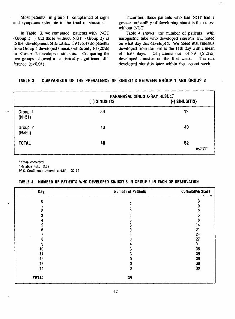
Most patients in group 1 complained of signs Therefore, these patients who had NGT had aand symptoms referable to the triad of sinusitis, greater probability of developing sinusitis than those
without NGT.
In Table 3, we compared patients with NGT Table 4 shows the number of patients with(Group 1 ) and those without NGT (Group 2) as nasogastric tube who developed sinusitis and notedto the development of sinusitis. 39 (76.47%) patients on what day this developed. We noted that sinusitisfrom Group 1developed sinusitis while only 10 (20%) developed from the 3rd to the 11th day with a meanin Group 2 developed sinusitis. Comparing the of 6.63 days. 24 patients out of 39 (61.5%)two groups showed a statistically significant dif- developed sinusitis on the first week. The restference (p<0.01). developed sinusitis later within the second week.
TABLE3. COMPARISONOF THE PREVALENCEOF SINUSITIS BETWEENGROUP 1 AND GROUP2
PARANASALSINUS X-RAY RESULT
(+) SINUSITIS (-) SINUSITIS)
• - Group 1 39 12(N=51)
Group 2 10 40(N=50)
TOTAL 40 52p<O.01*
*Yatescorrected*Relativerisk: 3.8295%Confidenceinterval= 4.61- 37.64
TABLE4. NUMBEROFPATIENTSWHO DEVELOPEDSINUSITISIN GROUP1 IN EACHOFOBSERVATION
Day Numberof Patients CumulativeScore
,_. 0 0 01 0 02 0 03 5 54 3 85 6 146 9 217 3 248 3 279 4 31
10 3 3611 3 3912 0 3913 0 3914 0 39
TOTAL 39
42

In Table 5, we compared the patients with However, when we compare the site of NGTdeviated septum and those with midline nasal septum insertion in the development of sinusitis, we got oneas to the development of sinusitis. Group 1 had interesting statistics. Among the 32 patients with32 patients with deviated nasal septum. Out of these deviated nasal septum in Group 1, 20 (62.5%) had
patients, 22 developed sinusitis. Nineteen patients their NGT place in the side of deviation and 12 (37.5%)from Group 1 had midline nasal septum. Out of these had them placed in the nondeviated side. 19 out ofpatients, 17 developed sinusitis. 20 patients (95%) showed evidence of sinusitis, while
3 of the 12 patients (25%) with NGT placed in theComparing the patients with deviated nasal septum nondeviated side had evidence of sinusitis. Compar-
and those with midline nasal septum as to the de- ing the two results showed a significant statisticalvelopment of sinusitis showed no statistical signifi- difference (p<0,01) TABLE 6.cant difference (p<0.05) (TABLE 5). Therefore, wecan say that in the development of sinusitis, we did Therefore, we can say that if the NGT is placednot show a difference whether patient had deviated in the deviated side, sinusitis is more likely to occur.nasal septum or not.
TABLE5. COMPARISONOFTHEPREVALENCEOFSINUSITISIN GROUP1 PATIENTSAS TO THENASALFINDINGS
PARANASALSINUSX-RAYRESULT(+) SINUSITIS (-) SINUSITIS
NASAL FINDINGS 22 10(N=32)MIDLINESEPTUM 17 2(N=19)TOTAL 39 12
p>O.05*= ,,
*Fishersexact2 tailedp value
TABLE6. COMPARISONOFPREVALENCEOFSINUSITISAMONGGROUP1 PATIENTSWITHDEVIATEDNASALSEPTUMIN RELATIONTO SITEOF NGTINSERTION.
±
PARANASALSINUSX-RAYRESULT(+) SINUSITIS (-) SINUSITIS
SITEOFNGT
NGTIN THE 19 1DEVIATEDSIDE(N=20)NGTIN THE 3 9NONDEVIATEDSIDE
(N=12)
TOTAL 22 10
p<O.01*1
•Yatescorrected•Relativerisk: 4.8095%Confidenceinterval= 2.46- 228.71
43

DISCUSSION: and septal deviation might be an additional predis-
posing factor to the development of sinusitis.Sinusitis is defined as an inflammation involving
the lining membrane of any paranasal sinus. While Between 1 to 7 days, 24 patients (61.5%)
the most common etiology of paranasal sinusitis is in Group 1 developed sinusitis. On the secondthe respiratory viruses, a variety of other agents such week of observation period, 15 more patients fromas nasal tumors, foreign bodies, and any 2.8.9 dcvices Group 1 showed evidence of sinusitis. In this study,placed inside the nasal cavity may play a major role. sinusitis was noted to occur between 3 to 11 daysVarious authors recognized predisposing factors with a mean of 6.63 days. Right on the firstin the development of hopsital acquired sinusitis day of NGT insertion, inflammation takes place.among which are nasotracheal tubes, facial or cranial With further inflammation of the mucosa, the sinusfractures, nasal packing 2.6.7 and nasogastric tubes, becomes blocked. This inflammation of the sinus
The critical importance of obstruction of the osti- mucosa is seen as haziness on x-rays. Manifestationsomeatal unit secondary to nasal intubation should of sinusitis begin to develop on the 3rd day onwardsbe emphasized. TM The sinuses drain through the and by the 7th day, many have developed sinusitis.ostiomeatal unit into the nasal cavity. Drainage of
the sinuses is also dependent upon 1.10 the size and It has been shown that there is no statisticallylocation as well as the patency of the ostia. Therefore, significant difference between the prevalence ofnasogastric tube as it impinge against the nasal sinusitis in patients with deviated nasal septum and
......mucosa can cause rhinitis and if the nasogastric tube those with midline nasal septum. But if the NGTis placed in such a way that it interferes with the is placed in the side of deviatior_, there is a greaterpatency of sinus ostium, this then causes sinusitis, risk of developing sinusitis as shown in TableIn our study, sinusitis among patients with nasogas- 6. Septal deviation per se is a potential cause oftric tube occurred in 39 out of 51 patients (76.5%) sinusitis. +.s,_ If the NGT is placed in the deviatedin Group 1 while only 10 patients had sinusitis in side, there will be more pressure contact on theGroup 2 (without NGT). Comparing the results of nasal mucosa. Nasal obstruction will be more
the groups, a statistically significant difference was evident in an already constricted nasal cavity. Thenoted (p<0.01). Patients with NGT has a higher potential factor should therefore be taken into con-risk of developing sinusitis than those patients without sideration during NGT insertion.NGT (Relative Risk of 3.82) as shown in Table 3.
The limitations of this study are:It is a common knowledge tlaat nasogastric tube
with its foreign causes inflammation/congestion of 1. In the evaluation of sinusitis, direct
the nasal mucosa resulting to rhinitis. Later on, aspiration of the sinuses or antral lavagemechnical obstruction of the 12 ostiomeatal unit in with culture of the aspirate 2.7.9 shouldthe lateral nasal wall may occur. Impairment of sinus have been done. But because of poordrainage enhances bacterial growth thus causing patient cooperation, the technical difficulties
-*sinusitis. 3 of this invasive procedure and that othersinuses are not directly accessible, this was
12 patients (23.5%) with NGT did not develop not done in this studysinusitis, it was noted that in this subgroup of
patients, the nasal examination showed the nasogas- 2. Reversibility of sinusitis should be nasogas-tric tube was inserted in the nondeviated side of tric tube be removed 2,5.7 was not ascer-
ten patients. It is also possible that the NGT was tained due to poor patient follow-up.placed in such a way that it hugs the floor of the
nasal cavity away from the middle meatai area hence 3. Owing to the unavailability and prohibitivemay not affect the ostiomeatal unit. cost of the sinuscopes x in our local set-up,
the ostiomeatal complex cannot be as-Among the group 2 patients, 10 (20%) developed sess_'d. Such assessment would have
sinusitis in their stay in the ward. All of these patients improved evaluation of the effect ofhad deviated nasal septum. These l_atients were nasogastric tube in the area of the ostiomeatalexposed to nasocomial infections while in the ward unit.
44

CONCLUSION & RECOMMENDATIONS: discarded as a routine procedure requiring no pre-
liminary knowledge of the area of insertion. For
The important findings in this study are the medical clerks, interns, resident physicians andsummarized as follows: even the specialists aspiring for quality medical care,
may this simple advise on nasogastric intubation
1. There is a higher prevalence of sinusitis be a symptom-free experience for the patients andamong patients with nasogastric tube than a trouble-free procedure for the physicians.those patients without it. Sinusitis was notedto occur in 76.47% of patients with NGTin this study. The risk of developing REFERENCES:sinusitis secondary to NGT is 4 times
greater than those patients without NGT. 1. Becker SP: Anatomy for Endoscopic SinusSurgery. In Endoscopic Sinus Surgery. The
2. Sinusitis in patients with NGT developed Otolaryngologic Clinics ofNorthAmerica, editedbetween 3 to 11 days with a mean of 6.63 by Friedman, M. 22:677-682. Aug. 1989.
days. As the duration of nasogastric tube 2. Caplan E, Hoyt N: Nosocomial Sinusitis.Journal of American Medical Association.is prolonged more patients develop sinusitis. 247:639-641. Feb. 1982.
3. Dretmer B; Johansson P; Kumlien J: Experimen-3. the risk of developing sinusitis is greater tally induced sinusitis: the imlxmance of
if the NGT is placed on the deviated portion vasomotor regulation. Archives of ORL-HNS.of a deviated nasal cavity. 246 (5); p. 315-317. 1989.
4. Evans FO Jr., Sydnor A Jr., Moore WE et al:In light of the results of this study, the following Sinusitis of Maxillary Antrum. New England
points are recommended: Journal of Medicine. 293: 735, 1979.5. Knodel A, Beckman J: Unexplained Fever in
1. Nasal examination should be done prior to patients with Nasotracheal Intubation. Journal
nasogastric intubation to determine side of of American Medical Association. 248:868-septal deviation, if present, and to take note 870. Aug. 1982.of other obstructivetissue such as enlarged 6. Levy C; Meye P; Guerin JM; DeberardinisF; Aouala D:Nasocomial sinusitis inan intensiveturbinates. The medical clerks, interns, care unit. Role of nasotracheal intubation. Ann.
and residents should know this basic step. Otolarygol Chir Cervicofac. 105 (7)p 549-552.2. Since sinusitis among patients with NGT 1988 (MEDLINE).
is noted to occur on the average on the 7. Linden B, Aguilar E, Alien S: Sinusitis in6th day, physicians are cautioned not to Nasotraceally lntubated patients. Archives ofprolong the stay of the NGT in one nasal Otolaryngology Head & neck Surgery. 114:860-cavity. If this is not possible, a change of 861. Aug, 1988/NGT site to the other nasal cavity on the 8. Mackay I, Cole P: "Phinitis, sinusitis and6th or 7th day is recommended, asscoiated Chest Disease". In Scott Brown's Oto-
3. If nasal examination showed deviated laryngology: Rhinology. 5th ed., edited by
nasal septum, putting the NGT in the Mackay & Bull. Butterworth & Co. pp. 73-deviated side is not recommended. If 77, 1987.
9. Malow JB, Creticos CM: "NonsurgicalNGT is indicated, put the NGT in the Treatment of Sinusitis". In Otolaryngologicnondeviated side. Clinics of North America. 22:809-817. Aug.
4. The attending physicians (especially the 1989.
neurologist) who deals with a comatose 10. Rhys Evans PH.: "Anatomy of Nose andpatient are advised to be more aware with Paranasal sinuses". In Scott Brown's Otolaryn-the problem. For any unexplained signs and gology: Basic Sciences. 5thed., editor by Davidsymptoms of infection such as fever W. pp 138-160 Butterworths & Co. 1987.
and leucocytosis, 5the NGT causing sinusi- 11. Stammberger H., Wolf O.: headaches and Sinustis could be the culprit. Disease, the Endoscopic Approach. Arch. of
Otolaryngotogy Head and Neck Surgery-supple-
Nasogastric intubation should not therefore be ment. pp 3-22, 1986.
45

OSPITAL NG MAYNILADEPARTMENT OF OTOLARYNGOLOGY
TITLE: HereditaryHemorrhagicTelaoiectasia
AUTHOR: NixonS. See, MO
This is the case of Rosa Param a 31 y/o female Head: normocephalicfrom Manila admitted for the first time on August10, 1989. Eyes: palepalpebralconjunctiva,anictericsclera
CHIEF COMPLAINT : nose bleeding Ears: intacttympanicmembrane,no discharge
HISTORY OF THE PRESENT ILLNESS: Nose:
Anterior rhinoscopy: with activebleedingnotedinleftOne year prior to admission patient started to ex- nasal cavity
perience several episodes of epistaxis which stopped
spontaneously on local pressure. Posteriorrhinoscopy: (+) activebleeding
•Three days prior to admission patient had pro- Oralcavity: (-) tonsillo-pharyngealcongestionfuse nose bleeding and was brought to the PLM-OM (-) mucosal lesionswhere anterior nasal packing was done which con-trolled her epistaxis. However patient removed the Neck: (-) mass, no cervicallymphadenopathypacking and epistaxis recurred thus requiring subse-quent admission. Chest: symmetricalthoracicexpansion
(-) retraction
PAST MEDICAL HISTORY: Lungs: clearbreath sounds,(-) tales/wheezes,with systolicblowing
murmurheard in the right interscapular1987DX:PTBwithintakeof unrscalledmedications areaaccentuatedon deepinspiration
OBSTETRICALHISTORY:Menarcheat 18 y/o,regular,30 days Heart:: adynamicprecordium,normalrate regu-cycle.lar rhythm,no murmur3 daysduration,moderatein amount
FAMILYHISTORY: (+) bleedingtendency-paternalgrand- Abdomen: flat, soft, non tender, no organomegalymotherandfather
Extremities: (=) clubbing, cyanoticnailbeds(+) PTB- grandmother
Neurologicalexamination: essentialnormal(+) Cancer- paternalandmaternalside
COURSE IN THE WARD:PHYSICAL EXAMINATION:
On admission posterior nasal packing was doneGeneralSurvey: conscious,coherent,pale,ambulatory,not to control the bleeding. Several laboratory exami-
in distress nation were done CBC and peripheral smear revealed
V,S.: PB:90/60PR:lO0/minRR:20/rainT:36.7 hypochromic normocytic anemia. Platelet count,C clotting time, andprothrombin time werenormal Chest
46

x-ray revealed a dense tortuous cylindrical nodule at leukemia and aplastic anemia were ruled out becausethe posterior RUL and a pleurodiaphragmatic adhe- of a normal blood morphology and bone marrowsion on the left. Possibility of an A-V malformation findings.and TB granuloma was entertained. Pulmonary andcarotid arteriography were contemplated but was not Hereditary Hemorrhagic Telangiectasia is the moredone due to financial constraint. ECG was normal, probable consideration for this case. It is autosomalABG showed hypoxemic hypocardia. Two units of dominant disease characterized as a sytemic fibrovas-
FWB were tranfused. Post BT Hgb and Hct were. cular dysplasia in which telangiectasia, A-V malfor-low, patient continued to have on and off epistaxis mation and aneurysm maybe widely distributed throughnecessitating transfusion of 4 units of FWB in a week out the body vasculature. This is recognized as atime. classic triad of telangiectasia, recurrent bleeding and
family history. The lesions maybe pinpoint in sizeOn the 10th hospital day bone mar'row aspiration or larger and form nodular vascular tumor, sometimes
biopsy was done to rule out blood dyscrasia, result it appear as a spider-like lession. Significant syrup-revealed normal bone marrow findings, tomatology include recurrent bleeding from mucosal
telangiectasia, hypoxemia, cerebral embOlism and brainOn the 13th hospital day the patient bled pro- abscess due to pulmonary A-V malformation.
fusely, she underwent emergency bilateral externalcarotid artery ligation in an attempt to control epis- Epistaxis is the most common form of bleedingtaxis. Post operative Hgb and Hct were still below and it is the first hemorrhagic event in I_Iereditarynormal value prompting another series of blood trans- Hemmorhagic Telangiectasia. In our patient severalfusion. Patient continued to have episodes of epis- attempts have been done to control epistaxis i.e., local
taxis which spontaneously stopped. Rigid nasopharyn- pressure, anterior and posterior nasalpacking, ligationgoscopy was done revealing an erythematous maculo- of the blood supply of nasal cavity . Yet inspite ofpapular lesion on the left side ol_ the nasal septum these interventions, there were still episodes of bleed-located posteriorly, ing. Bilateral external carotid artery ligation com-
pletely failed to control the hemorrhagic event in this
Upon discharge patient continued to experience patient because of the rich vascular channols supply-epistaxis relieved by local pressure. Two months later ing the nasal cavity.profuse nose bleeding recurred and she lost conscious-
ness for which she was rushed to Ospital ng Maynila Hereditary Hemorrhagic Telangiectasia is the mostand was re-admitted. Contrast C.T. scan was done common disease associated wth pulmonary A-V fis-
which revealed normal findings, tula. Pulmonary fistula maybe solitary of multiple,majority of which • receive blood supply from the
In summmary, we are presented with a 31 y.o pulmonary artery and are right to left shunts. Rightfemale patient with the following salient features; to left shunts allows desaturated blood to enter therecurrent epistaxis, erythematous maculo-papular systematic circulation to cause cyanosis by hypoxemia,lesion on the nasal septum clubbing and cyanosis of polycythemia and clubbing.nail beds, a blowing murmur at the right interscapulararea which appear to be extracardiac in origin, normal On chest x-ray the typical pulmonary A-Vhematologic studies except for anemia, possibility of malformation is peripheral, circumscribed, aon calcifican A-V formation on chest radiography and a family nodule connected to the hilus by vascultr strands.history which revealed that her grandmother had similarexperience. Withthese, the possible underlying cause Management of this case is variable dependingof recurrent epistaxis should be explored, on the severity of the disease. Oral iron and folic
acid supplements maybe necessary to avoid ironInfection would not produce such prolonged and deficiency. Transfusion requirements were variable.
massive epistaxis. There was no history nor evidenceof recent nasal truma. Patient was not hypertensive. Treatment modalities to control bleeding such asHematologic dysfunctions such as acute leukemia, local pressure, topical vasoconstrictors, claemical oraplastic anemia, and Heriditary Hemorrhagic Telang- electrical cat, tery and arterial ligaton offer onlyiectasia should be considered. However, acute
47

..temporary relief. According to Harrison "once nal of Laryngology and Otology.significant bleeding has started, local non specific June 1979; 93:589-595.theraphy is not only ineffective but frequently en-hance bleeding and, by destroying the septal mucosa McDonald, Tomas. Manifestation of sytemic Disease,or producing scar tissue, may reduce possibility of Charles Cummings. Otolaryngology,future control. Head and Neck Surgery. 1986
Morsby: 597-609.Two other modes of therapy were found to achieve
prolonged remission of epistaxis. These are the use Menefee, M,G. et al: Hereditary Hemorrhagicof estrogen and septal dermoplasty. Telangiectasia: An electron micro-
scopic study of the vascular lesionsSeveral studies have reported that estrogen may before and after the therapy with
decrease epistaxis in Hereditary Hemorrhagic Telang- hormone. Archives of Otolar-iectasia by endothelium or by undergoing squamous yngology.1975; 101:246-251.metaplasia of the nasal mucosa overlying the lesion.
Peery, William: Clinical spectrum of HereditaryAt present our patients have been receiving Hemorrhagic Telangiectasia. Ameri-
estrogen. Results have been favorable, can Joumal of Medicine. May 1987;__ 82:989-996.
There were reports that surgical management
should be contemplated for the case of Hereditary Reilly, Peter: Clinical Manifestation of HereditaryHemorrhagic Telangiectasia that bleed profusely. Hemorrhagic Telangiectasia.Since other forms of management have proved American Journal of Gastro-unsatisfactorily, several authors have advocated the enterology. 1984; 79:363-367.use of dermoplastly.
Strauss, M. et al: The management of severe recurrentHowever,septal dermoplastly can only be used for epistaxis due to Hereditary Hemor-
disabling epistaxis that is located anteriorly. Success rhagic Telangiectasia using regionalrate n relieving epistaxis by doing this procedure has facial cutaneous flap. Journal of
reached 75%. Failure is attributed to inadequate graft Laryngology and Otology. Aprilcoverage and graft shrinkage and re-expose of the 1985; 99:373-377.abnormal vessels.
Zohar, Yuval et al: Surgical Management of Epistaxisin Hereditary Hemorrhagic Telang-
REFERENCES: iectasia. Archives of Otolaryngol-ogy Head and Neck Surgery. July
'*Harrison, D.: Use of Estrogen in the treatment of 1987; 754-757.Familial Hemorrhagic Telangiectasia.Laryngoscope, March 1982; 92:314-320.
Hatcher, Virgil et al: Recurrent epistaxis and growthin lips and nose. Hospital Practice.July 1982; 17:40-41.
Jank, Volker: Ultrastructure of Hereditary Hemor-rhagic Telangiectasia: Archivcs ofOtolangology. 1970; 91:262-265.
Laurian, M.: Amniotic graft in the management ofsevere epistaxis due to HereditaryHemorrhagic Telangiectasia. Jour-
48

STO TOMAS UNIVERSITY HOSPITALDEPARTMENT OF OTOLARYNGOLOGY
TITLE:TheNeckMass,A diagnosticChallengeAUTHORS:GerardC. Agcaoili,MD
.AmadoC. Pacio,MD
The differential diagnosis of a neck mass covers INTRODUCTIONa broad spectrum of disease and carries implicationsfor treatment as varied as any area of medicine. The practice of Otolaryngology has always bee_
Cysticercosis is an uncommon disease that is dynamic and interesting brought about by the varietyrarely seen by an otolaryngologist. In fact, it is of cases involving the ear, nose and throat. It ha,not likely to be the first diagnosis the otolaryngolo- become more enticing with the more active involve.gist has in mind in regarding tumors of the head merit in the study of the head and neck, thus posingand neck. a wider challenge to the practicing otolaryngologist,
The case of a 28 year-old Ilocano male referred The complexity of the neck with particular attentio_for an enlarging neck masss is presented . The to neck mass presents this greater challenge.patient underwent excisional biopsy and hisphatol-ogic findings obtained were consistent with Cystic- The diagnosis of neck masses cover a broadercosis, spectrum of diseases and carries implications for
The presentation is supported by clinical color treatment as varied as any area of medicine.photography and histologic slides. Cysticercosis willbe discussed; its head and neck manifestations will All possible diagnosis and means of differemi-be emphasized, ating them are too numerous to mention,
The common neck masses are presented
Category Neoplastic Congenital/Devt'l Inflammatory"" t " I '
Metastatic Unknown Primary Sebaceous Cyst LymphadenopathyEpidermoid CA Branchial Cleft Bacterial
Cysts, fist and Viralsecond Granulomatous
Primary H & N Thyroglossal DuctEpidermoid CA Cystsor Melanoma
Adenocarcinoma Lymphangioma, TuberculosisHemangiona
Primary Thyroid tumor Deroid Cysts Cat-scratchLymphoma Ectopic Thyroid Sarcoidal
Salivary tumor Laryngocoele Fungal
Liporna Thymic Cysts SialadenitisAngioma ParotidCarotid Body Tumor Submaxillary
Rhabdomyogarcoma Congen. Cysts
49

CASE REPORT phonuclear cells. The reaction is attributed to a
hypersensitivity reaction to leaked cystic fluid. Rupture
A 28 year old healthy man from La Union suddenly of the cyst exites a more pronounced inflammatorynoticed a slowly growing nodule over the neck for reaction, characterized by fibroblastic proliferation andthe past five years. On consultation in February 1990, giant cell and granuloma formation, eventuating inthe local finding was a firm, non-tender tumor, focal granulomas followed by calcification.approximately 1.0 x 1.0 cm in size, along the right _\antero-lateral aspect of the neck, at the level of the By eating infected pig meat ("measly po_") man
cricoid cartilage, free from skin, however, apparently completes the life cycle and the cysticerci can _velopattached to the underlying muscle. There were no into adult intestinal tapeworms.other accompanying signs and symptoms. Complete
blood count (CBC) and chest x-ray were both normal. Man may replace the pig as the intermediate hostRoutine stool tests for parasites were likewise nega- by two means: (1) by oral consumption of fecally
tive. The tentativediagnosis was a nonspecific swelling contaminated food that contains proglottids or eggsof the lymph node. The small nodular mass along or (2) by intestinal auto-infection from reversethe stemocleidomastoid muscle was removed. The peristalsis, with the introduction of proglottids into
hispathological examination showed Cysticercosis the stomach where hatching commences.cellulosae. Repeated stool tests gave negative find-
_ ings for Taenia. No medications were prescribed, Human cysticercosis can involve multiple organhowever, patient was informed of hygienic and systems. The clinical features are extremely variableprophylactic measures. Pertinent personal history and depend on the location and number of cysticercirevealed that the patient is a heavy smoker and drinker, into the host. Light humaninfestations, the result ofWith a craving for "Kilawin" or raw spiced meat or swallowing a few eggs, may escape unnoticed unlessfish on every drinking spree, a cyst is so located as to cause physical signs. Heavy
infestations with hundreds of cysticerci cause very
grave manifestations chiefy due to the presence ofDISCUSSION cysterci in the central nervous system. When alive,
these parasites rarely cause trouble, but they begin
Very few reports exists concerning manifestation to die and degenerate within three to five years.of cysticercosis in the otolaryngological field, and thisdisease is not mentioned in standard textbooks of The larvae at first may cause a local, surrounding,
otolaryngology, inflammatory reaction with infiltration by neutrophilsand lymphocytes, and a stimulation to fibroblast
In the life cycle of Taenia solium, man is primary production.host. As the tape worm growns in the human intes-tinal tract, proglottids (segments filled with eggs) Subsequently, the larvae become enclosed withindetach from the adults and are passed with the feces a fibrous capsule, or necrosis may occur, followedinto the environment. The domestic pig is the only by caseation or calcification which often times cansignificant intermediate host; it becomes infected when be seen on roentogenologic study. Sections may showit eats the food contaminated by proglottid-contain- the hooks and suckers in the scoltex or the intestinaling feces. Ingested eggs hatch in the pig's stomach structures in the body of the parasite.and duodenum, where they penetrate the intestinalepithelium. The rarity of this parasitic infection, along with
the constitutional symptoms it presents, has beeen the
Vascular and lymphatic invasion allows a wide reason for its poor documentation. There have beendistribution of the larvae to various tissues. The larvae very few reports regarding Cysticercosis worldwide,
may localize in any site in the body such as skin, more so, its head. and neck manifestations.subcutaneous tissue, skeletal muscle, heart, brain, liver, In a limited study done from 1970-1980 in Latin
lungs and the eyes. The larvae develop within the America, a predominance of brain and ocular involve-tissues into small, thin, translucent cysts. The cyst ment is presented. It is further emphasized thatevokes an inflammamtory reaction which is charac- muscular involvement ranks second to last and theterized by large numbers of eosinophils and polymor- neck taken as a single entity may even be a more
minute fraction.
50

A report on 2188 cases: REFERENCES:
Enceohalon............................................1719 (78.6%) Markell, E., et al. Medical Parasitology, 6th Edition.Spinal C0rd ...............................................72 (3.3%) Sauders, W.B., Phila. 1986.Eye & Adnsxa......................................... 368 (16.8%)
Subcutaneous Tis ................................... 159 (7.3%) Flisser, A., et al. Cysticercosis: Present State ofMuscle ....................................................... 79 (3.6%) Knowledge and Perspectives.Heart..........................................................31 (1.4%) Academic Press, New York, 1982.Others ......................................................124 (5.7%)
Generalized ................................................23 (1.1%) Sabiston, D. Essentials of Surgery: Sauders Co. NewYork, 1987.100%
Tami, T. Laryngeal Cysticercosis. Otolaryngol-A diagnosis of cysticercosis is made by palpation
of cysticerci, by biopsy of the cyst, and by identi- ogy- Head and Neck Surgery. Vol.96, No. 3, March 1987.fication of the hooked head of the future worm in
each of these cysts. Kinman, J. Cysticercosis in Otolaryngology. Archivesof Otolaryngology. Vol. 102 March
Eradication of the adult worms from the intestine 1976.should not be delayed when infestation of Taeniasolium is detected. Early precautions must be taken Lee, K.J., Essential Otolargology, Head and Neackto ensure that dissemination of the eggs cannot occur. Surgery, Medical Exam PublishingIt is important that the head of the worm is released Co., New York, 1987.from its hold on the intestine. If it remains, even
though all the existing segments of the worm areremoved, formation of new segments will restore theworm to its full size within a few months. The drug
traditionally used is quinacrine hydrocloride.
Preventions primarily by personal hygiene. This
is followed by general sanitary measures such ascooking at 65.5 C or freezing at 20 C for not lessthan 12 hours. These are effective ways of destroyingthe larvae.
Mild infestations can cause little trouble, but in
cases of cerebral cysticercosis, the prognosis is very
poor.
CONCLUSION
The multitude of diagnostic possibilities presentedas a neck mass definitely arouses the curiosity and
scrutiny of every practicing physician. The possibilityof a parasitic infection in the form of Cysticercosis
has been presented. Although the possibility is rarelyencountered, knowledge of its occurence should makeeach clinician aware of the fact that behind every neck
mass, "worm maybe hiding".
51

TITLE:Cleft Lip Surgeryfor Rural Filipinos
AUTHOR:Edwin M. Cosalan,MD, FPSO-HNS
To "travel the last Travelled road " is how the This paper reviews the data and experience onkeynote speaker for our first annual ENT Convention, clel't lip surgery accumulated in my work as the lonethe honorable undersecretary of Health, Dr. Antonio ENT surgeon in all of the 41 surgical missions. WePeriquet puts it as he challenged the Society to reach have been doing 50 to 60 cleiloplasties a year underout to the rural population, the program. Of the estimated 240 lip and palate
surgeries done, about 200 were cheiloplasties.Our paper, "Cleft lip Surgery for Rural Filipinos"
will show that road is already being travelled, andthat challenge is being partly answered. REACHING THE TARGET POPULATION:
Orientals have the highest incidence of cleftdeformitites per thousand live births in the world.
INTRODUCTION: In our country, this is compounded by the lack of
available expertise and the unaffordability of theseIn 1986, an ENT surgeon, an opthalmologist, services, if present, in the rural areas. It stands
a pastor, with some friends what was soon to be to reason, therefore, that many cases await the willing
known as PROJECT LUKE. It was a Christian ENT surgeon who travels outside the comfortableorganization and derived its name from St. Luke enclaves of his urban, practice to the relative discom-who was himself a physician, fort of the rural area.
Born out of a perceived need to bring specialty How do we reach our beneficiaries ?health care to rural areas where 60% of the Philippine
population reside, and to provide these services to Organizing surgical outreach missions, much lessindigents---the very people who cannot afford them, 41 missions, has its difficulties, but it lies at the
it developed 2 separate programs: a prevention very core of our success in reaching the targetof blindness program under the ophthalmologist, and population.a RURAL ENT PROGRAM under the ENT surgeon.
Through the years, we have been establishingSince then, over 82,000 People have been contacts with non-governmental organizations
beneficiaries of the Project Luke program. Over its (NGOs) all over the country. We have been in
3 1/2 years of experience, 41 surgical outreach missions touch with government entities as well. This network-have been conducted, and over 1,500 surgeries for inghasnow been formalized or institutionalized evenboth programs have already been performed. Tray- as we continue seeking and forging parmershipselling by land, by sea, and sometimes by plane, we with other groups for future missions. It is throughhave reached places as far as Basilan and Zamboanga these local groups that all arrangements for thein the South, Samar and Cebu in the Visayas, the missions in their locality are made, including theCordilleras, the Cagayan Valley, the Iloeos prov- recruitment and screening of patients.inces, even-as far as Aparri in the North.
Screening is done at the two levels: a) screeningUnder the rural ENT program alone, over 440 for the indigency status of the patients where the
surgeries have been performed to date. Of these, local DSWD sometimes help b) medical screeeningabout half of the surgeries were for cleft lip and palate of the patients by local doctors or personneldeformities (although palate surgeries are generally of participating hospitals. This includes cardiopul-not done in outreach areas), monary and pediatric clearances after screening for
patients needing surgery.
52

Contacts with these participating organizations 2. Tennison or Triangular flap method.are made months before the actual mission to give Although more exact measurements haveample time for preparations to be made. At present, to be done, this method has given usfor example, we already have monthly schedules consistently good results. This isfor surgical missions up to October this year. especially true in case where the superior
rotation from the horizontal plane of thecupids bow is severe. It can be rightly
OPERATING ROOM PROCEDURES: argued that the low triangular flap usedin this method makes for an obvious lip
Our procedures at the OR remain standard scar. However, we feel that in youngexcept for some modifications adapted for other children below the age of one, the veryparticular situation. An example of this is the two favorable scar maturation makes the scarbed set up in the OR for faster transit and turnover almost unnoticeable.of patients. This is particularly helpful if theprocedures are successively done under local anes- 3. Rose-Thomson closure.thesia. Another example is the OR information
sheet. This paper is placed on the OR wall providing This method used with a Z-plasty modi-quick reference on needed patient information for the fication at the mucosal side have been
anesthesiologist as well as the surgeon, used with satisfactorily results in smallincomplete clefts.
The information sheet provides the patient'sname, age, sex,hemoglabin, blood type, diagnosis,
surgery to be done, weight in kg. allowable blood B. For Bilateral Cleft lips:loss, and remarks for any patient idiosyncracy. The
weight in kg. is particularly helpful beacauseit is used 1. Veau IiI tecniquefor computing local and general anaesthetic dosages
as well as allowable blood loss (14 cc/kg). Patients The need for single stage procedureare listed in sequence by which they are to be become more compelling in outreachoperated, work. This method, which we have used
in most of our cases, has the advantageof being a one stage prcedure giving results
CHEILOPLASTY TECHNIQUES USED: that are cosmetically acceptable.
Our cheiloplasty techniques have varied from 2. Two stage procedures.case to case. Definitely, we have come along waysince the "hare lip pins" of old. Among the numerous In rare instances, we have had to resort
techniques described for cleft lip repair since to 2 stage procedures when the protuding _Mirault introduced cross flap techniques in 1844, we prexamilla prevents a reasonably tensionhave been using the following: free closure, especially when compli-
cated by malnutrition in the child. WeA. For Unilateral Cleft lips: have used 2 successive Millard I tech-
niques to close a bilateral cleft lip with1. Milard I and II good cosmetic results.
This has been used in about 80% of the
cases. Being a "sight method " or "cut SEQUENCE OF SURGERY:as you go technique", the need for exact
measurements wi_ a caliper is not The sequence of doing the surgery has becomenecessary making this a convenient routine. It takes about 30 to 45 minutes when done
method when calipers are _navailable or under local anaesthesia (in adults and older children)when doing several cases in a short period at about an hour if done under general anesthesiaof time. (from intubation to extubation of patient).
53
"-|

After intubation and the endoctracheal tube placedin the midline to avoid lip distortion, the surgeonstays at the head of the OR table for a more sym-
metrical view of the patient lips.
1 2
Incision lines are marked Injection of incision site with Xylowith methylene blue or caine 1% with adrenaline 1:200,000gentian violet dilution. Done even under G.A. our
experience has shown that we have beenusing as much as 2/3 less maintenancedose of G.A.drugs (Ethrane). This
redounds to a cheaper, safer surgeryand a patient who wakes up faster
3 from general anesthesia.
Incisions are done first with ablade 15 for the skin then
with a blade 11 for the throughand through incision keeping inmind PROPER ANOTOMICAL SPATIAL
RELATIONSHIPS 4
After meticulous hemostasis, suturingin 3 layers is done (mucosa, muscle,
skin) using chromic 3-0 and silk 5-5 or 6-0,
The wound is cleaned and an antiseptic:solution (betadine) is applied 6
Band aid is then apllied which we feelrelease some of the skin tension and
has some pressure dressing effect.7 This is retained for 24 hours.
The wound is cleaned of dried blood
the next day and an antibiotic ointmentapplied. The ointment helps preventcrust Iormation. An open dressingis preferred.
8
Antibiotics am given routinely for9 3-5 days in areas where sterile
conditions are in doubt. This means inSutures are removed in 5-7 days most areas.by local medical personnel.
54

PROFILE OF THE FILIPINO CHILD WITH A Significantly, 57% of the respondents have a -CLEFT family history of clefts occuring in relatives, parents,
or siblings. These shows a strong generic trend inHaving gone over the country, what is the profile the pathogenesis of these cleft.
ofthe Filipino child with a cleft ?Parental concepts of cause also gauged in our
Although there are some numbers of epidemiol- survey because they play a role in the managementogical studies on cleft lip and palate in Western of cases. They were asked the question: "What doliterature, no local studies on the _Filipino patient you think caused your baby to develop such adeformityseems to be available. ?" The 97 who responded had the folllowing answers-
From our available records and survey conducted 1. Inherited - 35%
on our patients in the past 2 years, we have come 2. Slipped and fell while pregnant - 25.7%up with the following. This data tallies with epide- 3. Does not know - 15.4%miological studies done abroad such asthe Fogh- 4. "napaglihian"- 6.1%Anderson series. 5. Intake of medicine during pregnancy - 4.1%
6. Traveling during pregnancy - 2%Perhaps because it is such an obvious deformity, 7. "Menopause baby" - 2%
most cases are first seen in the below 5 age group 8. "gulat" - I%as shown in figure one.
The youngest patient operated on was a 3 mos. CONCLUSION:old infant while the oldest was a 73 year old femalefrom Candon, Uocos Sur who requested for a lip In summary we have a)described how we haverepair after seeing repairs done on the younger patients, reached our target population, b) described our standardIn the series, more males are seen than females at procedures and techniques, and c)described a profilea ratio of 1:5. of the Filipino child with a cleft lip and/or palate based
on our own survey and available data.As in studies done abroad, Cleft lips with palate
are more often seen than either cleft lip 'alone or Admittedly, there are disadvantages to aourcleft palate alone. Figure 2 also shows that cleft working set up. Foremost among thoeme are a)lip alone are more frequently seen than cleft palate impracticality of long term follow-up and treatmentalone. There are more males than females are seen of cases so important in plastic repair, and b) workingthan males with cleft palate alone, under SUBOPTIMAL conditions of sterelity and
competence of assisting personnel.Of the UNILATERAL CLEFT LIPS (N=71) seen,
they are more often found on the left than on right But then again we live in the third world where 'at a ratio of 2.3:1. conditions are NORMALLY suboptimal. Any facial
deformities, which are not only physical but psycho-A search for possible precipitating factors in cleft logical disabilities as well, are an added weight to
lip and palate genesis may show that family history the already heavy burden of poverty.may be more revealing than the maternal historyduring the trimester as shown in figure 3. Family On the bright side of it, such a Set up has itsor maternal histories was not significant in 18.9% advantages: a) many cases, otherwise hidden forof respondents. Maternal history revealed 6.3% had various reasons, are brought out for treatment, b)
taken medications during the first trimester of it is always a learning opportunity for both thepregnancy. These medications however are not community and the visiting surgeon, ¢) it is a goodknown teratogens (acetaminophen, aspirin, vitamins) opportunity to introduce our specialty to the people,unlike Valium and Dilantin which are implicated in and d) perhaps above all, it is an opportunity tothe genesis of these deformities. It is the FAMILY specialists like us, to be of service to the people.HISTORY however that is a revelation.
55

BIBLIOGRAPHY Converse, J.M., Reconstructive Plastic Surgery, vol.4 2nd edition, 1977
Georgiade, N.G.,Symposium on Cleft Lip and Palate, Smith, H.W., The Atlas of Cleft Lip and Cleft Palatevol. 8, 1974 Surgery, 1983 edition
Lesavoy, M.A., Reconstruction of the Head and Neck, Update on the Nutrition Situation, ACC/SCN, United14:254 1981. Nations
Publications, Jan/Feb. 1989.
N= 167
60--
50 pT_,._/Aff_
•/A40 - - .-..., F//A///I//A
30 - - "'_ _ M:F ratio - 1.5:1
0-5 6-10 11-15 16 and above
Fig. 1
56
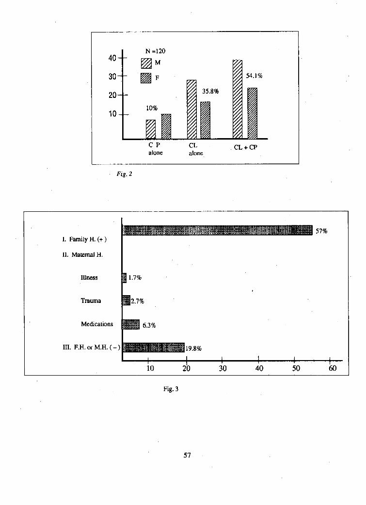
N =120
40-- [_ M
@30 - - F _ 54.1%
2010-- 10% ,-,.,"",,-,'"""":_35.8'__jj
r/J
r f,
¢1a4, , •
C P CL CL+CPalone alone
Fig. 2
'_--_-----"-- _"_-E__:_e_.,_, - 57%I. Family H. (+)
II. Maternal H.
Illness I 1.7%
j,
Trauma 12.7%
Medications _ 6.3%
Ill.F.H. or M.H.'(-) _19.8%
10 20 30 40 50 60
Fig. 3
57

UNIVERSITY OF THE PHILIPPINESPHILIPPINE GENERAL HOSPITAL
DEPARTMENT OF OTOLARYNGOLOGY
TITLE:MICROBIALFLORAIN CHRONICOTITISMEDIA:VALUEOFEARASPIRATECULTURESTUDIES
AUTHORS:Rhodoradel Rosario,MDCharlotteM. Chiong,MD
AbnerL. Chan,MDEditaC. Yap, MD
JoselitoC. Jamir,MDGenerosoT. Abes,MD
ABSTRACT A comparative study was done to compare the, microbiology of the ear aspirate to the contents in the
Culture and sensitivity studies on ear aspirate, middle ear cleft namely, cholesteatoma, bone andcholesteatoma, bone and granulation tissue were granulation tissue. The organisms were likewise
done on 30 patients with chronic otitis media who identified andthe sensitivity pattern of the commonlyunderwent radical mastoidectomy. The most com- isolated organisms were determined.mon organisms isolated were Proteus mirabilis,Proteus
vulgaris and Pseudomonas aeruginosa. All three
showed a 100% sensitivity to ampicillin, trimethoprim, MATERIALS AND METHODSchloramphenicol and cephalexin, Bacteroides, an
anaerobic organisms, was grown in only 4/100 Thirty patients with chronic otitis media, 20 males
isolates. Cultures from the transcanal aspirates were and ten females with age range 6-45 years (Table 1)compared to those obtained directly from the middle seen at the ENT Dept. who underwent radical mas-ear. Computation of the positive predictive value toidectomy were included in the study. Cultureshowed that the transcanal aspirate showed a good and sensitivity studies were done on ear aspirate,excellent correlation for the three most common cholesteatoma, bone and granulation tissue specimens.organisms cultured. Ear discharge was aspirated aseptically after the ear
canal was swabbed with aloohol soaked cotton. The
INTRODUCTION other specimens were collected intraoperatively. All_ were immediately placed in thioglycolate media. These
Chronic otitis media (COM) is a common problem were then inoculated in blood agar plate (BAP), andencountered in the ENT clinic. It is an insidious Mac Conkey agar plate. Incubation at 37 C for 24
and potentially destructive disease characterized by hours was done after which the plates were read.the presence of intractable mucosal, submucosal and/ Colonies were identified by their morphologic andor bone pathology of the middle cleft. In this biochemical characteristics. Antibiotic susceptabil-institution, 60% of 269 ears operated on during the ity of the aerobic isolates to various drugs wereyear 1989 were for chronically discharging ears. Inspite tested using standard Kirby-Bauer method. For the
of the prevalence of the disease, antibiotic treatment anaerobic culture, the specimen from the thioglycoteis still largely empirical as a culture and sensitivity medium were inoculated in a fresh blood agar platestudies are not routinely done. One method of under gas Pak method. If the isolates were the sameobtaining specimen for culture in the clinic is by as those found in the aerobic culture, no furthertranscanal aspiration of ear discharge. However, it susceptibility studies were done. However if theis yet to be determined whether this method would colonies identified were the same as those found in
be truly reflective of the microbial population of the aerobic culture, no further susceptibility studiesmiddle ear. were done. However if the colonies identified were
58

anaerobic they were reinoculated in thioglycolate media, The sensitivity pattern of the 3 most commonrestreaked in BAP and placed in an anaerobic jar for organisms isolated namely Proteus mirabills, Proteus
susceptibility studies, vulgaris and Pseudomonas aeruginosa was determined.All three organisms showed a 100 % sensitivity to the
quinolones (ciprofloxacin, ofloxacin, and norfloxacin).TABLE 1. Age and Sex Distribution of COM Patients Among the aminoglycosides (netilrnicin and gentami-
cin), there was likewise a 100% sensitivity except fora 6.3% resistance of Proteus mirabilis to Gentamicin.
For the commonly u_ed antibiotic in COM (ampicillin,AGE MALE FEMALE TOTAL trimethoprim, chloramphenicol, and cephalexin), there
is a note of <80% sensitivity pattern, the ,lowest beingfor ampiciilin at 59.5% for Proteus mirabilis and 13.3
0-5 0 0 0 % for Proteus vulgaris.
6-10 8 1 9(Figures 3, 4 and 5).
11-15 1 5 6Table 2. Microbiologic Profile of Specimens obtained
16-20 3 2 5 from Patients with COM
21-25 1 1 2ORGANISMS PURE MIXED TOTAL
26-30 1 0 1 CULTURECULTURE
31-35 5 0 5
Proteus mirabilis 42 6 4836-40 0 0 0
Proteus vulgaris 15 0 1541-45 1 1 2
Pseudomonas aeruginosa 11 2 13
Staphylococcus aureus 5 2 7
RESULTS Escherichia coli 3 0 3
The distribution of isolates obtained from the Klebsiella sp. 3 0 3ear aspirate, cholesteatoma, bone and granulation tissueis shown in Figure 1. Single isolates were obtained Enterobacter agglomerans 0 3 3in the majority of the specimens (67.5%). Mixedcultures were grown in 7.5% (9k120), while there was Providenciasp. 1 1 2no growth in 5.0% (6k120). Bacteroides sp 2 0 2
Gram negative organisms were more commonly Bacteroides melaninogenicus 0 2 2isolated compared to the Gram positive organisms
(Figure 2). The microbiologic profile is shown in Klebsiella ozanae 1 0 1
Table 2. Proteus mirabilis was the most commonly Acinetobacteranitratum 0 1 1isolated organism in pure and mixed cultures regard-
less of whether the specimen was obtained from the Acinetobacter wolfli 1 0 1ear aspirate, cholesteatoma, bone or granulation tissue(Table 3). This is followed by Proteus vulgaris and Enterococcus 0 1 1Pseudomonas aeruginosa. Staphylococcus aureus wasthe only gram positive organism cultures and wasfound in 7 isolates. There were only 4 anaerobic
isolates and they belong to the Bacteriodes genus.The rest are distributed among Enterobactcriaciae.
59

Table 3. Distribution of Organism by Specimen
ORGANISM Ear Granurn Bone Choles- TOTALaspirate tissue teatoma
Proteusrnirabilis 13 13 9 10 45Proteusvulgaris 5 1 4 5 15Pseudomonasaeruginosa 5 13 1 4 13Staphylococcusaureus 2 2 2 1 7Escherichiacoil 1 0 1 1 3Klebsiellasp. 1 0 0 2 3Enterobacteragglomerans 1 0 1 1 3Providencia 1 1 0 0 2Bacteroidesmelaninogenicus 1 0 0 1 2Bacteroidessp. 1 1 0 0 2Klebsiellaozanae 0 1 0 0 1Acinetobacteranitratum 0 0 1 0 1Acinetobacterwolfii 0 0 0 1 1Enterococcussp. 0 0 0 1 1
No growth 2 I) 3 1 §
No specimen 1 7 10 6 24
DISCUSSION chloramphenicol (IV) prior to surgery. This wasunavoidable as these patients had unsafe ears with
In any infectious disease, a clear understanding potentials for intracranial complications.of the microbial population is necessary in order to
choose the proper antibiotic. The microbial flora in The most common organisms grown in thischronically draining ears is constantly evolving such study were Proteus mirabilis (45/100), Proteus vulgaristhat regular surveilance cannot be overemphasized. (15/100), Pseudomonas aeruginosa (131100) and
Staphylococcus aureus (7/100). These results were
This study was conducted among COM patients comparable to that found by other investigators (Tablewho underwent radical mastoidectomy at the ENT 4). However, it was noted that although the microbialDepartment. These patients had discharging ears for flora has remained unchanged in the last two decades,several years with an average of 5 years. Most of the Proteus species has become more prevalent withthese patients however received penicillin and Pseudomonas becoming less frequent.
TABLE 4. Types of Aerobic Microorganisms Isolated in COM.
AUTHOR No.of "Pseudomonas Proteus StaphylococcusEars aeuruglnosa sp. aureus
Vizon,Torres(1969) 55 25.4% 18.2% 12.7%Jokipii,et al (1977) 70 4.0% 8.0% 19.0%Abes,Jarnir (1978) 30 48.8% 16,3% 11.6%Ojalaet al (1981) 806 19.0% 12.9% 22.0%Sugitaet al (1981) 62 7.8% 21.1% 6.3%Chionget al (1989) 120 3.8% 35.8% .....Presenistudy 30 13.0% 63.0°/, 7.0%
60

An antibiogram was constructed (Figures 3, 4 and Mackowiak (1978) cultured sinus tract drainage in5) and it showed that for the three most common patients with chronic osteomyelitis and found that
organisms, the quinolones are the most effective if the culture was positive for Staphylococcus aureuLantibiotics. This may be due to the fact that this there was good correlation with culture reports of.is a new drug and resistant strains have not yet the operative specimens. However, if the culture ofdeveloped. The use of ciprofloxacin has recently the sinus drainage was another organism,there was abeen underscored in a study by Picirillo (1989), poor correlation. Assuming that the same pattern
considering that the drug is effective in a wide range exists in the middle ear cleft since there is a similarityof both gram positive and gram negative organisms, in the organisms implicated, this might suggestHowever, the use of this ciprofloxacin is not recom- that the specimen obtained from the external auditorymended for chidren nor as a first line drug in the canal may not be the same as those from the middle
management of COM. Gentamicin, which is corn- eardischargewiththosetakenintraoperativelyeg, bone,monly used otic solution, still has a good sensitivity cholesteatoma and granulation tissue. Computation(93.7%) for the organisms. The commonly prescribed of the predictive value of transcanal aspirates showedoral antibiotics ampicillin, trimethoprim, chlo- that there is good to excellent correlation for these
ramphenicol and cephalexin showed a < 80% sensi- organisms using the rating of agreement by Kappativity pattern. This could probably be attributed statistics. This study specifically Shows that whento the administration of antibiotics intravenously or anyone of the three organisms is cultured, there is
previous oral intake prior to the collection of a positive correlation in 90% of cases. This findingspecimens, may be very imporant in the clinical setting as the
antimicrobial treatment of COM can be fairly accu-
The role of anaerobic organisms in COM has rately guided by doing transcanal aspiration of earbeen the subject of intensive investigation and specu- discharge alone.lation. The metabolism of facultative species in mixedinfections, by lowering the local concentration ofoxygen and reduction in oxidation-reduction potential, TABLE 5. Correlation of Ear Aspirate with Operativeprovides a suitable environment for the anaerobic Specimen Culture.
pathogens. Sugita (1981) elaborates that becauseof the obstruction of air around the cholesteatoma Earaspirate No.of Positive Negative Predictivethere is a reduction of the partial pressureof oxygen Patients correlation correlation valuewith an inverse increase in cartoon dioxide thus
favoring anaerobic growth. Synergy between the P.mlrabilis 29 29 0 100.0'/,negative bacilli particularly coliforms and Proteus and
Bacteroidesspecieshave beenreported0ngham, 1977); P.vuloaris 13 11 2 84.6%In this study, there were four isolates of Bacteroidesspand in thesetwo B. melaninogenlcus was isolated P.asuroginosa 10 7 3 70.0%together with Proteus mirabilis. Previous studieshave cited the importance of anaerobic organisms *Predictivevaluee.quahnumberoflmsitiveeorrelatianstimes100in COM. The apparent low incidence of anaerobic dividedby total numberof patients.
infection in this study maybe due to several factors,
most impor't,3nt of which is the inherent difficulty SUMMARYin isolating the organism.
A study of 30 patients with COM who underwent
It is a common practice to treat COM empiri- radical mastoidectomy showed that the three mosttally. However when confronted with patients re- common organisms were Proteus mirabilis, Proteusfractory to the usual medical management, as well vulgaris and Pseudomonas aeruginosa. The isolatesas the significant potential for developing complies- obtained from the transcanal ear aspirate showed goodtions in such cases, culture and sensitivity studies excellent correlation with the operative specimens frommaybe necessary in the proper management of COM. the middle ear (bone, granulation tissue, cholestea-
The question arises whether cultured, specimens toma). The transcanal ear aspirate cult'are maybeobtained from the external auditory canal would be valuable as a guide in the medicalmanagement ofreflective of those taken directly from the middle ear. COM.
61

J BIBLIOGRAPHY MACKOWIAK, P.A., Jones, S.R. and Smith, J.W.Y
Diagnostic value of sinus-tract cultures inchronic osteomyelitis. JAMA, 239-2772-
ABES, G.T. and Jamir, J.C. Bacteriologic study 2775, 1978.of chronic ear discharge. Acta Medica Phil-ippina, 14:28-31, 1978. OJALA, K., Son-i, M. Riihikangas, P. and Sipila, P.
Comparison of pre and post-operativeAnnual Report, Department of Otorhinilaryngology, bacteriology of chronic ears. Journal of
UP-PGH Medical Center, 1989. Laryngology and Otology, 95: 1023-1029,1981.
CHIONG, C.M., Chan, C.M., Yap, E.C., del Rosario,R.A., Pajarillo, P.R., Velmonte,M.A. et al. PICIRILLO, J.F. and Parnes, S.M. Ciprofloxacin forThe microbiology of chronic otitis media, the treatment of chronic ear disease. La-
Acta Medica Philippina, in press, 1990 ryngoseope, 99:510-513, 1989.
INGHAM, H.R., Sison, P.R., Tharagonet, D., Selkon, Research'Protocol Guidelines for Physicians. ClinicalJ.B. and Codd, A.A. Inhibition of Phagocy- Epidimiolgy Unit, UP-PGH, 1988.tosis in vitro by obligate anaerobes. The
__ Lancet, 2:1252-1254, 1977. SUGITA, R., Kawamura, S._ lchikawa, G., Goto, S.and Fujimaki, Y. Studies on anaerobic
JOKIPII, A.M., Karma, P., Ojala, K. and Jokipii, L. bacteria in chronic otitis media. Laryngo-Anaerobic bacteria in chronic otits media, scope, 91: 816-821, 1981:Arch Otolaryngol, 103: 278-280, 1977.
VIZON, C. and Tones, C. Bacteriology of chronicotitis media in children. Philippine Journalof Pediatrics, 18:46-49, 1969.
62
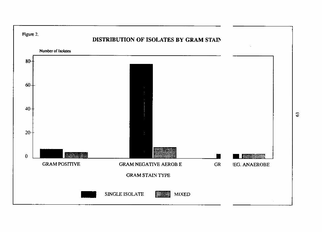
Figure 2.DISTRIBUTION OF ISOLATES BY GRAM STAIb
Numberof Isolates
8O-
0-
40-
20-
o EGRAM POSITIVE GRAM NEGATIVE AEROB E GR rEG. ANAEROBE
GRAM STAIN TYPE
SINGLE ISOLATE MIXED

Figure 3.SENSITIVITY PATTERN OF Proteus mirabilis
Percent ISOLATED FROM CSOM PATIENTS
,oo_
8oq-
604_
40--d ::.:.:. •
20__ _ i
I" " "I __
CIP OFL NOR NET GEN CHLOR TRI CEF AMP
PATFERN:
_ SENSITIVE _ hNTERMEDIATE '- ::::.......' RESISTANT

Flgu_ 4.SENSITIVITY PATTERN OF Proteus vulgaris
ISOLATED FROM CSOM PATIENTSPercent
f
[i:::.]80--
!..: ....
. r
i ...
i::. . _
'. :: .
20 - - f
i.: .j. , [
CIP OFL NOR NET GEN CHLOR TRI CEF AMP
PATFERN:
lSENSma _ _SISTA_
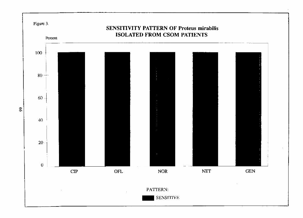
Figure 3.SENSITIVITY PATTERN OF Proteus mirabilis
ISOLATED FROM CSOM PATIENTSPercent
I
loo-I-i
iII
8O
60
40-
20 -_-
0
CIP OFL NOR NET GEN
PATI'ERN:
1 SENSITIVE
UNIVERSITY OF THE PHILIPPINESPHILIPPINE GENERAL HOSPITAL
DEPARTMENT OF OTOLARYNGOLOGY
TITLE" RandomizedClinicalTrialon the EfficacyandSafetyofLoratadineVersusAstemizolein Allergic Rhinitis
AUTHORS: MarianoB. Caparas,MDRomualdoA. Aragon,Jr., MDJoseAntonio M. Santos,MD
Jesus M. Jardin,MDCiriacoB. Gato,Jr. MD
INTRODUCTION MATERIALS AND METHODS
The management of allergic rhiaitis involves Study Design and Study Siteenvironmental control, pharmacologic interventions,and allergen immunotherapy (1). In the Philippines, This double-blind randomized clinical trial wasenvironmental control and allergen immunotherapy conducted at the Outpatient Section of the Departmenthave yet to be proven effective and feasible. On the of Otorhinolaryngology, Head and Neck Surgery ofother hand, pharmacologic interventions, which entail the UP-PGH Medical Center from February to June,the use of decongestants, antihistamines, corticoster- 1988.oids, and cromolyn sodium, have helped patients, incoping with acute attacks. Patient Section
Through the years, drowsiness or sedation was Seventy patients with unequivocal history of al-the most common complaint against antihistamines lergen-induced rhinitis were recruited provided thattherapy. Since 1941, many H1 antihistamines have they were more than 12 years of age and presentingbeen introduced to provide antiallergic effect with with at least two of the following nasal symptoms:minimal anticholinergic and sedative actions. One itchiness, stuffiness, discharge and sneezing affectingoftheseis astemizole, an orally potent highly selective to a moderate degree (i.e., bothersome but not dis-antihistamine administered once a day. Various clinical abling or intolerable).studies have confirmed its efficacy and safety (2,3,4). Excluded were pregnant or lactating patients,
those with asthmas, those on allergen immunother-Loratadine is a newer long,acting, tricyclic apy, or those receiving other anti-allergic drugs within
antihistamine with selective peripheral Hi-receptor an- the last six months. Patients with other significanttagonist activity. Like astemizole, it is also admini- current diseases or abnormal baseline laboratory tests
stered once daily. It has an added advantage of more results, those who haveaknownidiosyncraticreactionrapid response and various clinical trials have con- to antihistamines, and those who have taken any
firmed its efficacy and safety (5,6,7), inve f gatJonaldrugs within one month prior to thestart of this study were also excluded.
This double-blind randomized clinical trial was
conducted to compare the etlicacy and safety of a Interventions14-day course of loratadine with those Iff astem izole
given to Filipino patients with allergic rhinitis. After informed consent has been secured, thepatients were randomly allocated to receive either
67

loradatine 10 mg/tabletgiven once a day for 14 days To estalish the safety of either regimen, CBC,or astemizole 10 rag/tablet also given once daily for blood sugar, BUN, creatinine, total protein, alkaline14 days. phosphatase, SGCOT, SGPT, total bilirubin, and
urinalysis were done prior to and on the last dayEvaluation Criteria of treatment. Side effects were noted on the
symptom diary card and classified accordingly:Patients daily filled out symptom diary cards to
note the severity of symptoms according to the fol- Mild: Experience was consideredlowing scoring system: trivial and did not cause the
patient any real problem.
O None: No symptom evident1 - Mild: Trivial; definitely present, Moderate: Experience was a problem to the
but not bothersome patient, but did not interferesignificantly with daily activi-
2 - Moderate: Bothersome, but not ties or the clinical status of
disabling or intolerable the patient.
3 Severe: Disabling and/or intolerable Severe: Experience caused significantinterference with normal daily
On the 3rd, 7th and 14th days of treatment, activities or the clinical status ofoverall condition of rhinitis was evaluated by both the patient.the physician and patient using the following scheme:
O None: Virtually no symptoms are evi- The patient was deemed discharged from thedent study upon completion of the 14-day drug treatment,
with the development of adverse reactions that
1 Mild: Symptoms do not interfere with prompted discontinuation of the assigned regimen, ordaily activities and/or sleep with persistence of symptoms on the 4th day of
treatment (failure of treatment).2 Moderate: Some interference with daily
activities and/or sleepData Analysis
3 - Severe: Significant/major interferencewith daily activities and/or sleep Baseline characteristics and outcome measures of
those who received loratadine and those who received
Therapeutic response was also assessed by both astemizole were compared using Mann-Whitney Uthe physician and subject according to the following test or t-test for quantitative variables and chi-squareevaluation scale: or Fisher's exact tests for qualitative variables.
Differences in responses between groups and through1 - Excellent: Virtually all symptoms have time were evaluated using Friedman's 2-way ANOVA
been eliminated or McEmnar's test.
2 - Good: Most symptoms are im-
proved, but some symptoms RESULTSare still listed as mild
Seventy allergic rhinitis patients participated in3 - Fair: Some response, but most this study. Patient "#32, allocated to receive loratadine,
symptoms are still present was excluded because of abnormally high baselinelevel of serum alkaline phosphatase. Another 10 were
4 - Failure: Worse than pre-treatment excluded because of failure to comply with the assignedbaseline Failure: regimen.
68

PRE-INTERVENTION GROUP COMPARISON INTERVENTION COMPLIANCEAll 59 ptients took their medications daily for 14
Baseline analysis showed that the two groups were days.comparable in the absence of exposure to immunoth-erapy, in demographic characteristics, in physical OUTCOME GROUP COMPARISONSexamination and rhinos¢opic findings. Except for a
significantly longer duration of seasonal allergic rhinitis I. Efficacy
among those on loratadine, reveiw of medical hsitory A. The physicians' evaluation of signs andshowed no other clinically important differences
symptoms during the course of treatment was(Table I). compared between the two groups.
TABLE 1: BASELINE GROUP COMPARISONS
LORATADINE ASTEMIZOLE p VALUE
Demographic1. meanage (yrs) 26.7 +-1 25.7+-1 0.132. weight (ibs) 124.9+ol 21.9+4 0.643. height (cm) 160.4+-11 60.0+-1 0.814. male:female 16:13 17:13 (I.79
MedicalHistory
1. meandurationof 9.3+-1 5.2+-0 0.01seasonalallergicrhinitis(yrs)
2. meannumberof episodes 7.2+-2 8.0+-2 0.74of seasonalallergicrNnitis
3. meandurationof current 7.4+-2 7.5+-1 0.93episode(days)
4. perennialallergic 20 (69%) 21 (70%) 0.95rhinitis
Screeningrhinoscopyfindings:1, NASALMEMBRANE 25 (86%) 25 (83%) 0.97
pale& boggy 4 (14%) 5 (17%)erythematous
2. NASALPt,TENCYpartiallyoccluded 27 (94%) 29 (97%) {I.49
totallyoccluded 1 (3%) 1 (3%)normal 1 (3%) 0
3. NASALSECRETIONSthinwatery 18 (62%) 19 (63%) 0.55mucoid 11 (38%) 10 (34%)
4. QUANTITYOFNASALSECRETIONSslightto moderate 17 (59%) 20 (67%) 0.54minimal 9 (31%) 7 (23%)copious 3 (10%) 2 (7%)none 0 1 (3%)
69
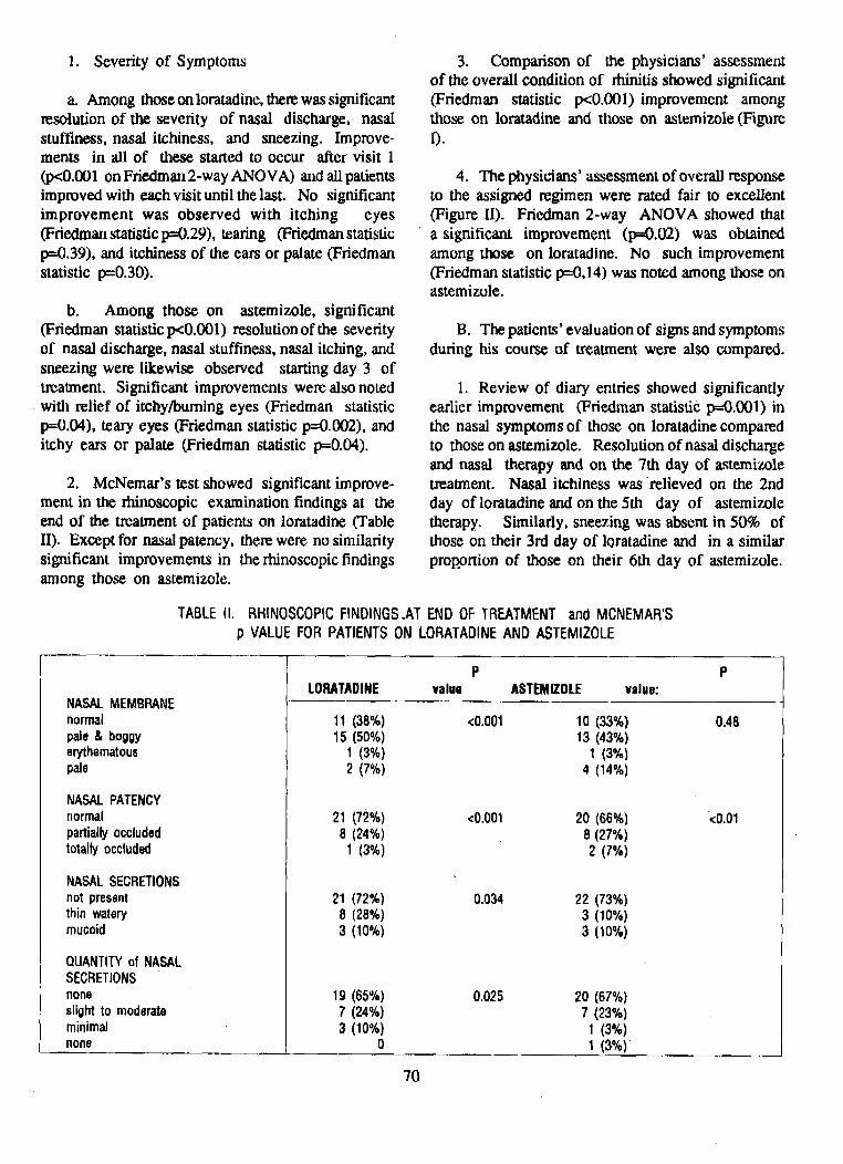
1. Severity of Symptoms 3. Comparison of the physicians' assessmentof the overall condition of thinitis showed significant
a. Among thoseonloratadine, there was significant (Friedman statistic p<0.001)improvement amongresolution of the severity of nasal discharge, nasal those on loratadine and those on astemizole (Figure
stuffiness, nasal itchiness, and sneezing. Improve- I).ments in all of these started to occur after visit 1
(p<0.001 on Friedman 2-way ANOVA) and all patients 4. The physicians' assessment of overall responseimproved with each visit until the last. No significant to the assigned regimen were rated fair to excellentimprovement was observed with itching eyes (Figure II). Friedman 2-way ANOVA showed that
(Friedman statisticp=0.29), tearing (Friedman statistic a significant improvement (p=0.02) was obtainedp=0.39), and itchiness of the ears or palate (Friedman among those on loratadine. No such improvementstatistic p=0.30). (Friedman statistic p=0,14) was noted among those on
astemizole.
b. Among those on astemizole, significant(Friedman statistic p<0.001) resolution of the severity B. The patients' evaluation of signs and symptoms
of nasal discharge, nasal stuffiness, nasal itching, and during his course of treatment were also compared.sneezing were likewise observed starting day 3 of
treatment. Significant improvements were also noted 1. Review of diary entries showed significantlywith relief of itchy/burning eyes (Friedman statistic earlier improvement (Friedman statistic p=0.001) inp=0.04), teary eyes (Friedman statistic p=0.002), and the nasal symptoms of those on loratadine compareditchy ears or palate (Friedman statistic p=0.04), to those on astemizole. Resolution of nasal discharge
and nasal therapy and on the 7th day of astemizole2. McNemar's test showed significant improve- treatment. Nasal itchiness was'relieved on the 2nd
merit in the rhinoscopic examination findings at the day of loratadine and on the 5th day of astemizoleend of the treatment of patients on loratadine (Table therapy. Similarly, sneezing was absent in 50% ofII). Except for nasal patency, there were no similarity those on their 3rd day of loratadine and in a similarsignificant improvements in the rhinoscopic findings proEortion of those on their 6th day of astemizole.among those on astemizole.
TABLEII. RHINOSCOPICFINDINGS.ATENDOFTREATMENTandMCNEMAR'Sp VALUEFORPATIENTSON LORATADINEANDASTEMIZOLE
P PLORATADINE value ASTEMIZOLE value:
NASALMEMBRANEnormal 11 (38%) <0.001 10 (33%) 0.48pale& boggy 15 (50%) 13 (43%)erythematous 1 (3%) 1 (3%)pale 2 (7%) 4 (14%)
NASALPATENCYnormal 21 (72%) <0.001 20 (66%) <0.01partiallyoccluded 8 (24%) 8 (27%)totallyoccluded 1 (3*/,) 2 (7%)
NASALSECRETIONSnotpresent 21 (72%) 0.034 22 (73%)thinwatery 8 (28%) 3 (10%)mucoid 3 (10%) 3 (10%)
QUANTITYof NASALSECRETIONSnone 19 (65%) 0.025 20 (67%)slightto moderate 7 (24%) 7 (23%)minimal 3 (10%) 1 (3%)none 0 1 (3%)
70

2. Both groups showed significant (Friedman DISCUSSIONstatistic p=0.001) improvement in the overallcondition of rhinitis with the most marked change Like the previous studies clone abroad (2-5,noted on day 3 of treatment (Figure III). 9-10), this randomized clinical trial conducted locally
has demonstrated the effectiveness of both loratadine
3. According to the patients' evaluation of overall and astemizole in the treatment of allergic rhinitis.response to treatment, those on loratadine as wen Both drugs were proven to be as efficacious as theas those on astemizole reported significant (Friedman non-sedating antihistamines like mequitazine andstatistic p=0.05) improvement in response rated as terfenadine (6,7,15) and both have been recommendedfair to excellent starting on Day 3 (Figure IV). because of their once a day dosage which ensures
better patient compliance.II. Safety
As in other comparative studies betweenA. Routine Physical Examination and Laboratory loratadine and astemizole, loratadine-treatCd Filipino
Studies ptients had significantly earlier relief of symptomsthan those treated with astemizole. Moresignificant
There were no significant changes between Day improvement by the third day of therapy was observed0 and Day 14 with regards to body temperature, among those on loratadine than among those on
pulse rate, respiratory rate, systolic and diastolic astemizole. This concurs with previous studies, whereinblood pressure, weight, urinalysis, glucose, BUN, partial relief of symptoms was observed as early asserum creatinine, total protein, alkaline phosphatase, within 4 hours of first treatment with loratadine (10,11).SGOT, SGPT, total bilirubin, hemoglobin, hema-
tocrit, RBC count, WBC differential counts and counts. The relief of ocular symptoms (itching and tear-ing) has been reported to be better with_astemizole
B. Adverse Effects over terfenadine and clemastine .(12,13). Our studyshows the same advantage of astemizole over lo-
The few adverse effects were reported in equal ratadine. -frequencies among those on loratadine and on
astemizole (Table III). Thse complaints were all of The unpleasant and undesirable effects ofthe mild to moderated severity with majority of the antihistamines have been somnolence, sedation,
incidences possibly orprobably rela_ed to the treatment insomia and anticholinergic action. In an earlierregimen long-term safety study among normal male volunteers
TABLE III: ADVERSE EFFECTS REPORTED
Day 3 Day 7 Day 14
Symptoms Lor Ast Lor Ast Lor AstDizziness 3 1 2 1 5 0Chest pain/body 1 1 0 1 0 1weaknessLight Headedness 0 1 0 1 1 1Sedated 2 1 1 1 0 1Nape pain 1 0 2 0 0 1Difficulty of 0 2 0 0 0 0Swallowing
Lor=-Loratadine; Ast=Astemizole
71

-J;r,ceiving 40 mg of loratadine daily for 13 weeks,tolerance was goal and there were no unusual clinical 1. Nalebuff DJ (Cumming CW ed.). Ototaryn-changes in laboratory test values, electrocardio- gology, Head and Neck Surgery; Ch. 36grams, and physical examinations (14). The only Allergic rhinitis. 1986. CV Mosbydrug-related side effect reported were hiccup and Company.headaches. The side effects of fatigue, sedation
headache, and dry mouth were noted in previous 2. Wihl JA, Petersen BN, et al. Effect of theclinical studies using loratadine at a dose 10 mg OD. non-sedative HI-receptor antagonist astern-These, however, occurred in the placebo and com- izole in perennial allergic and non-allergicparative groups at approximately the same frequen- rhinitis. J. Allergy and Clinical Immunol-cies (5,6,7,11). Such was true also for the Filipino ogy. 1985; 75:720-7.
patients included in this trial.3. Sooknundun M, Santosh KK, Sandaram KK.
In clinical studies with astemizole, sedation of Treatment of allergic rhinitis with a newvarying degrees has been reported (13,15,16). long-actiong H1 receptor antagonist:Sooknundun, et al (3) reported drowsiness in three astemizole. Annals of Allergy. 1987;of his patients persisting One week after the six- 58:184-188.
week trial period while another patient, a neurologist,,-has persistent drowsinees up to 3 days after 4. Bussche GV, Emmanuel MB, Rombaut N.
discontinuation of astemizole. These observations Clinical profile of astemizole. A survey
point to adverse cumulative effect of a long half- of 50 double blind trials, Annals of Allergylife (10 hours) for astemizole. After Richards et al 1987; 58:184-188.
observed a half-life of twelve days or longer forastemizole (17), Simons et al endorsed that, in general, 5. DoclOaomRJ et al. Safety and efficacy ofloratadineantihistamines should be given only once during its (Sch 29851) a new non-sedating anti-half life (18). If given more often, significant accu- histamine in seasonal allergic rhinitis.mulation of the drug may occur. Annals of Allergy. 1987; 58407-411.
Loratadine is expected to cause less sedation since 6. Gutkowski A. et al. Study of the effect of Schit has been shown to have a greater afrinity for 29851 10 mg OD versus teffenadine 60the peripheral H1 receptors than for central H1 mg BID and placebo in patients withreceptors (8). This dimished penetration into brain seasonal allergic rhinitis. Scheringtissue accounted for the absence of daytime sedation Corporation, Clinical Research Division,among those on loratadine compared to those on Kenilworth, N.J. 1985 (I 85-207).diphcnhydramine (19).
7. Michael RB, Sabbah A. Study of the effect of
'_- Sch 29851 10 mg OD versus mequitazineCONCLUSION 5 mg BID and placebo in patients with
seasonal allergic rhinitis, Schering
This locally conducted double-blind randomized Corporation, Clinical Research Division,clinical trial has shown that loratadine 10 mg per Kenilworth, N.J. 1985 (I 85-207).orem per day is as effective as astemizole given10rag perorem perday for the treatment of allergic 8. Health Registration Dossier Sch 29851.rhinitis. However, loratadine showed a faster onset Pharmacokinetics Section. Schering Inter-of relief in contrast to astcmizole. There were no national. January 1987.
significant differences in the side effects notcdamong the patients on loratadine and on astemizolc. 9. Heykkants J. Astemizole (R54512): a safe orally
very potent and long acting histamine HIantagonist devoid of central, sedative and
REFERENCES: anticholinergic effects. Basic MedicalInlormation Report N 19668. JanssenPharmaceutica, Belgium.
72

10. Kutwak A, et al. Study of the effect of Sch 14. Herson JM, Kkisick JC. Long term Safety and29851 40 mg OD versus astemizole 10 tolerance in normal male volunteers.mg OD in patients with allergic rhinitis. Schering Corporation, Clinic_ ResearchSchering Corporation, Clinical Research Division, Kenilworth N.J. 1985 (C 85-003).Division, Kenilworth, N.J. 1985. (I 84-111/118) 15. Wilson JD, Hilas JL. Astemizole: a new
long-acting antihistamine in the treatment11. Meiniche K, et as. Study of the effect of Sch of seasonal allergic rhinitis. Clinical
29851 10 mg OD versus astemizole and Allergy. 1984; 12:131.placebo in patients with seasonal allergicrhinitis. Schering Corporation, Clinical 16. Melmberg H, et al. Astemizole in the treatmentResearch Division. Kenilworth, N.J. 1985 of hay fever. Allergy 1983; 38:227.(I 850-209).
17. Richards DM, Brogden RN, et as. Astemizole;12. Howarth RH. Holgate St. Comparative trial a review of its pharmacodyrmmic prop-
of two non-sedative H1 antihistamines, ter- erties and therapeutic efficacy. Drugs 1984;fenadine and astemizole, for hay fever. 28 (1): 38-61.Thorax. 1984; 39:668-672
18. Simons FE, Simos KJ, et al. The,13. Fmstad AB, Lotkvist T, et al. Astemizole in comparative pharmacokinetias of H1
the treatment of seasonal allergic rhinitis, receptor antagonists. Annals of AllergyA double blind comparison with climastine 1987; 59 (II): 20-23.
in patients sensitive to tree and grass pollen.Special reference to side effects registered 19. Roth T, et as: Sedative effects of antihistamines.in psychological tests. Acta Otolaryngol. Journal of Allergy and Clinical lmmunol-1984; Suppl 409:5-30 ogy 1987 80 91): 94-98.
73


: ' : _ : ::::::: : : i : ::

An Evaluation of the Efficacy, Toleranceand Safety of Loratadine-D vs. Actifed
in Acute Rhinitis
ManuelG. Lim, M.D.,M. Sc., F.A.A.O.- HNS"MargaretC. Lim,M.D.
ManuelAnthonyC. Lim,Jr., M.D.JoseC. Ilao, M.D.**
ReginaV. Mangay,M.D.***
ABSTRACT In recent years, newer non-sedating antihis-tamines have been developed that do not readily
A randomized single blind comparative con- enter the central nervous system or interact withtrolled study of the efficacy, tolerance and safety neuroccptors associated with sedation, or concentrate
of Loratadine 10 mg. plus Pseudoepbedrine against in the brain, thereby producing fewer undesirableActifed was conducted in 60 cases of acute rhini- effects. One such preparation is a combination oftis with an allergic overlay as derived from the history. Loratadine, a potent non-sedating and long actingThe comparison was based on both a subjective as- H1 antagonist, plus pseudoepbedrine. Loratadinesessment by the patientand the objectiveevaluation is a pipemus compound relatedto azatadinc. It isof the physician. Based on these results, Clar- relatively specific in blocking peripheral receptorsitin-D has been shown superior to Actifed in terms with very little H2 receptor antagonism. Loratadineof : 1) improving the signs and symptoms of acute is virtually devoid of sedating or other central nervousrhinitis, 2) bringing faster relief of the miserable system side effects due to its inability to penetratesymptoms, and 3) good tolerance and lesser side the blood brain barrier. Its duration of action is
effects especially drowsiness, about 15-16 hours. This study describes the efficacy,tolerance and safety of Loratadine-D (Loratadine plusdecongestant) in comparison with Actifed in relieving
INTRODUCTION the signs and symptoms of acute rhinitis.
Classical antihistamine and antihistamine-de- OBJECTIVEcongestant combination are widely used for the treat-
ment of acute rhinitis. It is recognized that these drugs The objective of this study is to compare thedo not shorten coume of acute rhinitis. However, these efficacy, tolerance and safety of Claritin-D (Lorata-drugs, together with some other measures taken, may dine plus Decongestant in combination) vs. Actifedbe able to prevent the onset of a common cold (this in the treatment of the acute rhinitis.is beyond the scope of this pal)c0. Usually, in acute
rhinitis, these drugs provide relief for the patients Materials and Methodssuffering from signs and symptoms which we are
making miserable. The main problems that prevent This is a randomized single-blind comparativetheir use in some patients arc excessive sedation controlled study of the efficacy, tolerance and safetyor drOwsiness and anticholinergic properties (dryness of the use of Claritin-D vs. Actifed in a short termof the mouth, blurring vision, urinary retention,etc.), treaunent of 60 cases of acute rhinitis who have never
taken any other medication for their illness. This* Dr. Manuel G. Lira, M.D., F.A.A.O. - HNS, Chairman, DepL cases are divideii into 2 groups of 30 cases each,of Omlanyngology, Unive_ of the Philippines, PhilippineGeneralHospital(MedicalCe_u=r) one receiving Claritin-D and another receiving Ac-
tiled. The Claritin-D group is given the medication** Dr, Jose C. Ilao, Medical Di_ctor, Schcring-Plough Co_p. every 12 hours or 2 times a day, whereas the Actifed
***Dr.ReghutV.MAnsay,AssittantMexlicalDire.tor, Schering-Plough group is given the medication every 8 hours or 3cotr,, times daily.
77
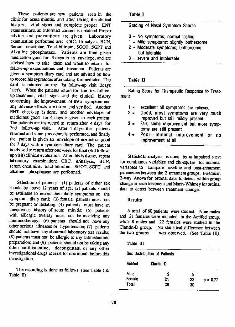
These patients are new patients seen in the Table Iclinic for acute rhinitis, and after taking the clinical
history, vital signs and complete proper ENT Gradingof NasalSymptomScoresexaminations, an informed consent is obtained. Proper
advice and precautions are given. Laboratory 0 = NOsymptoms;normal feelingexamination performed are: CBC, Urinalysis, BUN, 1 = Mild symptoms;slightly bothersomeSerum creatinine, Total bilirium, SGOT, SGPT and 2 = Moderatesymptoms;bothersomeAlkaline phosphatase. Patients are then given buttolerablemedication good for 3 days in an envelope, and are 3 = severe and intolerableadvised how to take them and when to return for
follow-up examinations and treatment. Patients aregiven a symptom diary card and are advised on howto recordhissypmtomsafter taking the medicine. The Table IIcard is returned on the 1st follow-up visit (3days
later). When the patients returnfor the first follow- RatingScoreforTherapeuticResponseto Treat-up treatment, vital signs and the clinical history mentconcerning the improvement of their symptom andany adverseeffects are taken andverified. Another 1 = excellent;all symptoms are relievedENT check-up is done, and another envelope of 2 = Good; most symptoms are very muchmedicines good for 4 days is given to eachpatient, improvedbut Still mildly presentThe patientsare instructed to returnafter 4 days for 3 = Fair; some improvementbut more symp-2nd foUow-up visit. After 4 days, the patients toms are still presentreturnedandsame procedureis performed, andfinally 4 = Poor; minimal improvementor nothe patient is given an envelopeof medicinesgood improvementat allfor 7 days with a symptom diary card. The patientis advisedto return afterone weekfor final (3rd follow-
up visit) clinical evaluation. After this is dsone, repeat Statistical analysis is done by unimpaired t-testlaboratory examination: CBC, urinalysis, BUN, for continuous variables and chi-square for nominalserum creatinine, total bilimbin, SGOT, SGPT and variables to compare baseline and post-treatmentalkaline phosphatase are performed, parameters between the 2 treatment groups. Friedman
2-way Anova for ordinal data to detect within groupSelection of patients: (1) patients of either sex changein each treatment and Mann-Whimey for ordinal
should be above 12 years of age; (2) patients should data to detect between treatment change.be available to record their daily symptoms on thesymptom diary card; (3) female patients must not Resultsbe pregnant or lactating; (4) patients must have an
unequivocal history of acute rhinitis; (5) patients Atotal of 60 patients were studied. Nine males
with alllergic overlay must not be receiving any and 21 females were included in the Actifed group,immunotherapy; (6) patients should not have any while 8 males and 22 females were studied in theother serious illnesses or hypertension; (7) patients Claritin-D group. No statistical difference between
should not have any abnormal laboratory test results; the two groups was observed. (See Table III)(8) patients must not be allergic to any antihistaminic
preparation; and (9) patients should not be taking any Table IIIother antihistamine, decongestant or any otherinvestigational drugs at least for onemonth before this SexDistributionof Patientsinvestigation.
Actifed Clarltin-D
The recording is done as follows: (See Table I &Table II) Male 9 8
Female 21 22 p = 0.77Total 30 30
• 78

The mean age, mean weight and mean heightof the two groups of patients are shown togetherin one table (Table IV). The differences betweentwo groups are statistically insignificant.
The baseline or initial rhinoscopic findings areshown in. Table V, and the baseline initial signs andsymptoms are shown in Table IV
Table IV
Age,Weightand HeightDistribution
Actlfetl Clarltln-D
MeanAge, S.D. 27.22,+-9.19 26.2,+- 5.89 p=0.28Range 14-56 19-41
MeanWeight,S.D. 54.28+-10.83 52. 23,+-6.2 p=0.19Range 30-75 40-67 '
MeanHeight 159.17,+-8.13 158.75,+-7.66 p=0.42Range 142-175 144-172
Table V
BaselineRhinoscopi¢Findings
Actlfed Clarltln-D
1. NasalMucosa:Normalappearance 5 9Palewith or without 7 8 p=0.16bogginessErythematous 18 13
2. NasalAirway:Normalor good 0 0Partialocclusion 21 23 p=0.56Totalocclusion 9 7
3. NasalDischarge:Absent 0 0Serousto Mucoid 24 23 p=0.76Mucopurulent 6 7
4. Amountof NasalDischarge:None 0 0Minimal 0 0Moderate 12 10 p=0.59Profuse 18 20
?9

- Table Vl
Baseline Signs and Symptoms
Actlfed Clarltln-D,
1. NasalDischarge:Moderate 2 2Severe 28 28 p=l.00
2. NasalStuffiness:Moderate 4 2Severe 26 28 p=U.39
3. NasalItchiness:None 3 5Mild 3 3 p=0.76Moderate 10 8Severe 14 14
- 4. Sneezing:None 0 2Mild 2 2Moderate 5 5 p=0.48Severe 23 21
5. Itching/BurningEyes:None 11 17Mild 8 3Moderate 8 7 p=O.30Severe 3 3
6. Tearing/Watel7Eyes:None 13 13Mild 6 6Moderate 8 8 p=l.00Severe 3 3
7, Rednessof Eyes:None 25 26Mild 3 2 p =0.74Moderate 2 2Severe
8. Ithchinessof Ears/PalateNone 19 22Mild 6 2Moderate 5 3 p=0.66Severe 0 3
9. SoreThroat:None 2 2Mild 25 19 p=O.06Moderate 1 2Severe 2 7
80
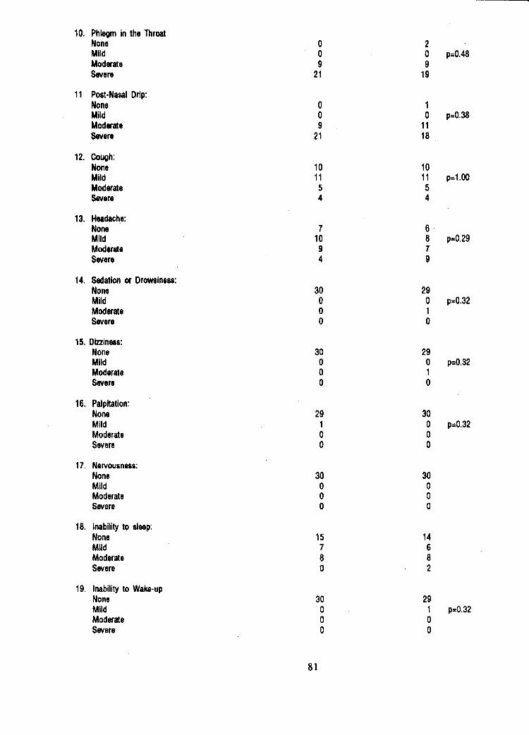
10. Phlegmin the ThroatNone 0 2Mild 0 0 ' p=0.48Moderate 9 9Severe 21 19
11 Post-NasalDrip:None 0 1Mild 0 0 p=0.38Moderate 9 11Severe 21 18
12. Cough:None 10 10Mild 11 11 p=l.00Moderate 5 5Severe 4 4
13. Headache:None 7 6 •Mild 10 8 p=0.29Moderate 9 7Severe 4 9
14. Sedationor Drowsiness:None 30 29Mild 0 0 p=0,32Moderate 0 1Severe 0 0
15. Dizziness:None 30 29Mild 0 0 p=0.32Moderate 0 1Severe 0 0
16, Palpitation:None 29 30Mild 1 0 p=0,32Moderate 0 0Severe 0 0
17. Nervousness:None 30 30Mild 0 0Moderate 0 0Severe 0 0
18. Inabilityto sleep:None 15 14Mild 7 6Moderate 8 8Severe 0 2
19. Inabilityto Wake-upNone 30 29Mild 0 1 p=0,32Moderate 0 0Severe 0 0
81

The following Table VII will show the baselineevaluation by the physician or investigator of the acuterhinitis in the patients.
Table VII
Table VIII shows the comparison of the signs andAcute Evaluation of Acute Rhinitis by Physician symptoms of these patients during the various sched-
uled follow-up visits to the clinic.Actlfed Clarllln-D
None 0 0Mild 0 0 p=1.00Moderate 30 30Severe 0 0
TableVIIIComparisonoftheSignsandSymptomsof Follow-upVisits
Actiled ClariUn-D
V1 V2 V3 V4 V1 V2 V3 V4
1. NasalDischage:None 0 11 1 4 16 0 14 18 28Mild 0 10 7 3 0 11 10 2Moderate 2 4 3 3 2 3 1 0 p=0.0158
•Severe 8 5 1 1 28 2 1 0P<O.O001 P<O.O001
2. NasalStuffiness:
None 0 14 14 15 0 13 18 27Mild 0 7 7 7 0 13 10 2 p=,0.0588Moderate 4 5 4 0 2 1 2 1
,!: Severe 26 4 0 1 28 3 0 0P<O.O001 P<O,O001
3. NasalItchiness:
None 3 23 23 21 5 21 27 29Mild 36 2 2 3 9 2 1Moderate 10 1 0 0 8 0 0 0 p=0.3047Severe 14 0 0 0 14 0 1 0
P<O.O001 P<O.O001
4. Sneezing:None 0 15 19 16 2 21 25 29Mild 2 10 5 6 2 7 4 1Moderate 5 5 1 1 5 2 0 0 p=0.0059Severe 23 0 0 0 21 0 1 0
P<O.O001 P<O.O001
5. ItchingEyes:
82
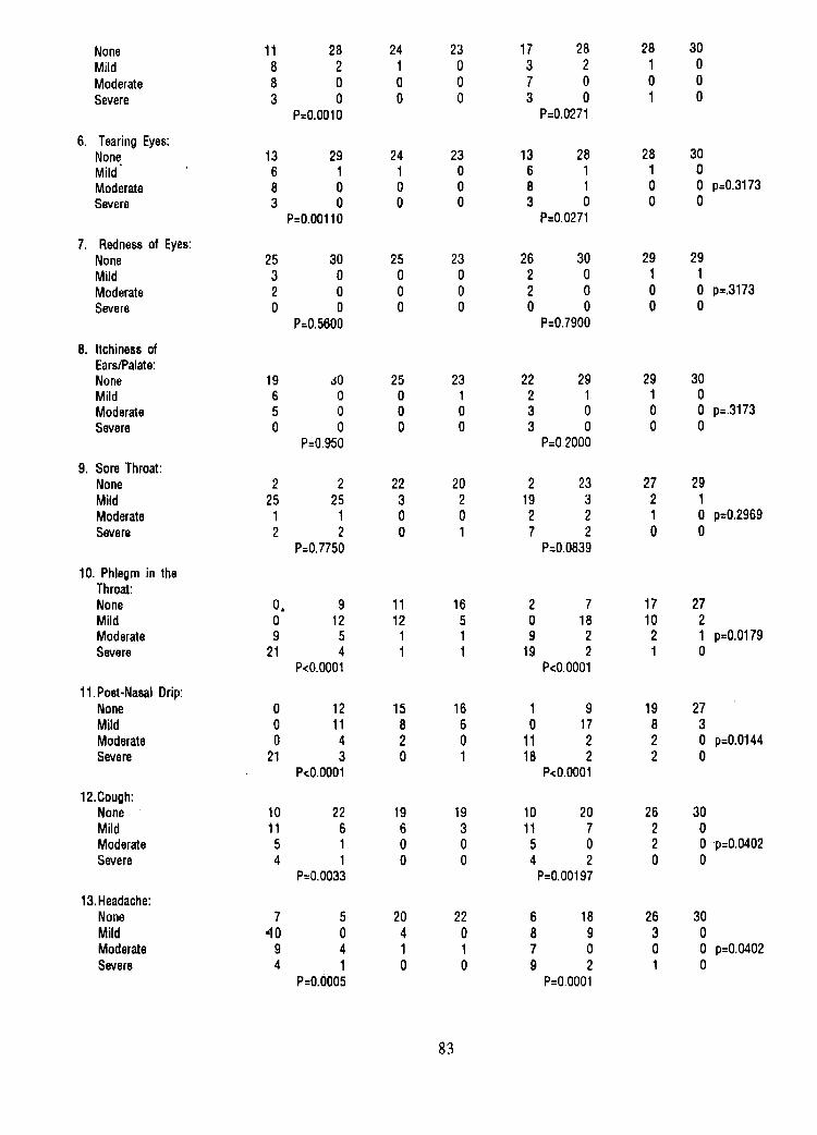
None 11 28 24 23 17 28 28 30Mild 8 2 1 0 3 2 1 0Moderate 8 0 0 0 7 0 0 0Severe 3 0 0 0 3 0 1 0
P=O.O010 P=0.0271
6. TearingEyes:None 13 29 24 23 13 28 28 30Mild 6 1 1 0 6 1 1 0Moderate 8 0 0 0 8 1 0 0 p=0.3173Severe 3 0 0 0 3 0 0 0
P=0.00110 P=0.0271
7. Rednessof Eyes:None 25 30 25 23 26 30 29 29Mild 3 0 0 0 2 0 1 1Moderate 2 0 0 0 2 0 0 0 p=.3173Severe 0 0 0 0 0 0 0 0
P=05600 P=0.7900
8. ItchinessofEars/Palate:None 19 JO 25 23 22 29 29 30Mild 6 0 0 1 2 1 1 0Moderate 5 0 0 0 3 0 0 0 p=.3173Severe 0 0 O 0 3 0 0 0
P=0.950 P=O.2000
9. SoreThroat:None 2 2 22 20 2 23 27 29Mild 25 25 3 2 19 3 2 1Moderate 1 1 0 0 2 2 1 0 p=0.2969Severe 2 2 0 1 7 2 0 0
P=0.7750 P=.0.0839
10. Phlegmin theThroat:None O, g 11 16 2 7 17 27Mild 0 12 12 5 0 18 10 2Moderate 9 5 1 1 9 2 2 1 p=0,0179Severe 21 4 1 1 19 2 1 0
P<O.O001 P<O.O001
11.Post-NasalDrip:None 0 12 15 16 1 9 19 27Mild 0 11 8 6 0 17 8 3Moderate 0 4 2 0 11 2 2 0 p=0.0144Severe 21 3 0 1 18 2 2 0
P<O.O001 P<O.O001
12.Cough:None 10 22 19 19 10 20 26 30Mild 11 6 6 3 11 7 2 0Moderate 5 1 0 0 5 0 2 0 p=0.0402Severe 4 1 0 0 4 2 0 0
P=0.0033 P=0.00197
13.Headache:None 7 5 20 22 6 18 26 30Mild .10 0 4 0 8 9 3 0Moderate 9 4 1 1 7 0 0 0 p=0.0402Severe 4 1 0 0 9 2 1 0
P=o.o005 P=O.O001
83
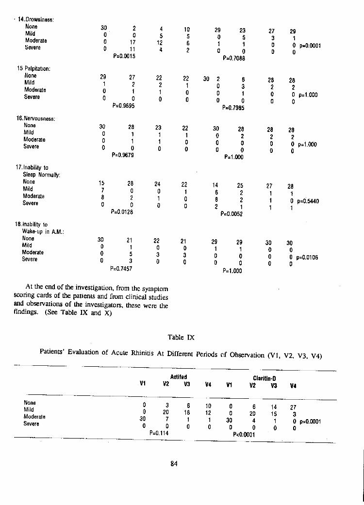
• 14.Drowsiness:
None 30 2 4 10 29 23 27 29Mild 0 0 5 5 0 5 3 1Moderate 0 17 12 6 1 1 0 0 p=O.O001Severe 0 11 4 2 0 0 0 0
P=0.0015 P=0,7088
15Palpitation:
None 29 27 22 22 30 2 6 28 28Mild 1 2 2 1 0 3 2 2Moderate 0 1 1 0 0 1 0 0 p=l.000Severe 0 0 0 0 0 0 0 0
P=0.9695 P=0.7985
16,Nervousness:
None 30 28 23 22 30 28 28 28Mild 0 1 1 1 0 2 2 2Moderate 0 1 1 0 0 0 0 0 p=l.000Severe 0 0 0 0 0 0 0 0
P=0.9679 P=I.000
17.InabilitytoSleepNormally:
None 15 28 24 22 14 25 27 28Mild 7 0 0 1 6 2 1 1Moderate 8 2 1 0 8 2 1 0 p=0.5440Severe 0 0 0 0 2 1 1 1
P=0.0128 P=0.0052
18,InabilitytoWake-upin A.M.:
None 30 21 22 21 29 29 30 30Mild 0 1 0 0 1 1 0 0Moderate 0 5 3 3 0 0 0 0 p=0.0106Severe 0 3 0 0 0 0 0 0
P=0.7457 P=I.000
At the end of the investigation, from the symptomscoring cards of the patients and from clinical studies
and observations of the investigators, these were thefindings. (See Table IX and X)
Table IX
Patients' Evaluation of Acute Rhinitis At Different Periods t_.f Observation (V1, V2, V3, V4)
Actlled ClariUn-D•Vl V2 V3 V4 Vl V2 V3 V4
None 0 3 6 10 0 6 14 27Mild 0 20 18 12 0 20 15 3Moderate 30 7 1 1 30 4 1 0 p=O.O001Severe 0 0 0 0 0 0 0 0
P=O.114 P<O.O001
84

Table X
Patients' Evaluation of Acute Rhinitis At Different Periods of Observation (V1, V2, V3, V4)
Actifed Clarllln-DVl V2 V3 V4 Vl V2 V3 V4
None 0 3 6 11 0 6 13 27Mild 0 20 18 11 0 20 16 3Moderate 30 7 1 1 30 4 1 O p=O.O001Severe 0 0 0 0 0 0 0 0
P=0.0061 P<O.O001
Table XI
Patients' Evaluation of the Therapeutic Response at Different Periods of Observation
Actlfed Clarltln-DV2 V3 V4 V2 V3 V4
Excellent 3 6 10 7 14 27Good 16 15 10 18 13 2Fair 7 3 2 2 2 1Poor 4 1 1 3 1 0 p=O.O001
P=0.0017 P=O.O001
Table XlI
Patients' Evaluation of the Therapeutic Response at Different Periods of Observation
Actifed Claritin-DV2 V3 V4 V2 V3 V4
Excellent 3 6 11 6 14 27Good 16 15 9 19 13 2Fair 8 4 2 3 2 1 p=O.O002Poor 3 0 1 2 1 0 -_
P=O.O009 P=O.O001
Safety:
drip, phlegm in the throat and occasionally experienceThe Laboratory tests of all patients (including those coughing probably secondary to the post-nasal drip.
drop-outs) before and after the medications were es- Both Claritin-D and Actifed relieve these signs andsentially normal, symptoms pretty well except that Claritin-D appears
to work faster and longer than Actifed. The averageduration of action of Claritin-D is 15 to 16 hours,
DISCUSSION and the average duration of Actifed is 4-5 hours.Claritin-D relieves the stuffy nose, sneezing and
Patients with acute rhinitis feel very miserable discharging nose much more effectively, and thisdue to clogged nose or stuffy nose, profuse running relief is achieved quicker and lasts longer than theor discharging nose, sneezing, sore throat, post-nasal relief brought about by Actifed.
85

Actifed's side effect of drowsiness is so severe REFERENCES:that 93.3 % of these 30 cases experienced drowsinessof varying degrees. In 10 cases or 33.3% in the
Actifed group, severe drowsiness was encountered so 1. Robert B. Berkowtz, MD; John T.Connel, MD;much that 5 cases or 16.6% were dropped after their Albert J, Dietz MD; Saul M. Greentein, MDfirst follow-up visit because they could not afibrd to and David G. Tinkelman, MD: The Effec-
continue their work and were afraid to lose their jobs. tiveness of the Non-Sedating AntihistamineTwo cases dropped off from the study after the 2nd Loratadine plus Pseudoephedrine in thefollow-up visit for similar reasons. On the contrary, Syptomatic Management of the Commonamong the 30 cases of Claritin-D group, no one Cold. Annals of Allergy 1989; 63:336-dropped out because of severe drowsiness. Twenty 339.percent of this group experienced slight drowsinesson the first follow-up visit but it disappeared after 2. Felicidad Cua-Lim, MD, FPCP; Margaret C. Lim,
3 days. At the end of the study, nobody complained MD; Jose S. Ilao, MD,FPCP and Reginaof drowsiness. Mangay,MD: An Evaluation STudy on the
Efficay and Safety of Loratadine vs.Other side effects were not significant in both Mebhyrolin in the Allergic Rhinitis. Phil.
groups except that one patient among the Actifed J. Internal Medicine 1990; 28:103-108.group developed hypertension. Among the Claritin-D group, 6 females developed loss of appetite although 3. Felicidad Cua-Lim, MD, FPCP; Margaret C.their laboratory studies did not show any abnormality; Lira, MD; Jose S. Ilao, MD,FPCP and
these 6 females were extra pleased because the drug Regina Mangay,MD: Efficay and Safety ofhelps them lose weight. Oral Loratadine 10 mg daily in Perennial
Allergic Rhinitis. Phil. J. Internal MedicineAll these observations were revealed at the end 1990; 28:109-115.
of the study when the symptom scoring cards were
received. ThisshowsthatClaritin-Disfarmoresuperior 4. Richard F. Lockey, MD and Roger W. Fox, MDto Actifed in the treatment of acute rhinitis, et al: Loratadine (SCH 29851) 10 mg o.d.
vs. Clemastine 1 mg b.i.d, in the treatmentClaritin-D is not just very effective in relieving of Perennial Allergic Rhinitis. Immunol-
the severe symptom of acute rhinitis, but it is also well ogy and Allergy Practice 1989; 423-429.tolerated by all patients, and it is very safe as dem-
onstrated by this study. 5. J. Grossman, MD; E.A Nronsky MD; B.O.
Lanier, MD; M.I. Linzmayer, MD; B.A.Mess, MD; E.J. Schenkel, MD and J.C.
CONCLUSION Slemer, MD: Loratadine -psedoephedrineCombination versus Placebo in Patients with
This study reveals that Claritin-D is far more Seasonal Allergic Rhinits. Annals ofsuperior to Actifed in terms of: Allergy 1989; 63:317-321.
1. improving the signs and symptoms 7. F.Estelle R. Simos, MD, FRCP (C): Loratadinea Non-sedating Hi-receptor Antagonist.
2, improving the overall evaluation of acute Annals of Allergy 1989; 63:266-268.rhinitis by the patients and the investigators.
3. well tolerated by the patients and absence ofside effects.
86

UNIVERSITY OFTHE PHILIPPINESPHILIPPINE GENERAL HOSPITAL
DEPARTMENT OF OTOLARYNGOLOGY
TITLE : A ComparativeStudyon theEfficacyofMedicatedMedium-stripGauzeandMedicatedFinger-cotasAnterior
Packsin MinorNasalSurgery
AUTHORS:JoseL. MontillaIII, M.D.JesusM. Jardin,M.D.JuanL. Rosas,M.D.
NelsonG.Magno,M.D.
ABSTRACT serted in a pleated manner within the nasal cavity, and
, removed several days later. Although this method re-mains to be an effective means of stopping the bleeding,
Thirty-two patients underwent bilateral, nearly it "alsocauses additional pain on insertion (when donesymmetricalnasalsurgeryunderlocalanesthesia. Post- under local anesthesia) and on removal. It is notoperatively, their nasal cavities were packed with the infrequent that an otolaryngologist's attention is calledconventional medicated medium-strip gauze on one because of a portion of the gauze pack dangling in hisside and with the medicated finger-cot on the other, patient's throat. Even more anxiety-provoking, bothforSelection of the side to be packed with one type of the patient and the specialist, is the recurrence of bleed-
packing was done on a stratified random manner. Both ing after removal ofthepackwhich, at times, maynotbemethods of packing were compared in terms of control that easy to control.of post-operative bleeding, subjective pain on insertion
and on removal, problems encountered during insertion Of the many innovations that have beendevised in
and at home, recurrence of bleeding and appearance of nasal packing, our interest is in the less-traumatic fin-nasal mucosa after removal, and microbiology of the get-cot method i.e., packs made by placing gauze inside
packing materials. Statistical analysis using non-par- cut-off fingers from surgeon's gloves. Although thisametric tests were employed whenever feasible. Both type of packing is already widely known, there appearsmethods were found out to be equally effective in con- to be no formal study comparing it with the standardtrolling post-operative bleeding. Although the finger- method of packing.cot required extra time tbr proper insertion in some
instances, it was associated with less pain on insertion This undertaking aims to compare both methods ofand on removal, and less recurrence of bleeding with packing in terms of: (1) control of post-operative bleed-"generally smooth mucosa after removal of the packs on ing, (2) subjective pain on insertion and removal ofthe 3rd post-operative day. Microbiologic results re- packs, (3) problems encountered during insertion and atvealed more microbial isolates from.the finger-cottype home, (4) recurrence of bleeding after removal, (5)but as a whole, growths from all packing materials were appearance of nasal mucosa after removal, (6) microbi-not clinically significant, ology of packing materials.
INTRODUCTION METHODOLOGY
One of the indications of anterior nasal packing is Patients who underwent nasal surgical proceduresthe control of post-operative bleeding in nasal surgcry, under local anesthesia that involved disruption or re-Conventionally, a medium (2-3 cm wide, I meter long) moval of nasal mucosa in a bilateral, mearly symmetri-strip gauze impregnated with antibiotic ointment is in- cal manner were included in the study. Selection of side
87

" to be packed with one type of packing was done on a mean age was 26.3 years. They were equally distributed
stratified random manner. The patients were numbered as to sex. Basically, all patients underwent operations in
consecutively, and then those who belonged to the odd- the area of the middle meatus plus variations which alsonumbered group had their left nasal cavity packed with involved bilateral, symmetrical disruption of nasal tis-the finger-cot type and their fight nasal cavity with sues (see Table 1). Of the 32 patients, five were notmedium-strip gauze. Those who belonged to the even- anesthesized equally on both sides. On insertion of thenumbered group had their right nasal cavity packed with nasal packs (see Table 2), 15 patients complained offinger-cot and their left nasal cavity with medium-strip, greater sensation of pain on the medium-strip side andIn each patient, the medium-strip gauze was impreg- only 4 on the finger-cot. The rest claimed that there washated with oxytetracycline/polymixin B ointment and no difference. Post-operative bleeding was controlledthe finger-cot externally coated with the same, but the in all patients although 5 patients had minimal transientmedium-strip gauze within the finger-cot extemally posteriorleak from the finger-cotside. Regarding prob-coated with the same, but the medium-strip gauze within lems encountered on insertion of the nasal packs, the
the finger-cot was only dampened with sterile water so surgeons pointed out that extra time was needed for theas to facilitate packing. (See appendix A.) proper insertion of the finger-cot type in 10 instances.
After the procedure, the patients were sent home At home (see Table 3), 2 patients experienced morewith oral medications consisting of an antibiotic* pain on the medium-strip side. Another 2 patients had(bacampicinin-PENGLOBE 400 mg BID x 10 days) minimal bleeding leak anteriorly, one for each type ofand analgesic-decongestant (acetaminophen- pheny- packing. Minor complaints like clicking sound onlpropanolamine 325/18 mg - 650/36 mg TID x 5 days) swallowing and excessive rhinorrhea on the finger-cotdrugs. They were followed up after 3 days for removal side were volunteered by 2 patients respectively. Fourof packs. The finger-cot was first decompressed by patients came back before the scheduled follow-up dayremoving the inner dressing from the cot and then re- due to protrusion of the medium-strip gauze into themoving the cot from the nose. This was followed by oropharynx. The dangling portions were cut transor-taking out the medium-strip gauze as gently as possible, ally, and the patients were sent home subsequently.Specimens from all nasal packing materials (two from
the finger-cot type--t hat is, one from the outer surface On the 3rd post-operative day, the packs wereand another from the non-medicated inner dressing) removed carefully. Twenty-three (23)patients claimedwere collected and sent to the laboratory for microbiol- that the removal of the medium-strip gauze was moreogic processing.** painful than that of the finger-cot. Nine (9) felt no
difference between the 2 methods of packing. None
Evaluation of the two methods of packing was singled out the finger-cot as more painful. As to theconducted through subjective responses from the pa- appearance of the nasal mucosa (see Table 4), there wastients, objective assessments made by the surgeons, and no portion (i.e., inferior turbinate, septum, middle mea-technical data provided by microbiologic studies. De- tus and turbinate) in the nasal cavity that was found totailed evaluation sheets were provided so as to standard- be abraded in the finger-cot side but smooth in theize recording and facilitate analysis of data (see Appen- medium-strip side. Nearly all portions (97% of inferiordix B). Statistical testing was done using non-par- turbinate, middleturbinate and meatus,72%ofseptum)ametric methods whenever required, of the nasal cavity on the medium strip side were
abraded. In contrast, to the finger-cot side generally hadsmooth mucosa (only 44% of inferior turbinate and 16%
RESULTS of septum, middle turbinate and meatus were abraded).Recurrence of bleeding in variable degree was noted in
A total of 32 patients was included in the study with all of the medium-strip side (see Tables 5 and 6). A1-age distribution ranging from 16 to 40 years old. The though 13 of these cases resolved spontaneously after 5
minutes, 16 required application of 1% ephedrine sul-....... fate-soaked cotton strips. Repacking of the nasal cavity* One patient with allergy to ampieillm; erythrornycin S00 TID prescribed was done in half, 13 patients had no recurrence ofinstead bleeding from the finger-cot side. There was no profuse
** Cultar¢ ,,rid identificatio_ were limited lo aerobic microorganisms since rebleeding encountered; all of those that rebled (16facilities for adequate anaerobic collection were not available, minimal, 2 moderate) resolved spontaneously. On sec-
88

ond follow-up, 25 patients did not experience motherepisode of rebleeding from both sides. Two patients on Problems encountered at home were minimal,the finger-cot and 6 on the medium-strip had minimal Although 2 patients complained of more pain on me-recurrence of bleeding which were all transient, hence dium-strip side and equal incidence (one each) of minor
did not require special attention, bleeding leak from both types, statistical evaluation isobviously not applicable. One problem intrinsic to the
Results from microbiologic studies showed growths standard method of packing, which occurred in 4 pa-in 46.7% of specimens* coming from the outer surface tients, is the protrusion of the gauze strip into theof the finger-cot, 67.7% from the inner dressing of the oropharynx when the pack loosens up, Tllis cannotfinger-cot, and 16.7% from the medium-strip gauze, happen in the other method since the dressing is con-Species of Staphylococcus particularly albus accounted tained within the finger.cot unless the whole pack slipsfor 85.7% of the microorganisms isolated from the down into the oropharynx. To avoid accidelatal extru-finger-cot surface, 61.9% from the inner dressing, and sion of the packs particularly the finger-cot on sneezing60% from the medium-strip. The rest of the microor- forexample, patients were advised to cover nose with anganisms cultured were various species of gram-nega- external dressing at all times.tive bacilli except for one isolate each of Candida sp.
from the inner dressing and from the medium-strip In our institution, the conventional anterior nasalgauze, packs are usually removed on the fifth posl-operative
day since incidence of rebleeding is said to bo high if thepacks are taken out earlier. In this study, both types of
DISCUSSION nasal packing were removed on the third post-operativeday. Early removal means early patient aelief from
Although the finger-cot method has been known to discomfort and decreased likelihood of other eomplica-be less traumatic than the standard gauze dressing, con- tions associated with nasal packing.trolled trials comparing both types are lacking. This
study makes use of each patient as his own control, Indeed all sides from wberethemedium-stripgauzeenabling the examiners to evaluate both types of ante- was removed showed minimal to profuse mbleeding.rior nasal packing in every aspect at the same instance. When compared to the finger-cot, the form_ is associ-
ated with an overwhelmingly significant greater degreeThe insertion of the medium strip gauze was asso- of rebleeding (Wilcoxon matched-pair rank test,
ciated with more pain than that of the finger-cot, which p<0.00003). Moreover, themedium-stripmellaod causedis statistically significant (sign test, p=0.02) despite the more pain on removal than the finger-cot, which is alsofact that all of the patients who singled out the finger-cot convincingly significant (sign test, 1><0.002).
as more painful were among those who were inade-quately anesthesized on that side to start with. Both The greater degree ofrebleeding from the medium-methods were equally effective in controlling post-op- strip side is related to its tendency to open up rawerative bleeding since the five instances of posterior mucosal surfaces upon removal. Being adherent to thebleeding leak from the finger-cot side (none from the nasal mucosa despite the liberal lubrication applied, itmedium-strip) were not statistically significant (sign disturbs the healing process and abrades the nasal tau-test, 19=0.062). Likewise, these instances were not cosa i.e., inferior turbinate, septum, middle turbinateclinically important because the leak in all cases was andmeatus, evenwhenitisgentlytakenout. The finger-
minimal as well as transient, and the patients were sent cot, on the other hand, is associated with a greatlyhome with the finger-cots in place. Expectedly, more significantsmoothmucosainallpartsofthenasaleavitytime was needed in the propoer insertion of the finger- (McNemar test, p<0.0001). The incidence of anothercot type (10 cases) since the technique required addi- bleeding episode after the first follow-up showed notional skill and practice. This problem is simply techni- statistical difference between the two methods of pack-cal, hence familiarity with the method will overcome it. ing (sign test, p=0.124), It has been pointed out earlier
that all of these instances of rebleeding at home after thepacks were removed (2 from the finger-cot and 6 from
*Atotalof5speeimens (2 fromthe outersudaee and I fromthe inner the medium-strip) were minimal and transient, hencedressing of the finger-cot, 2 from the medium-strip gaul) was not reee.ived not clinically significant.
by the laboratory for processing.
89

With regard to microbiology of nasal packing ma- with saliva particularly to account for the varied floraterials (see Table 7), the most number of isolates were recovered from the inner dressing of the finger-cot,from the inner dressing of the finger-cot. This was Contamination on collection could not be also dis-
expected because it was not lubricated intentionally counted since all packing materials passed through bothwith an antibiotic ointment. Colonization was not con- nares. In general, the presence of microbial growthsidereal clinically significant since it was confined within remained academic since all patients did not clinicallythe finger-cot. Nonetheless, the outer surface of the manifest signs of active infection.
finger-cot revealed statistically more isolates regardlessof kind than the medium-strip (sign, 19=0.04). This maybe due to the fact that the finger-cot is made of latex CONCLUSION
which is a non-absorbent and non-porous material,making the antibiotic ointment easily washed away by This study hasestablished thatthe finger-cotmethodthe excessive rhinorrhea associated with nasal packing, of nasal packing is equally effective as the standard_iStaphylococcus albus was the most common microor- medium-strip in controlling post-operative bleeding. Inganism cultured from all packing materials. Since it is addition, it offers the following advantages over thenon-pathogenic in healthy adults, its exclusion from sta- medium-strip method namely: (1) significant lessertistical testing will bring about no significant difference pain on insertion and on removal of the pack, (2) aigni-between the outer surface of the finger-cot and the ficant lesser recurrence of bleeding after removal withmediumstrip gauze in terms of potential pathogenic a generally smooth nasal mucosa, (3) relatively cost-microbes recovered (sign test, p= 1.0; see Appendix C). effective since little amount of antibiotic ointment isIn normal nasal flora as studied by Slavin et al, Staphy- needed (the surgical gloves may come from those to belococcus epidermidis (albus) was present in 83%, S. discarded), and (4) early removal i.e., on the third post-Aureus in 23 %, Streptococcus viridans in 17%, and operative day or even earlier. Although it requires extra
Enterobacter species in 4% (percentages total over time for proper insertion, this shortcoming can be over-100% due to mixed growths). All patients in this study, come by familiarity with the method. Microbial isolateshowever, had chronic sinusitis which might also add to from all nasal packing, though more from the finger-cot,the emergence of gram-negative bacteria. Another were not clinically significant as a whole.contributory source was the possible contamination
TABLE 1 : TYPES OF NASAL SURGERY
1. Polypectomy,ethmoidectomy,antrostomy-bilateralwithorwithoutpartialmiddleturbinectomy-bilateral...........................................................................................................................................13
2. Ethmoidectomy,antrostomy-bilateralwithorwithoutpartialmiddleturbinectomy-bilateral............................................................................................................................................7
3. Polypectomy,ethmoidectomy,antrostomy/ethmoidectomy,antrostomywithorwithoutpartialmiddleturbinectomy-bilateral............................................................................................................................................6
4. Submucousresection-septurnpolypectomy,ethmoidectomy,antrostomy-bilateralwithorwithoutpartialmiddleturbinectomy-bilateral............................................................................................................................................5
5. Submucousresection-septumEthmoidectomy,antrostomy-bilateral.....................................................................................................................1
TOTAL .................................................................................................................................. 32....................
90
t

TABLE 2: DIFFERENCES ON INSERTION
pain Bleedingieak Otherproblems....... i,
FINE COT 4 5 extratimeneededforproperinsertion..............................................10
MEDIUMSTRIP 15 0
NO ERENCE 13 27
TABLE 3: DIFFERENCES AT HOME
pain Bleedingleak Otherproblems
FINGER-COT 0 1 excessiverhinorrhea-1clickingsoundon
....MEDIUM-STRIP 2 1 protrusionswall°winGof........................................................medium- 1
stripintotheoropharynx...................................4
NODIFFERENCE 30 31
TABLE 4: APPEARANCE OF NASAL MUCOSA AFTER REMOVAL OF PACKS
MEDIUM-STRIPGAUZE
INF.TURBINATE SEPTUM MID.TURBINATF./MEAllU$smooth abraded smooth abraded smoothor abraded
presenceof or rawformedclots
FINGER-COT
smooth 1 17 9 18 1 26
0 14 0 5 0 5
TABLE 5: DEGREE OF REBLEEDING AFTER REMOVAL OF PACKS
1 2 3 4
FINGER-COT 13 17 2 0
MEDIUM-STRIP 0 10 16 6
1- Norebleeding2 - Minimal-thatis,notflowing3 - Moderate-thatis,intrickles4- Profuse-thatis,continuouslyflowing
91

TABLE 6: MANNER OF CONTROL OF REBLEEDING
1 2 3 4
FINGER-COT 19 0 0 13
MEDIUM-STRIP 13 16 3 0
1- Spontaneous resolution within 5 minutes2 - applications of 1% ephedrine sulfate-soaked cotton strips3 - repacking of nasal cavity4 - not applicable
TABLE 7: MICROBIAL ISOLATES
FINGER-COT FINGER-COT MEDIUM-STRIPoutersurface Innerdressing gauze
Staphy ;cussp. 1 1 1.L
Staphy _cusalbus 8 11 2
Staphy ;cusaureus 3 1
Pseudomonasaeruginosa 1 1
Achromobactersp. 1 2
Proteusvulgads 1
eranitratum 1 1
E.coli 1
Proteus abilis 1
Candidasp. 1 1
TOTAL qBEROFISOLATES 14 21 5
TOTAL P[BEROFSPECIMEN 30 31 30
92

APPENDIX A
INSERTION OF NASAL PACK
A. FINGER-COT TYPE
Materials:
1. Finger,cot (middle finger cut off from a size 7 1/2 surgical gloves _ 12. Antibiotic ointment (oxytetracycline/polymixin B) 5 g tube # 13. Medium-strip gauze # 1 or 24. Nasal speculum
5. Nasal dressing forceps or bayonet forceps
Procedure:
1. Coat liberally the outer suface of the finger-cot with the antibiotic ointment.2. Place the cot over the speculum which is then introduced into the nose.3. Maintain pressure between your index finger, which is resting on the ala, and the blade of the speculum to
avoid slipping of the finger-cot deep into the nasal chamber.4. Spread out the sides of the finger-cot by probing the nasal forceps within the finger-cot.5. Insert slowly the medium-strip gauze (dampened with sterile water) using the nasal dressing forcep until the
whole chamber is fully packed.
B. MEDIUM-STRIP GAUZE
Materials:
1. Medium-strip gauze # 1 or 22. Antibiotic ointment (oxytetracycline/polymixin B) 5 g # 2 or 33. Nasal speculum4. Nasal dressing forceps or bayonet forceps
Procedure:
1. Lubricate liberally the medium-strip gauze with the antibiotic ointment.2. With the speculum in place, insert the medicated strip using the nasal forceps in a pleated manner within the
nasal chamber until fully packed.
93

APPENDIX C
MICROBIALGROWTH POTENTIALLYPATHOGENICSTRAIN
Pt. Finger-cot Medlurn-strip Pt. Finger-cot Medium-stripoutersurface innersurface outersurface Innersurface
1. + - 1. - -2. - - 2. - -3. - - 3. - -4. + + 4. - +5. + - 5. + -6. - - 6. - -7. 0 0 7. 0 O8. + + 8. - -9. + + 9. - -
10. - - 10. - -11. + _ 11. - -12. + _ 12. - -13. + - 13. + -14. - - 14. - -15. + + 15.. - -16. + + 16. - -17. - - 17. - -18. + - 18. + -19. - 0 19. - 020. - - 20. - -21. + _ 21. - -:?2. 0 - 22. 0 -23. - _ 23. - -24. + + 24 + _25 - + 25. - +26. + - 26. + -27. - - 27. - _28. - 28 - _29. - 29. - _30. - - 30. - -31. + + 31. + +32. - 32. - -
(sign test, p = 0.04) (sign test, p = 1.0)
94

BIBLIOGRAPHY Keen, Monte. The Avitene Pack: A New Method to
Control Epistaxis In a Patient with Poor
Platelet Function. Laryngoscope 96:141 !-Brook, Itzhak. Bacteriology of Chronic Maxillary 1412, 1986.
Sinusitis in Adults. Ann Otol Rhinol Laryn-gol 98:426-428, 1989 Schaitkin, B., Strauss, M., Houk, J.R. EpistaJtis: Medi-
cal Versus Surgical Therapy: A ComparisonDerkay, Craig S., et al. Posterior Nasal Packing. Arch of Efficacy, Complications, and Economic
Otolaryngol Head Neck Surg 115:439-441, Considerations. Laryngoscope 97:t392-1989 1396, 1987.
Guyuran, Bahman. Is Packing After Septorhinoplasty Seigel, Sydney. Non-parametric Statistics for theNecessary? Plast Reconst Surg 71:196-198, Behavioral Sciences. New York: McGraw
1983 Hill Book Company, Inc., 1956, pp.61-63.
Jackson, J.A. and E.M. Kaswom. Toxic Shock Syn- Slavin, S.A., et al. An Investigation of Bacteremia
dromeAfterNasalSurgery. Arch Otolaryn- During Rhinoplasty. Plast Reconst Surggol Head Surg 112:329-332, 1986 71:196-198, 1983.
95

EAST AVENUE MEDICAL CENTERDEPARTMENT OF OTOLARYNGOLOGY
TITLE • CollisionTumor of the Nasopharynx(Liposarcoma,UndifferentiatedCa) Reportof a Case
AUTHORS:GeraldS. Chua,M.D.AngelV. Veloria,M.D.
Mamerto(3.Almelor, M.D.Milagros S. Lopez,M.D. ..
INTRODUCTION health center which provided temporary relief. Thepatient consulted at EAMC because of the persistence
We report a case of a rare, highly malignant collision of epistaxis and was subsequently admitted, He hastumor. A liposarcoma of the nasal cavity with extension a 35 pack year smoking history and has been takingto the ethmoid and orbit and an undifferentiated Ca alcoholic drinks for 30 years, In the past three monthsof the nasopharynx. Biopsy was done three times from he lost 35 pounds.the nasal mass and nasopharynx. Radical maxillec-tomy with orbital exenteration, radical neck dissection On admission, the significant findings were:and post-op radiation was contemplated. Unfortunately, retracted TM, AD, a grayish-reddish gelatinous friablethe patient deteriorated until his untimely demise before mass about .5 cm at the area of the middle (R) turbinate,
the proposed management was instituted, a 1 cm fleshy mass at the area of the posterior choanae,bilateral infra-auricular neck nodes both measuring 2x 3 cm. PNS x-ray reveals bilateral maxillary sinusitis.
OBJECTIVES Except for minimal PTB other laboratory work-upswere unremarkable. The admitting impression was
1, To report a rare collision tumor involving NPCA, stage III.liposarcoma of the nasal cavity, paranasal
sinuses and orbit and an undifferentiated Ca The first biopsy of the nasal mass revealedof the nasopharynx, liposarcoma. However, a repeat biopsy of the naso-
pharynx was suggested. The repeat biopsy was2. To review the literature regarding liposarcoma undifferentiate Ca. After a week, a third biopsy was
of the head and neck. done as per suggestion of the pathologist. The previous' diagnosis was confirmed - Liposarcoma, myxoid type
3. To present a local retrospective study on with some degree ofdifferentiadon at the nasal cavityliposarcoma of the head and neck. with an undifferentiated Ca of the nasopharynx, a
collision tumor.
CASE REPORT He was scheduled for radical maxillectomy (R)with possible orbital exenteration and RND (R) pend-
A 49-year old taxi driver was admitted for the first ing C-T scan results, However, he developed prop-time at the East Avenue Medical Center for recurrent tosis, lid edema, lateral and medial rectus palsy, ODepistaxis from the right nose, The patient had cough and hypoesthesia of the right half of the face. C-T scanand colds with yellowish, mucoid, blood-tinged spu- showed a mass in the right half of the nasal cavityturn, 7 months VIA which were temporarily relieved displacing the nasal septum to the left, with lyric changes
by self-prescribed medications. One month PTA, he in the medial wall of the right maxillary sinus withnoted decrease in hearing acuity (R) and (R) nasa] extension of the mass to the sella, ethmoid and sphe-obstruction. The epistaxis increased in frequency and noid sinuses. There is also involvement of the (R)amount. He was given hematinics IM at the local medial rectus muscle and the orbital roof. Two days
96

prior to the procedure, the patient became stuporous data that could be analyzed. In 1978, Kimlblom etand later succumbed to cardio-respiratory arrest, al included only 17 cases from the literature and added
4 cases but did not add the new cases mentioned b_'Baden and Newman. In 1979, Saunders et al also
DISCUSSION reviewed the literature and listed 25 previously re-ported cases and added 4 new ones. This series
TUMOR "INCIDENCE neglected the cases described by Kindblom andassociates, Baden and Newman, and also by Fu in
Liposarcoma, next to malignant fibrous histiocy- 1977. At the AFIP, 60 of 1067 cases reviewed weretoma is the most common soft tissue sarcoma of adult in the head and neck region. Smiler and Davies reported
life. Among soft tissue sarcomas its incidence ranges an incidence of only 4 liposareomas in the bead andfrom 16%-18%, but values as low as 5%-6% have also neck in a population of 8.5 million people in Southernbeen given by Hashimoto et al. Kindblom et al reported England, although how this figure was established wasan annual incidence of 2.5 million in Sweden. It is not mentioned. Other rare sites include the cheek,
likely that the true incidence ranges between 10%-12% forehead, scalp, orbit, floor of the mouth, soft palate,of all soft tissue sarcomas since earlier reviews included pharynx, meninges, larynx, supraclavicular fossa,
cases of malignant fibrous histiocytoma, parotid gland, maxilla and the mastoid. Knowles in1954, reported the case of a 12 year old with liposar-
AGE AND SEX INCIDENCE coma of the nasopharynx. Fu et al in 1977, involvingthe nasal cavity, PNS, hard palate, temporal and
Liposarcoma is primarily a tumor of adult life with sphenoid bones.a peak incidence of between 40 and 60 years with amean of 50 years. It is virtually unknown in infantsand small children, but there seems to be no upper HISTOLOGICAL CLASSIFICATION OFage limit. In a recent review of 1067 tumors at the LIPOSARCOMAAFIP during a 10-year period, the youngest patientwas 8 months and the oldest 87 with the average age Stout, Enterline and Enzinger have all given
depending to some extent on the anatomical distribu- different classifications of liposarcoma. For ourtion of the tumor. Males are more often affected than purpose, we adapted the classification of Bnzinger asfemales (55%-60%). For unexplained reasons, the right used by the AFIP. Liposareoma is classified into 4half is more often involved than "the left. types: Myxoid, well-differentiated Lipoma-like,
sclerosing, inflammatory, dedifferentiated), round cell,RACIAL PREDILECTION pleomorphic.
There is no mention of racial predilection in ourreview of literature. GROSS APPEARANCE
Grossly, the tumor varies as to the histelogic type.INCIDENCE IN HEAD AND NECK From the white-gray, slimy, translucent myxoid variants
to the brain like quality of the less-differentiated typesLiposarcoma is most commonly.found in the lower with the well-differentiated ones resembling yellowish-
extremities (thigh) and the retroperionium and only orange lipomas.found in about 2%-6% of cases in the head and neck
with the neck being most common site in this area.MICROSCOPIC APPEARANCE AND
Das Gupta in a combined series of 335 liposar- INCIDENCEcomas listed only 9 (1.8%) within the head and neck
region. In Aliens study of 126 confirmed liposarcomas Microscopically, the myxoid type is the most8 were in the head and neck (6.3%). In 1960, Spittle common (30%-50% in all sites), it is characterized
and associates reviewed 60 cases with only 1 case in by monomorphic, fusiform or stellate cells residing inthe neck. Baden and Newman in a review of literature a mucoid stroma rich in hyaluronic acid with delicatein 1977 had 40 cases, 35 of which contained su_eient plexiform capillary network. The well-differentiated
97
type (20%-30% of cases) varies from lipoma-like ceils the response may be striking. Chemotherapy may haveto inflammatory reaction and sclerosis all with bizarre a role in the treatment of liposarcoma. Kindblom etlipoblasts. Pleomorphic liposarcoma (10%-25% of al in 1977 reported on its use but the series was toocases) contains an abundant mixture of giant ceils that short to draw any conclusions.have a dense, glassy, eosinophilic cytoplasm withbizarre lipoblast and numerous abnormal mitotic fig- A collision tumor is a tumor that is found adjacent
ures. The round cell type (10%-15% of cases) has to and meeting another of different histological typeround to oval cells with multi-vacuolated cytoplasm in the samearea. In our view of foreign literature
and a central round nucleus that may be hyperchro- we see no incidence of a collision tumor involvingmatic but marked atypical mitosis are not common, a liposarcoma in the nasal cavity and an undifferen-
tiated Ca in the nasopharynx.
SURVIVAL AND RECURRENCE RATEREVIEW OF LOCAL LITERATURE AND CASES
The myxoid and the well-differentiated types havethe best survival rates (75%-100%). The round cell In our review of local literature, we have not yetranges from 18%-25%, with the poorest survival rate encountered any case of a llposarcoma occurring inbelonging to the pleomorphic type (0%-21%). The the nasal cavity nor a collision tumor involving li-myxoid and well-differentiated types have the most posarcoma and an undifferentiated Ca. However, uponrecurrence rates (50%-100%) with the pleomorphic retrospective study of liposarcoma (15 years) in twoand round cell ranging from 75%-80%. institutions, we have found only 1 unreported case of
myxoid liposarcoma involving the maxilla.
RATE, INCIDENCE AND SITE OF A review of our slides by different pathologistsMETASTASIS in different institutions gave the general consensus of
a collision tumor involving a liposarcoma (myxoidThe rate of metastasis is closely related to the type) of the nasal cavity, with extension to the PNS
degree of histological differentiation. The less dif- and orbit and an undifferentiated carcinoma of theferentiated,, the more cellular and the more pleomor- nasopharynx.
phic a given liposarcoma, the more likely it willmetastasize. Apparently, the incidence of metastasisis dependent to a lesser extent on the location and size CONCLUSIONof the tumor and perhaps also on the mode of therapy.
Sires of metastasis vary considerably. Mostly, poorly We have presented a case of a rare and highlydifferentiated types metastasize to the lungs and the malignant collision tumor involving liposarcoma ofvisceral organs. Myxoid liposarcomas produce sec- the nasal cavity with extension to the PNS and orbitondary lesions on serosal surfaces of pleura, pericar- and an undifferentiated carcinoma of the nasopharynx.dium and diaphragm and sometimes in the retroperi-tonium. A study by Giorgiades et al attempted to In itself, liposarcoma of the nasal cavity is raredelineate metastasis from aprobable multicentric origin and to our knowledge may be the first reported case
of liposarcoma by presenting 1 case in which they locally and the third reported case internationally.believe multicentric origin is present, Lymph node Furthermore, when combined with an undifferentiatedmetastasis is rare, and if present, indicates advanced Ca of the nasopharynx as a collision tumor makes itdisease, an even rarer case and thus we believe that this may
be the first reported case of its kind both locally andinternationally,
TREATMENT
Treatment consists of wide excision of the tumor
with post-operative irradiation. Studies have shownthat plain radiotherapy has not been so effective insome of the cases but when used on metastatic cases
98
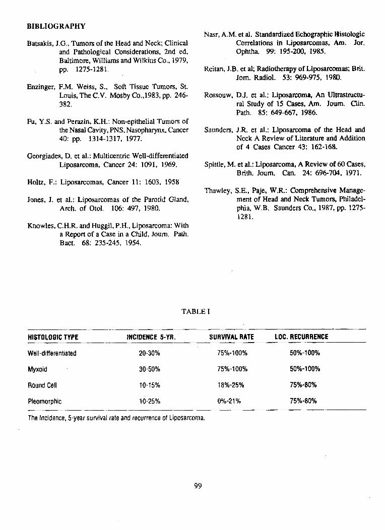
BIBLIOGRAPHYNasr, A.M. et al. Standardized Echographic Itistologic
Batsakis, J.G., Tumors of the Head and Neck; Clinical Correlations in Liposarcomas, Am. Jor.
and Pathological Considerations, 2nd ed, Ophtha. 99: 195-200, 1985.Baltimore, Williams and Wilkins Co., 1979,pp. 1275-1281. Reitan, J.B. et al; Radiotherapy of Liposarcomas; Brit.
Join. Radiol. 53: 969-975, 1980.
Enzinger, F.M. Weiss, S., Soft Tissue Tumors, St.Louis, The C.V. Mosby Co.,1983, pp. 246- Rossouw, D.J. et al.: Liposarcoma, An Ultrastructu-382. ral Study of 15 Cases, Am. Joum. Olin.
Path. 85: 649-667, 1986.
Fu, Y.S. and Perazin, K.H.: Non-epithelial Tumors ofthe Nasal Cavity, PNS, Nasopharynx, Cancer Saunders, J.R. et al.: Liposarcoma of the Head and40: pp. 1314-1317, 1977. Neck A Review of Literature and Addition
of 4 Cases Cancer 43: 162-168.
Georgiades, D. et al.: Multicentric Well-differentiatedLiposarcoma, Cancer 24: 1091, 1969. Spittle, M. et al.: Liposarcoma, A Review of 60 Cases,
Brith. Joum. Can. 24: 696-704, 1971.
Holtz, F.: Liposarcomas, Cancer 11: 1603, 1958Thawley, S.E., Paje, W,R.: Comprehensive Manage-
Jones, J. et al.: Liposarcomas of the Parotid Gland, ment of Head and Neck Tumors, Philadel-Arch. of Otol. 106: 497, 1980. phia, W.B. Saunders Co., 1987, pp. 1275-
1281.
Knowles, C.H.R. and Huggil, P.H., Liposarcoma: Witha Report of a Case in a Child, Joum. Path.Bact. 68: 235-245, 1954.
TABLE I
HISTOLOGICTYPE INCIDENCE5-YR. " SURVIVALRATE LOC.RECURRENCE
Well-differentiated 20-30% 75%-100% 50%-100%
Myxoid 30-50% 75%-100% 50%-100%
RoundCell 10-15% 18%-25% 75%-80%
Pleomorphic 10-25% 0%-21% 75%-80%
TheIncidence,5-yearsurvivalrateandrecurrenceof Liposarcoma.
99

TABLE Ii
HistologicalClassificationof Llposarcoma
STOUT ENTERLINEEl"AL. ENZIHGER& WINSLOW
1. Well-diff,myxoid Well-diff.myxoid Myxoid
2. Poorly-diff.myxoid Poorly-diff,myxoid Roundcell(mayhavefibrosarcoma-likeareas)
3. Round-cellor Lipoma-like Well-diff.adenoidgroup (lipomalikeor sclerosing)
4. Mixedgroup Myxoidmixed(includingfibrosarcoma-likeareas) Pleomorphic
5. Non-myxoid
250
200
Age distribution of 1067 liposarco-
mas. The tumor is most common during t50adult life and is rare in children, espe-cially in those below 10 years of age.
100
5O
10 20 30 40 50 60 70 80 90
80
60 lm mii illl i I ---. ,.._ ....
40 I/mmummmuumll
Survival.correlatedhistologically.
20
00 1 2 3 4 5 10
100

Lower leg (49)/"
Anatomical locations of liposarcoma (AFIP 1067 cases)
ANATOMICALLOCATION NO. OFCASES %
Head-neck 60 5.6Neck (36), face 03)Scalp(4)
Trunk 452 42.4Retroperitoneum(198)Inguinalregion (130)Back(29), chest (29)
Upperextremities 113 10.6Shoulder-axilla(44)Upperarm (44), forearm (19)
Lower extremities 442 41.4Thigh-knee(321)Buttock(62)Lower leg (49)
TOTAL 1067 100,0
]01

THE LATERAL NASAL WALL IN FILIPINOS: A STUDY BASED ON
FIFTY CONSECUTIVE CADAVER DISSECTIONS"
AUTHORS
CHARLOI-fEM. CHIONG,M.D.*ARMANDOM.CHIONG,JR.M.D.*CESARANTHONYP.YABUT,M.D.*
EDITHC.YAP,M.D.*ARMANO0T,CHIONG,M.D.'*JACOBS. MATUSIS,M.D.***
ABSTRACT within the major sinuses. Obstruction in this areaeither due to osteomeatal inflammaztion or anatomic
This study investigated anatomical variations and variation can perpetuate sinus infection. Functionalactual measurements of anatomic structures in the lateral endoscopic sinus surgery emphasizes endoscopic iden-nasal wait in fifty consecutive Filipino cadavers tification and operation of the diseased ethmoidaldissected. Such measurements were compared with infundibulum with the aim of reestablishing drainagevalues published in anatomy textbooks. This study and ventilation of the dependent large sinuses via theirproves that Caucasian-based figures need not reflect physiological ostia. Van Alyea and Myerson, with
thOSe in Filipinos. Anatomic variations were noted the knowledge of this important physiologic concept,and their clinical significance discussed. This study gave impetus to cannulation of the ostium maxillare
gives baseline values in Filipinos and is of potential to treat seemingly "irreversible" sinus pathology.use for the clinician in practice as well as for instru- Zuckekandl recognized the importance of anatomic
ment design for specific use in nasal surgery on Filipino variations in the middle meatal area responsible forpatients, pathogenesis of sinus disease. The basic knowledge
of the anatomy, of this critical importance. In thisrespect, the rhinologist who wants to cannulate the
INTRODUCTION maxillary ostium or the sinonasal endoscopist must befamiliar with the dimensions and spatial orientation
The specialty of Otorhinolaryngology-Head and of the important structures in this area. In our setting,Neck Surgery has recently gained major advances in these advances are now being introduced. However,the area of rhinology and sinus surgery. There has basic anatomy textbooks deal mainly with measure-been increased discussion regarding the advantages of ments and anatomic observations made on Caucasians.
newer surgical techniques like functional endoscopic Considering that Filipinos are generally anthorpom-surgery over conventional sinus surgery and vice-versa, etrically smaller, idiosyncrasies and variable dimen-The area of osteomeatal complex, while poorly visu- sions of the structures are expected. Consequently,
alized on anterior rhinoscopy or plain radiographs is these values may dictate the use of indigenous spe-most important in the pathogenesis of secondary disease cialized instruments designed for Filipinos as well as
guide the surgeons _in performing simple surgical•intervention or during endoscopy of the sinonasal areas.Clinical correlation with certain unique rhinopathol-
+ First prize, PSO-HN$ 8th Annual Residez_ts" Research Contest held ogic conditions and surgical intervention deserve specialon October 26, 19t_$, Manila Midtown Hotel
consideration. To investigate the hypothesis that foreign
* Residents,, Department of OtollLryngology, UP-PGH Medical Center published data on the anatomy of lhe lateral nasalwallneed not reflect those of the Filipinos, actual meas-
** Chairman, Department of Otolaryngology, UP-PGH Medical Centeruremcnts and description of the anatomy of the lateral
*** Consultant, Department ofOtola_yngology, UP-PGHMedic_I nasal wall was based on fifty consecutive cadaverCenter dissections of Filipino adult skulls. Particular attem
102
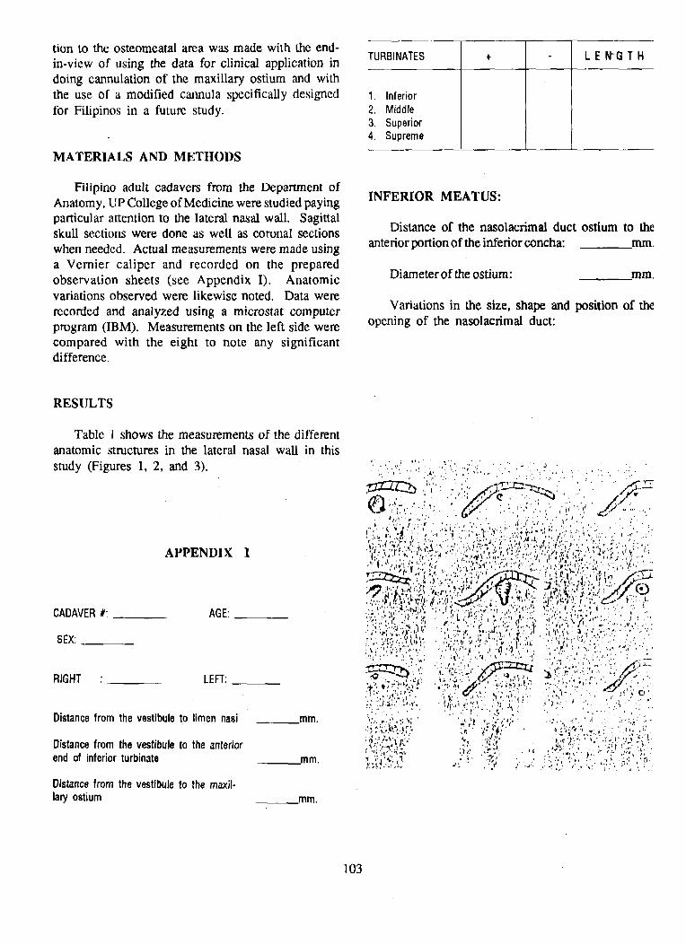
tion to the osteomeatal area was made with the end-
in-view of using the data for clinical application in TURBINATES + L E N'G T Hdoing cannulation of the maxillary ostium and withthe use of a modified cannula specifically designed 1. Inferiorfor Filipinos in a future study. 2. Middle
3. Superior4. Supreme
MATERIALS AND METHODS
Filipino adult cadavers from the Department ofAnatomy, UP College of Medicine were studied paying INFERIOR MEATUS:particular attention to the lateral nasal wall. Sagittalskull sections were clone as well as coronal sections Distance of the nasolacrimal duct ostium to the
when needed. Actual measurements were made using anterior portion of the inferior concha: ram.a Vernier caliper and recorded on the preparedobservation sheets (see Appendix I). Anatomic Diameteroftheostium: ram.variations observed were likewise noted. Data were
Variations in the size, shape and position of therecorded and analyzed using a microstat computerprogram (IBM). Measurements on the left side were opening of the nasolacrimal duct:compared with the eight to note any significantdifference.
RESULTS
Table 1 shows the measurements of the differentanatomic structures in the lateral nasal wall in this
study (Figures 1, 2, and 3). .... '...... .: .....', ."/i, ': '_ ....' ", j ,., . , , ., :..:... ,.,, . ,.
_'_-_ ' ' '.'.,' _C,P= _"'_,,. ,'" //,"_, , , ' .' "'"/,'i _ ' ", "_ ". ' I t
,'.: . ", .......' ' '_. _,g' '_. ''_ _t ' ' _ ' 't " ' '
".'. "' _ • ." t '_''," ., ,,, ",A,,' ..,,.;,,_ _ ,,, /J' ,'. , "' ;.... J ,"L.'.,_,:'t_,-- _,,_. __, " , " ,: '_' J'T'''. _'_ .,'p ...... ' r, '._' _., ,_: '!. _'. "_._ _ "_,,"'., ', _.',f.!'_ "_,, ;,',' _ _,,..,_t.,.'t, '_ .__,' , .,',.' . ;.._' .;,,,'_',
APPENDIX I ',':, .'r;, ,..,., ,'. ,'._ _,,..',,',_'_.... .: ./, _,:t.-,,,..,__'_,_'_ ',
'..:.':.' I,,,,//r' I_.,',._._ _ • ,"t" .,' -"" :'..'._','r •/JT.._.... , , ::_,.._ ,,. . _,,_,__, ,:.. .., _ "',-Ae_,_"'.t t . ,¢_ .I., ,. _ ., ., _ ,_ .,
CADAVER#: AGE ; ,'.,,.._..%,,':' _r,...-.,;,.,.;.",_,,,,.'._.:...',;',.fi.. _ ,_. -,'.,., _'-_,' 'i) _;_ , ,.,,*_.,.,_ _ /:._ _,,,,,',, ',,,,,.',,.,,.'r-., ,,,".' - , .,,_'_,',.._ t ,-fl _ , _, b "'," ,' P"Q ''""'-*_¢t'" '% v '' '
I';"_',':,'..,?,')_ _ ',_ ::.'_;' ,1_'_J,,_,' 'i, ,:, '_]', ,_'fr,' _,.",,_::.:':,:"," " YSEX:.. [':.Y_,",':,;_,_:' ,:,-",_'_"_:"',..'_".z '!"d _''_', '; "..":. :'
•_" _,_""'""_,_,",,, * ,'f _'¢' j,' , t_ ,_'_..... i ' _ , .,.e , _ _.,,' "., ,
_,_r_'."', ,.:.t'.' ,.. _E2S_rt '_j:,.._:'. ,'_"_''_.-_ ...._d__'_" " • " r;_" , _f_-_ _ _ " .',, " ' _*_ "
RIGHT ' LEFT: _ -"'_ ........... _ "_>" _ _" ._ .:....... ,, " t.,"..... , . , ",' ._ _, ".-,_ ,/ ." . :, ,*_.," ... ..'._ • .. ,'. . . _J ¢_.. ', ,:,I.. ,_.:,_, ._ _f_,.,. _, ,, .. .1 ._ . ,, . , C •,,.,;,q_,,..i_',:. ",,_,...,;..:, .., ,,,. ,,,., .;, :. _,,.., , ,,., ,., ,..,.
,.:.",.:,'. _"' '!. ,:'r .... :.'_' ._. ,-.'. '-.,...... " ,',":. '
Distancefromthevestibuleto limennasi __.ram. _,.;,;:_,::._,- _,_ ',._,,,',- ,.......• .._,.. _.., ..'..., .:,.),._,:_:3,,, :_ ' ]" ;¢: ' _;';iY" ",! : ': _'" "", _ * _. , : ' d'., • '" " ._ ;'; . .. :,, ,"," "_,'_ "
Distancefromthevestibuleto theanterior , .............. ,.... .,' . ;,,.: ..... .{ _,, _._.;_ "_¢ ,r "t, . ,_ ,'._."_,'_..'. _', ., ,t.I ,;, ,_"
endof inferiorturbinate ram. ,,h'__,_ . . _,_ . . :v :,, ;.., _, .,_...... ' _ _..... 'a" , _ , , t.,., • . ,,,...!.? ..... , , .1 ", ," ',,. '." ', ,I, ., , '_ '...
Distancefrom thevestibuletothe maxil-laryostium _,mm.
103

MIDDLE MEATUS:
Diameter of ostium: mm.. • ,. "_"_._,,; '< \_ ",,';: l.,._'...._,.-. _ ... ... ......... ,", .t-"' ";." '- "_........... "i-'.'
"" "."_ " ,ii ' :"' '".:_ " "" r"t _;':" _' " ° 'Configuration of ostium:
/.-)_ ::S'._';!:_ :_ L'_ '_? _ :L• ,. Jl.,.:_:: ;"_-.:" , .:,"! ' • ',', '"/ ",,",'Horizontal -/if :',,':L:,,A _ '_',.... _., _ ,
,. ,//'_. ,. .... _ ...... .. ,.... __.. :'".':;_. :l, t'- .,','-.(. ;. ;.. •
Vertical -, .y/2_ '.: ;:' ..;'1' " ;"'.4',:-,' .,, !;",t..z<:,.."...-_ "',":: '.... "l, ' ', _: ".'...,.' 7'_.",,.., ," ,_'..',. :,,_,." _", :; ..,
Oblique ...... '..... -'" .... '. '._,''.... ".... -a. ,....
Distance from the anterior end of the ethmoidalinfundibulum:
mm.
Distance from the anteriorend of the middle meatus: . .
ram. " i)""' /Z./_ "//_"
:.,; i:_,:,!. ' ", ,: ,:'.,;, ,_..:,,../ .., /;; .,, , ',,,.; ..,: j.., ,
":.' ,'_;_i.:2f. :.:<: '.'_! _L"///.: .::."Length ram.
Depth mm.
Maxillary sinus:
AP diameter mm. a. Uncinate process is moderately tall and hiatusis moderately wide.
Transverse diameter ram.b. Extreme development of the process. Hiatus
Height ram. is specially wide.
Accessory ostia in the hiatus c. Hiatus is narrow.semilunaris: + -
d. Uncinate process and infundibulum are rudi-Variations in the semilunar hiatus and mentary.
ethmoid infundibulum:
e. Bulla overhangs the uncinate process.Distance from the maxillary floor to the
maxillary ostium ram. f. The two are in contact.

TABLE 1. Measurements of the Different Anatomic Structures in
the Lateral Nasal Wall of Adult Skulls (n=50).
MEAN STANDARDDEVIATION(MM)
A. Distancebetweennaresand limen nasi 12.79 +/-3.60
B. Distancebetweennaresand anteriorendof inferior turbinate 18.66 +/-2.24
C. Distancebetweenanteriorend of inferiorturbinateand nasolacrimalostium 10.10 +/-1.38
D. Diameterof nasolacrimalostium 3.44 +/-1.24
E. Turbinatelength
1_ Inferior 41.46 +/-3,622. Middle 37.13 +/-4,13:3. Superior 13.1 +/-6,084. Supreme
F. Distancebetweennares andmaxillaryostium 37.97 +/-4,24
G. Diameterof maxillaryostium 4.23 +/-1,40
H. Heightof uncinateprocess 10.68 +/-3,73
I. Distancebetweenostium and maxillarysinus floor 30.84 +/-8,46
J. Length of Hiatussemilunaris 17.11 +1-3,64
105
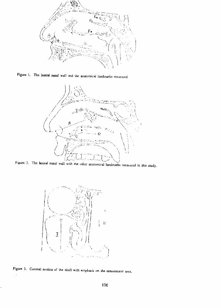
/.;4".,..:'.-".............'---.",_,,?,;.,-:................, .,,"..,-:>' ""_-,.:z.,_._t .. ,.?s"_-,, ....
,'V.::,....,,, ),;.).:.......i7--;,._.:.-<:,.( _<.]
.;".:/ \_,{_b_),..'>,\,: "=:::v?.C';_ . -:...,_•_,
,,,. ,.I..._, .......... <.......::......",, ,.., .-- .'_---_---<'.2.')_ • -""--.,/ "
......... _...... ' " ,- ', ,.. . r. ",<'.-..,.,-."--. ,: ) 1 ,:. r.-','.",'
I'..<::..L-::----:..-.__':"::.-::-J_.:.- ':"/,:/: " ".' .,, I\" .,_q_ ..... -.--=:....... •.... t _ .",.) ,"._"j,:.,._",....!:,. ":.........-. _-. ,' -
" ! -._-_'A.,.. ,.. _'..---._z:'-.- -,." .. ......... ,. _,:.". _ _ ,,)> , _. .. ; , _ . ., /
I,..:-.,.:]/_.'>.-.:..,..... .'..'<..'.,.:..;.:,.._. '_ ,,' _'":_',1
\':._
Figure 1. The lateral nasal wall and the anatomic,,d landmarks measured,
/'__._/::.:;_..-=_==,-__<:-_-_.-_..-,_,-_:_.:.,z.,.:,--.-_=............_........ _ ....... _'__',.....) _,
/ / t\._'_-.::,:.... ¢_m., "" " i: / \_ ' _ ',,,,.". ,., ....,.,
.' i" ...I._._.,,:._ . _<,',.\ ).. _, / .,.<.:.'...,L ......::.,_ _- ./ / ..: "'C:.',',,' " -::.__\., -,....... '_/ :' _:................y, ,,,<<,.....:..:.,..,
_.--- ...,,......_ ',.. . . .... .,," _.a _ .. "_-.._4.":" '_.... • .
\ .......I .,' ....._" """ " ", ".....-<"'_..., ""'.._ _ "<"./:......................0 I
......_..._4:"- _ _ ",, '<.-_7_L7"__-.._7["-................... _ .............. \ ',
t i" ',,;,, :.'_.,__-<7..:..<.:.::::.:--:::_"'-,,"\ "
"---"f> _-i-._..........i"--'--,.......\ i,.X..,,..:-.'.>--..'...................< .,>_'", j
Figure 2. The lateral nasal wall with the (:I_eranatomical landmarks measured in this study.
,, -., -...... .-.._...'-
,' "'-i i._ .. _f..../ '_I]"' ,'_Ji ],:(!.! '. •
"- -"0,"' , %.,................. ,_.,,,.,/• } ,'-,,,,.. -.
i i '.j ,
\ ,, (-
-,. .,.-- • ....,-
-. -.. _..... ,........ .,-......-'
Figure 3. Coronal section of the skull with emphasis on the osteomeatal area.
106

Appdendix II. Measure on left and right lateral nasal walls in Filipino adult cadavers. (n = 30)
Mean+/-S.D. (ram) PRight Left
A. Distancefrom nares 18.27 +/-4.11 18.20+/-3.42 .02 NS"to inferiorturbinate
B. Distancefrom nares 11.77 +/-2.36 11,73+/-1.76 .32 NS*to limennasi
C. Distancefrom nares 37.54+/-5.77 39.20 +/-4.71 .53 NS*
D. Lengthof turbinate
inferior 43.94 +/-3.61 42.31 +/-3.44 .06 NS*
Middle 37.92 +/-4.34 37.61 +/-4.94 .03 NS*,p
Superior 13.80+/-7.30 12.40+/-5.75 .01 NS°
E. Turbinateendto 9.72 +/-3,43 10.01+/-1.38 .09 NS*nasolacrimalostium
F. Diameterof naso-lacrimalostium 2.74 +/-1,09 2.58 +/-0.58 ,12 NS*
G. Diameterof maxillaryostium 3.88+ +/-1.26 3.61 +/-1.41 .67 NS*
H. Heightof uncinateprocess 17.27+/-5.03 16.32+/-4.22 .41 NS°
I. Lengthof hiatussemilunaris 17.27+/-5.02 16.38+/-421 .18 NS*
J. Depthof infun-dibulum 5.71 +/-4.87 5.01 +/-3.41 .18 NS*
K. Distancefromostiumto floorof maxillarysinus 29.82 +/-7.80 30.29 +/-7.03 .04 NS°
* NS- notsignificant
107

Figure 4. Variation in the location of the nasolacrimal ductostium "
¢,,:2. "" (,. ii...._:" (.9 _p, O , _,
1)' ..;7,_---_-::-:.= _,,'4":'_'-2_.... .....-._..- |Z i.-,c,_._:....•' _" ..,#,_';_"0 "' "-:"_ 0 _"_'" J
a. Wide diameter of oval ostium just below the Figure5. Differ ctmfigur_tion$ of themaxillary ostiumattachrnent of the anterior third of the inferiormrbinate.
_,_
b. Moderately wide round ostium just below the ,, ,. ,--..,,,,.attachment of the anterior third of the inferior
c. Slit-like ostium just below the attachment of " - ...............the anterior third of the inferior turbinate. _.3 _.',.
d. Wide diameter of oval ostium about 5 mm :_,(2: ' "below the attachment of the inferior turbinate,
; ,' i , /
e. Moderate diameter of oval ostium about 5 mm _ ( ;_/__/) _3", /below the inferior turbinate. ?.0 _'_ _ ._. /,
f. Slit-like ostium about 5 mm below the att_ch- / ,. \'%/of the. inferior turbinate. (_¢_r) " "
•ment
g. Slit-like ostium about 10 mm below the 7.9,) "'attachment of the inferior turbinate in the
inferior end.Figure 6. V_riadon in tim loe_tion of,the ma:tilln_ ostium
in relation to the infundibultan
108

Figure 7. Variations in the Osteomeatal Complex (4) Different configurations of the maxillary
ostium (Figure 5).
(a) Vertical 28 (56%)
Co) Horizontal 13 (26%)
• ) (c) Oblique 9 (18%)
/
]' :/, _!_ / (5) Variation in the location of the maxillary/ , ostium in relation to the infundibulum(Figure 6):
anterior 4 (8%)
a. Uncinate processis moderatelytall and middle third 22 (44%)hiatusis moderatelywide 26 (52%)
posteriorthird 24 (48%)b. Extremedevelopmentof the uncinateprocess
9 (18%) (6) Variation in the osteomeatal complex(Figure 7).
c. Hiatusis narrow 4 (8%)
d. Uncinateprocessandinfundibulumarerudimentary 2 (4%) DISCUSSION
e. Bulla overhangstheuncinate,process 8 (16%) In orderto testourhypothesisthat basicanatomy
textbook values basedon cadaver dissectiononf. The uncinate process and bulla Caucasians need not reflect those of Filipinos, a
are in contact 1 (2%) comparison of values obtained in this study andpublished date data was done.
There was no significant variation between the --_right and left lateral nasal walls (p<.05) using the T Foreign Present- test analysis (Appendix II). data study
Nares to inferior turbinate end 2540 mm 19 nunAnatomical variations noted in the study included
the following: Nares to nasolnerimal ostium 30-40 mm 28 mm
Limen nasi to nasolaerimal ostium 15-20 mm 17 mm(1) Absence of supremeturbinate in all cadavers
dissected. Turbinate end to nasolaerimal ostium 10 mm 10 mm
Inferior turbinate length 45-60 mm 38 mm
(2) Sixteen percent incidence of accessory Diameter of maxillary ostiurn < 5 mm 4 nunostium in the middle meatus with the most
common location being posterior to the Ostium to maxillary sinus floor 33 mm 31 nun
ethmoidal infundibulum. Depth of ethmoidal infundibulum 5 mm 7 mm
(3) Variation in the location of the nasolacrimal Length of hiatus semilunaris 15-20 mm 17 mm
duct ostium (see figure 4).
109

This study presents variations with regard to contemplates designing a cannula specifically formeasurements of anatomical structures in the lateral Filipinos.
nasal wall among Caucasians and Filipinos. Consid- Myerson (1931) stated that the maxillary ostiumeration of this observation must be borne in mind in occupies a vertical, horizontal, or oblique position indoing nasal surgery as well as in designing instruments the infundibulum. He was able to cannulate success-for use in Filipino patients, fully 138 of 170 maxillary ostia (82%). Ninety-eight
A discussion of the antomic variations found in (71%) were verticall placed, 34 (25%) horizontal, and
the study will be presen_ted. 6 (4%) were obliquely placed. Comparing these withthe results of this study, 28 (56%) were vertically
A. Absence of the supreme turbinate placed, 13 (26%) horizontal and 9 (18%) were obliquely
Schaeffer (1916) cited the presence of the supreme placed. This variation in position among Filipinosturbinat.e either unilaterally or,bilaterally in 60% of should be borne in mind in any attempt to catheterize
individuals. It is definitely the most rudimentary of the ostium such that unnecessary bruising and traumathe turbinates and may present only as a slight fold in this area are avoided.in the lateral nasal wall which makes identification Another important variation is the position of thequite difficult. There was no supreme turbinate found maxillary ostium in relation to the length of the in-
in any of the cadavers dissected in this study. To our fundibulum. Variation in the position of the ostiumknowletlge, there has been no published material whether in the anterior, middle or posterior third of
D regarding the specific clinical significance of the the ethmoidal infundibulum was cited by Van Alyeasupreme turbinate. (Figure 3). In his analysis,of 163 specimens, 9 (5.5%)
were anteriorly located in the uncinate groove, 18 (11%)B. Nasolaerimal ostium in the middle third and 117 (71.8%) were in the posterior
• The inferior meatus, with the most important third. However, he found 19 ostia with opening into
stru6ture therein, the nasolacrimal ostium, was ob- the posterior tip ofthe groove, unhiddenbytheuncinateserved to be similar in this study and that of the process. In this study, 4 (8%)were anteriorly located,published data. The nasolacrimal ostium is generally 22 (44%) in the.middle third and 24 (48%) were infound at the highest part of the meatus in its anterior the posterior third. In no case was the ostium found
portion approximately 28 mm from the nares. Similar to open beyond the posterior edge of the uncinateto previous observations, there is a wider diameter of process. The greater percentage of ostia placed in the
the ostium when placed just below the attached border middle third in Filipino cadavers suggests betterof the interior turbinate. When located lower in the accessibility of the maxillary ostium for cannulationlateral wall of the inferior meatus, it is silt-like, col- as compared to Caucasians.lapsed and guarded by a fold of mucous membrane Variations in the configuration of the infundibu-called the plica lacrimalis or valve of Hasner. The lum as well as the relation between the ethmoidal bulla
latter was observed in one of the cadavers (Figure 4g) and the uncinate process have also been cited by Vanalthough the nasolacrimal ostium was most commonly Alyea. Figure 2 shows the different observations Van
found just below the bend of the attached inferior Alyea noted. He reported a 25% incidence of patternturbinate anteriorly. C where the hiatus was quite narrow. In this study,
Costen pointed out that accessory openings into the most common observation was pattern A wherethe antrum through the inferior meatus should be made there is a moderately tall uncinate process with aas far back as possible in order to avoid scarfing or moderately wide hiatus in 26 (52%) of eases. Theredestruction of the nasolacrimal ostium. The sugges- is extreme development of the uncinate process in 9tion of Ritter to do an antrostomy at the upper central (18%),a narrow hia_s in 4 (8%) as well as a prominentpart of the meatus because of the thin bone in this and overchanging ethmoidal bulla in 8 (16%) of cases.area may prove a safe and practical as it avoids injury The ethmoidal buUa was in actual contact with.theto the nasolacrimal ostium, uncinate process in 1 cadaver (2%). It is clear from
these figures that in about 44% of cases, probing theC. Ostium of the maxillary siun ostium would be very difficult if not almost impossible
In conformity with the classic description of the in the living. The accessibility of the maxillary ostium• maxillary ostium, it was found in this study to be round is of utmost clinical ihaortance. A successful catheteri-
or oval, approximately 4 mm in diameter. The smallest zation and lavage of the maxillary sinus through thediameter was 1.59 ram. This is important when one natural ostium not only depends on the skill of the
110!

clinician but with the anatomy of the rgion as well. ostium as well as in designing instruments for accessVan Alyea had a •similar experience and reported that to this critical area of the osteomeatal complex inbased on his anatomical study, catheterization of the Filipinos.ostium may be impossible if not at least very dificultin nearby half of the cases.
Lastly, accessory ostia have been noted to be present REFERENCESin the area of the middle meatus. Myerson reporteda 30.7% incidence while Van Alyea had _23% inci- Journalsdence in 163 specimens. Its presence was noted inabout 8 (16%) of cadavers dissected in this study. Kennedy, DW: Endoscopic Middle Meatal Anthros-Multiple ostia were occasionally found. It is difficult tomy: Theory, Techniques and Pal_ncy. La-to explain their origin embryologically. Ritter (1978) ryngoscope, 97 (8) Suppl 43 August 1987.stated that the likely reason for their existence is that
•the bone of the middle meatus in the membranous area Myerson, MC: The Natural Orifice of the Maxillary
breaks down as a sequela of infection. Sinus I Anatomic Studies. Arch Otolaxyn15: 80, 1932a.
LIMITATIONS OF THE STUDY
Myerson, MC: The Natural Orifice of the MaxillaryIt would have been ideal to use fresh cadavers for Sinus II Clinical Studies, Arch Otolaryn 15:
dissections but there is a problem with acquisition of 716, 1932b.such in the Philippine setting. As such a certain marginof error with respect to them ucosal configuration of Schaeffer, JP: The Genesis, Development and Adultthe anatomic structures should be considered. Sagittal Anatomy of the Nasofrontal Region in Man.sections of fresh cadavers in the morgue are impossible Am J Anat 20: 125, 1916.since consent from relatives could •not be obtained.
Van Alyea, OE: The Ostium Maxillare: Anatomic SutdySUMMARY of Its Surgical Accessibility. Arch Otolaryn
24: 553, 1936.
This study proves that there are notable variationsin the anatomy of the lateral, wall of Filipino adult Wilkerson, WW: Antral Window in the Middle Meatus.cadavers, particularly the osteome_[tal complex. These Arch Otolaryn 49: 463, 1949.variationsincluded the absence of the supreme turbi-nate. The accessory ostium in this study is 16%compared to that of Van Alyea (23%) and Myerson Books(30.7%). The location of the nasolacrimal duct ostium
at the anterior end of the inferior turbinate was simi- Ballantyne, J and Groves, J. Disease of the Ear, Nose
laxly noted although the incidence of the different and Throat. Vol. 1, 4th ed. Butlerworth &variations differ. Similar to published data based on Co. (Publishers) Ltd., 1979.Caucasian cadavers, the maxillary ostium was notedmost frequently at the posterior third of the ethmoidal Clummings, CW. Otolaryngology-Head and Neckinfundibulum and had a vertical orientation. However, Surgery. Vol. 1 The C.V. Mosbythe greater incidence of cases wherein the ostium was Company, 1986.
located in the middle third and an oblique orientation Hollinshead, WH. Anatomy for Surgeons. Vol. 1:were found in Filipino specimens. Such observations The Head and Neck. 3rd ed. Harper andmay prove clinically significant in attempts to can- Row, Publishers, 1982.nulate the maxillary ostium. The data presented maybe ofpotential use not only to the rhinologist inpractice, Lee, KJ. Essential Otolaryngology-Head and Neckin his pefformaee of different s_rgical procedures like Surgery. 3rd ed. Medical Examinationantrostomy, foreign body removal, or cannulation. It Publishings Co., Inc., 1983.
may also prove valuable in guiding functional sinus Schaeffer, JP. In Otolaryngology, Coates, GM andendoscopy in Filipinos. Lastly, such data will be utilized Schenek, lip (eds). W.P. Prior Company,in further studies on direct cannulation of the maxillary Inc. 1966.
111

ACKNOWLEDGMENT
The authors would like to thank Drs. Sesan Castro and Archangel deLeon of the Department of Anatomy, UP College of Medicine, for the useof their facilities in this study.
112