T35792.pdf - E-Prints Complutense
206
UNIVERSIDAD COMPLUTENSE DE MADRID FACULTAD DE CIENCIAS BIOLÓGICAS DEPARTAMENTO DE MICROBIOLOGÍA III TESIS DOCTORAL Desarrollo y validación de técnicas de diagnóstico de infección fúngica invasora MEMORIA PARA OPTAR AL GRADO DE DOCTORA PRESENTADA POR Sara Gago Prieto Directoras María José Buitrago Serna Isabel Cuesta de la Plaza Madrid, 2014 ©Sara Gago Prieto, 2014
-
Upload
khangminh22 -
Category
Documents
-
view
0 -
download
0
Transcript of T35792.pdf - E-Prints Complutense

UNIVERSIDAD COMPLUTENSE DE MADRID
FACULTAD DE CIENCIAS BIOLÓGICAS DEPARTAMENTO DE MICROBIOLOGÍA III
TESIS DOCTORAL
Desarrollo y validación de técnicas de diagnóstico de infección fúngica invasora
MEMORIA PARA OPTAR AL GRADO DE DOCTORA
PRESENTADA POR
Sara Gago Prieto
Directoras
María José Buitrago Serna Isabel Cuesta de la Plaza
Madrid, 2014 ©Sara Gago Prieto, 2014
UNIVERSIDAD COMPLUTENSE DE MADRID
FACULTAD DE CIENCIAS BIOLÓGICAS
DEPARTAMENTO DE MICROBIOLOGÍA III
TESIS DOCTORAL
Desarrollo y Validación de Técnicas de
Diagnóstico de Infección Fúngica Invasora
Memoria para optar al grado de doctor presentada por:
SARA GAGO PRIETO
Servicio de Micología, Centro Nacional de Microbiología,
Instituto de Salud Carlos III
Madrid, 2014
Directoras:
Dra. María José Buitrago Serna Científico titular del Centro Nacional de Microbiología
Dra. Isabel Cuesta de la Plaza Científico titular del Centro Nacional de Microbiología


A mis padres y a mi tío Andrés

AGRADECIMIENTOS
Nunca imaginé que me iba a costar tanto trabajo agradecer a las personas que me han
ayudado y apoyado durante estos años, los cuales han pasado realmente rápido.
En primer lugar, y saltándome el protocolo por completo, me gustaría agradecer su
cariño, amor y paciencia a mis padres y abuelos. Gracias por soportar mi mal humor,
por renunciar a regañadientes a mi compañía, por quererme y hacerme cada día mejor
persona. ¡Os quiero!
También me gustaría agradecer a mis directoras de tesis MJ e Isabel el haber confiado
en mí para la realización de este proyecto. Gracias por vuestros consejos, confianza y
ayuda en especial en estos últimos meses. Ya está, ¡se acabó!
A Manolo y Juan Luis porque a pesar de no estar donde estaban siempre han estado
disponibles para aconsejarme y ayudarme. Mil gracias.
A “mamá Pepa” por escucharme, por las risas de los lunes por la mañana y por
sacarme una sonrisa todos los días. También me gustaría agradecer enormemente el
ánimo y la ayuda incondicional recibida en estos años a Alicia, Araceli, Óscar, Laura y
Leticia. Sin olvidarme de Emi, gracias por tus consejos tanto científicos como
personales en estos años. A todos, gracias por vuestro cariño.
Gracias a las “chicas del labo”, a Carballo, Susana, Ana B., María José Casas, Olga,
Gema y en especial a Charo por su ayuda y hacer que todo fuera mucho más sencillo.
Gracias por hacerme reír. También a las “chicas del despacho” a Nuria, Cristina Rueda,
Ana C, Lili y Agus por hacer cada día más fácil. También a los que ya no están, a Luis y a
Emilio intentar poner un poco de paz en este caos.
Me gustaría agradecer de corazón el cariño recibido a Rocío, Anita, Tere, Clara y Cris
Armentia que ya son parte de mi familia y a las que adoro. Quisiera agradecer en
especial a Ro, mi hermana, por quererme, cuidarme y protegerme durante estos años
ya que sin ella nada de esto hubiera sido posible.
I would like to say thank you to Dr. Stevens and his lab members, especially to Marife.
Thanks to Karl and Lynda, my American family, I will never forget you! Love you!
También me gustaría echar la mirada hacia atrás y ver donde empezó todo. Quisiera
agradecer a todos los miembros del Departamento de Microbiología III de la Facultad
de Biología su apoyo y confianza incondicional no solo durante mis años allí, sino por
preocuparse de mi evolución. En especial a Blanca, Merche, Susana y Lucía por
haberme inculcado el amor incondicional a la microbiología y transmitirme serenidad.
Gracias a Paco, Pili, Ruth, Silvia y Raquel por permanecer en mi vida.
Finalmente, hay muchísima gente que ha estado ayudándome durante estos años.
Gracias a mis tías postizas, Loli y Angelines, por quererme como a una hija y
preocuparse diariamente de mí. A mi familia de Sanabria, no sólo la de sangre sino a
los que cada día están ahí para escucharme o sacarme una sonrisa. Gracias a Mónica,
Natalia y mi “tito Vicen” por acogerme cada vez que aparezco por sorpresa. A Ruth,
Sara y Coli por las tardes en el lago, los cotilleos y las risas. En especial quería
agradecer su ayuda, paciencia y cariño a Ángel, Laura y Biz, ¡os quiero!
Como no agradecer el amor recibido a mis mejores amigos, gracias a Nacho y Rochy no
sólo por estos últimos años sino por los 25 años que llevamos juntos. Sin vosotros mi
vida no sería igual. Gracias a Kenneth, Laura y Marga por estar ahí, por sacarme una
sonrisa en el momento que lo necesito y por mimarme. No me puedo olvidar de mi
otra hermana postiza, Irene, gracias por aportar un poco de locura a mi vida, ¡te quiero
rubia!
Quisiera agradecer la fuerza que me aporta cada día mi tío Andrés, estoy segura de
que estarías orgulloso de mi. ¡Te querré siempre!
Finalmente, gracias a todas las personas que no he conseguido recoger en estas líneas
pero que a lo largo de estos años me han sacado una sonrisa en algún momento.
¡Gracias a todos!


ÍNDICE


ÍNDICE DE ACRÓNIMOS ....................................................................... - 1 -
OTROS ACRÓNIMOS INCLUIDOS EN LAS PUBLICACIONES .................... - 3 -
RESUMEN ............................................................................................ - 5 -
ABSTRACT ........................................................................................... - 7 -
1. INTRODUCCIÓN ........................................................................ - 15 -
1.1. Epidemiología de la infección fúngica ....................................................... - 19 -
1.1.1. Candidosis ............................................................................................ - 20 -
1.1.2. Criptococosis ........................................................................................ - 22 -
1.1.3. Histoplasmosis ...................................................................................... - 23 -
1.1.4. Coccidioidomicosis ................................................................................ - 24 -
1.1.5. Neumocistosis ...................................................................................... - 25 -
1.2. Situación actual del diagnóstico de la infección fúngica invasora ............... - 26 -
1.2.1. Métodos convencionales y sus limitaciones .......................................... - 26 -
1.2.2. Detección de biomarcadores ................................................................. - 28 -
1.2.2.1. Detección de 1,3 β-D-Glucano en sospecha de infección fúngica .................. - 28 -
1.2.2.2. Detección de Galactomanano en sospecha de aspergilosis ........................... - 29 -
1.2.2.3. Detección de antígeno manano-anticuerpo antimanano en candidosis ........ - 30 -
1.2.2.4. Determinación de antígeno de Cryptococcus ................................................. - 30 -
1.2.2.5. Detección de la interacción antígeno-anticuerpos en micosis endémicas ..... - 31 -
1.2.3. PCR en tiempo real para el diagnóstico de infección fúngica invasora .... - 33 -
1.3. Métodos moleculares de identificación de hongos patógenos humanos:
métodos clásicos y nuevas técnicas basadas en análisis de curvas de fusión de alta
resolución (HRM) ............................................................................................... - 38 -
2. OBJETIVOS ................................................................................ - 43 -
3. OBJETIVO PRIMERO: ................................................................. - 47 -
3.1. Diagnóstico e identificación de hongos que causan infección oportunista en
pacientes infectados con VIH (Pneumocystis jirovecii, Histoplasma capsulatum y
Cryptococcus neoformans/Cryptococcus gattii) mediante PCR en tiempo real ..... - 49 -
3.2. Desarrollo de una PCR cuantitativa en tiempo real para el diagnóstico de
coccidioidomicosis .............................................................................................. - 61 -
4. OBJETIVO SEGUNDO: ................................................................ - 71 -
4.1. Identificación de las especies del complejo Cryptococcus neoformans y
Cryptococcus gattii mediante HRM. .................................................................... - 73 -
4.2. Identificación molecular de especies del complejo psilosis mediante análisis de
HRM basado en la región intergénica del DNA ribosómico IGS-1. ........................ - 79 -
4.3. Tipado molecular en Candida albicans mediante análisis HRM ................. - 93 -
5. DISCUSIÓN ............................................................................. - 105 -
5.1. Detección de los hongos patógenos humanos que causan neumonía en el
paciente inmunodeprimido mediante PCR en tiempo real en un solo tubo ........ - 107 -
5.2. Diagnóstico de coccidioidomicosis en área endémica .............................. - 113 -
5.3. Identificación al nivel de especie y subespecie de levaduras patógenas
mediante HRM ................................................................................................. - 117 -
5.3.1. HRM aplicado a la identificación de complejos de especies ................. - 118 -
5.3.1.1. Identificación de los complejos de especies Cryptococcus
neoformans/Cryptococcus gattii ....................................................................................... - 118 -
5.3.1.2. Identificación del complejo de especies Candida parapsilosis. .................... - 119 -
5.3.2. HRM aplicado a la tipificación molecular: estudio de una infección
recurrente ........................................................................................................ - 121 -
5.4. Aplicación de los Resultados: Perspectivas ............................................. - 124 -
6. CONCLUSIONES ...................................................................... - 127 -
7. REFERENCIAS .......................................................................... - 131 -

8. ANEXOS .................................................................................. - 155 -
ANEXO 1: ......................................................................................................... - 157 -
ANEXO 2: ......................................................................................................... - 161 -
ANEXO 3: ......................................................................................................... - 167 -
ANEXO 4: ......................................................................................................... - 171 -
ANEXO 5: ......................................................................................................... - 175 -


Acrónimos
- 1 -
ÍNDICE DE ACRÓNIMOS ADN: Ácido desoxiribonucleico
ARN: Ácido ribonucleico
AI: Aspergilosis invasora
ATCC: Colección americana de cultivos tipo (American Type Culture Collection)
CBS: Centro de biodiversidad de hongos (Centraalbureau voor Schimmelcultures)
CECT: Colección Española de Cultivos Tipo
CI: Candidosis invasora
CL: Colección de Levaduras
CM: Colección de hongos Miceliales
CNM: Centro Nacional de Microbiología
DHFR: Dihidrofolato Reductasa
E: Especificidad
EAPCRI: Iniciativa europea para la estandarización de la PCR de Aspergillus (European
Aspergillus Polymerase Chain Reaction Initiative)
EBI: Instituto europeo de bioinformática (European Bioinformatic Institute)
EF-1α: Factor de elongación 1α (Elongation Factor 1α)
EIA: Electroinmunoensayo (Electroimmunoassay)
ELISA: Ensayo por inmunoabsorción ligado a enzimas (Enzyme-Linked Immunosorbent assay)
ENA: Archivo europeo de nucleótidos (European Nucleotide Archive)
EORTC: Organización Europea para la Investigación y tratamiento del cáncer (European
Organization for Research and Treatment of Cancer)
EPOC: Enfermedad Pulmonar Obstructiva Crónica
FDA: Agencia de alimentos y medicamentos (Food and Drug Administration)
HRM: Curvas de fusión de alta resolución (High Resolution Melting)
IDSA: Sociedad Americana de Enfermedades Infecciosas (Infectious Diseases American
Society)
IgG: Inmunoglobulina G

Acrónimos
- 2 -
IgM: Inmunoglobulina M
IGS: Espaciador intergénico (Intergenic Spacer)
ITS: Espaciador transcrito interno (Internal Transcribed Spacer)
LBA: Lavado broncoalveolar
LCR: Líquido cefalorraquídeo
LFA: Ensayo de flujo lateral (Lateral Flow Assay)
MLST: Tipado multilocus de secuencias (Multilocus Sequence Typing)
MSG: Grupo de Estudio de Micosis (Mycoses Study Group)
mtADN: ADN mitocondrial
NCBI: Centro nacional de información biotecnológica (National Center for
Biotechnology Information)
pb: pares de bases
PCR: Reacción en cadena de la polimerasa (Polymerase Chain Reaction)
PJP: Neumonía por Pneumocystis jirovecii (Pneumocystis jirovecii pneumonia)
rADN: ADN ribosómico
S: Sensibilidad
SIDA: Síndrome de la Inmunodeficiencia Adquirida
UCI: Unidad de Cuidados Intensivos
VIH: Virus de la Inmunodeficiencia Humana
VPN: Valor Predictivo Negativo
VPP: Valor Predictivo Positivo
Acrónimos
- 3 -
OTROS ACRÓNIMOS INCLUIDOS EN LAS PUBLICACIONES
5FC: 5 Fluorocytosine
AB: Amphoreticin B
AFLP: Amplified Fragment Length Polymorphism
AIDS: Acquired Immunodeficiency syndrome
AN: Anidulafungin
BAL: Bronchoalveolar Lavage
BLAST: Basic Local Alignment Search Tool
bp: base pairs
CA: Caspofungin
CE: Conidial Equivalent
CFU: Colony Forming Units
CIMR: California Institute for Medical Research
CM =Coccidioidal Meningitis
Cp: Crossing point
Cq: Cycle of quantification
CSF: Cerebrospinal Fluid
CV: Coeficient of Variation
CY5: Indodicarbocyanine
D: Diversity Index
DNA: Desoxyribonucleic Acid
EUCAST: European Committee on Antimicrobial Susceptibility Testing
FAM: 5-carboxyfluorescein
FI = other Fungal Infection
FZ: Fluconazole
gDNA: genomic DNA
HEX: 6-carboxy-2 ,4,4 ,5 ,7,7 -hexachlorofluorescein succinimidyl ester

Acrónimos
- 4 -
HIV: Human Immunodeficiency Virus
IFA: Immunofluorescence Assay
IZ: Itraconazole
LSU: Large Subunit
MC: Micafungin
MIC: Minimum Inhibitory Concentration
MLP: Microsatellite Length Polymorphism
Mrt-PCR: Multiplex Real Time PCR
NED: phenyl-1.4-dichloro-6-carboxyfluorescein
NIH: National Institute for Health
OD: Optical Density
PC = Pulmonary Coccidioidomycosis
PCP: Pneumocystis carinii pneumonia
pDNA: plasmidic DNA
PZ: Posaconazole
qPCR: quantitative PCR
RAPD: Random Amplified Polymorphic DNA
RFLP: Restriction Fragment Length Polymorphism
RNA: Ribonucleic Acid
ROX: 5-carboxy-X-rhodamine
rs: Spearman coefficient
RT-PCR: Real Time-PCR
SC = Systemic Coccidioidomycosis
SNPs: Single Nucleotide Polymorphism
TNF: Tumor Necrosis Factor
UNAIDS: United Nations Programme on HIV/AIDS
VZ: Voriconazole
WHO: World Health Organization

Resumen
- 5 -
RESUMEN
La infección fúngica grave causa la muerte a 1,3 millones de personas
anualmente. Se ha descrito que el diagnóstico temprano de la infección reduce la
mortalidad asociada. Sin embargo, muchas de las técnicas actuales no permiten una
respuesta rápida y varían en cuanto a sensibilidad y especificidad. Por ello, se
necesitan nuevas aproximaciones que den respuesta en tiempos más cortos, y que
permitan realizar un diagnóstico diferencial cuando varios microorganismos causan el
mismo cuadro clínico. En esta tesis se han desarrollado herramientas basadas en PCR
en Tiempo Real (RT-PCR), fácilmente transferibles al Sistema Nacional de Salud para el
diagnóstico e identificación de especies de hongos de gran importancia médica.
En el primer objetivo, se han desarrollado herramientas de diagnóstico rápido
basadas en RT-PCR para el diagnóstico diferencial de hongos que causan neumonía
oportunista en pacientes con SIDA (neumocistosis, histoplasmosis y
coccidioidomicosis) y para el diagnóstico de coccidioidomicosis. Se desarrollaron dos
ensayos, uno de ellos en formato múltiple para la detección simultánea de
Pneumocysitis jirovecii, Histoplasma capsulatum y Cryptococcus neoformans, y el otro
para la detección de ADN de Coccidioides spp. Los cebadores y sondas molecular
beacon específicas para cada una de las especies se diseñaron en la región ITS del ADN
ribosómico. Ambas técnicas presentaron valores de sensibilidad y especificidad in vitro
del 100% y coeficientes de variación del 3%. La validación en muestras clínicas de
pacientes con infección probada presentó valores de sensibilidad y especificidad del
90% y 100% en ambas técnicas. En el caso de la coccidioidomicosis, se comparó la
utilidad de la RT-PCR con el cultivo microbiológico en un modelo murino de infección.
Así, se determinó la carga fúngica en hígado, bazo y pulmón durante 14 días de
infección. Las técnicas mostraron una concordancia muy buena (kappa>0.95) y
evaluaron la progresión de la infección en el mismo sentido (p>0.05), aunque la PCR
cuantitativa detectó una carga fúngica entre 10-1000 veces mayor que el cultivo
microbiológico.
Resumen
- 6 -
En el segundo objetivo de la tesis, se ha evaluado la utilidad de la RT-PCR y el
análisis de curvas de fusión de alta resolución (HRM) para la identificación de hongos
patógenos humanos. Las herramientas de identificación molecular han ayudado a
descubrir que muchas especies clasificadas por los métodos morfológicos como una
sola, son en realidad un complejo de especies. Además, algunas de estas especies
presentan diferencias en cuanto a epidemiología, virulencia o sensibilidad a los
antifúngicos. Por ello, se han desarrollado dos métodos de HRM para la identificación
de especies del género Cryptococcus y el complejo Candida parapsilosis. Los
procedimientos desarrollados permitieron la amplificación específica de regiones del
DNA ribosómico, 100 pb de la región ITS de Cryptococcus y 70 pb de la región IGS-1 del
complejo C. parapsilosis. La concordancia entre HRM y la identificación mediante
secuenciación de las mismas dianas fue del 100%. Este método es sencillo, rápido y no
requiere de una base de secuencias curada para su aplicación, lo que facilita su
transferencia a laboratorios clínicos.
Finalmente, y dentro de este segundo objetivo, se analizaron regiones de
microsatélites en tres genes (CDC3, HIS3 y EF3) de varias cepas de Candida albicans
responsables de infección recurrente mediante HRM y electroforesis capilar. Ambas
técnicas demostraron que las cepas responsables de los episodios de infección tenían
un origen clonal a pesar de presentar diferencias en su susceptibilidad a los azoles.
Aunque el poder de discriminación mediante HRM (0.77) fue menor que mediante
electroforesis capilar (0.92), el HRM permite realizar un cribado de las cepas
relacionadas de forma rápida y económica. Además, esta metodología permitiría el
estudio de brotes y evita el uso de métodos de tipado más complejos de difícil
implantación en laboratorios asistenciales.
Abstract
- 7 -
ABSTRACT
INTRODUCTION
Invasive fungal infections are globally a major cause of mortality, with more people
dying from the 10 main invasive fungal diseases than from tuberculosis or malaria. The
development of new antifungal drugs and medical procedures has not led to a
decrease in the mortality rates and often infection continues to be diagnosed only
after it is disseminated.
Laboratory diagnosis of invasive fungal infections is based on classic microbiological
methods. Although culture is the gold standard to define proven invasive infection, this
method is slow and has low sensitivity and specificity. Histopathology may be useful,
but it is not often specific and fungal structures can only be detected when the
infection is widespread. In recent years, newer diagnostics tools based on the
detection of fungal cell wall components have been developed for earlier diagnosis of
invasive fungal infections than that afforded by more traditional methods. Some of
these tools allow for the detection of a wide range of fungal pathogens (e.g., 1,3-b-D
glucan), while others are more specific such as galactomannan detection for invasive
aspergillosis or the detection of the capsular antigen of Cryptococcus. Serologic tests
have also been developed mainly for the diagnosis of endemic mycoses, but these can
be falsely negative early after the initial infection. Thus, the development of new tools
for early diagnosis is needed.
Molecular methods for the diagnosis of fungal infections may be useful, but validation
of the assays is pending. Real Time polymerase chain reaction (RT-PCR) protocols are
useful tools for the diagnosis of fungal infections as they allow the detection of small
amounts of DNA in clinical samples. Conventional PCR and RT-PCR approaches have
been described for the diagnosis of a wide range of fungal pathogens (e.g., Aspergillus
spp, Candida spp, Pneumocystis jirovecii, Histoplasma capsulatum, or Mucorales);
however, their sensitivity and specificity in clinical samples have varied. Moreover,
some RT-PCR approaches for the diagnosis of invasive aspergillosis or pneumocystosis
have been commercialized but their sensitivity and specificity depend on the underling

Abstract
- 8 -
conditions of the patients and the clinical sample. Although these tools may contribute
to improved diagnosis, new approaches for other groups at risk of fungal infection are
needed. For these new tools it will be important for them to distinguish among
pathogens that cause non-specific symptomatology. This is the case of HIV/AIDS
patients for which pneumonia can be associated with Cryptococcus neoformans, H.
capsulatum or P. jirovecii. The development of a single step diagnostic tool to
distinguish among these three pathogens might be useful to determine the most
effective antifungal therapy and thus decrease the high rates of associated mortality.
Moreover, endemic mycoses have increased in non-endemic areas due to travel and
emigration. The development of diagnostic tools for these infections in non-endemic
area is important because of the lack of clinical awareness and suspicion during
diagnosis, and also because the risk of infection to laboratory personnel that is
associated with handling these microorganisms is high.
Fungal identification by molecular methods involves the sequencing of specific DNA
targets, mainly the ITS region of the ribosomal DNA. This specialized approach is time
consuming and requires an appropriate database for sequence comparison.
Consequently, institutions have to send samples to a reference laboratory, which
increases turnaround time for identification and removes the potential benefits of
early diagnosis. Interestingly, molecular identification has shown that morphological
species may encompass two or more phylogenetic species. The discovery of a cryptic
species or close-related species is medically relevant because different species can
differ in virulence or antifungal drug resistance. Additionally, the correct
characterization of strains involved in recurrent infection or outbreaks is very
important for patient management; these characterizations are performed in
reference laboratories using molecular tools, such as multilocus sequence typing or
microsatellite length polymorphisms, which are not user-friendly. Thus, the
development of easier and faster molecular tools based on RT-PCR and high resolution
melting analysis that can be applied routinely in the clinical setting may be useful.

Abstract
- 9 -
OBJECTIVES
The main objective of this thesis was to develop new and easily performed molecular
tools based on RT- PCR to be used for the early diagnosis and identification of invasive
fungal infections. We have described the utility of using a RT-PCR assay based on,
molecular beacon probes and high resolution melting curve analysis (HRM). For this
purpose, we have focused on some of the most relevant problems in medical
mycology. Particularly, we have addressed the following specific objectives.
1. Development of a Real Time Based assay to diagnose fungal pathogens that
cause fungal infection in HIV/AIDS patients (Histoplasma capsulatum,
Pneumocystis jirovecii, and Cryptococcus neoformans) and for diagnosis of
coccidioidomycosis in patients in non-endemic areas.
2. To identify closely related species that are epidemiology relevant: Cryptococcus
neoformans-Cryptococcus gattii and Candida parapsilosis complex.
3. To study the strain-relatedness among strains of Candida albicans involved in
recurrent infections.
RESULTS
A. Early diagnosis of fungal pathogens involved in coccidioidomycosis and
opportunistic pneumonia in immunocompromised patients by RT-PCR.
Two RT-PCR assays, based on molecular beacon probes, for the simultaneous detection
of P. jirovecii, H. capsulatum, and C. neoformans, as well as another single assay for
detection of Coccidioides spp. were developed. Species-specific primers and probes
were targeted to the ITS region of the rDNA. An internal control was included in each
reaction to avoid falsely negative results. These assays showed a high analytic
reproducibility, sensitivity (100%), specificity (100%), with a coefficient of variation
close to 3% on cultured strains. Validation of both techniques was performed using
clinical samples from patients with proven infection, non-related fungal infection and
patients with other underling conditions. A total of 69 patients were evaluated in the
multiplex assay and 17 patients were evaluated in the assay for coccidioidomycosis.
The sensitivity of both assays using clinical samples was higher than 90% and the
specificity was 100%. False negative results were related to viability lost due to long-

Abstract
- 10 -
term storage of specimens, and no specimen-related PCR inhibition was detected.
Moreover, for one of the patients included in the validation assay, mixed infection due
to H. capsulatum and P. jirovecii was detected.
Due to the lack of concurrent validation by RT-PCR and standard diagnostic techniques
in a prospective trial for coccidioidomycosis, a murine model of disseminated of
infection was established. Fungal burdens in the organs of mice infected with
Coccidioides posadasii strain Silveira were more accurately quantified by RT-PCR
compared to colony-forming unit for all tissues. The RT-PCR assay was positive for
97.7% of spleen and 100% of liver or lung. Progression of infection in all organs was
similar by both methods (P > 0.05). Moreover, the sensitivity of the assay was 100% for
paraffin-embedded samples.
B. Accurate identification and characterization of close related fungal species
and strains involved in recurrent infections by RT-PCR and HRM analysis.
Three RT-PCR assays based on HRM analysis were developed to distinguish among
closely related species and strains from medically relevant fungal species.
For this propose, two HRM assays were developed, one to distinguish among
Cryptococcus neoformans complex-Cryptococcus gattii groups and another one for the
C. parapsilosis species complex. First, we designed a two-step method based on HRM
that identifies species within the Cryptococcus species complex. A region of 100 bp
within the ITS region of the rDNA was amplified for this propose. In the first analysis
we could discriminate between C. gattii and C. neoformans (var. neoformans and var.
grubii). However, analysis conditions were changed to differentiate within C.
neoformans complex. AD hybrids and D strains could not be discerned using this
analysis, which is in agreement with the fact that the DNA sequences of the amplified
region are identical in these strains.
To distinguish among C. parapsilosis species complex we analyzed the sequence of the
IGS1 and the ITS regions of the rDNA from 33 C. parapsilosis sensu lato strains.
Although both regions are useful in identifying species, comparative sequence analysis
showed that the diversity in the IGS1 region was higher than that in the ITS sequence.
Therefore, we developed an HRM assay based on the amplification of 70 bp in the IGS1
region. All isolates were correctly identified with a confidence interval >98%.
Abstract
- 11 -
Finally, nine strains of C. albicans involved in a case of recurrent were analyzed using,
Microsatellite Length Polymorphism (MLP) with three microsatellite markers (HIS 3, EF
3 and CDC 3), and were also analyzed by a new method based on HRM. MICs of the
isolates showed the existence of fluconazole susceptible and resistant strains. An inter-
colony test using single concentration (8 and 16 mg/l) of fluconazole revealed the
coexistence of both fluconazole susceptible and resistant strains. Both genotyping
analysis methods showed that all of the nine patient isolates had a clonal origin. The
HRM analysis method developed was able to accurately establish strain relatedness
and with a discriminatory power of 0.77.
DISCUSSION
1. Diagnosis of opportunistic fungal pneumonia and coccidioidomycosis by Real
Time PCR
For the first time, we have developed two internally-controlled Real-Time PCR assays
based on specific-molecular beacon probes one for the simultaneous detection of
three fungal species involved in opportunistic pneumonia and one for
coccidioidomycosis. Our approaches may have clinical application, as sensitivity and
specificity were higher than 90% on both, cultured strains and clinical samples.
H. capsulatum, P. jirovecii, and C. neoformans are the primary causes of opportunistic
pneumonia in AIDS patients. Due to the lack of specific symptoms and because
tuberculosis symptoms are very similar to those caused by these fungal pneumonias, a
differential diagnosis is required. Conventional diagnosis is based on culture, serology,
and detection of antigens. However, the sensitivity and specificity values vary among
the different approaches and the clinical specimen used (30-90%).
In addition, the approach of a multiplex RT-PCR could be very useful to detect mixed
infections that appear in patients with advanced immunosuppression living in areas of
endemicity. Therefore, this assay would be useful for the clinical management of other
immunocompromised patients at risk for these fungal infections (e.g., patients under
corticosteroid therapy or chemotherapy or those treated with anti-tumor necrosis
factor). Furthermore, our single-plex RT-PCR assay was effective for the diagnosis and
monitoring of infection due to Coccidioides; its use also avoids the biohazard and time
delay of identifying cultures in the clinical laboratory setting.
Abstract
- 12 -
Although RT-PCR assays are more expensive than conventional approaches, they
provide a faster, more accurate diagnosis, and they avoid delays in the initiation of
appropriate antifungal therapy. In addition, these assays could be implemented in
clinical laboratory settings in developing countries and in countries that receive
immigrant populations from those regions where the diseases are more prevalent.
2. RT-PCR based on HRM analysis is useful for fungal species identification and
strain genotyping.
Fungal species identification is time consuming, delaying the benefits of early
diagnosis. The recent development of HRM analysis using fluorescent DNA binding
dyes with improved saturation properties allows a precise assessment of sequence
variation based on the analysis of DNA melting curves. Our results suggest that RT-PCR
protocols based on HRM analysis targeted to amplify a short region of the rDNA are
useful for fungal identification of closely related species that cannot be distinguished
by morphological examination. This is important for differentiation between C.
neoformans-C. gattii species, which show differences in virulence and epidemiology;
and the C. parapsilosis species-complex, which have shown differences in virulence
and antifungal susceptibility. To this end, we have shown that differences in the ITS
region of the rDNA of the C. parapsilosis complex are not sufficient to differentiate
among cryptic species, and found that another region, the IGS region, is more suitable.
We have also demonstrated the utility of a HRM-based approach for microsatellite
length genotyping of strains of C. albicans involved in a case of recurrent candiduria.
Recurrent infections and outbreaks due to C. albicans strains are important in the
clinical setting, and establish the strain relatedness is not easy to perform and is also
time consuming. Our results prove that only one clinical strain may be involved in each
of the episodes of infection, although the various strains show different susceptibility
to antifungals.
The development of molecular approaches for fungal species identification, with
clinical application, is important because it avoids delays in the clinical responses that
occur when institutions send samples to reference laboratories. However, our results
have shown some limitations, such as Cryptococcus AD hybrids cannot be distinguished
by our methods, and the discriminatory power of the microsatellite analysis was not as
Abstract
- 13 -
high as what can be obtained using capillary electrophoresis. Regardless, these
methods provided an easier, more rapid, and more cost-effective tool for genotyping
and fungal species identification.
To summarize, molecular approaches based on RT-PCR can be useful for the diagnosis
and identification of fungal infection. The use of this technology in the clinical setting
may contribute to initiation of early therapy and improved outcome.


Introducción
- 15 -
1. INTRODUCCIÓN


Introducción
- 17 -
La infección fúngica comprende un amplio espectro de enfermedades causadas
hasta por 600 especies de hongos (García-Solache y Casadevall, 2010). Afectan a
cualquier parte del cuerpo, desde la piel hasta el cerebro, el hígado o el pulmón. Unas
son agudas y muy graves, como la meningitis criptocócica o la queratitis (infección
traumática del ojo); otras recurrentes, como la vaginitis o la candidosis oral y otras
crónicas, como la aspergilosis pulmonar crónica o la tiña pedis (Brown et al., 2012).
Además, existen infecciones fúngicas delimitadas a una zona geográfica, lo que se
conoce como micosis endémicas, y otras que son extremadamente infrecuentes y por
tanto difíciles de estimar (Colombo et al., 2011).
Durante los últimos 15 años se han conseguido avances significativos en el
diagnóstico y tratamiento de algunas de estas infecciones, pero no en todas (Ostrosky-
Zeichner, 2012). Se han desarrollado nuevos métodos de diagnóstico basados en
biomarcadores, como la detección del antígeno polisacárido capsular, de 1,3-β-D
glucano, de galactomanano y de manano. Todos ellos presentan ventajas e
inconvenientes, ya que algunos solo indican la presencia de infección fúngica pero no
la especie que la causa (1,3-β-D glucano), y otros únicamente son útiles para detectar
un grupo reducido de patógenos como Aspergillus (galactomanano), Candida
(manano) y Cryptococcus (antígeno polisacárido) (Cuenca-Estrella et al., 2011; Cuenca-
Estrella et al., 2008). Simultáneamente, se han desarrollado técnicas moleculares,
basadas principalmente en la reacción en cadena de la polimerasa (PCR). Algunas de
estas técnicas se han comercializado, como el Septi Fast (Roche Diagnostics) que
detecta cinco especies de Candida y Aspergillus fumigatus en sangre o el MycAssay
(Myconostica) para el diagnóstico, en dos ensayos diferentes, de la aspergilosis
invasora en el paciente hematológico (Buchheidt y Hummel, 2005) y la neumonía por
Pneumocystis jirovecii (Hauser et al., 2011).
Estos avances han resuelto de forma parcial el problema del diagnóstico de la
infección fúngica. Sin embargo, se necesitan nuevas aproximaciones como por
ejemplo, técnicas que permitan realizar un diagnóstico diferencial cuando varios
microorganismos son los causantes del mismo cuadro clínico. Un claro ejemplo en el
campo de la micología médica es la neumonía del paciente con SIDA. Cualquiera de
estos patógenos, Cryptococcus neoformans, Histoplasma capsulatum y P. jirovecii,
Introducción
- 18 -
pueden causar neumonía por lo que sería de gran utilidad la existencia de técnicas de
diagnóstico rápido y exacto que confirmaran, en un solo paso, la etiología de la misma
(Huang y Crothers, 2009).
Además, es necesario abordar el diagnóstico de infecciones asociadas a la
población inmigrante, especialmente cuando dicha enfermedad es infrecuente en el
hábitat receptor. Esta situación se ha producido en España debido a un aumento
significativo de la inmigración proveniente de países con micosis endémicas que no
existen en nuestro entorno (Buitrago y Cuenca-Estrella, 2012). La ausencia de
metodología diagnóstica fiable en Europa, la peligrosidad en el manejo de estos
patógenos de nivel de contención 3 y la lentitud de las técnicas clásicas, basadas en la
observación microscópica y el cultivo de muestras obtenidas del paciente (Hsu et al.,
2011), ha obligado al desarrollo de técnicas sensibles, específicas y sencillas que
permitan un diagnóstico fiable de estas micosis endémicas.
Otra de las funciones de los laboratorios de referencia es simplificar las
metodologías complejas y de difícil realización fuera de ese ambiente súper
especializado, para que se puedan realizar en entornos menos complejos. Durante
estos últimos años, las herramientas de identificación molecular han ayudado a
descubrir que muchas especies clasificadas por los métodos tradicionales morfológicos
como una sola, albergan otras indistinguibles denominadas especies crípticas (Taylor
et al., 2000). En muchas ocasiones, la caracterización de estas especies crípticas tiene
importancia clínica, ya que, por ejemplo, su perfil de resistencia a los antifúngicos es
diferente, y por tanto su identificación exacta ayuda a la elección del tratamiento más
adecuado (Alastruey-Izquierdo et al., 2013). Además, en algunas infecciones se ha
detectado la coexistencia de cepas sensibles y resistentes. Debido a la naturaleza de
los ensayos clásicos de sensibilidad se puede llegar a la conclusión errónea de que la
cepa es sensible cuando en realidad hay clones resistentes dentro de la población, lo
que conlleva un tratamiento equivocado y una mala evolución del paciente (Odds et
al., 2006; Vanhee et al., 2010).
En el entorno económico actual, con la necesidad de mantener la calidad de la
asistencia médica al más alto nivel pero a un coste contenido, es necesario e
Introducción
- 19 -
importante el desarrollo de técnicas simples que puedan ser transferibles y que
conlleven una información beneficiosa para la evolución del paciente. En resumen, en
este trabajo de tesis se abordan problemas actuales en micología médica como son:
(i) el diagnóstico diferencial mediante PCR en tiempo real de hongos causantes de
neumonía oportunista en pacientes con SIDA; (ii) el diagnóstico de la
coccidioidomicosis mediante PCR en tiempo real y (iii) la identificación de especies
crípticas y clones con diferente perfil de sensibilidad a los antifúngicos mediante PCR
en tiempo real y análisis de curvas de fusión de alta resolución (HRM).
En los siguientes apartados de la introducción se van a describir, de forma
resumida, la situación actual de la epidemiología y el diagnóstico de las infecciones
fúngicas justificando la priorización de los patógenos incluidos en este trabajo de tesis.
1.1. Epidemiología de la infección fúngica
En la actualidad, se estima que más de 300 millones de personas sufren una
infección fúngica grave, que 25 millones son susceptibles de padecerla (Brown et al.,
2012), y que fallecen más de 1,3 millones de personas anualmente por una infección
de este tipo (http://www.gaffi.org).
Las infecciones más frecuentes afectan a la piel y a las mucosas (Brown et al.,
2012). La incidencia de las mismas varía en función de las condiciones
socioeconómicas, la geografía o los hábitos culturales, pero se ha estimado que
aproximadamente el 25% de la población mundial presenta una infección fúngica
cutánea fácil de tratar con los antifúngicos disponibles (Havlickova et al., 2008). La
candidosis vaginal recurrente afecta a más de 75 millones de mujeres (Aballea et al.,
2013). Otras infecciones más graves pero no sistémicas, como la queratitis fúngica,
aquejan a más de un millón de personas al año (Lalitha et al., 2014). Asimismo, otros
procesos englobados dentro de la enfermedad fúngica alérgica, como la aspergilosis
broncopulmonar alérgica y el asma grave con sensibilización fúngica, afectan
aproximadamente a 4,8 y 3,5 millones de personas respectivamente (Denning et al.,
2013; Knutsen et al., 2012). Por el contrario, la infección fúngica invasora, tiene una
incidencia menor pero una mortalidad asociada por encima del 50% a pesar de la
Introducción
- 20 -
comercialización de nuevos antifúngicos (Cuenca-Estrella et al., 2008; Cuenca-Estrella
et al., 2009; Pfaller, 2012; Shao et al., 2007). En consecuencia, la mortalidad anual
asociada a las diez principales causas de micosis invasora es equiparable a las que
ocasiona la tuberculosis (1,4 millones de muertes anuales) o la malaria (1,2 millones de
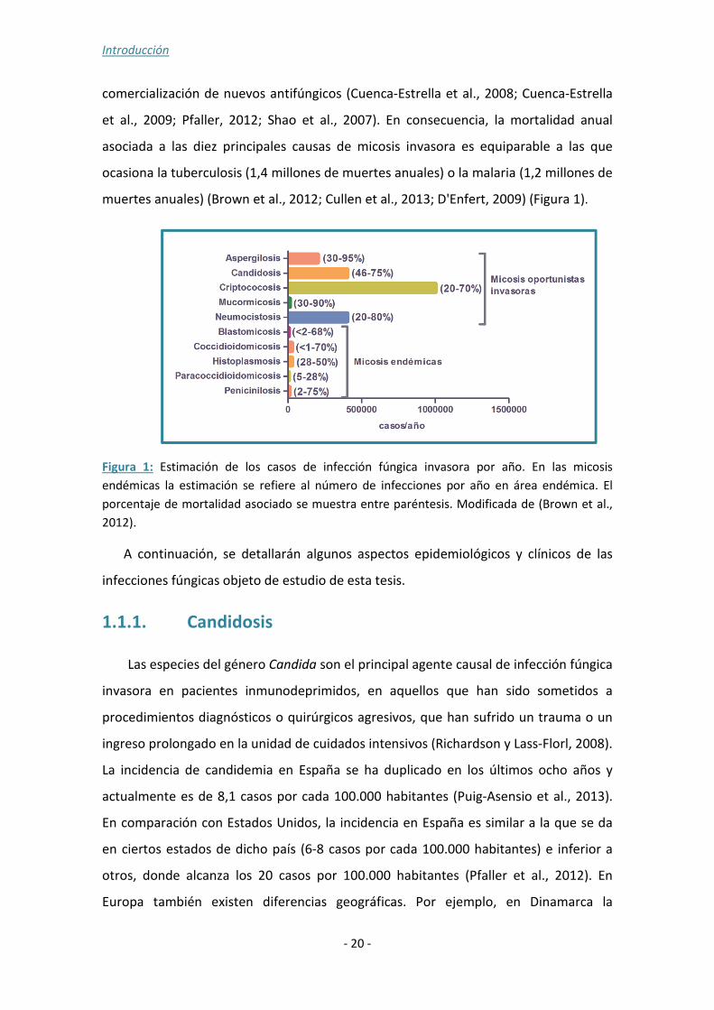
muertes anuales) (Brown et al., 2012; Cullen et al., 2013; D'Enfert, 2009) (Figura 1).
Figura 1: Estimación de los casos de infección fúngica invasora por año. En las micosis
endémicas la estimación se refiere al número de infecciones por año en área endémica. El
porcentaje de mortalidad asociado se muestra entre paréntesis. Modificada de (Brown et al.,
2012).
A continuación, se detallarán algunos aspectos epidemiológicos y clínicos de las
infecciones fúngicas objeto de estudio de esta tesis.
1.1.1. Candidosis
Las especies del género Candida son el principal agente causal de infección fúngica
invasora en pacientes inmunodeprimidos, en aquellos que han sido sometidos a
procedimientos diagnósticos o quirúrgicos agresivos, que han sufrido un trauma o un
ingreso prolongado en la unidad de cuidados intensivos (Richardson y Lass-Florl, 2008).
La incidencia de candidemia en España se ha duplicado en los últimos ocho años y
actualmente es de 8,1 casos por cada 100.000 habitantes (Puig-Asensio et al., 2013).
En comparación con Estados Unidos, la incidencia en España es similar a la que se da
en ciertos estados de dicho país (6-8 casos por cada 100.000 habitantes) e inferior a
otros, donde alcanza los 20 casos por 100.000 habitantes (Pfaller et al., 2012). En
Europa también existen diferencias geográficas. Por ejemplo, en Dinamarca la

Introducción
- 21 -
incidencia es similar a la de España, 8,6 casos/100.000 habitantes, pero en otras
regiones del centro y norte de Europa la incidencia oscila entre 3-5 casos por 100.000
habitantes (Arendrup et al., 2011; Poikonen et al., 2010; Poikonen et al., 2003;
Sandven et al., 2006).
En relación con las especies causantes, Candida albicans es responsable de más de
la mitad de los casos de candidosis invasora, pero otras especies como Candida
parapsilosis, Candida glabrata, Candida tropicalis, Candida krusei o Candida
guilliermondii se consideran patógenos emergentes (Diekema et al., 2012; Guinea et
al., 2014; Horn et al., 2009; Nucci et al., 2013; Nucci et al., 2010; Pemán et al., 2012;
Pfaller et al., 2012; Pfaller y Diekema, 2007; Puig-Asensio et al., 2013). Sin embargo, la
incidencia de estas especies emergentes depende de la localización geográfica, la edad
(Bouza y Muñoz, 2008), la población de riesgo (Pfaller et al., 2012) y del uso de
fluconazol (Malani et al., 2005). Así, mientras C. parapsilosis es la segunda causa de
candidemia en España y América Latina (Nucci et al., 2013; Puig-Asensio et al., 2013);
C. glabrata lo es en Estados Unidos (Pfaller et al., 2012) y el este de Europa (Arendrup
et al., 2011).
En función de la población de riesgo, C. albicans es responsable de hasta el 70% de
los episodios de candidemia en pacientes con tumores sólidos pero solo del 36% en los
pacientes con enfermedad hematológica (Pfaller et al., 2012). En relación con la edad,
C. parapsilosis es más frecuente en neonatos y C. glabrata en ancianos (Bouza y
Muñoz, 2008). Además, se ha producido un aumento de las infecciones mixtas por
especies de Candida alcanzando el 30% de los casos de candidemia en el paciente
infectado por el VIH (Pfaller et al., 2012). Asimismo, la generalización del uso del
fluconazol ha producido un desplazamiento de las especies más sensibles por otras
más resistentes como C. krusei o C. glabrata (Procop y Roberts, 2004).
El desarrollo de herramientas de identificación molecular ha introducido el
concepto de especies crípticas, definido como aquellos hongos morfológicamente
semejantes entre sí pero que a nivel genético constituyen una especie diferente
(Taylor et al., 2000). Dentro del género Candida, Candida nivariensis (Alcoba-Florez et
al., 2005) y Candida bracarensis (Correia et al., 2006) se han definido como especies
Introducción
- 22 -
crípticas de Candida glabrata, así como Candida orthopsilosis y Candida metapsilosis lo
son de C. parapsilosis (Tavanti et al., 2005). En el complejo psilosis (C. parapsilosis, C.
orthopsilosis y C. metapsilosis) no sólo existen diferencias genéticas entre dichas
especies, sino también de virulencia y sensibilidad a los antifúngicos (Gacser et al.,
2007; Lockhart et al., 2008a; Orsi et al., 2010; Silva et al., 2009a; Silva et al., 2009b). Se
ha señalado que C. orthopsilosis y C. metapsilosis son más sensibles a las
equinocandinas pero más resistentes a los azoles que C. parapsilosis (Silva et al.,
2009a). Otros estudios publicados recientemente, sitúan a C. orthopsilosis entre la
quinta o la octava causa de candidemia en España según los autores (Pemán et al.,
2012; Puig-Asensio et al., 2013).
1.1.2. Criptococosis
La criptococosis es una infección fúngica causada por especies del género
Cryptococcus y asociada principalmente al SIDA (Antinori, 2013). La infección se
produce tras la inhalación de las esporas del medio ambiente y, aunque su
presentación más frecuente es la meningoencefalitis, también puede causar
neumonía, infecciones diseminadas y localizadas (La Hoz y Pappas, 2013). En esta
infección están implicadas fundamentalmente variedades del complejo Cryptococcus
neoformans (var. neoformans y var. grubii) que afectan a individuos
inmunocomprometidos, y también la especie Cryptococcus gattii que puede afectar a
individuos inmunocompetentes (Kronstad et al., 2011). Se estima que la incidencia
global de meningitis criptocócica es de un millón de casos al año, causando la muerte a
más de 620.000 individuos en el África Subsahariana (Brown et al., 2012).
Además del SIDA hay otros factores de riesgo, como defectos en la inmunidad
celular (neutropenia), linfomas, esplenectomía y tratamiento con corticoides (Li y
Mody, 2010; Qazzafi et al., 2007). C. gattii tiene una distribución geográfica más
limitada a regiones tropicales y subtropicales (Región Central y Sur del Pacífico,
América Latina, California) (Chaturvedi y Chaturvedi, 2011). Desde principio de los años
noventa, el número de casos de criptococosis por C. gattii ha aumentado
considerablemente en la isla canadiense de Vancouver y se ha ido extendiendo por los
Estados Unidos en la Columbia Británica, Washington y Oregón, alcanzando una

Introducción
- 23 -
mortalidad del 33% (Dixit et al., 2009). Sin embargo, se desconocen las causas de su
propagación y del aumento de la incidencia (Fraser et al., 2005).
1.1.3. Histoplasmosis
La histoplasmosis es una micosis endémica de América, África y algunas
regiones de Asia (Kasuga et al., 2003). Se estima que anualmente se producen 300.000
casos con una mortalidad asociada del 3,33% (unos 10.000 casos anuales) (Armstrong-
James et al., 2014). El agente causal es Histoplasma capsulatum cuyo hábitat principal
es el guano de los pájaros (Chin et al., 1970). La infección se produce al inhalar las
microconidias de la fase micelial del hongo, las cuales se convierten en levaduras en el
pulmón, o bien tras una reactivación en individuos inmunodeprimidos previamente
expuestos (Wheat, 1995). Existen varias formas clínicas de la histoplasmosis y su
aparición depende del inóculo inhalado, la edad, el estado inmunológico del paciente o
la presencia de una enfermedad pulmonar concomitante (Colombo et al., 2011;
Queiroz-Telles et al., 2011). La gran mayoría de los individuos que inhalan esporas de
H. capsulatum no desarrollan sintomatología (>90%), pero existen casos de infecciones
agudas sintomáticas que varían desde síndromes respiratorios leves a infecciones
pulmonares que ponen en peligro la vida del enfermo. También se han descrito casos
de histoplasmosis pulmonar aguda en individuos que han realizado actividades al aire
libre consideradas de riesgo como visita a cuevas, excavaciones y actividades de
reconstrucción o agrícolas (Kasuga et al., 2003). Por último, en individuos
inmunocomprometidos, fundamentalmente infectados por el VIH, se pueden producir
infecciones diseminadas (Kauffman, 2007; Wheat et al., 2007). Así, en zonas
endémicas, la histoplasmosis es la primera manifestación de SIDA en el 50-70% de los
pacientes con VIH (Cano y Hajjeh, 2001). De hecho, más del 90% de los pacientes con
VIH que viven en regiones endémicas desarrollan histoplasmosis, siendo diseminada
entre el 2-5% de los casos (Cano y Hajjeh, 2001; Wheat, 1994; Wheat, 1995). En
pacientes VIH con infección diseminada no sometidos a terapia antirretroviral, la
mortalidad asociada es del 40% (Tobon et al., 2005). Sin embargo, desde finales de los
años noventa, la prevalencia de infección diseminada ha disminuido en pacientes VIH
en tratamiento antirretroviral de alta eficacia (Colombo et al., 2011).
Introducción
- 24 -
En España se ha registrado un aumento de la incidencia de las micosis
importadas, principalmente histoplasmosis, debido a la inmigración y a los viajes
(Buitrago y Cuenca-Estrella, 2012). Aproximadamente el 38% de la población
emigrante que reside en España procede de zonas endémicas y, además, un millón de
españoles viaja cada año a estos países. Así en los últimos treinta años se han
publicado 128 casos de histoplasmosis en España, de los cuales el 88% se describieron
a partir del año 2000 (Buitrago y Cuenca-Estrella, 2012).
1.1.4. Coccidioidomicosis
La coccidioidomicosis es una micosis endémica del sudoeste de Estados Unidos
y determinadas regiones de América Latina (Barker et al., 2007). Dicha micosis está
causada por dos especies del mismo género, Coccidioides immitis y Coccidioides
posadasii, que son similares en cuanto a las manifestaciones clínicas. La incidencia de
la coccidioidomicosis ha aumentado en los últimos diez años y se ha relacionado con
factores climatológicos, movimientos de tierra (excavaciones, construcciones, minería)
o viajes a áreas endémicas (Colombo et al., 2011). El número de casos registrados de
coccidioidomicosis entre 1998 y 2011, aumentó de 2.265 a 22.401 en Estados Unidos.
Además, de los más de 100.000 casos de coccidioidomicosis recogidos por el CDC
(Centers for Disease Prevention, Estados Unidos) durante este periodo, el 97%
ocurrieron en Arizona y California. La incidencia fue de 3,67 casos por cada 100.000
habitantes, falleciendo 70 pacientes al año (Centers for Disease and Prevention, 2013).
Tras la inhalación de las artroconidias se puede producir una infección
respiratoria pero en la mayoría de los casos es asintomática (Nguyen et al., 2013).
Puede aparecer una infección diseminada en individuos con factores de riesgo como la
pertenencia a ciertas etnias, inmunosupresión o embarazo (Hooper et al., 2007;
Nguyen et al., 2013). En individuos infectados por el VIH, suele producir neumonía y en
los casos más graves, tras la diseminación, meningitis (Laniado-Laborin, 2007).
Anteriormente a la introducción de la terapia antirretroviral de gran eficacia, la
coccidioidomicosis afectaba entre el 10 y el 25% de los pacientes VIH de área
endémica con una mortalidad asociada del 40% (Galgiani y Ampel, 1990). Sin embargo,
tras la aparición de la terapia antirretroviral se ha producido una importante

Introducción
- 25 -
disminución. Esto se ve reflejado en un estudio realizado entre 2003 y 2008, en el que
sólo el 11,3% de los pacientes VIH de área endémica desarrollaron coccidioidomicosis
con una incidencia anual del 0,9%, similar a la de la población general de la zona
(Ampel, 2005; Ampel, 2007; Ampel, 2010). En América Latina la incidencia varía en
función del país. Aunque en Méjico se han descrito 2-3 casos por cada 100.000
habitantes, la incidencia real en otras regiones se desconoce puesto que no es una
infección de obligada declaración (Ampel, 2005; Ampel, 2007; Laniado-Laborin, 2007).
Al ser una infección inexistente en Europa, el diagnóstico de los casos importados es
muy complejo.
1.1.5. Neumocistosis
La neumonía por Pneumocystis jirovecii afecta principalmente a individuos
infectados por el VIH, donde es una de las enfermedades fúngicas oportunistas más
frecuentes (Dworkin et al., 2000). Se ha estimado que la incidencia de neumonía por
P. jirovecii es superior a 400.000 casos por año, de los cuales aproximadamente
100.000 se producirían en países desarrollados con una tasa de mortalidad asociada
entre el 30 y el 85% (Figura 1). En los últimos años, el uso de los antirretrovirales y la
profilaxis con cotrimoxazol han producido una disminución de su incidencia en los
países desarrollados (Carmona y Limper, 2011). Por el contrario, se ha descrito un
aumento de la neumocistosis en el África Subsahariana donde se consideraba un
patógeno raro (Fisk et al., 2003; Schoffelen et al., 2013). Además, en países
desarrollados, se ha descrito neumocistosis en pacientes con otro tipo de
inmunosupresión como alteraciones hematológicas, trasplantados o pacientes con
terapia prolongada con corticoides o metotrexato (Watanabe et al., 2012). También se
ha descrito que la neumocistosis puede contribuir al empeoramiento de los pacientes
con enfermedad pulmonar obstructiva crónica (EPOC) y, con ello, a elevar la
mortalidad asociada hasta los tres millones de fallecimientos anuales (Brown et al.,
2012; Brown et al., 2014).

Introducción
- 26 -
1.2. Situación actual del diagnóstico de la infección fúngica invasora
La ausencia de síntomas tempranos y específicos condiciona la sospecha
diagnóstica de la infección fúngica invasora, especialmente en individuos
inmunocomprometidos (Perfect, 2013). Es conocido que un diagnóstico temprano y
preciso con un tratamiento acorde aumenta significativamente la supervivencia.
(Ostrosky-Zeichner, 2012).
1.2.1. Métodos convencionales y sus limitaciones
Los métodos convencionales de diagnóstico microbiológico se basan en la
visualización directa de las muestras mediante tinciones específicas para hongos, la
observación de los tejidos mediante histopatología y el cultivo (Hsu et al., 2011).
Aunque el examen directo y el cultivo pueden resultar útiles en el caso de las micosis
superficiales, la utilidad de estas técnicas es muy limitada en el caso de las micosis
sistémicas (Cuenca-Estrella et al., 2008; Garey et al., 2006).
Las técnicas de microscopía incluyen la visualización de las muestras en fresco y el
examen histopatológico. Estos procedimientos tienen limitaciones debido a que la
visualización de las estructuras fúngicas no es indicativa del estadio de la infección, y
además es inespecífica, puesto que diferentes especies adoptan el mismo patrón
morfológico (Cuenca-Estrella et al., 2011; Cuenca-Estrella et al., 2008). De forma
general se pueden diferenciar levaduras, hongos filamentosos y hongos dimórficos
pero resulta imposible saber de qué especie se trata (Perfect, 2013). Además, no
siempre es posible obtener una muestra de biopsia debido a la enfermedad de base
del paciente (Almyroudis y Segal, 2009). Sin embargo, existen casos en los que las
técnicas de microscopía son de gran utilidad. Así, debido a que P. jirovecii no crece en
los medios habituales de cultivo, el diagnóstico de la neumocistosis se realiza
fundamentalmente en base a la visualización microscópica del microorganismo
(Kaouech et al., 2009). Entre las tinciones más empleadas destacan Giemsa, plata
metanamina de Grocott, ácido peryódico de Shiff, azul de toloudina o calcofluor que
tienen una sensibilidad de entre el 50-90% y una especificidad superior al 95% (Procop
Introducción
- 27 -
et al., 2004; Tasaka y Tokuda, 2013). Además, se han desarrollado técnicas de
inmunofluorescencia directa o indirecta que tienen valores de sensibilidad cercanos al
90% cuando se emplean en muestras de lavado broncoalveolar o aspirados
bronquiales (Choe et al., 2014; Tasaka y Tokuda, 2013). En el caso de la meningitis
criptocócica, la tinción de tinta china suele ser positiva entre el 20-50% de los
pacientes aunque puede alcanzar el 80% en pacientes infectados por VIH (Hsu et al.,
2011).
El cultivo microbiológico es el método de referencia para el diagnóstico de
infección fúngica aunque su sensibilidad es muy baja y varía en función de la especie
(Hsu et al., 2011). Los hemocultivos se consideran el método de referencia para las
candidemias pero son positivos en menos del 50% de los casos (Pemán y Zaragoza,
2012). En más del 50% de los pacientes diagnosticados de candidosis profunda post-
mortem, los hemocultivos habían sido persistentemente negativos durante su
hospitalización (Morrell et al., 2005). Además, se ha descrito que las especies del
género Aspergillus, los Mucorales (Barnes, 2008) o Candida glabrata (Ramírez Aguilar
et al., 1992) se aíslan rara vez en los hemocultivos. El valor predictivo positivo de los
cultivos también depende de la enfermedad de base del paciente, ya que en pacientes
oncohematológicos con aspergilosis pulmonar invasora, un cultivo positivo indica
infección en el 90-100% de los casos pero sólo en el 15% cuando es un paciente con
VIH o con tumor de órgano sólido (Hsu et al., 2011).
En el caso de las micosis endémicas, la sensibilidad de los cultivos es muy baja. De
hecho, son positivos en tan solo un 0,5% de los pacientes con coccidioidomicosis
sistémica y en el 30% de los pacientes con histoplasmosis diseminada (Akpek et al.,
2001; Ampel, 2010). Además, estos hongos son de crecimiento lento, ya que al menos
se requieren entre 5-7 días para que las colonias sean visibles (Gómez, 2014). Para
complicar aún más la situación, la manipulación de los cultivos y la identificación de los
aislados tiene que hacerse en laboratorios de seguridad de nivel 3 (Buitrago y Cuenca-
Estrella, 2012).

Introducción
- 28 -
1.2.2. Detección de biomarcadores
Debido a las limitaciones del diagnóstico convencional descritas en la sección
anterior, se han desarrollado técnicas alternativas basadas en la detección de distintos
biomarcadores. En la actualidad, los métodos desarrollados, se basan en la detección
de 1,3 β-D-glucano para la infección fúngica, de galactomanano para la aspergilosis, de
mananos para la candidemia, antígeno capsular para la criptococosis y antígenos y
anticuerpos en el caso de las micosis endémicas.
1.2.2.1. Detección de 1,3 ββββ-D-Glucano en sospecha de infección fúngica
El 1,3 β−D-glucano es un polisacárido de la pared celular de los hongos,
prácticamente inexistente en los Mucorales y Cryptococcus spp., que se libera a la
sangre durante el proceso de invasión (Koo et al., 2009). Como está ausente en
mamíferos, bacterias y virus, la detección de 1,3 β−D-glucano en suero es un buen
marcador panfúngico de infección fúngica invasora (Obayashi et al., 2008). El ensayo
más empleado es el denominado Fungitell (Associates of Cape Cod, Inc) cuyo uso ha
sido aprobado para el diagnóstico de candidemia por la FDA (Food and Drug
Administration), y recomendado por la IDSA (Infectious DIseases Society of America) y
el EORTC/MSG (European Organization for Research and Treatment of Cancer/Invasive
Fungal Infections Cooperative Group and the National Institute of Allergy and
Infectious Diseases Mycoses Study Group) debido a su alta sensibilidad y especificidad
(Cuenca-Estrella et al., 2012; Hsu et al., 2011). Así, en un meta-análisis que investiga la
utilidad del 1,3 β−D-glucano en el diagnóstico de candidosis invasora, se obtuvieron
valores de sensibilidad y especificidad del 77% y el 85% respectivamente
(Karageorgopoulos et al., 2011). Según otro estudio realizado en pacientes ingresados
en la UCI quirúrgica, la determinación del 1,3 β−D-glucano consigue detectar la
candidosis invasora entre 4 y 8 días antes que el hemocultivo (Senn et al., 2008). Para
la neumocistosis se han descrito valores de sensibilidad entre el 90-100% y de
especificidad entre 65-100% (Boyles, 2013; Cuétara et al., 2008; Del Palacio et al.,
2010; Esteves et al., 2014). Además, esta técnica puede ser de utilidad para distinguir
entre población infectada y colonizada, evaluar el progreso de la infección o la eficacia
terapéutica (Hanson et al., 2012; Onishi et al., 2012; Sims et al., 2012).

Introducción
- 29 -
Sin embargo, la imposibilidad de identificar la especie causante de la infección y
la existencia de un buen número de factores que causan falsos positivos hacen que no
sea muy utilizado en los laboratorios clínicos (Cuenca-Estrella et al., 2011). Entre las
causas relacionadas con la presencia de falsos positivos destacan: la administración de
inmunoglobulina, las preparaciones de albúmina contaminadas con componentes
fúngicos, la realización de hemodiálisis con membranas de celulosa, la sepsis grave, el
uso de gasas que contengan trazas de glucano y el tratamiento con antibióticos (P.ej.
piperacilina-tazobactam) (Koo et al., 2009). Actualmente, las muestras se deben enviar
a laboratorios especializados lo que retrasa el diagnóstico de una infección grave
(Perfect, 2013).
1.2.2.2. Detección de Galactomanano en sospecha de aspergilosis
El galactomanano es un polisacárido de la pared celular del género Aspergillus
y, en menor medida, de otras especies fúngicas, que se libera durante su crecimiento e
invasión del tejido. El ensayo Platelia Galactomanano EIA (BioRad) fue aprobado por la
FDA en 2003. Posteriormente en Europa, el grupo de estudio EORTC/MSG incluyó el
resultado positivo de dicho ensayo como criterio microbiológico para la definición de
aspergilosis invasora probable en muestras de suero, plasma, lavado broncoalveolar o
líquido cefalorraquídeo (De Pauw et al., 2008). La determinación de galactomanano es
una técnica rápida y no invasora cuya sensibilidad y la aparición de resultados falsos
negativos varían en función del punto de corte. Así, se ha demostrado que la detección
de galactomanano en dos sueros consecutivos con un índice mayor o igual a 0,5
aumenta la especificidad y el valor predictivo positivo de la prueba; mientras que, para
una sola determinación, se recomienda un valor por encima de 0,7 (Ostrosky-Zeichner,
2012).
Los resultados de un meta-análisis han demostrado que la sensibilidad y
especificidad global del ensayo del galactomanano en sangre es del 71% y 89%
respectivamente para el diagnóstico de aspergilosis invasora (Pfeiffer et al., 2006). Sin
embargo, es más útil en unas poblaciones de pacientes que en otras. En pacientes
oncohematológicos los valores de sensibilidad y especificidad alcanzan el 90% y 98%
respectivamente, mientras que en pacientes trasplantados de órgano sólido
disminuyen hasta el 3% y el 78% respectivamente (Klont et al., 2004). También se ha

Introducción
- 30 -
evaluado la utilidad de la determinación de galactomanano como indicación del
tratamiento anticipado o la monitorización de la eficacia terapéutica (Cordonnier et al.,
2009; Maertens et al., 2009; Maertens et al., 2005). Otros estudios sugieren que las
determinaciones seriadas de galactomanano pueden ser útiles para la evaluación de la
respuesta al tratamiento, el progreso de la infección o para distinguir entre
empeoramiento y el síndrome de reconstitución inmune (Miceli et al., 2008).
Finalmente, una de las limitaciones de esta técnica es el elevado número de
falsos positivos que aparecen entre la población pediátrica, así como en pacientes
tratados con antibióticos betalactámicos, o la presencia de reacciones cruzadas con
otras micosis (peniciliosis, histoplasmosis o blastomicosis, fusariosis). Además, la
profilaxis o el tratamiento empírico con antifúngicos disminuyen considerablemente la
sensibilidad de la técnica (Klont et al., 2004; Mennink-Kersten et al., 2004a; Mennink-
Kersten et al., 2004b; Viscoli et al., 2004).
1.2.2.3. Detección de antígeno manano-anticuerpo antimanano en candidosis
El manano es un glicano con incorporación de manosa presente en la pared
celular de las especies de Candida y tiene propiedades inmunogénicas. La detección
combinada de anticuerpos y antígenos manano en suero de pacientes con candidosis
invasora tiene una sensibilidad del 80% y una especificidad del 93% para C. albicans
(Pontón, 2009). Sin embargo esta sensibilidad disminuye cuando es otra la especie
involucrada (Mikulska et al., 2010). A pesar de la comercialización de ensayos para la
determinación de antígeno manano y anticuerpo antimanano, como el Platelia
Candida (Biorad), su baja sensibilidad hace que sean poco utilizados en clínica.
1.2.2.4. Determinación de antígeno de Cryptococcus
El ensayo para la determinación del antígeno capsular de Cryptococcus se
desarrolló a finales de los setenta aunque debido a su elevada sensibilidad se sigue
utilizando (Bennett et al., 1977). En la actualidad se comercializa en formato de
sistema de aglutinación en látex o ELISA. Su eficacia varía en función del estado de
inmunosupresión del paciente y el tipo de muestra empleada (La Hoz y Pappas, 2013).
En pacientes con SIDA y meningitis criptocócica la detección de antígeno capsular en
Introducción
- 31 -
líquido cefalorraquídeo o suero es positiva en más del 99% de los casos, mientras que
en otros pacientes con otra enfermedad de base, la detección en suero es sólo positiva
en el 30% (Kralovic y Rhodes, 1998). En pacientes inmunocompetentes, el ensayo tiene
mejor sensibilidad en líquido cefalorraquídeo o lavado broncoalveolar que en suero
(Hsu et al., 2011).
Recientemente, se ha desarrollado un inmunoensayo para el diagnóstico
inmediato de criptococosis (“point of care testing”) basado en la detección del
antígeno capsular (Jarvis et al., 2011). Dicho ensayo ha sido aprobado por la FDA para
su uso en suero y líquido cefalorraquídeo y se caracteriza por ser estable a
temperatura ambiente, así como rápido y económico. Estudios recientes señalan unos
valores de sensibilidad y especificidad del 99% y 97% respectivamente en líquido
cefalorraquídeo de pacientes infectados por VIH y con meningitis criptocócica
(Boulware et al., 2014; Dhana, 2013).
1.2.2.5. Detección de la interacción antígeno-anticuerpos en micosis endémicas
El diagnóstico de las micosis endémicas es trabajoso, caro y lento. Por ello, se
han desarrollado diversas técnicas de detección de antígenos y anticuerpos que en
combinación con otras técnicas diagnósticas resultan de gran utilidad.
En el caso de la histoplasmosis, la determinación antigénica se realiza
principalmente en orina, aunque también se puede hacer en sangre o lavado
broncoalveolar. El sistema más empleado es un ELISA, desarrollado por Immy
diagnostics (Oklahoma Alpha Histoplama EIA Test Kit,) el cual tiene una sensibilidad y
especificidad global del 81% y 99% respectivamente (Theel et al., 2013). De hecho, en
pacientes inmunodeprimidos con histoplasmosis pulmonar subaguda y crónica la
sensibilidad del ensayo es superior al 95% (Srinivasan et al., 2009; Wheat, 2009; Zhang
et al., 2013). Sin embargo, existe reacción cruzada con otros hongos endémicos
(Blastomyces dermatitidis, Coccidioides spp., Paracoccidioides brasiliensis) y Aspergillus
(Connolly et al., 2007).
Otra técnica de referencia es un ELISA disponible en el laboratorio Miravista
(Indianapolis, Estados Unidos) que presenta una sensibilidad del 92% y del 100% en

Introducción
- 32 -
orina y suero respectivamente de pacientes con infección diseminada (Hage et al.,
2011; Theel et al., 2013). Sin embargo, en pacientes con histoplasmosis pulmonar
aguda o crónica la sensibilidad disminuye al 70% (Wheat, 2009). El 70% de los
pacientes con blastomicosis tiene un resultado positivo en esta prueba, lo que
complica su uso en área endémica de ambos patógenos (Gómez, 2014).
En cuanto a la detección de anticuerpos, existen pocas técnicas comerciales
(Immy Diagnostics, Meridien, Diamedix) las cuales se basan en la fijación del
complemento o en la inmunodifusión (Zhang et al., 2013). Tienen una sensibilidad
entre 30-70% en función del cuadro clínico y la enfermedad de base del paciente. Así,
el 50% de los pacientes inmunocomprometidos tienen resultados falsos negativos.
Además, hay que tener en cuenta que la seroconversión ocurre tras las 2-6 semanas y
los títulos de anticuerpos pueden permanecer elevados durante años tras la infección
primaria, con lo cual un resultado positivo no aclara si es una infección pasada, crónica
o una recidiva (Hamilton, 1998; Hsu et al., 2011).
Para el diagnóstico de la coccidioidomicosis, las técnicas de detección de
anticuerpos son las más empleadas. Esto se debe a que el sistema comercial de ELISA
específico para Coccidioides spp. presenta una sensibilidad del 70% en pacientes con
infección diseminada (Wheat, 2009). Hasta hace unos años, la prueba cutánea para la
detección de anticuerpos frente a la esferulina o la coccidioidina, era el método de
serodiagnóstico más empleado. Sin embargo, estos anticuerpos se han detectado en el
70% de la población de área endémica (Nguyen et al., 2013). En la actualidad, los
ensayos comerciales de serodiagnóstico para la coccidioidomicosis se basan en la
detección de IgM o IgG en formato de ELISA, mediante la fijación del complemento o
en inmunodifusión. La sensibilidad global es del 80% en los ensayos que detectan tanto
IgG como IgM pero pueden ser negativos en los estadios tempranos de infección. La
probabilidad de falsos positivos es muy elevada cuando sólo se evalúa IgM (82%) y
tienen sensibilidad reducida (50%) en pacientes inmunodeprimidos (Ampel, 2005; Blair
y Currier, 2008; Blair et al., 2013; Kuberski et al., 2010; Nguyen et al., 2013).
Introducción
- 33 -
1.2.3. PCR en tiempo real para el diagnóstico de infección
fúngica invasora
El método de amplificación de ADN mediante PCR fue descrito a mediados de los
ochenta adaptándose rápidamente en los laboratorios clínicos para la detección de
ADN de microorganismos, fundamentalmente de virus (Khot y Fredricks, 2009). Sin
embargo, las técnicas de PCR no se consideran como métodos de referencia para la
detección de hongos causantes de infección en humanos (Perfect, 2013) y tampoco se
han incluido en los criterios EORTC/MSG para definir infección probable (De Pauw et
al., 2008). Esto se debe principalmente a que los métodos de PCR existentes para el
diagnóstico de la infección fúngica están circunscritos a ciertos laboratorios y no han
sido completamente estandarizados.
La mayoría de los métodos desarrollados en micología se basan en la detección de
ácidos nucleicos mediante PCR cuantitativa o en tiempo real, para el diagnóstico de la
aspergilosis invasora y la candidemia (Tabla 1). Estudios recientes de meta-análisis,
demuestran que la sensibilidad y especificidad de estas técnicas son del 92% y 95%
respectivamente, en el caso de la candidemia (Avni et al., 2011) y del 88% y 75% en el
caso de la aspergilosis invasora (Mengoli et al., 2009). Los procedimientos más
novedosos permiten la detección de varias especies simultáneamente (Cuenca-Estrella
et al., 2009; Lau et al., 2010; McMullan et al., 2008). Esto es importante ya que
distintas especies del mismo género pueden tener un tratamiento antifúngico
diferente (Alastruey-Izquierdo et al., 2007; Alcázar-Fuoli et al., 2008; Arendrup, 2013).
Asimismo, permiten la detección de infecciones mixtas (Kirby et al., 2004). Se ha
descrito también que el uso de las técnicas de detección de ADN en combinación con
la detección de biomarcadores aumenta la sensibilidad al 98% en el caso de la
candidosis invasora (McMullan et al., 2008) y al 100% para la aspergilosis invasora
(Cuenca-Estrella et al., 2009). Igualmente, con el uso de la PCR diagnóstica, se podría
iniciar el tratamiento hasta 14 días antes que con los métodos convencionales
(Halliday et al., 2006). Actualmente, se está investigando el impacto que estas técnicas
pueden tener en el manejo de los pacientes. Así, se ha observado que la

Introducción
- 34 -
administración de antifúngicos en base a un resultado positivo de PCR reduciría los
costes del tratamiento en un 20% (Halliday et al., 2006).
La PCR es una herramienta prometedora para diagnosticar candidosis y aspergilosis
invasora. Sin embargo, en pacientes en riesgo de una infección fúngica diferente el
desarrollo ha sido prácticamente inexistente.
En el caso de la criptococosis los sistemas basados en PCR a tiempo real no se
encuentran muy desarrollados (Qishui et al., 2012; Veron et al., 2009) ya que los
sistemas de detección de antígeno son sensibles y económicos como se ha descrito en
la sección anterior. En los últimos años, se ha realizado un gran esfuerzo en el
desarrollo de PCR diagnósticas para la neumocistosis las cuales varían en cuanto a la
diana amplificada (ADN mitocondrial, ITS, DHFR, HSP70, etc.). La sensibilidad y
especificidad de estas técnicas en comparación con el examen directo es superior al
90%. Sin embargo, entre el 50-70% de los pacientes colonizados sin neumonía pueden
presentar un resultado de PCR positivo (Fan et al., 2013). Actualmente, no se ha
definido un valor umbral para distinguir entre pacientes colonizados e infectados.
Aunque se han descrito valores de 1000 células/ml, diversos estudios indican que
diferentes poblaciones de pacientes van a requerir valores umbrales diferentes
(Botterel et al., 2012; Dalpke et al., 2013; Matsumura et al., 2012; Muhlethaler et al.,
2012). Se ha señalado que aproximadamente en el 30% de los pacientes
inmunodeprimidos en los que se pensó que el resultado de la PCR era un falso positivo,
finalmente tenían neumocistosis (Fan et al., 2013). Por ello, en la actualidad un
resultado positivo de PCR debe ser evaluado por el clínico junto con la sintomatología
del paciente. Además, para el diagnóstico de neumocistosis se han desarrollado varios
ensayos comerciales aunque el único evaluado en diversos estudios es el Myc
Pneumocystis Assay (Myconostica) que presenta valores de sensibilidad del 90% y
especificidad del 100% en lavado broncoalveolar. Sin embargo, el valor predictivo
positivo depende del factor de riesgo del paciente siendo del 60% en paciente
trasplantado sometido a terapia con corticoides, 90% en paciente crítico y 100% en
paciente infectado por VIH (Hauser et al., 2011; McTaggart et al., 2012; Orsi et al.,
2012).
Introducción
- 35 -
El desarrollo de la PCR diagnóstica en el caso de la histoplasmosis está más
avanzado que en otras micosis endémicas. Este hecho es especialmente importante
debido a que la prevalencia de esta infección en zonas endémicas es muy elevada en
individuos infectados con VIH, el diagnóstico serológico no está siempre disponible y el
cultivo es muy lento (Gómez, 2014). De este modo, se han desarrollado diferentes
protocolos tanto de PCR en tiempo real como convencional dirigidos a la amplificación
de la región transcrita interna, ITS (Buitrago et al., 2006) o de genes de copia única
como el Hc100p o el SCAR220 (Bialek et al., 2002; Bialek et al., 2001; Martagon-Villamil
et al., 2003). Según un estudio multicéntrico reciente, la sensibilidad y especificidad
global de los métodos de PCR que se evaluaron en dicho trabajo para la detección de
H. capsulatum (convencional y tiempo real) fue del 86% y el 100% respectivamente
(Buitrago et al., 2013b) siendo más sensibles los métodos basados en PCR en tiempo
real que los que se basaban en PCR convencional. Con respecto a las otras micosis
endémicas (coccidioidomicosis, blastomicosis o paracoccidioidomicosis), el desarrollo
de herramientas de PCR diagnóstica no se encuentra muy avanzado. La mayor parte de
los protocolos descritos están circunscritos a ciertos laboratorios de referencia y la
validación se restringe a cepas clínicas y biopsias (Binnicker et al., 2007; Pitz Ade et al.,
2013; Sidamonidze et al., 2012).
Otra aproximación en el diagnóstico de la infección fúngica invasora ha sido el
desarrollo de técnicas panfúngicas basadas en PCR cuantitativa. Estas técnicas son
útiles cuando se ha visualizado el hongo en los tejidos pero no existe una clara
sospecha clínica ni epidemiológica de la especie que puede estar implicada en la
infección y, además, las otras técnicas, como el cultivo dan resultados negativos
(Buitrago et al., 2014). La PCR panfúngica tiene la desventaja de que no se realiza con
sondas específicas sino con cebadores universales y empleando agentes intercalantes
(ej. Sybergreen) y por tanto, los productos amplificados necesitan ser secuenciados lo
que retrasa el diagnóstico definitivo (Buitrago et al., 2014). Además, la PCR panfúngica
no puede usarse como técnica de cribado, ni tampoco para el diagnóstico rápido
debido a que la sensibilidad en muestras de fácil obtención como suero y sangre es del
70%, mientras que en tejido (parafinado o en fresco) alcanza el 87% (Buitrago et al.,
2013a; Landlinger et al., 2010; Lau et al., 2010).
Introducción
- 36 -
En la Tabla 1 se muestra una revisión de los trabajos más relevantes publicados
sobre PCR en los últimos años. Como se puede ver, hay una gran predominancia de las
publicaciones sobre la aspergilosis y la candidosis frente al resto de los patógenos
fúngicos.
Por último, las aproximaciones más recientes corresponden al desarrollo de
técnicas de PCR diagnósticas en formato múltiple. Estas técnicas permiten detectar en
un solo tubo las especies más frecuentemente implicadas en la infección o identificar
el microorganismo causante de un síndrome o un proceso con características clínicas
indistinguibles. En la literatura aparecen trabajos orientados a la detección de
microorganismos causantes de meningitis (Wang et al., 2012), enfermedad febril
sistémica (Waggoner et al., 2014) o para la detección de P. jirovecii o Mycobacterium
tuberculosis en pacientes infectados con VIH (Boondireke et al., 2010).
En los últimos años, se está realizando un gran esfuerzo para tratar de resolver
la falta de consenso, en el uso de la PCR cuantitativa en tiempo real como técnica
diagnóstica. Por ello, han surgido algunas iniciativas como la europea, European
Aspergillus PCR Iniciative (EAPCRI). Este estudio multicéntrico está constituido por más
de 60 centros en Europa y en Australia y tiene como finalidad estandarizar la
metodología de la PCR diagnóstica de Aspergillus spp., definir protocolos, tipo de
muestra más adecuada o método de extracción de ADN más eficiente (White et al.,
2010). Este proceso aún está en desarrollo y, una vez que se finalice y establezca un
consenso será necesario evaluar la utilidad clínica mediante ensayos clínicos a gran
escala. Se han realizado también otros estudios multicéntricos para evaluar los
métodos de PCR para el diagnóstico de histoplasmosis (Buitrago et al., 2013b); o para
el diagnóstico de mucormicosis (Dannaoui et al., 2010) aunque en general, a excepción
del diagnóstico de la aspergilosis, estos estudios son escasos todavía.
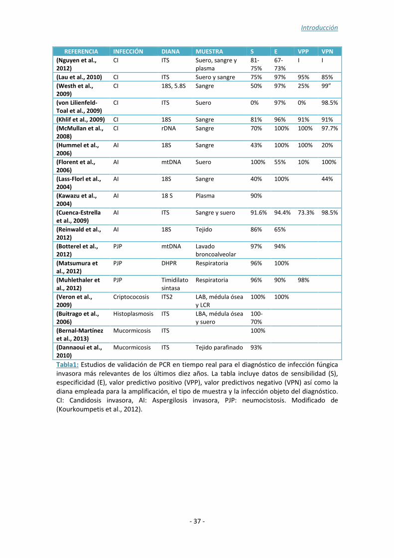
Introducción
- 37 -
REFERENCIA INFECCIÓN DIANA MUESTRA S E VPP VPN
(Nguyen et al., 2012)
CI ITS Suero, sangre y plasma
81-75%
67-73%
I I
(Lau et al., 2010) CI ITS Suero y sangre 75% 97% 95% 85%
(Westh et al., 2009)
CI 18S, 5.8S Sangre 50% 97% 25% 99”
(von Lilienfeld-Toal et al., 2009)
CI ITS Suero 0% 97% 0% 98.5%
(Khlif et al., 2009) CI 18S Sangre 81% 96% 91% 91%
(McMullan et al., 2008)
CI rDNA Sangre 70% 100% 100% 97.7%
(Hummel et al., 2006)
AI 18S Sangre 43% 100% 100% 20%
(Florent et al., 2006)
AI mtDNA Suero 100% 55% 10% 100%
(Lass-Florl et al., 2004)
AI 18S Sangre 40% 100% 44%
(Kawazu et al., 2004)
AI 18 S Plasma 90%
(Cuenca-Estrella et al., 2009)
AI ITS Sangre y suero 91.6% 94.4% 73.3% 98.5%
(Reinwald et al., 2012)
AI 18S Tejido 86% 65%
(Botterel et al., 2012)
PJP mtDNA Lavado broncoalveolar
97% 94%
(Matsumura et al., 2012)
PJP DHPR Respiratoria 96% 100%
(Muhlethaler et al., 2012)
PJP Timidilato sintasa
Respiratoria 96% 90% 98%
(Veron et al., 2009)
Criptococosis ITS2 LAB, médula ósea y LCR
100% 100%
(Buitrago et al., 2006)
Histoplasmosis ITS LBA, médula ósea y suero
100-70%
(Bernal-Martínez et al., 2013)
Mucormicosis ITS 100%
(Dannaoui et al., 2010)
Mucormicosis ITS Tejido parafinado 93%
Tabla1: Estudios de validación de PCR en tiempo real para el diagnóstico de infección fúngica invasora más relevantes de los últimos diez años. La tabla incluye datos de sensibilidad (S), especificidad (E), valor predictivo positivo (VPP), valor predictivos negativo (VPN) así como la diana empleada para la amplificación, el tipo de muestra y la infección objeto del diagnóstico. CI: Candidosis invasora, AI: Aspergilosis invasora, PJP: neumocistosis. Modificado de (Kourkoumpetis et al., 2012).

Introducción
- 38 -
1.3. Métodos moleculares de identificación de hongos patógenos humanos: métodos clásicos y nuevas técnicas basadas en análisis de curvas de fusión de alta resolución (HRM)
Actualmente, el método de referencia para identificar hongos a nivel de especie es
la secuenciación de dianas informativas. Se ha acordado que sea la región espaciadora
interna (ITS) del ADN ribosómico (rADN) la diana de referencia para la identificación
molecular de hongos (Figura 2). El ADN ribosómico contiene copias múltiples repetidas
en tándem de los genes que codifican para el ARN ribosómico (5,8S, 18S y 26S)
separados por regiones ITS (ITS-1 e ITS-2) que se transcriben pero no se traducen
(Schoch et al., 2012). Cada una de estas unidades, están repetidas un número variable
de veces en función de la especie y separadas por las regiones intergénicas, IGS-1 e
IGS-2. Para la mayoría de los hongos, las regiones ITS-1 e ITS-2 son suficientemente
variables para identificar a nivel de especie (Begerow et al., 2010). Sin embargo, para
algunas especies el grado de divergencia de la región ITS es limitado y se han buscado
dianas con mayor poder de resolución. Entre estos nuevos marcadores están los genes
que codifican para el factor de elongación 1α (EF-1α), la β tubulina, la calmodulina o la
región espaciadora intergénica (Rodríguez-Tudela et al., 2008). Así, para algunos
géneros de hongos se ha visto que la IGS permite la identificación de la especie
llegando incluso a variabilidad intraespecífica (Mekha et al., 2010; Sugita et al., 2003;
Sugita et al., 2002; Sugita et al., 2004). Aunque se han descrito diferencias en la IGS,
tanto en longitud como en composición nucleotídica entre algunas especies
relacionadas, esta región ha sido muy poco explorada (Cornet et al., 2011). Por tanto,
su análisis en profundidad podría ser útil para identificar especies en las cuales el
análisis de otras dianas no proporciona un resultado muy robusto.
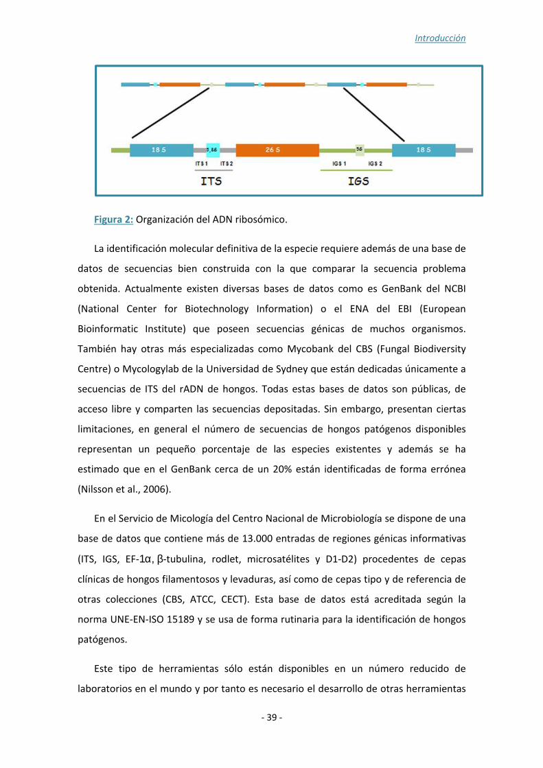
Introducción
- 39 -
Figura 2: Organización del ADN ribosómico.
La identificación molecular definitiva de la especie requiere además de una base de
datos de secuencias bien construida con la que comparar la secuencia problema
obtenida. Actualmente existen diversas bases de datos como es GenBank del NCBI
(National Center for Biotechnology Information) o el ENA del EBI (European
Bioinformatic Institute) que poseen secuencias génicas de muchos organismos.
También hay otras más especializadas como Mycobank del CBS (Fungal Biodiversity
Centre) o Mycologylab de la Universidad de Sydney que están dedicadas únicamente a
secuencias de ITS del rADN de hongos. Todas estas bases de datos son públicas, de
acceso libre y comparten las secuencias depositadas. Sin embargo, presentan ciertas
limitaciones, en general el número de secuencias de hongos patógenos disponibles
representan un pequeño porcentaje de las especies existentes y además se ha
estimado que en el GenBank cerca de un 20% están identificadas de forma errónea
(Nilsson et al., 2006).
En el Servicio de Micología del Centro Nacional de Microbiología se dispone de una
base de datos que contiene más de 13.000 entradas de regiones génicas informativas
(ITS, IGS, EF-1α, β-tubulina, rodlet, microsatélites y D1-D2) procedentes de cepas
clínicas de hongos filamentosos y levaduras, así como de cepas tipo y de referencia de
otras colecciones (CBS, ATCC, CECT). Esta base de datos está acreditada según la
norma UNE-EN-ISO 15189 y se usa de forma rutinaria para la identificación de hongos
patógenos.
Este tipo de herramientas sólo están disponibles en un número reducido de
laboratorios en el mundo y por tanto es necesario el desarrollo de otras herramientas

Introducción
- 40 -
más sencillas que permitan identificar a nivel de especie y subespecie a aquellos
patógenos de importancia clínica.
El análisis de curvas de fusión de alta resolución (HRM) es una técnica
relativamente reciente y ha resultado útil para la identificación (Douarre et al., 2012),
genotipado (Levesque et al., 2011), y análisis de mutaciones en genes implicados en la
resistencia a los antimicrobianos (Daniels et al., 2012). Este método de análisis de los
productos amplificados mediante PCR en tiempo real puede detectar fragmentos que
difieren en un solo nucleótido en su secuencia (Odell et al., 2005). Durante la
amplificación, una molécula fluorescente con capacidad para saturar el ADN (Resolight,
Evagreen o LCGreen) se intercala de forma estable entre el ADN de doble cadena. Tras
la amplificación, los productos de PCR van a ser desnaturalizados y rápidamente
renaturalizados lo que ocasiona cambios en la fluorescencia relacionados, tanto con la
longitud como con la composición de nucleótidos. De ahí, que estas técnicas resulten
especialmente útiles para la determinación de variables alélicas (Figura 3).
Figura 3: Representación esquemática del análisis de curvas de fusión de alta resolución (HRM) para la determinación de polimorfismos de una sola base. Las flechas indican la posición de los cebadores en la reacción de amplificación. El fluoróforo tiene afinidad por el ADN de doble cadena de modo que sólo cuando se une a éste, la fluorescencia es suficiente como para ser detectada por el sistema de detección. Los nucleótidos desapareados se muestran como líneas en diagonal, Los productos de PCR de alelos homocigotos (naranja), mutantes heterocigoto (rojo) o mutantes homocigoto (verde) son analizados tras amplificación mediante normalización de la fluorescencia.
Introducción
- 41 -
Este método se ha utilizado para la identificación de bacterias de diferentes
géneros (Slany et al., 2010), para detección de especies con distinta sensibilidad a los
antimicrobianos (Yin et al., 2013) o de aquellas que causan sepsis frecuentemente
(Ozbak et al., 2012). Una de las primeras aplicaciones del HRM fue para la
identificación del grupo Mycobacterium chelonae-Mycobacterium abcessus (Odell et
al., 2005). Sin embargo, esta técnica se ha utilizado poco para la identificación de
hongos, circunscribiéndose a especies del género Candida y Mucorales (Arancia et al.,
2011; Hrncirova et al., 2010; Somogyvari et al., 2012). En todas ellas hay un déficit
claro de estandarización y validación de las técnicas aunque cabe destacar su potencial
utilidad.
Debido a su simplicidad, el HRM podría suponer también una excelente
alternativa a las actuales técnicas de genotipado. Dos de las técnicas más
reproducibles y eficaces para la tipificación son, el análisis de la longitud de regiones
de microsatélites y la tipificación de secuencias multilocus (MLST). Aunque estas
técnicas presentan un gran poder de discriminación, son técnicas complejas, costosas y
prácticamente circunscritas a laboratorios de referencia (Vanhee et al., 2010).


Objetivos
- 43 -
2. OBJETIVOS


Objetivos
- 45 -
De acuerdo con las necesidades actuales en el diagnóstico de la infección fúngica
invasora e identificación de las especies causantes de la misma, los objetivos
planteados en este trabajo son:
Primero:
Desarrollo y validación de técnicas de PCR en tiempo real usando sondas de tipo
molecular beacon para:
a. Diagnóstico e identificación de especies de hongos causantes de neumonías
oportunistas (Histoplasma capsulatum, Pneumocystis jirovecii y Cryptococcus
neoformans-Cryptococcus gattii).
b. Diagnóstico de coccidioidomicosis en área endémica.
Segundo:
Desarrollo de técnicas rápidas de identificación y tipificación de especies fúngicas
basadas en el análisis de curvas de fusión de alta resolución (HRM) para:
a. La identificación de especies del complejo Cryptococcus gattii-neoformans.
b. La identificación de las especies del complejo psilosis: Candida parapsilosis,
Candida orthopsilosis y Candida metapsilosis.
c. Diferenciación molecular entre cepas de Candida albicans causantes de
infecciones recurrentes.

Objetivo primero
- 47 -
3. OBJETIVO PRIMERO: Desarrollo de técnicas de PCR
cuantitativa diagnóstica

Objetivo primero
- 49 -
3.1. Diagnóstico e identificación de hongos que causan infección oportunista en pacientes infectados con VIH (Pneumocystis jirovecii, Histoplasma capsulatum y Cryptococcus neoformans/Cryptococcus gattii) mediante PCR en tiempo real
El 70% de los pacientes infectados con el VIH tienen complicaciones pulmonares
durante el curso de la infección. La importancia del diagnóstico diferencial de las
especies responsables de los episodios de neumonía se debe a que los patógenos
fúngicos que los causan con más frecuencia (Histoplasma capsulatum, Cryptococcus
neoformans y Pneumocistis jirovecii) presentan una clínica similar y diferencias en el
tratamiento. Por ello, se desarrolló una técnica de diagnóstico molecular basada en
PCR en tiempo real para la detección simultánea de estas tres especies. La
metodología desarrollada pretende la identificación de las tres especies mediante el
diseño de sondas tipo molecular beacon dirigidas a la región ITS del ADN ribosómico.
Además, se incluyó un control interno para monitorizar posibles procesos de inhibición
de la reacción. La estandarización se realizó empleando 24 cepas clínicas y 43 muestras
de pacientes con SIDA. Asimismo, para los ensayos de especificidad se evaluaron 21
cepas de especies no relacionadas y 29 muestras clínicas de pacientes con otra
afección.
La técnica presentó gran sensibilidad (100%) y especificidad in vitro (100%) y
coeficientes de variación cercanos al 3%. El límite de detección fue de 2 fg de ADN
genómico por reacción para H. capsulatum y C. neoformans, y 2,92 log10 copias de
ADN plasmídico en 20 µl de reacción en el caso de P.jirovecii. La validación clínica de la
técnica mostró valores de sensibilidad del 90,75% y de especificidad del 100% en las
muestras analizadas. Asimismo, el 92,5% de los pacientes con infección probada
tuvieron un resultado positivo por la técnica de PCR desarrollada. Para uno de los
pacientes con histoplasmosis probada de acuerdo a los criterios EORTC/MSG, se pudo
detectar además ADN de P. jirovecii en el lavado broncoalveolar.
De acuerdo a los resultados obtenidos, la técnica de detección múltiple de especies
presentada en esta memoria, es un procedimiento rápido, sensible y altamente
específico que podría tener gran utilidad clínica en regiones en desarrollo.


Objetivo primero
- 51 -
A Multiplex Real-Time PCR Assay for Identification ofPneumocystis jirovecii, Histoplasma capsulatum, and Cryptococcusneoformans/Cryptococcus gattii in Samples from AIDS Patients withOpportunistic Pneumonia
Sara Gago,a Cristina Esteban,b Clara Valero,a Óscar Zaragoza,a Jorge Puig de la Bellacasa,b María José Buitragoa
Mycology Department, National Centre for Microbiology, Instituto de Salud Carlos III, Majadahonda, Madrid, Spaina; Department of Clinical Microbiology, Hospital Clinic
de Barcelona, Barcelona, Spainb
A molecular diagnostic technique based on real-time PCR was developed for the simultaneous detection of three of the most fre-
quent causative agents of fungal opportunistic pneumonia in AIDS patients: Pneumocystis jirovecii, Histoplasma capsulatum,
and Cryptococcus neoformans/Cryptococcus gattii. This technique was tested in cultured strains and in clinical samples from
HIV-positive patients. The methodology used involved species-specific molecular beacon probes targeted to the internal tran-
scribed spacer regions of the rDNA. An internal control was also included in each assay. The multiplex real-time PCR assay was
tested in 24 clinical strains and 43 clinical samples from AIDS patients with proven fungal infection. The technique developed
showed high reproducibility (r2 of >0.98) and specificity (100%). For H. capsulatum and Cryptococcus spp., the detection limits
of the method were 20 and 2 fg of genomic DNA/20 ml reaction mixture, respectively, while for P. jirovecii the detection limit
was 2.92 log10 copies/20 ml reaction mixture. The sensitivity in vitro was 100% for clinical strains and 90.7% for clinical samples.
The assay was positive for 92.5% of the patients. For one of the patients with proven histoplasmosis, P. jirovecii was also detected
in a bronchoalveolar lavage sample. No PCR inhibition was detected. This multiplex real-time PCR technique is fast, sensitive,
and specific and may have clinical applications.
HIV infection remains a threat for many people, as it is esti-mated that almost 34 million adults and children world-
wide are infected with the virus, according to WHO/UNAIDS(1). Although mortality associated with HIV infection has de-creased in recent years due to the development of highly activeantiretroviral therapy (HAART), the incidence and mortalityrates of non-AIDS-associated infections remain high, particu-larly in nondeveloped countries (1). Opportunistic pneumo-nias are one of the major causes of pulmonary complicationsamong AIDS patients. In fact, almost 70% of people infectedwith HIV suffer at least one pulmonary complication duringthe course of the infection (2). Although in early HIV infectionthe agents that cause pneumonia are similar to those foundin the general population, when the number of CD41 T cellsdecreases, opportunistic pneumonias are associated with highmorbidity and mortality (3–5). Tuberculosis and Pneumocystisjirovecii pneumonia (PCP; the abbreviation PCP reflects the pre-vious nomenclature for the Pneumocystis species causing oppor-tunistic pneumonia in humans, P. carinii; P. jirovecii is now rec-ognized as the species that infects humans) are the mostcommonly reported AIDS-defining illnesses (6). Moreover, op-portunistic pneumonias due to Histoplasma capsulatum andCryptococcus spp. in immunocompromised patients involve highmorbidity andmortality in areas where the virus is not endemic orin those regions where the infection is not very common. P. jirove-cii has been described as the main cause of pulmonary complica-tion in AIDS patients in developed countries; in fact, in theUnitedStates it has been estimated that 75% of HIV-positive personssuffer PCP during their lifetime (7, 8). Clinical diagnosis of PCP isbased mainly on microscopic procedures performed directly onthe clinical sample and usually involves immunofluorescence mi-
croscopy (IFA) or use of Grocott’s methenamine silver stain (9,10). Recently, some reports have described the utility of real-timePCR (rt-PCR) protocols to increase the sensitivity and specificityof PCP diagnosis (11–17), and some of these tests have been com-mercialized (18). Moreover, a recent study showed the utility ofrt-PCR assays for the early diagnosis and treatment of non-HIV-infected patients diagnosed with PCP, for whom a definitive diag-nosis could be performed using rt-PCR techniques that detectedthe low fungal burden present in the patients (19).
Histoplasmosis is a mycosis that is endemic in the UnitedStates and Africa that can produce disseminated infection as-sociated with high morbidity and mortality in HIV patients(20–22). Due to travel and immigration, this infection has in-creased in recent years in areas where H. capsulatum mycosis isnot endemic (23). Although culture is the reference procedure forthe diagnosis of histoplasmosis, it is time-consuming (requiring 3to 4 weeks of incubation) (24). In fact, the sensitivity and specific-ity of microscopy-based approaches are very low, and fungalstructures are only observed when the infection is advanced.Moreover, serology has shown significant limitations in AIDS pa-tients; it may be negative in 50% of such patients (25). Antigen
Received 16 October 2013 Returned for modification 13 December 2013
Accepted 24 January 2014
Published ahead of print 29 January 2014
Editor: D. W. Warnock
Address correspondence to María José Buitrago, [email protected].
Copyright © 2014, American Society for Microbiology. All Rights Reserved.
doi:10.1128/JCM.02895-13
1168 jcm.asm.org Journal of Clinical Microbiology p. 1168–1176 April 2014 Volume 52 Number 4
on
Ma
rch
12
, 20
14
by B
iblio
teca
Na
cio
na
l de
Cie
ncia
s d
e la
Sa
lud
http
://jcm
.asm
.org
/D
ow
nlo
ad
ed
from

Objetivo primero
- 52 -
detection could be useful for these patients, although it is onlyavailable in the United States (26). Several rt-PCR protocols withvaried sensitivities and specificities have been described for detec-tion of DNA from Histoplasma capsulatum in clinical samples(27–29). Recently, a comparison of different protocols for detec-tion of H. capsulatum DNA by PCR showed that the sensitivity ofthe assay depended on both the selected target region and the typeof amplification assay (conventional or rt-PCR) (24). Herein, themost sensitive and specific protocols were based on the amplifica-tion of the internal transcribed spacer (ITS) by rt-PCR (30), andthe less sensitive protocols were based on the amplification ofmonocopy genes (Hc100p and SCAR220) by conventional PCR(31, 32).
In Africa, cryptococcosis, caused by Cryptococcus neoformansvar. neoformans or C. neoformans var. grubii, is one of the maincauses of mortality associated with HIV infection (33). However,in the northwest United States, Cryptococcus gattii is the primarycausative agent of cryptococcosis and can also affect immuno-competent people; it has been classified endemic in some regionsof the eastern United States, and several cases of cryptococcosisoutside that area where the disease is endemic have been reportedrecently (34–36). This infection can produce both meningitis andpulmonary complications in these patients (37, 38). Routine clin-ical diagnosis of cryptococcosis is based on culture and detectionof a capsular antigen, with varied sensitivity and specificity (39,40). However, molecular methods based on PCR and new tech-nologies for antigen detection also have been described in recentyears, although validation of these methods is pending (41–44).
Fungal pneumonias are clinically and radiologically similar toeach other, and definitive diagnosis must be reached to initiate theappropriate therapy (45). Moreover, multiplex real-time PCR(Mrt-PCR) protocols allow the detection of mixed infections thatcannot be detected by routine protocols. Mixed infections involv-ing H. capsulatum, C. neoformans, and P. jirovecii have been de-scribed (46–48).
The aim of this study was the development of a Mrt-PCR forthe early detection of fungal opportunistic pneumonia due to H.capsulatum, C. neoformans, C. gattii, and P. jirovecii in clinicalsamples from HIV-positive patients.
MATERIALS AND METHODS
Control strains and plasmids. DNA from different strains was used tostandardize the Mrt-PCR assay. All the strains belonged to the Collec-tion of the Mycology Reference Laboratory, Spanish National Centerof Microbiology. To standardize the technique, we used the strains C.neoformans var. neoformans CNM-CL 2132, H. capsulatum CNM-CM2721, and a plasmid that included the sequence target of P. jirovecii(pSG1).
To assess the specificity of the technique, the following strains of yeastand molds were used: Candida albicans (CNM-CL 5719), Candida parap-silosis (CNM-CL5683), Candida tropicalis (CNM-CL 5742), Candidaglabrata (CNM-CL 5533), Candida guilliermondii (CNM-CL 7127), Can-dida krusei (CNM-CL 7057), Coccidioides posadasii (CNM-CM 2912),Paracoccidioides brasiliensis (CNM-CM 2908), Aspergillus fumigatus(CNM-CM AF237), Aspergillus terreus (CNM-CM 3508), Aspergillus fla-vus (CNM-CM 3509), Aspergillus niger (CNM-CM 3551), Fusariumverticillioides (CNM-CM 2975), Fusarium oxysporum (CNM-CM 2914),Fusarium solani (CNM-CM 3035), Scedosporium prolificans (CNM-CM1627), Scedosporium apiospermum (CNM-CM 3169), Rhizopus oryzae(CNM-CM 3020), and Rhizopus microsporum (CNM-CM 4244), Mucorcircinelloides (CNM-CM2437). In addition, we employedmouse genomic
DNA (Promega, Madrid, Spain) and human genomic DNA (Promega,Madrid, Spain).
Design of primers and probes. Primers and molecular beacon probeswere designed to specifically amplify a region of the ITS of the rDNA fromCryptococcus neoformans/C. gattii, Histoplasma capsulatum var. capsula-tum and H. capsulatum var. duboisii, and Pneumocystis jirovecii by Mrt-PCR. Beacon Designer 5.0 software (Premier Biosoft, Palo Alto, CA) wasused for primer and probe design. An internal control was included, asdescribed previously (49). The primers and probes designed were sub-jected to a BLAST search within the GenBank sequence database (http://blast.ncbi.nlm.nih.gov/Blast.cgi) and in the database of the Departmentof Mycology of the Spanish National Center for Microbiology (whichcontains more than 8,000 distinct sequences), to avoid cross-homologywith other microorganisms. The primer and probe sequences are shownin Table 1.
Multiplex real-time PCR assay. The amplificationwas carried out in aLightCycler 480 system (RocheDiagnostics,Mannheim,Germany). PCRswere performed in a 20-ml final volume containing 23 SensiMix DNA(Quantance; Ecogen, Madrid, Spain) used according to the manufactur-er’s instructions, 0.5 mM each primer for each one of the species, 0.25 mMinternal control primer, 0.2 mM each species-specific probe, 0.1 mM in-ternal control probe, 2 fg of the internal control plasmid (pICJF), and 2 mlof DNA extracted from the sample, used as the template. The PCR con-ditions were as follows: an initial step of 95°C for 10 min, followed by 50cycles of 95°C for 25 s, 50°C for 30 s, and 72°C for 5 s, with a cooling cycleat 40°C for 30 s. Results were considered positive when the productamount increased until its fluorescence intensity exceeded the back-ground, as determined by second-derivate analysis; these results were ex-pressed in terms of the quantification cycle (Cq). Each experiment wasperformed in duplicate and included quantification standards as well asnegative controls. Subsequently, a color compensation experiment wasperformed to prevent cross talk between dyes.
Standardization. Standard curves for H. capsulatum and C. neofor-mans were constructed based on the results of five PCR repetitions withdilutions of genomic DNA ranging from 20 ng to 2 fg/20 ml of reactionmixture. The strains CNM-CL 2132 and CNM-CM 2721 were used toconstruct the standard curves. For P. jirovecii, a fragment of 175 bp con-
TABLE 1 Sequences of primers and probes designed for the multiplexreal-time PCR assay
Species and its primers
and probe Sequencea
Pneumocystis jirovecii
OliPJMB1 (f) 5=-CCCTAGTGTTTTAGCATTTTTC-3=
OLIPJMB2 (r) 5=-CTGCAATTCACACTACTTATCG-3=
Probe PJ-MB1 5=-HEX-CGCGATACCTTTGGCGAGGCAAG
CAAATCGCG-BHQ1-3=
Histoplasma capsulatumb
Cryptococcus neoformans
OLI CRYPTO 1 2 (f) 5=-CCTGTTGGACTTGGATTTGG-3=
OLI CRYPTO 2 (r) 5=-AGCAAGCCGAAGACTACC-3=
Probe MB CRYPTO 5=-Cyan 500-CGCGATCATTACGCCGGGCT
GACAGGTAATCAGATCGCG-BHQ1-3=
Jellyfish (internal control)
Oli1-icjf1 (f) 5=-GCCTGGTGCAAAAATTGCTTATC-3=
Oli1-icjf2 (r) 5=-CTAAGACAAGTGTGTTTATGGTATTG-3=
Probe CIJF-MB 5=-Cy5-CGCGATGCTGTTCTTCCGCCACTT
CCAATCGCG-BHQ2-3=
a BHQ1, black hole quencher 1.b Sequences for the primers and probe for H. capsulatum are included in patent
PCT/ES2009070340.
Diagnosis of Fungal Opportunistic Pneumonia by Mrt-PCR
April 2014 Volume 52 Number 4 jcm.asm.org 1169
on
Ma
rch
12
, 20
14
by B
iblio
teca
Na
cio
na
l de
Cie
ncia
s d
e la
Sa
lud
http
://jcm
.asm
.org
/D
ow
nlo
ad
ed
from
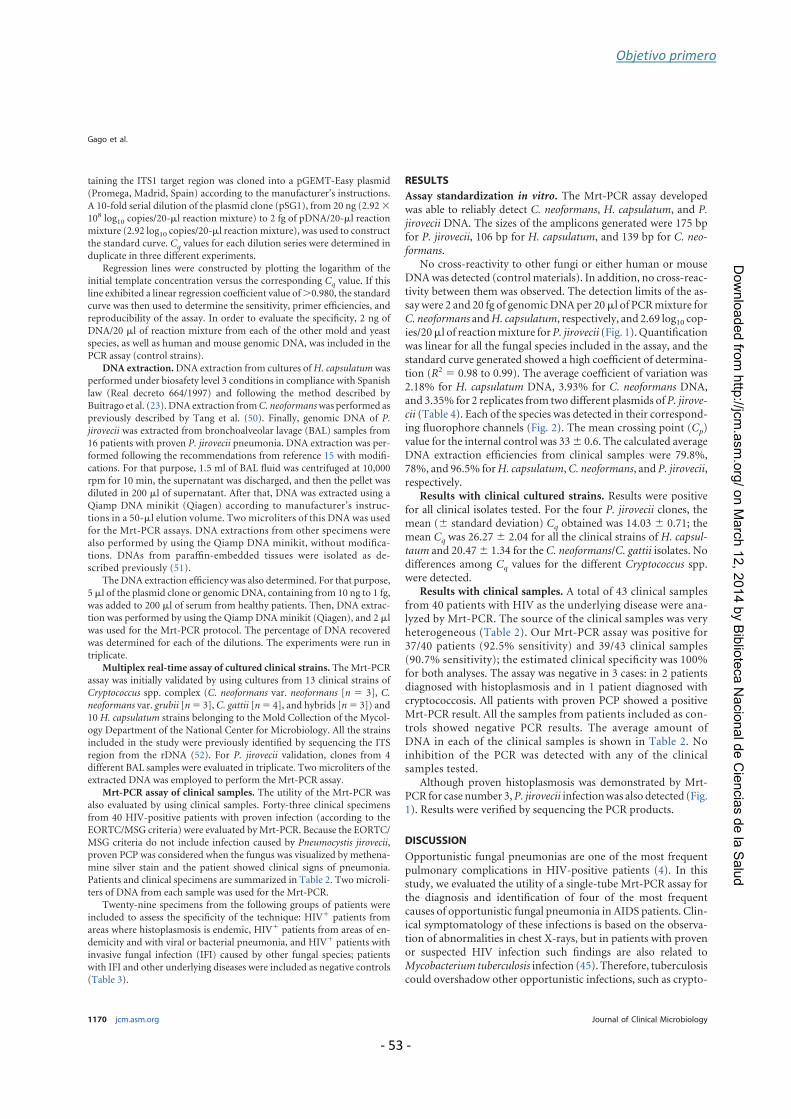
Objetivo primero
- 53 -
taining the ITS1 target region was cloned into a pGEMT-Easy plasmid(Promega, Madrid, Spain) according to the manufacturer’s instructions.A 10-fold serial dilution of the plasmid clone (pSG1), from 20 ng (2.92 3
108 log10 copies/20-ml reaction mixture) to 2 fg of pDNA/20-ml reactionmixture (2.92 log10 copies/20-ml reaction mixture), was used to constructthe standard curve. Cq values for each dilution series were determined induplicate in three different experiments.
Regression lines were constructed by plotting the logarithm of theinitial template concentration versus the corresponding Cq value. If thisline exhibited a linear regression coefficient value of .0.980, the standardcurve was then used to determine the sensitivity, primer efficiencies, andreproducibility of the assay. In order to evaluate the specificity, 2 ng ofDNA/20 ml of reaction mixture from each of the other mold and yeastspecies, as well as human and mouse genomic DNA, was included in thePCR assay (control strains).
DNA extraction. DNA extraction from cultures of H. capsulatum wasperformed under biosafety level 3 conditions in compliance with Spanishlaw (Real decreto 664/1997) and following the method described byBuitrago et al. (23). DNA extraction from C. neoformans was performed aspreviously described by Tang et al. (50). Finally, genomic DNA of P.
jirovecii was extracted from bronchoalveolar lavage (BAL) samples from16 patients with proven P. jirovecii pneumonia. DNA extraction was per-formed following the recommendations from reference 15 with modifi-cations. For that purpose, 1.5 ml of BAL fluid was centrifuged at 10,000rpm for 10 min, the supernatant was discharged, and then the pellet wasdiluted in 200 ml of supernatant. After that, DNA was extracted using aQiamp DNA minikit (Qiagen) according to manufacturer’s instruc-tions in a 50-ml elution volume. Two microliters of this DNA was usedfor the Mrt-PCR assays. DNA extractions from other specimens werealso performed by using the Qiamp DNA minikit, without modifica-tions. DNAs from paraffin-embedded tissues were isolated as de-scribed previously (51).
The DNA extraction efficiency was also determined. For that purpose,5 ml of the plasmid clone or genomic DNA, containing from 10 ng to 1 fg,was added to 200 ml of serum from healthy patients. Then, DNA extrac-tion was performed by using the Qiamp DNA minikit (Qiagen), and 2 mlwas used for the Mrt-PCR protocol. The percentage of DNA recoveredwas determined for each of the dilutions. The experiments were run intriplicate.
Multiplex real-time assay of cultured clinical strains. The Mrt-PCRassay was initially validated by using cultures from 13 clinical strains ofCryptococcus spp. complex (C. neoformans var. neoformans [n 5 3], C.
neoformans var. grubii [n 5 3], C. gattii [n 5 4], and hybrids [n 5 3]) and10 H. capsulatum strains belonging to the Mold Collection of the Mycol-ogy Department of the National Center for Microbiology. All the strainsincluded in the study were previously identified by sequencing the ITSregion from the rDNA (52). For P. jirovecii validation, clones from 4different BAL samples were evaluated in triplicate. Two microliters of theextracted DNA was employed to perform the Mrt-PCR assay.
Mrt-PCR assay of clinical samples. The utility of the Mrt-PCR wasalso evaluated by using clinical samples. Forty-three clinical specimensfrom 40 HIV-positive patients with proven infection (according to theEORTC/MSG criteria) were evaluated by Mrt-PCR. Because the EORTC/MSG criteria do not include infection caused by Pneumocystis jirovecii,proven PCP was considered when the fungus was visualized by methena-mine silver stain and the patient showed clinical signs of pneumonia.Patients and clinical specimens are summarized in Table 2. Two microli-ters of DNA from each sample was used for the Mrt-PCR.
Twenty-nine specimens from the following groups of patients wereincluded to assess the specificity of the technique: HIV1 patients fromareas where histoplasmosis is endemic, HIV1 patients from areas of en-demicity and with viral or bacterial pneumonia, and HIV1 patients withinvasive fungal infection (IFI) caused by other fungal species; patientswith IFI and other underlying diseases were included as negative controls(Table 3).
RESULTS
Assay standardization in vitro. The Mrt-PCR assay developedwas able to reliably detect C. neoformans, H. capsulatum, and P.jirovecii DNA. The sizes of the amplicons generated were 175 bpfor P. jirovecii, 106 bp for H. capsulatum, and 139 bp for C. neo-formans.
No cross-reactivity to other fungi or either human or mouseDNA was detected (control materials). In addition, no cross-reac-tivity between them was observed. The detection limits of the as-say were 2 and 20 fg of genomicDNAper 20 ml of PCRmixture forC. neoformans and H. capsulatum, respectively, and 2.69 log10 cop-ies/20ml of reactionmixture for P. jirovecii (Fig. 1).Quantificationwas linear for all the fungal species included in the assay, and thestandard curve generated showed a high coefficient of determina-tion (R2
5 0.98 to 0.99). The average coefficient of variation was2.18% for H. capsulatum DNA, 3.93% for C. neoformans DNA,and 3.35% for 2 replicates from two different plasmids of P. jirove-cii (Table 4). Each of the species was detected in their correspond-ing fluorophore channels (Fig. 2). The mean crossing point (Cp)value for the internal control was 33 6 0.6. The calculated averageDNA extraction efficiencies from clinical samples were 79.8%,78%, and 96.5% for H. capsulatum, C. neoformans, and P. jirovecii,respectively.
Results with clinical cultured strains. Results were positivefor all clinical isolates tested. For the four P. jirovecii clones, themean (6 standard deviation) Cq obtained was 14.03 6 0.71; themean Cq was 26.27 6 2.04 for all the clinical strains of H. capsul-taum and 20.47 6 1.34 for the C. neoformans/C. gattii isolates. Nodifferences among Cq values for the different Cryptococcus spp.were detected.
Results with clinical samples. A total of 43 clinical samplesfrom 40 patients with HIV as the underlying disease were ana-lyzed by Mrt-PCR. The source of the clinical samples was veryheterogeneous (Table 2). Our Mrt-PCR assay was positive for37/40 patients (92.5% sensitivity) and 39/43 clinical samples(90.7% sensitivity); the estimated clinical specificity was 100%for both analyses. The assay was negative in 3 cases: in 2 patientsdiagnosed with histoplasmosis and in 1 patient diagnosed withcryptococcosis. All patients with proven PCP showed a positiveMrt-PCR result. All the samples from patients included as con-trols showed negative PCR results. The average amount ofDNA in each of the clinical samples is shown in Table 2. Noinhibition of the PCR was detected with any of the clinicalsamples tested.
Although proven histoplasmosis was demonstrated by Mrt-PCR for case number 3, P. jirovecii infectionwas also detected (Fig.1). Results were verified by sequencing the PCR products.
DISCUSSION
Opportunistic fungal pneumonias are one of the most frequentpulmonary complications in HIV-positive patients (4). In thisstudy, we evaluated the utility of a single-tube Mrt-PCR assay forthe diagnosis and identification of four of the most frequentcauses of opportunistic fungal pneumonia in AIDS patients. Clin-ical symptomatology of these infections is based on the observa-tion of abnormalities in chest X-rays, but in patients with provenor suspected HIV infection such findings are also related toMycobacterium tuberculosis infection (45). Therefore, tuberculosiscould overshadow other opportunistic infections, such as crypto-
Gago et al.
1170 jcm.asm.org Journal of Clinical Microbiology
on
Ma
rch
12
, 20
14
by B
iblio
teca
Na
cio
na
l de
Cie
ncia
s d
e la
Sa
lud
http
://jcm
.asm
.org
/D
ow
nlo
ad
ed
from
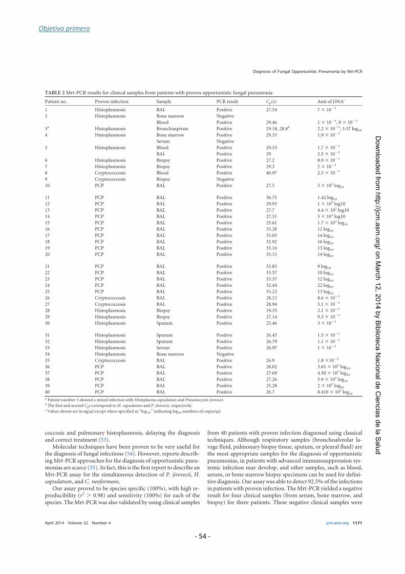
Objetivo primero
- 54 -
coccosis and pulmonary histoplasmosis, delaying the diagnosisand correct treatment (53).
Molecular techniques have been proven to be very useful forthe diagnosis of fungal infections (54). However, reports describ-ing Mrt-PCR approaches for the diagnosis of opportunistic pneu-monias are scarce (55). In fact, this is the first report to describe anMrt-PCR assay for the simultaneous detection of P. jirovecii, H.capsulatum, and C. neoformans.
Our assay proved to be species specific (100%), with high re-producibility (r2 . 0.98) and sensitivity (100%) for each of thespecies. The Mrt-PCR was also validated by using clinical samples
from 40 patients with proven infection diagnosed using classicaltechniques. Although respiratory samples (bronchoalveolar la-vage fluid, pulmonary biopsy tissue, sputum, or pleural fluid) arethe most appropriate samples for the diagnosis of opportunisticpneumonias, in patients with advanced immunosuppression sys-temic infection may develop, and other samples, such as blood,serum, or bone marrow biopsy specimens can be used for defini-tive diagnosis. Our assay was able to detect 92.5% of the infectionsin patients with proven infection. The Mrt-PCR yielded a negativeresult for four clinical samples (from serum, bone marrow, andbiopsy) for three patients. These negative clinical samples were
TABLE 2 Mrt-PCR results for clinical samples from patients with proven opportunistic fungal pneumonia
Patient no. Proven infection Sample PCR result Cp(s) Amt of DNAc
1 Histoplasmosis BAL Positive 27.54 7 3 1023
2 Histoplasmosis Bone marrow Negative
Blood Positive 29.46 1 3 1023, 8 3 1023
3a Histoplasmosis Bronchiaspirate Positive 29.18, 28.8b 2.2 3 1023, 3.37 log10
4 Histoplasmosis Bone marrow Positive 29.33 1.9 3 1023
Serum Negative
5 Histoplasmosis Blood Positive 29.53 1.7 3 1023
BAL Positive 29 2.5 3 1023
6 Histoplasmosis Biopsy Positive 27.2 8.9 3 1023
7 Histoplasmosis Biopsy Positive 29.3 2 3 1023
8 Cryptococcosis Blood Positive 40.97 2.5 3 1023
9 Cryptococcosis Biopsy Negative
10 PCP BAL Positive 27.5 5 3 102 log10
11 PCP BAL Positive 36.75 1.42 log10
12 PCP BAL Positive 29.93 1 3 102 log10
13 PCP BAL Positive 27.7 4.4 3 102 log10
14 PCP BAL Positive 27.51 5 3 102 log10
15 PCP BAL Positive 25.61 1.7 3 103 log10
16 PCP BAL Positive 33.28 12 log10
17 PCP BAL Positive 33.05 14 log10
18 PCP BAL Positive 32.92 16 log10
19 PCP BAL Positive 33.16 13 log10
20 PCP BAL Positive 33.15 14 log10
21 PCP BAL Positive 33.83 9 log10
22 PCP BAL Positive 33.57 10 log10
23 PCP BAL Positive 33.37 12 log10
24 PCP BAL Positive 32.44 22 log10
25 PCP BAL Positive 33.22 13 log10
26 Cryptococcosis BAL Positive 28.12 8.6 3 1023
27 Cryptococcosis BAL Positive 28.94 5.1 3 1023
28 Histoplasmosis Biopsy Positive 19.35 2.1 3 1023
29 Histoplasmosis Biopsy Positive 27.14 9.3 3 1023
30 Histoplasmosis Sputum Positive 25.46 3 3 1022
31 Histoplasmosis Sputum Positive 26.45 1.5 3 1022
32 Histoplasmosis Sputum Positive 26.79 1.1 3 1022
33 Histoplasmosis Serum Positive 26.97 1 3 1022
34 Histoplasmosis Bone marrow Negative
35 Cryptococcosis BAL Positive 26.9 1.8 31022
36 PCP BAL Positive 28.02 3.65 3 102 log10
37 PCP BAL Positive 27.69 4.50 3 102 log10
38 PCP BAL Positive 27.26 5.9 3 102 log10
39 PCP BAL Positive 25.28 2 3 103 log10
40 PCP BAL Positive 26.7 8.410 3 102 log10
a Patient number 3 showed a mixed infection with Histoplasma capsulatum and Pneumocystis jirovecii.b The first and second Cps correspond to H. capsulatum and P. jirovecii, respectively.c Values shown are in ng/ml except where specified as “log10,” indicating log10 numbers of copies/ml.
Diagnosis of Fungal Opportunistic Pneumonia by Mrt-PCR
April 2014 Volume 52 Number 4 jcm.asm.org 1171
on
Ma
rch
12
, 20
14
by B
iblio
teca
Na
cio
na
l de
Cie
ncia
s d
e la
Sa
lud
http
://jcm
.asm
.org
/D
ow
nlo
ad
ed
from

Objetivo primero
- 55 -
stored for more than 5 years at 220°C, which may have decreasedthe sensitivity of the assay (56).
Interestingly, for one of the patients diagnosed with histoplas-mosis, P. jirovecii DNA was detected. In fact, coinfections mayoccur, and accurate diagnosis and prompt appropriate antifungaltreatment are required. Several reports of cases of mixed infec-tions have been published, and for all of them the definitive diag-nosis was based on microbiological evidence. Bava et al. (47) re-ported a case in which a patient had mixed infection due toCryptococcus and Pneumocystis.Mixed infections of H. capsulatumand C. neoformans (46), as well as of P. jirovecii and H. capsulatum,have also been reported in individuals from areas (48). Finally,coinfection with H. capsulatum and M. tuberculosis is frequent insome areas where histoplasmosis is endemic and may involve im-portant implications for treatment (57).
To our knowledge, only one PCR-based protocol has been pre-viously reported for the detection of tuberculosis andPCP inHIV-infected patients (55). rt-PCR approaches for the detection of C.neoformans/C. gattii, H. capsulatum, and P. jirovecii separatelyhave been widely described based on the amplification of differenttargets, with varied sensitivity and specificity. Most of the rt-PCRapproaches for the diagnosis of cryptococcosis are based on theamplification of monocopy genes (43, 58), which may decreasethe sensitivity of the technique. Our technique allows detection of
both C. neoformas spp. complex and C. gattii, but it cannot differ-entiate species. Although the identification at species level is im-portant for epidemiological studies, clinical signs and treatmentare similar for infections caused by either species (59–61). More-over, amajority of the real-time protocols for the detection of PCPare based on nested PCR, which may decrease the specificity of thetechnique due to the possibility of false-positive results (58). Al-though there are several articles describing rt-PCR assays for thedetection of H. capsulatum based on the amplification of multi-copy regions, such as ITS, the use of molecular beacon probes canimprove the sensitivity of the technique due to their small size andsequence specificity (23). The technique we have described in thisarticle can detect several fungi in a single-tube reaction. The limitto detection of more fungal species is determined by the real-timePCR equipment.
In conclusion, this is the first report to describe an Mrt-PCRfor the diagnosis of opportunistic fungal pneumonias. Thisapproach could be very useful for differential diagnosis withtuberculosis, as clinical signs are similar, and also to detectmixed infections that could appear in patients with advancedimmunosuppression living in areas of endemicity. Moreover,this assay could be useful for the clinical management of otherimmunocompromised patients at risk for these fungal infec-tions (e.g., patients under corticosteroid therapy or chemo-
TABLE 3 Summary of control populations used to determine background colonization levels and the specificity of the Mrt-PCR assay
Population
Control
sample no. Specimen PCR result Species causing IFI
HIV1 patients from areas of endemicity 1 Bone narrow Negative
2 Biopsy Negative
3 Biopsy Negative
4 Bone narrow Negative
5 Whole Blood Negative
6 Whole Blood Negative
HIV1 patients from areas of endemicity and
with viral or bacterial pneumonia
7 BAL Negative
8 BAL Negative
9 BAL Negative
10 Biopsy Negative
HIV1 patients with invasive fungal infection
caused by other fungal species
11 Biopsy Negative Lichtheimia corymbifera
12 Biopsy Negative Rhizomucor pusillus
13 BAL Negative Cryptococcus laurentii
14 BAL Negative Cryptococcus humicola
15 BAL Negative Aspergillus flavus, Penicillium spp., and
Cryptococcus albidus
Non-HIV-infected patients with invasive
fungal infection
16 Biopsy Negative Coccidioides immitis
17 Biopsy Negative Scedosporium apiospermum
18 Biopsy Negative Bipolaris spp.
19 BAL Negative Aspergillus fumigatus
20 BAL Negative Aspergillus fumigatus
21 BAL Negative Aspergillus fumigatus
22 BAL Negative Aspergillus fumigatus
23 BAL Negative Aspergillus fumigatus
24 Ascitic fluid Negative Aspergillus fumigatus
25 Cerebrospinal fluid Negative Aspergillus fumigatus
26 BAL Negative Aspergillus fumigatus
27 Biopsy Negative Aspergillus fumigatus
28 Biopsy Negative Aspergillus fumigatus
29 BAL Negative Aspergillus fumigatus
Gago et al.
1172 jcm.asm.org Journal of Clinical Microbiology
on
Ma
rch
12
, 20
14
by B
iblio
teca
Na
cio
na
l de
Cie
ncia
s d
e la
Sa
lud
http
://jcm
.asm
.org
/D
ow
nlo
ad
ed
from
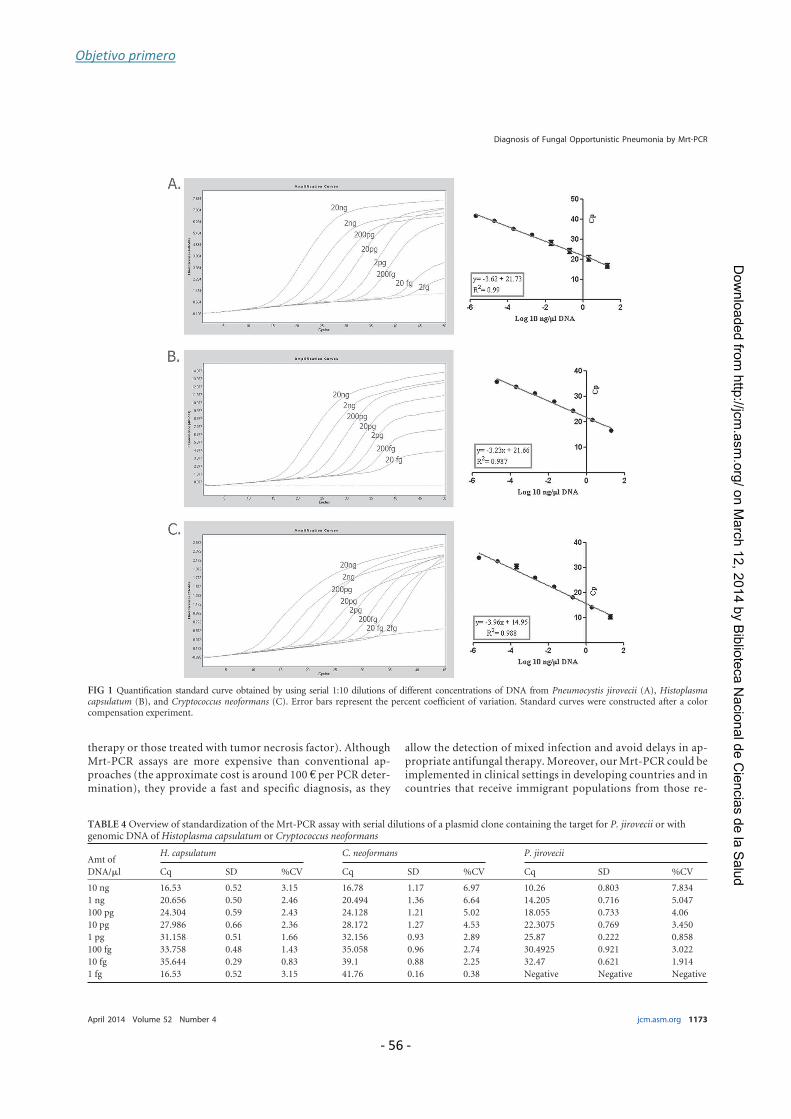
Objetivo primero
- 56 -
therapy or those treated with tumor necrosis factor). AlthoughMrt-PCR assays are more expensive than conventional ap-proaches (the approximate cost is around 100 € per PCR deter-mination), they provide a fast and specific diagnosis, as they
allow the detection of mixed infection and avoid delays in ap-propriate antifungal therapy. Moreover, our Mrt-PCR could beimplemented in clinical settings in developing countries and incountries that receive immigrant populations from those re-
FIG 1 Quantification standard curve obtained by using serial 1:10 dilutions of different concentrations of DNA from Pneumocystis jirovecii (A), Histoplasmacapsulatum (B), and Cryptococcus neoformans (C). Error bars represent the percent coefficient of variation. Standard curves were constructed after a colorcompensation experiment.
TABLE 4 Overview of standardization of the Mrt-PCR assay with serial dilutions of a plasmid clone containing the target for P. jirovecii or withgenomic DNA of Histoplasma capsulatum or Cryptococcus neoformans
Amt of
DNA/ml
H. capsulatum C. neoformans P. jirovecii
Cq SD %CV Cq SD %CV Cq SD %CV
10 ng 16.53 0.52 3.15 16.78 1.17 6.97 10.26 0.803 7.834
1 ng 20.656 0.50 2.46 20.494 1.36 6.64 14.205 0.716 5.047
100 pg 24.304 0.59 2.43 24.128 1.21 5.02 18.055 0.733 4.06
10 pg 27.986 0.66 2.36 28.172 1.27 4.53 22.3075 0.769 3.450
1 pg 31.158 0.51 1.66 32.156 0.93 2.89 25.87 0.222 0.858
100 fg 33.758 0.48 1.43 35.058 0.96 2.74 30.4925 0.921 3.022
10 fg 35.644 0.29 0.83 39.1 0.88 2.25 32.47 0.621 1.914
1 fg 16.53 0.52 3.15 41.76 0.16 0.38 Negative Negative Negative
Diagnosis of Fungal Opportunistic Pneumonia by Mrt-PCR
April 2014 Volume 52 Number 4 jcm.asm.org 1173
on
Ma
rch
12
, 20
14
by B
iblio
teca
Na
cio
na
l de
Cie
ncia
s d
e la
Sa
lud
http
://jcm
.asm
.org
/D
ow
nlo
ad
ed
from

Objetivo primero
- 57 -
gions. Further studies with a larger number of samples arewarranted.
ACKNOWLEDGMENTS
We warmly thank Frannk Hodkings for his careful reading and editing ofthe manuscript.
This work was supported by research project PI11/00412 from Span-ish Fondo de Investigaciones Sanitarias of the Instituto de Salud CarlosIII. S.G. andC.V. are supported by research fellowships from the FondodeInvestigaciones Biomedicas of the Spanish Ministry of Science and Inno-vation (FI10/00464 and FI12/00095, respectively).
We declare no conflicts of interest.
REFERENCES
1. Huang L, Cattamanchi A, Davis JL, den Boon S, Kovacs J, Meshnick S,Miller RF, Walzer PD, Worodria W, Masur H, International HIV-Associated Opportunistic Pneumonias (IHOP) Study, Lung HIV Study.2011. HIV-associated Pneumocystis pneumonia. Proc. Am. Thorac Soc.8:294–300. http://dx.doi.org/10.1513/pats.201009-062WR.
2. Gingo MR, Balasubramani GK, Kingsley L, Rinaldo CR, Jr, Alden CB,Detels R, Greenblatt RM, Hessol NA, Holman S, Huang L, Kleerup EC,Phair J, Sutton SH, Seaberg EC, Margolick JB, Wisniewski SR, MorrisA. 2013. The impact of HAART on the respiratory complications of HIVinfection: longitudinal trends in the MACS and WIHS cohorts. PLoS One8(3):e58812. http://dx.doi.org/10.1371/journal.pone.0058812.
3. Rosen MJ. 1992. Pulmonary complications of HIV infection. Mt. Sinai J.Med. 59:263–270.
4. Rosen MJ. 1994. Pneumonia in patients with HIV infection. Med. Clin.North Am. 78:1067–1079.
5. Rosen MJ. 2008. Pulmonary complications of HIV infection. Respirology13:181–190. http://dx.doi.org/10.1111/j.1440-1843.2007.01167.x.
6. Schneider E, Whitmore S, Glynn KM, Dominguez K, Mitsch A,McKenna MT, Centers for Disease Control and Prevention. 2008.Revised surveillance case definitions for HIV infection among adults, ad-olescents, and children aged ,18 months and for HIV infection and AIDSamong children aged 18 months to ,13 years—United States, 2008.MMWR Recommend. Rep. 57(RR-10):1–12.
7. Hay JW, Osmond DH, Jacobson MA. 1988. Projecting the medical costsof AIDS and ARC in the United States. J. Acquir. Immune Defic. Syndr.1:466–485.
8. McKinnell JA, Cannella AP, Kunz DF, Hook EW, III, Moser SA, MillerLG, Baddley JW, Pappas PG. 2012. Pneumocystis pneumonia in hospi-talized patients: a detailed examination of symptoms, management, andoutcomes in human immunodeficiency virus (HIV)-infected and HIV-uninfected persons. Transpl. Infect. Dis. 14:510–518. http://dx.doi.org/10.1111/j.1339-3062.2012.00739.x.
9. Calderon EJ, Gutierrez-Rivero S, Durand-Joly I, Dei-Cas E. 2010.Pneumocystis infection in humans: diagnosis and treatment. Expert Rev.Anti Infect. Ther. 8:683–701. http://dx.doi.org/10.1586/eri.10.42.
10. Carmona EM, Limper AH. 2011. Update on the diagnosis and treatmentof Pneumocystis pneumonia. Ther. Adv. Respir. Dis. 5:41–59. http://dx.doi.org/10.1177/1753465810380102.
11. Summah H, Zhu YG, Falagas ME, Vouloumanou EK, Qu JM. 2013. Useof real-time polymerase chain reaction for the diagnosis of Pneumocystispneumonia in immunocompromised patients: a meta-analysis. Chin.Med. J. 126:1965–1973. http://dx.doi.org/10.3760/cma.j.issn.0366-6999.20122506.
12. Tasaka S, Tokuda H. 2013. Recent advances in the diagnosis of Pneumo-cystis jirovecii pneumonia in HIV-infected adults. Expert Opin. Med.Diagn. 7:85–97. http://dx.doi.org/10.1517/17530059.2012.722080.
FIG 2 Dependence of fluorescence signal on the number of cycles in the multiplex real-time PCR assay for the patient with mixed infection. (A) Fluorescencedetection in the 6-carboxyfluorescein (FAM) fluorescence channel, based on positive signal for the Histoplasma capsulatum molecular beacon probe. (B)Fluorescence detection in the Cyan 500 fluorescence channel. The positive signal is for the Cryptococcus neoformans/C. gattii molecular beacon probe. (C)Fluorescence detection in the 6-carboxy-hexachlorofluorescein (HEX) fluorescence channel. The positive signal is for the Pneumocystis jirovecii molecularbeacon probe. (D) Fluorescence detection of Cy5 dye as end label to the specific pICJP molecular beacon probe.
Gago et al.
1174 jcm.asm.org Journal of Clinical Microbiology
on
Ma
rch
12
, 20
14
by B
iblio
teca
Na
cio
na
l de
Cie
ncia
s d
e la
Sa
lud
http
://jcm
.asm
.org
/D
ow
nlo
ad
ed
from
Objetivo primero
- 58 -
13. Fillaux J, Berry A. 2013. Real-time PCR assay for the diagnosis of Pneu-mocystis jirovecii pneumonia. Methods Mol. Biol. 943:159–170. http://dx.doi.org/10.1007/978-1-60327-4_11.
14. Fillaux J, Malvy S, Alvarez M, Fabre R, Cassaing S, Marchou B, LinasMD, Berry A. 2008. Accuracy of a routine real-time PCR assay for thediagnosis of Pneumocystis jirovecii pneumonia. J. Microbiol. Methods 75:258–261. http://dx.doi.org/10.1016/j.mimet.2008.06.009.
15. Botterel F, Cabaret O, Foulet F, Cordonnier C, Costa JM, Bretagne S.2012. Clinical significance of quantifying Pneumocystis jirovecii DNA byusing real-time PCR in bronchoalveolar lavage fluid from immunocom-promised patients. J. Clin. Microbiol. 50:227–231. http://dx.doi.org/10.1128/JCM.06036-1.
16. van Oosterhout JJ, Laufer MK, Perez MA, Graham SM, Chimbiya N,Thesing PC, Alvarez-Martinez MJ, Wilson PE, Chagomerana M, Zijl-stra EE, Taylor TE, Plowe CV, Meshnick SR. 2007. Pneumocystis pneu-monia in HIV-positive adults, Malawi. Emerg. Infect. Dis. 13:325–328.http://wwwnc.cdc.gov/eid/article/13/2/06-0462/htm.
17. Dalpke AH, Hofko M, Zimmermann S. 2013. Development and evalu-ation of a real-time PCR assay for detection of Pneumocystis jirovecii on thefully automated BD MAX platform. J. Clin. Microbiol. 51:2337–2343.http://dx.doi.org/10.1128/JCM.00616-13.
18. Orsi CF, Gennari W, Venturelli C, La Regina A, Pecorari M, Righi E,Machetti M, Blasi E. 2012. Performance of 2 commercial real-time poly-merase chain reaction assays for the detection of Aspergillus and Pneumo-cystis DNA in bronchoalveolar lavage fluid samples from critical care pa-tients. Diagn. Microbiol. Infect. Dis. 73:138–143. http://dx.doi.org/10.1016/j.diagmicrobio.2012.03.001.
19. Asai N, Motojima S, Ohkuni Y, Matsunuma R, Nakashima K, IwasakiT, Nakashita T, Otsuka Y, Kaneko N. 2012. Early diagnosis and treat-ment are crucial for the survival of Pneumocystis pneumonia patientswithout human immunodeficiency virus infection. J. Infect. Chemother.18:898–905. http://dx.doi.org/10.1007/s10156-012-0441-4.
20. Hernandez SL, Lopez De Blanc SA, Sambuelli RH, Roland H, CornelliC, Lattanzi V, Carnelli MA. 2004. Oral histoplasmosis associated withHIV infection: a comparative study. J. Oral Pathol. Med. 33:445–450.http://dx.doi.org/10.1111/j.1600-0714.2004.00200.x-i1.
21. Huber F, Nacher M, Aznar C, Pierre-Demar M, El Guedj M, Vaz T,Vantilcke V, Mahamat A, Magnien C, Chauvet E, Carme B, Couppie P.2008. AIDS-related Histoplasma capsulatum var. capsulatum infection: 25years experience of French Guiana. AIDS 22:1047–1053. http://dx.doi.org/10.1097/QAD.0b013e3282ffde67.
22. Couppie P, Aznar C, Carme B, Nacher M. 2006. American histoplas-mosis in developing countries with a special focus on patients with HIV:diagnosis, treatment, and prognosis. Curr. Opin. Infect. Dis. 19:443–449.http://dx.doi.org/10.1097/01.qco.0000244049.15888.b9.
23. Buitrago MJ, Canteros CE, Frias De Leon G, Gonzalez A, Marques-Evangelista De Oliveira M, Munoz CO, Ramirez JA, Toranzo AI,Zancope-Oliveira R, Cuenca-Estrella M. 2013. Comparison of PCR pro-tocols for detecting Histoplasma capsulatum DNA through a multicenterstudy. Rev. Iberoam. Micol. 30:256–260. http://dx.doi.org/10.1016/j.riam.2013.03.004.
24. Buitrago MJ, Cuenca-Estrella M. 2012. Current epidemiology and labo-ratory diagnosis of endemic mycoses in Spain. Enferm Infecc Microbiol.Clin. 30:407–413. (In Spanish.) http://dx.doi.org/10.1016/j.eimc.2011.09.014.
25. Deepe GS. 2000. Histoplasma capsulatum, p 2718–2733. In Mandell GL,Bennett JE, Dolin R (ed), Principles and practice of infectious diseases, 7thed. Churchill Livingston, Philadelphia, PA.
26. Wheat LJ, Freifeld AG, Kleiman MB, Baddley JW, McKinsey DS, LoydJE, Kauffman CA, Infectious Diseases Society of America. 2007. Clinicalpractice guidelines for the management of patients with histoplasmosis:2007 update by the Infectious Diseases Society of America. Clin. Infect.Dis. 45:807–825. http://dx.doi.org/10.1086/521259.
27. Bialek R, Feucht A, Aepinus C, Just-Nubling G, Robertson VJ, Knob-loch J, Hohle R. 2002. Evaluation of two nested PCR assays for detectionof Histoplasma capsulatum DNA in human tissue. J. Clin. Microbiol. 40:1644–1647. http://dx.doi.org/10.1128/JCM.40.5.1644-1647.2002.
28. Bialek R, Ernst F, Dietz K, Najvar LK, Knobloch J, Graybill JR, Schaum-burg-Lever G. 2002. Comparison of staining methods and a nested PCRassay to detect Histoplasma capsulatum in tissue sections. Am. J. Clin.Pathol. 117:597–603.
29. Martagon-Villamil J, Shrestha N, Sholtis M, Isada CM, Hall GS, BryneT, Lodge BA, Reller LB, Procop GW. 2003. Identification of Histoplasma
capsulatum from culture extracts by real-time PCR. J. Clin. Microbiol.41:1295–1298. http://dx.doi.org/10.1128/JCM.41.3.1295-1298.2003.
30. Buitrago MJ, Berenguer J, Mellado E, Rodriguez-Tudela JL, Cuenca-Estrella M. 2006. Detection of imported histoplasmosis in serum of HIV-infected patients using a real-time PCR-based assay. Eur. J.Clin.Microbiol.Infect. Dis. 25:665–668. http://dx.doi.org/10.1007/s10096-006-0207-y.
31. Porta A, Colonna-Romano S, Callebaut I, Franco A, Marzullo L, Ko-bayashi GS, Maresca B. 1999. An homologue of the human 100-kDaprotein (p100) is differentially expressed by Histoplasma capsulatum dur-ing infection of murine macrophages. Biochem. Biophys. Res. Commun.254:605–613.
32. Frias De Leon MG, Arenas Lopez G, Taylor ML, Acosta Altamirano G,Reyes-Montes Mdel R. 2012. Development of specific sequence-characterized amplified region markers for detecting Histoplasma capsu-latum in clinical and environmental samples. J. Clin. Microbiol. 50:673–679. http://dx.doi.org/10.1128/JCM.05271-11.
33. Park BJ, Wannemuehler KA, Marston BJ, Govender N, Pappas PG,Chiller TM. 2009. Estimation of the current global burden of cryptococcalmeningitis among persons living with HIV/AIDS. AIDS 23:525–530. http://dx.doi.org/10.1097/QAD.0b013e328322ffac.
34. Byrnes EJ, Heitman J. 2009. Cryptococcus gattii outbreak expands into thenorthwestern United States with fatal consequences. F1000 Biol. Rep.1:pii. http://dx.doi.org/10.3410/B1-62.
35. Fyfe M, MacDougall L, Romney M, Starr M, Pearce M, Mak S, MithaniS, Kibsey P. 2008. Cryptococcus gattii infections on Vancouver Island,British Columbia, Canada: emergence of a tropical fungus in a temperateenvironment. Can. Commun. Dis. Rep. 34:1–12.
36. Marr KA. 2012. Cryptococcus gattii as an important fungal pathogen ofwestern North America. Expert Rev. Anti Infect. Ther. 10:637–643. http://dx.doi.org/10.1586/eri.12.48.
37. Govender NP, Patel J, van Wyk M, Chiller TM, Lockhart SR, Group forEnteric, Respiratory and Meningeal Disease Surveillance in South Af-rica (GERMS-SA). 2011. Trends in antifungal drug susceptibility of Cryp-tococcus neoformans isolates obtained through population-based surveil-lance in South Africa in 2002–2003 and 2007–2008. Antimicrob. AgentsChemother. 55:2606–2611. http://dx.doi.org/10.1128/AAC.00048-11.
38. Lortholary O, Sitbon K, Dromer F, French Cryptococcosis Study G.2005. Evidence for human immunodeficiency virus and Cryptococcus neo-formans interactions in the pro-inflammatory and anti-inflammatory re-sponses in blood during AIDS-associated cryptococcosis. Clin. Microbiol.Infect. 11:296–300. http://dx.doi.org/10.1111/j.1469-0691.2005.01074.x.
39. Liechty CA, Solberg P, Were W, Ekwaru JP, Ransom RL, Weidle PJ,Downing R, Coutinho A, Mermin J. 2007. Asymptomatic serum cryp-tococcal antigenemia and early mortality during antiretroviral therapy inrural Uganda. Trop. Med. Int. Health 12:929–935. http://dx.doi.org/10.1111/j.1365-3156.2007.01874.x.
40. Jarvis JN, Percival A, Bauman S, Pelfrey J, Meintjes G, Williams GN,Longley N, Harrison TS, Kozel TR. 2011. Evaluation of a novel point-of-care cryptococcal antigen test on serum, plasma, and urine from pa-tients with HIV-associated cryptococcal meningitis. Clin. Infect. Dis. 53:1019–1023. http://dx.doi.org/10.1093/cid/cir613.
41. Carvalho VG, Terceti MS, Dias AL, Paula CR, Lyon JP, de Siqueira AM,Franco MC. 2007. Serotype and mating type characterization of Crypto-coccus neoformans by multiplex PCR. Rev. Inst. Med. Trop. Sao Paulo49:207–210. http://dx.doi.org/10.1590/S0036-46652007000400002.
42. Diaz MR, Fell JW. 2005. Use of a suspension array for rapid identificationof the varieties and genotypes of the Cryptococcus neoformans species com-plex. J. Clin. Microbiol. 43:3662–3672. http://dx.doi.org/10.1128/JCM.43.8.3662-3672.2005.
43. Veron V, Simon S, Blanchet D, Aznar C. 2009. Real-time polymerasechain reaction detection of Cryptococcus neoformans and Cryptococcus gat-tii in human samples. Diagn. Microbiol. Infect. Dis. 65:69–72. http://dx.doi.org/10.1016/j.diagmicrobio.2009.05.005.
44. Hansen J, Slechta ES, Gates-Hollingsworth MA, Neary B, Barker AP,Bauman S, Kozel TR, Hanson KE. 2013. Large-scale evaluation of theimmuno-mycologics lateral flow and enzyme-linked immunoassays fordetection of cryptococcal antigen in serum and cerebrospinal fluid. Clin.Vaccine Immunol. 20:52–55. http://dx.doi.org/10.1128/CVI.00536-12.
45. Reynolds JH, Banerjee AK. 2012. Imaging pneumonia in immunocom-petent and immunocompromised individuals. Curr. Opin. Pulm. Med.18:194–201. http://dx.doi.org/10.1097/MCP.0b013e328351f953.
46. Aronis ML, dos Santos RP, Goldani LZ. 2011. Disseminated Histoplasmacapsulatum and Cryptococcus neoformans co-infection in patients with
Diagnosis of Fungal Opportunistic Pneumonia by Mrt-PCR
April 2014 Volume 52 Number 4 jcm.asm.org 1175
on
Ma
rch
12
, 20
14
by B
iblio
teca
Na
cio
na
l de
Cie
ncia
s d
e la
Sa
lud
http
://jcm
.asm
.org
/D
ow
nlo
ad
ed
from
Objetivo primero
- 59 -
AIDS. Mycopathologia 172:233–236. http://dx.doi.org/10.1007/s11046-011-9422-x.
47. Javier B, Susana L, Santiago G, Alcides T. 2012. Pulmonary coinfectionby Pneumocystis jirovecii and Cryptococcus neoformans. Asian Pac. J. Trop.Biomed. 2:80–82. http://dx.doi.org/10.1016/S2221-1691(11)60195-0.
48. Tschudy J, Michail S. 2010. Disseminated histoplasmosis and pneu-mocystis pneumonia in a child with Crohn disease receiving inflix-imab. J. Pediatr. Gastroenterol. Nutr. 51:221–222. http://dx.doi.org/10.1097/MPG.0b013e3181c2c10d.
49. Bernal-Martinez L, Buitrago MJ, Castelli MV, Rodriguez-Tudela JL,Cuenca-Estrella M. 2013. Development of a single tube multiplex real-time PCR to detect the most clinically relevant Mucormycetes species.Clin. Microbiol. Infect. 19:E1–E7. http://dx.doi.org/10.1111/j.1469-0691.2012.03976.x.
50. Tang CM, Cohen J, Krausz T, Van Noorden S, Holden DW. 1993. Thealkaline protease of Aspergillus fumigatus is not a virulence determinant intwo murine models of invasive pulmonary aspergillosis. Infect. Immun.61:1650–1656.
51. Buitrago MJ, Aguado JM, Ballen A, Bernal-Martinez L, Prieto M,Garcia-Reyne A, Garcia-Rodriguez J, Rodriguez-Tudela JL, Cuenca-Estrella M. 2013. Efficacy of DNA amplification in tissue biopsy samplesto improve the detection of invasive fungal disease. Clin.Microbiol. Infect.19:E271–277. http://dx.doi.org/10.1111/1469-0691.12110.
52. White TJ, Bruns S, Lee S, Taylor J. 2011. Amplification and directsequencing of fungal ribosomal genes for phylogenetics, p 315–324. InInnis MA, Gelfand DH, Sninsky JJ, White TJ (ed), PCR protocols: a guideto methods and applications. Academic Press, San Diego, CA.
53. Kyeyune R, den Boon S, Cattamanchi A, Davis JL, Worodria W, YooSD, Huang L. 2010. Causes of early mortality in HIV-infected TB suspectsin an East African referral hospital. J. Acquir. Immune Defic. Syndr. 55:446–450. http://dx.doi.org/10.1097/qai.0b013e3181eb611a.
54. Cuenca-Estrella M, Bassetti M, Lass-Florl C, Racil Z, Richardson M,Rogers TR. 2011. Detection and investigation of invasive mould disease. J.
Antimicrob. Chemother. 66(Suppl. 1):i15–i24. http://dx.doi.org/10.1093/jac/dpkq438.
55. Boondireke S, Mungthin M, Tan-ariya P, Boonyongsunchai P, NaaglorT, Wattanathum A, Treewatchareekorn S, Leelayoova S. 2010. Evalua-tion of sensitivity of multiplex PCR for detection of Mycobacterium tuber-culosis and Pneumocystis jirovecii in clinical samples. J. Clin. Microbiol.48:3165–3168. http://dx.doi.org/10.1128/JCM.00323-10.
56. Lau A, Halliday C, Chen SC, Playford EG, Stanley K, Sorrell TC. 2010.Comparison of whole blood, serum, and plasma for early detection ofcandidemia by multiplex-tandem PCR. J. Clin. Microbiol. 48:811–816.http://dx.doi.org/10.1128/JCM.01650-09.
57. Agudelo CA, Restrepo CA, Molina DA, Tobon AM, Kauffman CA,Murillo C, Restrepo A. 2012. Tuberculosis and histoplasmosis co-infection in AIDS patients. Am. J. Trop. Med. Hyg. 87:1094–1098. http://dx.doi.org/10.4269/ajtmh.2012.12-0292.
58. Qishui O, Ling J, Ni L, Bin Y, Wen L. 2012. Comparison of real-timeflorescence quantitative PCR measurements of VAD1 mRNA with threeconventional methods in diagnosis and follow-up treatment of Cryptococ-cus neoformans infection. Mycoses 55:326–332. http://dx.doi.org/10.1111/j.1439-0507.2011.02100.x.
59. Antinori S. 2013. New insights into HIV/AIDS-associated cryptococcosis.ISRN AIDS 2013:471363. http://dx.doi.org/10.1155/2013/471363.
60. Bratton EW, El Husseini N, Chastain CA, Lee MS, Poole C, Sturmer T,Weber DJ, Juliano JJ, Perfect JR. 2013. Approaches to antifungal thera-pies and their effectiveness among patients with cryptococcosis. Antimi-crob. Agents Chemother. 57:2485–2495. http://dx.doi.org/10.1128/AAC.01800-12.
61. Perfect JR, Dismukes WE, Dromer F, Goldman DL, Graybill JR, HamillRJ, Harrison TS, Larsen RA, Lortholary O, Nguyen MH, Pappas PG,Powderly WG, Singh N, Sobel JD, Sorrell TC. 2010. Clinical practiceguidelines for themanagement of cryptococcal disease: 2010 update by theInfectious Diseases Society of America. Clin. Infect. Dis. 50:291–322. http://dx.doi.org/10.1086/649858.
Gago et al.
1176 jcm.asm.org Journal of Clinical Microbiology
on
Ma
rch
12
, 20
14
by B
iblio
teca
Na
cio
na
l de
Cie
ncia
s d
e la
Sa
lud
http
://jcm
.asm
.org
/D
ow
nlo
ad
ed
from


Objetivo primero
- 61 -
3.2. Desarrollo de una PCR cuantitativa en tiempo real para el diagnóstico de coccidioidomicosis
La dificultad del diagnóstico temprano de la coccidioidomicosis radica en que la
mayoría de los síntomas son inespecíficos, la serología no indica infección aguda y el
cultivo microbiológico es a menudo negativo. Por tanto es esencial el desarrollo de
técnicas sensibles como la PCR cuantitativa. La metodología desarrollada se basa en el
empleo de una sonda molecular beacon dirigida a la región ITS-1 del ADN ribosómico y
que es específica para Coccidioides immitis y Coccidioides posadasiii. El ensayo
presentó elevada sensibilidad (100%) y especificidad (100%) in vitro, y un límite de
detección de 1 fg de ADN/µl. La validación del ensayo se realizó con muestras de tejido
de un modelo murino de infección sistémica, muestras de pacientes y tejido
parafinado de modelos animales de meningitis por Coccidioides spp.
El modelo murino de infección sistémica se realizó en dos experimentos
independientes, los ratones CD-1 hembras se infectaron vía intravenosa con 250
artroconidias de la cepa Silveira de C. posadasii. La carga fúngica se determinó en los
días 5, 10 y 14 posteriores a la infección en hígado, pulmón y bazo. Para todos los días
de muestreo, el número de ufc/ml obtenido fue 10-1000 veces mayor cuando la
determinación se realizó mediante PCR en tiempo real que mediante recuento en
placa. Sin embargo, ambos métodos evaluaron la progresión de la infección de forma
similar (p> 0.05). Las muestras de sangre presentaron un resultado negativo en los días
en los que se realizaron las determinaciones. El ensayo fue positivo para todas las
muestras de tejidos procedentes de un modelo animal de meningitis por Coccidioides
spp. así como en aquellos pacientes con infección probada.
Los resultados de este trabajo han demostrado el potencial de la PCR en tiempo
real para el diagnóstico de coccidioidomicosis en muestras procedentes de animales y
de pacientes enfermos. Además, este ensayo reduce el riesgo asociado al trabajo con
patógenos de nivel de seguridad 3 y reduce el tiempo en el diagnóstico definitivo
basado en cultivo.


Objetivo primero
- 63 -
Development and validation of a quantitative real-time PCR assay for the earlydiagnosis of coccidioidomycosis☆
Sara Gago a, María José Buitrago a, Karl V. Clemons b,c,d,⁎, Manuel Cuenca-Estrella a,Laurence F. Mirels b,c, David A. Stevens b,c,d
a Servicio de Micología, Centro Nacional de Microbiología, Instituto de Salud Carlos III, Majadahonda, Madrid, Spainb California Institute for Medical Research, San Jose, CA, USAc Department of Medicine, Santa Clara Valley Medical Center, San Jose, CA, USAd Department of Medicine, Stanford University, Stanford, CA, USA
a b s t r a c ta r t i c l e i n f o
Article history:
Received 10 December 2013
Received in revised form 27 January 2014
Accepted 30 January 2014
Available online xxxx
Keywords:
Coccidioidomycosis
RT-PCR
Diagnosis
A new real-time polymerase chain reaction (RT-PCR) assay based on a Coccidioides genus–specific molecular
beacon probe was developed for the detection of coccidioidomycosis and validated with tissues from animal
models and clinical samples. The assay showed high analytic reproducibility (r2N 0.99) and specificity for
cultured strains (100%); the lower limit of detection was 1 fg of genomic DNA/μL of reaction. Fungal burdens
in the organs of mice infected with Coccidioides posadasii strain Silveira were more accurately quantified by
RT-PCR compared to colony-forming unit for all tissues. The RT-PCR assay was positive for 97.7% of spleen and
100% of liver or lung. Progression of infection in all organs was similar by both methods (P N 0.05). The
sensitivity of the assay also was 100% for paraffin-embedded samples and samples from patients with positive
cultures. Our RT-PCR assay is effective for the diagnosis and monitoring of Coccidioides infection, and its use
also avoids the biohazard and time delay of identifying cultures in the clinical setting.
© 2014 Elsevier Inc. All rights reserved.
1. Introduction
Coccidioidomycosis is endemic in the southwestern United States,
as well as in Latin America (Laniado-Laborin, 2007; Pappagianis,
1993, 1994). This endemic mycosis is caused by 2 closely related
species, Coccidioides immitis and Coccidioides posadasii, which are
described as Californian and non-Californian species, respectively.
Differences in the manifestation of disease or the diagnosis of the
infection have not been reported for these species (Fisher et al.,
2002). Infection usually occurs after inhalation of arthroconidia and
initiation of a respiratory infection. Predisposing factors for life-
threatening infection include racial ethnicity, immunosuppression,
and pregnancy (Pappagianis, 1988, 1993). Despite this, most in-
fections are asymptomatic, while symptomatic presentation can be
similar to community-acquired pneumonia. A minority of patients
develop disseminated infection, which can present in a variety of
tissues, such as skin lesions, osteomyelitis, or meningitis (Ampel,
2010). The incidence of coccidioidomycosis has increased in the last
10 years, which could be related to climatologic factors, soil
disturbance, or an influx of susceptible individuals to the endemic
areas (CDC, 2013; Hector et al., 2011).
Although culture is the gold standard for definitive diagnosis of
fungal infections, it often requires extended times of incubation (2–7
or more days) (Sutton, 2007). Moreover, for coccidioidomycosis
sensitivity of culture is less than 50% (Saubolle, 2007) and even less
than 1% for blood cultures (Saubolle et al., 2007). Although serological
tests for the diagnosis of coccidioidomycosis are the most frequently
used assays, serology can be falsely negative especially in immuno-
compromised hosts or early after infection. In addition, positive
serological results may not always be indicative of active infection as
antibodies can persist, even following appropriate therapy (Ampel,
2010; Crabtree et al., 2008; Durkin et al., 2009; Kuberski et al., 2010).
Although histopathological studies can also be useful, fungal struc-
tures are often observed only when the infection is advanced
(Cuenca-Estrella et al., 2011). Coccidioides has been included as a
potential pathogen for bioterrorism, and handling this hazardous
pathogen is limited to biosafety level 3 laboratories (Dixon, 2001).
Thus, the development of new tools for early diagnosis and reducing
the risk of working with cultures of Coccidioides are needed.
Molecular methods for the diagnosis of fungal infections may be
useful, but validation of the assays is needed (Cuenca-Estrella et al.,
2011; Hsu et al., 2011; Perfect, 2013). Real-time polymerase chain
reaction (RT-PCR) protocols are useful tools for the diagnosis of fungal
infections as they allow the detection of small amounts of fungal DNA
in clinical samples. Conventional PCR and RT-PCR approaches for the
Diagnostic Microbiology and Infectious Disease xxx (2014) xxx–xxx
☆ Presented in part at the 6th Trends in Medical Mycology conference in Copenhagen
October, 2013. Gago, S, M. J. Buitrago, K. V. Clemons, M. Cuenca-Estrella, and D. A.
Stevens. Development and validation of a quantitative Real-Time PCR assay for the early
diagnosis of coccidioidomycosis. 6th Trends in Medical Mycology, 24 Copenhagen, Oct.
2013.
⁎ Corresponding author. Tel.: +1-408-998-4557; fax: +1-408-998-2723.
E-mail address: [email protected] (K.V. Clemons).
0732-8893/$ – see front matter © 2014 Elsevier Inc. All rights reserved.
http://dx.doi.org/10.1016/j.diagmicrobio.2014.01.029
Contents lists available at ScienceDirect
Diagnostic Microbiology and Infectious Disease
j ourna l homepage: www.e lsev ie r .com/ locate /d iagmicrob io
Please cite this article as: Gago S, et al, Development and validation of a quantitative real-time PCR assay for the early diagnosis ofcoccidioidomycosis, Diagn Microbiol Infect Dis (2014), http://dx.doi.org/10.1016/j.diagmicrobio.2014.01.029
Objetivo primero
- 64 -
diagnosis and identification of Coccidioides spp. have been described
previously; however, their sensitivity and specificity in clinical
samples have varied (Bialek et al., 2004; Binnicker et al., 2007; Sheff
et al., 2010; Vucicevic et al., 2010).
Animal models of coccidioidomycosis are useful, since their
susceptibility to the infection is similar to that described in humans
(Shubitz, 2007). Although rabbits have proven to be a good model of
coccidioidal meningitis, mouse models are the most frequently used
for pulmonary and systemic infections (Clemons et al., 2007).
Furthermore, animal models have been used to demonstrate the
usefulness of RT-PCR protocols compared to colony-forming unit
(CFU) enumeration for monitoring fungal burdens of mycelial
organisms during the course of the infection (Abad-Diaz-De-Cerio
et al., 2013; Bowman et al., 2001; Buitrago et al., 2005; Clemons and
Stevens, 2009; Morton et al., 2011; Singh et al., 2005).
In this report, we describe the development of an RT-PCR approach
for the diagnosis of coccidioidomycosis. Validation of the technique
was performed using unrelated clinical strains and tissues from a
mouse model of systemic coccidioidomycosis. We analyzed the
correlation of the RT-PCR and the CFU enumeration to determine
the progression of the infection and the utility of the RT-PCR assay.
The sensitivity of the RT-PCR assay was also tested in paraffin-
embedded samples from animal models of coccidioidal meningitis
and samples from patients.
2. Materials and methods
2.1. Design of the RT-PCR assay
2.1.1. Primers and probe design
PCR primers and a molecular beacon probe were designed on the
basis of the consensus DNA sequence of the internal transcribed
spacer (ITS) region from the rDNA of 16 strains of C. immitis and C.
posadasii (Table 1). Three of these strains belonged to the Mould
Collection from the Spanish National Center of Microbiology.
Sequences for the remaining strains were obtained from GenBank.
The complete ITS region sequences, aligned by using Mega 5.1
Software (Tamura et al., 2011), were used for primer and molecular
beacon probe design using Beacon Designer 70 Software (Premier
Biosoft, Palo Alto, CA, USA). The primers and molecular beacon probe
selected were subjected to a BLASTn search in the GenBank database,
and in the database of the Department of Mycology of the Spanish
National Center for Microbiology, the latter of which contains more
than 8000 mould sequences, to avoid cross-homology with other
species, as well as be genus-specific for Coccidioides. The primers, CIP-
1 (′5-CACCCGTGTTTACTGAAC-3′), CIP-2 (′5-CAAAGATTCGATGATT-
CAC-3′), and the molecular beacon probe MB-CIP (6FAM-CGCGATCT-
TATGTGAAGATTGTCAGTCTGAGCAGATCGCG-BHQ1) are located in a
conserved region of the ITS1, which avoids differences between the 2
species of Coccidioides.
A previously described internal control, designed on the basis of a
jellyfish-derived sequence cloned into a pICJF plasmid, with a specific
molecular beacon end-labeled with CY5 and a pair of primers, was
also included in each assay (Bernal-Martinez et al., 2013).
2.1.2. Fungal strains
Genomic DNA (gDNA) from C. posadasii strain Silveira
(ATCC28898), C. posadasii (CNM-CM-2912), and C. immitis (CNM-
CMM 7056) were used to standardize the RT-PCR assay. The
specificity of this assay was assessed by including gDNA from other
fungal pathogens from the mould and yeast culture collection of the
Spanish National Center of Microbiology. These were: Candida
albicans (CNM-CL-5719), Candida parapsilosis (CNM-CL 5683), Candi-
da tropicalis (CNM-CL 5742), Candida glabrata (CNM-CL 5533),
Candida guilliermondii (CNM-CL 7127), Candida krusei (CNM-CL
7057), Histoplasma capsulatum (CNM-CM 2721), Paracoccidioides
brasiliensis (CNM-CL 2902), Aspergillus fumigatus (CNM-CM AF237,
CNM-CM 7012, CNM-CM 7168), Aspergillus ochraceus (CNM-CM
7033), Aspergillus alliaceus (CNM-CM 7036), Aspergillus terreus
(CNM-CM 7082, CNM-CM 3508), Aspergillus lentulus (CNM-CM
7152), Aspergillus clavatus (CNM-CM 7169), Aspergillus flavus (CNM-
CM 7198. CNM-CM 3509), Blastomyces dermatitidis (CNM-CM 3113,
CNM-CM 3114, CNM-CM 3115, CNM-CM 3192), Fusarium oxysporum
(CNM-CM 2914), Fusarium solani (CNM-CM 3035), Scedosporium
prolificans (CNM-CM 1627), Scedosporium apiospermum (CNM-CM
3169), Rhizopus arrhizus (CNM-CM 3020), Rhizopus microsporus
(CNM-CM 4244), Cryptococcus neoformans var. neoformans (CNM-CL
3801), Cryptococcus gattii (CNM-CL 5007). Human and mouse DNA
(Promega, Madrid, Spain) were also tested. All fungal strains had been
identified previously by amplification and sequencing of the ITS
region of their rDNA.
2.1.3. Fungal DNA extraction
DNA extractions from the mycelia of H. capsulatum, C. immitis, C.
posadasii, and P. brasiliensis were performed in biosafety level III
facilities using Wizard Genomic Purification Kit (Promega, Madison,
WI, USA) according to the manufacturer's instructions and in
compliance with the Spanish laws (Real Decreto 664/1997), following
the recommendations described by the CDC (2009). DNA extraction
from the other filamentous fungi and yeast species was performed
using a phenol-chloroform method (Tang et al., 1993).
2.1.4. RT-PCR assay
PCR reactions were performed in a Chromo 4 thermocycler real
time detection system (Biorad, Hercules, CA, USA). A 2× SensiMix
Probe II kit (Bioline, Taunton, MA, USA) was used following the
recommendations of the manufacturer. The PCR reactions (20 μL final
volume) contained 10 μL of the SensiMix mastermix (i.e., dNTPS, Taq,
MgCl2, etc.), 0.5 μmol/L of each CIP primer, 0.25 μmol/L of the internal
control primers, 0.2 μmol/L CPI probe, 0.1 μmol/L of internal control
probe, 2 fg of the internal control plasmid (pICJF), and 2 μL of the
fungal DNA. The thermocycling conditions were as follows: an initial
step of 95 °C for 10min followed by 50 cycles of 95 °C for 25 s, 50 °C for
30 s, and 72 °C for 15 s. Results were considered positive when a
significant fluorescent signal above the baseline was detected, as
determined by second-derivate analysis, and were expressed as the
crossing point (Cp).
Standard curves were constructed from PCR results from four
repetitions of dilutions (from 2 ng to 2 fg of gDNA per reaction) of C.
posadasii CNM-CM-7056, C. posadasii strain Silveira, and C. immitis
CNM-CM-7056. A linear regression line (y = mx + b) was generated
Table 1
Species identification and GenBank identification numbers from the strains used for
primers and probe design in this study.
Strain Species GenBank ID⁎
Strain Silveira (ATCC28868) C. posadasii AB232886
CBS 113846 C. posadasii AB186786
CBS 113843 C. posadasii AB186785
CNM-CM-2912 C. posadasii KF700275
CNM-CM-2911 C. posadasii KF700274
RMSCC1036 C. posadasii HQ219089
IFM 45815 C. immitis AB232890
IFM 45816 C. immitis AB232891
IFM 50992 C. immitis AB232894
IFM 50995 C. immitis AB232897
CBS 116.51 C. immitis EF186783
CBS 113852 C. immitis EF186787
CBS 113851 C. immitis EF186788
CBS 113856 C. immitis EF186789
RMSCC2099 C. immitis EF186790
CNM-CM-7056 C. immitis KF700276
⁎ Sequence submission ID.
2 S. Gago et al. / Diagnostic Microbiology and Infectious Disease xxx (2014) xxx–xxx
Please cite this article as: Gago S, et al, Development and validation of a quantitative real-time PCR assay for the early diagnosis of
coccidioidomycosis, Diagn Microbiol Infect Dis (2014), http://dx.doi.org/10.1016/j.diagmicrobio.2014.01.029

Objetivo primero
- 65 -
to determine sensitivity and reproducibility of the assay. To evaluate
the specificity of the assay, 2 ng of gDNA per reaction from each of the
other fungal species or nonfungal gDNA was tested in the assay.
Positive controls consisting of 20 fg of gDNA per reaction from C.
immitis CNM-CM-7056 or C. posadasii strain Silveira were included
in each run. Negative controls consisting of PCR-grade water were
also included.
2.2. RT-PCR assay on clinical isolates
The utility of the RT-PCR was assessed using gDNA extracted from
20 unrelated clinical isolates previously identified as Coccidioides spp.
by morphological characteristics and GenProbe hybridization. The
origins of the samples were diverse (Table 2). Strains were grown in
glucose yeast extract peptone broth (2% glucose, 0.3% yeast extract, 1%
peptone) for 3 days at ambient room temperature on a gyratory
shaker. gDNA from these isolates was obtained by using Wizard
Genomic Purification kit (Promega) according to manufacturer's
instructions. Two microliters of the gDNA was tested in duplicate
for each of the isolates. Positive controls consisted of 20 fg per reaction
of gDNA from C. immitis CNM-CM-7056 or C. posadasii strain Silveira
and were included in each run. Negative controls consisting of PCR-
grade water were also included.
2.3. RT-PCR assay on tissue samples from infected mice
2.3.1. Infection model
To establish a model of systemic coccidioidomycosis, 5-week-old
specific-pathogen-free female CD-1 mice (Charles River, Hollister, CA,
USA) were infected intravenously with 250 arthroconidia in 0.25 mL
from C. posadasii (Silveira strain) as previously described (Clemons
et al., 1985; Clemons et al., 1990). The viability of the primary
inoculum (103 arthroconidia/mL) was determined by quantitative
plating. Two separate experiments were done. In the first, there were
5 mice per time-point sampled, and in the second experiment, there
were 10 mice per time-point sampled. Thus, a total of 15 mice were
included in each group from 2 different experiments. All mice were
randomly preassigned to sample days prior to infection. Infected mice
were housed under conventional conditions in microisolator cages.
Eight uninfected mice were used as controls. Mice were provided
irradiated food and acidified water ad libitum. All procedures were
done in accordance with an approved protocol of the Institutional
Animal Care and Use Committee of the California Institute for Medical
Research following the guidelines set forth by the Office of Laboratory
Animal Welfare of the National Institutes for Health. Mice were
euthanatized on day 5, 10, or 14 postinfection. While under isoflurane
anesthesia, mice were exsanguinated by severing the brachial artery
and blood collected to tubes containing EDTA. After euthanasia, the
spleen, liver, and lung were removed aseptically.
The CFUs and conidial equivalents (CE) in organs were determined
by quantitative plating and RT-PCR, respectively. Whole blood was
also evaluated by RT-PCR. Each organ was weighed, placed in a
WhirlPak bag (Nasco, Fort Atkinson, WI, USA), and homogenized in 5
mL of sterile saline containing penicillin (1000 IU) and streptomycin
(1000 μg/mL) as previously described (Singh et al., 2005). For CFU
enumeration, the homogenates were serially diluted in 10-fold steps,
and the CFU were determined by plate counts (Clemons et al., 1985;
Clemons et al., 1990; Clemons et al., 2007). Fungal organ burdens
were expressed as the log10 number of CFU per entire organ.
For CE determinations by RT-PCR, DNA was extracted using a
QIAampDNAMini Kit (Qiagen Sciences, Gaithersburg, MD, USA). Prior
to the extraction, 1 mL of organ homogenate or the whole blood was
transferred to a sterile 2 mL tube containing 1 mL of 0.5-mm diameter
zirconium beads and 3 × 10⁵ copies of the pICJF plasmid used as
internal control in the RT-PCR assay. Tissue and hyphae were
disrupted further by agitation in a Bead Beater homogenizer (Biospec
Products, Inc., Bartlesville, OK, USA) (Bowman et al., 2001; Singh et al.,
2005). For DNA extraction from tissue samples, 20 μL of proteinase K
solution and 200 μL of ATL buffer (both included in the QIAamp kit)
were added to 500 μL of the secondary homogenate and incubated for
1 h at 56 °C. DNA from blood samples was isolated according to
manufacturer's instructions. The DNA was recovered in 50 μL of the
elution buffer and stored at −20 °C until analysis. DNA from each
tissue sample was run in duplicate, and the concentration of DNA/μL
of homogenate was determined from the regression equation of the
line obtained in the standardization of the PCR. Fungal burden was
reported as number of CE per organ. Because arthroconidia or
endospores are uninucleate, CE equates to numbers of nuclei in the
mycelium or arthroconidia (in vitro cultures) or immature spherules
or endospores (in vivo). For unit conversion, we considered that 1 fg
of gDNA equates to 1 Mb (Dolezel et al., 2003), and the size of the
genome of C. posadasii strain Silveira is 27 Mb (Sharpton et al., 2009).
RT-PCR was performed according to the conditions described earlier
and included positive and negative controls. Negative controls for the
DNA isolation from blood and tissue samples were also performed to
monitor crossover contamination.
The utility of the RT-PCR for the diagnosis of coccidioidomycosis
was also determined using 21 paraffin-embedded samples of brain
stored in our collection from previous studies on mouse (Kamberi et
al., 2003) and rabbit models of coccidioidal meningitis (Capilla et al.,
2007). Animals that were untreated or had been treated with
amphotericin B were included in the samples. Shavings from the
blocks were processed as described by Buitrago et al. (2013). Fifty
microliters of buffer was used for elution, and 2 μL of DNA was
included in each RT-PCR reaction.
2.4. RT-PCR on clinical samples:
The RT-PCRwas also validated using clinical samples from patients
with a known diagnosis. These samples were from the laboratories'
materials that had been stored for future reference. Samples were de-
identified, with no patient information included. A total of 40
specimens (17 clot, 17 serum, 1 lung biopsy, and 5 cerebrospinal
fluid [CSF]) from 17 patients diagnosed with systemic coccidioidal
infection, pulmonary coccidioidomycosis, coccidioidal meningitis, or
other fungal infections were evaluated. Patients diagnosed with
coccidioidal meningitis did not present acute infection when the
Table 2
Clinical isolates used for the standardization and validation of the RT-PCR assay.
Strain Geographic origin
CNM-CM-2911 Argentina
CNM-CM-2912 Argentina
CNM-CM-7056 California
Silveira (ATCC28868) California
98-82 Tucson, Arizona
98-84 Tucson, Arizona
98-118 Phoenix, Arizona
98-138 Bakersfield, California
01-226 Arizona
05-104 Texas
08-29 Fresno, California
11-46 San Jose, California
97-203 Phoenix, Arizona
98-81 Tucson, Arizona
98-83 Tucson, Arizona
98-128 Visalia, California
98-137 Bakersfield, California
05-97 Arizona
05-103 San Antonio, Texas
10-117 Salinas, California
01-251 Sacramento, California
99-35 Fresno, California
98-30 San Diego, California
3S. Gago et al. / Diagnostic Microbiology and Infectious Disease xxx (2014) xxx–xxx
Please cite this article as: Gago S, et al, Development and validation of a quantitative real-time PCR assay for the early diagnosis of
coccidioidomycosis, Diagn Microbiol Infect Dis (2014), http://dx.doi.org/10.1016/j.diagmicrobio.2014.01.029
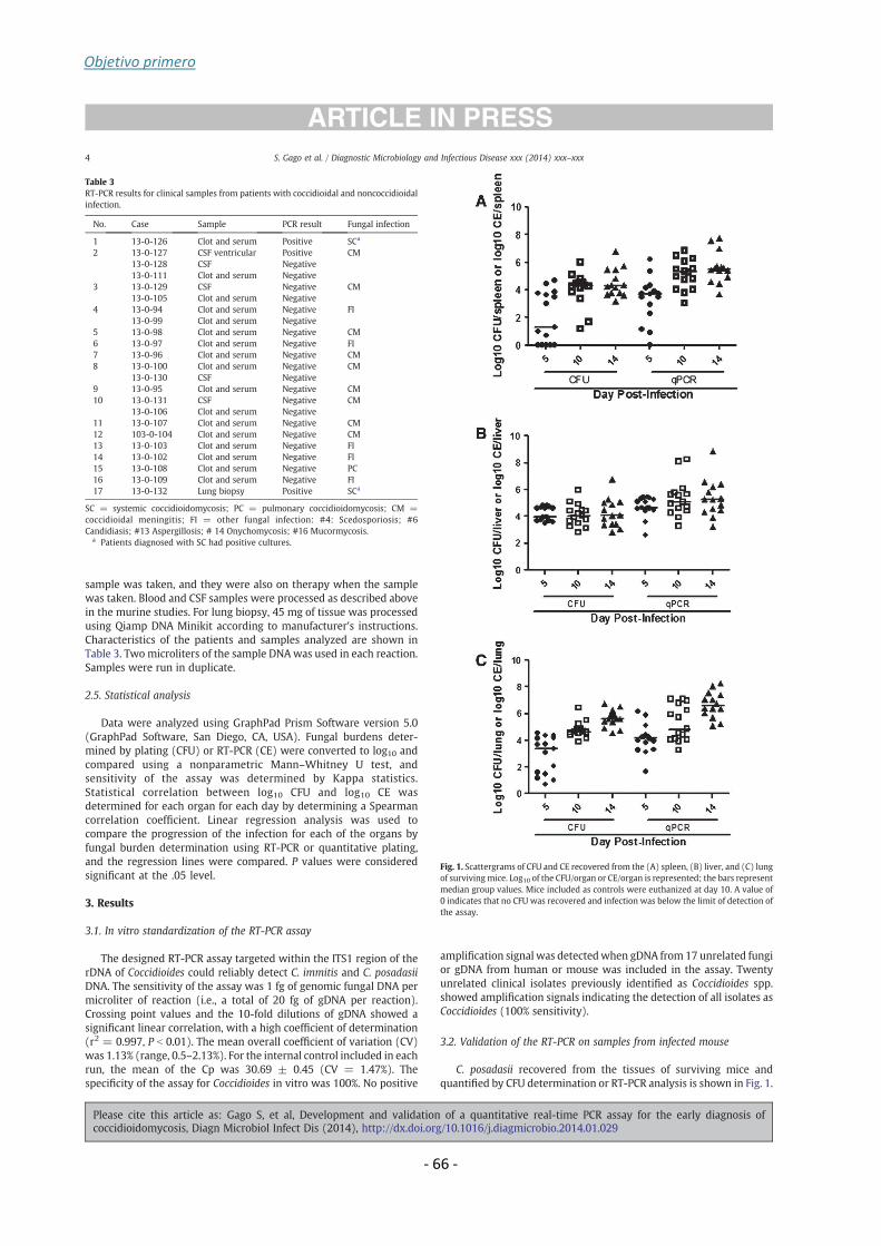
Objetivo primero
- 66 -
sample was taken, and they were also on therapy when the sample
was taken. Blood and CSF samples were processed as described above
in the murine studies. For lung biopsy, 45 mg of tissue was processed
using Qiamp DNA Minikit according to manufacturer's instructions.
Characteristics of the patients and samples analyzed are shown in
Table 3. Two microliters of the sample DNAwas used in each reaction.
Samples were run in duplicate.
2.5. Statistical analysis
Data were analyzed using GraphPad Prism Software version 5.0
(GraphPad Software, San Diego, CA, USA). Fungal burdens deter-
mined by plating (CFU) or RT-PCR (CE) were converted to log10 and
compared using a nonparametric Mann–Whitney U test, and
sensitivity of the assay was determined by Kappa statistics.
Statistical correlation between log10 CFU and log10 CE was
determined for each organ for each day by determining a Spearman
correlation coefficient. Linear regression analysis was used to
compare the progression of the infection for each of the organs by
fungal burden determination using RT-PCR or quantitative plating,
and the regression lines were compared. P values were considered
significant at the .05 level.
3. Results
3.1. In vitro standardization of the RT-PCR assay
The designed RT-PCR assay targeted within the ITS1 region of the
rDNA of Coccidioides could reliably detect C. immitis and C. posadasii
DNA. The sensitivity of the assay was 1 fg of genomic fungal DNA per
microliter of reaction (i.e., a total of 20 fg of gDNA per reaction).
Crossing point values and the 10-fold dilutions of gDNA showed a
significant linear correlation, with a high coefficient of determination
(r2= 0.997, P b 0.01). The mean overall coefficient of variation (CV)
was 1.13% (range, 0.5–2.13%). For the internal control included in each
run, the mean of the Cp was 30.69 ± 0.45 (CV = 1.47%). The
specificity of the assay for Coccidioides in vitro was 100%. No positive
amplification signal was detectedwhen gDNA from 17 unrelated fungi
or gDNA from human or mouse was included in the assay. Twenty
unrelated clinical isolates previously identified as Coccidioides spp.
showed amplification signals indicating the detection of all isolates as
Coccidioides (100% sensitivity).
3.2. Validation of the RT-PCR on samples from infected mouse
C. posadasii recovered from the tissues of surviving mice and
quantified by CFU determination or RT-PCR analysis is shown in Fig. 1.
Table 3
RT-PCR results for clinical samples from patients with coccidioidal and noncoccidioidal
infection.
No. Case Sample PCR result Fungal infection
1 13-0-126 Clot and serum Positive SCa
2 13-0-127 CSF ventricular Positive CM
13-0-128 CSF Negative
13-0-111 Clot and serum Negative
3 13-0-129 CSF Negative CM
13-0-105 Clot and serum Negative
4 13-0-94 Clot and serum Negative FI
13-0-99 Clot and serum Negative
5 13-0-98 Clot and serum Negative CM
6 13-0-97 Clot and serum Negative FI
7 13-0-96 Clot and serum Negative CM
8 13-0-100 Clot and serum Negative CM
13-0-130 CSF Negative
9 13-0-95 Clot and serum Negative CM
10 13-0-131 CSF Negative CM
13-0-106 Clot and serum Negative
11 13-0-107 Clot and serum Negative CM
12 103-0-104 Clot and serum Negative CM
13 13-0-103 Clot and serum Negative FI
14 13-0-102 Clot and serum Negative FI
15 13-0-108 Clot and serum Negative PC
16 13-0-109 Clot and serum Negative FI
17 13-0-132 Lung biopsy Positive SCa
SC = systemic coccidioidomycosis; PC = pulmonary coccidioidomycosis; CM =
coccidioidal meningitis; FI = other fungal infection: #4: Scedosporiosis; #6
Candidiasis; #13 Aspergillosis; # 14 Onychomycosis; #16 Mucormycosis.a Patients diagnosed with SC had positive cultures.
Fig. 1. Scattergrams of CFU and CE recovered from the (A) spleen, (B) liver, and (C) lung
of surviving mice. Log10 of the CFU/organ or CE/organ is represented; the bars represent
median group values. Mice included as controls were euthanized at day 10. A value of
0 indicates that no CFU was recovered and infection was below the limit of detection of
the assay.
4 S. Gago et al. / Diagnostic Microbiology and Infectious Disease xxx (2014) xxx–xxx
Please cite this article as: Gago S, et al, Development and validation of a quantitative real-time PCR assay for the early diagnosis of
coccidioidomycosis, Diagn Microbiol Infect Dis (2014), http://dx.doi.org/10.1016/j.diagmicrobio.2014.01.029
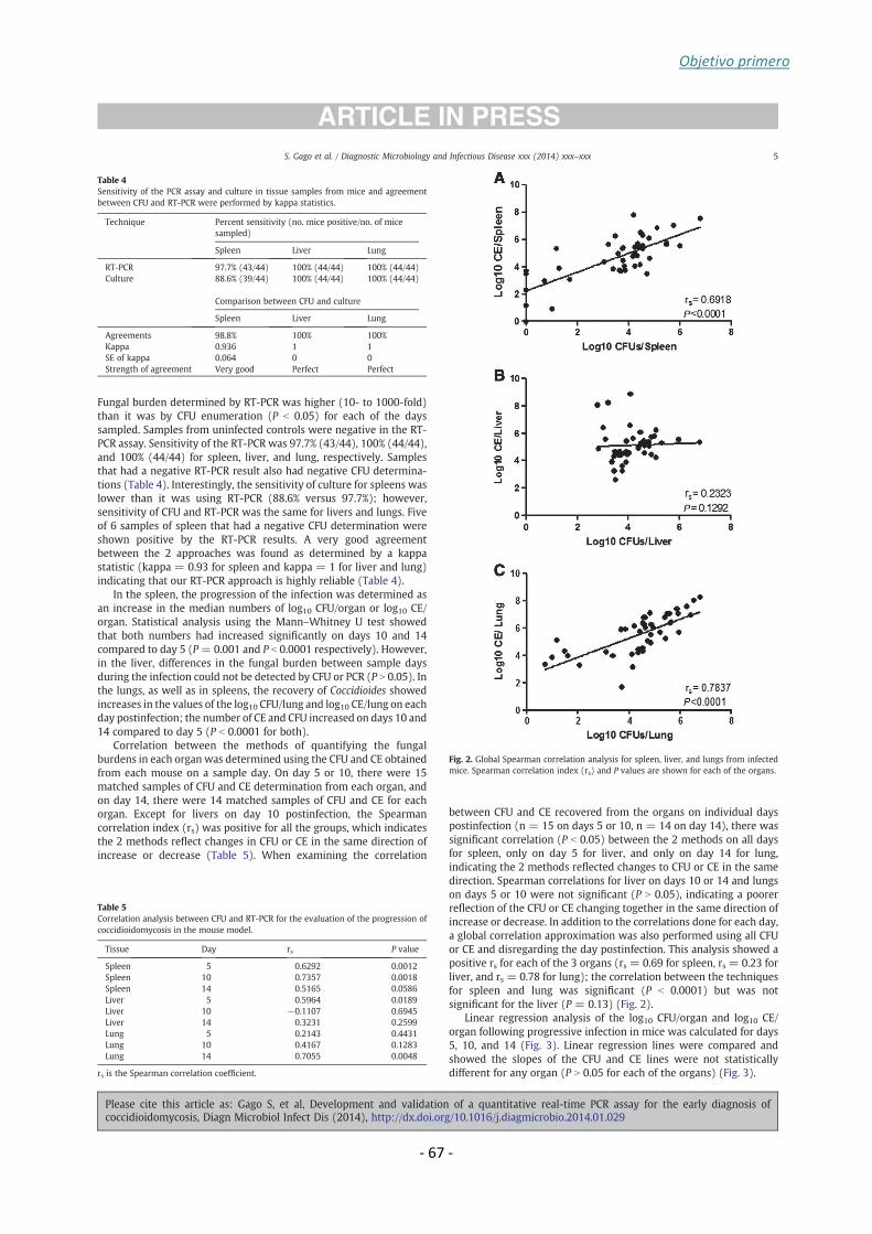
Objetivo primero
- 67 -
Fungal burden determined by RT-PCR was higher (10- to 1000-fold)
than it was by CFU enumeration (P b 0.05) for each of the days
sampled. Samples from uninfected controls were negative in the RT-
PCR assay. Sensitivity of the RT-PCR was 97.7% (43/44), 100% (44/44),
and 100% (44/44) for spleen, liver, and lung, respectively. Samples
that had a negative RT-PCR result also had negative CFU determina-
tions (Table 4). Interestingly, the sensitivity of culture for spleens was
lower than it was using RT-PCR (88.6% versus 97.7%); however,
sensitivity of CFU and RT-PCR was the same for livers and lungs. Five
of 6 samples of spleen that had a negative CFU determination were
shown positive by the RT-PCR results. A very good agreement
between the 2 approaches was found as determined by a kappa
statistic (kappa = 0.93 for spleen and kappa = 1 for liver and lung)
indicating that our RT-PCR approach is highly reliable (Table 4).
In the spleen, the progression of the infection was determined as
an increase in the median numbers of log10 CFU/organ or log10 CE/
organ. Statistical analysis using the Mann–Whitney U test showed
that both numbers had increased significantly on days 10 and 14
compared to day 5 (P= 0.001 and P b 0.0001 respectively). However,
in the liver, differences in the fungal burden between sample days
during the infection could not be detected by CFU or PCR (P N 0.05). In
the lungs, as well as in spleens, the recovery of Coccidioides showed
increases in the values of the log10 CFU/lung and log10 CE/lung on each
day postinfection; the number of CE and CFU increased on days 10 and
14 compared to day 5 (P b 0.0001 for both).
Correlation between the methods of quantifying the fungal
burdens in each organ was determined using the CFU and CE obtained
from each mouse on a sample day. On day 5 or 10, there were 15
matched samples of CFU and CE determination from each organ, and
on day 14, there were 14 matched samples of CFU and CE for each
organ. Except for livers on day 10 postinfection, the Spearman
correlation index (rs) was positive for all the groups, which indicates
the 2 methods reflect changes in CFU or CE in the same direction of
increase or decrease (Table 5). When examining the correlation
between CFU and CE recovered from the organs on individual days
postinfection (n = 15 on days 5 or 10, n = 14 on day 14), there was
significant correlation (P b 0.05) between the 2 methods on all days
for spleen, only on day 5 for liver, and only on day 14 for lung,
indicating the 2 methods reflected changes to CFU or CE in the same
direction. Spearman correlations for liver on days 10 or 14 and lungs
on days 5 or 10 were not significant (P N 0.05), indicating a poorer
reflection of the CFU or CE changing together in the same direction of
increase or decrease. In addition to the correlations done for each day,
a global correlation approximation was also performed using all CFU
or CE and disregarding the day postinfection. This analysis showed a
positive rs for each of the 3 organs (rs= 0.69 for spleen, rs= 0.23 for
liver, and rs= 0.78 for lung); the correlation between the techniques
for spleen and lung was significant (P b 0.0001) but was not
significant for the liver (P = 0.13) (Fig. 2).
Linear regression analysis of the log10 CFU/organ and log10 CE/
organ following progressive infection in mice was calculated for days
5, 10, and 14 (Fig. 3). Linear regression lines were compared and
showed the slopes of the CFU and CE lines were not statistically
different for any organ (P N 0.05 for each of the organs) (Fig. 3).
Table 4
Sensitivity of the PCR assay and culture in tissue samples from mice and agreement
between CFU and RT-PCR were performed by kappa statistics.
Technique Percent sensitivity (no. mice positive/no. of mice
sampled)
Spleen Liver Lung
RT-PCR 97.7% (43/44) 100% (44/44) 100% (44/44)
Culture 88.6% (39/44) 100% (44/44) 100% (44/44)
Comparison between CFU and culture
Spleen Liver Lung
Agreements 98.8% 100% 100%
Kappa 0.936 1 1
SE of kappa 0.064 0 0
Strength of agreement Very good Perfect Perfect
Table 5
Correlation analysis between CFU and RT-PCR for the evaluation of the progression of
coccidioidomycosis in the mouse model.
Tissue Day rs P value
Spleen 5 0.6292 0.0012
Spleen 10 0.7357 0.0018
Spleen 14 0.5165 0.0586
Liver 5 0.5964 0.0189
Liver 10 −0.1107 0.6945
Liver 14 0.3231 0.2599
Lung 5 0.2143 0.4431
Lung 10 0.4167 0.1283
Lung 14 0.7055 0.0048
rs is the Spearman correlation coefficient.
Fig. 2. Global Spearman correlation analysis for spleen, liver, and lungs from infected
mice. Spearman correlation index (rs) and P values are shown for each of the organs.
5S. Gago et al. / Diagnostic Microbiology and Infectious Disease xxx (2014) xxx–xxx
Please cite this article as: Gago S, et al, Development and validation of a quantitative real-time PCR assay for the early diagnosis of
coccidioidomycosis, Diagn Microbiol Infect Dis (2014), http://dx.doi.org/10.1016/j.diagmicrobio.2014.01.029

Objetivo primero
- 68 -
The RT-PCR assaywas also positive for all paraffin-embedded brain
tissues from rabbits (15/15) or mice (6/6), which were obtained from
models of coccidioidal meningitis (100% sensitivity).
3.3. Validation of the assay on clinical samples
The RT-PCR assay was performed on 40 samples from 17 patients
(Table 3). The PCRwas positive in clot, serum, lung biopsy, or CSF from
3 patients. The clinical origin of the samples was diverse, depending
on the underlying disease. Samples from 12 patients diagnosed with
acute infection or under treatment for coccidioidal infection were
included in the assay. For 2 of the 12 patients diagnosedwith systemic
coccidioidomycosis, both cultures and RT-PCR were positive in blood
(clot and serum) and lung biopsy, respectively. For the other 10
patients diagnosed with coccidioidomycosis (pulmonary or meningi-
tis), the PCR was negative; in these patients, the diagnosis was
previously established, and no signs of acute infection were
demonstrated at the time the samples had been obtained. For 1 of
these 10 patients, who had been diagnosed with coccidioidal
meningitis, the RT-PCR result was positive on a sample of the
ventricular CSF obtained via an indwelling Ommaya reservoir. No
positive PCR results were obtained for patients diagnosed with fungal
infections other than coccidioidomycosis (Table 3).
4. Discussion
The aim of this study was the development of an RT-PCR assay for
the specific diagnosis of coccidioidomycosis. Furthermore, we
assessed whether RT-PCR and CFU determinations of fungal burden
correlated in showing the progression of the infection in a mouse
model of systemic coccidioidomycosis as a measure of validating the
RT-PCR assay. Molecular techniques have been described to be very
useful for the correct identification of various fungal species (Cuenca-
Estrella et al., 2011). However, molecular diagnostic tools based on
RT-PCR for coccidioidomycosis have not been widely described. Some
are based on the analysis of melting curves (Bialek et al., 2004;
Binnicker et al., 2007), but an RT-PCR approach using molecular
beacon probes for the simultaneous detection of gDNA from C. immitis
and C. posadasii has not been described previously.
Because clinical signs of coccidioidomycosis are nonspecific and
the increased numbers of case reports of infections in nonendemic
areas (Desai et al., 2010; Desai et al., 2001; Kwok et al., 2009; Oberoi
et al., 2012), rapid, specific, and sensitive assays are needed for
diagnosis, especially if positive early in infection, and potentially could
also be useful for the evaluation of the therapeutic outcome. The RT-
PCR assay that we developed in this work specifically detects gDNA
from C. immitis or C. posadasii and includes an internal control to
monitor for false negatives due to PCR inhibition. The assay was
specific (100%), reproducible, and showed a good sensitivity with
results obtainable in about 3 h. Although the differentiation into C.
immitis and C. posadasii spp. is very interesting for evolutionary
studies, for clinical management, the progression of the infection and
management of disease is similar for both species. Moreover, because
there are few ITS sequences of Coccidioides available in databases, and
the differences between the 2 species are based on a very few
nucleotides (Brilhante et al., 2013), the design of a single probe to
detect either species makes the assay more specific and sensitive for
coccidioidomycosis (Schoch et al., 2012). Although there are some
studies examining the utility of mono-copy genes for Coccidioides RT-
PCR approaches (Bialek et al., 2004; Bialek et al., 2005), the ITS region
of the rDNA is considered the DNA barcode for fungal identification
(Schoch et al., 2012) and is a multi-copy target. The high number of
copies of the ITS per arthroconidium in C. posadasii (Sharpton et al.,
2009) increases the sensitivity of the assay.
The quantification of microorganisms is a microbiological tool to
estimate the total number of microorganisms present in a sample
(Clemons and Stevens, 2009). The utility of RT-PCR assays to quantify
fungal DNA in tissue samples from experimentally infected animals
has been described in several publications on disseminated fungal
infections, with these assays showing variable sensitivity, specificity,
and correlations with cultures (Abad-Diaz-De-Cerio et al., 2013;
Bernal-Martinez et al., 2012; Bowman et al., 2001; Buitrago et al.,
2005; Castelli et al., 2008; Clemons and Stevens, 2009; Morton et al.,
2011; Singh et al., 2005). The results presented in our study provide
the first description of an RT-PCR based quantification of C. posadasii
burden in the tissues in an animal model of infection. Although
agreement between CFU enumeration and RT-PCR determinations
was very good, we feel that our RT-PCR assay provided a more
accurate reflection of Coccidioides burdens than did CFU determina-
tions, for all tissue samples, and for each of the days tested. Fungal
burdens determined by RT-PCR were 10- to 1000-fold higher than
were the CFU. Tissue samples likely contained maturing or mature
spherules, which conceivably could yield only 1 CFU on culture but
Fig. 3. Linear regression lines of log10 CFU and log10 CE/organ from infected mice
following progressive infection in the (A) spleen, (B) liver, and (C) lung. Equation of the
lines, F statistics, and P values are shown. Data points represent the mean log10 value
and the 95% confidence interval.
6 S. Gago et al. / Diagnostic Microbiology and Infectious Disease xxx (2014) xxx–xxx
Please cite this article as: Gago S, et al, Development and validation of a quantitative real-time PCR assay for the early diagnosis of
coccidioidomycosis, Diagn Microbiol Infect Dis (2014), http://dx.doi.org/10.1016/j.diagmicrobio.2014.01.029
Objetivo primero
- 69 -
have many nuclei, which contributed to a better detection by
quantitative polymerase chain reaction. In addition, the sensitivity
in spleen was also higher by PCR (43/44; 97.7%) than by CFU
determination (39/44; 88%). The single mouse that showed a negative
RT-PCR result also had a negative culture. In evaluating progression of
the infection in the tissues of infected animals by CFU, burdens
increased each day postinfection for spleen and lung, but not for liver.
Interestingly, slight differences in the fungal load determined by RT-
PCR in liver could be detected, and it could be related to an increased
sensitivity of the RT-PCR approach. Although differences in the
magnitude of the burden for each of the organs studied were detected
by RT-PCR and CFU determinations, regression analysis suggested that
the assays are equivalent for the evaluation of the progression of the
infection, although RT-PCR approaches are more sensitive.
Blood samples from the infected mice showed a negative RT-PCR
result, and no PCR inhibition was detected; cultures with these
samples were not performed. Although it may be surprising, the
sensitivity of blood cultures is very low, and some studies have
shown that it may be lower than 0.4% for coccidioidomycosis
(Saubolle et al., 2007). It is possible that fungemia occurs when the
infection is uncontrolled and the animals are moribund; other
possibilities are that, in fungemia, the fungal load in serum is lower
than the limit of detection of the assay or fungemia could be
intermittent. Johnson et al. (2004) described the usefulness of a pair
of primers to amplify Coccidioides DNA from serum samples from
infected patients. However, Bialek et al. (2005) demonstrated that
those primers were nonspecific, and no other published article has
demonstrated the presence of coccidioidal DNA in serum or blood
samples from infected patients.
Our RT-PCR approach was also validated using 40 samples from
17 patients, including 4 patients diagnosed with other fungal
infections. The assay reliably detected Coccidioides DNA in the
specimens from the 2 patients who had positive cultures concurrent
with the samples used for performance of the RT-PCR assay;
however, the PCR was negative in those patients who had been
undergoing extended antifungal treatment, did not present signs of
acute infection at the time we received the sample, or presented
another unrelated fungal infection. Although we did not obtain
positive PCR results using blood from infected mice, for 1 patient, the
PCR was positive for both the clot and serum. It has been reported
that Coccidioides fungemia is associated with immunosuppression
and high mortality (Adam et al., 2009; Keckich et al., 2010; Rempe
et al., 2007). For 1 of the patients diagnosed with coccidioidal
meningitis, we obtained a positive PCR result, although no signs of
acute infection were noted at the time the clinical sample was
obtained. However, the sample was taken from a device used to
administer the antifungal, and our positive result could reflect
transient colonization of hardware; the sample collected 15 days
later showed a negative PCR result. Furthermore, our assay exhibited
100% (21/21) sensitivity using paraffin-embedded tissue samples
from animal models of coccidioidal meningitis. Human blood and
CSF specimens were largely used for validation; this was determined
by specimen availability, as these are the likeliest clinical specimens
to be stored. Tissue samples of patients will need to be collected
prospectively, to ascertain whether our assay is equally efficacious in
a variety of human solid tissues compared to what we have
demonstrated in murine tissues.
Several articles reporting the utility of RT-PCR for the diagnosis of
coccidioidomycosis have been published. Bialek et al. (2004) and
Binnicker et al. (2007) described RT-PCR based on the use of Taqman
probes and FRET hybridization probes, respectively, for the diagnosis
of coccidioidomycosis. These probes were based on the nucleotide
sequence of the Antigen2/Proline-Rich Antigen, a monocopy gene,
and the ITS-2 region (Bialek et al., 2004; Binnicker et al., 2007).
Although the specificity and sensitivity of both techniques on cultured
isolates were 100%, the lower limit of detection was lower in our RT-
PCR assay (i.e., 10 fg/μL of DNA per reaction versus 1 fg/μL per reaction
in our assay). The utility of the RT-PCR approach described by
Binnicker et al. (2007) has been validated for clinical samples from
patients with a positive culture result; however, the utility of this
technique seems to be limited in paraffin-embedded tissues (74%
sensitivity). In contrast, Thompson et al. (2013) compared a standard
RT-PCR with the PCR procedures from the Translational Genomics
Research Institute (Vucicevic et al., 2010). In that study, they showed
that the PCR assays did not improve the sensitivity of detection over
culture, which is in contrast to what our animal model studies
showed, and false negatives could be produced (Vucicevic et al.,
2010). In addition, Sheff et al. (2010) described an RT-PCR approach
for the differentiation of C. immitis and C. posadasii species based on
TaqMan probes designed by genome comparison; although this RT-
PCR was 100% specific, validation using clinical specimens was not
done. The advantages of our technique compared to those previously
described are: our limit of detection is lower than those previously
reported; we include an internal control, which ensures that there has
not been inhibition of the PCR reaction; our RT-PCR appeared to
accurately reflect the progression of infection in the mousemodel and
may prove useful in monitoring patients' outcome.
In conclusion, the RT-PCR protocol described here allows for rapid
and sensitive detection of Coccidioides spp. gDNA. This method could
be very useful to monitor the progression of the infection and to
contribute to the establishment of an early diagnosis. However,
further validation with substantial numbers of clinical samples from
patients is needed.
Acknowledgments
This work was supported by research project PI11/00412 from
Spanish “Fondo de Investigaciones Sanitarias” of the Instituto de Salud
Carlos III. S.G. is supported by a research fellowship from the “Fondo
de Investigaciones Biomedicas” of the Spanish Ministry of Science and
Innovation (FI10/00464). M.C.-E. has received grant support from
Astellas Pharma, bioMérieux, Gilead Sciences, MSD, Pfizer, Schering-
Plough, Soria Melguizo S.A., Ferrer International, the European Union,
the ALBAN program, the Spanish Agency for International Coopera-
tion, the Spanish Ministry of Culture and Education, The Spanish
Health Research Fund, The Instituto de Salud Carlos III, The Ramon
Areces Foundation, and The Mutua Madrileña Foundation. He has
been an advisor/consultant to the Panamerican Health Organization,
Gilead Sciences, MSD, Pfizer, Astellas, and Schering-Plough. He has
been paid for talks on behalf of Gilead Sciences, MSD, Pfizer, Astellas,
and Schering-Plough. M.J.B., K.V.C., and D.A.S. have no conflicts of
interest.
References
Abad-Diaz-De-Cerio A, Fernandez-Molina JV, Ramirez-Garcia A, Sendino J, Hernando FL,Peman J, et al. The aspHS gene as a new target for detecting Aspergillus fumigatusduring infections by quantitative real-time PCR. Med Mycol 2013;51:545–54.
Adam RD, Elliott SP, Taljanovic MS. The spectrum and presentation of disseminatedcoccidioidomycosis. Am J Med 2009;122:770–7.
Ampel NM. The diagnosis of coccidioidomycosis. F1000 Med Rep 2010;2. pii: 2. http://dx.doi.org/10.3410/M2-2.
Bernal-Martinez L, Buitrago MJ, Castelli MV, Rodriguez-Tudela JL, Cuenca-Estrella M.Detection of invasive infection caused by Fusarium solani and non-Fusarium solanispecies using a duplex quantitative PCR-based assay in a murine model offusariosis. Med Mycol 2012;50:270–5.
Bernal-Martinez L, Buitrago MJ, Castelli MV, Rodriguez-Tudela JL, Cuenca-Estrella M.Development of a single tube multiplex real-time PCR to detect the most clinicallyrelevant Mucormycetes species. Clin Microbiol Infect 2013;19:E1–7.
Bialek R, Kern J, Herrmann T, Tijerina R, Cecenas L, Reischl U, et al. PCR assays foridentification of Coccidioides posadasii based on the nucleotide sequence of theantigen 2/proline-rich antigen. J Clin Microbiol 2004;42:778–83.
Bialek R, Gonzalez GM, Begerow D, Zelck UE. Coccidioidomycosis and blastomycosis:advances in molecular diagnosis. FEMS Immunol Med Microbiol 2005;45:355–60.
Binnicker MJ, Buckwalter SP, Eisberner JJ, Stewart RA, McCullough AE, Wohlfiel SL, et al.Detection of Coccidioides species in clinical specimens by real-time PCR. J ClinMicrobiol 2007;45:173–8.
7S. Gago et al. / Diagnostic Microbiology and Infectious Disease xxx (2014) xxx–xxx
Please cite this article as: Gago S, et al, Development and validation of a quantitative real-time PCR assay for the early diagnosis ofcoccidioidomycosis, Diagn Microbiol Infect Dis (2014), http://dx.doi.org/10.1016/j.diagmicrobio.2014.01.029

Objetivo primero
- 70 -
Bowman JC, Abruzzo GK, Anderson JW, Flattery AM, Gill CJ, Pikounis VB, et al.
Quantitative PCR assay to measure Aspergillus fumigatus burden in a murine modelof disseminated aspergillosis: demonstration of efficacy of caspofungin acetate.
Antimicrob Agents Chemother 2001;45:3474–81.Brilhante RS, de Lima RA, Ribeiro JF, de Camargo ZP, Castelo-Branco Dde S, Grangeiro TB,
et al. Genetic diversity of Coccidioides posadasii from Brazil. Med Mycol 2013;51:
432–7.Buitrago MJ, Gomez-Lopez A, Mellado E, Rodriguez-Tudela JL, Cuenca-Estrella M.
Detection of Aspergillus spp. by real-time PCR in a murine model of pulmonaryinfection [article in Spanish]. Enferm Infecc Microbiol Clin 2005;23:464–8.
Buitrago MJ, Aguado JM, Ballen A, Bernal-Martinez L, Prieto M, Garcia-Reyne A, et al.
Efficacy of DNA amplification in tissue biopsy samples to improve the detection ofinvasive fungal disease. Clin Microbiol Infect 2013;19:E271–7.
Capilla J, Clemons KV, Sobel RA, Stevens DA. Efficacy of amphotericin B lipid complexin a rabbit model of coccidioidal meningitis. J Antimicrob Chemother 2007;60:
673–6.Castelli MV, Buitrago MJ, Bernal-Martinez L, Gomez-Lopez A, Rodriguez-Tudela JL,
Cuenca-Estrella M. Development and validation of a quantitative PCR assay for
diagnosis of scedosporiosis. J Clin Microbiol 2008;46:3412–6.Centers for Disease Control and Prevention (CDC). Biosafety in Microbiological and
Biomedical Laboratories 5th ed. Atlanta, GA: US Department of Health and HumanServices; 2009.
Centers for Disease Control and Prevention (CDC). Increase in reported coccidioidomy-
cosis–United States, 1998–2011. MMWR Morb Mortal Wkly Rep 2013;62:217–21.Clemons KV, Stevens DA. Conventional or molecular measurement of Aspergillus load.
Med Mycol 2009;47(Suppl 1):S132–7.Clemons KV, Leathers CR, Lee KW. Systemic Coccidioides immitis infection in nude and
beige mice. Infect Immun 1985;47:814–21.Clemons KV, Hanson LH, Perlman AM, Stevens DA. Efficacy of SCH39304 and
fluconazole in a murine model of disseminated coccidioidomycosis. Antimicrob
Agents Chemother 1990;34:928–30.Clemons KV, Capilla J, Stevens DA. Experimental animal models of coccidioidomycosis.
Ann N Y Acad Sci 2007;1111:208–24.Crabtree AC, Keith DG, Diamond HL. Relationship between radiographic hilar
lymphadenopathy and serologic titers for Coccidioides sp. in dogs in an endemicregion. Vet Radiol Ultrasound 2008;49:501–3.
Cuenca-Estrella M, Bassetti M, Lass-Florl C, Racil Z, Richardson M, Rogers TR. Detection
and investigation of invasive mould disease. J Antimicrob Chemother2011;66(Suppl 1):i15–24.
Desai SA, Minai OA, Gordon SM, O'Neil B, Wiedemann HP, Arroliga AC. Coccidioido-
mycosis in non-endemic areas: a case series. Respir Med 2001;95:305–9.Desai NR, McGoey R, Troxclair D, Simeone F, Palomino J. Coccidioidomycosis in
nonendemic area: case series and review of literature. J La State Med Soc 2010;162:97–103.
Dixon DM. Coccidioides immitis as a Select Agent of bioterrorism. J Appl Microbiol2001;91:602–5.
Dolezel J, Bartos J, Voglmayr H, Greilhuber J. Nuclear DNA content and genome size of
trout and human. Cytometry A 2003;51:127–8.Durkin M, Estok L, Hospenthal D, Crum-Cianflone N, Swartzentruber S, Hackett E, et al.
Detection of Coccidioides antigenemia following dissociation of immune com-plexes. Clin Vaccine Immunol 2009;16:1453–6.
Fisher MC, Koenig GL, White TJ, Taylor JW. Molecular and phenotypic description of
Coccidioides posadasii sp. nov., previously recognized as the non-Californiapopulation of Coccidioides immitis. Mycologia 2002;94:73–84.
Hector RF, Rutherford GW, Tsang CA, Erhart LM, McCotter O, Anderson SM, et al. Thepublic health impact of coccidioidomycosis in Arizona and California. Int J Environ
Res Public Health 2011;8:1150–73.Hsu JL, Ruoss SJ, Bower ND, Lin M, Holodniy M, Stevens DA. Diagnosing invasive fungal
disease in critically ill patients. Crit Rev Microbiol 2011;37:277–312.
Johnson SM, Simmons KA, Pappagianis D. Amplification of coccidioidal DNA in clinicalspecimens by PCR. J Clin Microbiol 2004;42:1982–5.
Kamberi P, Sobel RA, Clemons KV, Stevens DA, Pappagianis D, Williams PL. A murine
model of coccidioidal meningitis. J Infect Dis 2003;187:453–60.Keckich DW, Blair JE, Vikram HR. Coccidioides fungemia in six patients, with a review of
the literature. Mycopathologia 2010;170:107–15.Kuberski T, Herrig J, Pappagianis D. False-positive IgM serology in coccidioidomycosis. J
Clin Microbiol 2010;48:2047–9.
Kwok HK, Chan JW, Li IW, Chu SY, Lam CW. Coccidioidomycosis as a rare cause ofpneumonia in non-endemic areas: a short exposure history should not be ignored.
Respirology 2009;14:617–20.Laniado-Laborin R. Expanding understanding of coccidioidomycosis in the western
hemisphere. Ann N Y Acad Sc 2007;1111:19–34.
Morton CO, Clemons KV, Springer J, Mueller JG, Rogers TR, Stevens DA, et al. Real-timePCR and quantitative culture for monitoring of experimental Aspergillus fumigatusintracranial infection in neutropenic mice. J Med Microbiol 2011;60:913–9.
Oberoi JK, Wattal C, Aggarwal PK, Khanna S, Basu AK, Verma K. Pulmonary
coccidiomycosis in New Delhi, India. Infection 2012;40:699–702.Pappagianis D. Epidemiology of coccidioidomycosis. Curr Top Med Mycol 1988;2:
199–238.
Pappagianis D. Coccidioidomycosis. Semin Dermatol 1993;12:301–9.Pappagianis D. Marked increase in cases of coccidioidomycosis in California: 1991,
1992, and 1993. Clin Infect Dis 1994;19(Suppl 1):S14–8.Perfect JR. Fungal diagnosis: how do we do it and can we do better? Curr Med Res Opin
2013;29(Suppl 4):3–11.
Rempe S, Sachdev MS, Bhakta R, Pineda-Roman M, Vaz A, Carlson RW. Coccidioidesimmitis fungemia: clinical features and survival in 33 adult patients. Heart Lung2007;36:64–71.
Saubolle MA. Laboratory aspects in the diagnosis of coccidioidomycosis. Ann N Y Acad
Sci 2007;1111:301–14.Saubolle MA, McKellar PP, Sussland D. Epidemiologic, clinical, and diagnostic aspects of
coccidioidomycosis. J Clin Microbiol 2007;45:26–30.
Schoch CL, Seifert KA, Huhndorf S, Robert V, Spouge JL, Levesque CA, et al. Nuclearribosomal internal transcribed spacer (ITS) region as a universal DNA barcode
marker for Fungi. Proc Natl Acad Sci U S A 2012;109:6241–6.Sharpton TJ, Stajich JE, Rounsley SD, Gardner MJ, Wortman JR, Jordar VS, et al.
Comparative genomic analyses of the human fungal pathogens Coccidioides andtheir relatives. Genome Res 2009;19:1722–31.
Sheff KW, York ER, Driebe EM, Barker BM, Rounsley SD,Waddell VG, et al. Development
of a rapid, cost-effective TaqMan Real-Time PCR Assay for identification anddifferentiation of Coccidioides immitis and Coccidioides posadasii. Med Mycol
2010;48:466–9.
Shubitz LF. Comparative aspects of coccidioidomycosis in animals and humans. Ann N YAcad Sci 2007;1111:395–403.
Singh G, Imai J, Clemons KV, Stevens DA. Efficacy of caspofungin against central nervoussystem Aspergillus fumigatus infection in mice determined by TaqMan PCR and CFUmethods. Antimicrob Agents Chemother 2005;49:1369–76.
Sutton DA. Diagnosis of coccidioidomycosis by culture: safety considerations,
traditional methods, and susceptibility testing. Ann N Y Acad Sci 2007;1111:
315–25.Tamura K, Peterson D, Peterson N, Stecher G, Nei M, Kumar S. MEGA5: molecular
evolutionary genetics analysis using maximum likelihood, evolutionary distance,and maximum parsimony methods. Mol Biol Evol 2011;28:2731–9.
Tang CM, Cohen J, Krausz T, Van Noorden S, Holden DW. The alkaline protease of
Aspergillus fumigatus is not a virulence determinant in two murine models ofinvasive pulmonary aspergillosis. Infect Immun 1993;61:1650–6.
Thompson GR, Sharma S, Bays DJ, Pruitt R, Engelthaler DM, Bowers J, et al.Coccidioidomycosis: adenosine deaminase levels, serologic parameters, culture
results, and polymerase chain reaction testing in pleural fluid. Chest 2013;143:776–81.
Vucicevic D, Blair JE, Binnicker MJ, McCullough AE, Kusne S, Vikram HR, et al. The utility
of Coccidioides polymerase chain reaction testing in the clinical setting. Myco-pathologia 2010;170:345–51.
8 S. Gago et al. / Diagnostic Microbiology and Infectious Disease xxx (2014) xxx–xxx
Please cite this article as: Gago S, et al, Development and validation of a quantitative real-time PCR assay for the early diagnosis of
coccidioidomycosis, Diagn Microbiol Infect Dis (2014), http://dx.doi.org/10.1016/j.diagmicrobio.2014.01.029
Objetivo segundo
- 71 -
4. OBJETIVO SEGUNDO: Desarrollo de técnicas rápidas de
identificación y tipificación basadas
en el análisis de curvas de fusión de
alta resolución (HRM):


Objetivo segundo
- 73 -
4.1. Identificación de las especies del complejo
Cryptococcus neoformans y Cryptococcus gattii mediante HRM.
Las infecciones fúngicas debidas a Cryptococcus spp. afectan tanto a individuos
inmunocomprometidos como inmunocompetentes. La especie C. neoformans
clásicamente se ha dividido en 5 serotipos (A, B, C, D y AD) que se han agrupado en dos
variedades de C. neoformans, var. grubii (serotipo A) y var. neoformans (serotipo D).
Los serotipos B y C se han clasificado recientemente como la especie Cryptococcus
gattii. Además, se ha descrito la existencia de híbridos AD, BD y BA. En este trabajo se
ha desarrollado una técnica rápida para la diferenciación de las variedades de
Cryptococcus spp. mediante análisis de curvas de fusión de alta resolución (HRM). La
reacción de PCR se dirigió a la amplificación de una región de 100 pb en el espaciador
intergénico 1 (ITS-1) del ADN ribosómico. Las condiciones de PCR y el análisis de las
curvas se optimizaron para analizar los productos de amplificación de 27 cepas.
Simultáneamente se secuenciaron los fragmentos amplificados. Un procedimiento de
doble análisis mediante HRM del producto de PCR de las 27 cepas de Cryptococcus
spp., permitió discriminar 4 grupos que se correspondían con las variedades
neoformans (serotipo D e híbridos AD, 12 cepas), var. grubii (serotipo A,10 cepas), var.
gattii (Serotipos B y C, 4 cepas) e híbridos BD y AB (3 cepas). Los resultados de estos
grupos se correspondían con las posiciones polimórficas en la secuencia del fragmento
obtenido para cada cepa. Tan solo la cepa CNM-CL 5007 agrupada inicialmente como
C. gattii, se agrupó con los híbridos BD y AB mediante análisis de secuencia y HRM. Con
los resultados de este trabajo se concluye que: (i) el análisis mediante HRM de los
productos de PCR es un método rápido y sencillo que puede ser útil para la
diferenciación de variedades de Cryptococcus spp.; (ii) los híbridos AD son semejantes
en secuencia a la var. neoformans en el fragmento analizado; (iii) la primera fase del
análisis permite la discriminación de C. gattii del complejo C. neoformans.

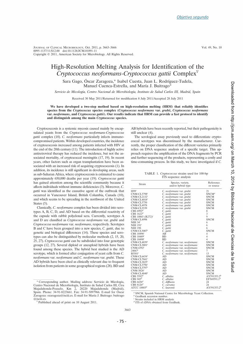
Objetivo segundo
- 75 -
JOURNAL OF CLINICAL MICROBIOLOGY, Oct. 2011, p. 3663–3666 Vol. 49, No. 100095-1137/11/$12.00 doi:10.1128/JCM.01091-11Copyright © 2011, American Society for Microbiology. All Rights Reserved.
High-Resolution Melting Analysis for Identification of theCryptococcus neoformans-Cryptococcus gattii Complex£
Sara Gago, Oscar Zaragoza,* Isabel Cuesta, Juan L. Rodríguez-Tudela,Manuel Cuenca-Estrella, and María J. Buitrago*
Servicio de Micología, Centro Nacional de Microbiología, Instituto de Salud Carlos III, Madrid, Spain
Received 30 May 2011/Returned for modification 8 July 2011/Accepted 28 July 2011
We have developed a two-step method based on high-resolution melting (HRM) that reliably identifiesspecies from the Cryptococcus species complex (Cryptococcus neoformans var. grubii, Cryptococcus neoformansvar. neoformans, and Cryptococcus gattii). Our results indicate that HRM can provide a fast protocol to identifyand distinguish among the main Cryptococcus species.
Cryptococcosis is a systemic mycosis caused mainly by encap-sulated yeasts from the Cryptococcus neoformans-Cryptococcus
gattii complex (18). C. neoformans particularly infects immuno-compromised patients. Within developed countries, the incidenceof cryptococcosis increased among patients infected with HIV atthe end of the 20th century (11). The introduction of highly activeantiretroviral therapy has reduced the incidence, but not the as-sociated mortality, of cryptococcal meningitis (17, 19). In recentyears, other factors such as organ transplantation have been as-sociated with an increased risk of acquiring cryptococcosis (1). Inaddition, its incidence is still significant in developing areas, suchas sub-Saharan Africa, where cryptococcosis is estimated to causeapproximately 650,000 deaths per year (19). Cryptococcus gattii
has gained attention among the scientific community because itaffects individuals without immune deficiencies (3). Moreover, C.
gattii was identified as the causative agent of the outbreak thatoccurred in Vancouver Island, British Columbia, Canada (10),and which seems to be spreading in the northwest of the UnitedStates (5).
Classically, C. neoformans complex has been divided into sero-types A, B, C, D, and AD based on the differential reactivity ofthe capsule with rabbit polyclonal sera. Currently, serotypes Aand D are classified as Cryptococcus neoformans var. grubii andCryptococcus neoformans var. neoformans, respectively. SerotypesB and C have been grouped into a new species, C. gattii, due togenetic and biological differences (14). These species and sero-types can also be distinguished by molecular methods (2, 15, 20,25, 27). Cryptococcus gattii can be subdivided into four genotypicgroups (12, 27). Several diploid or aneuploid hybrids have beenfound among these species. The hybrid best studied is the ADserotype, which is formed after conjugation of yeast cells from C.
neoformans var. neoformans and C. neoformans var. grubii. TheseAD hybrids have been cited as clinically relevant due to frequentisolation from patients in some geographical regions (28). BD and
AB hybrids have been recently reported, but their pathogenicity isstill unclear (4).
The serological assay previously used to differentiate crypto-coccal serotypes was discontinued by the manufacturer. Cur-rently, the proper classification of the different varieties primarilyrelies on DNA sequence analysis of a specific target. This ap-proach requires the amplification of the DNA fragments by PCRand further sequencing of the products, representing a costly andtime-consuming process. In this study, we have investigated if C.
* Corresponding author. Mailing address: Servicio de Micología,Centro Nacional de Microbiología, Instituto de Salud Carlos III, CtraMajadahonda-Pozuelo, Km 2, 28220 Majadahonda (Madrid),Spain. Phone: 34-91-8223661. Fax: 34-91-5097966. E-mail for OscarZaragoza: [email protected]. E-mail for María J. Buitrago: [email protected].
£ Published ahead of print on 10 August 2011.
TABLE 1. Cryptococcus strains used for 100-bpITS sequence analysis
StrainSpecies, variety,
and/or hybrid typeReferenceor source
H99c C. neoformans var. grubii 21CNM-CL3798c C. neoformans var. grubii SNCMa
CNM-CL2132c C. neoformans var. grubii SNCMCNM-CL4018c C. neoformans var. grubii SNCMCNM-CL5750 C. neoformans var. grubii SNCMCNM-CL4978 C. neoformans var. grubii SNCMCNM-CL5539 C. neoformans var. grubii SNCMCBS 6992d C. gattii 8CBS 1622d C. gattii 3CBS 10865 (R272)c C. gattii 9CNM-CL4998 C. gattii SNCMNIH 34c C. gattii 6NIH 191c C. gattii 6NIH 198 C. gattii 6CNM-CL5007c C. gattii SNCMCBS 10496c AB 4CBS 10489c BD 4CBS 10490c BD 4CNM-CL4839c C. neoformans var. neoformans SNCMCNM-CL3801c C. neoformans var. neoformans SNCMCNM-4392c C. neoformans var. neoformans SNCM24067c C. neoformans var. neoformans 7B3501c C. neoformans var. neoformans 13CNM-CL6434c AD SNCMCNM-CL7041c AD SNCMCNM-CL3825c AD SNCMCNM-CL3796c AD SNCMCNM-CL3797c AD SNCMCNM-3826c AD SNCMCNM-CL4840c AD SNCMCBS 5592d C. albidus AY591353.2b
CBS 945d C. albidus EF569591.1b
CBS 6436d C. diffluens 24CBS 8126d C. curvatus 24ATCC 18803d C. laurentii AY591353.2b
a SNCM, Spanish National Centre for Microbiology Yeast Collection.b GenBank accession number.c Strains included in HRM analysis.d ITS of rDNA obtained from GenBank.
3663
on
Ma
rch
10
, 20
14
by B
iblio
teca
Na
cio
na
l de
Cie
ncia
s d
e la
Sa
lud
http
://jcm
.asm
.org
/D
ow
nlo
ad
ed
from
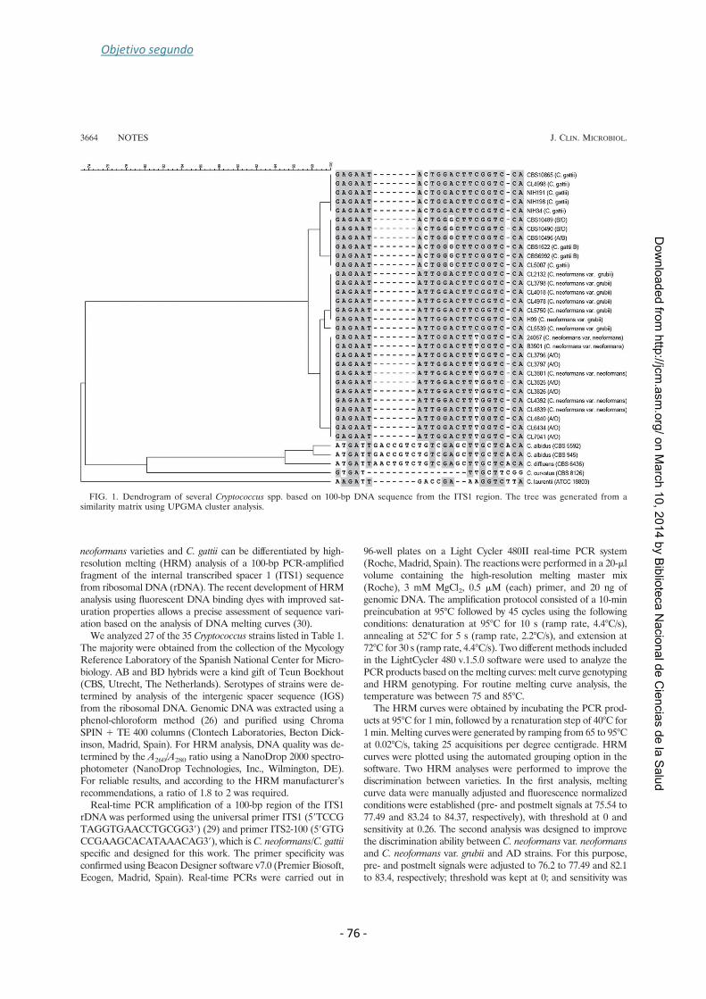
Objetivo segundo
- 76 -
neoformans varieties and C. gattii can be differentiated by high-resolution melting (HRM) analysis of a 100-bp PCR-amplifiedfragment of the internal transcribed spacer 1 (ITS1) sequencefrom ribosomal DNA (rDNA). The recent development of HRManalysis using fluorescent DNA binding dyes with improved sat-uration properties allows a precise assessment of sequence vari-ation based on the analysis of DNA melting curves (30).
We analyzed 27 of the 35 Cryptococcus strains listed in Table 1.The majority were obtained from the collection of the MycologyReference Laboratory of the Spanish National Center for Micro-biology. AB and BD hybrids were a kind gift of Teun Boekhout(CBS, Utrecht, The Netherlands). Serotypes of strains were de-termined by analysis of the intergenic spacer sequence (IGS)from the ribosomal DNA. Genomic DNA was extracted using aphenol-chloroform method (26) and purified using ChromaSPIN 1 TE 400 columns (Clontech Laboratories, Becton Dick-inson, Madrid, Spain). For HRM analysis, DNA quality was de-termined by the A260/A280 ratio using a NanoDrop 2000 spectro-photometer (NanoDrop Technologies, Inc., Wilmington, DE).For reliable results, and according to the HRM manufacturer’srecommendations, a ratio of 1.8 to 2 was required.
Real-time PCR amplification of a 100-bp region of the ITS1rDNA was performed using the universal primer ITS1 (59TCCGTAGGTGAACCTGCGG39) (29) and primer ITS2-100 (59GTGCCGAAGCACATAAACAG39), which is C. neoformans/C. gattii
specific and designed for this work. The primer specificity wasconfirmed using Beacon Designer software v7.0 (Premier Biosoft,Ecogen, Madrid, Spain). Real-time PCRs were carried out in
96-well plates on a Light Cycler 480II real-time PCR system(Roche, Madrid, Spain). The reactions were performed in a 20-mlvolume containing the high-resolution melting master mix(Roche), 3 mM MgCl2, 0.5 mM (each) primer, and 20 ng ofgenomic DNA. The amplification protocol consisted of a 10-minpreincubation at 95°C followed by 45 cycles using the followingconditions: denaturation at 95°C for 10 s (ramp rate, 4.4°C/s),annealing at 52°C for 5 s (ramp rate, 2.2°C/s), and extension at72°C for 30 s (ramp rate, 4.4°C/s). Two different methods includedin the LightCycler 480 v.1.5.0 software were used to analyze thePCR products based on the melting curves: melt curve genotypingand HRM genotyping. For routine melting curve analysis, thetemperature was between 75 and 85°C.
The HRM curves were obtained by incubating the PCR prod-ucts at 95°C for 1 min, followed by a renaturation step of 40°C for1 min. Melting curves were generated by ramping from 65 to 95°Cat 0.02°C/s, taking 25 acquisitions per degree centigrade. HRMcurves were plotted using the automated grouping option in thesoftware. Two HRM analyses were performed to improve thediscrimination between varieties. In the first analysis, meltingcurve data were manually adjusted and fluorescence normalizedconditions were established (pre- and postmelt signals at 75.54 to77.49 and 83.24 to 84.37, respectively), with threshold at 0 andsensitivity at 0.26. The second analysis was designed to improvethe discrimination ability between C. neoformans var. neoformans
and C. neoformans var. grubii and AD strains. For this purpose,pre- and postmelt signals were adjusted to 76.2 to 77.49 and 82.1to 83.4, respectively; threshold was kept at 0; and sensitivity was
FIG. 1. Dendrogram of several Cryptococcus spp. based on 100-bp DNA sequence from the ITS1 region. The tree was generated from asimilarity matrix using UPGMA cluster analysis.
3664 NOTES J. CLIN. MICROBIOL.
on
Ma
rch
10
, 20
14
by B
iblio
teca
Na
cio
na
l de
Cie
ncia
s d
e la
Sa
lud
http
://jcm
.asm
.org
/D
ow
nlo
ad
ed
from
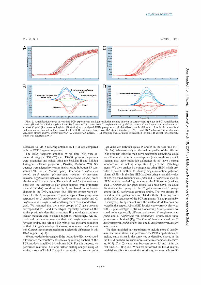
Objetivo segundo
- 77 -
decreased to 0.15. Clustering obtained by HRM was comparedwith the PCR fragment sequence.
The DNA fragments amplified by real-time PCR were se-quenced using the ITS1 (23) and ITS2-100 primers. Sequenceswere assembled and edited using the SeqMan II and EditSeqLasergene software programs (DNAstar, Madison, WI). Se-quences were aligned for cluster analysis using Infoquest FP soft-ware v.4.50 (Bio-Rad, Madrid, Spain). Other non-C. neoformans/
non-C. gattii species (Cryptococcus curvatus, Cryptococcus
laurentii, Cryptococcus diffluens, and Cryptococcus albidus) werealso included in the analysis. The method used for tree construc-tions was the unweighted-pair group method with arithmeticmean (UPGMA). As shown in Fig. 1, and based on nucleotidechanges in the DNA sequence, four different groups were ob-tained for the C. neoformans-C. gattii complex. Two groups cor-responded to C. neoformans (C. neoformans var. grubii and C.
neoformans var. neoformans), and two groups corresponded to C.
gattii. We assumed that these two groups of C. gattii isolatescorresponded to B and C serotypes, especially because all thestrains previously identified as serotype B by serological and mo-lecular methods were clustered together. Interestingly, AD hy-brids had the same sequence as that of C. neoformans var. neo-
formans strains, and AB and BD hybrids had the same sequenceas that of C. gattii serotype B. Cryptococcus non-C. neoformans/
non-C. gattii species presented more nucleotide differences in thisDNA region (Fig. 1).
We proceeded to investigate if the nucleotide differences coulddifferentiate the varieties and species by melting analysis of thePCR products amplified by real-time PCR. For this purpose, weperformed real-time PCR and further melting analysis using 23strains, shown in Table 1. Except for one strain, the crossing point
(Cp) value was between cycles 15 and 18 in the real-time PCR(Fig. 2A). When we analyzed the melting profiles of the differentPCR products using the melt curve genotyping analysis, we couldnot differentiate the varieties and species (data not shown), whichsuggests that these nucleotide differences do not have a stronginfluence on the melting temperature (Tm) of the DNA frag-ments. We then analyzed the fragments using HRM, which pro-vides a potent method to identify single-nucleotide polymor-phisms (SNPs). In the first HRM analysis using a sensitivity valueof 0.26, we could discriminate C. gattii and C. neoformans species.HRM analysis yielded 5 groups using the H99 strain (a widelyused C. neoformans var. grubii isolate) as a base curve. We coulddiscriminate two groups in the C. gattii strains and 3 groupsamong the C. neoformans complex strains. The two groups ob-tained in the C. gattii strains correlated with the clustering basedon the DNA sequence of the PCR fragments (B and presumablyC serotypes). In agreement with the nucleotide differences de-tected in this region, AB and BD hybrids were correctly clusteredwith C. gattii serotype B strains. Concerning C. neoformans, wecould not unequivocally differentiate between C. neoformans var.grubii and C. neoformans var. neoformans strains, since threegroups were obtained (Fig. 2B). One of them contained two C.neoformans var. grubii strains and one C. neoformans var. neofor-
mans strain.We then modified our experiment to include more C. neofor-
mans var. grubii strains and performed the PCR amplification andmelting curve assays in the same way as described above, but inthe HRM analysis, we used more restrictive conditions (sensitiv-ity, 0.15). The Cp value was between cycles 15 and 18 in thereal-time PCR (Fig. 2C). When we performed the HRM analysisestablishing this more restrictive sensitivity, we were able to dif-
FIG. 2. Amplification curves in real-time PCR experiments and high-resolution melting analysis of Cryptococcus spp. (A and C) Amplificationcurves. (B and D) HRM analysis. (A and B) A total of 23 strains from C. neoformans var. grubii (4 strains), C. neoformans var. neoformans (5strains), C. gattii (4 strains), and hybrids (10 strains) were analyzed. HRM groups were calculated based on the difference plots for the normalizedand temperature-shifted melting curves for ITS PCR fragments. Base curve, H99 strain. Sensitivity, 0.26. (C and D) Analysis of 7 C. neoformansvar. grubii strains and 9 C. neoformans var. neoformans/AD hybrids. HRM grouping was calculated as described for panel B, except for sensitivity,which was adjusted at 0.15.
VOL. 49, 2011 NOTES 3665
on
Ma
rch
10
, 20
14
by B
iblio
teca
Na
cio
na
l de
Cie
ncia
s d
e la
Sa
lud
http
://jcm
.asm
.org
/D
ow
nlo
ad
ed
from
Objetivo segundo
- 78 -
ferentiate between C. neoformans var. grubii and C. neoformans
var. neoformans (Fig. 2D). AD hybrids and D strains could not bediscriminated using this analysis, which is in agreement with thefact that the DNA sequences of the ITS100 regions are identicalin these strains. We have also observed that for proper discrimi-nation of the strains using HRM, the genomic DNA quality usedfor PCR is an important factor to obtain reliable results, as pre-viously described (16).
It has been previously reported that the ITS region is suitablefor the identification of Cryptococcus neoformans complex (12)with high sensitivity and specificity. We have demonstrated theuse of HRM analysis for the discrimination between C. neofor-
mans and C. gattii varieties, although this tool did not allow iden-tification of hybrid strains. We conclude that HRM represents aneasy and rapid tool for the identification of SNPs as previouslydescribed (16, 22) and that it is suitable for the early detection ofthe most clinically relevant serotypes and species from the C.
neoformans-C. gattii complex.
We warmly thank Emily Cook for her careful reading and editing ofthe manuscript.
This work was supported by research project PI09/1791 from Span-ish Fondo de Investigaciones Sanitarias of the Instituto de Salud Car-los III and by grant SAF2008-03761 (O.Z.) from the Spanish Ministryof Science and Innovation. S.G. is supported by a research fellowshipfrom the Fondo de Investigaciones Biomedicas of the Spanish Ministryof Science and Innovation (reference FI10/00464).
Regarding potential conflicts of interest, in the past 5 years, M.C.-E.has received grant support from Astellas Pharma, bioMerieux, GileadSciences, Merck Sharp and Dohme, Pfizer, Schering Plough, SoriaMelguizo SA, the European Union, the ALBAN program, the SpanishAgency for International Cooperation, the Spanish Ministry of Cultureand Education, the Spanish Health Research Fund, the Instituto deSalud Carlos III, the Ramon Areces Foundation, and the MutuaMadrilena Foundation. He has been an advisor/consultant to the Pa-namerican Health Organization, Gilead Sciences, Merck Sharp andDohme, Pfizer, and Schering Plough. He has been paid for talks onbehalf of Gilead Sciences, Merck Sharp and Dohme, Pfizer, and Sche-ring Plough. In the past 5 years, J.L.R.-T. has received grant supportfrom Astellas Pharma, Gilead Sciences, Merck Sharp and Dohme,Pfizer, Schering Plough, Soria Melguizo SA, the European Union, theSpanish Agency for International Cooperation, the Spanish Ministry ofCulture and Education, the Spanish Health Research Fund, the Insti-tuto de Salud Carlos III, the Ramon Areces Foundation, and theMutua Madrilena Foundation. He has been an advisor/consultant tothe Panamerican Health Organization, Gilead Sciences, Merck Sharpand Dohme, Mycognostica, Pfizer, and Schering Plough. He has beenpaid for talks on behalf of Gilead Sciences, Merck Sharp and Dohme,Pfizer, and Schering Plough.
REFERENCES
1. Bartlett, K., et al. 2011. The emergence of Cryptococcus gattii infections onVancouver Island and expansion in the Pacific Northwest, p. 313–325. InJ. Heitman, T. R. Kozel, K. J. Kwon-Chung, J. R. Perfect, and A. Casadevall(ed.), Cryptococcus: from human pathogen to model yeast. ASM Press,Washington, DC.
2. Bovers, M., F. Hagen, E. E. Kuramae, and T. Boekhout. 2008. Six mono-phyletic lineages identified within Cryptococcus neoformans and Cryptococcusgattii by multi-locus sequence typing. Fungal Genet. Biol. 45:400–421.
3. Bovers, M., et al. 2008. AIDS patient death caused by novel Cryptococcusneoformans 3 C. gattii hybrid. Emerg. Infect. Dis. 14:1105–1108.
4. Bovers, M., et al. 2006. Unique hybrids between the fungal pathogens Cryp-tococcus neoformans and Cryptococcus gattii. FEMS Yeast Res. 6:599–607.
5. Byrnes, E. J., and J. Heitman. 2009. Cryptococcus gattii outbreak expandsinto the Northwestern United States with fatal consequences. F1000 Biol.Rep. 1:pii:62.
6. Chaskes, S., S. Frases, M. Cammer, G. Gerfen, and A. Casadevall. 2008.
Growth and pigment production on D-tryptophan medium by Cryptococcusgattii, Cryptococcus neoformans, and Candida albicans. J. Clin. Microbiol.46:255–264.
7. Curtis, J. L., et al. 1994. Experimental murine pulmonary cryptococcosis.Differences in pulmonary inflammation and lymphocyte recruitment inducedby two encapsulated strains of Cryptococcus neoformans. Lab. Invest. 71:113–126.
8. Diaz, M. R., T. Boekhout, B. Theelen, and J. W. Fell. 2000. Molecularsequence analyses of the intergenic spacer (IGS) associated with rDNA ofthe two varieties of the pathogenic yeast, Cryptococcus neoformans. Syst.Appl. Microbiol. 23:535–545.
9. Fraser, J. A., et al. 2005. Same-sex mating and the origin of the VancouverIsland Cryptococcus gattii outbreak. Nature 437:1360–1364.
10. Fyfe, M., et al. 2008. Cryptococcus gattii infections on Vancouver Island,British Columbia, Canada: emergence of a tropical fungus in a temperateenvironment. Can. Commun. Dis. Rep. 34:1–12.
11. Govender, N., T. G. Mitchell, A. P. Litvintseva, and K. J. Miglia. 2011.Cryptococcosis in Africa, p. 269–285. In J. Heitman, T. R. Kozel, K. J.Kwon-Chung, J. R. Perfect, and A. Casadevall (ed.), Cryptococcus: fromhuman pathogen to model yeast. ASM Press, Washington, DC.
12. Katsu, M., et al. 2004. The internal transcribed spacers and 5.8S rRNA geneshow extensive diversity among isolates of the Cryptococcus neoformansspecies complex. FEMS Yeast Res. 4:377–388.
13. Kwon-Chung, K. J. 1976. Morphogenesis of Filobasidiella neoformans, thesexual state of Cryptococcus neoformans. Mycologia 68:821–833.
14. Kwon-Chung, K. J., and A. Varma. 2006. Do major species concepts supportone, two or more species within Cryptococcus neoformans? FEMS Yeast Res.6:574–587.
15. Leal, A. L., J. Faganello, M. C. Bassanesi, and M. H. Vainstein. 2008.Cryptococcus species identification by multiplex PCR. Med. Mycol. 46:377–383.
16. Levesque, S., S. Michaud, R. D. Arbeit, and E. H. Frost. 2011. High-resolu-tion melting system to perform multilocus sequence typing of Campylobacterjejuni. PLoS One 6:e16167.
17. Lortholary, O., et al. 2006. Long-term outcome of AIDS-associated crypto-coccosis in the era of combination antiretroviral therapy. AIDS 20:2183–2191.
18. Mitchell, T. G., E. Castaneda, K. Nielsen, B. Wanke, and M. S. Lazera. 2011.Environmental niches for Cryptococcus neoformans and Cryptococcus gattii,p. 237–260. In J. Heitman, T. R. Kozel, K. J. Kwon-Chung, J. R. Perfect, andA. Casadevall (ed.), Cryptococcus: from human pathogen to model yeast.ASM Press, Washington, DC.
19. Park, B. J., et al. 2009. Estimation of the current global burden of crypto-coccal meningitis among persons living with HIV/AIDS. AIDS 23:525–530.
20. Paschoal, R. C., et al. 2004. Neurocryptococcosis: diagnosis by PCR method.Rev. Inst. Med. Trop. Sao Paulo 46:203–207.
21. Perfect, J. R., S. D. Lang, and D. T. Durack. 1980. Chronic cryptococcalmeningitis: a new experimental model in rabbits. Am. J. Pathol. 101:177–194.
22. Reed, G. H., J. O. Kent, and C. T. Wittwer. 2007. High-resolution DNAmelting analysis for simple and efficient molecular diagnostics. Pharmacog-enomics 8:597–608.
23. Rodriguez-Tudela, J. L., et al. 2007. Genotype distribution of clinical isolatesof Trichosporon asahii based on sequencing of intergenic spacer 1. Diagn.Microbiol. Infect. Dis. 58:435–440.
24. Scorzetti, G., J. W. Fell, A. Fonseca, and A. Statzell-Tallman. 2002. System-atics of basidiomycetous yeasts: a comparison of large subunit D1/D2 andinternal transcribed spacer rDNA regions. FEMS Yeast Res. 2:495–517.
25. Sidrim, J. J., et al. 2010. Molecular methods for the diagnosis and charac-terization of Cryptococcus: a review. Can. J. Microbiol. 56:445–458.
26. Tang, C. M., J. Cohen, T. Krausz, S. Van Noorden, and D. W. Holden. 1993.The alkaline protease of Aspergillus fumigatus is not a virulence determinantin two murine models of invasive pulmonary aspergillosis. Infect. Immun.61:1650–1656.
27. Veron, V., S. Simon, D. Blanchet, and C. Aznar. 2009. Real-time polymerasechain reaction detection of Cryptococcus neoformans and Cryptococcus gattiiin human samples. Diagn. Microbiol. Infect. Dis. 65:69–72.
28. Viviani, M. A., et al. 2006. Molecular analysis of 311 Cryptococcus neofor-mans isolates from a 30-month ECMM survey of cryptococcosis in Europe.FEMS Yeast Res. 6:614–619.
29. White, T. J., T. Burns, S. Lee, and J. W. Taylor. 1990. Amplification anddirect sequencing of fungal ribosomal RNA genes for phylogenetics, p.315–322. In M. A. Innis, D. H. Gelfand, J. J. Sninsky, and T. J. White (ed.),PCR protocols: a guide to methods and applications. Academic Press, NewYork, NY.
30. Wittwer, C. T., G. H. Reed, C. N. Gundry, J. G. Vandersteen, and R. J. Pryor.2003. High-resolution genotyping by amplicon melting analysis usingLCGreen. Clin. Chem. 49:853–860.
3666 NOTES J. CLIN. MICROBIOL.
on
Ma
rch
10
, 20
14
by B
iblio
teca
Na
cio
na
l de
Cie
ncia
s d
e la
Sa
lud
http
://jcm
.asm
.org
/D
ow
nlo
ad
ed
from
Objetivo segundo
- 79 -
4.2. Identificación molecular de especies del complejo psilosis mediante análisis de HRM basado en la región intergénica del DNA ribosómico IGS-1.
Desde la definición en 2005 del complejo de especies C. parapsilosis (C. parapsilosis
sensu stricto, Candida orthopsilosis y Candida metapsilosis) la epidemiología de estas
especies ha cobrado importancia. La presencia de diferencias en cuanto a prevalencia,
eficacia del tratamiento antifúngico y determinantes de virulencia entre las especies
del complejo pone de manifiesto la necesidad de una identificación precisa. Aunque se
dispone de un gran número de técnicas de identificación molecular, la mayoría son
poco reproducibles, emplean marcadores filogenéticos con bajo poder de resolución o
presentan escasa sensibilidad para poderse utilizar de forma rutinaria en laboratorios
asistenciales. En este objetivo se ha demostrado la utilidad de la región IGS-1 del ADN
ribosómico para la identificación de especies del complejo psilosis y el desarrollo de
una metodología rápida de identificación mediante análisis de curvas de fusión de alta
resolución (HRM) basada en esta región. Se analizaron las secuencias de la región ITS e
IGS-1 del rADN de 30 cepas del complejo psilosis de la colección del Servicio de
Micología incluyéndose como cepas de referencia C. parapsilosis ATCC22019, C.
orthopsilosis ATCC96139 y C. metapsilosis ATCC96144. Aunque ambas regiones
coinciden en la clasificación a nivel de especie, la divergencia en secuencia de la región
IGS-1 es muy superior a la de la región ITS, convirtiéndola en un marcador filogenético
alternativo a la ITS. La gran abundancia de homopolímeros en esta región dificulta su
secuenciación y limita su uso en la rutina de identificación de un laboratorio. Sin
embargo, el hecho de conocer la secuencia permitió el desarrollo de un método
basado en HRM que puede ser incorporado a la rutina diagnóstica. Para ello, se
diseñaron una pareja de cebadores dirigidos a la región IGS-1 que para amplificar un
fragmento de 73 pb. Tras el proceso de amplificación, las curvas fueron normalizadas y
todas las cepas se agruparon correctamente con un intervalo de confianza superior al
98%. Los resultados muestran que el análisis mediante HRM de la región IGS
constituye una herramienta rápida y precisa de identificación de las especies
estrechamente relacionadas que componen el complejo psilosis.


Objetivo segundo
- 81 -
Medical Mycology, 2014, 00, 1–10
doi: 10.1093/mmy/myu009
Advance Access Publication Date: 0 2014
Original Article
Original Article
Ribosomic DNA intergenic spacer 1 region is
useful when identifying Candida parapsilosis
spp. complex based on high-resolution
melting analysis
Sara Gago1, Ana Alastruey-Izquierdo1,2, Marco Marconi3,
Marıa Jose Buitrago1, Arnaud Kerhornou4, Paul J. Kersey4,
Emilia Mellado1, Manuel Cuenca-Estrella1, Juan Luis Rodrıguez-Tudela1
and Isabel Cuesta3,∗
1Mycology Service, Centro Nacional de Microbiologıa, Instituto de Salud Carlos III, Majadahonda, Madrid,
Spain, 2Spanish Network for Research on Infectious Diseases, Instituto de Salud Carlos III, Madrid,
Spain, 3Bioinformatic Unit, Centro Nacional de Microbiologıa, Instituto de Salud Carlos III, Majadahonda,
Madrid, Spain and 4Protein and Nucleotide Database (PANDA) Group, European Bioinformatics Institute,
Wellcome Trust Genome Campus, Hinxton, Cambridge, United Kingdom
*To whom correspondence should be addressed. E-mail: [email protected]
Received 9 October 2013; Revised 15 January 2014; Accepted 2 February 2014
Abstract
The epidemiology of Candida parapsilosis and the closely related species C. orthopsilosis
and C. metapsilosis has changed in recent years, justify the need to identify this complex
at the species level. In this study we investigate the intergenic spacer 1 (IGS1) of the ribo-
somal DNA (rDNA) to evaluate the utility of this gene region as a phylogenetic molecular
marker and the suitability of a high-resolution melting (HRM) strategy based on this re-
gion for identification of members of the C. parapsilosis spp. complex. We sequenced the
IGS1 and the internal transcribed spacer (ITS) regions of the rDNA from 33 C. parapsilosis
sensu lato strains. Although both regions are useful in identifying species, comparative
sequence analysis showed that the diversity in the IGS1 region was higher than in the
ITS sequences. We also developed an HRM analysis that reliably identifies C. parapsilosis
spp. complex based on the amplification of 70 bp in the IGS1 region. All isolates were
correctly identified with a confidence interval >98%. Our results demonstrate that HRM
analysis based on the IGS1 region is a powerful tool for distinguishing C. parapsilosis
from cryptic species.
Key words: Candida parapsilosis complex, high-resolution melting, intergenic spacer 1.
Introduction
Infections due to Candida parapsilosis are among the most
frequent cause of candidemia in critically ill patients and
newborns [1,2]. Candida parapsilosis has been isolated
from healthcare workers’ hands [3] and catheters of par-
enteral hyperalimentation devices, suggesting likely trans-
mission routes [4].
C© The Author 2014. Published by Oxford University Press on behalf of The International Society for Human and Animal Mycology.
All rights reserved. For permissions, please e-mail: [email protected]
1
Medical Mycology Advance Access published May 20, 2014
at :: on
May
21
, 20
14
http
://mm
y.o
xfo
rdjo
urn
als.org
/D
ow
nlo
aded
from
2 Medical Mycology, 2014, Vol. 00, No. 0
In 1995, Lin et al. [5] described three genetically dis-
tinct groups of C. parapsilosis clinical isolates based on
isoenzyme profiles and the internal transcribed spacer (ITS)
sequences. Tavanti et al. [6], using multilocus sequence typ-
ing (MLST) analysis, proposed replacing the existing des-
ignations of C. parapsilosis groups II and III with two new
species, Candida orthopsilosis and Candida metapsilosis.
Although results on the prevalence and antifungal sus-
ceptibility of C. metapsilosis and C. orthopsilosis differ
among authors [7–10], an increasing number of reports
have noted the clinical importance of both species [11].
In fact, newly published data suggest that C. orthopsilo-
sis and C. metapsilosis show an increased susceptibility to
echinocandins and a decreased susceptibility to azoles when
compared with C. parapsilosis [8,12,13]. However, caspo-
fungin was effective for the treatment of candidemia in ex-
perimental mouse models when caused by either cryptic
species [14], while micafungin was only effective against C.
metapsilosis [15].
Several molecular methods have been developed to dif-
ferentiate the species within this complex, most of them
based on DNA mapping techniques (RAPD, Random Am-
plified Polymorphic DNA; RFLP, Restriction Fragment
Lenght Polymorphism; AFLP, Amplified Fragment Lenth
Polymorphism) or the sequence of single or multicopy genes
such as the ITS [4–6,16–22]. The ITS region has been des-
ignated as the “gold standard” for molecular identification
of fungal species [23]. However, it has been shown that this
region is not always effective when distinguishing among
closely related species because of the low rate of sequence di-
vergence [24,25]. Although differences in the ITS region are
great enough to discriminate among species of C. parapsilo-
sis complex [6], they are based on a few nucleotides. There-
fore, the study of other genes or regions would be of interest
in order to provide a more robust phylogenetic analysis.
The intergenic spacer (IGS) of the rDNA is placed be-
tween tandem repeat transcription rDNA units. In some
fungal groups, each repeat unit also has a separately tran-
scribed coding gene for 5S rRNA [26]. The 5S rDNA gene
divides the IGS into two smaller regions (IGS1 and IGS2),
which makes polymerase chain reaction (PCR) amplifica-
tion of this region easier. In fact, for some fungal species,
the IGS region is one of the most suitable markers for elu-
cidating inter- and intraspecies diversity [24,27,28].
Molecular methods based on real-time PCR (RT-PCR)
are an alternative to sequencing. RT-PCR procedures based
on high-resolution melting (HRM) analysis characterize
PCR products on their melting curves. Changes in the nu-
cleotide composition or in the sequence length can be de-
tected due to the existence of fluorescent molecules that
have the capability to saturate DNA [29]. This strategy has
been described for the identification of other fungal species
such as Candida spp. and Cryptococcus spp. [30–32].
The aims of this study were to evaluate the usefulness of
IGS1 rDNA as a phylogenetic molecular marker for species
identification and to evaluate the suitability of an HRM
strategy based on this region for the rapid and accurate
identification of these three species.
Materials and methods
Strains
Thirty C. parapsilosis sensu lato (s.l.) species from the
Spanish National Centre for Microbiology’s yeast collec-
tion were identified through the use of morphological
and molecular methods. Candida parapsilosis, American
Type Culture Collection (ATCC) 22019; C. orthopsilosis,
ATCC96139; and C. metapsilosis, ATCC96144 were in-
cluded as reference strains in this study. Clinical origins of
the isolates were diverse, but all were recovered in Spain.
Yeast strains and GenBank accession numbers for DNA
sequences are listed in Table 1.
DNA extraction
Genomic DNA was extracted using a phenol-chloroform
method [33] and purified using Chroma SPIN TE
400 columns (Clontech Laboratories, Becton Dickinson,
Madrid, Spain).
ITS sequences amplification
Yeasts were cultured in Sabouraud glucose agar for 24 h.
One colony was suspended in 500 µl of distilled sterile
water, and the DNA fragment comprising the ITS-1 and
ITS-2 rDNA regions was amplified with primers ITS-1 (TC-
CGTAGGTGAACCTGCGG) and ITS-4 (TCCTCCGCT-
TATTGATATGC), as previously described [7]. Primers
were synthesized by Sigma-Aldrich (Madrid, Spain). Reac-
tion mixtures contained 0.5 µM of each primer, 0.2 mM of
each deoxynucleotide triphosphate, 1× PCR buffer I (Ap-
plied Biosystems, Madrid, Spain), 2.5 U AmpliTaq DNA
polymerase (Applied Biosystems), and 5 µl of the colony
suspension in a final volume of 50 µl. Samples were ampli-
fied in a GeneAmp PCR system 9700 (Applied Biosystems)
using the following cycling parameters: an initial cycle of
2 min at 94◦C followed by 35 cycles of 30 s at 94◦C, 45 s
at 56◦C, and 2 min at 72◦C, with a final cycle of 5 min at
72◦C. Reaction products were analyzed in a 0.8% agarose
gel and purified with EXO-SAP IT (Affymetrix, Madrid,
Spain). Sequencing reactions were performed using 2 µl
from a sequencing kit (BigDye Terminator Cycle Sequenc-
ing Ready Reaction; Applied Biosystems), 1 µM of each
primer (ITS1, ITS4), and 3 µl of the PCR product in a final
volume of 10 µl. The nucleotide sequence of the ITS region
from the rDNA was used as a reference in order to identify
the three species.
at :: on
May
21
, 20
14
http
://mm
y.o
xfo
rdjo
urn
als.org
/D
ow
nlo
aded
from
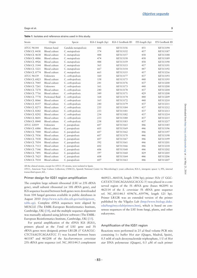
Objetivo segundo
- 83 -
Gago et al. 3
Table 1. Isolates and reference strains used in this study.
Strain Origin Specie IGS-1 length (bp) IGS-1 GenBank ID ITS length (bp) ITS GenBank ID
ATCC 96144 Human hand Candida metapsilosis 666 KF313156 451 KF313194
CNM CL 4438 Blood culture C. metapsilosis 574 KF313152 457 KF313187
CNM-CL 4638 Blood culture C. metapsilosis 488 KF313157 458 KF313188
CNM-CL 4886 Blood culture C. metapsilosis 474 KF313158 458 KF313189
CNM-CL 4926 Blood culture C. metapsilosis 488 KF313159 458 KF313190
CNM-CL 5144 Blood culture C. metapsilosis 565 KF313153 457 KF313191
CNM-CL 5221 Blood culture C. metapsilosis 667 KF313154 447 KF313192
CNM-CL 8774 Blood culture C. metapsilosis 665 KF313155 457 KF313216
ATCC 96139 Unknown C. orthopsilosis 160 KF313177 437 KF313193
CNM-CL 6823 Blood culture C. orthopsilosis 158 KF313175 440 KF313195
CNM-CL 7045 Blood culture C. orthopsilosis 241 KF313176 437 KF313200
CNM-CL 7261 Unknown C. orthopsilosis 161 KF313173 440 KF313200
CNM-CL 7270 Blood culture C. orthopsilosis 240 KF313178 437 KF313204
CNM-CL 7716 Blood culture C. orthopsilosis 349 KF313171 429 KF313208
CNM-CL 7778 Peritoneal fluid C. orthopsilosis 164 KF313174 440 KF313209
CNM-CL 8066 Blood culture C. orthopsilosis 165 KF313172 440 KF313210
CNM-CL 8157 Blood culture C. orthopsilosis 240 KF313179 437 KF313211
CNM-CL 8273 Blood culture C. orthopsilosis 235 KF313184 437 KF313212
CNM-CL 8282 Blood culture C. orthopsilosis 233 KF313181 437 KF313213
CNM-CL 8292 Blood culture C. orthopsilosis 234 KF313182 437 KF313214
CNM-CL 8601 Blood culture C. orthopsilosis 233 KF313180 437 KF313215
CNM-CL 8840 Blood culture C. orthopsilosis 238 KF313183 437 KF313217
ATCC 22019 Unknown C. parapsilosis 607 KF313165 446 KF313185
CNM-CL 6886 Blood culture C. parapsilosis 605 KF313169 446 KF313196
CNM-CL 7000 Blood culture C. parapsilosis 607 KF313162 446 KF313197
CNM-CL 7036 Blood culture C. parapsilosis 607 KF313170 446 KF313198
CNM-CL 7038 Blood culture C. parapsilosis 607 KF313167 446 KF313199
CNM-CL 7054 Blood culture C. parapsilosis 592 KF313161 446 KF313201
CNM-CL 7113 Blood culture C. parapsilosis 602 KF313166 446 KF313218
CNM-CL 7146 Blood culture C. parapsilosis 606 KF313168 446 KF313202
CNM-CL 7491 Blood culture C. parapsilosis 597 KF313160 429 KF313205
CNM-CL 7625 Blood culture C. parapsilosis 608 KF313164 446 KF313206
CNM-CL 7034 Blood culture C. parapsilosis 607 KF313163 446 KF313207
All the clinical strains, except for ATCC (T) strains, were isolated in Spain.
ATCC, American Type Culture Collection; CNM-CL, Spanish National Center for Microbiology’s yeast collection; IGS-1, intergenic spacer 1; ITS, internal
transcribed spacer; KF, .
Primer design for IGS1 region amplification
The complete large subunit ribosomal (LSU or 25S rRNA
gene), small subunit ribosomal (or 18S rRNA gene), and
IGS sequence located between both genes were downloaded
from 104 fungal genomes available in public databases in
August 2010 (http://www.ncbi.nlm.nih.gov/sutils/genom_
table.cgi). Complete rDNA sequences were aligned by
MUSCLE (The EMBL-European Bioinformatics Institute,
Cambridge, UK) [34], and the multiple sequence alignment
was manually adjusted using Jalview software (The EMBL-
European Bioinformatics Institute, Cambridge, UK) [35].
For partial amplification of the rDNA IGS (IGS1),
primers placed at the 3′end of LSU gene and 5S
rRNA genes were designed; primer LR12R (5′ GAACGC-
CTCTAAGTCAGAATCC 3′) was located between bases
461187 and 461208 of the Saccharomyces cerevisiae
25S rRNA gene sequence (ref. NC_001144.5 complement
460923...464318, length 3396 bp); primer 5S3r (5′ GGC-
CATATCTASCAGAAAGCACCG 3′) was placed in a con-
served region of the 5S rRNA gene (bases 462091 to
462114 of the S. cerevisiae 5S rRNA gene sequence
ref. NC_001144.5 459676...459796, length 121 bp).
Primer LR12R was an extended version of the primer
published by the Vilgalys Lab (http://www.biology.duke.
edu/fungi/mycolab/primers.htm), which is based on con-
sensus sequences of the LSU from fungi, plants, and other
eukaryotes.
Amplification of the IGS1 region
Reactions were performed in 25 µl final volume PCR mix
containing 1× buffer Hot star (Qiagen, Madrid, Spain),
0.5 mM of each deoxynucleotide triphosphate, 1 U of Hot
star DNA polymerase (Qiagen), 0.5 µM of each primer
at :: on
May
21
, 20
14
http
://mm
y.o
xfo
rdjo
urn
als.org
/D
ow
nlo
aded
from
4 Medical Mycology, 2014, Vol. 00, No. 0
(LR12R and 5S3r), and 50 ng of DNA. The PCR am-
plification and sequencing steps were the same as those
previously described for ITS sequence. For the amplicon
sequencing process, an extra-primer (25S1f: 5′ GTAGAG-
TAGCCTTGTTG 3′) was used.
Phylogenetic analyses
Phylogenetic analyses were conducted with MEGA soft-
ware, version 5.1 [36]. Multiple sequence alignments were
performed with MUSCLE software [34]. Phylogenetic in-
ferences were made using the maximum parsimony method
for ITS sequences or maximum likelihood method based
on the Tamura–Nei nucleotide substitution model for IGS1
sequences and ITS sequences (see Supplementary Material
for ITS analysis). A discrete gamma distribution was applied
to model evolutionary rate differences among nucleotides.
The bootstrap consensus tree inferred from 1000 replicates
was taken to represent the evolutionary history of the taxa
analyzed. Candida guilliermondii NCPG-3099 was used as
the outgroup in each phylogenetic tree. Pairwise similarity
was performed using InfoQuest FP 4.5 software (Biorad,
Madrid, Spain).
HRM protocol
A fragment of 70 bp regions in the IGS1 of C. para-
psilosis s.l. strains was amplified by RT-PCR followed
by HRM analysis. Primers psilIGS1f (5′ TGCGGGCGA-
GAGCTTGC 3′) and psilIGS1r (5′ CAATTTACCCG-
GCACTC 3′) were designed for this purpose. RT-PCR am-
plifications were carried out in the CFX 96 RT-PCR system
(Bio-Rad, Madrid, Spain). Reactions were performed in a
20-µL reaction mix that contained 1× Precision Melt su-
per mix (Bio-Rad), 200 nm of each primer, and 50 ng of
genomic DNA. The amplification protocol consisted of an
initial denaturation step for 2 min at 95oC followed by 45
cycles under the following conditions: denaturation (10 s
at 95oC), annealing (30 s at 56oC), and extension (30 s at
72oC). HRM curves were obtained by incubating the PCR
products for 30 s at 95oC and for 1 min at 60oC. Curves
were generated using Precision Melt analysis software
(Bio-Rad).
The following strains were used for specificity assays: C.
albicans (CNM-CL5719); C. glabrata (CNM-CL5533); C.
guilliermondii (CNM-CL7127); C. krusei (CNM-CL7057);
Coccidioides posadasii (CNM-CM2912); Paracoccidioides
brasiliensis (CNM-CL2908); Aspergillus fumigatus (CNM-
CM237); Fusarium solani (CNM-CM3035); Scedosporium
apiospermum (CNM-CM3169); Rhizopus oryzae (CNM-
CM3020); human DNA (Promega, Madrid, Spain); and
Lodderomyces elongisporus (CNM-CL8161), a species
closely related to C. parapsilosis complex.
Nucleotide sequence accession numbers
GenBank accession numbers for ITS sequences from all
strains used in this work were as follows:
ATCC22019:KF313185, NCPG3099:KF313186,
CNM-CL4438:KF313187, CNM-CL4638:KF313188,
CNM-CL4886:KF313189, CNM-CL4926:KF313190,
CNM-CL5144:KF313191, CNM-CL5221:KF313192,
ATCC96139:KF313193, ATCC96144:KF313194,
CNM-CL6823:KF313195, CNM-CL6886:KF313196,
CNM-CL7000:KF313197, CNM-CL7036:KF313198,
CNM-CL7038:KF313199, CNM-CL7045:KF313200,
CNM-CL7054:KF313201, CNM-CL7146:KF313202,
CNM-CL7261:KF313203, CNM-CL7270:KF313204,
CNM-CL7491:KF313205, CNM-CL7625:KF313206,
CNM-CL7034:KF313207, CNM-CL7716:KF313208,
CNM-CL7778:KF313209, CNM-CL8066:KF313210,
CNM-CL8157:KF313211, CNM-CL8273:KF313212,
CNM-CL8282:KF313213, CNM-CL8292:KF313214,
CNM-CL8601:KF313215, CNM-CL8774:KF313216,
CNM-CL8840:KF313217, CNM-CL7113:KF313218
GenBank accession numbers for IGS1 sequences from
all strains used in this work were as follows:
NCPG3099:KF313151, CNM-CL4438:KF313152,
CNM-CL5144:KF313153, CNM-CL5221:KF313154,
CNM-CL8774:KF313155, ATCC96144:KF313156,
CNM-CL4638:KF313157, CNM-CL4886:KF313158,
CNM-CL4926:KF313159, CNM-CL7491:KF313160,
CNM-CL7054:KF313161, CNM-CL7000:KF313162,
CNM-CL7034:KF313163, CNM-CL7625:KF313164,
ATCC22019:KF313165, CNM-CL7113:KF313166,
CNM-CL7038:KF313167, CNM-CL7146:KF313168,
CNM-CL6886:KF313169, CNM-CL7036:KF313170,
CNM-CL7716:KF313171, CNM-CL8066:KF313172,
CNM-CL7261:KF313173, CNM-CL7778:KF313174,
CNM-CL6823:KF313175, CNM-CL7045:KF313176,
ATCC96139:KF313177, CNM-CL7270:KF313178,
CNM-CL8157:KF313179, CNM-CL8601:KF313180,
CNM-CL8282:KF313181, CNM-CL8292:KF313182,
CNM-CL8840:KF313183, CNM-CL8273:KF313184.
Results
Species identification of C. parapsilosis complex
Thirty C. parapsilosis s.l. strains were identified as C.
parapsilosis species complex based on morphological char-
acteristics. The cryptic species, that is, C. parapsilosis,
C. orthopsilosis, and C. metapsilosis were differentiated
by their similarity with the ITS nucleotide sequences of the
type strains.
Using the maximum parsimony method, phylogenetic
analysis of the ITS region provided a well-supported
at :: on
May
21
, 20
14
http
://mm
y.o
xfo
rdjo
urn
als.org
/D
ow
nlo
aded
from

Objetivo segundo
- 85 -
Gago et al. 5
Figure 1. Candida parapsilosis sensu lato species based on parsimony analysis of the internal transcribed spacer region from the rDNA. The
phylogenetic tree was computed using MEGA, version 5.0 (1000 bootstrap repetitions).
monophyletic species clade for C. parapsilosis spp. (boot-
strap value 94%) but not for C. orthopsilosis or C. metap-
silosis. This indicates weak statistical support for these
species (Fig. 1), with pairwise sequence similarity percent-
ages for ITS of 98.16% (C. parapsilosis vs. C. orthopsilosis),
95.90% (C. parapsilosis vs. C. metapsilosis), and 95.64%
(C. orthopsilosis vs. C. metapsilosis). Nucleotide sequences
and length of the ITS region were similar for all isolates
of each species (Table 1). The percentages of intraspecies
identity based on the ITS sequences were 98.97% for
C. metapsilosis, 99.11% for C. orthopsilosis, and 98.73%
for C. parapsilosis. Phylogenetic analysis of the ITS region
at :: on
May
21
, 20
14
http
://mm
y.o
xfo
rdjo
urn
als.org
/D
ow
nlo
aded
from
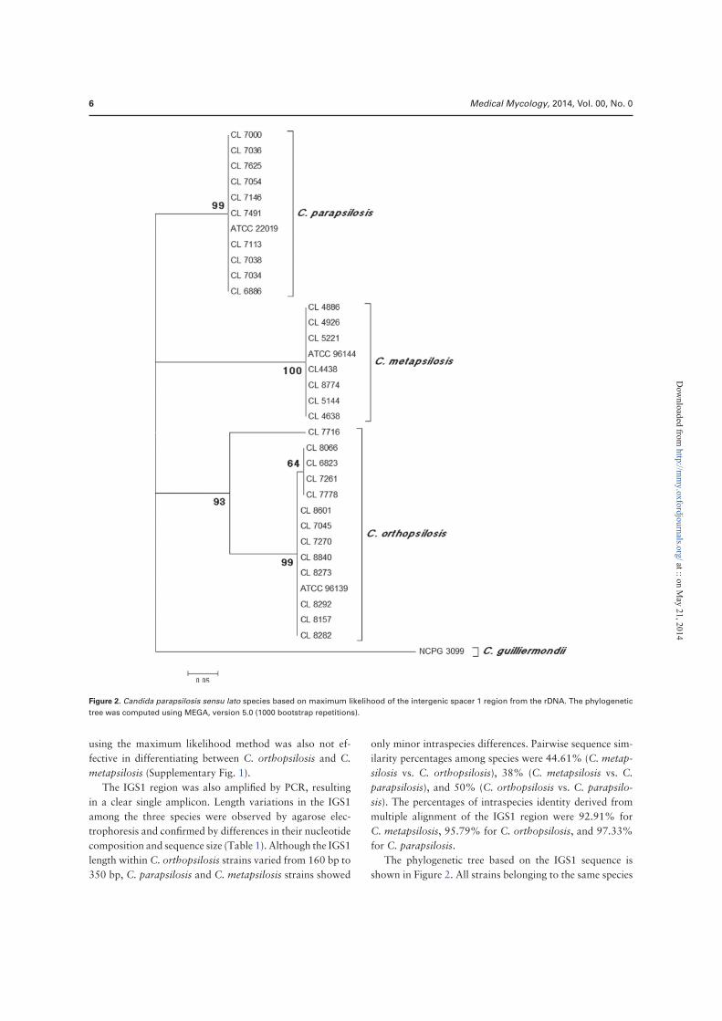
6 Medical Mycology, 2014, Vol. 00, No. 0
Figure 2. Candida parapsilosis sensu lato species based on maximum likelihood of the intergenic spacer 1 region from the rDNA. The phylogenetic
tree was computed using MEGA, version 5.0 (1000 bootstrap repetitions).
using the maximum likelihood method was also not ef-
fective in differentiating between C. orthopsilosis and C.
metapsilosis (Supplementary Fig. 1).
The IGS1 region was also amplified by PCR, resulting
in a clear single amplicon. Length variations in the IGS1
among the three species were observed by agarose elec-
trophoresis and confirmed by differences in their nucleotide
composition and sequence size (Table 1). Although the IGS1
length within C. orthopsilosis strains varied from 160 bp to
350 bp, C. parapsilosis and C. metapsilosis strains showed
only minor intraspecies differences. Pairwise sequence sim-
ilarity percentages among species were 44.61% (C. metap-
silosis vs. C. orthopsilosis), 38% (C. metapsilosis vs. C.
parapsilosis), and 50% (C. orthopsilosis vs. C. parapsilo-
sis). The percentages of intraspecies identity derived from
multiple alignment of the IGS1 region were 92.91% for
C. metapsilosis, 95.79% for C. orthopsilosis, and 97.33%
for C. parapsilosis.
The phylogenetic tree based on the IGS1 sequence is
shown in Figure 2. All strains belonging to the same species
at :: on
May
21
, 20
14
http
://mm
y.o
xfo
rdjo
urn
als.org
/D
ow
nlo
aded
from
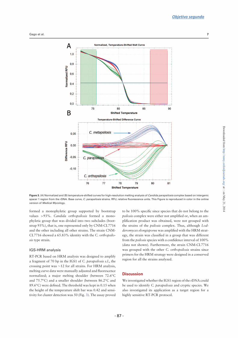
Objetivo segundo
- 87 -
Gago et al. 7
Figure 3. (A) Normalized and (B) temperature-shifted curves for high-resolution melting analysis of Candida parapsilosis complex based on intergenic
spacer 1 region from the rDNA. Base curve, C. parapsilosis strains. RFU, relative fluorescence units. This Figure is reproduced in color in the online
version of Medical Mycology.
formed a monophyletic group supported by bootstrap
values >93%. Candida orthopsilosis formed a mono-
phyletic group that was divided into two subclades (boot-
strap 93%), that is, one represented only by CNM-CL7716
and the other including all other strains. The strain CNM-
CL7716 showed a 65.83% identity with the C. orthopsilo-
sis type strain.
IGS-HRM analysis
RT-PCR based on HRM analysis was designed to amplify
a fragment of 70 bp in the IGS1 of C. parapsilosis s.l., the
crossing point was ∼12 for all strains. For HRM analysis,
melting curve data were manually adjusted and fluorescence
normalized; a major melting shoulder (between 72.6oC
and 75.7oC) and a smaller shoulder (between 86.2oC and
89.6oC) were defined. The threshold was kept in 0.15 when
the height of the temperature shift bar was 0.42 and sensi-
tivity for cluster detection was 50 (Fig. 3). The assay proved
to be 100% specific since species that do not belong to the
psilosis complex were either not amplified or, when an am-
plification product was obtained, were not grouped with
the strains of the psilosis complex. Thus, although Lod-
deromyces elongisporus was amplified with the HRM strat-
egy, the strain was classified in a group that was different
from the psilosis species with a confidence interval of 100%
(data not shown). Furthermore, the strain CNM-CL7716
was grouped with the other C. orthopsilosis strains since
primers for the HRM strategy were designed in a conserved
region for all the strains analyzed.
Discussion
We investigated whether the IGS1 region of the rDNA could
be used to identify C. parapsilosis and cryptic species. We
also investigated its application as a target region for a
highly sensitive RT-PCR protocol.
at :: on
May
21
, 20
14
http
://mm
y.o
xfo
rdjo
urn
als.org
/D
ow
nlo
aded
from
8 Medical Mycology, 2014, Vol. 00, No. 0
Candida parapsilosis sensu stricto is the second or third
most common cause of candidemia worldwide [9,37,38].
Although new data suggest C. orthopsilosis is the fifth most
common cause of candidemia in Spain, not C. krusei [39],
other studies have shown the incidence of C. parapsilosis
cryptic species to be lower, which could be related to cli-
matic and socioeconomic conditions [12,13].
Although molecular identification of C. parapsilosis s.l.
has been performed using the ITS region of the rDNA, our
study results show that phylogenetic analysis inferred from
this region is not as well supported as when the IGS region is
used. As with the ITS regions, the IGS1 region belongs to the
rDNA region that consists of hundreds of copies repeated
in tandem. The IGS sequence has been used for a variety
of intra- and interspecies fungal population studies, includ-
ing studies of S. cerevisiae, Cryptococcus neoformans, and
Trichosporon spp. [24,25,27].
Very low intraspecimen variation in the IGS1 among
paralogous copies for each strain was determined by Sanger
sequencing of the three species. This fact has also been ob-
served in C. guilliermondii [unpublished data, Cuesta I]
and in Penicillium marneffei [28]. A concerted evolution
model could explain these results, where homogenization is
faster and very efficient and differences among the repeat
units can finally disappear. This model of evolution has
also been proposed for the rDNA cassette in other fungal
species such as Ashbya gossypii, S. paradoxus, S. cerevisiae,
A. nidulans, and C. neoformans [40,41]. Moreover, the
IGS1 sequence analysis reveals that the level of interspecies
sequence dissimilarity is higher in this region than in the
ITS region, although we differentiated some conserved mo-
tif such as transcription stop site for RNA polymerase III
[42]. Slight differences in the IGS1 length for C. parapsilo-
sis and C. metapsilosis observed in this study could be due
to polymerase slippage in polyAs stretch, while length dis-
crepancies in this region for C. orthopsilosis could be due
to an ongoing evolutionary process. This last hypothesis is
supported by others who have described C. orthopsilosis
as the most heterogeneous of the psilosis species [16,43],
which could be related to sexual and parasexual reproduc-
tion [44,45].
The discriminatory power of the IGS region to define
C. parapsilosis species by PCR fingerprinting methods has
been reported [17], even though the sequence of the IGS1
region has not been previously described. The results of our
study have shown that although C. orthopsilosis is a well-
supported group by IGS1 analysis, a subgroup that could be
related to the existence of a fourth psilosis group member
can also be defined. Sai et al. [45] observed that most of the
C. orthopsilosis strains fall into two genetically distinguish-
able groups based on ITS sequence and mating type locus
analysis. Recently, findings on the C. orthopsilosis genome
[46] and the sequence analysis of the ITS, mtDNA, and
mtDNA restriction enzyme profiles [47] also suggest the
existence of two C. orthopsilosis subtypes. However, other
more diverse genes (eg, inteins) [48] and C. orthopsilosis
strains should be analyzed to determine if the genetic differ-
ences are great enough to support branding a new species.
Candida parapsilosis complex identification based on
the IGS1 sequence is problematic due to polyAs stretch,
which makes it difficult to obtain sequences and requires
development of other approaches. In this study, we pro-
pose a faster and cheaper real-time protocol followed by
HRM analysis for C. parapsilosis spp. complex identifi-
cation based on differences in the IGS1 sequence. Strains
were plotted with a confidence interval >98% according
to differences in length and sequence in the target region.
Concordance between the IGS1 sequence and the HRM
analysis was 100%. Another RT-PCR method for differen-
tiating C. parapsilosis, C. orthopsilosis, and C. metapsilosis
was described by Garcia- Effron et al. [49]; they used a mul-
tiplex RT-PCR with molecular beacons aimed at the FKS1
gene, with high specificity and sensitivity. However, this
procedure is more expensive compared with HRM. More-
over, the SADH gene has also been successfully used for
an RT-PCR analysis based on conventional melting curves,
although the differences in the melting curves are not as
well defined as with HRM approaches [18]. Other proto-
cols based on Taqman probes [50] are more cost effective
than HRM analysis. Finally, matrix-assisted laser desorp-
tion/ionization time-of-flight technology described for iden-
tifying [51] and genotyping [52] C. parapsilosis spp. com-
plex showed low discriminatory power for genotyping and
an inability to detect mixed infections.
In conclusion, the IGS1 sequence seems to be a more
suitable molecular marker for identifying C. parapsilosis
s.l. species than the ITS region. HRM analysis based on
the IGS1 sequence provides a rapid, less expensive, and
easier RT-PCR protocol for identifying psilosis complex at
a species level in a clinical laboratory.
Acknowledgments
We thank Frank Hodgkins for reading and editing the manuscript.
This work was supported as part of a research project of the
Spanish Ministry of Science and Innovation (MPY1461/11). S. G.
is supported by a research fellowship from the Fondo de Investi-
gaciones Biomedicas, Spanish Ministry of Science and Innovation
(FI10/00464). A. A. I. received a research contract from the Spanish
Network for Research on Infectious Diseases (REIPI RD06/0008),
supported by Plan Nacional de I+D+i 2008–2011 and Instituto de
Salud Carlos III, Subdireccion General de Redes y Centros de In-
vestigacion Cooperativa, Ministerio de Economıa y Competitividad,
and Spanish Network for Research on Infectious Diseases (REIPI
RD12/0015) and cofinanced by the European Development Regional
Fund, A Way to Achieve Europe, ERDF.
at :: on
May
21
, 20
14
http
://mm
y.o
xfo
rdjo
urn
als.org
/D
ow
nlo
aded
from

Objetivo segundo
- 89 -
Gago et al. 9
Declaration of interest
J. L. R.-T. has received grant support from Astellas Pharma, Gilead
Sciences, MSD, Pffizer, ScheringPlough, Soria Melguizo S.A., the
European Union, Spanish Agency for International Cooperation,
Spanish Ministry of Culture and Education, Spanish Health Re-
search Fund, Instituto de Salud Carlos III, Ramon Areces Foun-
dation, and Mutua Madrilena Foundation. He has been an advi-
sor/consultant to the Pan American Health Organization, Gilead
Sciences, MSD, Mycognostica, Pffizer, and ScheringPlough. He has
been paid for talks on behalf of Gilead Sciences, MSD, Pffizer, and
Schering-Plough. M. C.-E. has received grant support from Astellas
Pharma, bioMerieux, Gilead Sciences, MSD, Pffizer, ScheringPlough,
Soria Melguizo S.A., Ferrer International, the European Union, the
ALBAN Program (Becas de Alto Nivel para America Latina), Spanish
Agency for International Cooperation, Spanish Ministry of Culture
and Education, Spanish Health Research Fund, Instituto de Salud
Carlos III, Ramon Areces Foundation, and Mutua Madrilena Foun-
dation. He has been an advisor/consultant to the Pan American
Health Organization, Gilead Sciences, MSD, Pffizer, Astellas, and
Schering-Plough. He has been paid for talks on behalf of Gilead Sci-
ences, MSD, Pffizer, Astellas, and Schering-Plough. All other authors
report no conflicts of interest. The authors alone are responsible for
the content and the writing of the paper.
Supporting information
Supplementary material is available at Medical Mycology online
(http://www.mmy.oxfordjournals.org/).
References
1. Clerihew L, Lamagni TL, Brocklehurst P, McGuire W. Candida
parapsilosis infection in very low birthweight infants. Arch Dis
Child Fetal Neonatal Ed 2007; 92: F127–F129.
2. Horasan E, Ersoz G, Kurt A, Karacorlu S, Kaya A. Increase in
Candida parapsilosis fungemia in critical care units: a 6-years
study. Mycopathologia 2012; 170: 263–268.
3. Munoz P, Burillo A, Bouza E. Environmental surveillance and
other control measures in the prevention of nosocomial fungal
infections. Clin Microbiol Infect 2001; 7: 38–45.
4. Branchini ML, Pfaller MA, Rhine-Chalberg J, Frempong T,
Isenberg HD. Genotypic variation and slime production among
blood and catheter isolates of Candida parapsilosis. J Clin Mi-
crobiol 1994; 32: 452–456.
5. Lin D, Wu LC, Rinaldi MG, Lehmann PF. Three distinct geno-
types within Candida parapsilosis from clinical sources. J Clin
Microbiol 1995; 33: 1815–1821.
6. Tavanti A, Davidson AD, Gow NA, Maiden MC, Odds FC.
Candida orthopsilosis and Candida metapsilosis spp. nov. to
replace Candida parapsilosis groups II and III. J Clin Microbiol
2005; 43: 284–292.
7. Gomez-Lopez A, Alastruey-Izquierdo A, Rodriguez D et al.
Prevalence and susceptibility profile of Candida metapsilosis and
Candida orthopsilosis: results from population-based surveil-
lance of candidemia in Spain. Antimicrob Agents Chemother
2008; 52: 1506–1509.
8. Lockhart SR, Iqbal N, Cleveland AA et al. Species identifica-
tion and antifungal susceptibility testing of Candida bloodstream
isolates from population-based surveillance studies in two U.S.
cities from 2008 to 2011. J Clin Microbiol 2012; 50: 3435–3442.
9. Lockhart SR, Messer SA, Pfaller MA, Diekema DJ. Geographic
distribution and antifungal susceptibility of the newly described
species Candida orthopsilosis and Candida metapsilosis in com-
parison to the closely related species Candida parapsilosis. J Clin
Microbiol 2008; 46: 2659–2664.
10. Silva AP, Miranda IM, Lisboa C, Pina-Vaz C, Rodrigues AG.
Prevalence, distribution, and antifungal susceptibility profiles of
Candida parapsilosis, C. orthopsilosis, and C. metapsilosis in a
tertiary care hospital. J Clin Microbiol 2009; 47: 2392–2397.
11. van Asbeck EC, Clemons KV, Stevens DA. Candida parapsilosis:
a review of its epidemiology, pathogenesis, clinical aspects, typ-
ing and antimicrobial susceptibility. Crit Rev Microbiol 2009;
35: 283–309.
12. Canton E, Espinel-Ingroff A, Peman J, del Castillo L. In vitro
fungicidal activities of echinocandins against Candida metapsilo-
sis, C. orthopsilosis, and C. parapsilosis evaluated by time-kill
studies. Antimicrob Agents Chemother 2010; 54: 2194–2197.
13. Canton E, Peman J, Quindos G et al. Prospective multicenter
study of the epidemiology, molecular identification, and anti-
fungal susceptibility of Candida parapsilosis, Candida orthop-
silosis, and Candida metapsilosis isolated from patients with can-
didemia. Antimicrob Agents Chemother 2011; 55: 5590–5596.
14. Szilagyi J, Foldi R, Gestzelyi R et al. Comparison of the kid-
ney fungal burden in experimental disseminated candidiasis by
species of the Candida parapsilosis complex treated with flu-
conazole, amphotericin B and caspofungin in a temporarily neu-
tropenic murine model. Chemotherapy 2012; 58: 159–164.
15. Spreghini E, Orlando F, Tavanti A et al. In vitro and in vivo ef-
fects of echinocandins against Candida parapsilosis sensu stricto,
Candida orthopsilosis and Candida metapsilosis. J Antimicrob
Chemother 2012; 67: 2195–2202.
16. Asadzadeh M, Ahmad S, Al-Sweih N, Khan ZU. Rapid molecu-
lar differentiation and genotypic heterogeneity among Candida
parapsilosis and Candida orthopsilosis strains isolated from clin-
ical specimens in Kuwait. J Med Microbiol 2009; 58: 745–752.
17. Cornet M, Sendid B, Fradin C et al. Molecular identification of
closely related Candida species using two ribosomal intergenic
spacer fingerprinting methods. J Mol Diagn 2011; 13: 12–22.
18. Hays C, Duhamel C, Cattoir V, Bonhomme J. Rapid and accu-
rate identification of species belonging to the Candida parapsilo-
sis complex by real-time PCR and melting curve analysis. J Med
Microbiol 2011; 60: 477–480.
19. Iida S, Imai T, Oguri T et al. Genetic diversity of the internal
transcribed spacers (ITS) and 5.8S rRNA genes among the clin-
ical isolates of Candida parapsilosis in Brazil and Japan. Nihon
Ishinkin Gakkai Zasshi 2005; 46: 133–137.
20. Mirhendi H, Bruun B, Schonheyder HC et al. Molecular screen-
ing for Candida orthopsilosis and Candida metapsilosis among
Danish Candida parapsilosis group blood culture isolates: pro-
posal of a new RFLP profile for differentiation. J Med Microbiol
2010; 59: 414–420.
21. Tavanti A, Hensgens LA, Mogavero S et al. Genotypic and phe-
notypic properties of Candida parapsilosis sensu strictu strains
isolated from different geographic regions and body sites. BMC
Microbiol 2010; 10: 203.
22. Van Asbeck EC, Clemons KV, Markham AN, Stevens DA, Can-
dida parapsilosis Global Epidemiology Group. Molecular epi-
demiology of the global and temporal diversity of Candida para-
psilosis. Scand J Infect Dis 2008; 40: 827–834.
at :: on
May
21
, 20
14
http
://mm
y.o
xfo
rdjo
urn
als.org
/D
ow
nlo
aded
from
10 Medical Mycology, 2014, Vol. 00, No. 0
23. Schoch CL, Seifert KA, Huhndorf S et al. Nuclear ribosomal
internal transcribed spacer (ITS) region as a universal DNA bar-
code marker for Fungi. Proc Natl Acad Sci U S A 2012; 109:
6241–6246.
24. Sugita T, Nakajima M, Ikeda R, Matsushima T, Shinoda T.
Sequence analysis of the ribosomal DNA intergenic spacer 1 re-
gions of Trichosporon species. J Clin Microbiol 2002; 40: 1826–
1830.
25. Sugita T, Takeo K, Hama K et al. DNA sequence diversity of
intergenic spacer I region in the non-lipid-dependent species
Malassezia pachydermatis isolated from animals. Med Mycol
2005; 43: 21–26.
26. Hillis M, Dixon M. Ribosomal DNA: molecular evolution and
phylogenetic inference. Q Rev Biol 1991; 66: 411–453.
27. Diaz MR, Boekhout T, Kiesling T, Fell JW. Comparative anal-
ysis of the intergenic spacer regions and population structure of
the species complex of the pathogenic yeast Cryptococcus neo-
formans. FEMS Yeast Res 2005; 5: 1129–1140.
28. Mekha N, Sugita T, Makimura K et al. The intergenic spacer re-
gion of the ribosomal RNA gene of Penicillium marneffei shows
almost no DNA sequence diversity. Microbiol Immunol 2010;
54: 714–716.
29. Montgomery JL, Sanford LN, Wittwer CT. High-resolution
DNA melting analysis in clinical research and diagnostics. Ex-
pert Rev Mol Diagn 2010; 10: 219–240.
30. Arancia S, Sandini S, de Bernardis F, Fortini D. Rapid, sim-
ple, and low-cost identification of Candida species using high-
resolution melting analysis. Diagn Microbiol Infect Dis 2011;
69: 283–285.
31. Gago S, Zaragoza O, Cuesta I et al. High-resolution melt-
ing analysis for identification of the Cryptococcus neoformans-
Cryptococcus gattii complex. J Clin Microbiol 2011; 49: 3663–
3666.
32. Somogyvari F, Horvath A, Serly J et al. Detection of invasive
fungal pathogens by real-time PCR and high-resolution melting
analysis. In Vivo 2012; 26: 979–983.
33. Tang CM, Cohen J, Krausz T, Van NS, Holden DW. The alkaline
protease of Aspergillus fumigatus is not a virulence determinant
in two murine models of invasive pulmonary aspergillosis. Infect
Immun 1993; 61: 1650–1656.
34. Edgar RC. MUSCLE: a multiple sequence alignment method
with reduced time and space complexity. BMC Bioinformatics
2004; 5: 113.
35. Waterhouse AM, Procter JB, Martin DM, Clamp M, Barton GJ.
Jalview Version 2—a multiple sequence alignment editor and
analysis workbench. Bioinformatics 2009; 25: 1189–1191.
36. Tamura K, Peterson D, Peterson N et al. MEGA5: molecular
evolutionary genetics analysis using maximum likelihood, evo-
lutionary distance, and maximum parsimony methods. Mol Biol
Evol 2011; 28: 2731–2739.
37. Almirante B, Rodriguez D, Cuenca-Estrella M et al. Epidemiol-
ogy, risk factors, and prognosis of Candida parapsilosis blood-
stream infections: case-control population-based surveillance
study of patients in Barcelona, Spain, from 2002 to 2003. J
Clin Microbiol 2006; 44: 1681–1685.
38. Pfaller M, Neofytos D, Diekema D et al. Epidemiology and out-
comes of candidemia in 3648 patients: data from the Prospective
Antifungal Therapy (PATH Alliance R©) registry, 2004–2008. Di-
agn Microbiol Infect Dis 2012; 74: 323–331.
39. Peman J, Canton E, Quindos G et al. Epidemiology, species dis-
tribution and in vitro antifungal susceptibility of fungaemia in a
Spanish multicentre prospective survey. J Antimicrob Chemother
2012; 67: 1181–1187.
40. Ganley AR, Hayashi K, Horiuchi T, Kobayashi T. Identifying
gene-independent noncoding functional elements in the yeast
ribosomal DNA by phylogenetic footprinting. Proc Natl Acad
Sci U S A 2005; 102: 11787–11792.
41. Ganley AR, Kobayashi T. Highly efficient concerted evolution
in the ribosomal DNA repeats: total rDNA repeat variation re-
vealed by whole-genome shotgun sequence data. Genome Res
2007; 17: 184–191.
42. Bartholomew B, Durkovich D, Kassavetis GA, Geiduschek EP.
Orientation and topography of RNA polymerase-III in transcrip-
tion complexes. Mol Cel Biol 1993; 13: 942–952.
43. van Asbeck E, Clemons K, Markham A, Stevens DA. Corre-
lation of restriction fragment length polymorphism genotyping
with internal transcribed spacer sequence, randomly amplified
polymorphic DNA and multilocus sequence groupings for Can-
dida parapsilosis. Mycoses 2009; 56: 493–498.
44. Hensgens L, Tavanti A, Mogavero S, Ghelardi E, Senesi S. AFLP
genotyping of Candida metapsilosis clinical isolates: evidence for
recombination. Fungal Genet Biol 2009; 46: 750–758.
45. Sai S, Holland LM, McGee CF, Lynch DB, Butler G. Evolution of
mating within the Candida parapsilosis species group. Eukaryot
Cell 2011; 10: 578–587.
46. Riccombeni A, Vidanes G, Proux-Wera E, Wolfe KH, Butler G.
Sequence and analysis of the genome of the pathogenic yeast
Candida orthopsilosis. PLoS One 2012; 7: e35750.
47. Valach M, Pryszcz L, Tomaska L et al. Mitochondrial genome
variability within the Candida parapsilosis species complex. Mi-
tochondrion 2012; 12: 514–519.
48. Prandini TH, Theodoro RC, Bruder-Nascimento AC, Scheel
CM, Bagagli E. Analysis of inteins in the Candida parapsilo-
sis complex for simple and accurate species identification. J Clin
Microbiol 2013; 51: 2830–2836.
49. Garcia-Effron G, Canton E, Peman J et al. Assessment of two
new molecular methods for identification of Candida parapsilo-
sis sensu lato species. J Clin Microbiol 2011; 49: 3257–3261.
50. Souza AC, Ferreira RC, Goncalves SS et al. Accurate identifica-
tion of Candida parapsilosis (sensu lato) by use of mitochondrial
DNA and real-time PCR. J Clin Microbiol 2012; 50: 2310–2314.
51. Quiles-Melero I, Garcia-Rodriguez J, Gomez-Lopez A, Mingo-
rance J. Evaluation of matrix-assisted laser desorption/ionisation
time-of-flight (MALDI-TOF) mass spectrometry for identifica-
tion of Candida parapsilosis, C. orthopsilosis and C. metapsilo-
sis. Eur J Clin Microbiol 2012; 31: 67–71.
52. Pulcrano G, Roscetto E, Iula V et al. MALDI-TOF mass spec-
trometry and microsatellite markers to evaluate Candida para-
psilosis transmission in neonatal intensive care units. Eur J Clin
Microbiol 2012; 31: 2919–2928.
at :: on
May
21
, 20
14
http
://mm
y.o
xfo
rdjo
urn
als.org
/D
ow
nlo
aded
from
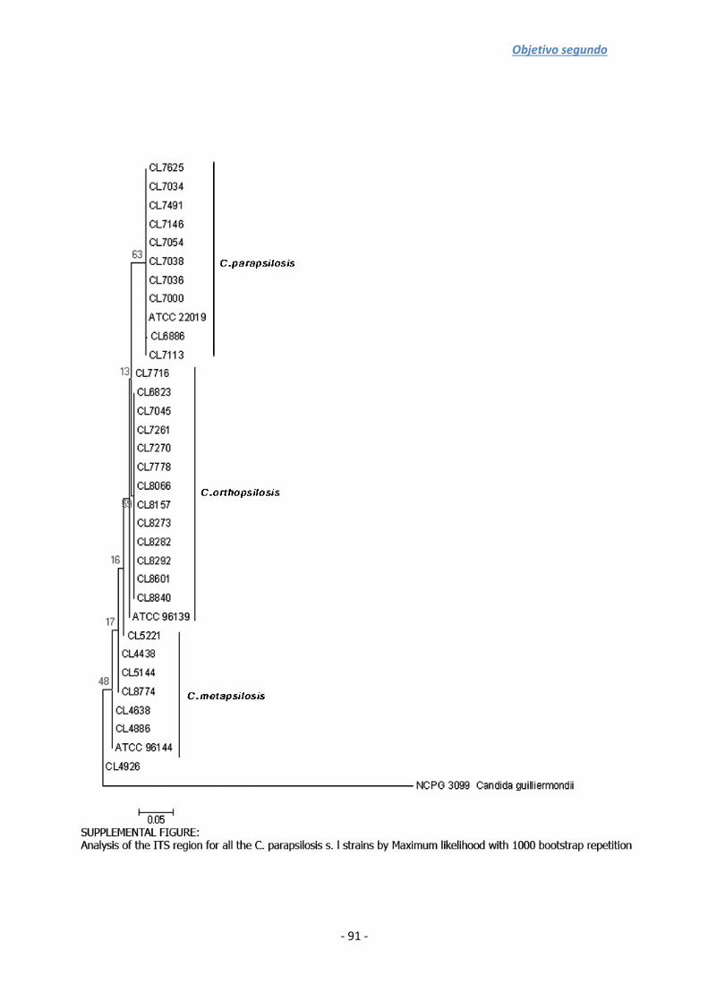
Objetivo segundo
- 91 -


Objetivo segundo
- 93 -
4.3. Tipado molecular en Candida albicans mediante
análisis HRM
Las infecciones recurrentes debidas a Candida albicans son frecuentes en
procesos de vaginitis o candidosis orofaríngea. Además, en la mayoría de los casos
todos los episodios se deben a cepas clonales que sufren procesos de microevolución.
Una de las técnicas de tipificación más empleadas en la actualidad es el análisis de
microsatélites mediante electroforesis capilar. Aunque la técnica presenta gran poder
de discriminación y reproducibilidad, el coste por determinación es elevado y se
requiere de un equipamiento complejo. Por ello, se evaluó la utilidad de las técnicas de
HRM para caracterizar tres regiones de microsatélites en nueve aislamientos de
C. albicans de un paciente con candiduria recurrente. Se seleccionaron tres
marcadores previamente descritos (HIS 3, EF 3 y CDC 3) y se analizaron mediante
electroforesis capilar y HRM. En el análisis de microsatélites mediante electroforesis
capilar se incluyeron 20 cepas no relacionadas geográficamente como población
control. Los electrogramas se resolvieron con el programa GenMapper 4.0. Para el
análisis de HRM, como población control se seleccionaron 7 cepas que habían
presentado perfiles diferentes mediante la metodología de electroforesis capilar. El
poder de discriminación de los tres marcadores mediante electroforesis capilar fue de
0,92 siendo el marcador CDC 3 el que presentaba menor capacidad de discriminación.
Las cepas del episodio de candiduria recurrente presentaron el mismo perfil y
diferente al de la población control para los tres marcadores, de modo que para HIS 3 y
CDC 3 eran homocigotas y heterocigotos para el marcador EF 3. En el análisis de HRM
en todos los casos, salvo la cepa CNM-CL 7020 con el marcador CDC 3, las cepas
procedentes del paciente se agruparon juntas a pesar de que el poder de
discriminación de los tres marcadores fue menor (0,77). Mediante secuenciación se
pudo comprobar que la cepa CNM-CL 7020 presentaba una inserción que no era capaz
de discriminar la electroforesis capilar. Según ambos análisis, las cepas que causaron
los episodios de candiduria recurrente están relacionadas genéticamente, por tanto el
análisis de microsatélites mediante HRM es una técnica rápida y sencilla para
establecer relaciones entre cepas de C. albicans.


Objetivo segundo
- 95 -
RESEARCH ARTICLE Open Access
Analysis of strain relatedness using HighResolution Melting in a case of recurrentcandiduriaSara Gago1, Belen Lorenzo2, Alicia Gomez-Lopez1, Isabel Cuesta3, Manuel Cuenca-Estrella1 and Maria J Buitrago1*
Abstract
Background: Several genotyping protocols have been described to study Candida albicans strains with different
sensitivity values. In this study we have analyzed the genetic relatedness and the antifungal susceptibility of several
Candida albicans strains isolated from a patient who from suffered recurrent candiduria for a period of five years.
Strains were genotyped using Microsatellite Length Polymorphism (MLP) with three microsatellite markers (HIS 3,
EF 3 and CDC 3), and a new method based on high resolution melting (HRM) was developed to analyze the
microsatellite region. This method was compared with the conventional technique that uses capillary
electrophoresis.
Results: MICs of the isolates showed the existence of fluconazole susceptible and resistant strains. An inter-colony
test using single concentration (8 and 16 mg/l) of fluconazole revealed the coexistence of both fluconazole
susceptible and resistant strains. Both genotyping analysis methods showed that all the patient’s isolates had a
clonal origin. HRM analysis method developed was able to accurately establish strain relatedness and presented a
discriminatory power of 0.77.
Conclusions: Although HRM analysis method presented a lower discriminatory power compared to methods
based on capillary electrophoresis, it provided a more cost-effective and suitable alternative for genotyping C.
albicans in a clinical laboratory.
Keywords: Candiduria, High resolution melting, Genotyping
BackgroundCandida albicans is a ubiquitous commensal in healthy
individuals; it is, however, a very important opportunistic
pathogen for immunologically weak and immuno-
compromised people [1]. Recurrent and/or persistent
infections by Candida species are frequent, particularly
in oropharyngeal and vaginal candidiasis, although it has
also been described in urinary tract infections [2]. Stud-
ies describing recurrent infections have focused on de-
termining the relatedness between colonizing and
infective strains [3,4], as well as between successive in-
fective strains [5-9]. It seems clear now that the majority
of commensal and infecting populations of C. albicans
from the same individuals are clonal in origin but subse-
quently undergo microevolution at the site of colonization
and through recurrent episodes of infection [5,10,11]. The
microevolution of the strains is a frequent process in re-
current infections and it takes place in response to adap-
tive changes [9,12]. A recent work which examined the
“in vitro” dynamics of C. albicans populations in the pres-
ence or absence of fluconazole has shown that mutations
that lead to increased drug resistance appear frequently
[13]. Others authors suggest that natural C. albicans
populations comprise a mixture of closely related strain
types [6].
Typing methods have been described as useful tools for the
differentiation between strains isolated only once and those
able to cause recurrent infections. Although several typing
methods have been described for C. albicans (AFLP, RFLP-
PCR or MLST), one of the most suitable is the fragment
length analysis of microsatellites called Microsatellite Length
* Correspondence: [email protected] de Micología, Centro Nacional de Microbiología, Instituto de Salud
Carlos III, Ctra Majadahonda-Pozuelo, Km 2, Majadahonda, Madrid 28220,
Spain
Full list of author information is available at the end of the article
© 2013 Gago et al.; licensee BioMed Central Ltd. This is an Open Access article distributed under the terms of the CreativeCommons Attribution License (http://creativecommons.org/licenses/by/2.0), which permits unrestricted use, distribution, andreproduction in any medium, provided the original work is properly cited.
Gago et al. BMC Microbiology 2013, 13:13
http://www.biomedcentral.com/1471-2180/13/13

Objetivo segundo
- 96 -
Polymorphism (MLP). This technique has a high discrimin-
atory power and reproducibility. MLP analysis has proved its
efficacy and reproducibility in a large number of epidemio-
logical studies [9,14-19]; however, this technique is not easy to
use and the estimated cost per isolate remains high.
The High Resolution Melting (HRM) provides a faster and
cheaper method for microsatellite fragment analysis. This
technique uses fluorescent DNA binding dyes with improved
saturation properties allowing a precise assessment of se-
quence variation based on DNA melting curves analysis
[20,21]. The suitability of HRM to discriminate PCR products
based on one nucleotide change has also been described.
Some recent articles, focusing on the capacity of HRM to
identify and genotype fungi, have been reported [15,22].
In this work, we developed a method based on HRM
to assess the relatedness of strains in a clinical case of
recurrent candiduria. The results were compared with
the conventional MLP genotyping techniques. The iso-
lates, recovered over a period of five years, additionally
showed significant differences in their susceptibility to
antifungal agents. Antifungal susceptibility test and se-
lection of resistant population was performed.
MethodsOrigin of the strains and clinical data from the patient
The strains were isolated from a 62 year old male with
medullary sponge right kidney (Carchi-Ricci disease) and
recurrent reno-urethral lithiasis subjected to several litho-
tripsies. The patient was admitted in a Tertiary General
Hospital (Hospital Virgen de la Concha, Zamora, Spain)
diagnosed with right pyelonephritis caused by obstructive
kidney stones. C. albicans was isolated in blood cultures
and urocultures. The antifungal susceptibility profile
showed that azoles and amphotericin B were active in vitro
against this strain (CNM-CL-4929, Yeast Collection of the
Spanish National Centre for Microbiology) as shown in
Table 1.
Treatment with ciprofloxacin 400 mg/12 h and flucona-
zole iv 200 mg/12 h was started. After three days of treat-
ment, as fever persisted and blood and urine cultures
remained positive, fluconazole was replaced by amphoteri-
cin B lipid complex 200 mg/24 h iv and 100 mg every other
day. Six days after admission, lithotripsy was performed
and a double J stent was placed. He was discharged from
hospital a month after admission. From 2003 to 2008, the
patient suffered from several episodes of Candida infection
and underwent multiple lithotripsies. He was treated with
oral fluconazole (200 mg/12 h) several times. A total of 18
strains were isolated from urine and sent to the Mycology
Reference Laboratory for identification and susceptibility
testing (Table 1). In 2006 the susceptibility of one of the iso-
lates (CNM-CL-6188) showed high MICs for fluconazole
(>8 mg/l). Three months later, the patient was readmitted
to the hospital and diagnosed with hydronephrosis and
Table 1 MICs values and isolation data of the clinical isolates used in this study
Strain Isolate origin Isolation data AB 5FZ FZ IZ VZ PZ CA MC AN
CNM-CL-4929 Blood culture 02-28-2003 0.03 0.12 0.12 0.015 0.015 * * * *
L06/31 Urine 02-01-2006 0.12 0.12 0.5 0.015 0.015 0.015 0.03 0.03 0.03
L06/32 Urine 02-01-2006 0.12 0.12 0.5 0.015 0.015 0.015 0.03 0.03 0.03
CNM-CL-6188# Urine 08-18-2006 0.25 0.12 > 64 > 8 > 8 > 8 0.25 0.03 0.03
L06/260 Urine 08-18-2006 0.06 0.12 8 1 0.12 1 0.03 0.03 0.03
L06/349 Urine 11-07-2006 0.06 0.12 0.25 0.015 0.015 0.015 0.12 0.03 0.03
L06/350 Urine 11-07-2006 0.06 0.12 0.25 0.015 0.015 0.015 0.12 0.03 0.03
CNM-CL-6361# Urine 03-20-2006 0.25 0.12 > 64 > 8 > 8 > 8 0.25 0.03 0.03 #
CNM-CL-6373# Urine 04-16-2006 0.12 0.12 > 64 1 > 8 4 0.25 0.03 0.03
L07/130 Urine 04-16-2006 0.12 128 16 16 16 16 0.25 0.03 0.03
CNM-CL-6399# Urine 05-21-2007 0.25 0.12 > 64 > 8 > 8 > 8 0.25 0.03 0.03 #
CNM-CL-6431# Urine 06-13-2007 0.25 0.12 > 64 > 8 > 8 > 8 0.25 0.03 0.03 #
CNM-CL-6488# Urine 07-27-2007 0.12 0.12 0.25 0.015 0.015 0.015 0.25 0.03 0.03 #
L07/453 Urine 11-21-2007 0.03 0.12 0.12 0.015 0.015 0.015 0.12 0.03 0.03
L07/454 Urine 11-21-2007 0.03 0.12 0.25 0.06 0.06 0.12 0.06 0.03 0.03
CNM-CL-6714# Urine 03-07-2008 0.25 0.12 > 64 0.06 > 8 > 8 0.25 0.03 0.03 #
CNM-CL-7019# Urine 11-12-2008 0.12 0.12 2 0.12 0.015 0.06 0.25 0.03 0.03 #
CNM-CL-7020# Urine 11-12-2008 0.25 0.12 0.25 0.015 0.015 0.015 0.25 0.03 0.03 #
# Strains included in the genotyping analysis.
* No MICs data available.
CNM-CL Yeast Collection of the Spanish National Center for Microbiology.
Gago et al. BMC Microbiology 2013, 13:13 Page 2 of 9
http://www.biomedcentral.com/1471-2180/13/13

Objetivo segundo
- 97 -
kidney obstruction after the placement of a double J stent
in the right kidney. The stent was removed and a percutan-
eous nephrostomy was performed. An initial treatment
with caspofungin 70 mg iv and then 50 mg iv/24 h was
established. Later, the patient was treated with amphoteri-
cin B lipid complex (11 days) and was subsequently put
back on caspofungin until patient was discharged from the
hospital. During the stay in the hospital, blood cultures
were negative while urine cultures remained positive until
the patient was treated with amphotericin B. The patient’s
isolates were controlled in an outpatient mode up to the
end of 2008, at which time the patient went to another in-
stitution and no more samples were taken. The written
informed consent was sought and obtained from the pa-
tient according to Spanish regulations at that date. The pa-
tient also signed his consent to the release of his clinical
and personal information in a scientific publication.
Antifungal susceptibility testing
Antifungal susceptibilities were tested in vitro according
to the EUCAST microdilution method (AFST-EUCAST,
definitive document 7.1). Interpretative breakpoints pro-
posed by EUCAST for fluconazole and voriconazole
were used [23]. For the rest of the antifungal tested, the
breakpoints proposed by Rodriguez-Tudela et al. were
used [24]. The antifungal agents used were amphotericin
B, flucytosine, fluconazole, itraconazole, voriconazole,
posaconazole, caspofungin, micafungin, and anidulafun-
gin. Isolates were stored at −20°C until use.
Selection of resistant population
In February of 2011, the isolates available in our culture
collection (Tables 1 and 2) were subcultured for genotyping
studies. To analyze the probability of the coexistence of
fluconazole resistant and susceptible populations in each
isolate, we performed a screening assay based on a single-
concentration fluconazole test [25]. The antifungal concen-
tration used in this assay was selected on the basis of the
MIC values previously obtained. The test of growth was
performed in microplates containing RPMI 1640 medium
supplemented with 2% glucose (Sigma-Aldrich, Madrid,
Spain) and a final fluconazole concentration of 8 and
16 mg/l. Ten colonies of each isolate were tested. For each
colony, a suspension of 105 cfu/ml was prepared. Plates
were inoculated with 0.1 ml from the cell suspension. A
growth control was also included. The Optical Density
(OD) at 530 nm was measured after 24 and 48 hours of in-
cubation. The reduction of the OD below 50% compared to
control was considered as susceptibility to fluconazole.
Genotyping studies
Nine representative strains isolated from the patient on
different days were selected for performance of genotyp-
ing studies (Tables 1 and 3). The control population
consisted of 20 strains from patients geographically and
temporally unrelated. Nineteen out of 20 isolates were
from whole blood and the remaining isolate was from
pleural fluid (Table 3). ATCC64548 and ATCC64550 C.
albicans reference strains were also included in this
study. All isolates were identified by physiological and
morphological tests, including microscopic examination
and biochemical tests. The identification was confirmed
by sequence analysis of the ITS (internal transcribed
spacer) region of the rDNA [26].
Yeast cells were grown for 24 hours in Sabouraud broth
medium at 30°C. Genomic DNA was extracted using a
phenol:chloroform method [27] followed by purification
using Chroma SPIN + TE 400 columns according to the
manufacturer’s instructions (Clontech Laboratories, Becton
Dickinson, Madrid, Spain).
Genotyping analysis of C. albicans was performed using
MLP procedure with three different markers previously
described, CDC 3 [28]; EF 3 [29] and HIS 3 [30]. The pri-
mers sequences used to amplify these markers were: CDC3
forward (50 CAGATGATTTTTTGTATGAGAAGAA30) and
reverse (50 CAGTCACAAGATTAAAATGTTCAAG30); EF3
forward (5 TTTCCTCTTCCTTTCATATAGAA30) and re-
verse (5 GGATTCACTAGCAGCAGACA30) and HIS3 for-
ward (5 TGGCAAAAATGATATTCCAA30) and reverse
(50 TACACTATGCCCCAAACACA 30).
MLP analysis using capillary electrophoresis was
modified from Botterel et al. [14]. Alleles were amplified
in a multiplex PCR in a 50 μl final volume containing
20 ng DNA, 1X PCR-Buffer II (Applied Biosystems, Ma-
drid, Spain), 0.2 mM of each deoxynucleotide triphos-
phate, 5 mM of MgCl2, and 0.15 μM of each primer and
1U of AmpliTaq Polymerase (Applied Biosystems).
Sense CDC3 primer was labelled with 4, 7, 20, 40, 50, 70-
hexachloro-6-carboxyfluorescein (HEX), EF3 antisense
primer with 6-carboxyfluorescein (FAM) and HIS 3
Table 2 Intercolony fluconazole susceptibility in single
concentration microdilution plates
No of colonies fluconazole resistant
Strain 8 mg /l 16 mg/l
CNM-CL-6188 2/10 1/10
CNM-CL-6361 5/10 4/10
CNM-CL-6373 9/10 9/10
CNM-CL-6399 10/10 4/10
CNM-CL-6431 2/10 2/10
CNM-CL-6488 0/10 0/10
CNM-CL-6714 4/10 4/10
CNM-CL-7019 0/10 0/10
CNM-CL-7020 0/10 0/10
CNM-CL Yeast Collection of the Spanish National Center for Microbiology.
Gago et al. BMC Microbiology 2013, 13:13 Page 3 of 9
http://www.biomedcentral.com/1471-2180/13/13
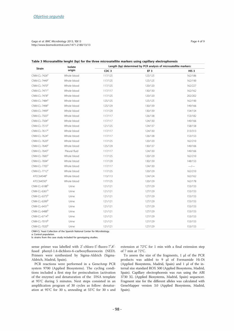
Objetivo segundo
- 98 -
sense primer was labelled with 20-chloro-50-fluoro-70,80-
fused phenyl-1.4-dichloro-6-carboxyfluorescein (NED).
Primers were synthesized by Sigma-Aldrich (Sigma-
Aldrich, Madrid, Spain).
PCR reactions were performed in a GeneAmp PCR
system 9700 (Applied Biosystems). The cycling condi-
tions included a first step for preincubation (activation
of the enzyme) and denaturation of the DNA template
at 95°C during 5 minutes. Next steps consisted in an
amplification program of 30 cycles as follow: denatur-
ation at 95°C for 30 s, annealing at 55°C for 30 s and
extension at 72°C for 1 min with a final extension step
of 7 min at 72°C.
To assess the size of the fragments, 1 μl of the PCR
products was added to 9 μl of Formamide Hi-Di
(Applied Biosystems, Madrid, Spain) and 1 μl of the in-
ternal size standard ROX 500 (Applied Biosystems, Madrid,
Spain). Capillary electrophoresis was run using the ABI
3730 XL (Applied Biosystems, Madrid, Spain) sequencer.
Fragment size for the different alleles was calculated with
GeneMapper version 3.0 (Applied Biosystems, Madrid,
Spain).
Table 3 Microsatellite lenght (bp) for the three microsatellite markers using capillary electrophoresis
StrainIsolateorigin
Length (bp) determined by PCR analysis of microsatellite markers:
CDC 3 EF 3 HIS 3
CNM-CL-7426a Whole blood 117/125 125/125 162/186
CNM-CL-7449a Whole blood 117/125 125/125 162/190
CNM-CL-7470a Whole blood 117/125 120/120 162/227
CNM-CL-7471a Whole blood 117/117 130/130 162/162
CNM-CL-7478a Whole blood 117/125 120/120 202/202
CNM-CL-7484a Whole blood 125/125 125/125 162/190
CNM-CL-7498a Whole blood 125/129 130/139 149/166
CNM-CL-7499a Whole blood 117/129 130/139 154/154
CNM-CL-7503a Whole blood 117/117 126/138 153/182
CNM-CL-7504a Whole blood 117/117 124/130 149/166
CNM-CL-7513a Whole blood 121/125 124/137 158/158
CNM-CL-7617a Whole blood 117/117 124/130 313/313
CNM-CL-7624a Whole blood 117/117 126/138 153/153
CNM-CL-7620a Whole blood 117/125 120/120 162/210
CNM-CL-7640a Whole blood 125/129 130/137 149/166
CNM-CL-7643a Pleural fluid 117/117 124/130 149/166
CNM-CL-7683a Whole blood 117/125 120/129 162/210
CNM-CL-7694a Whole blood 117/129 130/139 148/153
CNM-CL-7705a Whole blood 117/117 124/130 —/—
CNM-CL-7712a Whole blood 117/125 120/129 162/210
ATCC64548a Whole blood 113/113 124/124 162/162
ATCC64550a Whole blood 117/125 120/129 162/178
CNM-CL-6188b Urine 121/121 127/129 153/153
CNM-CL-6361b Urine 121/121 127/129 153/153
CNM-CL-6373b Urine 121/121 127/129 153/153
CNM-CL-6399b Urine 121/121 127/129 153/153
CNM-CL-6431b Urine 121/121 127/129 153/153
CNM-CL-6488b Urine 121/121 127/129 153/153
CNM-CL-6714b Urine 121/121 127/129 153/153
CNM-CL-7019b Urine 121/121 127/129 153/153
CNM-CL-7020b Urine 121/121 127/129 153/153
CNM-CL Yeast Collection of the Spanish National Center for Microbiology.
a: Control population.
b: strains from the case study included for genotyping studies.
Gago et al. BMC Microbiology 2013, 13:13 Page 4 of 9
http://www.biomedcentral.com/1471-2180/13/13
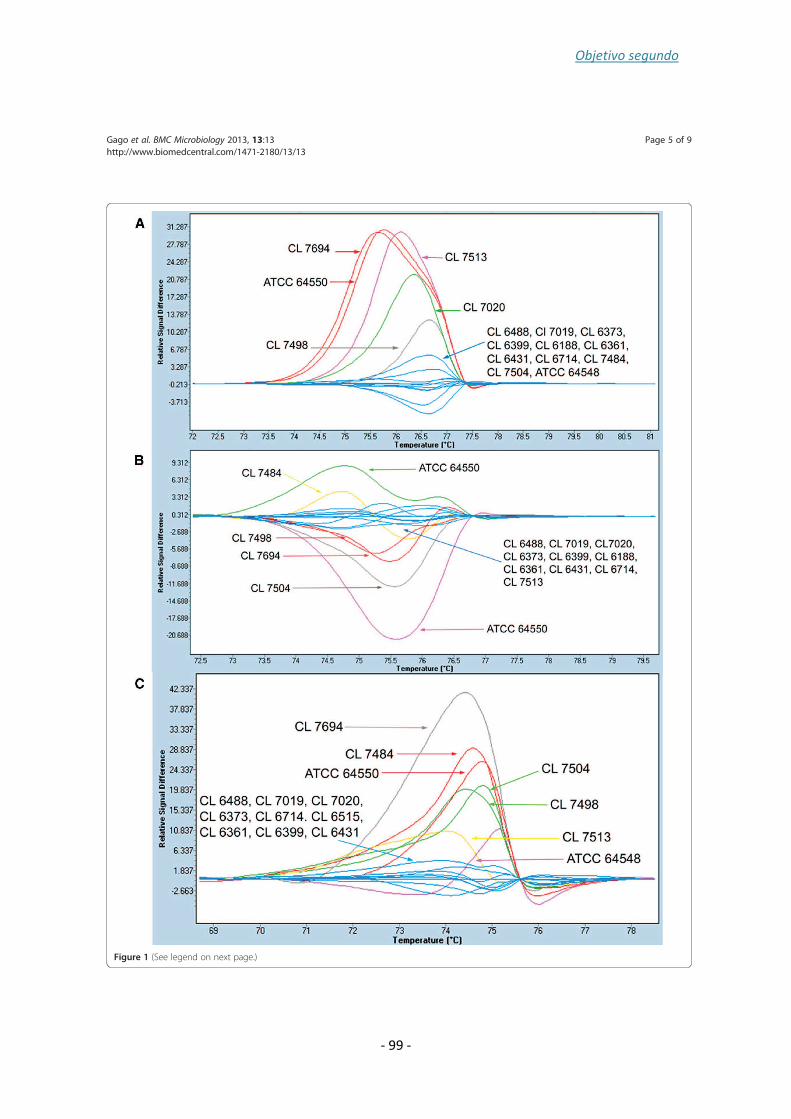
Objetivo segundo
- 99 -
Figure 1 (See legend on next page.)
Gago et al. BMC Microbiology 2013, 13:13 Page 5 of 9
http://www.biomedcentral.com/1471-2180/13/13
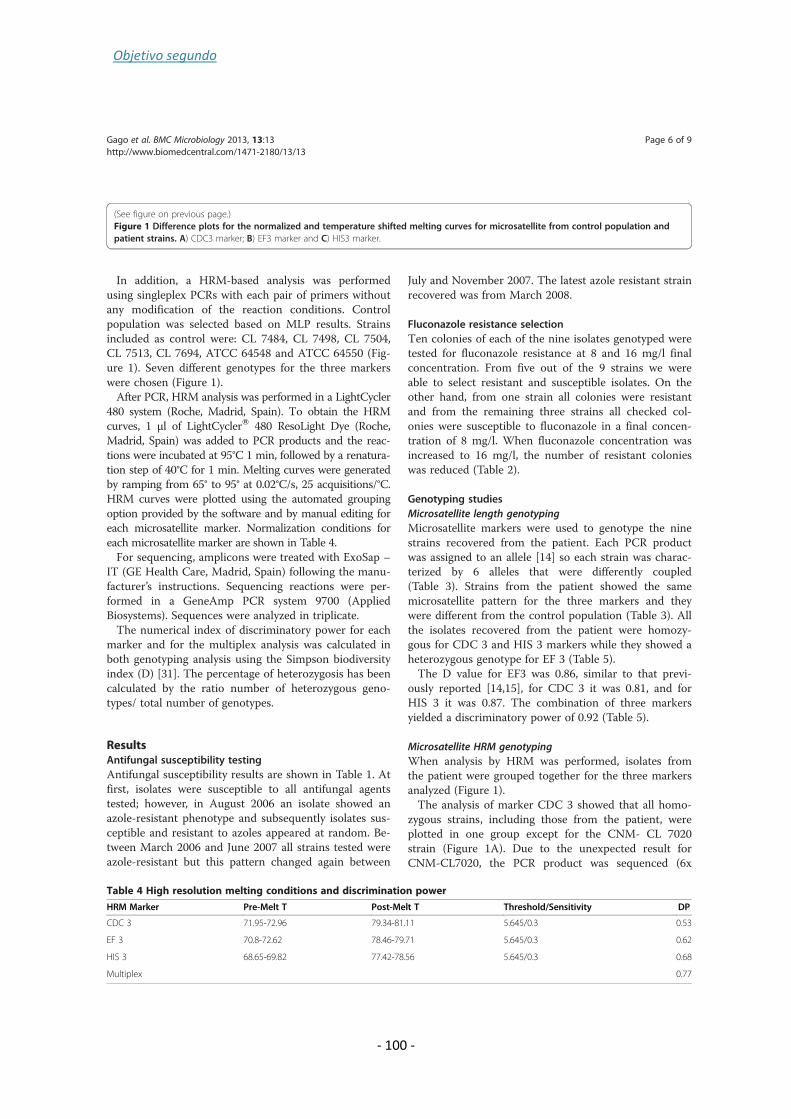
Objetivo segundo
- 100 -
In addition, a HRM-based analysis was performed
using singleplex PCRs with each pair of primers without
any modification of the reaction conditions. Control
population was selected based on MLP results. Strains
included as control were: CL 7484, CL 7498, CL 7504,
CL 7513, CL 7694, ATCC 64548 and ATCC 64550 (Fig-
ure 1). Seven different genotypes for the three markers
were chosen (Figure 1).
After PCR, HRM analysis was performed in a LightCycler
480 system (Roche, Madrid, Spain). To obtain the HRM
curves, 1 μl of LightCyclerW 480 ResoLight Dye (Roche,
Madrid, Spain) was added to PCR products and the reac-
tions were incubated at 95°C 1 min, followed by a renatura-
tion step of 40°C for 1 min. Melting curves were generated
by ramping from 65° to 95° at 0.02°C/s, 25 acquisitions/°C.
HRM curves were plotted using the automated grouping
option provided by the software and by manual editing for
each microsatellite marker. Normalization conditions for
each microsatellite marker are shown in Table 4.
For sequencing, amplicons were treated with ExoSap –
IT (GE Health Care, Madrid, Spain) following the manu-
facturer’s instructions. Sequencing reactions were per-
formed in a GeneAmp PCR system 9700 (Applied
Biosystems). Sequences were analyzed in triplicate.
The numerical index of discriminatory power for each
marker and for the multiplex analysis was calculated in
both genotyping analysis using the Simpson biodiversity
index (D) [31]. The percentage of heterozygosis has been
calculated by the ratio number of heterozygous geno-
types/ total number of genotypes.
ResultsAntifungal susceptibility testing
Antifungal susceptibility results are shown in Table 1. At
first, isolates were susceptible to all antifungal agents
tested; however, in August 2006 an isolate showed an
azole-resistant phenotype and subsequently isolates sus-
ceptible and resistant to azoles appeared at random. Be-
tween March 2006 and June 2007 all strains tested were
azole-resistant but this pattern changed again between
July and November 2007. The latest azole resistant strain
recovered was from March 2008.
Fluconazole resistance selection
Ten colonies of each of the nine isolates genotyped were
tested for fluconazole resistance at 8 and 16 mg/l final
concentration. From five out of the 9 strains we were
able to select resistant and susceptible isolates. On the
other hand, from one strain all colonies were resistant
and from the remaining three strains all checked col-
onies were susceptible to fluconazole in a final concen-
tration of 8 mg/l. When fluconazole concentration was
increased to 16 mg/l, the number of resistant colonies
was reduced (Table 2).
Genotyping studies
Microsatellite length genotyping
Microsatellite markers were used to genotype the nine
strains recovered from the patient. Each PCR product
was assigned to an allele [14] so each strain was charac-
terized by 6 alleles that were differently coupled
(Table 3). Strains from the patient showed the same
microsatellite pattern for the three markers and they
were different from the control population (Table 3). All
the isolates recovered from the patient were homozy-
gous for CDC 3 and HIS 3 markers while they showed a
heterozygous genotype for EF 3 (Table 5).
The D value for EF3 was 0.86, similar to that previ-
ously reported [14,15], for CDC 3 it was 0.81, and for
HIS 3 it was 0.87. The combination of three markers
yielded a discriminatory power of 0.92 (Table 5).
Microsatellite HRM genotyping
When analysis by HRM was performed, isolates from
the patient were grouped together for the three markers
analyzed (Figure 1).
The analysis of marker CDC 3 showed that all homo-
zygous strains, including those from the patient, were
plotted in one group except for the CNM- CL 7020
strain (Figure 1A). Due to the unexpected result for
CNM-CL7020, the PCR product was sequenced (6x
(See figure on previous page.)
Figure 1 Difference plots for the normalized and temperature shifted melting curves for microsatellite from control population and
patient strains. A) CDC3 marker; B) EF3 marker and C) HIS3 marker.
Table 4 High resolution melting conditions and discrimination power
HRM Marker Pre-Melt T Post-Melt T Threshold/Sensitivity DP
CDC 3 71.95-72.96 79.34-81.11 5.645/0.3 0.53
EF 3 70.8-72.62 78.46-79.71 5.645/0.3 0.62
HIS 3 68.65-69.82 77.42-78.56 5.645/0.3 0.68
Multiplex 0.77
Gago et al. BMC Microbiology 2013, 13:13 Page 6 of 9
http://www.biomedcentral.com/1471-2180/13/13
Objetivo segundo
- 101 -
sequence coverage) and a 3 bp insertion at 67 pb from
the forward primer was found. Heterozygous strains
were distributed in four groups according to their frag-
ment length. The heterozygous strains CNM-CL 7694
and ATCC 64550 were plotted together although one of
the alleles were different (Table 3). When we performed
EF 3 fragments analysis by HRM, six different groups
were plotted one of them contained strains from the pa-
tient while the control population was distributed into
five groups according to its fragment size or whether
they were homozygous or heterozygous (Figure 1B).
Finally, HRM analysis of the HIS3 marker showed six
different groups. Strains from the patient were grouped
together again. Strains in the control population were
grouped based on their fragment size pattern (Figure 1C).
Discrimination power for CDC 3 marker was 0.53, for
EF 3 it was 0.62 and for the HIS 3 marker it was 0.68.
The combination of the three markers provided a DP
value of 0.77 (Table 4).
DiscussionTyping methods have been described as useful tools for
the differentiation between strains isolated only once
and those able to cause recurrent infections. Several
methods have been developed to analyze microevolution
and structure of C. albicans species. Although MLST
(MultiLocus Sequence Typing) has been chosen as the
most discriminatory technique [5,32], several articles
have recently pointed towards the suitability of MLP
[14-16,29]. In this study, nine isolates from a case of re-
current urinary infection were genotyped using microsa-
tellites and a new HRM analysis method. Antifungal
susceptibility testing revealed that strains from the
patient were susceptible and resistant in vitro to flucona-
zole in a random way. Microvariation between colonies
due to exposure of C. albicans to azole antifungal agents
has been widely described [10,16] and the need to perform
intercolony assays has also been reported [25,33,34]. We
performed an inter-colony test modified from Schoofs
et al. [25] and we were able to prove the coexistence of
colonies resistant and susceptible to azoles in a high num-
ber of the strains tested. The number of azole-resistant
colonies was variable depending on azole concentration.
A genotyping method based on HRM analysis was
developed taking into account previous works showing
that if the number of genotypes is higher than seven, the
curve definition is not the best possible [35]. Based on that
premise, for each marker we selected seven strains with
different genotype, previously analysed by capillary elec-
trophoresis. C. albicans microsatellites (CDC3, EF3 and
HIS3) were amplified using LightCyclerW 480 ResoLight
as intercalating dye. When HRM analysis was performed,
the isolates from the patient were plotted together and
these results were consistent with those obtained by capil-
lary electrophoresis. There was only one exception for the
CDC3 marker where one strain (CNM-CL7020) was not
grouped, as expected, with the other strains showing the
same MLP genotype. The sequence of the fragment
showed a 3 bp insertion that explained the melting differ-
ences. This fact supports previous works in which HRM
allowed to identify changes in the sequence length and
one nucleotide changes [36].
Although the calculated discrimination power was
higher for the analysis using capillary electrophoresis
than for HRM analysis (0.92 vs. 0.77) as previously
reported [14]. The HRM analysis showed several advan-
tages; it was a very simple and fast technique and results
were obtained in 3 hours (including amplification), the
interpretation of results was easy and the cost per sam-
ple was much lower than MLP genotyping due to this
technique does not require sequencing equipment and
the primers are not end-labelled. Our estimate is that
the cost per sample using capillary electrophoresis is
more than twice that of using HRM analysis. Further-
more, it can be used in a routine laboratory setting as it
only requires real time PCR equipment. In this study, al-
though we were not able to demonstrate the mechanism
underlying the variability in the susceptibility to azoles
in the strains tested, we were able to confirm that resist-
ant and susceptible isolates were genetically closely
related with an easy method to analyze microsatellites.
The results highlight the need for more in-depth studies
to be performed on these kinds of infections for an ac-
curate and appropriate management thereof.
ConclusionsThis method is a useful tool for performing a fast
screening to establish relatedness between strains in out-
breaks or surveillance studies in cases of recurrent or
persistent infections. To our knowledge, this is the first
study in which three microsatellite markers were ana-
lyzed by HRM by using seven strains with different
Table 5 Characteristics of the microsatellite loci analyzed by capillary electrophoresis
Microsatellite Marker No. of alleles No. of genotypes No. of heterozygotic genotypes DP % heterozygosity
CDC 3 5 8 4 0.81 50.00
EF 3 10 11 7 0.86 63.63
HIS 3 14 15 11 0.88 53.30
Multiplex 0.92
Gago et al. BMC Microbiology 2013, 13:13 Page 7 of 9
http://www.biomedcentral.com/1471-2180/13/13

Objetivo segundo
- 102 -
genotype as control population and reaching HRM reso-
lution limits. Although HRM analysis method presented
a lower degree of discrimination compared to other
genotyping methods, it provided a more cost-effective
and suitable alternative for genotyping C. albicans in a
clinical laboratory.
Competing interest
In the past 5 years, M.C.E. has received grant support from Astellas Pharma,
bioMerieux, Gilead Sciences, Merck Sharp and Dohme, Pfizer, Schering
Plough, Soria Melguizo SA, the European Union, the ALBAN program, the
Spanish Agency for International Cooperation, the Spanish Ministry of
Culture and Education, The Spanish Health Research Fund, The Instituto de
Salud Carlos III, The Ramon Areces Foundation, The Mutua Madrileña
Foundation. He has been an advisor/consultant to the Panamerican Health
Organization, Gilead Sciences, Merck Sharp and Dohme, Pfizer, and Schering
Plough. He has received remuneration for talks on behalf of Gilead Sciences,
Merck Sharp and Dohme, Pfizer, and Schering Plough.
Authors’ contributions
SG performed the genotyping studies, the analysis of the results and also
participated in drafting the manuscript. BL participated in the collection of
clinical data and strains from the patient. AG-L has been involved in the
antifungal susceptibility testing. IC has made contributions to the analysis of
the results. MC-E has been involved in drafting the manuscript and in the
final approval of the version to be published following a critical review
thereof. MJB was responsible for the original design of the study and
participated in its further design and development as well as having been
involved in drafting the manuscript. All authors have read and approved the
final manuscript.
Acknowledgements
This work was supported by Research Projects from Spanish Fondo de
Investigaciones Sanitarias of the Instituto de Salud Carlos III (PI09/1791 and
PI11/00412) and by the Spanish Network for Research on Infectious Diseases
(REIPI RD06/0008/10). S. G. is supported by a research fellowship from the
“Fondo de Investigaciones Biomedicas” of the Spanish Ministry of Science
and Innovation (FI10/00464).
Author details1Servicio de Micología, Centro Nacional de Microbiología, Instituto de Salud
Carlos III, Ctra Majadahonda-Pozuelo, Km 2, Majadahonda, Madrid 28220,
Spain. 2Servicio de Microbiología, Hospital Virgen de la Concha, Zamora,
España. 3Unidad de Bioinformática, Centro Nacional de Microbiología,
Instituto de Salud Carlos III, Madrid, Spain.
Received: 23 January 2012 Accepted: 18 January 2013
Published: 23 January 2013
References
1. Pappas PG: Invasive candidiasis. Infect. Dis Clin North Am 2006,
20:485–506.
2. Khatib R, Ayeni O, Riederer KM, Briski LE, Wilson FM: Strain relatedness in
persistent and recurrent candiduria. J Urol 1998, 159:2054–2056.
3. Lockhart SR, Fritch JJ, Meier AS, Schroppel K, Srikantha T, Galask R, Soll DR:
Colonizing populations of Candida albicans are clonal in origin but
undergo microevolution through C1 fragment reorganization as
demonstrated by DNA fingerprinting and C1 sequencing. J Clin Microbiol
1995, 33:1501–1509.
4. Lockhart SR, Reed BD, Pierson CL, Soll DR: Most frequent scenario for
recurrent Candida vaginitis is strain maintenance with “substrain
shuffling”: demonstration by sequential DNA fingerprinting with probes
Ca3, C1, and CARE2. J Clin Microbiol 1996, 34:767–777.
5. Da Matta DA, Melo AS, Guimaraes T, Frade JP, Lott TJ, Colombo AL:
Multilocus sequence typing of sequential Candida albicans isolates from
patients with persistent or recurrent fungemia. Med Mycol 2010,
48:757–762.
6. Jacobsen MD, Duncan AD, Bain J, Johnson EM, Naglik JR, Shaw DJ, Gow NA,
Odds FC: Mixed Candida albicans strain populations in colonized and
infected mucosal tissues. FEMS Yeast Res 2008, 8:1334–1338.
7. Odds FC, Davidson AD, Jacobsen MD, Tavanti A, Whyte JA, Kibbler CC, Ellis
DH, Maiden MC, Shaw DJ, Gow NA: Candida albicans strain maintenance,
replacement, and microvariation demonstrated by multilocus sequence
typing. J Clin Microbiol 2006, 44:3647–3658.
8. Sabino R, Sampaio P, Carneiro C, Rosado L, Pais C: Isolates from hospital
environments are the most virulent of the Candida parapsilosis complex.
BMC Microbiol 2011, 11:180.
9. Sampaio P, Gusmao L, Correia A, Alves C, Rodrigues AG, Pina-Vaz C, Amorim
A, Pais C: New microsatellite multiplex PCR for Candida albicans strain
typing reveals microevolutionary changes. J Clin Microbiol 2005,
43:3869–3876.
10. Shin JH, Chae MJ, Song JW, Jung SI, Cho D, Kee SJ, Kim SH, Shin MG, Suh
SP, Ryang DW: Changes in karyotype and azole susceptibility of
sequential bloodstream isolates from patients with Candida glabrata
candidemia. J Clin Microbiol 2007, 45:2385–2391.
11. Shin JH, Park MR, Song JW, Shin DH, Jung SI, Cho D, Kee SJ, Shin MG, Suh
SP, Ryang DW: Microevolution of Candida albicans strains during
catheter-related candidemia. J Clin Microbiol 2004, 42:4025–4031.
12. Sampaio P, Santos M, Correia A, Amaral FE, Chavez-Galarza J, Costa-de-
Oliveira S, Castro AG, Pedrosa J, Pais C: Virulence attenuation of Candida
albicans genetic variants isolated from a patient with a recurrent
bloodstream infection. PLoS One 2010, 5:e10155.
13. Huang M, McClellan M, Berman J, Kao KC: Evolutionary dynamics of
Candida albicans during in vitro evolution. Eukaryot Cell 2011,
10:1413–1421.
14. Botterel F, Desterke C, Costa C, Bretagne S: Analysis of microsatellite
markers of Candida albicans used for rapid typing. J Clin Microbiol 2001,
39:4076–4081.
15. Costa JM, Garcia-Hermoso D, Olivi M, Cabaret O, Farrugia C, Lecellier G,
Dromer F, Bretagne S: Genotyping of Candida albicans using length
fragment and high-resolution melting analyses together with
minisequencing of a polymorphic microsatellite locus. J Microbiol
Methods 2010, 80:306–309.
16. Costa-de-Oliveira S, Sousa I, Correia A, Sampaio P, Pais C, Rodrigues AG,
Pina-Vaz C: Genetic relatedness and antifungal susceptibility profile of
Candida albicans isolates from fungaemia patients. Med Mycol 2011,
49:248–252.
17. Eloy O, Marque S, Botterel F, Stephan F, Costa JM, Lasserre V, Bretagne S:
Uniform distribution of three Candida albicans microsatellite markers in
two French ICU populations supports a lack of nosocomial cross-
contamination. BMC Infect Dis 2006, 6:162.
18. Garcia-Hermoso D, Cabaret O, Lecellier G, Desnos-Ollivier M, Hoinard D,
Raoux D, Costa JM, Dromer F, Bretagne S: Comparison of microsatellite
length polymorphism and multilocus sequence typing for DNA-Based
typing of Candida albicans. J Clin Microbiol 2007, 45:3958–3963.
19. Garcia-Hermoso D, MacCallum DM, Lott TJ, Sampaio P, Serna MJ, Grenouillet
F, Klaassen CH, Bretagne S: Multicenter collaborative study for
standardization of Candida albicans genotyping using a polymorphic
microsatellite marker. J Clin Microbiol 2010, 48:2578–2581.
20. Erali M, Voelkerding KV, Wittwer CT: High resolution melting applications
for clinical laboratory medicine. Exp Mol Pathol 2008, 85:50–58.
21. Erali M, Wittwer CT: High resolution melting analysis for gene scanning.
Methods 2010, 50:250–261.
22. Arancia S, Sandini S, De Bernardis F, Fortini D: Rapid, simple, and low-cost
identification of Candida species using high-resolution melting analysis.
Diagn Microbiol Infect Dis 2011, 69:283–285.
23. Rodriguez-Tudela JL Arendrup MC, Barchiesi F, Bille J, Chryssanthou E,
Cuenca-Estrella M, Dannaoui E, Denning DW, Donnelly JP, Dromer F,
Fegeler W, Lass-Flörl C, Moore C, Richardson M, Sandven P, Velegraki A,
Verweij P: EUCAST definitive document EDef 7.1: method for the
determination of broth dilution MICs of antifungal agents for
fermentative yeasts. Clin Microbiol Infect 2008, 14:398–405.
24. Rodriguez-Tudela JL, Arendrup MC, Cuenca-Estrella M, Donnelly JP, Lass-
Flörl C: EUCAST Breakpoints for Antifungals. Drugs News and Perspectives
2010, 23:93–97.
25. Schoofs A, Odds FC, Colebunders R, Ieven M, Wouters L, Goossens H:
Isolation of Candida species on media with and without added
fluconazole reveals high variability in relative growth susceptibility
phenotypes. Antimicrob Agents Chemother 1997, 41:1625–1635.
26. Cendejas-Bueno E, Gomez-Lopez A, Mellado E, Rodriguez-Tudela JL,
Cuenca-Estrella M: Identification of pathogenic rare yeast species in
Gago et al. BMC Microbiology 2013, 13:13 Page 8 of 9
http://www.biomedcentral.com/1471-2180/13/13

Objetivo segundo
- 103 -
clinical samples: comparison between phenotypical and molecular
methods. J Clin Microbiol 2010, 48:1895–1899.
27. Tang CM, Cohen J, Holden DW: An Aspergillus fumigatus alkaline protease
mutant constructed by gene disruption is deficient in extracellular
elastase activity. Mol Microbiol 1992, 6:1663–1671.
28. DiDomenico BJ, Brown NH, Lupisella J, Greene JR, Yanko M, Koltin Y:
Homologs of the yeast neck filament associated genes: isolation and
sequence analysis of Candida albicans CDC3 and CDC10. Mol Gen Genet
1994, 242:689–698.
29. Bretagne S, Costa JM, Besmond C, Carsique R, Calderone R: Microsatellite
polymorphism in the promoter sequence of the elongation factor 3
gene of Candida albicans as the basis for a typing system. J Clin Microbiol
1997, 35:1777–1780.
30. Magee BB, Koltin Y, Gorman JA, Magee PT: Assignment of cloned genes to
the seven electrophoretically separated Candida albicans chromosomes.
Mol Cell Biol 1988, 8:4721–4726.
31. Hunter PR, Gaston MA: Numerical index of the discriminatory ability of
typing systems: an application of Simpson’s index of diversity. J Clin
Microbiol 1988, 26:2465–2466.
32. Myoung Y, Shin JH, Lee JS, Kim SH, Shin MG, Suh SP, Ryang DW: Multilocus
sequence typing for Candida albicans isolates from candidemic patients:
comparison with Southern blot hybridization and pulsed-field gel
electrophoresis analysis. Korean J Lab Med 2011, 31:107–114.
33. Cartledge JD, Midgley J, Gazzard BG: Clinically significant azole cross-
resistance in Candida isolates from HIV-positive patients with oral
candidosis. AIDS 1997, 11:1839–1844.
34. Johnson EM, Warnock DW, Luker J, Porter SR, Scully C: Emergence of azole
drug resistance in Candida species from HIV-infected patients receiving
prolonged fluconazole therapy for oral candidosis. J Antimicrob
Chemother 1995, 35:103–114.
35. Mader E, Lukas B, Novak J: A strategy to setup codominant microsatellite
analysis for high-resolution-melting-curve-analysis (HRM). BMC Genet
2008, 9:69.
36. Wittwer CT, Reed GH, Gundry CN, Vandersteen JG, Pryor RJ: High-resolution
genotyping by amplicon melting analysis using LCGreen. Clin Chem 2003,
49:853–860.
doi:10.1186/1471-2180-13-13Cite this article as: Gago et al.: Analysis of strain relatedness using HighResolution Melting in a case of recurrent candiduria. BMC Microbiology2013 13:13.
Submit your next manuscript to BioMed Centraland take full advantage of:
• Convenient online submission
• Thorough peer review
• No space constraints or color figure charges
• Immediate publication on acceptance
• Inclusion in PubMed, CAS, Scopus and Google Scholar
• Research which is freely available for redistribution
Submit your manuscript at www.biomedcentral.com/submit
Gago et al. BMC Microbiology 2013, 13:13 Page 9 of 9
http://www.biomedcentral.com/1471-2180/13/13


Discusión
- 105 -
5. DISCUSIÓN


Discusión
- 107 -
La investigación en los laboratorios de referencia tiene entre sus objetivos
principales el desarrollo de nuevas técnicas de diagnóstico que sean transferibles al
Sistema Nacional de Salud o que se puedan desarrollar comercialmente. En el ámbito
de la micología médica, al contrario que en bacteriología o virología, el desarrollo de
métodos de PCR diagnóstica es limitado y se centra principalmente en el diagnóstico
de candidosis o aspergilosis invasora. En este contexto, se han desarrollado,
estandarizado y validado técnicas de PCR en tiempo real para el diagnóstico e
identificación de algunas de las infecciones fúngicas que conllevan una elevada
mortalidad. Los ensayos descritos en esta tesis son imprescindibles para facilitar su
posterior transferencia o desarrollo comercial para facilitar su uso en entornos donde
estas infecciones tienen una incidencia elevada y esta tecnología no está disponible.
5.1. Detección de los hongos patógenos humanos que causan neumonía en el paciente inmunodeprimido mediante PCR en tiempo real en un solo tubo
Histoplasma capsulatum, Cryptococcus neoformans y Pneumocystis jirovecii son
los principales patógenos causantes de la neumonía oportunista fúngica en individuos
infectados por el VIH (Armstrong-James et al., 2014; Huang y Crothers, 2009). La
sintomatología clínica y la radiología de estas infecciones es similar, confundiéndose
entre sí y también con la tuberculosis que, generalmente, es la primera sospecha
clínica (Denis y Lortholary, 2013; Rodríguez-Cerdeira et al., 2014). El proceso de
diagnóstico diferencial cuando un paciente desarrolla sintomatología respiratoria
aguda compatible con una neumonía fúngica, es complejo, largo y costoso. Con el
desarrollo de esta PCR múltiple se puede obtener una respuesta rápida de la etiología
del proceso.
La PCR en tiempo real en formato múltiple, desarrollada en este trabajo, está
basada en sondas molecular beacon dirigidas a la región ITS del ADN ribosómico. Se
eligieron este tipo de sondas por su elevada especificidad y baja fluorescencia residual
(Espy et al., 2006). Debido a su compleja estructura, su diseño se realizó mediante un
software especializado y utilizando la extensa base de datos de secuencias del Servicio
de Micología mencionada en la introducción. Además, el uso de las sondas de tipo

Discusión
- 108 -
molecular beacon, facilita el diseño de ensayos en formato múltiple que no pueden
realizarse con otras sondas menos estables como las sondas FRET o TaqMan (Espy et
al., 2006).
Pneumocystis jirovecii se puede cultivar en líneas celulares, sin embargo la
propagación del microorganismo de forma indefinida in vitro no se ha conseguido
(Martínez et al., 2013). Por ello, los controles positivos se obtuvieron mediante
clonación en un plásmido comercial. El inserto clonado fue el producto de
amplificación de la región diana (ITS del rADN), a partir del lavado broncoalveolar de
un paciente con infección probada.
La sonda diseñada para la detección de especies del género Cryptococcus, también
dirigida a la región ITS1, permite la detección del complejo Cryptococcus neoformans
(var neoformans y var grubbi), y, Cryptococcus gattii. Para simplificar la técnica, se
diseñó una única sonda capaz de detectar cualquiera de estas especies puesto que el
tratamiento de la infección es similar (La Hoz y Pappas, 2013). Con respecto a la
detección de ADN de Histoplasma capsulatum, se empleó una sonda dirigida a la
región ITS1 que se había utilizado en trabajos previos y que está registrada bajo el
número de patente P 200802665.
Además de los controles habituales en cualquier técnica de PCR, se incluyó un
control interno para descartar los falsos negativos debidos a la inhibición de la
reacción. Dicho control consiste en la introducción, en el mismo tubo de PCR, de 1 fg
de un plásmido que contiene un fragmento de una secuencia del genoma de una
medusa (Limaye et al., 2001), para la cual se diseñaron cebadores y una sonda
específica. Un retraso en el valor del ciclo de amplificación del control interno se
corresponde con una inhibición en la amplificación. Además, la aparición de falsos
positivos debidos a contaminaciones se reduce, en gran parte, con el uso de un
sistema de tubo cerrado.
En el proceso de estandarización in vitro, la sensibilidad y la especificidad de la PCR
fue del 100%, el coeficiente de variación en torno al 3% y la eficiencia del método de
extracción de ADN de la muestra clínica superior al 80%. No se amplificó ADN de 21
Discusión
- 109 -
cepas de especies de los géneros Candida (n=7), Coccidioides (n=1), Paracoccidioides
(n=1), Aspergillus (n=4), Fusarium (n=3), Scedosporium (n=2) y Mucorales (n=3).
Asimismo, se realizó una validación clínica con 43 muestras de 40 pacientes con
infección probada de acuerdo a los criterios EORTC/MSG (5 criptococosis, 14
histoplasmosis, 21 neumocistosis) (De Pauw et al., 2008). Debido a que en estos
criterios no se incluye la neumonía por P. jirovecii, se consideró infección definitiva
cuando los pacientes tenían sintomatología clínica de neumonía y se visualizaron al
microscopio las formas tróficas. El ensayo se validó no sólo en muestras respiratorias
(n=29) sino también en muestras de suero (n=2), sangre (n=3), biopsias (n=5) y
aspirado de médula ósea (n=3) ya que son muestras útiles para detectar los episodios
de infección sistémica (Huang y Crothers, 2009). En función del tipo de muestra, la PCR
fue positiva en 29/29 muestras respiratorias, 1/3 aspirados de médula ósea, 3/3
muestras de sangre, 1/2 muestras de suero y 4/5 biopsias. Así, la PCR mostró una
sensibilidad del 92,5% por pacientes y del 90,7% por muestra clínica. La PCR fue
negativa en cuatro de las muestras clínicas (3 histoplasmosis y 1 criptococosis), pero
dichas muestras se habían almacenado durante más de cinco años a -20ºC lo que
podría afectar a la calidad del ADN obtenido, como ya se ha descrito previamente (Lau
et al., 2010). Por último, la técnica fue 100% específica y no se detectó ADN en 29
muestras clínicas de 29 pacientes incluidos en los siguientes grupos: (i) pacientes con
VIH de área endémica sin neumonía (n=6); (ii) pacientes con VIH de área endémica con
neumonía bacteriana o viral (n=4); y (iii) pacientes VIH (n=5) y no VIH (n=14) con otra
infección fúngica invasora.
Una ventaja de este tipo de técnicas de detección múltiple es que permiten el
diagnóstico de coinfecciones. A pesar de que el número de pacientes incluidos no fue
muy elevado, se identificó un paciente diagnosticado de histoplasmosis que tenía una
infección concomitante con P. jirovecii. Las coinfecciones en pacientes
inmunodeprimidos son frecuentes y el diagnóstico exacto es determinante para poder
establecer el tratamiento más adecuado (Javier et al., 2012; Pena et al., 2013). De
hecho, en el 15% de los casos de histoplasmosis en VIH se ha descrito coinfección con
tuberculosis (Gutiérrez et al., 2005). Asimismo, se han publicado varios casos de
infecciones mixtas diagnosticados mediante métodos clásicos en los que
Discusión
- 110 -
H. capsulatum, C. neoformans y P. jirovecii estaban implicados (Baumann et al., 2013;
Javier et al., 2012; Pena et al., 2013; Tschudy y Michail, 2010).
El sistema de detección múltiple desarrollado tiene, además, una gran flexibilidad,
dependiendo por ejemplo del área geográfica donde se quiera aplicar. Así, mientras
que en América Latina el sistema de triple detección sería útil (Colombo et al., 2011),
en África se podría simplificar para sólo diagnosticar la criptococosis y la neumocistosis
(Cogliati, 2013; Morrow et al., 2014). La incorporación de nuevos canales de detección
y nuevos compuestos fluorescentes, que emitan a diferentes longitudes de onda
permitiría la inclusión de nuevos patógenos en el ensayo como Mycobacterium
tuberculosis, Penicillium marneffei, etc.
El coste del ensayo es comparable o incluso más barato que los métodos
tradicionales o de detección de biomarcadores empleados para el diagnóstico
independiente de cada una de estas patologías (coste aproximado 100 € por
determinación). Sin embargo, en países en desarrollo, este precio puede no ser
competitivo. Por ello, para este tipo de países, puede ser necesario adaptar estas
técnicas a sistemas de diagnóstico inmediato más sencillos y económicos. Esto incluiría
mejoras en la extracción de ADN, que debe realizarse por un método simple y
eficiente.
La comparación de esta PCR múltiple con los otros métodos de diagnóstico que se
emplean actualmente no es posible, ya que no existe ninguno disponible que detecte
los tres patógenos al mismo tiempo. Sin embargo, se pueden discutir los aspectos más
importantes, tanto de las técnicas disponibles como las publicaciones recientes en
diagnóstico para cada patógeno individualmente, ya que hay otros grupos de pacientes
donde los sistemas de diagnóstico actuales no tienen buena sensibilidad y la PCR
diagnóstica desarrollada en esta tesis puede ser útil.
En el diagnóstico de la neumocistosis, las técnicas de microscopía que se emplean
rutinariamente tienen una sensibilidad cercana al 90% en lavado broncoalveolar de
pacientes infectados por VIH (Fan et al., 2013). Sin embargo, cuando la carga fúngica es
baja, la sensibilidad y el valor predictivo negativo de la prueba disminuyen hasta el
70% (Carmona y Limper, 2011; Wilson et al., 2011). Asimismo, la detección de 1,3-B-D
Discusión
- 111 -
glucano tiene una sensibilidad del 93% en pacientes con VIH y síntomas respiratorios;
sin embargo, el valor predictivo negativo de la técnica es del 60% y en pacientes con
neumonía bacteriana o por otro hongo también ocasiona resultados positivos (Boyles,
2013; Esteves et al., 2014).
Respecto a las técnicas de PCR descritas, a diferencia de lo que ocurre en la
mayoría de los hongos, sólo hay una copia de la región ITS en el genoma de P. jirovecii
(Nahimana et al., 2000); lo que es una ventaja para diferenciar entre población
colonizada y población infectada (Alanio et al., 2011), ya que el número de copias que
tienen los pacientes con infección es significativamente mayor que los que están
colonizados (Matsumura et al., 2012). Otros genes monocopia como cdc2 (Hauser et
al., 2011), DHPS, (Munoz et al., 2012) y timidilato sintasa (Lu et al., 1995) se han
empleado previamente con el mismo fin, aunque no se ha definido un valor umbral
definitivo que discrimine colonización de infección (Arcenas et al., 2006). El
establecimiento de un punto de corte entre colonización e infección sería de gran
utilidad ya que se ha descrito colonización con P. jiroveccii en el 16% de los pacientes
tratados con corticoides, en el 40% de los pacientes con EPOC, en el 6% de los
pacientes con fibrosis quística y en el 46% de pacientes con infección por VIH
asintomática (Morris y Norris, 2012). En concreto, el 60% de los pacientes tratados con
corticoides, sometidos además a quimioterapia u otro fármaco inmunosupresor,
desarrollan neumocistosis a pesar de un resultado negativo en microscopía (Ribes et
al., 1997). Estos pacientes presentan síntomas atípicos, lo que conlleva un retraso en el
establecimiento del tratamiento adecuado por lo que tiene una mortalidad asociada
de entre el 35 y el 55% (Roblot et al., 2002; Roux et al., 2014). Por tanto, parece
necesario estandarizar esta PCR con las diferentes poblaciones de riesgo de forma que
se establezca un valor umbral que diferencie los pacientes realmente colonizados de
los que van a desarrollar o ya tienen infección.
El potencial de las técnicas de PCR diagnóstica de neumocistosis como técnicas de
referencia se ha evaluado en un meta-análisis reciente (Fan et al., 2013). Hasta el
momento, la mayor parte de los sistemas descritos se basan en un sistema de
amplificación mediante PCR anidada (Khodadadi et al., 2013; Maillet et al., 2014;
Morrow et al., 2014). Aunque tienen una elevada sensibilidad (90%), la probabilidad de

Discusión
- 112 -
contaminación es de hasta el 19% (Wilson et al., 2011). La importancia de estas
técnicas de PCR diagnóstica se ha visto reflejada en la comercialización de ensayos, el
más extendido es el Myc(PCP)Assay que tiene una sensibilidad mayor del 90% en
lavado broncoalveolar (Hauser et al., 2011; McTaggart et al., 2012; Orsi et al., 2012),
aunque está pendiente su validación en pacientes colonizados.
Al igual que en el caso de la neumocistosis, los métodos disponibles para el
diagnóstico de la meningitis criptocócica en pacientes con VIH tienen gran sensibilidad
con los métodos disponibles y son muy económicos desde la comercialización de los
dispositivos de diagnóstico inmediato (Boulware et al., 2014; Escandon et al., 2013; La
Hoz y Pappas, 2013). De hecho, la sensibilidad del examen directo mediante tinta china
de líquido cefalorraquídeo es superior al 80% y la detección del antígeno criptocócico
en suero o líquido cefalorraquídeo es mayor del 99% (Binnicker et al., 2012). Sin
embargo, en otro tipo de pacientes estas técnicas no son tan sensibles y los sistemas
de detección de ADN pueden ser útiles. Este es el caso de los pacientes con
criptococosis pulmonar y VIH donde la detección de antígeno en suero tiene una
sensibilidad del 30% y es indicativa de infección diseminada (Liaw et al., 1995).
Además, en estos pacientes la detección del antígeno capsular en lavado
broncoalveolar y líquido pleural tiene una sensibilidad del 90%, pero un valor
predictivo positivo del 63% (Kralovic y Rhodes, 1998). El diagnóstico de criptococosis
en pacientes VIH con afección pulmonar es importante ya que los síntomas pueden
preceder a la meningoencefalitis y el establecimiento de la terapia podría tener gran
influencia en la supervivencia (Jarvis et al., 2010). En un estudio reciente (Lindsley et
al., 2011), se ha evaluado la utilidad del dispositivo inmediato de diagnóstico de
criptococosis en pacientes con neumonía y SIDA. Los resultados muestran que sólo el
13% tenían criptococosis lo que significa que el 87% tenían otro tipo de infección
respiratoria. Es indudable que la aplicación de una tecnología de detección múltiple
habría alcanzado un porcentaje mayor de diagnóstico etiológico en un solo paso, ya
que muchos de estos pacientes podrían tener P. jirovecii. Además, la PCR diagnóstica
podría ser útil para la monitorización de la respuesta al tratamiento o el
establecimiento de profilaxis con fluconazol, donde la determinación antigénica es útil
Discusión
- 113 -
en el líquido cefalorraquídeo pero no en el suero que es una muestra más fácil de
obtener (Jarvis et al., 2013).
Finalmente, la PCR diagnóstica para la histoplasmosis reduce el riesgo asociado a la
manipulación de este patógeno en el laboratorio y evita el retraso en obtener un
diagnóstico definitivo debido al lento crecimiento de este hongo. Existen diversos
ensayos de PCR diagnóstica para histoplasmosis obteniéndose la mejor sensibilidad
con dianas en copia múltiple (86%) y ensayos de tiempo real (90%), siendo éstos
últimos comparables a los de nuestro estudio (Buitrago et al., 2013b). El aumento en la
sensibilidad que aporta la PCR diagnóstica permite también la detección temprana de
la histoplasmosis cuando la serología es negativa (Wheat, 2009); y de los casos de
histoplasmosis pulmonar aguda o subaguda donde la detección de antígeno en suero y
orina tiene una sensibilidad del 30% (Wheat, 1995). Aunque el sistema de detección de
antígenos desarrollado por Miravista (Indianapolis, Estados Unidos) tiene una buena
sensibilidad y especificidad en paciente con VIH (92% y 100% respectivamente), este
ensayo sólo se realiza en Estados Unidos, por lo que el envío de muestras supone un
considerable retraso en el diagnóstico definitivo (Perfect, 2013; Wheat, 2009). Por ello,
resulta de gran importancia el desarrollo de sistemas de diagnóstico que se puedan
transferir entre laboratorios.
5.2. Diagnóstico de coccidioidomicosis en área endémica
La coccidioidomicosis, al igual que la histoplasmosis, es una micosis endémica
causada por las especies Coccidioides immitis y Coccidioides posadasii que difieren en
su distribución geográfica (Fisher et al., 2002). Así, mientras que C. immitis se limita al
estado de California, C. posadasii tiene una distribución más amplia (Texas, Arizona,
Méjico y Sudamérica) (Fisher et al., 2002; Hector et al., 2011). En los últimos años, se
ha producido un aumento de estas micosis en zonas no endémicas que se ha asociado
a viajes, inmigración o a largas estancias en estas regiones (Brugiere et al., 2009;
Carvalho et al., 2010; Gobbi et al., 2012; Kantarcioglu et al., 2014; Kwok et al., 2009;
Requena et al., 2012). Debido a que son patologías extremadamente raras en nuestro
entorno, los laboratorios asistenciales tienen un déficit de capacitación diagnóstica y

Discusión
- 114 -
cuando ésta se sospecha hay que recurrir a los laboratorios de referencia, lo que con
frecuencia conlleva un retraso en el diagnóstico. En los laboratorios de regiones
endémicas el diagnóstico se suele hacer mediante el examen directo y el cultivo. La
detección de anticuerpos (IgM e IgG) es útil y tiene una sensibilidad global del 82% con
las técnicas disponibles (inmunodifusión, fijación del complemento y
electroinmunoensayo) (Nguyen et al., 2013; Wheat, 2009). Sin embargo, en el paciente
inmunodeprimido la sensibilidad de estas técnicas se reduce hasta el 53% (Ampel,
2010). Por tanto, el desarrollo de técnicas rápidas, como la PCR en tiempo real
presentada en esta memoria son una ayuda inestimable en el diagnóstico de estas
infecciones.
Esta PCR en tiempo real se basa en la amplificación y detección mediante una
sonda molecular beacon de un fragmento de 187 pb en la región espaciadora interna
del ADN ribosómico (rADN), ITS. Para simplificar la técnica, puesto que el tratamiento
recomendado es el mismo para las dos especies ya que no se han descrito diferencias
en la sensibilidad a los antifúngicos, se diseñó una única sonda. También se incluyó el
control interno mencionado en la sección anterior, que permite descartar la existencia
de falsos negativos debidos a inhibiciones de la PCR. El ensayo de PCR desarrollado
tiene una especificidad y una sensibilidad in vitro del 100%, y una reproducibilidad
analítica, r2 >0.98. La elevada sensibilidad de este ensayo es muy ventajosa, ya que se
ha demostrado que la carga fúngica que circula en sangre o líquido cefalorraquídeo
durante la infección es muy baja (Bicanic et al., 2007; Lengerova et al., 2012; Pfeiffer et
al., 2011). Para los estudios de sensibilidad y especificidad in vitro se evaluaron 20
cepas clínicas de Coccidioides no relacionadas geográficamente, y 30 cepas de otras
especies incluyendo hongos dimórficos como H. capsulatum, Paracoccidioides
brasiliensis y Blastomyces dermatitidis.
El ensayo también se validó en un modelo murino de coccidioidomicosis
sistémica donde se comparó la estimación de la carga fúngica mediante PCR en tiempo
real y la técnica convencional de recuento de colonias en placa. La realización de este
modelo implica un procedimiento especialmente peligroso al ser Coccidioides spp. el
hongo patógeno primario más virulento. De hecho, las artroconidias de Coccidioides
son muy pequeñas, se desplazan largas distancias con facilidad y el inóculo necesario
Discusión
- 115 -
para producir infección es inferior a 10 conidias (Clemons et al., 2007). El modelo se
realizó en un laboratorio de nivel 3 de contención situado en San José, California,
región endémica para este patógeno y donde existe una elevada incidencia de
coccidioidomicosis. La concordancia entre ambas aproximaciones (PCR en tiempo real
y recuento en placa) fue muy buena para los órganos evaluados (98% bazo, 100%
hígado y pulmón). Sin embargo, los resultados mediante PCR a tiempo real muestran
que la carga fúngica es entre 10-1000 veces mayor que cuando la determinación se
realiza mediante cultivo microbiológico, lo que demuestra que la PCR es más sensible.
Así, se pudo observar un aumento en la carga fúngica según progresaba la infección en
bazo y pulmón, usando ambas técnicas, mientras que en el caso del hígado esta
tendencia sólo se detectó mediante PCR cuantitativa. Aun así, la comparación de las
pendientes de las rectas que reflejan la evolución de la infección no presentó
diferencias significativas entre ambas técnicas para ninguno de los órganos analizados.
El cultivo y la PCR de todas las muestras de hígado y pulmón fue positivo, sin embargo,
en tres de las muestras de bazo, el cultivo fue negativo y el resultado de la PCR
positivo. Estos resultados se consideraron verdaderos positivos, puesto que los
animales tenían síntomas de infección y además los cultivos de los otros órganos
analizados fueron positivos. La confirmación de estas discrepancias podría llevarse a
cabo mediante el empleo de un mayor número de ratones aunque no estaría
justificado debido a consideraciones éticas. Durante el tiempo de infección, también se
evaluó la fungemia, sin embargo en todos los casos los resultados fueron negativos. En
estudios previos se ha visto que los hemocultivos en la coccidioidomicosis son
negativos en el 99,6% de los casos (Saubolle, 2007). Por último, la utilidad de esta
técnica se demostró en cortes de tejido parafinado de ratón y conejo procedentes de
un modelo de meningitis por Coccidioides (Capilla et al., 2007; Kamberi et al., 2007), y
donde la sensibilidad fue superior a la descrita en tejido parafinado mediante otras
técnicas de PCR cuantitativa (Binnicker et al., 2007).
Finalmente, el ensayo desarrollado se evaluó en 17 muestras clínicas de
pacientes con infección probada (n=2), de pacientes que habían sido diagnosticados
previamente de coccidioidomicosis y no tenían síntomas de infección (n=10) y de
pacientes con otra infección fúngica (n=5). En el caso de los pacientes con
Discusión
- 116 -
coccidioidomicosis probada, la carga fúngica que se detectó fue de 96,25 equivalentes
de conidia/gramo de tejido de pulmón y un promedio de 30 equivalentes de
conidia/ml en el caso del suero y el coágulo sanguíneo. La técnica fue negativa en
pacientes con otra infección fúngica y en aquellos previamente diagnosticados de
coccidioidomicosis y bajo terapia antifúngica. De estos resultados se puede concluir
que la técnica permitiría la detección de la infección y también tendría utilidad en el
seguimiento de los pacientes una vez comenzado el tratamiento. Una excepción a
estos buenos resultados fue un paciente diagnosticado con meningitis por Coccidioides
que tuvo un resultado de PCR positivo a pesar de que se encontraba bajo terapia
antifúngica. Sin embargo, la muestra de líquido cefalorraquídeo se tomó de un
reservorio que se empleaba para la administración del antifúngico el cual pudiera estar
contaminado con ADN de Coccidioides.
En los últimos años se han publicado varias aproximaciones para el diagnóstico
de coccidioidomicosis mediante PCR en tiempo real basada en el uso de otro tipo de
sondas (Taqman o FRET) que son sensibles (90-100%) en muestras respiratorias (Bialek
et al., 2004; Binnicker et al., 2007; Wiwanitkit, 2010). Sin embargo, la sensibilidad en
muestras de tejido parafinado es baja y el límite de detección es menor que en nuestro
ensayo, lo que es importante para la detección temprana de la infección cuando la
carga fúngica no es elevada (Bialek et al., 2004; Binnicker et al., 2007). Esta técnica de
PCR en tiempo real puede ser especialmente útil en los casos de meningitis por
Coccidioides, donde se necesita una gran cantidad de LCR para que la sensibilidad del
cultivo sea aceptable (Saubolle, 2007), el crecimiento del patógeno es lento y la técnica
de serodiagnóstico más empleada es la de fijación del complemento, que está
circunscrita a laboratorios de referencia en Estados Unidos (Thompson et al., 2013).
La técnica de PCR desarrollada en este trabajo para el diagnóstico rápido de la
coccidioidomicosis es reproducible y sensible. La introducción de la PCR en tiempo real
en área no endémica es especialmente relevante ya que generalmente en estas
regiones no hay sospecha clínica lo que retrasa el diagnóstico y, como consecuencia en
el laboratorio de microbiología, frecuentemente se manipula el hongo antes de ser
identificado lo cual es muy peligroso para el trabajador (Stevens et al., 2009). También
es importante destacar que las especies del género Coccidioides spp. están incluidas en

Discusión
- 117 -
varias listas de distintos organismos internacionales (OMS, convención de armas
biológicas, CDC) como potenciales agentes bioterroristas por lo que el desarrollo de
herramientas de detección y diagnóstico rápido de este microorganismo es necesario
para dar respuesta a una posible situación de alerta (Buitrago Serna et al., 2007). Para
finalizar, la incorporación de esta técnica a la cartera de servicios del Instituto de Salud
Carlos III permitirá diagnosticar aquellas sospechas de coccidioidomicosis con un
tiempo de respuesta de 24 horas tras la llegada de la muestra al servicio de micología
del CNM y de este modo también se evitará la manipulación de los cultivos.
5.3. Identificación al nivel de especie y subespecie de levaduras patógenas mediante HRM
El sistema de referencia para la identificación de hongos patógenos se basa en
la secuenciación de dianas informativas (Schoch et al., 2012). Sin embargo, esta
aproximación es cara, necesita de una base de datos de secuencias fiable que no está
siempre disponible. Una alternativa que se ha incorporado recientemente, es el
análisis de perfiles proteicos mediante MALDI-TOF (Kolecka et al., 2013; Patel, 2013;
Posteraro et al., 2013; Westblade et al., 2013) que aunque es un sistema muy utilizado
en la identificación de bacterias, en el ámbito de la micología médica tiene ciertas
limitaciones (Posteraro et al., 2013). Una de ellas es que la base de datos de espectros
no es pública y no está bien validada con hongos, especialmente con los filamentosos,
por lo que sólo identifica a un número relativamente pequeño de especies (L'Ollivier et
al., 2013; Ranque et al., 2014).
Los equipos de PCR en tiempo real están disponibles en la mayoría de los
laboratorios clínicos por lo que la disponibilidad de técnicas de identificación rápidas,
sencillas y baratas en esta plataforma pueden ser útiles. Como ya se ha mencionado en
la introducción, las técnicas basadas en el análisis de curvas de fusión de alta
resolución (HRM) son de utilidad en el ámbito de la microbiología clínica aunque en
micología están poco desarrolladas (Erali et al., 2008). Por este motivo, en este trabajo
de tesis se ha empleado esta técnica para diseñar dos metodologías basadas en la
identificación y el genotipado de especies fúngicas con importancia clínica.
Discusión
- 118 -
5.3.1. HRM aplicado a la identificación de complejos de especies
El desarrollo de herramientas moleculares ha permitido la descripción de
especies que antes eran indistinguibles mediante los métodos clásicos de observación
macroscópica y microscópica de las estructuras morfológicas (Taylor et al., 2000).
Como alternativa más simple a la secuenciación, se han desarrollado dos métodos
basados en el análisis de las curvas de fusión de alta resolución para la identificación
de especies del género Cryptococcus y del complejo Candida parapsilosis.
5.3.1.1. Identificación de los complejos de especies Cryptococcus
neoformans/Cryptococcus gattii
El estudio de la diversidad genética de Cryptococcus proporciona información
para comprender la distribución de las especies, la estructura de su población o cómo
ha evolucionado para adquirir características patogénicas (Cogliati, 2013). Muchos de
estos estudios utilizan técnicas complejas como el Luminex (Vanhee et al., 2010), los
patrones de digestión enzimática de determinados genes (Sidrim et al., 2010), los
análisis de secuencias multilocus (Meyer et al., 2009), los microsatélites (Hanafy et al.,
2008) o el MALDI-TOF (Firacative et al., 2012; Posteraro et al., 2012). Estas técnicas
tienen gran poder de discriminación y pueden llegar a distinguir hasta 9 genotipos
distintos. Sin embargo, la interpretación de los resultados es compleja y requiere
experiencia. Con el fin de caracterizar, de forma rápida y sencilla, las principales
especies y variedades del género Cryptococcus que causan infección, se desarrolló un
método de análisis de HRM. Para ello, se diseñó una PCR en tiempo real que permite la
amplificación específica de un fragmento de 100 pb en la región ITS-1 de dichas
especies. Se analizaron un total de 30 cepas distribuidas de la siguiente forma:
C. neoformans var. grubii (serotipo A, n=7), C. neoformans var. neoformans (serotipo
D, n =5), C. gattii (serotipos B y C, n=8) e híbridos AD, BD y AB (n=10).
Debido a que en la región amplificada tan sólo existen tres polimorfismos, no se
pudieron agrupar todos los genotipos con un análisis en un único paso. Por ello, se
diseñó un método alternativo de análisis en dos pasos. En el primer paso se diferencia
el complejo C. neoformans complex y C. gattii ya que tiene cambios en dos bases de la

Discusión
- 119 -
secuencia amplificada y en el segundo C. neoformans var. neoformans de
C. neoformans var. grubii que sólo tiene cambios en una base. En función de las
propiedades termodinámicas del producto amplificado, las condiciones óptimas de
análisis permiten observar uno de los cambios en la secuencia pero enmascaran los
otros (Palais et al., 2005). Además, desde el punto de vista epidemiológico, la
identificación correcta de C. gattii es importante ya que además de causar infección en
individuos inmunocompetentes, su aislamiento en zonas no endémicas puede indicar
circulación del patógeno (McMullan et al., 2013).
Aunque existen muchas formas de analizar variaciones de los nucleótidos de las
regiones amplificadas, las técnicas disponibles requieren un paso adicional. Por lo
general, este paso incluye el análisis de perfiles electroforéticos, la amplificación con
un único oligonucleótido cebador, ligación y secuenciación que en general no suele
estar disponible en los laboratorios asistenciales (Palais et al., 2005). El análisis de
productos de PCR de pequeño tamaño mediante HRM es un método sencillo, en el
cual se pueden ver variaciones en la secuencia de regiones conocidas (Erali et al.,
2008). Se hace en un tubo cerrado, sin la necesidad de sondas ni procedimientos
analíticos posteriores. Aunque este método no permite diferenciar los híbridos AD, se
acorta considerablemente el tiempo de respuesta en comparación con los
procedimientos que requieren secuenciación. Además, la técnica es barata ya que no
se necesitan cebadores modificados y la molécula fluorescente empleada es genérica.
5.3.1.2. Identificación del complejo de especies Candida parapsilosis.
Las identificación correcta de las especies del complejo Candida parapsilosis
(Candida parapsilosis, Candida orthopsilosis y Candida metapsilosis) es necesaria
debido a las diferencias en virulencia (Gago et al., 2014; Nemeth et al., 2013; Trevino-
Rangel Rde et al., 2014) y sensibilidad a los antifúngicos (Moris et al., 2012; Spreghini
et al., 2012; Szilagyi et al., 2012; Tosun et al., 2013). Estas especies son idénticas
morfológicamente y bioquímicamente por lo que para su identificación hay que
secuenciar dianas informativas. La región de referencia para la identificación de
hongos patógenos es la región ITS del ADN ribosómico (Schoch et al., 2012). Sin
embargo, los resultados de este estudio demuestran que en estas especies el poder
Discusión
- 120 -
resolutivo de esta diana no es bueno desde el punto de vista filogenético. En nuestro
estudio se ha demostrado que la región IGS-1 del rADN, presente al igual que la ITS en
copia múltiple, permite la identificación correcta y de forma robusta de las especies
del complejo C. parapsilosis, siendo una diana más adecuada que la región ITS. El
análisis de las secuencias de la región IGS-1 de este estudio demostró que las
diferencias observadas entre las especies del complejo C. parapsilosis no se debían
sólo a composición nucleotídica sino a variaciones en la longitud. Cabe destacar que
aunque C. orthopsilosis se pudo definir en un único grupo filogenético, las cepas de
esta especie tienen diferencias en su secuencia IGS-1. De hecho, aunque el número de
cepas incluidas en este análisis es reducido, los resultados apoyan la hipótesis de la
posible existencia de una cuarta especie críptica en el complejo C. orthopsilosis. Esta
hipótesis ya ha sido evaluada en otros estudios mediante el análisis de regiones como
el ADN mitocondrial (Valach et al., 2012) o las inteínas (Prandini et al., 2013). Sin
embargo, para poder definir este cuarto grupo se necesita analizar un mayor número
de genes y de cepas. En un estudio anterior se ha demostrado la utilidad de la región
IGS-1 para identificar especies de este complejo mediante análisis de patrones
electroforéticos tras digestión con enzimas de restricción (Cornet et al., 2011). Sin
embargo, nunca se había secuenciado esta región en C. parapsilosis debido a la
existencia de regiones homopoliméricas en el ADN cuya resolución con los métodos
convencionales de secuenciación es compleja y necesita del diseño de cebadores
internos y varias reacciones de secuenciación.
Debido a la complejidad que entraña la secuenciación de esta zona IGS-1 en
este complejo de especies, una vez que se consiguió su secuenciación completa se
diseñó una PCR en tiempo real con análisis de HRM para la diferenciación rápida y
sencilla de las mismas. Se diseñó una pareja de cebadores comunes para las tres
especies que permitió la amplificación de un fragmento de 70 pb de la región IGS-1. Se
analizaron un total de 33 cepas, incluidas las cepas tipo ATCC, de especies del complejo
C. parapsilosis (11 C. parapsilosis, 14 C. orthopsilosis, 8 C. metapsilosis), no
relacionadas geográficamente y previamente identificadas mediante el análisis de las
regiones ITS e IGS-1 del rADN. Tras el proceso de amplificación se realizó un ajuste
manual de las condiciones de análisis. Los grupos se establecieron con un intervalo de
Discusión
- 121 -
confianza del 98% y la concordancia entre la secuencia amplificada y los grupos
constituidos fue del 100%. Puesto que la región IGS es una diana poco explorada, se
realizó un ensayo de especificidad usando cepas de otros hongos y de Lodderomyces
elongiosporus, especie muy cercana al complejo –psilosis. Ninguno de los hongos no
relacionados fue amplificado en el sistema de PCR pero si L. elongiosporus que no se
agrupó junto a ninguna de las especies del complejo (James et al., 1994). Esta especie
se ha descrito como la causa de candidemia en una decena de pacientes en el mundo.
A pesar de su baja incidencia, su identificación por métodos convencionales es difícil ya
que es indistinguible morfológicamente y presenta el mismo perfil de asimilación de
azúcares que C. parapsilosis, excepto en la coloración en agar cromogénico (Lockhart
et al., 2008b).
Aunque se han publicado diferentes metodologías para la identificación de las
especies crípticas del complejo C. parapsilosis muchas están basadas en la digestión de
fragmentos de genes (RFLP, AFLP) y son poco reproducibles (Mirhendi et al., 2010;
Tavanti et al., 2005; Tavanti et al., 2007; Van Asbeck et al., 2009). También se han
desarrollado metodologías más complejas que implican la secuenciación de varios
genes, como el MLST (Tavanti et al., 2005), o el uso de PCR en tiempo real mediante
sondas, lo cual encarece el ensayo (García-Effron et al., 2011; Souza et al., 2012).
El método presentado en este trabajo es sencillo, rápido y puede ser
transferido fácilmente a laboratorios asistenciales. La interpretación de los datos es
simple, una vez establecidas las condiciones óptimas de análisis y los resultados se
pueden obtener en menos de 24 horas.
5.3.2. HRM aplicado a la tipificación molecular: estudio de una infección recurrente
Tradicionalmente, se ha pensado que cada proceso infeccioso está producido
por una única cepa. Sin embargo, estudios recientes basados en el análisis de la
sensibilidad a los antimicrobianos, en métodos de secuenciación o de metagenómica,
han permitido concluir que existe una gran diversidad microbiana durante los procesos
de colonización o infección (Desnos-Ollivier et al., 2010). Las infecciones recurrentes o
persistentes son frecuentes en individuos inmunodeprimidos con candidosis
Discusión
- 122 -
orofaríngea, candiduria, candidosis vulvovaginal o criptococosis tras el tratamiento
prolongado con antifúngicos (Khatib et al., 1998; Lockhart et al., 1995). Los métodos
más utilizados para establecer la relación entre aislados responsables de brotes o de
procesos recurrentes son el análisis de secuencias de múltiples locus y el análisis de
polimorfismos de regiones microsatélites (Vanhee et al., 2010). Ambas técnicas son
complicadas técnicamente y requieren personal entrenado para su realización. Por
ello, se necesita del desarrollo de herramientas fáciles de usar y transferibles.
Como método alternativo, se evaluó la utilidad del HRM para el análisis de la
longitud de regiones microsatélites en tres genes de Candida albicans. Los tres
marcadores moleculares, EF3, CDC3 e HIS3 han sido evaluados en estudios
multicéntricos y se han definido como los más adecuados para la caracterización
molecular de cepas de C. albicans mediante electroforesis capilar (García-Hermoso et
al., 2010). En concreto, se analizaron, tanto por HRM como por electroforesis capilar,
las regiones de microsatélites anteriormente descritas en 9 cepas de C. albicans. Las
cepas se aislaron durante varios años en un caso de candiduria recurrente en un
paciente con la enfermedad de Cachi-Riccii o ectasia tubular renal. Esta enfermedad
congénita es una malformación de los túbulos colectores distales, cuya principal
manifestación son las infecciones urinarias recurrentes y la litiasis renal (Gambaro et
al., 2006).
En primer lugar, se analizó la longitud de las tres regiones amplificadas
mediante la técnica de electroforesis capilar. Las cepas responsables de los episodios
de candiduria formaron un único grupo, independiente de la población control y de su
perfil de sensibilidad a los azoles. Posteriormente, la longitud de los fragmentos
amplificados mediante PCR convencional se determinó mediante HRM obteniéndose
los mismos resultados, aunque con menor poder de discriminación. Esto se debe a que
cuando el número de genotipos que se van a analizar es mayor de siete, la definición
de las curvas no es la adecuada y se pueden producir errores en los agrupamientos
(Mader et al., 2008). Además, el análisis de regiones repetidas mediante HRM puede
ser difícil, debido a que el cambio en el número de repeticiones podría no suponer
cambios en el porcentaje del contenido de guaninas y citosinas en el producto
amplificado, y con ello en la temperatura de fusión (Erali et al., 2008; Tong y Giffard,
Discusión
- 123 -
2012). Por ello, los fragmentos homocigotos se agruparon juntos, independientemente
de su tamaño, para los tres marcadores. El bajo poder de discriminación asociado a las
técnicas de genotipado mediante HRM se ha descrito para el análisis de secuencias
multilocus (Liew et al., 2004; Tong y Giffard, 2012). Sin embargo, estas técnicas tienen
como ventaja respecto a las técnicas clásicas que permiten cribado rápido y sencillo de
las cepas sin necesidad de un análisis posterior.
Las cepas aisladas en los distintos episodios de infección presentaron un patrón
de sensibilidad diferente a los azoles. Durante los tres primeros años, las cepas aisladas
fueron sensibles a fluconazol (CMI≤0,05 mg/l) pero posteriormente se fueron aislando
cepas sensibles y resistentes al azar (CMI 0,25 - >64 mg/l). Se comprobó además que
en 7 de las 9 cepas recuperadas existía una mezcla de poblaciones sensibles y
resistentes al analizar la sensibilidad a fluconazol de colonias individuales en cada
aislado. La coexistencia de poblaciones con diferente sensibilidad a los antifúngicos se
ha relacionado en procesos de candidosis orofaríngea con fallo terapéutico y mala
evolución de los pacientes (Da Matta et al., 2010; Khatib et al., 1998; Odds et al.,
2006). Por ello, en episodios de recurrencia en los que se observa fallo terapéutico es
importante realizar este tipo de ensayos y comprobar si existen poblaciones
resistentes que se estén seleccionando durante el tratamiento antifúngico y sean la
causa de los episodios de recurrencia.
De acuerdo con estos resultados, las infecciones recurrentes se pueden
producir por una única cepa que, como consecuencia de la terapia prolongada con
azoles, puede adquirir algún mecanismo de resistencia frente a los antifúngicos, y no
por la reinfección con una nueva cepa. Caracterizar molecularmente de forma
adecuada todas las cepas implicadas en episodios de recurrencia o en brotes
hospitalarios es importante, ya que en ocasiones hay implicaciones legales y se
requiere de un análisis preciso y urgente.
A pesar de que el HRM tiene menor poder de discriminación, presenta ventajas
respecto a las técnicas clásicas. Así, se puede emplear en el laboratorio asistencial
como primer abordaje del problema, ya que es fácil de interpretar, rápido (24 horas
desde la recepción de la cepa) y tan sólo se requiere de un equipo de PCR en tiempo

Discusión
- 124 -
real. Este análisis permitiría conocer en una primera aproximación si las cepas están
relacionadas y posteriormente realizar un análisis más profundo si fuera necesario.
5.4. Aplicación de los Resultados: Perspectivas
Una de las principales líneas de investigación del Servicio de Micología del CNM
es la mejora del diagnóstico e identificación de la infección fúngica invasora mediante
el desarrollo de técnicas de diagnóstico rápido, como la PCR en tiempo real. Esta línea
de trabajo ha permitido la inclusión de algunas de las técnicas desarrolladas en la
cartera de servicios del laboratorio y su transferencia a laboratorios asistenciales de
diferentes hospitales españoles. Además, algunas de estas aproximaciones se han
patentado y/o intentado comercializar. Hasta la fecha, existe una patente aprobada
para el diagnóstico simultáneo de histoplasmosis y paracoccidioidomicosis
(P 200802665). La licencia de explotación de dicha patente la posee la empresa de
base tecnológica del Instituto de Salud Carlos III MICOLOGÍA MOLECULAR S.L., surgida
del Servicio de Micología.
Este trabajo de tesis es la continuación de esta trayectoria pero además, aporta
algunas novedades importantes a las técnicas previamente desarrolladas. En primer
lugar, se ha desarrollado la primera PCR múltiple para el diagnóstico diferencial de las
especies que causan neumonía fúngica en paciente inmunocomprometido
(Pneumocystis jirovecii, Histoplasma capsulatum y Cryptococcus neoformans). Los
sistemas orientados al diagnóstico sindrómico son ventajosos ya que permiten la
detección de especies que causan el mismo cuadro clínico pero que deben ser tratadas
de diferente modo. Estos métodos son rápidos, sencillos y presentan elevada
sensibilidad y especificidad. Además, la transferencia de esta tecnología a sistemas
más económicos o de diagnóstico inmediato permitiría atender las necesidades
concretas en los países en los que estas enfermedades tienen mayor prevalencia pero
recursos limitados. Asimismo, permiten una reducción de los costes y la posibilidad de
realizar un diagnóstico en los puntos de atención de regiones en desarrollo.
En segundo lugar, se ha desarrollado una técnica que permite la detección de
especies del género Coccidioides, patógeno de nivel 3 y poco conocido fuera de las

Discusión
- 125 -
regiones endémicas. Sin embargo, ya se han descrito los primeros casos de
coccidioidomicosis importadas en Europa. El desarrollo de herramientas de diagnóstico
rápido para las micosis endémicas es esencial ya que existe elevado riesgo de infección
al manipular estos microorganismos y los métodos de diagnóstico disponibles tienen
limitaciones como se han discutido anteriormente. La técnica desarrollada es sensible
y específica y se ha validado en un modelo murino de infección pero requiere de una
validación en muestras clínicas. Puesto que la validación clínica de estas técnicas
requiere de un gran número de muestras y, para contar con la experiencia de
micólogos de regiones endémicas, entre los años 2008 y 2009 se estableció una red de
colaboración con países de dichas regiones (Red del programa de Ciencia y Tecnología
para el desarrollo, Red MICOMOL-CYTED). De esta colaboración han surgido diversos
trabajos y publicaciones, así como la solicitud de creación de una nueva Red para la
validación y transferencia de estas tecnologías (Red CYTED, convocatoria 2014).
Por último, se ha empleado la PCR en Tiempo Real para el desarrollo de
técnicas basadas en HRM, muy poco desarrolladas en el campo de la micología hasta la
fecha. Los resultados obtenidos son muy prometedores ya que se trata de técnicas
muy sencillas, fácilmente transferibles a los laboratorios asistenciales y que permiten
tanto la identificación de especies crípticas, como el genotipado de aislados implicados
en brotes hospitalarios o en infecciones recurrentes. Estas técnicas pueden ser una
alternativa a las técnicas de secuenciación o a los sistemas basados en perfiles
proteicos como el MALDI-TOF que no siempre están disponibles. Se abre así un campo
con múltiples aplicaciones en la micología médica que hasta ahora apenas se había
explorado.
El trabajo desarrollado en esta tesis doctoral y los desarrollos futuros que se
generarán a partir de este trabajo, permitirán llegar al objetivo final de mejorar la
calidad de la asistencia sanitaria en un campo muy complejo como es el diagnóstico de
la infección fúngica invasora.


Conclusiones
- 127 -
6. CONCLUSIONES

Conclusiones
- 129 -
1. La PCR múltiple desarrollada en este trabajo permite detectar los patógenos
que causan con mayor frecuencia neumonía fúngica, Cryptococcus neoformans,
Histoplasma capsulatum y Pneumocystis jirovecii, con una sensibilidad del
90,2% y una especificidad del 100% en muestras de pacientes con infección
probada. Los mejores resultados se obtuvieron en muestras respiratorias por la
naturaleza de proceso que se diagnostica. La técnica permite además la
detección de coinfecciones.
2. La PCR en tiempo real diseñada para el diagnóstico de la coccidioidomicosis
tiene una sensibilidad superior al 90% y una especificidad del 100% en
muestras clínicas de un modelo murino de infección sistémica y en pacientes
con infección probada. La técnica permite evaluar la progresión de la infección,
realizar el diagnóstico temprano y evita la manipulación de los cultivos. Se
requiere un mayor número de muestras para realizar una validación de la
técnica.
3. El análisis de curvas de fusión de alta resolución (HRM) permite la identificación
específica y rápida de especies relacionadas del complejo Cryptococcus
neoformans-Cryptococcus gattii en base a un la secuencia de un fragmento de
la región ITS del ADN ribosómico.
4. La región IGS-1 del ADN ribosómico es más adecuada que la región ITS para la
identificación del complejo Candida parapsilosis: (Candida orthopsilosis,
Candida metapsilosis y Candida parapsilosis). La caracterización de la región
IGS-1 ha permitido el desarrollo de una técnica basada en HRM que facilita la
identificación rápida de las especies de dicho complejo.
5. El análisis de regiones de microsatélites mediante HRM es un método
alternativo a la electroforesis capilar para el genotipado de Candida albicans. El
estudio de un episodio de recurrencia mediante este método permitió obtener
resultados similares a la electroforesis capilar aunque con menor poder de
discriminación. Asimismo, se demostró la coexistencia de poblaciones sensibles
y resistentes a fluconazol en un mismo aislado clínico del paciente.
6. Las técnicas de PCR en tiempo real para el diagnóstico e identificación de
especies que causan infección fúngica invasora son rápidas, sencillas y
Conclusiones
- 130 -
específicas lo que permite su fácil transferencia a los laboratorios asistenciales
del sistema nacional de Salud.

Referencias
- 131 -
7. REFERENCIAS

Referencias
- 133 -
Aballea, S., Guelfucci, F., Wagner, J., Khemiri, A., Dietz, J. P., Sobel, J., Toumi, M. 2013. Subjective health status and health-related quality of life among women with Recurrent Vulvovaginal Candidosis (RVVC) in Europe and the USA. Health Qual Life Outcomes 11:169.
Akpek, G., Lee, S. M., Gagnon, D. R., Cooley, T. P., Wright, D. G. 2001. Bone marrow aspiration, biopsy, and culture in the evaluation of HIV-infected patients for invasive mycobacteria and histoplasma infections. Am J Hematol 67:100-6.
Alanio, A., Desoubeaux, G., Sarfati, C., Hamane, S., Bergeron, A., Azoulay, E., Molina, J. M., Derouin, F., Menotti, J. 2011. Real-time PCR assay-based strategy for differentiation between active Pneumocystis jirovecii pneumonia and colonization in immunocompromised patients. Clin Microbiol Infect 17:1531-7.
Alastruey-Izquierdo, A., Cuenca-Estrella, M., Monzón, A., Rodríguez-Tudela, J. L. 2007. Prevalence and susceptibility testing of new species of Pseudallescheria and Scedosporium in a collection of clinical mold isolates. Antimicrob Agents Chemother 51:748-51.
Alastruey-Izquierdo, A., Mellado, E., Peláez, T., Pemán, J., Zapico, S., Álvarez, M., Rodríguez-Tudela, J. L., Cuenca-Estrella, M., Filpop Study. 2013. Population-based survey of filamentous fungi and antifungal resistance in Spain (FILPOP Study). Antimicrob Agents Chemother 57:3380-7.
Alcázar-Fuoli, L., Mellado, E., Alastruey-Izquierdo, A., Cuenca-Estrella, M., Rodríguez-Tudela, J. L. 2008. Aspergillus section Fumigati: antifungal susceptibility patterns and sequence-based identification. Antimicrob Agents Chemother 52:1244-51.
Alcoba-Florez, J., Méndez-Álvarez, S., Cano, J., Guarro, J., Pérez-Roth, E., Arévalo Mdel, P. 2005. Phenotypic and molecular characterization of Candida
nivariensis sp. nov., a possible new opportunistic fungus. J Clin Microbiol 43:4107-11.
Almyroudis, N. G., Segal, B. H. 2009. Prevention and treatment of invasive fungal diseases in neutropenic patients. Curr Opin Infect Dis 22:385-93.
Ampel, N. M. 2005. Coccidioidomycosis in persons infected with HIV type 1. Clin Infect Dis 41:1174-8.
Ampel, N. M. 2007. Coccidioidomycosis in persons infected with HIV-1. Ann N Y Acad Sci 1111:336-42.
Ampel, N. M. 2010. The diagnosis of coccidioidomycosis. F1000 Med Rep 2. doi: 10.3410/M2-2. (En prensa).
Antinori, S. 2013. New Insights into HIV/AIDS-Associated Cryptococcosis. ISRN AIDS 2013: e471363.
Arancia, S., Sandini, S., De Bernardis, F., Fortini, D. 2011. Rapid, simple, and low-cost identification of Candida species using high-resolution melting analysis. Diagn Microbiol Infect Dis 69:283-5.
Arcenas, R. C., Uhl, J. R., Buckwalter, S. P., Limper, A. H., Crino, D., Roberts, G. D., Wengenack, N. L. 2006. A real-time polymerase chain reaction assay for detection of Pneumocystis from bronchoalveolar lavage fluid. Diagn Microbiol Infect Dis 54:169-75.
Arendrup, M. C. 2013. Update on antifungal resistance in Aspergillus and Candida. Clin Microbiol Infect. doi: 10.1111/1469-0691.12513. (En prensa).
Referencias
- 134 -
Arendrup, M. C., Bruun, B., Christensen, J. J., Fuursted, K., Johansen, H. K., Kjaeldgaard, P., Knudsen, J. D., Kristensen, L., Moller, J., Nielsen, L., Rosenvinge, F. S., Roder, B., Schonheyder, H. C., Thomsen, M. K., Truberg, K. 2011. National surveillance of fungemia in Denmark (2004 to 2009). J Clin Microbiol 49:325-34.
Armstrong-James, D., Meintjes, G., Brown, G. D. 2014. A neglected epidemic: fungal infections in HIV/AIDS. Trends Microbiol 22:120-127.
Avni, T., Leibovici, L., Paul, M. 2011. PCR diagnosis of invasive candidiasis: systematic review and meta-analysis. J Clin Microbiol 49:665-70.
Barker, B. M., Jewell, K. A., Kroken, S., Orbach, M. J. 2007. The population biology of Coccidioides: epidemiologic implications for disease outbreaks. Ann N Y Acad Sci 1111:147-63.
Barnes, R. A. 2008. Early diagnosis of fungal infection in immunocompromised patients. J Antimicrob Chemother 61 Suppl 1:i3-6.
Baumann, S., Reinwald, M., Haghi, D., Hofmann, W. K., Buchheidt, D. 2013. Coinfection of Pneumocystis jirovecii and invasive pulmonary aspergillosis in an immunocompromised patient: a diagnostic challenge. Onkologie 36:582-4.
Begerow, D., Nilsson, H., Unterseher, M., Maier, W. 2010. Current state and perspectives of fungal DNA barcoding and rapid identification procedures. Appl Microbiol Biotechnol 87:99-108.
Bennett, J. E., Kwon-Chung, K. J., Howard, D. H. 1977. Epidemiologic differences among serotypes of Cryptococcus neoformans. Am J Epidemiol 105:582-6.
Bernal-Martínez, L., Buitrago, M. J., Castelli, M. V., Rodríguez-Tudela, J. L., Cuenca-Estrella, M. 2013. Development of a single tube multiplex real-time PCR to detect the most clinically relevant Mucormycetes species. Clin Microbiol Infect 19:E1-7.
Bialek, R., Feucht, A., Aepinus, C., Just-Nubling, G., Robertson, V. J., Knobloch, J., Hohle, R. 2002. Evaluation of two nested PCR assays for detection of Histoplasma capsulatum DNA in human tissue. J Clin Microbiol 40:1644-7.
Bialek, R., Fischer, J., Feucht, A., Najvar, L. K., Dietz, K., Knobloch, J., Graybill, J. R. 2001. Diagnosis and monitoring of murine histoplasmosis by a nested PCR assay. J Clin Microbiol 39:1506-9.
Bialek, R., Kern, J., Herrmann, T., Tijerina, R., Cecenas, L., Reischl, U., González, G. M. 2004. PCR assays for identification of Coccidioides posadasii based on the nucleotide sequence of the antigen 2/proline-rich antigen. J Clin Microbiol 42:778-83.
Bicanic, T., Meintjes, G., Wood, R., Hayes, M., Rebe, K., Bekker, L. G., Harrison, T. 2007. Fungal burden, early fungicidal activity, and outcome in cryptococcal meningitis in antiretroviral-naive or antiretroviral-experienced patients treated with amphotericin B or fluconazole. Clin Infect Dis 45:76-80.
Binnicker, M. J., Buckwalter, S. P., Eisberner, J. J., Stewart, R. A., McCullough, A. E., Wohlfiel, S. L., Wengenack, N. L. 2007. Detection of Coccidioides species in clinical specimens by real-time PCR. J Clin Microbiol 45:173-8.
Binnicker, M. J., Jespersen, D. J., Bestrom, J. E., Rollins, L. O. 2012. Comparison of four assays for the detection of cryptococcal antigen. Clin Vaccine Immunol 19:1988-90.
Referencias
- 135 -
Blair, J. E., Currier, J. T. 2008. Significance of isolated positive IgM serologic results by enzyme immunoassay for coccidioidomycosis. Mycopathologia 166:77-82.
Blair, J. E., Mendoza, N., Force, S., Chang, Y. H., Grys, T. E. 2013. Clinical specificity of the enzyme immunoassay test for coccidioidomycosis varies according to the reason for its performance. Clin Vaccine Immunol 20:95-8.
Boondireke, S., Mungthin, M., Tan-ariya, P., Boonyongsunchai, P., Naaglor, T., Wattanathum, A., Treewatchareekorn, S., Leelayoova, S. 2010. Evaluation of sensitivity of multiplex PCR for Detection of Mycobacterium tuberculosis and Pneumocystis jirovecii in clinical samples. J Clin Microbiol 48:3165-8.
Botterel, F., Cabaret, O., Foulet, F., Cordonnier, C., Costa, J. M., Bretagne, S. 2012. Clinical significance of quantifying Pneumocystis jirovecii DNA by using real-time PCR in bronchoalveolar lavage fluid from immunocompromised patients. J Clin Microbiol 50:227-31.
Boulware, D. R., Rolfes, M. A., Rajasingham, R., von Hohenberg, M., Qin, Z., Taseera, K., Schutz, C., Kwizera, R., Butler, E. K., Meintjes, G., Muzoora, C., Bischof, J. C., Meya, D. B. 2014. Multisite validation of cryptococcal antigen lateral flow assay and quantification by laser thermal contrast. Emerg Infect Dis 20:45-53.
Bouza, E., Muñoz, P. 2008. Epidemiology of candidemia in intensive care units. Int J Antimicrob Agents 32 Suppl 2:S87-91.
Boyles, T. H. 2013. Test performance of beta-glucan for Pneumocystis jirovecii pneumonia put in a clinical context. AIDS 27:1679.
Brown, G. D., Denning, D. W., Gow, N. A., Levitz, S. M., Netea, M. G., White, T. C. 2012. Hidden killers: human fungal infections. Sci Transl Med 4:165rv13.
Brown, G. D., Meintjes, G., Kolls, J. K., Gray, C., Horsnell, W., Working Group from the Embo-Aids Related Mycoses. 2014. AIDS-related mycoses: the way forward. Trends Microbiol 22:107-9.
Brugiere, O., Forget, E., Biondi, G., Metivier, A. C., Mal, H., Dauriat, G., Faussart, A., Dupont, B., Lortholary, O., Dromer, F. 2009. Coccidioidomycosis in a lung transplant recipient acquired from the donor graft in France. Transplantation 88:1319-20.
Buchheidt, D., Hummel, M. 2005. Aspergillus polymerase chain reaction (PCR) diagnosis. Med Mycol 43 Suppl 1:S139-45.
Buitrago, M. J., Aguado, J. M., Ballén, A., Bernal-Martínez, L., Prieto, M., García-Reyne, A., García-Rodríguez, J., Rodríguez-Tudela, J. L., Cuenca-Estrella, M. 2013a. Efficacy of DNA amplification in tissue biopsy samples to improve the detection of invasive fungal disease. Clin Microbiol Infect 19:E271-7.
Buitrago, M. J., Berenguer, J., Mellado, E., Rodríguez-Tudela, J., Cuenca-Estrella, M. 2006. Detection of imported histoplasmosis in serum of HIV-infected patients using a real-time PCR-based assay. Eur J Clin Microbiol Infect Dis 25:665-8.
Buitrago, M. J., Bernal-Martínez, L., Castelli, M. V., Rodríguez-Tudela, J. L., Cuenca-Estrella, M. 2014. Analysis of Performance of Panfungal and Specific PCR-based Procedures for Etiological Diagnosis of Invasive Fungal Diseases on Tissue Biopsies with Proven Infection: a Seven Year Retrospective Analysis from a Reference Laboratory. J Clin Microbiol. doi: 10.1128/JCM.00328-14. (En
prensa). Buitrago, M. J., Canteros, C. E., Frias De León, G., González, A., Marques-Evangelista
De Oliveira, M., Muñoz, C. O., Ramírez, J. A., Toranzo, A. I., Zancope-Oliveira,

Referencias
- 136 -
R., Cuenca-Estrella, M. 2013b. Comparison of PCR protocols for detecting Histoplasma capsulatum DNA through a multicenter study. Rev Iberoam Micol 30:256-60.
Buitrago, M. J., Cuenca-Estrella, M. 2012. [Current epidemiology and laboratory diagnosis of endemic mycoses in Spain]. Enferm Infecc Microbiol Clin 30:407-13.
Buitrago Serna, M. J., Casas Flecha, I., Eiros-Bouza, J. M., Escudero Nieto, R., Fedele, G., Jado García, I., Pozo Sánchez, F., Rubio Muñoz, J. M., Sánchez-Seco Farinas, M. P., Valdezate Ramos, S., Verdejo Ortés, J. 2007. [Biodefense: a new challenge for microbiology and public health]. Enferm Infecc Microbiol Clin 25:190-8.
Cano, M. V., Hajjeh, R. A. 2001. The epidemiology of histoplasmosis: a review. Semin Respir Infect 16:109-18.
Capilla, J., Clemons, K. V., Sobel, R. A., Stevens, D. A. 2007. Efficacy of amphotericin B lipid complex in a rabbit model of coccidioidal meningitis. J Antimicrob Chemother 60:673-6.
Carmona, E. M., Limper, A. H. 2011. Update on the diagnosis and treatment of Pneumocystis pneumonia. Ther Adv Respir Dis 5:41-59.
Carvalho, C., Ferreira, I., Gaiao, S., Guimaraes, S., Costa, R., Santos, J., Sampaio, S., Bustorff, M., Oliveira, G., Pestana, M. 2010. Cerebral coccidioidomycosis after renal transplantation in a non-endemic area. Transpl Infect Dis 12:151-4.
Centers for Disease Control and Prevention. 2013. Increase in reported coccidioidomycosis--United States, 1998-2011. MMWR Morb Mortal Wkly Rep 62:217-21.
Clemons, K. V., Capilla, J., Stevens, D. A. 2007. Experimental animal models of coccidioidomycosis. Ann N Y Acad Sci 1111:208-24.
Cogliati, M. 2013. Global Molecular Epidemiology of Cryptococcus neoformans and Cryptococcus gattii: An Atlas of the Molecular Types. Scientifica (Cairo) 2013: e675213.
Colombo, A. L., Tobon, A., Restrepo, A., Queiroz-Telles, F., Nucci, M. 2011. Epidemiology of endemic systemic fungal infections in Latin America. Med Mycol 49:785-98.
Connolly, P. A., Durkin, M. M., Lemonte, A. M., Hackett, E. J., Wheat, L. J. 2007. Detection of Histoplasma antigen by a quantitative enzyme immunoassay. Clin Vaccine Immunol 14:1587-91.
Cordonnier, C., Pautas, C., Maury, S., Vekhoff, A., Farhat, H., Suarez, F., Dhedin, N., Isnard, F., Ades, L., Kuhnowski, F., Foulet, F., Kuentz, M., Maison, P., Bretagne, S., Schwarzinger, M. 2009. Empirical versus preemptive antifungal therapy for high-risk, febrile, neutropenic patients: a randomized, controlled trial. Clin Infect Dis 48:1042-51.
Cornet, M., Sendid, B., Fradin, C., Gaillardin, C., Poulain, D., Nguyen, H. V. 2011. Molecular identification of closely related Candida species using two ribosomal intergenic spacer fingerprinting methods. J Mol Diagn 13:12-22.
Correia, A., Sampaio, P., James, S., Pais, C. 2006. Candida bracarensis sp. nov., a novel anamorphic yeast species phenotypically similar to Candida glabrata. Int J Syst Evol Microbiol 56:313-7.

Referencias
- 137 -
Cuenca-Estrella, M., Bassetti, M., Lass-Florl, C., Racil, Z., Richardson, M., Rogers, T. R. 2011. Detection and investigation of invasive mould disease. J Antimicrob Chemother 66 Suppl 1:i15-24.
Cuenca-Estrella, M., Bernal-Martínez, L., Buitrago, M. J., Castelli, M. V., Gómez-López, A., Zaragoza, O., Rodríguez-Tudela, J. L. 2008. Update on the epidemiology and diagnosis of invasive fungal infection. Int J Antimicrob Agents 32 Suppl 2:S143-7.
Cuenca-Estrella, M., Meije, Y., Díaz-Pedroche, C., Gómez-López, A., Buitrago, M. J., Bernal-Martínez, L., Grande, C., Juan, R. S., Lizasoain, M., Rodríguez-Tudela, J. L., Aguado, J. M. 2009. Value of serial quantification of fungal DNA by a real-time PCR-based technique for early diagnosis of invasive Aspergillosis in patients with febrile neutropenia. J Clin Microbiol 47:379-84.
Cuenca-Estrella, M., Verweij, P. E., Arendrup, M. C., Arikan-Akdagli, S., Bille, J., Donnelly, J. P., Jensen, H. E., Lass-Florl, C., Richardson, M. D., Akova, M., Bassetti, M., Calandra, T., Castagnola, E., Cornely, O. A., Garbino, J., Groll, A. H., Herbrecht, R., Hope, W. W., Kullberg, B. J., Lortholary, O., Meersseman, W., Petrikkos, G., Roilides, E., Viscoli, C., Ullmann, A. J., Escmid Fungal Infection Study Group. 2012. ESCMID* guideline for the diagnosis and management of Candida diseases 2012: diagnostic procedures. Clin Microbiol Infect 18 Suppl 7:9-18.
Cuétara, M. S., Alhambra, A., Chaves, F., Moragues, M. D., Pontón, J., del Palacio, A. 2008. Use of a serum (1-->3)-beta-D-glucan assay for diagnosis and follow-up of Pneumocystis jiroveci pneumonia. Clin Infect Dis 47:1364-6.
Cullen, K. A., Arguin, P. M., Center for Global Health and Centers for Disease Control Prevention. 2013. Malaria surveillance--United States, 2011. MMWR Surveill Summ 62:1-17.
Chaturvedi, V., Chaturvedi, S. 2011. Cryptococcus gattii: a resurgent fungal pathogen. Trends Microbiol 19:564-71.
Chin, T. D., Tosh, F. E., Weeks, R. J. 1970. Ecological and epidemiological studies of histoplasmosis in the United States of America. Mycopathol Mycol Appl 41:35-44.
Choe, P. G., Kang, Y. M., Kim, G., Park, W. B., Park, S. W., Kim, H. B., Oh, M. D., Kim, E. C., Kim, N. J. 2014. Diagnostic value of direct fluorescence antibody staining for detecting Pneumocystis jirovecii in expectorated sputum from patients with HIV infection. Med Mycol 52:326-30.
D'Enfert, C. 2009. Hidden killers: persistence of opportunistic fungal pathogens in the human host. Curr Opin Microbiol 12:358-64.
Da Matta, D. A., Melo, A. S., Guimaraes, T., Frade, J. P., Lott, T. J., Colombo, A. L. 2010. Multilocus sequence typing of sequential Candida albicans isolates from patients with persistent or recurrent fungemia. Med Mycol 48:757-62.
Dalpke, A. H., Hofko, M., Zimmermann, S. 2013. Development and evaluation of a real-time PCR assay for detection of Pneumocystis jirovecii on the fully automated BD MAX platform. J Clin Microbiol 51:2337-43.
Daniels, R., Ndiaye, D., Wall, M., McKinney, J., Sene, P. D., Sabeti, P. C., Volkman, S. K., Mboup, S., Wirth, D. F. 2012. Rapid, field-deployable method for genotyping and discovery of single-nucleotide polymorphisms associated with
Referencias
- 138 -
drug resistance in Plasmodium falciparum. Antimicrob Agents Chemother 56:2976-86.
Dannaoui, E., Schwarz, P., Slany, M., Loeffler, J., Jorde, A. T., Cuenca-Estrella, M., Hauser, P. M., Shrief, R., Huerre, M., Freiberger, T., Gaustad, P., Rodríguez-Tudela, J. L., Bille, J., Denning, D. W., Bretagne, S., Lortholary, O. 2010. Molecular detection and identification of zygomycetes species from paraffin-embedded tissues in a murine model of disseminated zygomycosis: a collaborative European Society of Clinical Microbiology and Infectious Diseases (ESCMID) Fungal Infection Study Group (EFISG) evaluation. J Clin Microbiol 48:2043-6.
De Pauw, B., Walsh, T. J., Donnelly, J. P., Stevens, D. A., Edwards, J. E., Calandra, T., Pappas, P. G., Maertens, J., Lortholary, O., Kauffman, C. A., Denning, D. W., Patterson, T. F., Maschmeyer, G., Bille, J., Dismukes, W. E., Herbrecht, R., Hope, W. W., Kibbler, C. C., Kullberg, B. J., Marr, K. A., Muñoz, P., Odds, F. C., Perfect, J. R., Restrepo, A., Ruhnke, M., Segal, B. H., Sobel, J. D., Sorrell, T. C., Viscoli, C., Wingard, J. R., Zaoutis, T., Bennett, J. E., EORTC/MSG Group. 2008. Revised definitions of invasive fungal disease from the European Organization for Research and Treatment of Cancer/Invasive Fungal Infections Cooperative Group and the National Institute of Allergy and Infectious Diseases Mycoses Study Group (EORTC/MSG) Consensus Group. Clin Infect Dis 46:1813-21.
Del Palacio, A., Cuétara, M. S., Llenas-García, J., Álvarez, M. E., Chaves, F., Pulido, F., Catalán, M., Pontón, J., Del Bono, V., Viscoli, C. 2010. Serum (1->3)-{beta}-D-Glucan assay for the diagnosis of Pneumocystis jiroveci pneumonia. Clin Vaccine Immunol 17:202-3; author reply 203.
Denis, B., Lortholary, O. 2013. [Pulmonary fungal infection in patients with AIDS]. Rev Mal Respir 30:682-95.
Denning, D. W., Pleuvry, A., Cole, D. C. 2013. Global burden of allergic bronchopulmonary aspergillosis with asthma and its complication chronic pulmonary aspergillosis in adults. Med Mycol 51:361-70.
Desnos-Ollivier, M., Patel, S., Spaulding, A. R., Charlier, C., García-Hermoso, D., Nielsen, K., Dromer, F. 2010. Mixed infections and In Vivo evolution in the human fungal pathogen Cryptococcus neoformans. MBio 1. doi: 10.3410/f.4799956.4708054. (En prensa).
Dhana, A. 2013. Diagnosis of cryptococcosis and Prevention of cryptococcal Meningitis Using a Novel Point-of-Care Lateral Flow Assay. Case Rep Med 2013: doi: 640216. (En prensa).
Diekema, D., Arbefeville, S., Boyken, L., Kroeger, J., Pfaller, M. 2012. The changing epidemiology of healthcare-associated candidemia over three decades. Diagn Microbiol Infect Dis 73:45-8.
Dixit, A., Carroll, S. F., Qureshi, S. T. 2009. Cryptococcus gattii: An Emerging Cause of Fungal Disease in North America. Interdiscip Perspect Infect Dis 2009: e840452.
Douarre, P. E., Cashman, W., Buckley, J., Coffey, A., O'Mahony, J. M. 2012. High resolution melting PCR to differentiate Mycobacterium avium subsp. paratuberculosis"cattle type" and "sheep type". J Microbiol Methods 88:172-4.
Dworkin, M. S., Hanson, D. L., Kaplan, J. E., Jones, J. L., Ward, J. W. 2000. Risk for preventable opportunistic infections in persons with AIDS after antiretroviral

Referencias
- 139 -
therapy increases CD4+ T lymphocyte counts above prophylaxis thresholds. J Infect Dis 182:611-5.
Erali, M., Voelkerding, K. V., Wittwer, C. T. 2008. High resolution melting applications for clinical laboratory medicine. Exp Mol Pathol 85:50-8.
Escandón, P., Lizarazo, J., Agudelo, C. I., Chiller, T., Castaneda, E. 2013. Evaluation of a rapid lateral flow immunoassay for the detection of cryptococcal antigen for the early diagnosis of cryptococcosis in HIV patients in Colombia. Med Mycol 51:765-8.
Espy, M. J., Uhl, J. R., Sloan, L. M., Buckwalter, S. P., Jones, M. F., Vetter, E. A., Yao, J. D., Wengenack, N. L., Rosenblatt, J. E., Cockerill, F. R., 3rd, Smith, T. F. 2006. Real-time PCR in clinical microbiology: applications for routine laboratory testing. Clin Microbiol Rev 19:165-256.
Esteves, F., Lee, C. H., de Sousa, B., Badura, R., Seringa, M., Fernandes, C., Gaspar, J. F., Antunes, F., Matos, O. 2014. (1-3)-Beta-D-glucan in association with lactate dehydrogenase as biomarkers of Pneumocystis pneumonia (PcP) in HIV-infected patients. Eur J Clin Microbiol Infect Dis. doi: 10.1007/s10096-014-2054-6. (En prensa).
Fan, L. C., Lu, H. W., Cheng, K. B., Li, H. P., Xu, J. F. 2013. Evaluation of PCR in bronchoalveolar lavage fluid for diagnosis of Pneumocystis jirovecii pneumonia: a bivariate meta-analysis and systematic review. PLoS One 8:e73099.
Firacative, C., Trilles, L., Meyer, W. 2012. MALDI-TOF MS enables the rapid identification of the major molecular types within the Cryptococcus
neoformans/C. gattii species complex. PLoS One 7:e37566. Fisher, M. C., Koenig, G. L., White, T. J., Taylor, J. W. 2002. Molecular and phenotypic
description of Coccidioides posadasii sp. nov., previously recognized as the non-California population of Coccidioides immitis. Mycologia 94:73-84.
Fisk, D. T., Meshnick, S., Kazanjian, P. H. 2003. Pneumocystis carinii pneumonia in patients in the developing world who have acquired immunodeficiency syndrome. Clin Infect Dis 36:70-8.
Florent, M., Katsahian, S., Vekhoff, A., Levy, V., Rio, B., Marie, J. P., Bouvet, A., Cornet, M. 2006. Prospective evaluation of a polymerase chain reaction-ELISA targeted to Aspergillus fumigatus and Aspergillus flavus for the early diagnosis of invasive aspergillosis in patients with hematological malignancies. J Infect Dis 193:741-7.
Fraser, J. A., Giles, S. S., Wenink, E. C., Geunes-Boyer, S. G., Wright, J. R., Diezmann, S., Allen, A., Stajich, J. E., Dietrich, F. S., Perfect, J. R., Heitman, J. 2005. Same-sex mating and the origin of the Vancouver Island Cryptococcus gattii outbreak. Nature 437:1360-4.
Gacser, A., Schafer, W., Nosanchuk, J. S., Salomon, S., Nosanchuk, J. D. 2007. Virulence of Candida parapsilosis, Candida orthopsilosis, and Candida
metapsilosis in reconstituted human tissue models. Fungal Genet Biol 44:1336-41.
Gago, S., García-Rodas, R., Cuesta, I., Mellado, E., Alastruey-Izquierdo, A. 2014. Candida parapsilosis, Candida orthopsilosis, and Candida metapsilosis virulence in the non-conventional host Galleria mellonella. Virulence 5:278-85.
Galgiani, J. N., Ampel, N. M. 1990. Coccidioides immitis in patients with human immunodeficiency virus infections. Semin Respir Infect 5:151-4.
Referencias
- 140 -
Gambaro, G., Feltrin, G. P., Lupo, A., Bonfante, L., D'Angelo, A., Antonello, A. 2006. Medullary sponge kidney (Lenarduzzi-Cacchi-Ricci disease): a Padua Medical School discovery in the 1930s. Kidney Int 69:663-70.
García-Effron, G., Cantón, E., Pemán, J., Dilger, A., Roma, E., Perlin, D. S. 2011. Assessment of two new molecular methods for identification of Candida
parapsilosis sensu lato species. J Clin Microbiol 49:3257-61. García-Hermoso, D., MacCallum, D. M., Lott, T. J., Sampaio, P., Buitrago, M. J.,
Grenouillet, F., Klaassen, C. H., Bretagne, S. 2010. Multicenter collaborative study for standardization of Candida albicans genotyping using a polymorphic microsatellite marker. J Clin Microbiol 48:2578-81.
García-Solache, M. A., Casadevall, A. 2010. Global warming will bring new fungal diseases for mammals. MBio 1. doi: 10.1128/mBio.00061-10. (En prensa).
Garey, K. W., Rege, M., Pai, M. P., Mingo, D. E., Suda, K. J., Turpin, R. S., Bearden, D. T. 2006. Time to initiation of fluconazole therapy impacts mortality in patients with candidemia: a multi-institutional study. Clin Infect Dis 43:25-31.
Gobbi, F., Angheben, A., Farina, C., Buonfrate, D., Postiglione, C., Rossanese, A., Anselmi, M., Savio, C., Gobbo, M., Bisoffi, Z. 2012. Coccidioidomycosis: first imported case in Italy. J Travel Med 19:192-4.
Gómez, B. L. 2014. Molecular diagnosis of endemic and invasive mycoses: advances and challenges. Rev Iberoam Micol 31:35-41.
Guinea, J., Zaragoza, O., Escribano, P., Martín-Mazuelos, E., Pemán, J., Sánchez-Reus, F., Cuenca-Estrella, M., Candipop Project Group. 2014. Molecular identification and antifungal susceptibility of yeast isolates causing fungemia collected in a population-based study in Spain in 2010 and 2011. Antimicrob Agents Chemother 58:1529-37.
Gutiérrez, M. E., Cantón, A., Sosa, N., Puga, E., Talavera, L. 2005. Disseminated histoplasmosis in patients with AIDS in Panama: a review of 104 cases. Clin Infect Dis 40:1199-202.
Hage, C. A., Ribes, J. A., Wengenack, N. L., Baddour, L. M., Assi, M., McKinsey, D. S., Hammoud, K., Alapat, D., Babady, N. E., Parker, M., Fuller, D., Noor, A., Davis, T. E., Rodgers, M., Connolly, P. A., El Haddad, B., Wheat, L. J. 2011. A multicenter evaluation of tests for diagnosis of histoplasmosis. Clin Infect Dis 53:448-54.
Halliday, C., Hoile, R., Sorrell, T., James, G., Yadav, S., Shaw, P., Bleakley, M., Bradstock, K., Chen, S. 2006. Role of prospective screening of blood for invasive aspergillosis by polymerase chain reaction in febrile neutropenic recipients of haematopoietic stem cell transplants and patients with acute leukaemia. Br J Haematol 132:478-86.
Hamilton, A. J. 1998. Serodiagnosis of histoplasmosis, paracoccidioidomycosis and penicilliosis marneffei; current status and future trends. Med Mycol 36:351-64.
Hanafy, A., Kaocharoen, S., Jover-Botella, A., Katsu, M., Iida, S., Kogure, T., Gonoi, T., Mikami, Y., Meyer, W. 2008. Multilocus microsatellite typing for Cryptococcus
neoformans var. grubii. Med Mycol 46:685-96. Hanson, K. E., Pfeiffer, C. D., Lease, E. D., Balch, A. H., Zaas, A. K., Perfect, J. R.,
Alexander, B. D. 2012. Beta-D-glucan surveillance with preemptive anidulafungin for invasive candidiasis in intensive care unit patients: a randomized pilot study. PLoS One 7:e42282.

Referencias
- 141 -
Hauser, P. M., Bille, J., Lass-Florl, C., Geltner, C., Feldmesser, M., Levi, M., Patel, H., Muggia, V., Alexander, B., Hughes, M., Follett, S. A., Cui, X., Leung, F., Morgan, G., Moody, A., Perlin, D. S., Denning, D. W. 2011. Multicenter, prospective clinical evaluation of respiratory samples from subjects at risk for Pneumocystis jirovecii infection by use of a commercial real-time PCR assay. J Clin Microbiol 49:1872-8.
Havlickova, B., Czaika, V. A., Friedrich, M. 2008. Epidemiological trends in skin mycoses worldwide. Mycoses 51 Suppl 4:2-15.
Hector, R. F., Rutherford, G. W., Tsang, C. A., Erhart, L. M., McCotter, O., Anderson, S. M., Komatsu, K., Tabnak, F., Vugia, D. J., Yang, Y., Galgiani, J. N. 2011. The public health impact of coccidioidomycosis in Arizona and California. Int J Environ Res Public Health 8:1150-73.
Hooper, J. E., Lu, Q., Pepkowitz, S. H. 2007. Disseminated coccidioidomycosis in pregnancy. Arch Pathol Lab Med 131:652-5.
Horn, D. L., Neofytos, D., Anaissie, E. J., Fishman, J. A., Steinbach, W. J., Olyaei, A. J., Marr, K. A., Pfaller, M. A., Chang, C. H., Webster, K. M. 2009. Epidemiology and outcomes of candidemia in 2019 patients: data from the prospective antifungal therapy alliance registry. Clin Infect Dis 48:1695-703.
Hrncirova, K., Lengerova, M., Kocmanova, I., Racil, Z., Volfova, P., Palousova, D., Moulis, M., Weinbergerova, B., Winterova, J., Toskova, M., Pospisilova, S., Mayer, J. 2010. Rapid detection and identification of mucormycetes from culture and tissue samples by use of high-resolution melt analysis. J Clin Microbiol 48:3392-4.
Hsu, J. L., Ruoss, S. J., Bower, N. D., Lin, M., Holodniy, M., Stevens, D. A. 2011. Diagnosing invasive fungal disease in critically ill patients. Crit Rev Microbiol 37:277-312.
Huang, L., Crothers, K. 2009. HIV-associated opportunistic pneumonias. Respirology 14:474-85.
Hummel, M., Spiess, B., Kentouche, K., Niggemann, S., Bohm, C., Reuter, S., Kiehl, M., Morz, H., Hehlmann, R., Buchheidt, D. 2006. Detection of Aspergillus DNA in cerebrospinal fluid from patients with cerebral aspergillosis by a nested PCR assay. J Clin Microbiol 44:3989-93.
James, S. A., Collins, M. D., Roberts, I. N. 1994. The genetic relationship of Lodderomyces elongisporus to other ascomycete yeast species as revealed by small-subunit rRNA gene sequences. Lett Appl Microbiol 19:308-11.
Jarvis, J. N., Harrison, T. S., Lawn, S. D., Meintjes, G., Wood, R., Cleary, S. 2013. Cost effectiveness of cryptococcal antigen screening as a strategy to prevent HIV-associated cryptococcal meningitis in South Africa. PLoS One 8:e69288.
Jarvis, J. N., Percival, A., Bauman, S., Pelfrey, J., Meintjes, G., Williams, G. N., Longley, N., Harrison, T. S., Kozel, T. R. 2011. Evaluation of a novel point-of-care cryptococcal antigen test on serum, plasma, and urine from patients with HIV-associated cryptococcal meningitis. Clin Infect Dis 53:1019-23.
Jarvis, J. N., Wainwright, H., Harrison, T. S., Rebe, K., Meintjes, G. 2010. Pulmonary cryptococcosis misdiagnosed as smear-negative pulmonary tuberculosis with fatal consequences. Int J Infect Dis 14 Suppl 3:e310-2.

Referencias
- 142 -
Javier, B., Susana, L., Santiago, G., Alcides, T. 2012. Pulmonary coinfection by Pneumocystis jiroveci and Cryptococcus neoformans. Asian Pac J Trop Biomed 2:80-2.
Kamberi, P., Sobel, R. A., Clemons, K. V., Waldvogel, A., Striebel, J. M., Williams, P. L., Stevens, D. A. 2007. Comparison of itraconazole and fluconazole treatments in a murine model of coccidioidal meningitis. Antimicrob Agents Chemother 51:998-1003.
Kantarcioglu, A. S., Sandoval-Denis, M., Aygun, G., Kiraz, N., Akman, C., Apaydin, H., Karaman, E., Guarro, J., de Hoog, G. S., Gurel, M. S. 2014. First imported coccidioidomycosis in Turkey: A potential health risk for laboratory workers outside endemic areas. Med Mycol Case Rep 3:20-5.
Kaouech, E., Kallel, K., Anane, S., Belhadj, S., Abdellatif, S., Mnif, K., Ben Othmane, T., Ben Lakhal, S., Kilani, B., Ben Chaabane, T., Chaker, E. 2009. [Pnemocystis
jiroveci pneumonia: Comparison between conventional PCR and staining techniques]. Pathol Biol (Paris) 57:373-7.
Karageorgopoulos, D. E., Vouloumanou, E. K., Ntziora, F., Michalopoulos, A., Rafailidis, P. I., Falagas, M. E. 2011. Beta-D-glucan assay for the diagnosis of invasive fungal infections: a meta-analysis. Clin Infect Dis 52:750-70.
Kasuga, T., White, T. J., Koenig, G., McEwen, J., Restrepo, A., Castaneda, E., Da Silva Lacaz, C., Heins-Vaccari, E. M., De Freitas, R. S., Zancope-Oliveira, R. M., Qin, Z., Negroni, R., Carter, D. A., Mikami, Y., Tamura, M., Taylor, M. L., Miller, G. F., Poonwan, N., Taylor, J. W. 2003. Phylogeography of the fungal pathogen Histoplasma capsulatum. Mol Ecol 12:3383-401.
Kauffman, C. A. 2007. Histoplasmosis: a clinical and laboratory update. Clin Microbiol Rev 20:115-32.
Kawazu, M., Kanda, Y., Nannya, Y., Aoki, K., Kurokawa, M., Chiba, S., Motokura, T., Hirai, H., Ogawa, S. 2004. Prospective comparison of the diagnostic potential of real-time PCR, double-sandwich enzyme-linked immunosorbent assay for galactomannan, and a (1-->3)-beta-D-glucan test in weekly screening for invasive aspergillosis in patients with hematological disorders. J Clin Microbiol 42:2733-41.
Khatib, R., Ayeni, O., Riederer, K. M., Briski, L. E., Wilson, F. M. 1998. Strain relatedness in persistent and recurrent candiduria. J Urol 159:2054-6.
Khlif, M., Mary, C., Sellami, H., Sellami, A., Dumon, H., Ayadi, A., Ranque, S. 2009. Evaluation of nested and real-time PCR assays in the diagnosis of candidaemia. Clin Microbiol Infect 15:656-61.
Khodadadi, H., Mirhendi, H., Mohebali, M., Kordbacheh, P., Zarrinfar, H., Makimura, K. 2013. Pneumocystis jirovecii Colonization in Non-HIV-Infected Patients Based on Nested-PCR Detection in Bronchoalveolar Lavage Samples. Iran J Public Health 42:298-305.
Khot, P. D., Fredricks, D. N. 2009. PCR-based diagnosis of human fungal infections. Expert Rev Anti Infect Ther 7:1201-21.
Kirby, A., Chapman, C., Hassan, C., Burnie, J. 2004. The diagnosis of hepatosplenic candidiasis by DNA analysis of tissue biopsy and serum. J Clin Pathol 57:764-5.
Klont, R. R., Mennink-Kersten, M. A., Verweij, P. E. 2004. Utility of Aspergillus antigen detection in specimens other than serum specimens. Clin Infect Dis 39:1467-74.
Referencias
- 143 -
Knutsen, A. P., Bush, R. K., Demain, J. G., Denning, D. W., Dixit, A., Fairs, A., Greenberger, P. A., Kariuki, B., Kita, H., Kurup, V. P., Moss, R. B., Niven, R. M., Pashley, C. H., Slavin, R. G., Vijay, H. M., Wardlaw, A. J. 2012. Fungi and allergic lower respiratory tract diseases. J Allergy Clin Immunol 129:280-91; quiz 292-3.
Kolecka, A., Khayhan, K., Groenewald, M., Theelen, B., Arabatzis, M., Velegraki, A., Kostrzewa, M., Mares, M., Taj-Aldeen, S. J., Boekhout, T. 2013. Identification of medically relevant species of arthroconidial yeasts by use of matrix-assisted laser desorption ionization-time of flight mass spectrometry. J Clin Microbiol 51:2491-500.
Koo, S., Bryar, J. M., Page, J. H., Baden, L. R., Marty, F. M. 2009. Diagnostic performance of the (1-->3)-beta-D-glucan assay for invasive fungal disease. Clin Infect Dis 49:1650-9.
Kourkoumpetis, T. K., Fuchs, B. B., Coleman, J. J., Desalermos, A., Mylonakis, E. 2012. Polymerase chain reaction-based assays for the diagnosis of invasive fungal infections. Clin Infect Dis 54:1322-31.
Kralovic, S. M., Rhodes, J. C. 1998. Utility of routine testing of bronchoalveolar lavage fluid for cryptococcal antigen. J Clin Microbiol 36:3088-9.
Kronstad, J. W., Attarian, R., Cadieux, B., Choi, J., D'Souza, C. A., Griffiths, E. J., Geddes, J. M., Hu, G., Jung, W. H., Kretschmer, M., Saikia, S., Wang, J. 2011. Expanding fungal pathogenesis: Cryptococcus breaks out of the opportunistic box. Nat Rev Microbiol 9:193-203.
Kuberski, T., Herrig, J., Pappagianis, D. 2010. False-positive IgM serology in coccidioidomycosis. J Clin Microbiol 48:2047-9.
Kwok, H. K., Chan, J. W., Li, I. W., Chu, S. Y., Lam, C. W. 2009. Coccidioidomycosis as a rare cause of pneumonia in non-endemic areas: a short exposure history should not be ignored. Respirology 14:617-20.
L'Ollivier, C., Cassagne, C., Normand, A. C., Bouchara, J. P., Contet-Audonneau, N., Hendrickx, M., Fourquet, P., Coulibaly, O., Piarroux, R., Ranque, S. 2013. A MALDI-TOF MS procedure for clinical dermatophyte species identification in the routine laboratory. Med Mycol 51:713-20.
La Hoz, R. M., Pappas, P. G. 2013. Cryptococcal infections: changing epidemiology and implications for therapy. Drugs 73:495-504.
Lalitha, P., Sun, C. Q., Prajna, N. V., Karpagam, R., Geetha, M., O'Brien, K. S., Cevallos, V., McLeod, S. D., Acharya, N. R., Lietman, T. M., Mycotic Ulcer Treatment Trial Group. 2014. In vitro susceptibility of filamentous fungal isolates from a corneal ulcer clinical trial. Am J Ophthalmol 157:318-26.
Landlinger, C., Preuner, S., Baskova, L., van Grotel, M., Hartwig, N. G., Dworzak, M., Mann, G., Attarbaschi, A., Kager, L., Peters, C., Matthes-Martin, S., Lawitschka, A., van den Heuvel-Eibrink, M. M., Lion, T. 2010. Diagnosis of invasive fungal infections by a real-time panfungal PCR assay in immunocompromised pediatric patients. Leukemia 24:2032-8.
Laniado-Laborin, R. 2007. Expanding understanding of epidemiology of coccidioidomycosis in the Western hemisphere. Ann N Y Acad Sci 1111:19-34.
Lass-Florl, C., Gunsilius, E., Gastl, G., Bonatti, H., Freund, M. C., Gschwendtner, A., Kropshofer, G., Dierich, M. P., Petzer, A. 2004. Diagnosing invasive aspergillosis
Referencias
- 144 -
during antifungal therapy by PCR analysis of blood samples. J Clin Microbiol 42:4154-7.
Lau, A., Halliday, C., Chen, S. C., Playford, E. G., Stanley, K., Sorrell, T. C. 2010. Comparison of whole blood, serum, and plasma for early detection of candidemia by multiplex-tandem PCR. J Clin Microbiol 48:811-6.
Lengerova, M., Kocmanova, I., Racil, Z., Hrncirova, K., Pospisilova, S., Mayer, J., Najvar, L. K., Wiederhold, N. P., Kirkpatrick, W. R., Patterson, T. F. 2012. Detection and measurement of fungal burden in a guinea pig model of invasive pulmonary aspergillosis by novel quantitative nested real-time PCR compared with galactomannan and (1,3)-beta-D-glucan detection. J Clin Microbiol 50:602-8.
Levesque, S., Michaud, S., Arbeit, R. D., Frost, E. H. 2011. High-resolution melting system to perform multilocus sequence typing of Campylobacter jejuni. PLoS One 6:e16167.
Li, S. S., Mody, C. H. 2010. Cryptococcus. Proc Am Thorac Soc 7:186-96. Liaw, Y. S., Yang, P. C., Yu, C. J., Chang, D. B., Wang, H. J., Lee, L. N., Kuo, S. H., Luh, K.
T. 1995. Direct determination of cryptococcal antigen in transthoracic needle aspirate for diagnosis of pulmonary cryptococcosis. J Clin Microbiol 33:1588-91.
Liew, M., Pryor, R., Palais, R., Meadows, C., Erali, M., Lyon, E., Wittwer, C. 2004. Genotyping of single-nucleotide polymorphisms by high-resolution melting of small amplicons. Clin Chem 50:1156-64.
Limaye, A. P., Huang, M. L., Leisenring, W., Stensland, L., Corey, L., Boeckh, M. 2001. Cytomegalovirus (CMV) DNA load in plasma for the diagnosis of CMV disease before engraftment in hematopoietic stem-cell transplant recipients. J Infect Dis 183:377-82.
Lindsley, M. D., Mekha, N., Baggett, H. C., Surinthong, Y., Autthateinchai, R., Sawatwong, P., Harris, J. R., Park, B. J., Chiller, T., Balajee, S. A., Poonwan, N. 2011. Evaluation of a newly developed lateral flow immunoassay for the diagnosis of cryptococcosis. Clin Infect Dis 53:321-5.
Lockhart, S. R., Fritch, J. J., Meier, A. S., Schroppel, K., Srikantha, T., Galask, R., Soll, D. R. 1995. Colonizing populations of Candida albicans are clonal in origin but undergo microevolution through C1 fragment reorganization as demonstrated by DNA fingerprinting and C1 sequencing. J Clin Microbiol 33:1501-9.
Lockhart, S. R., Messer, S. A., Pfaller, M. A., Diekema, D. J. 2008a. Geographic distribution and antifungal susceptibility of the newly described species Candida orthopsilosis and Candida metapsilosis in comparison to the closely related species Candida parapsilosis. J Clin Microbiol 46:2659-64.
Lockhart, S. R., Messer, S. A., Pfaller, M. A., Diekema, D. J. 2008b. Lodderomyces
elongisporus masquerading as Candida parapsilosis as a cause of bloodstream infections. J Clin Microbiol 46:374-6.
Lu, J. J., Chen, C. H., Bartlett, M. S., Smith, J. W., Lee, C. H. 1995. Comparison of six different PCR methods for detection of Pneumocystis carinii. J Clin Microbiol 33:2785-8.
Mader, E., Lukas, B., Novak, J. 2008. A strategy to setup codominant microsatellite analysis for high-resolution-melting-curve-analysis (HRM). BMC Genet 9:e69.
Maertens, J., Buve, K., Theunissen, K., Meersseman, W., Verbeken, E., Verhoef, G., Van Eldere, J., Lagrou, K. 2009. Galactomannan serves as a surrogate endpoint

Referencias
- 145 -
for outcome of pulmonary invasive aspergillosis in neutropenic hematology patients. Cancer 115:355-62.
Maertens, J., Theunissen, K., Verhoef, G., Verschakelen, J., Lagrou, K., Verbeken, E., Wilmer, A., Verhaegen, J., Boogaerts, M., Van Eldere, J. 2005. Galactomannan and computed tomography-based preemptive antifungal therapy in neutropenic patients at high risk for invasive fungal infection: a prospective feasibility study. Clin Infect Dis 41:1242-50.
Maillet, M., Maubon, D., Brion, J. P., Francois, P., Molina, L., Stahl, J. P., Epaulard, O., Bosseray, A., Pavese, P. 2014. Pneumocystis jirovecii (Pj) quantitative PCR to differentiate Pj pneumonia from Pj colonization in immunocompromised patients. Eur J Clin Microbiol Infect Dis 33:331-6.
Malani, A., Hmoud, J., Chiu, L., Carver, P. L., Bielaczyc, A., Kauffman, C. A. 2005. Candida glabrata fungemia: experience in a tertiary care center. Clin Infect Dis 41:975-81.
Martagon-Villamil, J., Shrestha, N., Sholtis, M., Isada, C. M., Hall, G. S., Bryne, T., Lodge, B. A., Reller, L. B., Procop, G. W. 2003. Identification of Histoplasma
capsulatum from culture extracts by real-time PCR. J Clin Microbiol 41:1295-8. Martínez, A., Halliez, M. C., Aliouat el, M., Chabe, M., Standaert-Vitse, A., Frealle, E.,
Gantois, N., Pottier, M., Pinon, A., Dei-Cas, E., Aliouat-Denis, C. M. 2013. Growth and airborne transmission of cell-sorted life cycle stages of Pneumocystis carinii. PLoS One 8:e79958.
Matsumura, Y., Ito, Y., Iinuma, Y., Yasuma, K., Yamamoto, M., Matsushima, A., Nagao, M., Takakura, S., Ichiyama, S. 2012. Quantitative real-time PCR and the (1-->3)-beta-D-glucan assay for differentiation between Pneumocystis jirovecii pneumonia and colonization. Clin Microbiol Infect 18:591-7.
McMullan, B. J., Sorrell, T. C., Chen, S. C. 2013. Cryptococcus gattii infections: contemporary aspects of epidemiology, clinical manifestations and management of infection. Future Microbiol 8:1613-31.
McMullan, R., Metwally, L., Coyle, P. V., Hedderwick, S., McCloskey, B., O'Neill, H. J., Patterson, C. C., Thompson, G., Webb, C. H., Hay, R. J. 2008. A prospective clinical trial of a real-time polymerase chain reaction assay for the diagnosis of candidemia in nonneutropenic, critically ill adults. Clin Infect Dis 46:890-6.
McTaggart, L. R., Wengenack, N. L., Richardson, S. E. 2012. Validation of the MycAssay Pneumocystis kit for detection of Pneumocystis jirovecii in bronchoalveolar lavage specimens by comparison to a laboratory standard of direct immunofluorescence microscopy, real-time PCR, or conventional PCR. J Clin Microbiol 50:1856-9.
Mekha, N., Sugita, T., Makimura, K., Poonwan, N., Sawanpanyalert, P., Ikeda, R., Nishikawa, A. 2010. The intergenic spacer region of the ribosomal RNA gene of Penicillium marneffei shows almost no DNA sequence diversity. Microbiol Immunol 54:714-6.
Mengoli, C., Cruciani, M., Barnes, R. A., Loeffler, J., Donnelly, J. P. 2009. Use of PCR for diagnosis of invasive aspergillosis: systematic review and meta-analysis. Lancet Infect Dis 9:89-96.
Mennink-Kersten, M. A., Donnelly, J. P., Verweij, P. E. 2004a. Detection of circulating galactomannan for the diagnosis and management of invasive aspergillosis. Lancet Infect Dis 4:349-57.

Referencias
- 146 -
Mennink-Kersten, M. A., Klont, R. R., Warris, A., Op den Camp, H. J., Verweij, P. E. 2004b. Bifidobacterium lipoteichoic acid and false ELISA reactivity in aspergillus antigen detection. Lancet 363:325-7.
Meyer, W., Aanensen, D. M., Boekhout, T., Cogliati, M., Diaz, M. R., Esposto, M. C., Fisher, M., Gilgado, F., Hagen, F., Kaocharoen, S., Litvintseva, A. P., Mitchell, T. G., Simwami, S. P., Trilles, L., Viviani, M. A., Kwon-Chung, J. 2009. Consensus multi-locus sequence typing scheme for Cryptococcus neoformans and Cryptococcus gattii. Med Mycol 47:561-70.
Miceli, M. H., Grazziutti, M. L., Woods, G., Zhao, W., Kocoglu, M. H., Barlogie, B., Anaissie, E. 2008. Strong correlation between serum aspergillus galactomannan index and outcome of aspergillosis in patients with hematological cancer: clinical and research implications. Clin Infect Dis 46:1412-22.
Mikulska, M., Calandra, T., Sanguinetti, M., Poulain, D., Viscoli, C., Third European Conference on Infections in Leukemia Group. 2010. The use of mannan antigen and anti-mannan antibodies in the diagnosis of invasive candidiasis: recommendations from the Third European Conference on Infections in Leukemia. Crit Care 14:R222.
Mirhendi, H., Bruun, B., Schonheyder, H. C., Christensen, J. J., Fuursted, K., Gahrn-Hansen, B., Johansen, H. K., Nielsen, L., Knudsen, J. D., Arendrup, M. C. 2010. Molecular screening for Candida orthopsilosis and Candida metapsilosis among Danish Candida parapsilosis group blood culture isolates: proposal of a new RFLP profile for differentiation. J Med Microbiol 59:414-20.
Moris, D. V., Melhem, M. S., Martins, M. A., Souza, L. R., Kacew, S., Szeszs, M. W., Carvalho, L. R., Pimenta-Rodrigues, M. V., Berghs, H. A., Mendes, R. P. 2012. Prevalence and antifungal susceptibility of Candida parapsilosis complex isolates collected from oral cavities of HIV-infected individuals. J Med Microbiol 61:1758-65.
Morrell, M., Fraser, V. J., Kollef, M. H. 2005. Delaying the empiric treatment of Candida bloodstream infection until positive blood culture results are obtained: a potential risk factor for hospital mortality. Antimicrob Agents Chemother 49:3640-5.
Morris, A., Norris, K. A. 2012. Colonization by Pneumocystis jirovecii and its role in disease. Clin Microbiol Rev 25:297-317.
Morrow, B. M., Samuel, C. M., Zampoli, M., Whitelaw, A., Zar, H. J. 2014. Pneumocystis pneumonia in South African children diagnosed by molecular methods. BMC Res Notes 7: e26.
Muhlethaler, K., Bogli-Stuber, K., Wasmer, S., von Garnier, C., Dumont, P., Rauch, A., Muhlemann, K., Garzoni, C. 2012. Quantitative PCR to diagnose Pneumocystis pneumonia in immunocompromised non-HIV patients. Eur Respir J 39:971-8.
Muñoz, C., Zuluaga, A., Restrepo, A., Tobon, A., Cano, L. E., González, A. 2012. Molecular diagnosis and detection of Pneumocystis jirovecii DHPS and DHFR genotypes in respiratory specimens from Colombian patients. Diagn Microbiol Infect Dis 72:204-13.
Nahimana, A., Francioli, P., Blanc, D. S., Bille, J., Wakefield, A. E., Hauser, P. M. 2000. Determination of the copy number of the nuclear rDNA and beta-tubulin genes of Pneumocystis carinii f. sp. hominis using PCR multicompetitors. J Eukaryot Microbiol 47:368-72.
Referencias
- 147 -
Nemeth, T., Toth, A., Szenzenstein, J., Horvath, P., Nosanchuk, J. D., Grozer, Z., Toth, R., Papp, C., Hamari, Z., Vagvolgyi, C., Gacser, A. 2013. Characterization of virulence properties in the C. parapsilosis sensu lato species. PLoS One 8:e68704.
Nguyen, C., Barker, B. M., Hoover, S., Nix, D. E., Ampel, N. M., Frelinger, J. A., Orbach, M. J., Galgiani, J. N. 2013. Recent advances in our understanding of the environmental, epidemiological, immunological, and clinical dimensions of coccidioidomycosis. Clin Microbiol Rev 26:505-25.
Nguyen, M. H., Wissel, M. C., Shields, R. K., Salomoni, M. A., Hao, B., Press, E. G., Shields, R. M., Cheng, S., Mitsani, D., Vadnerkar, A., Silveira, F. P., Kleiboeker, S. B., Clancy, C. J. 2012. Performance of Candida real-time polymerase chain reaction, beta-D-glucan assay, and blood cultures in the diagnosis of invasive candidiasis. Clin Infect Dis 54:1240-8.
Nilsson, R. H., Ryberg, M., Kristiansson, E., Abarenkov, K., Larsson, K. H., Koljalg, U. 2006. Taxonomic reliability of DNA sequences in public sequence databases: a fungal perspective. PLoS One 1:e59.
Nucci, M., Queiroz-Telles, F., Alvarado-Matute, T., Tiraboschi, I. N., Cortes, J., Zurita, J., Guzmán-Blanco, M., Santolaya, M. E., Thompson, L., Sifuentes-Osornio, J., Echevarria, J. I., Colombo, A. L., Latin American Invasive Mycosis Network. 2013. Epidemiology of candidemia in Latin America: a laboratory-based survey. PLoS One 8:e59373.
Nucci, M., Queiroz-Telles, F., Tobon, A. M., Restrepo, A., Colombo, A. L. 2010. Epidemiology of opportunistic fungal infections in Latin America. Clin Infect Dis 51:561-70.
Obayashi, T., Negishi, K., Suzuki, T., Funata, N. 2008. Reappraisal of the serum (1-->3)-beta-D-glucan assay for the diagnosis of invasive fungal infections--a study based on autopsy cases from 6 years. Clin Infect Dis 46:1864-70.
Odds, F. C., Davidson, A. D., Jacobsen, M. D., Tavanti, A., Whyte, J. A., Kibbler, C. C., Ellis, D. H., Maiden, M. C., Shaw, D. J., Gow, N. A. 2006. Candida albicans strain maintenance, replacement, and microvariation demonstrated by multilocus sequence typing. J Clin Microbiol 44:3647-58.
Odell, I. D., Cloud, J. L., Seipp, M., Wittwer, C. T. 2005. Rapid species identification within the Mycobacterium chelonae-abscessus group by high-resolution melting analysis of hsp65 PCR products. Am J Clin Pathol 123:96-101.
Onishi, A., Sugiyama, D., Kogata, Y., Saegusa, J., Sugimoto, T., Kawano, S., Morinobu, A., Nishimura, K., Kumagai, S. 2012. Diagnostic accuracy of serum 1,3-beta-D-glucan for Pneumocystis jiroveci pneumonia, invasive candidiasis, and invasive aspergillosis: systematic review and meta-analysis. J Clin Microbiol 50:7-15.
Orsi, C. F., Colombari, B., Blasi, E. 2010. Candida metapsilosis as the least virulent member of the 'C. parapsilosis' complex. Med Mycol 48:1024-33.
Orsi, C. F., Gennari, W., Venturelli, C., La Regina, A., Pecorari, M., Righi, E., Machetti, M., Blasi, E. 2012. Performance of 2 commercial real-time polymerase chain reaction assays for the detection of Aspergillus and Pneumocystis DNA in bronchoalveolar lavage fluid samples from critical care patients. Diagn Microbiol Infect Dis 73:138-43.
Ostrosky-Zeichner, L. 2012. Invasive mycoses: diagnostic challenges. Am J Med 125:S14-24.
Referencias
- 148 -
Ozbak, H., Dark, P., Maddi, S., Chadwick, P., Warhurst, G. 2012. Combined molecular gram typing and high-resolution melting analysis for rapid identification of a syndromic panel of bacteria responsible for sepsis-associated bloodstream infection. J Mol Diagn 14:176-84.
Palais, R. A., Liew, M. A., Wittwer, C. T. 2005. Quantitative heteroduplex analysis for single nucleotide polymorphism genotyping. Anal Biochem 346:167-75.
Patel, R. 2013. Matrix-assisted laser desorption ionization-time of flight mass spectrometry in clinical microbiology. Clin Infect Dis 57:564-72.
Pemán, J., Cantón, E., Quindós, G., Eraso, E., Alcoba, J., Guinea, J., Merino, P., Ruíz-Pérez-de-Pipaon, M. T., Pérez-del-Molino, L., Linares-Sicilia, M. J., Marco, F., García, J., Rosello, E. M., Gómez, G., Borrell, N., Porras, A., Yagüe, G., Fungemyca Study Group. 2012. Epidemiology, species distribution and in vitro antifungal susceptibility of fungaemia in a Spanish multicentre prospective survey. J Antimicrob Chemother 67:1181-7.
Pemán, J., Zaragoza, R. 2012. [Towards an early diagnosis of invasive candidiasis in the critically ill patient]. Rev Iberoam Micol 29:71-5.
Pena, Z. G., Byers, H. R., Lehmer, L. M., Smith, C. A., Ragsdale, B. D. 2013. Mixed Pneumocystis and Cryptococcus cutaneous infection histologically mimicking xanthoma. Am J Dermatopathol 35:e6-10.
Perfect, J. R. 2013. Fungal diagnosis: how do we do it and can we do better? Curr Med Res Opin 29 Suppl 4:3-11.
Pfaller, M., Neofytos, D., Diekema, D., Azie, N., Meier-Kriesche, H. U., Quan, S. P., Horn, D. 2012. Epidemiology and outcomes of candidemia in 3648 patients: data from the Prospective Antifungal Therapy (PATH Alliance(R)) registry, 2004-2008. Diagn Microbiol Infect Dis 74:323-31.
Pfaller, M. A. 2012. Antifungal drug resistance: mechanisms, epidemiology, and consequences for treatment. Am J Med 125:S3-13.
Pfaller, M. A., Diekema, D. J. 2007. Epidemiology of invasive candidiasis: a persistent public health problem. Clin Microbiol Rev 20:133-63.
Pfeiffer, C. D., Fine, J. P., Safdar, N. 2006. Diagnosis of invasive aspergillosis using a galactomannan assay: a meta-analysis. Clin Infect Dis 42:1417-27.
Pfeiffer, C. D., Samsa, G. P., Schell, W. A., Reller, L. B., Perfect, J. R., Alexander, B. D. 2011. Quantitation of Candida CFU in initial positive blood cultures. J Clin Microbiol 49:2879-83.
Pitz Ade, F., Koishi, A. C., Tavares, E. R., Andrade, F. G., Loth, E. A., Gandra, R. F., Venancio, E. J. 2013. An optimized one-tube, semi-nested PCR assay for Paracoccidioides brasiliensis detection. Rev Soc Bras Med Trop 46:783-5.
Poikonen, E., Lyytikainen, O., Anttila, V. J., Koivula, I., Lumio, J., Kotilainen, P., Syrjala, H., Ruutu, P. 2010. Secular trend in candidemia and the use of fluconazole in Finland, 2004-2007. BMC Infect Dis 10:e312.
Poikonen, E., Lyytikainen, O., Anttila, V. J., Ruutu, P. 2003. Candidemia in Finland, 1995-1999. Emerg Infect Dis 9:985-90.
Pontón, J. 2009. [Usefulness of biological markers in the diagnosis of invasive candidiasis]. Rev Iberoam Micol 26:8-14.
Posteraro, B., De Carolis, E., Vella, A., Sanguinetti, M. 2013. MALDI-TOF mass spectrometry in the clinical mycology laboratory: identification of fungi and beyond. Expert Rev Proteomics 10:151-64.
Referencias
- 149 -
Posteraro, B., Vella, A., Cogliati, M., De Carolis, E., Florio, A. R., Posteraro, P., Sanguinetti, M., Tortorano, A. M. 2012. Matrix-assisted laser desorption ionization-time of flight mass spectrometry-based method for discrimination between molecular types of Cryptococcus neoformans and Cryptococcus gattii. J Clin Microbiol 50:2472-6.
Prandini, T. H., Theodoro, R. C., Bruder-Nascimento, A. C., Scheel, C. M., Bagagli, E. 2013. Analysis of inteins in the Candida parapsilosis complex for simple and accurate species identification. J Clin Microbiol 51:2830-6.
Procop, G. W., Haddad, S., Quinn, J., Wilson, M. L., Henshaw, N. G., Reller, L. B., Artymyshyn, R. L., Katanik, M. T., Weinstein, M. P. 2004. Detection of Pneumocystis jiroveci in respiratory specimens by four staining methods. J Clin Microbiol 42:3333-5.
Procop, G. W., Roberts, G. D. 2004. Emerging fungal diseases: the importance of the host. Clin Lab Med 24:691-719, vi-vii.
Puig-Asensio, M., Padilla, B., Garnacho-Montero, J., Zaragoza, O., Aguado, J. M., Zaragoza, R., Montejo, M., Muñoz, P., Ruíz-Camps, I., Cuenca-Estrella, M., Almirante, B., Candipop Project Group. 2013. Epidemiology and predictive factors for early and late mortality in Candida bloodstream infections: a population-based surveillance in Spain. Clin Microbiol Infect. doi: 10.1111/1469-0691.12380. (En prensa).
Qazzafi, Z., Thiruchunapalli, D., Birkenhead, D., Bell, D., Sandoe, J. A. 2007. Invasive Cryptococcus neoformans infection in an asplenic patient. J Infect 55:566-8.
Qishui, O., Ling, J., Ni, L., Bin, Y., Wen, L. 2012. Comparison of real-time florescence quantitative PCR measurements of VAD1 mRNA with three conventional methods in diagnosis and follow-up treatment of Cryptococcus neoformans infection. Mycoses 55:326-32.
Queiroz-Telles, F., Nucci, M., Colombo, A. L., Tobon, A., Restrepo, A. 2011. Mycoses of implantation in Latin America: an overview of epidemiology, clinical manifestations, diagnosis and treatment. Med Mycol 49:225-36.
Ramírez Aguilar, M. L., Pérez Miravete, A., Santos Preciado, J. I. 1992. [Biological features and experimental pathogenicity of Candida strains isolated by hemoculture at the Hospital Infantil de Mexico "Federico Gómez"]. Rev Latinoam Microbiol 34:259-65.
Ranque, S., Normand, A. C., Cassagne, C., Murat, J. B., Bourgeois, N., Dalle, F., Gari-Toussaint, M., Fourquet, P., Hendrickx, M., Piarroux, R. 2014. MALDI-TOF mass spectrometry identification of filamentous fungi in the clinical laboratory. Mycoses 57:135-40.
Reinwald, M., Spiess, B., Heinz, W. J., Vehreschild, J. J., Lass-Florl, C., Kiehl, M., Schultheis, B., Krause, S. W., Wolf, H. H., Bertz, H., Maschmeyer, G., Hofmann, W. K., Buchheidt, D. 2012. Diagnosing pulmonary aspergillosis in patients with hematological malignancies: a multicenter prospective evaluation of an Aspergillus PCR assay and a galactomannan ELISA in bronchoalveolar lavage samples. Eur J Haematol 89:120-7.
Requena, M. I., Moreno-García, M., Martínez-Rubio, C., Hens-Pérez, A., Órtiz-López, A., Vázquez-Rodríguez, M. C., Infección diseminada por Coccidioides immitis: A propósito de un caso. XXVI Congreso Nacional SEAP-IAP, Vol. P-382, Almería, España, 2012.

Referencias
- 150 -
Ribes, J. A., Limper, A. H., Espy, M. J., Smith, T. F. 1997. PCR detection of Pneumocystis carinii in bronchoalveolar lavage specimens: analysis of sensitivity and specificity. J Clin Microbiol 35:830-5.
Richardson, M., Lass-Florl, C. 2008. Changing epidemiology of systemic fungal infections. Clin Microbiol Infect 14 Suppl 4:5-24.
Roblot, F., Godet, C., Le Moal, G., Garo, B., Faouzi Souala, M., Dary, M., De Gentile, L., Gandji, J. A., Guimard, Y., Lacroix, C., Roblot, P., Becq-Giraudon, B. 2002. Analysis of underlying diseases and prognosis factors associated with Pneumocystis carinii pneumonia in immunocompromised HIV-negative patients. Eur J Clin Microbiol Infect Dis 21:523-31.
Rodríguez-Cerdeira, C., Arenas, R., Moreno-Coutino, G., Vasquez, E., Fernández, R., Chang, P. 2014. Systemic fungal infections in patients with human inmunodeficiency virus. Actas Dermosifiliogr 105:5-17.
Rodríguez-Tudela, J. L., Cuesta, I., Gómez-López, A., Alastruey-Izquierdo, A., Bernal-Martínez, L., Cuenca-Estrella, M. 2008. [Molecular techniques in mycology]. Enferm Infecc Microbiol Clin 26 Suppl 13:47-53.
Roux, A., González, F., Roux, M., Mehrad, M., Menotti, J., Zahar, J. R., Tadros, V. X., Azoulay, E., Brillet, P. Y., Vincent, F. 2014. Update on pulmonary Pneumocystis
jirovecii infection in non-HIV patients. Med Mal Infect. doi: 10.1016/j.medmal.2014.01.007. (En prensa).
Sandven, P., Bevanger, L., Digranes, A., Haukland, H. H., Mannsaker, T., Gaustad, P., Norwegian Yeast Study Group. 2006. Candidemia in Norway (1991 to 2003): results from a nationwide study. J Clin Microbiol 44:1977-81.
Saubolle, M. A. 2007. Laboratory aspects in the diagnosis of coccidioidomycosis. Ann N Y Acad Sci 1111:301-14.
Schoch, C. L., Seifert, K. A., Huhndorf, S., Robert, V., Spouge, J. L., Levesque, C. A., Chen, W., Fungal Barcoding Consortium. 2012. Nuclear ribosomal internal transcribed spacer (ITS) region as a universal DNA barcode marker for Fungi. Proc Natl Acad Sci U S A 109:6241-6.
Schoffelen, A. F., van Lelyveld, S. F., Barth, R. E., Gras, L., de Wolf, F., Netea, M. G., Hoepelman, A. I., Athena national observational cohort Study. 2013. Lower incidence of Pneumocystis jirovecii pneumonia among Africans in the Netherlands host or environmental factors? AIDS 27:1179-84.
Senn, L., Robinson, J. O., Schmidt, S., Knaup, M., Asahi, N., Satomura, S., Matsuura, S., Duvoisin, B., Bille, J., Calandra, T., Marchetti, O. 2008. 1,3-Beta-D-glucan antigenemia for early diagnosis of invasive fungal infections in neutropenic patients with acute leukemia. Clin Infect Dis 46:878-85.
Shao, P. L., Huang, L. M., Hsueh, P. R. 2007. Recent advances and challenges in the treatment of invasive fungal infections. Int J Antimicrob Agents 30:487-95.
Sidamonidze, K., Peck, M. K., Perez, M., Baumgardner, D., Smith, G., Chaturvedi, V., Chaturvedi, S. 2012. Real-time PCR assay for identification of Blastomyces
dermatitidis in culture and in tissue. J Clin Microbiol 50:1783-6. Sidrim, J. J., Costa, A. K., Cordeiro, R. A., Brilhante, R. S., Moura, F. E., Castelo-Branco,
D. S., Neto, M. P., Rocha, M. F. 2010. Molecular methods for the diagnosis and characterization of Cryptococcus: a review. Can J Microbiol 56:445-58.
Silva, A. P., Miranda, I. M., Lisboa, C., Pina-Vaz, C., Rodrigues, A. G. 2009a. Prevalence, distribution, and antifungal susceptibility profiles of Candida

Referencias
- 151 -
parapsilosis, C. orthopsilosis, and C. metapsilosis in a tertiary care hospital. J Clin Microbiol 47:2392-7.
Silva, S., Henriques, M., Oliveira, R., Azeredo, J., Malic, S., Hooper, S. J., Williams, D. W. 2009b. Characterization of Candida parapsilosis infection of an in vitro reconstituted human oral epithelium. Eur J Oral Sci 117:669-75.
Sims, C. R., Jaijakul, S., Mohr, J., Rodríguez, J., Finkelman, M., Ostrosky-Zeichner, L. 2012. Correlation of clinical outcomes with beta-glucan levels in patients with invasive candidiasis. J Clin Microbiol 50:2104-6.
Slany, M., Vanerkova, M., Nemcova, E., Zaloudikova, B., Ruzicka, F., Freiberger, T. 2010. Differentiation of Staphylococcus spp. by high-resolution melting analysis. Can J Microbiol 56:1040-9.
Somogyvari, F., Horvath, A., Serly, J., Majoros, H., Vagvolgyi, C., Peto, Z. 2012. Detection of invasive fungal pathogens by real-time PCR and high-resolution melting analysis. In Vivo 26:979-83.
Souza, A. C., Ferreira, R. C., Goncalves, S. S., Quindós, G., Eraso, E., Bizerra, F. C., Briones, M. R., Colombo, A. L. 2012. Accurate identification of Candida
parapsilosis (sensu lato) by use of mitochondrial DNA and real-time PCR. J Clin Microbiol 50:2310-4.
Spreghini, E., Orlando, F., Tavanti, A., Senesi, S., Giannini, D., Manso, E., Barchiesi, F. 2012. In vitro and in vivo effects of echinocandins against Candida parapsilosis sensu stricto, Candida orthopsilosis and Candida metapsilosis. J Antimicrob Chemother 67:2195-202.
Srinivasan, A., Kleiman, M. B., Debelenko, L., Stokes, D. C., De Vincenzo, J., Wheat, J. L. 2009. False-negative Histoplasma antigen in acute pulmonary histoplasmosis: the value of urinary concentration by ultrafiltration and heat denaturation of serum proteins in detection of Histoplasma antigen. Pediatr Infect Dis J 28:447-9.
Stevens, D. A., Clemons, K. V., Levine, H. B., Pappagianis, D., Baron, E. J., Hamilton, J. R., Deresinski, S. C., Johnson, N. 2009. Expert opinion: what to do when there is Coccidioides exposure in a laboratory. Clin Infect Dis 49:919-23.
Sugita, T., Kodama, M., Saito, M., Ito, T., Kato, Y., Tsuboi, R., Nishikawa, A. 2003. Sequence diversity of the intergenic spacer region of the rRNA gene of Malassezia globosa colonizing the skin of patients with atopic dermatitis and healthy individuals. J Clin Microbiol 41:3022-7.
Sugita, T., Nakajima, M., Ikeda, R., Matsushima, T., Shinoda, T. 2002. Sequence analysis of the ribosomal DNA intergenic spacer 1 regions of Trichosporon species. J Clin Microbiol 40:1826-30.
Sugita, T., Takeo, K., Ohkusu, M., Virtudazo, E., Takashima, M., Asako, E., Ohshima, F., Harada, S., Yanaka, C., Nishikawa, A., Majoros, L., Sipiczki, M. 2004. Fluconazole-resistant pathogens Candida inconspicua and C. norvegensis: DNA sequence diversity of the rRNA intergenic spacer region, antifungal drug susceptibility, and extracellular enzyme production. Microbiol Immunol 48:761-6.
Szilagyi, J., Foldi, R., Gesztelyi, R., Bayegan, S., Kardos, G., Juhasz, B., Majoros, L. 2012. Comparison of the kidney fungal burden in experimental disseminated candidiasis by species of the Candida parapsilosis complex treated with

Referencias
- 152 -
fluconazole, amphotericin B and caspofungin in a temporarily neutropenic murine model. Chemotherapy 58:159-64.
Tasaka, S., Tokuda, H. 2013. Recent advances in the diagnosis of Pneumocystis jirovecii
pneumonia in HIV-infected adults. Expert Opin Med Diagn 7:85-97. Tavanti, A., Davidson, A. D., Gow, N. A., Maiden, M. C., Odds, F. C. 2005. Candida
orthopsilosis and Candida metapsilosis spp. nov. to replace Candida parapsilosis groups II and III. J Clin Microbiol 43:284-92.
Tavanti, A., Hensgens, L. A., Ghelardi, E., Campa, M., Senesi, S. 2007. Genotyping of Candida orthopsilosis clinical isolates by amplification fragment length polymorphism reveals genetic diversity among independent isolates and strain maintenance within patients. J Clin Microbiol 45:1455-62.
Taylor, J. W., Jacobson, D. J., Kroken, S., Kasuga, T., Geiser, D. M., Hibbett, D. S., Fisher, M. C. 2000. Phylogenetic species recognition and species concepts in fungi. Fungal Genet Biol 31:21-32.
Theel, E. S., Jespersen, D. J., Harring, J., Mandrekar, J., Binnicker, M. J. 2013. Evaluation of an enzyme immunoassay for detection of Histoplasma
capsulatum antigen from urine specimens. J Clin Microbiol 51:3555-9. Thompson, G. R., Sharma, S., Bays, D. J., Pruitt, R., Engelthaler, D. M., Bowers, J.,
Driebe, E. M., Davis, M., Libke, R., Cohen, S. H., Pappagianis, D. 2013. Coccidioidomycosis: adenosine deaminase levels, serologic parameters, culture results, and polymerase chain reaction testing in pleural fluid. Chest 143:776-81.
Tobon, A. M., Agudelo, C. A., Rosero, D. S., Ochoa, J. E., De Bedout, C., Zuluaga, A., Arango, M., Cano, L. E., Sampedro, J., Restrepo, A. 2005. Disseminated histoplasmosis: a comparative study between patients with acquired immunodeficiency syndrome and non-human immunodeficiency virus-infected individuals. Am J Trop Med Hyg 73:576-82.
Tong, S. Y., Giffard, P. M. 2012. Microbiological applications of high-resolution melting analysis. J Clin Microbiol 50:3418-21.
Tosun, I., Akyuz, Z., Guler, N. C., Gulmez, D., Bayramoglu, G., Kaklikkaya, N., Arikan-Akdagli, S., Aydin, F. 2013. Distribution, virulence attributes and antifungal susceptibility patterns of Candida parapsilosis complex strains isolated from clinical samples. Med Mycol 51:483-92.
Trevino-Rangel Rde, J., Rodríguez-Sánchez, I. P., Elizondo-Zertuche, M., Martínez-Fierro, M. L., Garza-Veloz, I., Romero-Díaz, V. J., González, J. G., González, G. M. 2014. Evaluation of in vivo pathogenicity of Candida parapsilosis, Candida
orthopsilosis, and Candida metapsilosis with different enzymatic profiles in a murine model of disseminated candidiasis. Med Mycol 52:240-5.
Tschudy, J., Michail, S. 2010. Disseminated histoplasmosis and Pneumocystis pneumonia in a child with Crohn disease receiving infliximab. J Pediatr Gastroenterol Nutr 51:221-2.
Valach, M., Pryszcz, L. P., Tomaska, L., Gacser, A., Gabaldón, T., Nosek, J. 2012. Mitochondrial genome variability within the Candida parapsilosis species complex. Mitochondrion 12:514-9.
Van Asbeck, E. C., Clemons, K. V., Markham, A. N., Stevens, D. A. 2009. Correlation of restriction fragment length polymorphism genotyping with internal transcribed

Referencias
- 153 -
spacer sequence, randomly amplified polymorphic DNA and multilocus sequence groupings for Candida parapsilosis. Mycoses 52:493-8.
Vanhee, L. M., Nelis, H. J., Coenye, T. 2010. What can be learned from genotyping of fungi? Med Mycol 48 Suppl 1:S60-9.
Veron, V., Simon, S., Carme, B. 2009. Multiplex real-time PCR detection of P.
falciparum, P. vivax and P. malariae in human blood samples. Exp Parasitol 121:346-51.
Viscoli, C., Machetti, M., Cappellano, P., Bucci, B., Bruzzi, P., Van Lint, M. T., Bacigalupo, A. 2004. False-positive galactomannan platelia Aspergillus test results for patients receiving piperacillin-tazobactam. Clin Infect Dis 38:913-6.
Von Lilienfeld-Toal, M., Lehmann, L. E., Raadts, A. D., Hahn-Ast, C., Orlopp, K. S., Marklein, G., Purr, I., Cook, G., Hoeft, A., Glasmacher, A., Stuber, F. 2009. Utility of a commercially available multiplex real-time PCR assay to detect bacterial and fungal pathogens in febrile neutropenia. J Clin Microbiol 47:2405-10.
Waggoner, J. J., Abeynayake, J., Balassiano, I., Lefterova, M., Sahoo, M. K., Liu, Y., Vital-Brazil, J. M., Gresh, L., Balmaseda, A., Harris, E., Banaei, N., Pinsky, B. A. 2014. A Multiplex Nucleic Acid Amplification Test for the Diagnosis of Dengue, Malaria, and Leptospirosis. J Clin Microbiol. doi: 10.1128/JCM.00341-14. (En
prensa). Wang, X., Theodore, M. J., Mair, R., Trujillo-López, E., du Plessis, M., Wolter, N.,
Baughman, A. L., Hatcher, C., Vuong, J., Lott, L., von Gottberg, A., Sacchi, C., McDonald, J. M., Messonnier, N. E., Mayer, L. W. 2012. Clinical validation of multiplex real-time PCR assays for detection of bacterial meningitis pathogens. J Clin Microbiol 50:702-8.
Watanabe, K., Sakai, R., Koike, R., Sakai, F., Sugiyama, H., Tanaka, M., Komano, Y., Akiyama, Y., Mimura, T., Kaneko, M., Tokuda, H., Iso, T., Motegi, M., Ikeda, K., Nakajima, H., Taki, H., Kubota, T., Kodama, H., Sugii, S., Kuroiwa, T., Nawata, Y., Shiozawa, K., Ogata, A., Sawada, S., Matsukawa, Y., Okazaki, T., Mukai, M., Iwahashi, M., Saito, K., Tanaka, Y., Nanki, T., Miyasaka, N., Harigai, M. 2012. Clinical characteristics and risk factors for Pneumocystis jirovecii pneumonia in patients with rheumatoid arthritis receiving adalimumab: a retrospective review and case-control study of 17 patients. Mod Rheumatol. doi: 10.1007/s10165-012-0796-5. (En prensa).
Westblade, L. F., Jennemann, R., Branda, J. A., Bythrow, M., Ferraro, M. J., Garner, O. B., Ginocchio, C. C., Lewinski, M. A., Manji, R., Mochón, A. B., Procop, G. W., Richter, S. S., Rychert, J. A., Sercia, L., Burnham, C. A. 2013. Multicenter study evaluating the Vitek MS system for identification of medically important yeasts. J Clin Microbiol 51:2267-72.
Westh, H., Lisby, G., Breysse, F., Boddinghaus, B., Chomarat, M., Gant, V., Goglio, A., Raglio, A., Schuster, H., Stuber, F., Wissing, H., Hoeft, A. 2009. Multiplex real-time PCR and blood culture for identification of bloodstream pathogens in patients with suspected sepsis. Clin Microbiol Infect 15:544-51.
Wheat, J. 1994. Histoplasmosis and coccidioidomycosis in individuals with AIDS. A clinical review. Infect Dis Clin North Am 8:467-82.
Wheat, J. 1995. Endemic mycoses in AIDS: a clinical review. Clin Microbiol Rev 8:146-59.

Referencias
- 154 -
Wheat, L. J. 2009. Approach to the diagnosis of the endemic mycoses. Clin Chest Med 30:379-89, viii.
Wheat, L. J., Freifeld, A. G., Kleiman, M. B., Baddley, J. W., McKinsey, D. S., Loyd, J. E., Kauffman, C. A., Infectious Diseases Society of America. 2007. Clinical practice guidelines for the management of patients with histoplasmosis: 2007 update by the Infectious Diseases Society of America. Clin Infect Dis 45:807-25.
White, P. L., Bretagne, S., Klingspor, L., Melchers, W. J., McCulloch, E., Schulz, B., Finnstrom, N., Mengoli, C., Barnes, R. A., Donnelly, J. P., Loeffler, J., European Aspergillus PCR Initiative. 2010. Aspergillus PCR: one step closer to standardization. J Clin Microbiol 48:1231-40.
Wilson, J. W., Limper, A. H., Grys, T. E., Karre, T., Wengenack, N. L., Binnicker, M. J. 2011. Pneumocystis jirovecii testing by real-time polymerase chain reaction and direct examination among immunocompetent and immunosuppressed patient groups and correlation to disease specificity. Diagn Microbiol Infect Dis 69:145-52.
Wiwanitkit, V. 2010. TaqMan real-time PCR assay for Coccidioides. Med Mycol 48:679; author reply 680.
Yin, X., Zheng, L., Liu, Q., Lin, L., Hu, X., Hu, Y., Wang, Q. 2013. High-resolution melting curve analysis for rapid detection of rifampin resistance in Mycobacterium
tuberculosis: a meta-analysis. J Clin Microbiol 51:3294-9. Zhang, X., Gibson, B., Jr., Daly, T. M. 2013. Evaluation of commercially available
reagents for diagnosis of histoplasmosis infection in immunocompromised patients. J Clin Microbiol 51:4095-101.

Anexos
- 155 -
8. ANEXOS


Anexos
- 157 -
ANEXO 1:
CEBADORES Y SONDAS

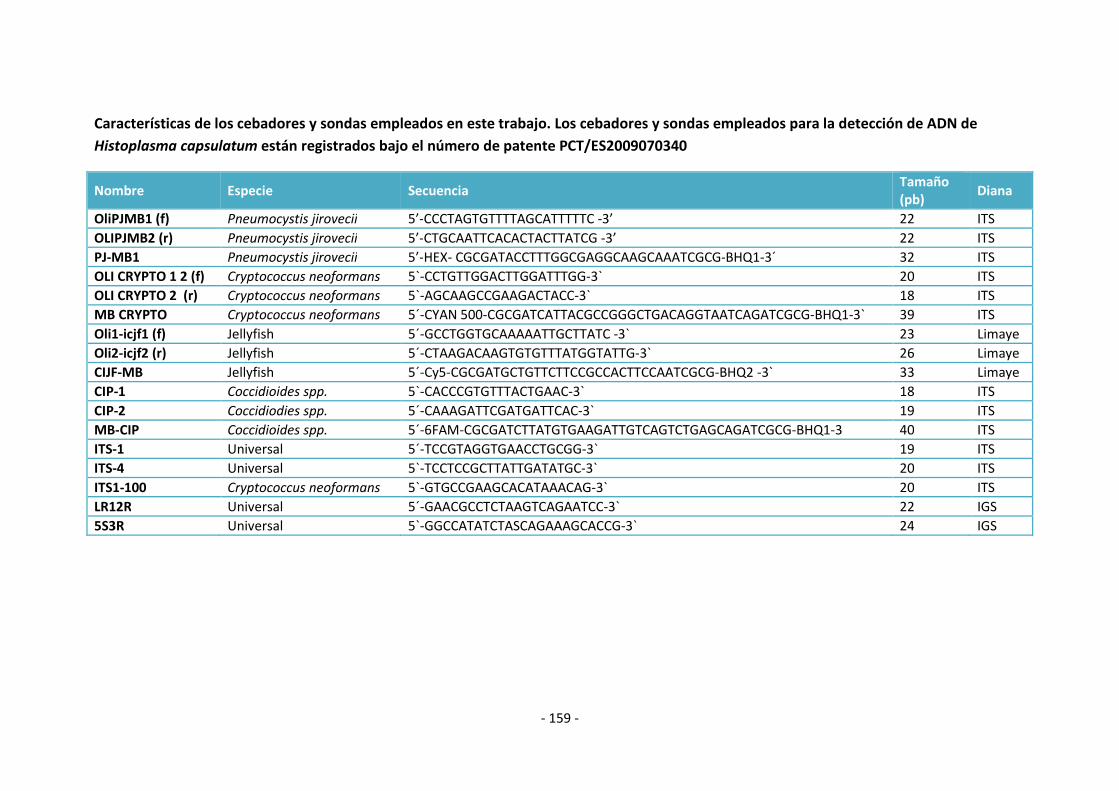
- 159 -
Características de los cebadores y sondas empleados en este trabajo. Los cebadores y sondas empleados para la detección de ADN de
Histoplasma capsulatum están registrados bajo el número de patente PCT/ES2009070340
Nombre Especie Secuencia Tamaño (pb)
Diana
OliPJMB1 (f) Pneumocystis jirovecii 5’-CCCTAGTGTTTTAGCATTTTTC -3’ 22 ITS
OLIPJMB2 (r) Pneumocystis jirovecii 5’-CTGCAATTCACACTACTTATCG -3’ 22 ITS
PJ-MB1 Pneumocystis jirovecii 5’-HEX- CGCGATACCTTTGGCGAGGCAAGCAAATCGCG-BHQ1-3´ 32 ITS
OLI CRYPTO 1 2 (f) Cryptococcus neoformans 5`-CCTGTTGGACTTGGATTTGG-3` 20 ITS
OLI CRYPTO 2 (r) Cryptococcus neoformans 5`-AGCAAGCCGAAGACTACC-3` 18 ITS
MB CRYPTO Cryptococcus neoformans 5´-CYAN 500-CGCGATCATTACGCCGGGCTGACAGGTAATCAGATCGCG-BHQ1-3` 39 ITS
Oli1-icjf1 (f) Jellyfish 5´-GCCTGGTGCAAAAATTGCTTATC -3` 23 Limaye
Oli2-icjf2 (r) Jellyfish 5´-CTAAGACAAGTGTGTTTATGGTATTG-3` 26 Limaye
CIJF-MB Jellyfish 5´-Cy5-CGCGATGCTGTTCTTCCGCCACTTCCAATCGCG-BHQ2 -3` 33 Limaye
CIP-1 Coccidioides spp. 5`-CACCCGTGTTTACTGAAC-3` 18 ITS
CIP-2 Coccidiodies spp. 5´-CAAAGATTCGATGATTCAC-3` 19 ITS
MB-CIP Coccidioides spp. 5´-6FAM-CGCGATCTTATGTGAAGATTGTCAGTCTGAGCAGATCGCG-BHQ1-3 40 ITS
ITS-1 Universal 5´-TCCGTAGGTGAACCTGCGG-3` 19 ITS
ITS-4 Universal 5`-TCCTCCGCTTATTGATATGC-3` 20 ITS
ITS1-100 Cryptococcus neoformans 5`-GTGCCGAAGCACATAAACAG-3` 20 ITS
LR12R Universal 5´-GAACGCCTCTAAGTCAGAATCC-3` 22 IGS
5S3R Universal 5`-GGCCATATCTASCAGAAAGCACCG-3` 24 IGS
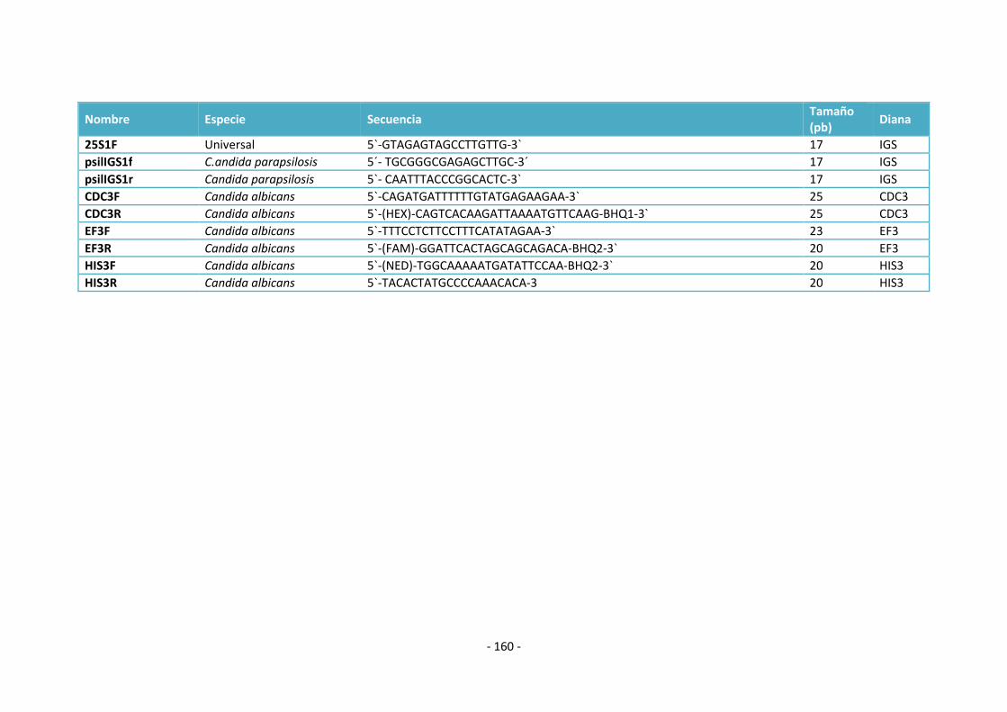
- 160 -
Nombre Especie Secuencia Tamaño (pb)
Diana
25S1F Universal 5`-GTAGAGTAGCCTTGTTG-3` 17 IGS
psilIGS1f C.andida parapsilosis 5´- TGCGGGCGAGAGCTTGC-3´ 17 IGS
psilIGS1r Candida parapsilosis 5`- CAATTTACCCGGCACTC-3` 17 IGS
CDC3F Candida albicans 5`-CAGATGATTTTTTGTATGAGAAGAA-3` 25 CDC3
CDC3R Candida albicans 5`-(HEX)-CAGTCACAAGATTAAAATGTTCAAG-BHQ1-3` 25 CDC3
EF3F Candida albicans 5`-TTTCCTCTTCCTTTCATATAGAA-3` 23 EF3
EF3R Candida albicans 5`-(FAM)-GGATTCACTAGCAGCAGACA-BHQ2-3` 20 EF3
HIS3F Candida albicans 5`-(NED)-TGGCAAAAATGATATTCCAA-BHQ2-3` 20 HIS3
HIS3R Candida albicans 5`-TACACTATGCCCCAAACACA-3 20 HIS3

Anexos
- 161 -
ANEXO 2:
LISTADO DE CEPAS EMPLEADAS EN ESTE TRABAJO
DE TESIS


Anexos
- 163 -
Características de las cepas utilizadas para la estandarización y validación de las
técnicas de PCR en tiempo real desarrolladas en este trabajo de tesis.
Nombre Especie Origen Identificación
CNM-CM 7036 Aspergillus alliaceus Clínico Morfológica/Molecular
CNM-CM 7169 Aspergillus clavatus Clínico Morfológica/Molecular
CNM-CM 3509 Aspergillus flavus Clínico Morfológica/Molecular
CNM-CM 7198 Aspergillus flavus Clínico Morfológica/Molecular
CNM-CM AF237 Aspergillus fumigatus Clínico Morfológica/Molecular
CNM-CM 7012 Aspergillus fumigatus Clínico Morfológica/Molecular
CNM-CM 7168 Aspergillus fumigatus Clínico Morfológica/Molecular
CNM-CM 7152 Aspergillus lentulus Clínico Morfológica/Molecular
CNM-CM 3551 Aspergillus niger Clínico Morfológica/Molecular
CNM-CM 7033 Aspergillus ochraceus Clínico Morfológica/Molecular
CNM-CM 3508 Aspergillus terreus Clínico Morfológica/Molecular
CNM-CM 7082 Aspergillus terreus Clínico Morfológica/Molecular
CNM-CM 3113 Blastomyces dermatitidis Clínico Morfológica/Molecular
CNM-CM 3114 Blastomyces dermatitidis Clínico Morfológica/Molecular
CNM-CM 3115 Blastomyces dermatitidis Clínico Morfológica/Molecular
CNM-CM 3192 Blastomyces dermatitidis Clínico Morfológica/Molecular
CNM-CL 5719 Candida albicans Clínico Molecular
CNM-CL 7426 Candida albicans Clínico Molecular
CNM-CL 7449 Candida albicans Clínico Molecular
CNM-CL 7470 Candida albicans Clínico Molecular
CNM-CL 7471 Candida albicans Clínico Molecular
CNM-CL 7478 Candida albicans Clínico Molecular
CNM-CL 7484 Candida albicans Clínico Molecular
CNM-CL 7498 Candida albicans Clínico Molecular
CNM-CL 7499 Candida albicans Clínico Molecular
CNM-CL 7503 Candida albicans Clínico Molecular
CNM-CL 7504 Candida albicans Clínico Molecular
CNM-CL 7513 Candida albicans Clínico Molecular
CNM-CL 7617 Candida albicans Clínico Molecular
CNM-CL 7624 Candida albicans Clínico Molecular
CNM-CL 7620 Candida albicans Clínico Molecular
CNM-CL 7640 Candida albicans Clínico Molecular
CNM-CL 7643 Candida albicans Clínico Molecular
CNM-CL 7683 Candida albicans Clínico Molecular
CNM-CL 7694 Candida albicans Clínico Molecular
CNM-CL 7705 Candida albicans Clínico Molecular
CNM-CL 7712 Candida albicans Clínico Molecular
CNM-CL 6188 Candida albicans Clínico Molecular
CNM-CL 6361 Candida albicans Clínico Molecular
CNM-CL 6373 Candida albicans Clínico Molecular
CNM-CL 6399 Candida albicans Clínico Molecular
CNM-CL 6431 Candida albicans Clínico Molecular
CNM-CL 6488 Candida albicans Clínico Molecular
CNM-CL 6714 Candida albicans Clínico Molecular
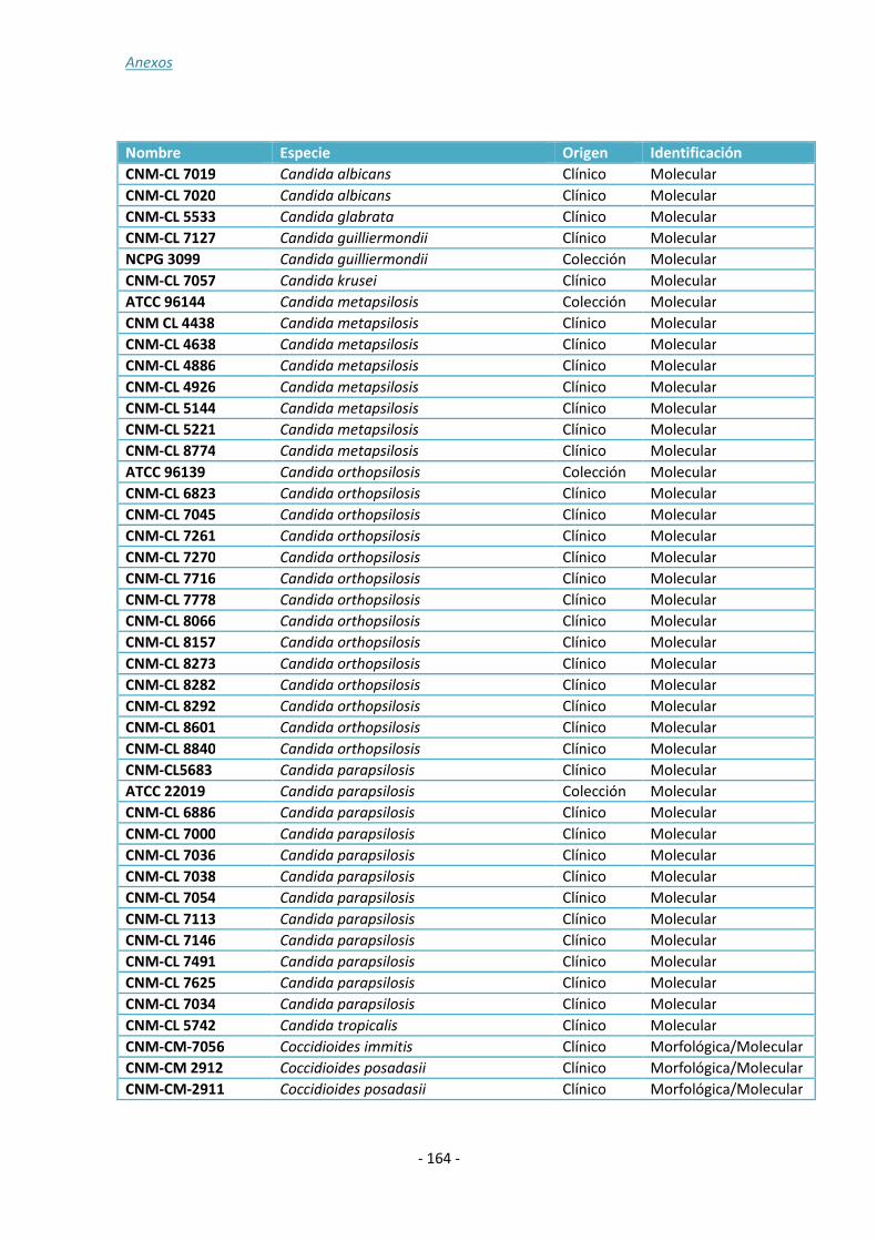
Anexos
- 164 -
Nombre Especie Origen Identificación
CNM-CL 7019 Candida albicans Clínico Molecular
CNM-CL 7020 Candida albicans Clínico Molecular
CNM-CL 5533 Candida glabrata Clínico Molecular
CNM-CL 7127 Candida guilliermondii Clínico Molecular
NCPG 3099 Candida guilliermondii Colección Molecular
CNM-CL 7057 Candida krusei Clínico Molecular
ATCC 96144 Candida metapsilosis Colección Molecular
CNM CL 4438 Candida metapsilosis Clínico Molecular
CNM-CL 4638 Candida metapsilosis Clínico Molecular
CNM-CL 4886 Candida metapsilosis Clínico Molecular
CNM-CL 4926 Candida metapsilosis Clínico Molecular
CNM-CL 5144 Candida metapsilosis Clínico Molecular
CNM-CL 5221 Candida metapsilosis Clínico Molecular
CNM-CL 8774 Candida metapsilosis Clínico Molecular
ATCC 96139 Candida orthopsilosis Colección Molecular
CNM-CL 6823 Candida orthopsilosis Clínico Molecular
CNM-CL 7045 Candida orthopsilosis Clínico Molecular
CNM-CL 7261 Candida orthopsilosis Clínico Molecular
CNM-CL 7270 Candida orthopsilosis Clínico Molecular
CNM-CL 7716 Candida orthopsilosis Clínico Molecular
CNM-CL 7778 Candida orthopsilosis Clínico Molecular
CNM-CL 8066 Candida orthopsilosis Clínico Molecular
CNM-CL 8157 Candida orthopsilosis Clínico Molecular
CNM-CL 8273 Candida orthopsilosis Clínico Molecular
CNM-CL 8282 Candida orthopsilosis Clínico Molecular
CNM-CL 8292 Candida orthopsilosis Clínico Molecular
CNM-CL 8601 Candida orthopsilosis Clínico Molecular
CNM-CL 8840 Candida orthopsilosis Clínico Molecular
CNM-CL5683 Candida parapsilosis Clínico Molecular
ATCC 22019 Candida parapsilosis Colección Molecular
CNM-CL 6886 Candida parapsilosis Clínico Molecular
CNM-CL 7000 Candida parapsilosis Clínico Molecular
CNM-CL 7036 Candida parapsilosis Clínico Molecular
CNM-CL 7038 Candida parapsilosis Clínico Molecular
CNM-CL 7054 Candida parapsilosis Clínico Molecular
CNM-CL 7113 Candida parapsilosis Clínico Molecular
CNM-CL 7146 Candida parapsilosis Clínico Molecular
CNM-CL 7491 Candida parapsilosis Clínico Molecular
CNM-CL 7625 Candida parapsilosis Clínico Molecular
CNM-CL 7034 Candida parapsilosis Clínico Molecular
CNM-CL 5742 Candida tropicalis Clínico Molecular
CNM-CM-7056 Coccidioides immitis Clínico Morfológica/Molecular
CNM-CM 2912 Coccidioides posadasii Clínico Morfológica/Molecular
CNM-CM-2911 Coccidioides posadasii Clínico Morfológica/Molecular
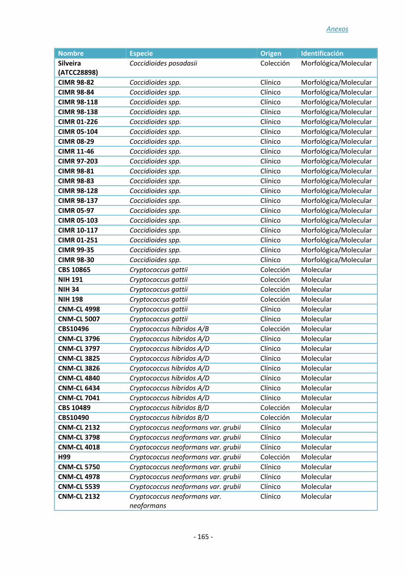
Anexos
- 165 -
Nombre Especie Origen Identificación
Silveira (ATCC28898)
Coccidioides posadasii Colección Morfológica/Molecular
CIMR 98-82 Coccidioides spp. Clínico Morfológica/Molecular
CIMR 98-84 Coccidioides spp. Clínico Morfológica/Molecular
CIMR 98-118 Coccidioides spp. Clínico Morfológica/Molecular
CIMR 98-138 Coccidioides spp. Clínico Morfológica/Molecular
CIMR 01-226 Coccidioides spp. Clínico Morfológica/Molecular
CIMR 05-104 Coccidioides spp. Clínico Morfológica/Molecular
CIMR 08-29 Coccidioides spp. Clínico Morfológica/Molecular
CIMR 11-46 Coccidioides spp. Clínico Morfológica/Molecular
CIMR 97-203 Coccidioides spp. Clínico Morfológica/Molecular
CIMR 98-81 Coccidioides spp. Clínico Morfológica/Molecular
CIMR 98-83 Coccidioides spp. Clínico Morfológica/Molecular
CIMR 98-128 Coccidioides spp. Clínico Morfológica/Molecular
CIMR 98-137 Coccidioides spp. Clínico Morfológica/Molecular
CIMR 05-97 Coccidioides spp. Clínico Morfológica/Molecular
CIMR 05-103 Coccidioides spp. Clínico Morfológica/Molecular
CIMR 10-117 Coccidioides spp. Clínico Morfológica/Molecular
CIMR 01-251 Coccidioides spp. Clínico Morfológica/Molecular
CIMR 99-35 Coccidioides spp. Clínico Morfológica/Molecular
CIMR 98-30 Coccidioides spp. Clínico Morfológica/Molecular
CBS 10865 Cryptococcus gattii Colección Molecular
NIH 191 Cryptococcus gattii Colección Molecular
NIH 34 Cryptococcus gattii Colección Molecular
NIH 198 Cryptococcus gattii Colección Molecular
CNM-CL 4998 Cryptococcus gattii Clínico Molecular
CNM-CL 5007 Cryptococcus gattii Clínico Molecular
CBS10496 Cryptococcus híbridos A/B Colección Molecular
CNM-CL 3796 Cryptococcus híbridos A/D Clínico Molecular
CNM-CL 3797 Cryptococcus híbridos A/D Clínico Molecular
CNM-CL 3825 Cryptococcus híbridos A/D Clínico Molecular
CNM-CL 3826 Cryptococcus híbridos A/D Clínico Molecular
CNM-CL 4840 Cryptococcus híbridos A/D Clínico Molecular
CNM-CL 6434 Cryptococcus híbridos A/D Clínico Molecular
CNM-CL 7041 Cryptococcus híbridos A/D Clínico Molecular
CBS 10489 Cryptococcus híbridos B/D Colección Molecular
CBS10490 Cryptococcus híbridos B/D Colección Molecular
CNM-CL 2132 Cryptococcus neoformans var. grubii Clínico Molecular
CNM-CL 3798 Cryptococcus neoformans var. grubii Clínico Molecular
CNM-CL 4018 Cryptococcus neoformans var. grubii Clínico Molecular
H99 Cryptococcus neoformans var. grubii Colección Molecular
CNM-CL 5750 Cryptococcus neoformans var. grubii Clínico Molecular
CNM-CL 4978 Cryptococcus neoformans var. grubii Clínico Molecular
CNM-CL 5539 Cryptococcus neoformans var. grubii Clínico Molecular
CNM-CL 2132 Cryptococcus neoformans var.
neoformans
Clínico Molecular
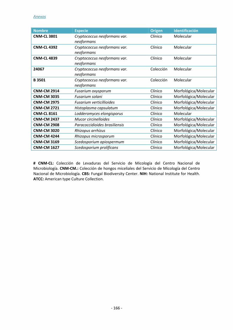
Anexos
- 166 -
Nombre Especie Origen Identificación
CNM-CL 3801 Cryptococcus neoformans var.
neoformans
Clínico Molecular
CNM-CL 4392 Cryptococcus neoformans var.
neoformans
Clínico Molecular
CNM-CL 4839 Cryptococcus neoformans var.
neoformans
Clínico Molecular
24067 Cryptococcus neoformans var.
neoformans
Colección Molecular
B 3501 Cryptococcus neoformans var.
neoformans
Colección Molecular
CNM-CM 2914 Fusarium oxysporum Clínico Morfológica/Molecular
CNM-CM 3035 Fusarium solani Clínico Morfológica/Molecular
CNM-CM 2975 Fusarium verticillioides Clínico Morfológica/Molecular
CNM-CM 2721 Histoplasma capsulatum Clínico Morfológica/Molecular
CNM-CL 8161 Lodderomyces elongisporus Clínico Molecular
CNM-CM 2437 Mucor circinelloides Clínico Morfológica/Molecular
CNM-CM 2908 Paracoccidioides brasiliensis Clínico Morfológica/Molecular
CNM-CM 3020 Rhizopus arrhizus Clínico Morfológica/Molecular
CNM-CM 4244 Rhizopus microsporum Clínico Morfológica/Molecular
CNM-CM 3169 Scedosporium apiospermum Clínico Morfológica/Molecular
CNM-CM 1627 Scedosporium prolificans Clínico Morfológica/Molecular
# CNM-CL: Colección de Levaduras del Servicio de Micología del Centro Nacional de Microbiología. CNM-CM.: Colección de hongos miceliales del Servicio de Micología del Centro Nacional de Microbiología. CBS: Fungal Biodiversity Center. NIH: National Institute for Health. ATCC: American type Culture Collection.

Anexos
- 167 -
ANEXO 3:
NÚMEROS DE ACCESO DE LAS SECUENCIAS
DEPOSITADAS EN GENBANK

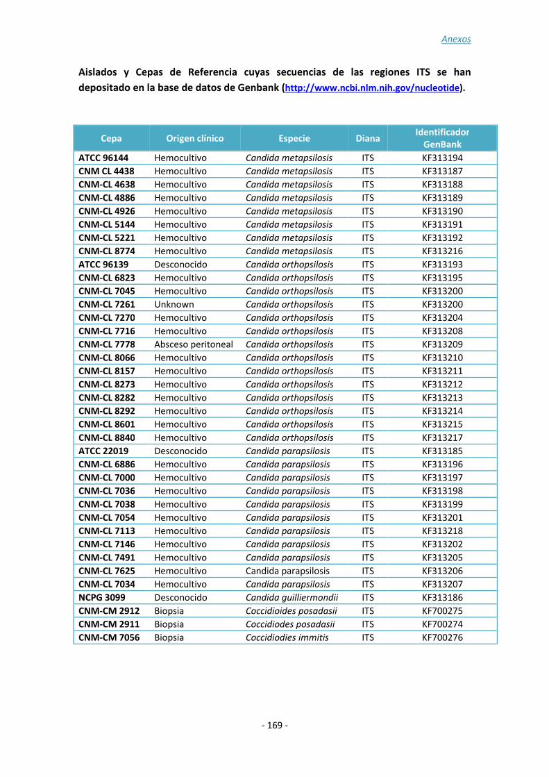
Anexos
- 169 -
Aislados y Cepas de Referencia cuyas secuencias de las regiones ITS se han
depositado en la base de datos de Genbank (http://www.ncbi.nlm.nih.gov/nucleotide).
Cepa Origen clínico Especie Diana Identificador
GenBank
ATCC 96144 Hemocultivo Candida metapsilosis ITS KF313194
CNM CL 4438 Hemocultivo Candida metapsilosis ITS KF313187
CNM-CL 4638 Hemocultivo Candida metapsilosis ITS KF313188
CNM-CL 4886 Hemocultivo Candida metapsilosis ITS KF313189
CNM-CL 4926 Hemocultivo Candida metapsilosis ITS KF313190
CNM-CL 5144 Hemocultivo Candida metapsilosis ITS KF313191
CNM-CL 5221 Hemocultivo Candida metapsilosis ITS KF313192
CNM-CL 8774 Hemocultivo Candida metapsilosis ITS KF313216
ATCC 96139 Desconocido Candida orthopsilosis ITS KF313193
CNM-CL 6823 Hemocultivo Candida orthopsilosis ITS KF313195
CNM-CL 7045 Hemocultivo Candida orthopsilosis ITS KF313200
CNM-CL 7261 Unknown Candida orthopsilosis ITS KF313200
CNM-CL 7270 Hemocultivo Candida orthopsilosis ITS KF313204
CNM-CL 7716 Hemocultivo Candida orthopsilosis ITS KF313208
CNM-CL 7778 Absceso peritoneal Candida orthopsilosis ITS KF313209
CNM-CL 8066 Hemocultivo Candida orthopsilosis ITS KF313210
CNM-CL 8157 Hemocultivo Candida orthopsilosis ITS KF313211
CNM-CL 8273 Hemocultivo Candida orthopsilosis ITS KF313212
CNM-CL 8282 Hemocultivo Candida orthopsilosis ITS KF313213
CNM-CL 8292 Hemocultivo Candida orthopsilosis ITS KF313214
CNM-CL 8601 Hemocultivo Candida orthopsilosis ITS KF313215
CNM-CL 8840 Hemocultivo Candida orthopsilosis ITS KF313217
ATCC 22019 Desconocido Candida parapsilosis ITS KF313185
CNM-CL 6886 Hemocultivo Candida parapsilosis ITS KF313196
CNM-CL 7000 Hemocultivo Candida parapsilosis ITS KF313197
CNM-CL 7036 Hemocultivo Candida parapsilosis ITS KF313198
CNM-CL 7038 Hemocultivo Candida parapsilosis ITS KF313199
CNM-CL 7054 Hemocultivo Candida parapsilosis ITS KF313201
CNM-CL 7113 Hemocultivo Candida parapsilosis ITS KF313218
CNM-CL 7146 Hemocultivo Candida parapsilosis ITS KF313202
CNM-CL 7491 Hemocultivo Candida parapsilosis ITS KF313205
CNM-CL 7625 Hemocultivo Candida parapsilosis ITS KF313206
CNM-CL 7034 Hemocultivo Candida parapsilosis ITS KF313207
NCPG 3099 Desconocido Candida guilliermondii ITS KF313186
CNM-CM 2912 Biopsia Coccidioides posadasii ITS KF700275
CNM-CM 2911 Biopsia Coccidiodes posadasii ITS KF700274
CNM-CM 7056 Biopsia Coccidiodies immitis ITS KF700276
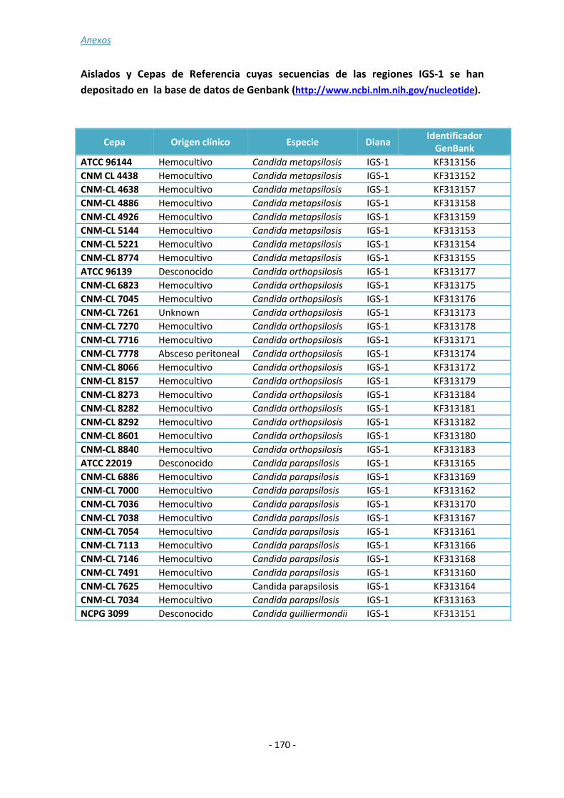
Anexos
- 170 -
Aislados y Cepas de Referencia cuyas secuencias de las regiones IGS-1 se han
depositado en la base de datos de Genbank (http://www.ncbi.nlm.nih.gov/nucleotide).
Cepa Origen clínico Especie Diana Identificador
GenBank
ATCC 96144 Hemocultivo Candida metapsilosis IGS-1 KF313156
CNM CL 4438 Hemocultivo Candida metapsilosis IGS-1 KF313152
CNM-CL 4638 Hemocultivo Candida metapsilosis IGS-1 KF313157
CNM-CL 4886 Hemocultivo Candida metapsilosis IGS-1 KF313158
CNM-CL 4926 Hemocultivo Candida metapsilosis IGS-1 KF313159
CNM-CL 5144 Hemocultivo Candida metapsilosis IGS-1 KF313153
CNM-CL 5221 Hemocultivo Candida metapsilosis IGS-1 KF313154
CNM-CL 8774 Hemocultivo Candida metapsilosis IGS-1 KF313155
ATCC 96139 Desconocido Candida orthopsilosis IGS-1 KF313177
CNM-CL 6823 Hemocultivo Candida orthopsilosis IGS-1 KF313175
CNM-CL 7045 Hemocultivo Candida orthopsilosis IGS-1 KF313176
CNM-CL 7261 Unknown Candida orthopsilosis IGS-1 KF313173
CNM-CL 7270 Hemocultivo Candida orthopsilosis IGS-1 KF313178
CNM-CL 7716 Hemocultivo Candida orthopsilosis IGS-1 KF313171
CNM-CL 7778 Absceso peritoneal Candida orthopsilosis IGS-1 KF313174
CNM-CL 8066 Hemocultivo Candida orthopsilosis IGS-1 KF313172
CNM-CL 8157 Hemocultivo Candida orthopsilosis IGS-1 KF313179
CNM-CL 8273 Hemocultivo Candida orthopsilosis IGS-1 KF313184
CNM-CL 8282 Hemocultivo Candida orthopsilosis IGS-1 KF313181
CNM-CL 8292 Hemocultivo Candida orthopsilosis IGS-1 KF313182
CNM-CL 8601 Hemocultivo Candida orthopsilosis IGS-1 KF313180
CNM-CL 8840 Hemocultivo Candida orthopsilosis IGS-1 KF313183
ATCC 22019 Desconocido Candida parapsilosis IGS-1 KF313165
CNM-CL 6886 Hemocultivo Candida parapsilosis IGS-1 KF313169
CNM-CL 7000 Hemocultivo Candida parapsilosis IGS-1 KF313162
CNM-CL 7036 Hemocultivo Candida parapsilosis IGS-1 KF313170
CNM-CL 7038 Hemocultivo Candida parapsilosis IGS-1 KF313167
CNM-CL 7054 Hemocultivo Candida parapsilosis IGS-1 KF313161
CNM-CL 7113 Hemocultivo Candida parapsilosis IGS-1 KF313166
CNM-CL 7146 Hemocultivo Candida parapsilosis IGS-1 KF313168
CNM-CL 7491 Hemocultivo Candida parapsilosis IGS-1 KF313160
CNM-CL 7625 Hemocultivo Candida parapsilosis IGS-1 KF313164
CNM-CL 7034 Hemocultivo Candida parapsilosis IGS-1 KF313163
NCPG 3099 Desconocido Candida guilliermondii IGS-1 KF313151

Anexos
- 171 -
ANEXO 4:
TABLAS Y FIGURAS


Anexos
173
Índice de tablas y figuras:
Figura 1: Estimación de los casos de infección fúngica invasora por año.
Figura 2: Organización del ADN ribosómico.
Figura 3: Representación esquemática del análisis de curvas de fusión de alta
resolución (HRM) para la determinación de polimorfismos de una sola base.
Tabla 1: Publicaciones más relevantes de los estudios de validación de PCR en tiempo
real para el diagnóstico de IFI, en los últimos diez años.


Anexos
- 175 -
ANEXO 5:
COMUNICACIONES A CONGRESOS DERIVADAS DEL
TRABAJO DE TESIS


Anexos
- 177 -
Comunicaciones a congresos Nacionales:
CONGRESO: XV Congreso SEIMC, en nombre de la Sociedad
Española de Enfermedades Infecciosas y Microbiología
Clínica. Málaga, 1-4 Junio 2011.
PRESENTACIÓN: Poster: Identificación de las variedades de
Cryptococcus neoformas y Cryptococcus gattii mediante
High Resolution Melting. (P-276)
AUTORES: Sara Gago, María José Buitrago Serna, Óscar
Zaragoza Hernández, Isabel Cuesta de La Plaza, Juan Luis
Rodríguez-Tudela y Manuel Cuenca-Estrella. Málaga, Spain.
Introduccion: Las infecciones fúngicas debidas a Cryptococcus spp. afectan a individuos inmunocomprometidos e inmunocompetentes. En la etapa post-sida, C. neoformans
sigue siendo una amenaza para la salud de pacientes inmunodeprimidos no solo en pacientes HIV+,también en pacientes afectados con otro tipo de inmunosupresión. Además, su incidencia en países en vías de desarrollo sigue siendo muy significativa. La especie relacionada C. gattii afecta principalmente a individuos inmunocompetentes, y ha sido descrita como el agente causante de brotes en Canadá y EEUU. C. neoformans
clásicamente ha sido dividido en 5 serotipos (A, B, C, D y AD) que se han agrupado en dos variedades de C. neoformans, var. grubii (serotipo A) y var. neoformans (serotipo D). Los serotipos B y C han sido considerados recientemente como la especie Cryptococcus gattii. Además, se ha descrito la existencia de híbridos AD, BD y BA. Objetivos: Desarrollo de una técnica rápida que permita diferenciar variedades de Cryptococcus spp. mediante análisis de High Resolution Melting (HRM). Material y metodos: Las reacciones de PCR y el análisis de HRM se llevaron a cabo en el equipo LightCycler 480 II (Roche Applied Science). Se diseñaron iniciadores en la región de ITS-1 (Internal Transcribed Spacer-1) del ADN ribosomal para amplificar una región de 110pb. Se optimizaron las condiciones de PCR y se analizaron mediante HRM los productos obtenidos empleando ADN de 24 cepas de Cryptococcus spp. de la colección del Servicio de Micología. Simultáneamente se secuenciaron los fragmentos amplificados. Resultados: Un procedimiento de doble análisis mediante HRM del producto de PCR de las 24 cepas de Cryptococcus spp., permitió la discriminación de 4 grupos que se correspondían con las variedades neoformans e híbridos AD (10 cepas), var. grubii (6 cepas), var. gatti (3 cepas) e híbridos BD y AB (5 cepas). Los resultados de estos grupos se correspondían con las posiciones de mismatch en la secuencia del fragmento obtenido para cada cepa. Tan solo la cepa CL-5007 agrupada inicialmente como var. gattii, fue agrupada con los híbridos BD y AB mediante análisis de secuencia y HRM. Conclusiones: (i) El análisis mediante HRM de los productos de PCR es un método rápido y sencillo que puede ser útil para la diferenciación de variedades de Cryptococcus spp. (ii) Los híbridos AD en el fragmento analizado son semejantes en
Anexos
- 178 -
secuencia a la var. neoformans. (iii) La primera fase del análisis permite la discriminación de C. gattii del complejo C. neoformans. (iv) Deben realizarse nuevos estudios con un mayor número de cepas.
Anexos
- 179 -
CONGRESO: V Fórum Micológico. Alicante, 25 y 26
Noviembre 2011.
PRESENTACIÓN: Oral: Análisis de microsatélites mediante
High Resolution Melting en un caso de candiduria
recurrente por C. albicans (CO-1).
AUTORES: Sara Gago, Isabel Cuesta, Belén Lorenzo, Alicia Gómez-López, Juan Luis Rodríguez-Tudela, Manuel Cuenca-Estrella y María José Buitrago.
Antecedentes: Las infecciones recurrentes debidas a C. albicans son frecuentes en los casos de vaginitis y candidiasis orofaríngea; y los estudios de genotipado demuestran que en la mayoría de los casos todos los episodios se deben a cepas clonales que sufren procesos de microevolución. Una de las técnicas de tipificación más empleadas en la actualidad es el análisis de microsatélites mediante electroforesis capilar, técnica que presenta un gran poder de discriminación y alta reproducibilidad aunque el coste por muestra es elevado. Recientemente se ha señalado que el desarrollo de técnicas basadas en High Resolution Melting (HRM) podría resultar de interés para el análisis de microsatélites.
Objetivos: Evaluar la utilidad de las técnicas de HRM para caracterizar microsatélites de 9 aislamientos de C. albicans de un paciente con candiduria recurrente.
Métodos: Se analizaron 9 aislados de C. albicans de un paciente con candiduria recurrente durante 5 años. Se seleccionaron 3 marcadores previamente descritos (HIS 3, EF 3 y CDC 3) y se analizaron mediante electroforesis capilar y HRM. El análisis de microsatélites mediante electroforesis capilar se realizó siguiendo la metodología publicada por Botterell et al. (2001). Para ello, se incluyeron 20 cepas no relacionadas geográficamente como población control y los electrogramas se resolvieron con el programa GenMapper 4.0. Para el análisis de HRM, tras la amplificación las curvas fueron delineadas en presencia de ResoLight® (Roche, Madrid, España) tras un ciclo de desnaturalización/renaturalización en Light Cycler 450® (Roche, Madrid, España). Como población control se seleccionaron 7 cepas que habían presentado perfiles diferentes mediante la metodología de electroforesis capilar. El poder de discriminación de ambos métodos se calculó según el índice de biodiversidad de Simpson.
Resultados: El poder de discriminación de los tres marcadores mediante electroforesis capilar fue de 0.92 siendo el marcador CDC 3 el que presentaba menor capacidad de discriminación. Las cepas del episodio de candiduria recurrente presentaron el mismo perfil y diferente al de la población control para los tres marcadores, de modo que para HIS 3 y CDC 3 eran homocigotas y heterocigotos para el marcador EF 3. En el análisis de HRM en todos los casos, salvo la cepa CNM-CL 7020 con el marcador CDC 3, las cepas procedentes del paciente con episodio de candiduria recurrente formaron un cluster único a pesar de que el poder de discriminación de los tres marcadores fue
Anexos
- 180 -
menor (0.77). Mediante secuenciación se pudo comprobar que la cepa CL 7020 presentaba una inserción que no era capaz de discriminar la electroforesis capilar.
Conclusiones: (i) Según ambos análisis, las cepas que causaron los episodios de candiduria recurrente están relacionadas genéticamente, (ii) el análisis de microsatélites mediante HRM es una técnica rápida y sencilla para establecer relaciones entre cepas de C. albicans (iii) se realizarán más estudios con un mayor número de cepas.
Bibliografía:
Botterel, F., C. Desterke, C. Costa, and S. Bretagne. 2001. Analysis of microsatellite markers of Candida
albicans used for rapid typing. J. Clin. Microbiol. 39:4076-4081.
Anexos
- 181 -
CONGRESO: XI Congreso Nacional de Micología.
Cádiz, 20-22 Septiembre 2012.
PRESENTACIÓN: Oral: Identificación molecular de
especies del complejo psilosis mediante análisis de
High Resolution Melting (HRM) basado en la región
intergénica del DNA ribosomal IGS-1 (CO-1).
AUTORES: Sara Gago, Ana Alastruey-Izquierdo, María José Buitrago, Manuel Cuenca-Estrella e Isabel Cuesta.
Desde la definición en 2005 del complejo de especies C. parapsilosis (C. parapsilosis
sensu stricto, C. orthopsilosis y C. metapsilosis) (1) la epidemiología de estas especies ha cobrado importancia. La presencia de un patrón de sensibilidad a los antifúngicos disponibles, diferente entre las especies del complejo, pone de manifiesto la necesidad de una identificación precisa. Además, la prevalencia de C. orthopsilosis y C.
metapsilosis difiere entre autores, señalándose a C. orthopsilosis como la quinta causa de fungemia en nuestro país (2) Aunque se dispone de gran número de técnicas de identificación molecular , la mayoría son poco reproducibles, emplean marcadores filogenéticos con bajo poder de resolución o presentan escasa sensibilidad para ser útiles en diagnóstico. El objetivo de este trabajo fue (i) demostrar la utilidad de la región IGS-1 del rDNA para la identificación de especies del complejo psilosis y (ii) el desarrollo de una metodología rápida de identificación mediante HRM basado en esta región. Se analizaron las secuencias de la región ITS e IGS-1 del rDNA de 32 cepas del complejo psilosis de la colección del Centro Nacional de Microbiología incluyéndose como cepas de referencia C. parapsilosis ATCC22019, C. orthopsilosis ATCC96139 y C. metapsilosis ATCC96144. Aunque ambas regiones coinciden en la clasificación a nivel de especie, la divergencia en secuencia de la región IGS1 es muy superior a la de la región ITS, convirtiéndola en un marcador filogenético alternativo a la ITS hasta ahora considerado como gold standard. Sin embargo, la complejidad de esta región debida a la gran abundancia de homopolímeros dificulta su secuenciación, por lo que se desarrolló un método alternativo para su análisis basado en HRM (High Resolution Melting). Se diseñó una pareja de primers en IGS-1 que permitieron amplificar un fragmento de 73 pb. Tras el proceso de amplificación, las curvas fueron normalizadas y todas las cepas se agruparon correctamente con un intervalo de confianza superior al 98%. Nuestros resultados sugieren que el análisis mediante HRM de la región IGS constituye una herramienta rápida y precisa de identificación de las especies estrechamente relacionadas que componen el complejo psilosis. (1) Tavanti A et al. J. Clin. Microbiol. (2005), 43: 284-292. (2) Pemán J et al. J Antimicrob Chemother. (2012), 67:1181-1187.


Anexos
- 183 -
Comunicaciones a congresos internacionales:
CONGRESO: 23rd European Congress of Clinical
Microbiology and Infectious Diseases. Berlin, 27-30
April 2013.
PRESENTACIÓN: Poster: Diagnosis of fungal
opportunistic pneumonia in hiv patients by real
time PCR (P-1490).
AUTORES: Sara Gago, Cristina Esteban, Óscar
Zaragoza, Clara Valero, Jorge Puig de la Bellacasa,
Manuel Cuenca-Estrella y María José Buitrago.
Objective: Pneumocystis pneumonia, histoplasmosis and cryptococcosis are three of
the most frequent causes of fungal opportunistic pneumonia in AIDS patients. A
molecular diagnostic technique based on Real Time PCR was developed for the
simultaneous detection of Pneumocystis jiroveci, Histoplasma capsulatum and
Cryptoccocus neoformans-gattii spp. This technique was tested in cultured strains and
clinical samples from HIV patients.
Methods: A multiplex real time PCR assay was developed for the simultaneous
detection of the fungal species described above. The methodology used was species-
specific molecular beacon probes targeting the ITS region of the rDNA. An internal
control was also included in the assay. Since P. jiroveci cannot grow under laboratory
conditions, a fragment of 175 bp was cloned in a P-GEMt plasmid and used as positive
control. For H. capsulatum and C. neoformans-gattii, genomic DNA from strains
belonging to the collection of the Spanish National Centre for Microbiology were used
as standard. The Multiplex Real Time PCR assay was tested in 24 clinical strains and 28
clinical samples from AIDS patients with proven histoplamosis, cryptococcosis or
pneumocystis pneumonia.
Results: The technique developed had high reproducibility (r > 0.98) and specificity
(100%). No PCR inhibition was detected. For H. capsulatum and C. neoformans-gattii,
the detection limit of the method was 1 fg of genomic fungal DNA per microliter of
sample while for P. jiroveci was 2.92 Log10 copies/microliter. The sensitivity was 100%
for clinical strains and 92.6% for clinical samples. H. capsulatum DNA was detected in
bronchoalveolar lavage, bronchi aspirate, bone marrow, biopsies and whole blood
from 8/10 patients (Ct ~ 28), C. neoformans-gattii was detected in cerebrospinal fluid
from two patients (Ct~ 40) and P. jiroveci was detected in 16 BAL from 16 patients (Ct
~ 31). For one of the patients with proven histoplasmosis, Pneumocystis jiroveci was
also detected in bronchi aspirate.

Anexos
Conclusions: (i)This multiplex real time PCR technique is fast, sensitive and specific (ii)
It can be useful for the clinical diagnosis of fungal opportunistic pneumonia in AIDS
patients and it could also reduce morbidity and mortality associated to an inadequate
therapy. (iii) Further studies are warranted.

Anexos
- 185 -
CONGRESO: 6th Trends in Medical Mycology.
Copenhagen, 11-14 October 2013.
PRESENTACIÓN: Poster: Development and validation of
a quantitative Real-Time PCR assay for the early
diagnosis of coccidioidomycosis.
AUTORES: Sara Gago, María José Buitrago, Karl V.
Clemons, Manuel Cuenca-Estrella y David A. Stevens.
Objectives: Coccidioidomycosis is endemic in the southwestern United States and Latin America. Although primary disease is self-limited, coccidioidomycosis also presents with disseminated disease, especially in immunocompromised patients. Early diagnosis of coccidioidomycosis is difficult, as most clinical signs are non-specific, and cultures often negative. Moreover, serology may be also negative, especially early after infection or not indicative of acute infection. Thus, the development of molecular methods for early diagnosis is needed. The aim of this study was the development of a Real-Time PCR assay for the early diagnosis of coccidioidomycosis, and validation of the assay in a murine model of disseminated infection.
Methods: A Real-Time PCR (RT-PCR) was developed for the detection of Coccidioides
immitis and C. posadasii DNA. Specific primers and a molecular beacon probe were designed to amplify 267 bp from the ITS2 region of the rDNA. An internal control, based on a jellyfish-derived sequence, was also included in each assay. Standard curves were constructed with PCR results from 5 repetitions of 10-fold dilutions of genomic DNA ranging from 1 ng to 1 fg/µl from C. immitis (CNM-CM 7056) and C.
posadasii (CNM-CM 2912), belonging to the mould collection of the Spanish National Center for Microbiology, and C. posadasii strain Silveira. The specificity of the assay was assessed using DNA from 10 unrelated clinicalCoccidioides spp. strains and DNA from 17 other fungal species, and also mouse and human DNA. A murine model of disseminated coccidioidomycosis was used for RT-PCR validation. Female 5-week-old CD-1 mice were infected iv with 250 arthroconidia of C. posadasii strain Silveira. Fungal burdens were determined by CFU and RT-PCR in spleen, lung, and liver on days 5, 10 and 14 days postinfection. Uninfected mice served as controls. CFU and CE (conidia equivalent) values were correlated.
Results: The RT-PCR assay showed high reproducibility (r> 0.99) and specificity (100%); the lower detection limit was 1 fg of genomic DNA per microliter of sample. Fungal burdens after 5, 10, and 14 days of infection were higher by RT-PCR than by CFU enumeration for all tissues analyzed; RT-PCR was negative for blood on those days. No PCR inhibition was detected. Spearman correlations of RT-PCR and CFUs results showed a significant correlation for lungs (p < 0.05) but not for liver or spleen (p > 0.05). Comparative linear regression analysis of RT-PCR and CFUs showed similar slopes (p>0.05) for temporal burden of infection in all organs, indicating RT-PCR, while more sensitive, is equivalent to CFU for disease progression. .

Anexos
Conclusions: We have demonstrated the utility of a fast, sensitive, and specific RT-PCR for the detection of coccidioidomycosis in clinical samples from mice. RT-PCR appeared to better reflect the progression of infection. Further studies are warranted.