Subthalamic nucleus involvement in children: A neuroimaging pattern-recognition approach
Upload
independentCategory
view
5download
0
Systemically administered oxytocin decreasesmethamphetamine activation of the subthalamicnucleus and accumbens core and stimulatesoxytocinergic neurons in the hypothalamus
Dean S. Carson1, Glenn E. Hunt2, Adam J. Guastella1, Lachlan Barber1,3, Jennifer L. Cornish4,Jonathon C. Arnold1,5, Aurelie A. Boucher1 & Iain S. McGregor1,3
Brain & Mind Research Institute1, Discipline of Psychiatry2, School of Psychology3, School of Medical Sciences (Pharmacology), University of Sydney5 andDepartment of Psychology, Macquarie University, Australia4
ABSTRACT adb_247 448..463
Recent preclinical evidence indicates that the neuropeptide oxytocin may have potential in the treatment of drugdependence and drug withdrawal. Oxytocin reduces methamphetamine self-administration, conditioned place prefer-ence and hyperactivity in rodents. However, it is unclear how oxytocin acts in the brain to produce such effects. Thepresent study examined how patterns of neural activation produced by methamphetamine were modified byco-administered oxytocin. Male Sprague-Dawley rats were pretreated with either 2 mg/kg oxytocin (IP) or saline andthen injected with either 2 mg/kg methamphetamine (IP) or saline. After injection, locomotor activity was measuredfor 80 minutes prior to perfusion. As in previous studies, co-administered oxytocin significantly reducedmethamphetamine-induced behaviors. Strikingly, oxytocin significantly reduced methamphetamine-induced Fosexpression in two regions of the basal ganglia: the subthalamic nucleus and the nucleus accumbens core. The sub-thalamic nucleus is of particular interest given emerging evidence for this structure in compulsive, addiction-relevantbehaviors. When administered alone, oxytocin increased Fos expression in several regions, most notably in theoxytocin-synthesizing neurons of the supraoptic nucleus and paraventricular nucleus of the hypothalamus. Thisprovides new evidence for central actions of peripheral oxytocin and suggests a self-stimulation effect of exogenousoxytocin on its own hypothalamic circuitry. Overall, these results give further insight into the way in which oxytocinmight moderate compulsive behaviors and demonstrate the capacity of peripherally administered oxytocin to inducewidespread central effects.
Keywords Accumbens core, addiction, hyperactivity, methamphetamine, oxytocin, subthalamic nucleus.
Correspondence to: Iain McGregor, School of Psychology, University of Sydney, NSW, 2006, Australia. E-mail: [email protected]
INTRODUCTION
Methamphetamine (METH) is a powerful psychostimu-lant that is one of the most addictive and widely abuseddrugs in the world. The ability of METH to stimulate therelease and block the reuptake of dopamine (DA) iscentral to its addictive, stimulant actions (Elkashef et al.2008; Cruickshank & Dyer 2009; Krasnova & Cadet2009). Chronic METH use leads to neurotoxic effects anda variety of psychological problems, including depres-sion, anxiety, aggressiveness, social isolation, psychosis
and neuropsychological deficits (Rose & Grant 2008;Krasnova & Cadet 2009).
There are few effective psychological or pharmacologi-cal treatments for METH addiction (Lee & Rawson 2008).However, new evidence from preclinical studies suggeststhe neuropeptide oxytocin (OXY) may be a promisingcandidate for the treatment of drug (including METH)dependence. In mice, centrally administered OXY reducesMETH-induced hyperactivity, and associated DA releasein the striatum and nucleus accumbens (Qi et al. 2008).Moreover, OXY attenuated METH-induced conditioned
PRECLINICAL STUDY
Addiction Biologydoi:10.1111/j.1369-1600.2010.00247.x
© 2010 The Authors, Addiction Biology © 2010 Society for the Study of Addiction Addiction Biology, 15, 448–463

place preference and abolished stress-induced reinstate-ment of METH-induced conditioned place preference (Qiet al. 2009). In rats, systemic administration of OXYdose-dependently reduced METH self-administration,METH-induced relapse to drug seeking and METH-induced locomotor hyperactivity (Carson et al. 2010).More generally, it has been proposed that OXY has thecapacity to reduce the long-term habit-forming proper-ties of drugs, decrease aversive symptoms during acutewithdrawal and restore the rewarding value of socialinteraction that is impaired by the addiction process(Kovacs, Sarnyai & Szabo 1998; McGregor, Callaghan &Hunt 2008).
The mechanisms through which OXY can modulatethe effects of abused drugs in general, and METH in par-ticular are, at present, unclear. Accordingly, the presentstudy investigated how systemically administered OXY, atbehaviorally relevant doses, affects METH-induced Fosexpression. A secondary question, also of significance,was how peripherally delivered OXY might affect thebrain in itself. Like many neuropeptides, OXY penetratesthe blood–brain barrier only poorly (Landgraf, Ermisch &Hess 1979), and this may represent a major limitation inthe clinical use of the drug. Nonetheless, there is anabundance of evidence showing peripheral OXY effec-tively alters behavior and brain activity in animal models,with anxiolytic, prosocial, and locomotor effects wellestablished (Kovacs & Telegdy 1987; Szabo, Kovacs &Telegdy 1987; Cushing et al. 2003; Kramer, Cushing &Carter 2003; Carson et al. 2010). A recent explosion ofhuman studies, often involving an intranasal route ofdelivery (Kosfeld et al. 2005; Guastella et al. 2009, 2010)but sometimes an intravenous route (Hollander et al.2007), further demonstrate the capacity of OXY to affectaffiliative behaviors and social cognition and to treatvarious psychopathologies. From a practical perspective,it is clear that peripherally administered OXY is morelikely to find clinical use than the centrally administeredneuropeptide.
MATERIALS AND METHODS
Subjects
Thirty-two male Sprague-Dawley rats (ARC, Perth, Aus-tralia) aged 10–14 weeks and weighing 420–567 g werehoused in groups of eight rats per home cage. Food andwater was available at all times except in the experimentalchambers. Lighting was maintained on a 12 hour con-ventional light/dark cycle (lights on between 6 a.m. and6 p.m.), with all experiments performed during the lightcycle. All experimental procedures were conducted inaccordance with the Australian Code of Practice for theCare and Use of Animals for Scientific Purposes (7th
Edition, 2004) and were approved by The University ofSydney Animal Ethics Committee.
Drugs
(+/–) Methamphetamine HCl was purchased from theAustralian Government Analytical Laboratories (Pymble,NSW, Australia) while oxytocin peptide was synthesizedby AusPep Ltd (Parkville, VIC, Australia). All drugs weredissolved in 0.9% saline (SAL).
Apparatus
Test chambers consisted of 4 large black vinyl tubs(80 ¥ 47 ¥ 60 cm) that had a thick layer (10 cm) of grayabsorbent paper pellets on the floor, as describedpreviously (Carson et al. 2010). An overhead miniatureinfrared sensitive video camera (Jaycar Ltd, Australia,model QC3468) above each test chamber was connectedto a PC (Dell) running LABVIEW 8.5 software (NationalInstruments, Austin, TX, USA). The cameras sent imagesto the PC, where automated video tracking software(MotMen 3.0, Motion Mensura, Cooks Hill, NSW, Austra-lia) written in LABVIEW measured distance traveled (cm)and time spent rearing (seconds) by individual rats overtime, with these data collected in eight 10-minute timebins covering the 80-minute test period. The room inwhich testing took place was illuminated with a red 40 Wlight bulb, and maintained at an ambient temperature of21 � 1°C.
Habituation
To minimize any hyperactivity or Fos expression due tonovelty, rats were extensively habituated to the test cham-bers prior to the test day on which drugs were given. Oneach of two consecutive days, the rats were placed ingroups of eight in the test chambers for 1 hour. Followingthis group habituation, rats were individually placed intothe test chambers on three consecutive days for 60minutes. Rats were also injected twice intraperitoneally(IP; right and left side) with SAL during the final twohabituation sessions. The locomotor activity of ratsduring habituation sessions was recorded with scores onthe final day guiding allocation to treatment groups (datanot shown).
Drug testing
Rats were injected IP (right side) with either 2 mg/kgOXY or SAL and placed into a small plastic container forinduction. Ten minutes later, they were injected IP (leftside) with either 2 mg/kg METH or SAL and immediatelyplaced into the test chamber and locomotor activity wasthen recorded for an 80-minute period. The experimental
Oxytocin and addiction 449
© 2010 The Authors, Addiction Biology © 2010 Society for the Study of Addiction Addiction Biology, 15, 448–463

design involved four different treatment groups: SAL/SAL(n = 6), OXY/SAL (n = 6), SAL/METH (n = 10) and OXY/METH (n = 10).
Immunohistochemistry
Immediately following the 80-minute locomotor testsession, rats were anaesthetized with sodium pentobar-bital (120 mg/kg, IP) and transcardially perfused with200 mL of 0.1 M phosphate buffered saline (PBS,pH = 7.3), followed by 400 mL of cold 4% paraformal-dehyde in PBS. Transcardial perfusion was performedwith a Gilson perfusion pump (Gilson Inc., Middleton,WI, USA) at a flow rate of 48 mL/min. Brains were thenremoved, blocked in the coronal plane immediatelycaudal to the olfactory bulbs (~Bregma +5.0) andmidway through the cerebellum (~Bregma -12.0), andpost-fixed overnight in 4% paraformaldehyde bufferedwith 0.1 M PBS at 4°C. They were then placed in 15%phosphate-buffered (PB) sucrose for 24 hours at 4°C,and then 30% sucrose in PB until sectioned. Coronalsections (40 mm) of the whole brain were obtainedusing a cryostat. Consecutive sections were placedsequentially across four vials of 0.1 M PB. Three vialsfrom each brain were transferred into freezing solution(45% 0.1 M PB, 30% ethylene glycol, 25% glycerol) andstored in a freezer (-15°C) as back up tissue (Dielenberg,Hunt & McGregor 2001). From the remaining vial free-floating sections were incubated for 30 minutes in 1%hydrogen peroxide in PB and then for 30 minutes in 3%normal horse serum in PB at room temperature. Sec-tions processed for c-Fos staining were placed in glassvials with 3 mL of primary c-Fos antibody for 72 hoursat 4°C (rabbit polyclonal antisera sc-52 reacts with c-Fosp62 of mouse, rat and human; non-cross reactive withFos B, Fra-1 or Fra-2; Santa Cruz Biotechnology, SantaCruz, CA, USA). The primary antibody was diluted1 : 2000 in phosphate-buffered horse serum (PBH: 0.1%bovine serum albumin, 0.2% Triton X-100, 2% normalhorse serum in PB). After the primary antibody step,sections were washed for 30 minute in PB and incu-bated for 1 hour in secondary antibody (Vector Labora-tories; biotinylated antirabbit IgG made in goat; diluted1 : 500 in PBH). They were then washed in PB for afurther 30 minute and incubated for 1.5 hour in Extra-Avidin-horseradish peroxidase (Sigma; diluted 1 : 1000in PBH). After three further washes (30 minutes each)in PB, horseradish peroxidase activity was visualizedwith nickel diaminobenzidine and glucose oxidase reac-tion as previously described (McGregor et al. 2004). Thisreaction was terminated after 10 minutes by washing inPB. Sections were then mounted onto subbed slides,dehydrated in ascending concentrations of ethanol,xylene cleared and cover slipped.
One of the vials placed in freezing solution was used tovisualize Fos expression in oxytocin expressing cells. Thetissue was washed three times in PB (30 minutes each)prior to staining for Fos using the identical proceduredescribed previously, followed by a second sequence inwhich oxytocin (Chemicon, Boronia, VIC, Australia: anti-oxytocin polyclonal antibody diluted 1 : 5000, catalogueNo. AB911) was added instead of Fos as the primaryantibody and nickel ammonium chloride was notincluded in the visualization process (Thompson et al.2007).
Quantification of Fos and oxytocin expressing cells
The first method described previously produces a blackoval-shaped immunoprecipitate confined to the cellnucleus of c-Fos positive cells. This was quantified micro-scopically using a brain atlas for guidance (Paxinos &Watson 1997). Only darkly labeled oval-shaped nucleiwere counted. All counts were made by an observer whowas blind to group assignment.
Sections were viewed under either a ¥20 or ¥40 objec-tive and an optical graticule was used to manually quan-tify the number of c-Fos positive neurons in each region.A schematic diagram of the 39 quantified regions isshown in Fig. 1.
The number of positive nuclei that fell within a0.5 ¥ 0.5 mm area (¥20 objective) or 0.25 ¥ 0.25 area(¥40 objective) in each region of interest were countedfrom one section per rat as described previously (vanNieuwenhuijzen, McGregor & Hunt 2009). In severalcases, the designated area to be counted was substantiallylarger than the boundaries of the graticule. In such cases,the graticule was placed in a fixed position within theregion of interest relative to known anatomical land-marks. In other cases, the designated area was smallerthan the boundaries of the graticule and only the regionsof interest were counted.
A similar counting method was used to quantify Fos-only expressing cells, oxytocin-only expressing cells, andcells that expressed Fos and oxytocin (double-labeled)within the SON and the PVN. For each rat, bilateral PVNcounts were made at two levels of Bregma (-1.40 and-1.80 mm) using a ¥20 objective, while bilateral countsfor the SON were made at -1.30 and -1.40 mm relativeto Bregma using a ¥40 objective (Thompson et al. 2007).The higher magnification was used for counting the SONas this region was too small to fill the graticule at lowermagnifications. Counts were then summed over the fourregions and averaged to give a final count for single anddouble-labeled cells within the PVN and SON. The datawere also expressed as a percentage of double-labeledcells based on the number of oxytocin expressing cells ineach nucleus.
450 Dean S. Carson et al.
© 2010 The Authors, Addiction Biology © 2010 Society for the Study of Addiction Addiction Biology, 15, 448–463

D3V D3V
BSTld
14 MnPO
Bregma –0.26
SO
Bregma –1.30
17
PVa
PVN
18
19
barrel20
Bregma –1.80 Bregma –2.12 Bregma –2.80
26
22DMH
PMV
Bregma –4.16
31
38LBN
DRD
Bregma –8.00
3632
Bregma –9.30
STh
Bregma –9.68
LC
Bregma –8.72
VLPAG37
CEA
CPU
Bregma +1.00
CPU
CPULSV
ICjM
shell
core
6
7
9
10
11
12
8
Bregma +3.70
LOVO1 2
PrL
Cl
3
IL4 5
Bregma +2.70
15MPA
13
16LPO
VMH
23
24PeF
21CA3
D3V
MePD
MePV
Bregma –3.14
29
30
28LHb
39
Bregma –6.30
DMPAG
33DLPAG
34
VTA
35
25 27BLALH
Figure 1 Schematic diagram of brain regions counted.The figure is adapted from the atlas of Paxinos & Watson (1997), and shows the 39regions in which Fos immunoreactivity was quantified. Open squares indicate the approximate positions of the grid within which Fos countswere made. For explanation of abbreviations see corresponding numbers in Table 1
Oxytocin and addiction 451
© 2010 The Authors, Addiction Biology © 2010 Society for the Study of Addiction Addiction Biology, 15, 448–463

Preparation of images
Digital images were made of representative pieces of tissuefor illustrating the distribution of Fos-positive cells in keyareas as previously described (van Nieuwenhuijzen et al.2009). The digitized images were produced with anOlympus DP70 12.5 megapixel camera system (Olympus,Tokyo, Japan) attached to an Olympic Optical BX51 lightmicroscope. Images were acquired with a Dell precision380 personal computer using custom software suppliedwith the camera. All images were then directly importedinto Adobe Photoshop (CS, Adobe Systems, San Jose, CA,USA), where image size was reduced and the only post-production enhancements were uniform conversion toblack and white, increasing contrast and whitening thenon-tissue surfaces using the eraser tool. Images werethen placed side-by-side in Adobe Illustrator (CS), and textwas added to identify key areas and regions of interest.
Data analysis
SPSS for Windows (SPSS Inc., Chicago, IL, USA) was usedfor all statistical analyses. One-way analysis of variance(ANOVA) was used to analyze locomotor activity acrossthe four groups (SAL/SAL; OXY/SAL; SAL/METH; OXY/METH). A two-way repeated-measures ANOVA examinedgroup by time (8 ¥ 10 minute bins) effects on traveledactivity. One-way ANOVAs were used to analyze c-Fospositive nuclei in the brain regions counted across all fourgroups. All significant one-way ANOVAs were followed bypost hoc tests (Tukey tests). Group values are expressed asmeans � SEM. A probability level of 0.05 was used todetermine statistically significant differences betweengroups.
RESULTS
Locomotor Activity
Figure 2 shows graphical representation of distance trav-eled and time spent rearing for all four treatment groups.During behavioral testing, two rats displayed locomotoractivity that was more than 2 SDs from the group mean(outliers) and were subsequently removed from allanalyses.
One-way ANOVAs revealed significant treatmenteffects for distance traveled (Fig. 2a) (F(3,26) = 10.26,P � 0.001) and rearing behavior (Fig. 2b) (F(3,26) =7.79, P = 0.001). Subsequent post-hoc analyses revealeddistance traveled and time spent rearing for the SAL/METH group was significantly increased relative to theSAL/SAL group. Analyses also revealed significantlyreduced distance traveled and time spent rearing in theOXY/METH group relative to the SAL/METH group againoutlining the ability of OXY to reduce METH-induced
DISTANCE TRAVELLED
SAL/S
AL
OXY/S
AL
SAL/M
ETH
OXY/M
ETH
0
5000
10 000
15 000
20 000
(a)
******
*
Ce
nti
me
ters
TIME SPENT REARING
SAL/S
AL
OXY/S
AL
SAL/M
ETH
OXY/M
ETH
0
100
200
300
(b)
*
***
**S
eco
nd
s
TIME-LINE TRAVELLED ACTIVITY
10 20 30 40 50 60 70 80
0
1000
2000
3000
4000
SAL/SAL
OXY/SAL
SAL/METH
OXY/METH
(c)
Mins
Ce
nti
me
ters
Figure 2 Oxytocin given systemically at 2 mg/kg reduced distancetraveled (a) and time spent rearing (b) in a time-dependent manner(c) following 2 mg/kg methamphetamine challenge. From left to righton graphs (a & b): Control groups were treated with either salinealone (SAL/SAL; n = 6) or 2 mg/kg oxytocin (OXY/SAL; n = 5). Othergroups were treated with 2 mg/kg methamphetamine either alone(SAL/METH; n = 10) or with 2 mg/kg oxytocin pre-treatment (OXY/METH; n = 9). *� 0.05, **� 0.01, and ***� 0.001 relative to SAL/METH group (solid bar) (Tukey tests). Data are shown asmean + SEM
452 Dean S. Carson et al.
© 2010 The Authors, Addiction Biology © 2010 Society for the Study of Addiction Addiction Biology, 15, 448–463
hyperactivity. There was no significant difference betweenthe SAL/SAL and the OXY/SAL group in distance traveledor time spent rearing. Repeated measures ANOVA of theeight 10 minute time bins for the travelled activity(Fig. 2c) indicated a significant effect of treatment(F(3,26) = 6.58, P = 0.002) and a treatment by timeinteraction (F(21, 182) = 1.64, P = 0.044), but no effectof time (F(7, 182) = 1.21, P = 0.298).
Fos immunoreactivity
Fos counts for the four groups across 39 brain regions areshown in Table 1. Fos expression in SAL only treatedanimals was low across all brain regions in line with thethorough habituation to the locomotor chambers andextensive handling these rats received.
Importantly, the OXY/METH group showed signifi-cantly reduced Fos expression relative to the SAL/METHgroup in two areas: the subthalamic nucleus (Fig. 3)(F(3,26) = 11.38, P � 0.001) and the nucleus accum-bens core (Table 1) (F(3,26) = 29.22, P � 0.001). Crossgroup comparisons also revealed there was increased Fosexpression in the SAL/METH but not the OXY/METHgroups relative to the SAL/SAL group in 2 regions:the prelimbic cortex (F(3,26) = 3.08, P = 0.045) andthe perifornical area of the lateral hypothalamus(F(3,26) = 3.44, P = 0.032). This gives some presump-tive evidence of an OXY effect on METH activation inthese regions.
An increase in Fos positive cells was observedin both the SAL/METH and OXY/METH groupsrelative to the SAL/SAL group in 12 different brainareas. This indicates no significant effect of OXYon METH-induced activation in the following re-gions: the orbital cortex (ventral [F(3,26) = 10.52,P � 0.001] and lateral [F(3,26) = 8.35, P � 0.001]),claustrum (F(3,26) = 10.32, P � 0.001), Islands ofCalleja (major) (F(3,26) = 43.41, P � 0.001), caudate-putamen (central [F(3,26) = 39.65, P � 0.001], medial[F(3,26) = 40.31, P � 0.001], and dorsolateral[F(3,26) = 7.84, P = 0.001]), bed nucleus of the striaterminalis (dorsolateral) (F(3,26) = 4.28, P = 0.014),barrel cortex (F(3,26) = 14.79, P � 0.001), lateralhabenula (F(3,26) = 9.56, P � 0.001), ventral tegmen-tal area (F(3,26) = 35.60, P � 0.001), and dorsal raphenucleus (F(3,26) = 6.85, P = 0.001).
The OXY/SAL group as well as the OXY/METH groupsshowed increased Fos expression in five brain regionsrelative to the SAL/SAL group (Table 1). These were thesupraoptic nucleus (F(3,26) = 10.86, P � 0.001),paraventricular hypothalamic nucleus (F(3,26) = 20.31,P � 0.001), central amygdala (Fig. 4) (F(3,26) = 13.99,P � 0.001), lateral parabrachial nucleus (Fig. 5)(F(3,23) = 20.63, P � 0.001), and locus coeruleus
(Fig. 6) (F(3,21) = 10.74, P � 0.001). These regions aretherefore ones where OXY has an activating effect, inde-pendent of METH administration. It is clear, however,from examining the SAL/METH group that METH itselfactivates two of these regions independently of OXY: thecentral amygdala and lateral parabrachial nucleus.
There were five regions in which the combination ofOXY and METH appeared to produce greater effects thaneither METH or OXY given alone, as shown by significantdifferences from the SAL/SAL group only in the OXY/METH group. These were the accumbens shell(F(3,26) = 4.39, P = 0.013), the median preopticnucleus (F(3,26) = 4.19, P = 0.015), the medial preopticarea (F(3,26) = 4.17, P = 0.015), paraventricular ante-rior thalamic nucleus (F(3,26) = 3.91, P = 0.020) andthe medial amygdala (posteroventral) (F(3,26) = 4.88,P = 0.008). In these regions, there appear to be potenti-ating effects of METH on OXY and vice versa (Table 1).
Fos expression and oxytocin labeled neurons
Table 2 shows the number of Fos and oxytocin positivecells in the SON and PVN. OXY/SAL and OXY/METH ratsshowed a significantly increased number and percentageof double-labeled cells in both regions (Fig. 7) relativeto the SAL/SAL group. In OXY-treated rats, there wasa similar proportion of double-labeled cells in theSON (44.9% of the OXY cells were Fos positive)(F(3,26) = 8.77, P � 0.001) and PVN (49.2% of theOXY cells were Fos positive) (F(3,26) = 17.12,P � 0.001).
DISCUSSION
The current study addresses two significant and relatedresearch questions: firstly the way in which OXY acts inthe brain to reduce METH-induced behaviors, and sec-ondly to characterize the central effects of peripherallyadministered OXY per se. Striking results were obtainedwith respect to both questions. Firstly, OXY suppressedMETH-induced Fos activity in the subthalamic nucleus(STh) and also affected activation in a small set of othersites including the nucleus accumbens core and prelimbiccortex. Secondly, peripherally administered OXY stronglyinfluenced a number of brain regions, but particularly inthe oxytocin-positive cells of the SON and PVN, where themajority of brain oxytocin is synthesized and released.
Behavioral effects of oxytocin and methamphetamine
In agreement with previous reports, METH producedcharacteristic hyperactivity and increased rearing, andthese effects were attenuated by IP oxytocin (Qi et al.2008; Carson et al. 2010). A more complete reversal ofMETH hyperactivity was seen in our previous study that
Oxytocin and addiction 453
© 2010 The Authors, Addiction Biology © 2010 Society for the Study of Addiction Addiction Biology, 15, 448–463

used 1 mg/kg of METH and OXY, suggesting that escalat-ing the METH dose may reduce the capacity of OXY toreverse METH stimulant effects. OXY doses between 0.25and 1 mg/kg given systemically can cause sedation inSprague-Dawley rats with maximal effects taking place inthe hour following injection (Uvnas-Moberg et al. 1994).
Here, we showed that OXY was capable of dampeningMETH-induced hyperactivity throughout the entire80-minute behavioral assay. In addition to the sedativeeffects of systemically administered OXY, a wide variety ofother effects can be observed in rodents such as increasesin maternal (Jin et al. 2007), social (Popik, Vetulani & van
Table 1 Mean number (�SEM) of Fos-positive cells.
Region (see Fig. 1) BregmaSAL/SAL(n = 6)
OXY/SAL(n = 5)
SAL/METH(n = 10)
OXY/METH(n = 9)
Areas with significant METH-induced Fos expression reduced by OXY3. Prelimbic cortex +2.70 12.8 � 5.1 18.8 � 5.1 28.4 � 3.1a 24.3 � 39. Nucleus accumbens core +1.00 0.2 � 0.2 0.4 � 0.2 11.5 � 1.9a,b,d 5.2 � 1.1a,b
24. Perifornical area -2.80 8.8 � 1.7 9.8 � 3.1 18.9 � 3.1a 11.7 � 1.532. Subthalamic nucleus -4.16 2.3 � 0.8 3.6 � 1.9 30.3 � 5.1a,b,d 11.2 � 3.3
Areas with METH-induced Fos expression not affected by OXY1. Orbital cortex (ventral) +3.70 12.3 � 1.5 16 � 4.3 47.7 � 5.9a,b 49.1 � 8.2a,b
2. Orbital cortex (lateral) +3.70 7.3 � 1.3 13.8 � 5.6 35.7 � 5.1a,b 24.2 � 3.1a
5. Claustrum +2.70 16.5 � 4.6 22.8 � 5.6 59.4 � 5.7a,b 52 � 7.4a,b
7. Islands of Calleja (major) +1.00 4 � 1.5 9 � 2.3 80.4 � 8.4a,b 75.6 � 14.4a,b
10. Caudate-putamen (medial) +1.00 3.3 � 0.8 2.0 � 0.8 63 � 9.4a,b 37.3 � 7.4a,b
11. Caudate-putamen (central) +1.00 0 0.2 � 0.2 35.5 � 5.2a,b 31.9 � 5.9a,b
12. Caudate-putamen (dorsolateral) +1.00 0.2 � 0.2 0 5.5 � 1.4a,b 5.8 � 2.2a,b
13. Bed nucleus of the stria terminalis (dorsolateral) -0.26 3.3 � 0.6 8.4 � 2.2 16.1 � 4.5a 13.8 � 2.5a
20. Barrel cortex -2.12 4.2 � 0.9 6.4 � 1.9 29.9 � 4a,b 21.2 � 4.8a
28. Lateral habenula -3.14 8.8 � 2.9 8.2 � 1.7 30 � 4.5a,b 22.9 � 2.7a,b
35. Ventral tegmental area -6.30 1.2 � 0.5 1.2 � 1.2 30.7 � 2a,b 28.6 � 5.4a,b
36. Dorsal raphe nucleus -8.00 3 � 2.1 4.4 � 1 15.8 � 1.9a,b 13 � 3.1a
Areas with significant OXY-induced Fos expression above SAL/SAL17. Supraoptic nucleus (x40) -1.30 1.3 � 0.6 25.8 � 5.6a,c 9.4 � 2.9 28.8 � 4.7a,c
19. Paraventricular hypothalamic nucleus -1.80 27.3 � 7.9 124.4 � 27.6a,c 39 � 8 155.7 � 18.4a,c
26. Central amygdala -2.80 2.5 � 1.6 24.2 � 5.1a 41.7 � 8.1a 33.4 � 5.4a
38. Lateral parabrachial nucleus -9.30 0.8 � 0.4 27 � 8a 27.2 � 4.3a 37 � 4.2a
39. Locus coeruleus -9.68 2.2 � 0.6 20.7 � 2.7a,c 3.3 � 0.9 14.6 � 3.1a,c
Areas with OXY/METH-induced Fos expression above SAL/SAL8. Nucleus accumbens shell +1.00 6 � 1.5 7.2 � 3.2 21.7 � 4.6 28.2 � 6.2a
14. Median preoptic nucleus -0.26 9.7 � 1.7 18 � 2 12.2 � 1.3 25.4 � 5.1a
15. Medial preoptic area -0.26 9.2 � 2.4 11.4 � 1.7 11.4 � 2.4 24.7 � 4.4a,c
18. Paraventricular anterior thalamic nucleus -1.80 28.2 � 4.7 30.2 � 5.4 44.2 � 3.8 52.3 � 7.2a
29. Medial amygdala (posteroventral) -3.14 13.5 � 4.1 11.2 � 3.7 24.8 � 2.7 34.7 � 6.5a,b
No effect of METH or OXY compared to saline controls4. Infralimbic cortex +2.70 21.8 � 5.1 19.8 � 3.6 28.5 � 3.1 31.6 � 3.86. Lateral septal nucleus (ventral) +1.00 23.2 � 9.2 15.2 � 5.2 21.1 � 3.2 21.1 � 4
16. Lateral preoptic area -0.26 13 � 3.4 10.8 � 1.8 11.9 � 1.7 10 � 1.821. CA3 layer of the hippocampus -2.80 0.5 � 0.3 0.4 � 0.2 3.1 � 0.8 3.3 � 0.822. Dorsomedial hypothalamus -2.80 30.5 � 4.8 31.4 � 5 35.6 � 2.9 37.1 � 4.723. Ventromedial hypothalamus (x40) -2.80 0.8 � 0.3 0.6 � 0.2 3.3 � 1.6 5.0 � 2.825. Lateral hypothalamus -2.80 1.5 � 0.6 2.4 � 0.7 4.2 � 1.2 3.6 � 0.827. Basolateral amygdala -2.80 3.2 � 1.4 3.2 � 1.5 7.7 � 1.8 7.2 � 1.530. Medial amygdala (posterodorsal) -3.14 13.5 � 2.4 12.2 � 2.6 16.4 � 1.3 25.2 � 4.331. Premammillary nucleus (ventral) -4.16 13.7 � 6.2 27.8 � 5.8 21.6 � 4.8 33.8 � 6.333. Periaqueductal gray (dorsomedial) -6.30 7.8 � 1.7 7.4 � 2.3 10.9 � 1.6 9.2 � 234. Periaqueductal gray (dorsolateral) -6.30 9.8 � 2.2 9 � 0.8 14.3 � 1.2 12.4 � 1.337. Periaqueductal gray (ventrolateral) -8.72 15 � 2.1 17.2 � 3.5 26.2 � 3.7 29.2 � 4.1
asignificantly different from SAL/SAL group (Tukey test). bsignificantly different from OXY/SAL group. csignificantly different from SAL/METH group.dsignificantly different from OXY/METH group.
454 Dean S. Carson et al.
© 2010 The Authors, Addiction Biology © 2010 Society for the Study of Addiction Addiction Biology, 15, 448–463

Ree 1992) and sexual behaviors (Moody & Adler 1995),increased wound healing (Dusunceli et al. 2008),reduced anxiety and stress (Uvnas-Moberg et al. 1994;Ring et al. 2006), and a reduction in compulsive drugtaking and drug-induced stereotypic behaviors (Kovacset al. 1998).
Methamphetamine-induced neural activity and reversalby oxytocin
Rats exposed to an acute dose of METH showed wide-spread Fos expression in cortical, amygdala, dorsal andventral striatal regions consistent with previous reports(Pontieri et al. 1990; Gross & Marshall 2009; Holder et al.2010). Rats pretreated with OXY prior to METH admin-istration showed a reduction in Fos activity in the SThand the accumbens core to a level significantly below thatof animals pretreated with SAL prior to METH adminis-tration. Additionally, there was a less remarkable reduc-tion of neural activity within the prelimbic cortex and theperifornical region of the lateral hypothalamus to a levelsimilar to that of SAL only treated rats.
Subthalamic nucleus
The STh has previously been implicated in METH-induced locomotor hyperactivity (Pontieri et al. 1990)and plays a complex role in motivation (Baunez, Amalric
& Robbins 2002; Lardeux & Baunez 2008), reward(Baunez et al. 2005; Lardeux et al. 2009) and obsessive–compulsive behaviors (Winter et al. 2008a; Klavir et al.2009). Additionally, an abundance of evidence alsoimplicates the STh in motor dysfunction seen in Parkin-son’s Disease (Bergman, Wichmann & DeLong 1990;Cooper & Mitchell 1995; Benabid et al. 2001).
Bilateral lesions of the STh markedly reduced themotivation to self-administer intravenous cocaine butincreased the motivation to acquire a natural food reward(Baunez et al. 2005). These findings are startling giventhe lack of specificity of current treatments for drugversus natural rewards. Previous work by the same groupfound that lesions of the STh increases the motivation toconsume food without increasing the quantity of foodconsumed (Baunez et al. 2002). Other important findingsare that high frequency stimulation and pharmacologicalinactivation of the STh reduces compulsive lever-pressing(Klavir et al. 2009) and drug-induced stereotypic behav-iors (Bergmann et al. 2004).
Albeit impressive, the findings discussed above conflictwith other preclinical and clinical research which sug-gests that lesions or stimulation of the STh leads to anincrease in motivation for drug reward (Uslaner, Yang &Robinson 2005), an increase in compulsive gambling(Smeding et al. 2007), and increased compulsivelever-pressing (Winter et al. 2008a). Regardless of these
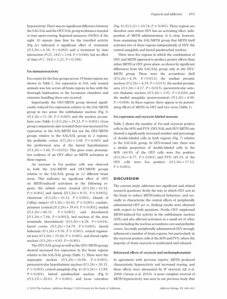
STh
cp
Y/SALXO(b)L/SALAS(a)
Y/METHXO(d) /METHLAS(c)
Figure 3 The distribution of Fos immunoreactive cells (black dots) in the subthalamic nucleus (STh) from representative rats treated withsaline/saline (a; SAL/SAL), oxytocin/saline (b; OXY/SAL), saline/methamphetamine (c; SAL/METH), and oxytocin/methamphetamine (d;OXY/METH). Scale bar, 500 mm. cp, cerebral peduncle
Oxytocin and addiction 455
© 2010 The Authors, Addiction Biology © 2010 Society for the Study of Addiction Addiction Biology, 15, 448–463

conflicting findings, our data show that OXY inhibitsMETH-induced activity in the STh, and we argue that thisdiffers greatly from the inactivation or complete ablationof this region. Thus, we maintain that the development ofnovel therapeutics that suppress over-activity in the SThmay prove to be a beneficial target for drug dependence(Baunez et al. 2005) and possibly motor dysfunction inParkinson’s Disease.
Nucleus Accumbens core
The nucleus accumbens is an important part of themesolimbic dopamine system and has been implicated inacute METH intoxication (Pontieri et al. 1990). Here, wefound that METH-induced activity in the accumbens corewas significantly reduced by OXY pretreatment. Previousresearch shows OXY given centrally reduces METH-induced DA activity in the accumbens (Qi et al. 2008).Interestingly, lithium has been shown to increase oxyto-cin release in rodents (Cui et al. 2001) and a previous
report provides evidence that chronic lithium treatmentprevents METH-induced Fos immunoreactivity in the pre-limbic cortex, accumbens core, caudate-putamen andcentral amygdala (CEA) (Lee et al. 1999), possiblythrough an oxytocin-dependent mechanism. Althoughthe accumbens shell is usually associated with the acuteeffects of drug and natural reward (Bassareo & Di Chiara1999; Lardeux & Baunez 2008; Engleman et al. 2009)substantial evidence outlines the involvement of theaccumbens core in the instrumental motivational com-ponent of drug-seeking and relapse to drug-seekingbehavior (Smith-Roe & Kelley 2000; Di Chiara 2002;Chiang et al. 2009).
There is clear connectivity between the accumbenscore and STh with synergistic involvement of theseregions in microcircuitry that controls motor, cognitive,and limbic function. Specifically, the accumbens core hasprojections to the ventral pallidum area that projects tothe medial part of the STh (Maurice et al. 1998) and thusmutual inhibition of drug-induced activity in these brain
4VLC
Me5
/SALYXO (b)/SALLAS (a)
/METHYXO (d)/METHLAS (c)
Figure 4 The distribution of Fos immunoreactive cells (black dots) in the locus coeruleus (LC) from representative rats treated withsaline/saline (a; SAL/SAL), oxytocin/saline (b; OXY/SAL), saline/methamphetamine (c; SAL/METH), and oxytocin/methamphetamine (d;OXY/METH). Scale bar, 200 mm. 4V, fourth ventricle; Me5, mesencephalic trigeminal nucleus
456 Dean S. Carson et al.
© 2010 The Authors, Addiction Biology © 2010 Society for the Study of Addiction Addiction Biology, 15, 448–463

(b) OXY/SAL
Y/METHXO (d)L/METHAS (c)
(a) SAL/SAL
LPB
scp
Figure 5 The distribution of Fos immunoreactive cells (black dots) in the lateral parabrachial nucleus (LPB) from representative rats treatedwith saline/saline (a; SAL/SAL), oxytocin/saline (b; OXY/SAL), saline/methamphetamine (c; SAL/METH), and oxytocin/methamphetamine (d;OXY/METH). Scale bar, 200 mm. scp, superior cerebellar peduncle
/SALYXO (b)L/SALAS (a)
HTEM/YXO (d)HTEM/LAS (c)
CEA
Figure 6 The distribution of Fos immunoreactive cells (black dots) in the central amygdala (CEA) from representative rats treated withsaline/saline (a; SAL/SAL), oxytocin/saline (b; OXY/SAL), saline/methamphetamine (c; SAL/METH), and oxytocin/methamphetamine (d;OXY/METH). Scale bar, 200 mm
Oxytocin and addiction 457
© 2010 The Authors, Addiction Biology © 2010 Society for the Study of Addiction Addiction Biology, 15, 448–463

regions suggests OXY is more capable of moderatingactivity within this complex neural circuitry compared tothe crude inactivation of the STh with lesions or high-frequency stimulation. This argument is supported byresearch showing that increased compulsive behaviorfollowing lesions of the STh is linked to decreased sero-tonin activity in a number of brain areas including thecaudate-putamen but not in the nucleus accumbens(Winter et al. 2008a). Further, high frequency stimula-tion of the STh has been shown to powerfully stimulateDA activity in the accumbens shell which may explainsome of the detrimental changes in psychiatric functionexhibited by patients following deep brain stimulationtreatment for Parkinson’s Disease (Winter et al. 2008b).
Although marginal, OXY also reduced METH-inducedFos activity in the prelimbic cortex, an area with directconnections to the accumbens core and the STh. Thus, itmay be that OXY reduces activity within a cortico-basalganglia-thalamocortical ‘limbic’ loop (Maurice et al.1998; Temel et al. 2005), which is responsible for reduc-ing the incentive motivational properties of METH. Giventhe complex neural circuitry involved in motor functionand drug reward it would be interesting to examine theeffects of OXY on drug-induced stereotypical behaviorsthat are commonly seen after chronic, or acute high dose,psychostimulant administration. Stereotypy may involvespecific sub-regions of the striatum and associated basalganglia and limbic neural circuits (Canales & Graybiel2000) that may well be open to influence by OXY.
Neural activity from systemic administrationof oxytocin
Only a small proportion (approximately 1%) of peripher-ally administered OXY is thought to cross the blood–brainbarrier (Landgraf et al. 1979; Ermisch et al. 1985).However, by giving peripheral doses of large magnitude,clear behavioral effects can be obtained in rodents,
suggesting effective CNS penetration. This is further dem-onstrated by the results of the current study, in which weshow that systemic administration of OXY is capable ofactivating a network of brain regions, particularly thosewith high concentration of oxytocin receptors includingthe SON, PVN, CEA and locus coeruleus (LC) (Yoshimuraet al. 1993; Vaccari, Lolait & Ostrowski 1998). Theseresults are in agreement with two previous studies involv-ing intracerebroventricular administration of lower dosesof oxytocin (Popeski & Woodside 2001; Kita, Yoshida &Nishino 2006).
The oxytocin synthesizing neurons of the SON andPVN were strongly activated by peripheral OXY (Swanson& Sawchenko 1983). These neurons contain OXY recep-tors, the stimulation of which may lead to cellular OXYrelease in a feed-forward fashion (Rossoni et al. 2008).This, in turn, has been linked to the profound neuroplas-ticity of hypothalamic oxytocin neurons, whereby smallamounts of stimulation lead to long lasting morphologi-cal and functional changes (Catheline et al. 2006). Closeinspection of the distribution of Fos activity in the SONand PVN (Fig. 7) suggests that OXY may also induceactivation of neurons known to be high in both vaso-pressin and corticotropin-releasing hormone. This mayrelate to the relatively high affinity of OXY for otherreceptors, particularly the vasopressin V1a receptor(Akerlund et al. 1999) or could reflect indirect activationof these neurons from directly activated OXY containingneurons nearby.
Interestingly, not all regions known to contain highlevels of oxytocin receptors (Yoshimura et al. 1993;Vaccari et al. 1998) showed Fos activation in the presentstudy after systemic OXY treatment. Examples of thisinclude the ventromedial hypothalamus, bed nucleus ofthe stria terminalis and the medial amygdala. Thereasons for this are not entirely clear, although it is wellknown that not all neurons have a phenotype that readilyexpresses the Fos protein. Thus, absence of Fos does not
Table 2 Number and proportion of Fos/oxytocin labeled cells in SON and PVN.
RegionSAL/SAL(n = 6)
OXY/SAL(n = 5)
SAL/METH(n = 10)
OXY/METH(n = 9)
Supraoptic nucleus (SON)Fos only 0.8 � 0.5 2.6 � 0.5 1.4 � 0.5 4.1 � 1.0a,b
Fos + oxytocin 2.6 � 1.3 16.5 � 2.8a 7.8 � 2.2 17.5 � 2.7a,b
oxytocin labeled cells 28.7 � 2.5 35.7 � 2.3 31.1 � 2.0 37.9 � 1.4a
% oxytocin labeled 8% 44.9%a 23.1% 45%a,b
Paraventricular hypothalamic nucleus (PVN)Fos only 17.8 � 4.8 48.2 � 14.3a 20.9 � 4.4 67.2 � 7.4a,b
Fos + oxytocin 5.7 � 1.6 30.8 � 5.5a,b 11.2 � 3.1 36.1 � 5.4a,b
oxytocin labeled cells 59.4 � 2.7 60.9 � 4.3 60.4 � 3.2 68.5 � 4.6% oxytocin labeled 9.5% 49.2%a,b 17.7% 52.3%a,b
asignificantly different from SAL/SAL group (Tukey test). bsignificantly different from SAL/METHgroup.
458 Dean S. Carson et al.
© 2010 The Authors, Addiction Biology © 2010 Society for the Study of Addiction Addiction Biology, 15, 448–463

(a) SAL/SAL
(c) OXY/SAL
(e) SAL/METH
(g) OXY/METH (h) OXY/METH
(f) SAL/METH
(d) OXY/SAL
(b) SAL/SAL
SON
opt
3VPVN
Figure 7 The distribution of Fos immunoreactive cells (black dots) and oxytocin expressing neurons (amber immunoprecipitate) in thesupraoptic nucleus (SON; a, c, e, g) and the paraventricular nucleus of the hypothalamus (PVN; b, d, f, h) from representative rats treated withsaline/saline (a, b; SAL/SAL), oxytocin/saline (c, d; OXY/SAL), saline/methamphetamine (e, f; SAL/METH), and oxytocin/methamphetamine (g,h; OXY/METH). Pre-treatment with oxytocin increased Fos immunoreactivity in oxytocin positive cells in the SON and PVN.The black arrowdepicts Fos only labeling and the white two-headed arrow depicts Fos and oxytocin double-labeling. Scale bar, 100 mm (a, c, e, g) and 200 mm(b, d, f, h). opt, optic tract; 3V, third ventricle
Oxytocin and addiction 459
© 2010 The Authors, Addiction Biology © 2010 Society for the Study of Addiction Addiction Biology, 15, 448–463

preclude the possibility of exogenous OXY affecting theseregions, and future studies involving alternative markersof activation may elucidate such effects.
Nonetheless, it remains possible that some of theneural activity observed after systemically delivered OXYreflects an afferent feedback response associated withstimulation of peripheral organs such as the kidneys(Cushing et al. 2003; Baskerville & Douglas 2008). Thismay help to explain why Fos immunoreactivity was notseen in all brain regions known to express OXY receptorsas well as perhaps explaining the apparent activity notedin vasopressin and corticotropin releasing hormone con-taining neurons in the SON and PVN (Swanson &Sawchenko 1983). Systemic OXY may affect osmoregu-lation leading to stimulation of brain stem regionsincluding the lateral parabrachial nucleus, which hasprojections to the PVN and is involved in the regulation ofthirst (McKinley & Johnson 2004). Other evidence sug-gests that OXY acts as an antidiuretic hormone (Joo et al.2004). It is also interesting to note that OXY stimulatedactivity in the LC which is involved in the regulation ofblood pressure. Interestingly, lesions of the LC abolish allbaroreceptor input into SON vasopressin neurons (Grind-staff et al. 2000) which are highly influenced by oxytocinto facilitate the regulation of blood pressure by alteringosmolality (Antunes-Rodrigues et al. 2004).
It is also important here to point out the ability ofsystemically administered OXY to stimulate Fos activityin the CEA. The CEA maintains reciprocal connectionswith the lateral parabrachial nucleus to regulate thecontrol of thirst and sodium intake (Andrade-Franzeet al. 2010). Additionally, the CEA is frequently associ-ated with the anxiolytic properties of OXY (Viviani &Stoop 2008) as well as playing a crucial role in both socialand reproductive behaviors (Veenema & Neumann2008). Emerging neuroimaging studies utilizing bothhuman intranasal (Domes et al. 2007; Baumgartner et al.2008) and animal central (Febo, Numan & Ferris 2005;Febo et al. 2009) OXY administration provides interestingdata outlining OXY’s effects on a number of brain regionsincluding the amygdala and how this is related tocomplex social and maternal behaviors. Taken together,our data further supports the use of peripherally admin-istered OXY to assess changes in a complex array ofbehaviors controlled by the central nervous system.
Synergistic interactions between oxytocinand methamphetamine
Interestingly, in a number of discrete regions, thereappeared to be an additive effect of OXY and METH onneuronal activation (Table 1). The combined OXY/METHgroup showed a marginally greater increase in Fos immu-noreactivity than the other treatment groups in the
nucleus accumbens shell, median preoptic nucleus, andparaventricular thalamic nucleus, while the medial pre-optic area and the posterodorsal part of the medialamygdala showed more substantial increases in Fosexpression in this group only. Both the medial amygdalaand preoptic areas are strongly implicated in maternaland sexual behaviors and appear to be highly influencedby both DA and OXY (Hull & Dominguez 2007; Basker-ville & Douglas 2008; Holder et al. 2010). The synergisticneural activity resulting from co-administered OXY andMETH may lead to a heightened sexual or social responsein a similar manner to that seen with co-administrationof progesterone and METH (Holder et al. 2010). Interest-ingly, a previous study from our group strongly impli-cated medial amygdala and medial preoptic areas in theprosocial effects of the drug MDMA (ecstasy), effects thatare likely to involve OXY (Thompson et al. 2007). Oneidea worth speculating upon is that OXY may be effectivein decreasing METH self-administration (Carson et al.2010) because it enhances the incentive value of socialinteraction relative to that provided by drug-related cues.
CONCLUSION
In summary, the present study shows that systemicadministration of OXY reduces METH-induced activity ina discrete number of addiction related brain areas, andthat this supports its use as a beneficial treatment foraddictive disorders. We also show that OXY stimulatesOXY-containing neurons in the hypothalamus and anumber of regions of high oxytocin receptor density,including the amygdala. In certain brain regions, par-ticularly those involved in maternal and sexual behavior,METH and OXY appear to have an additive rather thansubtractive effect on activation. Taken together these find-ings suggest that OXY may be beneficial as a novel thera-peutic for disorders of addiction and that human trials ofOXY for drug dependence should be a priority in futureresearch.
Acknowledgements
Research supported by a National Health & MedicalResearch Council grant (512523) to Iain S. McGregor,Glenn E. Hunt, and Jennifer L. Cornish and a NationalHealth & Medical Research Council Biomedical Scholar-ship to Dean S. Carson. We are grateful to Robert Battistiand Nigel Chen for discussions regarding data analysisand to M. Gabriela Leighton and Mei Tran for their assis-tance with tissue processing.
Authors Contribution
DSC, GEH, AJG and ISM were responsible for the studyconcept and design. DSC, LLB and JLC collected animal
460 Dean S. Carson et al.
© 2010 The Authors, Addiction Biology © 2010 Society for the Study of Addiction Addiction Biology, 15, 448–463

behavioral data. JCA and AAB performed perfusions andbrain extractions. DSC and GEH performed c-Fos immu-nohistochemistry and GEH quantified Fos and oxytocinlabeling. DSC drafted the manuscript. GEH, AJG and ISMprovided critical revision of the manuscript. All authorscritically reviewed content and approved the final versionof the manuscript for publication.
References
Akerlund M, Bossmar T, Brouard R, Kostrzewska A, LaudanskiT, Lemancewicz A, Serradeil-Le Gal C, Steinwall M (1999)Receptor binding of oxytocin and vasopressin antagonists andinhibitory effects on isolated myometrium from preterm andterm pregnant women. Br J Obstet Gynaecol 106:1047–1053.
Andrade-Franze GMF, Andrade CAF, De Luca LA Jr, De PaulaPM, Menani JV (2010) Lateral parabrachial nucleus andcentral amygdala in the control of sodium intake. Neuro-science 165:633–641.
Antunes-Rodrigues J, de Castro M, Elias LLK, Valenca MM,McCann SM (2004) Neuroendocrine control of body fluidmetabolism. Physiol Rev 84:169–208.
Baskerville TA, Douglas AJ (2008) Interactions between dopam-ine and oxytocin in the control of sexual behaviour. ProgBrain Res 170:277–290.
Bassareo V, Di Chiara G (1999) Differential responsiveness ofdopamine transmission to food-stimuli in nucleus accumbensshell/core compartments. Neuroscience 89:637–641.
Baumgartner T, Heinrichs M, Vonlanthen A, Fischbacher U,Fehr E (2008) Oxytocin shapes the neural circuitry of trustand trust adaptation in humans. Neuron 58:639–650.
Baunez C, Amalric M, Robbins TW (2002) Enhanced food-related motivation after bilateral lesions of the subthalamicnucleus. J Neurosci 22:562–568.
Baunez C, Dias C, Cador M, Amalric M (2005) The subthalamicnucleus exerts opposite control on cocaine and ‘natural’rewards. Nat Neurosci 8:484–489.
Benabid AL, Koudsie A, Benazzouz A, Vercueil L, Fraix V, Chab-ardes S, Lebas JF, Pollak P (2001) Deep brain stimulation ofthe corpus luysi (subthalamic nucleus) and other targets inParkinson’s disease. Extension to new indications such as dys-tonia and epilepsy. J Neurol 248 (Suppl. 3):III37–III47.
Bergman H, Wichmann T, DeLong MR (1990) Reversal ofexperimental parkinsonism by lesions of the subthalamicnucleus. Science 249:1436–1438.
Bergmann O, Winter C, Meissner W, Harnack D, Kupsch A, Mor-genstern R, Reum T (2004) Subthalamic high frequencystimulation induced rotations are differentially mediated byD1 and D2 receptors. Neuropharmacology 46:974–983.
Canales JJ, Graybiel AM (2000) A measure of striatal functionpredicts motor stereotypy. Nat Neurosci 3:377–383.
Carson DS, Cornish JL, Guastella AJ, Hunt GE, McGregor IS(2010) Oxytocin decreases methamphetamine self-administration, methamphetamine hyperactivity, and relapseto methamphetamine-seeking behaviour in rats. Neurophar-macology 58:38–43.
Catheline G, Touquet B, Lombard MC, Poulain DA, Theodosis DT(2006) A study of the role of neuro-glial remodeling in theoxytocin system at lactation. Neuroscience 137:309–316.
Chiang C-Y, Cherng CG, Lai Y-T, Fan H-Y, Chuang J-Y, Kao G-S,Chang W-T, Yu L (2009) Medial prefrontal cortex and nucleus
accumbens core are involved in retrieval of themethamphetamine-associated memory. Behav Brain Res197:24–30.
Cooper AJ, Mitchell IJ (1995) Fos immuno-positive neurons inthe subthalamic nucleus following reversal of parkinsoniansymptoms by antagonism of excitatory amino acid transmis-sion in the entopeduncular nucleus of the monoaminedepleted rat. Neurosci Lett 201:251–254.
Cruickshank CS, Dyer KR (2009) A review of the clinicalpharmacology of methamphetamine. Addiction 104:1085–1099.
Cui SS, Bowen RC, Gu GB, Hannesson DK, Yu PH, Zhang X(2001) Prevention of cannabinoid withdrawal syndrome bylithium: involvement of oxytocinergic neuronal activation. JNeurosci 21:9867–9876.
Cushing BS, Yamamoto Y, Hoffman GE, Carter CS (2003)Central expression of c-Fos in neonatal male and femaleprairie voles in response to treatment with oxytocin. Dev BrainRes 143:129–136.
Di Chiara G (2002) Nucleus accumbens shell and core dopam-ine: differential role in behavior and addiction. Behav BrainRes 137:75–114.
Dielenberg RA, Hunt GE, McGregor IS (2001) ‘When a rat smellsa cat’: the distribution of Fos immunoreactivity in rat brainfollowing exposure to a predatory odor. Neuroscience104:1085–1097.
Domes G, Heinrichs M, Glascher J, Buchel C, Braus DF, HerpertzSC (2007) Oxytocin attenuates amygdala responses to emo-tional faces regardless of valence. Biol Psychiatry 62:1187–1190.
Dusunceli F, Iseri SO, Ercan F, Gedik N, Yegen C, Yegen BC(2008) Oxytocin alleviates hepatic ischemia-reperfusioninjury in rats. Peptides 29:1216–1222.
Elkashef A, Vocci F, Hanson G, White J, Wickes W, Tiihonen J(2008) Pharmacotherapy of methamphetamine addiction: anupdate. Subst Abus 29:31–49.
Engleman EA, Ding ZM, Oster SM, Toalston JE, Bell RL, MurphyJM, McBride WJ, Rodd ZA (2009) Ethanol is self-administeredinto the nucleus accumbens shell, but not the core: evidence ofgenetic sensitivity. Alcohol Clin Exp Res 33:2162–2171.
Ermisch A, Barth T, Ruhle HJ, Skopkova J, Hrbas P, Landgraf R(1985) On the blood–brain barrier to peptides: accumulationof labelled vasopressin, DesGlyNH2-vasopressin and oxytocinby brain regions. Endocrinol Exp 19:29–37.
Febo M, Numan M, Ferris CF (2005) Functional magnetic reso-nance imaging shows oxytocin activates brain regions associ-ated with mother-pup bonding during suckling. J Neurosci25:11637–11644.
Febo M, Shields J, Ferris CF, King JA (2009) Oxytocin modulatesunconditioned fear response in lactating dams: an fMRI study.Brain Res 1302:183–193.
Grindstaff RJ, Grindstaff RR, Sullivan MJ, Cunningham JT(2000) Role of the locus ceruleus in baroreceptor regulationof supraoptic vasopressin neurons in the rat. Am J PhysiolRegul Integr Comp Physiol 279:R306–R319.
Gross NB, Marshall JF (2009) Striatal dopamine and glutamatereceptors modulate methamphetamine-induced cortical Fosexpression. Neuroscience 161:1114–1125.
Guastella AJ, Howard AL, Dadds MR, Mitchell P, Carson DS(2009) A randomized controlled trial of intranasal oxytocinas an adjunct to exposure therapy for social anxiety disorder.Psychoneuroendocrinology 34:917–923.
Guastella AJ, Einfeld SL, Gray KM, Rinehart NJ, Tonge BJ,Lambert TJ, Hickie IB (2010) Intranasal oxytocin improves
Oxytocin and addiction 461
© 2010 The Authors, Addiction Biology © 2010 Society for the Study of Addiction Addiction Biology, 15, 448–463

emotion recognition for youth with autism spectrum disor-ders. Biol Psychiatry 67:692–694.
Holder MK, Hadjimarkou MM, Zup SL, Blutstein T, Benham RS,McCarthy MM, Mong JA (2010) Methamphetamine facilitatesfemale sexual behavior and enhances neuronal activation inthe medial amygdala and ventromedial nucleus of the hypo-thalamus. Psychoneuroendocrinology 35:197–208.
Hollander E, Bartz J, Chaplin W, Phillips A, Sumner J, Soorya L,Anagnostou E, Wasserman S (2007) Oxytocin increasesretention of social cognition in autism. Biol Psychiatry61:498–503.
Hull EM, Dominguez JM (2007) Sexual behavior in male rodents.Horm Behav 52:45–55.
Jin D, Liu H-X, Hirai H, Torashima T, Nagai T, Lopatina O,Shnayder NA, Yamada K, Noda M, Seike T, Fujita K, TakasawaS, Yokoyama S, Koizumi K, Shiraishi Y, Tanaka S, Hashii M,Yoshihara T, Higashida K, Islam MS, Yamada N, Hayashi K,Noguchi N, Kato I, Okamoto H, Matsushima A, Salmina A,Munesue T, Shimizu N, Mochida S, Asano M, Higashida H(2007) CD38 is critical for social behaviour by regulating oxy-tocin secretion. Nature 446:41–45.
Joo KW, Jeon US, Kim G-H, Park J, Oh YK, Kim YS, Ahn C, Kim S,Kim SY, Lee JS, Han JS (2004) Antidiuretic action of oxytocinis associated with increased urinary excretion of aquaporin-2.Nephrol Dial Transplant 19:2480–2486.
Kita I, Yoshida Y, Nishino S (2006) An activation of parvocellu-lar oxytocinergic neurons in the paraventricular nucleus inoxytocin-induced yawning and penile erection. Neurosci Res54:269–275.
Klavir O, Flash S, Winter C, Joel D (2009) High frequency stimu-lation and pharmacological inactivation of the subthalamicnucleus reduces ‘compulsive’ lever-pressing in rats. ExpNeurol 215:101–109.
Kosfeld M, Heinrichs M, Zak PJ, Fischbacher U, Fehr E (2005)Oxytocin increases trust in humans. Nature 435:673–676.
Kovacs GL, Telegdy G (1987) Beta-endorphin tolerance is inhib-ited by oxytocin. Pharmacol Biochem Behav 26:57–60.
Kovacs GL, Sarnyai Z, Szabo G (1998) Oxytocin and addiction: areview. Psychoneuroendocrinology 23:945–962.
Kramer KM, Cushing BS, Carter CS (2003) Developmentaleffects of oxytocin on stress response: single versus repeatedexposure. Physiol Behav 79:775–782.
Krasnova IN, Cadet JL (2009) Methamphetamine toxicity andmessengers of death. Brain Res Rev 60:379–407.
Landgraf R, Ermisch A, Hess J (1979) Indications for a brainuptake of labelled vasopressin and ocytocin and the problemof the blood–brain barrier. Endokrinologie 73:77–81.
Lardeux S, Baunez C (2008) Alcohol preference influences thesubthalamic nucleus control on motivation for alcohol in rats.Neuropsychopharmacology 33:634–642.
Lardeux S, Pernaud R, Paleressompoulle D, Baunez C (2009)Beyond the reward pathway: coding reward magnitude anderror in the rat subthalamic nucleus. J Neurophysiol102:2526–2537.
Lee NK, Rawson RA (2008) A systematic review of cognitive andbehavioural therapies for methamphetamine dependence.Drug Alcohol Rev 27:309–317.
Lee Y, Hamamura T, Ohashi K, Fujiwara Y, Kuroda S (1999) Theeffect of lithium on methamphetamine-induced regional Fosprotein expression in the rat brain. Neuroreport 10:895–900.
McGregor IS, Hargreaves GA, Apfelbach R, Hunt GE (2004)Neural correlates of cat odor-induced anxiety in rats: region-specific effects of the benzodiazepine midazolam. J Neurosci24:4134–4144.
McGregor IS, Callaghan PD, Hunt GE (2008) From ultrasocial toantisocial: a role for oxytocin in the acute reinforcing effectsand long-term adverse consequences of drug use? Br J Phar-macol 154:358–368.
McKinley MJ, Johnson AK (2004) The physiological regulationof thirst and fluid intake. News Physiol Sci 19:1–6.
Maurice N, Deniau JM, Glowinski J, Thierry AM (1998) Rela-tionships between the prefrontal cortex and the basal gangliain the rat: physiology of the corticosubthalamic circuits. JNeurosci 18:9539–9546.
Moody KM, Adler NT (1995) The role of the uterus and cervix insystemic oxytocin-PGE2 facilitated lordosis behavior. HormBehav 29:571–580.
van Nieuwenhuijzen PS, McGregor IS, Hunt GE (2009) The dis-tribution of gamma-hydroxybutyrate-induced Fos expressionin rat brain: comparison with baclofen. Neuroscience158:441–455.
Paxinos G, Watson C (1997) The Rat Brain in Stereotaxic Coor-dinates, 3rd edn Sydney: Academic Press.
Pontieri FE, Crane AM, Seiden LS, Kleven MS, Porrino LJ (1990)Metabolic mapping of the effects of intravenous methamphet-amine administration in freely moving rats. Psychopharma-cology 102:175–182.
Popeski N, Woodside B (2001) Effect of nitric oxide synthaseinhibition on fos expression in the hypothalamus of femalerats following central oxytocin and systemic urethane admin-istration. J Neuroendocrinol 13:596–607.
Popik P, Vetulani J, van Ree JM (1992) Low doses of oxytocinfacilitate social recognition in rats. Psychopharmacology106:71–74.
Qi J, Yang JY, Song M, Li Y, Wang F, Wu CF (2008) Inhibitionby oxytocin of methamphetamine-induced hyperactivityrelated to dopamine turnover in the mesolimbic region inmice. Naunyn-Schmiedebergs. Arch Pharmacol 376:441–448.
Qi J, Yang J-Y, Wang F, Zhao Y-N, Song M, Wu C-F (2009) Effectsof oxytocin on methamphetamine-induced conditioned placepreference and the possible role of glutamatergic neurotrans-mission in the medial prefrontal cortex of mice in reinstate-ment. Neuropharmacology 56:856–865.
Ring RH, Malberg JE, Potestio L, Ping J, Boikess S, Luo B,Schechter LE, Rizzo S, Rahman Z, Rosenzweig-Lipson S (2006)Anxiolytic-like activity of oxytocin in male mice: behavioraland autonomic evidence, therapeutic implications. Psychop-harmacology 185:218–225.
Rose ME, Grant JE (2008) Pharmacotherapy for methamphet-amine dependence: a review of the pathophysiology of meth-amphetamine addiction and the theoretical basis and efficacyof pharmacotherapeutic interventions. Ann Clin Psychiatry20:145–155.
Rossoni E, Feng J, Tirozzi B, Brown D, Leng G, Moos F (2008)Emergent synchronous bursting of oxytocin neuronalnetwork. Plos Comput Biol 4:e1000123.
Smeding HMM, Goudriaan AE, Foncke EMJ, Schuurman PR,Speelman JD, Schmand B (2007) Pathological gambling afterbilateral subthalamic nucleus stimulation in Parkinsondisease. J Neurol Neurosurg Psychiatry 78:517–519.
Smith-Roe SL, Kelley AE (2000) Coincident activation of NMDAand dopamine D1 receptors within the nucleus accumbenscore is required for appetitive instrumental learning. J Neuro-sci 20:7737–7742.
Swanson LW, Sawchenko PE (1983) Hypothalamic integration:organization of the paraventricular and supraoptic nuclei.Annu Rev Neurosci 6:269–324.
462 Dean S. Carson et al.
© 2010 The Authors, Addiction Biology © 2010 Society for the Study of Addiction Addiction Biology, 15, 448–463

Szabo G, Kovacs GL, Telegdy G (1987) Effects of neurohypophy-seal peptide hormones on alcohol dependence and with-drawal. Alcohol Alcohol 22:71–74.
Temel Y, Blokland A, Steinbusch HWM, Visser-Vandewalle V(2005) The functional role of the subthalamic nucleus in cog-nitive and limbic circuits. Prog Neurobiol 76:393–413.
Thompson MR, Callaghan PD, Hunt GE, Cornish JL, McGregor IS(2007) A role for oxytocin and 5-HT(1A) receptors in theprosocial effects of 3,4 methylenedioxymethamphetamine(‘ecstasy’). Neuroscience 146:509–514.
Uslaner JM, Yang P, Robinson TE (2005) Subthalamic nucleuslesions enhance the psychomotor-activating, incentive moti-vational, and neurobiological effects of cocaine. J Neurosci25:8407–8415.
Uvnas-Moberg K, Ahlenius S, Hillegaart V, Alster P (1994) Highdoses of oxytocin cause sedation and low doses cause ananxiolytic-like effect in male rats. Pharmacol Biochem Behav49:101–106.
Vaccari C, Lolait SJ, Ostrowski NL (1998) Comparativedistribution of vasopressin V1b and oxytocin receptor
messenger ribonucleic acids in brain. Endocrinology139:5015–5033.
Veenema AH, Neumann ID (2008) Central vasopressin and oxy-tocin release: regulation of complex social behaviours. ProgBrain Res 170:261–276.
Viviani D, Stoop R (2008) Opposite effects of oxytocin and vaso-pressin on the emotional expression of the fear response. ProgBrain Res 170:207–218.
Winter C, Flash S, Klavir O, Klein J, Sohr R, Joel D (2008a) Therole of the subthalamic nucleus in ‘compulsive’ behavior inrats. Eur J Neurosci 27:1902–1911.
Winter C, Lemke C, Sohr R, Meissner W, Harnack D, Juckel G,Morgenstern R, Kupsch A (2008b) High frequency stimula-tion of the subthalamic nucleus modulates neurotransmis-sion in limbic brain regions of the rat. Exp Brain Res 185:497–507.
Yoshimura R, Kiyama H, Kimura T, Araki T, Maeno H, TanizawaO, Tohyama M (1993) Localization of oxytocin receptor mes-senger ribonucleic acid in the rat brain. Endocrinology133:1239–1246.
Oxytocin and addiction 463
© 2010 The Authors, Addiction Biology © 2010 Society for the Study of Addiction Addiction Biology, 15, 448–463
Copyright © 2022 FDOKUMEN