Systematic analysis of secreted proteins reveals synergism between IL6 and other proteins in soft...
12
RESEARCH Open Access Systematic analysis of secreted proteins reveals synergism between IL6 and other proteins in soft agar growth of MCF10A cells Sofie C Van Huffel 1 , Jill M Tham 1 , XiaoQian Zhang 1 , KohPang Lim 1 , ChunXuan Yang 1 , YikLoo Tan 1 , Felicia Ong 1 , Ian Lee 2 and WanJin Hong 1* Abstract Introduction: Breast cancer, the most common malignancy in women, still holds many secrets. The causes for non-hereditary breast cancer are still unknown. To elucidate any role for circulating naturally secreted proteins, a screen of secreted proteins’ influence of MCF10A cell anchorage independent growth was set up. Methods: To systematically screen secreted proteins for their capacity to transform mammalian breast epithelial cells, a soft agar screen of MCF10A cells was performed using a library of ~ 470 secreted proteins. A high concentration of infecting viral particles was used to obtain multiple infections in individual cells to specifically study the combined effect of multiple secreted proteins. Results: Several known breast cancer factors, such as Wnt, FGF and IL were retained, as well as factors that were previously unknown to have a role in breast cancer, such as paraoxonase 1 and fibroblast growth factor binding protein 2. Additionally, a combinatory role of Interleukin 6 with other factors in MCF10A anchorage-independent growth is demonstrated. Conclusion: The transforming effect of combinations of IL6 with other secreted proteins allows studying the transformation of mammary epithelial cells in vitro, and may also have implications in in vivo studies where secreted proteins are upregulated or overexpressed. Introduction Breast cancer is the most common malignancy in women, accounting for 1/3 of all cancers in women and the highest cancer-related mortality in women. While many breast cancer oncogenes have been identified, the causes for the majority of sporadic breast cancers are unknown. Studies with Mouse Mammary tumor Virus (MMTV) in mice have indicated that enhanced expression of a number of secreted proteins, such as Wingless-type MMTV integration site family (Wnt), Fibroblast growth factor (FGF) and R-spondin (RSPO), are associated with the development of breast cancer [1-7]. However, when overexpressed in mammary epithelial cells, these proteins do not transform cells. Also, mice transgenic for Wnt or FGF proteins develop breast cancer in a sto- chastic manner, indicating that a second event is neces- sary for breast cancer development [1,8]. Taken together, these findings indicate that while these secreted factors are important contributing factors, indi- vidually, they are not sufficient for full transformation of breast epithelial cells. It has also been shown that cytokines can have both a stimulatory or inhibitory effect on breast cancer cells, depending on their relative concentrations and the pre- sence of other factors in the microenvironment [9]. This indicates that the cooperative or antagonistic effects of multiple secreted factors may be more decisive than the individual factors in determining cellular behavior. We have set out to investigate the co-operative action of secreted proteins in the transformation of breast epithelial cells. We have constructed an expression * Correspondence: [email protected] 1 Cancer and Developmental Cell Biology Division (CDCBD), Institute of Molecular and Cell Biology, A-star, 61 Biopolis Drive, Proteos, 138673, Singapore Full list of author information is available at the end of the article Van Huffel et al. Cell & Bioscience 2011, 1:13 http://www.cellandbioscience.com/content/1/1/13 Cell & Bioscience © 2011 Van Huffel et al; licensee BioMed Central Ltd. This is an Open Access article distributed under the terms of the Creative Commons Attribution License (http://creativecommons.org/licenses/by/2.0), which permits unrestricted use, distribution, and reproduction in any medium, provided the original work is properly cited.
Transcript of Systematic analysis of secreted proteins reveals synergism between IL6 and other proteins in soft...
RESEARCH Open Access
Systematic analysis of secreted proteins revealssynergism between IL6 and other proteins in softagar growth of MCF10A cellsSofie C Van Huffel1, Jill M Tham1, XiaoQian Zhang1, KohPang Lim1, ChunXuan Yang1, YikLoo Tan1, Felicia Ong1,Ian Lee2 and WanJin Hong1*
Abstract
Introduction: Breast cancer, the most common malignancy in women, still holds many secrets. The causes fornon-hereditary breast cancer are still unknown. To elucidate any role for circulating naturally secreted proteins, ascreen of secreted proteins’ influence of MCF10A cell anchorage independent growth was set up.
Methods: To systematically screen secreted proteins for their capacity to transform mammalian breast epithelialcells, a soft agar screen of MCF10A cells was performed using a library of ~ 470 secreted proteins. A highconcentration of infecting viral particles was used to obtain multiple infections in individual cells to specificallystudy the combined effect of multiple secreted proteins.
Results: Several known breast cancer factors, such as Wnt, FGF and IL were retained, as well as factors that werepreviously unknown to have a role in breast cancer, such as paraoxonase 1 and fibroblast growth factor bindingprotein 2. Additionally, a combinatory role of Interleukin 6 with other factors in MCF10A anchorage-independentgrowth is demonstrated.
Conclusion: The transforming effect of combinations of IL6 with other secreted proteins allows studying thetransformation of mammary epithelial cells in vitro, and may also have implications in in vivo studies wheresecreted proteins are upregulated or overexpressed.
IntroductionBreast cancer is the most common malignancy inwomen, accounting for 1/3 of all cancers in women andthe highest cancer-related mortality in women. Whilemany breast cancer oncogenes have been identified, thecauses for the majority of sporadic breast cancers areunknown.Studies with Mouse Mammary tumor Virus (MMTV)
in mice have indicated that enhanced expression of anumber of secreted proteins, such as Wingless-typeMMTV integration site family (Wnt), Fibroblast growthfactor (FGF) and R-spondin (RSPO), are associated withthe development of breast cancer [1-7]. However, whenoverexpressed in mammary epithelial cells, these
proteins do not transform cells. Also, mice transgenicfor Wnt or FGF proteins develop breast cancer in a sto-chastic manner, indicating that a second event is neces-sary for breast cancer development [1,8]. Takentogether, these findings indicate that while thesesecreted factors are important contributing factors, indi-vidually, they are not sufficient for full transformation ofbreast epithelial cells.It has also been shown that cytokines can have both a
stimulatory or inhibitory effect on breast cancer cells,depending on their relative concentrations and the pre-sence of other factors in the microenvironment [9]. Thisindicates that the cooperative or antagonistic effects ofmultiple secreted factors may be more decisive than theindividual factors in determining cellular behavior.We have set out to investigate the co-operative action
of secreted proteins in the transformation of breastepithelial cells. We have constructed an expression
* Correspondence: [email protected] and Developmental Cell Biology Division (CDCBD), Institute ofMolecular and Cell Biology, A-star, 61 Biopolis Drive, Proteos, 138673,SingaporeFull list of author information is available at the end of the article
Van Huffel et al. Cell & Bioscience 2011, 1:13http://www.cellandbioscience.com/content/1/1/13 Cell & Bioscience
© 2011 Van Huffel et al; licensee BioMed Central Ltd. This is an Open Access article distributed under the terms of the CreativeCommons Attribution License (http://creativecommons.org/licenses/by/2.0), which permits unrestricted use, distribution, andreproduction in any medium, provided the original work is properly cited.

library of 470 secreted proteins in a retroviral vector,and used it to infect human breast epithelial cells athigh multiplicity of infection (MOI), with the goal toachieve multiply infected cells, expressing combinationsof secreted factors. These cells were selected by growthin soft agar, and the infecting cDNA’s were recovered.Analysis of about 300 colonies revealed that it is indeedpossible to obtain multiple infection of MCF10A cellsthrough retroviral infection. The cDNA’s recoveredfrom the selected cells reveal a high recurrence of genesthat have previously been implicated in breast cancerdevelopment, such as Wnt and FGF proteins, while atthe same time revealing high recurrences of genes thathad previously not been directly linked to breast cancerdevelopment, such as FGF-binding proteins and paraox-onases. Furthermore, the high incidence of multipleinfections in the selected colonies indicate that multiplefactors are needed for successful anchorage-independentgrowth of MCF10A cells. We demonstrate that particu-larly interleukin 6, when combined with other factors,but not alone, is effectively driving anchorage indepen-dent growth in MCF10A cells.
Materials and methods1. Cell linesMCF10A cells were obtained from American TypeCulture Collection and grown in Dulbecco’s modifiedEagle’s medium supplemented with 5% horse serum,20 ng/ml epidermal growth factor, 0.5 μg/ml hydrocorti-sone, 100 mg/ml cholera toxin, 10 μg/ml insulin andpenicillin/streptomycin. The amphotropic Phoenixpackaging cell line was obtained from the Nolan Labora-tory, Stanford University and grown in DME medium,supplemented with 10% Fetal Calf Serum.
2. Cloning of libraryA total of 469 cDNA’s were cloned into either pBABE-puro, or a slightly modified pBABE-puro, pBABE-I, ret-roviral vectors (see additional file 1: figure S1). pBABE-Iwas created by replacing the SV40 promoter in pBABE-puro with the PGK promoter by PCR from the pSIRENvector (Clontech). The PGK promoter fragment wasamplified by PCR with primers forward: GAGGGATCC-GAATTCCACCATGGCTGACAGATCTTAATGAGTCGACAATTGTACCGGTAGGGGAGGC; and reverse:AGAGTTCTTGCAGCTCGGTGAC; and cloned intothe BamHI-BsiWI cut pBABE-puro. The primers con-tain restriction enzyme recognition sites (underlined) foran altered MCS in pBABE-I. No difference in produc-tion of viral particles by either vector has been observed.About half of the clones were cloned in pBABE-puro,and about half in pBABE-I. Most cDNA’s were clonedby PCR from the MegaMan Human TranscriptomeLibrary (Stratagene), or were obtained by RT-PCR from
tissue-specific mRNA (purchased from Clontech).Others were cloned by PCR from EST clones that wereobtained via RZPD (Germany) or the IMAGE consor-tium (via ATCC). Before the ATG codon of the clones,a KOZAK sequence was inserted. All clones were veri-fied by full-length sequencing.
3. Soft agar screenThe library was subdivided in 13 random groups ofabout 37 clones, and transfected in the AMPHO cellline using Effectene (Qiagen). Forty-eight hours later,virus-containing supernatants from all groups werecombined (about 790 ml was recovered), filtered(0.45 μM filter; Millipore) and concentrated by spinningin SA600 rotor at 15000 RPM for 2 hours in a Sorvallcentrifuge. The viral pellets were resuspended in a finalvolume of 90 ml. This medium was supplemented with5 μg/ml polybrene (Sigma-Aldrich), and evenly distribu-ted over 6 15-cm dishes of MCF10A cells that had beenseeded at 3 × 10⁶ cell/dish 24 hours before.After 24 hours, the infected MCF10A cells were tryp-
sinized, counted, and seeded in puromycin (1 μg/ml)containing soft agar (2 × 10⁶ cell/15 cm dish). Thedishes were maintained for four weeks, and 15 ml ofmedium with puromycin was added once per week.Many differently sized colonies could be observed.A total of 400 larger colonies were picked and plated innormal puromycin-containing medium and allowed togrow and expand. About 300 colonies were found to beviable. Total DNA was purified from 106 cells of eachexpanded colony using the FlexiGene DNA kit (Qiagen).Three different PCR reactions were performed witheach genomic DNA samples, one with G3PDH primersto verify DNA quality, one with a set of pBABE-puro(Primers: For: ctaagcctccgcctcctcttcttcc; Rev: gcctcccctacccggtagaattgtc) and the last with pBABE-I (Primers:For: ctaagcctccgcctcctcttcttcc; Rev: ggactttccacacctggttgctgac) specific primers, respectively. All PCR productswere purified from agarose gels and subjected to directsequencing with primer gcctcctcttcttccatccg. Sequencingresults were subjected to BLAST searches on the NCBIwebservice to identify the inserts.
4. Soft agar setups (small scale)AMPHO cells were transfected using FuGene 6 (Roche)or Effectene (Qiagen) according to manufacturer recom-mendations. The obtained viral supernatant was filteredand used to infect 6-well of MCF10A cells. Threechanges of the viral supernatant was used over a period48 hours. When combinations of different protein inter-actions were required, individual retroviral constructswere transfected, and viral supernatants were combinedat the time of infection. Infected cells underwentpuromycin-selection and were grown for 3-5 days,
Van Huffel et al. Cell & Bioscience 2011, 1:13http://www.cellandbioscience.com/content/1/1/13
Page 2 of 12
trypsinized, counted and seeded in triplicates of soft agarbase at 20,000 cells/well in 6-well dishes. Fresh Mediumwas added twice per week with the old medium beingremoved once during the week. After 4 weeks, colonieswere stained with Thiazolyl Blue Tetrazolium Bromidefor 4 hours at room temperature, destained multipletimes with water, and then analyzed.
Results and Discussion1. Construction of libraryTo construct the secretomics library, we selected candi-date proteins on basis of reported secretion, predictedsecretory domains or signals, or homology to knownsecreted proteins. Most of the proteins in our library are500 or less amino acids in size, so as to allow for effi-cient infection and expression in a retroviral system. Atthe time of the experiments described in this report, ourlibrary contained 467 different proteins. Our library con-tained both individual proteins, and multiple membersof protein families, such as 16 different Wnt proteins,22 FGF proteins, 35 interleukins, and 15 kalikreins (seeadditional file 2: table S1). The library was constructedin the selectable retroviral vector pBABE to allow forwide application.
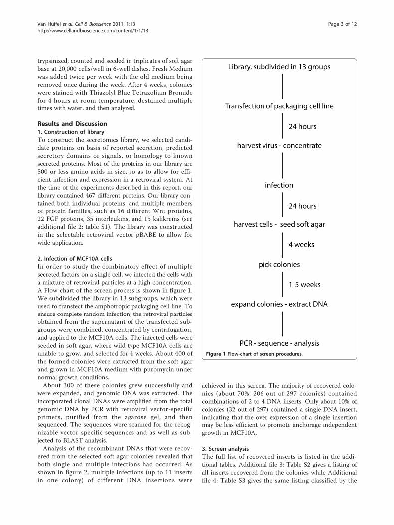
2. Infection of MCF10A cellsIn order to study the combinatory effect of multiplesecreted factors on a single cell, we infected the cells witha mixture of retroviral particles at a high concentration.A Flow-chart of the screen process is shown in figure 1.We subdivided the library in 13 subgroups, which wereused to transfect the amphotropic packaging cell line. Toensure complete random infection, the retroviral particlesobtained from the supernatant of the transfected sub-groups were combined, concentrated by centrifugation,and applied to the MCF10A cells. The infected cells wereseeded in soft agar, where wild type MCF10A cells areunable to grow, and selected for 4 weeks. About 400 ofthe formed colonies were extracted from the soft agarand grown in MCF10A medium with puromycin undernormal growth conditions.About 300 of these colonies grew successfully and
were expanded, and genomic DNA was extracted. Theincorporated clonal DNAs were amplified from the totalgenomic DNA by PCR with retroviral vector-specificprimers, purified from the agarose gel, and thensequenced. The sequences were scanned for the recog-nizable vector-specific sequences and as well as sub-jected to BLAST analysis.Analysis of the recombinant DNAs that were recov-
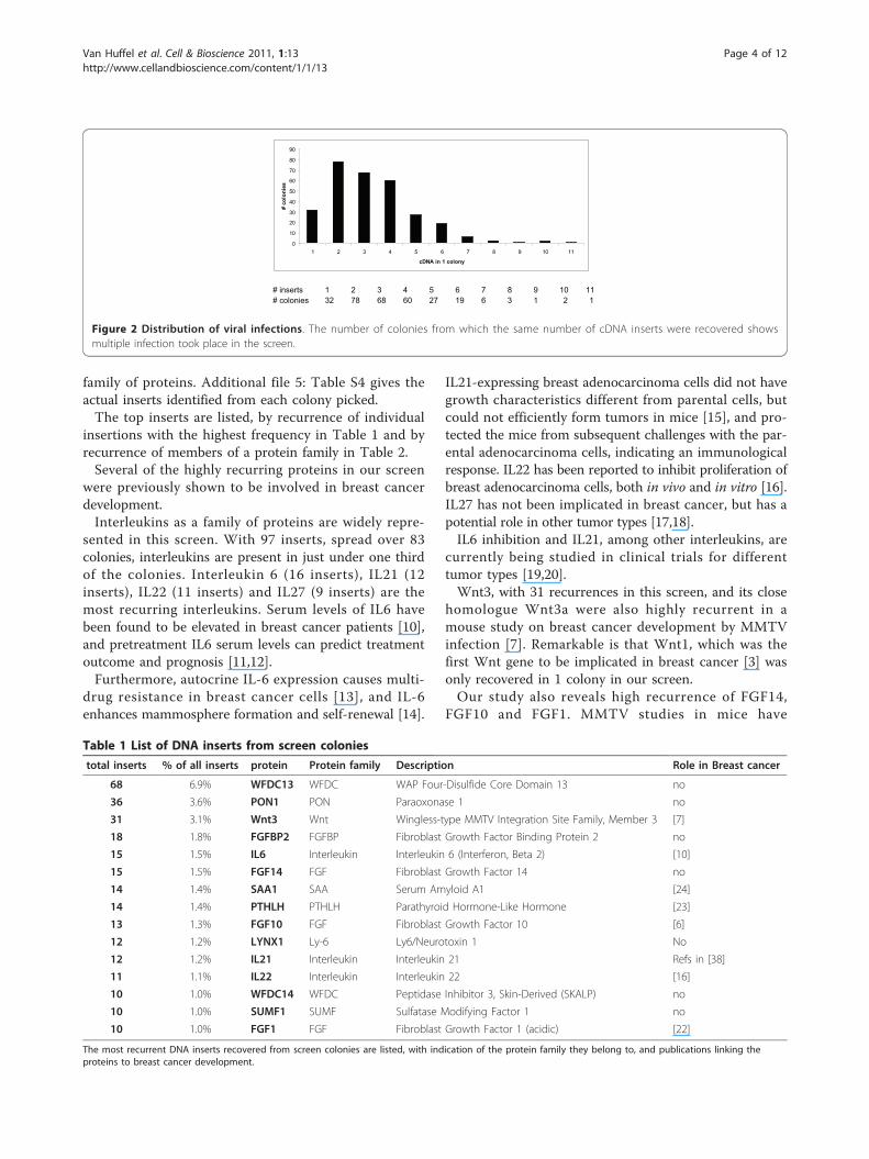
ered from the selected soft agar colonies revealed thatboth single and multiple infections had occurred. Asshown in figure 2, multiple infections (up to 11 insertsin one colony) of different DNA insertions were
achieved in this screen. The majority of recovered colo-nies (about 70%; 206 out of 297 colonies) containedcombinations of 2 to 4 DNA inserts. Only about 10% ofcolonies (32 out of 297) contained a single DNA insert,indicating that the over expression of a single insertionmay be less efficient to promote anchorage independentgrowth in MCF10A.
3. Screen analysisThe full list of recovered inserts is listed in the addi-tional tables. Additional file 3: Table S2 gives a listing ofall inserts recovered from the colonies while Additionalfile 4: Table S3 gives the same listing classified by the
Transfection of packaging cell line
harvest virus - concentrate
infection
harvest cells - seed soft agar
pick colonies
expand colonies - extract DNA
PCR - sequence - analysis
Library, subdivided in 13 groups
24 hours
24 hours
4 weeks
1-5 weeks
Figure 1 Flow-chart of screen procedures.
Van Huffel et al. Cell & Bioscience 2011, 1:13http://www.cellandbioscience.com/content/1/1/13
Page 3 of 12
family of proteins. Additional file 5: Table S4 gives theactual inserts identified from each colony picked.The top inserts are listed, by recurrence of individual
insertions with the highest frequency in Table 1 and byrecurrence of members of a protein family in Table 2.Several of the highly recurring proteins in our screen
were previously shown to be involved in breast cancerdevelopment.Interleukins as a family of proteins are widely repre-
sented in this screen. With 97 inserts, spread over 83colonies, interleukins are present in just under one thirdof the colonies. Interleukin 6 (16 inserts), IL21 (12inserts), IL22 (11 inserts) and IL27 (9 inserts) are themost recurring interleukins. Serum levels of IL6 havebeen found to be elevated in breast cancer patients [10],and pretreatment IL6 serum levels can predict treatmentoutcome and prognosis [11,12].Furthermore, autocrine IL-6 expression causes multi-
drug resistance in breast cancer cells [13], and IL-6enhances mammosphere formation and self-renewal [14].
IL21-expressing breast adenocarcinoma cells did not havegrowth characteristics different from parental cells, butcould not efficiently form tumors in mice [15], and pro-tected the mice from subsequent challenges with the par-ental adenocarcinoma cells, indicating an immunologicalresponse. IL22 has been reported to inhibit proliferation ofbreast adenocarcinoma cells, both in vivo and in vitro [16].IL27 has not been implicated in breast cancer, but has apotential role in other tumor types [17,18].IL6 inhibition and IL21, among other interleukins, are
currently being studied in clinical trials for differenttumor types [19,20].Wnt3, with 31 recurrences in this screen, and its close
homologue Wnt3a were also highly recurrent in amouse study on breast cancer development by MMTVinfection [7]. Remarkable is that Wnt1, which was thefirst Wnt gene to be implicated in breast cancer [3] wasonly recovered in 1 colony in our screen.Our study also reveals high recurrence of FGF14,
FGF10 and FGF1. MMTV studies in mice have
# inserts 1 2 3 4 5 6 7 8 9 10 11# colonies 32 78 68 60 27 19 6 3 1 2 1
0
10
20
30
40
50
60
70
80
90
1 2 3 4 5 6 7 8 9 10 11
cDNA in 1 colony
# co
loni
es
Figure 2 Distribution of viral infections. The number of colonies from which the same number of cDNA inserts were recovered showsmultiple infection took place in the screen.
Table 1 List of DNA inserts from screen colonies
total inserts % of all inserts protein Protein family Description Role in Breast cancer
68 6.9% WFDC13 WFDC WAP Four-Disulfide Core Domain 13 no
36 3.6% PON1 PON Paraoxonase 1 no
31 3.1% Wnt3 Wnt Wingless-type MMTV Integration Site Family, Member 3 [7]
18 1.8% FGFBP2 FGFBP Fibroblast Growth Factor Binding Protein 2 no
15 1.5% IL6 Interleukin Interleukin 6 (Interferon, Beta 2) [10]
15 1.5% FGF14 FGF Fibroblast Growth Factor 14 no
14 1.4% SAA1 SAA Serum Amyloid A1 [24]
14 1.4% PTHLH PTHLH Parathyroid Hormone-Like Hormone [23]
13 1.3% FGF10 FGF Fibroblast Growth Factor 10 [6]
12 1.2% LYNX1 Ly-6 Ly6/Neurotoxin 1 No
12 1.2% IL21 Interleukin Interleukin 21 Refs in [38]
11 1.1% IL22 Interleukin Interleukin 22 [16]
10 1.0% WFDC14 WFDC Peptidase Inhibitor 3, Skin-Derived (SKALP) no
10 1.0% SUMF1 SUMF Sulfatase Modifying Factor 1 no
10 1.0% FGF1 FGF Fibroblast Growth Factor 1 (acidic) [22]
The most recurrent DNA inserts recovered from screen colonies are listed, with indication of the protein family they belong to, and publications linking theproteins to breast cancer development.
Van Huffel et al. Cell & Bioscience 2011, 1:13http://www.cellandbioscience.com/content/1/1/13
Page 4 of 12

implicated FGF3, FGF4, FGF8 and FGF10 (reviewed in:[6,21]) in mouse breast cancer development. OtherFGF’s have not been directly implicated in breast cancer,but the role of FGF receptor 2 (FGFR2) indirectly impli-cates FGF1, FGF2, FGF3, FGF4, FGF6, FGF7, FGF9,FGF10, FGF16, FGF20 and FGF22 [22]. While severalFGF proteins had been previously implicated in breastcancer development, we found FGF14, previously notimplicated in breast cancer development, to be the high-est recurring FGF in our screen. Equally remarkable isthat, while FGF3, FGF4 and FGF8 had been implicatedbefore, they did not feature high in our screen, withFGF14, FGF10 and FGF1 being high up in our screen.Parathyroid hormone like hormone (PTHLH, also
Parathyroid hormone related protein, PTHrP), recurring14 times in our screen, is elevated in 60 to 90% of breastcancers, and is implicated in breast cancer metastasis tobone [23]. Serum amyloid proteins (SAA1/2) are repre-sented by SAA1 in our screen, and SAA1 present in 14of our colonies. Serum levels of this acute-phase proteinare increased in cancer patients, including breast cancer
patients [24], with an indication that higher SAA levelsin breast cancer patients represent more metastasis andpoorer prognosis.WAP Four-Disulfide Core Domain (WFDC) proteins
have not been thoroughly studied. Many WFDC codinggenes are clustered on 20q12-13.1, a region that isamplified in several cancers, including breast cancer[25,26]. Four WFDC proteins have been indicated ascandidate cancer biomarkers (WFDC14 (elafin), WFDC4(SLPI), WFDC2 and WFDC1) [27]. WFDC13, the mostprevalent insert in our screen has not been studiedbefore. Other prevalent WFDC proteins in our screenare WFDC14, WFDC4 and WFDC11.The second-most prevalent protein in our screen is
paraoxonase1 (PON1), a High density lipoprotein-asso-ciated enzyme, naturally secreted by the liver, thatshows protective properties in cardiovascular diseaseand organophosphate poisoning [28]. Few reports sug-gest a possible link between PON1 and breast cancer:the L55M single nucleotide polymorphism in PON1 hasbeen associated with increased risk of breast cancer in
Table 2 List of protein families recovered from screen colonies
total inserts Total colonies % of all colonies Protein family
97 83 27.9% Interleukin Interleukin
97 88 29.5% WFDC WAP Four-Disulfide Core Domain
78 72 24.2% FGF Fibroblast Growth Factor
54 54 18.1% Wnt Wingless-type MMTV Integration Site Family
40 39 13.1% PON Paraoxonase
35 34 11.4% KLK Kalikrein
33 28 9.4% TGFB Transforming Growth Factor
21 19 6.4% CXCL CXC Chemokine Ligand
21 21 7.0% DEF Defensin
21 21 7.0% FGFBP Fibroblast Growth Factor Binding Protein
26 26 8.7% TIMP Tissue Inhibitor of Metalloproteinase
19 19 6.4% CCL CC Chemokine Ligand
19 19 6.4% IGFBP Insulin-like Growth Factor Binding Protein
18 18 6.0% RSPO R-Spondin
16 15 5.0% DKK Dickkopf
15 15 5.0% SAA Serum Amyloid
14 14 4.7% PTHLH Parathyroid Hormone-Like Hormone
14 14 4.7% SFTP Surfactant Protein
14 14 4.7% WISP Connective Tissue Growth Factor
13 13 4.4% APO Apolipoprotein
13 13 4.4% Ly-6 Ly6/uPAR
13 13 4.4% PGLYRP Peptidoglycan Recognition Protein
12 12 4.0% MASP MBL-associated Serine Protease
10 10 3.4% FAM3 Family with Sequence Similarity 3
10 10 3.4% IFN Interferon
10 10 3.4% SPARC Secreted Protein Acidic and Rich in Cysteine
10 10 3.4% SUMF Sulfatase Modifying Factor
The DNA recovered from the screen colonies was grouped in protein families, and the most prevalent protein families are listed here. As multiple protein familymembers may co-occur in a single colony, the number of colonies may be less than the number of inserts where the protein family is represented. Proteins thathave no family members in this screen are listed as individual proteins.
Van Huffel et al. Cell & Bioscience 2011, 1:13http://www.cellandbioscience.com/content/1/1/13
Page 5 of 12

post menopausal women [29], while another SNP(Q192R) had no effect in one study [29], but showed adecreased breast cancer risk in another study [30]. ThePON1 clone in our library has L on pos 55 and Q onpos 192. PON1 expression is reported to be upregu-lated by estrogen receptor alpha (ERa) [31], and inde-pendently by resveratrol, a substance believed to bethe main active ingredient in the cardio-protectiveeffects of red wine, and which is also activating the ERreceptor [31].Fibroblast growth factor binding protein 2 or FGFBP2
(also termed Ksp37, killer specific secretory protein of37 kDa) is secreted by Th1-type CD4+ lymphocyteswith cytotoxic potential [32]. Increased FGFBP2 expres-sion has been correlated with longer survival in glioblas-toma [33]. Other links to cancer have not beenpublished. Family member FGF-BP1 however has beenimplicated in breast cancer.Lynx1 is a member of the lymphocyte Antigen 6 (Ly-6)
superfamily of cysteine-rich proteins that are mostly GPIanchored to the cell membrane. Only 3 proteins in thefamily are secreted, and Lynx-1 (or SLURP-2) has beendescribed to be upregulated in Psoriasis vulgaris [34],while another non-secreted family member, Ly-6K, wasidentified as a biomarker for breast cancer [35].The sulfatase modifying factor 1 (SUMF1) that is pre-
sent in 10 of our colonies has previously never beendescribed as involved in any types of tumors. It isessential for the post-translational modification of sulfa-tases, and deficiency results in an autosomal recessivedisorder [36].
4. Reconstructive analysisAs had been previously reported for a number of theproteins tested in this screen, the infection of MCF10Acells with single viral vectors led to no or very weakgrowth in soft agar (data not shown).In performing this screen, MCF10A cells were purpo-
sefully infected with concentrated viral particles, inorder to achieve multiple infection of the cells. In thisway, the importance of the combination of different fac-tors on the growth of MCF10A cells in soft agar couldbe assessed. A schema of some combinations of factorsis provided in figure 3, where the more prevalent recur-ring combinations are indicated in red. While we cre-ated viral particles by transfecting 13 distinct subgroups,the pooling and concentration of the viral particles ren-dered a random distribution in infection. This wasobserved by the fact that the factors found in the softagar colonies did not return with a clear correlation tothe distribution in the 13 subgroups.It had been previously observed in MMTV-mouse
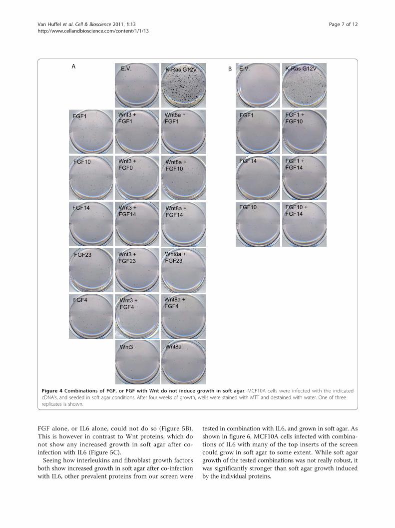
studies that FGF and Wnt genes may co-operate inbreast cancer development. Mice that are transgenic fora Wnt protein predominantly overexpressed FGF genesin MMTV-induced breast tumors, and vice versa[1,2,37]. A total of 14 of the colonies in our screen con-tained combinations of Wnt and FGF family members,suggesting that FGF-Wnt co-operation in transformationof human mammalian cells may also occur. However,when any of the top 5 FGFs were co-infected with Wnt3 or Wnt8a in MCF10A cells, we did not observe stronggrowth of these cells in soft agar (figure 4A), indicatingthat on a cellular level, the FGF-Wnt combination is notsufficient to drive efficient transformation. Wnt9a couldnot be tested in this assay, as infection of Wnt9areduced the survival of the MCF10A cells, not allowingthe soft agar assay.Six of the colonies in the present screen contained a
combination of two FGF proteins. However, co-infectionof two FGF proteins into MCF10A cells did not lead tosignificant growth in soft agar (figure 4B).In 13 colonies, multiple interleukin family members
were observed to be present. Infection of combinationsof interleukins in MCF10A cells, lead to the observationthat Interleukin 6 (IL6), when combined with IL9 orIL21 efficiently drives soft agar growth in MCF10A cells.Other combinations, such as IL6 with IL22, IL21 withIL27; or IL9 with IL27, did result in some soft agargrowth, but to a much lesser extent than IL6-containingcombinations (figure 5A).Another prevalent combination in the screen was
interleukins and FGF proteins (19 colonies). When thesecombinations were tested in MCF10A cells, IL6 in com-bination with FGF1, FGF10 or FGF14, proved to effi-ciently drive these cells to grow in soft agar, while the
Inte
rl
WFD
C
FGF
Wnt
PON
KLK
TGFB
TIM
P
CX
CL
Interl 13
WFDC 24 9
FGF 19 19 6
Wnt 9 19 14
PON 12 6 10 4 1
KLK 10 9 5 6 5 1
TGFB 6 12 8 3 1 2 1
TIMP 5 11 10 5 5 1 6
CXCL 3 3 4 3 2 2 4 2
Figure 3 Analysis of combinations recovered from the screen.The number of times proteins from the indicated protein familiesoccur in a single colony. Yellow boxes indicate numbers from 2-4,orange indicate 5-7, and red indicate more than 7 coloniescontaining the indicated combination.
Van Huffel et al. Cell & Bioscience 2011, 1:13http://www.cellandbioscience.com/content/1/1/13
Page 6 of 12
FGF alone, or IL6 alone, could not do so (Figure 5B).This is however in contrast to Wnt proteins, which donot show any increased growth in soft agar after co-infection with IL6 (Figure 5C).Seeing how interleukins and fibroblast growth factors
both show increased growth in soft agar after co-infectionwith IL6, other prevalent proteins from our screen were
tested in combination with IL6, and grown in soft agar. Asshown in figure 6, MCF10A cells infected with combina-tions of IL6 with many of the top inserts of the screencould grow in soft agar to some extent. While soft agargrowth of the tested combinations was not really robust, itwas significantly stronger than soft agar growth inducedby the individual proteins.
Wnt8a + FGF4Wnt8a + FGF4
Wnt8a + FGF23Wnt8a + FGF23
Wnt8a + FGF14Wnt8a + FGF14
Wnt3 + FGF23Wnt3 + FGF23
Wnt8a + FGF10Wnt8a + FGF10
Wnt8a + FGF1Wnt8a + FGF1
Wnt8aWnt8aWnt3Wnt3
Wnt3 + FGF14Wnt3 + FGF14
Wnt3 + FGF0Wnt3 + FGF0
Wnt3 + FGF4Wnt3 + FGF4
Wnt3 + FGF1Wnt3 + FGF1
FGF4FGF4
FGF23FGF23
FGF14FGF14
K-Ras G12VK-Ras G12V
FGF10FGF10
FGF1FGF1
E.V.E.V.A E.V.E.V. K-Ras G12VK-Ras G12V
FGF14FGF14
FGF1FGF1
FGF1 + FGF14FGF1 + FGF14
FGF10FGF10
FGF1 + FGF10FGF1 + FGF10
FGF10 + FGF14FGF10 + FGF14
B
Figure 4 Combinations of FGF, or FGF with Wnt do not induce growth in soft agar. MCF10A cells were infected with the indicatedcDNA’s, and seeded in soft agar conditions. After four weeks of growth, wells were stained with MTT and destained with water. One of threereplicates is shown.
Van Huffel et al. Cell & Bioscience 2011, 1:13http://www.cellandbioscience.com/content/1/1/13
Page 7 of 12

ConclusionsWith this screen, the combinatory effect of secreted fac-tors on breast cancer development was assessed,through their action on anchorage-independent growthof MCF10A cells. While screens looking for individualproteins involved in tumor development have been
described many times, this is the first screen looking forcombinatory action of proteins. In the living organism,it is understood that every cell and tissue is constantlysurrounded by a mix of hormones, secreted factors, aswell as factors in the extracellular matrix. A differentcomposition of this environment can seriously affect the
E.V.E.V. K-Ras G12VK-Ras G12V
IL6IL6
IL9IL9
IL27IL27
IL21IL21
IL6 + IL21IL6 + IL21
IL6 + IL9IL6 + IL9
IL27 + IL9IL27 + IL9
IL21 + IL27IL21 + IL27
IL22 IL6 + IL22
A E.V.E.V. K-Ras G12VK-Ras G12V
IL6IL6
FGF14 + IL6FGF14 + IL6
FGF10 + IL6FGF10 + IL6
FGF1 + IL6FGF1 + IL6
FGF14FGF14
FGF1FGF1
FGF10FGF10
B
E.V.E.V. K-Ras G12VK-Ras G12V
Wnt3 + IL6Wnt3 + IL6Wnt3Wnt3
IL6IL6
C
Figure 5 Combinations of Interleukins, or interleukin 6 with FGF induce soft agar growth, while IL6 combined with Wnt does not.MCF10A cells were infected with the indicated cDNA’s, and seeded in soft agar conditions. After four weeks of growth, wells were stained withMTT and destained with water. One of three replicates is shown.
Van Huffel et al. Cell & Bioscience 2011, 1:13http://www.cellandbioscience.com/content/1/1/13
Page 8 of 12

signaling events within the cells or tissue, and influencethe growth and other characteristics of the cells andtissue.One of the benefits of this screen is also that it
screened a large number of proteins that were
categorized in familial clusters (e.g. 22 FGF proteins, 16Wnt proteins, 35 interleukins). This brought moreobscure protein family members in the spotlight thanconventional research had done so far. Often, the morerecently discovered members of protein families remain
PON1 + IL6
TIMP1
E.V.E.V. K-Ras G12VK-Ras G12V WFDC13 WFDC13 + IL6
PON1
TIMP1 + IL6E.V.
E.V.
E.V.
K Ras G12V
K Ras G12V
K Ras G12V FGFBP2
WFDC14E.V. K-RAS G12V WFDC14 + IL6
FGFBP2 + IL6
E.V.
E.V. K-RAS G12V
K-RAS G12V
GDF8 + IL6
NODAL + IL6NODAL
GDF8
K Ras G12V
Figure 6 Combinations of IL6 with other proteins results in soft agar growth. MCF10A cells were infected with the indicated cDNA’s, andseeded in soft agar conditions. After four weeks of growth, wells were stained with MTT and destained with water. One of three replicates isshown.
Van Huffel et al. Cell & Bioscience 2011, 1:13http://www.cellandbioscience.com/content/1/1/13
Page 9 of 12

obscure for longer periods of time, as their functions areoften extrapolated from more researched close familymembers.Alternative screens have utilized expression profiles or
2D-gel electrophoresis combined with sequencing todirectly identify proteins expressed or secreted in speci-fic tissues. While these assays are definitely valid, theyusually will have problems picking up subtle differencesin expression, or proteins that are secreted at low levels.Furthermore, the validation of these proteins in in vitroassays may be hampered by the fact that alone, theseproteins may not induce the phenotype they werescreened for, and specific combinations of factors maynot be recognized.Proteins that had previously been implicated in breast
cancer development, such as fibroblast growth factorsand wnt proteins, were also highly recurrent in the pre-sent screen. As had been shown before in mouse stu-dies, individual MMTV insertions appear to be sufficientfor tumor formation in mouse, but in MCF10A cells,overexpression of the corresponding single protein can-not induce strong soft agar growth. While studies withMMTV in mouse indicated a potential combinatory rolefor Wnt and FGF [1,2,37] in breast cancer development,in the human cell line MCF10A, these two factors didnot induce anchorage-independent growth. This appar-ent discrepancy could be due to the difference in spe-cies, but also to the in vivo nature of the mouseexperiments, and the in vitro nature of the presentscreen. In mouse, natural secreted proteins present inthe tissue could further influence the cancer develop-ment, while in the controlled environment of tissue cul-ture, this is not the case.A surprising combination of proteins that did induce
anchorage independent growth in MCF10A cells wereinterleukins, which, when combined with other factors,often greatly enhanced soft agar growth. Interleukin 6,when co-expressed with other proteins prevalent in ourscreen, induced growth in soft agar, albeit to varyingdegrees, depending on the co-expressed protein. Themost growth-inducing combination we detected werecombinations between interleukins, such as IL6-IL21, orIL6-IL9. The combination between IL6 and FGF1,FGF10 or FGF14 also resulted in strong growth ofMCF10A in soft agar. Only a few of the tested combina-tions with IL6 did not significantly induce soft agargrowth of MCF10A. Among these was Wnt3, which,when expressed alone, already has a limited soft agargrowth-inducing potential.We have discussed above what combinations of
highly expressing proteins promote anchorage inde-pendent growth in soft agar. However, some of thecombinations found in the initial screen did not inducecolony formation in the validation studies. For this we
should consider other factors such as over-expression(both relative and absolute) and the influence of pro-teins secreted by surrounding cells during the screen-ing process.The findings in the present study can have important
implications, even for seemingly unrelated researchfields. For example, paraoxonase 1, overexpression ofwhich had hitherto not been associated with breast can-cer, has been mainly studied for its beneficial effect inorganophosphate poisoning. Using this protein in pre-vention of poisoning (e.g. in chemical warfare) may haveserious implications, such as breast tumor development,should at the time of high PON1 levels, IL6 levels alsobe raised.The results of this screen show that it is possible to
find combinatory effects of proteins in a cell-basedscreen, and that such combinatory effects can be verydifferent from the individual proteins’ effects. Furtheranalysis will have to be done to examine which signaltransduction pathways are involved in the stimulatoryeffect IL6 has with other proteins in anchorage-indepen-dent growth of mammary epithelial cells.
Additional material
Additional file 1: The viral vectors pBABE-puro and pBABE-I. Aschematic representation of the vectors used in this study: pBABE-puroand the derived pBABE-I
Additional file 2: Protein library. List of all proteins in the proteinlibrary used in this study. Proteins were selected if they were known tobe secreted, contained secretory domains, or bore high homology withsecreted proteins. The cloned cDNA’s include any signal peptides thatmay be cleaved off during maturation of the protein. The vast majorityof proteins is less than 500 amino acids in length.
Additional file 3: List of DNA inserts from screen colonies. The DNAinserts recovered from screen colonies are listed, with indication of theprotein family they belong to, and their relative recurrence (% of totalinserts)
Additional file 4: List of protein families recovered from screencolonies. The DNA recovered from the screen colonies was grouped inprotein families, and the most prevalent protein families are listed here.As multiple protein family members may co-occur in a single colony, thenumber of colonies may be less than the number of inserts where theprotein family is represented. Proteins that have no family members inthis screen are listed as individual proteins.
Additional file 5: DNA inserts in each colony. The actual DNA insertsrecovered from each individual colony.
List of abbreviations(FGF): Fibroblast growth factor; (FGFBP2): Fibroblast growth factor bindingprotein 2; (IL): Interleukin; (MMTV): Mouse Mammary tumor Virus; (MOI):Multiplicity of infection; (PON1): Paraoxonase1; (PTHLH): Parathyroid hormonelike hormone; (RSPO): R-spondin; (SAA1/2): Serum amyloid proteins; (SUMF1):sulfatase modifying factor 1; (WFDC): WAP Four-Disulfide Core Domain;(wnt): Wingless-type MMTV integration site family
AcknowledgementsThe authors wish to thank the Agency for Science, Technology and Researchfor the funding of this work.
Van Huffel et al. Cell & Bioscience 2011, 1:13http://www.cellandbioscience.com/content/1/1/13
Page 10 of 12
Author details1Cancer and Developmental Cell Biology Division (CDCBD), Institute ofMolecular and Cell Biology, A-star, 61 Biopolis Drive, Proteos, 138673,Singapore. 2Genome Institute of Singapore, A-star, 60 Biopolis Street, #02-01,Genome, 138672, Singapore.
Authors’ contributionsVHS performed the initial screen, performed the soft agar reconstruction,compiled and analyzed the insert data, cloned library constructs, and draftedthe manuscript. TJM participated in discussions and analysis, expandedscreen colonies, sequenced colony inserts, cloned library constructs, andhelped to draft the manuscript. YCX prepared genomic DNA, performed thesequencing of colony inserts, soft agar staining, and cloned library clones.TYL participated in sequencing, and cloning of library constructsZXQ, LKP and OF participated in cloning of library constructs. IL didbioinformatics analysis for the library listing. HWJ conceived of the project,listed the library, participated in discussions, and helped draft themanuscript. All authors have read and approved the final manuscript.
Competing interestsThe authors declare that they have no competing interests.
Received: 24 January 2011 Accepted: 25 March 2011Published: 25 March 2011
References1. Lee FS, Lane TF, Kuo A, Shackleford GM, Leder P: Insertional mutagenesis
identifies a member of the Wnt gene family as a candidate oncogene inthe mammary epithelium of int-2/Fgf-3 transgenic mice. Proc Natl AcadSci USA 1995, 92:2268-2272.
2. MacArthur CA, Shankar DB, Shackleford GM: Fgf-8, activated by proviralinsertion, cooperates with the Wnt-1 transgene in murine mammarytumorigenesis. J Virol 1995, 69:2501-2507.
3. Nusse R, Varmus HE: Many tumors induced by the mouse mammarytumor virus contain a provirus integrated in the same region of the hostgenome. Cell 1982, 31:99-109.
4. Peters G, Brookes S, Smith R, Dickson C: Tumorigenesis by mousemammary tumor virus: evidence for a common region for provirusintegration in mammary tumors. Cell 1983, 33:369-377.
5. Roelink H, Wagenaar E, Lopes da Silva S, Nusse R: Wnt-3, a gene activatedby proviral insertion in mouse mammary tumors, is homologous to int-1/Wnt-1 and is normally expressed in mouse embryos and adult brain.Proc Natl Acad Sci USA 1990, 87:4519-4523.
6. Theodorou V, Boer M, Weigelt B, Jonkers J, van d V, Hilkens J: Fgf10 is anoncogene activated by MMTV insertional mutagenesis in mousemammary tumors and overexpressed in a subset of human breastcarcinomas. Oncogene 2004, 23:6047-6055.
7. Theodorou V, Kimm MA, Boer M, Wessels L, Theelen W, Jonkers J, Hilkens J:MMTV insertional mutagenesis identifies genes, gene families andpathways involved in mammary cancer. Nat Genet 2007, 39:759-769.
8. Tsukamoto AS, Grosschedl R, Guzman RC, Parslow T, Varmus HE: Expressionof the int-1 gene in transgenic mice is associated with mammary glandhyperplasia and adenocarcinomas in male and female mice. Cell 1988,55:619-625.
9. Rao VS, Dyer CE, Jameel JK, Drew PJ, Greenman J: Potential prognosticand therapeutic roles for cytokines in breast cancer (Review). Oncol Rep2006, 15:179-185.
10. Benoy I, Salgado R, Colpaert C, Weytjens R, Vermeulen PB, Dirix LY: Seruminterleukin 6, plasma VEGF, serum VEGF, and VEGF platelet load inbreast cancer patients. Clin Breast Cancer 2002, 2:311-315.
11. Yokoe T, Iino Y, Morishita Y: Trends of IL-6 and IL-8 levels in patients withrecurrent breast cancer: preliminary report. Breast Cancer 2000, 7:187-190.
12. Bachelot T, Ray-Coquard I, Menetrier-Caux C, Rastkha M, Duc A, Blay JY:Prognostic value of serum levels of interleukin 6 and of serum andplasma levels of vascular endothelial growth factor in hormone-refractorymetastatic breast cancer patients. Br J Cancer 2003, 88:1721-1726.
13. Conze D, Weiss L, Regen PS, Bhushan A, Weaver D, Johnson P, Rincon M:Autocrine production of interleukin 6 causes multidrug resistance inbreast cancer cells. Cancer Res 2001, 61:8851-8858.
14. Sansone P, Storci G, Tavolari S, Guarnieri T, Giovannini C, Taffurelli M,Ceccarelli C, Santini D, Paterini P, Marcu KB, Chieco P, Bonafè M: IL-6
triggers malignant features in mammospheres from human ductalbreast carcinoma and normal mammary gland. J Clin Invest 2007,117:3988-4002.
15. di Carlo E, Comes A, Orengo AM, Rosso O, Meazza R, Musiani P,Colombo MP, Ferrini S: IL-21 induces tumor rejection by specific CTL andIFN-gamma-dependent CXC chemokines in syngeneic mice. J Immunol2004, 172:1540-1547.
16. Weber GF, Gaertner FC, Erl W, Janssen KP, Blechert B, Holzmann B,Weighardt H, Essler M: IL-22-mediated tumor growth reduction correlateswith inhibition of ERK1/2 and AKT phosphorylation and induction of cellcycle arrest in the G2-M phase. J Immunol 2006, 177:8266-8272.
17. Ho MY, Leu SJ, Sun GH, Tao MH, Tang SJ, Sun KH: IL-27 directly restrainslung tumorigenicity by suppressing cyclooxygenase-2-mediatedactivities. J Immunol 2009, 183:6217-6226.
18. Zhu S, Lee DA, Li S: IL-12 and IL-27 sequential gene therapy viaintramuscular electroporation delivery for eliminating distal aggressivetumors. J Immunol 2010, 184:2348-2354.
19. Trikha M, Corringham R, Klein B, Rossi JF: Targeted anti-interleukin-6monoclonal antibody therapy for cancer: a review of the rationale andclinical evidence. Clin Cancer Res 2003, 9:4653-4665.
20. Hashmi MH, Van Veldhuizen PJ: Interleukin-21: updated review of Phase Iand II clinical trials in metastatic renal cell carcinoma, metastaticmelanoma and relapsed/refractory indolent non-Hodgkin’s lymphoma.Expert Opin Biol Ther 2010, 10:807-817.
21. Callahan R, Smith GH: Common integration sites for MMTV in viralinduced mouse mammary tumors. J Mammary Gland Biol Neoplasia 2008,13:309-321.
22. Katoh M: Cancer genomics and genetics of FGFR2 (Review). Int J Oncol2008, 33:233-237.
23. Zhang Y, Ma B, Fan Q: Mechanisms of breast cancer bone metastasis.Cancer Lett 2010, 292:1-7.
24. Malle E, Sodin-Semrl S, Kovacevic A: Serum amyloid A: an acute-phase protein involved in tumour pathogenesis. Cell Mol Life Sci2009, 66:9-26.
25. Kallioniemi A, Kallioniemi OP, Piper J, Tanner M, Stokke T, Chen L, Smith HS,Pinkel D, Gray JW, Waldman FM: Detection and mapping of amplifiedDNA sequences in breast cancer by comparative genomic hybridization.Proc Natl Acad Sci USA 1994, 91:2156-2160.
26. Muleris M, Almeida A, Gerbault-Seureau M, Malfoy B, Dutrillaux B: Detectionof DNA amplification in 17 primary breast carcinomas withhomogeneously staining regions by a modified comparative genomichybridization technique. Genes Chromosomes Cancer 1994, 10:160-170.
27. Bouchard D, Morisset D, Bourbonnais Y, Tremblay GM: Proteins with whey-acidic-protein motifs and cancer. Lancet Oncol 2006, 7:167-174.
28. Stevens RC, Suzuki SM, Cole TB, Park SS, Richter RJ, Furlong CE: Engineeredrecombinant human paraoxonase 1 (rHuPON1) purified from Escherichiacoli protects against organophosphate poisoning. Proc Natl Acad Sci USA2008, 105:12780-12784.
29. Stevens VL, Rodriguez C, Pavluck AL, Thun MJ, Calle EE: Association ofpolymorphisms in the paraoxonase 1 gene with breast cancer incidencein the CPS-II Nutrition Cohort. Cancer Epidemiol Biomarkers Prev 2006,15:1226-1228.
30. Gallicchio L, McSorley MA, Newschaffer CJ, Huang HY, Thuita LW,Hoffman SC, Helzlsouer KJ: Body mass, polymorphisms in obesity-relatedgenes, and the risk of developing breast cancer among women withbenign breast disease. Cancer Detect Prev 2007, 31:95-101.
31. Gouedard C, Barouki R, Morel Y: Induction of the paraoxonase-1 geneexpression by resveratrol. Arterioscler Thromb Vasc Biol 2004, 24:2378-2383.
32. Ogawa K, Tanaka K, Ishii A, Nakamura Y, Kondo S, Sugamura K, Takano S,Nakamura M, Nagata K: A novel serum protein that is selectivelyproduced by cytotoxic lymphocytes. J Immunol 2001, 166:6404-6412.
33. Yamanaka R, Arao T, Yajima N, Tsuchiya N, Homma J, Tanaka R, Sano M,Oide A, Sekijima M, Nishio K: Identification of expressed genescharacterizing long-term survival in malignant glioma patients. Oncogene2006, 25:5994-6002.
34. Tsuji H, Okamoto K, Matsuzaka Y, Iizuka H, Tamiya G, Inoko H: SLURP-2, anovel member of the human Ly-6 superfamily that is up-regulated inpsoriasis vulgaris. Genomics 2003, 81:26-33.
35. Lee JW, Lee YS, Yoo KH, Lee KH, Park K, Ahn T, Ko C, Park JH: LY-6K gene:a novel molecular marker for human breast cancer. Oncol Rep 2006,16:1211-1214.
Van Huffel et al. Cell & Bioscience 2011, 1:13http://www.cellandbioscience.com/content/1/1/13
Page 11 of 12

36. Annunziata I, Bouche V, Lombardi A, Settembre C, Ballabio A: Multiplesulfatase deficiency is due to hypomorphic mutations of the SUMF1gene. Hum Mutat 2007, 28:928.
37. Shackleford GM, MacArthur CA, Kwan HC, Varmus HE: Mouse mammarytumor virus infection accelerates mammary carcinogenesis in Wnt-1transgenic mice by insertional activation of int-2/Fgf-3 and hst/Fgf-4.Proc Natl Acad Sci USA 1993, 90:740-744.
38. Skak K, Kragh M, Hausman D, Smyth MJ, Sivakumar PV: Interleukin 21:combination strategies for cancer therapy. Nat Rev Drug Discov 2008,7:231-240.
doi:10.1186/2045-3701-1-13Cite this article as: Van Huffel et al.: Systematic analysis of secretedproteins reveals synergism between IL6 and other proteins in soft agargrowth of MCF10A cells. Cell & Bioscience 2011 1:13.
Submit your next manuscript to BioMed Centraland take full advantage of:
• Convenient online submission
• Thorough peer review
• No space constraints or color figure charges
• Immediate publication on acceptance
• Inclusion in PubMed, CAS, Scopus and Google Scholar
• Research which is freely available for redistribution
Submit your manuscript at www.biomedcentral.com/submit
Van Huffel et al. Cell & Bioscience 2011, 1:13http://www.cellandbioscience.com/content/1/1/13
Page 12 of 12