Biosorption of carbaryl from aqueous solution onto Pistia stratiotes biomass
Upload
independentCategory
view
1download
0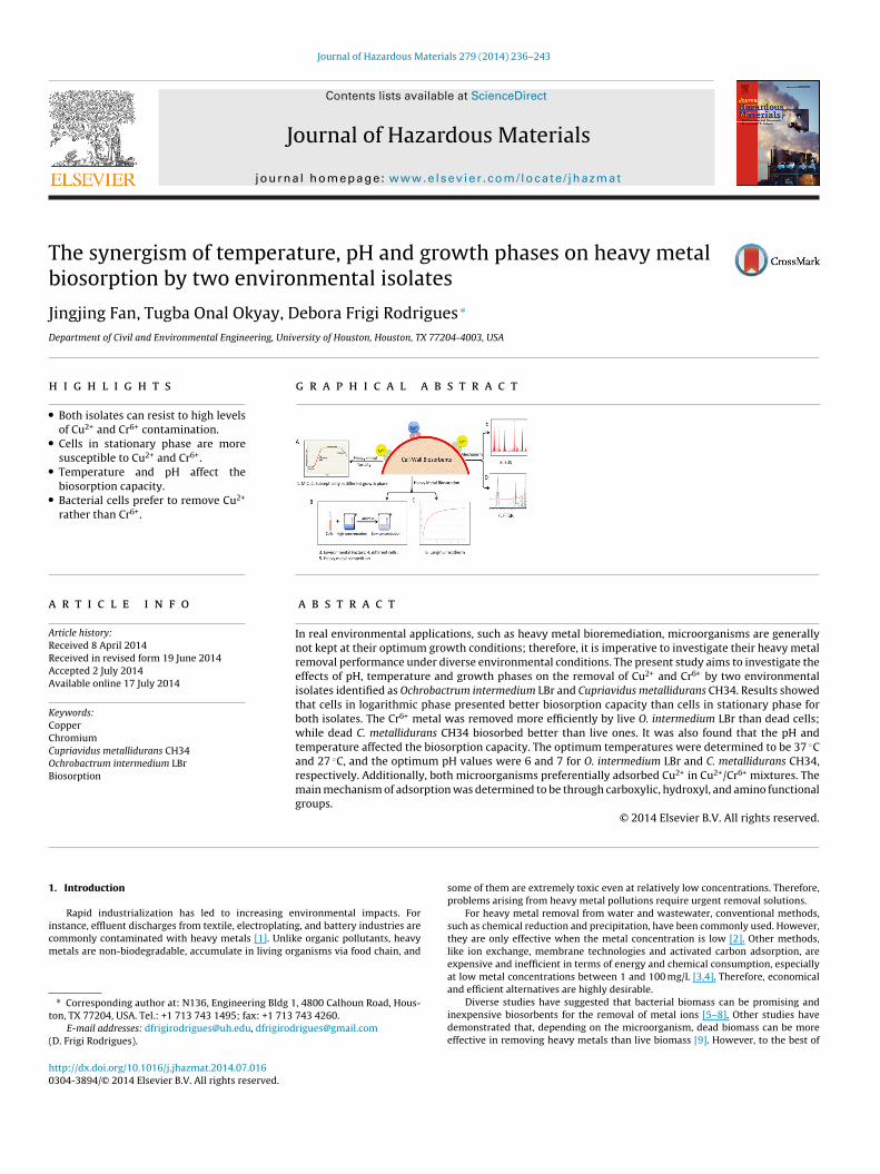
Tb
JD
h
•
•
•
•
a
ARRAA
KCCCOB
1
icm
t
(
h0
Journal of Hazardous Materials 279 (2014) 236–243
Contents lists available at ScienceDirect
Journal of Hazardous Materials
jo ur nal ho me p ag e: www.elsev ier .com/ locate / jhazmat
he synergism of temperature, pH and growth phases on heavy metaliosorption by two environmental isolates
ingjing Fan, Tugba Onal Okyay, Debora Frigi Rodrigues ∗
epartment of Civil and Environmental Engineering, University of Houston, Houston, TX 77204-4003, USA
i g h l i g h t s
Both isolates can resist to high levelsof Cu2+ and Cr6+ contamination.Cells in stationary phase are moresusceptible to Cu2+ and Cr6+.Temperature and pH affect thebiosorption capacity.Bacterial cells prefer to remove Cu2+
rather than Cr6+.
g r a p h i c a l a b s t r a c t
r t i c l e i n f o
rticle history:eceived 8 April 2014eceived in revised form 19 June 2014ccepted 2 July 2014vailable online 17 July 2014
eywords:opperhromium
a b s t r a c t
In real environmental applications, such as heavy metal bioremediation, microorganisms are generallynot kept at their optimum growth conditions; therefore, it is imperative to investigate their heavy metalremoval performance under diverse environmental conditions. The present study aims to investigate theeffects of pH, temperature and growth phases on the removal of Cu2+ and Cr6+ by two environmentalisolates identified as Ochrobactrum intermedium LBr and Cupriavidus metallidurans CH34. Results showedthat cells in logarithmic phase presented better biosorption capacity than cells in stationary phase forboth isolates. The Cr6+ metal was removed more efficiently by live O. intermedium LBr than dead cells;while dead C. metallidurans CH34 biosorbed better than live ones. It was also found that the pH and
◦
upriavidus metallidurans CH34chrobactrum intermedium LBriosorptiontemperature affected the biosorption capacity. The optimum temperatures were determined to be 37 Cand 27 ◦C, and the optimum pH values were 6 and 7 for O. intermedium LBr and C. metallidurans CH34,respectively. Additionally, both microorganisms preferentially adsorbed Cu2+ in Cu2+/Cr6+ mixtures. Themain mechanism of adsorption was determined to be through carboxylic, hydroxyl, and amino functionalgroups.
© 2014 Elsevier B.V. All rights reserved.
. Introduction
Rapid industrialization has led to increasing environmental impacts. Fornstance, effluent discharges from textile, electroplating, and battery industries areommonly contaminated with heavy metals [1]. Unlike organic pollutants, heavyetals are non-biodegradable, accumulate in living organisms via food chain, and
∗ Corresponding author at: N136, Engineering Bldg 1, 4800 Calhoun Road, Hous-on, TX 77204, USA. Tel.: +1 713 743 1495; fax: +1 713 743 4260.
E-mail addresses: [email protected], [email protected]. Frigi Rodrigues).
ttp://dx.doi.org/10.1016/j.jhazmat.2014.07.016304-3894/© 2014 Elsevier B.V. All rights reserved.
some of them are extremely toxic even at relatively low concentrations. Therefore,problems arising from heavy metal pollutions require urgent removal solutions.
For heavy metal removal from water and wastewater, conventional methods,such as chemical reduction and precipitation, have been commonly used. However,they are only effective when the metal concentration is low [2]. Other methods,like ion exchange, membrane technologies and activated carbon adsorption, areexpensive and inefficient in terms of energy and chemical consumption, especiallyat low metal concentrations between 1 and 100 mg/L [3,4]. Therefore, economical
and efficient alternatives are highly desirable.Diverse studies have suggested that bacterial biomass can be promising andinexpensive biosorbents for the removal of metal ions [5–8]. Other studies havedemonstrated that, depending on the microorganism, dead biomass can be moreeffective in removing heavy metals than live biomass [9]. However, to the best of

ous M
obhceg
iehsd
ddtms
2
2
afw1mdegsf4M0[Nf1t
obSmecicl8sts
2
wwfiav9hti
J. Fan et al. / Journal of Hazard
ur knowledge, detailed comparative mechanistic investigations on heavy metaliosorption by dead and live biomasses under relevant environmental conditionsave not been fully assessed. This is the first study, to the best of our knowledge, toompare heavy metal biosorption under optimum microbial growth conditions withnvironmental relevant conditions that are not necessarily optimal for microbialrowth and heavy metal biosorption.
In the present study, we compare two environmental isolates, Ochrobactrumntermedium LBr and Cupriavidus metallidurans CH34. O. intermedium LBr is a newnvironmental isolate obtained from a lysimeter containing organic wastes andeavy metals. C. metallidurans CH34 is also an environmental isolate that waselected as a model organism for comparison, because it has been extensivelyescribed as one of the best biosorbents [10].
The present study aims to investigate Cu2+ and Cr6+ bacterial resistance andetermine the maximum adsorption capacity of these two microorganisms underifferent growth phases, pH values and temperatures. Finally, competitive biosorp-ion assays with both Cu2+ and Cr6+ are also investigated under their optimum
icrobial growth and under environmental conditions similar to their isolationites.
. Material and methods
.1. Microorganisms and growth conditions
C. metallidurans CH34 ATCC 43123 was isolated from sludge of zinc decantation tank [11] and O. intermedium LBr was obtainedrom leachate samples generated by a lysimeter containing organicastes and heavy metals in Brazil. This isolate was identified by
6S rRNA gene sequencing as described in the supporting infor-ation (Figs. S1 and S2). The sequence is deposited in the NCBI
atabase with the accession number KM017062. Prior to eachxperiment, C. metallidurans CH 34 and O. intermedium LBr wererown on Tryptic Soy Agar (TSA) at 27 ◦C overnight (Difco) to obtainingle colonies. Single colonies for each microorganism was trans-erred to Tris-medium (TSM), which contained 6.06 g/L base Tris,.68 g/L NaCl, 1.49 g/L KCl, 1.07 g/L NH4Cl, 0.43 g/L Na2SO4, 0.2 g/LgCl2·6H2O, 0.03 g/L CaCl2·2H2O, 0.23 g/L Na2HPO4·12H2O, and
.005 g/L of Fe(III)(NH4) citrate for the subsequent experiments12]. The pH of TSM was adjusted to 7.0 ± 0.5 using 1 M HCl and 1 MaOH. Sterilization of TSM was achieved by autoclaving at 121 ◦C
or 20 min, and then it was cooled at room temperature. Afterwards, mL of filter-sterilized (0.22 �m) trace element solution was addedo the TSM in aseptic conditions [13].
Single colonies of each microorganism were grown in 25 mLf TSM in 50 mL PP centrifuge tubes (Corning Inc., USA) by incu-ating them at 27 ◦C and 80 rpm (INNOVA 44, New Brunswickcientific Co., USA) for 36 h and 48 h to obtain the cells in logarith-ic and stationary phases, respectively (Fig. S3). In the adsorption
xperiments, 27 ◦C and pH 7 were defined as the environmentalonditions [14], and the optimum condition will be determinedn the optimization section. Dead cells were prepared by auto-laving the 48 h grown cells at 121 ◦C for 20 min [15]. Afterwards,ive or dead cells were harvested by centrifugation at 8000 rpm for
min, and washed once with fresh TSM. Finally, the cells were re-uspended in phosphate buffered saline (PBS) (Amresco Inc., USA)o an optical density of 0.3 at the 600 nm (OD600), and the PBS-cellolutions were used in further experiments.
.2. Determination of minimum inhibitory concentration (MIC)
Copper and chromium heavy metal stock solutions (2000 ppm)ere prepared using CuSO4·5H2O and K2Cr2O7 (Fisher Scientific)ithin TSM. They were filter-sterilized by 0.2 �m PTFE membranelters (Millipore). Cells grown in TSM broth without heavy metalst 27 ◦C and 80 rpm for 48 h were used for the MIC experiment. Aolume of 20 �L of each PBS-cell suspension was inoculated into a
6-well plate (Corning Inc., USA), which contained 200 �L of TSMaving Cu2+ (5–1000 ppm) or Cr6+ (10–1000 ppm). Negative con-rols were performed in the same concentrations of heavy metalsn TSM without bacteria. Positive controls were prepared by 10%aterials 279 (2014) 236–243 237
(v/v) inoculation of C. metallidurans CH34 and O. intermedium LBrinto heavy metal-free TSM broth. The 96-well plate was incubatedin the plate reader (Synergy MX Microplate reader, BioTek, USA)at 27 ◦C and 80 rpm for 48 h, and every hour absorbance was readat 600 nm in order to determine the MIC. Each experiment wasperformed in triplicate.
2.3. Assessing the relative susceptibilities of logarithmic- andstationary-phases to high heavy metal concentrations
A modified method of Teitzel [16] was used to assess themicrobial susceptibility on heavy metals. As previously described,PBS-cell solutions in logarithmic- and stationary-phases were inoc-ulated into 100 ppm Cu2+ and Cr6+ solutions. Cells were exposed toheavy metals for 3 h at 27 ◦C. The negative controls were also con-ducted with heavy metal-free TSM. After 3 h of exposure, cellularviability was estimated by the plate count method [14].
2.4. Time-course biosorption under environmental conditions
The PBS-cell solutions in logarithmic and stationary phases, asdescribed previously, were inoculated into 30 mL TSM containing100 ppm Cu2+ or Cr6+ and incubated under environmental condi-tions for 400 min. Heavy metal solutions without microbes wereused as controls to take into consideration any heavy metal pre-cipitation. During the procedure, 3 mL samples were asepticallyremoved at specific time intervals for 400 min and filtered through0.2 �m filters. Filtrates were analyzed with the atomic absorptionspectrometer (AAS) (Perkin Elmer AAnalystTM 200). The biosorp-tion capacity (q; mg metal/g dry cell) was calculated using theformula: q = (C0 − Ce)/X, where C0 and Ce is the initial and the resid-ual metal concentration (mg/L); and X is the biomass concentration(g/L) [7]. In order to calculate the biosorption capacity and representthe dry cell mass of the proposed cell concentration (OD600 = 0.3),absorbance-dry cell mass curves were obtained (Fig. S4).
2.5. Optimization of pH and temperature for biosorption
Batch experiments were conducted in metal solutions at pHvarying from 3.0 to 8.0 (±0.1). The experiments were all incubatedat 27 ◦C and 80 rpm for 400 min. The effects of different tempera-tures on the heavy metal sorption by the biomass were determinedat pH 7 in the following temperatures: 22 ◦C, 27 ◦C, 32 ◦C, and 37 ◦C.Heavy metal solutions (100 ppm) were inoculated with PBS-cellsolution, as previously described.
In both pH and temperature experiments, aliquots of 3 mL wereremoved aseptically for 400 min and then filtered. The metal con-centrations in the filtrates were analyzed using AAS. Biomass-freeheavy metal solutions were also investigated as controls.
2.6. Determination of adsorption isotherms
Same amount of cell concentrations of C. metallidurans CH34and O. intermedium LBr in logarithmic and stationary phases, aswell as samples of dead biomasses were suspended in Cu2+ orCr6+ solution with concentrations ranging from 10 to 100 ppm with10 ppm intervals. Controls were prepared for each metal concen-tration without biomass. To obtain the isotherms, the adsorptionexperiments were performed for a period of 400 min under envi-ronmental and optimum growth conditions. Experimental datawere modeled using the Langmuir isotherm to determine the char-
acteristic parameters (Qmax and Kd) of biosorption. The Langmuirisotherm equation used was as follows: q = QmaxCe/(Kd + Ce), whereqmax represents maximum adsorption capacity (mg/g dry cell), andKd represents the Langmuir constant (mg/L) [3].
238 J. Fan et al. / Journal of Hazardous Materials 279 (2014) 236–243
Table 1MIC for C. metallidurans CH34 and O. intermedium LBr.
Strain MIC (ppm)
Cu2+ Cr6+
2
c(eetfC[
2X
amwttww
2
CgitdSwibs
2
rMulc
3
3
r(vmd
3.0x1 07
3.0x1 08
6.0x1 08
9.0x10⁸
1.2x1 09
1.5x1 09
Cr6+Cu2+
+
+
+
+
+
++
*
**
**
**
O. inte rmedium LBr
Pla
te C
ount
s (C
FU/m
L)
Loga rithmic phas e St ationa ry ph ase Contr ol
C. metallidu rans CH 34
*
+
Cu2+ Cr6+
Fig. 1. Survival of C. metallidurans CH34 and O. intermedium LBr in different growthphases after exposure to 100 ppm of Cu2+ and Cr6+. The symbols * and + correspond
C. metallidurans CH34 750 100O. intermedium LBr 300 1000
.7. Competitive biosorption assays
Solutions of 30 mL containing both heavy metals at 100 ppmoncentration with different ratios (Cu2+:Cr6+; 4:1, 2:2, and 1:4)v/v) were investigated to determine heavy metal biosorption pref-rence by the microorganisms. The cell concentrations and thexperimental conditions were kept identical to the previous sec-ion. The removal ratios of Cu2+ and Cr6+ were determined by theollowing equation: removal ratio (%) = [(C0 – Ce)/C0] × 100, where0 and Ce are the initial and residual metal concentrations (mg−1)3].
.8. Scanning electron microscopy (SEM) and energy dispersive-ray spectroscopy (EDS) analyses
SEM and EDS analyses were employed to confirm the presencend identity of the metal ions adsorbed by the biomasses. Theicroorganisms exposed to heavy metals and the negative controls,hich had no exposure to heavy metals, were prepared based on
he procedure described by Mejias Carpio et al. [17]. After dryinghe samples overnight at room temperature, they were analyzedith SEM/EDS (JEOL JSM-6010LA) at 15 kV of accelerating voltageith an emitting current of 15 �A and 5000× magnification.
.9. Fourier-transform infrared spectroscopy (FTIR) analysis
Infrared spectra of live and dead biomasses exposed to Cu2+ andr6+ and negative controls were used to characterize the functionalroups responsible for adsorbing the heavy metals. After expos-ng the samples to the heavy metals, 2 mL aliquots were filteredhrough 0.2 �m filters and dried at room temperature for 48 h. Theried filters were analyzed with the Nicolet iS10 Mid Infrared FT-IRpectrometer equipped with ZnSe crystal and the Omnic 8 Soft-are (Thermo Fisher Scientific). Infrared spectra were recorded
n the range of 4000–400 cm−1 with a resolution at 4 cm−1. Theackground noise of atmospheric water and CO2 was automaticallyubtracted from the sample’s spectra.
.10. Statistical analysis
Each set of experiments was carried out in triplicate. For allesults, averages and standard deviations were calculated in theicrosoft Excel. Further statistical analyses were performed using
npaired t-test to determine statistical difference between theogarithmic phases and stationary phases versus their respectiveontrols.
. Results and discussions
.1. Minimum inhibitory concentration (MIC) of Cu2+ and Cr6+
The MIC results showed that C. metallidurans CH34 was moreesistant to Cu2+ (750 ppm) than O. intermedium LBr (300 ppm)
Table 1). O. intermedium LBr, on the other hand, showed a MICalue of 900 ppm higher than C. metallidurans CH34 for Cr6+. Theseicroorganisms were isolated from different environments withifferent levels of heavy metal contaminations; therefore, it was
to statistically different results between the control and the samples with a 95%confidence interval. Controls have no heavy metals. This is the result of triplicateexperiments.
expected that they would have different levels of tolerance to dif-ferent heavy metals as observed in this study.
Additionally, this study also showed that both isolated microor-ganisms, C. metallidurans CH34 and O. intermedium LBr, presentedhigher MIC to Cu2+ than other well-known microorganisms, suchas Escherichia coli, Bacillus subtilis, and Staphylococcus aureus [18].In the case of Cr6+, the MIC results for O. intermedium LBr was con-siderably higher than for C. metallidurans CH34 and other reportedstudies with other bacteria, such as Pantoea sp. [19], and fungi, e.g.Aspergillus niger and Penicillium sp. [20]. These results suggest thatthis new isolate is well adapted to survive in environments pollutedwith high levels of Cu2+ and Cr6+ and has a higher tolerance for Cr6+
than C. metallidurans CH34.
3.2. Microbial survival after exposure to high concentrations ofCu2+ and Cr6+
The results of the microbial survival after exposure to 100 ppmof Cu2+ and Cr6+ revealed a 0.5–2.0 log removal when compared tothe controls (Fig. 1). The results also show that the cells in stationaryphase were less susceptible to heavy metals than the cells in thelogarithmic phase, since the cells in logarithmic phase were at least60% less viable than the ones in stationary phase.
The decreased inactivation of cells in stationary phase, whenexposed to the heavy metals, can be explained by the reducedgrowth rates at that phase, as opposed to the logarithmic phase thatpresents exponential growth. The reduced growth rates of microor-ganisms under nutrient starvation have been widely reportedto influence microbial susceptibility to antimicrobial agents fordiverse microorganisms [21]. Additionally, microorganisms indifferent growth phases can have different membrane lipid compo-sitions, cell metabolism, and cell wall compositions, which woulddirectly affect their susceptibility to toxic chemicals [22]. Thesephenomena explain the lower inactivation of C. metallidurans CH34and O. intermedium LBr in the stationary phase.
3.3. Time-course biosorption experiments of live biomass instationary and logarithmic phases and dead biomass under
environmental conditionsIn addition to investigating the cell susceptibilities in differ-ent growth phases, this study also investigated the bacterial heavy

J. Fan et al. / Journal of Hazardous Materials 279 (2014) 236–243 239
0 100 200 300 4000
3
6
9
12
Hea
vy M
etal
Rem
oval
(%)
Time (mi n)
Log ph ase Stationary phase Dead phase
A
0 100 200 300 4000
3
6
9
12
Hea
vy M
etal
Rem
oval
(%)
Time (min)
Log phase Statio nary pha se Dead phase
B
0 10 0 200 300 40 00
3
6
9
12
15
18
21
Hea
vy M
etal
Rem
oval
(%)
Time (min)
Lo g phase Stationary phase Dead ph ase
C
0 10 0 20 0 30 0 4000
3
6
9
12
15
18
21
Hea
vy M
etal
Rem
oval
(%)
Time (min)
Log phase Statio nary phase Dea d phase
D
F r for 1C 400 m
mabaplOiptctr(
maocacdsn
lCCLs[rerh
ig. 2. Equilibrium biosorption (A) C. metallidurans CH34 and (B) O. intermedium LBr6+. All incubations were accomplished in triplicates with TSM at 27 ◦C, 80 rpm for
etal biosorption capacities in the same growth phases, as wells with dead biomass. As can be seen in Fig. 2A, Cu2+ sorptiony C. metallidurans CH34 was 10.9, and 7.7% for the cells in log-rithmic and stationary phases, respectively. For the logarithmichase, higher Cu2+ adsorption capacity was attained by C. metal-
idurans CH34 compared to O. intermedium LBr (Fig. 2A and 2B).n the other hand, the Cr6+ removal by O. intermedium LBr cells
n logarithmic phase was 1.4 times higher than cells in stationaryhase (Fig. 2D). On the other hand, the C. metallidurans CH34 cells inhe logarithmic and stationary phases presented Cr6+ biosorptionapacities of 16.0 and 11.5%, respectively (Fig. 2C). In summary, allhe results showed that microorganisms in stationary phase haveeduced biosorption capacities than the ones in logarithmic phaseFig. 2).
In a previous study, Daughney also obtained better heavyetal biosorption performance by using microorganisms in log-
rithmic phase. In their study, they claimed that the effectf growth phase on proton adsorption is likely related to thehanges in the structure and composition of the cell wallsnd membranes, which are directly affected by changes in thehemistry of growth medium [23]. As a result, the high abun-ance and availability of nutrients in the logarithmic phasetimulates the synthesis of cell components, which contains largeumber of molecules with heavy metal binding sites [24].
In the case of dead cells, dead O. intermedium LBr and C. metal-idurans CH34 presented higher adsorption capacities for Cu2+ andr6+, respectively, than any of the other growth phases (Fig. 2B and). For instance, the biosorption of Cu2+ by dead O. intermediumBr was approximately 12.0 and 58.0% higher than logarithmic andtationary phases, respectively (Fig. 2B). Simmons and Singleton25] explained that intracellular components can play important
oles in the heavy metal binding capacity. The biosorption capacitynhancement of dead biomass could be attributed to an increasingelease and exposure of intracellular binding sites caused by theeat treatment [9].00 ppm Cu2+, and (C) C. metallidurans CH34 and (D) O. intermedium LBr for 100 ppmin.
3.4. Effects of pH
Heavy metal removal capacity can be affected not onlyby different growth phases of the biosorbents as mentionedabove but also by certain environmental parameters, suchas pH and temperature. In the present study, the effect ofpH on Cu2+ removal is shown in Fig. 3. The results indi-cate that the Cu2+ sorption capacity of C. metallidurans CH34and O. intermedium LBr increased steadily with pH increaseand reached a peak removal at pH 6.0, followed by a dras-tic decrease from pH 6.0 to 8.0. The maximum Cu2+ removalcapacities for C. metallidurans CH34 (14.6%) and O. inter-medium LBr (18.8%) were observed at pH 6.0. Our study is inagreement with Chen’s study which also determined that theoptimum pH value was 6.0 for Cu2+ removal by C. metalliduransCH34 [7].
In the case of Cr6+, only a slight change was observed in theremoval capacity by O. intermedium LBr and C. metallidurans CH34with increasing pH. The maximum Cr6+ removal by C. metallidu-rans CH34 and O. intermedium LBr was achieved at pH 7 for bothmicroorganisms, which were 12.1% and 14.6%, respectively. Themain reason for investigating the pH effect in heavy metal removalis because at low pH (pH < 4) heavy metal removal efficiency isreduced. This phenomenon is caused by competition of the hydro-nium ions (H3O+) with the heavy metals for binding sites on themicrobial surface [26]. In another study, it was demonstrated thatif Cu2+ precipitation does not occur, the adsorption capacity ofCu2+ will not decrease at pH greater than 7 [27]. In our study,no precipitation of Cu2+ was observed until the pH reached 8.Therefore, the decrease in Cu2+ removal at pH > 6 may be causedby the complexation of Cu2+ with some soluble organic ligands
released by the microorganisms. In the case of Cr6+, the optimumpH obtained for O. intermedium LBr was the same to another iso-late of the same species, O. intermedium SDCr-5, which was pH7 [28].
240 J. Fan et al. / Journal of Hazardous M
2 3 4 5 6 7 8 90
5
10
15
20
25
Cr6+ re moval by C. metallid urans CH3 4 Cr6+ re moval by O. inter mediu m LBr Cu2+ removal by C. metal lid urans CH3 4 Cu2+ removal by O. inte rmediu m LBr
Hea
vy M
etal
Rem
oval
(%)
pH
Frm
3
mmrdir
sIiaw
tibF
FmhT
ig. 3. Effect of pH on biosorption. All solid symbols correspond to heavy metalemoval by C. metallidurans CH34, while all hollow symbols correspond to heavyetal removal by O. intermedium LBr. The results present error bars from triplicates.
.5. Effects of temperature
Another environmental parameter significantly affecting heavyetal biosorption is temperature, since it is directly linked toicrobial growth and metabolism. The results obtained for the
emoval of Cu2+ (Fig. 4) showed that the percentage of Cu2+ removalecreased from 19.8% to 11.8% for O. intermedium LBr with the
ncrease in temperature from 22 ◦C to 32 ◦C. The maximum Cu2+
emoval value was found to be 24.4% at 37 ◦C.On the other hand, the Cu2+ removal by C. metallidurans CH34
howed no significant difference (p > 0.95) at 22 ◦C, 27 ◦C and 37 ◦C.n the case of Cr6+, the maximum removal of heavy metal by O.ntermedium LBr was obtained at 37 ◦C (23%). In the case of C. met-llidurans CH34, the best removal rate of Cr6+ occurred at 27 ◦C,ith 12.1% removal.
Our results confirmed that temperature played a major role in
he adsorption of heavy metal. A previous study with another O.ntermedium strain showed that the optimum temperature for Cr6+iosorption was 37 ◦C, which is consistent with our results [29].or the first time, the optimum temperature for Cu2+ biosorption
20 25 30 35 400
10
20
30
Cr6+ re moval by C. metallidurans CH34 Cr6+ re moval by O. i ntermed ium LBr Cu2+ rem oval by C. metallidurans CH34 Cu2+ rem oval by O. inter med ium LBr
Hea
vy M
etal
Rem
oval
(%)
Temperature (oC)
ig. 4. Temperature effect on biosorption. All solid symbols correspond to heavyetal removal by C. metallidurans CH34, while all hollow symbols correspond to
eavy metal removal by O. intermedium LBr The experiment was conducted at pH 7.he results present error bars from triplicate experiments.
aterials 279 (2014) 236–243
by O. intermedium LBr was determined to be 37 ◦C. It is worth tonote that in the case of Cu2+ removal by C. metallidurans CH34, thetemperature did not play a major role. A similar lack of relation-ship was observed for other microorganisms with copper over atemperature range of 25 ◦C to 55 ◦C [30].
3.6. Langmuir isotherms of biosorption
After determining the effects of temperature and pH in theadsorption of Cu2+ and Cr6+, adsorption capacities were modeledusing the Langmuir model [31]. As seen in Table 2, the Cu2+ removalcapacity of C. metallidurans CH34 was 86.78 mg/g by dead cells andcells in logarithmic phase, which is about 2.2 times higher thancells in stationary phase. The Qmax value for Cr6+ biosorption ofdead C. metallidurans CH34 was 47.79 mg/g, indicating a higherCr6+ biosorption capacity compared to both cells in logarithmicand stationary phases. Furthermore, when the results obtained forCu2+ and Cr6+ removal by the same microorganism were compared,C. metallidurans CH34 always showed higher biosorption capacityfor Cu2+ in a range of 1.8 to 2.6 times higher than that for Cr6+.O. intermedium LBr also presented a similar adsorption trend toC. metallidurans CH34 for Cu2+. These results suggested that Cu2+
has greater affinity to the binding sites presented on the surfaceof C. metallidurans CH34 and O. intermedium LBr than Cr6+. Pre-vious reports also suggested that metal sorption increased withincreasing valence and atomic number [32]. Therefore, as expected,in this study, the sorption capacity for 29Cu2+ was higher thanfor 24Cr6+, which is in agreement with previous reports that alsodemonstrated this sorption preference [33].
In this study, the dead biomass Langmuir isotherm model pre-sented an R2 value higher than the logarithmic and stationaryphases for the biosorption of Cu2+. This indicates that the biosorp-tion of Cu2+ onto dead cells of C. metallidurans CH34 and O.intermedium LBr were likely to be of a monolayer type of sorption.Similar results were reported by a number of studies on adsorptionof heavy metals using dead cells [3].
However, in the case of Cr6+ removal, the dead biomass ofO. intermedium LBr was not as effective as the live biomass. Theadsorption capacity of Cr6+ for O. intermedium LBr presented a Qmax
of 51.96 mg/g and 23.61 mg/g for logarithmic and stationary phases,respectively; while the dead biomass presented the lowest Qmax
(10.94 mg/g). Therefore, not all dead biomass from microorganismspresent a good removal capacity.
3.7. Cu2+ and Cr6+ adsorption competition assays underenvironmental and optimum conditions
After investigating the single metal biosorption, the next stepwas to understand the heavy metal binding preference by eachmicroorganism. The results of the competitive biosorption assayswith O. intermedium LBr indicated that Cu2+removal increased withthe decreasing ratio of Cr6+/Cu2+ (Fig. 5). It is worth noting that thepercentage of Cu2+ removal was always 0.5–2 times higher than theremoval of Cr6+ under both environmental and optimum growthconditions with different ratios of Cr6+/Cu2+.
Moreover, the optimum microbial growth condition was animportant parameter which led to 10%–47% higher removal of Cu2+
and Cr6+ than the environmental condition. The optimum tem-peratures for heavy metal removal were 37 ◦C and 27 ◦C for O.intermedium LBr and C. metallidurans CH34, respectively. For bothmicroorganisms, the optimum pH was 6 for Cu2+ removal, whilethe best pH for Cr6+ biosorption was 7. The results for the Langmuir
isotherm model parameters also showed similar results (Table S1).These results not only demonstrated the importance of the envi-ronmental parameters like pH and temperature in the removal ofheavy metal by microbial biomass but also allowed to determine
J. Fan et al. / Journal of Hazardous Materials 279 (2014) 236–243 241
Table 2Langmuir model estimated parameters for C. metallidurans CH34 and O. intermedium LBr in different bacterial growth phases.
Heavy metals Bacterial growth phases Bacterial strains as absorbents Estimated parameters for the Langmuir isotherm
Kd Qmax (mg/g) R2
Cu2+
Logarithmic C. metallidurans CH34 0.0067 86.78 0.93O. intermedium LBr 0.0062 57.68 0.95
Stationary C. metallidurans CH34 0.0025 38.72 0.97O. intermedium LBr 0.0039 49.93 0.96
Dead C. metallidurans CH34 0.0080 86.78 0.97O. intermedium LBr 0.0046 75.14 0.96
Cr6+
Logarithmic C. metallidurans CH34 0.0311 32.63 0.90O. intermedium LBr 0.0116 51.96 0.96
Stationary C. metallidurans CH34 0.0229 20.48 0.99O. intermedium LBr 0.0132 23.61 0.95
Dead C. metallidurans CH34 0.0288 47.79 0.96m LBr
K ent.
tui
abilrlt
iaoiiiAicabmFosiC
TT
O. intermediu
d, Langmuir constant; Qmax, maximum adsorption capacity; R2, regression coeffici
he heavy metal removal efficiencies of these two microorganismsnder optimum and non-optimum conditions for each microorgan-
sm.When comparing the results of the environmental conditions of
single metal with a mixture of Cu2+ and Cr6+, the removal of Cu2+
y O. intermedium LBr decreased by 9% with the highest ratio of Cu2+
n the presence of Cr6+, the competing metal ion (Fig. 5). A muchower removal was achieved with a higher ratio of Cr6+. Similaresults were observed for C. metallidurans CH34, which presented aower removal capacity of Cu2+ and Cr6+ in the heavy metal mixturehan in a solution with a single metal (Fig. 2).
The mechanism of multi-metal ions uptake by microorgan-sms is quite complex. Competition for the surface binding sitesmong the different metal-ions will occur and will partially dependn the metal ionic characteristics [34]. There are also possiblenteractive effects of different species in solution and potentialnteractions with the biomass surface [35]. This antagonistic actions due to the competition for the same adsorption sites on the cells.nother widely reported explanation for this antagonistic effect
s the screening effect [36]. The screening effect may arise as theoncentration of the biomass increases, leading to the decrease inttraction between electron and nucleus, and then rendering someinding sites partially hindered to metal ions. Hence, the specificetal uptake capacity will decrease. Additionally, as depicted in
ig. 5, the uptake percentage of Cu2+ increased with the increasing
f Cu2+ concentration. This result suggests that the Cu2+ bindingites are more abundant or the cell surface have preferential bind-ng sites for Cu2+ rather than Cr6+, which enhanced the uptake ofu2+ metal ions [37].able 3he FTIR band assignments and the possible metal binding sites in both bacteria.
Wave number(cm−1)
Functional gassignment
Logarithmic phase Stationary phase Dead
1440 1440 1440 C H
1651 1651 1620 C O
1710 1710 1703 C O
– – 1728 C O and COO
2940 2940 2940 C H
3327 3327 3327 N H2 and O
0.0173 10.94 0.95
3.8. Biosorption mechanisms
The initial confirmation of heavy metal adsorption by C. metal-lidurans CH34 and O. intermedium LBr was obtained with EDS (Fig.S5). Among various proposed removal mechanisms, ion exchangewas thought to be one of the most important processes for heavymetal removal by bacterial biomass. For instance, Tunali [38]reported that ion exchange was involved in the Pb2+ and Cu2+
removal by Bacillus sp. Moreover, Srivastava and collaborators [39]revealed through TEM and EDS analyses that Cr3+ was adsorbedand later up taken inside the cells.
Furthermore, heavy metal removal by surface complexationwith the cell membrane and cell wall functional groups is alsowidely reported to be an important mechanism for biosorption[40,41]. Therefore, FTIR was investigated to assess the contributionof the functional groups in the cell membrane (Fig. S6). The sum-mary of the possible metal binding sites is presented in Table 3.The results of the FTIR spectra of the live biomass without inter-action with heavy metal showed five distinct peaks. The changesin the corresponding functional groups for Cu2+ and Cr6+ biosorp-tion observed in our results are identical to previous studies withother microorganisms [42]. They concluded that the main functiongroups responsible for biosorption of heavy metals in bacteria arecarboxylic, hydroxyl and amino groups [43].
When comparing the heat-treated biomass with the live
biomass, multiple peaks of low intensity for the C O functionalgroups coming from proteins between 1500 cm−1 and 1800 cm−1were observed for heat-treated biomass, as opposed to one to twopeaks of high intensity for the live biomass. This suggested that
roup Probable sites for Cu2+
and Cr6+ interactionwith live O.intermedium LBr and C.metallidurans CH34
Probable sites for Cu2+
and Cr6+ interactionwith dead O.intermedium LBr and C.metallidurans CH34
Binding to lipoprotein Binding to lipoproteinInteraction withprotein
Interaction withprotein
Interaction withprotein
Interaction withprotein
H – Interaction withprotein
– – H Interaction with
polysaccharide andproteins
Interaction withpolysaccharide andproteins

242 J. Fan et al. / Journal of Hazardous M
0
6
12
18H
eavy
Met
al R
emov
al (%
)
Ratio of Cr6+/ Cu2+
Environm ental con ditio n4:1 2:21:44:1 1:4
Cu2+ removal by C. metallidurans CH34 Cr6+ removal by C. metallidurans CH34 Cu2+ removal by O. intermedium LBr Cr6+ r emova l b y O. interme dium LBr
2:2
Optimum condition
FCs
mai
4
iTmratorfCm
A
Pcf
A
i2
R
[
[
[
[
[
[
[
[
[
[
[
[
[
[
[
[
[
[
[
[
[
[
ig. 5. Heavy metal removal in the presence of different ratios (4:1, 2:2, and 1:4) ofu2+ and Cr6+. Different ratios were prepared using 100 ppm Cu2+ and 100 ppm Cr6+
tock solutions.
ore binding sites became available after the thermal treatmentnd that the cellular membranes were no longer intact, whichncreased the biosorption capacity of the dead cells.
. Conclusion
This study demonstrated that C. metallidurans CH34 and O.ntermedium LBr have an excellent tolerance to Cu2+ and Cr6+.he biosorption results showed that adsorption of Cu2+ and Cr6+
icroorganisms are largely dependent on their growth phase. Envi-onmental factors, such as pH and temperature, also affected thedsorption capacity significantly. The optimized condition of pH,emperature and growth phase led to 10% to 47% higher removalf Cu2+ and Cr6+ than environmental conditions. Therefore, theseesults suggest that these microorganisms can be good candidatesor cleaning up environments contaminated with both Cu2+andr6+, as long as the proper environmental conditions for their heavyetal removal is met.
cknowledgements
We would like to thank the Texas Association of Environmentalrofessionals for the fellowship given to Jinjing Fan during the exe-ution of this study. We also would like to thank Dr. Solange Sakataor providing the O. intermedium LBr isolate for this investigation.
ppendix A. Supplementary data
Supplementary data associated with this article can be found,n the online version, at http://dx.doi.org/10.1016/j.jhazmat.014.07.016.
eferences
[1] V.B.H. Dang, H.D. Doan, T. Dang-Vu, A. Lohi, Equilibrium and kinetics of biosorp-
tion of cadmium(II) and copper(II) ions by wheat straw, Bioresour. Technol. 100(2009) 211–219.[2] Y.L.F. Musico, C.M. Santos, M.L.P. Dalida, D.F. Rodrigues, Improved removal oflead(ii) from water using a polymer-based graphene oxide nanocomposite, J.Mater. Chem. A 1 (2013) 3789–3796.
[
aterials 279 (2014) 236–243
[3] H. Li, Y. Lin, W. Guan, J. Chang, L. Xu, J. Guo, G. Wei, Biosorption of Zn(II) by liveand dead cells of Streptomyces ciscaucasicus strain CCNWHX 72-14, J. Hazard.Mater. 179 (2010) 151–159.
[4] J.L. Wang, C. Chen, Biosorbents for heavy metals removal and their future,Biotechnol. Adv. 27 (2009) 195–226.
[5] J. Tangaromsuk, P. Pokethitiyook, M. Kruatrachue, E.S. Upatham, Cadmiumbiosorption by Sphingomonas paucimobilis biomass, Bioresour. Technol. 85(2002) 103–105.
[6] J.S. Chang, R. Law, C.C. Chang, Biosorption of lead, copper and cadmium bybiomass of Pseudomonas aeruginosa PU21, Water Res. 31 (1997) 1651–1658.
[7] W.M. Chen, C.H. Wu, E.K. James, J.S. Chang, Metal biosorption capability ofCupriavidus taiwanensis and its effects on heavy metal removal by nodulatedMimosa pudica, J. Hazard. Mater. 151 (2008) 364–371.
[8] D.F. Rodrigues, Biofilters: a solution for heavy metals removal from water, J.Bioremed. Biodegrad. 2 (2011) e101.
[9] R.M. Gabr, S.H.A. Hassan, A.A.M. Shoreit, Biosorption of lead and nickel by liv-ing and non-living cells of Pseudomonas aeruginosa ASU 6a, Int. Biodeterior.Biodegrad. 62 (2008) 195–203.
10] M.L. Ledrich, S. Stemmler, P. Laval-Gilly, L. Foucaud, J. Falla, Precipitation ofsilver-thiosulfate complex and immobilization of silver by Cupriavidus metal-lidurans CH34, Biometals 18 (2005) 643–650.
11] J. Goris, P. De Vos, T. Coenye, B. Hoste, D. Janssens, H. Brim, L. Diels, M. Mergeay,K. Kersters, P. Vandamme, Classification of metal-resistant bacteria from indus-trial biotopes as Ralstonia campinensis sp. nov., Ralstonia metallidurans sp. nov.and Ralstonia basilensis Steinle et al., 1998 emend, Int. J. Syst. Evol. Microbiol.51 (2001) 1773–1782.
12] M. Mergeay, D. Nies, H.G. Schlegel, J. Gerits, P. Charles, F. Vangijsegem, Alca-ligenes eutrophus Ch34 is a facultative chemolithotroph with plasmid-boundresistance to heavy-metals, J. Bacteriol. 162 (1985) 328–334.
13] K.V. Lakshmi, C. Sasikala, V. Ramana Ch, Rhodoplanes pokkaliisoli sp. nov., a pho-totrophic alphaproteobacterium isolated from a waterlogged brackish paddysoil, Int. J. Syst. Evol. Microbiol. 59 (2009) 2153–2157.
14] G.M. Balestra, I.J. Misaghi, Increasing the efficiency of the plate counting methodfor estimating bacterial diversity, J. Microbiol. Methods 30 (1997) 111–117.
15] M.I. Kefala, A.I. Zouboulis, K.A. Matis, Biosorption of cadmium ions by Actino-mycetes and separation by flotation, Environ. Pollut. 104 (1999) 283–293.
16] G.M. Teitzel, M.R. Parsek, Heavy metal resistance of biofilm and planktonicPseudomonas aeruginosa, Appl. Environ. Microbiol. 69 (2003) 2313–2320.
17] I.E. Mejias Carpio, C.M. Santos, X. Wei, D.F. Rodrigues, Toxicity of a polymer-graphene oxide composite against bacterial planktonic cells, biofilms, andmammalian cells, Nanoscale 4 (2012) 4746–4756.
18] J.P. Ruparelia, A.K. Chatterjee, S.P. Duttagupta, S. Mukherji, Strain specificityin antimicrobial activity of silver and copper nanoparticles, Acta Biomater. 4(2008) 707–716.
19] G. Ozdemir, N. Ceyhan, T. Ozturk, F. Akirmak, T. Cosar, Biosorption ofchromium(VI), cadmium(II) and copper(II) by Pantoea sp TEM, Chem. Eng. J.102 (2004) 249–253.
20] I. Ahmad, M.I. Ansari, F. Aqil, Biosorption of Ni, Cr and Cd by metal toler-ant Aspergillus niger and Penicillium sp. using single and multi-metal solution,Indian J. Exp. Biol. 44 (2006) 73–76.
21] J.L. Meers, D.W. Tempest, The influence of growth-limiting substrate andmedium NaCl concentration on the synthesis of magnesium-binding sites inthe walls of Bacillus subtilis var. niger, J. Gen. Microbiol. 63 (1970) 325–331.
22] S. Kjelleberg, M. Hermansson, P. Marden, G.W. Jones, The transient phasebetween growth and nongrowth of heterotrophic bacteria, with emphasis onthe marine environment, Annu. Rev. Microbiol. 41 (1987) 25–49.
23] C.J. Daughney, D.A. Fowle, D.E. Fortin, The effect of growth phase on protonand metal adsorption by Bacillus subtilis, Geochim. Cosmochim. Acta 65 (2001)1025–1035.
24] R.J.C. Mclean, D. Beauchemin, L. Clapham, T.J. Beveridge, Metal-binding char-acteristics of the gamma-glutamyl capsular polymer of Bacillus licheniformisAtcc-9945, Appl. Environ. Microbiol. 56 (1990) 3671–3677.
25] P. Simmons, I. Singleton, A method to increase silver biosorption by an indus-trial strain of Saccharomyces cerevisiae, Appl. Microbiol. Biotechnol. 45 (1996)278–285.
26] S. Karthikeyan, P.R. Palaniappan, S. Sabhanayakam, Influence of pH and waterhardness upon nickel accumulation in edible fish Cirrhinus mrigala, J. Environ.Biol./Acad. Environ. Biol. India 28 (2007) 489–492.
27] C.-P. Huang, C.-P. Huang, A.L. Morehart, The removal of Cu(II) from dilute aque-ous solutions by Saccharomyces cerevisiae, Water Res. 24 (1990) 433–439.
28] S. Sultan, S. Hasnain, Reduction of toxic hexavalent chromium by Ochrobactrumintermedium strain SDCr-5 stimulated by heavy metals, Bioresour. Technol. 98(2007) 340–344.
29] M. Faisal, S. Hasnain, Comparative study of Cr(VI) uptake and reductionin industrial effluent by Ochrobactrum intermedium and Brevibacterium sp,Biotechnol. Lett. 26 (2004) 1623–1628.
30] W.M. Antunes, A.S. Luna, C.A. Henriques, A.C.A. da Costa, An evaluation of cop-per biosorption by a brown seaweed under optimized conditions, Electron. J.Biotechnol. 6 (2003) 174–184.
31] I.E. Mejias Carpio, J. Mangadlao, H.N. Nguyen, R. Advincula, D.F. Rodrigues,Graphene oxide functionalized with ethylenediamine triacetic acid for
heavy metal adsorption and anti-microbial applications, Carbon (2014),http://dx.doi.org/10.1016/j.carbon.2014.05.032.32] A. Ozer, D. Ozer, Comparative study of the biosorption of Pb(II), Ni(II) and Cr(VI)ions onto S-cerevisiae: determination of biosorption heats, J. Hazard. Mater.100 (2003) 219–229.

ous M
[
[
[
[
[
[
[
[
[
[tions by pretreated biomass of Neurospora crassa, Process Biochem. 40 (2005)
J. Fan et al. / Journal of Hazard
33] B. Nasernejad, T.E. Zadeh, B.B. Pour, M.E. Bygi, A. Zamani, Camparison forbiosorption modeling of heavy metals (Cr(III), Cu(II), Zn(II)) adsorption fromwastewater by carrot residues, Process Biochem. 40 (2005) 1319–1322.
34] S. Tunali, T. Akar, Zn(II) biosorption properties of Botrytis cinerea biomass, J.Hazard. Mater. 131 (2006) 137–145.
35] Z. Aksu, G. Donmez, Binary biosorption of cadmium(II) and nickel(II) onto driedChlorella vulgaris: co-ion effect on mono-component isotherm parameters, Pro-cess Biochem. 41 (2006) 860–868.
36] Y. Sag, T. Kutsal, The selective biosorption of chromium(VI) and copper(II)ions from binary metal mixtures by R-arrhizus, Process Biochem. 31 (1996)561–572.
37] F. Pagnanelli, M. Trifoni, F. Beolchini, A. Esposito, L. Toro, F. Veglio, Equilibriumbiosorption studies in single and multi-metal systems, Process Biochem. 37(2001) 115–124.
38] S. Tunali, A. Cabuk, T. Akar, Removal of lead and copper ions from aqueous solu-tions by bacterial strain isolated from soil, Chem. Eng. J. 115 (2006) 203–211.
[
aterials 279 (2014) 236–243 243
39] S. Srivastava, I.S. Thakur, Biosorption potency of Aspergillus niger for removalof chromium (VI), Curr. Microbiol. 53 (2006) 232–237.
40] C. Quintelas, B. Femandes, J. Castro, H. Figueiredo, T. Tavares, Biosorption ofCr(VI) by a Bacillus coagulans biofilm supported on granular activated carbon(GAC), Chem. Eng. J. 136 (2008) 195–203.
41] I.E. Mejias Carpio, G. Santelli, S.K. Sakata, S.S.F. Filho, D.F. Rodrigues, Cop-per removal using a heavy-metal resistant microbial consortium in afixed-bed reactor, Water Res. 62 (2014) 156–166, http://dx.doi.org/10.1016/j.watres.2014.05.043, ISSN 0043-1354.
42] I. Kiran, T. Akar, S. Tunali, Biosorption of Pb(II) and Cu(II) from aqueous solu-
3550–3558.43] T. Akar, S. Tunali, Biosorption performance of Botrytis cinerea fungal by-
products for removal of Cd(II) and Cu(II) ions from aqueous solutions, Miner.Eng. 18 (2005) 1099–1109.
Copyright © 2022 FDOKUMEN