Synthesis, Thermodynamic Stability and Enzymic Behavior of ...
122
Louisiana State University LSU Digital Commons LSU Historical Dissertations and eses Graduate School 1999 Synthesis, ermodynamic Stability and Enzymic Behavior of Oligonucleotides Containing Pyrazole Nucleobase Analogs. Andrea Sapp Saurage Louisiana State University and Agricultural & Mechanical College Follow this and additional works at: hps://digitalcommons.lsu.edu/gradschool_disstheses is Dissertation is brought to you for free and open access by the Graduate School at LSU Digital Commons. It has been accepted for inclusion in LSU Historical Dissertations and eses by an authorized administrator of LSU Digital Commons. For more information, please contact [email protected]. Recommended Citation Saurage, Andrea Sapp, "Synthesis, ermodynamic Stability and Enzymic Behavior of Oligonucleotides Containing Pyrazole Nucleobase Analogs." (1999). LSU Historical Dissertations and eses. 7123. hps://digitalcommons.lsu.edu/gradschool_disstheses/7123
-
Upload
khangminh22 -
Category
Documents
-
view
3 -
download
0
Transcript of Synthesis, Thermodynamic Stability and Enzymic Behavior of ...

Louisiana State UniversityLSU Digital Commons
LSU Historical Dissertations and Theses Graduate School
1999
Synthesis, Thermodynamic Stability and EnzymicBehavior of Oligonucleotides Containing PyrazoleNucleobase Analogs.Andrea Sapp SaurageLouisiana State University and Agricultural & Mechanical College
Follow this and additional works at: https://digitalcommons.lsu.edu/gradschool_disstheses
This Dissertation is brought to you for free and open access by the Graduate School at LSU Digital Commons. It has been accepted for inclusion inLSU Historical Dissertations and Theses by an authorized administrator of LSU Digital Commons. For more information, please [email protected].
Recommended CitationSaurage, Andrea Sapp, "Synthesis, Thermodynamic Stability and Enzymic Behavior of Oligonucleotides Containing PyrazoleNucleobase Analogs." (1999). LSU Historical Dissertations and Theses. 7123.https://digitalcommons.lsu.edu/gradschool_disstheses/7123

INFORMATION TO USERS
This manuscript has been reproduced from the microfilm master. UMI films the text directly from the original or copy submitted. Thus, some thesis and dissertation copies are in typewriter face, while others may be from any type of computer printer.
The quality of th is reproduction is dependent upon the quality of the copy subm itted. Broken or indistinct print, colored or poor quality illustrations and photographs, print bleedthrough, substandard margins, and improper alignment can adversely affect reproduction.
In the unlikely event that the author did not send UMI a complete manuscript and there are missing pages, these will be noted. Also, if unauthorized copyright material had to be removed, a note will indicate the deletion.
Oversize materials (e.g., maps, drawings, charts) are reproduced by sectioning the original, beginning at the upper left-hand comer and continuing from left to right in equal sections with small overlaps.
Photographs included in the original manuscript have been reproduced xerographically in this copy. Higher quality 6* x 9” black and white photographic prints are available for any photographs or illustrations appearing in this copy for an additional charge. Contact UMI directly to order.
Bell & Howell Information and Learning 300 North Zeeb Road, Ann Arbor, Ml 48106-1346 USA
UMI”800-521-0600
Reproduced with permission of the copyright owner. Further reproduction prohibited without permission.

Reproduced with permission of the copyright owner. Further reproduction prohibited without permission.

SYNTHESIS, THERMODYNAMIC STABILITY AND ENZYMIC BEHAVIOR OF OLIGONUCLEOTIDES
CONTAINING PYRAZOLE NUCLEOBASE ANALOGS
A Dissertation
Submitted to the Graduate Faculty o f the Louisiana State University and
Agricultural and Mechanical College in partial fulfillment o f the
requirements for the degree o f Doctor o f Philosophy
in
The Department o f Chemistry
byAndrea Sapp Saurage
B.S., North Carolina State University, 1993 B.S., North Carolina State University, 1994
December 1999
Reproduced with permission of the copyright owner. Further reproduction prohibited without permission.

UMI Number 9960094
UMI*UMI Microform9960094
Copyright 2000 by Bell & Howell Information and Learning Company. All rights reserved. This microform edition is protected against
unauthorized copying under Title 17, United States Code.
Bell & Howell Information and Learning Company 300 North Zeeb Road
P.O. Box 1346 Ann Arbor, Ml 48106-1346
Reproduced with permission of the copyright owner. Further reproduction prohibited without permission.

Dedication
This collection o f my work over the last four years is dedicated to my parents. This
is a culmination o f over 23 years o f education provided by my parents. They have
constantly provided love, encouragement, strength and support throughout all o f my
endeavors. I also dedicate this to my husband, Kevin. He endured the daily aspects
o f the degree with continuous love and support. I would not have been able to finish
without his encouragement and positive attitude. To my son Evan, I hope this will
serve as an inspiration to work hard for what you want and to always finish what
you start. To the rest my family, thank you for the continuous support, love, and
encouragement you have provided.
ii
Reproduced with permission of the copyright owner. Further reproduction prohibited without permission.

Acknowledgments
First, I would like to thank Prof. Robert Hammer for his first phone call to me over
4 years ago. My life would have been very different without it. He has been not
only a wealth o f knowledge, but a great friend and advisor. Next I would like to
thank the two postdocs, Dr. Kris Moffett and Dr. Tod Miller. They have provided
many hours o f discussion and guidance over the years, both in and out o f the lab.
Dr. Tod Miller also provided an "extra" set o f hands during and after my pregnancy.
Without him, I would not have been able to finish. The rest o f the research group
has also been very helpful in my progress. The first being Dr. Melissa Cameron
who began the work in the thermodynamic stability and the synthesis o f the
pyrazole compounds. Hannah Andrepont Farquar has also been a valuable member,
she helped with the loose ends o f the DNA polymerase project. Glenna Sowa
provided starting material for making nucleosides and general support for various
aspects o f the project. I would also like to thank the rest o f my committee members,
the faculty, and the staff o f the Chemistry Department for their assistance over the
years. Dr. G eoff Hoops, Prof. Jo Davisson, Prof. Donald Bergstrom and Natasha
Paul (Ph.D. candidate) at Purdue University have provided many helpful discussions
and shared unpublished research results and protocols that have allowed the DNA
polymerase project to proceed. Prof. Kathleen Morden provided much needed
guidance and instrumentation for our thermodynamic melting studies. Prof. Steve
Soper provided space in his research lab for the DNA polymerase project that
required the use o f radioactive materials. Prof. Sue Bartlett provided access to the
iii
Reproduced with permission of the copyright owner. Further reproduction prohibited without permission.

STORM scanner and software to analyze our data for the DNA polymerase project.
Without these people and many others in the department, the research presented
herein would have been much more difficult to complete.
iv
Reproduced with permission of the copyright owner. Further reproduction prohibited without permission.

Table of Contents
Dedication...................................................................................................................... ii
Acknowledgm ents........................................................................................................iii
List of T ab les.............................................................................................................. ix
List of Figures ............................................................................................................x
List o f Schemes ..........................................................................................................xiii
List o f Abbreviations................................ xiv
Abstract ....................................................................................................................xvii
Chapter 1 Introduction Non-hydrogen Bonding Nucleosides......................... 11.1 Background..............................................................................................11.2 Design ......................................................................................................31.3 Synthesis ................................................................................................ 41.4 Thermodynamic Stability....................................................................51.5 Enzymic Behavoir...................................................................................7
1.5.1 DNA Polymerases.....................................................................71.5.2 Polymerase Chain Reaction.................................................... 81.5.3 Sanger Method for Sequencing DNA................................. 111.5.4 Exonuclease Activity..............................................................121.5.5 Polymerase Reactions with Non-Natural Nucleosides.... 14
1.6 References ............................................................................................16
Chapter 2 Synthesis of Pyrazole Containing Nudeobase Analogs andIncorporation into DNA Oligonucleotides...................................... 18
2.1 Synthetic Design of Pyrazole Nucleosides........................................182.2 Synthesis o f Nucleosides...................................................................... 202.3 Incorporation o f Pyrazole Nucleotides into DNA Oligomers 22
2.3.1 Solid Phase DNA Synthesis...................................................222.3.2 Preparation o f Oligonucleotides with Analogs.................252.3.3 Calculation o f Extinction Coefficient..................................26
2.4 Experimental......................................................................................... 262.4.1 l-(2'-Deoxy-3',5,-bis-0-/»-toluoyl-P-D-ribofuranosyl)-4-
iodopyrazole (la ) Procedure A ............................................. 262.4.2 l-(2'-Deoxy-P-D-ribofuranosyl)-4-iodopyrazole (2a)
Procedure B.............................................................................. 272.4.3 l-O'-Deoxy-S'-dimethoxytrityl-P-D-ribofuranosyl)-^
iodopyrazole (3a) Procedure C ........................................... 28
V
Reproduced with permission of the copyright owner. Further reproduction prohibited without permission.

2.4.4 l-(2 ,-Deoxy-5,-dimethoxytrityl-3'-0-2-cyanoethyl-N,N-diisopropyIphosphoramidite-p-D-ribofuranosyl)-4- iodopyrazole (4a) Procedure D ......................................... 28
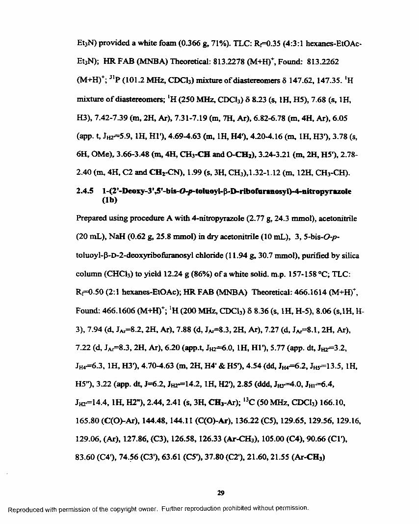
2.4.5 l>(2,-Deoxy>3’,5'-bis-0-/rtoluoyl-P-D-ribofuraiiosyl)-4-nitropyrazole (lb ) .................................................................29
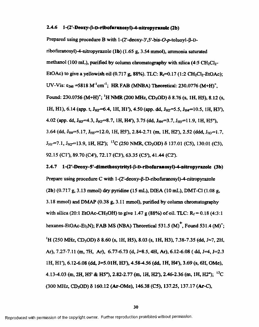
2.4.6 l-(2'-Deoxy-P-D-ribofuranosyl)-4-nitropyrazole (2b)...... 302.4.7 l-^'-Deoxy-S'-dimethoxytrityl-P-D-ribofuranosyl)-^-
nitropyrazole (3b) .................................................................302.4.8 1 -(2 '-Deoxy-5 '-dimethoxytrity 1-3 '-O-2-cyanoethy l-N,N-
diisopropylphosphoramidite-p-D-ribofuranosyl)-4- nitropyrazole (4b).................................................................. 31
2.4.9 l-(2'-Deoxy-3’,5,-bis-(7-/»-toluoyl-P-D-ribofuranosyl)-4-propynylpyrazole (lc ) ....... ......... .......................................31
2.4.10 l-(2'-Deoxy-p-D-ribofuranosyl)-4-propynylpyrazole (2c)........................................................... 32
2.4.11 l-(2'-Deoxy-5'-dimethoxytrityl P-D-ribofuranosyl)-4- propynylpyrazole (3c)........................................................... 33
2.4.12 l-(2'-Deoxy-5,-dimethoxytrityl-3,-0-2-cyanoethyI-IV,N- diisopropylphosphoramidite-p-D-ribofuranosyl)-4- propynylpyrazole (4c)........................................................... 34
2.4.13 H P - D -2'-Deoxyribofuranosyl)-3',5'-bis-0-(/>-toIuoyI)-4- frimethylstannyl-pyrazole (Id) .......................................... 34
2.4.14 1-(P-D -2'-Deoxyribofuranosyl)-3 \5'-bis-0-(/>-toluoyl)-4- (2-thiazolyl)-pyrazole ( l e ) ..................................................... 35
2.4.15 1-(P"D -2'-DeoxyribofuranosyI)-4-(2-fh/az0/yO-pyrazole (2 e )............................................................................................ 36
2.4.16 1-(P_D -2'-Deoxyribofuranosyl)- 5' -0-{dimeth oxytritytyA- (2-f/iiazo/yO-pyrazole (3e) .................................................... 37
2.4.17 l-( P~D -2'-DeoxyribofuranosyI) -5'-0-(dimethoxytrityl)-3'- 0-(2-cyanoethyl-N,N-dilsopropyIphosphoramldite)-4-(2- tbiazo!yl)-pyrazole (4e).......................................................... 38
2.5 References...............................................................................................38
Chapter 3 Thermodynamic Stability o f Oligonucleotides ContainingPyrazole Nudeobase Analogs.......................................................... 40
3.1 Structure o f DNA.................................................................................. 403.2 DNA Thermal Denaturation and Thermodynamic Analysis 41
3.2.1 Sequences Investigated.......................................................... 413.2.2 Thermal Denaturation.......................................................... 423.2.3 Analysis o f Data from Thermal Denaturation Studies.. .43
3.3 Results.................................................................................................... 443.3.1 Self-Complementary Sequences...........................................443.3.2 Non-Self-Complementary Sequences.................................. 45
3.4 Discussion...............................................................................................46
v i
Reproduced with permission of the copyright owner. Further reproduction prohibited without permission.

3.4.1 Self-Complementary Sequences............................................463.4.2 Non-Self-Complementary Sequences................................... 50
3.5 Summary.................................................................................................513.6 Experimental.......................................................................................... 52
3.6.1 UV Thermal Denaturation Measurements................ .523.6.2 Solvent Preparation.................................................................523.6.3 Extinction Coefficients for Oligonucleotides......................533.6.4 Calculation o f Thermodynamic Parameters......................533.6.5 DNA M elting Experiments and Thermodynamic
Analysis...................................................................................... 543.7 References................................................................................................63
Chapter 4 Enzymic Behavior o f Pyrazole-Containing Nudeobase Analogswith Various Thermostable DNA Polymerases............................. 65
4.1 Target for PCR Reactions.................................................................. 654.2 Design o f Enzymic Experiment.......................................................... 654.3 Analysis o f PCR Products.............................................. 694.4 Sequencing o f PCR Product............................................................. 694.5 Results...................................................................................................... 69
4.5.1 PCR Reactions......................................................................... 694.5.2 Sequencing o f PCR Products................................................. 734.5.3 Quantitation o f Acrylamide Gel Sequencing Results 73
4.6 Discussion................................................................................................764.7 Experim ental.......................................................................................... 79
4.7.1 PCR Reactions......................................................................... 794.7.1.1 Preparation of Template............................................794.7.1.2 Synthesis o f Primer..................................................... 804.7.1.3 Sequence o f Primers................................................... 804.7.1.4 Conditions for PCR Reactions.................................804.7.1.5 Gel Electrophoresis o f PCR Products.....................844.7.1.6 Purification of PCR Products................................... 85
4.7.2 Sequencing Reactions..............................................................854.7.2.1 Synthesis o f Primer for Sequencing Reaction 854.7.2.2 Reaction Conditions for Sequencing Reactions...854.7.2.3 Gel Electrophoresis o f Sequencing Reaction 854.7.2.4 Quantitation of Images from Sequencing G els....86
4.8 References...............................................................................................87
Chapter 5 Synthesis o f 2-ThiazoIe C-Nucleoside................................................885.1 Design....................................................................................................... 885.2 Synthesis..................................................................................................895.3 Experimental.......................................................................................... 91
5.3.1 5-Anhydro-|3-3-deoxy-4,6-di-0-/»-toluoyl-D-ri6o-hexononitrile (5a)....................................................................91
v i i
Reproduced with permission of the copyright owner. Further reproduction prohibited without permission.

5.3.2 5-Anhydro-{J-3-<ieoxy-4,6-di-0 -p-toluoyl-D-ri60- hexonothiamide (5b)................................................................ 91
5.3.3 2>(3',5,-Bis-0-/»-toluoyl-P-D-2'-deoxyribosyl)-thiazole (5c).............................................................................. 92
5.3.4 2-(3’,5'-Bis-0-/>-toIuoyI-p-D-2'-deoxyribosyI)-thiazole-3- oxide (5d).............. 93
5.4 References............................................................................................... 93
Chapter 6 Substitution o f 5-Nitroindole Nucleotides During OligonucleotideSynthesis................................................................................................ 95
6.1 Introduction...........................................................................................956.2 Discussion............................................................................................... 956.3 R e fe r e n c e s ....................................................................................... 97
Vita.................................................................................................................................99
v i i i
Reproduced with permission of the copyright owner. Further reproduction prohibited without permission.
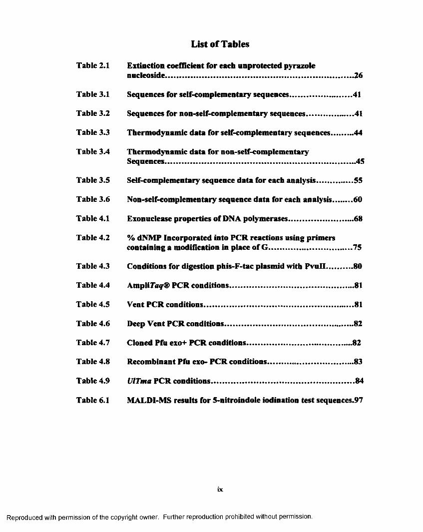
List of Tables
Table 2.1 Extinction coefficient for each unprotected pyrazolenucleoside.............................................................. 26
Table 3.1 Sequences for self-complementary sequences............................... 41
Table 3.2 Sequences for non-self-complementary sequences.........................41
Table 3.3 Thermodynamic data for self-complementary sequences............44
Table 3.4 Thermodynamic data for non-self-complementarySequences........................................................... .45
Table 3.5 Self-complementary sequence data for each analysis................... 55
Table 3.6 Non-self-complementary sequence data for each analysis...........60
Table 4.1 Exonuclease properties o f DNA polymerases................................. 68
Table 4.2 % dNMP Incorporated into PCR reactions using primerscontaining a modification in place o f G ..........................................75
Table 4.3 Conditions for digestion phis-F-tac plasmid with PvuII..............80
Table 4.4 AmpliTaq® PCR conditions............................................................... 81
Table 4.5 Vent PCR conditions............................................................................81
Table 4.6 Deep Vent PCR conditions................................................................. 82
Table 4.7 Cloned Pfu exo+ PCR conditions..................................................... 82
Table 4.8 Recombinant Pfu exo- PCR conditions............................................83
Table 4.9 UlTma PCR conditions.........................................................................84
Table 6.1 MALDI-MS results for 5-nitroindole iodination test sequences.97
ix
Reproduced with permission of the copyright owner. Further reproduction prohibited without permission.

Figure 1.1
Figure 1.2
Figure 1.3
Figure 1.4
Figure 1.5
Figure 1.6
Figure 1.7
Figure 1.8
Figure 1.9
Figure 1.10
Figure 1.11
Figure 1.12
Figure 1.13
Figure 2.1
Figure 2.2
Figure 2.3
Figure 2.4
Figure 3.1
Figure 3.2
List of Figures
Conversion o f C-G to T-A base pair..................................................2
Series of pyrazole analogs......................................................................4
N-nudeoside and C-nucleoside synthesis.......................................... 5
Comparison o f hydrogen bonding base pairs with a nonhydrogen bonding analog......................................................................6
Structures o f analogs studied by Berstrom, Brown and Kool 6
Structures o f thymidine, 5<l-propynyl)-2,deoxyuridine and 5-(2- thiazoy0-2'-deoxyuridine.......................................................................7
Polymerase extension ................................................................ 9
PCR amplification cycle.........................................................................9
Sanger method for dideoxy sequencing.......................................... 12
3'->5' Exonuclease activity-proofreading...................................... 13
5'->3' Exonuclease activity-misprinted sequences....................... 14
Structures o f 3-nitropyrrole, 3-pyrrole-carboxamide, 4-pyrazole- carboxamide, 4-imidazole-carboxamide and inosine...................15
Structures o f 6H , 8H-3,4-dihydropyrimido[4,5-cJ[l,2]oxazin-7- one (P) and N*-methoxy-2,6-diaminopurine (K ).......................... 15
Target pyrazole nucleosides................................................................ 18
Charge distribution in natural base pairs....................................... 19
Charge distribution of pyrazole analogs..........................................19
Phosphoramidites used in solid phase synthesis........................... 23
Structure o f 5VTCGA-3' nucleotide.................................................40
Hydrogen bond pairs between purines and pyrimidines............41
x
Reproduced with permission of the copyright owner. Further reproduction prohibited without permission.

Figure 3.3
Figure 3.4
Figure 3.5
Figure 3.6
Figure 4.1
Figure 4.2
Figure 4.3
Figure 4.4
Figure 4.5
Figure 4.6
Figure 4.7
Figure 4.8
Figure 4.9
Figure 4.10
Figure 4.11
Structures o f 4-substitued pyrazole nucleoside analogs.............41
Comparison o f -ATm for each modification with all the natural bases....................................................................................................... 48
Comparison o f ATm for each natural base with all o f the modifications........................................................................................ 49
Comparison o f AT., for non-self-complementary sequences...51
Analogs studied in PCR reactions................................................... 65
Diagram of PCR reaction containing a pyrazole analog in the sense prim er....................................................................................... 67
Sequence o f the target PCR product, the hisF gen e.................. 68
Schematic diagram of Sanger method for sequencing..............70
Agarose gel analyzing PCR reactions with AmpliTaq® and UlTma enzymes with control primer (C), 5-nitroindole (Ind) primer, 4-nitropyrazole (PzN) primer, 3-nitropyrrole (PyN) primer and 4-(2-thiazolyl)-pyrazole (PzT) primer......................71
Agarose gel analyzing PCR reactions with Pfu exo+ and P/u exo- Enzymes with control primer (C), 5-nitroindole (Ind) primer, 4- nitropyrazole (PzN) primer, 3-nitropyrrole (PyN) primer and 4- (2-thiazolyl)-pyrazole (PzT) primer................................................71
Agarose gel analyzing PCR reactions with Deep Vent and Vent enzymes with control primer (C), 5-nitroindole (Ind) primer, 4- nitropyrazole (PzN) primer, 3-nitropyrrole (PyN) primer and 4- (2-thiazolyl)-pyrazoIe (PzT) primer................................................72
Agaorse gel analyzing PCR reactions with AmpliTaq® enzyme with control Primer (Con), 4-nitropyrazole primer (PzN), 4- propynyl pyrazole primer (PzP), and 4-(2-thiazolyI)pyrazole primer (PzT)........................................................................................72
Sequencing gels of PCR products from Pfu exo-.........................73
Sequencing gels of PCR products from A m plifa?® ...................74
Sequencing gels o f PCR products from Vent enzyme................ 74
xi
Reproduced with permission of the copyright owner. Further reproduction prohibited without permission.

Figure 4.12 Sequencing gels o f PCR products from DeepVent enzyme......... 74
Figure 4.13 Sequencing gels o f PCR products from Pfu exo+..........................74
Figure 4.14 Sequencing gels o f PCR products from UlTma..............................74
Figure 4.15 Schematic o f 3'-^5' Exonuclease Activity Removing theM odification Q from the Primer and Replacing with a Natural Nucleotide...............................................................................................78
Figure 5.1 Structure o f thiazole nucleoside and thiazole N-oxidenucleoside..............................................................................................88
Figure 6.1 Iodination o f 5-nitroindole.................................................................. 96
xii
Reproduced with permission of the copyright owner. Further reproduction prohibited without permission.

Scheme 2.1
Scheme 2.2
Scheme 2.3
Scheme 2.4
Scheme 5.1
List of Schemes
Synthesis o f 3,5-bis-0-/>-toluoyl-P-D-2-deoxyribosyl chloride....21
Synthesis o f 4-substitued pyrazole protected nucleosides 21
Synthesis o f 4-substituted pyrazole phosphoramidite for incorporation into oligomers using automated DNA synthesis...22
Solid phase DNA synthesis, phosphoramidite method................. 24
Synthesis o f protected 2-thiazolyl C-nudeoside and thiazole —N- oxide C-nudeoside.................. 90
xiii
Reproduced with permission of the copyright owner. Further reproduction prohibited without permission.

List of Abbreviations
Abbreviation Name
Me Methyl
dCTP Deoxycytidine triphosphate
dGTP Deoxyguanosine triphosphate
Con Control
dTTP Deoxythymidine triphosphate
dATP Deoxyadenosine triphosphate
a Mole fraction o f single stranded DNA in theduplex state
A Deoxyadenosine
AcOH Acetic acid
bp Base pair
bz Benzoyl
C Deoxycytidine
Cone. Concentration
CPG Control pore glass
CSO (1S )-(+)-(10-caniphorsulfonyl)-oxaziridine
DCM Dichloromethane
dNTP Deoxynucleotide triphosphate
ddNTP Dideoxynucleotide triphosphate
DFT 2,4-Difluorotoluene
xiv
Reproduced with permission of the copyright owner. Further reproduction prohibited without permission.

Abbreviation Name
DIEA Diisopropyylethylamine
DMAP 4,4-Dimethylarainopyridine
DMF Dimethylform amide
DMT Dimethoxytrityl
DMT-C1 Dimethoxytrityl chloride
DNA Deoxyribonucleic acid
dNTP Deoxynucleotide triphospate
dsDNA Double stranded DNA
EDTA Ethylenediaminetetraacetic acid
Et3N Triethylamine
EtOAc Ethyl acetate
EtOH Ethanol
FABMS Fast atom bombardment mass spectrometry
G Deoxygaunosine
HR FAB High resolution fast atom bombardment mass spectrometry
MALDI-MS Matrix assisted laser desorption ionization mass spectrometry
MCPBA m-Chloroperoxybenzoic acid
MeOH Methanol
m.p. Melting point
NMR Nuclear Magnetic Resonance
XV
Reproduced with permission of the copyright owner. Further reproduction prohibited without permission.

Abbreviation
PCR
Rr
RNA
ssDNA
T
TAE
TBE
TEAA
TEAB
THF
TLC
TA m
Tol
UV
Name
Polymerase Chain Reaction
Ratio o f the distance traveled by a compound
relative to the solvent front
Ribonucleic acid
Single stranded DNA
Deoxythimidine
Tris-acetic acid EDTA buffer
Tris-boric acid-EDTA buffer
Triethyammonium acetate
Triethyammonium bicarbonate
Tetrahydrofuran
Thin layer chromatography
Melting temperature
/Moluoyl
Ultraviolet
xvi
Reproduced with permission of the copyright owner. Further reproduction prohibited without permission.

Abstract
Non-hydrogen bonding nucleosides with iodo, nitro, propynyl and thiazolyl
substituted at the 4-position o f pyrazole were prepared. These nucleosides were
converted to their corresponding nucleoside phosphoramidites and incorporated into
a series o f complementary oligonucleotides in order to determine the effect that
varying size, charge distribution and polarizability has on duplex stability and
structure. The self-complementary Dickerson dodecamer sequence 5’-
CGCXAATTYGCG-3’, as well as the non-self complementary sequence 5’-
CAAAATGGTGGCCAAGT-3’ previously investigated by Brown, were used to
determine the duplex stabilization and thermodynamic consequences o f placing 5-
nitroindole and 4-substituted pyrazole nucleosides across from each natural base. In
the pyrazole series, the largest and smallest duplex destabilization in all cases
studied was found with cytosine and adenosine, respectively, while the 4-thiazolyl
substitution was determined to form the most stable duplex regardless of the
complementary base. We investigated the directing ability of the pyrazole analogs
for incorporation of dNTP’s. The DNA polymerases chosen were Taq and Pfu. exo-,
which lack 3'-5' proofreading exonuclease activity, and Vent, Deep Vent, Pfu which
contain exonuclease activity. The enzymes were able to incorporate natural
nucleotides across from our modification as detected using the polymerase chain
reaction. The Sanger method o f dideoxysequencing was used to determine the
natural base incorporated across from the modification. The non-proofreading
xvii
Reproduced with permission of the copyright owner. Further reproduction prohibited without permission.

enzymes mainly incorporated deoxyadenosine across from our modification where
as the proofreading enzymes removed our modification but not with total efficiency.
Automated DNA synthesis procedures might modifying the 5-nitroindole
base via electrophilic aromatic substitution by replacing the nitro group or addition
o f iodine to the ring system. An alternative oxidation reagent, CSO, was used to
prepare a series o f sequences for comparison with sequences prepared using
standard oxidation conditions. Results from MALDI-MS did not provide conclusive
evidence for either substitution or iodination o f the bicyclic ring system.
Examination o f the thermodynamic results led to the preparation o f thiazole-
C-nucleoside, which historically is more difficult to synthesize. The thiazole ring
offers a site for the formation o f an N-oxide. It has been suggested this group is
responsible for enzyme recognition. We prepared the thiazole and thiazole-N-oxide
nucleosides for incorporation into oligonucleotides for thermodynamic and enzymic
investigations.
xviii
Reproduced with permission of the copyright owner. Further reproduction prohibited without permission.

Chapter 1 Introduction
Non-hydrogen Bonding Nucleosides
1.1 Background
The design and synthesis o f non-natural base analogs was originally sought
after in hopes o f a universal base. A universal base is defined as one that would not
appreciably destabilize the duplex and would pair equally wee with both purines and
pyrimidines. Also, enzymes would recognize and incorporate equal amounts o f the
natural nucleotides (dNTPs) across from the universal base. Incorporation o f all
bases across from a universal base would allow random mutagensis at directed sites.
3-Nitropyrrole was one o f the first bases to show almost universal
thermodynamic stability when paired with any of the natural bases. * However,
polymerase reaction with 3-nitropyrrole showed a large preference for incorporation
o f purines over p y rim id in es.^ Thus another application o f this type o f nucleoside
analogs is for directed conversion o f one base to another.
Often mismatches have to be created when attempting to convert one base
pair to another (e. g. GC bp AT bp). A G-T “wobble” mismatch in the primer
results in reduced polymerase extension efficiency (poor read) and thus low
efficiency o f conversion. As shown with the use of a non-natural base could
improve this by eliminating the poor read step as the analog could pair well
(Watson-Crick bonding pattern) with the natural base, unlike the wobble mismatch.
The resulting “good read” would improve the overall conversion o f the sequence.
One application o f polymerase-based conversion with nucleobase analogs is
creation o f restriction sites in DNA for enhancing detection o f low level mutations
l
Reproduced with permission of the copyright owner. Further reproduction prohibited without permission.

in a large background o f wild-type (non-mutated) DNA-3*4 The introduction o f
non-natural base into a PCR primer will allow for conversion o f the sequence to
generate a restriction site in wild type DNA while the low-level mutant DNA will
not posess a restriction site. Following this PCR step (repeating many times if
necessary), a restriction enzyme will digest the DNA removing the converted wild-
type DNA and leaving the mutant DNA uncut.3*4
y — C' - 3' y — t y — t— ■ -3- y — t3" ----- G- ----------y
J 94 PC 65 »C | Taq Pol 65 PC J Taq Pol 65 PC J Taq Pol
y — c- y — t — ^ y — t -3’ y — t —3’ ----- G — - -y -y
3’ ----- G-^ 94 PC ^ 94 PC
y ---- r --------- - 3' y — t * 3'
• y -yH C M , O M
C H , 0 - « - N N
f t , - h -n H -1 --------------- ^ ~ M. , ............... , , f Vh-n^Vm,H~ i > =N R R O — H - N y - M , N - ^
R O — f )= N R R OH -H G T A
H
5’ C - - 3 ’ 5' 06 y ——Q * ^ ——— 3' y — T
« 3 » c | Taq Pol 65 PC J TaqPol 65 PC J Taq Pol
36J
1
S' Qe " - » y — Q « — 3’ y — T
94 »C | M K
y — G - - y y - a - - ■ y
> -R * M ~ i > -R R f , - i W k r „ ^ RR O —M -R R O —44-R R 0 R O
C H « Q # (C , H G Q «(T ) A
S o e d R n d Got
Figure 1.1 Conversion o f C -G to T-A base pair
T A
2
Reproduced with permission of the copyright owner. Further reproduction prohibited without permission.

1.2 Design
The introduction o f non-natural nucleosides has been under intense
investigation as a way to understand the extremely efficient design o f DNA and its
properties. We and others are interested in dissecting the roles hydrogen bonding
and “base stacking” play in controlling duplex stability, structure, and DNA
enzymology. One approach to studying these factors is to remove the hydrogen
bonding o f the bases altogether. A first foray into this approach was replacing the
heteroaromatic bases with either a hydrogen atom or a phenyl ring,5 which found
that both substitutions were similarly destabilizing to the duplex. The pioneering
work o f Bergstrom and coworkers used an intercalation model to develop a 3-
nitropyrrole a n a l o g ,^> 7 which had much great stability in a duplex relatively to the
Millican phenyl or abasic site analogs.^
Another approach is to mimic the size and charge of the natural nucleosides
as Kool and coworkers have suggested. They designed 2,4-difluorotoluene as a
shape mimic o f thymidine.8>9 The methyl group is in same position and the fluoro
groups are similar to the oxygens on thymidine both in size and electronegativity.
Kool also designed 4,6-dimethyindole as a hydrophobic adenine analog.8>9(Figure
1.5) The methy groups are in the proximity o f the oxygens in T to offer similar
shape without specific hydrogen bonding characteristics.
We have developed a series non-hydrogen bonding analogs (Figure 1.2) to
investigate the properties o f DNA when hydrogen bonding is removed while
varying the charge and size characteristics o f substituents on the aromatic core. We
designed a series of pyrazole analogs with substituents in the 4-position with
3
Reproduced with permission of the copyright owner. Further reproduction prohibited without permission.

varying charge and size, iodo, propynyl, nitro and thiazolyl (2a, 2b, 2c, and 2e).
The differing characteristics o f the substituents allows for investigation o f charge
distribution and size variation on the stability o f DNA duplexes as well as the
enzymic acceptance o f the analogs. The size o f the small 5-membered should fit
into the active site o f enzyme and not offer steric hindrance when incorporated
across from purines.
NO;
V N V N HO HO
HO HO HO HO
P z l P zN P zP P zT2 a 2b 2c 2 e
Figure 1.2 Series of pyrazole analogs
1.3 Synthesis
Once the series o f pyrazole analogs was chosen, the synthesis o f these non
natural analogs had to be designed. There are two traditional approaches for the
synthesis o f nucleosides. The first and more popular approach uses the
corresponding base analog reacting with a sugar resulting in the nucleoside. This
type o f reaction is called glycoslyation. The Hilbert-Johnson method is a classic
glycosylation method used in the synthesis o f Nl-glycosyl p y r i m i d i n e s . ^ The
second approach requires the construction o f the base analog system from an N or
C-glycoslated precursor. There are 2 different types o f nucleosides, C or N. We are
interested in the synthesis N-nucleosides. Meaning, the Cl position o f the sugar is
attached to the bases through a nitrogen in the ring o f the base (Figure 1.3). The
4
Reproduced with permission of the copyright owner. Further reproduction prohibited without permission.

other type is a C-nucleoside. These have the sugar attached to a carbon in the ring
o f the base. C-nucleosides are much more difficult to synthesize. This difficulty
arises from the unstable precursors, like glycals^ or a low yielding multistep
reactions which builds the aryl unit from a cyano or some other C-glycoside (Figure
1.3). We are interested in attaching the N1 o f the commercially available pyrazole
ring to the sugar precursor. The substituents are either already present on the ring
when purchased (iodo and nitro) or they are synthesized (propynyl and thiazolyl)
once the pyrazole ring is attached to the sugar using palladium mediated chemistry.
r= \To,0n / \ , N' ToIO- v o Y n
To|o Cl ToioTolO
N-nucleoside C-nucleoside
Figure 1.3 N-nudeoside and C-nucleoside synthesis
1.4 Thermodynamic Stability
The arrangement o f the aromatic bases in DNA orthogonally, along the
center axis o f the duplex allows pyrimidine-purine hydrogen bonding between the
two strands (edge-edge interactions) as well as stacking o f bases with the bases
above and below it in the duplex (7c-tc interactions). After removal o f hydrogen
bonding, a heterobase analog utilizes mainly ir-stacking along the axis o f the duplex,
though dipole-dipole interactions across the duplex with its putative ‘‘pairing”
partner may also influence structure and stability of the duplex.
One factor that affects stacking ability is the charge distribution in the 7t-
aromatic system o f the analog. For example, 3-nitropyrrole (Figure 1.5) is much
Reproduced with permission of the copyright owner. Further reproduction prohibited without permission.
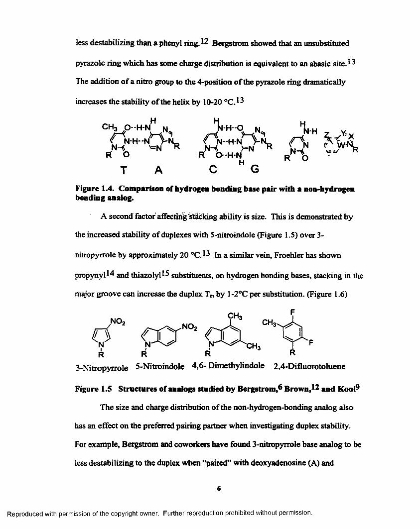
less destabilizing than a phenyl ring. 12 Bergstrom showed that an unsubstituted
pyrazole ring which has some charge distribution is equivalent to an abasic s i t e . 13
The addition o f a nitro group to the 4-position o f the pyrazole ring dramatically
increases the stability o f the helix by 10-20 °C.13
Figure 1.4. Comparison of hydrogen bonding base pair with a non-hydrogen bonding analog.
A second factor affecting stacking ability is size. This is demonstrated by
the increased stability o f duplexes with 5-nitroindole (Figure 1.5) over 3-
nitropyrrole by approximately 20 °C.13 in a similar vein, Froehler has shown
propynyll4 and th iazolyl^ substituents, on hydrogen bonding bases, stacking in the
major groove can increase the duplex Tm by 1-2°C per substitution. (Figure 1.6)
3-Nitropyrrole 5-Nitroindole 4,6- Dimethylindole 2,4-Difluorotohiene
Figure 1.5 Structures of analogs studied by Bergstrom/* Brown,12 and Kool9
The size and charge distribution o f the non-hydrogen-bonding analog also
has an effect on the preferred pairing partner when investigating duplex stability.
For example, Bergstrom and coworkers have found 3-nitropyrrole base analog to be
less destabilizing to the duplex when “paired” with deoxyadenosine (A) and
Reproduced with permission of the copyright owner. Further reproduction prohibited without permission.
HT A C G
6

deoxygaunosine (G) ^ Whereas, the 5-nitroindole nucleoside base analog is less
destabilizing when paired with deoxycytidine ( Q . ^ Kool has investigated several
analogs (Figure 1.5) such as 4,6-dimethylindole and 2,4-difluorotoluene, while
diflourotoluene is a deoxythimidine (T) analog it is least stable across from A and in
fact is most stable across from itself in the duplex.^
To determine the stacking ability o f a variety o f substituents without
hydrogen bonding effects, we have prepared a series o f 4-substituted pyrazole
nucleoside analogs and studied their effects on DNA duplex stability in a variety of
contexts.
1.5 Enzymic Activity
1.5.1 DNA Polymerases
DNA polymerases are responsible for the replication o f DNA. Komberg
isolated the first DNA polymerase in 1955 from an extract o f E. coli. This
polymerase, now called DNA polymerase I, has two main segments with three main
functions. The N- terminal small fragment contains 5'->3' exonuclease activity.
Exonuclease activity is the ability o f a polymerase to remove an imperfect match.
The large fragment, better known as the Klenow fragment, has polymerase activity
as well as 3'->5' exonuclease activity. These three functions are crucial to correct
replication o f DNA.
Figure 1.6 Structures o f thymidine, 5-(l-propynyl)-2’deoxyuridine and 5-(2- thiazoyl)-2’-deoxyuridine
o
HO
R= — C H , . *CH j . o r
7
Reproduced with permission of the copyright owner. Further reproduction prohibited without permission.

Replication o f DNA, also called extension o f a primer, involves several
components. First, the enzyme must posses polymerase activity. The sequence to
be copied, called the template, must be present. Next, a primer (much shorter in
length) matching a portion o f the template sequence must anneal to the template
making a double strand o f DNA. For the polymerase to extend o ff the 3'-end o f the
primer, deoxynucleotide triphophates must be availble to the polymerase and an
optimal temperature reached for extension to begin. Once these main components
are present, replication o f the template is possible.
1.5.2 Polymerase Chain Reaction (PCR)
The introduction o f the polymerase chain reaction (PCR) revolutionized
DNA extension technology. 1'6,17(Figure 1.8) This tool allowed for rapid
amplification o f finol quantities o f DNA. Whereas the previous extension
technology (Figure 1.7) only provided linear amplification. The concept is similar
to that described in Section 1.5.1 but the cycle is repeated many times varying the
temperature to allow for annealing o f the primer, extension and denaturing o f the
double stranded product. Also the product is increased exponentially due to the
presence o f a double stranded template and a primer for each strand o f the double
stranded template. The extension explained earlier only required a single stranded
template and one primer. The desired double stranded sequence to be amplified is
called the template. The template is normally present in finol quantities. At least
some o f the template sequence must be known so as to offer a place for annealing o f
the primer so that amplification can occur. The primers serve as the starting place
for the DNA polymerase to start copying the template. A primer must be designed
8
Reproduced with permission of the copyright owner. Further reproduction prohibited without permission.

P rim erS tra n g
C iN T P
Figure 1.7 Polymerase extension
PCR Amplification of ds DNA
separate strands bv healing to 95-C
Anneal p rim ers by cooling to 37°C
H eat to 70°C to allow for exicastion
SI ’C
Anneal prim ns by cooling to 37°C
Hear to 70°C to allow forex tenstion
Repeat There are now four sets of double strandedfds) template where only one set was used to start. The third round of PCR willprovide 8 sets of ds template, 16,32 e tc .
Figure 1.8 PCR amplification cycle
9
Reproduced with permission of the copyright owner. Further reproduction prohibited without permission.
97

for each strand o f the template. Once the reaction starts, the primers are extended
in the 3' ->5' direction to the end o f both strands o f the template. The polymerase
inserts the complementary dNTP that creates a natural base pair w ith the template
strand and then continues on to the next base. At the completion o f this cycle, a
copy o f the original template is created. The double stranded products are separated
by heating to 95 °C and now a place is available for new primers to be positioned
for extension. To position the primers on the template an annealing step is
performed (normally 50-60 °C) followed by raising the temperature to 70-75 °C
(depending on polymerase) for optimal extension temperature. On the second cycle,
copies o f old and new pieces o f template are made resulting in a total of four
templates. The third cycle copies the four strands resulting in a total o f eight and so
the template is amplified exponentialyl.
The Klenow fragment was not particularly efficient for PCR as the high heat
required in thermal cycling kills the polymerase activity o f the enzyme. Therefore
the discovery o f thermostable polymerase was necessary for the success o f PCR.
The thermostable polymerase under the most intense investigation was Thermus
aquaticus (Taq). * 8-21 Other thermostable polymerases that are commonly used are
Thermotoga maritima (T m a )^ , Pyrococcus furiosus (P fup3-25t Thermus
thermophilus (Tth) 26,27} Pyrococcus species GB-D 28 (Deep Vent from New
England Biolabs) and Thermococcus litoralis (Tli) 29 (Vent from New England
Bioiabs). These polymerases as well as many others provide different tools for
investigating modified primers, modified triphosphates or modified templates.
10
Reproduced with permission of the copyright owner. Further reproduction prohibited without permission.
1.5.3 Sanger M ethod for Sequencing DNA
The Sanger method is very similar to PCR in the fact that a polymerase, a
primer, a template and dNTP’s are used. (Figure 1.9) This technique only provides
linear amplification o f the desired product. What makes this method different is
there are dideoxy nucleotide triphosphates (ddNTP’s) present. ddNTP’s lack the 3 '
hydroxyl group removing the active group for extension. This creates truncated
pieces that have been extended somewhat but once the ddNTP is incorporated no
extension is possible. For the convenience o f determining the ddNTP that
terminates the strand the ddNTP’s are radiolabeled with 33P for the purpose of
imaging the truncated sequences. (Figure 1.9) Each o f the four natural bases are
used as ddNTPs but in separate reaction so as to be able to determine the exact
sequence. Once the extension reaction is complete, the truncated pieces are loaded
onto a polyacryamide gel to be separated using gel electrophoresesis based on
differences in size. The shorter pieces move at a faster rate through the gel as the
longer pieces move at a slower rate. Once the electrophoresis is complete, a ladder
o f the different sized pieces is present. Each separate reaction with an individual
ddNTP is loaded in individual lanes and separated at the same time. This creates a
ladder o f the different sized pieces and allows for determination o f the
sequence.(Figure 1.9) An alternative method for sequencing is the Maximum-
Gilbert method. This required chemical degradation with regents specific for one or
two o f the natural bases followed removal o f the base creating an abasic site along
the backbone o f the DNA. Once the abasic cite is present cleavage o f the backbone
is much easier resulting in truncated sequences as well.
11
Reproduced with permission of the copyright owner. Further reproduction prohibited without permission.

Sanger Method using
Radiolabeled Dideoxyterminators
a —“ P-ddNTP’s
1
Dideoxy Sequencing
»Pw p
upu p
u pJ J p
8% PAGE of Sequencing Products
•Gel result yields sequence information about the orginal template
•The sequence shown by the gel is the compliment to the original sequence
Figure 1.9 Sanger method for dideoxy sequencing
1.5.4 Exonuclease Activity
There are two directions for exonuclease activity to occur 3 '-> 5 ' and 5 '->3
5 '-> 3 ' Exonuclease is useful in removing misprinted sequences and to repair
damaged nucleotides.^ The 3 '-> 5 ' activity can remove unnatural modification by
cutting up the DNA until the unnatural analog is removed. W ith 3'->5' exonuclease
activity, also known as proofreading, the enzyme can remove an incorrectly inserted
nucleotide at the primer terminus or 3'-end before continuing with
polymerization.(Figure 1.10) The mismatched nucleotide is not involved in
hydrogen bonding with the template strand. The ability to proofread drastically
12
Reproduced with permission of the copyright owner. Further reproduction prohibited without permission.

increases the accuracy o f the DNA replication by allowing the polymerase to correct
any mistakes before continuing.
—6- - - e
3'OH
5'Figure 1.10 3'->5* Exonuclease activity-proofreading
5'-> 3' exonuclease activity allows the polymerase to excise any misprimed
sequences and continue with polymerization. (Figure 1.11) Without this feature, the
polymerase would discontinue polymerization o f the template when misprimed
sequences were found (most often in nature with RNA primers). W ithout this
activity, DNA replication is not as accurate and complete replication o f the template
is hindered.
13
Reproduced with permission of the copyright owner. Further reproduction prohibited without permission.

mtspnmeaor otnerds
■ ' - template
r,™™- sequence w other dsDNApnmer
or other dsDNApnmer extension sequence
— • ' template
'fspnmen w 0tper<jsONA extension sequence
' template
Without 5->3* actwtythe extension is not / \ V/tth5’->3’activity thecomplete as the / \ rmspnmeO sequence ismisprimed sequence / \ removed and extenstion iscannot be removed / \ aSovved to continue
mtsprmedprimer extension sequence pnmer extension
. template • - ' - — templateExtension Terminated Extension Complete
Figure 1.11 5'->3' Exouuclease activity -m isprim ed sequences
1.5.5 Polymerase Reactions with Non-Natural Nucleosides
The behavior o f enzymes with non-natural nucleosides has drawn much
attention and research. Bergstrom reported one o f the first successfid PCR reactions
using a modified primer containing 3 n itrop yrro leA ^ Other work by Bergstrom
utilizing azole nucleobase analogs provided the model for our experiments.^
Bergstrom’s research studied 3-nitropyrrole, inosine, 3-pyrrolecarboxamide, 4-
pyrazole-carboxamide and 4- imidazolecarboxamide, along with an abasic site with
PCR utilizing Taq DNA polymerase. (Figure 1.12) These results showed a 3:1 A:T
incorporation in PCR for 3-nitropyrrole using the Sanger sequencing method with
33P radiolabeled dideoxyterminators. Sim ilar research by Kool and coworkers uses
2,4-diflorotoulene (DFT) as a T mimic. Kool has successful incorporation o f DFT
triphosphates across from A and incorporation o f dATP across from DFT analogs in
14
Reproduced with permission of the copyright owner. Further reproduction prohibited without permission.

R R R R R
Figure 1.12 Structures o f 3-nitropyrrole, 3-pyrroIe-carboxamide, 4-pyrazole- carboxamide, 4-imidazole-carboxamide, and inosine (left to right)
dihydropyrimido[4,5-c][l,2]oxazin-7-one, a pyrimidine analog, and N^-methoxy-
results with these modification incorporated into a primer using Taq polymerase
show these analog behave as designed a purine analog, K and a pyrimidine analog,
P. The research o f Bergstrom^ and Brown^ 1 has shown the ability o f Taq
polymerase to accept a non-natural nucleobase in the DNA primer as well as non
natural triphosphates.
Figure 1.13 Structures of 6H, 8H-3,4-dihydropyrimido[4,5-c][l,2]oxazin-7- one (P) and N6-methoxy-2,6-diaminopurine (K)
Our research w ill investigate the acceptance o f the pyrazole analogs with Taq
DNA polymerase as well as other thermostable polymerases with a variety o f
inherent properties using PCR. We will also investigate how the pyrazole analogs
are read by the enzymes using sequencing methods to determine the natural base
incorporated across from the analogs.
primers.^(Figure 1.5) Brown and coworkers have used 6H, 8H-3,4-
2,6-diaminopurine, a purine analog, P and K respectively.3 1 (Figure 1.13) The
R
P K
15
Reproduced with permission of the copyright owner. Further reproduction prohibited without permission.

1.6 References
1) Zhang, P .; Johnson, W. T.; Klewer, D.; Paul, N.; Hoops, G.; Davisson, V. J.; Bergstrom, D. E. Nucleic Acids Res. 1998,2 6 ,2208-2215.
2) Hoops, G. C.; Zhang, P.; Johnson, W. T.; Paul, N.; Bergstrom, D. E.; Davisson, V. J. Nucleic Acids Res. 1997,2 5 ,4866-4871.
3) Day, J. P.; Hammer, R. P.; Bergstrom, D.; Barany, F. Nucleic Acids Res 1999, 27, 1819-1827.
4) Day, J. P.; Bergstrom, D.; Hammer, R. P.; Barany, F. Nucleic Acids Res 1999, 27, 1810-1818.
5) Millican, T. A.; Mock, G. A.; Chauncey, M. A.; Patel, T. P.; Eaton, M. A. W.; Gunning, J.; Cutbush, S. D.; Niedle, S.; Mann, J. Nucleic Acids Res. 1984, 12, 7435-7453.
6 ) Nichols, R.; Andrews, P. C.; Zhang, P.; Bergstrom, D. E. Nature 1994,369, 492-493.
7) Bergstrom, D. E.; Zhang, P.; Toma, P. H.; Andrews, P. C.; Nichols, R. J. Am. Chem. Soc. 1995,117, 1201-1209.
8 ) Schweitzer, B. A.; Kool, E. T. J. Org. Chem. 1994, 59, 7238-7242.
9) Schweitzer, B. A.; Kool, E. T. J. Am. Chem. Soc. 1995,117, 1863-1872.
10) Pliml, J.; Pyrstas, M .;. Adv. Heterocyc. Chem 1967, 8 ,115.
11) Cameron, M. A.; Cush, S. B.; Hammer, R. P. J. Org. Chem. 1997, 62, 9065- 9069.
12) Loakes, D. Nucleic Acids Res. 1994, 22, 4039-4043.
13) Bergstrom, D. E.; Zhang, P.; Johnson, W. T. Nucleic Acids Res. 1997, 25, 1935-1942.
14) Froehler, B. C.; Wadwani, S.; Terhorst, T. J.; Gerrard, S. R. Tetrahedron Lett. 1992, 33, 5307-5310.
15) Guiterrez, A. J.; Terhorst, T. J.; Matteucci, M. D.; Froehler, B. C. J. Am. Chem. Soc. 19 9 4 ,116, 5540-5544.
16) Mullis, K. B.; Faloona, F. Meth. Enzymol. 1987,155, 335-350.
17) Saiki, R. K.; Gefland, D. H.; Stoffel, S.; Scharf, S.; Higuchi, R.; Horn, G. T.; Mullis, K. B.; Erlich, H. A. Science 1988,239,487-491.
16
Reproduced with permission of the copyright owner. Further reproduction prohibited without permission.

18) Chein, A.; Edgar, D. B.; Trela, J . M. J. Bacteriol. 1976,127,1550-1557.
19) Kaledin, S. S.; Sliusarenko, A. G .; Gorodetskii, S. I. Biokhimiya 1980, 45, 644- 651.
20) Lawyer, F. C.; Stoffel, S.; Saiki, R. K.; Myambo, K.; Drummond, R.; Gefland, D. H. J. B io l Chem. 1989,2 64 ,6427-6437.
21) Longley, M. J.; Bennett, S. E.; Mosbaugh, D. W. Nucleic Acids Res. 1990,18, 7317-7322.
22) Bost, D. A.; Stoffel, S.; Landre, P.; Lawyer, F. C.; Akers, J.; Abramson, R. D.; Gefland, D. H. FASEB J. 1994, 8, A1395.
23) Lundberg, K. S.; Shoemaker, D. D.; Adams, M. W.; Short, J. M.; Sorge, J. A.; Mathur, E. J. Gene 1991,108, 1-6.
24) Mathur, E. J.; Adams, M. W.; Callen, W. N.; Cline, J. M. Nucleic Acids Res. 1991,19, 6952.
25) Uemori, T.; Ishino, Y.; Toh, H.; Asada, K.; kato, L. Nucleic Acids Res. 1993, 21.
26) Ruttimann, C.; Cotoras, M.; Zalvidar, J.; Vicuna, R. Eur. J. Biochem. 1985,149, 41-45.
27) Carballeira, N.; Nazabal, M.; Brito, J.; Garcia, O. BioTechniques 1990, 9, 276- 281.
28) Perler, F. B. unpublished data, 1994.
29) Perler, F. B.; Comb, D. G.; Jack, W. E.; Moran, L. S.; Qiang, B.; Kucera, R. B.; Benner, J.; Slatko, B. E.; Nwankwo, D. O.; Hempstead, S. K.; Carlow, C. K. S.; Jannaach, H. Proc. Natl. Acad. Sci. U.S.A. 1992, 6+, 5577-5581.
30) Komberg, A.; Baker, T. A. DNA Replication-, Freeman: New York, 1992.
31) Hill, F.; Loakes, D.; Brown, D. M . Proc. Natl. Acad. Sci. USA 1998, 95 ,4258- 4263.
Reproduced with permission of the copyright owner. Further reproduction prohibited without permission.

Chapter 2Synthesis of Pyrazole containing Nucleobase Analogs and
Incorporation into DNA oligonucleotides
2.1 Synthetic Design o f Pyrazole Nucleosides
H3c
HO HO
HO HO HO HOP zl PzN P z P PzT2a 2b 2c 2e
Figure 2.1 Target pyrazole nucleosides
For N-glycosides, our synthetic approach, sim ilar to Bergstrom, 1 uses a
chloro group as the leaving group . 1 Nucleophilic attack on the C l position by N1
o f the pyrazole displaces the chloro and results in the pyrazole nucleoside. Iodo and
nitro substituents on the 4 position o f pyrazole are available commercially.
Palladium mediated chemistry is used to convert iodo into propyne and thiazole
substituents. The propyne is directly converted using Pd catalyzed chemistry
introduced by Robins.^ The thiazole group is attached via a tin intermediate
following Stille type coupling methods used by Froehler.^ An initial approach to le
involved stannlyation o f 2 -bromothiazole using n-butyl lithium and
tributyltinchloride, but attempts to isolate 2 -tributyltin-thiazole away from the tin
byproducts proved difficult by distillation.^ Therefore the tin group was introduced
18
Reproduced with permission of the copyright owner. Further reproduction prohibited without permission.

to the iodonucleoside and coupling o f the thiazole to the pyrazole-nucleoside proved
more effective.
The natural nucleosides offer a charge distribution about the heterocyclic systems
(Figure 2.2). The charge distribution not only offers electrostatic attractions across the
base pairs but also between base pairs along the axis o f the helix. The natural bases also
offer large aromatic systems, which are capable o f Tt-stacking, meaning favorable
interactions between the large 7t-clouds along the center o f the helix.
Figure 2.2 Charge Distribution in Natural Base Pairs
The pyrazole analogs were designed to offer both charge distribution and the
ability to stack. By varying the type of substituents, the importance of each contribution
might be better understood. The thiazole ring offers a 7t-system for stacking as well as
positive and negative centers for charge distribution. The propynyl unit offers tr-stacking
and the nitro unit offers a large negative center while the iodo group offers similar size
but not the same charge or7t-system (Figure 2.3).
C-G Base Pair A-T Base Pair
HH H H
P zl PzN PzP PzT
Figure 2.3 Charge Distribution of Pyrazole Analogs
19
Reproduced with permission of the copyright owner. Further reproduction prohibited without permission.

2.2 Synthesis o f Nucleosides
4-Iodopyrazole and 4-nitropyrazole toluoyl protected nucleosides (la and
lb ) were synthesized by treating the appropriate pyrazole (4-iodopyrazole or 4-
nitropyrazole) w ith NaH followed by addition to 3,5-bis-0-p-toluoyl-P-D-2-
deoxyribose chloride (Scheme2.1)^ in 72% to 86% yield (Scheme 2.2). 4-Propynyl-
pyrazole nucleoside was prepared from la by treatment with propyne gas,
Pd(Cl2XPPh3)2, triethylam ine and copper iodide in 60% yield .2 When water is not
rigorously excluded, then a reduction o f 4-iodopyrazole la to an unsubstituted
pyrazole nucleoside is observed (see experimental section for details). 4-(2-
Thiazolyl) pyrazole nucleoside (le ) was also made from la by conversion to 4-
(trim ethyldn) pyrazole tolouyl protected nucleoside (Id ) (8 6 %) followed by
coupling with 2 -bromothiazole using Pd(PPh3)4 (60%).^
This final synthetic approach for le deserves some further comment. As
mention in Section 2.1, introduction o f the tin group directly to the thiazole did not
prove to be effective. Inverting the approach by introduction o f the tin group to the
nucleoside allowed purification o f the tin intermediate Id by flash chromatography
in excellent yield (8 6 %). However, care must be taken to avoid protodestannylation
o f the nucleoside on the silica column, which again results in an unsubstituted
pyrazole nucleoside, by the use o f triethylamine (0.5%) in the eluant. Addition o f
the tin moiety to the nucleoside in our hands worked best in a non-polar solvent like
toluene and the thiazole coupling required a polar solvent to provide any thiazolyl-
pyrazole product (DMF yeilds 50-60%, dioxane yeilds 10-20%).
20
Reproduced with permission of the copyright owner. Further reproduction prohibited without permission.

The free nucleosides, 2a, 2b, 2c, and 2e, were synthesized by treatment o f
the toluoyl protected nucleosides with methanol saturated with ammonia. The free
nucleosides were then prepared for incorporation into DNA oligomers by utilizing
standard chem istry to achieve the dimethoxytrityl-protected phosphoramidite (4a,
4b, 4c, 4e) (Scheme 2.3)6
Scheme 2.1 Synthesis o f 3,5-bis-0-p-toluoyl-P-D-2-deoxyribosyl chlorideHO—| n Pyridine TolO-
OH
HCl/MeOH
OH HjC - ^ - I l c |
pToI-Cl
OTol
HCVAcOH
OTolScheme 2.2 Synthesis of 4-snbstitued pyrazole protected nucleosides
h3c
V.TolO
lc
A.
b l.NaH
JX=NOj TolO
bTo,° l a X= I
lb x=no2
bTolO-, 0 N yne ^ Nv y EtjN cui v°yr " PdcciiXPPhj y-*
DMF TolO la
Tol = - g ^ > C H 3
Me3Sih-SnMe3
Pd(PPh3)4toluene
M©3Sn
bToKV y
TOO ,
<l ,.n
^ c ° yTolO ,e
21
Reproduced with permission of the copyright owner. Further reproduction prohibited without permission.

Scheme 2.3 Synthesis o f 4-substitnted pyrazole phosphoramidite for incorporation into oligom ers using automated DNA synthesis
x x X
TolO la-c ,e
DMTO
> x- Ib X - N O , c X - PROPYNL e X - 2-THIAZOLYL
A. * N U ,X - PROPYNL
^OOCH,
OCH 3DMTO
X JDMT =
OTol - —C
CN
4a-c,eReagents: (i) NH3(sat.) MeOH; (ii) dimethoxytrityl chloride, DMAP, DIEA, DCM; (iii) 2-cyanoethyl N,N-diisopropylchlorophosphoramidite, pyridine
2.3 Incorporation o f Nucleosides Analogs into DNA Oligomers
2.3.1 Solid Phase DNA Synthesis
Solid phase DNA synthesis (Scheme 2.4) is the most practical and efficient
method for incorporation o f the modified bases. This method is rapid and simple.
Utilizing the phosphoram idite method for synthesizing o l i g o m e r s (Scheme 2.4), the
reactive phosphorous (EH)® on the 3' end o f one base couples w ith the free S'
hydroxyl o f a tethered nucleoside. Control Pore Glass (CPG)9 beads are used as the
solid support w ith the first base attached through a succinate ester linkage which is
base labile. The natural bases are protected using base labile protecting groups such
as benzoyl. (Figure 2.4) These protecting groups are cleaved once the synthesis is
complete at SS °C for 8 hours in ammonium hydroxide. The S' hydroxyl o f the
incoming base is protected w ith dimethoxytrityl, an acid labile group .6 The acid
22
Reproduced with permission of the copyright owner. Further reproduction prohibited without permission.

labile protecting group is removed using mild acid. The next step requires the
coupling o f the activated 3'-P(HI) center o f the incoming base to the free 5'OH o f
the tethered using tetrazole as the activating species. 10 Once the coupling o f the
new base with the tethered base is complete, a capping step is performed using
acetic anhydride to block any unreacted hydroxyl groups from reacting in later
steps, thus avoiding the creation o f deletion sequences. Once the capping step is
complete. The P(IH) center is oxidized to P(V) using aqueous iodine. This cycle is
repeated n times until the desired sequence is synthesized. Cleavage o f the oligomer
o ff the support requires the used o f ammonium hydroxide for periods o f 15 minutes.
Once removed from the support the base protecting groups are removed at 55 °C for
8 h.
o
DMTODMTO
CN
d Cbz T
O
DMTODMTO
Figure 2.4 Phosphoramidites used in solid phase synthesis
23
Reproduced with permission of the copyright owner. Further reproduction prohibited without permission.

Scheme 2.4 Solid phase DNA synthesis, phosphoramidite method
D M D —i q B(9 TCA H O _ D M D n q J W
\ DEPRQTECTION ^a Soid Support *** SuPP°rt N 'P 'O ^ — c n
COUPUNGH
n -n11 h
_r .™ .D“ rp r
a Soid Support a SoSd SupportAfter n-1 cyc|ps
CLEAVAGE from
Solid Suppct
CLEAVAGE \ NKjOH
ftDm \ d m d ^ o ,8 ®
CAPPING
WO O
N C ^ O ^
NH4OHr5S,C
HOIqj^\ ^SoidSuppo
HO
24
Reproduced with permission of the copyright owner. Further reproduction prohibited without permission.

2.3.2 Preparation o f Oligonucleotides with Analogs
Incorporation o f the pyrazole analogs into DNA strands was done using
standard automated DNA chem istry. (See Sections 3.3 and 4.7.1.3 for sequences
synthesized.) Oligodeoxyribonucleotides were prepared from commercially
available dA, dC, dG, dT (ABD-PE) and 5-nitroindole phosphoramidite (Glen
Research) on an A B I394 synthesizer using standard solid phase phosphoramidite
chem istry (1 pm ol scale). An alternative oxidation method (Chapter 6 ) was used
with long sequences containing 5-nitroindole. Stepwise coupling yields ranged from
80 to 100%. The 5-O-DM T protected oligonucleotides were synthesized and
cleaved o ff the Control Pore Glass (CPG) column using ammonium hydroxide. The
protecting groups for the natural bases were removed by ammonium cleavage at 55
°C for 8 hours. The crude reaction mixture was then purified using “trityl on”
purification techniques with Oligopurification Cartridges (OPC) from ABI-Perkin-
Elmer according to the m anufacture's instructions. The eluant was checked for
purity by reverse phase o r anion exchange high performance liquid chromatography
(HPLC). In some instances the oligom er was purified using preparative anion
exchange HPLC. Anion Exchange HPLC was performed using a Hydrocell
NS1000 polymeric column 4.5 x 150 mm on a Perkin Elmer Series 200 HPLC with
785A UV/VIS detector at 260 nm. Characterization was also by MALDI mass
spectrometry (PerSeptive Biosystems Inc, Voyager Linear MALDI-TOF w ith N2
laser and ammonium citrate matrix).
25
Reproduced with permission of the copyright owner. Further reproduction prohibited without permission.

2.3.3 Calculation o f Extinction Coefficient
The extinction coefficient for each o f the unprotected nucleosides (nitro,
iodo, propynyl and thiazolyl) was determined using a Beer's Law plot o f the
absorbance (260 nm) at each o f 5 known concentrations. The extinction coefficient
o f 5-nitroindole is 15,296 M*1 cm '1. H
Table 2.1 Extinction coefficient for each unprotected pyrazole nucleoside
Compound Extinction coefficientM'1 cm'1
Ind 15,296
(2a) Pzl 285
(2b) PzN 5810
(2c) PzP 954
(2e) PzT 6927
2.4 Experimental
Compounds 2a, 3a, 4a, lb , 2b, 3b, 4b, 2c, 3c, and 4c were synthesized by
either M elissa Cameron or Tod M iller for incorporation into oligomers
2.4.1 (2'-Deoxy-3', 5'-bis-£?-/MoIuoyl-P-D-ribofuranosyl)-4-iodopyrazole (la)
Procedure A: To an oven dried two neck flask containing 4-iodopyrazole (6.48 g,
33.4 mmol), acetonitrile (30 mL) was added under Ar by cannulation, cooled to 0
°C, followed by addition NaH (1.01 g, 42.1 mmol). Once the solution ceased
bubbling, 3, 5-bis-0-p-toluoyl-[}-D-2-deoxy-ribofuranosyl chloride^ was added
(13.0 g, 33.7 mmol) and the solution was stirred under A r and allowed to warm to
room tem perature overnight The solution was concentrated under reduced pressure
26
Reproduced with permission of the copyright owner. Further reproduction prohibited without permission.

and purified by silica column (2:3 hexanes-Cf^C^) to yield 13.3 g (72%) o f a white
solid, m.p. 110 °C; TLC: Rf= 0.66 (2:1 hexanes-EtOAc); FABMS (Glycerol)
Theoretical 547.3 (M+H)+, Found 547.3 (M+H)+; 'H (250 MHz, CDC13), 7.94 (d,
JAr=4.1, 2H, Ar), 7.91 (d, J at= 4 .2 , 2H, Ar), 7.64 (s, 1H, H5), 7.54 (s, 1H, H3), 7.27
(d, J at= 2 .9 , 2H, A t) , 7.22 (d, Jat= 7 .8 , 2H, Ar), 6.20 (t, J h2=6-2, 1H, H I'), 5.75 (dd,
J=2.8, J=5.9, 1H, H3'), 4.63-4.49 (m, 3H, H4 and H5), 3.20-3.09 (m, 1H, H2’), 2.66
(ddd, J= 3 .7 ,6 .2 ,9 .8 ,1H, H2’), 2.42 (s, 3H, CH3), 2.41 (s, 3H, CH3); 13C (75 MHz,
CDCl3) 6,166.17 (C (0)), 165.82 (C (0)), 145.23 (C5), 144.26 (C(O)-Ar), 143.82
(C(O)-Ar), 133.19 (C3), 129.66 (Ar), 129.11 (Ar), 126.83 (Ar-CH3), 126.55 (Ar-
CH3), 89.84 (C l’), 82.84 (C4'), 75.06 (C3'), 64.03 (C5'), 37.41 (C2'), 21.66 (CH3);
Anal. Calc’d for C24H23lN205: C 52.76, H 4.24, N 5.13,. Found: C 52.57, H 4.50, N
5.11.
2.4.2 (l-(2'-Deoxy-P-D-ribofuranosyl)-4-iodopyrazoIe (2a)
Procedure B: Deprotection o f 1 -(2'-deoxy-3',5'-bis-0-p-toluoyl-p-D-ribofuranosyl)-
4-iodopyrazole ( la ) (2.08 g, 3.82 mmol) using ammonia saturated methanol (100
mL) overnight. The solvent was removed under reduced pressure followed by flash
chromatography (4:5 CHiCb-EtOAc) to yield a yellowish oil (1.01 g, 86%). TLC:
Rf=0.39 (9:1 CHCl3-CH3OH); UV-Vis: £260 =285 M^cm*1; FABMS (Glycerol)
Theoretical 311.1 (M+H)+, Found 311.3 (M+H)+; lH (250 MHz, CDC13) 5 7.60 (s,
1H, H5), 7.59 (s, 1H, H3), 6.11 (t, J h2=6.4 , 1H, HI*), 4.74 (dd, J=2.7, 5 .6 ,1H, H3’),
4.13 (app. d, J=2.4,1H , H4’), 3.88 (dd, JH4-=2.0, ^ 5= 12 .6 , 1H, H5'), 3.72 (dd,
J h4-=2.0, J hs-= 12 .6 , 1H, H51), 2.89-2.78 (m, 1H, H 2'), 2.41 (ddd, J= 3.l, J=6 .8 ,
J=13.8, 1H, H2'). 13C (62.5 MHz, CDCI3) 8 145.78 (C5), 134.11 (C3), 89.92 (C l'),
27
Reproduced with permission of the copyright owner. Further reproduction prohibited without permission.

89.04 (C4’>, 72.72 (C3’), 63.36 (C5’>, 41.64 (C2'); Anal, calc’d for C8HUN2 0 3: C
30.99, H 3.58, N 9.03, Found: C 30.75, H 3.80, N 9.05
2.4.3 1 -(2 '-Deoxy-5 '-dimethoxytrityl- P- D-ribofuranosyl)-4-iodopyrazole (3a)
Procedure C: l-(2'-Deoxy-P-D-ribofuranosyl)-4-iodopyrazole (2a) (0.999 g, 3.23
mmol) was coevaporated with 2 portions o f pyridine then dissolved in pyridine (10
mL). Next addition o f DMAP (0.424 g, 3.47 mmol) was followed by DIEA (8 mL)
and DMT-C1 (1.05 g, 3.09 mmol). The reaction was left stirring overnight under
inert atmosphere. Purification o f the crude product by flash chromatography
yielded 1.45 g (73%) o f oil. m.p. = 35 °C; TLC: Rf=0.34 (4:3:1 hexanes-EtOAc-
Et3N); FABMS (NBA) 612.6 (M)+; lH (200 MHz, CDC13) 6 7.64 (s, 1H, H5), 7.46
(s, 1H, H3), 7.40 (app. d, J=8.0,2H , Ar), 7.24-7.16 (m, 7H, Ar), 6.80 (app. d,
J=8.2,4H , Ar), 6.09 ( app. t, Jh2-=5 .1, 1H, H I'), 4.62-4.57 (m, 1H, H3'), 4.19 (app.
dd, J=4.2, J=8 .8 , 1H, H4'), 3.78 (s, 6 H, OCH3), 3.34-3.17 (m, 2H, H5’ & H5"), 2.79-
2.67 (m, 1H, H 20,2.57-2.438 (m, 1H, H2”); Anal, calc’d for C29H29IN2O5: C 56.87,
H 4.77, N 4.57, Found: C 56.71, H 4.89, N 4.61
2.4.4 1 -(2 '-Deoxy-5 '-dimethoxytrity 1-3 '-0-2-cy an oethyl-N,N- diisopropylphosphoramidityl-P-D-ribofuranosyl)-4-iodopyrazole (4a)
Procedure D: l-(2'-Deoxy-5'-dimethoxytrityl-p-D-ribofuranosyl)-4-iodopyrazole
(3a) (0.39 g, 0.638 mmol) was twice co-evaporated with pyridine then dissolved in
CH2CI2 (5 mL) and triethylamine (0.4 mL) under Ar. Next,
diisopropylchlorocyanoethylphosphoramidite (150 pL, 0.67 mmol) was added
slowly and the solution was stirred for 1 h. The crude reaction was quenched with
methanol and then washed with brine, dried with Na2SC>4 and concentrated under
reduced pressure. Purification by flash chromatography (2:1:0.05 hexanes-EtOAc-
28
Reproduced with permission of the copyright owner. Further reproduction prohibited without permission.
Et3N) provided a white foam (0.366 g, 71%). TLC: Rf=0.35 (4:3:1 hexanes-EtOAc-
Et3N); HR FAB (MNBA) Theoretical: 813.2278 (M +H f, Found: 813.2262
(M+H)+; 3lP (101.2 MHz, CDC13) mixture o f diastereomers 5 147.62, 147.35. lH
m ixture o f diastereomers; !H (250 MHz, CDC13) 6 8.23 (s, 1H, H5), 7.68 (s, 1H,
H3), 7.42-7.39 (m, 2H, Ar), 7.31-7.19 (m, 7H, A r), 6.82-6.78 (m, 4H, Ar), 6.05
(app. t, Jh2- 5 .9 , 1H, H I'), 4.69-4.63 (m, 1H, H4r), 4.20-4.16 (m, 1H, H3'), 3.78 (s,
6 H, OM e), 3.66-3.48 (m, 4H, CH3-CH and O C H 2), 3.24-3.21 (m , 2H, H5’), 2.78-
2.40 (m, 4H, C2 and CH 2-CN), 1.99 (s, 3H, CH3) ,1.32-1.12 (m, 12H, CH3-CH).
2.4.5 l-(2 ’-Deoxy-3,,5,-bis-0-p-toIuoyl-P-D-ribofuranosyl)-4-nitropyrazole (lb )
Prepared using procedure A with 4-nitropyrazole (2.77 g, 24.3 mmol), acetonitrile
(20 mL), NaH (0.62 g, 25.8 mmol) in dry acetonitrile (10 mL), 3, 5-bis-O-p-
toluoyl-p-D-2-deoxyribofuranosyl chloride (11.94 g, 30.7 mmol), purified by silica
column (CHC13) to yield 12.24 g (8 6 %) o f a white solid. m.p. 157-158 °C; TLC:
Rr=0.50 (2:1 hexanes-EtOAc); HR FAB (MNBA) Theoretical: 466.1614 (M+H)+,
Found: 466.1606 (M+H)+; lH (200 MHz, CDC13) 8 8.36 (s, 1H, H-5), 8.06 (s,lH , H-
3), 7.94 (d, Jat=8.2, 2H, A t), 7.88 (d, Jat=8.3, 2H, A t), 7.27 (d, Jat=8.1, 2H, Ar),
7.22 (d, JAr=8.3, 2H, Ar), 6.20 (app.t, Jh2-=6.0, 1H, H I’), 5.77 (app. dt, Jht=3.2,
Jh4-=6-3, 1H, H3’), 4.70-4.63 (m, 2H, H4’ & H5’), 4.54 (dd, JH4-=6-2, JH5-=13.5, 1H,
H5”), 3.22 (app. dt, J=6.2, Jh2-=14.2, 1H, H2'), 2.85 (ddd, JH3-=4 0, JHi-=6.4,
Jh2^14.4, 1H, H2"), 2.44, 2.41 (s, 3H, CH3-Ar); ,3C (50 MHz, CDC13) 166.10,
165.80 (C(O)-Ar), 144.48,144.11 (C (0)-A r), 136.22 (C5), 129.65, 129.56, 129.16,
129.06, (Ar), 127.86, (C3), 126.58, 126.33 (Ar-CH3), 105.00 (C4), 90.66 (Cl*),
83.60 (C41), 74.56 (C3'), 63.61 (C5’), 37.80 (C2'), 21.60,21.55 (Ar-CH3)
29
Reproduced with permission of the copyright owner. Further reproduction prohibited without permission.
2.4.6 l-(2'-Deoxy-P-D-ribofuranosyI)-4-nitropyrazoIe (2b)
Prepared using procedure B with l-(2'-deoxy-3',5'-bis-0-p-toIuoyl-P-D-
ribofuranosyl)-4-nitropyrazoIe (lb ) (1.65 g, 3.54 mmol), ammonia saturated
methanol (100 mL), purified by column chromatography with silica (4:5 CH2CI2-
EtOAc) to give a yellowish oil (0.717 g, 8 8 %). TLC: Rf=0.17 (1:2 CH2Cl2-EtOAc);
UV-Vis: e260 =5818 M^cm*1; HR FAB (MNBA) Theoretical: 230.0776 (M+H)+,
Found: 230.0756 (M+H)+; lH NMR (200 MHz, CD3OD) 6 8.76 (s, 1H, H5), 8 .12 (s,
1H, H I), 6.14 (app. t, Jh2-=6 4, 1H, 4.50 (app. dd, Jh2-=5 5, JH4-=10 .5 ,1H , H31),
4.02 (app. dd, Jhs-4 .3 , JH3-=8.7, 1H, H4'), 3.75 (dd, JH4-=3 .7 , Jh5-=1 1.9, 1H, H5"),
3.64 (dd, JH4-=5.17, JH5-=12.0, 1H, H5’), 2.84-2.71 (m, 1H, H2*), 2.52 (ddd, Jh3-=17,
J h i-=7.1, Jht=13.9, 1H, H2’); ,3C (250 NMR, CD3OD) 5 137.01 (C5), 130.01 (C3),
92.15 (C l'), 89.70 (C4'), 72.17 (C3’), 63.35 (C5*), 41.44 (C2’)-
2.4.7 l-(2 ,-Deoxy-5,-dimethoxytrityl-(3-D-ribofuranosyl)-4-nitropyrazole (3b)
Prepare using procedure C w ith l-(2'-deoxy-p-D-ribofuranosyl)-4-nitropyrazole
(2b) (0.717 g, 3.13 mmol) dry pyridine (15 mL), DIEA (10 mL), DMT-C1 (1.08 g,
3.18 mmol) and DMAP (0.38 g, 3.11 mmol), purified by column chromatography
with silica (20:1 EtOAc-CH3OH) to give 1.47 g (8 8 %) o f oil. TLC: Rf = 0.18 (4:3:1
hexanes-EtOAc-Et3N); FAB MS (NBA) Theoretical 531.5 (M)+, Found 531.4 (M)+;
'H (250 MHz, CD3OD) 6 8.60 (s, 1H, H5), 8.03 (s, 1H, H3), 7.38-7.35 (dd, J=7, 2H,
Ar), 7.27-7.11 (m, 7H, Ar), 6.77-6.73 (d, J=8.5,4H , Ar), 6.12-6.08 ( dd, J=4, J=2.3
1H, H I'), 6.12-6.08 (dd, J=5.01H, H3’), 4.58-4.56 (dd, 1H, H4’), 3.69 (s, 6 H, OMe),
4.13-4.03 (m, 2H, H5' & H5"), 2.82-2.77 (m, 1H, H2'), 2.46-2.36 (m, 1H, H2"); 13C
(300 MHz, CD3.OD) 8 160.12 (Ar-OM e), 146.38 (C5), 137.25,137.17 (Ar-C),
30
Reproduced with permission of the copyright owner. Further reproduction prohibited without permission.

136.96 (C3), 131.35,129.99,129.38, 128.83,127.87 (Ar), 114.15 (C4), 91.98 (C l’),
88.35 (C4‘), 87.62 (C-Ar), 72.43 (C3’), 65.21 (C5r), 55.79 (OMe), 40.91 (C2f).
2.4.8 l-(2'-Deoxy-5,-dimethoxytrftyl-3,-0-2-cyaiioethyl-N,N- diisopropylphosphoramidftyl-P-D-ribofuranosyI)-4-nitropyrazole(4b)
Prepared using procedure D with l-^'-deoxy-S'-dimethoxytrityl-P-D-ribofuranosyl)-
4-nitropyrazole (3b) (0.52 g, 0.9787 mmol), CH2CI2 (5 mL), triethylamine (0.75
mL), diisopropylchlorocyanoethylphosphoramidite (0.3 mL, 1.34 mmol) and
purified by column chromatography with silica (2:1:0.05 hexanes~EtOAc-Et3N) to
give a white foam (0.622 g, 87%). TLC: Rf = 0.70 (4:3:1 hexanes-EtOAc-Et3N);
HR FAB (MNBA) Theoretical: 732.3161 (M+H)+, Found: 732.3153 (M+H)+; 3lP
mixture o f diastereomers (101.2 MHz, CDC13) 5 147.81,147.63. 'H mixture o f
diastereomers (250 MHz, CDCI3) 8 8.37, 8.35 (s, 1H, H5), 8.04 (s, 1H, H3), 7.39-
7.36 (m, 2H, Ar), 7.29-7.20 (m, 7H, Ar), 6.82-6.78 (m, 4H, Ar), 6.08 (dd, Jh2-=5 .8 ,
Jh2-=8 .7 , 1H, H I’), 4.68 (m, 1H, H4’), 4.28 (app. t, J= 4 .2 ,1H, H3’), 3.78 (s, 6 H,
OMe), 3.70-3.52 (m, 4H, CH3-CH and 0-C H 2), 3.26 (dd, J=7.4, J=14.3, 2H, H5'and
H5"), 2.84-2.56 (m, 4H, C2 and C H a-CN), 1.26-1.08 (m, 12H, CH3-CH).
2.4.9 l-(2'-Deoxy-3',5,-bis-0-p-toluoyl-P-l>-ribofuranosyl)-4-propynylpyrazole(lc )
l-(2'-Deoxy-3',5'-bis-(9-/?-toluoyl-P-D-ribofuranosyl)-4-iodopyrazole (lb ) (0.545 g,
0.998 mmol) was dissolved in DMF (5 mL) and triethylamine (1 mL, 7.5 mmol)
into a 90 mL pressure vessel. The solution was purged with A r and cooled to -78
°C. 2 mL o f propyne (33 mmol) was condensed into the vessel followed by
addition o f Cul (0.053 g, 2.63 mmol) and Pd(PPh3)2Cl2 (0.072 g, 0.103 mmol). The
flask was sealed and warmed to room temperature with stirring for 24 hours. The
31
Reproduced with permission of the copyright owner. Further reproduction prohibited without permission.

solvents were removed under reduced pressure and the crude product was purified
using flash chromatography with silica (CHCI3) to give 0.400 g (0.873mmol) o f a
white solid (87%). m .p. 110-112 °C; TLC: R fO .22 (3:1 ether-hexanes); HR FAB
(MNBA) Theoretical 459.1920 (M+H)+, Found 459.1920 (M+H)+; *H (250 MHz,
CDCI3) 8 7.93 (d, Jat=7.9, 4H, A t), 7.65 (s, 1H, H5), 7.57 (s, 1H, H3), 7.27 (d,
Jat=2.4, 2H, A t), 7.21 (d, Jat=7.7, 2H, Ar), 6.16 (t, J h2-=6.1, 1H, H I’), 5.76 (app. dd,
Jht=3.2, JH4-=6 .4 ,1 H , H3'), 4.60-4.48 (m, 3H, H4* and HS’), 3J21-3.11 (m, IH , H2'),
2 .6 6 (ddd, JHi=6.6, J h3=3.7 , J h2= 14 .4 , 1H, H2’), 2.42 (s, 3H, A r-CH3), 2.40 (s, 3H,
Ar-CH3), 1.99 (s, 3H, C-CH3); l3C (50 MHz, CDC13) 8 166.25 (C(O)), 165.87
(C (0)), 144.26 (C(O)-Ar), 143.78 (C (0)-A r), 142.75 (C5), 131.10 (C3), 129.78
(Ar), 129.22 (Ar), 129.12 (Ar), 129.02 (Ar), 126.98 (Ar-CH3), 126.67 (Ar-CH3),
104.83 (C4), 89.78 (C l’), 86.52 (Pyrazole-C), 82.74 (C4’), 75.21 (C31), 70.24 (C-
CH3), 64.21 (C5’)» 37.35 (C2’), 21.69 (Ar-CH3), 4.27 (C-CH3); Anal, calc’d for
C27H26N2O5: C 70.73, H 5.72, N 6.11, Found: C 70.56, H 5.71, N 5.92
2.4.10 l-(2'-Deoxy-[}-I>-ribofuranosyl)-4-propynylpyrazole (2c)
Prepared using procedure B with l-(2'-deoxy-3,,5'-bis-0-p-toluoyl-P-D-
ribofuranosyl)-4-propynylpyrazole (lc ) (1.0 g, 2.18 mmol), ammonia saturated
m ethanol (100 mL) and purified using flash chromatography (1 :2 CH2Cl2-EtOAc) to
give a yeUowish oil.(0.41 g, 85%). TLC: Rf=0.24 (9:1 CHCI3-CH3OH); UV-Vis:
£260 =127 M '1cm l; HR FAB (MNBA) Theoretical 223.1083 (M+H)+, Found
223.1082 (M+H)+; ‘H (250 MHz, CDCI3) 8 7.60 (s, 1H, H5), 7.56 (s, 1H, H3), 6.00
(t, J h2= 6 .2 , 1H, H I’), 5.09 (app. d, J=5.4,1H , H3*), 4.04 (app. d, J=2.3, 1H, H4'),
3.81-3.77 (m, 1H, H5’), 3.69-3.61 (m, 1H, H5’), 2.74-2.63 (m, 1H, H2’), 2.32 (ddd,
32
Reproduced with permission of the copyright owner. Further reproduction prohibited without permission.

J=1.5, J=5.8, J=13.0,1H , H2’), 1.96 (s, 3H, CH3); l3C (62.5 MHz, CDCI3) 5 143.00
(C5), 131.68 (C3), 104.04 (C4), 89.79 (C l1), 88.87 (C4*), 86.69 (pyrazole-C), 72.44
(C3’>, 69.76 (C-CH3), 63.25 (C51), 41.50 (C2’), 4.15 (C-CH3); Anal. C alc’d for
C n H,4N20 3 : C 59.45, H 6.35, N 12.61, Found: C 59.27, H 6.34, N 12.36.
2.4.11 l~(2'-Deoxy-5'-dimethoxytrityl-P-D-ribofuranosy[)-4-propynylpyrazole(3c)
Prepared using procedure C w ith 1 -(2'-deoxy- P-D-ribofurano syl)-4-
propynylpyrazole (2c)(0.41 g, 1.845 mmol), DMT-C1 (0.938 g, 2.77 mmol) and
DMAP (0.338 g, 2.76 mmol) and purified using flash chromatography (4:3:1
hexanes-EtOAc-Et3N) to give 0.757 g (83%) o f oil. TLC: Rr=0.54 (4:3:1 hexanes-
EtOAc-Et3N); FABMS (glycerol) Theoretical 525.6 (M+H)+, Found 525.5(M+H)+;
'H (200 MHz, CDCI3) 5 7.54 (s, 1H, H5), 7.41 (s, 1H, H3), 7.34-7.31 (m , 2H,
DMT), 7.23-7.07 (m, 7H, DM T), 6.75-6.69 (m, 4H, DMT), 5.93 (dd, Jh2«=4.6,
Jh2-=6 -6 , 1H, H 10,4.50 (dd, J=5.8, J=10.9, 1H, H3'), 3.92 (dd, J=5.1, J=10.1,1H ,
H4'), 3.71 (s, 6H, OCH3), 3.27-3.09 (m, 2H, H5'), 2.73-2.61 (m, 2H, H 2'), 1.91 (s,
3H, CH3); I3C (50 MHz, CDC13) 8 158.48 (Ar-OCH3), 149.10,144.67 (DMT),
142.24 (C5), 135.84 (C3), 130.73,130.02, 128.13, 127.82, 126.76,113.14 (DMT),
104.39 (C4), 89.24 (C l'), 86.40 (C-(At)3), 85.63 (Pyrazole-C), 85.63 (C4'), 72.37
(C3'), 70.42 (C-CH3), 64.06 (C5'), 55.15 (OCH3), 39.73 (C2), 4.21 (C-CH 3); Anal.
Calc’d for C32H32N20s*H20: C 70.83, H 6.32, N 5.16, Found: C 69.95, H6.08,
N5.12.
33
Reproduced with permission of the copyright owner. Further reproduction prohibited without permission.

2.4.12 1 -{2 '-Deoxy-5 '-dimethoxytrity 1-3 '-tf-2-cyanoethy l-N,N- diisopropyIphosphorainidityl-P-D-ribofunuiosyI)-4-propynylpyrazole (4c)
Prepared using procedure D with l-(2'-deoxy-5'-dimethoxytrityl-P-D-ribofuranosyI)-
4-propynylpyrazole (3c) (1.093 g, 2.22 mmol),
diisopropylchlorocyanoethylphosphoramidite (0.3 mL, 1.34 mmol), purified using
column chromatography with silica (2:1:0.05 hexanes-EtOAc-Et3N) to give a white
foam (1.313 g, 89%). TLC: R<= 0.56 (4:3:1 hexanes-EtOAc-Et3N); HR FAB
(MNBA) Theoretical: 725.3479 (M+H)+, Found: 725.3427 (M+H)+; 3lP (101.2
MHz, CDCI3) mixture o f diasteromers 8 155.28, 155.01; *H mixture o f
diastereomers (250 MHz, CDC13) 8 7.67 (s, 1H, H5), 7.50 (s, 1H, H3), 7.43-7.40 (m,
2H, Ar), 7.31-7.19 (m, 7H, Ar), 6.82-6.78 (m, 4H, Ar), 6.05 (app. t, Jh2-=5.9, 1H,
H I'), 4.69-4.63 (m, 1H, H4'), 4.20-4.16 (m, 1H, H3’), 3.78 (s, 6H, OCH3), 3.66-3.48
(m, 4H, CH3-CH and Q-CH2), 3.24-3.21 (m, 2H, H5'), 2.78-2.40 (m, 4H, C2 and
CH 2-CN), 1.99 (s, 3H, CH3),1.32-1.12 (m, 12H, CH3-CH).
2.4.13 l-(p - D -2'-Deoxyrlbofuranosyl)-3',5'-bis-0-(p-toluoyl)-4- trimethylstannyl-pyrazole (Id)
To a solution o f l-(P-D-2'-deoxyribofuranosyl)-4-iodo-pyrazole (la) ( 3.21 g, 5.88
mmol) and toluene (70 mL) under N2 was added hexamethyldistannane (2.7 mL,
13.0 mmol) followed by rapid addition o f Pd(PPh3)4(60 mg, 5.19 pmol). The
reaction mixture was stirred and heated to 90 °C for 3 h and then concentrated under
reduced pressure. The crude product was purified by flash chromatography (8:2:1
hexanes-EtOAc-Et3N) providing a clear oil (2.90 g, 8 6 %). TLC: R /0.42 (4:1,
hexanes-EtOAc); HR FAB (MNBA) Theoretical 585.1411(M+H)+, Found 585.1385
34
Reproduced with permission of the copyright owner. Further reproduction prohibited without permission.

(M+H)+; !H (200 MHz, CDCI3) 8 8.06 (d, 4H, Ar,), 7.68 (s, 2H, pyrH), 5 7.29 (dd,
J= 8.0 Hz, 4H, Ar,), 6.4 (t, J=7.9 Hz, 1H, H I'), 5.90 (br s, 1H, H3'), 4.60-4.74 (m,
3H, H47H5'), 3.31 (dt, J h t-1 3 .7 H z, ./= 6.7 H z, 1H, H 2'), 2.76 (ddd, 13.7, J =
6 .1, 3.1 Hz, 1H, H 2'), 2.47 (s,3H, Ar-CH3), 2.45 (s, 3H, Ar-CH3), 0.32 (s, 9H
(CH3)3-Sn; ll7Sn/ ,19Sn sateUites, 2d, J= 56,46 Hz); l3C (50 MHz, CDC13) 8 165.97
(A r-C (0)), 165.65 (A r-C (0)), 145.91 (C5; u 7Sn/ 119 Sn satellite, 2d^= approx. 443
Hz), 143.92 ((A r-C(0)), 143.48 (A r-C (0)), 133.15 ( C3; u7Sn/ ll9Sn satellite, 2d,
J= approx. 66 Hz), 128.59 (Ar), 129.52 (Ar), 129.30 (Ar), 128.96 (Ar), 126.83 (A r-
CH3), 126.57 (Ar-CH3). 111.84 ( C4; U7Sn ll9Sn satellite, 2d, J= approx. 443 Hz),
89.27 (C l'), 82.33 (C3'), 75.27 (C4'), 64.21 (C5'), 37.37 (C2'), 21.43 (Ar-CH3), -
9.43 ((CH3)3-Sn); (ll7Sn/U9Sn satellites, 2d, J=Z52, 371 Hz); Anal, calc’d for
C27H32N20 5 Sn: C, 55.47.; H, 5.52; N, 4.79; Found C, 55.60: H, 5.33: N, 4.78.
2.4.14 1-(P~D -2'-DeoxyribofuranosyI)-3 ,5 '-bis-0-(p-toluoyl)-4-(2-thiazolyl)- pyrazole (le )
To a solution o f l-(P-D-2'-deoxyribofuranosyl)-4-trimethylstannyl-pyrazole (Id )
(2.90 g, 4.97 mmol) in DMF (62 mL) under N2 was added 2-bromothiazole (2.77
mL, 30.4 mmol) followed by rapid addition o f Pd(PPh3)4 (0.055 g, 47.5 pmol). The
reaction mixture was stirred and heated to 90 °C for 20 h and then concentrated
under reduced pressure. The crude product was purified by flash chromatography
(2:1 hexanes-EtOAc) providing a white solid (1.49 g, 60%).TLC: R/0.38 (2:1
hexanes-EtOAc); m.p. 134-136 °C; HR FAB (MNBA) Theoretical 504.1595
(M+H)+, Found 504.1624 (M + H f; *H (200 MHz CDCI3) 8 8.10 (s, 1H, thiazole
H4), 7.90-7.97 (m, 5H, Ar, thiazole H5), 7.75 (d, J=3.5 Hz, IH, H5), 8 7.16-7.28
35
Reproduced with permission of the copyright owner. Further reproduction prohibited without permission.

(m, 5H, Ar, H3), 6.25 ppm (t, J=6.2 Hz, 1H, H I'), 5.82 (dt, > 6 .2 ,3 .1 Hz, 1H, H3'),
4.49-4.68 (m, 3H, H 4'/H 5'), 3.19 (dt, Jm -1 3 .8 , J= 6.7 Hz, 1H, H 2'), 2.73 (ddd, J m
= 13.8, J= 7.1, 3.5 Hz, 1H, H 2'), 2.43 (s, 3H, Ar-CH3), 2.38 (s, 3H , Ar-CH3); ,3C
(50MHz, CDC13) 8 165.62 (A r-C (0)), 165.25, (A r-C (0», 159.52 (thiazole C2),
143.16 ( pyrazole C5), 143.68 (A r-C (0» , 142.39 (A r-C (0)), 138.08 (pyrazole C3),
129.17 (Ar), 128.61 (Ar), 128.46 (Ar), 126.7 (thiazolyl C5), 126.33 (Ar-CH3),
126.07 (At-CH3), 117.84 (pyrazole C4), 116.5 (thiazolyl C4), 89.47 (C l'), 82.41
(C3'), 74.56 (C4'), 63.52 (C5'), 37.01 (C2'), 21.07 (Ar-CH3), 21.00 (Ar-CH3);
Anal, calc’d for C27H25N30 5S: C, 64.40; H, 5.00; N, 8.31; found C, 64.27: H, 5.33:
N, 8.31
2.4.15 l-(f$-D -2'-DeoxyribofuranosyI)-4-(2-thiazolyl)-pyrazole (2e)
Prepare using procedure B withl-({i-D-2'-deoxyribo£uranosyl)-3 ',5 -bis-0-{p-
toluoyl)-4-(2-thiazolyl)-pyrazole (le ) (1.26 g, 2.43), ammonia saturated methanol
(120 mL) and purified using flash chromatography (30:1 CHCl3-CH3OH) yielded a
white solid (0.50 g, 76%). TLC: R /0.16 (CHC13); UV-Vis: e26o=6,927 NT‘cm 1; m.p.
89-91 °C; HR FAB (glycerol) Theoretical 268.0746 (M+H)+, Found 268.0759
(M+H)+; ‘H (250MHz, CDC13) 8 8.10 (s, 1H, thiazolyl H4), 7.95 (s, 1H, thiazolyl
H5), 7.72 (d, >=3.3 Hz, 1H, H5), 7.21 (d, J=2.85,1H, H3), 6.17 (t, J=6.2 Hz, 1H,
H I'), 4.91 (br s, 1H, OH3'), 4.74 (s, 1H, H 3'), 4.26 (br s, 1H, OH5'), 4.16 (s, 1H,
H 5'), 3.80 (dd, > 1 2 .2 ,1 1.7, 2H, H 4') 2.83 (dt, /h 2 = 1 3 .1 , J= 6.4 Hz, 1H, H2'), 2.45
(ddd, Jm = 13.6, J= 6.5, 3.4 Hz, 1H, H2'); 13C (50MHz, CDC13) 8 159.52 (thiazole
C2), 142.3 ( pyrazole C5), 138.08 (pyrazole C3), 127.5 (thiazolyl C5), 117.0
36
Reproduced with permission of the copyright owner. Further reproduction prohibited without permission.

(pyrazole C4), 116.9 (thiazolyl C4), 89.7 (C l'), 88.4 (C3'), 71.7 (C4'), 62.7 (C5'),
41.0 (C2’); Anal, calc’d for C, lH ,4N 30 3 S: C, 49.43; H, 4.91; N, 15.73; Found C,
49.32: H, 5.11; N, 15.53.
2.4.16 (3e) l-[5'-0-(D im etlioxytrityI -P-D -2'-deoxyribofuranosyl)]-4-(2- thiazolyl)-pyrazole
Prepared using procedure C with 1-(P-D -2'-deoxyribofuranosyl)-4-(2-thiazolyl)-
pyrazole (2e) (0.409 g, 1.53 mmol) , pyridine (10 mL), DMAP (0.28 g 2.29 mmol),
DIEA (3 mL) and DMT-C1 (0.789 g 2.32 mmol). Purification o f the crude product
by flash chromatography (200:2:1 CHCl3-CH3OH-Et3N) yielded a yellowish solid
(0.400 g, 46%). TLC: R/0.33 (9:1 CHCh/CT^OH); m.p. 63-65 °C; HR FAB
(MNBA) Theoretical 570.2062 (M + H )\ Found 570.2064 (M+H)+; lH (300MHz
CDC13) 6 8.07 (s, 1H, thiazolyl H4), 7.91 (s, 1H, thiazolyl H5), 7.73 (s, 1H, H5),
7.41-7.17 (m. 9H, Ar-OCH3, Ar-OGH3, H3), 6.78 (d, 5H, Ar), 6.11 (dd, JH a^-2,
4.6 Hz, 1H, H I'), 4.65 (app. dd, J h ^ I O .5 Hz, Jh i^ IO .5 H z, Jm-=6.0 H z,lH , H3'),
4.05 (app. dd, ^ 3-= 10.5 Hz, Jhs^IO .5 Hz, J h s ^ .I Hz,1H, H4'), 3.36 (app. dd,
Jh ^ IO .5 Hz, Jh5'=10.5 Hz, JH5=5.5 Hz,1H, H5'), 3.25 (app. dd, Jhs^IO .5 Hz,
Jh ^ IO .5 Hz, Jh2'=5.1 Hz,1H, H5”), 3.76(s, 6H, At-OCH3, Ar-OCH3) 2.85 (ddd
Jh2^ 12.3 Hz, Jh3'= 10 .5 Hz, JHr= 6.4 Hz, 1H, H2'); ,3C (75MHz, CDC13) 5 158.8 (
pyrazole C5), 145.0 (thiazolyl C2), 138.6 (pyrazole C3), 127.5 (thiazolyl C4), 136.2
(pyrazole C4), 130.4, 128.5, 128.2, 127.5, 127.2 (DMT), 117.5 (thiazolyl C5),
113.5 (DMT), 89.95 (C l') , 86.8 DM T, 86.1 (C3'), 72.9 (C4'), 64.4 (C5'),
55.5(DMT), 40.3 (C2'); Anal, calc’d for C32H3iN30 5S: C, 67.46; H, 5.49; N, 7.38;
Found C,67.28; H,5.29 ; N, 7.35.
37
Reproduced with permission of the copyright owner. Further reproduction prohibited without permission.

2.4.17 l-(2'-Deoxy-5'-dimethoxytrityl>3,>0~2-cyanoetIiyl>NyN-diisopropylphosphoramidityl-P-D-ribofuraiiosyI)-4-(2-thiazolyI)- pyrazole (4e)
Prepared using procedure D with 1 -(2'-deoxy-5’-dimethoxytrityl-P- D -
ribofuranosyl)-4-(2-thiazolyl)-pyrazole (3e) (150 mg, 195 pmol), CH2CI2 (2 mL),
triethylamine (1.5 mL), diisopropylchlorocyanoethylphosphoramidite (100 pL, 0.44
mmol). Purification by flash chromatography (2:1:1 hexanes/EtOAc/Et3N)
provided a yellowish oil (55 mg, 25%). TLC: R /0 .25 ,0.18 (2:1 hexanes-EtOAc);
HR FAB (MNBA) Theoretical 770.3140 (M+H)+, Found 770.3182 (M+H)+; 3lP
(101.2 MHz) 8 147.7, 147.4 mixture o f 2 diastereomers; lH NMR (300MHz C<>D6)
5 8.05 (m, 1H, thiazole H4), 7.90 (m, 1H, thiazolyl H5), 7.65 (m , 2H, H3, H5),
7.56-7.47 (m. 6H, Ar), 7.04-6.99 (m, 2H, Ar), 6.75-6.72 (m, 4H, Ar), 6.51 (m. 1H,
H I'), 5.78 (m, 1H, H3'), 4.92 (m, 1H, H4'), 4.43 (m , 2H, H 5'), 3.51-3.4 (m, 4H,
CH3-CH and O-CH2,). 3.31 (m, 6H, Ar-OCH3), 3.27-3.19 (m, 4H,), 2.98-2.86 (m,
1H, H2').2.41-2.19 (m, 1H, H2'), 1.79-1.61 (m) ,1.36-1.24 (m), 1.14-1.01 (m), 1.01-
0.9(m) mixture o f 2 diastereomer.
2.5 References
1) Johnson, W. T.; Zhang, P.; Bergstrom, D. E. Nucleic Acids Res. 1997,25, 559- 567.
2) Robins, M. J.; Vinayak, R. S.; Wood, S. G. Tetrahedron Lett. 1990, 31, 3731- 3734.
3) Guiterrez, A. J.; Terhorst, T. J.; Matteucci, M. D.; Froehler, B. C. J. Am. Chem. Soc. 1994,116, 5540-5544.
4) Peters, D.; Homfeldt, A. B.; Gronowitz, S. J. Heterocylic Chem. 1990, 27, 2165-2173.
5) Huttle, R.; Buchele, F. Chem. Ber. 1955, 88, 1586-1590.
38
Reproduced with permission of the copyright owner. Further reproduction prohibited without permission.

6 ) Caruthers, M . H .; Barone, A. D.; Beaucage, S. L.; Dodds, D. R.; Fisher, E. F.; McBride, L. J.; M atteucci, M .; Stabinsky, Z. Meth. Enzymol. 1985, 287-313.
7) Beaucage, S. L.; Caruthers, M. H. Tetrahedron Lett. 1981,22, 1859-1862.
8 ) McBride, L. J.; Caruthers, M. H. Tetrahedron Lett. 1983 ,24,245-248.
9) Adams, S. P.; Kavka, K. S.; Wykes, E. J.; Holder, S. B.; Gallupo, G . R. J. Am. Chem. Soc. 1 9 8 3 ,105,661-663.
10) Dahl, B. H.; N ielson, J.; Dahl, O. Nucleic Acids Res. 1987, 4, 1729-1743.
11) Bergstrom, D. E., personal communication o f 5-nitroindole nucleoside extinction coefficient.
39
Reproduced with permission of the copyright owner. Further reproduction prohibited without permission.

Chapter 3Thermodynamic Stability of Oligonucleotides containing Pyrazole
Nucleobase Analogs3.1 Structure o f DNA
Figure 3.1 Structure of 5'-TCGA-3' nucleotide
DNA is a polymer o f nucleotides connected from the 3 ' end o f one base to
two antiparallel DNA strands. The bases o f one strand hydrogen bond with the
bases on the opposite strand in the center o f the helix. The bases are planar with the
helical axis perpendicular to the base planes. The bases form hydrogen bonds
across the plane specifically between purines and pyrimidines. Hydrogen bonds
form “exclusively” between A and T and between G and C (Figure 3.2). Any other
pairing between the two strands is considered a mismatch. Another interaction
o
OH
the 5' end o f the next base (Figure 3.1). In nature DNA structure can be found as a
double helix as discovered by Watson and Crick 1. The double helix is comprised of
40
Reproduced with permission of the copyright owner. Further reproduction prohibited without permission.

between the bases is called base stacking. Base stacking occurs between bases
perpendicular to the helix axis.(See Section 2.1) The aromatic systems o f the bases
offer n rich clouds as well as charge variation due to the positive and negative
centers from the nitrogens and oxygens in the bases. (See Figure 2.2)
Figure 3.3. Structures of 4-substituted pyrazole nucleoside analogs
3.2 DNA Thermal Denaturation and Thermodynamic Analysis
3.2.1 Sequences Investigated
Two series o f sequences were investigated. The first set o f sequences were
designed based on the self-complementary Dickerson sequence. The Dickerson
sequence is a well known and studied sequences with many published NMR and
crystal structures.
HCH, o - - H —N u
HN-H—O N
R O R O --H -NH
T A C G
Figure 3.2 Hydrogen bond pairs between purines and pyrimidines
HO HO HO HOPzl PzN PzP PzT
41
Reproduced with permission of the copyright owner. Further reproduction prohibited without permission.

Table 3.1 Sequences for self-complementary sequences
X-A 5’ CGC QAA TTA GCG 3’
X-C 5’ CGC QAA TTC GCG 3’
X-G 5’ CGC QAA TTG GCG 3’
X-T 5’ CGC QAA TTT GCG 3’
Table 3.2 Sequences for non-self-complementary sequences
Sequence 1 5'CAA AQT GGT GGC CAA GT 3’
3’GTT TTA CCA CCG GTT CA 5’
Sequence 2 5’ CAA AAT GGQ GGC CAA GT 3’
3' GTT TTA CCA CCG GTT CA 5'
Sequence 3 5’ CAA AAQ GGQ GGC QAA GT 3’
3* GTT TTA CCA CCG GTT CA 5’
Sequence 4 5’ CAA QQT GGT GGC CQA GT 3’
3' GTT TTA CCA CCG GTT CA 5'
3.2.2 Thermal Denaturation
Thermal denaturation o f a DNA duplex is when the duplex is converted to
two individual strands with heat. The point at which half o f the helix is unwound
our half o f the duplex is in single stranded state is called the melting temperature or
Tm. The Tm can be measured in several different ways. The method most often
used is temperature-dependence UV absorbance spectroscopy and the second
method is differential scanning calorimetry (DSC).^ DSC monitors the change in
42
Reproduced with permission of the copyright owner. Further reproduction prohibited without permission.

heat capacity while changing the temperature. The first method monitors the
change in UV absorbance while changing the temperature. Each method analyzes
the shape o f or the area under the curve to provide the initial thermodynamic data.
3.2.3 Analysis o f Data from Thermal Denaturation Studies
UV melting was performed as previously described.^ The melting
temperature, Tm, is defined as the point at which a = 0.5, where a is defined as the
mole fraction o f single stranded DNA in the duplex state. It is assumed melting
behavior is two state and there are no intermediates. In a non two-state transition,
the melting curve will not have the characteristic shapeindicative o f two state
transtions. The equilibrium constant, at Tm, is given by the following,
K = -------C——-t— for self-complementary sequences and K = ---- — for non-2(1 — a ) Ct {\ — a ) Ct
self complementary sequences. Analysis o f melting data was performed with the
program, Meltwin©, a non-linear least squares fitting program which determines Tm
and thermodynamic data from single melting curves. For each sequence a series of
melts were performed over at least 100-fold concentration range. To determine the
thermodynamic parameters, AHvh and ASvh> van’t Hoff style analysis was
performed according to the following equations:
The free energy, AG o f the system was calculated at 298K using Gibb’s equation:
AG = A H -T A S .
for self-complementary sequences
Tn,+ for non-self complementary sequences
AH
43
Reproduced with permission of the copyright owner. Further reproduction prohibited without permission.
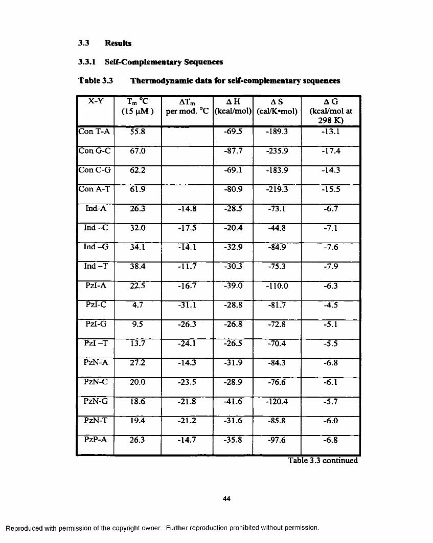
3.3 Results
3.3.1 Self-Complementary Sequences
Table 3.3 Thermodynamic data for self-complementary sequences
X-Y Tm°C (15 p M )
ATm per mod. °C
A H(kcal/mol)
AS(cal/K»mol)
A G (kcal/mol at
298 K)Con T-A 55.8 -69.5 -189.3 -13.1
Con G-C 67.0 -87.7 -235.9 -17.4
Con C-G 62.2 -69.1 -183.9 -14.3
Con A-T 61.9 -80.9 -219.3 -15.5
Ind-A 26.3 -14.8 -28.5 -73.1 -6.7
Ind -C 32.0 -17.5 -20.4 -44.8 -7.1
In d -G 34.1 -14.1 -32.9 -84.9 -7.6
In d -T 38.4 -11.7 -30.3 -75.3 -7.9
Pzl-A 22.5 -16.7 -39.0 -110.0 -6.3
Pzl-C 4.7 -31.1 -28.8 -81.7 -4.5
Pzl-G 9.5 -26.3 -26.8 -72.8 -5.1
P z l-T 13.7 -24.1 -26.5 -70.4 -5.5
PzN-A 27.2 -14.3 -31.9 -84.3 -6.8
PzN-C 20.0 -23.5 -28.9 -76.6 -6.1
PzN-G 18.6 -21.8 -41.6 -120.4 -5.7
PzN-T 19.4 -21.2 -31.6 -85.8 -6.0
PzP-A 26.3 -14.7 -35.8 -97.6 -6.8
Table 3.3 continued
Reproduced with permission of the copyright owner. Further reproduction prohibited without permission.

X-Y Tm C (15 pM )
ATm per mod. °C
AH(kcal/mol)
AS(cal/K*mol)
AG (kcal/mol at
298 K)PzP-C 15.4 -25.8 -41.3 -121.2 -5.2
PzP-G 26.0 -18.1 -43.0 -121.8 -6.7
PzP-T 15.7 -23.1 -25.3 -65.6 -5.8
PzT-A 33.4 -11.2 -47.6 -133.2 -7.9
PzT-C 24.0 -21.5 -32.7 -88.0 -6.5
PzT-G 36.2 -13.0 -42.9 -116.7 -8.2
PzT-T 27.9 -17.0 -33.8 -90.3 -6.9
3.3.2 Non-Self-Complementary Sequences
Table 3.4 Melting temperature (Tm) and thermodynamic parameters for nonself-complementary duplexes
Nucleoside-Sequence
Tm C (15 pM )
ATm per mod.
°C
A H(kcal/mol)
AS(cal/K*mol)
A G (kcal/mol at
298 K)Control 72.2 -172.5 -475.0 -31.0
Ind-1 71.6 -0.6 -106.4 -283.8 -21.8
Ind-2 65.9 -6.3 -125.2 -344.5 -22.5
Ind-3 48.2 -8.0 -108.9 -314.1 -15.2
Ind-4 51.9 -6.8 -52.4 -136.6 -11.7
PzN-1 66.2 -6.1 -66.1 -170.1 -15.4
PzN-2 64.8 -7.4 -119.0 -327.4 -21.4
PzN-3 41.0 -10.4 -78.2 -224.4 -11.4
Table 3.4 continued
45
Reproduced with permission of the copyright owner. Further reproduction prohibited without permission.

X-Y Tm AC (15 p M )
ATm per mod.
°C
AH(kcal/mol)
AS(cal/K*mol)
AG (kcal/mol at
298 K)PzN-4 58.1 -4.7 -51.1 -129.5 -12.5
PzP-1 65.7 -6.6 -104.0 -282.1 -19.9
PzP-2 65.4 -6.9 -126.2 -348.2 -22.5
PzP-3 41.2 -10.3 -94.9 -277.3 -12.3
PzP-4 56.3 -5.3 -93.8 -259.9 -16.3
PzT-1 69.3 -2.9 -83.3 -218.4 -18.2
PzT-2 64.5 -7.8 -85.7 -229.2 -17.4
PzT-3 45.0 -9.1 -81.7 -232.2 -12.5
PzT-4 59.8 -4.2 -87.5 -238.2 -16.5
3.4 Discussion
3.4.1 Self-Complementary Sequences
We chose these Dickerson-based self-complementary sequences to be
consistent with the analysis Bergstrom and coworkers have perfo rm ed .^ The
general trend of our data agrees with their work^ (Table 3.4) but our reported Tms o f
sequences common between the two are generally higher. This is likely due to the
difference in the methods used to analyze the data. They used the method o f
Carothers and Gralla^ whereby T \a and T3/4 are used to determine AH from a single
curve. In contrast, we have used the program Meltwin© which fits a throughout
the curve via a Marquardt-Levenberg non-linear least squares algorithm to
determine Tm. Additionally, we have used four Dickerson-like control
s e q u e n c e s 4 > 6 - 8 f o r purposes o f comparing a series o f an analog across from all four
46
Reproduced with permission of the copyright owner. Further reproduction prohibited without permission.

natural bases. This is particularly important in that the sequences with purines in
the “Y” position, ConT-A and ConC-G (see Table 3.4), have Tms ~5 degrees lower
than the sequences in which the purines are in the “X” position (ConAT and ConGC
resp.). Thus we analyze our data in terms o f AT„/substitution to take into account
the different stacking energies already present in the parent sequences. As
suggested by Freier,^ we feel the ATm analysis is probably more valid than
comparison o f thermodynamic parameters as the cooperativity o f the melting
transitions is variable with the different analogs.
The average ATm’s show the pyrazole analogs are much more stable when
paired with purines than with pyrimidines. This is partly the case with Ind except
with T. Possibly a variation in the syn or anti configuration o f the indole ring could
explain this phenomena with the lowest ATm with T as its pairing partner. All the
analogs show decreased stability increase with C as the pairing partner. In spite of
Con-TA sequence having the lowest Tm o f the four natural controls, each of the
pyrazole analogs have the highest Tm when paired with A when compared to the
other three complements. In each self-complementary data set, the pyrazole analog
that destabilizes the duplex the least is PzT. This is probably due to the increased
size and increased charge distribution the thiazole ring has over the other pyrazole
substituents. Froehler has shown the propynyl unit substituted on pyrim idines^
and purines^ I raises the Tm by approx. 1.5 °C and 1-1.4 °C per substitution,
respectively. The average ATm for the propynyl unit was -7°C, perhaps lack of the
charge distribution or lower polarizability are responsible for the lower
thermodynamic stability as well as the substituents being placed on a non-hydrogen
47
Reproduced with permission of the copyright owner. Further reproduction prohibited without permission.

bonding base. Pzl was extremely destabilizing as demonstrated by the very low
Tm's. Thus, the iodo group provides no added stability over an unsubstitued pyrazole
wich Bergstrom showed to be equivalent to an abasic site.4 The nitro group is much
less destabilizing than the iodo group. The smaller size and better charge
distribution would make it a more suitable substituent. Comparing the Tm values for
PzN to the values reported by Bergstrom,^ the same trends are found with a slight
variation in the actual numbers. This could be due to the different methods o f
analysis.
Self-Complementary Sequences
c 3 5 -
Ind Pzl PzN PzP PzT
Modification (X)
Figure 3.4 Comparison of -ATm for each modification with all the natural bases
The pyrazole analogs overall are not as indiscriminate as Ind, but PzT is
more stable than Ind when paired with A as well as with G. This may be related to
the pairing partner size. It appears part o f the reason for the pairing preferences may
be an attempt at maintaining the traditional size o f the natural pyrimidine-purine
pair. The pyrazole analogs prefer pairing with A. When the pyrazole analogs are
paired with A, they are replacing a pyrimidine, comparable in size, in center o f the
duplex.
48
Reproduced with permission of the copyright owner. Further reproduction prohibited without permission.

S e lf-C o m p lem en ta ry S e q u e n c e s
■ Ind i|Pzl■ PzN B PzP■ PzT■ Avg.
Figure 3.5 Comparison of ATm for each natural base with all of the modifications
This maintains the traditional purine-pyrimidine size in the duplex. This may
also explain the increase in the ATm. When the analogs are paired with C, the
pyrazole analogs are replacing G in the sequence, offering a "pyrimidine-
pyrimidine" or mismatch in size in the center o f the duplex, therefore much more
destabilizing to the duplex than the natural complement. The pyrazole analogs are
less destabilizing to the duplex than Kool’s analogs 4,6-dimethyindole and 2,4-
diflourotoulene. Both o f these analogs are very non-polar and do not offer the same
charge variation the pyrazole analogs offer. 4,6-Dimethylindole, which is designed
as an A analog, does not thermodynamically favor pairing with T and shows the
following preference O A> G> T.12 2,4-Difluorotoluene was found to favor
49
(0o■oOE
<I
CNatural I CO
Reproduced with permission of the copyright owner. Further reproduction prohibited without permission.

pairing with C while it was designed as a T mimic and shows the following
thermodynamically favored pairing preferences C > G> A> T.12 As both these
analog are favoring pairing with C they are “replacing” a G in the natural sequence
3.4.2 Non-Self Complementary Sequences
P r e v i o u s l y , 13-15 it has been shown a single modification in the center o f a
duplex is more destabilizing than a modification near an end. Our data (Table 3.5),
Sequence 1 vs. Sequence 2 and Sequence 3 vs. Sequence 4, supports this
observation as well. As expected from the self-complementary data, PzT
modification appears to be the least destabilizing o f the pyrazole modifications but
not as stable as Ind. The results from Seq. 4 show that the pyrazole bases are able
to stack side well side-by-side. Bergstrom has suggested a ring nitrogen and its
ability to hydrogen bond if in the correct position makes the base more
discriminating. The imidazole N3 when paired with G, according to models, is in a
position capable o f H-bonding similar to a C without disturbance of the duplex,as
suggested by Bergstrom et al. The ring nitrogen for pyrazole is in the 2 position and
would require some distortion o f the duplex to H-bond with G and additionall it is a
much weaker hydrogen-bond acceptor than an imidazole N-3. The natural bases
have charge centers in different positions abling them to stack with favorable charge
interaction with the bases above and below. Some o f the analogs studied although
7r-rich, lack the charge distribution and are not good stacking bases. The thiazole
analog provides both positive and negative centers which can improve its ability to
stack, as well as the Tt-systems o f the two 5 membered rings, which may explain
higher Tms o f PzT containing oligonucleotides.
50
Reproduced with permission of the copyright owner. Further reproduction prohibited without permission.

Non-Self Complementary Sequences
1 2 -
■ Ind■ PzN■ PzP■ PzT
1 2 3 4Sequence
Figure 3.6 Comparison o f ATm for non-self-complementary sequences
A possible explanation for the very low -ATm for Ind in Sequence 1 when
compared to the pyrazole analogs could be due to preferences in pairing partners.
The self-complementary data shows Ind prefers pairing with pyrimidines over
purines. The self-complementary data also shows the pyrazole analogs do not favor
pairing with pyrimidines. Sequence 1 has the modification across from C. As Ind
favors this pairing and the pyrazole analogs do not this might explain the large
difference in the -ATm shown in Figure 3.6.
3.5 Summary
Comparison of the data for both each sequences show the pyrazole analogs
have pairing preference for the natural base A. The analogs are capable of being
incorporated into a duplex without disrupting the natural order o f the duplex. The
thiazolyl substituent shows the best ability to stabilize the duplex when compared to
51
Reproduced with permission of the copyright owner. Further reproduction prohibited without permission.

the other substituents. The pyrazole analogs are not a indiscriminate as Ind when
pairing with the four natural bases.
3.6 Experimental
3.6.1 UV Thermal Denaturing Measurements.
Absorbance measurments were recorded at 260 nm on a Gilford Response
UV/VIS Spectrophotomer equipped with a Peltier temperature controlling device
and thermal software while the temperature was raised from 5 °C to 95 °C in 0.5 °C
increments under an inert atmosphere. To determine that evaporation was less than
5%, the average o f 3-5 absorbance readings was determined at 5 °C and/or 25 °C
before and after each melt. Prior to measurement all samples were heated to 95°C
for 2 min to drive the equilibrium to single strand. They were then cooled to 5°C
and equilibrated for 5 min to allow for duplex formation. One concentration for
each duplex was determined in both a heating and cooling ramp to ensure that the
duplex behaved the same way. Then 3-8 concentrations (covering at least a 100-
fold concentration difference) were run in a heating run at 1 °C / min.
3.6.2 Solvent Preparation
An estimate o f the amount o f oligonucleotide needed was determined by
UV(260 nm) (Gilford) in H2O and the sample was evaporated to dryness. The
DNA duplexes were dissolved in water and had a final buffer concentration o f 1 M
NaCl, 1 mM EDTA, 10 mM Na2HP0 4 - The cell pathlength varied from 0.01 cm to
1 cm. All solvents were degassed in a vacuum desiccator prior to use. The
concentration was determined using the assumption at high temperature the
oligonucleotides are unstacked and unpaired. The absorbance was determined at 85
52
Reproduced with permission of the copyright owner. Further reproduction prohibited without permission.

°C and Beer's Law was used to determine the concentration utilizing an estimate of
the extinction coefficients as calculated below.
3.6.3 Extinction Coefficients fo r Oligonucleotides
The nearest neighbor estimate for each non-modified sequences was determined
using the method and values from the literature. 16,17 The straight sum o f the
extinction o f the mononucleotides in each non-modified sequence was made. The
ratio o f nearest neighbor values: straight sum method was calculated. The ratio was
then multiplied by the straight sum o f the individual mononucleotides (incorporating
the determined extinction coefficients of the modified nucleosides) for the modified
sequences. This will provide a nearest neighbor estimate for the extinction
coefficient as the extinction coefficient for each dinucleotide needed for nearest
neighbor estimates is not known.
3.6.4 Calculation of Thermodynamic Param eters
The Tm as well as AH, AS and AG were determined for each concentration
using Meltwin© provided by Jeffrey McDowell. 18 This program fits a curve to the
raw data points. The parameters from the curve allow for determination o f the Tm,
AH, AS and calculation o f AG for each individual concentration. Once analysis was
completed for three or more concentrations, van’t Hoff analysis was done to provide
the thermodynamic values reported. The program provided Tm at a concentration of
100 pM and AG at 310 K, using the 1/Tm and In Ct/C0 relationship for self and non
self complementary sequences the Tm’s were converted to be reported at 15 pM and
AG at 298 K using the relationship o f AG = A H —TAS and AG = —R T In K ,
53
Reproduced with permission of the copyright owner. Further reproduction prohibited without permission.

rearrangement results in the following equation K = exp^ - ^ - —~ ~ j , which allows
for van’t H off analysis o f the linear relationship between 1/Tm and In Ct/C0 to be
used.
3.6.5 DNA Melting Experiments and Therm odynam ic Analysis
UV melting was performed as previously described. 3 The melting temperature, Tm,
is defined as the point at which a =0.5 when a is defined as the mole fraction o f
single stranded DNA in the duplex state. It is assumed melting behavior is two state
and there are no intermediates. The equilibrium constant, at Tm, is given by the
following, K = — —— ^ for self complementary sequences and K =2(1 —a ) C (1 - a ) 2Cr
for non-self complementary sequences. Analysis o f melting data was performed
with the program, Meltwin©, a non-linear least squares fitting program which
determines Tm and thermodynamic data from single melting curves. For each
sequence a series o f melts were performed over at least 100-fold concentration
range. To determine the thermodynamic parameters, A H vh and ASvh, van’t Hoff
style analysis was performed according to the following equations:
^ or self 'cornPlementary sequencesTm I AH
Tm I AH
'C ACo4
+ for non-self complementary sequences
The free energy, AG, was calculated from the determined AHvh and ASvh using the
equation: AG=AH-TAS.
54
Reproduced with permission of the copyright owner. Further reproduction prohibited without permission.

Table 3.S Self-Complementary Sequence Data for each analysisSequence Cone.
HMAH
kcal/molAS
cal/mol KAG
kcal/molTm°C
R value
Cont TA 3.6 -50.15 -129.77 -9.91 51.06 0.998
12.4 -57.5 -152.58 -10.18 55.66
90.4 -76.26 -208.92 -11.47 62.19
268.0 -78.03 -214.47 -11.51 64.92
599.0 -78.63 -216.3 -11.54 67.17
Con GC 9.3 -70.91 -186.45 -13.08 65.35 0.997
17.9 -75.1 -198.86 -13.43 67.33
98.9 -83.62 -223.66 -14.26 72.43
9 -76.02 -201.3 -13.58 65.62
41.9 -82.7 -220.92 -14.18 70.09
110.0 -82.94 -221.84 -14.14 72.5
922.0 -99.88 -270.81 -15.89 77.69
Cont C-G 15.7 -46.06 -115.37 -10.27 62.15 0.999
32.8 -54.89 -141.83 -10.9 64.97
118.0 -62.74 -165.3 -11.47 69.2
764.0 -82.06 -221.21 -13.45 75.33
Cont A-T 10.1 -61.99 -162.81 -11.49 60.7 0.999
15.8 -65.73 -174.07 -11.74 62.12
635.0 -81.21 -220.53 -12.81 72.18
Table 3.5 continued
Reproduced with permission of the copyright owner. Further reproduction prohibited without permission.
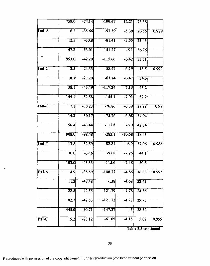
759.0 -74.14 -199.67 -12.21 73.38
Ind-A 6.2 -35.66 -97.59 -5.39 20.56 0.989
12.5 -30.8 -81.41 -5.55 23.43
47.2 -53.01 -151.27 -6.1 36.76
953.0 -42.29 -115.66 -6.42 53.51
Ind-C 3.5 -24.33 -58.47 -6.19 18.5 0.992
18.7 -2129 -67.14 -6.47 34.3
38.1 -43.49 -117.24 -7.13 43.2
145.1 -52.58 -144.1 -7.91 52.2
Ind-G 7.1 -30.23 -76.86 -6.39 27.88 0.99
14.2 -30.17 -75.76 -6.68 34.94
50.4 -43.44 -117.8 -6.9 42.84
908.0 -98.48 -283.1 -10.68 58.43
Ind-T 13.8 -32.59 -82.81 -6.9 37.06 0.986
30.0 -37.6 -97.8 -7.26 44.1
103.0 -43.33 -115.6 -7.48 50.6
Pzl-A 4.9 -38.59 -108.77 -4.86 16.88 0.995
11.3 -47.48 -138 -4.68 22.43
22.8 -42.55 -121.79 -4.78 24.36
82.7 -42.53 -121.73 -4.77 29.73
445.0 -50.71 -147.37 -5 38.52
Pzl-C 15.2 -23.12 -61.05 -4.18 5.02 0.999
Table 3.5 continued
56
Reproduced with permission of the copyright owner. Further reproduction prohibited without permission.
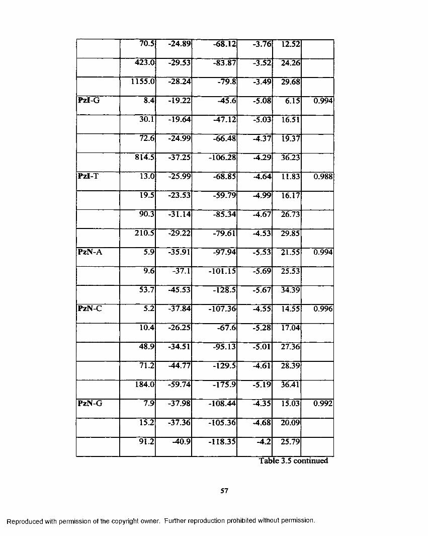
70.5 -24.89 -68.12 -3.76 12.52
423.0 -29.53 -83.87 -3.52 24.26
1155.0 -28.24 -79.8 -3.49 29.68
Pzl-G 8.4 -19.22 -45.6 -5.08 6.15 0.994
30.1 -19.64 -47.12 -5.03 16.51
72.6 -24.99 -66.48 -4.37 19.37
814.5 -37.25 -106.28 -4.29 36.23
Pzl-T 13.0 -25.99 -68.85 -4.64 11.83 0.988
19.5 -23.53 -59.79 -4.99 16.17
90.3 -31.14 -85.34 -4.67 26.73
210.5 -29.22 -79.61 -4.53 29.85
PzN-A 5.9 -35.91 -97.94 -5.53 21.55 0.994
9.6 -37.1 -101.15 -5.69 25.53
53.7 -45.53 -128.5 -5.67 34.39
PzN-C 5.2 -37.84 -107.36 -4.55 14.55 0.996
10.4 -26.25 -67.6 -5.28 17.04
48.9 -34.51 -95.13 -5.01 27.36
71.2 -44.77 -129.5 -4.61 28.39
184.0 -59.74 -175.9 -5.19 36.41
PzN-G 7.9 -37.98 -108.44 -4.35 15.03 0.992
15.2 -37.36 -105.36 -4.68 20.09
91.2 -40.9 -118.35 -4.2 25.79
Table 3.5 continued
57
Reproduced with permission of the copyright owner. Further reproduction prohibited without permission.
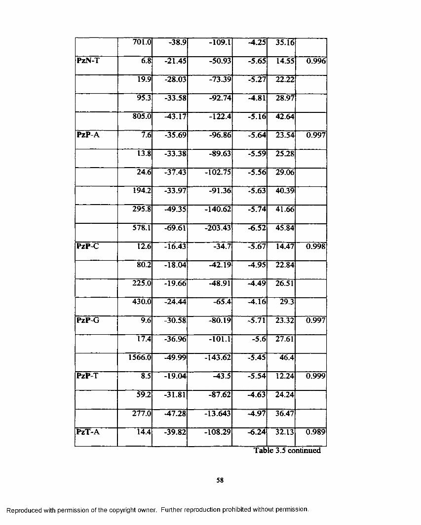
701.0 -38.9 -109.1 -4.25 35.16
PzN-T 6.8 -21.45 -50.93 -5.65 14.55 0.996
19.9 -28.03 -73.39 -5.27 22.22
95.3 -33.58 -92.74 -4.81 28.97
805.0 -43.17 -122.4 -5.16 42.64
PzP-A 7.6 -35.69 -96.86 -5.64 23.54 0.997
13.8 -33.38 -89.63 -5.59 25.28
24.6 -37.43 -102.75 -5.56 29.06
194.2 -33.97 -91.36 -5.63 40.39
295.8 -49.35 -140.62 -5.74 41.66
578.1 -69.61 -203.43 -6.52 45.84
PzP-C 12.6 -16.43 -34.7 -5.67 14.47 0.998
80.2 -18.04 -42.19 -4.95 22.84
225.0 -19.66 -48.91 -4.49 26.51
430.0 -24.44 -65.4 -4.16 29.3
PzP-G 9.6 -30.58 -80.19 -5.71 23.32 0.997
17.4 -36.96 -101.1 -5.6 27.61
1566.0 -49.99 -143.62 -5.45 46.4
PzP-T 8.5 -19.04 -43.5 -5.54 12.24 0.999
59.2 -31.81 -87.62 -4.63 24.24
277.0 -47.28 -13.643 -4.97 36.47
PzT-A 14.4 -39.82 -108.29 -6.24 32.13 0.989
Table 3.5 continued
58
Reproduced with permission of the copyright owner. Further reproduction prohibited without permission.

23.9 -42.89 -117.68 -6.4 35.82
91.2 -49.39 -138.65 -6.38 41.13
210.4 -50.58 -142.13 -6.5 45.08
775.1 -53.84 -153.09 -6.36 48.65
PzT-C 3.9 -3627 -101.3 -4.85 14.61 0.977
11.4 -33.74 -90.23 -5.75 25.81
478.0 -88.75 -269.1 -5.29 36.45
314.0 -40.85 -114.23 -5.42 40.47
PzT-G 1.3 -41.33 -111.82 -6.65 24.7 0.994
7.3 -42.42 -114.65 -6.87 33.91
18.5 -47.89 -132.19 -6.89 38.15
56.5 -53.61 -150.19 -7.02 42.88
133.8 -44.83 -122.86 -6.72 45.72
245.7 -55.02 -154.63 -7.06 48.33
PzT-T 4.0 -30.83 -80.12 -5.98 20.94 0.995
14.6 -26.75 -66.74 -6.05 27.87
70.7 -32.56 -86.4 -5.76 35.76
176.0 -50.29 -142.26 -6.16 42.24
388.0 -54.75 -154.9 -6.71 47.98
59
Reproduced with permission of the copyright owner. Further reproduction prohibited without permission.

Table 3.6 Non-Self-Complementary Sequence Data for each analysisSequence Cone.
|j.MAH
kcal/molAS
cal/mol KAG
kcal/molT m°C
Rvalue
Control 2.03 -160.06 -438.66 -24.01 69.24 0.998
15.7 -117.53 -315.52 -19.67 72.25
26.9 -120.62 -324.72 -19.91 72.93
63.6 -112.95 -303.3 -18.89 74.13
174.8 -102.76 -273.94 -17.49 75.33
Ind-1 0.42 -104.6 -278.48 -18.24 63.86 0.999
5.29 -109.81 -294.16 -18.58 68.88
55.4 -96.19 -254.42 -17.28 74.55
Ind-2 3.01 -119.73 -328.05 -17.99 63.12 0.997
28.8 -97.6 -263.83 -15.77 66.5
533 -54.27 -139.21 -11.09 72.61
Ind-3 0.156 -68.12 -184.19 -10.99 39.2 0.993
1.18 -55.68 -145.99 -10.4 43.43
12.48 -53.48 -141.02 -9.74 48.59
216.9 -34.99 -87.93 -7.72 52.5
Ind-4 0.351 -13.63 -11.56 -10.04 37.63 0.999
4.49 -21.93 -41.39 -9.09 46.43
38.9 -27.97 -62.08 -8.72 55.93
Ind-l 2.52 -74.43 -194.67 -14.05 60.55 0.991
12.7 -66.05 -170.54 -13.16 64.37
Table 3.6 continued
Reproduced with permission of the copyright owner. Further reproduction prohibited without permission.

120.7 -108.39 -291.7 -17.92 73.84
PzN-2 0.562 -95.05 -255.53 -15.8 58.18 0.989
2.5 -123.25 -339.6 -17.92 61.77
6.41 -100.15 -271.61 -15.91 62.77
11.4 -111.05 -303.41 -16.95 64.61
22 -108.83 -297.05 -16.7 65.77
54.6 -100.61 -273.84 -15.68 66.64
PzN-3 1.54 -62.69 -174.09 -8.69 34.98
7.94 -63.52 -177.01 -8.62 39.58
75.3 -69.46 -196.91 -8.39 44.72
PzN-4 0.895 -61.43 -160.81 -11.55 48.07 0.996
7.6 -55.99 -144.11 -11.29 55.65
12 -61.8 -160.71 -11.96 59.15
PzP-1 0.519 -126.78 -350.42 -18.09 58.82 0.991
10.6 -117.95 -315.22 -17.18 59.73
3.96 -91 -262 -15.74 61.93
36.4 -56.9 -143.78 -12.3 67.87
PzP-2 0.813 -141.83 -394.83 -19.37 60.2 0.999
1.75 -156.3 -438.31 -20.36 61.26
8.045 -156.93 -439.55 -20.6 63.89
77.7 -141.75 -393.58 -19.68 68.3
PzP-3 0.95 -73.27 -207.19 -9.01 35.35 0.999
Table 3.6 continued
61
Reproduced with permission of the copyright owner. Further reproduction prohibited without permission.

1.79 -85.61 -247.18 -8.94 36.76
8.63 -91.52 -266.09 -8.99 40.24
81.6 -81.12 -233.91 -8.57 44.49
PzP-4 0.99 -83.68 -229.1 -12.63 49.56 0.998
2 -73.46 -196.97 -12.37 52.2
891 -66 -178.02 -10.79 65.75
PzT-l 0.252 -90.04 -238.26 -16.15 58.87 0.988
1.94 -103.97 -280.12 -17.09 63.31
10.5 -99.88 -267.57 -16.89 67.61
17.9 -102.3 -275.04 -17 68.42
PzT-2 0.3 -64.79 -165.82 -13.36 53.39 0.981
2.67 -106.22 -289.26 -16.5 61.38
11.1 -103.64 -282.14 -1614 63.83
11.2 -104.33 -284.36 -16.13 63.64
20.92 -57.11 -144.98 -12.14 64.46
42.86 -102.36 -278.53 -15.97 66.61
PzT-3 0.787 -63.46 -173.64 -9.6 37.42 0.991
2.65 -71.3 -198.99 -9.59 40.6
7.09 -74.33 -208.13 -9.78 43.88
12.24 -70.83 -197.57 -9.55 44.75
23.84 -68.64 -191.57 -9.22 45.4
69.5 -77.28 -218.47 -9.52 48.52
Table3.6 continued
62
Reproduced with permission of the copyright owner. Further reproduction prohibited without permission.

PzT-4 0.71 -69.13 -182.47 -12.54 50.88 0.992
2.81 -71.74 -190.27 -12.72 55.28
12.9 -55.36 -142.03 -11.31 58.07
25.2 -66.99 -177.31 -11.99 59.94
3.7 References
1) Watson, J. D.; Crick, F. H. C. Nature 1953,171, 737-738.
2) Marky, L. A.; Breslauer, K. J. Biopolymers 1987, 2 6 ,1601-1620.
3) LeBlanc, D. A.; Morden, K. M. Biochemsitry 1991,3 0 ,4042-4047.
4) Bergstrom, D. E.; Zhang, P.; Johnson, W. T. Nucleic Acids Res. 1997,25, 1935-1 9 4 2 .
5) Caruthers, M. H.; Barone, A. D.; Beaucage, S. L.; Dodds, D. R.; Fisher, E. F.;McBride, L. J.; Matteucci, M.; Stabinsky, Z. Meth. Enzymol. 1985,287-313.
6) Ahmadian, M. H.; Zhang, P.; Bergstrom, D. E. Nucleic Acids Res. 1998, 26,3127-3135.
7) Johnson, W. T.; Zhang, P.; Bergstrom, D. E. Nucleic Acids Res. 1997,25, 559-567.
8) Zhang, P.; Johnson, W. T.; Klewer, D.; Paul, N.; Hoops, G.; Davisson, V. J.;Bergstrom, D. E. Nucleic Acids Res. 1998, 26, 2208-2215.
9) Freier, S. M.; Altman, K. H. Nucleic Acids Res. 1997,25, 4429-4443.
10) Froehler, B. C.; Wadwani, S.; Terhorst, T. J.; Gerrard, S. R. Tetrahedron Lett. 1992, 33, 5307-5310.
11) Buhr, C. A.; Wagner, R. W.; Grant, D.; Froehler, B. C. Nucleic Acids Res.1996, 24, 2974-2980.
12) Schweitzer, B. A.; Kool, E. T. J. Am. Chem. Soc. 1995,117, 1863-1872.
13) Martin, F. H.; Castro, M. M.; Aboul-ela, F.; Ohtsuka, E. Nucleic Acids Res. 1986,14, 7727-7736.
63
Reproduced with permission of the copyright owner. Further reproduction prohibited without permission.

14) Kaimya, H.; Sakaguchi, T.; Murata, N.; Fujimuro, M.; Miura, H.; Ishikawa, K.; Shimizu, M.; Inoue, H.; Nishimura, S.; Matsukage, A.; Masutani, C.; Hanaoka, F.; Ohtsuka, E. Chem. Pharm. B ull 1992, 40,, 2792-2795.
15) Bischofberger, N.; Matteucci, M. D. J. Am. Chem. Soc. 1989, 111, 3041-3046.
16) Cantor, C. R.; Warshaw, M. M.; Shapiro, H. Biopolymers 1970, 9, 1059.
17) Warshaw, M. M.; Tinoco Jr., I. J. Mol. Biol. 1966, 26, 29.
18) McDowell, J. A.; Turner, D. Biochemistry 1996,25, 14077-14089.
64
Reproduced with permission of the copyright owner. Further reproduction prohibited without permission.

Chapter 4Enzymic Behavior o f Pyrazole-Containing Nucleobase Analogs with
Various Thermostable DNA Polymerases
4.1 Target for PCR Reactions
The goal o f this research is to determine the ability o f polymerases to
recognize the pyrazole analogs. We studied 4-nitropyrazole (PzN) and 4-(2-
thiazolyl)-pyrazole (PzT)of the pyrazole analogs as well as 2 other well-studied
modifications, 5-nitroindoIe (Ind) and 3-nitropyrroIe (PyN). (Figure 4.1) Two
different control experiments were designed. The first control experiment uses a
non-modified primer containing the natural complement (G) to the position on the
template. The second control experiment uses 4 identical primers except in the
position of interest. Each primer contains one o f each o f the four natural nucleosides.
These four primers are added in equal mole amounts to allow for quantitation o f a
mixture that should be 25% o f each.
H° ^ p
oCTNO
HO
HO HO HO HO
Ind PzN PyN PzT
Figure 4.1 Analogs studied in PCR reactions
4.2 Design o f Enzymic Experiment
Using a procedure originally described at Purdue University by Davisson and
coworkers,! the expression vectorphisF-tac p lasm id^ is digested using the
65
Reproduced with permission of the copyright owner. Further reproduction prohibited without permission.

restriction endonuclease, Pvu n . One o f the fragments from this digestion contains
the hisF gene indicated in capital letters (Figure 4.3). This gene serves as part o f a
797 bp template for the PCR amplification experiments. The Pvu n recognition site
is 5'-CAGACTG-3\ When this sequence is present the enzyme will cut the dsDNA
between the G and C. The sense primer (40 bases) is designed for positions -10 to 30
o f the hisF gene (underline on the top portion o f Figure 4.3). The modification is in
position 26. The antisense primer (30 bases) is corresponds to positions 757 to +10
(underlined on the bottom o f Figure 4.3). The hisF gene is shown in capital letters.
The modifications were incorporated using standard automated DNA synthesis once
the phosphoramidites o f the analogs were either purchased or synthesized as
described in Chapter 2. During PCR the modified primers are initially extended
resulting in full-length template containing the modification. The next round o f PCR
actually incorporates a natural nucleoside across from the pyrazole analog. Figure
4.2 is a pictorial representation o f this process. This figure is very similar to Figure
1.7 describing PCR except for the pyrazole analogs present in the sense primers.
Several different enzymes were chosen for this study. These include AmpliJaq®
DNA polymerase (Perkin-Elmer), Vent DNA polymerase (New England Biolabs),
Deep Vent DNA polymerase (New England Biolabs), Pfu DNA polymerase exo+,
Pfu DNA polymerase exo-, and UlTma DNA polymerase (Perkin Elmer). As
enzymes have different inherent abilities a wide variety o f enzymes were chosen to
determine the directing ability o f the analogs. The question to be answered was
whether the analogs directed specifically for one base or was it dependent on the
enzyme’s inherent properties. See Table 4.1 for a list o f the exonuclease properties
66
Reproduced with permission of the copyright owner. Further reproduction prohibited without permission.

of the DNA polymerases studied. The different type o f exonuclease activities is
discussed in Section 1.5.4.
PCR Amplification to Produce DNA that Contains a Non-Natural Nucleotide
cG
separate strands by heating to 95°C
Anneal primers by cooling to 37°C
QC
Heat to 70°C to allow for extensionQC
cG
Cycle Repeatsseparate strands by heating to 9S°C
- Q
CG
Anneal primers by cooling to 37°C
QC
QC
Heat to 70°C to allow for extension
Q iC
Cycle Repeats
Figure 4.2 Diagram of PCR reaction containing a pyrazole analog in thesense p rim er
The fidelity o f these enzymes is described by the number o f mutations/ bp/
duplication.^ UlTma is a very poor proofreading enzyme resulting in such a fidelity
it is almost considered a non-proofreading enzyme. The best proofreading enzyme
studied was Pfu exo+ followed by Vent and then Deep Vent.
67
Reproduced with permission of the copyright owner. Further reproduction prohibited without permission.

aggaatt.catATGCTGGCAAAACGCATAATCCCATGTCTCGACGTTCGTGATGGTCAGGTtccttaagtaTACGACCGTTTTGCGTATTAGGGTACAGAGCTGCAAGCACTACCAGTCCA
CCACTTTCCCGTACAGTTTCGCAACCATGAAATCATTGGCGATATCGTGCCGCTGGCAAAAGGTGAAAGGGCATGTCAAAGCGTTGGTACTTTAGTAACCGCTATAGCACGGCGACCGTTTT
CGCTACGCTGCGATGCGAGCTGACGAACTGGTGTTCTACGATATCACCGCTTCCAGCGATGGAAGAAGGCCTTCTTCCGCGACTGCTTGACCACAAGATGCTATAGTGGCGAAGGTCGCTAC
GCCGTGTGGTAGATAAAAGCTGGGTATCTCGCGTGGCGGAAGTGATCGACATTCCGTTTTGCGGCACACCATCTATTTTCGACCCATAGAGCGCACCGCCTTCACTAGCTGTAAGGCAAAAC
TGTGGCGGGTGGGATTAAGTCTCTGGAAGATGCCGCGAAAATTCTTTCCTTTGGCGCGGATACACCGCCCACCCTAATTCAGAGACCTTCTACGGCGCTTTTAAGAAAGGAAACCGCGCCTA
AAAATTTCCATCAACTCTCCTGCGCTGGCAGACCCAACATTAATTACTCGCCTGGCCGATCTTTTAAAGGTAGTTGAGAGGACGCGACCGTCTGGGTTGTAATTAATGAGCGGACCGGCTAG
GCTTTGGCGTGCAGTGTATTGTGGTCGGTATTGATGGAAACGCTCGACTGGGTACAGGAAGCGAAACCGCACGTCACATAACACCAGCCATAACTACCTTTGCGAGCTGACCCATGTCCTTC
TGCAAAAACGCGGTGCCGGAGAAATCGTCCTCAATATGATGAATCAGGACGGCGTGCGTAAACGTTTTTGCGCCACGGCCTCTTTAGCAGGAGTTATACTACTTAGTCCTGCCGCACGCATT
CGGTTACGACCTCGAACAACTGAAAAAAGTGCGTGAAGTTTGCCACGTCCCGCTGATTGCCGCCAATGCTGGAGCTTGTTGACTTTTTTCACGCACTTCAAACGGTGCAGGGCGACTAACGG
TCCGGTGGCGCGGGCACCATGGAACACTTCCTCGAAGCCTTCCGCGATGCCGACGTTGACGAGGCCACCGCGCCCGTGGTACCTTGTGAAGGAGCTTCGGAAGGCGCTACGGCTGCAACTGC
GCGCGCTGGCAGCTTCCGTATTCCACAAACAAATAATCAATATTGGTGAATTAAAAGCGTACGCGCGACCGTCGAAGGCATAAGGTGTTTGTTTATTAGTTATAACCACTTAATTTTCGCAT
CCTGGCAACACAGGGCGTGGAGATCAGGATATGTTAacccgggtacg ( 7 97 ) GGACCGTTGTGTCCCGCA CCTCTAG TCCTA TA CA ATtgqqcccatgcFigure 4.3 Sequence o f the target PCR product, the hisF gene.*
Table 4.1 Exonuclease properties o f DNA polymerasesDNA Polymerase 3'- 5' exonuclease
(proof-reading)5 '-3 '
exonucleaseFidelity3
AmpliTaq® - + 8 x l 0 >Pfu exo - + sxio-05UlTma® + + 5x10-°*Pfu exo+ + + 1.3x1 O'™Vent + + 2.7 x 10-™Deep Vent + + 2.8 x 10^
68
Reproduced with permission of the copyright owner. Further reproduction prohibited without permission.

4.3 Analysis o f PCR Products
The technique most often used to analyze the completed PCR reaction is gel
electrophoresis using a sieving medium such as agarose. The crude PCR reaction
will have unused primers (30mer and 40mer) and full length product (797bp) as well
as buffers present. A small portion of the PCR reaction is combined with a loading
buffer consisting o f 2 dyes which migrate at different rates and a reagent Ficoll which
is used to “weigh” down the solution to aid in loading the samples into the wells.
Electrophoresis is used to separate the DNA to determine the size o f the DNA present
in the PCR reaction. Size markers are used to determine the size o f the DNA found
in the PCR reaction. Ethidium bromide is used to visualize the agarose gel.
Ethidium bromide is a DNA intercalator and is visible under UV light when
intercalated into DNA. An inverted image o f an agarose gel is present in Figure 4.5,
Figure 4.6, Figure 4.7 and Figure 4.8.
4.4 Sequencing o f PCR Product
Once the PCR reaction is complete, the sequence o f the antisense strand
needs to be determined. This sequence will indicate the ratio o f natural nucleotides
incorporated across from the pyrazole analog. The sequence of the antisense strand
will be determined using the Sanger Method as described in Section 1.4.3.
4.5 Results
4.5.1 PCR Reactions
The PCR reactions using the modified nucleoside analogs were successful as
indicated by full-length product in the agarose gel. Figures 4.5-4.8 are analytical
69
Reproduced with permission of the copyright owner. Further reproduction prohibited without permission.

agarose gels showing the results o f each PCR reaction with the individual primers
with each different enzyme.
Analysis of How the Analog Was Recognized by the Thermostable DNA Polvmerase
a-^P-ddNTP’s i Dideoxy Sequencing Using the Thermosequenase Kit
33p
33p33p
33p
33p33p
8% PAGE of Sequencing Products
•Gel result yields informaticHi about how die analog was recognized by the polymerase.
•The recognition o f the analog is then translated to percent dNMP incorporation.
A C G TPosition on the gel where the information is obtained about analog recognition.
Figure 4.4 Schematic diagram of Sanger method for sequencing
70
Reproduced with permission of the copyright owner. Further reproduction prohibited without permission.

Figure 4.5 Agarose gel analyzing PCR reactions with AmpliTaq® and UlTma enzymes with control primer (C), 5-nitroindole (Ind) primer, 4-nitropyrazole (PzN) primer, 3-nitropyrrole (PyN) primer and 4-(2-thiazolyl)-pyrazole (PzT) primer
Figure 4.6 Agarose gel analyzing PCR reactions with Pfu exo+ and Ffu exo- Enzymes with control primer (C), 5-nitroindole (Ind) primer, 4-nitropyrazole (PzN) primer, 3-nitropyrrole (PyN) primer and 4-(2-thiazolyl)-pyrazoIe (PzT) primer
71
Reproduced with permission of the copyright owner. Further reproduction prohibited without permission.

Figure 4.7 Agarose gel analyzing PCR reactions with Deep Vent and Vent enzymes with control primer (C), 5-nitroindole (Ind) primer, 4-nitropyrazole (PzN) primer, 3-nitropyrrole (PyN) primer and 4-(2-thiazolyl)-pyrazole (PzT) primer
Lambdap-Gem
I I
Con PzN PzT Con PzP PzT Con. PzN PzT Con PzP PzT ***2027 bp
831 bp
797 bp
■ —primer
Figure 4.8 Agaorse gel analyzing PCR reactions with AmpliTaq® enzyme with control Primer (Con), 4-nitropyrazole primer (PzN), 4-propynyl pyrazole primer (PzP), and 4-(2-thiazoIyl)pyrazole primer (PzT)
72
Reproduced with permission of the copyright owner. Further reproduction prohibited without permission.

4.5.2 Sequencing of PCR Products
Sequencing o f the PCR products was successful using Thermo Sequenase
Radiolabeled Terminator Cycle Sequencing Kit (Amersham/USB). Analysis o f the
sequencing reaction again used gel electrophoresis yet acrylamide/bisacrylamide was
used as the separation medium. This is more appropriate for shorter strands o f DNA
when compared with agarose. When using the PCR product from the reaction with
the control sense primer, the position o f interest should show G. With modified
primers other bases can be incorporated as shown in Figure 4.8. In most instances
with non-proofreading enzymes, T was incorporated across from PzN and PzT.
Proofreading enzymes tended to remove the analogs but not with total efficiency.
4.5.3 Quantitation o f Acrylamide Gel Sequencing Results
The results of this research are ultimately determined by quantitation o f the
gels o f the sequencing reactions. One o f the actual gel images used for quantitation
is included for each primer and each enzyme in Figure 4.8 Pfu exo-, Figure 4.9
A m plify® , Figure 4.10 Vent, Figure 4.10 Deep Vent, and Figure 4.12 Pfu exo+,
and Figure 4.14 UlTma. The analysis o f the gels was performed using ImageQuant
by Molecular Dynamics. Table 4.2 contains the relative amount o f dNMP
incorporated into the antisense strand across from the modification. ( Note: the gel
image contains the complement to the base found on the PCR product.)
Coo Irxl PZN PyNA C G T A C G T A C G T A C G T
- — ““ < * -
IIIII
- S < -■ 5
PZTA C G T
Figure 4.9 Sequencing o f PCR products from Pfu exo- enzyme
73
Reproduced with permission of the copyright owner. Further reproduction prohibited without permission.

Figure 4.10 Sequencing gels o f PCR products from AmpliTaq®
Figure 4.11 Sequencing gels o f PCR products from Vent enzyme
Figure 4.12 Sequencing o f PCR products from Deep Vent enzyme
Con Ind PzN PyNA C G T A C G T A C G T A C G T
— — _ " i i . “3 . T #^ -z<-
" 3 s 2 2
PzT A C G T
Figure 4.13 Sequencing o f PCR products from Pfu exo+ enzymeCon
A C G T
£
Ind PZNA C G T A C G T
- 9m ** • m•*
0 4 -
I
PyNA C G T
W
Figure 4.14 Sequencing G el o f PCR products from UlTma enzyme
74
Reproduced with permission of the copyright owner. Further reproduction prohibited without permission.
•Ill

Table 4.2 % dNMP Incorporated into PCR Reactions Using PrimersContaining M odification in place of GEnzyme AmpliTaq®
(-)
Pfu exo- Deep Vent (+)
Vent(+) Pfu exo+ UlTma (+)
Primer
Control
(G)
A 3(1)
C 96 (2)
G 1 (1) T 0 (0)
A 7.5(1)
C 91.5(2)
G 0 (0)T 1 (1)
A 1 (2)
C 99 (2)
G 0 (0)
T 0 (0)
A 0 (0)
C 82 (8)
G 18 (8)
T 0 (0)
A 2 (2)
C 98 (2)
G 0 (0)
T 0 (0)
A 0 (0)
C 92(10)
G 1 (1) T 7 (11)
Ind A 51 (4)
C 22(20)
G 26(20) T 1 (1)
A 15(3)
C 27 (1)
G 29 (1)
T 29 (5)
A 31 (37)
C 65 (37)
G 1 (2)
T 3 (2)
A 48(15)
C 52 (15)
G 0 (0)
T 0 (0)
A 3 (4)
C 96 (3)
G 1 (2) T 0 (0)
Not
determined
PzN A 94(2)
C 4 (1)
G 2 (1)
T 0 (0)
A 99 (2)
C 0 ( 0 )
G 1 (2) T 0 (0)
A 15(13)
C 85(13)
G 0 (0)
T 0 (0)
A 7(7)
C 93 (7)
G 0 (0)
T 0 (0)
A 10(6)
C 77(10)
G 4 (4)
T 9 (8)
A 99(1)
C 1 (1) G 0 (0)
T 0 (0)
PyN A 83 (4)
C 2 (2)
G 3 (2)
T 12(8)
A 45 (13)
C 18 (8)
G 10 (3)
T 25 (14)
A 7(12)
C 87 13)
G 1 (2)
T 5 (4)
A 51 (22)
C 43 (21)
G 2 (2)
T 5 (3)
A 2 (1) C 98 (2)
G 0 (0)
T 0 (0)
A 97(6)
C 0 (1)
G 3 (5)
T 0 (0)
PzT A 93 (6)
C 5 (5 )
G 1 (1) T 0 (0)
A 100(0)
C 0 (0)
G 0 (0)
T 0 (0)
A 28 (2)
C 63 (2)
G 6 (2)
T 3 (2)
A 80 (6)
C 18 (9)
G 2 (4)
T 0 (0)
A 8(4)
C 91 (5)
G 1 (1) T 0 (0)
A 96(8)
C 4 (8)
G 0 (0)
T 0 (0)
25%A/
C/G/T
A 26 (8)
C 32 (5)
G 23 (8)
T 19(5)
75
Reproduced with permission of the copyright owner. Further reproduction prohibited without permission.

4.6 Discussion
Non-proofreading enzymes AmpliTaq€> and Pfu- mainly paired an A with the
pyrazole analogs. This would agree with the thermodynamic analysis indicating the
pyrazole analogs show a preference for pairing with A. Bergstrom and coworkers
report mainly A incorporation across from an abasic site. 1 Our research found
mixtures o f the nucleotides incorporated across from Ind and PyN. The ratio of
nucleotides for PyN is very similar to those found by Bergstrom. * A possible
explanation o f a purine/pyrimdines mixture is the ability o f the base to rotate about
the glycosidic bond in an anti-syn configuration, allowing for size accommodations
depending on the pairing partner. The pyrazole analogs are unable to have this
rotation due to the placement of the substituents on the 4-position. The position of
the substituents, although the entire molecule is no larger than indole, would disturb
the complementary chain is the other conformation is adopted.
Two control experiments were performed. This first contained the natural
complement, G, in the primer to the base in the template strand. The first control
allows us to determine whether the polymerase and all the other reagent were correctl
added. This also provides a contrasting base in the sequencing reaction as to what
base we expected to be incorporated. If our analogs were expected to direct for a C
in the PCR reaction it would be difficult to detect. With the proof reading enzymes,
it the modification was removed from the initial primer and replaced with the natural
complement to the template, a G is indicated in the sequencing reaction. I f the
modification is not removed then something other than G normally has been
76
Reproduced with permission of the copyright owner. Further reproduction prohibited without permission.

observed. The second control experiment used each natural base in place of the
modification in equal amounts provided close to 25% with error o f each base. (Noted
in Table 4.2 as 25%A/C/G/T) The C base had a slightly higher amount but this is the
natural complement allowing possibly for better annealing as well as ease of
extension o ff o f a mismatch. Proofreading enzymes, Deep Vent, Vent and Pfu+,
removed all o f the analogs but with varying efficiency. If the modifications are not
removed an A is incorporated across from the pyrazole analog. The exception to the
proofreading enzymes is UlTma. UlTma does not remove modifications and
incorporates an A with the analogs. These results indicate the pyrazole analogs direct
for exclusively A incorporation with the enzymes studied whereas Ind and PyN do
not exclusively direct for one o f the natural bases.
Another explanation o f incorporation o f A across from the pyrazole analogs
could be suggested as the “A-rule” possessed by some enzymes.^ The “A-rule”
suggests if an enzyme does not recognize the modification then an A will be
incorporated as a “fail safe” and the polymerization will continue.^ Polymerases
extend off an A-mismatch with better efficiency than any o f the other bases and thus
“A” incorporation may reflect selective amplification of thie m i s m a t c h . 5 - 6
Exonuclease activity can remove abnormalities 5 bases back from the 3' end. This
position o f our modification in the primer was such that exonuclease activity could
remove it. Depending on the enzyme, the non-natural analogs were tolerated to
varying degrees. The sequencing does not indicate evidence o f the polymerase
skipping or looping out the pyrazole analog.
77
Reproduced with permission of the copyright owner. Further reproduction prohibited without permission.

Insertion o f A across from the pyrazole analogs seems to be favored. Although
thermodynamic data supports A as the base favored across from the pyrazoles, the
fail safe insertion o f A by the polymerases cannot yet be ruled out. To determine if
the pyrazole analogs are actually directing for A to be incorporated, kinetic
experiments will need to be performed to determine the rate o f insertion for the
natural nucleotides against the pyrazole analogs.
Figure 4.15 Schematic o f 3'->5' Exonuclease Activity Removing the Modification Q from the Primer and Replacing with a Natural Nucleotide.
Related to the kinetic question is whether termination of the extension is
occurring across from or immediately following the modification and to what extent.
If termination were occurring, the position of termination would provide useful
information about the “reading” efficiency by various polymerasesof the analogs. A
shortcoming o f the original experimental design does not allow for the determination
78
Reproduced with permission of the copyright owner. Further reproduction prohibited without permission.

of whether the polymerase is inserting across from the analog and then stalling on the
next insertion or if the polymerase is stalling at the modification. An assay which
involves a fluorescently labeled antisense primer is being used to probe this topic.
This primer results in shorter products (237 bp) which are simpler to analyze due to
their size as well as the fluorescent label, which allows for more convenient analysis.
According to preliminary results some termination is occurring.
4.7 Experimental
4.7.1 PCR Reactions
4.7.1.1 Preparation of Template
Pvu H restriction endonuclease was used to digest phisF-tac plasmid,
generously provided by Davisson and coworkers,! at Purdue University into the hisF
gene (797bp) (Figure 4.3) following the protocol provided by Promega with the
enzyme (Table 4.3). The Pvu II recognition site is 5,-CAGACTG-3'. Once the
reagents were added the reaction was incubated at 37 °C for 1 hour and then diluted
10 fold to a final concentration of 20 finol/ pL.
Table 4.3. Conditions for Digesting >his-F-tac plasmid with Pvu IIReagent Stock
ConcentrationVolumepL
Final Concentration (50pL)
h 2o 41.75phis-F tac 1.7pg/pL 1.25 42.5ng/pLPVU II 10 Units/ pL 2 20 Units10X NEBuffer 2 500mM NaCl
lOOmM Tris-HCl lOOmM MgCl2 lOmMDTT
5 50mM NaCl lOmM Tris-HCl lOmM MgCl2 ImMDTT
Reproduced with permission of the copyright owner. Further reproduction prohibited without permission.

4.7.1.2 Synthesis o f Primers
Most primers were synthesized by Midland Certified Reagent Company using
standard automated DNA chemistry and purified by AE-HPLC. Several primers
were synthesized at LSU. These were prepared using the same procedure as for the
oligomers used in the thermal denaturation studies. See Section 2.3.2.
4.7.1.3 Sequence o f Primers
Forward Sense Primer fo r PCR
5’ AGG AAT TCA TAT GCT GGC AAA ACG CAT AAT CCC ATQ TCT C3’
Q= G control
4-nitropyrazole nucleotide,
4-propynylpyrazole nucleotide,
4-(2-thiazolyl)pyrazole nucleotide
5-nitroindole nucleotide
Antisense Primer fo r PCR
5’ CGT ACC CGG GTT AAC ATA TCC TGA TCT CCA 3’
4.7.1.4 Conditions for PCR Reactions
The enzymes and respective buffers were purchased from commercial sources
with one exception. UlTma was originally purchased from Perkin-Elmer and then a
generous gift by Dr. Gefland supplied enough enzyme to complete the experiments.
The commercial sources for each enzyme are: Ampli Ta^r® DNA polymerase (Perkin-
Elmer), Vent DNA polymerase (New England Biolabs), Deep Vent DNA polymerase
(New England Biolabs), Pfu DNA polymerase exo+, Pfu DNA polymerase exo-, and
UlTma DNA polymerase (Perkin Elmer). The dNTP’s were purchased from Roche.
The following reagents were added into a 200 pL reaction tube. The reagents and
80
Reproduced with permission of the copyright owner. Further reproduction prohibited without permission.

reaction tube were kept on ice until added to the thermal cycler heating block at 75
°C. The reaction was mixed thoroughly between each addition in the order listed.
The cycle for the reaction included an initial warm up o f 94 °C for 5 min., followed
by 32 cycles o f 95 °C for 30 seconds, 37 °C for 30 seconds and 70 °C for 1 min., and
a final 10 min. extension at 70 °C and then temperature was lowered to 4 °C until
further workup.
Table 4.4 Ampli7gy<S> PCR ConditionsReagent Stock
ConcentrationVolume
pLFinal Concentration (lOOpL)
h 2o 52.51 OX PCR Buffer II
500mM KC1, lOOmM Tris- HC1, pH 8.3
10 50mM KC1, lOmM Tris- HC1
dNTP's 2mM total of each
10 200pM
Forward Primer (Modified)
lOpM 10 lpM
AntisensePrimer
lOpM 10 lpM
MgCl2 Solution 25mM MgCl2 6 1.5mM MgCl2phis-F-tacdigestedplasmid
finol quantities of template
1 finol quantity
AmpliTaq®Polymerase
5 Units/pL 0.5 2.5 Units
Table 4.5 Vent PCR ConditionsReagent Stock Concentration Volume
pLFinal Concentration (lOOpL)
h 2o 5810X ThermoPol Reaction Buffer
lOOmM KC1, lOOmM (NH4)2S04, 200mM Tris-HCl (pH 8 .8 @25 °C)
20mM MgS04 0.1%TritonX-100
10 lOmM KCl,10 mM (NH4)2S 0 4,
20mM Tris-HCl
2mM MgSQ4
Table 4.5 continued
81
Reproduced with permission of the copyright owner. Further reproduction prohibited without permission.

dNTP's 2mM total o f each 10 200pMForward Primer (Modified)
lOpM 10 lpM
AntisensePrimer
lOpM 10 lpM
phis-F-tacdigestedplasmid
finol quantities o f template
1 finol quantity
Vent DNA Polymerase
2 Units/pL 1 1 Unit
Table 4.6 Deep Vent PCR Conditions
Reagent Stock Concentration VolumepL
Final Concentration (lOOpL)
h 2o 58lOXThermoPol Reaction Buffer
lOOmM KC1, lOOmM (NH4)2S 0 4,
200mM Tris-HCl, (pH 8.8 @25 °C)
20mM MgS04 0.1%Triton X-100,
10 lOmM KC1,10 mM (NH4)2S 0 4, 20mM Tris-HCl,
2mM M gS04,
dNTP's 2mM total o f each 10 200pMForward Primer (Modified)
lOpM 10 lpM
AntisensePrimer
lOpM 10 lpM
phis-F-tacdigestedplasmid
finol quantities o f template
1 finol quantity
Deep Vent DNAPolymerase
2 Units/pL I lU n it
Table 4.7 Cloned Pfu exo+ PCR Conditions
Reagent Stock Concentration VolumepL
Final Concentration (lOOpL)
H20 58dNTP's 2mM total o f each 10 200pMForward Primer (Modified)
lOpM 10 lpM
Table 4.7 continued
Reproduced with permission of the copyright owner. Further reproduction prohibited without permission.

10X ThermoPol Reaction Buffer
lOOmM KC1, lOOmM (NH4)2S 0 4, 200mM Tris-HCl (pH 8 .8 @25 °C) 20mM M gS04 l%Triton X-100,1 mg/ml nuclease- free bovine serum albumin
10 lOmM KCl,10 mM (NH4)2S 0 4,
20mM Tris-HCl
2mM MgS04
AntisensePrimer
lOpM 10 lpM
phis-F-tacdigestedplasmid
finol quantities o f template
1 finol quantity
Cloned Pfu+ DNAPolymerase
2.5 Units/pL 1.25 1 Unit
Table 4.8 Recombinant Pfu exo- PCR Conditions
Reagent Stock Concentration VolumepL
Final Concentration (lOOpL)
h 2o 5810X ThermoPol Reaction Buffer
lOOmM KCl, lOOmM (NH4)2S 0 4, 200mM Tris-HCl (pH 8 .8 @25 °C) 20mM M gS04 l%Triton X-100,1 mg/ml nuclease- free bovine serum albumin
10 lOmM KCl,10 mM (NH4)2S 0 4, 20mM Tris-HCl
2mM MgS04
dNTP's 2mM total o f each 10 200pMForward Primer (Modified)
lOpM 10 lpM
AntisensePrimer
lOpM 10 lpM
phis-F-tacdigestedplasmid
finol quantities o f template
1 finol quantity
Recombinant Pfu- DNA Polymerase
2.5 Units/pL 1.25 lU n it
Reproduced with permission of the copyright owner. Further reproduction prohibited without permission.

Table 4.9 U1 Tma PCR ConditionsReagent Stock Concentration Volume
pLFinal Concentration (lOOpL)
h 2o 52.51 OX PCR Buffer
lOOmM KCl, lOOmM Tris-HCl, pH 8 .8 )
10 lOmMKCl lOmM Tris-HCl
dNTP's 2mM total o f each 10 200pMForward Primer (Modified)
lOpM 10 lpM
AntisensePrimer
lOpM 10 lpM
MgCl2 25 mM MgCl2 6 1.5 pM MgCl2phis-F-tacdigestedplasmid
finol quantities of template
1 finol quantity
UlTma 6 Units/pL 0.5 3 Unit
4.7.1.5 Gel Electrophoresis of PCR Products.
The products from the PCR reaction were separated and visualized using
agarose gel electrophoresis (BioRad). The samples were loaded onto a 1% (mol / v)
agarose gel with a buffer o f Tris-Acetate-EDTA (TAE)(0.04 M Tris-Acetate, 0.001
M EDTA). The gel was prepared by dissolving 1 g o f agarose in 75 mL o f IX TAE
(0.04 M Tris-Acetate, 0.001M EDTA) by heating. Once all the agarose was
dissolved, 3 pL o f ethidium bromide was added for later visualization, the warm
solution was poured into the electrophoresis tray, allowed to cool and solidify into a
whitish gel. Next, the gle was placed into the electrophoresis tray with buffer and
loaded with equal volumes o f the samples and a 2X Load buffer (0.08%
bromophenol blue, 0.08% xylene cyanol FF and 5% Ficoll (Type 400)). The gel was
run with 100V applied for 30 minutes or until the first dye marker traveled half the
distance of the gel. The ethidium bromide intercalated into the DNA and allowed for
84
Reproduced with permission of the copyright owner. Further reproduction prohibited without permission.

visualization using UV light. Two DNA size markers from Promega were used to
detemine size o f PCR products, pGem ®(Catalog # g l7 4 i) and Lambda DNA/£coR
I + Hind HI Markers (Catalog #1731), abbreviated as lambda in the figures. The
source for pGem® DNA markers are made by digesting pGem®-3 DNA to
completion using H in fl, Rsa I and Sin I. The marker called lambda results from the
complete digestion o f Lambda DNA with EcoR. I and H ind m enzymes.
4.7.1.6 Purification o f PCR Products
Wizard PCR preps (Promega) were used for purification o f the PCR products
following the manufacture's protocol.
4.7.2 Sequencing Reactions
4.7.2.1 Synthesis o f DNA Primer for Sequencing Reactions
The primer for sequencing was synthesized by Midland Certified Reagent
Company and purified by AE-HPLC.
Sequence 5’ GAA TTC ATA TGC TGG C 3’
4.7.2.2 Reaction Conditions for Sequencing Reactions
The sequencing primer (pmol) along with the purified PCR products (finol)
were added to the reagents provided by Thermosequenase™ kit (Amersham). The
manufacturer’s protocols were followed for sequencing. The thermal cycle required
for the reaction was provided by the k i t
4.7.2.3 Gel Electrophoresis o f Sequencing Reactions
The quenched reaction products were analyzed under denaturing conditions
(7 M urea 50-55 °C) using gel electrophoresis (8 %, 19:1 acrlyamide:bisacrylamide)
85
Reproduced with permission of the copyright owner. Further reproduction prohibited without permission.

under constant voltage (1100 V) on a BioRad Sequi-Gen GT System (0.4 mm x 38
cm x 30 cm). The gels were cast using the precision cast assembly provided by Bio-
Rad and equilibrated for lh before loading. The 4 (A, C, G, T) samples were heated
to 70 °C and then 4.0 pL was immediately added to the gel. A positive displacement
pipette was used to assure the exact same amount o f sample was added to each o f the
four lanes. I f different amounts were added the quantitation would not be acceptable.
The gels were run until the bromophenol blue in the loading buffer (provided by kit)
had run 75% the length o f the gel. The gels were then sandwiched between blotting
paper and Saran Wrap® and dried for 2 h at 80 °C under vacuum on a Bio-Rad 583
gel dryer. Once the gel had dried and cooled, it was placed on the imaging plate
(Molecular Dynamics) for 24 h and scanned using Image-Quant (Molecular
Dynamics) software
4.7.2.4 Quantitation o f Images from Sequencing Gels
Anaylsis o f the image was performed using ImageQuant. All four lanes at the
position the modification was analyzed. To quantitate, a rectangle was drawn around
each lane at the position o f interest. A second rectangle was placed in a position o f
representative. Each lane was corrected for the background. The software then
analyzed the four lanes for relative intensities in the lanes. This analysis was
performed on alteast three separate sequencing reactions. The average o f the anaylsis
is reported with the standard deviation.
86
Reproduced with permission of the copyright owner. Further reproduction prohibited without permission.

4.8 References
1) Hoops, G. C.; Zhang, P.; Johnson, W. T.; Paul, N.; Bergstrom, D. E.; Davisson, V. J. Nucleic Acids Res. 1997, 2 5 ,4866-4871.
2) Klem, T. J.; Davisson, V. J. Biochemistry 1993,32, 5177-5186.
3) Cline, J.; Braman, J. C.; Hogrefe, H. H. Nucleic Acids Res. 1996,24, 3546-3551.
4) Strauss, B. S. BioEssays 1991,13, 79-84.
5) Goodman, M. F.; Creighton, S.; Bloom, L. B.; Petruska, J. Critical Reviews in Biochemsitry and M olecular Biology 1993, 28, 83-126.
6 ) Cai, H.; Bloom, L. B.; Erijia, R.; Goodman, M. F. J. Biol. Chem. 1993, 268, 23567-23572.
87
Reproduced with permission of the copyright owner. Further reproduction prohibited without permission.

Chapter 5 Synthesis of 2-thiazole C-nucleoside
5.1 Design
Examination o f the thermodynamic results for 4-(2-thiazolyl)-pyrazole
(PzT) (Chapter 3) led to an interest in synthesizing 2-thiazole C-nucleoside. The
thermodynamic data showed PzT to be the best o f the four pyrazole analogs studied.
The thiazole substituent provided increased Tm’s with the self-complementary
sequences when compared to the other three substituents. We would like to
investigate thiazole as a main heteroaromatic unit in thermodynamic studies as well
as enzymic studies. Substituted thiazole nucleosides have been studied before
mainly in RNA for their antiviral activity. * Unsubstituted thiazole nucleoside had
not been previously been synthesized in the literature.
r= \HO HO
HO HO
Figure 5.1 S tructure o f thiazole nucleoside and thiazole N-oxide nucleoside
Two design features with thiazole as the main heteroaromatic unit are a C-
nucleoside and the introduction o f both positive and negative charge in the ring.
(Figure 2.3) A third design aspect with thiazole offers the ability to prepare an N-
oxide. It has been shown that modification o f the minor groove binding site o f the
Klenow fragment markedly decreased the DNA-binding affinity and the rate o f
DNA synthesis.3-5 it has been proposed by Kool and others that the C-2-oxygen on
88
Reproduced with permission of the copyright owner. Further reproduction prohibited without permission.

pyrimidines or N-3 in purines in the minor groove is a key feature for recognition by
DNA p o ly m erases.6 For example, the nucleoside triphosphate o f 3-nitropyrrole
which lacks a minor groove hydrogen-bond donor, was shown to either inhibit or
terminate polymerase extension with various p o ly m erases.^ The thiazole N-oxide
would offer a non-hydrogen-bonding analog that allows for the placement o f an
oxygen in the minor groove. Comparison o f enzyme behavior with the N-oxide
versus the thiazole compound could provide insight into this theory.
The synthesis o f the thiazole compound (5c) and N-oxide compound (5d)
are complete. The crystal structure o f deprotected 5c has been obtained.
Incorporation into oligonucleotides has not been attempted. The stability o f the
oxide to standard automated DNA chemistry needs to be determined.
5.2 Synthesis
The synthetic approach developed for thiazole C-nucleoside is similar to the
synthesis o f compounds l a and lb in Chapter 2. The initial difficulty is the
synthesis o f a C-nucleoside. C-nucleosides are much more difficult to when
compared to N-nucleosides. Sodium cyanide is dissolved in freshly distilled
dimethoxyethane (DME) and o f 3,5-bis-Op-toluoyl-p-D-2-deoxyribose chloride
was added portion wise and allowed to stir under inert atmosphere overnight
forming yellowish salts on the bottom o f the reaction flask. This solvent system
according to the literature afforded a 2:1 ratio o f (J:a.8 Using freshly distilled DME
we found almost 95% o f the (3 anomer. The cyano sugar required only an aqueous
extraction to provide material pure enough for the next reaction. The next series of
89
Reproduced with permission of the copyright owner. Further reproduction prohibited without permission.

synthetic steps required the conversion o f the cyano group into thioamide using H2S
(g) followed by cyclization using chloroacetaldehyde. This series o f steps is a
modification o f the synthesis o f 4-thiazolecarboxamide. 9 This conversion o f a
cyano group into thioamide is a very common synthetic procedure condensing H2S
(g) with the cyano functional group. The crude product from this conversion
required purification by column chromatography. The final cyclization step
required refluxing chloroacetaldehye with the thioamide in a mixture o f
tetrahydrofuran/ethanol.iO Attempts to cyclize without heat were not successful.
The N-oxide o f Sc was prepared with exclusive oxidation on the ring nitrogen
without oxidation o f the ring sulfur as proposed by Hansen and coworkers using m-
chloroperoxybenzoic acid (MCPBA). 11 This reaction required several days with
only a modest yield. (Scheme 5.1)
Scheme 5.1 Synthesis o f 2-thiazole Protected C-nucleoside and Thiazole-N- oxide C-nucleoside
T0,O > c ,TolO DME
NaCN TolO y O C =N H2S4 eqv. V" 'v'
x o ii 5a d m a p
■nh2
TolO 5b
TolO
5dTolO
THF/EtOH reflux 24h H
/ = \
MCPBA
3 days
TolO
TolO
90
Reproduced with permission of the copyright owner. Further reproduction prohibited without permission.

5.3 Experimental
5.3.1 5-anhydro-3-deoxy-4,6-di-0-/MoiuoyI-P~D-ri&0-hexononitrtle (5a)
Sodium cyanide ( 1.49 g, 0.03 mol) was dissolved in DME (100 mL) freshly
distilled from Na (s) and benzophenone under Ar. This solution stirred for 30
minutes followed by the addition o f 3,5-bis-0-p-toluoyl-P-D-2-deoxyribose chloride
(2.97 g, 7.6 mmol) (Scheme 2.1). The cloudy white solution turned yellowish with
a small amount o f solid precipitate when left overnight to stir under inert
atmosphere. The DME was removed under reduced pressure. Next, the crude
product was redissolved in ethyl acetate and washed with saturated sodium
bicarbonate, water and brine, dried and filtered. The solvent was again removed
under reduced pressure and the yellowish oil (2.56 g, 67%) was used directly in the
next reaction. Characterization by 'H NMR (250 MHz, CDCb), 8 7.97 (d, J=8 , 2H
aromatic), 7.85 (d, J=8 ,2 H aromatic), 7.20 (d, J=8 , 4H, aromatic), 5.60 (m, 1H, H3),
4.90 (t, J=7.7,1H, H la ), 4.52 (m, 3H, H4, H5, H5'), 2.82 (m, 1H, H2) 2.52 (m, 1H,
H2'), 2.39 (s, 6H, Ar-CHa). The ratio o f P:a is 95:5. Characterization o f a anomer
by 'H NMR (250 MHz, CDC13), 8 7.97 (d, J=8 , 2H aromatic), 7.85 (d, J=8 ,2H
aromatic), 7.20 (d, J=8 ,4H , aromatic), 5.60 (m, 1H, H3), 5.04 (d, J=7Hz, 1H, Hip),
4.52 (m, 3H, H4, H5, H5'), 2.82 (m, 1H, H2) 2.52 (m, 1H, H2’), 2.39 (s, 6H, Ar-
CHj).
5.3.2 5-anhydro-3-deoxy-4,6-di-0-/Moluoyl-p-D-ri&0-hexonothiainide (5b)
5-anhydro-3-deoxy-4,6-di-0-p-toluoyl-P-D-rifro-hexononitrile, (5a), (2.33 g,
0.06 mmol) was dried in the reaction vessel overnight under vaccum. Next, DMAP
(0.05 g, 0.41 mmol) was added, the pressure vessel cooled in a dry ice/acetone bath
91
Reproduced with permission of the copyright owner. Further reproduction prohibited without permission.

and H2S condensed into the vessel. Once a sufficient amount o f gas had condensed,
the vessel was seal and left to warm to room temperature overnight. The vessel was
cooled again in a dry ice/acetone bath, opened and allowed to warm to room
temperature. The orangish oil was dissolved in DCM and transferred to a second
flask. The DCM was removed under reduced pressure. The crude reaction was then
dissolved in ether and washed with sodium bicarbonate, water and brine, dried with
sodium sulfate, filtered, and the solvent removed under reduced pressure. The
(3 anomer was isolated using silica with 95:5 CHCls/EtOAc (Rf = 0.03) in 27% yield
(0.68 g, 1.6 mmol). ‘H NMR (250 MHz, CDCI3), 5 8.35 (s, 1H, amide), 7.85 (m,
4H aromatic), 7.57 (s, 1H, amide), 7.20 (d, J=8 , 4H, aromatic), 5.60 (m, 1H, H3),
4.90 (t, J=7.7, 1H, H I), 4.52 (m, 3H, H4, H5, H5’), 2.82 (m, 1H, H2) 2.52 (m, 1H,
H2 '), 2.39 (s, 6H, Ar-CH3)
5.3.3 2-(3,,5’-Bis-0-p-toluoyl-[3-D-2’-deoxyribosyl)-thiazole (5c)
5-anhydro-3-deoxy-4,6-di-0-p-toluoyl-(3-D-ribo-hexonothiamide, (5b),
(1.16 g, 3.0 mmol) was dissolved in THF (33 mL), ethanol (16 mL) followed by
chloroacetaldehyde (50% wt. in H2O) (3.18 mL, 25 mmol). This solution was left at
reflux for 24 h followed by addition o f a second portion o f chloroacetaldehyde (3.18
mL, 25 mmol) with refluxing for 24 h. The solvents were removed under reduced
pressure. The residue was dissolved in ethyl acetate washed with saturated sodium
bicarbonate, water and brine, dried with sodium sulfate, filtered and the solvents
were removed under reduced pressure. Isolation o f the (3 anomer in 42% yield (0.55
g, 1.2 mmol) required silica with hexanes/ethyl acetate. *H NM R (250 MHz, CDCI3,
7.85 (m, 4H aromatic, 1H H4), 7.75 (s, 1H, H5), 7.20 (d, J=8 ,4H , aromatic), 5.60
92
Reproduced with permission of the copyright owner. Further reproduction prohibited without permission.

(m, 2H, H3\ HI'), 4.52 (m, 3H, H4, H 5\ H5"), 2.82 (m, 1H, H2") 2.52 (m, 1H, H2'),
2.39 (s, 6H, Ar-CH3)
5.3.4 2-(3,,5,-Bis-0-p-toluoyl-(}-D-2,-deoxyribosyl)-thiazole-3-oxide (5d)
2-(3',5'-Bxs-0-p-toluoyl-P-D-2'-deoxyribosyl)-thiazole (5c) (160 mg, 0.39
mmol) was dissolved in 2 mL o f ethyl acetate followed by addition o f m-
chloroperoxybenzoic acid (MCPBA) (152 mg, 0.78 mmol). The reaction was
allowed to stir for 2 days, followed by addition o f a second portion o f MCPBA (114
mg, 0.58 mmol) and left to stir for another 24 h. The reaction was quenched by
ethyl acetate (25 mL) and sodium bicarbonate (25 mg). The mixture was filtered
through a pad o f solid sodium bicarbonate. The bicarbonate pad was rinsed with
ethyl acetate followed by 20% methanol/ethyl acetate until no UV active
compounds were eluting as indicated by TLC. The solvents were removed under
reduced pressure and the residue was purified using silica with a gradient o f 0-25%
ethyl acetate in methanol. The clear oil was isolated in 42% yield. [H NMR (250
MHz, CDC13, 7.85 (m, 4H aromatic, 1H H4), 7.75 (s, 1H, H5), 7.20 (d, J=8,4H,
aromatic), 5.60 (m, 2H, H3\ HI'), 4.52 (m, 3H, H4, H5', H5"), 2.82 (m, 1H, H2”)
2.52 (m, 1H, H2'), 2.39 (s, 6H, Ar-CH3)
5.4 References
1) Phelan, M. J.; Gabrielsen, B.; Kirsi, J. J.; Shannon, W. M.; Usser, M. A.; Barthel-Rosa, L.; Schubert, E. M.; Kini, G. D.; Robins, R. K. Nucleosides Nucleotides 1 995 ,14, 1315-1327.
2) Upadha, K.; DaRae, J.; Schubert, E. Nucleosides Nucleotides 1990, 9 ,649-662.
3) Loeb, L. A.; Kunkel, T. A. Annu. Rev. Biochem. 1982, 5 2 ,429.
4) Kuchta, R. D.; Mizrahi, V.; Benkovic, P. A.; Johnson, K. A.; Benkovic, S. J. Biochemistry 1987,26, 8410-8417.
93
Reproduced with permission of the copyright owner. Further reproduction prohibited without permission.

5) Kuchta, R. D.; Benkovic, P. A.; Benkovic, S. J. Biochemistry 1988,2 7 ,6716- 6725.
6) Morales, J. C.; Kool, E. T. J. Am. Chem. Soc. 1999,121 ,2323-2324.
7) Jacutin, S.; Zhang, A. J.; Russell, D. H.; Gibbs, R. A.; Burgess, K. Nucleic Acids Res 1997,25, 5072-5076.
8) Kolb, A.; Huynh Dinh, T.; Igolen, J. Bull. Soc. Che. Fr. 1973,12, 3447-3448.
9) Srivastava, P. C.; Robbins, R. K. Heterocyclic Chem. 1981,18, 1659—1663.
10) Brandsma, L.; deJong, R. L.; ver Kruisse, H. D. Synthesis 1985,948.
11) Begtrup, M.; Hansen, L. L. Acta. Chem. Scand. 1992, 46, 372-383.
94
Reproduced with permission of the copyright owner. Further reproduction prohibited without permission.

Chapter 6 Substitution of 5-NitroindoIe Nucleotides During Oligonucleotide
Synthesis
6.1 Introduction
Due to the repetitive cycling o f automated DNA synthesis, it has been
suggested by Dr. Donald Bergstrom at Purdue University as well as Dr. Lyn Meyers
at Midland Certified Reagent Company, iodination o f 5-nitroindole occurs by
replacement o f the nitro group in the 5 position or addition o f iodine to the 3-
position o f the bicyclic ring. I f substitution or addition occurred this might affect
the enzyme behavior and recognition o f primers containing this modification.
Electrophilic aromatic substitution is the more common route for addition o f
halogens. Substitution or halogenation o f aromatic systems can follow a radical
cation mechanism when the appropriate activating substituents are present such as a
nitro gro u p . The oxidation solution containing iodide, water and pyridine
provides a source o f I* that could allow for radical collapse and therefore iodination
o f the indole ring. The presence o f a radical cation might occur if the 5-nitroindole
ring were to undergo oxidation (oxidation potential 1.59 eV^) due to excess
reagents not totally removed from the column. The combination o f the aromatic
substitution and radical cation and addition could alter the characteristics o f the
indole ring when exposed to enzymes.
6.2 Discussion
Due to the position o f 5-nitroindole in the primer sequence used in Section
4.7.1.3, the nucleoside is exposed to “excess” amount o f oxidation cycles, which
may increase the chance for addition or substitution. The common oxidation
95
Reproduced with permission of the copyright owner. Further reproduction prohibited without permission.

procedures utilize iodide solutions but a few alternative solutions are available. One
oxidation alternative is t-butyl peroxide but as this produces radicals much easier
than I2 solution another alternative was needed. The reagent ultimately used for
oxidation is (1 S )-(+)-( 10-camphorsulfonyl)-oxaziridine (CSO). Glen Research
optimized the conditions for oxidation using CSO as compared to I2 and found a
concentration o f 0.5 M in acetonitrile with a 3 minute oxidation time is as effective
as a 0.02 M I2 solution with a 30 second oxidation time.^ Therefore this oxidation
procedure was used when preparing the 5-nitroindole primer for reactions. It was
decided the sequences synthesized for thermodynamic studies did not have the
excessive exposure as the primer sequence and resynthesis o f the sequences was not
necessary.
Ot:
Ipso SubstitutionAddition
Figure 6.1 Iodination o f 5-nitroindole
A series o f test sequences were prepared in order to determine the extent o f
substitution. Two sequences composed o f 4 natural bases with 5-nitroindole on the
5’ end were prepared. One sequences was cleaved at the completion o f the synthesis
while a second synthesis was exposed to 35 more synthesis cycles yet no
nucleotides were added. The two sequences were analyzed using MALDI-MS in
96
Reproduced with permission of the copyright owner. Further reproduction prohibited without permission.

THA matrix by Tracey Simmons. The analysis o f these spectra did not provide
conclusive evidence. Most often a mass peak o f +100 units was found in the test
sequence. This would indicate the replacement the nitro group with iodine as a
sodium salt. These results need to be repeated using a more complete set o f internal
standards for calibration. Further testing o f 5-nitroindole nucleoside with exposure
to 0.02 M I2 solution over several days again did not provide sufficient evidence to
support either addition or substitution.
NMR and mass spectrometry o f the resulting solution did not show any
evidence o f replacement or addition. Investigation into the automated capping cycle
and the possibility o f any residual capping agents affecting the oxidation step might
offer an alternative explanation.
Table 6.1 MALDI-MS results for iodination test sequences
Sequence Oxidation M+H* M+H*
Condition calculated found
5’ CGTAQ 3’ 0.02M I2 solution 1515 147814861503
5 ’ CGTAQ 3’ 0.5M CSO solution 1515 14871504
5’ CGTAQ 3’ 35 more cycles
0.02M I2 solution 1515 148815031583
5’ CGTAQ 3’ 35 more cycles
0.5M CSO solution 1515 147814871504
6.3 References
1) Hubig, S. M.; Jung, W.; Kochi, J. K. J. Org. Chem. 1994, 5 9 ,6233-6244.
2) Eberson, L.; Hartshorn, M. P.; Radner, F.; Persson, O. J. Chem. Soc., Perkin Trans. 2. 1998, 59-70.
Reproduced with permission of the copyright owner. Further reproduction prohibited without permission.

3) Perrin, C. L. J. Am. Chem. Soc. 1977,99, 5516-5518.
4) McCarley, R. L., Oxidation Potential for 5-nitroindole.
5) Non-Aqueous Oxidation with 10-CamphorsufonyI-oxazirdine; Glen Research Archive, 1999.
98
Reproduced with permission of the copyright owner. Further reproduction prohibited without permission.
Vita
The author, Andrea Sapp Saurage, was bom September 23,1971 in Asheboro, North
Carolina. She was raised in Burlington, North Carolina, and attended school in
Burlington until high school where she attended and graduated from Foxcroft
School in Middleburg, Virginia. She earned a bachelor o f science degree in
chemistry in 1993 from North Carolina State University, Raleigh, North Carolina,
and followed with a bachelor o f science degree in textile chemistry in 1994 from
North Carolina State University, Raleigh, North Carolina. She began graduate
school at Louisiana State University and Agricultural and Mechanical College in
1995 and joined the research group o f Dr. Robert P. Hammer. She completed the
degree requirements to receive the degree o f Doctor o f Philosophy in 1999. She is
currently residing in Cary, North Carolina, with her husband, Kevin, and son, Evan.
99
Reproduced with permission of the copyright owner. Further reproduction prohibited without permission.

DOCTORAL EXAMINATION AND DISSERTATION REPORT
i Andrea Sapp Saurage
Major Plaid: Chemistry
Title of Pi ■■artation: Synthesis, Thermodynamic Stability and Enzymic Behavior of Oligonucleotides Containing Pyrazole Nucleobase Analogs
Major Profasaor and rha i naan
iuata School
EXAMINING COMMITTEE:
Data of *>«ti»i«tioB:
July 26, 1999
Reproduced with permission of the copyright owner. Further reproduction prohibited without permission.