Synthesis of Fe 3O 4 nanoparticles at 100 °C and its magnetic characterization
6
Journal of Alloys and Compounds 472 (2009) 18–23 Contents lists available at ScienceDirect Journal of Alloys and Compounds journal homepage: www.elsevier.com/locate/jallcom Synthesis of Fe 3 O 4 nanoparticles at 100 ◦ C and its magnetic characterization Tevhide Ozkaya a , Muhammet S. Toprak b , Abdulhadi Baykal a,∗ ,H¨ useyin Kavas c , Y¨ uksel K ¨ oseo˘ glu c , Bekir Aktas ¸ d a Department of Chemistry, Fatih University, 34500 Buyukcekmece, Istanbul, Turkey b Department of Microelectronics and Applied Physics, Royal Institute of Technology, 16440 Stockholm, Sweden c Department of Physics, Fatih University, 34500 Buyukcekmece, Istanbul, Turkey d Department of Physics, Gebze Institute of Technology, Cayirova, ˙ Izmit, Turkey article info Article history: Received 2 April 2008 Received in revised form 29 April 2008 Accepted 30 April 2008 Available online 17 June 2008 Keywords: Magnetic materials Chemical synthesis X-ray diffraction Magnetite abstract Superparamagnetic iron oxide nanoparticles were synthesized by a novel, simple and cost-effective gel-to- crystalline method by alkalizing ferrous chloride with ammonium hydroxide at 80–100 ◦ C under refluxing conditions. Average crystallite size is determined as 11nm from XRD and 11.4nm from TEM: magnetic domain size is 9.7 nm. The saturation magnetization is 390 emu/cm 3 at 300 K, and T B is 134 K. The reduc- tion of magnetic particle size and M s is attributed to the presence of non-magnetic (dead) surface layer, compositional variations, superparamagnetic relaxation and spin canting because of the ultrafine nature of the material. The maximization in magnetization near the blocking temperature, T B , is attributed to a large degree of inversion of the Fe 3 O 4 particles. Results also indicated the existence of interparticle interactions in the thermomagnetic characteristics of spinels at lower field strengths. © 2008 Elsevier B.V. All rights reserved. 1. Introduction Transition metal oxides have been of scientific and techno- logical interest for many decades due to their interesting optical, magnetic, electrical and catalytical properties. Among these, mag- netite (Fe 3 O 4 ) is a common magnetic iron oxide that has a cubic inverse spinel structure with fcc close packed oxygens and Fe cations occupying interstitial tetrahedral and octahedral sites [1]. Magnetite unit cell can be represented with the formula (Fe 8 3+ ) A [Fe 40/3 3+ Fe 8/3 2+ ] B O 32 , where A and B indicates tetrahedral and octahedral positioning, respectively. The electrons can hop between Fe 2+ and Fe 3+ ions in the octahedral sites at room tem- perature, rendering magnetite an important class of half-metallic materials [2,3]. Its particle dispersions are widely used as fer- rofluid in, for example, rotary shaft sealing, oscillation damping, and position sensing [4,5]. The use of surface functionalized aque- ous suspension of magnetite nanoparticles in clinical medicine has also intensified [6–8]. Magnetic nanoparticles are also used exten- sively in the field of biomagnetics for a broad range of applications, such as drug delivery [9–11], cell labeling and sorting [12], magnetic resonance imaging, sensing [13,14] as well as therapeutic applica- tions [15,16] such as and AC magnetic field-assisted cancer therapy, ∗ Corresponding author. Tel.: +90 212 8663300x2061; fax: +90 212 8663402. E-mail address: [email protected] (A. Baykal). i.e. hyperthermia, PDT [17]. All these technological and medical applications require that the nanoparticles are superparamagnetic with sizes smaller than 20 nm with narrow size distribution to have uniform physical and chemical properties. Their reduced size and large surface-to-volume ratio leads to distinct magnetic [18], elec- tronic [19], and optical [20] properties which are different from those of their bulk counterparts. However, producing magnetite particles with the desired size and acceptable size distribution without particle aggregation has consistently been a problem. Wet-chemical syntheses, such as coprecipitation, sol–gel, hydrothermal and micro-emulsion techniques effectively control the morphology and chemical composition of prepared powder. Particle synthesis by gel to crystalline conversion method helps to obtain final products at temperatures around 100 ◦ C [21–23]. This method differs from the traditional sol–gel technique in two aspects: (i) no expensive alkoxide reactants are required, and (ii) no need of higher temperature calcinations to produce final product. A metal hydroxide gel is formed by the addition of a strong base to ferrous chloride solution. Metal hydroxide gels are in general polymeric chains forming an entangled network in which solvent is entrapped. The gel is stable due to osmotic pressure, which is the sum of the rubber elasticity, polymer–polymer affinity and hydro- gen ion pressure, and if any one of these factors is altered, the gel collapses irreversibly. In the gel to crystalline method for particle synthesis, a continuous influx of the solvent breaks the gel network and creates small crystalline iron-oxide regions. This crystallization 0925-8388/$ – see front matter © 2008 Elsevier B.V. All rights reserved. doi:10.1016/j.jallcom.2008.04.101
-
Upload
independent -
Category
Documents
-
view
1 -
download
0
Transcript of Synthesis of Fe 3O 4 nanoparticles at 100 °C and its magnetic characterization

S
TYa
b
c
d
a
ARRAA
KMCXM
1
lmncF[(abpmraoassrt
0d
Journal of Alloys and Compounds 472 (2009) 18–23
Contents lists available at ScienceDirect
Journal of Alloys and Compounds
journa l homepage: www.e lsev ier .com/ locate / ja l l com
ynthesis of Fe3O4 nanoparticles at 100 ◦C and its magnetic characterization
evhide Ozkayaa, Muhammet S. Toprakb, Abdulhadi Baykala,∗, Huseyin Kavasc,uksel Koseogluc, Bekir Aktasd
Department of Chemistry, Fatih University, 34500 Buyukcekmece, Istanbul, TurkeyDepartment of Microelectronics and Applied Physics, Royal Institute of Technology, 16440 Stockholm, SwedenDepartment of Physics, Fatih University, 34500 Buyukcekmece, Istanbul, TurkeyDepartment of Physics, Gebze Institute of Technology, Cayirova, Izmit, Turkey
r t i c l e i n f o
rticle history:eceived 2 April 2008eceived in revised form 29 April 2008
a b s t r a c t
Superparamagnetic iron oxide nanoparticles were synthesized by a novel, simple and cost-effective gel-to-crystalline method by alkalizing ferrous chloride with ammonium hydroxide at 80–100 ◦C under refluxingconditions. Average crystallite size is determined as 11 nm from XRD and 11.4 nm from TEM: magnetic
ccepted 30 April 2008vailable online 17 June 2008
eywords:agnetic materials
hemical synthesis-ray diffraction
domain size is 9.7 nm. The saturation magnetization is 390 emu/cm3 at 300 K, and TB is 134 K. The reduc-tion of magnetic particle size and Ms is attributed to the presence of non-magnetic (dead) surface layer,compositional variations, superparamagnetic relaxation and spin canting because of the ultrafine natureof the material. The maximization in magnetization near the blocking temperature, TB, is attributed toa large degree of inversion of the Fe3O4 particles. Results also indicated the existence of interparticleinteractions in the thermomagnetic characteristics of spinels at lower field strengths.
iawulttpw
htPtTanA
agnetite
. Introduction
Transition metal oxides have been of scientific and techno-ogical interest for many decades due to their interesting optical,
agnetic, electrical and catalytical properties. Among these, mag-etite (Fe3O4) is a common magnetic iron oxide that has aubic inverse spinel structure with fcc close packed oxygens ande cations occupying interstitial tetrahedral and octahedral sites1]. Magnetite unit cell can be represented with the formulaFe8
3+)A[Fe40/33+Fe8/3
2+]BO32, where A and B indicates tetrahedralnd octahedral positioning, respectively. The electrons can hopetween Fe2+ and Fe3+ ions in the octahedral sites at room tem-erature, rendering magnetite an important class of half-metallicaterials [2,3]. Its particle dispersions are widely used as fer-
ofluid in, for example, rotary shaft sealing, oscillation damping,nd position sensing [4,5]. The use of surface functionalized aque-us suspension of magnetite nanoparticles in clinical medicine haslso intensified [6–8]. Magnetic nanoparticles are also used exten-
ively in the field of biomagnetics for a broad range of applications,uch as drug delivery [9–11], cell labeling and sorting [12], magneticesonance imaging, sensing [13,14] as well as therapeutic applica-ions [15,16] such as and AC magnetic field-assisted cancer therapy,∗ Corresponding author. Tel.: +90 212 8663300x2061; fax: +90 212 8663402.E-mail address: [email protected] (A. Baykal).
tpisgcsa
925-8388/$ – see front matter © 2008 Elsevier B.V. All rights reserved.oi:10.1016/j.jallcom.2008.04.101
© 2008 Elsevier B.V. All rights reserved.
.e. hyperthermia, PDT [17]. All these technological and medicalpplications require that the nanoparticles are superparamagneticith sizes smaller than 20 nm with narrow size distribution to haveniform physical and chemical properties. Their reduced size and
arge surface-to-volume ratio leads to distinct magnetic [18], elec-ronic [19], and optical [20] properties which are different fromhose of their bulk counterparts. However, producing magnetitearticles with the desired size and acceptable size distributionithout particle aggregation has consistently been a problem.
Wet-chemical syntheses, such as coprecipitation, sol–gel,ydrothermal and micro-emulsion techniques effectively controlhe morphology and chemical composition of prepared powder.article synthesis by gel to crystalline conversion method helpso obtain final products at temperatures around 100 ◦C [21–23].his method differs from the traditional sol–gel technique in twospects: (i) no expensive alkoxide reactants are required, and (ii) noeed of higher temperature calcinations to produce final product.metal hydroxide gel is formed by the addition of a strong base
o ferrous chloride solution. Metal hydroxide gels are in generalolymeric chains forming an entangled network in which solvent
s entrapped. The gel is stable due to osmotic pressure, which is the
um of the rubber elasticity, polymer–polymer affinity and hydro-en ion pressure, and if any one of these factors is altered, the gelollapses irreversibly. In the gel to crystalline method for particleynthesis, a continuous influx of the solvent breaks the gel networknd creates small crystalline iron-oxide regions. This crystallization
s and Compounds 472 (2009) 18–23 19
iftCrmct
2
2
D
imi
Vmc
Stw
2
u(itbt4tfw
3
ir
F
3
F
Fhcblo
3
bitotoo
FF
“l6bsFwilmIsae[fpiiutuaicfi[3o
3
Fpm5
T. Ozkaya et al. / Journal of Alloy
s favored by the reduction in the free energy, resulting in theormation of crystalline phase at around 100 ◦C [24]. Though gelo crystalline synthesis of nanocrystalline TiO2 [25], SnO2 [26],e0.75Zr0.25O2 [27], and Mn3O4 [28] are reported, this is the firsteport of synthesis of magnetic Fe3O4 nanoparticles by this novelethod. Other advantages of this method includes, use of ferrous
hloride as the sole iron precursor, cost-effective, and lower syn-hesis temperatures.
. Experimental
.1. Instrumentation
X-ray powder diffraction (XRD) analysis was conducted on a Huber JSO-EBYEFLEX 1001 Diffractometer operated at 40 kV and 35 mA using Cu K� radiation.
FTIR transmission spectra were taken on Mattson Satellite Infrared Spectrometern the range of 4000–400 cm−1. Samples for FTIR characterization was prepared by
ixing 3 mg of sample powder with 100 mg of KBr, which were ground and pressednto a transparent pellet with a diameter of 1 cm.
Magnetic measurements were carried out with the Quantum Design Model 6000ibrating Sample Magnetometer (VSM) option for the Physical Property Measure-ent System (PPMS) and parameters like specific saturation magnetization (Ms),
oercive force (Hc) and remanence (Mr) were deduced.Transmission electron microscopy (TEM) was performed using a FEI Tecnai G2
phera Microscope. A drop of diluted sample in alcohol was put on a TEM grid andhe solvent was allowed to dry under ambient conditions. Particle size distributionas obtained from three micrographs, counting a minimum of 100 particles.
.2. Procedure
All chemicals are obtained from Sigma–Aldrich, are of analytical grade andsed without any further purification. A 0.2 M aqueous solution of ferrous chlorideFeCl2·4H2O) was prepared in deionized water. A 2 M aqueous solution of NH4OHs added dropwise to precipitate metals as hydroxide gel. The hydrated iron gel ishoroughly washed and transferred to a flask fitted with a water condenser. It is toe noted that the presence of anion contaminants, such as Cl− , NO3
− , might impedehe reaction by forming soluble salts with Fe2+. The gel was stirred under reflux forh at 100 ◦C under N2 flow to ensure inert atmosphere. The continuous influx of
he solvent during the reflux process breaks the gel network into more energeticallyavorable small crystalline iron-oxide regions [24]. The solid product after refluxingas filtered and oven-dried.
. Results and discussion
Alkalization reaction of ferrous ions has been extensively stud-ed by Refait and Olowe [29,30] and they proposed the followingeactions for the mechanism of formation of Fe3O4:
e2+ + 2OH− → Fe(OH)2 (I)
Fe(OH)2 + 1/2O2 → Fe(OH)2 + 2FeOOH + H2O (II)
e(OH)2 + 2FeOOH → Fe3O4 + 2H2O (III)
Thus, in the synthesis with ferrous ions alone, as in our case,e3O4 is formed as a result of the dehydration reaction of ferrousydroxide and ferric oxyhydroxide (reaction (III)) in which the latterompound is produced by the partial oxidation of ferrous hydroxidey O2 dissolved in air (reaction (II)). This is the mechanism control-ing the transformation of iron hydroxide phases to the final phasef magnetite.
.1. XRD analysis
Phase investigation of the crystallized product was performedy XRD and the pattern is shown in Fig. 1. The XRD patternndicates that the product is iron oxide, Fe3O4, and the diffrac-
ion peaks are broadened owing to small crystallite size. All thebserved diffraction peaks could be indexed by the cubic struc-ure of Fe3O4 (JCPDS no. 19-629) indicating a high phase purityf iron oxide. The lattice parameter “a” for the synthesized ironxide nanoparticles was refined using TREOR 90 program [31] andmoioo
ig. 1. X-ray powder diffraction pattern and theoretical profile fit of as-synthesizede3O4 nanoparticles.
a” was obtained as 8.388 ± 0.002 A. This value is between theattice parameters of magnetite, Fe3O4 (8.393 A, JCPDS file no.: 19-29), and maghemite, �-Fe2O3 (8.3515 A, JCPDS file no.: 39-1346),oth of which have spinel structure. Maghemite has basically theame crystal as magnetite, however it can also be considered as ane2+-deficient magnetite with formula (Fe8
3+)A[Fe40/33+�8/3]BO32,
here � represents a vacancy, A indicates tetrahedral position-ng and B octahedral. The obtained phase has an intermediateattice parameter which could be defined as non-stoichiometric
agnetite, showing that a partial oxidation probably occurs in air.ndeed, Fe2+ cations in nanoparticles are not thermodynamicallytable in air. Because of the high reactivity of nanoparticles, theyre easily oxidized into Fe3+ in air. The cell parameter decreases lin-arly as the deviation, ı, from stoichiometry (in Fe3−ıO4) increases32–35]. Calculations of ı purely based on the lattice parameteror our case yielded ı as 0.15 (Fe2.85O4). Although the crystallinehase has lattice parameter very close to magnetite, this deviation
n stoichiometry may be attributed to a surface oxidation of approx-mately 30% of Fe2+ ions. On the basis of the Scherrer equation [36],sing the width of most intense diffraction line, the average crys-allite size for Fe3O4 was found as ∼11 nm, however it is found asnreliable at estimating particle size, because the assumption ofn underlying crystal structure (translational symmetry) is oftennvalid [37]. Therefore, in order to test the reliability of estimatedrystallite size from Scherrer equation, we use the diffraction pro-le fitting to estimate the size using Eq. (1) in Wejrzanowski et al.38,39]. The line profile, shown in Fig. 1, is fitted for 5 peaks (220,11, 400, 511 and 440) and the average crystallite size, D ± �, isbtained as 11 ± 6 nm.
.2. FTIR analysis
FTIR spectra of commercial Fe3O4 powder and as synthesizede3O4 nanocrystals are shown in Fig. 2. As prepared powderresents characteristic peaks that are exhibited by the com-ercial magnetite powder: metal-oxygen band, �1, observed at
90 cm−1 corresponds to intrinsic stretching vibrations of the
etal at tetrahedral site (Fetetra ↔ O), whereas metal-oxygen bandbserved at 445 cm−1, �2, is assigned to octahedral-metal stretch-ng (Feocta ↔ O) [40–42]. Characteristic peaks for maghemite are notbserved probably due to the overlap with wide absorption peaksf dominant magnetite phase.

20 T. Ozkaya et al. / Journal of Alloys and
Fn
3
sittt
P
wtoldrOu
3
psas1oTmsepnvottstht
ncpltttteztbgmdaiot
t
D
w�otmt3
at
M
wptoiaf
tip1tps(tftfim
ig. 2. FTIR spectra of as-synthesized iron oxide nanoparticles and commercial mag-etite powder.
.3. TEM analysis
TEM micrograph of as-synthesized iron oxide nanoparticles ishown in Fig. 3(a) and their particle size distribution is shownn Fig. 3(b). Particles have approximately spherical shapes andheir size (diameters) changes in the range of 7–21 nm. In ordero analyze the size distribution quantitatively, the particle size dis-ribution was fitted using a log-normal function [43].
(D) = A
D�D√
2�exp
(− 1
2�2D
ln2(
D
D0
))(1)
here �D is the standard deviation of the diameter and D0 ishe mean diameter. A mean diameter D0, of 11.4 ± 0.22 nm wasbtained using Eq. (1), showing a good agreement with the crystal-ite size obtained from XRD peak broadening. Selected area electroniffraction (SAED) pattern of nanoparticles exhibits spots andings indicating that nanoparticles are well crystallized (Fig. 3(c)).bserved diffraction rings are indexed for the magnetite systemsing the calculated lattice parameter, “a”.
.4. Magnetization
Magnetization measurements of iron oxide nanoparticles wereerformed using VSM technique and results at 10 and 300 K arehown in Fig. 4. The saturation magnetization (Ms calculated fromplot of M vs. 1/H (M at 1/H ≥ 0)) value of the sample is mea-
ured as 390 emu/cm3 at room temperature and 440 emu/cm3 at0 K, respectively. These values are comparatively lower than thatf bulk magnetite with an Ms of 480–500 emu/cm3 [19,44–46].he magnetization of Fe3O4 nanoparticles increases with externalagnetic field strength, however, it did not reach the saturation
tate yet under a high magnetic field of 5 kOe, as observed inarlier works [47–50]. The reduction of the Ms in Fe3O4 with aarticle size of 11 nm can be attributed partly to the presence ofon-magnetic (dead) surface layer, perhaps due to compositionalariations, superparamagnetic relaxation and spin canting becausef antiferromagnetic interactions among the Fe spins in the nanos-ructured material [50,51]. As the temperature is decreased down
o 10 K, the magnetization of the sample increases, exhibiting aymmetric hysteresis loop, and the sample experiences a phaseransition from superparamagnetic to ferromagnetic-like state. Theysteresis curve has an immeasurable coercivity at room tempera-ure while it exhibits a coercivity of 274 Oe at 10 K.otind
Compounds 472 (2009) 18–23
Below the transition temperature, the sample shows ferrimag-etic behavior with an increase in remnant magnetization (Mr) andoercivity (Hc). An external field is required to bring the total sam-le moment to zero since the thermal energy of the system (kBT) is
ess than the energy barrier and there is no other mechanism otherhan the external field to rotate the particle/domain orientationso randomize the system. In the paramagnetic phase, kBT is greaterhan the energy barrier, and thus thermal energy can “randomize”he system and bring the average moment to zero. It is naturallyxpected that Hc and Mr are greater than zero for ferrimagnetic andero for superparamagnetic systems [52]. Mr also changes with theemperature: the area under M–H curve, which is the work doney the magnetic field, also changes. In the superparamagnetic case,iven the fact that kBT is greater than the energy barrier, only ther-al energy is required to reorient the domains/particles and thus
iminishing hysteresis is observed as expected in the superparam-gnetic behavior. At room temperature, the absence of hysteresis,mmeasurable Mr and Hc, and the no saturation at a high fieldf 5 kOe indicate the presence of superparamagnetic behavior atemperatures T ≥ 300 K.
The magnetic domain size can be calculated from these magne-ization curves using the following formula [53]:
m =(
18kBT
�
�i
M2s
)(2)
here �i is the initial magnetic susceptibility �i = (dM/dH)H→0 andis the density of Fe3O4 (5.18 g/cm3). The initial slope near the
rigin was determined from the hysteresis plots by curve-fittinghe linear portion (H ∼ 0) of the magnetization data. The saturation
agnetization Ms is 390 emu/cm3 at 300 K, shown in Fig. 4. Usinghis Ms value, a magnetic domain size Dm of 9.7 nm is obtained at00 K for the sample.
For superparamagnetic particles, the true magnetic moment atparticular temperature can be calculated using the Langevin func-
ion [54]:
= Ms
(coth
(�H
kBT
)− kBT
�H
)(3)
here (� = Ms�D3/6) is the true (total) magnetic moment of eacharticle, kB is the Boltzmann constant, T is the absolute tempera-ure and Ms is the saturation magnetization. The simulated databtained by using the Langevin function in Eq. (2) are also givenn inset in Fig. 4. There is a near perfect fit between experimentalnd theoretical values. The mean-magnetic moment per particle isound to be 19,300�B from this simulation.
The Ms of bulk Fe3O4 is reported to be 500 emu/cm3 at roomemperature [46,47], however, our sample shows an Ms of ∼70% ofts theoretical bulk value. Moreover, the domain size of the sam-le determined from the magnetization curve by using Eq. (2) is2% smaller than the physical size determined from XRD and TEMechniques. Two possibilities could explain these differences: Firstossibility is related to the stoichiometry, that is, chemical compo-ition of nanoparticles varies from Fe3O4 to �-Fe2O3. Maghemite�-Fe2O3) has a magnetic moment of 2.3�B per unit molecule whilehat of magnetite is 4.1�B per unit molecule [45]. Therefore trans-ormation of magnetite to maghemite might cause the decrease inhe average saturation magnetization. The second possibility is thenite size effects on the magnetic properties of nanoparticles ofagnetite and other ferrites [44,55–57]. In this case, the decrease
f Ms value in nanoparticles can be attributed to the canted spins inhe surface layers due to a decrease in the exchange coupling whichs caused by the lack of oxygen mediating super exchange mecha-ism between nearest iron ions at the surface. Thus magneticallyead surface layers can appear [55,58]. Since 30% of the magnetic

T. Ozkaya et al. / Journal of Alloys and Compounds 472 (2009) 18–23 21
n diff
iesTdm
np
meaF
Fl
Fig. 3. TEM micrograph (a), calculated histogram (b), and electro
ons lie within the outer shell of thickness about four atomic lay-rs (around 6.5 A), this dead layer can account for the decrease ofaturation magnetization down to 70% of that of the bulk samples.his means that as the particles become smaller (or magnetically
isordered) the dead layer shell could entirely dominate over theagnetic property of the single domain nanoparticles.The temperature dependence of magnetization (Fig. 5) ofanoparticles exhibits a cusp that corresponds to the blocking tem-erature, TB around 134 K. There are various TB values reported for
nap1n
ig. 4. Magnetization versus magnetic field curves for iron oxide nanoparticles at (a) 300ine) data representing the best fit for the Langevin function (Eq. (1)) for iron oxide nanop
raction pattern, (c) for as-synthesized iron oxide nanoparticles.
agnetite nanoparticles prepared by different techniques. Shufengt al. [59] reported monodisperse magnetite nanoparticles with anverage diameter of 12.7 nm, synthesized via the reaction betweene powder and FeCl3·6H2O, exhibited a TB of 95 K. Magnetite
anoparticles with an average diameter of 13 nm, synthesized viafacile room temperature coprecipitation route in the presence ofoly(vinyl pyrrolidone) (PVP), has been reported to have a TB of40 K [60]. Hou et al. [61] reported TB of 180 and 225 K for mag-etite nanoparticles, synthesized by solvothermal reduction, withK and (b) 10 K. The inset shows experimental (solid rectangle) and calculated (solidarticles at 300 K.
22 T. Ozkaya et al. / Journal of Alloys and
Fe
aTertoinwroin
mz1fifauniaWattttCtagrsaid
mtn
tfsbiTts
4
sm8nmsbsicsnv(mcTpeaobda
A
FopAfiS
R
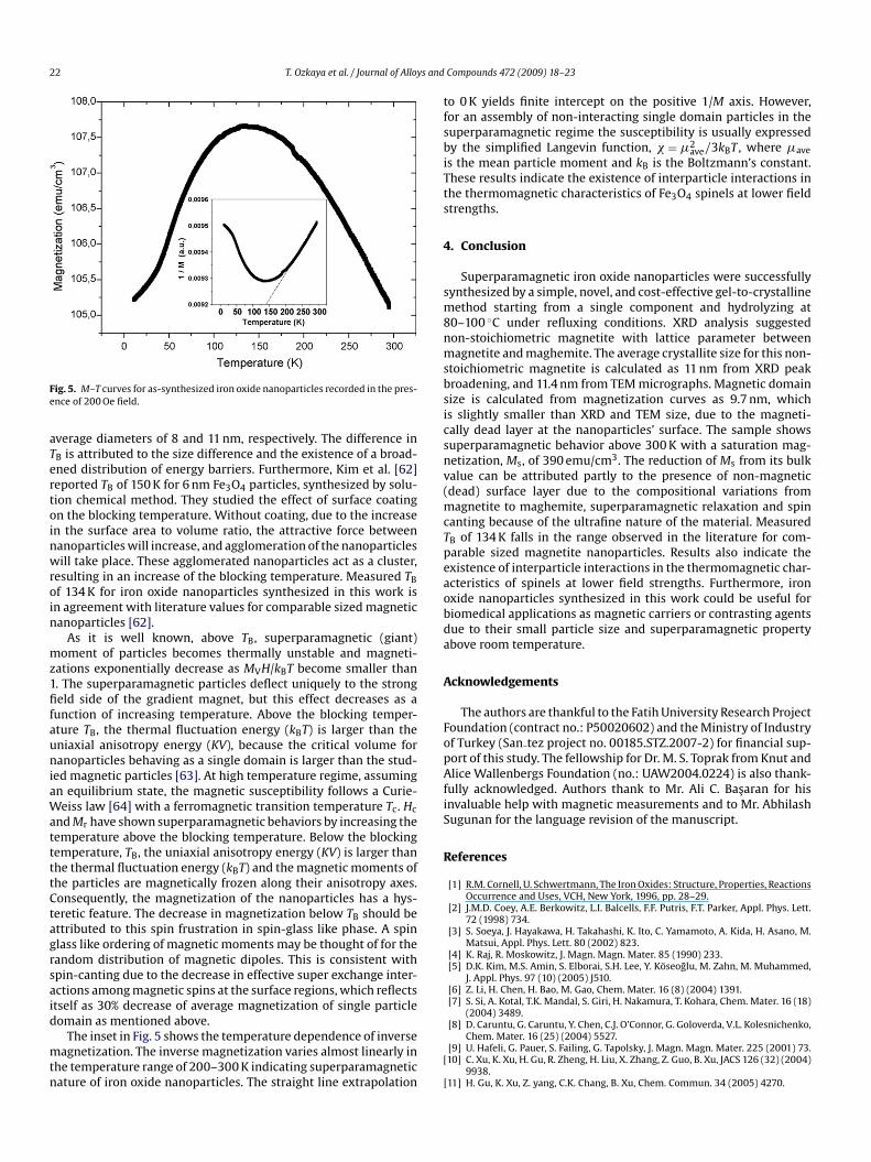
ig. 5. M–T curves for as-synthesized iron oxide nanoparticles recorded in the pres-nce of 200 Oe field.
verage diameters of 8 and 11 nm, respectively. The difference inB is attributed to the size difference and the existence of a broad-ned distribution of energy barriers. Furthermore, Kim et al. [62]eported TB of 150 K for 6 nm Fe3O4 particles, synthesized by solu-ion chemical method. They studied the effect of surface coatingn the blocking temperature. Without coating, due to the increasen the surface area to volume ratio, the attractive force betweenanoparticles will increase, and agglomeration of the nanoparticlesill take place. These agglomerated nanoparticles act as a cluster,
esulting in an increase of the blocking temperature. Measured TBf 134 K for iron oxide nanoparticles synthesized in this work isn agreement with literature values for comparable sized magneticanoparticles [62].
As it is well known, above TB, superparamagnetic (giant)oment of particles becomes thermally unstable and magneti-
ations exponentially decrease as MVH/kBT become smaller than. The superparamagnetic particles deflect uniquely to the strongeld side of the gradient magnet, but this effect decreases as a
unction of increasing temperature. Above the blocking temper-ture TB, the thermal fluctuation energy (kBT) is larger than theniaxial anisotropy energy (KV), because the critical volume foranoparticles behaving as a single domain is larger than the stud-
ed magnetic particles [63]. At high temperature regime, assumingn equilibrium state, the magnetic susceptibility follows a Curie-eiss law [64] with a ferromagnetic transition temperature Tc. Hc
nd Mr have shown superparamagnetic behaviors by increasing theemperature above the blocking temperature. Below the blockingemperature, TB, the uniaxial anisotropy energy (KV) is larger thanhe thermal fluctuation energy (kBT) and the magnetic moments ofhe particles are magnetically frozen along their anisotropy axes.onsequently, the magnetization of the nanoparticles has a hys-eretic feature. The decrease in magnetization below TB should bettributed to this spin frustration in spin-glass like phase. A spinlass like ordering of magnetic moments may be thought of for theandom distribution of magnetic dipoles. This is consistent withpin-canting due to the decrease in effective super exchange inter-ctions among magnetic spins at the surface regions, which reflectstself as 30% decrease of average magnetization of single particleomain as mentioned above.
The inset in Fig. 5 shows the temperature dependence of inverseagnetization. The inverse magnetization varies almost linearly in
he temperature range of 200–300 K indicating superparamagneticature of iron oxide nanoparticles. The straight line extrapolation
[
[
Compounds 472 (2009) 18–23
o 0 K yields finite intercept on the positive 1/M axis. However,or an assembly of non-interacting single domain particles in theuperparamagnetic regime the susceptibility is usually expressedy the simplified Langevin function, � = �2
ave/3kBT , where �ave
s the mean particle moment and kB is the Boltzmann’s constant.hese results indicate the existence of interparticle interactions inhe thermomagnetic characteristics of Fe3O4 spinels at lower fieldtrengths.
. Conclusion
Superparamagnetic iron oxide nanoparticles were successfullyynthesized by a simple, novel, and cost-effective gel-to-crystallineethod starting from a single component and hydrolyzing at
0–100 ◦C under refluxing conditions. XRD analysis suggestedon-stoichiometric magnetite with lattice parameter betweenagnetite and maghemite. The average crystallite size for this non-
toichiometric magnetite is calculated as 11 nm from XRD peakroadening, and 11.4 nm from TEM micrographs. Magnetic domainize is calculated from magnetization curves as 9.7 nm, whichs slightly smaller than XRD and TEM size, due to the magneti-ally dead layer at the nanoparticles’ surface. The sample showsuperparamagnetic behavior above 300 K with a saturation mag-etization, Ms, of 390 emu/cm3. The reduction of Ms from its bulkalue can be attributed partly to the presence of non-magneticdead) surface layer due to the compositional variations from
agnetite to maghemite, superparamagnetic relaxation and spinanting because of the ultrafine nature of the material. MeasuredB of 134 K falls in the range observed in the literature for com-arable sized magnetite nanoparticles. Results also indicate thexistence of interparticle interactions in the thermomagnetic char-cteristics of spinels at lower field strengths. Furthermore, ironxide nanoparticles synthesized in this work could be useful foriomedical applications as magnetic carriers or contrasting agentsue to their small particle size and superparamagnetic propertybove room temperature.
cknowledgements
The authors are thankful to the Fatih University Research Projectoundation (contract no.: P50020602) and the Ministry of Industryf Turkey (San tez project no. 00185.STZ.2007-2) for financial sup-ort of this study. The fellowship for Dr. M. S. Toprak from Knut andlice Wallenbergs Foundation (no.: UAW2004.0224) is also thank-
ully acknowledged. Authors thank to Mr. Ali C. Basaran for hisnvaluable help with magnetic measurements and to Mr. Abhilashugunan for the language revision of the manuscript.
eferences
[1] R.M. Cornell, U. Schwertmann, The Iron Oxides: Structure, Properties, ReactionsOccurrence and Uses, VCH, New York, 1996, pp. 28–29.
[2] J.M.D. Coey, A.E. Berkowitz, L.I. Balcells, F.F. Putris, F.T. Parker, Appl. Phys. Lett.72 (1998) 734.
[3] S. Soeya, J. Hayakawa, H. Takahashi, K. Ito, C. Yamamoto, A. Kida, H. Asano, M.Matsui, Appl. Phys. Lett. 80 (2002) 823.
[4] K. Raj, R. Moskowitz, J. Magn. Magn. Mater. 85 (1990) 233.[5] D.K. Kim, M.S. Amin, S. Elborai, S.H. Lee, Y. Koseoglu, M. Zahn, M. Muhammed,
J. Appl. Phys. 97 (10) (2005) J510.[6] Z. Li, H. Chen, H. Bao, M. Gao, Chem. Mater. 16 (8) (2004) 1391.[7] S. Si, A. Kotal, T.K. Mandal, S. Giri, H. Nakamura, T. Kohara, Chem. Mater. 16 (18)
(2004) 3489.
[8] D. Caruntu, G. Caruntu, Y. Chen, C.J. O’Connor, G. Goloverda, V.L. Kolesnichenko,Chem. Mater. 16 (25) (2004) 5527.[9] U. Hafeli, G. Pauer, S. Failing, G. Tapolsky, J. Magn. Magn. Mater. 225 (2001) 73.10] C. Xu, K. Xu, H. Gu, R. Zheng, H. Liu, X. Zhang, Z. Guo, B. Xu, JACS 126 (32) (2004)
9938.11] H. Gu, K. Xu, Z. yang, C.K. Chang, B. Xu, Chem. Commun. 34 (2005) 4270.

s and
[
[
[
[
[[[[
[[[[[[[
[
[[[[[
[
[
[[[[
[
[[
[
[[[
[
[
[[[
[[
[[
[
[[
[[
T. Ozkaya et al. / Journal of Alloy
12] Y.R. Chemla, H.L. Crossman, Y. Poon, R. McDermott, R. Stevens, M.D. Alper, J.Clarke, Proc. Natl. Acad. Sci. U.S.A. 97 (2000) 14268.
13] D. Hogemann, L. Josephson, R. Weissleder, J.P. Basilion, Bioconjugate Chem. 11(2000) 941.
14] P. Oswald, O. Clement, C. Chambon, E. Schouman-Claeys, G. Frija, Magn. Reson.Imaging 15 (1997) 1025.
15] P. Tartaj, M.P. Morales, S. Veintemillas-Verdaguer, T. Gonzalez-Carreno, C.J.Serna, J. Phys. D: Appl. Phys. 36 (2003) R182.
16] L. Fu, V.P. Dravid, D.L. Johnson, Appl. Surf. Sci. 181 (2001) 173.17] J. Murbe, A. Rechtenbach, J. Topfer, Mater. Chem. Phys. 110 (2008) 426.18] Y. Koseoglu, H. Kavas, B. Aktas, Phys. Stat. Sol. (a) 203 (7) (2006) 1595.19] W.J. Liang, M. Bockrath, D. Bozovic, J.H. Hafner, M. Tinkham, H. Park, Nature 411
(2001) 665.20] X.F. Duan, Y. Huang, Y. Cui, J. Wang, C.M. Lieber, Nature 409 (2001) 6816.21] T.R.N. Kutty, P. Padmini, Mater. Res. Bull. 27 (1992) 945.22] T.R.N. Kutty, P. Padmini, Mater. Chem. Phys. 39 (1995) 200.23] T.R.N. Kutty, P. Padmini, J. Mater. Chem. 4 (1994) 1875.24] M. Anilkumar, V. Ravi, Mater. Res. Bull. 40 (2005) 605.25] S.R. Dhage, V. Choube, V. Samuel, V. Ravi, Mater. Lett. 58 (2004) 2310.26] S.R. Dhage, S.P. Gaikwad, V. Samuel, V. Ravi, Bull. Mater. Sci. 27 (2004)
221.27] S.R. Dhage, S.P. Gaikwad, P. Muthukumar, V. Ravi, Ceram. Int. 31 (2005)
211.28] A. Baykal, Y. Koseoglu, M. Senel, Cent. Eur. J. Chem. 5 (1) (2007) 169.29] P.H. Refait, J.M.R. Genin, Corros. Sci. 34 (1993) 797.30] A.A. Olowe, J.M.R. Genin, Corros. Sci. 32 (1991) 965.31] P.E. Werner, L. Eriksson, M. Westdahl, J. Appl. Cryst. 18 (1985) 367.32] T.J. Daou, G. Pourroy, S. Begin-Collin, J.M. Greneche, C. Ulhaq-Bouillet, P. Legare,
P. Bernhardt, C. Leuvrey, G. Rogez, Chem. Mater. 18 (18) (2006) 4399.33] T. Belin, N. Guigue-Millot, T. Caillot, D. Aymes, J.C. Niepce, J. Solid State Chem.
163 (2002) 459.34] N. Guigue-Millot, Y. Champion, M.J. Hytch, F. Bernard, S. Begin-Collin, P. Perriat,
J. Phys. Chem. 105 (2001) 2175.35] J.P. Jolivet, E.J. Tronc, J. Colloid Interface Sci. 125 (1998) 688.36] C.N.J. Wagner, E.N. Aqua, Adv. X-ray Anal. 7 (1964) 46.37] B.D. Hall, D. Zanchet, D. Ugarte, J. Appl. Cryst. 33 (2000) 1335.38] T. Wejrzanowski, R. Pielaszek, A. Opalinska, H. Matysiak, W. Łojkowski, K.J.
Kurzydłowski, Appl. Surf. Sci. 253 (2006) 204.
[[[[[
Compounds 472 (2009) 18–23 23
39] R. Pielaszek, in: Proceedings of the XIX Conference, Krakow, Poland, September,Appl. Crystallography (2003) 43.
40] S. Wei, Y. Zhu, Y. Zhang, J. Xu, React. Funct. Polym. 66 (2007) 1272.41] R.A. Nyquist, R.O. Kagel, Infrared Spectra of Inorg. Comp., Academic Press, New
York, 1971.42] I.J. Bruce, J. Taylor, M. Todd, M.J. Davies, E. Borioni, C. Sangregorio, T. Sen, J. Magn.
Magn. Mater. 284 (2004) 145.43] T. Kim, M. Shima, J. Appl. Phys. 101 (2007), 09M516.44] D.H. Han, J.P. Wang, H.L. Luo, J. Magn. Magn. Mater. 136 (1994) 176.45] S. Chikazumi, Physics of Ferromagnetism, second edition, Clarendon Press,
Oxford, 1997.46] E. Blum, A. Cebers, M.M. Maiorov, Magnetic Fluids, Walter de Gruyter, Berlin,
1997.47] I. Nedkov, T. Merodiiska, L. Milenova, T. Koutzarova, J. Magn. Magn. Mater. 211
(2000) 296.48] Z. Wang, H. Guo, Y. Yu, N. He, J. Magn. Magn. Mater. 302 (2006) 397.49] B. Jia, L. Gao, Scripta Materialia 56 (2007) 677.50] D.K. Kim, Y. Zhang, W. Voit, K.V. Rao, M. Muhammed, J. Magn. Magn. Mater. 225
(2001) 30.51] R.H. Kodama, A.E. Berkowitz, E.J. McNiff, S. Foner, Phys. Rev. Lett. 77 (1996) 394.52] J.K. Vassiliou, V. Mehrotra, M.W. Russell, E.P. Giannelis, R.D. McMichael, R.D.
Shull, R.F. Ziolo, J. Appl. Phys. 73 (1993) 5109.53] R. Massart, IEEE Trans. Magn. 17 (1981) 1247.54] B.D. Cullity, Introduction to Magnetic Materials, Addison-Wesley, Reading, MA,
1974, p. 94.55] C.M. Sorensen, in: K.J. Klabunde (Ed.), Nanoscale Materials in Chemistry, John
Wiley and Sons, Inc., New York, 2001, p. 169.56] M. Blanco-Manteco, K. O’Grady, J. Magn. Magn. Mater. 296 (2006) 124.57] Z.X. Tang, C.M. Sorensen, K.J. Klabunde, G.C. Hadjipanayis, J. Appl. Phys. 69
(1991) 5279.58] R. Kaiser, G. Miskolczy, J. Appl. Phys. 41 (1970) 1064.59] S. Shufeng, C. Li, X. Wang, D. Yu, Q. Peng, Y. Li, Crystal Growth Design 5 (2005)
391.60] J. Wan, G. Ttang, Y. Qian, Appl. Phys. A 86 (2007) 261.61] Y. Hou, J. Yu, S. Gao, J. Mater. Chem. 13 (2003) 1983.62] D.K. Kim, Y. Zhang, W. Voit, K.V. Rao, M. Muhammed, JMMM 225 (2001) 30.63] D.L. Pelecky, R.D. Rieke, Chem. Mater. 8 (1996) 1770.64] A. Sawatsky, F. van der Woude, A.H. Morrish, J. Appl. Phys. 39 (1968) 1204.