Synthesis, characterization, DNA binding and anticancer property of...
10
IOSR Journal of Applied Chemistry (IOSR-JAC) e-ISSN: 2278-5736. Volume 7, Issue 4 Ver. II. (Apr. 2014), PP 71-80 www.iosrjournals.org www.iosrjournals.org 71 | Page Synthesis, characterization, DNA binding and anticancer property of mer-trichlorodimethylsulphoxide-S-(1, 10- phenanthroline) ruthenium (III) P. Hazarika, J. Deka, S. Kamarkar, S.Bhola, R.K.Bhola,O.K Medhi and C. Medhi* Chemistry Department, Gauhati University P.O.Gauhati University Abstract: The mer-trichloro dimethylsulphoxide-S-(1,10-phenanthroline) ruthenium (III) ([Ru(phen)(DMSO)Cl 3 ]) complex has been synthesized. The structure has been characterized from FT-IR, 1 H NMR and UV-Vis spectroscopic data. The geometry of this complex determined by single crystal X-ray diffraction study was found to be octahedral. The binding of this complex with Calf Thymus DNA (CT-DNA) has been investigated by spectroscopic, electrochemical and electrophoresis studies. Anticancer activity of this compound studied both in vivo and in vitro against Dalton’s lymphoma in mice was found to be significant. Keywords: DNA, Ruthenium complex, Calf thymus, Anticancer activity, Dalton’s Lymphoma. I. Introduction: Metal-based compounds constitute a discrete class of chemotherapeutics widely used as anticancer and antiviral agents. Most established antitumor metal-based drugs are cisplatin and its analogues, such as carboplatin and oxaliplatin. Cisplatin is an anticancer agent being used for the treatment of testicular, ovarian, head, neck and germ cell tumors (Rosenberg B,1985 and Bertino JR et al., 1997). However, the optimal use of this drug is limited due to its dose limiting nephrotoxicity. The new platinum compounds like carboplatin and oxaliplatin are found to possess comparatively very narrow therapeutic index, and also it has limited clinical utility due to drug resistance and side effects(Dabholkar M et al.,1996 and Shimada H et al.,1993). Hence it is necessary to look for more effective and less toxic other metal-based anticancer agents. Ruthenium complexes have attracted much interest as alternative drug to cisplatin in cancer chemotherapy. Ruthenium complexes have similar ligand exchange kinetics as platinum (II) complexes and exist in different oxidation states under different physiological conditions(Jakupec M A et al., 2005 and Schluga P et al., 2006). Many reviews that illustrate the unique DNA binding modes and antitumor effects have been reported (Kostova I 2006). Ruthenium complexes are presently receiving great attention in the fields of biological, pharmaceutical and medicinal chemistry as anti-tumor agents. Ruthenium compounds show low systematic toxicity and appear to penetrate reasonably well within the tumor cells, binding effectively to DNA and proteins and presenting in some cases selective anti-metastatic properties (Brabec V and Novakova O, 2006) However, it is essential to design superior drugs in terms of effectiveness compared to cisplatin and other metal drugs (Rosenberg B et al.,1965). The 1-10 phenanthroline (phen) ligand has been found in many metal- based anticancer agents (Clarke MJ et al., 1999 Vessieres A et al., 2006, Morris RE et al., 2001, Hong-Ke Liu et al., 2011 and Finlay GJ et al., 2000). There are certain metal complexes with different types of ligands, which are in fact found to be quite effective anticancer agents(Kostova I 2006, Clarke M J 2003 and Bratsos I et al., 2007). Moreover, extensive studies on the biological properties of potential metal-based anticancer agents of phen and DMSO ligands have been found in literatures. There are numerous ruthenium complexes containing phenanthroline ligand and some of these complexes acquire good anticancer activity (Tysoe SA et al., 1993 and Liu J et al., 2008). Again the Ru(II) and Ru(III) complexes of dimethyl sulfoxide (DMSO) ligand are known for good anticancer activity (Murali S et al., 2002, Sun S et al., 2009 Yong-guang Y et al., 2011, D. P. Rillema et al.,1982 and Hartinge CG et al., 2008). The cis-[RuCl 2 (NH 3 ) 4 ]Cl, fac-[RuCl 3 (NH 3 ) 3 ] and trans-HIm[RuCl 4 (Im) 2 ] complexes are some of the anticancer agents effective against primary tumour cells (Thota S et al., 2012, Stubbs M et al., 1996, Li-Feng Tan et al., 2005, Anghileri L J, et al., 1975, Allardyce CS et al., 2005, Paul H et al., 2012, Wang F et al., 2003 and Nomizo A, et al., 2010). The ligands like isatin, thio semicarbazones, and chloro-fluoro-phenyl are also found in some Ru(II) complexes and the cytotoxic activity both in vivo and in vitro studies of these complexes are promising (Karki SS et al., 2007). Similarly, NAMI emerges out as anti-metastatic agent, but only small fraction of the compound can reach the tumour target (Galanski M et al., 2003, Sava G et al., 2002 and Arandjelovic SS et al.,2009). Here, the synthesized complex [Ru(phen)(DMSO)Cl 3 ]) Ru(III) complex has been characterized and the DNA binding ability as well as antitumor activity against Dalton’s Lymphoma have been examined.
-
Upload
independent -
Category
Documents
-
view
1 -
download
0
Transcript of Synthesis, characterization, DNA binding and anticancer property of...

IOSR Journal of Applied Chemistry (IOSR-JAC)
e-ISSN 2278-5736 Volume 7 Issue 4 Ver II (Apr 2014) PP 71-80
wwwiosrjournalsorg
wwwiosrjournalsorg 71 | Page
Synthesis characterization DNA binding and anticancer
property of mer-trichlorodimethylsulphoxide-S-(1 10-
phenanthroline) ruthenium (III)
P Hazarika J Deka S Kamarkar SBhola RKBholaOK Medhi and C
Medhi Chemistry Department Gauhati University POGauhati University
Abstract The mer-trichloro dimethylsulphoxide-S-(110-phenanthroline) ruthenium (III)
([Ru(phen)(DMSO)Cl3]) complex has been synthesized The structure has been characterized from FT-IR 1H
NMR and UV-Vis spectroscopic data The geometry of this complex determined by single crystal X-ray
diffraction study was found to be octahedral The binding of this complex with Calf Thymus DNA (CT-DNA) has
been investigated by spectroscopic electrochemical and electrophoresis studies Anticancer activity of this
compound studied both in vivo and in vitro against Daltonrsquos lymphoma in mice was found to be significant
Keywords DNA Ruthenium complex Calf thymus Anticancer activity Daltonrsquos Lymphoma
I Introduction Metal-based compounds constitute a discrete class of chemotherapeutics widely used as anticancer and
antiviral agents Most established antitumor metal-based drugs are cisplatin and its analogues such as
carboplatin and oxaliplatin Cisplatin is an anticancer agent being used for the treatment of testicular ovarian
head neck and germ cell tumors (Rosenberg B1985 and Bertino JR et al 1997) However the optimal use of
this drug is limited due to its dose limiting nephrotoxicity The new platinum compounds like carboplatin and
oxaliplatin are found to possess comparatively very narrow therapeutic index and also it has limited clinical
utility due to drug resistance and side effects(Dabholkar M et al1996 and Shimada H et al1993) Hence it is
necessary to look for more effective and less toxic other metal-based anticancer agents
Ruthenium complexes have attracted much interest as alternative drug to cisplatin in cancer
chemotherapy Ruthenium complexes have similar ligand exchange kinetics as platinum (II) complexes and
exist in different oxidation states under different physiological conditions(Jakupec M A et al 2005 and Schluga
P et al 2006) Many reviews that illustrate the unique DNA binding modes and antitumor effects have been
reported (Kostova I 2006) Ruthenium complexes are presently receiving great attention in the fields of
biological pharmaceutical and medicinal chemistry as anti-tumor agents Ruthenium compounds show low
systematic toxicity and appear to penetrate reasonably well within the tumor cells binding effectively to DNA
and proteins and presenting in some cases selective anti-metastatic properties (Brabec V and Novakova O
2006) However it is essential to design superior drugs in terms of effectiveness compared to cisplatin and other
metal drugs (Rosenberg B et al1965) The 1-10 phenanthroline (phen) ligand has been found in many metal-
based anticancer agents (Clarke MJ et al 1999 Vessieres A et al 2006 Morris RE et al 2001 Hong-Ke Liu et
al 2011 and Finlay GJ et al 2000) There are certain metal complexes with different types of ligands which
are in fact found to be quite effective anticancer agents(Kostova I 2006 Clarke M J 2003 and Bratsos I et al
2007) Moreover extensive studies on the biological properties of potential metal-based anticancer agents of
phen and DMSO ligands have been found in literatures There are numerous ruthenium complexes containing
phenanthroline ligand and some of these complexes acquire good anticancer activity (Tysoe SA et al 1993 and
Liu J et al 2008) Again the Ru(II) and Ru(III) complexes of dimethyl sulfoxide (DMSO) ligand are known
for good anticancer activity (Murali S et al 2002 Sun S et al 2009 Yong-guang Y et al 2011 D P Rillema
et al1982 and Hartinge CG et al 2008)
The cis-[RuCl2(NH3)4]Cl fac-[RuCl3(NH3)3] and trans-HIm[RuCl4(Im)2] complexes are some of the
anticancer agents effective against primary tumour cells (Thota S et al 2012 Stubbs M et al 1996 Li-Feng
Tan et al 2005 Anghileri L J et al 1975 Allardyce CS et al 2005 Paul H et al 2012 Wang F et al 2003
and Nomizo A et al 2010) The ligands like isatin thio semicarbazones and chloro-fluoro-phenyl are also
found in some Ru(II) complexes and the cytotoxic activity both in vivo and in vitro studies of these complexes
are promising (Karki SS et al 2007) Similarly NAMI emerges out as anti-metastatic agent but only small
fraction of the compound can reach the tumour target (Galanski M et al 2003 Sava G et al 2002 and
Arandjelovic SS et al2009) Here the synthesized complex [Ru(phen)(DMSO)Cl3]) Ru(III) complex has been
characterized and the DNA binding ability as well as antitumor activity against Daltonrsquos Lymphoma have been
examined
Synthesis characterization DNA binding and anticancer property hellip
wwwiosrjournalsorg 72 | Page
II Experimental section Materials
Analytical grade RuCl33H2O was purchased from Sigma and used without purification Calf Thymus
DNA(CT-DNA) tris-buffer and tetrabutylammonium perchlorate(TBAP) were obtained from Sigma-Aldrich
USA CT-DNA was dissolved in tris-buffered saline ( pH 76 TBS) and dialyzed overnight against the same
buffer so that A260A280 of the dialyzed solution should be gt 18 (Anghileri L J et al 1975) The 110-
phenanthroline (phen) and other solvents were used as received
Synthesis of mer-trichlorodimethylsulphoxide-S-(110-phenanthroline) ruthenium (III) complex
The compound was prepared by refluxing cis-Ru(phen)Cl42H2O with dimethylsulphoxide (DMSO) in
solvent of ethanol and water mixture for half hour Cis-Ru(phen)Cl42H2O was synthesised by refluxing
RuCl33H2O with phen according to the procedure given in literature (Krause RA 1977) Then evaporated the
solvent by heating in water bath and subsequently purified the compound The purity of the compound was
checked by thin layer chromatography (TLC) The mer-trichlorodimethylsulphoxide-S-(110-phenanthroline)
ruthenium (III) compound was obtained as a red colored crystal with 80 yield IR (KBr) values are 1646
(C=N aromatic) 1090 (S=O) 3064 (C-H SP2
Carbon) 3012 2926 2852 (C-H methyl) 1591 1538 1413 (C=C
aromatic) 450 and 429 (Ru-N and Ru-S) 1H NMR (CDCl3-d6 300 MHz) values at 1018 852 and 825 ppm for
N-H C-H and C-H bonds of 110 phenanthroline ligand was found The values at 219 and 266 ppm for two
methyl groups of DMSO 724 ppm for solvent CDCl3 and 15 ppm for water impurity in CDCl3 were found
Anal calculated for C14 H14 N2 C 3478 H 290 N 580 Found C 3475 H 280 N 572
Crystal structure determination
Single crystal X-ray diffraction data was obtained at 100 K with Brunker smart AXS diffractometer
with graphite-monochromatised Mo-K radiation by - scans We used full matrix least square on F2 The
molecular graphic structure was analyzed by ORTEP plot program The structure was refined by using
SHELXL-97 other materials were prepared by wingx publication routine (Figure 1) (Barton JK and Raphael
AL et al1984)
Crystal data Molecular formula C14H14Cl3N2O1S1Ru1 formula weight 46575 gmol-1
a = 88458(8) Aring
b = 119754(10) Aring c = 167314(14) Aring α = 92220(4)ordm β = 104317ordm(4)γ = 92945(4)ordm cell volume = 17127(3)
Aring3 Z=4 Dcal = 1806 gcm
-3 triclinic space group p-1 Data Collection Bruker Smart Apex II CCD Mo-Kα
radiation (λ= 071073 Aring) graphite monochromator Crystal size 052 X 023 X 014 mm3 296(2)K ω and Φ
scan 39ordm le 2θ le6018ordm -12 le h le12 -16 le k le16 -23leIle23 35146 reflection measured 9885 unique μ(Mo-Kα)
151 mm-1
experimental absorption collection with Multi-scan (Sheldrick GM 1996 )
Structure solution and refinement The structure was solved by SHELXS97 (Sheldrick GM 1997) and
refinement was carried out by full-matrix least square on F2 using the same program SHELXL97 All non-H
atoms are refined with anisotropic temperature factors and the hydrogen atoms were positioned from the
different synthesis map with isotropic temperature factors of Uiso(H) = 12 Uiso (C) for CH and 0114 Uiso(C)
for CH3401 refined parameters final R1= 00347 wR2 = 00892 for 8001 reflections with I gt 2σI and final
R1= 00478 wR2= 00994 for all data with largest peak difference and hole of 0780-0726 e Aring-3
in the
vicinity of the Cl3 and O1 atom The structural data has been deposited to Cambridge Crystallographic Data
Center (CCDC 889313)
Spectroscopic studies on DNA binding
The spectroscopic electrochemical and electrophoresis studies of this Ru complex with Calf Thymus
DNA (CT-DNA) have been carried out We observed distinct spectral shift after mixing the complex with CT-
DNA It might be due to CT-DNA binding by this complex
UV-Visible Absorption Titration The UV-visible absorption spectra of [Ru(phen)(DMSO)Cl3] complex in presence of CT-DNA has
been performed (Figure 2) The experiment was performed by maintaining constant concentration of the
complex with varying CT-DNA concentrations within the range of 059X10-5
M to 296X10-5
M The absorption
spectra of the complex are characterized by two distinct intense transitions at 400 nm and 273 nm which are the
characteristic of metal to ligand charge transfer(Ru (dpi)-(Lpi) transition) With the of the concentrations CT-
DNA from 059X10-5
M to 296X10-5
M absorbance intensity of both the two peaks decreased without shifting
the wavelengths(Tables 1 and Figure 2) These spectral characteristics might be due to the binding of CT-DNA
with the complex To estimate quantitatively the binding strength of [Ru(phen)(DMSO)Cl3] the intrinsic
binding constants (Kb ) was calculated from the following equation (Shahabadi N et al 2011 and Rathinasamy
S et al2006)
Synthesis characterization DNA binding and anticancer property hellip
wwwiosrjournalsorg 73 | Page
where εa εf and εb are the extinction coefficients of observed solution free complex and the solution of the
complex with maximum CT-DNA concentration The value of Kb was obtained from the slope of the plot
(Figure 2B) It was found to be 277 X 104 M
-1
Fluorescence Emission Titration
To further investigate the interaction of the complex with CT-DNA fluorescence titration experiment
was performed The emission spectra of the complex at excitation wavelength of 280 nm and in presence of
varying amounts of CT-DNA are shown in Figure 3 On increasing the concentrations of CT-DNA from 059
X10-5
M to 41X10-5
M a new peak appeared at 445 nm in addition to the other peak at 324 nm The intensities
of both these peaks have been increased with the increase of CT-DNA (Table 2 and Figure 3) Intercalation by
phen ligand of the complex within DNA base pair upon DNA binding might enhance the luminescence of the
complex which is similar to the nature of DNA binding of other previously reported complexes (KB=277 X 104
M-1
) ( Rathinasamy S et al2006) The value is typical for intercalation by hydrophobic phen ligand
Electrochemistry
Cyclic voltammetric study for [Ru(phen)(DMSO)Cl3] complex was carried out in DMSO
solution(0005 M) containing 01 M TBAP as supporting electrolyte using AgAg+ as reference electrode and
a glassy carbon electrode was used as working electrode Inert environment was maintained by passing N2 gas
through the solution to remove oxygen The cyclic voltamogram shows distinct oxidation and reduction peaks
of a reversible electron transfer couple at scan rate 100 mVs-1
The shift of redox potential (E12) from 222 mV
to 256 mV on addition of CT-DNA was observed (Figure 4)
Electrophoresis experiment
The binding of trichlorodimethylsulphoxide-S-(110-phenanthroline) ruthenium (III) complex with CT-
DNA was further supported by electrophoresis experiment The solution of CT-DNA was prepared in tris-HCl
buffer at pH 76 The binding of complex with CT-DNA was monitored by preparing solutions of the complex
having three different concentrations 6 mM 3 mM and 2 mM with CT-DNA which was incubated for 24 hours
at 37ordm C before running Gel Electrophoresis The three solutions were placed at three different lanes 1 2 and 3
in the gel along with free CT-DNA (5 mM) which was kept at lane C The composition of base pairs in the CT-
DNA used in this experiment was 42 mole for GC 58 mole for AT in 100 mole The samples were run from -ve
to +ve potential for 3 hours at different voltages (half an hour at 50 V 1 hour at 60 V half an hour at 70 V and 1
hour at 80 V) on a 1 agarose gel prepared in tris-borate EDTA After photographed the gel under UV light it
appears that lane C moves faster than lane 1 2 and 3 and the brightness decreases from lane C lane 1 2 and 3
(Figure 5)
Biological studies of trichlorodimethylsulphoxide-S-(110-phenanthroline) ruthenium (III) complex
Effect of different concentrations of trichloro dimethylsulphoxide-S-(110-phenanthroline) ruthenium
(III) complex on mice bearing Daltonrsquos lymphoma
C3HHe strain of mice both male and female 8-10 weeks old and weighing 20-22g was used in all sets of
experiments Animals were kept in polypropylene cages and were fed on a commercial diet (Goldmohar Lipton
India) and tap water ad libitum Mice were kept under standard condition (especially pathogen free temperature
ranging from 22-230C and relative humidity 65-70 Transplantable ascites Daltonrsquos lymphoma was obtained
from Chittaranjan National Cancer Research Centre Kolkata India and maintained in the laboratory by regular
serial transplantations by injecting 2x10⁷ cellsmice in PBS
after a regular interval of 10 days Ruthenium
compound trichlorodimethylesulphoxide-S-(110-phenanthroline) ruthenium (III) was prepared in the
laboratory as mentioned All other chemicals were purchased from Hi-media Mumbai India and were of
analytical grade
Control and experimental animals were selected randomly and divided into groups of 10 mice each
according to randomized block design Each animal was transplanted with 3x10⁶
cellsmice After 4 days of
post-tumor transplantation the experimental mice were treated with single ip injection of different
concentrations of trichloro dimethylsulphoxide-S-(110-phenanthroline) ruthenium (III) dissolved in Phosphate
buffered saline (PBS) (pH 74) Control animals were injected with equal amount of PBS In each group
Survival increase in the mean survival time of tumor bearing mice and tumor free survivors were observed
Effect of trichlorodimethylsulphoxide-S-(110-phenanthroline) ruthenium (III) complex on the survival of
Daltonrsquos lymphoma cells in vivo
Synthesis characterization DNA binding and anticancer property hellip
wwwiosrjournalsorg 74 | Page
Daltonrsquos lymphoma cells were isolated from the peritoneal cavity of tumor bearing mice (control and
treated with different concentrations of this Ru complex) 2-3 ml of sterile phosphate buffered saline (PBS) was
injected into the peritoneal cavity and the fluid containing the tumor cells was withdrawn and collected in
sterile petridishes for incubation at 370
C for 2 hours The cells of macrophage lineage adhered to the bottom of
petridishes to form a confluent monolayer The non-adherent population of lymphoma cells was gently aspirated
out and washed repeatedly with PBS The viability was tested by tryphan blue exclusion test
Effect of trichlorodimethylsulphoxide-S-(110-phenanthroline) ruthenium (III) on the survival of
Daltonrsquos lymphoma cells in vitro
For cytotoxicity assay in vitro Daltonrsquos lymphoma cells were plated at high density (8x10⁷cellsdish)
at time 0 in DMEM containing fetal calf serum 10 mM NaHCO3 03 glutamine and different concentrations
of Ru complex for a fixed treatment duration of 1 hour Control dishes were treated with equal amount of PBS
used as a solvent for trichlorodimethyl sulphoxide -S- (110-phenanthroline) ruthenium(III) At the end of drug
treatment cells were harvested and washed with PBS suspended again in ADM with DFCS and incubated for
72 hours at 370
C After incubation cells were trypsinized and viable cells were counted by tryphan blue
exclusion test
III Results and Discussion The geometry around the ruthenium atom of [Ru(phen)(DMSO)Cl3] is found to be octahedral with a
phen ligand occupying two coordination sites while the other coordination sites are occupied by one DMSO and
three chlorine atoms The compound was prepared by refluxing the RuCl33H2O and phen in solvent mixture of
ethanol and water and subsequent addition of DMSO Initially the two Cl atoms might be replaced by phen
ligand and then DMSO molecule reacts with Ru(phen)Cl42H2O to replace the inner Cl atom to give
[Ru(phen)(DMSO)Cl3] The crystalline compound so obtained was analyzed by XRD studies and the ORTEP
structure is shown in Figure 1 The molecular structure of [Ru(phen)(DMSO)Cl3] complex shows a slightly
distorted octahedral arrangement around the ruthenium atom The metal atom is bonded to one bi-dentate 110-
phenanthroline ligand three chlorine atoms and a dimethylsulphoxide ligand through the sulphur atom The
bond distances between Ru metal and other coordinating ligands reasonable with those of other already
synthesized complexes like RuCl(DMSO)(110-phen)2][BPh4] cis-Ru(DMSO)4Cl2 and [(η6-
C6Me6)Ru(C5H4N)2C=NMeCl]PF6 (Karki SS et al 2007)The Ru-N bond trans to S is found longer than the
other Ru-N bond The variation of Ru-N bond lengths may be due to the influence of π electrons of Cl and S
atoms to the Ru-N bonding (Karki SS et al 2007) Both the solid as well as the solution state absorption spectra
of the complex show two distinct peaks at the λmax at 273 nm and 400 nm The infrared spectra of the complex
are observed at wavelengths 1086 cm-1
449 cm-1
and 414 cm-1
respectively The potential bonds corresponding
to these observed spectral peaks are -S=O -Ru-N and Ru-S The 1H NMR study cannot clearly detect the peaks
of corresponding ligands
Further studies were carried out to understand the binding of this complex with CT-DNA Absorbance
intensities of the peaks at 400 nm and 273 nm have decreased with the increase of CT-DNA concentrations from
059X10-5
M to 296X10-5
M (Table 1 and Figure 2) The observed spectral characteristics suggest strong
interactions between [Ru(phen)(DMSO)Cl3] complex with CT-DNA The intrinsic binding constant Kb is found
to be 277 X104 M
-1 The fluorescence spectra of the complex is found different from the spectra taken after
mixing with CT DNA(Figure 3) In addition to the original apex at 324 nm of the complex a new peak was
observed at 445 nm which becomes more intense on increasing the concentrations of CT-DNA from 059 X10-5
M to 41X10-5
M (Table 2 and Figure 3) Such observable spectral shifts and change in intensity indicate the
binding of [Ru(phen)(DMSO)Cl3] complex with CT-DNA The spectral peak of CT-DNA taken at pH 74 is
260 nm and the π- π transition for aromatic ligand(phen) is found at 266 nm A broad band gt 400 nm
corresponds to metal to ligand charge transfer (E2g- π) (Lever ABP 1984) Such typical spectral characteristics
suggest intercalation of phen ligand with CT-DNA(Li L et al 2012) base pairs
The redox potentials obtained from Osteryoung Square Wave Voltammetry (OSWV) of the complex
and after mixing with CT-DNA were found at 224 mV and 256 mV respectively (Figure 4) The binding of the
complex with CT-DNA can be inferred from the positive shift in the redox potential (34 mV) after mixing with
CT-DNA Such positive shift of potential might be due to intercalation of hydrophobic phen ligand with DNA
base pairs (Medhi OK et al 1997) The positive shift in the potential might also increase the stability of RuI
after binding to DNA The observed E12 of this complex is very close to the reported value of NAMI
[Na+(RuCl4(dmso)(Ima)] which is +235 mv (Alessio E et al1993) Moreover we performed electrophoresis
experiments to understand the binding of [Ru(phen)(DMSO)Cl3] complex with CT-DNA Lane C moves
slightly faster from ndashve to +ve side in the gel electrophoresis than lanes 1 2 and 3 The electrophoretic
mobilities and brightness of the bands decrease significantly as the concentrations of the complex increases from
lane 3 to 1 Hence the binding of this complex with CT-DNA has been clearly shown (Figure 5)
Synthesis characterization DNA binding and anticancer property hellip
wwwiosrjournalsorg 75 | Page
Electrophoresis of CT-DNA (5 mM) revealed a more intense band but when a higher concentrations of CT-
DNA (6 mM) was mixed with same amount of Ru complex a very weak band was observed (Figure 5) This
suggests the formation of DNA adduct with Ru complex Further as the concentration of CT-DNA was
increased from 2-6 mM and mixed with same amount of Ru complex the sharpness of DNA band was found to
decrease indicating direct correlation between the amounts of DNA and adduct formation
As we know that the metallic drugs cisplatin is an important class of chemotherapeutic agent widely
used in cancer chemotherapy but its clinical use is limited due to nephrotoxicity (Kim YK et al 1995 and
Wolfgang GHI et al 1994) Among the transition metal compounds Ruthenium appears to be a likely
candidate in near future even though its chemistry differs from that of platinum The most significant differences
are its octahedral geometry and greater propensity to undergo redox reactions The hypoxic environment of
many tumors may favor the reduction of ruthenium (III) compounds to ruthenium (II) species (Pegg D G et al
1994 and Clarke MJ et al1988) The hydrophobicity of phenanthroline ligand is responsible for DNA binding
ability and for possible anticancer activity of this complex (Chen T et al 2012)
In the present studies a newly synthesized ruthenium compound trichlorodimethylsulphoxide-S- (110-
phenanthroline) ruthenium(III) was tested against murinersquos Daltonrsquos lymphoma Percentage survival of mice
bearing tumor after treatment with this compound is shown in Figure 6 A direct correlation was observed
between the dose of compound administered and increase in the survival of tumor bearing mice All the
control animals without any treatment failed to survive beyond 10 days post tumor transplantation When the
tumor bearing mice were treated with 20 mgkg of compound 100 of animals survived till day 18 post tumor
transplant and the maximum survival period was found to be 28 days When the dose of ruthenium compound
was increased to 30 and 40 mgkg the maximum survival time increased to 30-32 days When the concentration
was further increased to 50 mgkg the maximum survival period increased to 45 days (Figure 6) The viability
of tumor cells was found to depend on the concentrations of ruthenium compound injected in mice Increasing
concentrations of Ruthenium compound had adverse effect on viability of tumor cells Almost 90 of viability
was observed with 10-20 mgkg of compound However 30-40 mgkg dose has shown 70-80 viable cells The
LC50 was found to be 50 mgkg (Figure 7)
Present study has also shown the effects of Ruthenium compound at the cellular level through a study
of its lethal activity in an in vitro system Results obtained in the present study with Daltonrsquos lymphoma cells
are expressions of cell killing assay and not the growth inhibition assay It was found that cytotoxicity of
ruthenium compound depends upon its concentration in the culture media Survival of Daltonrsquos lymphoma cells
treated in vitro with different concentrations of ruthenium complex for a fixed time duration of 1h is shown in
Figure 8 There exists an inverse linear correlation between the concentration of ruthenium in the medium and
survival of lymphoma cells IC50 of ruthenium was found to be 40 μgml (Figure 8)
Most of the investigations reporting ruthenium compounds as anti-tumor agents have been carried out
as in vitro studies and only a few are in vivo studies Ruthenium- DMSO complexes have been reported to
increase the life span of tumor bearing mice in vivo (Pieper T and Keppler BK 1998) These compounds are
also effective against several murine models including a cisplatin resistant P 388 leukemia cell line While a
number of Ruthenium compounds have been tested the prototype compound RAPTA-C has been reported to be
most effective anti-cancer compound of this series and has been used extensively against a number of murine
cell lines Chatterjee S et al studied the effect of RAPTA-C induced apoptosis in EAC cells isolated from
peritoneal cavity of tumor bearing mice and found that the apoptosis was dose dependent (Chatterjee S et al
2008) In the present studies the effect of compound has also been found to be dose dependent
In a similar study flow cytometry analysis of RAPTA-C induced cell cycle phase distribution of nuclear DNA
has shown increased content of haploid DNA in RAPTA-C treated cells (Chatterjee S et al 2008) However
the DNA in S-phases decreased from 63 to 36 suggesting that tumor suppressor gene P53
is central to the
induction of cell cycle arrest and apoptosis in response to DNA damage of cellular stage in human cells Earlier
it has been reported that ruthenium (III) compounds having significant cytotoxic properties are able to bind
firmly to DNA and modify its structural conformation (Brabec V et al1995) So the anticancer activity of the
present compound may be related to its DNA binding ability
IV Conclusion The synthesis of ruthenium complex its CT-DNA binding and biological studies have been performed
The structure analyzed by single crystal XRD study shows six-coordinated octahedral geometry The crystal
structure is a representative complex [Ru(phen)(DMSO)Cl3] and the structural features are in accord with
already reported ruthenium complexes The CT-DNA binding of this complex is evidenced by various
experimental techniques On the basis of spectroscopic shift the intrinsic binding constant (Kb) is found to be
277X104
M-1
Based on the observed distinct shift of electrochemical E12 and electrophoresis band shifts the
complex is expected to bind with CT-DNA The hydrophobicity of the phen ligand may be responsible for DNA
binding and possibly related to the anticancer activity of ruthenium complex
Synthesis characterization DNA binding and anticancer property hellip
wwwiosrjournalsorg 76 | Page
This compound acquires good antitumor property against Daltonrsquos lymphoma both in vivo and in vitro
The compound exhibited dose dependent increase in the survival time of tumor bearing mice however no
tumor free survivor was observed From the biological studies in vivo the highest dose of this complex is 50
mgkg Electrophoretic analysis of CT-DNA with the complex suggests the formation of DNA adduct which
might be responsible for the antitumor activity of this compound
References [1] Rosenberg B (1985) Fundamental studies with cisplatin Cancer 55 2303-2316 [2] Pil P Lippard S J Cisplatin and related drugs Bertino JR (1997) Encyclopedia of cancer Academic Press Inc San Diego 1
392-410
[3] Dabholkar M and Reed E (eds Pinedo H M Longo D L Chabner B A) (1996) Elsevier Science Publishers Netherlands 88-110
[4] Shimada H Sugimachi N Funakoshi T and Kojima S (1993) Prevention of renal toxicity of cis-diamminedichloroplatinum by
dithiocarbamates in rats Toxicol Lett 66193-198
[5] Jakupec M A Reisner E Eichinger A Pongratz M Arion V B Galanski M Hartinger C Keppler B K (2005) Redox-Active
Redox-Active Antineoplastic Ruthenium Complexes with Indazole Correlation of in Vitro Potency and Reduction Potential J Med
Chem 48 2831-2837 [6] Schluga P Hartinger G Egger A Reisner E Galanski M Jakupee MA and Keppler BK (2006) Redox behavior of tumor-
inhibiting ruthenium (III) complexes and effects of physiological reductants on their binding to GMP Dalton Trans 1796-1802
[7] Kostova I (2006) Ruthenium complexes as anticancer agents Curr Med Chem 131085-107 [8] Clarke M J (2003) Ruthenium Metallopharmaceuticals Coord Chem Rev 236 207-231
[9] Bratsos I Jedner S Gianferrara T Alessio E (2007) Ruthenium anticancer compounds Challenges and Expectations Chimia 61
692-697 [10] Rosenberg B Camp LV Krigas T(1965) Inhibition of Cell Division in Escherichia coli by Electrolysis Products from a Platinum
Electrode Nature 205 698 ndash 699
[11] Clarke MJ Zhu F Frasca DR (1999) Non-Platinum Chemotherapeutic MetallopharmaceuticalsChem Rev 99 2511-2533 [12] Vessieres A Top S Beck W Hillard E Jaouen G (2006) Metal complex SERMs (selective oestrogen receptor modulators) The
influence of different metal units on breast cancer cell antiproliferative effects Dalton Trans 529 529-541
[13] Morris RE Aird R E Mudroch P S Chen H Cummins J Hughes ND Parsons S Parkin A Boyd G Jodrell DI Sadler PJ (2001) Inhibition of Cancer Cell Growth by Ruthenium(II) Arene Complexes J Med Chem44 3616-3621
[14] Hong-Ke Liu Sadler PJ (2011) Metal Complexes as DNA Intercalators Acc Chem Res 44 349-359
[15] Finlay GJ Baguley BC (2000) Effects of protein binding on the in vitro activity of antitumour acridine derivatives and related anticancer drugs Cancer Chemother Pharmacol45 417-22
[16] Thota S Imran M Udugula M Karki S S Kanjarla N Yerra R Balzarini J and Clercq E D (2012) Synthesis spectroscopic
characterization antineoplastic in vitro-cytotoxic and antibacterial activities of mononuclear ruthenium(II) complexes J Coord Chem 65 823ndash839
[17] Clarke MJ and Stubbs M(1996) Interactions of Metallopharmaceuticals with DNA Metal Ions in Biological Systems 32 727-780
[18] Li-Feng Tan Hui Chao Yun-Jun Liu Hong Li Bin Sun Liang-Nian Ji (2005) DNA-binding and photocleavage studies of [Ru(phen)2(NMIP)]2+ Inorganica Chimica Acta 358 2191-2198
[19] Anghileri L J Krebsforsch Z Onkol K( 1975) The in vivo inhibition of tumor growth by ruthenium red its relationship with the
metabolism of calcium in the tumor Cancer Res Clin Oncol 83 213 [20] Allardyce CS Dorcier A Scolaro C Dyson PJ (2005) Development of organometallic (organo-transition metal) pharmaceuticals
Appl Organomet Chem 19 1-10
[21] Paul H Mukherjee T Drew MGB and Chattopadhyay P (2012) Synthesis characterization crystal structure and DNA-binding of ruthenium(II) complexes of heterocyclic nitrogen ligands resulting from a benzimidazole-based quinazoline derivative J Coord
Chem 65 1289-1302
[22] Wang F Chen H Parsons S Oswald IDH Davidson JE Sadler PJ (2003) Kinetics of Aquation and Anation of Ruthenium(ii) Arene Anticancer Complexes Acidity and X-ray Structures of Aqua Adducts Chem Eur J 9 5810-5820
[23] Elisacircngel de Paula Silveira-Lacerda Cesar Augusto Sam Tiago Vilanova-Costa Pereira FC Hamaguchi A Pavanin LA Goular LR Homsi-Brandenburgo MI Martins SA Santos W B and Nomizo A (2010) The Ruthenium Complex cis-(Dichloro)
Tetraammineruthenium(III) Chloride Presents Immune Stimulatory Activity on Human Peripheral Blood Mononuclear Cells Biol
Trace Elem Res 133 270ndash283 [24] Karki SS Thota S Darj S Y Balzarinib J and Clercqb E D Bioorg (2007) Synthesis anticancer and cytotoxic activities of some
mononuclear Ru(II) compounds Med Chem 15 6632ndash6641
[25] Tysoe SA Morgan R J Baker D (1993) Spectroscopic Investigation of Differential Binding Modes of Delta and Lambda-[Ru(bpy)2ppz]2+ with Calf Thymus DNA J Phys Chem 97 1707-1711
[26] Liu J Zheng W Shi S Tan C Chen J Zheng K Ji L (2008) Synthesis antitumor activity and structurendashactivity relationships of a series of Ru(II) complexes J Inorg Biochem 102 193-202
[27] Murali S Sastri CV Maiya BG (2002) New mixed ligand complexes of ruthenium(II) that incorporate a modified phenanthroline
ligand Synthesis spectral characterization and DNA binding Proc Indian Acad Sci (Chem Sci)114 403- 415 [28] Sun S Yang Y Liu F Fan J Peng X Kehr J Sun L (2009) Intra- and intermolecular interaction ECL study of novel ruthenium
tris-bipyridyl complexes with different amine reductants Dalton Trans 38 7969-7974
[29] Yong-guang Y Du L Yu X Yan-hui Z Xue-yun Z Jie L (2011) Development of NAMI-A-loaded PLGA-mPEG Nanoparticles Physicochemical Characterization in vitro Drug Release and in vivo Antitumor Efficacy Chem Res Chinese Universities 27
345-349
[30] D P Rillema K B Mack (1982) The Low lying Excited State in Ligand-Acceptor Complexes of Ruthenium(II) Mononuclear and Binuclear Species Inorg Chem 21 3849-3854
[31] Hartinge CG Jakupec MA Seifried S Z Groessl M Egger A Berger W Zorbas H Dyson PJ Keppler BK (2008) KP1019 a New
Redox-Active Anticancer Agent-Preclinic Development and Results of a Clinical Phase I Study in Tumor Patients Chemistry amp
Biodiversity (Review) 5 2140-2155
[32] Galanski M Arion VB Jakupec MA Keppler BK (2003) Recent Developments in the Field of Tumor-Inhibiting Metal
Complexes Curr Pharm Des 9 2078-2089
Synthesis characterization DNA binding and anticancer property hellip
wwwiosrjournalsorg 77 | Page
[33] Sava G Bergamo A Zorzet S Gava B Casarsa C Cocchietto M Furlani A Scarcia V Serli BIengo E Alessio E Mestroni G
(2002) Influence of chemical stability on the activity of the antimetastasis ruthenium compound NAMI-A Eur J Cancer 38 427-
435 [34] Arandjelovic SS Bjelogrlic KS Malesevic NN Tesic LjZ Radulovic SS (2009) Antitumor activity of Ru(III) complexes carrying
beta-diketonato ligands in vitro and in vivo J Boun 14 271-279
[35] Krause RA (1977) Synthesis of mixed complexes of ruthenium(II) with 22prime-dipyridyl Inorganica Chimica Acta 22 209-213 [36] Barton JK Raphael AL (1984) Photoactivated Stereospecific Cleavage of Double-Helical DNA by Cobalt(II1) Complexes J Am
Chem Soc 106 2466-2468
[37] (a) Sheldrick GM (1996) SADABS University of Goumlttengen Germany (b) Sheldrick GM (1997) A program for automatic solution of crystal structure University of Goumlttengen Germany c) Sheldrick GM (1997) A program for crystal structure
refinement University of Goumlttengen Germany
[38] Shahabadi N Mohammadi S Alizadeh R (2011) DNA Interaction Studies of a New Platinum(II) Complex Containing Different Aromatic Dinitrogen Ligands Bioinorganic Chemistry and Application 20111-8
[39] [39] Rathinasamy S Karki SS Bhattacharya S Manikandan L Prabakaran S G Gupta M Mazumder U K (2006) Synthesis and
anticancer activity of certain mononuclear Ru(II) complexes J Enzyme Inhibition and Medicinal Chemistry 21 501-507
[40] Lever ABP (1984) Inorganic Electronic Spectroscopy 2nd Edition Elsevier Newyork
[41] Li L Cao W Zheng W Fan C and Chen T (2012) Dalton Trans 4112766-12772
[42] Das D K Bhattaray C and Medhi OK(1997) Electrochemical behaviour of (protoporphyrinato IX) iron(III) encapsulated in aqueous surfactant micellesDalton Trans 4713-4718
[43] Alessio E Balducci G Lutman A Mestroni G Calligaris M and Attia WM (1993) Synthesis and characterization of two new
classes of ruthenium(III)-sulfoxide complexes with nitrogen donor ligands Inorg Chim Acta 203 205-217 [44] Kim YK Byun H S Kim Y H Woo JS and Lee SH (1995) Effect of cisplatin on renal function in rabbits mechanism of reduced
glucose reabsorption Toxicol Appl Pharmacol 130 19-26
[45] Wolfgang GH I Dominick MA Walsh KM Hoeschele J D and Pegg D G (1994) Comparative Nephrotoxicity of a Novel Platinum Compound Cisplatin and Carboplatin in Male Wistar Rats Fundamental Appl Toxicol 22 73-79
[46] Clarke MJ (1988) Ruthenium in Cancer Chemotherapy Platinum Metals Rev 32 198-199
[47] Brabec V Novakova O (2006) DNA binding mode of ruthenium complexes and relationship to tumor cell toxicity Drug Resistance Updates 9111-122
[48] Pieper T and Keppler BK (1998) Tumor-inhibiting ruthenium complexes formulation and analytical characterization Analusis
26 84-90 [49] Chatterjee S Subhadip K Bhattacharyya A Hartinger G C and Dyson PJ (2008) The ruthenium(II)ndasharene compound RAPTA-C
induces apoptosis in EAC cells through mitochondrial and p53ndashJNK pathways J Biol Inorg Chem 13 1149-1155
[50] Novakova O Kasparkova O Vrano O Vanviet PM Reedijk J and Brabec V (1995) Correlation between Cytotoxicity and DNA
Binding of Polypyridyl Ruthenium Complexes Olga Biochemistry 34 12369-12378
Acknowledgements The authors thank the Department of Science and Technology (DST) New Delhi for financial support
and SAIF Gauhati University for collection Single crystal X-ray intensity data
Table 1 The wavelengths and absorbances of the complex with and without mixing CT-DNA at different concentrations
Samples Wavelengths (nm) Absorbances
λmax (1) λmax (2) 1 2
DNA 260 0061 b Complex 273 400 2034 0367
c Complex +059 X10-5M DNA 273 400 1977 0333
d Complex +110 X10-5M DNA 273 400 1908 0327 e Complex +178 X10-5M DNA 273 400 1884 0322
f Complex +237 X10-5M DNA 273 400 1869 0319
g Complex +296 X10-5M DNA 273 400 1844 0315
Table 2 The emission peaks and fluorescence intensities of the complex with and without mixing CT-DNA at different concentrations
Samples Apex (nm) Intensities(1)
a DNA 563 3380
b Complex 324 7647
c Complex +059X10-5M DNA 324 11840
445 3369
d Complex +178X10-5M DNA 324 12490
445 3766
e Complex +237X10-5M DNA 324 13790
445 4612
f Complex + 35X10-5M DNA 324 14330
445 5171 g Complex + 41X10-5M DNA 324 16370
445 5871
Synthesis characterization DNA binding and anticancer property hellip
wwwiosrjournalsorg 78 | Page
Figure1 ORTEP molecular structure of the Ru(phen)(DMSO)Cl3 complex Selected distancesAring and bond
anglesordm
Ru1-N1=2082(2) Ru1-N2=2088(2) Ru1-S1=2320(4) Ru1-Cl1=2336(2) Ru1-Cl2=2347(1) Ru1-
Cl3=2346(1) S1-O1=1393(4) S1-C13=1784(4) S1-C14=1770(4) N1-Ru1-N2=7922(10) Cl1-Ru1-
N2=8494(7) Cl2-Ru1-N2=9223(7) Cl3-Ru1- N2=9571(8) Cl1-Ru1-N1=8748(7) Cl2-Ru1-N1=8731(7)
Cl3-Ru1-N1=17489(4) Cl1-Ru1-Cl2=17445(3) Cl3-Ru1-Cl1=9279(3) Cl3-Ru1-Cl2=9224(3) S1-Ru1-
N1=9792(6) S1-Ru1-Cl1=9131(3) S1-Ru1-Cl2=9126(3) S1-Ru1 Cl3=8738(3) Ru1-S1-O1=11940(1) O1-
S1-C13=10250(3) O1-S1-C14=11160(3) C13-S1- C14=9927(4)
(A) (B)
(C) (D)
Figure 2 (A) UV-visible spectra of the 5mM complex with and without mixing CT-DNA at different
concentrations and (A) plot of εbndashεf εandashεf against reciprocal concentration of CT-DNA (1[DNA])(C) and (D)
are the UV-visible spectra of the complex in DMSO solvent and solid state The maximum absorption peaks
03
04
05
06
07
257 307 357 407
Wavelength (nm)
Ab
so
rba
nc
e
00
05
10
15
20
25
257 307 357 407
Wavelength (nm)
Ab
so
rba
nc
e
Synthesis characterization DNA binding and anticancer property hellip
wwwiosrjournalsorg 79 | Page
(λmax) for the both state are obtained around at 273 nm and 400 nm which may indicates the complex has same
structure in the solution as well as solid state
Figure 3 Fluorescence intensity of the 5mM complex with and without mixing CT-DNA at different
concentrations along with free CT-DNA
(A) (B)
(C) (D)
Figure 4 (A) Cyclic voltammogram of 5 mM complex at different scan rate (50 mVS to 250 mVS) (B)
Cyclic voltammogram of 5 mM the complex (a) and after mixing with 5mM CT-DNA (b) (C) and (D) are the
square wave voltammogram for the 5 mM complex (E12=222 mV) and the complex in presence of 5mM CT-
DNA (E12=256 mV) respectively The redox potential shows 34 mV positive shift on addition of CT-DNA
Figure 5 Gel electrophoresis of trichlorodimethylsulphoxide-S-(110-phenanthroline) ruthenium (III) complex
with CT-DNA (5 mM) studied in gel electrophoresis in tris-HCl buffer at pH 76 (concentration taken are 2
mM 3 mM 6 mM as indicated lane 32 1)
Synthesis characterization DNA binding and anticancer property hellip
wwwiosrjournalsorg 80 | Page
Figure 6 Effect of this Ru complex on mice bearing Daltonrsquos lymphoma No tumor bearing mice became tumor
free after treatment 50 mgkg of the complex Rest of the animals also (plt 005) increase in their life span
compared to control (compound is dissolved in PBS pH 74)
Figure 7 Effect of the complex on the survival of Daltonrsquos lymphoma cells in vivo The results are an average
of five (n=5) independent experiments in triplicate and represented as mean plusmn SE p lt 005 vs control
Figure 8 The results are an average of five (n=5) independent experiments in triplicate and represented as mean
plusmn SE p lt 005 vs control

Synthesis characterization DNA binding and anticancer property hellip
wwwiosrjournalsorg 73 | Page
where εa εf and εb are the extinction coefficients of observed solution free complex and the solution of the
complex with maximum CT-DNA concentration The value of Kb was obtained from the slope of the plot
(Figure 2B) It was found to be 277 X 104 M
-1
Fluorescence Emission Titration
To further investigate the interaction of the complex with CT-DNA fluorescence titration experiment
was performed The emission spectra of the complex at excitation wavelength of 280 nm and in presence of
varying amounts of CT-DNA are shown in Figure 3 On increasing the concentrations of CT-DNA from 059
X10-5
M to 41X10-5
M a new peak appeared at 445 nm in addition to the other peak at 324 nm The intensities
of both these peaks have been increased with the increase of CT-DNA (Table 2 and Figure 3) Intercalation by
phen ligand of the complex within DNA base pair upon DNA binding might enhance the luminescence of the
complex which is similar to the nature of DNA binding of other previously reported complexes (KB=277 X 104
M-1
) ( Rathinasamy S et al2006) The value is typical for intercalation by hydrophobic phen ligand
Electrochemistry
Cyclic voltammetric study for [Ru(phen)(DMSO)Cl3] complex was carried out in DMSO
solution(0005 M) containing 01 M TBAP as supporting electrolyte using AgAg+ as reference electrode and
a glassy carbon electrode was used as working electrode Inert environment was maintained by passing N2 gas
through the solution to remove oxygen The cyclic voltamogram shows distinct oxidation and reduction peaks
of a reversible electron transfer couple at scan rate 100 mVs-1
The shift of redox potential (E12) from 222 mV
to 256 mV on addition of CT-DNA was observed (Figure 4)
Electrophoresis experiment
The binding of trichlorodimethylsulphoxide-S-(110-phenanthroline) ruthenium (III) complex with CT-
DNA was further supported by electrophoresis experiment The solution of CT-DNA was prepared in tris-HCl
buffer at pH 76 The binding of complex with CT-DNA was monitored by preparing solutions of the complex
having three different concentrations 6 mM 3 mM and 2 mM with CT-DNA which was incubated for 24 hours
at 37ordm C before running Gel Electrophoresis The three solutions were placed at three different lanes 1 2 and 3
in the gel along with free CT-DNA (5 mM) which was kept at lane C The composition of base pairs in the CT-
DNA used in this experiment was 42 mole for GC 58 mole for AT in 100 mole The samples were run from -ve
to +ve potential for 3 hours at different voltages (half an hour at 50 V 1 hour at 60 V half an hour at 70 V and 1
hour at 80 V) on a 1 agarose gel prepared in tris-borate EDTA After photographed the gel under UV light it
appears that lane C moves faster than lane 1 2 and 3 and the brightness decreases from lane C lane 1 2 and 3
(Figure 5)
Biological studies of trichlorodimethylsulphoxide-S-(110-phenanthroline) ruthenium (III) complex
Effect of different concentrations of trichloro dimethylsulphoxide-S-(110-phenanthroline) ruthenium
(III) complex on mice bearing Daltonrsquos lymphoma
C3HHe strain of mice both male and female 8-10 weeks old and weighing 20-22g was used in all sets of
experiments Animals were kept in polypropylene cages and were fed on a commercial diet (Goldmohar Lipton
India) and tap water ad libitum Mice were kept under standard condition (especially pathogen free temperature
ranging from 22-230C and relative humidity 65-70 Transplantable ascites Daltonrsquos lymphoma was obtained
from Chittaranjan National Cancer Research Centre Kolkata India and maintained in the laboratory by regular
serial transplantations by injecting 2x10⁷ cellsmice in PBS
after a regular interval of 10 days Ruthenium
compound trichlorodimethylesulphoxide-S-(110-phenanthroline) ruthenium (III) was prepared in the
laboratory as mentioned All other chemicals were purchased from Hi-media Mumbai India and were of
analytical grade
Control and experimental animals were selected randomly and divided into groups of 10 mice each
according to randomized block design Each animal was transplanted with 3x10⁶
cellsmice After 4 days of
post-tumor transplantation the experimental mice were treated with single ip injection of different
concentrations of trichloro dimethylsulphoxide-S-(110-phenanthroline) ruthenium (III) dissolved in Phosphate
buffered saline (PBS) (pH 74) Control animals were injected with equal amount of PBS In each group
Survival increase in the mean survival time of tumor bearing mice and tumor free survivors were observed
Effect of trichlorodimethylsulphoxide-S-(110-phenanthroline) ruthenium (III) complex on the survival of
Daltonrsquos lymphoma cells in vivo
Synthesis characterization DNA binding and anticancer property hellip
wwwiosrjournalsorg 74 | Page
Daltonrsquos lymphoma cells were isolated from the peritoneal cavity of tumor bearing mice (control and
treated with different concentrations of this Ru complex) 2-3 ml of sterile phosphate buffered saline (PBS) was
injected into the peritoneal cavity and the fluid containing the tumor cells was withdrawn and collected in
sterile petridishes for incubation at 370
C for 2 hours The cells of macrophage lineage adhered to the bottom of
petridishes to form a confluent monolayer The non-adherent population of lymphoma cells was gently aspirated
out and washed repeatedly with PBS The viability was tested by tryphan blue exclusion test
Effect of trichlorodimethylsulphoxide-S-(110-phenanthroline) ruthenium (III) on the survival of
Daltonrsquos lymphoma cells in vitro
For cytotoxicity assay in vitro Daltonrsquos lymphoma cells were plated at high density (8x10⁷cellsdish)
at time 0 in DMEM containing fetal calf serum 10 mM NaHCO3 03 glutamine and different concentrations
of Ru complex for a fixed treatment duration of 1 hour Control dishes were treated with equal amount of PBS
used as a solvent for trichlorodimethyl sulphoxide -S- (110-phenanthroline) ruthenium(III) At the end of drug
treatment cells were harvested and washed with PBS suspended again in ADM with DFCS and incubated for
72 hours at 370
C After incubation cells were trypsinized and viable cells were counted by tryphan blue
exclusion test
III Results and Discussion The geometry around the ruthenium atom of [Ru(phen)(DMSO)Cl3] is found to be octahedral with a
phen ligand occupying two coordination sites while the other coordination sites are occupied by one DMSO and
three chlorine atoms The compound was prepared by refluxing the RuCl33H2O and phen in solvent mixture of
ethanol and water and subsequent addition of DMSO Initially the two Cl atoms might be replaced by phen
ligand and then DMSO molecule reacts with Ru(phen)Cl42H2O to replace the inner Cl atom to give
[Ru(phen)(DMSO)Cl3] The crystalline compound so obtained was analyzed by XRD studies and the ORTEP
structure is shown in Figure 1 The molecular structure of [Ru(phen)(DMSO)Cl3] complex shows a slightly
distorted octahedral arrangement around the ruthenium atom The metal atom is bonded to one bi-dentate 110-
phenanthroline ligand three chlorine atoms and a dimethylsulphoxide ligand through the sulphur atom The
bond distances between Ru metal and other coordinating ligands reasonable with those of other already
synthesized complexes like RuCl(DMSO)(110-phen)2][BPh4] cis-Ru(DMSO)4Cl2 and [(η6-
C6Me6)Ru(C5H4N)2C=NMeCl]PF6 (Karki SS et al 2007)The Ru-N bond trans to S is found longer than the
other Ru-N bond The variation of Ru-N bond lengths may be due to the influence of π electrons of Cl and S
atoms to the Ru-N bonding (Karki SS et al 2007) Both the solid as well as the solution state absorption spectra
of the complex show two distinct peaks at the λmax at 273 nm and 400 nm The infrared spectra of the complex
are observed at wavelengths 1086 cm-1
449 cm-1
and 414 cm-1
respectively The potential bonds corresponding
to these observed spectral peaks are -S=O -Ru-N and Ru-S The 1H NMR study cannot clearly detect the peaks
of corresponding ligands
Further studies were carried out to understand the binding of this complex with CT-DNA Absorbance
intensities of the peaks at 400 nm and 273 nm have decreased with the increase of CT-DNA concentrations from
059X10-5
M to 296X10-5
M (Table 1 and Figure 2) The observed spectral characteristics suggest strong
interactions between [Ru(phen)(DMSO)Cl3] complex with CT-DNA The intrinsic binding constant Kb is found
to be 277 X104 M
-1 The fluorescence spectra of the complex is found different from the spectra taken after
mixing with CT DNA(Figure 3) In addition to the original apex at 324 nm of the complex a new peak was
observed at 445 nm which becomes more intense on increasing the concentrations of CT-DNA from 059 X10-5
M to 41X10-5
M (Table 2 and Figure 3) Such observable spectral shifts and change in intensity indicate the
binding of [Ru(phen)(DMSO)Cl3] complex with CT-DNA The spectral peak of CT-DNA taken at pH 74 is
260 nm and the π- π transition for aromatic ligand(phen) is found at 266 nm A broad band gt 400 nm
corresponds to metal to ligand charge transfer (E2g- π) (Lever ABP 1984) Such typical spectral characteristics
suggest intercalation of phen ligand with CT-DNA(Li L et al 2012) base pairs
The redox potentials obtained from Osteryoung Square Wave Voltammetry (OSWV) of the complex
and after mixing with CT-DNA were found at 224 mV and 256 mV respectively (Figure 4) The binding of the
complex with CT-DNA can be inferred from the positive shift in the redox potential (34 mV) after mixing with
CT-DNA Such positive shift of potential might be due to intercalation of hydrophobic phen ligand with DNA
base pairs (Medhi OK et al 1997) The positive shift in the potential might also increase the stability of RuI
after binding to DNA The observed E12 of this complex is very close to the reported value of NAMI
[Na+(RuCl4(dmso)(Ima)] which is +235 mv (Alessio E et al1993) Moreover we performed electrophoresis
experiments to understand the binding of [Ru(phen)(DMSO)Cl3] complex with CT-DNA Lane C moves
slightly faster from ndashve to +ve side in the gel electrophoresis than lanes 1 2 and 3 The electrophoretic
mobilities and brightness of the bands decrease significantly as the concentrations of the complex increases from
lane 3 to 1 Hence the binding of this complex with CT-DNA has been clearly shown (Figure 5)
Synthesis characterization DNA binding and anticancer property hellip
wwwiosrjournalsorg 75 | Page
Electrophoresis of CT-DNA (5 mM) revealed a more intense band but when a higher concentrations of CT-
DNA (6 mM) was mixed with same amount of Ru complex a very weak band was observed (Figure 5) This
suggests the formation of DNA adduct with Ru complex Further as the concentration of CT-DNA was
increased from 2-6 mM and mixed with same amount of Ru complex the sharpness of DNA band was found to
decrease indicating direct correlation between the amounts of DNA and adduct formation
As we know that the metallic drugs cisplatin is an important class of chemotherapeutic agent widely
used in cancer chemotherapy but its clinical use is limited due to nephrotoxicity (Kim YK et al 1995 and
Wolfgang GHI et al 1994) Among the transition metal compounds Ruthenium appears to be a likely
candidate in near future even though its chemistry differs from that of platinum The most significant differences
are its octahedral geometry and greater propensity to undergo redox reactions The hypoxic environment of
many tumors may favor the reduction of ruthenium (III) compounds to ruthenium (II) species (Pegg D G et al
1994 and Clarke MJ et al1988) The hydrophobicity of phenanthroline ligand is responsible for DNA binding
ability and for possible anticancer activity of this complex (Chen T et al 2012)
In the present studies a newly synthesized ruthenium compound trichlorodimethylsulphoxide-S- (110-
phenanthroline) ruthenium(III) was tested against murinersquos Daltonrsquos lymphoma Percentage survival of mice
bearing tumor after treatment with this compound is shown in Figure 6 A direct correlation was observed
between the dose of compound administered and increase in the survival of tumor bearing mice All the
control animals without any treatment failed to survive beyond 10 days post tumor transplantation When the
tumor bearing mice were treated with 20 mgkg of compound 100 of animals survived till day 18 post tumor
transplant and the maximum survival period was found to be 28 days When the dose of ruthenium compound
was increased to 30 and 40 mgkg the maximum survival time increased to 30-32 days When the concentration
was further increased to 50 mgkg the maximum survival period increased to 45 days (Figure 6) The viability
of tumor cells was found to depend on the concentrations of ruthenium compound injected in mice Increasing
concentrations of Ruthenium compound had adverse effect on viability of tumor cells Almost 90 of viability
was observed with 10-20 mgkg of compound However 30-40 mgkg dose has shown 70-80 viable cells The
LC50 was found to be 50 mgkg (Figure 7)
Present study has also shown the effects of Ruthenium compound at the cellular level through a study
of its lethal activity in an in vitro system Results obtained in the present study with Daltonrsquos lymphoma cells
are expressions of cell killing assay and not the growth inhibition assay It was found that cytotoxicity of
ruthenium compound depends upon its concentration in the culture media Survival of Daltonrsquos lymphoma cells
treated in vitro with different concentrations of ruthenium complex for a fixed time duration of 1h is shown in
Figure 8 There exists an inverse linear correlation between the concentration of ruthenium in the medium and
survival of lymphoma cells IC50 of ruthenium was found to be 40 μgml (Figure 8)
Most of the investigations reporting ruthenium compounds as anti-tumor agents have been carried out
as in vitro studies and only a few are in vivo studies Ruthenium- DMSO complexes have been reported to
increase the life span of tumor bearing mice in vivo (Pieper T and Keppler BK 1998) These compounds are
also effective against several murine models including a cisplatin resistant P 388 leukemia cell line While a
number of Ruthenium compounds have been tested the prototype compound RAPTA-C has been reported to be
most effective anti-cancer compound of this series and has been used extensively against a number of murine
cell lines Chatterjee S et al studied the effect of RAPTA-C induced apoptosis in EAC cells isolated from
peritoneal cavity of tumor bearing mice and found that the apoptosis was dose dependent (Chatterjee S et al
2008) In the present studies the effect of compound has also been found to be dose dependent
In a similar study flow cytometry analysis of RAPTA-C induced cell cycle phase distribution of nuclear DNA
has shown increased content of haploid DNA in RAPTA-C treated cells (Chatterjee S et al 2008) However
the DNA in S-phases decreased from 63 to 36 suggesting that tumor suppressor gene P53
is central to the
induction of cell cycle arrest and apoptosis in response to DNA damage of cellular stage in human cells Earlier
it has been reported that ruthenium (III) compounds having significant cytotoxic properties are able to bind
firmly to DNA and modify its structural conformation (Brabec V et al1995) So the anticancer activity of the
present compound may be related to its DNA binding ability
IV Conclusion The synthesis of ruthenium complex its CT-DNA binding and biological studies have been performed
The structure analyzed by single crystal XRD study shows six-coordinated octahedral geometry The crystal
structure is a representative complex [Ru(phen)(DMSO)Cl3] and the structural features are in accord with
already reported ruthenium complexes The CT-DNA binding of this complex is evidenced by various
experimental techniques On the basis of spectroscopic shift the intrinsic binding constant (Kb) is found to be
277X104
M-1
Based on the observed distinct shift of electrochemical E12 and electrophoresis band shifts the
complex is expected to bind with CT-DNA The hydrophobicity of the phen ligand may be responsible for DNA
binding and possibly related to the anticancer activity of ruthenium complex
Synthesis characterization DNA binding and anticancer property hellip
wwwiosrjournalsorg 76 | Page
This compound acquires good antitumor property against Daltonrsquos lymphoma both in vivo and in vitro
The compound exhibited dose dependent increase in the survival time of tumor bearing mice however no
tumor free survivor was observed From the biological studies in vivo the highest dose of this complex is 50
mgkg Electrophoretic analysis of CT-DNA with the complex suggests the formation of DNA adduct which
might be responsible for the antitumor activity of this compound
References [1] Rosenberg B (1985) Fundamental studies with cisplatin Cancer 55 2303-2316 [2] Pil P Lippard S J Cisplatin and related drugs Bertino JR (1997) Encyclopedia of cancer Academic Press Inc San Diego 1
392-410
[3] Dabholkar M and Reed E (eds Pinedo H M Longo D L Chabner B A) (1996) Elsevier Science Publishers Netherlands 88-110
[4] Shimada H Sugimachi N Funakoshi T and Kojima S (1993) Prevention of renal toxicity of cis-diamminedichloroplatinum by
dithiocarbamates in rats Toxicol Lett 66193-198
[5] Jakupec M A Reisner E Eichinger A Pongratz M Arion V B Galanski M Hartinger C Keppler B K (2005) Redox-Active
Redox-Active Antineoplastic Ruthenium Complexes with Indazole Correlation of in Vitro Potency and Reduction Potential J Med
Chem 48 2831-2837 [6] Schluga P Hartinger G Egger A Reisner E Galanski M Jakupee MA and Keppler BK (2006) Redox behavior of tumor-
inhibiting ruthenium (III) complexes and effects of physiological reductants on their binding to GMP Dalton Trans 1796-1802
[7] Kostova I (2006) Ruthenium complexes as anticancer agents Curr Med Chem 131085-107 [8] Clarke M J (2003) Ruthenium Metallopharmaceuticals Coord Chem Rev 236 207-231
[9] Bratsos I Jedner S Gianferrara T Alessio E (2007) Ruthenium anticancer compounds Challenges and Expectations Chimia 61
692-697 [10] Rosenberg B Camp LV Krigas T(1965) Inhibition of Cell Division in Escherichia coli by Electrolysis Products from a Platinum
Electrode Nature 205 698 ndash 699
[11] Clarke MJ Zhu F Frasca DR (1999) Non-Platinum Chemotherapeutic MetallopharmaceuticalsChem Rev 99 2511-2533 [12] Vessieres A Top S Beck W Hillard E Jaouen G (2006) Metal complex SERMs (selective oestrogen receptor modulators) The
influence of different metal units on breast cancer cell antiproliferative effects Dalton Trans 529 529-541
[13] Morris RE Aird R E Mudroch P S Chen H Cummins J Hughes ND Parsons S Parkin A Boyd G Jodrell DI Sadler PJ (2001) Inhibition of Cancer Cell Growth by Ruthenium(II) Arene Complexes J Med Chem44 3616-3621
[14] Hong-Ke Liu Sadler PJ (2011) Metal Complexes as DNA Intercalators Acc Chem Res 44 349-359
[15] Finlay GJ Baguley BC (2000) Effects of protein binding on the in vitro activity of antitumour acridine derivatives and related anticancer drugs Cancer Chemother Pharmacol45 417-22
[16] Thota S Imran M Udugula M Karki S S Kanjarla N Yerra R Balzarini J and Clercq E D (2012) Synthesis spectroscopic
characterization antineoplastic in vitro-cytotoxic and antibacterial activities of mononuclear ruthenium(II) complexes J Coord Chem 65 823ndash839
[17] Clarke MJ and Stubbs M(1996) Interactions of Metallopharmaceuticals with DNA Metal Ions in Biological Systems 32 727-780
[18] Li-Feng Tan Hui Chao Yun-Jun Liu Hong Li Bin Sun Liang-Nian Ji (2005) DNA-binding and photocleavage studies of [Ru(phen)2(NMIP)]2+ Inorganica Chimica Acta 358 2191-2198
[19] Anghileri L J Krebsforsch Z Onkol K( 1975) The in vivo inhibition of tumor growth by ruthenium red its relationship with the
metabolism of calcium in the tumor Cancer Res Clin Oncol 83 213 [20] Allardyce CS Dorcier A Scolaro C Dyson PJ (2005) Development of organometallic (organo-transition metal) pharmaceuticals
Appl Organomet Chem 19 1-10
[21] Paul H Mukherjee T Drew MGB and Chattopadhyay P (2012) Synthesis characterization crystal structure and DNA-binding of ruthenium(II) complexes of heterocyclic nitrogen ligands resulting from a benzimidazole-based quinazoline derivative J Coord
Chem 65 1289-1302
[22] Wang F Chen H Parsons S Oswald IDH Davidson JE Sadler PJ (2003) Kinetics of Aquation and Anation of Ruthenium(ii) Arene Anticancer Complexes Acidity and X-ray Structures of Aqua Adducts Chem Eur J 9 5810-5820
[23] Elisacircngel de Paula Silveira-Lacerda Cesar Augusto Sam Tiago Vilanova-Costa Pereira FC Hamaguchi A Pavanin LA Goular LR Homsi-Brandenburgo MI Martins SA Santos W B and Nomizo A (2010) The Ruthenium Complex cis-(Dichloro)
Tetraammineruthenium(III) Chloride Presents Immune Stimulatory Activity on Human Peripheral Blood Mononuclear Cells Biol
Trace Elem Res 133 270ndash283 [24] Karki SS Thota S Darj S Y Balzarinib J and Clercqb E D Bioorg (2007) Synthesis anticancer and cytotoxic activities of some
mononuclear Ru(II) compounds Med Chem 15 6632ndash6641
[25] Tysoe SA Morgan R J Baker D (1993) Spectroscopic Investigation of Differential Binding Modes of Delta and Lambda-[Ru(bpy)2ppz]2+ with Calf Thymus DNA J Phys Chem 97 1707-1711
[26] Liu J Zheng W Shi S Tan C Chen J Zheng K Ji L (2008) Synthesis antitumor activity and structurendashactivity relationships of a series of Ru(II) complexes J Inorg Biochem 102 193-202
[27] Murali S Sastri CV Maiya BG (2002) New mixed ligand complexes of ruthenium(II) that incorporate a modified phenanthroline
ligand Synthesis spectral characterization and DNA binding Proc Indian Acad Sci (Chem Sci)114 403- 415 [28] Sun S Yang Y Liu F Fan J Peng X Kehr J Sun L (2009) Intra- and intermolecular interaction ECL study of novel ruthenium
tris-bipyridyl complexes with different amine reductants Dalton Trans 38 7969-7974
[29] Yong-guang Y Du L Yu X Yan-hui Z Xue-yun Z Jie L (2011) Development of NAMI-A-loaded PLGA-mPEG Nanoparticles Physicochemical Characterization in vitro Drug Release and in vivo Antitumor Efficacy Chem Res Chinese Universities 27
345-349
[30] D P Rillema K B Mack (1982) The Low lying Excited State in Ligand-Acceptor Complexes of Ruthenium(II) Mononuclear and Binuclear Species Inorg Chem 21 3849-3854
[31] Hartinge CG Jakupec MA Seifried S Z Groessl M Egger A Berger W Zorbas H Dyson PJ Keppler BK (2008) KP1019 a New
Redox-Active Anticancer Agent-Preclinic Development and Results of a Clinical Phase I Study in Tumor Patients Chemistry amp
Biodiversity (Review) 5 2140-2155
[32] Galanski M Arion VB Jakupec MA Keppler BK (2003) Recent Developments in the Field of Tumor-Inhibiting Metal
Complexes Curr Pharm Des 9 2078-2089
Synthesis characterization DNA binding and anticancer property hellip
wwwiosrjournalsorg 77 | Page
[33] Sava G Bergamo A Zorzet S Gava B Casarsa C Cocchietto M Furlani A Scarcia V Serli BIengo E Alessio E Mestroni G
(2002) Influence of chemical stability on the activity of the antimetastasis ruthenium compound NAMI-A Eur J Cancer 38 427-
435 [34] Arandjelovic SS Bjelogrlic KS Malesevic NN Tesic LjZ Radulovic SS (2009) Antitumor activity of Ru(III) complexes carrying
beta-diketonato ligands in vitro and in vivo J Boun 14 271-279
[35] Krause RA (1977) Synthesis of mixed complexes of ruthenium(II) with 22prime-dipyridyl Inorganica Chimica Acta 22 209-213 [36] Barton JK Raphael AL (1984) Photoactivated Stereospecific Cleavage of Double-Helical DNA by Cobalt(II1) Complexes J Am
Chem Soc 106 2466-2468
[37] (a) Sheldrick GM (1996) SADABS University of Goumlttengen Germany (b) Sheldrick GM (1997) A program for automatic solution of crystal structure University of Goumlttengen Germany c) Sheldrick GM (1997) A program for crystal structure
refinement University of Goumlttengen Germany
[38] Shahabadi N Mohammadi S Alizadeh R (2011) DNA Interaction Studies of a New Platinum(II) Complex Containing Different Aromatic Dinitrogen Ligands Bioinorganic Chemistry and Application 20111-8
[39] [39] Rathinasamy S Karki SS Bhattacharya S Manikandan L Prabakaran S G Gupta M Mazumder U K (2006) Synthesis and
anticancer activity of certain mononuclear Ru(II) complexes J Enzyme Inhibition and Medicinal Chemistry 21 501-507
[40] Lever ABP (1984) Inorganic Electronic Spectroscopy 2nd Edition Elsevier Newyork
[41] Li L Cao W Zheng W Fan C and Chen T (2012) Dalton Trans 4112766-12772
[42] Das D K Bhattaray C and Medhi OK(1997) Electrochemical behaviour of (protoporphyrinato IX) iron(III) encapsulated in aqueous surfactant micellesDalton Trans 4713-4718
[43] Alessio E Balducci G Lutman A Mestroni G Calligaris M and Attia WM (1993) Synthesis and characterization of two new
classes of ruthenium(III)-sulfoxide complexes with nitrogen donor ligands Inorg Chim Acta 203 205-217 [44] Kim YK Byun H S Kim Y H Woo JS and Lee SH (1995) Effect of cisplatin on renal function in rabbits mechanism of reduced
glucose reabsorption Toxicol Appl Pharmacol 130 19-26
[45] Wolfgang GH I Dominick MA Walsh KM Hoeschele J D and Pegg D G (1994) Comparative Nephrotoxicity of a Novel Platinum Compound Cisplatin and Carboplatin in Male Wistar Rats Fundamental Appl Toxicol 22 73-79
[46] Clarke MJ (1988) Ruthenium in Cancer Chemotherapy Platinum Metals Rev 32 198-199
[47] Brabec V Novakova O (2006) DNA binding mode of ruthenium complexes and relationship to tumor cell toxicity Drug Resistance Updates 9111-122
[48] Pieper T and Keppler BK (1998) Tumor-inhibiting ruthenium complexes formulation and analytical characterization Analusis
26 84-90 [49] Chatterjee S Subhadip K Bhattacharyya A Hartinger G C and Dyson PJ (2008) The ruthenium(II)ndasharene compound RAPTA-C
induces apoptosis in EAC cells through mitochondrial and p53ndashJNK pathways J Biol Inorg Chem 13 1149-1155
[50] Novakova O Kasparkova O Vrano O Vanviet PM Reedijk J and Brabec V (1995) Correlation between Cytotoxicity and DNA
Binding of Polypyridyl Ruthenium Complexes Olga Biochemistry 34 12369-12378
Acknowledgements The authors thank the Department of Science and Technology (DST) New Delhi for financial support
and SAIF Gauhati University for collection Single crystal X-ray intensity data
Table 1 The wavelengths and absorbances of the complex with and without mixing CT-DNA at different concentrations
Samples Wavelengths (nm) Absorbances
λmax (1) λmax (2) 1 2
DNA 260 0061 b Complex 273 400 2034 0367
c Complex +059 X10-5M DNA 273 400 1977 0333
d Complex +110 X10-5M DNA 273 400 1908 0327 e Complex +178 X10-5M DNA 273 400 1884 0322
f Complex +237 X10-5M DNA 273 400 1869 0319
g Complex +296 X10-5M DNA 273 400 1844 0315
Table 2 The emission peaks and fluorescence intensities of the complex with and without mixing CT-DNA at different concentrations
Samples Apex (nm) Intensities(1)
a DNA 563 3380
b Complex 324 7647
c Complex +059X10-5M DNA 324 11840
445 3369
d Complex +178X10-5M DNA 324 12490
445 3766
e Complex +237X10-5M DNA 324 13790
445 4612
f Complex + 35X10-5M DNA 324 14330
445 5171 g Complex + 41X10-5M DNA 324 16370
445 5871
Synthesis characterization DNA binding and anticancer property hellip
wwwiosrjournalsorg 78 | Page
Figure1 ORTEP molecular structure of the Ru(phen)(DMSO)Cl3 complex Selected distancesAring and bond
anglesordm
Ru1-N1=2082(2) Ru1-N2=2088(2) Ru1-S1=2320(4) Ru1-Cl1=2336(2) Ru1-Cl2=2347(1) Ru1-
Cl3=2346(1) S1-O1=1393(4) S1-C13=1784(4) S1-C14=1770(4) N1-Ru1-N2=7922(10) Cl1-Ru1-
N2=8494(7) Cl2-Ru1-N2=9223(7) Cl3-Ru1- N2=9571(8) Cl1-Ru1-N1=8748(7) Cl2-Ru1-N1=8731(7)
Cl3-Ru1-N1=17489(4) Cl1-Ru1-Cl2=17445(3) Cl3-Ru1-Cl1=9279(3) Cl3-Ru1-Cl2=9224(3) S1-Ru1-
N1=9792(6) S1-Ru1-Cl1=9131(3) S1-Ru1-Cl2=9126(3) S1-Ru1 Cl3=8738(3) Ru1-S1-O1=11940(1) O1-
S1-C13=10250(3) O1-S1-C14=11160(3) C13-S1- C14=9927(4)
(A) (B)
(C) (D)
Figure 2 (A) UV-visible spectra of the 5mM complex with and without mixing CT-DNA at different
concentrations and (A) plot of εbndashεf εandashεf against reciprocal concentration of CT-DNA (1[DNA])(C) and (D)
are the UV-visible spectra of the complex in DMSO solvent and solid state The maximum absorption peaks
03
04
05
06
07
257 307 357 407
Wavelength (nm)
Ab
so
rba
nc
e
00
05
10
15
20
25
257 307 357 407
Wavelength (nm)
Ab
so
rba
nc
e
Synthesis characterization DNA binding and anticancer property hellip
wwwiosrjournalsorg 79 | Page
(λmax) for the both state are obtained around at 273 nm and 400 nm which may indicates the complex has same
structure in the solution as well as solid state
Figure 3 Fluorescence intensity of the 5mM complex with and without mixing CT-DNA at different
concentrations along with free CT-DNA
(A) (B)
(C) (D)
Figure 4 (A) Cyclic voltammogram of 5 mM complex at different scan rate (50 mVS to 250 mVS) (B)
Cyclic voltammogram of 5 mM the complex (a) and after mixing with 5mM CT-DNA (b) (C) and (D) are the
square wave voltammogram for the 5 mM complex (E12=222 mV) and the complex in presence of 5mM CT-
DNA (E12=256 mV) respectively The redox potential shows 34 mV positive shift on addition of CT-DNA
Figure 5 Gel electrophoresis of trichlorodimethylsulphoxide-S-(110-phenanthroline) ruthenium (III) complex
with CT-DNA (5 mM) studied in gel electrophoresis in tris-HCl buffer at pH 76 (concentration taken are 2
mM 3 mM 6 mM as indicated lane 32 1)
Synthesis characterization DNA binding and anticancer property hellip
wwwiosrjournalsorg 80 | Page
Figure 6 Effect of this Ru complex on mice bearing Daltonrsquos lymphoma No tumor bearing mice became tumor
free after treatment 50 mgkg of the complex Rest of the animals also (plt 005) increase in their life span
compared to control (compound is dissolved in PBS pH 74)
Figure 7 Effect of the complex on the survival of Daltonrsquos lymphoma cells in vivo The results are an average
of five (n=5) independent experiments in triplicate and represented as mean plusmn SE p lt 005 vs control
Figure 8 The results are an average of five (n=5) independent experiments in triplicate and represented as mean
plusmn SE p lt 005 vs control
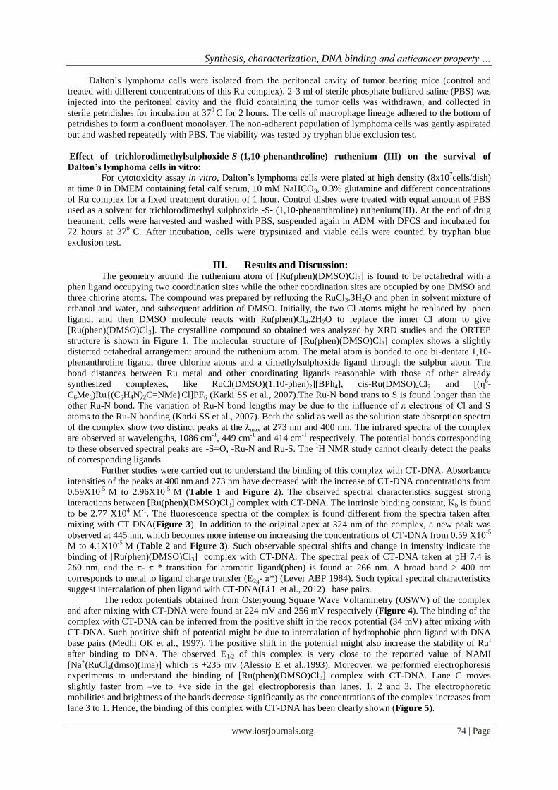
Synthesis characterization DNA binding and anticancer property hellip
wwwiosrjournalsorg 74 | Page
Daltonrsquos lymphoma cells were isolated from the peritoneal cavity of tumor bearing mice (control and
treated with different concentrations of this Ru complex) 2-3 ml of sterile phosphate buffered saline (PBS) was
injected into the peritoneal cavity and the fluid containing the tumor cells was withdrawn and collected in
sterile petridishes for incubation at 370
C for 2 hours The cells of macrophage lineage adhered to the bottom of
petridishes to form a confluent monolayer The non-adherent population of lymphoma cells was gently aspirated
out and washed repeatedly with PBS The viability was tested by tryphan blue exclusion test
Effect of trichlorodimethylsulphoxide-S-(110-phenanthroline) ruthenium (III) on the survival of
Daltonrsquos lymphoma cells in vitro
For cytotoxicity assay in vitro Daltonrsquos lymphoma cells were plated at high density (8x10⁷cellsdish)
at time 0 in DMEM containing fetal calf serum 10 mM NaHCO3 03 glutamine and different concentrations
of Ru complex for a fixed treatment duration of 1 hour Control dishes were treated with equal amount of PBS
used as a solvent for trichlorodimethyl sulphoxide -S- (110-phenanthroline) ruthenium(III) At the end of drug
treatment cells were harvested and washed with PBS suspended again in ADM with DFCS and incubated for
72 hours at 370
C After incubation cells were trypsinized and viable cells were counted by tryphan blue
exclusion test
III Results and Discussion The geometry around the ruthenium atom of [Ru(phen)(DMSO)Cl3] is found to be octahedral with a
phen ligand occupying two coordination sites while the other coordination sites are occupied by one DMSO and
three chlorine atoms The compound was prepared by refluxing the RuCl33H2O and phen in solvent mixture of
ethanol and water and subsequent addition of DMSO Initially the two Cl atoms might be replaced by phen
ligand and then DMSO molecule reacts with Ru(phen)Cl42H2O to replace the inner Cl atom to give
[Ru(phen)(DMSO)Cl3] The crystalline compound so obtained was analyzed by XRD studies and the ORTEP
structure is shown in Figure 1 The molecular structure of [Ru(phen)(DMSO)Cl3] complex shows a slightly
distorted octahedral arrangement around the ruthenium atom The metal atom is bonded to one bi-dentate 110-
phenanthroline ligand three chlorine atoms and a dimethylsulphoxide ligand through the sulphur atom The
bond distances between Ru metal and other coordinating ligands reasonable with those of other already
synthesized complexes like RuCl(DMSO)(110-phen)2][BPh4] cis-Ru(DMSO)4Cl2 and [(η6-
C6Me6)Ru(C5H4N)2C=NMeCl]PF6 (Karki SS et al 2007)The Ru-N bond trans to S is found longer than the
other Ru-N bond The variation of Ru-N bond lengths may be due to the influence of π electrons of Cl and S
atoms to the Ru-N bonding (Karki SS et al 2007) Both the solid as well as the solution state absorption spectra
of the complex show two distinct peaks at the λmax at 273 nm and 400 nm The infrared spectra of the complex
are observed at wavelengths 1086 cm-1
449 cm-1
and 414 cm-1
respectively The potential bonds corresponding
to these observed spectral peaks are -S=O -Ru-N and Ru-S The 1H NMR study cannot clearly detect the peaks
of corresponding ligands
Further studies were carried out to understand the binding of this complex with CT-DNA Absorbance
intensities of the peaks at 400 nm and 273 nm have decreased with the increase of CT-DNA concentrations from
059X10-5
M to 296X10-5
M (Table 1 and Figure 2) The observed spectral characteristics suggest strong
interactions between [Ru(phen)(DMSO)Cl3] complex with CT-DNA The intrinsic binding constant Kb is found
to be 277 X104 M
-1 The fluorescence spectra of the complex is found different from the spectra taken after
mixing with CT DNA(Figure 3) In addition to the original apex at 324 nm of the complex a new peak was
observed at 445 nm which becomes more intense on increasing the concentrations of CT-DNA from 059 X10-5
M to 41X10-5
M (Table 2 and Figure 3) Such observable spectral shifts and change in intensity indicate the
binding of [Ru(phen)(DMSO)Cl3] complex with CT-DNA The spectral peak of CT-DNA taken at pH 74 is
260 nm and the π- π transition for aromatic ligand(phen) is found at 266 nm A broad band gt 400 nm
corresponds to metal to ligand charge transfer (E2g- π) (Lever ABP 1984) Such typical spectral characteristics
suggest intercalation of phen ligand with CT-DNA(Li L et al 2012) base pairs
The redox potentials obtained from Osteryoung Square Wave Voltammetry (OSWV) of the complex
and after mixing with CT-DNA were found at 224 mV and 256 mV respectively (Figure 4) The binding of the
complex with CT-DNA can be inferred from the positive shift in the redox potential (34 mV) after mixing with
CT-DNA Such positive shift of potential might be due to intercalation of hydrophobic phen ligand with DNA
base pairs (Medhi OK et al 1997) The positive shift in the potential might also increase the stability of RuI
after binding to DNA The observed E12 of this complex is very close to the reported value of NAMI
[Na+(RuCl4(dmso)(Ima)] which is +235 mv (Alessio E et al1993) Moreover we performed electrophoresis
experiments to understand the binding of [Ru(phen)(DMSO)Cl3] complex with CT-DNA Lane C moves
slightly faster from ndashve to +ve side in the gel electrophoresis than lanes 1 2 and 3 The electrophoretic
mobilities and brightness of the bands decrease significantly as the concentrations of the complex increases from
lane 3 to 1 Hence the binding of this complex with CT-DNA has been clearly shown (Figure 5)
Synthesis characterization DNA binding and anticancer property hellip
wwwiosrjournalsorg 75 | Page
Electrophoresis of CT-DNA (5 mM) revealed a more intense band but when a higher concentrations of CT-
DNA (6 mM) was mixed with same amount of Ru complex a very weak band was observed (Figure 5) This
suggests the formation of DNA adduct with Ru complex Further as the concentration of CT-DNA was
increased from 2-6 mM and mixed with same amount of Ru complex the sharpness of DNA band was found to
decrease indicating direct correlation between the amounts of DNA and adduct formation
As we know that the metallic drugs cisplatin is an important class of chemotherapeutic agent widely
used in cancer chemotherapy but its clinical use is limited due to nephrotoxicity (Kim YK et al 1995 and
Wolfgang GHI et al 1994) Among the transition metal compounds Ruthenium appears to be a likely
candidate in near future even though its chemistry differs from that of platinum The most significant differences
are its octahedral geometry and greater propensity to undergo redox reactions The hypoxic environment of
many tumors may favor the reduction of ruthenium (III) compounds to ruthenium (II) species (Pegg D G et al
1994 and Clarke MJ et al1988) The hydrophobicity of phenanthroline ligand is responsible for DNA binding
ability and for possible anticancer activity of this complex (Chen T et al 2012)
In the present studies a newly synthesized ruthenium compound trichlorodimethylsulphoxide-S- (110-
phenanthroline) ruthenium(III) was tested against murinersquos Daltonrsquos lymphoma Percentage survival of mice
bearing tumor after treatment with this compound is shown in Figure 6 A direct correlation was observed
between the dose of compound administered and increase in the survival of tumor bearing mice All the
control animals without any treatment failed to survive beyond 10 days post tumor transplantation When the
tumor bearing mice were treated with 20 mgkg of compound 100 of animals survived till day 18 post tumor
transplant and the maximum survival period was found to be 28 days When the dose of ruthenium compound
was increased to 30 and 40 mgkg the maximum survival time increased to 30-32 days When the concentration
was further increased to 50 mgkg the maximum survival period increased to 45 days (Figure 6) The viability
of tumor cells was found to depend on the concentrations of ruthenium compound injected in mice Increasing
concentrations of Ruthenium compound had adverse effect on viability of tumor cells Almost 90 of viability
was observed with 10-20 mgkg of compound However 30-40 mgkg dose has shown 70-80 viable cells The
LC50 was found to be 50 mgkg (Figure 7)
Present study has also shown the effects of Ruthenium compound at the cellular level through a study
of its lethal activity in an in vitro system Results obtained in the present study with Daltonrsquos lymphoma cells
are expressions of cell killing assay and not the growth inhibition assay It was found that cytotoxicity of
ruthenium compound depends upon its concentration in the culture media Survival of Daltonrsquos lymphoma cells
treated in vitro with different concentrations of ruthenium complex for a fixed time duration of 1h is shown in
Figure 8 There exists an inverse linear correlation between the concentration of ruthenium in the medium and
survival of lymphoma cells IC50 of ruthenium was found to be 40 μgml (Figure 8)
Most of the investigations reporting ruthenium compounds as anti-tumor agents have been carried out
as in vitro studies and only a few are in vivo studies Ruthenium- DMSO complexes have been reported to
increase the life span of tumor bearing mice in vivo (Pieper T and Keppler BK 1998) These compounds are
also effective against several murine models including a cisplatin resistant P 388 leukemia cell line While a
number of Ruthenium compounds have been tested the prototype compound RAPTA-C has been reported to be
most effective anti-cancer compound of this series and has been used extensively against a number of murine
cell lines Chatterjee S et al studied the effect of RAPTA-C induced apoptosis in EAC cells isolated from
peritoneal cavity of tumor bearing mice and found that the apoptosis was dose dependent (Chatterjee S et al
2008) In the present studies the effect of compound has also been found to be dose dependent
In a similar study flow cytometry analysis of RAPTA-C induced cell cycle phase distribution of nuclear DNA
has shown increased content of haploid DNA in RAPTA-C treated cells (Chatterjee S et al 2008) However
the DNA in S-phases decreased from 63 to 36 suggesting that tumor suppressor gene P53
is central to the
induction of cell cycle arrest and apoptosis in response to DNA damage of cellular stage in human cells Earlier
it has been reported that ruthenium (III) compounds having significant cytotoxic properties are able to bind
firmly to DNA and modify its structural conformation (Brabec V et al1995) So the anticancer activity of the
present compound may be related to its DNA binding ability
IV Conclusion The synthesis of ruthenium complex its CT-DNA binding and biological studies have been performed
The structure analyzed by single crystal XRD study shows six-coordinated octahedral geometry The crystal
structure is a representative complex [Ru(phen)(DMSO)Cl3] and the structural features are in accord with
already reported ruthenium complexes The CT-DNA binding of this complex is evidenced by various
experimental techniques On the basis of spectroscopic shift the intrinsic binding constant (Kb) is found to be
277X104
M-1
Based on the observed distinct shift of electrochemical E12 and electrophoresis band shifts the
complex is expected to bind with CT-DNA The hydrophobicity of the phen ligand may be responsible for DNA
binding and possibly related to the anticancer activity of ruthenium complex
Synthesis characterization DNA binding and anticancer property hellip
wwwiosrjournalsorg 76 | Page
This compound acquires good antitumor property against Daltonrsquos lymphoma both in vivo and in vitro
The compound exhibited dose dependent increase in the survival time of tumor bearing mice however no
tumor free survivor was observed From the biological studies in vivo the highest dose of this complex is 50
mgkg Electrophoretic analysis of CT-DNA with the complex suggests the formation of DNA adduct which
might be responsible for the antitumor activity of this compound
References [1] Rosenberg B (1985) Fundamental studies with cisplatin Cancer 55 2303-2316 [2] Pil P Lippard S J Cisplatin and related drugs Bertino JR (1997) Encyclopedia of cancer Academic Press Inc San Diego 1
392-410
[3] Dabholkar M and Reed E (eds Pinedo H M Longo D L Chabner B A) (1996) Elsevier Science Publishers Netherlands 88-110
[4] Shimada H Sugimachi N Funakoshi T and Kojima S (1993) Prevention of renal toxicity of cis-diamminedichloroplatinum by
dithiocarbamates in rats Toxicol Lett 66193-198
[5] Jakupec M A Reisner E Eichinger A Pongratz M Arion V B Galanski M Hartinger C Keppler B K (2005) Redox-Active
Redox-Active Antineoplastic Ruthenium Complexes with Indazole Correlation of in Vitro Potency and Reduction Potential J Med
Chem 48 2831-2837 [6] Schluga P Hartinger G Egger A Reisner E Galanski M Jakupee MA and Keppler BK (2006) Redox behavior of tumor-
inhibiting ruthenium (III) complexes and effects of physiological reductants on their binding to GMP Dalton Trans 1796-1802
[7] Kostova I (2006) Ruthenium complexes as anticancer agents Curr Med Chem 131085-107 [8] Clarke M J (2003) Ruthenium Metallopharmaceuticals Coord Chem Rev 236 207-231
[9] Bratsos I Jedner S Gianferrara T Alessio E (2007) Ruthenium anticancer compounds Challenges and Expectations Chimia 61
692-697 [10] Rosenberg B Camp LV Krigas T(1965) Inhibition of Cell Division in Escherichia coli by Electrolysis Products from a Platinum
Electrode Nature 205 698 ndash 699
[11] Clarke MJ Zhu F Frasca DR (1999) Non-Platinum Chemotherapeutic MetallopharmaceuticalsChem Rev 99 2511-2533 [12] Vessieres A Top S Beck W Hillard E Jaouen G (2006) Metal complex SERMs (selective oestrogen receptor modulators) The
influence of different metal units on breast cancer cell antiproliferative effects Dalton Trans 529 529-541
[13] Morris RE Aird R E Mudroch P S Chen H Cummins J Hughes ND Parsons S Parkin A Boyd G Jodrell DI Sadler PJ (2001) Inhibition of Cancer Cell Growth by Ruthenium(II) Arene Complexes J Med Chem44 3616-3621
[14] Hong-Ke Liu Sadler PJ (2011) Metal Complexes as DNA Intercalators Acc Chem Res 44 349-359
[15] Finlay GJ Baguley BC (2000) Effects of protein binding on the in vitro activity of antitumour acridine derivatives and related anticancer drugs Cancer Chemother Pharmacol45 417-22
[16] Thota S Imran M Udugula M Karki S S Kanjarla N Yerra R Balzarini J and Clercq E D (2012) Synthesis spectroscopic
characterization antineoplastic in vitro-cytotoxic and antibacterial activities of mononuclear ruthenium(II) complexes J Coord Chem 65 823ndash839
[17] Clarke MJ and Stubbs M(1996) Interactions of Metallopharmaceuticals with DNA Metal Ions in Biological Systems 32 727-780
[18] Li-Feng Tan Hui Chao Yun-Jun Liu Hong Li Bin Sun Liang-Nian Ji (2005) DNA-binding and photocleavage studies of [Ru(phen)2(NMIP)]2+ Inorganica Chimica Acta 358 2191-2198
[19] Anghileri L J Krebsforsch Z Onkol K( 1975) The in vivo inhibition of tumor growth by ruthenium red its relationship with the
metabolism of calcium in the tumor Cancer Res Clin Oncol 83 213 [20] Allardyce CS Dorcier A Scolaro C Dyson PJ (2005) Development of organometallic (organo-transition metal) pharmaceuticals
Appl Organomet Chem 19 1-10
[21] Paul H Mukherjee T Drew MGB and Chattopadhyay P (2012) Synthesis characterization crystal structure and DNA-binding of ruthenium(II) complexes of heterocyclic nitrogen ligands resulting from a benzimidazole-based quinazoline derivative J Coord
Chem 65 1289-1302
[22] Wang F Chen H Parsons S Oswald IDH Davidson JE Sadler PJ (2003) Kinetics of Aquation and Anation of Ruthenium(ii) Arene Anticancer Complexes Acidity and X-ray Structures of Aqua Adducts Chem Eur J 9 5810-5820
[23] Elisacircngel de Paula Silveira-Lacerda Cesar Augusto Sam Tiago Vilanova-Costa Pereira FC Hamaguchi A Pavanin LA Goular LR Homsi-Brandenburgo MI Martins SA Santos W B and Nomizo A (2010) The Ruthenium Complex cis-(Dichloro)
Tetraammineruthenium(III) Chloride Presents Immune Stimulatory Activity on Human Peripheral Blood Mononuclear Cells Biol
Trace Elem Res 133 270ndash283 [24] Karki SS Thota S Darj S Y Balzarinib J and Clercqb E D Bioorg (2007) Synthesis anticancer and cytotoxic activities of some
mononuclear Ru(II) compounds Med Chem 15 6632ndash6641
[25] Tysoe SA Morgan R J Baker D (1993) Spectroscopic Investigation of Differential Binding Modes of Delta and Lambda-[Ru(bpy)2ppz]2+ with Calf Thymus DNA J Phys Chem 97 1707-1711
[26] Liu J Zheng W Shi S Tan C Chen J Zheng K Ji L (2008) Synthesis antitumor activity and structurendashactivity relationships of a series of Ru(II) complexes J Inorg Biochem 102 193-202
[27] Murali S Sastri CV Maiya BG (2002) New mixed ligand complexes of ruthenium(II) that incorporate a modified phenanthroline
ligand Synthesis spectral characterization and DNA binding Proc Indian Acad Sci (Chem Sci)114 403- 415 [28] Sun S Yang Y Liu F Fan J Peng X Kehr J Sun L (2009) Intra- and intermolecular interaction ECL study of novel ruthenium
tris-bipyridyl complexes with different amine reductants Dalton Trans 38 7969-7974
[29] Yong-guang Y Du L Yu X Yan-hui Z Xue-yun Z Jie L (2011) Development of NAMI-A-loaded PLGA-mPEG Nanoparticles Physicochemical Characterization in vitro Drug Release and in vivo Antitumor Efficacy Chem Res Chinese Universities 27
345-349
[30] D P Rillema K B Mack (1982) The Low lying Excited State in Ligand-Acceptor Complexes of Ruthenium(II) Mononuclear and Binuclear Species Inorg Chem 21 3849-3854
[31] Hartinge CG Jakupec MA Seifried S Z Groessl M Egger A Berger W Zorbas H Dyson PJ Keppler BK (2008) KP1019 a New
Redox-Active Anticancer Agent-Preclinic Development and Results of a Clinical Phase I Study in Tumor Patients Chemistry amp
Biodiversity (Review) 5 2140-2155
[32] Galanski M Arion VB Jakupec MA Keppler BK (2003) Recent Developments in the Field of Tumor-Inhibiting Metal
Complexes Curr Pharm Des 9 2078-2089
Synthesis characterization DNA binding and anticancer property hellip
wwwiosrjournalsorg 77 | Page
[33] Sava G Bergamo A Zorzet S Gava B Casarsa C Cocchietto M Furlani A Scarcia V Serli BIengo E Alessio E Mestroni G
(2002) Influence of chemical stability on the activity of the antimetastasis ruthenium compound NAMI-A Eur J Cancer 38 427-
435 [34] Arandjelovic SS Bjelogrlic KS Malesevic NN Tesic LjZ Radulovic SS (2009) Antitumor activity of Ru(III) complexes carrying
beta-diketonato ligands in vitro and in vivo J Boun 14 271-279
[35] Krause RA (1977) Synthesis of mixed complexes of ruthenium(II) with 22prime-dipyridyl Inorganica Chimica Acta 22 209-213 [36] Barton JK Raphael AL (1984) Photoactivated Stereospecific Cleavage of Double-Helical DNA by Cobalt(II1) Complexes J Am
Chem Soc 106 2466-2468
[37] (a) Sheldrick GM (1996) SADABS University of Goumlttengen Germany (b) Sheldrick GM (1997) A program for automatic solution of crystal structure University of Goumlttengen Germany c) Sheldrick GM (1997) A program for crystal structure
refinement University of Goumlttengen Germany
[38] Shahabadi N Mohammadi S Alizadeh R (2011) DNA Interaction Studies of a New Platinum(II) Complex Containing Different Aromatic Dinitrogen Ligands Bioinorganic Chemistry and Application 20111-8
[39] [39] Rathinasamy S Karki SS Bhattacharya S Manikandan L Prabakaran S G Gupta M Mazumder U K (2006) Synthesis and
anticancer activity of certain mononuclear Ru(II) complexes J Enzyme Inhibition and Medicinal Chemistry 21 501-507
[40] Lever ABP (1984) Inorganic Electronic Spectroscopy 2nd Edition Elsevier Newyork
[41] Li L Cao W Zheng W Fan C and Chen T (2012) Dalton Trans 4112766-12772
[42] Das D K Bhattaray C and Medhi OK(1997) Electrochemical behaviour of (protoporphyrinato IX) iron(III) encapsulated in aqueous surfactant micellesDalton Trans 4713-4718
[43] Alessio E Balducci G Lutman A Mestroni G Calligaris M and Attia WM (1993) Synthesis and characterization of two new
classes of ruthenium(III)-sulfoxide complexes with nitrogen donor ligands Inorg Chim Acta 203 205-217 [44] Kim YK Byun H S Kim Y H Woo JS and Lee SH (1995) Effect of cisplatin on renal function in rabbits mechanism of reduced
glucose reabsorption Toxicol Appl Pharmacol 130 19-26
[45] Wolfgang GH I Dominick MA Walsh KM Hoeschele J D and Pegg D G (1994) Comparative Nephrotoxicity of a Novel Platinum Compound Cisplatin and Carboplatin in Male Wistar Rats Fundamental Appl Toxicol 22 73-79
[46] Clarke MJ (1988) Ruthenium in Cancer Chemotherapy Platinum Metals Rev 32 198-199
[47] Brabec V Novakova O (2006) DNA binding mode of ruthenium complexes and relationship to tumor cell toxicity Drug Resistance Updates 9111-122
[48] Pieper T and Keppler BK (1998) Tumor-inhibiting ruthenium complexes formulation and analytical characterization Analusis
26 84-90 [49] Chatterjee S Subhadip K Bhattacharyya A Hartinger G C and Dyson PJ (2008) The ruthenium(II)ndasharene compound RAPTA-C
induces apoptosis in EAC cells through mitochondrial and p53ndashJNK pathways J Biol Inorg Chem 13 1149-1155
[50] Novakova O Kasparkova O Vrano O Vanviet PM Reedijk J and Brabec V (1995) Correlation between Cytotoxicity and DNA
Binding of Polypyridyl Ruthenium Complexes Olga Biochemistry 34 12369-12378
Acknowledgements The authors thank the Department of Science and Technology (DST) New Delhi for financial support
and SAIF Gauhati University for collection Single crystal X-ray intensity data
Table 1 The wavelengths and absorbances of the complex with and without mixing CT-DNA at different concentrations
Samples Wavelengths (nm) Absorbances
λmax (1) λmax (2) 1 2
DNA 260 0061 b Complex 273 400 2034 0367
c Complex +059 X10-5M DNA 273 400 1977 0333
d Complex +110 X10-5M DNA 273 400 1908 0327 e Complex +178 X10-5M DNA 273 400 1884 0322
f Complex +237 X10-5M DNA 273 400 1869 0319
g Complex +296 X10-5M DNA 273 400 1844 0315
Table 2 The emission peaks and fluorescence intensities of the complex with and without mixing CT-DNA at different concentrations
Samples Apex (nm) Intensities(1)
a DNA 563 3380
b Complex 324 7647
c Complex +059X10-5M DNA 324 11840
445 3369
d Complex +178X10-5M DNA 324 12490
445 3766
e Complex +237X10-5M DNA 324 13790
445 4612
f Complex + 35X10-5M DNA 324 14330
445 5171 g Complex + 41X10-5M DNA 324 16370
445 5871
Synthesis characterization DNA binding and anticancer property hellip
wwwiosrjournalsorg 78 | Page
Figure1 ORTEP molecular structure of the Ru(phen)(DMSO)Cl3 complex Selected distancesAring and bond
anglesordm
Ru1-N1=2082(2) Ru1-N2=2088(2) Ru1-S1=2320(4) Ru1-Cl1=2336(2) Ru1-Cl2=2347(1) Ru1-
Cl3=2346(1) S1-O1=1393(4) S1-C13=1784(4) S1-C14=1770(4) N1-Ru1-N2=7922(10) Cl1-Ru1-
N2=8494(7) Cl2-Ru1-N2=9223(7) Cl3-Ru1- N2=9571(8) Cl1-Ru1-N1=8748(7) Cl2-Ru1-N1=8731(7)
Cl3-Ru1-N1=17489(4) Cl1-Ru1-Cl2=17445(3) Cl3-Ru1-Cl1=9279(3) Cl3-Ru1-Cl2=9224(3) S1-Ru1-
N1=9792(6) S1-Ru1-Cl1=9131(3) S1-Ru1-Cl2=9126(3) S1-Ru1 Cl3=8738(3) Ru1-S1-O1=11940(1) O1-
S1-C13=10250(3) O1-S1-C14=11160(3) C13-S1- C14=9927(4)
(A) (B)
(C) (D)
Figure 2 (A) UV-visible spectra of the 5mM complex with and without mixing CT-DNA at different
concentrations and (A) plot of εbndashεf εandashεf against reciprocal concentration of CT-DNA (1[DNA])(C) and (D)
are the UV-visible spectra of the complex in DMSO solvent and solid state The maximum absorption peaks
03
04
05
06
07
257 307 357 407
Wavelength (nm)
Ab
so
rba
nc
e
00
05
10
15
20
25
257 307 357 407
Wavelength (nm)
Ab
so
rba
nc
e
Synthesis characterization DNA binding and anticancer property hellip
wwwiosrjournalsorg 79 | Page
(λmax) for the both state are obtained around at 273 nm and 400 nm which may indicates the complex has same
structure in the solution as well as solid state
Figure 3 Fluorescence intensity of the 5mM complex with and without mixing CT-DNA at different
concentrations along with free CT-DNA
(A) (B)
(C) (D)
Figure 4 (A) Cyclic voltammogram of 5 mM complex at different scan rate (50 mVS to 250 mVS) (B)
Cyclic voltammogram of 5 mM the complex (a) and after mixing with 5mM CT-DNA (b) (C) and (D) are the
square wave voltammogram for the 5 mM complex (E12=222 mV) and the complex in presence of 5mM CT-
DNA (E12=256 mV) respectively The redox potential shows 34 mV positive shift on addition of CT-DNA
Figure 5 Gel electrophoresis of trichlorodimethylsulphoxide-S-(110-phenanthroline) ruthenium (III) complex
with CT-DNA (5 mM) studied in gel electrophoresis in tris-HCl buffer at pH 76 (concentration taken are 2
mM 3 mM 6 mM as indicated lane 32 1)
Synthesis characterization DNA binding and anticancer property hellip
wwwiosrjournalsorg 80 | Page
Figure 6 Effect of this Ru complex on mice bearing Daltonrsquos lymphoma No tumor bearing mice became tumor
free after treatment 50 mgkg of the complex Rest of the animals also (plt 005) increase in their life span
compared to control (compound is dissolved in PBS pH 74)
Figure 7 Effect of the complex on the survival of Daltonrsquos lymphoma cells in vivo The results are an average
of five (n=5) independent experiments in triplicate and represented as mean plusmn SE p lt 005 vs control
Figure 8 The results are an average of five (n=5) independent experiments in triplicate and represented as mean
plusmn SE p lt 005 vs control
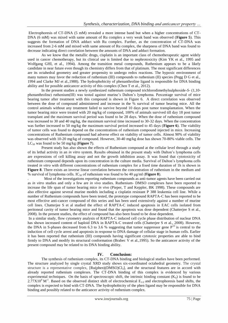
Synthesis characterization DNA binding and anticancer property hellip
wwwiosrjournalsorg 78 | Page
Figure1 ORTEP molecular structure of the Ru(phen)(DMSO)Cl3 complex Selected distancesAring and bond
anglesordm
Ru1-N1=2082(2) Ru1-N2=2088(2) Ru1-S1=2320(4) Ru1-Cl1=2336(2) Ru1-Cl2=2347(1) Ru1-
Cl3=2346(1) S1-O1=1393(4) S1-C13=1784(4) S1-C14=1770(4) N1-Ru1-N2=7922(10) Cl1-Ru1-
N2=8494(7) Cl2-Ru1-N2=9223(7) Cl3-Ru1- N2=9571(8) Cl1-Ru1-N1=8748(7) Cl2-Ru1-N1=8731(7)
Cl3-Ru1-N1=17489(4) Cl1-Ru1-Cl2=17445(3) Cl3-Ru1-Cl1=9279(3) Cl3-Ru1-Cl2=9224(3) S1-Ru1-
N1=9792(6) S1-Ru1-Cl1=9131(3) S1-Ru1-Cl2=9126(3) S1-Ru1 Cl3=8738(3) Ru1-S1-O1=11940(1) O1-
S1-C13=10250(3) O1-S1-C14=11160(3) C13-S1- C14=9927(4)
(A) (B)
(C) (D)
Figure 2 (A) UV-visible spectra of the 5mM complex with and without mixing CT-DNA at different
concentrations and (A) plot of εbndashεf εandashεf against reciprocal concentration of CT-DNA (1[DNA])(C) and (D)
are the UV-visible spectra of the complex in DMSO solvent and solid state The maximum absorption peaks
03
04
05
06
07
257 307 357 407
Wavelength (nm)
Ab
so
rba
nc
e
00
05
10
15
20
25
257 307 357 407
Wavelength (nm)
Ab
so
rba
nc
e
Synthesis characterization DNA binding and anticancer property hellip
wwwiosrjournalsorg 79 | Page
(λmax) for the both state are obtained around at 273 nm and 400 nm which may indicates the complex has same
structure in the solution as well as solid state
Figure 3 Fluorescence intensity of the 5mM complex with and without mixing CT-DNA at different
concentrations along with free CT-DNA
(A) (B)
(C) (D)
Figure 4 (A) Cyclic voltammogram of 5 mM complex at different scan rate (50 mVS to 250 mVS) (B)
Cyclic voltammogram of 5 mM the complex (a) and after mixing with 5mM CT-DNA (b) (C) and (D) are the
square wave voltammogram for the 5 mM complex (E12=222 mV) and the complex in presence of 5mM CT-
DNA (E12=256 mV) respectively The redox potential shows 34 mV positive shift on addition of CT-DNA
Figure 5 Gel electrophoresis of trichlorodimethylsulphoxide-S-(110-phenanthroline) ruthenium (III) complex
with CT-DNA (5 mM) studied in gel electrophoresis in tris-HCl buffer at pH 76 (concentration taken are 2
mM 3 mM 6 mM as indicated lane 32 1)
Synthesis characterization DNA binding and anticancer property hellip
wwwiosrjournalsorg 80 | Page
Figure 6 Effect of this Ru complex on mice bearing Daltonrsquos lymphoma No tumor bearing mice became tumor
free after treatment 50 mgkg of the complex Rest of the animals also (plt 005) increase in their life span
compared to control (compound is dissolved in PBS pH 74)
Figure 7 Effect of the complex on the survival of Daltonrsquos lymphoma cells in vivo The results are an average
of five (n=5) independent experiments in triplicate and represented as mean plusmn SE p lt 005 vs control
Figure 8 The results are an average of five (n=5) independent experiments in triplicate and represented as mean
plusmn SE p lt 005 vs control
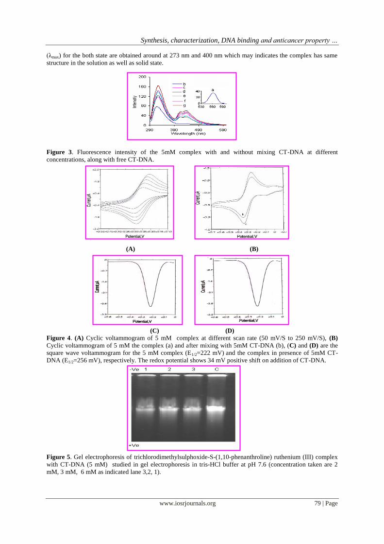
Synthesis characterization DNA binding and anticancer property hellip
wwwiosrjournalsorg 80 | Page
Figure 6 Effect of this Ru complex on mice bearing Daltonrsquos lymphoma No tumor bearing mice became tumor
free after treatment 50 mgkg of the complex Rest of the animals also (plt 005) increase in their life span
compared to control (compound is dissolved in PBS pH 74)
Figure 7 Effect of the complex on the survival of Daltonrsquos lymphoma cells in vivo The results are an average
of five (n=5) independent experiments in triplicate and represented as mean plusmn SE p lt 005 vs control
Figure 8 The results are an average of five (n=5) independent experiments in triplicate and represented as mean
plusmn SE p lt 005 vs control